2021 Northwest Open - 1/22/2021 to 1/23/2021 Hughes Fieldhouse ...
Upload
khangminh22Category
view
0download
0
Review Article
Mohamed Abd Elkodous*#, Hussein M. El-Husseiny*#, Gharieb S. El-Sayyad*#,Amr Hosny Hashem*#, Ahmed S. Doghish*#, Dounia Elfadil, Yasmine Radwan,Hayam M. El-Zeiny, Heba Bedair, Osama A. Ikhdair, Hisham Hashim, Ahmed M. Salama,Heba Alshater, Ahmed Ali Ahmed, Mahmoud Gamal Elsayed, Maria Nagy, Nouran Y. Ali,Maryam Elahmady, Ahmed M. Kamel, Mahmoud Abd Elkodous, Imene Maallem,Maria B. Sh. Kaml, Nayera Nasser, Ahmed AlaaEldin Nouh, Fatma M. Safwat, Mai M. Alshal,Salma K. Ahmed, Taha Nagib, Fatma M. El-sayed, Manal Almahdi, Yahia Adla,Noha T. ElNashar, Aya Misbah Hussien, Alaa S. Salih, Somaya Abdulbaset Mahmoud,Shireen Magdy, Diana I. Ahmed, Fayrouz Mohamed Saeed Hassan, Nermin A. Edward,Kirolos Said Milad, Shereen R. Halasa, Mohamed M. Arafa, Abdullah Hegazy, Go Kawamura,Wai Kian Tan, and Atsunori Matsuda*
Recent advances in waste-recyclednanomaterials for biomedical applications:Waste-to-wealthhttps://doi.org/10.1515/ntrev-2021-0099received August 16, 2021; accepted September 27, 2021
Abstract: Global overpopulation, industrial expansion,and urbanization have generated massive amounts of
* Corresponding author: Mohamed Abd Elkodous, Department ofElectrical and Electronic Information Engineering, ToyohashiUniversity of Technology, 1-1 Hibarigaoka, Tempaku-Cho,Toyohashi, Aichi 441-8580, Japan, e-mail: [email protected]* Corresponding author: Hussein M. El-Husseiny, Department ofVeterinary Medicine, Laboratory of Veterinary Surgery, Faculty ofAgriculture, Tokyo University of Agriculture and Technology, 3-5-8Saiwai Cho, Fuchu-shi, Tokyo 183-8509, Japan; Department ofSurgery, Anesthesiology, and Radiology, Faculty of VeterinaryMedicine, Benha University, Moshtohor, Toukh, Elqaliobiya, 13736,Egypt, e-mail: [email protected]* Corresponding author: Gharieb S. El-Sayyad, Drug RadiationResearch Department, Drug Microbiology Lab, National Center forRadiation Research and Technology (NCRRT), Egyptian AtomicEnergy Authority (EAEA), Cairo, Egypt; Chemical EngineeringDepartment, Military Technical College (MTC), Egyptian ArmedForces, Cairo, Egypt, e-mail: [email protected]* Corresponding author: Amr Hosny Hashem, Botany andMicrobiology Department, Faculty of Science, Al-Azhar University,Cairo, 11884, Egypt, e-mail: [email protected]* Corresponding author: Ahmed S. Doghish, Department ofBiochemistry, Faculty of Pharmacy (Boys), Al-Azhar University, NasrCity, Cairo, Egypt; Department of Biochemistry, Faculty of Pharmacy,Badr University in Cairo (BUC), Badr City, Cairo, Egypt,e-mail: [email protected]
# These authors contributed equally to this work.
* Corresponding author: Atsunori Matsuda, Department of Electricaland Electronic Information Engineering, Toyohashi University ofTechnology, 1-1 Hibarigaoka, Tempaku-Cho, Toyohashi, Aichi 441-8580, Japan, e-mail: [email protected], [email protected] Elfadil: Faculty of Sciences and Techniques, Hassan IIUniversity of Casablanca, Mohammedia, MoroccoYasmine Radwan: Center forMaterials Science, Zewail City of Science andTechnology, Giza, 12578, Egypt; NanoscaleScienceProgram,Department ofChemistry, University of North Carolina, Charlotte, NC 28223, USAHayam M. El-Zeiny: Chemistry Department, Faculty of Science,Beni-Suef University, Beni Suef city, EgyptHeba Bedair: Botany Department, Faculty of Science, TantaUniversity, 31527, Tanta, EgyptOsama A. Ikhdair: Faculty of Medicine, Jordan University of Scienceand Technology, Ar-Ramtha, JordanHisham Hashim: Department of Physics, Faculty of Science, TantaUniversity, Tanta, Elgharbiya, 31111, EgyptAhmed M. Salama: State Key Laboratory of Chemical ResourceEngineering, Beijing University of Chemical Technology, P. O. Box98, Beisanhuan East Road 15, Beijing, 100029, ChinaHeba Alshater: Forensic Medicine and Clinical ToxicologyDepartment, Menoufia Hospital University, Shibin el Kom, EgyptAhmed Ali Ahmed: Faculty of Pharmacy, Alexandria University,Alexandria, EgyptMahmoud Gamal Elsayed: Faculty of Biotechnology, MSA University,Giza, EgyptMaria Nagy: Biotechnology/Biomolecular Chemistry Program, Facultyof Science, Cairo University, 1 Gamaa Street, Giza, 12613, EgyptNouran Y. Ali: Faculty of Agriculture, Biotechnology Program, AinShames University, Cairo, EgyptMaryam Elahmady: Faculty of Dentistry, New Giza University, Giza, Egypt
Nanotechnology Reviews 2021; 10: 1662–1739
Open Access. © 2021 Mohamed Abd Elkodous et al., published by De Gruyter. This work is licensed under the Creative Commons Attribution4.0 International License.

wastes. This is considered as a significant worldwide chal-lenge that requires an urgent solution. Additionally,remarkable advances in the field of biomedicine haveimpacted the entire spectrum of healthcare and medicine.This has paved the way for further refining of the outcomesof biomedical strategies toward early detection and treat-ment of different diseases. Various nanomaterials (NMs)have been dedicated to different biomedical applicationsincluding drug delivery, vaccinations, imaging modalities,and biosensors. However, toxicity is still the main factorrestricting their use. NMs recycled from different types ofwastes present a pioneering approach to not only avoidhazardous effects on the environment, but to also imple-ment circular economy practices, which are crucial toattain sustainable growth. Moreover, recycled NMs havebeen utilized as a safe, yet revolutionary alternative withoutstanding potential for many biomedical applications.This review focuses on waste recycled NMs, their synthesis,properties, and their potential for multiple biomedical appli-cations with special emphasis on their role in the earlydetection and control ofmultiple diseases. Their pivotal ther-apeutic actions as antimicrobial, anticancer, antioxidantnanodrugs, and vaccines will also be outlined. The ongoingadvancements in the design of recycled NMs are expandingtheir diagnostic and therapeutic roles for diverse biomedicalapplications in the era of precision medicine.
Keywords: recycled nanomaterials, biomedical applica-tions, antimicrobial activity, anticancer agents, biomasswastes
Graphical abstract
Ahmed M. Kamel: Biotechnology Program, Faculty of Agriculture,Ain Shams University, Cairo, EgyptMahmoud Abd Elkodous: Faculty of Veterinary Medicine, KafrElsheikh University, Kafr Al Sheikh, EgyptImene Maallem: Department of Pharmacy, Faculty of Medicine, BadjiMokhtar University, Zaafrania Road, Annaba, 23000, AlgeriaMaria B. Sh. Kaml: Faculty of Medicine, Helwan university, Cairo,EgyptNayera Nasser: Department of Pharmaceutics and IndustrialPharmacy, Faculty of Pharmacy, Ain Shams University, Cairo, EgyptAhmed AlaaEldin Nouh: Department Zoology, Faculty of Science,Alexandria University, Alexandria, EgyptFatma M. Safwat: Faculty of Pharmacy, Deraya University,New Minya, EgyptMai M. Alshal: Faculty of Physical Therapy, October 6th University,6 October City, Giza, 12585, EgyptSalma K. Ahmed: Department of Biotechnology and BiomolecularChemistry, Faculty of Science, Cairo University, Cairo, EgyptTaha Nagib: Faculty of Medicine, University of Tripoli, Tripoli, LibyaFatma M. El-sayed: Biotechnology Program, Faculty of Agriculture,Ain Shams University, Cairo, EgyptManal Almahdi: Al-Neelain University, Khartoum, SudanYahia Adla: Department of Biology, College of Arts and Science,Stetson University, 421N. Woodland Blvd, Deland, Florida, 32723,United States of AmericaNoha T. ElNashar: Department of Pharmaceutical Technology, TheGerman University in Cairo (GUC), Cairo, Egypt
Aya Misbah Hussien: Biotechnology Department, Institute of GraduateStudies and Research, Alexandria University, Alexandria, EgyptAlaa S. Salih: Department of Human Medicine, Faculty of Medicine &Health Sciences, An-Najah National University, Nablus, PalestineSomaya Abdulbaset Mahmoud: Faculty of Pharmacy, October 6thUniversity, Giza, EgyptShireen Magdy: Biomedical Sciences Program, University of Science andTechnology, Zewail City of Science and Technology, Giza, 12578, EgyptDiana I. Ahmed: Department of Microbiology and Chemistry, Facultyof Science, Aswan University, Aswan, EgyptFayrouz Mohamed Saeed Hassan: Information Technology andComputer Science, Nile University, Giza, EgyptNermin A. Edward: Department of Biochemistry, Faculty of Science,Ain Shams University, Cairo, EgyptKirolos Said Milad: Department of NanoScience, Faculty of Science,Zewail City of Science and Technology, Giza, 12578, EgyptShereen R. Halasa: Faculty of Nursing, Al-Quds University, Al-Quds,PalestineMohamed M. Arafa: Department of Chemistry, Faculty of Science,Tanta University, Tanta, EgyptAbdullah Hegazy: Faculty of Science, University of Science andTechnology, Zewail City, Cairo, EgyptGo Kawamura: Department of Electrical and Electronic InformationEngineering, Toyohashi University of Technology, 1-1 Hibarigaoka,Tempaku-Cho, Toyohashi, Aichi 441-8580, JapanWai Kian Tan: Institute of Liberal Arts and Sciences, ToyohashiUniversity of Technology, 1-1 Hibarigaoka, Tempaku-Cho,Toyohashi, Aichi, 441-8580, Japan
Recent advances in waste-recycled NMs for biomedical applications 1663

Abbreviations
AC activated carbonAnti-EGFR anti-epidermal growth factor receptorBBB blood–brain barrierCT computed tomographyCAs contrast agentsCDs carbon dotsCG chitosan-doped grapheneCNCs cellulose nanocrystalsCNFs carbon nanofibersCQDs carbon quantum dotsCNMs carbon nanomaterialsCNTs carbon nanotubesDCs dendritic cellsAu NPs gold nanoparticlesGO graphene oxideGQDs graphene quantum dotsIO-CDs iron oxide nanoparticle-doped
carbon dotsIQDs inorganic quantum dotsIO NPs iron oxide nanoparticlesMOs metal oxidesMNPs magnetic nanoparticlesMPI magnetic particle imagingMRI magnetic resonance imagingMWCNTs multi-walled carbon nanotubesMIC minimum inhibitory concentrationNMs nanomaterialsNGO nanographene oxidePEG polyethylene glycolPEG-AuIONPs PEG-coated iron-oxide–gold core–shell
nanoparticlesQDs quantum dotsROS reactive oxygen speciesRNMs recycled nanomaterialsrGO reduced graphene oxideSPIO NPs superparamagnetic iron oxide
nanoparticlesSWCNTs single-walled carbon nanotubesWCO waste cooking oilZnO NPs zinc oxide nanoparticles
1 Introduction
Since the emergence of the term nanotechnology, mucheffort has been dedicated to the investigation of the out-standing properties of nanomaterials (NMs) and their
potential applications [1]. Materials having at least onedimension on the nanoscale (<100 nm) are identified asNMs [2]. NMs are quite fascinating and have wide appli-cations in different fields such as biology, medicine,industry, energy, and so on [3–7].
Among all the possible applications of NMs, theirbiomedical applications are of particular interest andhave led to the maturation of nanomedicine [8–21]. Var-ious kinds of NMs have been prepared and their applic-ability has been extensively studied. NMs have shown thepotential to overcome many severe drawbacks comparedto other pharmacologically active agents and chemicals,including biological instability, lack of water solubility,and ineffectiveness in vivo [22]. Nowadays, NMs are usedin many biomedical applications, such as drug deliverysystems, treatment of diseases, early disease diagnosis,vaccines, biosensors, and bioimaging [23,24]. For the pre-paration of NMs, many methods have been developed, asshown in Figure 1.
However, there are concerns about the toxicity of NMs[25,26]. Thus, other more benign routes for the preparationof biocompatible NMs are important due to their interac-tion with the biological environment. Sustainability, refer-ring to the production of NMs from everyday domesticand industrial wastes, can be a revolutionary solutionand a route to achieve waste-to-wealth and zero-wasteinitiatives.
Many valuable NMs, such as carbon NMs (CNMs), goldnanoparticles (Au NPs), inorganic quantum dots (IQDs),and metal oxides (MOs) can be extracted from waste mate-rials that include cooking oil, biomass, and industrialwastes. Interestingly, agricultural biomass wastes can bea valuable source for the green synthesis of NMs posses-sing many attractive properties such as lower toxicity,cost-effectiveness, tiny size, and higher stability [27].These NMs have found many uses in biomedical appli-cations [28–34].
More than 6 million tons of agricultural biomass isproduced worldwide every year. Of this, 10% is producedin Europe (600,000 tons per year) [35]. Every year, hugeamounts of waste cooking oils (WCOs) are producedworldwide, especially in developed countries [36]. Inthe USA, the daily estimation of WCO by the Energy Infor-mation Administration is 100 million gallons and theaverage per capita amount of WCO is reported to benine pounds. The total WCO produced yearly in Canadais reaching approximately 135,000 tons [37]. In EuropeanUnion countries, the amount of yearly WCO ranges from750,000 to one million tons [38] and 200,000 tons are pro-duced annually in the United Kingdom. Moreover, electronic
1664 Mohamed Abd Elkodous et al.

waste (e-waste) has become a rapidly growing global con-cern. In European countries, 17 kg of e-waste per capita isproduced yearly. In China and India, 1 kg of e-waste percapita is generated annually and includes Cu, Al, and ironmetals [39]. According to the US Environmental ProtectionAgency, 4.6 million tons of e-waste entered US landfills in2000 and that amount is rapidly increasing and expected togrow four-fold in coming years [40].
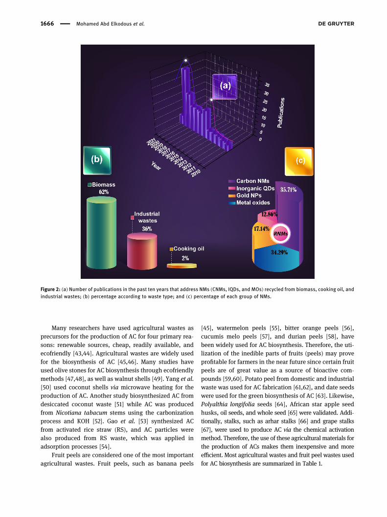
Within the past few years, a circular economy hasbecome an important academic research domain. A sig-nificant increase in the number of articles addressing thistopic has been published, as shown in Figure 2.
Further investigation on the progress of recycled NMs(RNMs) from wastes and their biomedical applicationsare of significant importance. In this study, various typesof RNMs from three different sources (cooking oil, bio-mass, and industrial waste) are summarized. In addition,their synthesis, properties, and vital biomedical applica-tions are presented. Our review offers a comprehensive,critical, and accessible overview of this innovative andsustainable trending topic.
2 NMs recycled from cooking oil,biomass, and industrial wastesfor biomedical applications
2.1 Carbon-based NMs
2.1.1 Activated carbon (AC)
The AC available in the market is usually made from coal,lignite, peat, petroleum residue, and wood, which areknown to be very expensive and exhaustible [41]. Che-mical methods, such as chemical vapor deposition (CVD)and laser ablation, are the dominant fabrication methodsfor ACNPs, but thesemethods are toxic and highly expensive.Therefore, cooking oil waste, agricultural wastes, fruit peels,and industrial wastes, which cause many problems in theenvironment, have been used for CNM synthesis by greenmethods. Arie et al. [42] usedWCO for CNM synthesis throughnebulized spray pyrolysis while varying the processing tem-perature from 650 to 750°C.
Figure 1: Synthesis routes for NMs.
Recent advances in waste-recycled NMs for biomedical applications 1665
Many researchers have used agricultural wastes asprecursors for the production of AC for four primary rea-sons: renewable sources, cheap, readily available, andecofriendly [43,44]. Agricultural wastes are widely usedfor the biosynthesis of AC [45,46]. Many studies haveused olive stones for AC biosynthesis through ecofriendlymethods [47,48], as well as walnut shells [49]. Yang et al.[50] used coconut shells via microwave heating for theproduction of AC. Another study biosynthesized AC fromdesiccated coconut waste [51] while AC was producedfrom Nicotiana tabacum stems using the carbonizationprocess and KOH [52]. Gao et al. [53] synthesized ACfrom activated rice straw (RS), and AC particles werealso produced from RS waste, which was applied inadsorption processes [54].
Fruit peels are considered one of the most importantagricultural wastes. Fruit peels, such as banana peels
[45], watermelon peels [55], bitter orange peels [56],cucumis melo peels [57], and durian peels [58], havebeen widely used for AC biosynthesis. Therefore, the uti-lization of the inedible parts of fruits (peels) may proveprofitable for farmers in the near future since certain fruitpeels are of great value as a source of bioactive com-pounds [59,60]. Potato peel from domestic and industrialwaste was used for AC fabrication [61,62], and date seedswere used for the green biosynthesis of AC [63]. Likewise,Polyalthia longifolia seeds [64], African star apple seedhusks, oil seeds, and whole seed [65] were validated. Addi-tionally, stalks, such as arhar stalks [66] and grape stalks[67], were used to produce AC via the chemical activationmethod. Therefore, the use of these agricultural materials forthe production of ACs makes them inexpensive and moreefficient. Most agricultural wastes and fruit peel wastes usedfor AC biosynthesis are summarized in Table 1.
Figure 2: (a) Number of publications in the past ten years that address NMs (CNMs, IQDs, and MOs) recycled from biomass, cooking oil, andindustrial wastes; (b) percentage according to waste type; and (c) percentage of each group of NMs.
1666 Mohamed Abd Elkodous et al.

Table 1: Different types of recycled wastes used for the biosynthesisof NMs
NMs Wastes References
AC WCO [42]Olive stone [47–49]Walnut shell [49]Coconut shell [230]Nicotiana tabacum stem [52]RS [53,54]Banana peel [45]Watermelon peel [55]Orange peel [56]Cucumis melo peel [57]Durian peel [58]Potato peel [106,288]Date seed [288]Polyalthia longifolia seed [64]Star apple seed husk [65]Arhar stalk [336]Grape stalk [67]
CNFs Palm kernel shell [74]Pine nut shell [75]Walnut shell [78]Sugarcane bagasse [73]Palm fruit stalk [80]Oil palm empty fruit and oilpalm trunk
[81]
Rice stems [82]Banana peel [84]Pine fruit wastes [85]Liquid organic waste [86]Shrimp shell [87]Coal fly ash [88]Waste tire [88]
CNTs Wheat straw [102,104]Oat hull [102]Rapeseed cake [102]Hazelnut hull [102]Sugarcane bagasse [103]Potato peel [106]Chickpea peel [107]Banana peel [105]
G Waste cooking palm oil [108]Chicken frying oil [32]Oil palm fiber [127]Peanut shell [110]Tea tree plant extract [112]Wheat straw [113]Soybean shell [114]Coconut shell [115]Sugarcane bagasse [117,122,123]Camphor leaf [116]Alfalfa plant shoot [118]Coir pith [119]Rice husk [120]Wild carrot root [121]Bougainvillea glabra flower [124]Mango peel [125]
Table 1: Continued
NMs Wastes References
Banana peel [126]CQDs Frying oil waste [131]
Egg white and yolk [132]Eggshell [133]Sugarcane bagasse [122,142–144]Peanut shell [145]Coconut husk [146]Tofu waste [147]Beet [148]Walnut shell [149]Corn stalk shell [150]Corncob residue [151]Date kernel [152]Mangosteen pulp [153]Coconut shell [154]Papaya waste pulp [337]Rice residue [156]Platanus waste [157]Wheat straw [158,159]Bamboo residue [159]Banana pseudo-stem [160]Onion peel [161]Prawn shell [162]Corn bran [163]Tea stalk [164]Coriander leaf [165]Purslane leaf [166]Azadirachta indica leaf [167]Catharanthus roseus leaf [168]Prosopis juliflora leaf [169]Tea leaves [170]Orange peel [174–177]Watermelon peel [178]Lemon peel [179]Mango peel [181,182]Pomegranate peel [179]
GQDs Cooking palm oil [186]Rice husk [187,188,190,191,338]Coconut husk [192]Coffee grounds [190]Sugarcane bagasse [193,194]Bamboo timber waste [195]Neem leaf [190,196,197]Mango leaf [198,199]Guava leaf [200]Fenugreek leaf [197]
IO NPs Sugarcane bagasse [205]Sorghum bran [206]Eucalyptus leaf [207]Carob leaf [208]Ruellia tuberosa leaf [155]Platanus orientalis leaf [209]Green tea leaf [210]Mango leaf [210]Rose leaf [210]
(Continued)
Recent advances in waste-recycled NMs for biomedical applications 1667
2.1.2 Carbon nanofibers (CNFs)
CNFs belong to a new category of outstanding nanostruc-tured materials due to their extraordinary mechanicaland electrical properties [68]. In previous years, CNFswere manufactured using a variety of techniques such aslaser ablation, arc discharge, sonochemical/hydrothermalmethods, electrospinning, dry autoclaving, CVD, and theplasma-enhanced CVD process [69,70]. As these methodsare toxic and expensive, wastes are now being used forCNF biosynthesis. WCO becomes an issue when solidwaste laws prohibit liquids from being disposed of in land-fills [71]. Utilizing WCO as a carbon precursor for the
Table 1: Continued
NMs Wastes References
Oregano leaf [210]Curry leaf [210]Oak leaf [211]Pomegranate leaf [211]Green tea leaf [211]Grape leaf [212]Garlic vine leaf [213]Oolong tea leaf [214]Salvia officinalis leaf [215]Syzygium cumini seed [216]Watermelon peel [217]Orange peel [219,220]Pomegranate peel [218]Tangerine peel [221]Aloe vera peel extract [222]
TiO2 NPs Calotropis gigantea (L.)Dryand leaf
[231]
S. cumini leaf [232]Psidium guajava leaf [233]Orange peel [234,293]Plum peel [228]Peach peel [228]Kiwi peel [228]Tangerine peel [236]Annona squamosa peel [227]Watermelon peel [237]
ZnO NPs Rice husk [242]Sugarcane bagasse [243]Albizia lebbeck stem bark [244]Potato peel [245]Sheep and goat fecal matter [246,247]Coriandrum sativum leaf [250]Calotropis gigantea leaf [250]Acalypha leaf [250]Moringa oleifera [251]Saffron leaf [252]C. sativum leaf [253]Aloe vera leaf [254]Water hyacinth leaf [255]Black nightshade leaf [256]Santa maria leaf [257]Banana peel [259–262]Orange peel [263–267]Pomegranate peel [268–271]Rambutan peel [272]Pineapple peel [273]
CuO NPs Cooking oil waste [281]Sugarcane bagasse [282]Walnut shell [284–286]Zea mays L. dry husk [287]Date stone [288]Pomegranate leaf [289]Andean sacha inchi leaf [290]Brassica oleracea subsp.botrytis
[291]
Table 1: Continued
NMs Wastes References
Eupatorium odoratum leaf [339]Acanthospermumhispidum leaf
[339]
Hylotelephium telephiumflower
[340]
Kalopanax pictus leaf [341]Pterolobiumhexapetalum leaf
[342]
Juglans regia leaf [293]Adhatoda vasica Nees leaf [294]Blumea balsamifera leaf [295]Ailanthus altissima leaf [296]Orange peel [298,299]Papaya peel [280]Garcinia mangostana fruitpericarp
[300]
Pomegranate peel [301,302]Banana peel [303]Lemon peel [299]
Au NPs Coconut oil [308]Palm oil [309]De-oiled jatropha waste [311]Nepenthes khasiana leaf [312]Crinum latifolium leaf [315]Azadirachta indica leaf [316]Coleus aromaticus leaf [317]Elettaria cardamomum seed [318]Abelmoschusesculentus seed
[319]
Crocus sativus extract [316]Cassava starch [313]Nut shells [314]Orange peel [324,332,333]Pomegranate peel [325–327]Banana peel [328,329]Avocado peel [330]Garcinia mangostana peel [331]Watermelon peel [334]
1668 Mohamed Abd Elkodous et al.
production of CNFs is a low-cost ecofriendly emerging tech-nology. Indeed, mustard, turpentine, Eucalyptus, camphor,sunflower, palm, castor, and sesame oils have been provedto contain high carbon compositions [72]. The production ofCNFs is fueled by the decomposition of hydrocarbon chainsin oil [69].
Agricultural wastes, including plant shooting sys-tems and wood, contain lignocellulosic materials suchas lignin, cellulose, and hemicellulose [73]. In recentyears, researchers have succeeded in the fabrication ofCNFs from several different agricultural precursors. CNFshave been derived from palm kernel shell [74], andZhang et al. [75]manufactured CNFs from pine nut shellsby microwave pyrolysis. Furthermore, Wang et al. [76]reported that CNFs can be produced by the pyrolysismethod using fir sawdust, bamboo, palm kernel shell,and pine nut shell. Likewise, loofah sponge was used toproduce porous AC for use as a supercapacitor [77].Porous CNFs produced from walnut shells were utilizedas anodes in lithium-ion batteries [78]. In the same con-text, Chen et al. [73] produced highly effective CNFs fromsugarcane bagasse and activated CNFs were also derivedfrom hemp straw through carbonization and KOH [79].In addition, CNFs can be fabricated from palm fruitstalks [80], oil palm trunk, and fruit bunches [81], whichcan be widely adopted for water purification. Furthermore,stems of rice plants were used as raw materials for CNFsynthesis by the thermal decomposition method [82].
Industrial byproducts, that is, fruit peels, petrochem-ical wastes, or organic liquid wastes, could be used forCNF manufacturing. The fruit juice industry consumesfruits as juice and the peels are detached from the fleshypart of the fruit. These peels can be used as a carbonprecursor for CNF production, thus reducing the environ-mental pollution resulting from the disposal of thesewastes. Yadav and Sharma [83] prepared an extractfrom orange peels and utilized it as a precursor for CNFpreparation by pyrolysis and chemical activation (usingKOH). Likewise, banana peels were utilized for CNF bio-synthesis [84]. Pine fruit wastes were also used as pre-cursors for CNF synthesis by Shahba and Sabet [85].Liquid organic wastes generated by petrochemical andchemical industries have been used to produce gas byelectro-cracking; the gas acts as a carbon source forCNF manufacturing [86]. Moreover, shrimp shell wasteswere used as a precursor of chitin nanofibers under acidicconditions utilizing a simple blending treatment [87],while coal fly ash and waste tires were utilized as pre-cursors in CNF synthesis by pyrolysis [88]. Most of theagricultural and fruit peel wastes used for CNF biosynth-esis are summarized in Table 1.
2.1.3 Carbon nanotubes (CNTs)
Nanocarbon material is a carbon-based material witha specified size and particle structure [42]. Numerousprecursors of carbon, such as methane, acetylene, andbenzene, have been used as carbon source material tosynthesize CNTs [89–91]. In 1991, CNTs were discoveredby Iijima as a cylindrical carbon nanostructure [92]. CNTshave been notable for many applications including energystorage electrodes in supercapacitors, polymers, gas storagematerials, sensors, electronics, catalysts, and separation[34,93,94]. After the increase of environmental concernsand the growing demand for CNTs, many researchershave tried to increase CNT production while developinggreen technology [34]. CNT production is costly using tra-ditional sources. So there is a need to find renewableresources for their production. Innovative research effortsthat focus on the use of cost-effective and readily availablerenewable materials, like biomass, are required [95].Therefore, the use of WCO is necessary, as it is generatedfrom natural vegetable oils and animal fats that lose theirnutritional value during the cooking process or deep pro-cessing [96]. The average amount of WCOs is remarkableworldwide and it may cause severe environmental, eco-nomic, and social problems [97]. The estimated worldwideannual production of waste vegetable oils is over 15 milliontons, with around 1 million tons per year by the EuropeanUnion [97–99]. WCO can be used as a starting material inthe industrial-scale production of CNTs, which makes itboth economical and environmentally beneficial [100].
Agricultural wastes have a high carbon content, withcellulose, hemicellulose, and lignin used extensively ascarbon precursors [34]. Besides increasing its value, theuse of cellulose-rich biomass (agricultural waste) wouldalso help resolve environmental problems [101]. There-fore, renewable precursors should be widely used infuture as they can be replenished fairly rapidly [95].Hidalgo et al. [102] used wheat straw, oat hulls, rapeseed cake, and hazelnut hulls as biomass waste for CNTproduction using a novel method of solvent self-ignition.Likewise, Mugadza et al. [103] produced nitrogen-dopedmulti-walled CNTs from sugarcane bagasse using thefloating catalyst CVD method at 850°C. CNTs were synthe-sized from pretreated RS through the CVD of camphor[104]. Fruit peels were one of the agricultural wastesused for CNT synthesis. Mopoung [105] reported thatbanana peel mixed with mineral oil is the main precursorfor the synthesis of CNTs. Another waste, potato peel, wasused for the synthesis of CNTs for bio removal of heavymetals [106]. Moreover, chickpea peel waste was used forthe synthesis of CNTs for bioimaging applications [107].
Recent advances in waste-recycled NMs for biomedical applications 1669
Most of the agricultural and fruit peel wastes used for CNTbiosynthesis are summarized in Table 1.
2.1.4 Graphene (G)
G consists of pure carbon, wherein carbon atoms arearranged in a single layer to create a honeycomb pattern.It should be stressed that this layer of carbon is only oneatom thick, although some authors consider up to tenlayers of carbon to be G [108]. G is one of the most attrac-tive CNMs due to its extraordinary electronic, optical, mag-netic, thermal, and mechanical properties and wide appli-cations [108,109]. There are many methods used in Gsynthesis, such as chemical synthesis, chemical exfolia-tion, mechanical exfoliation, and pyrolysis [109]. How-ever, while these methods are very effective for G synthesisthey are highly expensive and not ecofriendly. Therefore,there is a new direction in G synthesis by green methodsusing wastes, such as cooking oil, agricultural wastes,and industrial wastes. Robaiah et al. [108] verified wastecooking palm oil for G synthesis at different temperatures.In addition, Azam et al. [32] used palm-based wastechicken frying oil for G nanotablet synthesis.
Recently, scientists have been working on the largescale manufacture of G from biological sources like wasteplant shells, plant aerial parts, plant seeds, biochar, egg-shell, microorganisms, and even human hair [110]. Ligno-cellulosic agricultural wastes essentially consist of proteins,lignin, carbohydrates, cellulose, and hemicelluloses; theiruse for the production of adsorbents has piqued interest[110,111]. In this context, Jacob et al. [112] synthesized Gfrom the tea tree plant (Melaleuca alternifolia) while Chenet al. [113] prepared value-added G from wheat chaff (Tri-ticum sp.) by graphitization and hydrothermal methods,which were then applied in lithium-ion batteries. Further-more, soybean shell (Glycine max) was used as a carbonprecursor for G production by KOH followed by thermaltreatment; it proved its efficiency in oxygen reduction reac-tions [114]. Likewise, Sun et al. [115] prepared G from theshell of coconut (Cocos nucifera) using ZnCl2 and FeCl3, andthen used the resultant G as a supercapacitor. Additionally,ref. [110] used KOH for activation and then exfoliation torender G from peanut shells. Sugarcane bagasse and driedcamphor leaves (Cinnamomum camphora) have also beenvalidated in the biosynthesis of G [116,117]. G was producedfrom the aerial parts of alfalfa plants (Medicago sativa L.)with the oxidative action of HNO3 under a feasible process[118] and coir pithwas used for Gbiosynthesis by a ballmillingactivationmethod [119]. Furthermore, Gwas synthesizedusingrice husk activated by KOH at 900°C for use in energystorage applications [120]. Kuila et al. [121] utilized wild
carrot roots for the green biosynthesis of G oxide (GO)where the roots contain endophytic microorganismsresponsible for the reduction process through an environ-mentally safe method. Baweja and Jeet [122] reported thatsugarcane bagasse is an effective, economical, and sui-table source for G biosynthesis. Likewise, Somanathanet al. [123] validated sugarcane bagasse for the biosynth-esis of GO. Moreover, the flower extract of Bougainvilleaglabrawas used as a reducing agent for the biosynthesis ofGO used in sensing applications [124].
Many fruits have been used for producing juice, syrup,nectar, sugar, or in the flavor industry. Consequently,large quantities of their waste products can be harmfulto the environment. These byproducts are abundant inpectin, lipids, cellulose, proteins, hemicellulose, andenzymes, which can be exploited as raw material for Gmanufacturing [125]. Multi G layers were produced frommango peels on copper sheets using plasma [125]. Further-more, highly effective antibacterial GO was producedfrom banana peel waste [126]. Tahir et al. [127] grewpure G on copper sheets by CVD using oil palm fiber andfruit trash. Ruan et al. [128] synthesized high-qualitysingle-layered G from food wastes, for example, chocolateand cookie wastes, and solid wastes, for example, bladesof grass, dog feces, and bulk polystyrene plastic, by CVDusing hydrogen gas. Dung and the bones of cows, news-papers, and soot powders in diesel vehicle exhausts wereused as precursors for G synthesis via the chemical exfo-liation method [129]. Most of the agricultural and fruit peelwastes used for G biosynthesis are summarized in Table 1.
2.2 IQDs
2.2.1 Carbon quantum dots (CQDs)
CQDs are carbon NPs with a size less than 10 nm and haveacted as surface passivation for inorganic materials.Moreover, they have good solubility, high stability, andare easily controllable by size and functional groups[130]. There is a need for clean materials in the synthesisof CQDs in the modern era of nanoscale materials. Pre-vious studies have validated the use of agri-based wastes,such as frying oil waste [131], egg whites and yolks [132],and eggshells [133], for the synthesis of CQDs. Muthoniet al. [131] used frying oil waste as a precursor for the one-step synthesis of sulfur-doped carbon dots (CDs) withpH-sensitive photoluminescence. Egg yolk oil is knownin traditional medicine in China and can be obtained byrefining cooked egg yolks of Gallus domesticus Brisson[134]. Nowadays, focus has been on the preparation ofCQDs from “green”materials with no chemicals included
1670 Mohamed Abd Elkodous et al.

in the CQD synthesis and waste management utilized tocreate cheap and renewable materials with potential forcommercial scale-up [135].
Agriculture wastes are considered a big threat to ourenvironment as it is found within the soil as a byproductfrom agriculture, industry, and domestic activities use [136].However, most of these wastes are discarded, which causeenvironmental problems that threaten human health.
These wastes are known as renewable, naturallyinviting, liberally accessible, and harmless carbon sourcesfor carbon dot (C-dot) generation [137,138]. There are manytypes of these wastes such as sugar cane bagasse, coir,banana, and pineapple leaf, which cause pollution [139].Sugarcane bagasse is one of the most abundant wastes,hence, efforts have been made to use it as a bio-fuel, infood products, and in carbon production [140,141]. Sugar-cane bagasse was used for CQD biosynthesis via hydro-thermal [142] and carbonization methods [143]. Sugarcanebagasse has also been used in CQD biosynthesis througha simple, efficient, economic, and sustainable approach[122,144]. Large amounts of peanut shells are dumped aswaste every year. The quantity of waste peanut shells ishuge and it is difficult to recycle [145]. Zhu et al. [145]used a novel method for synthesis of CQDs using peanutshells. Their generated CQDs were soluble in water. Chun-duri et al. [146] reported that coconut husk can be used as acarbon source for the green synthesis of CQDs. Zhang et al.[147] synthesized two types of CQDs with tofu waste. Wanget al. [148] reported a strategy to prepare CQDs utilizing beetas the carbon source, as beets are rich in sucrose and othercarbohydrates and are utilized to manufacture granulatedsugar. Walnut shells were also used for the preparation ofgreen photoluminescent CQDs, which are used in intracel-lular bioimaging [149]. Likewise, corn stalk shell [150],corncob residues [151], date kernel [152], mangosteen pulp[153], coconut shell [154], papaya waste pulp [155], riceresidue [156], platanus waste [157], wheat straw [158,159],banana pseudo-stem [160], onion peels [161], prawn shells[162], corn bran [163], and tea stalks [164]were used for CQDbiosynthesis by hydrothermal and carbonization methods.Additionally, plant leaf extracts, such as coriander [165],purslane [166],Azadirachta indica [167],Catharanthus roseus[168], Prosopis juliflora [169], and tea [170], were used for thegreen biosynthesis of CQDs. Fruit peel wastes have also beenused for the green biosynthesis of CQDs. Banana peel is anagricultural solid waste that was used in the green hydro-thermal synthesis of CQDs without the addition of any sup-plements [171–173]. Furthermore, orange peel [174–177],watermelon peel [178,179], lemon peel [180], mango peel[181,182], and pomegranate peel wastes [179] were used forthe green and ecofriendly biosynthesis of CQDs. Most of the
agricultural and fruit peel wastes used for CQDs biosynthesisare summarized in Table 1.
2.2.2 G quantum dots (GQDs)
GQDs are a new kind of quantum dot (QD) that havean intrinsic inert carbon property, which explains theirchemical and physical stability [183]. Recently GQDshave received a lot of attention due to their unique prop-erties, which include being environmentally friendly,non-toxic, biologically inert, biocompatible, and havinghigh conductivity, broad surface area, low toxicity, andlong lifetimes [184]. As a result of the high market cost ofinorganic QDs, their industrial use has been limited. Inaddition, application development has been interrupted bythe high toxicity of inorganic QDs. Therefore, the synthesisof GQDs from biowaste is considered a cost-effective alterna-tive [185]. Cooking palm oil has been used for GQDs biosynth-esis [186] and biomass, such as plant leaves, grass, coffeegrounds, rice husks, andwood charcoal, stand out as a green,natural, cheap, sustainable, and renewable carbon sourcefor the scalable production of GQDs [184]. Agriculturalwastes, such as rice husk [187–191], coconut husk [192], coffeegrounds [188,190], sugarcane bagasse [193,194], and bambootimber waste [195], are widely used for the green biosynthesisof GQDs through ecofriendly methods. Moreover, plant leafextracts, such as neem [190,196,197], mango [198,199], guava[200], fenugreek [197], and dried pine leaves have been usedto synthesize GQDs [201]. Most of the agricultural and fruitpeel wastes used for GQD biosynthesis are summarized inTable 1.
2.3 MO-based NMs
2.3.1 Iron oxide NPs (IO NPs)
IO NPs have shown many advantages for use in medicineand pharmaceutical applications. IO NPs are character-ized by their low toxicity, high biocompatibility, andinjectability; their magnetic and semiconductor proper-ties have made them perfect candidates for drug delivery,magnetic resonance imaging, and cancer treatment anddiagnosis [202]. Essential oils can be used to increase thestabilization of IO NPs as they have shown antimicrobialactivity and can prevent pathogens from forming biofilms.These oils can be obtained from aromatic plants by steamdistillation or mechanically from the pericarp [203].
Researchers continue efforts to develop facile, effec-tive, and reliable green chemistry processes for the
Recent advances in waste-recycled NMs for biomedical applications 1671

production of NMs. Recent studies have used agriculturalwastes for IO NP synthesis [155,204]. Sugarcane bagassewas used to create magnetic IO NP for the removal ofchromium ions from tannery effluent [205]. Njagi et al.[206] used waste sorghum bran to synthesize IO NPsusing green technology. Leaf extracts, such as Eucalyptus[207] and carob [208], are widely used for IO NPs.Vasantharaj et al. [155] used the leaf extract of Ruelliatuberosa for IO NP biosynthesis through a green methodthat targeted photocatalytic degradation. Similarly, Devi etal. [209] synthesized ecofriendly IO NPs using the leafextract of Platanus orientalis. IO NPs have been synthe-sized using green tea, mango, rose, oregano, and curryleaves [210]. Another study utilized oak, pomegranate,and green tea leaves and produced the richest extractsfor green biosynthesis of IO NPs [211]. Similar studiesalso utilized grape leaf extract [212] and garlic vine leafextract [213] for IO NPs through green and ecofriendlymethods. Furthermore, Oolong tea and Salvia officinalisleaves were used in IO NP biosynthesis [214,215]. Venka-teswarlu et al. [216] used the seed extract of Syzygiumcumini for green biosynthesis of IO NPs.
Recent studies used fruit peel extract, such as water-melon rinds [217] and pomegranate peel [218], to createIO NPs through green and ecofriendly methods. More-over, orange peel extract was used for IO NP biosynthesis[219,220] and IO NPs were synthesized in an ecofriendlymanner from other peels such as tangerine [221] and Aloevera extract [222]. Bishnoi et al. [223] validated Cynometraramiflora fruit extract waste for the synthesis of magneticIO NPs. Most of the agricultural and fruit peel wastesused for IO NP biosynthesis are summarized in Table 1.
2.3.2 TiO2 NPs
TiO2 NPs have been extensively studied because theyhave high quantum efficiency, application-appropriateelectronic band structure, high specific surface area, che-mical innerness, and stability [224]. TiO2 NPs are widelyused in cosmetics and pharmaceuticals [225] and are alsoused as antibacterial agents [226]. Agricultural wastes,WCO, and fruit peel wastes are used in TiO2 NP biosynth-esis through a green strategy [227–229].
Recently developed technologies have the potentialto convert agricultural wastes into functional NPs. Oneimportant agricultural waste that may act as a promisingbiotemplate for the synthesis of TiO2 NPs is RS. It wasdemonstrated that mesoporous silica (MCM-41) obtainedfrom rice husks enhanced the performance of TiO2-basedphotocatalyst for the degradation of trimethyl ammo-nium [230]. Another biomass waste of interest is the
leaves of plants, which are incredibly useful for the pro-duction of MO NPs. Marimuthu et al. [231] reported thatthe leaf extract of Calotropis gigantea (L.) Dryand is pro-mising for TiO2 NP fabrication. After 6 h, the extract con-tains primary amines that have an important role in thebioreduction of TiO2 to TiO2 NPs. Furthermore, Sethyet al. [232] used the leaves of S. cumini for producingTiO2 NPs through a non-toxic, simple, cost-effective,and ecofriendly fabrication method. Kalyanasundaramet al. synthesized TiO2 NPs from the aqueous leaf extractof Pithecellobium dulce and Lagenaria siceraria in 2018.Moreover, the aqueous leaf extract of Psidium guajavawas used in TiO2 NP biosynthesis [233].
Orange peel extract from sweet orange (Citrus sinensis(L.)) was used in a green method for the synthesis of TiO2
NPs [234]. Additionally, Amanulla and Sundaram [235]synthesized TiO2 NPs from titanium tetra chloride usingorange peel extract (C. sinensis), showing that the greenbiological synthesis methods used in preparing NPsexhibit better results compared to TiO2 NPs obtained chemi-cally. Moreover, TiO2 NPs were synthesized by revalorizationof agri-waste material such as peels of the rosaceous fruitsPrunus domestica L. (plum), Prunus persica L. (peach), andActinidia deliciosa (Kiwi), where the peel extract acts asreducing and capping agents [228]. Ajmal et al. [228]reported the ease, cost-effectiveness, economic viability,and useful biomedical property of the green chemistryapproach for the biosynthesis of TiO2 NPs. Nanocrystals(NCs) produced with alternative synthesis methods trans-form the TiO2 precursor in TiO2 NCs using two volumetricratios of tangerine peels (Citrus reticulata), an organicwaste, as bio-mediator of chemical reactions [236]. Thepeel extract of Annona squamosa was utilized as a pre-cursor for TiO2 NPs biosynthesis [227] and TiO2 NPs loadedonto AC prepared from watermelon peel waste were usedfor dye removal [237]. On the other hand, nanowaste man-agement needs further study to determine their environ-mental impact, as it was reported that TiO2 NPs and ZnONPs from sunscreen could have a role in the formation offree radicals in skin cells, which further damage to DNAand favor tumorigenesis and cancer development [238].Most of the agricultural and fruit peel wastes used forTiO2 NPs biosynthesis are summarized in Table 1.
2.3.3 ZnO NPs
There is extensive research underway to commercializeNPs due to their remarkable features. There have alsobeen huge efforts to synthesize different types of NPsfrom unconventional sources, like cooking oil. ZnONPs are one of the MO NPs that can be used as an
1672 Mohamed Abd Elkodous et al.

antimicrobial and antioxidant agent [239,240]. Agricul-tural biomass waste is considered one of the most commonorganic and nontoxic materials that could be used inseveral industrial domains. These agricultural biomassesmainly contain lignin, which is a byproduct from paperand pulp industries. Annually, tons of lignin are producedand more than 95% are dumped in rivers, which is a lossas it is an environmentally friendly product not beingproperly utilized [241]. The usage of lignin, which is cheapbiomass, could serve two important purposes: reducingwastes and transforming them into valuable materials,instead of dumping in rivers and causing major pollution.This transformation could be done through ZnO NP pro-duction from safe sources.
Agricultural wastes, such as rice husk [242], sugar-cane bagasse [243], and Albizia lebbeck stem bark [244],are widely used for the green biosynthesis of ZnO NPs.Moreover, potato peels have been used for the biosynth-esis of ZnO NPs, which were in turn used to target photo-catalytic activity against methylene blue [245]. Otherwastes can also be used in the biosynthesis process,such as sheep and goat fecal matter [246,247]. Chikkannaet al. [248] used sheep and goat fecal matter as a reducingagent for the production of ZnO NPs.
Ali et al. [249] explained the importance of utilizinggreen sources, such as leaves, roots, or shoot powdersand flowers, in a form of solvent-based extract as stabi-lizing or capping agents to synthesize pure ZnO NPs. Leafextracts from sources such as Coriandrum sativum plantleaves, C. gigantea leaves, and Acalypha leaves [250]have also been used for the biosynthesis of ZnO NPs.Surendra et al. [251] used Moringa oleifera for the bio-synthesis of ZnO NPs and applied them for their antimi-crobial activity. Rahaiee et al. [252] used the leaf extractof saffron as a reducing and stabilizing agent for ZnO NPproduction through a facile green approach. Further-more, the leaf extract of C. sativum was also used forZnO NP production [253]. Other leaf extracts, such asAloe vera [254], water hyacinth [255], black nightshade[256], and Santa maria [257] have been used in the eco-friendly biosynthesis of ZnO NPs.
Fruit peel wastes are an ecofriendly and economicsource for ZnO NP production. Peels of tomato, orange,grapefruit, and lemon were used for green biosynthesis ofZnO NPs [258]. More recent studies used banana peelextract for ZnO NP synthesis. The synthesized ZnO NPswere used in different applications due to their antimi-crobial, antioxidant, and anticancer actions, and in photo-catalytic degradation [259–262]. Furthermore, orange peelextract was used for the green synthesis of ZnO NPs
that were applied in antibacterial activity, strawberrypreservation, and photocatalytic degradation [263–267].Other fruit peel wastes, such as pomegranate [268–271],rambutan [272], and pineapple peels [273], have also beenused in the synthesis of ZnO NPs. Most of the agriculturaland fruit peel wastes used for ZnO NP biosynthesis aresummarized in Table 1.
2.3.4 CuO NPs
Copper is considered an important trace element in humans,animals, and plants [274]. CuO NPs have received muchattention in recent years due to their wide application inmany fields [275]. General applications of CuO NPs concernantimicrobial, antioxidant, and anticancer materials, gassensors, conducting materials, magneto resistant materials,remediation, and biological removal of dyes and heavymetals [276–278]. In the last two decades, wastes, such asfrying oil, agricultural, and industrial wastes, have beenwidely used for the synthesis of CuO NPs [279,280]. CuONPs were synthesized in situwithin ionic liquid-in-vegetableoil micro-emulsions [279]. Sarno et al. [281] prepared Cu NPsdirectly from WCO through a green strategy.
There have been various challenges in the productionof biomass-assisted MO NPs, like CuO NPs, from agricul-tural wastes. Kumar et al. [282] reported that the biosynth-esis of MO NPs using agricultural wastes is essential asthese wastes are renewable sources, cheap, safe, andenvironmentally friendly. Agricultural wastes, specificallythe lignocellulosic wastes such as rice husk, straws, andwalnut shells, are very well known for their availabilityand low cost. Sugarcane bagasse [282,283], walnut shells[284–286], Zea mays L. dry husk [287], and date stones[288] have been used for green biosynthesis of CuO NPs.
Leaf extracts, such as those from pomegranate[289], Andean sacha inchi [290], Brassica oleracea subsp.botrytis (L.) [291], Eupatorium odoratum, Acanthospermumhispidum, Hylotelephium telephium, Kalopanax pictus,and Pterolobium hexapetalum [292], Juglans regia [293],Adhatoda vasica Nees [294], Blumea balsamifera [295],Ailanthus altissima [296], and Drypetes sepiaria [297],have also been used to create CuO NPs. Furthermore,fruit peels, such as orange peel [298,299], papaya extract(Carica papaya L.) peel waste [280], Garcinia mangostanafruit pericarp [300], pomegranate peel extract [301,302],banana peel [303], and lemon peel [299], were used forthe green and ecofriendly biosynthesis of CuO NPs. Mostof the agricultural and fruit peel wastes used for CuO NPbiosynthesis are summarized in Table 1.
Recent advances in waste-recycled NMs for biomedical applications 1673

2.4 Au NPs
There are various techniques for Au NP synthesis, suchas chemical reduction, microwave, ultraviolet radiation,photochemical, and sonochemical methods [304,305].While these methods are effective for the biosynthesisof Au NPs, they are toxic and expensive. Therefore, greenmethods using WCO, biomass, and agricultural wastesare an optimistic approach toward NP synthesis in aclean, simple, safe, and environment friendly manner[306,307]. Au NPs are a bio-based product that can besynthesized using WCO, which is a cheap and renewablestock for biomedical products when efficiently collectedand recycled. Otherwise, if WCO is illegally disposed ofmany environmental problems may occur [71]. Coconutoil has been used as a reducing and stabilizing agent forthe synthesis of Au NPs, which were spherical with variedparticle sizes [308]. Likewise, Sadrolhosseini et al. [309]reported the synthesis of Au NPs dispersed in palm oil (asa stabilizing agent) at different temperatures using thelaser ablation technique. Palm oil is characterized bylong hydrocarbon chains, which prevent NP agglomera-tion, and polar ester bonds, which can cap the NPs.
Agricultural wastes used for Au NP biosynthesis dependon upcoming methods that reduce the target biomass byconverting Au(III) to Au(0), forming Au NPs. Armendarizet al. [310] utilized wheat and oat (Avena sativa) biomassand successfully confirmed Au NPs with different morphol-ogies, including face-centered cubic (FCC) tetrahedral, dec-ahedral, hexagonal, etc. Moreover, fungal biomass can beused as it too has reducing capability. Kanchi et al. [311] usedde-oiled jatropha waste for Au NP synthesis. Recent researchprojects synthesized Au NPs from the leaf extract ofNepenthes khasiana [312], liquefiedmash of cassava starch[313], macadamia nut shell waste [314], and Crinum latifo-lium leaf [315]. Additionally, Crocus sativus liquid extract,A. indica leaves [316], and the leaf extract of Coleus aro-maticus [317] were used for Au NP biosynthesis throughgreen and ecofriendly methods. Seeds of an herbaceousplant called Elettaria cardamomum (of the ginger familywhose seeds contain many phytochemicals) are capable ofreducing gold ions and stabilizing formed NPs [318]. Also,the seed aqueous extract of Abelmoschus esculentus wasutilized for the biosynthesis of Au NPs and their fungalactivity was evaluated [319].
Fruit peel wastes are a promising source for thesynthesis of metallic NPs, such as Au NPs, where theirextracts act as a reducing agent in the synthesis [320,321].The recycling of these biomass wastes makes it an envir-onment-friendly method due to efficient managementand usage [322]. Different fruit wastes, such as grape,
jatropha, mango peel, banana peel, and watermelonrind wastes, were successfully used for Au NP synthesis[323]. Castro et al. [324] reported the use of orange peelfor the synthesis of Au NPs wherein the initial pHwas adjusted to control their size, shape, and structure.Furthermore, pomegranate peel waste is widely used forthe conversion of HAuCl₄ to Au NPs for biomedical appli-cations [325–327]. In the same context, banana peel wastewas used for Au NP biosynthesis [328,329] while Adebayoet al. [330] used an aqueous extract of avocado (Perseaamericana) fruit peel to synthesize spherical and FCCAu NPs. In addition, G. mangostana fruit, sweet orange(C. sinensis), and Citrus maxima peel extracts have allbeen approved for Au NP synthesis, confirming that fruitwastes are a promising resource for Au NP stability andsynthesis [331–333]. Chums-ard et al. [334] synthesized AuNPs using the red and green parts of watermelon waste.The red part extract formed spherical and hexagonalplates of Au NPs while the green part waste formed trian-gular NPs. Likewise, watermelon peel waste has been con-firmed useful for the biosynthesis process [335]. Most ofthe agricultural and fruit peel wastes used for Au NP bio-synthesis are summarized in Table 1.
3 Synthesis and propertiesof RNMs
3.1 Carbon-based NMs
3.1.1 AC
The use of fruits and vegetable peels as an activator forcarbon has prevailed for the purification of aqueousenvironments. In 2009, orange peels were used for wastewater purification and the orange peels were then usedwith lemon peels to produce AC [343]. Many other agri-cultural wastes and biomass seeds have been used toproduce AC, such as potato peels, which were used inthe treatment of pharmaceutical effluents [344,345] andto absorb heavy metals from aqueous media [61].
Torres et al. extracted soybean peroxidase (SP), aresidue with large carbon and moderate ash content [345].This residue was applied as a carbonaceous precursor toprovide ACwith a high surface area (1,603m2g−1), as shownin Figure 3. The waste originating from SP was washed withdistilled water, dried at 60°C for 24 h, then treated with zincchloride, and pyrolyzed in a tubular oven under an N2 flowat 550°C for 3 h. The produced AC was used as support for
1674 Mohamed Abd Elkodous et al.

SP immobilization for different biomedical and pharmaceu-tical applications.
With industrial and technological development andthe emergence of the plastic waste crisis, many researchershave used polyethylene terephthalate (PET) to produceAC. PET was used for the first time in 1999 by Marzecet al., who investigated the effect of the adsorption capa-city of carbon [346]. The preparation of AC from a pre-cursor rich in carbon content has received attention. TheAC can be produced in two ways: physical and chemicalactivation [347]. Two steps are required in the physical acti-vation method: first is the carbonization of the precursor
material, which takes place at a temperature that rangesfrom 400 to 800°C, and the second is activation, whichtakes place at a very high temperature, beginning from800°C and reaching 1,000°C in the presence of the oxidizingagent (CO2 or air). A complete list of the methods are notedin Tables 2 and 3 [347].
Additionally, chemical activation can be done in onestep and at a low temperature. Therefore, many researchersprefer to use this method to save time and money. Thechemical agent represents the oxidant and dehydratingagent added to the precursor and mixed at a temperatureless than 800°C, as noted in Tables 4 and 5 [347]. No result
Figure 3: Removal of soybean hulls to produce AC and scanning electron microscopy (SEM) images of precursor (soybean hulls) for AC (a)after activation with ZnCl2 and (b) after immobilization at different magnifications (c) and (d). Copyright © 2017, Royal Society PublishingGroup [345].
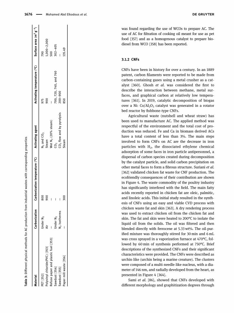
Table 2: Different physical methods for AC production from biomass with corresponding properties
Material Activating agent Temperature (°C) Surface area (m2 g−1)
Coconut shell [347] CO2 — —Corncob [347,348] CO2 800 919–986Rice husk [347] CO2 700 and 750 —Rice husk [349] Hydrothermal carbonization — 243Almond tree pruning (non-catalytic gasification) [347,350] Air 190 116–469Almond tree pruning (catalytic gasification) [347] Air 190–260 959Olive tree wood [347,351] Air 400 413Corn Stover [351] Microwave pyrolysis — 1671.4
Recent advances in waste-recycled NMs for biomedical applications 1675
was found regarding the use of WCOs to prepare AC. Theuse of AC for filtration of cooking oil meant for use as petfood [357] and as a homogenous catalyst to prepare bio-diesel from WCO [358] has been reported.
3.1.2 CNFs
CNFs have been in history for over a century. In an 1889patent, carbon filaments were reported to be made fromcarbon-containing gases using a metal crusher as a cat-alyst [360]. Ghosh et al. was considered the first todescribe the interaction between methane, metal sur-faces, and graphical carbon at relatively low tempera-tures [361]. In 2019, catalytic decomposition of biogasover a Ni: Co/Al2O3 catalyst was generated in a rotatorbed reactor by fishbone-type CNFs.
Agricultural waste (nutshell and wheat straw) hasbeen used to manufacture AC. The applied method wasrespectful of the environment and the total cost of pro-duction was reduced. Fe and Ca in biomass derived ACshave a total content of less than 3%. The main stepsinvolved to form CNFs on AC are the decrease in ironparticles with H2, the dissociated ethylene chemicaladsorption of some faces in iron particle antipersonnel, adispersal of carbon species created during decompositionby the catalyst particle, and solid carbon precipitation onother metal faces to form a fibrous structure. Suriani et al.[362] validated chicken fat waste for CNF production. Theecofriendly consequences of their contribution are shownin Figure 4. The waste commodity of the poultry industryhas significantly interfered with the field. The main fattyacids recently reported in chicken fat are oleic, palmitic,and linoleic acids. This initial study resulted in the synth-esis of CNFs using an easy and viable CVD process withchicken waste fat and skin [363]. A dry rendering processwas used to extract chicken oil from the chicken fat andskin. The fat and skin were heated to 200°C to isolate theliquid oil from the solids. The oil was filtered and thenblended directly with ferrocene at 5.33wt%. The oil-pur-ified mixture was thoroughly stirred for 30min and 6mLwas cross sprayed in a vaporization furnace at 470°C, fol-lowed by 60min of synthesis performed at 750°C. Briefdescriptions of the synthesized CNFs and their significantcharacteristics were provided. The CNFs were described asurchin-like (urchin being a marine creature). The clusterswere composed of a multi-needle-like nucleus, with a dia-meter of 146 nm, and radially developed from the heart, aspresented in Figure 4 [364].
Sami et al. [86], showed that CNFs developed withdifferent morphology and graphitization degrees throughTa
ble3:
Differen
tph
ysical
metho
dsforACprod
uction
from
indu
strial
was
teswithco
rres
pond
ingprop
erties
Material
Carbon
ization
Carbon
izationtempe
rature
(°C)
Activatingag
ent
Activatingtempe
rature
(°C)
Surface
area
(m2g−
1 )
PET[352
]Und
erN2
800
N2an
dCO2
975
790
Polyviny
lch
loride
(PVC)[35
3 ]Air
900
Steam
900
1,000–2
,000
Refuse
pape
ran
dplas
ticfuel
[353
]—
—Wet
N2(20%
stea
m)
—50
0Saw
dust
[354
]Und
erN2
—CO2
700,72
0,74
0,an
d76
030
2–40
5Saw
dust
[355]
N2isothe
rms
—CO2flow
andby
pyrolysis
200–9
00
—Pa
permill
was
te[356
]—
300
Steam
850
135.49
1676 Mohamed Abd Elkodous et al.

decomposition of the electrical cracking gas on Fe2O3.Compared to conventional physical methods, such aslaser ablation and CVD, the benefits of this process areCNFs with high performance, high selectiveness, and lowcost. The use of electric cracking gas to synthesize CNFs ispromising [86]. It enables organic liquid waste from thechemical industry to be used while carbon and hydrogenare obtained at moderate temperatures. The synthesis ofCNFs was done using an integrated reactor based on afluid laboratory design. The electrical cracking gas wasremoved from the electrical gas storage using a valve,regulated by a valve, and operated by a water U-tubemanometer at atmospheric pressure. Physical, morpholo-gical, chemical, and environmental features of producednanofibers were studied by scanning electron microscopy(SEM). Many advanced properties were exhibited whereinthe catalytic decomposition was shown to be the mainpathway for CNF growth [365].
3.1.3 CNTs
Suriani et al. [100] produced CNTs from refined domesticcooking palm oil that was used to fry three loads of fish.The synthesis process was carried out in a floating-cata-lyst thermal CVD reactor. Alves et al. [366] synthesizedCNTs from sugarcane bagasse supplied from the ethanolindustry. In that study, pyrolysis was used in which thematerials were decomposed by a thermal treatment in theabsence of oxygen. Qu et al. [367] synthesized CNTs byheating leaves of poplar to 450°C in air and Suriani et al.[368] succeeded in the production of vertically alignedCNTs by catalytic thermal CVD using waste chicken fat
and skin collected from a wet market and then convertedinto carbon precursor through a rendering process.
Bernd et al. [371] produced CNTs through pyrolysis ofwood sawdust in a tubular reactor and Lotfy et al. [369]applied a novel pyrolysis system to prepare CNTs fromRS, which is considered an undesirable biomass in Egypt.Singh et al. [370] succeeded in the synthesis of CNTs frompolyvinyl alcohol (PVA) using fly ash, red mud, and rocksamples via an alkali treatment followed by the fabricationof thin films and pyrolysis of composite films. Finally,Bernd et al. [371] synthesized carbon nanostructures bythe pyrolysis of wood sawdust in a tubular reactor, asshown in Figure 5. Table 6 summarizes the differentmethods used for the production of CNTs and their corre-sponding properties.
3.1.4 G
Renewable biomass resources are essential for G precur-sors such as glucose, rice husk, hemp, and disposablepaper cups. Recently, glucose was used as a preparatorymaterial for G with the aid of FeCl3. The obtained G hadeffective electrical conductivity [374]. Researchers alsosynthesized “patched graphene,” G produced using glu-cose with the aid of dicyandiamide (DCDA). The author ofthis study claimed the resulting G to be highly crystallineand clean. Luyi et al. succeeded in the production ofbiocompatible G from rice husk [375–377]. The obtainedG could be used for bioimaging and bioprobing [188]. Fibroushemp waste was successfully converted to G by Wang et al.[378] while Zhao obtained high quality and high yield Gusing disposable paper cups as a precursor [379]. Industrial
Table 4: Different chemical methods for AC production from biomass with corresponding properties
Material Activating agent Temperature (°C) Surface area (m2 g−1)
Potato peel [143] H3PO4, KOH, and ZnCl2 400, 600, and/or 800 —Rice husk [351] KOH 750 2,696Rice husk [351] H3PO4 500 1,741Rice husk [351] ZnCl2 500 2,434Peanut shell [351] KOH — 706.1Coconut shell [351] ZnCl2 900 332.4Coconut shell [351] KOH 900 682.0Banana peel [351] KOH 800 217.3Soybean [351] KOH — 2690.3Wheat straw [351] KOH — 1,066Pomelo peel [351] KOH — 1,533Pomelo peel[351] H3PO4 — 1,272Citrus peel [351] KOH — 1,167Olive stone [351] KOH — 587
Recent advances in waste-recycled NMs for biomedical applications 1677

wastes, such as plastic, can be used as precursors for Gproduction. A group of researchers developed an approachto produce G from various wastes and food using CVD.This new approach is sensible, however, its yield is amatter of concern [374]. The production of G from WCOis a very interesting topic to many researchers. Recently, agroup of researchers developed amethod to produce G fromwaste cooking palm oil due to the presence of palmitic acid,CH3(CH2)14COOH, which is the source of carbon. Conse-quently, this method indicates the possibility of using wastecooking palm oil to obtain G at a low cost and withoutconsumption of fossil fuels [380].
Graphite and organicmaterials are fundamental sourcesfor the preparation of G. The approach of each method toprepare G, CVD on metallic films [381], liquid exfoliation ofgraphite crystal [382], mechanical cleavage [383], growth onsilicon carbide [384], and chemical reduction of GO [385],has been illustrated by Ren and Cheng [386].
Glucose extracted from vegetable waste (as biomass)was first used to produce G in the presence of FeCl3 [387].Initially, the glucose and FeCl3 were dissolved in water at80°C to form carbonized glucose and FeCl2 (H2O2). Theproduction of G was obtained at 700°C. DCDA was addedto glucose in the presence of nitrogen to form graphitecarbon nitride (g-C3N4) [388]. At a temperature higherthan 750°C, G sheets were obtained, as shown in Figure 6.
Biomass material, like hemp, can be converted to Gby the hydrothermal method [378]. In this technique,fibrous hemp is heated around 180°C for an entire day.Then, KOH is added to produce porous G. DisposableTa
ble5:
Differen
tch
emical
metho
dsforACprod
uction
from
indu
strial
was
teswithco
rres
pond
ingprop
erties
Material
Carbon
ization
Carbon
izationtempe
rature
(°C)
Activatingag
ent
Activatingtempe
rature
(°C)
Surface
area
PET[346
]N2forAC-a
402m
2g−
1forAC-a
N2+H2OforAC-b
359m
2g−
1forAC-b
CO2forAC-c
50FeCl 3
500
123m
2g−
1forAC-c
PET[353
]N2
600
KOHin
anitrog
enstream
850
2,831
and2,666m
2g−
1
PVC[353
]Air
300
KOHin
thepres
ence
ofnitrog
en75
0—
Refuse
pape
ran
dplas
ticfuel
[353
]—
—K2CO3
—1,33
0m
2g−
1
Saw
dust
[359
]—
—KOH
500,
1876
.16m
2g−
1
Figure 4: (Bottom) SEM images of CNFs synthesized using wastechicken fat. Copyright © 2015, Elsevier Publishing Group [362].
1678 Mohamed Abd Elkodous et al.

paper cups can be used as a precursor for G production.In this approach, iron has been used as a catalyst to formFe3C layers by heating the activated paper cup at hightemperatures [379]. Industrial waste, such as plastic, isalso a good source of carbon for G production. CVD is themost common approach to produce G from industrialwaste [128]. Finally, cooking palm oil is an organic mole-cule that has been used as a precursor for G productionvia double thermal CVD using nickel as the depositionsurface with variable temperature (850–1,100°C in 50°Cincrements) [389].
3.2 IQDs
3.2.1 CQDs
Biomass has a high carbon and oxygen content and is anon-toxic and widely available feedstock, making it afascinating CQD source. Currently, several agricultural,forestry, and food waste types, such as rice husk, wheatstraw, sugar cane bagasse, soya, onion, peanut cottonshell, potato, durian, and fruit/vegetable peel (orange,banana, pineapple, onion, seaweed, watermelon peel,etc.), have been utilized to manufacture low-cost CQDs[390,391]. Several precursors have been used for the
preparation of CQDs, including banana peel waste, whileusing easy and green hydrothermal methods withoutpassivates or additives [392].
By using red cabbage (RC) and a one-step hydro-thermal method, Sharma et al. [393] evaluated a greenmethod for the creation of CQDs. Yang et al. [394] studiedexpired fruits as carbon sources for the preparation ofCQDs and Luo et al. [395] prepared CQDs using loofahsponge-based AC fiber as the raw material. Zhang et al.[396] used black soya beans (BS) as a cheap source ofcarbon for the green synthesis of BS-CQDs. Such high-value CNMs, such as the CQDs, can be synthesized usingother abundant waste, such as low-cost petroleum cokebyproducts and humic acid. Tajik et al. [397] also describedthe graphitic framework of petroleum cokewith benzene oraromatic fields, which permits a chemical oxidation pro-cess for the synthesis of CQDs.
Compounds such as chitosan, citric acid, urea, andascorbic acid, as well as a variety of foods, plants, waxes,and wastes, such as peels (mango, orange, banana,pineapple, onion, seaweed, etc.), egg yolk oil, meat,grains, nuts, or vegetable byproducts, have been usedin the synthesis of CQDs. These compounds are lowcost starting material for the creation of carbon materials[398]. Due to their green chemistry, hydrothermal andsolvothermal methods are ecofriendly, mass-producible,
Figure 5: Schematic of the preparation of CNTs with corresponding particle size determination using high resolution transmission electronmicroscopy (HRTEM) imaging [371].
Recent advances in waste-recycled NMs for biomedical applications 1679

Table6:Differen
tsynthe
sismetho
dsan
dco
rres
pond
ingprop
erties
ofCNTs
Metho
dof
prep
aration
Carbon
source
Type
andsh
ape
ofCN
TsCa
talyst
andtempe
rature
Diameter
Surface
area
Notes
Referen
ces
WCO
Catalytic
cracking
WCOmod
elco
mpo
und
Few
multi-w
alled
CNTs
andso
me
CNTs
withan
open
-top
structure
Catalyst:Ni-C
o/SBA-15
catalysts
50–1
00nm
——
[96]
Tempe
rature:75
0°C
Was
tebiom
ass
Pyrolysis
Woo
dsa
wdu
stSinglean
ddo
uble-
walledCNTs
Catalyst:Ferroc
eneor
Fe/
Mo/
MgO
25nm
—Woo
dsa
wdu
stwas
mixed
with
redu
cing
agen
ts(com
mercial
zinc
),calcite(bed
material),
and
catalyst
andarrang
edin
the
columnreactor,as
show
nin
Figu
re5
[371]
Tempe
rature:75
0°C
for3h
Pyrolysis
Hyd
rocarbon
sof:
Type
ofCNTs
depe
ndson
the
type
ofRS
precurso
r
Catalyst:Fe-Niox
ides
supp
ortedon
Al 2O3
Thediam
eter
ofCNTs
differ
acco
rdingto
the
type
ofRS
precurso
r
Thesu
rfacearea
ofCNTs
from
rawRS
is18
8m
2g−
1Hyd
rothermal
trea
tmen
twas
carriedou
tto
prod
uce
hydroc
arbo
nsas
aninterm
ediate
carbon
source
[369]
Raw
RSMulti-w
alledCNTs
(from
raw
RSan
dRS
-neu
tral)
Raw
RS(from
15to
40nm
)
RS-neu
tral
pulp
Nee
dle-sh
aped
CNTs
(from
RS-AP
andRS
-SP)
Tempe
rature:75
0°C
for
120min
RS-NP(fr
om14.7
to47
.9nm
)RS
-alkaline
pulp
(AP)
RS-AP(fr
om2.5
to6.8
nm)
RS-sulfite
pulp
(SP)
RS-SP(fr
om4
to8nm
)Hyd
rothermal
+tempe
rature
calcination
approa
ch
Recyclab
leplan
tTyph
aorientalis
Nitroge
n-do
pedCNTs
Catalyst:–
CNTs:150nm
281.3m
2g−
1—
[372
]Tempe
rature:18
0°C
for
12h,
was
hed,
free
zedried,
andan
nealed
at800°C
for
1hin
NH3atmos
phere
Pore
diam
eter:4
nm
Indu
strial
was
tes
Pyrolysis
Flyas
h,redmud
,an
drock
sample
Differen
ttype
sof
CNTs
Catalyst:–
20–3
0nm
CNTs
from
:flyas
h(42.3m
2g−
1 ),redmud
(38.8
m2g−
1 ),an
drock
sample(19.7m
2g−
1 )
—[373
]Tempe
rature:50
0°C
for
20min
1680 Mohamed Abd Elkodous et al.

cost effective, clear, and applicable for the synthesis ofCQDs using various organic sources [393]. Başoğlu et al.[399] studied a simple CQD green synthesis method thatused rusty chickpea as the single-stage carbon source,with no chemical additives. In another example, expiredpassion fruit shell (EPFS) and pulp were separated phy-sically and the EPFS was cleaned using pure water. Then,the EPFS was placed into a furnace at 120°C for 28 h. Afterdrying, the EPFS was crushed into a fine powder andplaced into a 60-mesh sieve. Next, 2 g of EPFS powderwith 40mL of ultra-pure water was added to a polytetra-fluoroethylene lined reactor and heated for 3 h at 175°C.Then, the received brown solution was filtered by a poly-ether sulfone membrane and centrifuged for 20min at15,000 rpm. After centrifugation, the product was dialyzed
using a 2,000 days dialysis bag for two days. After thedialysis, the solution was freeze-dried to achieve thedesired solid CQDs. The obtained CQDs were uniformlydispersed and spherical, with a size below 5 nm, indicatinggood dispersion (Figure 7).
3.2.2 GQDs
High-quality GQDs with a yield of ∼15 wt% were preparedfrom rice husk biomass by Wang et al. [188]. Also, GQDswere produced utilizing a low-cost, green, and renewablebiomass resource viamicrowave treatment by Abbas et al.[400]. Recently, scientists have targeted the transferenceof 2D G into 0D GQDs and studied the impact of edge
Figure 6: Proposed synthesis method for free-standing G. Bottom: repetition motifs of an ideal g-C3N4 plane (middle) and of G (right); C isindicated by black or gray, N is indicated by blue. Copyright © 2012, Wiley Publishing Group [387].
Figure 7: Proposed synthesis technique for powder CQDs by the hydrothermal method and corresponding transmission electron microscopy(TEM) images at different magnifications. Copyright © 2021, Springer Publishing Group [399].
Recent advances in waste-recycled NMs for biomedical applications 1681

effects and quantum confinement on the properties of thenewmaterial [401]. GQDs are synthesized frommoleculeswith aromatic structures (biomass and cooking oils) wherethe formation of GQD moieties can undergo a step-by-stepchemical reaction bymolecular precursors. These approachesallow for excellent control of the properties of the final pro-duct [401,402].
The synthesis of GQDs from a low cost, green, andrenewable biomass resource, rich in carbon, was achievedusing microwave treatment of carbon biochar through thepyrolysis of biomass waste. Increasing the reaction time ormicrowave power resulted in an increase in the product
yield (∼84% yield was achieved) [400]. Wang et al. [188]synthesized high-quality GDQs from rice husk biomassand it was found to be a useful source that produced ayield of ∼15 wt%. HRTEM, atomic force microscopy, andRaman spectroscopy were used to determine the size,structure, and morphology of the rice husk, as shown inFigure 8.
3.3 MO-based NMs
3.3.1 Iron oxide
Bishnoi et al. [223] used the extract of C. ramiflora, aninedible fruit waste, for the synthesis of magnetic IONPs. C. ramiflora belongs to the leguminosae family. Itis largely found in Africa, as shown in Figure 9, and isused for biomedical and environmental applications.
Additionally, IO NPs have been synthesized usingtangerine peel extract and used as an adsorbent for cadmiumion removal from contaminated solutions. Ehrampoush et al.[221] prepared IO NPs by the co-precipitation method in thepresence of tangerine peel extract, which prevented accumu-lation and reduced the diameter of the synthesized IO NPs.Sundaram et al. [403] isolated the Bacillus subtillis bacterialstrain from rhizosphere soil to produce IO NPs through thefermentation process. Khan et al. [404] described the synth-esis of superparamagnetic IO NPs in bio char from the con-densed tannin extract of Acacia mearnsii. They utilized asingle-step process compliant with the principles of greenchemistry. By the warm deterioration of iron oleate withinsight of sodium oleate (from biomass waste), Xie et al. [405]
Figure 8: Proposed synthesis for powder GQDs by the hydrolysismethod and corresponding TEM imaging. Copyright © 2016, ACSPublishing Group [188].
Figure 9: Biosynthesis of IO NPs using Cynometra ramiflora leaf extract for biomedical and environmental applications.
1682 Mohamed Abd Elkodous et al.

integrated IO NPs with different shapes. By tuning the molarproportion of sodium/iron oleate or the response temperature,plates, tetrahedrons, octahedrons, curves, multi-branches, orself-gathered designs could be achieved (Figure 10).
Another study [155] synthesized IO NPs utilizingR. tuberosa leaf fluid concentrate and their ultraviolet-visible spectroscopy investigation showed obvious topsat 405 nm. Dynamic light dissipation determined thenormal size of IO NPs to be around 52.78 nm and differ-ential scanning colorimetry demonstrated the strength ofIO NPs until reaching the high temperature of 165.52°C.
Promptly after adding iron sulfate to the leaf extract,IO NPs were shaped and in this manner, the technique isstraightforward and quick [406]. In this investigation, abasic, fast, and eco-accommodating green technique wasused to create magnetite IO NPs utilizing the fluid con-centrates of two food-handling wastes, in particular thevelvety hairs of corn (Zea mays L.) and external leaves ofChinese cabbage (Brassica rapa L. subsp. pekinensis).The bubbled arrangements of satiny hairs (MH) and
external leaves of Chinese cabbage (CCP) were utilizedto blend IO NPs under photograph uncovered conditions[407]. Another study revealed that C. maxima peel extractyielded IO NPs with a diameter of 10–100 nm and highstability [408].
3.3.2 TiO2
TiO2 NPs have been prepared from biomass, cooking oils,and industrial waste through chemical, physical, andbiological approaches. There are also different chemicalmethods to create TiO2 NPs, such as sol–gel, solvo-thermal,hydrothermal, and co-precipitation methods [409]. Table 7summarizes the different chemical methods used to fabri-cate TiO2 NPs from biomass and their correspondingproperties.
On the other hand, physical methods, such as elec-trophoretic, thermal plasma, spray pyrolysis, and micro-wave-assisted techniques, have been applied for TiO2 NP
Figure 10: Example morphologies of 0D IO NPs synthesized from iron oleate and sodium oleate (biomass wastes): (a) nanospheres, (b)plates, (c) tetrahedrons, (d) cubes, (e) truncated octahedrons, (f) octahedrons, (g) concaves, (h) octapods, and (i) multi branches [405].
Recent advances in waste-recycled NMs for biomedical applications 1683

synthesis from different biomass wastes [409]. Biologicalsynthesis methods (also called green synthesis methods),use plants and fruit extracts, in addition to their wastematerials and microorganisms (via fermentation methods),for the synthesis of NMs, to reduce the use of costly, toxicchemicals. Thus, they are considered as cost-effective andecofriendly methods [409]. Table 8 summarizes the dif-ferent biological methods used for the synthesis of TiO2
NPs from microorganisms and fruit peel extracts with theircorresponding properties.
3.3.3 ZnO
Ramimoghdam et al. [412] suggested a method for recy-cling waste oil for the synthesis of ZnO NPs. The groupused palm olein, the liquid fraction of palm oil, as a bio-template to synthesize NPs with different morphologies.Bio-templating is the use of a biological structure thatserves as a template, guiding the assembly of inorganicmaterials into NPs of the desired morphology.
Industrial waste first supplied a pure zinc source in2011 when Deep et al. [413] dismantled spent Zn–MnO2
batteries, separated their metal components, and usedthe purified zinc for NP synthesis. In a later study, Deepet al. (2016) [414] also used spent Zn–MnO2 batteries topropose a simpler, more efficient synthesis method: a
one-pot solvothermal process. This method resulted inthe isolation of highly pure ZnO NPs at a recovery per-centage ≥95%. Farzana et al. [415] similarly synthesizedZnO NPs from Zn–C spent batteries.
The use of microbial biomass (nanofactories), actingas reducing and capping agents through the fermentationprocess, was first investigated by Jayaseelan et al. [424]for the preparation of ZnO NPs by adding ZnO to a sus-pension of Aeromonas hydrophila bacteria. More recentstudies have reported the use of C-phycocyanin pigmentfrom cyanobacteria in the synthesis of phycocyanin–ZnOnanorods [417] with the use of zinc-tolerant Lactobacillusplantarum [425].
Agricultural waste and inedible plant leaves andpeels are also being used as bio-reductants for greenZnO NP synthesis, mainly due to their polyphenolic con-tent. Different wastes have been used for the biosynthesisof TiO2 NPs, such as rambutan peel [272], M. oleifera peel[251], and agricultural waste [426], which contains bior-eductant and secondary metabolites such as terpenoids,alkaloids, and flavonoids. In 2020, ZnO NPs were synthe-sized using orange peel [427], non-edible cauliflowerleaves [291], garlic skin extract [428], and banana peel[429]. In 2021, Rambabu et al. [423] used date pulp wasteto synthesize ZnO NPs. Table 9 includes all the methodsapplied for ZnO NP synthesis from industrial waste, bio-mass, and cooking oils.
Table 8: Different synthetic biological methods, and corresponding properties of TiO2 NPs
Material Initial temperature (°C) Final temperature (°C) Size (nm)
Orange peel extract [235] 100 500 21.61 and 17.30Plum peel extract [228] 70–80 400–500 47.1 and 63.21Peach peel extract [228] 70–80 400–500 200Kiwi peel extract [228] 70–80 400–500 54.17 and 85.13Sugar-apple [411] — — 23Aeromonas hydrophila [411] — — 40.50Bacillus amyloliquefaciens [411] — — 22.1–97.2Bacillus mycoides [411] — — 40–60Bacillus subtilis [411] — — 30–40Lactobacillus sp. [411] — — 24.63Planomicrobium sp [411] — — 100Aspergillus niger [411] — — 73.58
Table 7: Different chemical methods for the synthesis of TiO2 NPs and their corresponding properties
Material Method Initial temperature (°C) Final temperature (°C) Pressure (bar) Surface area (m2 g−1) Size (nm)
RS [229] Sol–gel 80 500 — 97 13.0 ± 3.3Eggshell [410] Solvothermal — 454 227 — 78.73
1684 Mohamed Abd Elkodous et al.

Table9:
Differen
twas
tematerials
andsynthe
sismetho
dsus
edto
crea
teZn
ONPs
withdifferen
tsizesan
dmorph
olog
ies
Sou
rceof
was
tematerial
Type
ofwas
tematerial
Zinc
source
Type
ofsynthe
sis
Nam
eof
synthe
sis
metho
dRea
ction
temp.
(°C)
Calcination
temp.
(°C)
Size(nm)
Morph
olog
yRef.
Indu
strial
was
teSpe
ntZn
–MnO
2
batteries
Zinc
extracted
from
was
teelectrod
ematerial
Che
mical
Solvo
thermal
synthe
sis
250
NA
5Sph
erical
[414]
Indu
strial
was
teSpe
ntZn
–Cba
tteries
Zinc
from
was
teelectrod
ematerial
Che
mical
Thermal
nano
sizing
900
NA
50Sph
erical
[416
]
WCO
Palm
oil
Zinc
acetate
Che
mical
Hyd
rothermal
metho
dus
ingpa
lmoleinas
abio-
template
120
NA
Micro-a
ndna
no-sized
particles,
depe
nding
onthevo
lumeof
palm
oleinad
dedan
dreaction
time
Differen
tmorph
olog
ies,
depe
ndingon
the
volumeof
palm
olein
adde
dan
dreaction
time
[412]
Microbial
biom
ass
Phycoc
yaninextract
from
Cyan
obacterium
Limno
thrixsp
.K005
Zinc
acetate
Biological
(ferm
entation
)NA
30NA
33–3
5Nan
orod
s[417]
Microbial
biom
ass
Serratia
nematod
iphila
Zinc
sulfate
Biological
(ferm
entation
)NA
NA
NA
15–3
0Sph
erical
[418
]
Plan
tbiom
ass
Moringa
oleifera
peel
extract
Zinc
acetate
Biological+
physical
Microwave-as
sisted
synthe
sis
Microwave
power
of30
0W
400an
d600
40–4
5Sph
erical
[419]
Plan
tbiom
ass
She
epan
dgo
atfecal
matter(agriwas
te)
Zinc
sulfate
Biological
NA
Room
tempe
rature
350–4
00
24.4–2
8.5
Spo
ngyan
dflow
ersh
aped
[248
]
Plan
tbiom
ass
Orang
epe
elextract
Zinc
nitrate
Biological
NA
60
400
Differen
tsizes
depe
ndingon
reaction
tempe
rature
andpH
Differen
tmorph
olog
ies
depe
ndingon
reaction
tempe
rature
andpH
[263]
Plan
tbiom
ass
Cau
liflow
erleaf
extract
Zinc
nitrate
Biological
NA
Son
ication
at30
NA
100
Sph
erical
[419]
Plan
tbiom
ass
Pectin
from
durian
rind
Zinc
acetate
Biological
NA
Room
temp.
NA
280–4
56Sph
erical
[420
]
Plan
tbiom
ass
Pectin
from
bana
nape
elZinc
acetate
Biological+
chem
ical
Co-precipitation
Room
temp.
NA
20–4
0Th
inflak
es,na
noco
nes,
flow
er-sha
ped,
and
cubic-sh
aped
[421]
Plan
tbiom
ass
Ban
anape
elextract
Zinc
acetate
Biological
NA
NA
NA
20–9
0Sph
erical,triang
ular
[422
]
Plan
tbiom
ass
Garlic
skin
extract
Zinc
chloride
Biological
NA
NA
NA
7.77
Rodsh
aped
,he
xago
nal
[428
]
Plan
tbiom
ass
Datepu
lpwas
teZinc
nitrate
Biological
NA
Room
temp.
400
30Sph
erical
[423
]
Recent advances in waste-recycled NMs for biomedical applications 1685

3.3.4 CuO
The green biosynthesis of CuO NPs from different wastematerials, such as cooking oil, biomass (including plantsand microorganisms), and industrial wastes, is common,as shown in Figure 11. Javed et al. used waste palm oil asa base solvent for the synthesis of ecofriendly, highlystable, and bio-renewable CuO NPs [430]. The synthesisof CuO NPs using bio-waste plant peels, lignocellulosicbiomass-rich materials, and seed extract, such as citrus peel(orange, lemon, and tangerine) [430,431], banana peel (Musaacuminate) [303,432], pomegranate peel (Punica granatum)[302,433,434], pomegranate seeds [435], papaya peel (C.papaya) [280], date stone [434], Zea mays husk [435–437],onion peel (Allium cepa) [438], fava bean seed peel [439],cauliflower (B. oleracea), potato (Solanum tuberosum) andpea (Pisum sativum) peels [440], and Colocasia esculentaleaf waste has been reported (Table 10) [441]. Recycling avariety of industrial wastes is a low-cost and safe methodfor CuO NP production. CuO NPs have been recovered fromCu-rich industrial waste, such as e-waste that includes SIMcards [442] and printed circuit boards (PCBs) [443–446], bat-teries, including LIBs and NiMH batteries [447], waste sludge[448,449], and wastewater (Table 11) [450].
3.4 Au NPs
The synthesis of NPs using chemicalmethods is usually asso-ciated with the formation of toxic byproducts. Therefore, the
use of biosurfactants/biological materials (biomasses likemicrobes and plants) for the synthesis and stabilization ofAu NPs is gaining importance since a decade [458]. Themicrobial route for the synthesis of Au NPs is a very excitingprocess, as several factors including microbial cultivationmethods on organic and/or inorganic wastes and extractiontechniques can be optimized for the fast synthesis of mono-dispersed NPs by the fermentation process. Many biologicalsystems such as those of fungi [459] and algae [460] havebeen studied for the biosynthesis of Au NPs.
A newer Au NP synthesis technique includes mate-rials such as oat and wheat biomasses, which have beenshown to reduce aqueous gold ions to gold zero-valentforming Au NPs [310]. Recently, a method was developedfor the preparation of Au NPs that involves application ofan alternating current voltage to metal electrodes in anelectrolyte solution containing a water-soluble polymerdispersant and a reducing agent [461]. A method for theextraction of gold from waste electrical and electronicequipment as NPs stabilized with phospholipids was sug-gestedusing1,2-dioleoylsn-glycero-3-phosphocholine [461].
WCO contains long hydrocarbon chains and polarester bonds that make it a good choice for stabilizingAu NPs. In this preparation method, Au NPs are synthe-sized in WCO using the laser ablation technique at dif-ferent temperatures. A gold plate (high impurity 99.99%)was immersed in 10mL of pure palm oil, which wasextracted from WCO. A laser beam of 532 nm wavelength,with a period and energy of 10 ns and 1,200MJ, respec-tively, was used to ablate the gold plate at different tem-peratures for 5–60min with a 30–60 Hz repetition rate.
Figure 11: An illustration of the green synthesis of CuO NPs from waste materials by different physical, chemical, and biological methods.
1686 Mohamed Abd Elkodous et al.

Table10:Green
synthe
sismetho
dsforprod
ucingCuO
NPs
from
was
tematerials
andtheirprop
erties
Metho
dWas
tematerial
Cuprecurso
rSize
Sha
peYield
Proc
ess
Adv
antage
sRef.
Indu
strial
was
tes
Physical
Was
tePC
Bs
(WPC
Bs)
PCBsof
oldteleph
ones
andPC
s5–
40nm
Sph
erical
90%
Electro-kine
tic(EK)m
etho
dSim
ple,
ecofrien
dly,
andco
steff
ective
[444
]
Che
mical
Was
teSIM
cards
Was
temob
ileSIM
cards
2–50
nmCau
liflow
erlik
e95
.3%
Wet
chem
ical
metho
dSim
plean
decofrien
dly
[442
]
WPC
Bs
WPC
Bs
Differen
tsizes
Irregu
larsh
eet
98%
Mecha
noch
emical
metho
dSim
plean
decofrien
dly
[445
]WPC
Bs
Old
compu
terWPC
Bs
15–1
6nm
Sph
erical
Over90
%Che
mical
redu
ction
“hyd
rometallurgical
approa
ch”
Cos
teff
ective/eco
friend
ly,high
pure
particles.
[451]
WPC
Bs
Cop
per-c
ontaining
was
teetch
ants
and
CuC
l 2
2–50
nmSph
erical
>99.90
%Microem
ulsion
Sim
ple,
econ
omical,an
dsa
fe,
withlow
energy
cons
umption,
high
prod
uction
yieldwithhigh
purity
(>99
.0%)
[443
]
WPC
Bs
WPC
Bsan
dCuS
O4
—Sph
erical
96.9%
Che
mical
redu
ction
Sim
ple,
ecofrien
dly,
cost
effective,high
prod
uction
yield
[452
]
Was
tewater
Was
tewater
from
electrop
lating
indu
stry
andCuS
O4
20–8
0nm
Crystallin
e99
.98–9
9.99
%Precipitationmetho
dEcon
omic
andsa
feproc
ess,
with
high
prod
uction
yield
[450
]
Was
tewater
slud
geIndu
strial
was
tewater
200–4
00nm
Feathe
rlik
e90
%Hyd
rometallurgical
metho
dby
acid
leaching
Sim
ple,
cost
effective,an
dhigh
prod
uction
yieldw
ithhigh
purity
98.26%
[449
]
Was
teoil
Biologicalan
dch
emical
Palm
oil
Cop
pernitrate
25nm
Sph
erical
—Precipitationmetho
dSim
ple,
cost
effective,an
decon
omic
proc
ess
[430
]
Plan
twas
tes
Biologicalan
dch
emical
Lemon
pulp
CuC
l 220
–40nm
Irregu
lar
103mg/g
Hyd
rothermal
metho
dSim
ple,
rapid,
ecofrien
dly,
and
cost
effective
withhigh
yields
[453
]
Was
teba
mbo
oleaf
Cop
peracetate
70–8
0nm
Hierarchical
—Hyd
rothermal
metho
dSim
ple,
rapid,
ecofrien
dly,
cost
effective,high
prod
uction
yield
[454
]
Onion
peel
Cop
perch
loride
11.7–4
5.5nm
Berry-like
—Microwave-as
sisted
Sim
ple,
rapid,
ecofrien
dly,
cost
effective
[438
]
Biologicalan
dph
ysical
Fava
bean
seed
peel
Cop
persu
lfate
29.82nm
Sph
erical
—Gam
maradiation
Sim
ple,
rapid,
ecofrien
dly,
cost
effective
[439
]
Biological
metho
dBan
anapu
lpwas
teCop
peracetate
1–4µm
Octah
edral
—Ban
anapu
lpwas
teextractas
aredu
ctan
tSim
ple,
gree
n,ecofrien
dly
[455]
Ban
anape
elCop
persu
lfate
50–6
0nm
Sph
erical
—Ban
anape
elextractas
aredu
ctan
tSim
ple,
gree
n,ecofrien
dly
[432
]
Pomeg
rana
tepe
elan
dda
teston
eCop
persu
lfate
6–20
nmCrystallin
e—
Pomeg
rana
tepe
elan
dda
teston
eextractas
aredu
ctan
tSim
ple,
gree
n,ecofrien
dly
[434
]
(Con
tinue
d)
Recent advances in waste-recycled NMs for biomedical applications 1687

The solution temperature was raised using a hot plate. AuNPs formed during the laser ablation with an ablationtime of 25 min. This experiment was conducted at dif-ferent temperatures (40, 64, 75, 83, and 92°C). The solu-tion was stirred during the ablation and the solution con-tainer was moved horizontally to obtain a fresh surfacearea for the Au NPs to guarantee that the Au NPs weredispersed equally in the WCO [309].
Velmurugan et al. [462] synthesized Au NPs fromginger waste, which includes the aqueous extract ofwaste ginger. Ginger waste acts as both a reducing andstabilizing agent. For a typical synthesis, 5 mL of theextract is added to 3 mL of freshly prepared 1 mM solutionof HAuCl4. The mixture was allowed to boil by rapid micro-wave heating. The appearance of the red color in the reac-tion mixture indicated the formation of Au NPs. The coloredsolution of Au NPs was then centrifuged, and the Au NPswere redisposed to remove any unreacted ions and mole-cules from the product. The produced Au NPs were highlystable and spherical, with diameters of 15.5 nm.
Liu et al. [463] synthesized Au NPs from banana peelby the hydrothermal process. After mixing with HAuCl₄as the precursor, the banana peel was heated with waterand acetone at 98°C. No extra chemical protectant orreductant was added. Finally, Au NPs with four leaf clover(dendrite) morphology were obtained, which were namedAu dendrites, as indicated in Figure 12.
4 Biomedical applications ofRNMs: Role of RNMs in the earlydetection of diseases
4.1 Fluorescence imaging
Biological components are too small to be observed bytraditional microscopy, which lacks the accuracy andhigh-resolution localization to image the biological pro-cesses inside these components. A fluorescent dye fixesthis problem by providing several optimal approaches totrack cells and evaluate their functionality. Fluorescenceimaging is a common tool used to visualize preciousmolecular mechanisms [464,465], gene and protein expres-sions [466–468], and interactions between cellular compo-nents, providing a deeper understanding of the biologicalprocess.
The advantages of fluorescence imaging over otherimaging techniques are high selectivity of the desiredTa
ble10:Co
ntinue
d
Metho
dWas
tematerial
Cuprecurso
rSize
Sha
peYield
Proc
ess
Adv
antage
sRef.
Pomeg
rana
tepe
elCop
perch
loride
5–25
nmRo
dan
dsp
herical
—Po
meg
rana
tepe
elextracta
sa
redu
ctan
tSim
ple,
rapid,
safe,withlow
energy
cons
umption
[433
]
Pomeg
rana
tese
eds
Cop
pernitrate
—Broccoli
—Po
meg
rana
tese
edsextractas
aredu
ctan
tSim
ple,
gree
n,ecofrien
dly
[435
]
Citruspe
elCop
pernitrate
22.4–9
4.8nm
Sph
erical
—Citruspe
elextractas
aredu
ctan
tSim
ple,
gree
n,ecofrien
dly
[431]
Papa
yape
elCop
pernitrate
85–
140nm
Crystallin
e—
Papa
yape
elextractas
aredu
ctan
tSim
ple,
rapid,
cost
effective
[280]
Zeamay
shu
skCop
peracetate
20–7
0nm
Sph
erical
Was
temaize
extractas
aredu
ctan
tSim
ple,
gree
n,eco-
and
econ
omically
friend
ly[437
]
Brownalga
eMacrocystis
pyrifera
Cop
persu
lfate
2–50
nmSph
erical
—Brownalga
eextractas
aredu
ctan
tSim
ple,
ecofrien
dly
[456
]
1688 Mohamed Abd Elkodous et al.

Table 11: : Various waste materials for CuO NP synthesis and their corresponding properties
Waste material Cu precursor Size (nm) Shape Ref.
Plant wastesCitrus peel (orange, lemon, and tangerine) Copper nitrate 22.4–94.8 and Spherical and [430]
8–8.9 irregular [431]Banana peel Copper acetate 7.33 Spherical [457]
60 [303]Pomegranate peel Copper acetate and 10–100 Spherical [302]
Copper sulfate 6 Crystalline [434]Pomegranate seeds Copper nitrate — Broccoli [435]Papaya peel Copper nitrate 85–140 Crystalline [280]Date stone Copper sulfate 20 Crystalline [434]Zea mays husk Copper acetate 36–73 Spherical [436]
20–70 Spherical [437]Onion peel Copper chloride — Berry like [438]Cauliflower peels, potato peels, and peapeels
Copper chloride <100 Irregular [440]
Colocasia esculenta leaf Copper nitrate <100 — [441]Industrial wastes
e-waste Waste mobile SIM cards 2–50 Cauliflower like and spherical [442]SIM cards Waste telephones, PCBs 11, 25, and 40 [444]PCBs PCBs and chloride (CuCl2) 20–50 [443]Wastewater Copper sulfate 20–80 Crystalline [450]
Figure 12: Simplified diagram of the synthesis process of Au dendrites and the different shapes with their corresponding colors. Copyright© 2020, Dove press Publishing Group [463].
Recent advances in waste-recycled NMs for biomedical applications 1689

area, high-resolution power, identification of microele-ments inside the cellular component, and productionof high-contrast images [469]. Fluorescent NPs havepromising qualifications for fitting the small size of thecellular components while providing more abilities com-pared to other fluorescent techniques, such as a low cyto-toxicity level, better selectivity, smooth entrance, theability to target a large number of cells at the same time,low chemical structure changes, and high-throughputscreening [470,471].
Long-period cellular tracking in vivo provides valu-able information regarding the biological process and bio-medical therapeutics. Several biological imaging modalitieshave been developed for those applications, includingmag-netic resonance imaging (MRI), magnetic particle imaging(MPI), and fluorescence imaging. Merging nanotechnologywith these methods has improved the safety of cellulartracking in vivo and has overcome some challenges of tradi-tional imaging methods. Compared to conventional fluores-cent methods, fluorescent NPs support the unique advan-tages of photofading resistance, higher brightness, andbetter size control [472]. Cellular tracking fluorescent NPsare inorganic QDs [473,474], nanodiamonds [475], fluores-cent latex/silica nanobeads [476], and organic NPs [477].
The early detection of diseases is currently a criticalpoint of focus in clinical practices. One of the most pro-minent examples is the early detection of cancer. It wasreported that patients who were diagnosed early withgastrointestinal cancer had a higher survival rate thanthose who did not benefit from early detection (92.3%versus 33.3%) [478]. One of the key players in the earlydetection of diseases is biomedical imaging, which includesMRI, positron emission tomography, ultrasound, X-rayradiography, single-photon emission computed tomo-graphy, computed tomography (CT), and fluorescenceimaging [479]. However, structural imaging modalities,such as CT, ultrasound, and MRI, are not useful in thedetection of some tumors due to their detection limitations(0.5 cm) and spatial resolution. Imaging modalities can becombined to improve the efficiency of tumor detection[480]. Contrast agents (CAs) are used to provide accuratefunctional and anatomical information. Medical imagingCAs usually used are small molecules that have low unde-sirable toxicity, non-specific distribution, and fast meta-bolism. However, nanoimaging CAs are currently beingstudied extensively due to their outstanding propertiescompared to traditional CAs. Nanoimaging CAs exhibit ahigh surface-area-to-volume ratio, leading to enhancedsurface functionalization and lower toxicity profiles [481].Additionally, nanoimaging CAs exhibit a longer plasma cir-culation time and better biodistribution and bioavailability,
thus enhancing the tumor-to-background contrast signal.Furthermore, themanipulation of NP properties allows opti-mized loading of imaging compounds [482,483].
4.2 Role of NPs in fluorescence imaging
Fluorescence imaging technology is currently widely usedfor disease diagnosis as it provides the highest spatial reso-lution compared to other imaging technologies [484]. How-ever, fluorescence imaging in clinical settings is hindered bysome limitations, including non-specific tissue auto-fluores-cence, and limited penetration depth. Also, photobleachingeffects and limited fluorescence in the target site decreasethe sensitivity of the technology for disease detection[483,485–487].
NMs play a key role in the enhancement of fluores-cence imaging to overcome limitations. Near infrared(NIR) fluorescence quenching could be avoided by usingmodified NPs [488]. NPs could also reduce photobleachingeffects by converting low-energy photons into high-energyphotons [489]. In addition, NPs allow the loading of moredye molecules, thus providing higher signals [490]. NPscould also be targeted to tumors or target sites throughpassive or active targeting, allowing accumulation indesired sites and increasing the fluorescent dye concen-trations and thus the signal. Hence, fluorescent NPs arebeing extensively developed and investigated for biome-dical applications [483]. For example, NP fluorescenceimaging is currently used in several biomedical applica-tions, such as enzyme activity evaluation, gene detec-tion, cell tracking, protein analysis, real-time moni-toring of therapeutic effects, and early detection ofdiseases [490–493]. For cancer diagnoses, fluorescentNPs play a crucial role as they provide high-resolutionimages at the cellular level. Examples of promisingfluorescent NPs include QDs, organic dye-doped NPs,upconversion NPs, and Au NPs [494]. The differentexamples of NPs used in fluorescence imaging for earlydetection of different diseases are provided in Table 12.
4.3 CQDs for early disease detection viafluorescence imaging
Fluorescence imaging has gained increasing attentionrecently due to its high sensitivity, selectivity, and high-throughput ability. Developing optimum fluorescentprobes has always been a challenge. QDs are inorganic
1690 Mohamed Abd Elkodous et al.

semiconductor NCs that possess exceptional photophy-sical characteristics. Based on their quantum properties,QDs can emit a wide and broad spectrum of different fluor-escent wavelengths that give them preference for use inbiomedical applications, particularly in the tracking of cir-culating tumor cells and early diagnosis of abnormal tumormasses [495–497]. Briefly, QDs are made from a metallic-semiconductor core that includes elements of both II–VI(e.g., ZnSe, CdTe, CdS, and CDSe) and III–V (e.g., InAs andInP) groups of the periodic table, which are coated with ashell of biomolecules, like polysaccharides, peptides, or pro-teins, to prevent the leakage of toxic heavy metals and toprovide good stability for the NP in a biological environment.
Although QDs have shown highly promising resultsamong traditional fluorescent probes, it has been foundthat QDs exhibit some toxic effects, which limit the appli-cations of heavy metal QDs. Alternatively, using safe,cost-effective, and ecofriendly materials that have beenrecycled from biological wastes has become a trend sincethey are readily available and less toxic. For example,CQDs produced from recycled biomass waste have beenapplied in fluorescent imaging.
CQDs are small nanostructures with carbon as thecore element. Currently, this type of QD has receivedmuch interest due to its unique properties, including itssize (below 10 nm), chemical stability, wide range offluorescence emission, high availability, easy synthesis,and water solubility. The fluorescence of CQD emissionranges in the NIR spectrum and, in addition, CQDs havelow physiological toxicity compared to semiconductorQDs, making them a superior choice for biomedical appli-cations. The biomedical applications of CQDs are in vitroand in vivo bioimaging, drug delivery [498,499], tumormass detection [500], and cancer therapy [501–503].
One of the main properties of CQDs is their fluores-cence, which promotes their application in fluorescenceimaging. Narrow emission spectrum, wide excitationspectrum, high fluorescence stability, and resilient resis-tance to photobleaching are all outstanding fluorescenceproperties of CQDs [137]. However, these excellent CQDproperties are affected by the CQD source, such as CQDsbiomass wastes. A study by Zhang et al. reported that theorganic solvent used for CQDs affected the fluorescenceemission peak position and fluorescence emission inten-sities of CQDs synthesized from polystyrene foam waste[504]. Another study by Cheng et al. stated that CQDs co-doped with nitrogen or sulfur and prepared from cellu-lose-based biowaste showed low cytotoxicity, good bio-compatibility, and high fluorescence quantum yield and,thus, could be used in intracellular imaging applications[137,505].Ta
ble12:Selectedexam
ples
ofNPs
recycled
from
differen
twas
tesan
dem
ploy
edin
fluo
rescen
ceim
agingforea
rlyde
tectionof
differen
tdise
ases
Autho
rsNPs
Was
teso
urce
Disea
sediag
nose
d/cells
imag
edRef.
Zhou
etal.(2012)
CQDs
Was
tewatermelon
peel
HeLacells
[178
]Huet
al.(2014)
CQDsdo
pedwithsu
lfur
Was
tefrying
oil
HeLacells
[513]
Park
etal.(2014)
CQDs
Food
was
teHep
G2cells
/Liver
Carcino
ma
[514]
Xueet
al.(210
5)CQDs
Lych
eese
edHep
G2cells
[515]
Xueet
al.(2016
)CQDs
Pean
utsh
ellwas
teMultico
lorim
agingof
Hep
G2liv
ecells
[516
]Ya
oet
al.(2017)
Gd@
CQDs
Crabsh
ellw
aste
HeLaan
dHep
G2cells
[517]
Ban
kotiet
al.(2017)
CQDsco
-dop
edwithnitrog
en,su
lfur,an
dph
osph
orou
sCulinaryon
ionpe
elwas
teMG63an
dHFFscells
[518
]
Man
afet
al.(2017)
CQDs
Sag
oba
rkindu
strial
biow
aste
N2a
andmelan
omaA-375
cells
[519]
Che
nget
al.(2017)
CQDs
Walnu
tsh
ellb
iowas
teMC3T 3
cells
[520
]Che
nget
al.(2019)
N/S
-CQDs
Cellulose
-bas
edbiow
aste
from
willow
catkin
HeLacells
[505]
Antho
nyet
al.(2020
)Re
cycled
CQDs
Rice
was
teA54
9cells
[551]
Dinget
al.(2018
)GQDs
Lign
inbiow
aste
RAW
264.7cells
,mon
ocytes
/macroph
age-lik
ecells
[552
]Zh
anget
al.(2019)
N-G
QDs
Marigoldplan
tDetecting
ofFe
3+ions
asatarget
forbioimag
inginside
the
livingcells
[553
]
Recent advances in waste-recycled NMs for biomedical applications 1691

The potential applications of CQDs in bioimaginghave got much attention, particularly in cellular imaging.This application was first applied in 2006 to stain humancolorectal adenocarcinoma cells (Caco-2 cell line) in vitro[398]. Additionally, several attempts have been made ondifferent cell lines in vitro, such as HeLa [506,507], HepG2[507], and A549 cells [508], for internal imaging. Also,bioimaging in vivo has been reported since researchershave managed to track murine alveolar macrophage in vivo[509], human neural stem cells [510], cardiac and pancreaticprogenitors [511], as well as imaging ClO-ions inside nudemice [512].
Zhou et al. used waste watermelon peel for large-scale synthesis of CQDs. The produced CQDs exhibitedhigh stability in a high salinity solution and a wide rangeof pH values with exceptional water solubility. The systemwas effectively used in fluorescence imaging of HeLa cells.Thus, the produced CQDs could be used as optical imagingprobes in biomedical applications [178]. Moreover, Hu et al.demonstrated the synthesis of pH sensitive fluorescentCQDs doped with sulfur from waste frying oil for imagingand sensing of pH changes in HeLa living cells. Theobtained CQDs exhibited high photostability, good fluores-cence quantum yield, and low cytotoxicity, making thembiolabeling probe candidates for imaging living cells [513].
Park et al. used ultrasonic treatment for the large-scale production of CQDs from food waste. The producedCQDs exhibited high photostability, good fluorescence,low toxicity, and high water solubility. The CQDs wereused for the fluorescence imaging of human hepatocel-lular liver carcinoma (HepG2) cells and the nuclei of thecells produced a strong, red-colored fluorescence, indi-cating the ability of the CQDs to penetrate the cells whilemaintaining their properties intracellularly [137,514].
Xue et al. produced CQDs from lychee seeds byone-step pyrolysis. The CQDs exhibited low cytotoxicity,excellent biocompatibility, and could be used as an eco-friendly material for in vitro and in vivo bioimaging appli-cations in cell biology and other sensing applications.Moreover, these CQDs were used for the fluorescenceimaging of HepG2 live cells [515].
Another study conducted by Xue et al. used peanutshell waste to produce CQDs via the pyrolysis technique.The produced CQDs exhibited excellent photostability,high stability for a wide range of pH values, low photo-bleaching, outstanding biocompatibility, and low cyto-toxicity. The CQDs were used as a fluorescent probefor multicolor imaging of HepG2 live cells. Bright blue,green, and red fluorescence were observed in the cyto-plasm upon excitation at 405, 488, and 514 nm, respec-tively [516].
Yao et al. demonstrated the synthesis of magnetofluorescent CQDs through a one-pot microwave-assistedhydrothermal method using the transition metal ionsGd3+, Mn2+, and Eu3+ and crab shell waste. The formedGd@CQDs were conjugated to folic acid (FA) and doxor-ubicin (DOX). The conjugated Gd@CQDs allowed specifictargeting and fluorescence imaging of HeLa and HepG2cells and displayed high cytotoxic effects on HeLa cells.Also, this prepared nanocomposite system showed highbiocompatibility and low cytotoxic effects in vitro and invivo (zebrafish embryos model). Hence, the nanocompo-site could be used as theranostic agents or cancer diag-nostic probes [137,517].
A study conducted by Bankoti et al. used onion peelfrom cooking waste to synthesize CQDs co-doped withnitrogen, sulfur, and phosphorous. The co-doped CQDsshowed stable strong green fluorescence when used forimaging live MG63 and HFFs cells, with maximum emis-sion at a pH of 7–8, indicating its potential for use inbiomedical imaging applications. The reported systemalso exhibited good cytocompatibility, high blood com-patibility, high stability against pH, andconsiderablewoundhealing activity [137,518].
Manaf et al. used sago bark industrial biowaste tosynthesize CQDs via the pyrolysis method. The cell viabi-lity results indicated the produced CQDs had low cyto-toxicity. Thus, they were used for fluorescence imaging ofN2a and melanoma A-375 cells. Due to its safety, it isproposed that a higher concentration of the producedCQD fluorescence probes could be used to image samplesthat cannot be imaged using low probe concentrations[519].
A study conducted by Cheng et al. reported the synth-esis of CQDs from walnut shell biowaste via the chemicalcutting method. The obtained CQDs exhibited high photo-stability, outstanding cytocompatibility, and high ionicstrength resistance. The CQDs emitted green fluorescencewhen used to image MC3T3 live cells. A high ability forintracellular trafficking by only staining the cytoplasmand not the nucleus was also demonstrated [520].
Another study by Cheng et al. demonstrated the useof cellulose-based biowaste, obtained fromwillow catkin,to synthesize CQDs doped with nitrogen or sulfur. Thedoped N/S-CQDs showed resistance to high ionic strength,outstanding photostability, excellent fluorescence quantumyield, good biocompatibility, and low cytotoxicity. Theyalso exhibited the ability to detect intracellular Fe3+ ions,leading to quenching of their fluorescence. HeLa cells wereused to demonstrate the detectability of the intracellular Fe3+
ions by N/S-CQDS through fluorescence imaging. Uponincreasing the Fe3+ ion concentration, the blue fluorescence
1692 Mohamed Abd Elkodous et al.

intensity diminished due to significant quenching. Hence,the obtained N/S-CQDs could be used for labeling intracel-lular Fe3+ ions in living cells and aid in the detection of anyimbalance in intracellular Fe3+, which leads to several dis-eases [505].
4.4 Au NPs for early disease detection viafluorescence imaging
Due to the increased commercial demand for Au NPs invarious applications, more green and sustainable methodshave been attempted for their synthesis. One of the promi-nent approaches to restore this resource-limitedmaterial isthe recycling of nanowaste and, also, development ofnanoparticles synthesis methods depending on the recov-ered materials. Pati et al. (2016) developed a method torecover gold from nanowaste using α-cyclodextrin. Thislaboratory-scale strategy successfully recovered gold thatwas used for the production of Au NPs [521]. A study byOestreicher et al. recycled aqueous laboratory nanowasteinto aqueous HAuCl4. The recovery rate was more than99% and the recovered gold solution was used in thesynthesis of Au NPs through the seed-mediated growthmethod. This work highlights the completion of a recyclingcycle following green chemistry guidelines and consid-ering ecological and economical aspects to achieve sus-tainability and follow the circular economy approach[522].
Au NPs have great potential in the early detectionand diagnosis of diseases, especially cancer. This is dueto their outstanding properties. For example, the highfluorescence quenching efficiency of Au NPs is used influorescence imaging for switching from on to off states offluorescence [523–525]. In addition, Au NPs are very bio-compatible and highly photostable. Au NPs range fromsingle to hundreds of nanometers in size and can beeasily functionalized and modified [526]. When excitedwith a wavelength equivalent to their surface plasmonresonance frequency, Au NPs scatter light resonantly,showing a distinctive optical response [527]. Moreover,Au NPs have outstanding anti-photobleaching behaviorwhen excited by strong light. Although Au NPs have lowquantum yields, they strongly display native fluores-cence when excited with high energy light [528]. Basedon this, fluorescence imaging using Au NPs was devel-oped for cell imaging; the procedures include stainingthe cells with Au NPs then emitting high energy light,followed by collecting the native fluorescence of Au
NPs from cells to develop a cell image [529]. Therefore,Au NPs are a good candidate for the early detection ofdiseases [483].
A study conducted by He et al. assessed the use of AuNPs as targeted probes for the detection of cancer cells byfluorescence imaging. First, the autofluorescence of HeLacells was photobleached by strong excitation with lightand then Au NP fluorescence was observed. Au NPs weretaken in the cells via endocytosis in the differentiationand proliferation phases of the cells. The Au NPs wereconjugated to anti-epidermal growth factor receptor anti-bodies to function as targeted probes for cancer cells,allowing their fluorescence imaging. The fluorescenceimaging exhibited high selectivity when compared toother imaging techniques, including dark field imaging.While comparing the Au NPs as a fluorescent probe withother probes, such as QDs and fluorescent dyes, the AuNPs have better potential as a fluorescent probe due totheir good photostability, high biocompatibility, ease ofsynthesis, and ease of functionalization. Hence, as fluor-escent probes Au NPs have high potential in targeteddrug delivery, cell imaging, and early cancer detection[529].
Au NPs are used extensively in tumor detection.Ultra-small Au NPs are reported to have an enhancedpermeability and retention (EPR) effect in tumor sites;they could escape kidney filtration and have a longerhalf-life in the plasma [530]. A study conducted by Zhenget al. used ultrasmall Au NPs (2.5 nm) functionalized byglutathione (GSH) for fluorescent imaging of mice bearingMCF-7 tumors. The functionalized Au NPs were comparedto IRDye 800CW (a small dye molecule) and the resultsrevealed that the Au NP functionalized system had betterfluorescence. Moreover, the system showed an enhancedEPR effect and was rapidly cleared from normal tissue afterimaging. It has also been reported that the application ofGSH–AuNPs is very promising in fluorescent imaging forcancer diagnosis [531,532].
Intracellular pH variations could be used for diseasediagnosis. For example, cancer cells deregulate pH intra-cellularly as they flourish only in an acidic environment[533]. Yu et al. (2016) investigated cellular pH variationsthrough fluorescence imaging. The group developed “sub-marines” formed from Au NPs that were pH sensitive andfunctionalizedwith fluorescent dyes that responded to basicand acidic pH. The submarines showed a great ability tomap intracellular pH variations and to pass the blood–brainbarrier (BBB). The BBB permeable submarines could beexploited in imaging and treating central nervous system(CNS) diseases, such as adult CNS tumors [534].
Recent advances in waste-recycled NMs for biomedical applications 1693

Another study conducted by Yu et al. investigatedpH-responsive MRI and fluorescence imaging dual-modalAu NPs for tumor diagnosis and imaging. These nanove-hicles have the ability for intercellular pH sensing, MRI,and fluorescence imaging of in vivo tumors, and cancerdetection due to their modification by the tumor-targetingpeptide cyclic arginine–glycine–aspartate (cRGD). Also, ithas been reported that these modified nanovehicles couldpass the BBB; thus, they could potentially be used in thedetection of CNS diseases. To conclude, the developed AuNP based imaging agent, based on sensing pH changes inliving cells and determining the size and location oftumors via fluorescence imaging, is a very promising toolfor early detection of cancer [535].
4.5 GQDs for early disease detection viafluorescence imaging
GQDs are 0D NPs derived from carbon and consist of atleast one G sheet in the nanoscale. Similar to CQDs, GQDsprovided high-quality imaging, low toxicity, water solu-bility, and safe renal clearance with fluorescence emis-sion at the NIR spectral region. Several applicationsregarding cell motion tracking and tumor imaging withGQDs have been tested. Chen et al. successfully synthe-sized GQDs to interact with cell surface carbohydrates fortracking cell receptor monosaccharides [536]. After oneyear, the same group managed to image the hydrogensulfide level in living cells [537]. Due to the ultra-smallsize of the GQDs, they can penetrate cell nuclease andhave been used for tumor cell detection in vitro. It wasreported that GQDs can emit multifluorescent colors,which help detect more than one component within thecell-like emission of red, yellow, and blue fluorescence,allowing multicolor imaging of a glioblastoma U78 tumorcell line in vitro [538]. Similarly, coating GQDs withphotoluminescence material has supported multicolorimaging inside the HeLa cell line in vitro [539,540]. Addi-tionally, GQDs showed high selectivity and specificity fordetecting more cell lines in vitro, including the Schwancell line rsc96 [541] and murine alveolar macrophagecells (MH-S) [509].
In cancer detection applications, GQDs have beencombined with well-known biological NPs, like IO NPs,to enhance the detection of carcinogenic HeLa and MCF-7cell lines in vitro [542]. Similarly, GQDs were combinedwith fluorine for detecting HepG2 cells within a 3D modelstructure, mimicking the tumor microenvironment of thesecells [543]. From this point, bioimaging in vivo has begun.
Initially, GQDs were injected intravenously, subcutaneously,and intramuscularly in nude mice to evaluate the bioima-ging of biological components in vivo as well as the biosafetyof GQDs [509,540,541]. Furthermore, GQDs have beenreported to have the potential for targeting tumor cells insidemice models. Nurunnabi et al. evaluated carboxylated GQDscytotoxicity in addition to detecting the KB tumor of micemodels [544]. Also, the targeting of reactive oxygen species(ROS), as a tumor biomarker of a pancreatic cancer cell line(PANC-1) injected inside a nude mice model, by synthesizedcyclotriphosphazene-doped GQDs (C-GQDs) has beenreported [545]. These results gave hope for treating cancercells with more accurate drug delivery by targeting onlytumor cells rather than normal cells [499,546]. Huanget al.managed to introduce folate–GQDs as an anti-cancertreatment for human liver carcinoma (HepG2) and humancervical (HeLa) cancer cell lines in vitro by injecting folate–GQDs inside zebrafish embryos to assess cytotoxicity [547].Yao et al. used a combination of mesoporous silica NPGQDs (GQD–MSNs) using DOX as chemical – a photo-thermal drug for targeting the 4T1 breast cancer cell line[548]. Joshi et al. introduced sliver–GQDs loaded withinulin as a cancer therapy into pancreatic cancer cellsin vitro and in vivo [549].
Biomass wastes originate from living organisms thatcontain carbon atoms as their major chemical element.For example, fossils, coal, petroleum, and natural gasesrelease CO2 when burned. Interestingly, CQDs success-fully synthesized from biological wastes have been usedin several bioimaging applications. For example, Atchudanet al. created recycled CQDs from banana peel waste andused them for bioimaging in nematodes [550]. Anthonyet al. recycled CQDs from rice wastes and imaged A549cells in vitro and in vivo (injected inside rat models)[551]. Although the synthesis of GQDs from biologicalwastes is rarely reported, some successful attempts havebeen established [184]. In 2018, Ding et al. synthesizedGQDs from lignin biowaste and introduced them to RAW264.7 cells (monocytes/macrophage-like cells) for bio-imaging [552]. Likewise, Zhang et al. synthesized nitrogen-doped GQDs (N-GQDs) from the marigold plant fordetecting Fe3+ ions as a target for bioimaging insideliving cells [553].
4.6 Magnetic imaging
The application of novel diagnostic tools in biomedicalsciences enables early diagnosis of many diseases, suchas cancers [554] and Alzheimer’s disease, which may help
1694 Mohamed Abd Elkodous et al.

to ensure appropriate therapy, delay disease progression[555], and limit complications [556]. This would implyintensive development of different molecular imagingtechniques [557] and medical diagnosis, including med-ical imaging [483], which allows identification of anato-mical structures, as well as molecular and cellular ima-ging [558]. These techniques are classified as low andhigh magnetic field techniques, including MRI [559],which represents one of the medical imaging modalitieswidely used in detecting many diseases, like rheumatoidarthritis while differentiating it from other inflammatoryarthropathies [560].
MRI is one of the most powerful diagnostic methodsfor in vivo molecular imaging through non-invasive pre-diction of diseases at the molecular level. This allowsvisualization of the molecular characteristics of physio-logical or pathological processes, without invasive pro-cedures, in living organisms before they manifest in theform of anatomic changes [561]. MRI is based on the fea-tures of hydrogen nuclei preceding an applied magneticfield. The relaxation processes, through which the nucleireturn to their original aligned condition, can be takenadvantage of with the application of a radiofrequencypulse and magnetic field gradients [562–564]. Neverthe-less, the resolution and imaging sensitivity of MRI reliesheavily on the use and performance of metallic CAs [563].Three categories of MRI CAs have been distinguished. Thefirst is T1-weighted CAs, wherein most medical agentsbelong and confer a positive contrast (i.e., paramagneticmaterials) [565]. T2-weighted CAs, which shorten the T2relaxation time and confer a dark contrast (i.e., super-paramagnetic iron oxide) [566], have been widely usedas MRI CAs to detect cancer [567]. Dual-weighted con-trast, the third category, shortens both T1 and T2 relaxa-tion times [568]. MRI presents advantages over the othermedical imaging techniques thanks to its high resolution,deep penetration [569], efficient intrinsic contrast [570],and its ability to image soft tissues [571].
Although its applications are vast, magnetic imagingmodality lacks specificity, which may leads to false-posi-tive results [572]. It also presents technological, physical,financial, and safety limitations [573 as well as bulkyinstruments [574] and inhomogeneity of the magneticfield, which may result in non-homogeneity in image sig-nals [575]. To overcome some of these disadvantages, theapplication of NMs in medical imaging can help minimizedetection limits [576] and increase sensitivity throughthe generation of efficient interaction of molecules withsignal generating particles [577]. Compared to bulk mate-rials, NMs provide more advantages due to their magneticsusceptibility [578], finite sizes, and the magnetic behaviors
of particles. Magnetic NPs (MNPs) have demonstrated wideapplications as CAs that enhance the MRI signal and imagequality and present different characteristics, like superpar-amagnetism [557], that provide more magnetization [558].Depending on their medical applications, MNPs are dividedinto paramagnetic, ferromagnetic, and superparamagneticparticles, which impact MRI contrast differently and havedifferent core sizes and shapes that define their magneticproperties [579].
Recently, more attention has been given to the devel-opment of new NMs, such as QDs and MOs, for the diag-nosis and/or treatment of various diseases [580]. Here,we cite some of the NMs, as shown in Figure 13, used withMRI for the early detection of diseases.
4.7 Magnetic iron oxide
Magnetic iron oxide has unparalleled properties, such asits interaction with magnetic fields and field gradients.These properties enable magnetic separation, improve-ment, and recycling of MNPs, as well as the trackingand visualization of the local MNP environment andlabeling of cells through MRI. Yan et al. found that ironoxide (Fe3O4) MNPs exhibited an intrinsic peroxidasemimicking activity. Moreover, the enzyme sensitivity ofFe3O4 measures the enzyme activity of proteases, methy-lases, and restriction endonucleases [561,581]. For in vivotumor imaging, MNP probes could be delivered to tumorsby either a passive or active targeting mechanism. Inthe passive mode, macromolecules and nanometer-sizedparticles have been accumulated preferentially at tumorsites through EPR effects. This effect is believed to arisefrom two factors. The first factor is antigenic tumors,which produce vascular endothelial growth factors thathyper permeabilize tumor-associated neo-vasculaturesand cause leakage of circulating macromolecules andsmall particles. The second factor is that tumors decreasethe effective lymphatic drainage system, which leads to asubsequent macromolecule or NP accumulation. Passivetumor targeting requires MNP probes to have long plasmacirculation times, which are typically achieved by shield-ing the MNPs with polymers, such as polyethylene glycol(PEG) and dextran. PEG and dextran are hydrophilicpolymers with low immunogenicity and can help NPsescape uptake by the reticuloendothelial system (RES)[561,582–584]. Radionuclides, such as 18F or 64 Cu,have been loaded to surface-limited organic ligands onMNPs for tumor diagnosis [580]. Lai et al. covered MNPswith an iridium complex that reacted with silane and
Recent advances in waste-recycled NMs for biomedical applications 1695

combined them with tetraethyl orthosilicate. The finalmixture was hydrolyzed over the NP surface. This multi-modal system was used for phosphorous labeling and, atthe same time, for apoptosis of cancer cells induction bysynthesis of O2 [580,585].
4.8 Superparamagnetic IO NPs (SPIONs)
SPIONs are the most widely used NMs in clinical MRI[586], where they have shown remarkable enhancementin sensitivity [587]. These NPs can be functionalized andadapted to different soft tissues for paramagnetic agents[588]. SPIONs have presented the unique biological prop-erty of uptake by macrophages during the inflammatoryresponse. This property could present advantages forvisualizing several affllictions indirectly using MRI [589],such as atherosclerosis [590], myocarditis, and severalkinds of infections [591]. SPIONs were effective as negativeCAs due to their ability to decrease the transverse relaxa-tion of tissue water protons and enhance the relaxivity (r2)
value. Different T2 CAs (i.e., magnetic ferrites CoFe2O4 andMnFe2O4) have been used as a T2 due to their superiorproperties in enhancing the distinction between healthyand infected tissues [592–594].
SPIONs are characterized by low toxicity, but theypresent difficulty in being distinguished from other iron-rich tissue [595,596] that would interfere with the biolo-gical iron metabolism [597,598]. On the other hand, theseMNPs present an instability problem. This could be reme-diated by the application of surface coating, which coulddecrease their potential toxicity, increase their efficiency,as well as control their size and limit their accumulation[599]. Feridex®, an example illustrating this procedure, isa commercialized medical product used for the detectionof liver lesions and identifying abnormal hepatic cells. Theprinciple of this product is based on the uptake of theSPIONs [595] and biocompatible NPs that can be recycledusing simple magnets [600] with phagocytic kupffer cellslocated in the RES. These cells are absent within malignantliver cells. This would create a signal intensity differencebetween normal and abnormal cells, hence generating adifference in contrast when visualizing using MRI [595].
Figure 13: Schematic representation of some NPs recycled from wastes and used in MRI for the diagnosis of diseases, especially tumors.
1696 Mohamed Abd Elkodous et al.

Another study realized by Lindemann et al. presented dex-tran-coated SPIONs as innovative tracer material for tumorcell analysis inMPI [601]. ThisMPI presented better sensitivitythanMRI and used an oscillatingmagnetic field that served togenerate the signal derived from MNPs [602].
The use of intraprostatic injections of SPIONs by MRI issafe and efficient in the identification of sentinel lymphnodes for prostate cancer detection [568]. The conjugationof the chitosan-coated MNPs and of surviving antisenseoligonucleotides to formulate survivin-targeted NPs couldbe used with MRI for pancreatic tumor detection [572].
4.9 Cellulose nanocrystals (CNCs)/Fe3O4 NPs
The use of CNCs/Fe3O4 nanocomposites has attractedgreat attention because of their current and potentialability as dual T1–T2 CAs for MRI [603]. CNCs are a typeof cellulose NM that act as dual CAs because of theiradvantages, such as nontoxicity, large surface area, lowprice, high mechanical strength, and stiffness [604,605].Moreover, they have been used in biomedicine, tissueengineering, and biosensors [606]. CNC/Fe3O4 nanocom-posite for Pickering emulsions based on ultra-small Fe3O4
NPs and CNC–poly citric acid (CNC–PCA) nanoplatformshave been investigated as dual magnetic CAs [603]. Theresults of in vitro cytotoxicity tests on HeLa cell lines at 24and 48 h revealed compatibility and low cytotoxicity (upto 200 μgmL−1 Fe). In vitro, the MR relaxivity ratio (r2/r1)of CNC–PCA/Fe3O4 nanocomposites was calculated to be7.0. Consequently, these novel composites have potentialfor MRI applications as dual CAs for the early detection ofdisease.
4.10 Au NPs (magnetic/gold non-hybridparticles)
Au NPs are an exciting class of NMs that can be recycledfrom various types of wastes, such as wastes using micro-organisms (biometallurgy) [607] and food wastes [329,334].Au NPs could be used for various biomedical uses, includ-ing bioimaging, because of their bioinertness, surfacechemistry controllability, X-ray opacity, and optical fea-tures [608,609].
A green chemistry process using grape seed proantho-cyanidin as a reducing agent has been used to producecrystalline magnetite/gold nanohybrid particles (∼35 nmin size) from a single iron precursor (FeCl3) [608,610].
The hybrid Fe3O4 compound showed superparamagnetiza-tion with dark T2 contrast (124.2 ± 3.02mM−1 s−1) from MRIand magnetization analysis. Moreover, phantom CT ima-ging showed sufficient X-ray contrast due to the presenceof nanogold in the hybrid component. Given its possibleuse for stem cell monitoring and imaging, compatibilityassessment was carried out on human mesenchymalstem cells. The serial optical parts of the cells incubatedwith NPs showed that the particles were distributed afterthey were taken up. Moreover, even at a high concentra-tion of 500 μgmL−1 and incubation up to 48 h with noapoptotic signals or ROS formation, strong biocompat-ibility was observed. This is in line with the outcomes ofcytotoxicity experiments and can be considered strongevidence that the biocompatibility of the diagnosticnanoprobe is valid [608]. Considering the described prop-erties of Fe3O4 and Au NPs, it seems that mixing both NPsin a single nanocomplex may have potential in the fields ofcancer diagnosis and therapy [611].
4.11 PEGylated iron-oxide–goldcore–shell NPs
PEG-coated Fe3O4 NPs produced to date are too large toenter pancreatic tumors as they have insufficient circula-tion in the blood compartment [612,613]. Recently, super-paramagnetic gold-coated core–shell NPs have beenconsidered very important in biomedical applications[613–618]. NPs with Au and iron oxides are consideredbiocompatible and have been widely used in both opticaland magnetic applications. In addition, a gold layer onNPs was found to be stable under biological conditions[611,613,619].
Kumagai et al. worked on developing PEG-coatediron-oxide–gold core–shell NPs (PEG–AuIONPs) for useas a T2-weighted MRI CA for pancreatic tumor modelimaging. Their findings showed that PEG–AuIONPs haveextended circulation in blood and improved the MRI ofmodels of subcutaneous colon and pancreatic tumors.These results indicate that PEG–AuIONPs are a successfulway to develop MRI CAs for the diagnosis of many tumorgroups, for example, pancreatic cancer. By using theirmethod, precise control of hydrodynamic size and efficientPEG density on the AuIONPs was achieved [613].
4.12 Carbon NPs
CNMs have shown wide applications in biomedical tumorimaging due to their optical and magnetic properties.
Recent advances in waste-recycled NMs for biomedical applications 1697

CNTs and G are some of the CNMs used due to theirstructural and physicochemical properties [620] as wellas for their ability to be functionalized [621]. These twoagents could be used in biomedicine as nanocarriers [622]for cancer theranostics, which combine both diagnosisand therapy [623,624]. A research study has been carriedout to develop a GO and gadolinium (Gd3+) complex foruse as a theranostic agent. This was achieved throughthe conjugation of diethylene-triamine-penta-acetic acidwith GO and complexing it with Gd3+ followed by loadingthe surface of the GO with DOX, an anti-cancer drug. Theformed complex showed better contrast imaging abilitythan Gd3+, abased T1 CA, and DOX was released success-fully during the chemotherapy [625].
The conjugation of GO–Fe3O4 could be utilized as aCA for magnetic imaging [626]. Another study referred tothe conjugation of amino dextran coated Fe3O4 NPs toGO. This hybridization showed an enhancement in theMRI detection of human cervical cancer compared toFe3O4 NPs alone [627]. From another context, a researchstudy succeeded in covering magnetic single-walledcarbon nanotubes (SWCNTs) with magnetic Fe3O4 NPsand coating them with multilayer polyelectrolytes, whichwere then linked covalently to FA as a targeting agent. Thein vitro results showed an enhancement in the MRI forhuman cervical cancer HeLa cells [628]. Another in vitrostudy related to the enhancement of single-walled carbon-NPsmagnetic targeting properties throughMRI used Fe3O4
tagged SWCNTs to enhance its sensitivity for the detec-tion of 4T1 induced breast cancer in murine. The resultsshowed efficient and safe employment of the MRI signalsfor cancer cell detection, which encourages further studiesutilizing this nanocarrier for other biomedical applica-tions, including diagnosis and drug delivery [629].
4.13 GO NPs
GO has huge potential for integration in biomedicalapplications as it is a most promising NM among thecarbon family for efficient multifunctional theranosticagents. GO has high bioavailability and biocompatibleproperties in biological systems. Also, it is photothermal,magnetically thermal, paramagnetic, hydrophobic, andhas low cytotoxicity. To enhance the biocompatibility ofCAs and improve the MRI capabilities of Fe3O4, GO–Fe3O4
has been synthesized in conjunction with SPIONs andGO. This succeeded in improving physiological stability,cellular MRI, and decreased cytotoxicity [517,630–632].Moreover, GO–Fe3O4 with SPIONs helped overcome sideeffects, such as heart failure, aggregation in the skin,
kidneys, and brain, which may lead to kidney diseaseand nephrotic system fibrosis, common in other MRICAs, such as Gd3+ or Mn2+ [633,634].
4.14 CDs
CDs include carbon nanodots, CQDs, quantum graphdots, and carbonized polymer dots [635]. Biomass is agood source for the production of CDs due to its abun-dance, availability, environmentally friendly nature, andbeing a source of low-cost renewable raw substances[137]. Biomass is an abundant, complex, heterogeneous,biodegradable, and bioorganic material that can be col-lected from various sources including perennial grass,organic domestic waste, agricultural residues, poultry,fishery, forestry, and related industries. Natural organicbiomass waste is primarily made up of cellulose, hemi-cellulose, lignin, proteins, ash, and some other ingredi-ents [137,636,637]. Xu et al. synthesized CQDs from applejuice [638,639]. CDs from Trapa bispinosa peel were pre-pared by Mewada et al. and showed low cytotoxicity andgood biocompatibility in Madin–Derby canine kidneycells. Hence, they are promising for use in biomedicalapplications [137,640].
There are two methods for adapting CDs for theimprovement of MRI CAs: (1) incorporate Gd3+ or Mn2+
in CDs for T1 contrast enhancement [517,641] and (2)hybridize CDs with IO NPs for T2/T2* contrast enhance-ment [641–643]. Using natural chemical exchangesaturation transfer (CEST), the MRI contrast propertiesof metal-free CDs could be utilized to create contrastwithout (super)paramagnetic labels. This is in contrastto paramagnetic metals that might present toxicity pro-blems or the CD/iron oxide combination, which couldpresent undesirable outcomes. CDs, with their favorablesize and biocompatibility properties, are appealing dia-magnetic CEST CAs. These agents have been used for awide range of biomedical applications, such as tumorscreening and cancer cell theragnostics [639,644,645],or are used as specific markers for tumor vasculature[641,646]. Besides iron oxides, both fluorescence and ferro-magnetic properties were shown in the transition metalion doped CDs. Ji et al. have synthesized Mn-doped CDs(Mn-CDs) that presented clear MR imaging abilities of thebrain glioma area after 30min and 2 h [645]. Moreover,Pakkath et al. produced TM2+ doped CDs (Mn2+, Fe2+,Co2+, and Ni2+) and applied them as T1-weighted con-trasts. Upon injection of 10 mL TM2+ doped CDs into zeb-rafish, in vivo fluorescence and magnetic imaging showedthat the TM-doped CDs were suitable agents for T1-
1698 Mohamed Abd Elkodous et al.

weighted contrast with bright green emission at admin-istrable amounts [646]. Although these magnetic charac-teristics are too poor to apply to MRI, this indicates a newpath for the combination of optical and magnetic proper-ties. A composite of IO NP doped CDs (IO-CDs) providesboth fluorescent and magnetic capabilities. These proper-ties could be used for MRI/fluorescence multimodal bioi-maging. IO-CDs are characterized by low cytotoxicity andgood fluorescence imaging ability and could be potentialT1/T2 CAs and efficient fluorescent biomarkers for in vivomagnetic fluorescence/resonance imaging [635,647].
4.15 ZnO NPs
Due to their biodegradable properties in biological media,both ZnO NPs and aluminum-doped ZnO (AZO) NPs havebeen used for biomedical applications such as monitoringtumor progression by MRI, delivering substances throughthe gut barrier, and surgery. On the other hand, AZO NPscan be a non-degradable matrix and, therefore, mightbe used for their biostability properties in fluorescence-directed biopsy [648]. A ZnO matrix doped with foreignions has luminescence properties. These nanocompositescould be used in biomedical applications thanks to theirphonons, efficient excitation, and emission properties inthe visible region [649]. Gd-doped ZnO QDs, a ZnO nano-composite containing Gd3+ ions, can decrease the time oflongitudinal (T1) relaxation of water protons with brightcontrast at its location. These QDs and ZnO have weaktoxicity, and the nanocomposites were effective in biome-dical imaging for in vitro MRI [650]. In a study related tothe functionalization of NMs, Gd-doped ZnO QDs nano-composites were formulated (with sizes <6 nm) for bothoptical and MRI owing to the direct relationship betweenthe emission intensity of the Gd-doped ZnO QDs and theconcentration of Gd3+. Remarkable maximum emissionintensity was observed at 550 nm. Surface coating of Gd-doped ZnO QDs with N-(2-aminoethyl)aminopropyltri-methoxysilane showed low toxicity to HeLa cells and theability to enhance imaging quality for in vitro visualizationof HeLa cells with both confocal microscopy andMRI [651].
4.16 Biosensors
There are biological markers that need frequent and con-tinuous measurement. Traditional methods for biomarkerdetection are costly, time consuming, and need complex
devices and experts to work devices. The use of biosen-sors with nanotechnology has been one of the best bio-medical applications for the rapid and accurate diagnosisof disease as appropriate treatment can be given at anearly stage reducing the mortality rate [652].
Biosensors are high-sensitivity analytical devicesthat have become an essential part of our modern lifebecause they provide precise monitoring of our bodiesand the surrounding environment. Biosensors, a hybridof biological elements and physicochemical componentsused to detect particular biological molecules or biomar-kers of a medium and generate a measurable signal(usually a voltage), analyze the biological response ofliving organisms [653,654].
The first biosensor was designed by the biochemistLeland Clark, who is called the father of biosensors, in1950 [655]. The term “biosensor” was coined in 1977 byKarl Cammann [656] and consists of two terms: the term“bio” is short for “biological response” and the term“sensor” originates from the Latin word “sentire,” whichmeans “to feel or to identify” something [657]. Biosensorsare characterized as highly sensitive, highly specific, andeasy to use analytical devices. In principle, the biosen-sing technique consists of three fundamental compo-nents: receptor, transducer, and reader device. Thesecomponents measure the physical or chemical changesin the element or biological marker to be measured. Areceptor, which is a biological element, senses the phy-sical or chemical stimuli and transforms them into elec-trical energy, wherein the generated electrical energydepends on the type of biological element. The trans-ducer performs the function of converting this energyinto an analytical signal, which can be analyzed andpresented in an electronic form that is transmitted tothe reader device. The reader device consists of an ampli-fier, conditional filter circuits, and a display unit, whichfurther implement the detection response and represent itas a numerical value [652,658].
Biosensors are classified according to the parameterthat is measured by the physicochemical transducer ofthe biological biomarkers, such as optical, electroche-mical, acoustic, and thermal [659]. In the last few dec-ades, biosensors have been widely used for the detectionof several components including gases, metabolites, pol-lutants, microbial load, and other organic compounds,and in a wide range of biological applications such asfood analysis, detection of biomolecules that are eitheran indicator of a disease or target of a drug, as clinicaltools to detect diabetes, and to monitor protein fragmentsthat are basic cancer biomarkers. Scientists anticipatethat, in the near future, we could detect several diseases,
Recent advances in waste-recycled NMs for biomedical applications 1699

such as diabetes, airborne diseases, and cancer, at a veryearly stage to provide a good chance of treatment [660,661].
4.17 Early detection of diabetes
Diabetes is a common disease that strikes different agegroups, the young, the elderly, and even children, andrequires periodic, rapid, and accurate analysis of glucoselevel to keep it at normal levels [662]. Electrochemicalbiosensors have contributed to the rapid evolution ofglucose level monitoring in blood. Enzymatic biosensorsare traditional electrochemical glucose biosensors thatuse glucose oxidase enzyme (GOx) as a catalyst to oxidizethe glucose at the electrode, producing gluconolactone,H2O2, and electrons [663,664]. The generated H2O2 andelectrons are related to the glucose concentration in thesample. Limitations of this technique include some diffi-culties in handling, short storage life, and being easilyaffected by temperature and chemicals, which preventaccurate early detection of high blood glucose levels.
Nanostructuredmaterials have been utilized to produceelectrochemical non-enzymatic biosensors to overcome theabove-mentioned challenges, receive an accurate record ofglucose levels, obtain stability and reproducibility, andform nanozymes on the surface of the electrode that offerseveral advantages in the detection of glucose, such as sim-plicity, low cost, long storage life, and high selectivity/sen-sitivity [664,665].
In enzyme-free glucose biosensing, CuO and CD nano-composites have been used as nanozymes with uniqueproperties, such as enhanced surface area and improvedelectrode sensitivity. CuO NPs have been used as a catalystbecause of their low cost, facile preparation, low toxicity,and notable electrochemical catalytic properties [666].Moreover, CDs composed of CNPs with diameters of lessthan 10 nm, high surface area, and chemical stabilitycould improve sensing sensitivity due to the enhancedelectron transfer characteristics associated with CuO NPsand increased electrode surface area. This makes non-enzymatic electrochemical biosensors effective in the earlydetection of diabetes [667,668]. Mehrab et al. proved thatrecycled CuO could be used effectively and repeatedly asnanocatalysts with notable electrochemical catalytic prop-erties, low cost, facile, and non-toxic preparation [667].Nanocomposites of recycled CuO nanocatalysts and CDsmerge the advantages of both recycled NPs and could beused effectively for the detection of diabetes at early stages[669,670].
Ahmad et al. (2017) fabricated non-enzymatic glucosedetecting electrodes using ZnO nanorods (NRs) grown
vertically on fluorine-doped tin oxide (FTO) electrodes.The electrodes were then decorated by CuO NPs to enhancethe stability and efficiency of the CuO–ZnO NR/FTO elec-trode. In addition to improved reproducibility and anti-interference, the electrodes were able to be reused morethan five times. The response of the sensor was found torange from 93 to 97% of the original response [671].
Several NPs recycled from different sources of wastehave been utilized as biosensors for the early detection ofdifferent diseases, as illustrated in Table 13. RecycledFe3O4 MNPs were utilized in electrochemical biosensorsowing to their low biotoxic effect, biocompatibility,and strong superparamagnetic properties [672]. Li et al.revealed that recycled Fe3O4 NPs could be used as a NP-based catalyst qualified for recycling more than eighttimes while maintaining a high level of operation withoutany significant losses in activity [673]. Zhang et al. incor-porated Fe3O4 NPs with synthesized nitrogen-containingchitosan-doped G (CG), wherein chitosan improved thebiocompatibility of the electrode and provided a suitableenvironment for enzyme immobilization. This combina-tion increased catalytic activity, provided a larger activesurface area, and enhanced the transportation of elec-trons, which enabled Fe3O4/CG to exhibit high-perfor-mance catalytic activity as electrochemical biosensorsfor the detection of glucose in blood [674–676].
4.18 Early detection of cancer
Cancer is the second leading cause of death in the world[677]. Early diagnosis of cancer is crucial to increase thechances of survival and provide more affordable treat-ment [678]. Recycled NP biosensors have shown highefficiency in the early detection of different cancers wherecurrent diagnostic methods failed [679]. Biosensors man-ufactured from RNMs are designed with many bindingsites that capture various tumor biomarkers. VariousCNMs have been used in the development of biosensorsas they offer biocompatibility, high specific area, andexcellent optical, electrical, and mechanical properties[680].
Two-dimensional nano-GO (NGO) conjugated withan aptamer biosensor showed high sensitivity and selec-tivity in the early stage detection of the main types ofcancer biomarkers [681]. NGO chemical properties allowthe formation of many covalent and non-covalent bondswith other NP, polymer, and biological entities [682]. Froman industrial approach, NGO has low cost, high produc-tion yield, and distinctive physicochemical properties that
1700 Mohamed Abd Elkodous et al.

present great potential in biology [683], as shown in Table 13.Aptamers, single-stranded nucleic acid particles, DNA orRNA, attach to NGO instead of antibodies because of theireasy synthesis, low cost, low immunogenicity, and lowvariability empower. Upon conjugation to the NGO, apta-mers act as exchangeable building blocks for functiona-lizing CNMs, which facilitates their clinical application[681]. Tan et al. designed a probe using NGO and carbo-xyfluorescein-labeled Sgc8 aptamer (FAM-apt) to detectCCRF-CEM cells (human acute leukemic lymphoblast celllines) based on the fluorescent quenching property of NGO[684]. Their probe can isolate CCRF-CEM cells from a per-ipheral blood sample that contains target and non-targetcells suffering from acute lymphocytic leukemia. Whenthe aptamers are adsorbed to the NGO surface by π–πstacking, the fluorescence of the FAM-apt is quenchedwhen there are no target cells as the NGO and the fluor-ophore are too close to transfer energy. In case CCRF-CEMcells are detected, the weak binding force of the NGO–ap-tamer allows the aptamer to fall off the NGO surface andbind to the cells, causing fluorescence. In the absence ofthe cells, the aptamer is still connected to the NGO withno fluorescence to restore. The more CCRF-CEM cellsfound, the higher the fluorescence intensity. In additionto CCRF-CEM cells, NGO–aptamer has the potential todetect early other cancer cells, such as Ramos cells, H22cells, and 293T.
Multi-walled carbon nanotubes (MWCNTs) have alsobeen used to develop biosensors. Berkmans et al. havedeveloped the rotating cathode arc discharge techniqueto create MWCNTs from PET waste [685]. This methodrecycles PET mineral water bottles and creates MWCNTsin the cathode, paving the way for the manufactureof biosensors. Moreover, Singh et al. have developedMWCNT biosensors that detect aflatoxins in the bodyrelated to the early diagnosis of cancer [686]. The Inter-national Agency for Research on Cancer has categorizedB1 aflatoxins as a group one carcinogen, while G1, G2, andB2 aflatoxins are group two carcinogens [687]. Thesematerials are found in many agricultural products andhave mutagenic and carcinogenic effects [688]. Livercancer is most prevalent in people affected by aflatoxins,even in small concentrations [689]. Hence, the earlydetection of aflatoxins in the body helps either preventthe primary occurrence or avoid the reoccurrence of cancer.The MWCNT electrochemical-based biosensor will help indetecting aflatoxins because of its captivating propertiessuch as high conductivity, large surface area, fast electrontransfer properties, biocompatibility, and high mechanicalstrength. MWCNTs have been treated using nitric/sulfuricacid solutions to form carboxylated MWCNTs (c-MWCNTs)that form covalent bonds with anti-aflatoxin B1 antibodies(anti-AFB1). Then, the c-MWCNTs are deposited onto anindium tin oxide substrate using the electrophoretic
Table 13: NPs recycled from different sources of waste and utilized as biosensors for the early detection of different diseases
NP Usage Advantage Signal transduction Limitation Source Ref.
CuO Detection ofglucose in theblood
Recyclability Electrochemical — Vegetable waste [666]SelectivityLow costChemical stability
ZnO Detection ofglucose in theblood
Reproducibility Electrochemical Complex fabricationprocess
Zn–C andZn–MnO2
batteries
[671,701]Anti-interferenceRecyclability
Fe3O4 Detection ofglucose in theblood
Recyclability Electrochemical Easy aggregate Steel industrywaste
[672,702]Low toxic effect High oxidization
probabilityBiocompatibilityStrong superparamagneticproperty
NGO Aptamers, DNA orRNA biosensors
Low cost Electrochemical Hard to dissolve inwater
Agriculturalwaste
[683]High yield productionDistinctivephysicochemicalproperties
CNTs Early cancerdiagnosis
High conductivity Electrochemical,optical
Need to functionalizethe surface
Polymer waste [683]Large surface areaFast electron transfer Irreversible
agglomerates inaqueous solution
High mechanical strength
Recent advances in waste-recycled NMs for biomedical applications 1701
deposition technique to give it electrochemical sensation.The anti-AFB1/MWCNT/ITO bioelectrode showed sensitivedetection of aflatoxin B1; the electrode current increasedwhen the concentration of aflatoxin B1 antigen increased[686].
Au NPs have played an important role in the evolu-tion of biosensors. Their electronic, magnetic, optical,physical, and chemical properties make them great sen-sing material. They are noble, biocompatible, their shapeand size can be easily tuned, and their surfaces can be easilyfunctionalized [690]. However, the production methods ofAu NPs are based on the use of reducing agents, which aretoxic and considered a human health threat in addition totheir environmental pollution burden. Another traditionalmethod depends on the breakdown of Au NPs, whichrequires a lot of energy and negatively affects the environ-ment. A green method was developed to biosynthesize AuNPs from plant tissue extract and different plant parts,including leaf, bark, stem, and root [691]. This extract wassimply treated with auric salt solutions, which helped toseparate the Au from the extract and change it to NPs.Then, the Au NPs could be obtained by centrifugation foruse in many biomedical applications, including biosen-sors [692].
4.19 Early detection of Parkinson’sdisease (PD)
Recycled waste materials, such as Au NPs and SWCNTs,have been involved in the manufacture of biosensors forthe early detection of PD [693]. PD is a neurodegenerativedisorder where brain neurons die, especially in the Sub-stantia nigra area, with a high incidence in elderly people[694]. Following the death of nerve cells, Lewy bodiesare the surviving lesions found in PD-affected persons.The early diagnosis of PD is important for prompt andeffective treatment using neuroprotective therapeuticsto avoid complete loss of the substantia nigra of dopami-nergic neurons [695].
In a study conducted by Zhang et al., SWCNTs seededwith Au NP–urchin conjugated antibodies on an interdi-gitated electrode (IDE) was developed and had high per-formance in detecting α-synuclein [693]. SWCNTs wereused to act as an attractive NM due to its high-affinitymolecular recognition, biocompatibility, and higher levelof fluorescence quantum efficiency [696]. SWCNTs aremainly used in electrically based applications as theirelectrical signals reflect the movement of biomolecules andtheir interactions with the biosensor [697]. Au NPs have
easy functionalization and excellent optical properties, inaddition to their good conductivity, biocompatibility, andcatalytic properties [698]. Sea urchin-shaped Au NPs wereused either on the surface to conjugate the capture moleculeor to immobilize the target [699]. An IDE has been fabricatedwith nanocomposite on silica. This IDE sensor was designedwith a dielectrode, uniformly arranged fingers, and gaps thatcannot be modified and where molecules can be assembledto transduce current [700].
5 Role of RNMs in the treatment ofdiseases
5.1 Antimicrobial activity
A key biomedical application of NPs is their use as inno-vative antimicrobial agents, as shown in Table 14, whichovercomes the drawbacks of conventional antimicrobialdrugs [703]. NPs have diverse mechanisms of action tokill pathogenic microorganisms, such as oxidative stressand metal ion release [704]. It has been proven that thesmaller the particle size, the better the antimicrobialactivity [705].
CNMs, such as CNFs, G, AC, and CNTs, can causemembrane damage in bacteria owing to oxidative stressand other modes of action [706]. CNMs synthesized fromrapeseed oil cake, which is an industrial bulk wastebyproduct produced during the oil extraction process,can prevent the growth of Staphylococcus aureus, Pseu-domonas diminuta, Yersinia enterocolitica, Salmonellaenterica Typhimurium, Candida albicans, and Escheri-chia coli [707].
CNFs killed methicillin-resistant Staphylococcus epi-dermidis after 2 h of direct contact [708]. The antibacterialeffect of CNFs from electrospun biomass using biomasstar and silver NPs has been confirmed [709]. This resultcan be explained by the disruption of the bacteria mem-brane or the oxidative stress process [708], as shown inFigure 14. Moreover, GO, especially rGO synthesized frombiomass sources, has shown superior antibacterial activity[710]. Furthermore, biomass-synthesized G from the leafextract of Tridax procumbens has shown antibacterialpotential against both gram-positive and gram-negativebacteria through the interaction of G with the microbialcell; they connect to the cell surface, causing cell mem-brane disruption and oxidative stress [711].
rGO using Chenopodium album demonstrated efficacyagainst S. aureus and E. coli [710]. Theminimum inhibitory
1702 Mohamed Abd Elkodous et al.

concentration (MIC) of rGO against both bacteria strainswas 125 µg/mL, unlike conventional GO, which had a MICvalue of 250 µg/mL [710]. This reflects the benefits of thebiomass-derived reduction method versus other synthesismethods. The suggested rGO mechanism of action is dis-ruption of the bacteria cell wall or cytotoxicity throughoxidative stress formation [710]. Another antimicrobialagent from biomass-synthesized CNMs is AC. Charcoalfrom bamboo sources significantly reduced the counts ofcoliform bacteria and Salmonellawhile increasing the levelof beneficial fecal anaerobicbacteria [712]. Furthermore,ACNPs produced from kitchen soot, which is a carbon-richbyproduct of the combustion process, are antimicrobial aswell [713]. ACcanpenetrate thebacteria cellmembraneandthen interact with intracellular sites, which leads to celldivision inhibition and, consequently, cell death [713].
CNTs are considered ideal antimicrobial agents dueto their small diameter and higher surface area [714].CNTs begin their effect by first connecting to the micro-organisms and then disrupting their cellular membraneand morphology [714]. They also prevent DNA replicationand generate ROS [714]. CNTs showed antibacterial effi-cacy toward E. coli and S. enterica [714].
MO NPs encompass many elements that have demon-strated powerful antimicrobial properties. CuO NPs synthe-sized from fungi biomass showed antimicrobial effects againstE. coli, S. aureus, Klebsiella pneumoniae, Micrococcus luteus,and B. subtilis [715]. Cu NPs biosynthesized from Aspergillusniger biomass were highly metal resistant and simple to treat[715]. After adsorption of theseNPs into the bacterial cellmem-brane, they disrupt the membrane and cause damage to thebacteria [715]. Another study confirmed the antimicrobial
Table 14: Application of recycled NPs as therapeutic antimicrobial agents
NM Biomass sources Applications Ref.
Au NPs Watermelon rind Antibacterial activity against Escherichia coli, Bacillus cereus, and Listeriamonocytogenes
[335]
Au NPs Citrus maxima peel Antibacterial activity against E. coli and Staphylococcus aureus [333]Au NPs Allium noeanum leaf Antibacterial activity against E. coli, Streptococcus pneumoniae, Bacillus
subtilis, S. aureus, Staphylococcus saprophyticus, Salmonellatyphimurium, Pseudomonas aeruginosa, and Shigella flexneri
[731]
rGOX Chenopodium album Antibacterial activity against S. aureus and E. coli [710]AC Bamboo charcoal Antibacterial activity against coliform bacteria and Salmonella [712]AC Kitchen soot Antibacterial activity [713]CNMs Rapeseed oil cake Antibacterial activity against S. aureus, Pseudomonas diminuta, Yersinia
enterocolitica, Salmonella enterica Typhimurium, Candida albicans, andE. coli
[707]
CNFs Electrospun biomass using biomass tarand silver NPs
Antibacterial activity against E. coli and S. aureus [709]
G Tridax procumbens leaf Antibacterial activity against E. coli and S. aureus [711]CuO NPs Aspergillus niger Antibacterial activity against E. coli, S. aureus, Klebsiella pneumoniae,
Micrococcus luteus, and B. subtilis[715]
Cu NPs Cissus arnotiana biomass Antibacterial activity against E. coli [716]CuO NPs Camellia sinensis leaf biomass Antibacterial activity against K. pneumoniae and S. aureus [717]ZnO NPs Punica granatum fruit peel biomass Antibacterial activity against E. coli and Enterococcus faecalis [718]ZnO NPs Tabernaemontana divaricata leaf Antibacterial activity against E. coli and S. aureus [719]Iron NPs Citrullus colocynthis pulp and seed
biomassAntibacterial activity against B. subtillis, S. aureus, E. coli, andP. aeruginosa pathogenic bacterial strains, and moderately inhibitedC. albicans
[721]
Iron NPs Azadirachta indica (neem) leaf Antibacterial activity [724]Iron NPs T. procumbens leaf Antibacterial activity against P. aeruginosa [725]Iron NPs Kappaphycus alvarezii Antibacterial activity against E. coli and S. aureus [726]Iron NPs Mimosa pudica root Antibacterial activity [722]Iron NPs Dodonaea viscosa leaf Antibacterial activity against E. coli, B. cereus, S. aureus, K. pneumoniae,
P. fluorescens, B. subtilis, and L. monocytogenes[723]
Iron NPs Eucalyptus robusta leaf Antibacterial activity against S. aureus, P. aeruginosa, and B. subtilis [727]TiO2 NPs Plum, peach, and kiwi fruits Antibacterial activity against E. coli and B. substilis [228]TiO2 NPs Artemisia haussknechtii leaf biomass Antibacterial activity against E. coli [730]TiO2 NPs Morinda citrifolia leaf biomass Antibacterial activity against E. coli [728]CQDs Lactobacillus plantarum biomass Antibacterial activity against E. coli [733]
Recent advances in waste-recycled NMs for biomedical applications 1703

potency of biosynthesized Cu NPs [716]. A medicinal plant,Cissus arnotiana, was used to synthesize Cu NPs and theyexhibited promising antibacterial activity against E. coli,with an inhibition zone of 22.20mm at a concentrationof 75 μg/mL [716]. The authors explained this activitythrough two mechanisms: (1) damaging of the cell mem-brane and the cellular genetic materials and (2) initiationof oxidative stress [716]. CuO NPs from Camellia sinensisleaf biomass is active against S. aureus (as a gram-positivebacteria) and K. pneumoniae (as a gram-negative bac-teria) [717].
ZnO NPs synthesized from biological agents are lesstoxic, affordable, and ecofriendly compared to traditionalchemical and physical synthesis methods [718]. As anantibacterial agent, ZnO NPs from P. granatum fruit peelextract induces oxidative stress that leads to permanentdamage to the cell membrane and DNA. Consequently,the ZNO NPs lead to apoptosis of the bacteria. Thus,P. granatum/ZnO-NPs killed both E. coli and Enterococcusfaecalis [718]. ZnO NPs from Tabernaemontana divaricataleaf extract exhibited high antibacterial activity againstE. coli and S. aureus [719]. Recently, substrates from plantsand microorganisms have been used to fabricate magneticIO NPs that exhibit antimicrobial potency [720]. For example,IO NPs produced from pulp and seed aqueous extractof Citrullus colocynthis, a wild medicinal plant, effectivelyinhibited the growth of B. subtillis, S. aureus, E. coli, andPseudomonas aeruginosa pathogenic bacterial strains, andmoderately inhibited C. albicans [721]. The iron NPs synthe-sized from the aqueous extract ofMimosa pudica root, Dodo-naea viscosa, A. indica (neem), Kappaphycus alvarezii, andT. procumbens leaf biomass have antibacterial activity[722–726]. Moreover, iron NPs from Eucalyptus robusta leavessignificantly inhibited S. aureus, P. aeruginosa, and B. subtilis[727]. An inverse relationship between NP size and inhibitoryefficacy was observed, which highlights the benefits of ironNPs fabricated from plant aqueous extract [727]. These IO
NPs produce ROS, such as hydroxyl radicals, which lead tooxidative stress and bacteria apoptosis [727]. TiO2 is anothereffective antimicrobial agent in the MO NP group. After acti-vation of TiO2 by the photocatalysis process, it produces ROS,which causes damage to the protein, lipids, mitochondria,and DNA of the bacteria [228]. TiO2 produced from the extractof three rosaceous fruits (plum, peach, and kiwi) peel agri-waste, Artemisia haussknechtii, and Morinda citrifolia leafbiomass exhibited dose-dependent antibacterial activitiesagainst E. coli [228,728–730]. TiO2 produced from peel agri-waste also inhibited the growth ofB. substilis, a gram-positivebacteria, with a zone of inhibition of 17–19mm at 100 µg/mLconcentration [228].
Au NPs have exhibited antimicrobial potency andwhen these NPs are produced from plant biomass, theirsolubility becomes better and presents a better antimicro-bial effect [731]. According to a recent study, Au NPssynthesized from the fresh leaves of Allium noeanumworked as a strong antibacterial agent against a widespectrum of bacterium: Streptococcus pneumoniae, B.subtilis, S. aureus, Staphylococcus saprophyticus, Salmo-nella typhimurium, P. aeruginosa, Shigella flexneri, and E.coli [731]. Also, Au NPs from an aqueous extract of water-melon rind waste was active against E. coli, Bacilluscereus, and Listeria monocytogenes [335]. Another bio-mass waste used for the biosynthesis of Au NPs was C.maxima peel extract. C. maxima–Au NPs exhibited astrong antibacterial effect against S. aureus and E. coli[333]. In addition, the inhibitory effect slightly increasedwith the increasing Au NP concentration and, therefore,this biosynthesized Au NP can be used as an effectivedisinfectant with good antibacterial activity [333].
IQDs, such as CQDs and GQDs, have been used inbiomedicine as antimicrobial agents [732]. CDs from thebiomass of L. plantarum prevented E. coli biofilm forma-tion, did not interfere with the growth of E. coli and mam-malian cells, and had high biocompatibility and low cyto-toxicity [733].
5.2 Anticancer activity
Recently, cancer hasmotivated researchers to seek outmoreadvanced treatments than chemo and radiative therapy.High toxicity and deficient specificity are the major concernsof these traditional therapies [734]. The optimal cancer drugshould have a specific targeted delivery system, in additionto appropriate drug-release. Therefore, a variety of nano-scale-based drug carriers have recently been developed andinvestigated [735]. The typical approach of cancer treatment
Figure 14: General mechanisms for antimicrobial action of NPs.
1704 Mohamed Abd Elkodous et al.

with NMs is based on the transportation of the chemother-apeutic agent, which has a narrow therapeutic index,reduced cellular penetration, and highly toxic effects [736].
Biomass-based CNMs have shown promise as theyhave excellent chemical and physical properties, makingthem excellent nanocarriers with a large efficacy and lowsystemic toxicity for targeted drug transportation [737].
Biomass-synthesized CNFs have demonstrated anti-cancer activity based on the drug type. For example, aprevious study showed Cu NP dispersed CNFs encapsu-lated in PVA and cellulose acetate phthalate compositematerial for drug delivery. The CNFswere capable of liftingthe Cu NPs to their surface in the composite, allowingthem to control metal ion release into the required cancersites [738]. Additionally, Wang et al. provided anotherexample of CNF’s anticancer activity through the synthesisof CNF-coated Ni metal, which worked as a drug trans-porter and favorably delivered the anticancer drug DOXto the targeted cancer cells [739].
Biomass-based CNTs have revealed ecofriendly anti-cancer activity when they were functionalized withchitosan and thereafter coated by DOX to target MDA-MB-231-TXSA breast cancer cells. The CNTs overexpressedCD44, resulting in decreased cell metastasis, inhibition ofproliferation, and the beginning of apoptosis. In addition,photothermal therapy has been used for targeting bothbladder and prostate cancer [735,736].
G particles originating from C. album and T. procum-bens have anti-cancer activity against breast, lung, andcolon cancers. The particles produce mitochondrial dis-ruption and DNA damage. Moreover, Lantana camaraand T. procumbens precursors of G have shown a cyto-toxic effect against A549 cell lines [710,740].
AC originating from biomass materials, such as wood,coal, bamboo, and coconut shells, has shown excellentphotothermal properties against human H-1299 lung cancercells. The AC converts light energy into heat exceeding42.5°C, which eventually kills cancerous cells [740]. Addi-tionally, AC has shown long-term photothermal therapyas no antibodies or peptides conjugate onto AC particleswhen they migrate to cancerous cells, thus they are takenup by endocytosis, which consequently prevents NPs fromescaping cells once they have penetrated [741].
Inorganic QDs, specifically CQDs, have been provento possess excellent anti-cancer activity. Interestingly,CQDs from an Andrographis paniculata were applied toboth malignant human umbilical vein endothelial cells(HUVECs) and mesothelioma cell lines (H2452). It accu-mulated in their nucleus or cytoplasm and inhibited theirgrowth in a time and dose-dependent fashion [742].Furthermore, CQDs originating from Allium fistulosum
exhibited high viability against myelogenous leukemiacells (K562, suspension cells) and a breast cancer cellline (MCF-7, adherent cells) through the penetrationof cell membranes and distribution over the cytoplasm[742,743]. In addition, CQDs made from Nerium oleanderleaf biomass have an anti-proliferative effect in vitroagainst breast cancer MCF7 cells [742]. Hypocrella bam-busae CQDs have shown an efficient capability for thegeneration of O2 and heat for synergistic photodynamicand photothermal therapy, which exhibits significantanticancer efficacy [744].
Similarly, biomass-synthesized GQDs have shownexcellent anti-cancer activity. For example, Ficus race-mose-derived GQDs showed excellent photodynamic andphotothermal properties, which make them a potent antic-ancer agent in the targeting of breast cancer cells [744].Furthermore, a previous study demonstrated that bio-mass-synthesized GQDs with Cys–berberine hydrochloride(BHC) composite can be used for the delivery of BHC (anti-cancer agent). The GQDs with the Cys–BHC system werealso investigated for their antiproliferative activity againstHeLa cells and MDA-MB-231 cells [746].
Recent research has also demonstrated the anti-cancer activity of Au NPs [747]. For example, the anti-cancer properties of Au NPs synthesized from Anacardiumoccidentale leaves were studied. These Au NPs showed23.56% availability on MCF-7 cell lines at a maximum con-centration of 100mg/mL [748]. The apoptosis producedby Curcuma wenyujin–AuNPs was investigated by ROS.The caspases levels in a C. wenyujin–AuNP-treated MDA-MB231/HER2 cell line are shown in Figure 15. Additionally,stable Au NPs made from Taxus baccata extracts havedemonstrated potent, selective, dose, and time-dependentanticancer activity on breast (MCF-7), cervical (HeLa), and
Figure 15: Mechanisms of anticancer activity for Au NPs.
Recent advances in waste-recycled NMs for biomedical applications 1705

ovarian (Caov-4) cancer cells [749]. Researchers have usedflow cytometry and reverse transcription polymerase chainreaction to conduct in vitro studies, wherein cells wereexposed to synthesized Au NPs, and have claimed thatthe caspase-independent death program was the mostviable anti-cancer mechanism of action [749]. The leafbiomass of Mappia foetida was used to efficiently deliverDOX to cancer cells in humans in conjugation withFA [750].
MO NPs have piqued the interest of biomedicalresearchers due to their structural, physical, and chemicalfeatures [751]. The anticancer activity of MO NPs has beendemonstrated through binding to cell surfaces or by cel-lular internalization. Therefore, because of their low stabi-lity, some NPs dissociate into their ionic states in bodyfluid after being administered while other stable NPsdirectly enter the cancer cells. The formation of ROS byNPs is the main mechanism of action by which cancerouscells are inhibited. [752].
CuO NPs have shown anticancer activity by enhan-cing the intensity of apoptosis (programmed cell death)in cancer cells [752]. CuO NPs from Ficus religiosa leafextract instigate anticancer activity in A549 lung cancerthrough the inhibition of histone deacetylase [753,754].In addition, CuO NPs reduced the expression of onco-genes while increasing the expression of tumor sup-pressor proteins in A549 cells [754]. MCF-7 cell viabilitywas reduced by 50% when CuO NPs from C. sinensis leafbiomass were used at a concentration of 50 g/mL. Theimaging-based quantification of cellular CuO NP uptakeshowed a clear dependent toxic activity [717].
In SKOV3 and AMJ13 cancer cells, Cu NPs made fromOlea europaea leaf biomass inhibited cell growth andinduced apoptosis [755]. CuO/Zn NP hybrids made fromDuchesnea indica leaf biomass were used to improvekidney cancer therapy [756].
ZnO NPs have a distinct electrostatic property thataids in the selective targeting of cancer cells. Anionicphospholipids are abundant on the surface of cancercells, resulting in electrostatic attraction with ZnO NPs.Small ZnO NPs increase the permeation and retention ofNPs within tumor cells and thus help the NPs act uponthem, while large ZnO NPs promote cellular uptake ofZnO NPs by cancer cells and ultimately leads to cytotoxi-city [757]. When compared to normal cells, ZnO NPs pro-duce a large amount of ROS in cancer cells, resulting in alarge amount of oxidative stress from which the cancercells die [758]. Furthermore, the synthesis of ZnO NPsfrom Laurusobilis leaves has shown anticancer activity inA549 lung cancer. Normal murine macrophage RAW264.7cells were found to be unaffected by ZnO NPs, but the NPs
were effective in inhibiting the viability of human A549lung cancer cells at greater concentrations of 80 g/mL[759]. Additionally, the ZnO NPs synthesised from Eclipta-prostrata leaves have been tested on the human liver car-cinoma line (HeO-G2) [760] while ZnO NPs synthesisedfrom Ziziphus nummularia leaves have shown anticanceractivity against the HeLa cancer cell line [761].
Primary solid–solution NPs, or ions, have potentialapplications in drug and gene delivery, in specific med-ical specialties and as theranostic NPs [754]. In essence,two distinct approaches for treating tumors with SPIONsare frequently used. Magnetically evoked hyperthermia,drug targeting, and selective neoplasm growth suppres-sion are implemented. Researchers have shown that cancercells become more aggressive when exposed to iron com-pound NPs. Consequently, it was concluded that these NPscould be used to treat tumors safely, without harminghealthy cells. In addition, researchers have observed howmuch ROS was produced when A549 cancer cells weretreated with ions, which accordingly induced autophagy[754]. SPIONs successfully synthesized from fruit peel bio-mass had anticancer activity against HeLa [762] and ironNPs synthesized from brown seaweed biomass had anti-cancer activity [763].
TiO2 NPs from Vigna radiata legume extract effec-tively killed HeLa cells through photocatalysis, whichsuggests that TiO2 NPs could be used to decrease therecurrence of osteosarcoma and chondrosarcoma by indu-cing cytotoxicity in bone tumor cells [764,765]. This can beachieved by increasing ROS levels. Interestingly, these find-ings suggested that TiO2 NPs could be used to reduce orprevent osteosarcoma and chondrosarcoma recurrence[764]. Table 15 shows the anticancer activity of recycledNPs.
5.3 Antioxidant activity
An antioxidant is a material that prevents moleculeswithin a cell from oxidizing. During the biological oxida-tion reaction, free radicals are produced, and a chainreaction is started concurrently because the radicals arereactive. A cell may be damaged or may die as a result ofthis [767]. Antioxidants bind to free radicals, stoppingharmful chain reactions and converting them to harmlessbyproducts. Antioxidants thus reduce oxidative stressand are important in the treatment of diseases causedby free radicals, such as heart disease, neurodegenerativedisorders, cancer, and the aging process.
However, the efficiency of natural and synthetic anti-oxidants is limited due to poor absorption, difficulty
1706 Mohamed Abd Elkodous et al.

crossing cell membranes, and degradation during delivery,all of which contribute to their low bioavailability [768].Antioxidants have been covalently connected to NPs to pro-vide improved stability, sustained release, compatibility,and targeted delivery of antioxidants [768]. Therefore, wenow discuss some examples of NPs used as antioxidantsand their biomass waste-derived origins, as shown inTable 16.
CQDs were made using a carbonization technique thatused waste green tea leaves as a carbon source. 2,2-diphenyl-1-picrylhydrazyl (DPPH) quenching by hydrogenatom transfer from the carboxyl, hydroxyl, and/or aminogroups for CQDs is a potential mechanism for DPPHreduction by CQDs [769]. Au NPs created as a waste pro-duct from an aqueous extract of A. noeanum leaves,Abroma augusta L. bark, or watermelon rind have exhib-ited antioxidant properties [335,731,770]. This may be dueto the ability of Au NPs to neutralize and scavenge DPPH
free radicals by donating hydrogen ions or electrons[335,731,770]. The antioxidant activity of iron NPs
Table 15: Application of recycled NPs as therapeutic anti-cancer agents
NM Biomass sources Applications Ref.
GNPs Chenopodium album Anti-cancer activity against breast, lung, and colon cancers [710]GNPs Tridax procumbens Anti-cancer activity against breast, lung, and colon cancers [766]GNPs Lantana camara leaves Cytotoxic effect against A549 cell lines [711]AC Wood biomass Anti-cancer activity against human H-1299 lung cancer cells [741]AC Coal biomass Anti-cancer activity against human H-1299 lung cancer cells [741]AC Bamboo biomass Anti-cancer activity against human H-1299 lung cancer cells [741]AC Coconut biomass Anti-cancer activity against human H-1299 lung cancer cells [741]CQDs Andrographis paniculata Anti-cancer activity against malignant HUVECs and mesothelioma cell lines
(H2452)[742]
CQDs Allium fistulosum Anti-cancer activity against myelogenous leukemia cells (K562, suspensioncells) and breast cancer cell line (MCF-7)
[743]
CQDs Nerium oleander leaves Anti-cancer activity against breast cancer MCF7 cells [743]CQDs Hypocrella bambusae trees Anti-cancer activity [744]GQDs Ficus racemose leaves Anti-cancer activity against breast cancer cell [745]Au NPs Anacardium occidentale
leavesAnti-cancer activity against human cell lines for breast cancer (MCF-7 celllines)
[748]
Au NPs Taxus baccata extract Anti-cancer activity against human cell lines for breast (MCF-7), cervical(HeLa), and ovarian (Caov-4) cancer cells
[749]
Au NPs Mappia foetida leaf Deliver DOX to cancer cells [750]Toxic to many human cancer cells, such as HeLa, SiHa, MDA-MB-231, andHep-G2
[750]
CuO NPs Ficus religiosa leaves Anti-cancer activity against A549 lung cancer [757,759]CuO NPs Camellia sinensis leaves Anti-cancer activity against MCF-7 (breast cancer cells) [717]CuO/Zn NPs Duchesnea indica leaves Improve kidney cancer therapy [756]ZnO NPs Laurus nobilis leaves Anti-cancer activity against A549 lung cancer [759]ZnO NPs Ecliptaprostrata leaves Anti-cancer activity against human liver carcinoma line (HeO-G2) [760]ZnO NPs Ziziphus nummularia leaves Anti-cancer activity against human cervical epithelial cancer cells (HeLa) [761]SPIONs Fruit peel biomass Anti-cancer activity against human cervical epithelial cancer cells (HeLa) [762]Iron NPs Brown seaweed biomass Anti-cancer activity against human cell lines for breast cancer, leukemia,
liver cancer, and cervical cancer[763]
TiO2 NPs Vigna radiata legume Anti-cancer activity against Mg 63 osteosarcoma and chondrosarcomacancer cell lines
[764,765]
Table 16: Application of recycled NPs as antioxidants
NM Biomass sources Ref.
CQDs Green tea waste [769]Au NPs Allium noeanum leaves [731]Au NPs Abroma augusta L. bark [770]Au NPs Watermelon rind [335]ZnO NPs Punica granatum peel [718]Iron NPs Camellia sinensis (green tea) leaves [772]Iron NPs Murraya koenigii leaves [773]Iron NPs Phoenix dactylifera [771]TiO2 NPs Kola seed [774]TiO2 NPs Artemisia haussknechtii [730]TiO2 NPs Psidium guajava [233]TiO2 NPs Morinda citrifolia leaves [728]
Recent advances in waste-recycled NMs for biomedical applications 1707

synthesized from Phoenix dactylifera, which contain ahigh amount of polyphenols, has been well established.Their antioxidant potential was carried out by deter-mining the products from oxidation or by assessing theability to retain the radicals of the reaction patterns [771].Antioxidant properties have also been demonstrated iniron NPs derived from C. sinensis (green tea) andMurrayakoenigii leaves [772,773].
After incubation, biosynthesized TiO2 NPs were cap-able of causing DPPH decolorization, indicating thatthey are antioxidants in nature. DPPH was scavenged ina dose-dependent manner at tested concentrations of10–80 g/mL, yielding responses of 32.61–62.06% [772,773].Kola seed-mediated particles have been the most intenseamong photosynthesized TiO2 NPs, with the least activitiesobtained for KS–TiO2 NPs, which had the highest perfor-mance of 52.37% [774]. Antioxidant activities have beenalso reported for TiO2 NPs made from fruit peel agri-waste, L. siceraria, P. dulce, P. guajava, Allium eriophyllum,A. haussknechtii, and the leaf biomass of M. citrifolia[228,233,728,730]. Furthermore, plant aqueous extractand synthesized TiO2 NPs were found to possess maximumantioxidant activity compared to ascorbic acid [233].
5.4 Nanodrugs and nanovaccines
Nanopharmaceuticals combine nanotechnology with bio-medical and pharmaceutical sciences. Nanomedicine is anew and rapidly growing field. NPs can be used in drugformulations due to their pharmacokinetics, safety, effi-cacy, and targeting ability [775]. Many drug-based NPshave enrolled in clinical trials, however, drug-based NPsface a lot of challenges because of their toxicity issues,need for better characterization, and the absence of reg-ulatory guidelines [775].
IO NPs have been used in many drugs as iron repla-cement therapy. These include Ferrlecit (sodium ferricgluconate complex in sucrose injection, Sanofi-Aventis,US), Venofer (iron sucrose injection, American Regent,Inc.), Dexferrum (iron dextran injection, American Regent,Inc.), and Infed (iron dextran injection, Actavis Pharma),which are all used in the treatment of anemia associatedwith chronic kidney disease [775,776]. These nano-formula-tions contain an iron oxide core covered with hydrophilicpolymers, such as dextran or sucrose that allow the ironto be dissolved slowly after injection. These formulationspermit fast administration of large doses without increasediron-free blood levels, preventing toxicity [777]. SPIONs arebiodegradable, have low toxicity, and remain in blood
circulation for a long time. SPIONs made from fruit peelbiomass respond strongly when exposed to magnetic fields,releasing energy that can be used in targeting specifictumors [762]. Nanotherm, for example, is a drug that treatsglioblastoma tumors. A different magnetic field is appliedto selectively heat particles after injection of Nanothermdirectly into the tumor [778]. This leads to local heating ofthe tumor to 40–45°C, resulting in programmed and non-programmed cell death. Nanotherm is now awaiting Foodand Drug Administration (FDA) approval.
The negative nature of functional groups loaded onCNFs made from chitin biowaste has enabled their elec-trostatic binding to the protonated drug R-NH3+, as in theDOX anticancer drug [779]. In the last decade, CNTs madefrom agricultural biomass sources have received moreinterest in research applications in the field of drugdelivery [34]. Various strategies for binding differentCNT drugs were established through either covalentbinding or non-covalent adsorption [780]. Functionaliza-tion of CNTs is therefore required for benign compatibilityand good solubility. Additionally, GO has the benefits oflow cost, two external surfaces, simple manufacturing,and alteration, and lack of toxic metal particles. GOalso has an ultrahigh surface area for effective drugbinding for all sp2 carbon atoms exposed on its surface.In addition, planar GO sheets for future multi-modal ima-ging and treatment applications can easily be complexedwith functional NPs [780]. G made from biomass is alsogaining popularity in research applications in the field ofdrug delivery [781,782].
Due to the large surface area of Au NPs, they areuseful in the delivery of drugs. Drug molecules canbind to the conjugate Au NPs by physical absorption orby a covalent or ionic bond. DOX is an anticancer drugthat can attach to Au NPs via a pH-sensitive linker. ThisDOX–Au NP attachment allows the intracellular releaseof DOX from Au NPs inside acidic organelles. This releaseallows a rapid increase in the concentration of intracel-lular DOX, enhancing therapeutic efficacy in tumor cells[783].M. foetida leaf biomass and onion peel extract wereused as a bioreductant in the synthesis of Au NPs, whichwere then used to efficiently deliver DOX to cancer cells[750,784].
The presence of efficient fluorescent labels makesQDs suitable for drug delivery systems as these labelstrack the metabolism process of the drug in the body.QDs are also able to cross cell membranes and their largesurface area provides several drug targeting attachmentsites. GQDs can load drugs on their surface hydrophobi-cally or through noncovalent π–π interactions [785].GQDs are also biocompatible and the small size of CQDs
1708 Mohamed Abd Elkodous et al.

make them ideal for drug delivery. CQDs made from crabshell have enabled specific targeting to cancer cells [517]while those made from bamboo-leaf cellulose have beenused to deliver DOX to cancer cells [786]. Sugarcanebagasse CQDs have exhibited a greater drug loadingcapacity [787].
In summary, NPs have several characteristics that areuseful in drug manufacturing. Their biocompatibility andsmall size makes them useful for the delivery of drugsand carrying the drug into the cell while their fluores-cence or photoluminescence can be used to track themetabolism of the drugs in vivo. Additionally, the mag-netic and thermal energy of NPs kills tumor cells whenexposed to light or a magnetic field.
Nanotechnology has merged with vaccine develop-ment in the fight against diseases caused by bacterialor viral infections, as well as malignant tumors, resultingin nanovaccines, which are crucial [788]. Nanovaccineshavemany advantages over conventional vaccines as anti-gens can be encapsulated in nanocarriers to avoid anti-genic degradation. Also, antigen-presenting cells (APCs)can quickly process and phagocytize nanovaccines [789].
NMs have provided one-of-a-kind opportunities toincrease the therapeutic efficacy of cancer vaccines’ anti-gens while molecular or nanoadjuvants, and nanocarriersare commonly used in nanovaccines [790]. In melanoma,colon cancer, and human papillomavirus E6/E7, nano-vaccines have triggered significant immune responses thatinhibited tumor growth [791]. Tumor-specific immuneresponses can be induced by cancer vaccines and T cellsthat are specific to tumors and ensure that they are activatedand expanded. Recent progress in NP-based nanovaccineshas focused on the co-delivery of tumor-specific antigensand adjuvants to APCs, such as dendritic cells (DCs) [789].
Immunization with colloidal gold conjugated to hap-tens (small molecules that can only induce an immuneresponse when attached to a carrier) and antigens resultedin higher antibody levels than immunization with com-plete Freund’s adjuvant. Furthermore, when compared tocomplete Freund’s adjuvant immunization, the amount ofantigen required with colloidal gold is lower [792]. In addi-tion, Au NPs are appealing nanocarriers because they arenon-toxic, inert, and easily endocytosed by DCs. DCs canfilter vaccine peptides on the particles to activate cytotoxicT lymphocytes (CTLs) and uptake Au nanovaccines withminimal toxicity. Au nanovaccines with high peptide den-sity stimulate CTLs better than free peptides and have a lotof potential as vaccine carriers [793].
SPIONs are the NPs used for vaccine delivery [794].SPIONs provide a novel way to activate the humoral andcellular immune systems in the fight against cancer.
SPIONs coated with recombinant heat shock protein 70(Hsp70), which is known to chaperone antigenic peptides,had increased immunostimulatory ability. Hsp70–SPIONsbind to tumor lysates and deliver immunogenic peptides toDCs, triggering a tumor-specific CD8+ cytotoxic T cellresponse [795]. The use of DCs pulsed with Hsp70–SPIONsand tumor lysates to immunize C6 glioma-bearing ratsresulted in a delay in tumor development (as measuredby MRI) and an improvement in overall survival. Parallelto this, increased interferon secretion was found in theserum of these animals and immunohistological examina-tion of subsequent glioma cryosections showed increasedinfiltration of memory CD45RO+ and cytotoxic CD8+ Tcells. The results demonstrate that magnetic nanocarriers,such as SPIONs coated with Hsp70, can be used asa medium for enhancing anti-cancer immune responses[795]. Many basic objectives in this field include magneticproperties of IO NPs, complete control over particle sizedistribution, ability to carry different types of biomoleculeswith respect to surface functional groups, low-cost devel-opment, and good immune status [794].
DC vaccines have a lot of promise in cancer immu-notherapy but their anti-tumor effectiveness is hamperedby their poor design and the immunosuppressive tumormicroenvironment. Semiconductor QDs have been designedto serve as fluorescence nanoprobes, immunomodulatoryadjuvants, and nanocarriers for tumor antigens and Toll-like receptor 9 agonists, among other applications [796].QD-pulsed DC vaccines allow for spatiotemporal lymphaticdrainage monitoring and DC immunotherapy efficacy eva-luation, as well as potent immune activation. Designer DCvaccines combined with macrophage polarization elicits apowerful immune response that stimulates both innate andadaptive antitumor immunity while improving the immu-nosuppressive tumor microenvironment. As a unique com-bination treatment, this approach significantly increasesantigen-specific T-cell immunity, inhibiting local tumorgrowth and metastasis in vivo. This research could lead toa new cancer immunotherapy treatment [797]. Table 17shows the application of recycled NPs in nanodrugs andnanovaccines.
6 Cytotoxicity of NMs
6.1 CNMs
The CNMs’ complexity and variety in size, chemical com-positions, shape, charge, manufacturing methods, andsurface functionalization are all important variables in
Recent advances in waste-recycled NMs for biomedical applications 1709
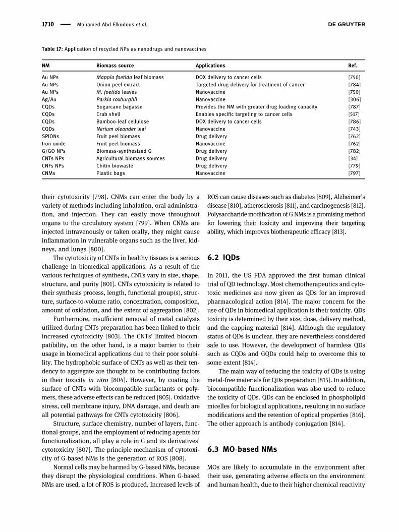
their cytotoxicity [798]. CNMs can enter the body by avariety of methods including inhalation, oral administra-tion, and injection. They can easily move throughoutorgans to the circulatory system [799]. When CNMs areinjected intravenously or taken orally, they might causeinflammation in vulnerable organs such as the liver, kid-neys, and lungs [800].
The cytotoxicity of CNTs in healthy tissues is a seriouschallenge in biomedical applications. As a result of thevarious techniques of synthesis, CNTs vary in size, shape,structure, and purity [801]. CNTs cytotoxicity is related totheir synthesis process, length, functional group(s), struc-ture, surface-to-volume ratio, concentration, composition,amount of oxidation, and the extent of aggregation [802].
Furthermore, insufficient removal of metal catalystsutilized during CNTs preparation has been linked to theirincreased cytotoxicity [803]. The CNTs’ limited biocom-patibility, on the other hand, is a major barrier to theirusage in biomedical applications due to their poor solubi-lity. The hydrophobic surface of CNTs as well as their ten-dency to aggregate are thought to be contributing factorsin their toxicity in vitro [804]. However, by coating thesurface of CNTs with biocompatible surfactants or poly-mers, these adverse effects can be reduced [805]. Oxidativestress, cell membrane injury, DNA damage, and death areall potential pathways for CNTs cytotoxicity [806].
Structure, surface chemistry, number of layers, func-tional groups, and the employment of reducing agents forfunctionalization, all play a role in G and its derivatives’cytotoxicity [807]. The principle mechanism of cytotoxi-city of G-based NMs is the generation of ROS [808].
Normal cells may be harmed by G-based NMs, becausethey disrupt the physiological conditions. When G-basedNMs are used, a lot of ROS is produced. Increased levels of
ROS can cause diseases such as diabetes [809], Alzheimer’sdisease [810], atherosclerosis [811], and carcinogenesis [812].Polysaccharidemodification of GNMs is a promisingmethodfor lowering their toxicity and improving their targetingability, which improves biotherapeutic efficacy [813].
6.2 IQDs
In 2011, the US FDA approved the first human clinicaltrial of QD technology. Most chemotherapeutics and cyto-toxic medicines are now given as QDs for an improvedpharmacological action [814]. The major concern for theuse of QDs in biomedical application is their toxicity. QDstoxicity is determined by their size, dose, delivery method,and the capping material [814]. Although the regulatorystatus of QDs is unclear, they are nevertheless consideredsafe to use. However, the development of harmless QDssuch as CQDs and GQDs could help to overcome this tosome extent [814].
The main way of reducing the toxicity of QDs is usingmetal-freematerials for QDs preparation [815]. In addition,biocompatible functionalization was also used to reducethe toxicity of QDs. QDs can be enclosed in phospholipidmicelles for biological applications, resulting in no surfacemodifications and the retention of optical properties [816].The other approach is antibody conjugation [814].
6.3 MO-based NMs
MOs are likely to accumulate in the environment aftertheir use, generating adverse effects on the environmentand human health, due to their higher chemical reactivity
Table 17: Application of recycled NPs as nanodrugs and nanovaccines
NM Biomass source Applications Ref.
Au NPs Mappia foetida leaf biomass DOX delivery to cancer cells [750]Au NPs Onion peel extract Targeted drug delivery for treatment of cancer [784]Au NPs M. foetida leaves Nanovaccine [750]Ag/Au Parkia roxburghii Nanovaccine [306]CQDs Sugarcane bagasse Provides the NM with greater drug loading capacity [787]CQDs Crab shell Enables specific targeting to cancer cells [517]CQDs Bamboo-leaf cellulose DOX delivery to cancer cells [786]CQDs Nerium oleander leaf Nanovaccine [743]SPIONs Fruit peel biomass Drug delivery [762]Iron oxide Fruit peel biomass Nanovaccine [762]G/GO NPs Biomass-synthesized G Drug delivery [782]CNTs NPs Agricultural biomass sources Drug delivery [34]CNFs NPs Chitin biowaste Drug delivery [779]CNMs Plastic bags Nanovaccine [797]
1710 Mohamed Abd Elkodous et al.

resulting in an increased generation of ROS [817]. Indeed,the surface of MOs is a major factor in their toxicity as aresult of the interaction of their surfaces with the biolo-gical system [818]. Different MOs can be found in nature,however, some of them have a wide range of biomedicalapplications, including IO NPs, TiO2 NPs, ZnO NPs, andCuO NPs [819].
The cytotoxicity of IO NPs is one of the major chal-lenges to their translation into biomedical applications.Generation of ROS, including hydroxyl radicals, super-oxide anion, and nonradical H2O2, is responsible for themajority of IO NPs intracellular cytotoxicity [820]. Thegeneration of ROS from the NP surface, ROS creation byleaching of iron molecules, modifying mitochondrial func-tions, and stimulation of signaling pathways are the fourprimary sources of oxidative damage in response to IO NPs[821]. Surface coating is one method for making IO NPsnon-toxic and biocompatible [822].
With rapid nanotechnology development, TiO2 NPsand ZnO NPs are widely used in various fields. Peopleworking in such environments, as well as researchers inlaboratories, are more likely to be exposed to TiO2 NPsand ZnO NPs [823,824]. Furthermore, increasing datasuggest that TiO2 NPs concentrations are elevated inanimal organs following systematic exposure, and thatthese accumulated NPs may cause organ damage. Oxida-tive stress, autophagy, apoptosis, inflammatory response,genotoxic effects, and signaling pathways all contribute toTiO2 NPs toxicity [823]. Cell membrane damage, cytotoxi-city, and increased oxidative stress have been described indiversemammalian cell lines as themost common harmfuleffect of ZnO NPs [825]. ZnO NPs have been shown to havegenotoxic potential in both in vivo and in vitro experi-ments, in addition to cytotoxicity [826].
CuO NPs have been shown to cause harmful effectson the liver and kidneys in many experiments [827]. Inexperimental animals, CuONPs have caused severe damagein the liver, spleen, and kidneys. Highly reactive ioniccopper is generated after oral administration and interac-tion with gastric fluid, which is then deposited in the kid-neys of exposed animals [828].
6.4 Au NPs
Au NPs have received a lot of interest in the biomedicinefield because of its electrical, chemical, thermal, mechan-ical, and optical properties, With the high potentials, thehigh risk of toxicity came, as a result of their interactionwith biological tissues and biomolecules [829]. In vivo
and in vitro experiments on Au NPs were carried out.Both showed some oxidative damage to organs and celllines in vivo and in vitro respectively, with the liver,kidney, and spleen being the most impacted [829].
The size and shape of Au NPs are the most essentialfactors that contribute to their cytotoxicity, because theyare directly associated to the process of cellular interna-lization [830]. The cytotoxicity of Au NPs is size depen-dent, with smaller (1.4 nm) Au NPs being more cytotoxicthan larger (15 nm) ones [831]. In HeLa cells, SK-Mel-28melanoma cells, L929 animal fibroblasts, and mousemonocytic/macrophage cells, Pan et al. found that 1.4 nmAu NPs caused rapid cell death through necrosis, whereas1.2 nm Au NPs caused cell death through apoptosis [831].The large surface areas of these extremely small particlescan contribute to the production of ROS. ROS injure cells bydestroying proteins, membranes, DNA, and other organelleslike the mitochondria, cytoplasm, and nucleus [829].
Vales et al. found that, in addition to size and shape,surface functionalization of Au NPs has a role in theircytotoxicity. Three functionalization were used in thisstudy: negative (carboxylate), positive (ammonium), andneutral (PEG). In comparison to anionic and neutral AuNPs, cationic Au NPs were more cytotoxic. A positive sur-face charge appears to exacerbate the cytotoxicity of AuNPs [832].
7 Conclusion and future prospects
To summarize, the authors have shed light on valuableNMs that can be extracted from waste sources originatingfrom everyday life, including biomass wastes, cookingoil, and industrial wastes, to achieve “waste-to-wealth”and meet the “zero-waste” challenge. Different CNMs,including inorganic QDs, various MOs, and Au NPs, canbe easily extracted from the above-mentioned sources.Then the different synthesis approaches used to synthe-size these NMs, which could be classified into top-downand bottom-up routes, were presented. Additionally, theirpotential roles in the early detection of serious diseases,with a special focus on their roles in fluorescent imaging,magnetic imaging, and when acting as biosensors, weresummarized. Moreover, the promising therapeutic modal-ities of NMs as antimicrobial, anticancer, antioxidant agents,and as nanodrugs and vaccines were analyzed in detail.
Many other recyclable NMs are valuable for biome-dical applications. Recently, palladium and platinumNPs were introduced as potential and novel mineralderived NPs synthesized from different sources of waste
Recent advances in waste-recycled NMs for biomedical applications 1711
via diverse green synthesis strategies [833–837]. They areamongst the widely used NPs for applications in biome-dicine like drug delivery, biosensors, imaging, photothermaltherapy, cosmetics, antimicrobials, and therapeutics formany afflictions (AIDS, cancer, and malaria), and implants[838–842]. Furthermore, they have unique anticancer [843],antioxidant, and antibacterial capabilities [844]. However,further research is needed to highlight their role in the earlydetection of different diseases. Although RNMs have shownmany outstanding characteristics such as cost-effectiveness,ease of manipulation, and good physiological properties,many investigations are still required for their further isola-tion and purification. In addition, the use of RNMs for otherbiomedical applications, such as bio-scaffolds in tissue engi-neering and regenerative medicine and wound-healingapplications, has not yet been addressed.
Acknowlegement: A.Matsuda andG. Kawamura acknowl-edge Japan Society for Promotion of Science (JSPS)KAKENHI Grant No. 18H03841 and No. 21K18823, forpartially funding this study.
Funding information: JapanSociety forPromotionofScience(JSPS) KAKENHI Grant No. 18H03841 and No. 21K18823.
Author contributions: All authors have accepted respon-sibility for the entire content of this manuscript andapproved its submission.
Conflict of interest: The authors state no conflict ofinterest.
References
[1] Aflori M. Smart nanomaterials for biomedical applications – areview. Nanomaterials. 2021;11(2):396.
[2] Jeevanandam J, Barhoum A, Chan YS, Dufresne A,Danquah MK. Review on nanoparticles and nanostructuredmaterials: history, sources, toxicity and regulations.Beilstein J Nanotechnol. 2018;9(1):1050–74.
[3] Lyu Q, Peng L, Hong X, Fan T, Li J, Cui Y, et al. Smart nano-micro platforms for ophthalmological applications: the state-of-the-art and future perspectives. Biomaterials.2021;270:120682.
[4] Lan L, Yao Y, Ping J, Ying Y. Recent advances in nanomaterial-based biosensors for antibiotics detection. BiosensBioelectron. 2017;91:504–14.
[5] Colino CI, Lanao JM, Gutiérrez-Millán C. Recent advances infunctionalized nanomaterials for the diagnosis and treat-ment of bacterial infections. Mater Sci Eng: C.2021;121:111843.
[6] Nejati K, Dadashpour M, Gharibi T, Mellatyar H,Akbarzadeh A. Biomedical applications of functionalizedgold nanoparticles: a review. J Clust Sci. 2021;1–16doi: 10.1007/s10876-020-01955-9.
[7] Jadoun S, Arif R, Jangid NK, Meena RK. Green synthesis ofnanoparticles using plant extracts: a review. Environ ChemLett. 2020;19:1–20.
[8] Abd Elkodous M, El-Sayyad GS, Abdelrahman IY, El-Bastawisy HS, Mohamed AE, Mosallam FM, et al. Therapeuticand diagnostic potential of nanomaterials for enhancedbiomedical applications. Colloids Surf B: Biointerfaces.2019;180:411–28.
[9] Abd Elkodous M, El-Sayyad GS, Youssry SM, Nada HG,Gobara M, Elsayed MA, et al. Carbon-dot-loadedCoxNi1−xFe2O4; x = 0.9/SiO2/TiO2 nanocomposite withenhanced photocatalytic and antimicrobial potential: anengineered nanocomposite for wastewater treatment.Sci Rep. 2020;10(1):11534.
[10] El-Sayyad GS, Abd Elkodous M, El-Khawaga AM, Elsayed MA,El-Batal AI, Gobara M. Merits of photocatalytic and antimi-crobial applications of gamma-irradiated CoxNi1−xFe2O4/SiO2/TiO2; x = 0.9 nanocomposite for pyridine removal andpathogenic bacteria/fungi disinfection: implication for was-tewater treatment. RSC Adv. 2020;10(9):5241–59.
[11] Wong CW, Chan YS, Jeevanandam J, Pal K, Bechelany M, AbdElkodous M, et al. Response surface methodology optimiza-tion of mono-dispersed MgO nanoparticles fabricated byultrasonic-assisted sol–gel method for outstanding antimi-crobial and antibiofilm activities. J Clust Sci.2020;31(2):367–89.
[12] Abd Elkodous M, El-Sayyad GS, Nasser HA, Elshamy AA,Morsi M, Abdelrahman IY, et al. Engineered nanomaterials aspotential candidates for HIV treatment: between opportu-nities and challenges. J Clust Sci. 2019;30(3):531–40.
[13] Abd Elkodous M, El-Sayyad GS, Abdel Maksoud MIA,Abdelrahman IY, Mosallam FM, Gobara M, et al. Fabricationof ultra-pure anisotropic zinc oxide nanoparticles via simpleand cost-effective route: implications for UTI and EAC medi-cations. Biol Trace Elem Res. 2020;196(1):297–317.
[14] El-Batal AI, Mosallam FM, Ghorab MM, Hanora A, Gobara M,Baraka A, et al. Factorial design-optimized and gamma irra-diation-assisted fabrication of selenium nanoparticles bychitosan and Pleurotus ostreatus fermented fenugreek for avigorous in vitro effect against carcinoma cells. Int J BiolMacromol. 2020;156:1584–99.
[15] Elkhenany H, Abd Elkodous M, Ghoneim NI, Ahmed TA,Ahmed SM, Mohamed IK, et al. Comparison of differentuncoated and starch-coated superparamagnetic iron oxidenanoparticles: implications for stem cell tracking. Int J BiolMacromol. 2020;143:763–74.
[16] El-Batal AI, Abd Elkodous M, El-Sayyad GS, Al-Hazmi NE,Gobara M, Baraka A. Gum Arabic polymer-stabilized andGamma rays-assisted synthesis of bimetallic silver-goldnanoparticles: powerful antimicrobial and antibiofilm activ-ities against pathogenic microbes isolated from diabetic footpatients. Int J Biol Macromol. 2020;165:169–86.
[17] Abd Elkodous M, El-Sayyad GS, Abdel-Daim MM. Engineerednanomaterials as fighters against SARS-CoV-2: the way tocontrol and treat pandemics. Environ Sci Pollut Res.2020;28:40409–15.
1712 Mohamed Abd Elkodous et al.

[18] Eldebany N, Abd Elkodous M, Tohamy H, Abdelwahed R,El-kammar M, Abou-Ahmed H, et al. Gelatin loaded titaniumdioxide and silver oxide nanoparticles: implication for skintissue regeneration. Biol Trace Elem Res.2020;199:3688–99.
[19] Elkhenany H, Elkodous MA, Newby SD, El-Derby AM, Dhar M,El-Badri N. Tissue Engineering Modalities andNanotechnology. In: El-Badri N, editor. Regenerative medi-cine and stem cell biology. Cham: Springer InternationalPublishing; 2020. p. 289–322.
[20] Attia MS, El-Sayyad GS, Abd Elkodous M, Khalil WF,Nofel MM, Abdelaziz AM, et al. Chitosan and EDTA conju-gated graphene oxide antinematodes in Eggplant: towardimproving plant immune response. Int J Biol Macromol.2021;179:333–44.
[21] Attia MS, Balabel NM, Ababutain IM, Osman MS, Nofel MM,Abd Elkodous M, et al. Protective role of copper oxide-strep-tomycin nano-drug against potato brown rot disease causedby ralstonia solanacearum. J Clust Sci. 2021;28:27577–92.
[22] Jung Y, Huh Y, Kim D. Recent advances in surface engineeringof porous silicon nanomaterials for biomedical applications.Microporous Mesoporous Mater. 2020;310:110673.
[23] Tang H, Zhao X, Jiang X. Synthetic multi-layer nanoparticlesfor CRISPR-Cas9 genome editing. Adv drug Delivery Rev.2021;168:55–78.
[24] Zaheer T, Pal K, Zaheer I. Topical review on nano-vacci-nology: biochemical promises and key challenges. ProcessBiochem. 2020;100:237–44.
[25] Lubick N. Nanosilver toxicity: ions, nanoparticles or both?ACS Publications; 2008.
[26] Zoroddu MA, Medici S, Ledda A, Nurchi VM, Lachowicz JI,Peana M. Toxicity of nanoparticles. Curr Med Chem.2014;21(33):3837–53.
[27] Al-Tamimi SA. Biogenic green synthesis of metal oxidenanoparticles using oat biomass for ultrasensitive modifiedpolymeric sensors. Green Chem Lett Rev. 2021;14(2):166–79.
[28] Nasrollahzadeh M, Sajjadi M, Iravani S, Varma RS. Carbon-based sustainable nanomaterials for water treatment: state-of-art and future perspectives. Chemosphere.2020;263:128005.
[29] Goldoni R, Farronato M, Connelly ST, Tartaglia GM, Yeo W-H.Recent advances in graphene-based nanobiosensors forsalivary biomarker detection. Biosens Bioelectron.2020;171:112723.
[30] Omoriyekomwan JE, Tahmasebi A, Dou J, Wang R, Yu J.A review on the recent advances in the production of carbonnanotubes and carbon nanofibers via microwave-assistedpyrolysis of biomass. Fuel Process Technol.2020;214:106686.
[31] Esposito Corcione C, Ferrari F, Striani R, Greco A. Transportproperties of natural and artificial smart fabrics impregnatedby graphite nanomaterial stacks. Nanomaterials.2021;11(4):1018.
[32] Azam MA, Abd Mudtalib NESA, Seman RNAR. Synthesis ofgraphene nanoplatelets from palm-based waste chickenfrying oil carbon feedstock by using catalytic chemicalvapour deposition. Mater Today Commun. 2018;15:81–7.
[33] Asiya S, Pal K, Kralj S, El-Sayyad G, de Souza F, Narayanan T.Sustainable preparation of gold nanoparticles via green
chemistry approach for biogenic applications. Mater TodayChem. 2020;17:100327.
[34] Fathy NA, Basta AH, Lotfy VF. Novel trends for synthesis ofcarbon nanostructures from agricultural wastes, carbonnanomaterials for agri-food and environmental applications.Elsevier; 2020. p. 59–74
[35] de Diego-Díaz B, Cerdán JMA, Peñas FJ, Fernández-Rodríguez J. Impact of supplementary nutrients on codiges-tion of agricultural waste: study of temperatures. FoodBioprod Process. 2018;110:120–5.
[36] Radich A. Biodiesel performance, costs, and use. US EnergyInformation Administration; 2006.
[37] Kulkarni MG, Dalai AK. Waste cooking oil an economicalsource for biodiesel: a review. Ind Eng Chem Res.2006;45(9):2901–13.
[38] Carter D. How to make biodiesel, low-impact living initia-tive; 2005.
[39] Stewart E, Lemieux P. Emissions from the incineration ofelectronics industry waste. IEEE International Symposium onElectronics and the Environment. IEEE; 2003. p. 271–5.
[40] Huo X, Peng L, Xu X, Zheng L, Qiu B, Qi Z, et al. Elevated bloodlead levels of children in Guiyu, an electronic waste recycl-ing town in China. Environ Health Perspect.2007;115(7):1113–7.
[41] Ozdemir I, Şahin M, Orhan R, Erdem M. Preparation andcharacterization of activated carbon from grape stalk byzinc chloride activation. Fuel Process Technol.2014;125:200–6.
[42] Arie AA, Hadisaputra L, Susanti RF, Devianto H, Halim M,Enggar R, et al. Synthesis of carbon nano materials origi-nated from waste cooking oil using a nebulized spray pyro-lysis. J Phys Conf Ser. IOP Publishing; 2017. p. 012020.
[43] Nazem MA, Zare MH, Shirazian S. Preparation and optimi-zation of activated nano-carbon production using physicalactivation by water steam from agricultural wastes. RSC Adv.2020;10(3):1463–75.
[44] Sivaprakash S, Kumar PS, Krishna S. Synthesis & charac-teristic study of agricultural waste activated carbon/Fe3O4–Nano particles. Int J Mater Sci. 2017;12(1):97–105.
[45] Sinto J, Nugroho YW, Naf’an HI. Preparation of activatedcarbon from banana peel waste as adsorbent for motorvehicle exhaust emissions, E3S web of conferences. EDP Sci.2018;03020.
[46] Afdhol M, Amiliana R, Hanafi A, Rachmanda B. Preparation ofactivated carbon from palm shells using KOH and ZnCl2 asthe activating agent. IOP Conf Ser Mater Sci Eng. IOPPublishing; 2017. p. 012282
[47] Román S, González J, González-García C, Zamora F. Control ofpore development during CO2 and steam activation of olivestones. Fuel Process Technol. 2008;89(8):715–20.
[48] Kula I, Uğurlu M, Karaoğlu H, Celik A. Adsorption of Cd (II)ions from aqueous solutions using activated carbon pre-pared from olive stone by ZnCl2 activation. BioresourTechnol. 2008;99(3):492–501.
[49] Martinez ML, Torres MM, Guzman CA, Maestri D. Preparationand characteristics of activated carbon from olive stones andwalnut shells. Ind Crop Products. 2006;23(1):23–8.
[50] Yang K, Peng J, Srinivasakannan C, Zhang L, Xia H, Duan X.Preparation of high surface area activated carbon from
Recent advances in waste-recycled NMs for biomedical applications 1713

coconut shells using microwave heating. Bioresour Technol.2010;101(15):6163–9.
[51] Yahya MA, Al-Qodah Z, Ngah CZ. Agricultural bio-wastematerials as potential sustainable precursors used for acti-vated carbon production: a review. Renew Sustain EnergyRev. 2015;46:218–35.
[52] Musuna-Garwe CC, Mukaratirwa-Muchanyereyi N, Mupa M,Mahamadi C, Mujuru M. Preparation and characterization ofnanocarbons from Nicotiana tabacum stems. 2018
[53] Gao P, Liu Z-h, Xue G, Han B, Zhou M-h. Preparation andcharacterization of activated carbon produced from ricestraw by (NH) 2HPO4 activation. Bioresour Technol.2011;102(3):3645–8.
[54] Nandiyanto ABD, Putra ZA, Andika R, Bilad MR, Kurniawan T,Zulhijah R, et al. Porous activated carbon particles from ricestraw waste and their adsorption properties. J Eng SciTechnol. 2017;12(8):1–11.
[55] Gin W, Jimoh A, Abdulkareem A, Giwa A. Production of acti-vated carbon from watermelon peel. Int J Sci Eng Res.2014;5:66–71.
[56] Roopa D. Preparation of activated carbon from bitter orangepeel (citrus aurantium) and preliminary studies on its char-acteristics. J Clin Diag Res: JCDR. 2016;10:6–11.
[57] Elaiyappillai E, Srinivasan R, Johnbosco Y, Devakumar P,Murugesan K, Kesavan K, et al. Low cost activatedcarbon derived from Cucumis melo fruit peel for electro-chemical supercapacitor application. Appl Surf Sci.2019;486:527–38.
[58] Yuliusman MPAyu, Hanafi A, Nafisah AR. Activated carbonpreparation from durian peel wastes using chemical andphysical activation. AIP Conf Proc. AIP Publishing LLC; 2020.p. 030020
[59] Murugesan K, Tareke K, Gezehegn M, Kebede M, Yazie A,Diyana G. Rapid development of activated carbon and ZnOnanoparticles via green waste conversion using avocado fruitpeel powder and its high performance efficiency in aqueousdye removal application. J Inorg Organomet Polym Mater.2019;29(4):1368–74.
[60] Vinha AF, Moreira J, Barreira SV. Physicochemical para-meters, phytochemical composition and antioxidant activityof the algarvian avocado (Persea americana Mill.). J Agric Sci.2013;5(12):100.
[61] Osman AI, Blewitt J, Abu-Dahrieh JK, Farrell C, Ala’a H,Harrison J, et al. Production and characterisation of activatedcarbon and carbon nanotubes from potato peel waste andtheir application in heavy metal removal. Environ Sci PollutRes. 2019;26(36):37228–41.
[62] Moreno-Piraján J, Giraldo L. Activated carbon obtained bypyrolysis of potato peel for the removal of heavy metalcopper (II) from aqueous solutions. J Anal Appl Pyrolysis.2011;90(1):42–7.
[63] Ogungbenro AE, Quang DV, Al-Ali KA, Vega LF, Abu-Zahra MR.Synthesis and characterization of activated carbon frombiomass date seeds for carbon dioxide adsorption. J EnvironChem Eng. 2020;8(5):104257.
[64] Srinivasan R, Elaiyappillai E, Pandian HP, Vengudusamy R,Johnson PM, Chen S-M, et al. Sustainable porous activatedcarbon from Polyalthia longifolia seeds as electrode materialfor supercapacitor application. J Electroanalytical Chem.2019;849:113382.
[65] Abatan OG, Oni BA, Agboola O, Efevbokhan V, Abiodun OO.Production of activated carbon from African star apple seedhusks, oil seed and whole seed for wastewater treatment.J Clean Prod. 2019;232:441–50.
[66] Prakash MO, Raghavendra G, Ojha S, Panchal M.Characterization of porous activated carbon prepared fromarhar stalks by single step chemical activation method.Mater Today: Proc. 2020;39:1476–81.
[67] Shahraki ZH, Sharififard H, Lashanizadegan A. Grape stalksbiomass as raw material for activated carbon production:synthesis, characterization and adsorption ability. Mater Resexpress. 2018;5(5):055603.
[68] Khanna V, Bakshi BR, Lee LJ. Carbon nanofiber production:life cycle energy consumption and environmental impact.J Ind Ecol. 2008;12(3):394–410.
[69] Alfarisa S, Abu Bakar S, Mohamed A, Hashim N, Kamari A,Md Isa I, et al. Carbon nanostructures production from wastematerials: a review. Advanced Materials Research. TransTech Publ; 2015. p. 50–4.
[70] Hussain CM. Handbook of nanomaterials for industrialapplications. Elsevier; 2018.
[71] Tsai W-T. Mandatory recycling of waste cooking oil fromresidential and commercial sectors in Taiwan. Resources.2019;8(1):38.
[72] Janas D. From bio to nano: a review of sustainable methods ofsynthesis of carbon nanotubes. Sustainability.2020;12(10):4115.
[73] Chen W, Meng X-T, Wang H-H, Zhang X-Q, Wei Y, Li Z-Y, et al.Way to produce carbon nanofiber by electrospinning fromsugarcane Bagasse. Polymers. 2019;11(12):1968.
[74] Chen X-W, Timpe O, Hamid SB, Schlögl R, Su DS. Directsynthesis of carbon nanofibers on modified biomass-derivedactivated carbon. Carbon. 2009;47(1):340–3.
[75] Zhang J, Tahmasebi A, Omoriyekomwan JE, Yu J. Directsynthesis of hollow carbon nanofibers on bio-char duringmicrowave pyrolysis of pine nut shell. J Anal Appl Pyrolysis.2018;130:142–8.
[76] Wang Z, Shen D, Wu C, Gu S. State-of-the-art on the pro-duction and application of carbon nanomaterials from bio-mass. Green Chem. 2018;20(22):5031–57.
[77] Su X-L, Chen J-R, Zheng G-P, Yang J-H, Guan X-X, Liu P, et al.Three-dimensional porous activated carbon derived fromloofah sponge biomass for supercapacitor applications. ApplSurf Sci. 2018;436:327–36.
[78] Tao L, Huang Y, Zheng Y, Yang X, Liu C, Di M, et al. Porouscarbon nanofiber derived from a waste biomass as anodematerial in lithium-ion batteries. J Taiwan Inst Chem Eng.2019;95:217–26.
[79] Shi G, Liu C, Wang G, Chen X, Li L, Jiang X, et al. Preparationand electrochemical performance of electrospun biomass-based activated carbon nanofibers. Ionics.2019;25(4):1805–12.
[80] Hassan M, Abou-Zeid R, Hassan E, Berglund L, Aitomäki Y,Oksman K. Membranes based on cellulose nanofibers andactivated carbon for removal of escherichia coli bacteria fromwater. Polymers. 2017;9(8):335.
[81] Fadillah A, Hartoyo A, Solikhin A. Business model develop-ment of oil palm empty fruit bunch and trunk carbon nano-fibers–based water purifier. IOP Conf Ser Earth Environ Sci.IOP Publishing; 2020. p. 012018.
1714 Mohamed Abd Elkodous et al.
[82] Gaud B, Singh A, Jaybhaye S, Narayan H. Synthesis of carbonnano fiber from organic waste and activation of its surfacearea. Int J. 2019;2766:2748.
[83] Yadav S, Sharma CS. Novel and green processes for citruspeel extract: a natural solvent to source of carbon. PolymBull. 2018;75(11):5133–42.
[84] Sari SN, Melati A. Facile preparation of carbon nanofiber frombanana peel waste. Mater Today: Proc. 2019;13:165–8.
[85] Shahba H, Sabet M. Two-step and green synthesis of highlyfluorescent carbon quantum dots and carbon nanofibersfrom pine fruit. J Fluorescence. 2020;30(4):927–38.
[86] Sami A, MohammedM, Dhary A. Synthesis of carbon nanofibersfrom decomposition of liquid organic waste from chemical andpetrochemical industries. Energy Proc. 2015;74:4–14.
[87] Biswas SK, Das AK, Yano H, Shams M. Development of highperformance transparent nanocomposites reinforced withnanofibrillated chitin extracted from shrimp wastes. J ChitinChitosan Sci. 2013;1(2):138–43.
[88] Rambau KM, Musyoka NM, Manyala N, Ren J, Langmi HW,Mathe MK. Preparation of carbon nanofibers/tubes usingwaste tyres pyrolysis oil and coal fly ash derived catalyst.J Environ Sci Health, Part A. 2018;53(12):1115–22.
[89] Widiatmoko P, Nugraha N, Devianto H, Nurdin I, Prakoso T.Upscaled synthesis of carbon nanotube from palm oil milleffluent using pyrolysis for supercapacitor application. IOPConf Ser Mater Sci Eng. IOP Publishing; 2020. p. 012040
[90] Vinayan B, Nagar R, Raman V, Rajalakshmi N,Dhathathreyan K, Ramaprabhu S. Synthesis of graphene-multiwalled carbon nanotubes hybrid nanostructure bystrengthened electrostatic interaction and its lithium ionbattery application. J Mater Chem. 2012;22(19):9949–56.
[91] Robati D. Pseudo-second-order kinetic equations for mod-eling adsorption systems for removal of lead ions usingmulti-walled carbon nanotube. J Nanostruct Chem.2013;3(1):1–6.
[92] Iijima S. Helical microtubules of graphitic carbon. Nature.1991;354(6348):56–8.
[93] Yan Y, Miao J, Yang Z, Xiao F-X, Yang HB, Liu B, et al. Carbonnanotube catalysts: recent advances in synthesis, charac-terization and applications. Chem Soc Rev.2015;44(10):3295–3346.
[94] De Volder MF, Tawfick SH, Baughman RH, Hart AJ. Carbonnanotubes: present and future commercial applications.Science. 2013;339(6119):535–9.
[95] Mugadza K, Stark A, Ndungu PG, Nyamori VO. Synthesis ofcarbon nanomaterials from biomass utilizing ionic liquids forpotential application in solar energy conversion and storage.Materials. 2020;13(18):3945.
[96] Liu W, Yuan H. Simultaneous production of hydrogen andcarbon nanotubes from cracking of a waste cooking oil modelcompound over Ni‐Co/SBA‐15 catalysts. Int J Energy Res.2020;44(14):11564–82.
[97] Mannu A, Ferro M, Pietro MED, Mele A. Innovative applica-tions of waste cooking oil as raw material. Sci Prog.2019;102(2):153–60.
[98] Gui MM, Lee K, Bhatia S. Feasibility of edible oil vs. non-edible oil vs. waste edible oil as biodiesel feedstock. Energy.2008;33(11):1646–53.
[99] Lin CSK, Pfaltzgraff LA, Herrero-Davila L, Mubofu EB,Abderrahim S, Clark JH, et al. Food waste as a valuable
resource for the production of chemicals, materials andfuels. Current situation and global perspective. EnergyEnviron Sci. 2013;6(2):426–64.
[100] Suriani A, Nor RM, Rusop M. Vertically aligned carbonnanotubes synthesized from waste cooking palm oil. J CeramSoc Jpn. 2010;118(1382):963–8.
[101] Sangeetha J, Thangadurai D, Hospet R, Purushotham P,Manowade KR, Mujeeb MA, et al. Production of bionanoma-terials from agricultural wastes. Nanotechnology. Springer;2017. p. 33–58
[102] Hidalgo P, Coronado G, Sánchez A, Hunter R. Agro-industrialwaste as precursor source for carbon nanotubes (cnts)synthesis using a new technical of solvent autoignition. IOPConf Ser Earth Environ Sci. 2020;503:012025.
[103] Mugadza K, Ndungu PG, Stark A, Nyamori VO. Conversion ofresidue biomass into value added carbon materials: utilisa-tion of sugarcane bagasse and ionic liquids. J Mater Sci.2019;54(19):12476–87.
[104] Fathy NA. Carbon nanotubes synthesis using carbonizationof pretreated rice straw through chemical vapor deposition ofcamphor. RSC Adv. 2017;7(45):28535–41.
[105] Mopoung S. Occurrence of carbon nanotube from bananapeel activated carbon mixed with mineral oil. Int J Phys Sci.2011;6(7):1789–92.
[106] Osman AI, Blewitt J, Abu-Dahrieh JK, Farrell C, Al-Muhtaseb AAH,Harrison J, et al. Production and characterisation of activatedcarbon and carbon nanotubes from potato peel waste and theirapplication in heavy metal removal. Environ Sci Pollut Res.2019;26(36):37228–41.
[107] Singh V, Chatterjee S, Palecha M, Sen P, Ateeq B, Verma V.Chickpea peel waste as sustainable precursor for synthesisof fluorescent carbon nanotubes for bioimaging application.Carbon Lett. 2020;31:1–7.
[108] Robaiah M, Rusop M, Abdullah S, Khusaimi Z, Azhan H,Asli N. Synthesis graphene layer at different waste cookingpalm oil temperatures. AIP Conf Proc. AIP Publishing LLC;2017. p. 030008
[109] Bhuyan MSA, Uddin MN, Islam MM, Bipasha FA, Hossain SS.Synthesis of graphene. Int Nano Lett. 2016;6(2):65–83.
[110] Purkait T, Singh G, Singh M, Kumar D, Dey RS. Large areafew-layer graphene with scalable preparation from wastebiomass for high-performance supercapacitor. Sci Rep.2017;7(1):1–14.
[111] Gupta K, Gupta D, Khatri OP. Graphene-like porous carbonnanostructure from Bengal gram bean husk and its applica-tion for fast and efficient adsorption of organic dyes.Appl Surf Sci. 2019;476:647–57.
[112] Jacob MV, Rawat RS, Ouyang B, Bazaka K, Kumar DS,Taguchi D, et al. Catalyst-free plasma enhanced growthof graphene from sustainable sources. Nano Lett.2015;15(9):5702–8.
[113] Chen F, Yang J, Bai T, Long B, Zhou X. Facile synthesis of few-layer graphene from biomass waste and its applicationin lithium ion batteries. J Electroanal Chem.2016;768:18–26.
[114] Zhou H, Zhang J, Amiinu IS, Zhang C, Liu X, Tu W, et al.Transforming waste biomass with an intrinsically porousnetwork structure into porous nitrogen-doped graphene forhighly efficient oxygen reduction. Phys Chem Chem Phys.2016;18(15):10392–99.
Recent advances in waste-recycled NMs for biomedical applications 1715

[115] Sun L, Tian C, Li M, Meng X, Wang L, Wang R, et al. Fromcoconut shell to porous graphene-like nanosheets for high-power supercapacitors. J Mater Chem A. 2013;1(21):6462–70.
[116] Shams SS, Zhang LS, Hu R, Zhang R, Zhu J. Synthesis ofgraphene from biomass: a green chemistry approach. MaterLett. 2015;161:476–9.
[117] Long S-Y, Du Q-S, Wang S-Q, Tang P-D, Li D-P, Huang R-B.Graphene two-dimensional crystal prepared from cellulosetwo-dimensional crystal hydrolysed from sustainable bio-mass sugarcane bagasse. J Clean Prod. 2019;241:118209.
[118] Qu J, Luo C, Zhang Q, Cong Q, Yuan X. Easy synthesis ofgraphene sheets from alfalfa plants by treatment of nitricacid. Mater Sci Eng: B. 2013;178(6):380–2.
[119] Karuppannan M, Kim Y, Sung Y-E, Kwon OJ. Nitrogen andsulfur co-doped graphene-like carbon sheets derived fromcoir pith bio-waste for symmetric supercapacitor applica-tions. J Appl Electrochem. 2019;49(1):57–66.
[120] Singh P, Bahadur J, Pal K. One-step one chemical synthesisprocess of graphene from rice husk for energy storageapplications. Graphene. 2017;6(3):61–71.
[121] Kuila T, Bose S, Khanra P, Mishra AK, Kim NH, Lee JH. A greenapproach for the reduction of graphene oxide by wild carrotroot. Carbon. 2012;50(3):914–21.
[122] Baweja H, Jeet K. Economical and green synthesis of gra-phene and carbon quantum dots from agricultural waste.Mater Res Exp. 2019;6(8):0850g8.
[123] Somanathan T, Prasad K, Ostrikov KK, Saravanan A,Krishna VM. Graphene oxide synthesis from agro waste.Nanomaterials. 2015;5(2):826–34.
[124] Mahendran GB, Ramalingam SJ, Rayappan JBB, Kesavan S,Periathambi T, Nesakumar N. Green preparation of reducedgraphene oxide by Bougainvillea glabra flower extract andsensing application. J Mater Sci: Mater Electron.2020;31(17):14345–56.
[125] Shah J, Lopez-Mercado J, Carreon MG, Lopez-Miranda A,Carreon ML. Plasma synthesis of graphene from mango peel.ACS Omega. 2018;3(1):455–63.
[126] Subramaniyan R, Kiruthika N, Somanathan T, Sriraman V,Shanmugam M. High performance of graphene oxide frombanana peel against Pseudomonas aeruginosa andEscherichia coli. Res J Pharm Technol. 2018;11(7):2932–4.
[127] Tahir NAM, Abdollah MFB, Tamaldin N, Zin MRBM,Amiruddin H. Optimisation of graphene grown from solidwaste using CVD method. Int J Adv Manuf Technol.2020;106(1):211–8.
[128] Ruan G, Sun Z, Peng Z, Tour JM. Growth of graphene fromfood, insects, and waste. ACS Nano. 2011;5(9):7601–7.
[129] Akhavan O, Bijanzad K, Mirsepah A. Synthesis of graphenefrom natural and industrial carbonaceous wastes. RSC Adv.2014;4(39):20441–48.
[130] Chandrasekar S, Viswanathan PK, Uthirakumar P,Montanari GC. Investigations on novel carbon quantum dotscovered nanofluid insulation for medium voltage applica-tions. J Electr Eng Technol. 2020;15(1):269–78.
[131] Muthoni J, Shimelis H, Melis R, Kinyua Z. Response of potatogenotypes to bacterial wilt caused by Ralstonia solana-cearum (Smith)(Yabuuchi et al.) in the tropical highlands. AmJ Potato Res. 2014;91(2):215–32.
[132] Wang J, Wang CF, Chen S. Amphiphilic egg‐derived carbondots: rapid plasma fabrication, pyrolysis process, and
multicolor printing patterns. Angew Chem Int Ed.2012;51(37):9297–9301.
[133] Ke Y, Garg B, Ling Y-C. Waste chicken eggshell as low-costprecursor for efficient synthesis of nitrogen-doped fluores-cent carbon nanodots and their multi-functional applica-tions. RSC Adv. 2014;4(102):58329–36.
[134] Zhao Y, Zhang Y, Liu X, Kong H, Wang Y, Qin G, et al. Novelcarbon quantum dots from egg yolk oil and their haemostaticeffects. Sci Rep. 2017;7(1):1–8.
[135] Thangaraj B, Solomon PR, Ranganathan S. Synthesis ofcarbon quantum dots with special reference to biomass as asource-a review. Curr Pharm Des. 2019;25(13):1455–76.
[136] Thambiraj S, Shankaran R. Green synthesis of highly fluor-escent carbon quantum dots from sugarcane bagasse pulp.Appl Surf Sci. 2016;390:435–43.
[137] Kang C, Huang Y, Yang H, Yan XF, Chen ZP. A review of carbondots produced from biomass wastes. Nanomaterials.2020;10(11):2316.
[138] Babbar N, Oberoi HS. Enzymes in value-addition of agricul-tural and agro-industrial residues. In: Brar SK, Verma M,editors. Enzymes in value-addition of wastes; 2014.p. 29–50.
[139] Zhuo C, Alves JO, Tenorio JA, Levendis YA. Synthesis ofcarbon nanomaterials through up-cycling agricultural andmunicipal solid wastes. Ind Eng Chem Res.2012;51(7):2922–30.
[140] Jaguaribe EF, Medeiros LDL, Barreto MDCS, Araujo LPD. Theperformance of activated carbons from sugarcane bagasse,babassu, and coconut shells in removing residual chlorine.Braz J Chem Eng. 2005;22(1):41–7.
[141] Lakshmidevi R, Muthukumar K. Enzymatic saccharificationand fermentation of paper and pulp industry effluent forbiohydrogen production. Int J Hydrog Energy.2010;35(8):3389–400.
[142] Du F, Zhang M, Li X, Li J, Jiang X, Li Z, et al. Economical andgreen synthesis of bagasse-derived fluorescent carbon dotsfor biomedical applications. Nanotechnology.2014;25(31):315702.
[143] Arampatzidou AC, Deliyanni EA. Comparison of activationmedia and pyrolysis temperature for activated carbonsdevelopment by pyrolysis of potato peels for effectiveadsorption of endocrine disruptor bisphenol-A. J ColloidInterface Sci. 2016;466:101–12.
[144] Xu-Cheng F, Xuan-Hua L, Jin J-Z, Zhang J, Wei G. Facilesynthesis of bagasse waste derived carbon dots for tracemercury detection. Mater Res Exp. 2018;5(6):065044.
[145] Zhu J, Zhu F, Yue X, Chen P, Sun Y, Zhang L, et al. Wasteutilization of synthetic carbon quantum dots based on teaand peanut shell. J Nanomater. 2019;2019(2019):1–7.
[146] Chunduri L, Kurdekar A, Patnaik S, Dev BV, Rattan TM,Kamisetti V. Carbon quantum dots from coconut husk: eva-luation for antioxidant and cytotoxic activity. Mater Focus.2016;5(1):55–61.
[147] Zhang J, Wang H, Xiao Y, Tang J, Liang C, Li F, et al. A simpleapproach for synthesizing of fluorescent carbon quantumdots from tofu wastewater. Nanoscale Res Lett.2017;12(1):611.
[148] Wang K, Ji Q, Xu J, Li H, Zhang D, Liu X, et al. Highly sensitiveand selective detection of amoxicillin using carbon quantumdots derived from beet. J Fluorescence. 2018;28(3):759–65.
1716 Mohamed Abd Elkodous et al.

[149] Cheng C, Shi Y, Li M, Xing M, Wu Q. Carbon quantum dotsfrom carbonized walnut shells: structural evolution, fluores-cence characteristics, and intracellular bioimaging. Mater SciEng: C. 2017;79:473–80.
[150] Li Z, Wang Q, Zhou Z, Zhao S, Zhong S, Xu L, et al. Greensynthesis of carbon quantum dots from cQuantum YieldMeasurements.orn stalk shell by hydrothermal approach innear-critical water and applications in detecting and bioi-maging. Microchem J. 2021;166:106250.
[151] Zhang L, Wang Y, Liu W, Ni Y, Hou Q. Corncob residues ascarbon quantum dots sources and their application indetection of metal ions. Ind Crop Products. 2019;133:18–25.
[152] Amin N, Afkhami A, Hosseinzadeh L, Madrakian T. Green andcost-effective synthesis of carbon dots from date kernel andtheir application as a novel switchable fluorescence probefor sensitive assay of Zoledronic acid drug in human serumand cellular imaging. Anal Chim Acta. 2018;1030:183–93.
[153] Yang R, Guo X, Jia L, Zhang Y, Zhao Z, Lonshakov F. Greenpreparation of carbon dots with mangosteen pulp for theselective detection of Fe3+ ions and cell imaging. Appl SurfSci. 2017;423:426–32.
[154] Narayanan DP, Cherikallinmel SK, Sankaran S,Narayanan BN. Functionalized carbon dot adorned coconutshell char derived green catalysts for the rapid synthesis ofamidoalkyl naphthols. J Colloid Interface Sci.2018;520:70–80.
[155] Vasantharaj S, Sathiyavimal S, Senthilkumar P,LewisOscar F, Pugazhendhi A. Biosynthesis of iron oxidenanoparticles using leaf extract of Ruellia tuberosa: antimi-crobial properties and their applications in photocatalyticdegradation. J Photochem Photobiol B: Biol.2019;192:74–82.
[156] Qi H, Teng M, Liu M, Liu S, Li J, Yu H, et al. Biomass-derivednitrogen-doped carbon quantum dots: highly selectivefluorescent probe for detecting Fe3+ ions and tetracyclines.J Colloid Interface Sci. 2019;539:332–41.
[157] Ren X, Zhang F, Guo B, Gao N, Zhang X. Synthesis of N-dopedmicropore carbon quantum dots with high quantum yieldand dual-wavelength photoluminescence emission frombiomass for cellular imaging. Nanomaterials (Basel, Switz).2019;9(4):495–508.
[158] Yuan M, Zhong R, Gao H, Li W, Yun X, Liu J, et al. One-step,green, and economic synthesis of water-soluble photolumi-nescent carbon dots by hydrothermal treatment of wheatstraw, and their bio-applications in labeling, imaging, andsensing. Appl Surf Sci. 2015;355:1136–44.
[159] Huang C, Dong H, Su Y, Wu Y, Narron R, Yong Q. Synthesis ofcarbon quantum dot nanoparticles derived from byproductsin bio-refinery process for cell imaging and in vivo bioima-ging. Nanomaterials (Basel). 2019;9(3):387.
[160] Vandarkuzhali SAA, Jeyalakshmi V, Sivaraman G,Singaravadivel S, Krishnamurthy KR, Viswanathan B. Highlyfluorescent carbon dots from Pseudo-stem of banana plant:applications as nanosensor and bio-imaging agents. SensActuators B: Chem. 2017;252:894–900.
[161] Bandi R, Gangapuram BR, Dadigala R, Eslavath R, Singh SS,Guttena V. Facile and green synthesis of fluorescent carbondots from onion waste and their potential applications assensor and multicolour imaging agents. RSC Adv.2016;6(34):28633–9.
[162] Gedda G, Lee C-Y, Lin Y-C, Wu H-F. Green synthesis of carbondots from prawn shells for highly selective and sensitivedetection of copper ions. Sens Actuators B: Chem.2016;224:396–403.
[163] Zheng M, Li Y, Liu S, Wang W, Xie Z, Jing X. One-pot tosynthesize multifunctional carbon dots for near infraredfluorescence imaging and photothermal cancer therapy.ACS Appl Mater Interfaces. 2016;8(36):23533–41.
[164] Chen K, Qing W, Hu W, Lu M, Wang Y, Liu X. On-off-onfluorescent carbon dots from waste tea: their properties,antioxidant and selective detection of CrO42−, Fe3+,ascorbic acid and L-cysteine in real samples. SpectrochimActa Part A: Mol Biomole Spectrosc. 2019;213:228–34.
[165] Sachdev A, Gopinath P. Green synthesis of multifunctionalcarbon dots from coriander leaves and their potential appli-cation as antioxidants, sensors and bioimaging agents.Analyst. 2015;140(12):4260–9.
[166] Amer WA, Rehab AF, Abdelghafar ME, Torad NL, Atlam AS,Ayad MM. Green synthesis of carbon quantum dots frompurslane leaves for the detection of formaldehyde usingquartz crystal microbalance. Carbon. 2021;179:159–71.
[167] Yadav PK, Singh VK, Chandra S, Bano D, Kumar V, Talat M,et al. Green synthesis of fluorescent carbon quantum dotsfrom azadirachta indica leaves and their peroxidase-mimeticactivity for the detection of H2O2 and ascorbic acid incommon fresh fruits. ACS Biomater Sci Eng.2019;5(2):623–32.
[168] Arumugham T, Alagumuthu M, Amimodu RG, Munusamy S,Iyer SK. A sustainable synthesis of green carbon quantumdot (CQD) from Catharanthus roseus (white flowering plant)leaves and investigation of its dual fluorescence responsivebehavior in multi-ion detection and biological applications.Sustain Mater Technol. 2020;23:e00138.
[169] Pourreza N, Ghomi M. Green synthesized carbon quantumdots from Prosopis juliflora leaves as a dual off-on fluores-cence probe for sensing mercury (II) and chemet drug. MaterSci Eng: C. 2019;98:887–96.
[170] Shivaji K, Mani S, Ponmurugan P, De Castro CS, LloydDavies M, Balasubramanian MG, et al. Green-synthesis-derived CdS quantum dots using tea leaf extract: antimicro-bial, bioimaging, and therapeutic applications in lung cancercells, cancer cells. ACS Appl Nano Mater. 2018;1(4):1683–93.
[171] Atchudan R, Jebakumar Immanuel Edison TN, ShanmugamM,Perumal S, Somanathan T, Lee YR. Sustainable synthesis ofcarbon quantum dots from banana peel waste using hydro-thermal process for in vivo bioimaging. Phys E: Low-dimen-sional Syst Nanostruct. 2021;126:114417.
[172] Nguyen TN, Le PA, Phung VBT. Facile green synthesis ofcarbon quantum dots and biomass-derived activated carbonfrom banana peels: synthesis and investigation. BiomassConvers Biorefinery. 2020;1–10 doi: 10.1007/s13399-020-00839-2.
[173] Atchudan R, Edison TNJI, Perumal S, Muthuchamy N, Lee YR.Hydrophilic nitrogen-doped carbon dots from biowaste usingdwarf banana peel for environmental and biological appli-cations. Fuel. 2020;275:117821.
[174] Surendran P, Lakshmanan A, Vinitha G, Ramalingam G,Rameshkumar P. Facile preparation of high fluorescentcarbon quantum dots from orange waste peels for nonlinearoptical applications. Luminescence. 2020;35(2):196–202.
Recent advances in waste-recycled NMs for biomedical applications 1717
[175] Prasannan A, Imae T. One-pot synthesis of fluorescentcarbon dots from orange waste peels. Ind Eng Chem Res.2013;52(44):15673–78.
[176] Wang C, Shi H, Yang M, Yan Y, Liu E, Ji Z, et al. Facilesynthesis of novel carbon quantum dots from biomass wastefor highly sensitive detection of iron ions. Mater Res Bull.2020;124:110730.
[177] Hu X, Li Y, Xu Y, Gan Z, Zou X, Shi J, et al. Green one-stepsynthesis of carbon quantum dots from orange peel forfluorescent detection of Escherichia coli in milk. Food Chem.2021;339:127775.
[178] Zhou J, Sheng Z, Han H, Zou M, Li C. Facile synthesis offluorescent carbon dots using watermelon peel as a carbonsource. Mater Lett. 2012;66(1):222–4.
[179] Muktha H, Sharath R, Kottam N, Smrithi SP, Samrat K,Ankitha P. Green synthesis of carbon dots and evaluation ofits pharmacological activities. BioNanoScience.2020;10(3):731–44.
[180] Tyagi A, Tripathi KM, Singh N, Choudhary S, Gupta RK. Greensynthesis of carbon quantum dots from lemon peel waste:applications in sensing and photocatalysis. RSC Adv.2016;6(76):72423–32.
[181] Sun X, Liu Y, Niu N, Chen L. Synthesis of molecularlyimprinted fluorescent probe based on biomass-derivedcarbon quantum dots for detection of mesotrione. AnalBioanal Chem. 2019;411(21):5519–30.
[182] Jiao X-Y, Li L-S, Qin S, Zhang Y, Huang K, Xu L. The synthesisof fluorescent carbon dots from mango peel and their mul-tiple applications. Colloids Surf A: Physicochem Eng Asp.2019;577:306–14.
[183] Tian P, Tang L, Teng K, Lau S. Graphene quantum dots fromchemistry toapplications.Mater TodayChem.2018;10:221–58.
[184] Abbas A, Mariana LT, Phan AN. Biomass-waste derived gra-phene quantum dots and their applications. Carbon.2018;140:77–99.
[185] Iravani S, Varma RS. Green synthesis, biomedical and bio-technological applications of carbon and graphene quantumdots. A review. Environ Chem Lett. 2020;18(3):703–27.
[186] Robaiah M, Mahmud M, Salifairus M, Khusaimi Z, Azhan H,Abdullah S, et al. Synthesis and characterization of graphenefrom waste cooking palm oil at different deposition tem-peratures. AIP Conf Proc. AIP Publishing LLC; 2019.p. 020026
[187] Berktas I, Hezarkhani M, Haghighi Poudeh L, Saner Okan B.Recent developments in the synthesis of graphene and gra-phene-like structures from waste sources by recycling andupcycling technologies: a review. Graphene Technol.2020;5(3):59–73.
[188] Wang Z, Yu J, Zhang X, Li N, Liu B, Li Y, et al. Large-scale andcontrollable synthesis of graphene quantum dots from ricehusk biomass: a comprehensive utilization strategy. ACSAppl Mater Interfaces. 2016;8(2):1434–9.
[189] Li Y, Yu H, Han F, Wang M, Luo Y, Guo X. Biochanin A inducesS phase arrest and apoptosis in lung cancer cells. BioMedRes Int. 2018;2018:1–12.
[190] Mahesh S, Lekshmi CL, Renuka KD. New paradigms for thesynthesis of graphene quantum dots from sustainable bior-esources. N J Chem. 2017;41(17):8706–10.
[191] Mahmoud ME, Fekry NA, Abdelfattah AM. A novel nanobio-sorbent of functionalized graphene quantum dots from rice
husk with barium hydroxide for microwave enhancedremoval of lead (II) and lanthanum (III). Bioresour Technol.2020;298:122514.
[192] Kumar YR, Deshmukh K, Sadasivuni KK, Pasha SKK.Graphene quantum dot based materials for sensing, bio-imaging and energy storage applications: a review. RSC Adv.2020;10(40):23861–98.
[193] Fathima N, Pradeep N, Balakrishnan J. Green synthesis ofgraphene quantum dots and the dual application of gra-phene quantum dots-decorated flexible MSM p-type ZnOdevice as UV photodetector and piezotronic generator. BullMater Sci. 2021;44(1):33.
[194] Chai X, He H, Fan H, Kang X, Song X. A hydrothermal-carbo-nization process for simultaneously production of sugars,graphene quantum dots, and porous carbon from sugarcanebagasse. Bioresour Technol. 2019;282:142–7.
[195] Tade RS, Patil PO. Green synthesis of fluorescent graphenequantum dots and its application in selective curcumindetection. Curr Appl Phys. 2020;20(11):1226–36.
[196] Suryawanshi A, Biswal M, Mhamane D, Gokhale R, Patil S,Guin D, et al. Large scale synthesis of graphene quantumdots (GQDs) from waste biomass and their use as an efficientand selective photoluminescence on–off–on probe for Ag+ions. Nanoscale. 2014;6(20):11664–70.
[197] Roy P, Periasamy AP, Chuang C, Liou Y-R, Chen Y-F, Joly J,et al. Plant leaf-derived graphene quantum dots and appli-cations for white LEDs. N J Chem. 2014;38(10):4946–51.
[198] Singh J, Kaur S, Lee J, Mehta A, Kumar S, Kim K-H, et al.Highly fluorescent carbon dots derived fromMangifera indicaleaves for selective detection of metal ions. Sci Total Environ.2020;720:137604.
[199] Kumawat MK, Thakur M, Gurung RB, Srivastava R. Graphenequantum dots from mangifera indica: application in near-infrared bioimaging and intracellular nanothermometry. ACSSustain Chem Eng. 2017;5(2):1382–91.
[200] Khose RV, Chakraborty G, Bondarde MP, Wadekar PH,Ray AK, Some S. Red-fluorescent graphene quantum dotsfrom guava leaf as a turn-off probe for sensing aqueous Hg(ii). N J Chem. 2021;45(10):4617–25.
[201] Yunus Ö, Şifa K, İ, Dehri, Ramazan E. Synthesis and charac-terization of graphene quantum dots from dried pine leaves.J Turkish Chem Soc Sect B: Chem Eng. 2019;2(2):109–20.
[202] Siddique S, Chow JC. Application of nanomaterials in bio-medical imaging and cancer therapy. Nanomaterials.2020;10(9):1700.
[203] Miguel MG, Lourenço JP, Faleiro ML. Superparamagnetic ironoxide nanoparticles and essential oils: a new tool for biolo-gical applications. Int J Mol Sci. 2020;21(18):6633.
[204] Khandanlou R, Bin Ahmad M, Shameli K, Kalantari K.Synthesis and characterization of rice straw/Fe3O4 nano-composites by a quick precipitation method. Molecules.2013;18(6):6597–607.
[205] Jabasingh SA, Belachew H, Yimam A. Iron oxide inducedbagasse nanoparticles for the sequestration of Cr6+ ionsfrom tannery effluent using a modified batch reactor. J ApplPolym Sci. 2018;135(36):46683.
[206] Njagi EC, Huang H, Stafford L, Genuino H, Galindo HM,Collins JB, et al. Biosynthesis of iron and silver nanoparticlesat room temperature using aqueous sorghum bran extracts.Langmuir. 2011;27(1):264–71.
1718 Mohamed Abd Elkodous et al.

[207] Wang Z. Iron complex nanoparticles synthesized byEucalyptus leaves. ACS Sustain Chem Eng.2013;1(12):1551–4.
[208] Awwad AM, Salem NM. A green and facile approach forsynthesis of magnetite nanoparticles. Nanosci Nanotechnol.2012;2(6):208–13.
[209] Devi HS, Boda MA, Shah MA, Parveen S, Wani AH. Greensynthesis of iron oxide nanoparticles using Platanus orien-talis leaf extract for antifungal activity. Green Process Synth.2019;8(1):38–45.
[210] Monalisa P, Nayak P. Ecofriendly green synthesis of ironnanoparticles from various plants and spices extract. Int JPlant, Anim Environ Sci. 2013;3(1):68–78.
[211] Machado S, Pinto SL, Grosso JP, Nouws HPA, Albergaria JT,Delerue-Matos C. Green production of zero-valent ironnanoparticles using tree leaf extracts. Sci Total Environ.2013;445–446:1–8.
[212] Luo F, Chen Z, Megharaj M, Naidu R. Biomolecules in grapeleaf extract involved in one-step synthesis of iron-basednanoparticles. RSC Adv. 2014;4(96):53467–74.
[213] Prasad AS. Iron oxide nanoparticles synthesized by con-trolled bio-precipitation using leaf extract of Garlic Vine(Mansoa alliacea). Mater Sci Semicond Process.2016;53:79–83.
[214] Huang L, Weng X, Chen Z, Megharaj M, Naidu R. Synthesis ofiron-based nanoparticles using oolong tea extract for thedegradation of malachite green. Spectrochim Acta Part A:Mol Biomole Spectrosc. 2014;117:801–4.
[215] Wang Z, Fang C, Mallavarapu M. Characterization of iron–polyphenol complex nanoparticles synthesized by Sage(Salvia officinalis) leaves. Environ Technol Innov.2015;4:92–7.
[216] Venkateswarlu S, Natesh Kumar B, Prasad CH,Venkateswarlu P, Jyothi NVV. Bio-inspired green synthesis ofFe3O4 spherical magnetic nanoparticles using Syzygiumcumini seed extract. Phys B: Condens Matter.2014;449:67–71.
[217] Prasad C, Gangadhara S, Venkateswarlu P. Bio-inspiredgreen synthesis of Fe3O4 magnetic nanoparticles usingwatermelon rinds and their catalytic activity. Appl Nanosci.2016;6(6):797–802.
[218] Yusefi M, Shameli K, Ali RR, Pang S-W, Teow S-Y. Evaluatinganticancer activity of plant-mediated synthesized iron oxidenanoparticles using punica granatum fruit peel extract. J MolStruct. 2020;1204:127539.
[219] Lopez-Tellez G, Balderas-Hernández P, Barrera-Díaz C,Vilchis-Nestor A, Roa-Morales G, Bilyeu B. Green method toform iron oxide nanorods in orange peels for chromium (VI)reduction. J Nanosci Nanotechnol. 2013;13(3):2354–61.
[220] Kumar B, Smita K, Galeas S, Sharma V, Guerrero VH, Debut A,et al. Characterization and application of biosynthesized ironoxide nanoparticles using Citrus paradisi peel: a sustainableapproach. Inorg Chem Commun. 2020;119:108116.
[221] Ehrampoush MH, Miria M, Salmani MH, Mahvi AH. Cadmiumremoval from aqueous solution by green synthesis iron oxidenanoparticles with tangerine peel extract. J Environ HealthSci Eng. 2015;13(1):84.
[222] Chaudhary A, Kumar N, Kumar R, Salar RK. Antimicrobialactivity of zinc oxide nanoparticles synthesized from Aloevera peel extract. SN Appl Sci. 2019;1(1):136.
[223] Bishnoi S, Kumar A, Selvaraj R. Facile synthesis of magneticiron oxide nanoparticles using inedible Cynometra ramiflorafruit extract waste and their photocatalytic degradation ofmethylene blue dye. Mater Res Bull. 2018;97:121–7.
[224] Lai Y, Wang L, Liu D, Chen Z, Lin C. TiO2-based nanomaterials:design, synthesis, and applications. Hindawi: 2015.
[225] Sinha A, Khare SK. Mercury bioaccumulation and simulta-neous nanoparticle synthesis by Enterobacter sp. cells.Bioresour Technol. 2011;102(5):4281–4.
[226] Allahverdiyev AM, Abamor ES, Bagirova M, Rafailovich M.Antimicrobial effects of TiO2 and Ag2O nanoparticles againstdrug-resistant bacteria and leishmania parasites. FutureMicrobiol. 2011;6(8):933–40.
[227] Roopan SM, Bharathi A, Prabhakarn A, Rahuman AA,Velayutham K, Rajakumar G, et al. Efficient phyto-synthesisand structural characterization of rutile TiO2 nanoparticlesusing Annona squamosa peel extract. Spectrochim Acta PartA: Mol Biomole Spectrosc. 2012;98:86–90.
[228] Ajmal N, Saraswat K, Bakht MA, Riadi Y, Ahsan MJ,Noushad M. Cost-effective and eco-friendly synthesisof titanium dioxide (TiO2) nanoparticles using fruit’s peelagro-waste extracts: characterization, in vitro antibacterial,antioxidant activities. Green Chem Lett Rev.2019;12(3):244–54.
[229] Ramimoghadam D, Bagheri S, Abd Hamid SB. Biotemplatedsynthesis of anatase titanium dioxide nanoparticles via lig-nocellulosic waste material. BioMed Res Int.2014;2014:205636.
[230] Yang X, Ikehata K, Lerner R, Hu Y, Josyula K, Chang SX, et al.Agricultural wastes. Water Environ Res.2010;82(10):1396–425.
[231] Marimuthu S, Rahuman AA, Jayaseelan C, Kirthi AV,Santhoshkumar T, Velayutham K, et al. Acaricidal activity ofsynthesized titanium dioxide nanoparticles using Calotropisgigantea against Rhipicephalus microplus andHaemaphysalis bispinosa. Asian Pac J Tropical Med.2013;6(9):682–8.
[232] Sethy NK, Arif Z, Mishra PK, Kumar P. Green synthesis of TiO2
nanoparticles from Syzygium cumini extract for photo-cata-lytic removal of lead (Pb) in explosive industrial wastewater.Green Process Synth. 2020;9(1):171–81.
[233] Kalyanasundaram S, Prakash MJ. Biosynthesis and charac-terization of titanium dioxide nanoparticles using pithecel-lobium dulce and lagenaria siceraria aqueous leaf extractand screening their free radical scavenging and antibacterialproperties. ILCPA. 2015;50:80–95.
[234] Rao KG, Ashok C, Rao KV, Chakra CS, Rajendar V. Synthesisof TiO2 nanoparticles from orange fruit waste. Synthesis.2015;2(1):1.
[235] Amanulla AM, Sundaram R. Green synthesis of TiO2 nano-particles using orange peel extract for antibacterial, cyto-toxicity and humidity sensor applications. Mater Today: Proc.2019;8:323–31.
[236] Rueda D, Arias V, Zhang Y, Cabot A, Agudelo AC, Cadavid D.Low-cost tangerine peel waste mediated production of tita-nium dioxide nanocrystals: synthesis and characterization.Environ Nanotechnol, Monit Manag. 2020;13:100285.
[237] Masoudian N, Rajabi M, Ghaedi M. Titanium oxide nanopar-ticles loaded onto activated carbon prepared from bio-wastewatermelon rind for the efficient ultrasonic-assisted
Recent advances in waste-recycled NMs for biomedical applications 1719

adsorption of congo red and phenol red dyes from waste-waters. Polyhedron. 2019;173:114105.
[238] Samaddar P, Ok YS, Kim K-H, Kwon EE, Tsang DCW. Synthesisof nanomaterials from various wastes and their new ageapplications. J Clean Prod. 2018;197:1190–209.
[239] Baum MK, Shor-Posner G, Campa A. Zinc status in humanimmunodeficiency virus infection. J Nutr.2000;130(5):1421S–3S.
[240] Siddiqi KS, Ur Rahman A, Husen A. Properties of zinc oxidenanoparticles and their activity against microbes. NanoscaleRes Lett. 2018;13(1):1–13.
[241] Tribot A, Amer G, Abdou Alio M, de Baynast H, Delattre C,Pons A, et al. Wood-lignin: supply, extraction processes anduse as bio-based material. Eur Polym J. 2019;112:228–40.
[242] Alomair NA, Mohamed HH. Green synthesis of ZnO hollowmicrospheres and ZnO/rGO nanocomposite using red ricehusk extract and their photocatalytic performance. Mater ResExp. 2018;5(9):095012.
[243] Kumar H, Gehlaut AK, Gaur A, Park J-W, Maken S.Development of zinc-loaded nanoparticle hydrogel madefrom sugarcane bagasse for special medical application.J Mater Cycles Waste Manag. 2020;22(6):1723–33.
[244] Umar H, Kavaz D, Rizaner N. Biosynthesis of zinc oxidenanoparticles using Albizia lebbeck stem bark, and evalua-tion of its antimicrobial, antioxidant, and cytotoxic activitieson human breast cancer cell lines. Int J Nanomed.2019;14:87–100.
[245] Alharthi FA, Al-Zaqri N, El Marghany A, Alghamdi AA,Alorabi AQ, Baghdadi N, et al. Synthesis of nanocauliflowerZnO photocatalyst by potato waste and its photocatalyticefficiency against dye. J Mater Sci: Mater Electron.2020;31(14):11538–47.
[246] Jha AK, Prasad K. Synthesis of ZnO nanoparticles from goatslaughter waste for environmental protection. Int J Curr EngTechnol. 2016;6(1):147–51.
[247] Agarwal H, Venkat Kumar S, Rajeshkumar S. A review ongreen synthesis of zinc oxide nanoparticles – An eco-friendlyapproach. Res-Effic Technol. 2017;3(4):406–13.
[248] Chikkanna MM, Neelagund SE, Rajashekarappa KK. Greensynthesis of zinc oxide nanoparticles (ZnO NPs) and theirbiological activity. SN Appl Sci. 2019;1(1):1–10.
[249] Ali A, Phull A-R, Zia M. Elemental zinc to zinc nanoparticles: isZnO NPs crucial for life? Synthesis, toxicological, and envir-onmental concerns. Nanotechnol Rev. 2018;7(5):413–41.
[250] Sabir S, Arshad M, Chaudhari SK. Zinc oxide nanoparticlesfor revolutionizing agriculture: synthesis and applications.Sci World J. 2014;2014:925494.
[251] Surendra TV, Roopan SM, Al-Dhabi NA, Arasu MV, Sarkar G,Suthindhiran K. Vegetable peel waste for the production ofZnO nanoparticles and its toxicological efficiency, antifungal,hemolytic, and antibacterial activities. Nanoscale Res Lett.2016;11(1):546.
[252] Rahaiee S, Ranjbar M, Azizi H, Govahi M, Zare M. Greensynthesis, characterization, and biological activities of saf-fron leaf extract‐mediated zinc oxide nanoparticles: a sus-tainable approach to reuse an agricultural waste. ApplOrganomet Chem. 2020;34(8):e5705.
[253] Hassan SS, El Azab WI, Ali HR, Mansour MS. Green synthesisand characterization of ZnO nanoparticles for photocatalytic
degradation of anthracene. Adv Nat Sci: NanosciNanotechnol. 2015;6(4):045012.
[254] Ali K, Dwivedi S, Azam A, Saquib Q, Al-Said MS,Alkhedhairy AA, et al. Aloe vera extract functionalized zincoxide nanoparticles as nanoantibiotics against multi-drugresistant clinical bacterial isolates. J Colloid Interface Sci.2016;472:145–56.
[255] Vanathi P, Rajiv P, Narendhran S, Rajeshwari S,Rahman PKSM, Venckatesh R. Biosynthesis and characteri-zation of phyto mediated zinc oxide nanoparticles: a greenchemistry approach. Mater Lett. 2014;134:13–5.
[256] Ramesh M, Anbuvannan M, Viruthagiri G. Green synthesis ofZnO nanoparticles using Solanum nigrum leaf extract andtheir antibacterial activity. Spectrochim Acta Part A, MolBiomol Spectrosc. 2015;136(Pt B):864–70.
[257] Raliya R, Tarafdar JC. ZnO nanoparticle biosynthesis and itseffect on phosphorous-mobilizing enzyme secretion and gumcontents in clusterbean (Cyamopsis tetragonoloba L.).Agric Res. 2013;2(1):48–57.
[258] Nava OJ, Soto-Robles CA, Gómez-Gutiérrez CM, Vilchis-Nestor AR, Castro-Beltrán A, Olivas A, et al. Fruit peel extractmediated green synthesis of zinc oxide nanoparticles.J Mol Struct. 2017;1147:1–6.
[259] Hamed O. Fabrication of zinc oxide nanoparticles and filmsby banana peel extract food waste and investigation on theirantioxidant and antibacterial activities. American Universityin Cairo Scholarly Communications; 2018. http://dar.aucegypt.edu/handle/10526/5314.
[260] Ruangtong J, T-Thienprasert J, T-Thienprasert NP. Greensynthesized ZnO nanosheets from banana peel extract pos-sess anti-bacterial activity and anti-cancer activity. MaterToday Commun. 2020;24:101224.
[261] Abdol Aziz R, Abd Karim SF, Rosli NA. The effect of ph on zincoxide nanoparticles characteristics synthesized from bananapeel extract. Key Eng Mater, Trans Tech Publ.2019;797:271–9.
[262] Abdullah FH, Abu Bakar NHH, Abu Bakar M. Low temperaturebiosynthesis of crystalline zinc oxide nanoparticles fromMusa acuminata peel extract for visible-light degradation ofmethylene blue. Optik. 2020;206:164279.
[263] Thi TUD, Nguyen TT, Thi YD, Thi KHT, Phan BT, Pham KN.Green synthesis of ZnO nanoparticles using orange fruit peelextract for antibacterial activities. RSC Adv.2020;10(40):23899–907.
[264] Luque PA, Soto-Robles CA, Nava O, Gomez-Gutierrez CM,Castro-Beltran A, Garrafa-Galvez HE, et al. Green synthesis ofzinc oxide nanoparticles using Citrus sinensis extract.J Mater Sci: Mater Electron. 2018;29(12):9764–70.
[265] Krishnan RA, Mhatre O, Sheth J, Prabhu S, Jain R, Dandekar P.Synthesis of zinc oxide nanostructures using orange peel oilfor fabricating chitosan-zinc oxide composite films and theirantibacterial activity. J Polym Res. 2020;27(8):206.
[266] Gao Y, Xu D, Ren D, Zeng K, Wu X. Green synthesis of zincoxide nanoparticles using Citrus sinensis peel extract andapplication to strawberry preservation: a comparison study.LWT. 2020;126:109297.
[267] Ibrahim TA, Salman TA, Abbas SA-R. Biosynthesis of zincoxide nanoparticles using orange peels extract for biologicalapplications. Plant Arch. 2021;21(1):329–32.
1720 Mohamed Abd Elkodous et al.
[268] Mohamad Sukri SNA, Shameli K, Mei-Theng Wong M,Teow S-Y, Chew J, Ismail NA. Cytotoxicity and antibacterialactivities of plant-mediated synthesized zinc oxide (ZnO)nanoparticles using Punica granatum (pomegranate) fruitpeels extract. J Mol Struct. 2019;1189:57–65.
[269] Husain WM, Araak JK, Ibrahim OM. Green synthesis of zincoxide nanoparticles from (Punica granatum L) pomegranateaqueous peel extract. Iraqi J Veter Med. 2019;43(2):6–14.
[270] Kokabi M, Ebrahimi SN. Polyphenol enriched extract ofpomegranate peel; a novel precursor for the biosynthesis ofzinc oxide nanoparticles and application in sunscreens.Pharm Sci. 2020;27(1):102–10.
[271] Rashtbari Y, Hazrati S, Azari A, Afshin S, Fazlzadeh M,Vosoughi M. A novel, eco-friendly and green synthesis ofPPAC-ZnO and PPAC-nZVI nanocomposite using pomegra-nate peel: cephalexin adsorption experiments, mechanisms,isotherms and kinetics. Adv Powder Technol.2020;31(4):1612–23.
[272] Yuvakkumar R, Suresh J, Nathanael AJ, Sundrarajan M,Hong SI. Novel green synthetic strategy to prepare ZnOnanocrystals using rambutan (Nephelium lappaceum L.) peelextract and its antibacterial applications. Mater Sci Eng C,Mater Biol Appl. 2014;41:17–27.
[273] Hassan Basri H, Talib RA, Sukor R, Othman SH, Ariffin H.Effect of synthesis temperature on the size of ZnO nanopar-ticles derived from pineapple peel extract and antibacterialactivity of ZnO–starch nanocomposite films. Nanomaterials.2020;10(6):1061.
[274] Raha S, Mallick R, Basak S, Duttaroy AK. Is copper beneficialfor COVID-19 patients? Med Hypoth. 2020;142:109814.
[275] Krishnan B, Mahalingam S. Improved surface morphology ofsilver/copper oxide/bentonite nanocomposite using ali-phatic ammonium based ionic liquid for enhanced biologicalactivities. J Mol Liq. 2017;241:1044–58.
[276] Verma N, Kumar N. Synthesis and biomedical applications ofcopper oxide nanoparticles: an expanding horizon. ACSBiomater Sci Eng. 2019;5(3):1170–88.
[277] Chang M-H, Liu H-S, Tai CY. Preparation of copper oxidenanoparticles and its application in nanofluid. PowderTechnol. 2011;207(1–3):378–86.
[278] Devi HS, Singh TD. Synthesis of copper oxide nanoparticlesby a novel method and its application in the degradation ofmethyl orange. Adv Electron Electr Eng. 2014;4(1):83–8.
[279] Wang A, Chen L, Xu F, Yan Z. In situ synthesis of coppernanoparticles within ionic liquid-in-vegetable oil micro-emulsions and their direct use as high efficient nanolubri-cants. RSC Adv. 2014;4(85):45251–7.
[280] Phang Y-K, Aminuzzaman M, Akhtaruzzaman M,Muhammad G, Ogawa S, Watanabe A, et al. Green synthesisand characterization of cuo nanoparticles derived frompapaya peel extract for the photocatalytic degradation ofpalm oil mill effluent (POME). Sustainability. 2021;13(2):796.
[281] Sarno M, Spina D, Senatore A. One-step nanohybrid synth-esis in waste cooking oil, for direct lower environmentalimpact and stable lubricant formulation. Tribol Int.2019;135:355–67.
[282] Kumar H, Gehlaut AK, Gupta H, Gaur A, Park JW. Developmentof copper loaded nanoparticles hydrogel made fromwaste biomass (Sugarcane Bagasse) for special medical
application. Key Eng Mater, Trans Tech Publ.2020;847:102–7.
[283] Yadav S, Chauhan M, Mathur D, Jain A, Malhotra P.Sugarcane bagasse-facilitated benign synthesis of Cu2Onanoparticles and its role in photocatalytic degradation oftoxic dyes: a trash to treasure approach. Environ, DevelopSustain. 2021;23(2):2071–91.
[284] Abtahi Froushani SM, Zamani A, Abbasi A. Cytotoxic effectsof combined walnut shell-extracted copper oxide: nanopar-ticles and hydroalcoholic extracts of aloe vera against humanleukemic cell line. Zahedan J Res Med Sci.2021;23(1):e94572.
[285] Mehdizadeh T, Zamani A, Abtahi Froushani SM. Preparationof Cu nanoparticles fixed on cellulosic walnut shell materialand investigation of its antibacterial, antioxidant and anti-cancer effects. Heliyon. 2020;6(3):e03528.
[286] Mohammadi SZ, Lashkari B, Khosravan A. Green synthesis ofCo3O4 nanoparticles by using walnut green skin extract as areducing agent by using response surface methodology. SurfInterfaces. 2021;23:100970.
[287] Nwanya AC, Razanamahandry LC, Bashir AKH, Ikpo CO,Nwanya SC, Botha S, et al. Industrial textile effluent treat-ment and antibacterial effectiveness of Zea mays L. Dry huskmediated bio-synthesized copper oxide nanoparticles.J Hazard Mater. 2019;375:281–9.
[288] Mahmoud NMR, Mohamed HI, Ahmed SB, Akhtar S. Efficientbiosynthesis of CuO nanoparticles with potential cytotoxicactivity. Chem Pap. 2020;74(9):2825–35.
[289] Vidovix TB, Quesada HB, Bergamasco R, Vieira MF,Vieira AMS. Adsorption of Safranin-O dye by copper oxidenanoparticles synthesized from Punica granatum leafextract. Environ Technol. 2021;1–40. doi: 10.1080/09593330.2021.1914180.
[290] Kumar B, Smita K, Debut A, Cumbal L. Andean Sacha Inchi(Plukenetia Volubilis L.) leaf-mediated synthesis of Cu2Onanoparticles: a low-cost approach. BioEng. 2020;7(2):54.
[291] Gupta V, Chandra N. Biosynthesis and antibacterial activityof metal oxide nanoparticles using Brassica oleracea subsp.botrytis (L.) leaves, an agricultural waste. Proc Natl Acad Sci,India Sect B: Biol Sci. 2020;90(5):1093–100.
[292] Akintelu SA, Folorunso AS, Folorunso FA, Oyebamiji AK.Green synthesis of copper oxide nanoparticles for biomedicalapplication and environmental remediation. Heliyon.2020;6(7):e04508.
[293] Asemani M, Anarjan N. Green synthesis of copper oxidenanoparticles using Juglans regia leaf extract and assess-ment of their physico-chemical and biological properties.Green Process Synth. 2019;8(1):557–67.
[294] Bhavyasree PG, Xavier TS. Green synthesis of copper oxide/carbon nanocomposites using the leaf extract of Adhatodavasica Nees, their characterization and antimicrobial activity.Heliyon. 2020;6(2):e03323.
[295] Ginting B, Maulana I, Karnila I. Biosynthesis copper nano-particles using blumea balsamifera leaf extracts: character-ization of its antioxidant and cytotoxicity activities. SurfInterfaces. 2020;21:100799.
[296] Awwad A, Amer M. Biosynthesis of copper oxide nanoparti-cles using Ailanthus altissima leaf extract and antibacterialactivity. Chem Int. 2020;6:210–17.
Recent advances in waste-recycled NMs for biomedical applications 1721

[297] Narasaiah P, Mandal BK, Sarada N. Biosynthesis of copperoxide nanoparticles from Drypetes sepiaria leaf extract andtheir catalytic activity to dye degradation. IOP Conf Ser MaterSci Eng. IOP Publishing; 2017. p. 022012
[298] EL-Moslamy H, Shokry H, Rezk H, Abdel-Fattah R. Bioprocessstrategies and characterization of anti-multidrug resistanthuman pathogens copper/copper oxide nanoparticles fromcitrus peel waste extracts. J Nanomater Mol Nanotechnol.2017;13:21–3.
[299] Tshireletso P, Ateba CN. Spectroscopic and antibacterialproperties of CuONPs from orange, lemon and tangerine peelextracts: potential for combating bacterial resistance.Molecules. 2021;26(3):586.
[300] Aminuzzaman M, Ying LP, Goh W-S, Watanabe A. Greensynthesis of zinc oxide nanoparticles using aqueous extractof Garcinia mangostana fruit pericarp and their photocata-lytic activity. Bull Mater Sci. 2018;41(2):1–10.
[301] Siddiqui VU, Ansari A, Chauhan R, Siddiqi WA. Green synth-esis of copper oxide (CuO) nanoparticles by Punica granatumpeel extract. Mater Today: Proc. 2021;36:751–5.
[302] Ghidan AY, Al-Antary TM, Awwad AM. Green synthesis ofcopper oxide nanoparticles using Punica granatum peelsextract: effect on green peach Aphid. Environ Nanotechnol,Monit Manag. 2016;6:95–8.
[303] Aminuzzaman M, Kei LM, Liang WH. Green synthesis ofcopper oxide (CuO) nanoparticles using banana peel extractand their photocatalytic activities. AIP Conf Proc. AIPPublishing LLC; 2017. p. 020016
[304] Dong S-A, Zhou S-P. Photochemical synthesis of colloidalgold nanoparticles. Mater Sci Eng: B. 2007;140(3):153–9.
[305] Shang Y, Min C, Hu J, Wang T, Liu H, Hu Y. Synthesis of goldnanoparticles by reduction of HAuCl4 under UV irradiation.Solid State Sci. 2013;15:17–23.
[306] Paul B, Bhuyan B, Purkayastha DD, Dhar SS. Photocatalyticand antibacterial activities of gold and silver nanoparticlessynthesized using biomass of Parkia roxburghii leaf.J Photochem Photobiol B: Biol. 2016;154:1–7.
[307] Ahmed S, Ikram S. Biosynthesis of gold nanoparticles:a green approach. J Photochem Photobiol B: Biol.2016;161:141–53.
[308] Kumari MM, Philip D. Facile one-pot synthesis of gold andsilver nanocatalysts using edible coconut oil. SpectrochimActa Part A: Mol Biomole Spectrosc. 2013;111:154–60.
[309] Sadrolhosseini AR, Abdul Rashid S, Zakaria A. Synthesis ofgold nanoparticles dispersed in palm oil using laser ablationtechnique. J Nanomater. 2017;2017:1–5.
[310] Armendariz V, Herrera I, Jose-yacaman M, Troiani H,Santiago P, Gardea-Torresdey JL. Size controlled gold nano-particle formation by Avena sativa biomass: use of plants innanobiotechnology. J Nanopart Res. 2004;6(4):377–82.
[311] Kanchi S, Kumar G, Lo A-Y, Tseng C-M, Chen S-K, Lin C-Y,et al. Exploitation of de-oiled jatropha waste for gold nano-particles synthesis: a green approach. Arab J Chem.2018;11(2):247–55.
[312] Bhau B, Ghosh S, Puri S, Borah B, Sarmah D, Khan R. Greensynthesis of gold nanoparticles from the leaf extract ofNepenthes khasiana and antimicrobial assay. Adv Mater Lett.2015;6(1):55–8.
[313] Zeng S, Du L, Huang M, Feng J-X. Biological synthesis of Aunanoparticles using liquefied mash of cassava starch and
their functionalization for enhanced hydrolysis of xylan byrecombinant xylanase. Bioprocess Biosyst Eng.2016;39(5):785–92.
[314] Dang H, Fawcett D, Poinern GEJ. Green synthesis of goldnanoparticles from waste macadamia nut shells and theirantimicrobial activity against Escherichia coli andStaphylococcus epidermis. Int J Res Med Sci.2019;7(4):1171.
[315] Vo T-T, Nguyen TT-N, Huynh TT-T, Vo TT-T, Nguyen TT-N,Nguyen D-T, et al. Biosynthesis of silver and gold nanopar-ticles using aqueous extract from Crinum latifolium leaf andtheir applications forward antibacterial effect and waste-water treatment. J Nanomater. 2019;140:420–33.
[316] Castillo-Henríquez L, Alfaro-Aguilar K, Ugalde-Álvarez J,Vega-Fernández L, Montes de Oca-Vásquez G, Vega-Baudrit JR. Green synthesis of gold and silver nanoparticlesfrom plant extracts and their possible applications as anti-microbial agents in the agricultural area. Nanomaterials.2020;10(9):1763.
[317] Boomi P, Ganesan R, Poorani G, Prabu HG, Ravikumar S,Jeyakanthan J. Biological synergy of greener gold nanopar-ticles by using Coleus aromaticus leaf extract. Mater Sci Eng:C. 2019;99:202–10.
[318] Rajan A, Rajan AR, Philip D. Elettaria cardamomum seedmediated rapid synthesis of gold nanoparticles and its bio-logical activities. OpenNano. 2017;2:1–8.
[319] Jayaseelan C, Ramkumar R, Rahuman AA, Perumal P. Greensynthesis of gold nanoparticles using seed aqueous extractof Abelmoschus esculentus and its antifungal activity. IndCrop Products. 2013;45:423–9.
[320] Vijayakumar S. Eco-friendly synthesis of gold nanoparticlesusing fruit extracts and in vitro anticancer studies. J SaudiChem Soc. 2019;23(6):753–61.
[321] Kumar H, Bhardwaj K, Dhanjal DS, Nepovimova E, Șen F,Regassa H, et al. Fruit extract mediated green synthesis ofmetallic nanoparticles: a new avenue in pomology applica-tions. Int J Mol Sci. 2020;21(22):8458.
[322] Devadiga A, Shetty KV, Saidutta MB. Timber industry waste-teak (Tectona grandis Linn.) leaf extract mediated synthesisof antibacterial silver nanoparticles. Int Nano Lett.2015;5(4):205–14.
[323] Adelere IA, Lateef A. A novel approach to the green synthesisof metallic nanoparticles: the use of agro-wastes, enzymes,and pigments. Nanotechnol Rev. 2016;5(6):567–87.
[324] Castro L, Blázquez ML, González F, Muñoz JA, Ballester A.Gold, silver and platinum nanoparticles biosynthesizedusing orange peel extract. Adv Mater Res.2013;825:556–9.
[325] Ahmad N, Sharma S, Rai R. Rapid green synthesis of silverand gold nanoparticles using peels of Punica granatum. AdvMater Lett. 2012;3(5):376–80.
[326] Patel M, Siddiqi NJ, Sharma P, Alhomida AS, Khan HA.Reproductive toxicity of pomegranate peel extract synthe-sized gold nanoparticles: a multigeneration study in C. ele-gans. J Nanomater. 2019;2019:1–7.
[327] Manna K, Mishra S, Saha M, Mahapatra S, Saha C, Yenge G,et al. Amelioration of diabetic nephropathy using pomegra-nate peel extract-stabilized gold nanoparticles: assessmentof NF-κB and Nrf2 signaling system. Int J Nanomed.2019;14:1753–77.
1722 Mohamed Abd Elkodous et al.

[328] Bankar A, Joshi B, Ravi Kumar A, Zinjarde S. Banana peelextract mediated synthesis of gold nanoparticles. ColloidsSurf B: Biointerfaces. 2010;80(1):45–50.
[329] Deokar GK, Ingale AG. Green synthesis of gold nanoparticles(Elixir of Life) from banana fruit waste extract – an efficientmultifunctional agent. RSC Adv. 2016;6(78):74620–9.
[330] Adebayo AE, Oke AM, Lateef A, Oyatokun AA, Abisoye OD,Adiji IP, et al. Biosynthesis of silver, gold and silver–goldalloy nanoparticles using Persea americana fruit peel aqu-eous extract for their biomedical properties. NanotechnolEnviron Eng. 2019;4(1):13.
[331] Xin Lee K, Shameli K, Miyake M, Kuwano N, Bt AhmadKhairudin NB, Bt Mohamad SE, et al. Green synthesis of goldnanoparticles using aqueous extract of garcinia mangostanafruit peels. J Nanomater. 2016;2016:8489094.
[332] Yang B, Qi F, Tan J, Yu T, Qu C. Study of green synthesis ofultrasmall gold nanoparticles using citrus sinensis peel. ApplSci. 2019;9(12):2423.
[333] Yuan CG, Huo C, Gui B, Cao WP. Green synthesis of goldnanoparticles using Citrus maxima peel extract and theircatalytic/antibacterial activities. IET Nanobiotechnol.2017;11(5):523–30.
[334] Chums-ard W, Fawcett D, Fung CC, Poinern GEJ. Biogenicsynthesis of gold nanoparticles from waste watermelon andtheir antibacterial activity against Escherichia coli andStaphylococcus epidermidis. Int J Res Med Sci.2019;7(7):2499–505.
[335] Patra JK, Baek K-H. Novel green synthesis of gold nanopar-ticles using Citrullus lanatus rind and investigation of pro-teasome inhibitory activity, antibacterial, and antioxidantpotential. Int J Nanomed. 2015;10:7253.
[336] Om Prakash M, Raghavendra G, Ojha S, Panchal M.Characterization of porous activated carbon prepared fromarhar stalks by single step chemical activation method.Mater Today: Proc. 2021;39:1476–81.
[337] Pooja D, Singh L, Thakur A, Kumar P. Green synthesis ofglowing carbon dots from Carica papaya waste pulp and theirapplication as a label-freechemo probe for chromiumdetection in water. Sens Actuators B: Chem.2019;283:363–72.
[338] Wang W, Wang Z, Liu J, Peng Y, Yu X, Wang W, et al. One-potfacile synthesis of graphene quantum dots from rice husksfor Fe3+ sensing. Ind Eng Chem Res. 2018;57(28):9144–50.
[339] Gowri M, Latha N, Rajan M. Copper oxide nanoparticlessynthesized using eupatorium odoratum, acanthospermumhispidum leaf extracts, and its antibacterial effects againstpathogens: a comparative study. BioNanoScience.2019;9(3):545–52.
[340] Karunakaran G, Jagathambal M, Kumar GS, Kolesnikov E.Hylotelephium telephium flower extract-mediated biosynth-esis of CuO and ZnO nanoparticles with promising antioxi-dant and antibacterial properties for healthcare applications.JOM. 2020;72(3):1264–72.
[341] Moon SA, Salunke BK, Saha P, Deshmukh AR, Kim BS.Comparison of dye degradation potential of biosynthesizedcopper oxide, manganese dioxide, and silver nanoparticlesusing Kalopanax pictus plant extract. Korean J Chem Eng.2018;35(3):702–8.
[342] Nagaraj E, Karuppannan K, Shanmugam P, Venugopal S.Exploration of bio-synthesized copper oxide nanoparticles
using pterolobium hexapetalum leaf extract by photocata-lytic activity and biological evaluations. J Clust Sci.2019;30(4):1157–68.
[343] Bhatnagar A, Sillanpää M, Witek-Krowiak A. Agriculturalwaste peels as versatile biomass for water purification – areview. Chem Eng J. 2015;270:244–71.
[344] Kyzas GZ, Deliyanni EA. Modified activated carbons frompotato peels as green environmental-friendly adsorbents forthe treatment of pharmaceutical effluents. Chem Eng ResDes. 2015;97:135–44.
[345] Torres JA, Nogueira FGE, Silva MC, Lopes JH, Tavares TS,Ramalho TC, et al. Novel eco-friendly biocatalyst: soybeanperoxidase immobilized onto activated carbon obtained fromagricultural waste. RSC Adv. 2017;7(27):16460–6.
[346] Marzec M, Tryba B, Kaleńczuk RJ, Morawski AW. Poly (ethy-lene terephthalate) as a source for activated carbon. PolymAdv Technol. 1999;10(10):588–95.
[347] Bubanale S, Shivashankar M. History, method of production,structure and applications of activated carbon. Int J Eng TechRes. 2017;6:495–8.
[348] Kemp K, Griffiths J, Campbell S, Lovell K. An exploration ofthe follow-up up needs of patients with inflammatory boweldisease. J Crohn’s Colitis. 2013;7(9):e386–95.
[349] Tang W, Zhang Y, Zhong Y, Shen T, Wang X, Xia X, et al.Natural biomass-derived carbons for electrochemical energystorage. Mater Res Bull. 2017;88:234–41.
[350] Ganan J, Gonzalez J, Gonzalez-Garcia C, Ramiro A, Sabio E,Roman S. Air-activated carbons from almond tree pruning:preparation and characterization. Appl Surf Sci.2006;252(17):5988–92.
[351] Ould-Idriss A, Stitou M, Cuerda-Correa E, Fernández-González C, Macías-García A, Alexandre-Franco M, et al.Preparation of activated carbons from olive-tree woodrevisited. I. Chemical activation with H3PO4. Fuel ProcessTechnol. 2011;92(2):261–5.
[352] Esfandiari A, Kaghazchi T, Soleimani M. Preparation andevaluation of activated carbons obtained by physical acti-vation of polyethyleneterephthalate (PET) wastes. J TaiwanInst Chem Eng. 2012;43(4):631–7.
[353] Bazargan A, Hui CW, McKay G. Porous carbons from plasticwaste. Porous carbons–hyperbranched polymers–polymersolvation; 2013. p. 1–25.
[354] Mazlan MAF, Uemura Y, Yusup S, Elhassan F, Uddin A,Hiwada A, et al. Activated carbon from rubber wood sawdustby carbon dioxide activation. Proc Eng. 2016;148:530–7.
[355] Matos J, Nahas C, Rojas L, Rosales M. Synthesis and char-acterization of activated carbon from sawdust of Algarrobawood. 1. Physical activation and pyrolysis. J Hazard Mater.2011;196:360–9.
[356] Li W-H, Yue Q-Y, Gao B-Y, Wang X-J, Qi Y-F, Zhao Y-Q, et al.Preparation of sludge-based activated carbon madefrom paper mill sewage sludge by steam activation fordye wastewater treatment. Desalination.2011;278(1–3):179–85.
[357] Panadare D. Applications of waste cooking oil other thanbiodiesel: a review. Iran J Chem Eng (IJChE).2015;12(3):55–76.
[358] Yao M, Wang H, Zheng Z, Yue Y. Experimental study ofn-butanol additive and multi-injection on HD diesel engineperformance and emissions. Fuel. 2010;89(9):2191–201.
Recent advances in waste-recycled NMs for biomedical applications 1723

[359] Gu KX, Li ZQ, Wang JJ, Zhou Y, Zhang H, Zhao B, et al. Theeffect of cryogenic treatment on the microstructure andproperties of Ti-6Al-4V titanium alloy. Materials scienceforum. Trans Tech Publication; 2013. p. 899–903.
[360] Pinilla J, De Llobet S, Moliner R, Suelves I. Ni-Co bimetalliccatalysts for the simultaneous production of carbon nano-fibres and syngas through biogas decomposition. Appl CatalB: Environ. 2017;200:255–64.
[361] Ghosh S, Chynoweth DP, Packer ML, Sedzielarz FS, Roess AC,Van Ryzin EM. Anaerobic processes. J Water Pollut ControlFed. 1978;50(10):2388–410.
[362] Suriani A, Dalila A, Mohamed A, Isa I, Kamari A, Hashim N,et al. Synthesis of carbon nanofibres from waste chicken fatfor field electron emission applications. Mater Res Bull.2015;70:524–9.
[363] Barranco V, Lillo-Rodenas M, Linares-Solano A, Oya A, Pico F,Ibañez J, et al. Amorphous carbon nanofibers and their acti-vated carbon nanofibers as supercapacitor electrodes. J PhysChem C. 2010;114(22):10302–7.
[364] Alptekin E, Canakci M. Optimization of transesterification formethyl ester production from chicken fat. Fuel.2011;90(8):2630–8.
[365] Nikolaev A, Peshnev B, Ismail A. Preparation of carbonnanofibers from electrocracking gas on an iron oxide cata-lyst. Solid Fuel Chem. 2009;43(1):35–7.
[366] Alves JO, Zhuo C, Levendis YA, Tenório JAS. Síntese denanotubos de carbono a partir do bagaço da cana-de-açúcar.Rem: Rev Esc de Minas. 2012;65(3):313–8.
[367] Qu J, Cong Q, Luo C, Yuan X. Adsorption and photocatalyticdegradation of bisphenol A by low-cost carbon nanotubes synthe-sized using fallen leaves of poplar. Rsc Adv. 2013;3(3):961–5.
[368] Suriani A, Dalila A, Mohamed A, Mamat M, Salina M,Rosmi M, et al. Vertically aligned carbon nanotubes synthe-sized from waste chicken fat. Mater Lett. 2013;101:61–4.
[369] Lotfy VF, Fathy NA, Basta AH. Novel approach for synthesizingdifferent shapes of carbon nanotubes from rice strawresidue. J Environ Chem Eng. 2018;6(5):6263–74.
[370] Singh R, Volli V, Lohani L, Purkait MK. Synthesis of carbon nano-tubes from industrial wastes following alkali activation and filmcasting method. Waste Biomass Valoriz. 2020;11(9):4957–66.
[371] Bernd MGS, Bragança SR, Heck N, da Silva Filho LC.Synthesis of carbon nanostructures by the pyrolysis of woodsawdust in a tubular reactor. J Mater Res Technol.2017;6(2):171–7.
[372] Zhao J-R, Hu J, Li J-F, Chen P. N-doped carbon nanotubesderived from waste biomass and its electrochemical perfor-mance. Mater Lett. 2020;261:127146.
[373] Singh R, Volli V, Lohani L, Purkait MK. Synthesis of carbonnanotubes from industrial wastes following alkali activationand film casting method. Waste Biomass Valoriz.2019;11(9):4957–66.
[374] Zhang B, Song J, Yang G, Han B. Large-scale production ofhigh-quality graphene using glucose and ferric chloride.Chem Sci. 2014;5(12):4656–60.
[375] Le Van K, Luong Thi TT. Activated carbon derived from ricehusk by NaOH activation and its application in supercapa-citor. Prog Nat Sci: Mater Int. 2014;24(3):191–8.
[376] Bakar RA, Yahya R, Gan SN. Production of high purityamorphous silica from rice husk. Proc Chem.2016;19:189–95.
[377] Liu N, Huo K, McDowell MT, Zhao J, Cui Y. Rice husks as asustainable source of nanostructured silicon for high per-formance Li-ion battery anodes. Sci Rep. 2013;3(1):1919.
[378] Wang H, Xu Z, Kohandehghan A, Li Z, Cui K, Tan X, et al.Interconnected carbon nanosheets derived from hemp forultrafast supercapacitors with high energy. ACS Nano.2013;7(6):5131–41.
[379] Zhao H, Zhao TS. Graphene sheets fabricated from dispo-sable paper cups as a catalyst support material for fuel cells.J Mater Chem A. 2013;1(2):183–7.
[380] Salifairus MJ, Soga T, Alrokayan SAH, Khan HA, Rusop M. Thesynthesis of graphene at different deposition time from palmoil via thermal chemical vapor deposition. AIP Conf Proc.2018;1963(1):020007.
[381] Strudwick AJ, Weber NE, Schwab MG, Kettner M, Weitz RT,Wünsch JR, et al. Chemical vapor deposition of high qualitygraphene films from carbon dioxide atmospheres. ACS Nano.2015;9(1):31–42.
[382] Coleman JN. Liquid exfoliation of defect-free graphene. AccChem Res. 2013;46(1):14–22.
[383] Bonaccorso F, Lombardo A, Hasan T, Sun Z, Colombo L,Ferrari AC. Production and processing of graphene and 2dcrystals. Mater today. 2012;15(12):564–89.
[384] Mishra N, Boeckl J, Motta N, Iacopi F. Graphene growthon silicon carbide: a review. Phys Status Solidi (a).2016;213(9):2277–89.
[385] Abdolhosseinzadeh S, Asgharzadeh H, Kim HS. Fast andfully-scalable synthesis of reduced graphene oxide. Sci Rep.2015;5(1):1–7.
[386] Boland CS, Khan U, Ryan G, Barwich S, Charifou R, Harvey A,et al. Sensitive electromechanical sensors using viscoelasticgraphene-polymer nanocomposites. Science.2016;354(6317):1257–60.
[387] Wu Y, Ye P, Capano MA, Xuan Y, Sui Y, Qi M, et al. Top-gatedgraphene field-effect-transistors formed by decompositionof SiC. Appl Phys Lett. 2008;92(9):092102.
[388] Li XH, Kurasch S, Kaiser U, Antonietti M. Synthesis ofmonolayer‐patched graphene from glucose. Angew Chem IntEd. 2012;51(38):9689–92.
[389] Salifairus M, Soga T, Alrokayan SA, Khan HA, Rusop M. Thesynthesis of graphene at different deposition time from palmoil via thermal chemical vapor deposition. AIP Conf Proc. AIPPublishing LLC; 2018. p. 020007.
[390] Janus Ł, Radwan-Pragłowska J, Piątkowski M, Bogdał D.Smart, tunable CQDs with antioxidant properties for biome-dical applications – ecofriendly synthesis and characteriza-tion. Molecules. 2020;25(3):736.
[391] Zhu L, Shen D, Wu C, Gu S. State-of-the-art on the prepara-tion, modification, and application of biomass-derivedcarbon quantum dots. Ind Eng Chem Res.2020;59(51):22017–39.
[392] Atchudan R, Edison TNJI, Shanmugam M, Perumal S,Somanathan T, Lee YR. Sustainable synthesis of carbonquantum dots from banana peel waste using hydrothermalprocess for in vivo bioimaging. Phys E: Low-dimensional SystNanostructures. 2020;126:114417.
[393] Sharma N, Das GS, Yun K. Green synthesis of multipurposecarbon quantum dots from red cabbage and estimation oftheir antioxidant potential and bio-labeling activity. ApplMicrobiol Biotechnol. 2020;104(16):7187–200.
1724 Mohamed Abd Elkodous et al.

[394] Yang H, Zhou B, Zhang Y, Liu H, Liu Y, He Y, et al. Valorizationof expired passion fruit shell by hydrothermal conversioninto carbon quantum dot: physical and optical properties.Waste Biomass Valoriz. 2020;12(4):1–9.
[395] Luo B, Yang H, Zhou B, Ahmed SM, Zhang Y, Liu H, et al. Facilesynthesis of luffa sponge activated carbon fiber basedcarbon quantum dots with green fluorescence and theirapplication in Cr (VI) determination. ACS Omega.2020;5(10):5540–7.
[396] Zhang Y, Wang J, Wu W, Li C, Ma H, Green A. Economic“Switch-On” sensor for cefixime analysis based on blacksoya bean carbon quantum dots. J AOAC Int.2020;103(5):1230–6.
[397] Tajik S, Dourandish Z, Zhang K, Beitollahi H, Van Le Q,Jang HW, et al. Carbon and graphene quantum dots: a reviewon syntheses, characterization, biological and sensingapplications for neurotransmitter determination. RSC Adv.2020;10(26):15406–29.
[398] Sun Y-P, Zhou B, Lin Y, Wang W, Fernando KS, Pathak P, et al.Quantum-sized carbon dots for bright and colorful photo-luminescence. J Am Chem Soc. 2006;128(24):7756–7.
[399] Başoğlu A, Ocak Ü, Gümrükçüoğlu A. Synthesis of micro-wave-assisted fluorescence carbon quantum dots usingroasted–chickpeas and its applications for sensitive andselective detection of Fe3+ ions. J Fluorescence.2020;30(3):1–12.
[400] Abbas A, Tabish TA, Bull SJ, Lim TM, Phan AN. High yieldsynthesis of graphene quantum dots from biomass waste asa highly selective probe for Fe3+ sensing. Sci Rep.2020;10(1):1–16.
[401] Haque E, Kim J, Malgras V, Reddy KR, Ward AC, You J, et al.Recent advances in graphene quantum dots: synthesis,properties, and applications. Small Methods.2018;2(10):1800050.
[402] Bacon M, Bradley SJ, Nann T. Graphene quantum dots. PartPart Syst Charact. 2014;31(4):415–28.
[403] Sundaram PA, Augustine R, Kannan M. Extracellular bio-synthesis of iron oxide nanoparticles by Bacillus subtilisstrains isolated from rhizosphere soil. Biotechnol BioprocessEng. 2012;17(4):835–40.
[404] Khan M, Mangrich A, Schultz J, Grasel F, Mattoso N, Mosca D.Green chemistry preparation of superparamagnetic nano-particles containing Fe3O4 cores in biochar. J Anal ApplPyrolysis. 2015;116:42–8.
[405] Xie W, Guo Z, Gao F, Gao Q, Wang D, Liaw B-S, et al. Shape-,size-and structure-controlled synthesis and biocompatibilityof iron oxide nanoparticles for magnetic theranostics.Theranostics. 2018;8(12):3284.
[406] Groiss S, Selvaraj R, Varadavenkatesan T, Vinayagam R.Structural characterization, antibacterial and catalytic effectof iron oxide nanoparticles synthesised using the leaf extractof Cynometra ramiflora. J Mol Struct. 2017;1128:572–8.
[407] Patra JK, Baek K-H. Green biosynthesis of magnetic ironoxide (Fe3O4) nanoparticles using the aqueous extracts offood processing wastes under photo-catalyzed condition andinvestigation of their antimicrobial and antioxidant activity.J Photochem Photobiol B: Biol. 2017;173:291–300.
[408] Bibi I, Nazar N, Ata S, Sultan M, Ali A, Abbas A, et al. Greensynthesis of iron oxide nanoparticles using pomegranateseeds extract and photocatalytic activity evaluation for the
degradation of textile dye. J Mater Res Technol.2019;8(6):6115–24.
[409] Irshad MA, Nawaz R, Ur Rehman MZ, Adrees M, Rizwan M,Ali S, et al. Synthesis, characterization and advanced sus-tainable applications of titanium dioxide nanoparticles:a review. Ecotoxicol Environ Saf. 2021;212:111978.
[410] Singh R, Kumari P, Chavan PD, Datta S, Dutta S. Synthesis ofsolvothermal derived TiO2 nanocrystals supported on groundnano egg shell waste and its utilization for the photocatalyticdye degradation. Optical Mater. 2017;73:377–83.
[411] Nadeem M, Tungmunnithum D, Hano C, Abbasi BH,Hashmi SS, Ahmad W, et al. The current trends in the greensyntheses of titanium oxide nanoparticles and their appli-cations. Green Chem Lett Rev. 2018;11(4):492–502.
[412] Ramimoghadam D, Hussein MZB, Taufiq-Yap YH. Synthesisand characterization of ZnO nanostructures using palm oleinas biotemplate. Chem Cent J. 2013;7(1):1–10.
[413] Deep A, Kumar K, Kumar P, Kumar P, Sharma AL, Gupta B,et al. Recovery of pure ZnO nanoparticles from spentZn-MnO₂ alkaline batteries. Env Sci Technol.2011;45(24):10551–6.
[414] Deep A, Sharma AL, Mohanta GC, Kumar P, Kim K-H. A facilechemical route for recovery of high quality zinc oxide nano-particles from spent alkaline batteries. Waste Manag.2016;51:190–5.
[415] Farzana R, Rajarao R, Behera PR, Hassan K, Sahajwalla V.Zinc oxide nanoparticles from waste Zn-C battery via thermalroute: characterization and properties. Nanomaterials(Basel, Switz). 2018;8(9):717.
[416] Farzana R, Rajarao R, Behera PR, Hassan K, Sahajwalla V.Zinc oxide nanoparticles from waste Zn-C battery via thermalroute: characterization and properties. Nanomaterials.2018;8(9):717.
[417] Davaeifar S, Modarresi M-H, Mohammadi M, Hashemi E,Shafiei M, Maleki H, et al. Synthesizing, characterizing, andtoxicity evaluating of Phycocyanin-ZnO nanorod composites:a back to nature approaches. Colloids Surf B: Biointerfaces.2019;175:221–30.
[418] Jain D, Shivani AAB, Singh H, Daima HK, Singh M,Mohanty SR, et al. Microbial fabrication of zinc oxide nano-particles and evaluation of their antimicrobial and photo-catalytic properties. Front Chem. 2020;8:778.
[419] Surendra T, Roopan SM, Al-Dhabi NA, Arasu MV, Sarkar G,Suthindhiran K. Vegetable peel waste for the production ofZnO nanoparticles and its toxicological efficiency, antifungal,hemolytic, and antibacterial activities. Nanoscale Res Lett.2016;11(1):1–10.
[420] Ravichandran V, Sumitha S, Ning CY, Xian OY, Kiew Yu U,Paliwal N, et al. Durian waste mediated green synthesis ofzinc oxide nanoparticles and evaluation of their antibac-terial, antioxidant, cytotoxicity and photocatalytic activity.Green Chem Lett Rev. 2020;13(2):102–16.
[421] Dmochowska A, Czajkowska J, Jędrzejewski R, Stawiński W,Migdał P, Fiedot-Toboła M. Pectin based banana peel extractas a stabilizing agent in zinc oxide nanoparticles synthesis.Int J Biol Macromol. 2020;165:1581–92.
[422] Abdullah F, Bakar NA, Bakar MA. Comparative study of che-mically synthesized and low temperature bio-inspired Musaacuminata peel extract mediated zinc oxide nanoparticles forenhanced visible-photocatalytic degradation of organic
Recent advances in waste-recycled NMs for biomedical applications 1725
contaminants in wastewater treatment. J Hazard Mater.2021;406:124779.
[423] Rambabu K, Bharath G, Banat F, Show PL. Green synthesis ofzinc oxide nanoparticles using Phoenix dactylifera waste asbioreductant for effective dye degradation and antibacterialperformance in wastewater treatment. J Hazard Mater.2021;402:123560.
[424] Jayaseelan C, Rahuman AA, Kirthi AV, Marimuthu S,Santhoshkumar T, Bagavan A, et al. Novel microbial route tosynthesize ZnO nanoparticles using Aeromonas hydrophilaand their activity against pathogenic bacteria and fungi.Spectrochim Acta Part A, Mol Biomole Spectrosc.2012;90:78–84.
[425] Mohd Yusof H, Mohamad R, Zaidan UH, Rahman NAA.Sustainable microbial cell nanofactory for zinc oxidenanoparticles production by zinc-tolerant probioticLactobacillus plantarum strain TA4. Microb Cell Factories.2020;19(1):10.
[426] Chikkanna MM, Neelagund SE, Rajashekarappa KK. Greensynthesis of zinc oxide nanoparticles (ZnO NPs) and theirbiological activity. SN Appl Sci. 2018;1(1):117.
[427] Doan Thi TU, Nguyen TT, Thi YD, Ta Thi KH, Phan BT, Pham KN.Green synthesis of ZnO nanoparticles using orange fruit peelextract for antibacterial activities. RSC Adv.2020;10(40):23899–907.
[428] Modi S, Fulekar MH. Green synthesis of zinc oxide nanopar-ticles using garlic skin extract and its characterization.J Nanostruct. 2020;10(1):20–7.
[429] Abdullah FH, Abu Bakar NHH, Abu Bakar M. Comparativestudy of chemically synthesized and low temperature bio-inspired Musa acuminata peel extract mediated zinc oxidenanoparticles for enhanced visible-photocatalytic degrada-tion of organic contaminants in wastewater treatment.J Hazard Mater. 2021;406:124779.
[430] Javed M, Shaik AH, Khan TA, Imran M, Aziz A, Ansari AR, et al.Synthesis of stable waste palm oil based CuO nanofluid forheat transfer applications. Heat Mass Transf.2018;54(12):3739–45.
[431] Tshireletso P, Ateba CN, Fayemi OEJM. Spectroscopic andantibacterial properties of CuONPs from orange, lemon andtangerine peel extracts: potential for combating bacterialresistance. Molecules. 2021;26(3):586.
[432] Chaudhary S, Rohilla D, Umar A, Kaur N, Shanavas AJCI.Synthesis and characterizations of luminescent copper oxidenanoparticles: toxicological profiling and sensing applica-tions. Ceram Int. 2019;45(12):15025–35.
[433] Fuku X, Modibedi M, Mathe MJSAS. Green synthesis of Cu/Cu2O/CuO nanostructures and the analysis of their electro-chemical properties. SN Appl Sci. 2020;2(5):1–15.
[434] Mahmoud NM, Mohamed HI, Ahmed SB, Akhtar SJCP.Efficient biosynthesis of CuO nanoparticles with potentialcytotoxic activity. Chem Pap. 2020;74(9):1–11.
[435] Alhumaimess MS, Essawy AA, Kamel MM, Alsohaimi IH,Hassan HJN. Biogenic-mediated synthesis of mesoporousCu2O/CuO nano-architectures of superior catalyticreductive towards nitroaromatics. Nanomaterials.2020;10(4):781.
[436] Nwanya AC, Ndipingwi MM, Mayedwa N, Razanamahandry L,Ikpo CO, Waryo T, et al. Maize (Zea mays L.) fresh huskmediated biosynthesis of copper oxides: Potentials for
pseudo capacitive energy storage. Electrochim Acta.2019;301:436–48.
[437] Nwanya AC, Botha S, Ezema FI, Maaza M. Functional metaloxides synthesized using natural extracts from wastemaize materials. Curr Res Green SustaChem.2021;4:100054.
[438] Segovia-Guzmán MO, Román-Aguirre M, Verde-Gomez JY,Collins-Martínez VH, Zaragoza-Galán G, Ramos-Sánchez VH.Green Cu2O/TiO2 heterojunction for glycerol photoreforming.Catal Today. 2018;349:88–97.
[439] El-Batal AI, El-Sayyad GS, El-Ghamery A, Gobara M.Response surface methodology optimization of melaninproduction by Streptomyces cyaneus and synthesis ofcopper oxide nanoparticles using gamma radiation. J ClustSci. 2017;28(3):1083–112.
[440] Ullah H, Ullah Z, Fazal A, Irfan M. Use of vegetable wasteextracts for controlling microstructure of CuO nanoparticles:green synthesis, characterization, and photocatalytic appli-cations. J Chem. 2017;2017:2721798.
[441] Barman K, Dutta P, Chowdhury D, Baruah PK. Green bio-synthesis of copper oxide nanoparticles using waste colo-casia esculenta leaves extract and their application asrecyclable catalyst towards the synthesis of 1, 2, 3-triazoles.BioNanoScience. 2021;11(1):1–11.
[442] Rajkumar S, Elanthamilan E, Balaji TE, Sathiyan A, Jafneel NE,Merlin JP. Compounds, recovery of copper oxide nanoparti-cles from waste SIM cards for supercapacitor electrodematerial. J Alloy Compd. 2020;849:156582.
[443] Mdlovu NV, Chiang C-L, Lin K-S, Jeng R-C. Recycling coppernanoparticles from printed circuit board waste etchants via amicroemulsion process. J Clean Prod. 2018;185:781–96.
[444] Xiu F-R, Zhang F-S. Size-controlled preparation of Cu2Onanoparticles from waste printed circuit boards by super-critical water combined with electrokinetic process. J HazardMater. 2012;233:200–6.
[445] Liu K, Yang J, Hou H, Liang S, Chen Y, Wang J, et al. Facile andcost-effective approach for copper recovery from wasteprinted circuit boards via a sequential mechanochemical/leaching/recrystallization process. Environ Sci Technol.2019;53(5):2748–57.
[446] Xiu F-R, Zhang F-S. Preparation of nano-Cu2O/TiO2 photoca-talyst from waste printed circuit boards by electrokineticprocess. J Hazard Mater. 2009;172(2–3):1458–63.
[447] Nan J, Han D, Yang M, Cui M, Hou X. Recovery of metal valuesfrom a mixture of spent lithium-ion batteries and nickel-metal hydride batteries. Hydrometallurgy.2006;84(1–2):75–80.
[448] Sun J, Zhou W, Zhang L, Cheng H, Wang Y, Tang R, et al.Bioleaching of copper-containing electroplating sludge.J Environ Manag. 2021;285:112133.
[449] Wu J-Y, Chou W-S, Chen W-S, Chang F-C, Shen Y-H, Chang J-E,et al. Recovery of cupric oxide from copper-containing was-tewater sludge by acid leaching and ammonia purificationprocess. Desal Water Treat. 2012;47(1–3):120–9.
[450] Heuss-Aßbichler S, John M, Klapper D, Bläß UW, Kochetov G.Recovery of copper as zero-valent phase and/or copper oxidenanoparticles from wastewater by ferritization. J EnvironManag. 2016;181:1–7.
[451] El-Nasr RS, Abdelbasir S, Kamel AH, Hassan SS.Environmentally friendly synthesis of copper nanoparticles
1726 Mohamed Abd Elkodous et al.

from waste printed circuit boards. Sep Purif Technol.2020;230:115860.
[452] Li H, Oraby E, Eksteen J. Recovery of copper and the deport-ment of other base metals from alkaline glycine leachatesderived from waste printed circuit boards (WPCBs).Hydrometallurgy. 2021;199:105540.
[453] Chaudhary K, Prakash K, Mogha NK, Masram DT. Fruit waste(Pulp) decorated CuO NFs as promising platform forenhanced catalytic response and its peroxidase mimicsevaluation. Arab J Chem. 2020;13(4):4869–81.
[454] Wang Q, Zhang Y, Xiao J, Jiang H, Hu T, Meng C. Copper oxide/cuprous oxide/hierarchical porous biomass-derived carbonhybrid composites for high-performance supercapacitorelectrode. J Alloy Compd. 2019;782:1103–13.
[455] Torres-Arellano S, Reyes-Vallejo O, Enriquez JP, Aleman-Ramirez J, Huerta-Flores A, Moreira J, et al. Biosynthesis ofcuprous oxide using banana pulp waste extract as reducingagent. Fuel. 2021;285:119152.
[456] Araya-Castro K, Chao T-C, Durán-Vinet B, Cisternas C,Rubilar O. Green synthesis of copper oxide nanoparticlesusing protein fractions from an aqueous extract of BrownAlgae Macrocystis pyrifera. Processes. 2021;9(1):78.
[457] Borah R, Saikia E, Bora SJ, Chetia B. Banana pulp extractmediated synthesis of Cu2O nanoparticles: an efficient het-erogeneous catalyst for the ipso-hydroxylation of arylboronicacids. Tetrahedron Lett. 2017;58(12):1211–5.
[458] Kasture M, Patel P, Prabhune A, Ramana C, Kulkarni A,Prasad B. Synthesis of silver nanoparticles by sophorolipids:effect of temperature and sophorolipid structure on the sizeof particles. J Chem Sci. 2008;120(6):515–20.
[459] Jain N, Bhargava A, Majumdar S, Tarafdar J, Panwar J.Extracellular biosynthesis and characterization of silvernanoparticles using Aspergillus flavus NJP08: a mechanismperspective. Nanoscale. 2011;3(2):635–41.
[460] Singaravelu G, Arockiamary J, Kumar VG, Govindaraju K.A novel extracellular synthesis of monodisperse gold nano-particles using marine alga, Sargassum wightii Greville.Colloids Surf B: Biointerfaces. 2007;57(1):97–101.
[461] Moriwaki H, Yamada K, Usami H. Electrochemical extractionof gold from wastes as nanoparticles stabilized by phos-pholipids. Waste Manag. 2017;60:591–5.
[462] Velmurugan P, Anbalagan K, Manosathyadevan M, Lee K-J,Cho M, Lee S-M, et al. Green synthesis of silver and goldnanoparticles using Zingiber officinale root extract andantibacterial activity of silver nanoparticles against foodpathogens. Bioprocess Biosyst Eng. 2014;37(10):1935–43.
[463] Liu Y, Song X, Cao F, Li F, Wang M, Yang Y, et al. Banana peel-derived dendrite-shaped Au nanomaterials with dual inhibi-tion toward tumor growth and migration. Int J Nanomed.2020;15:2315.
[464] van Ham TJ, Esposito A, Kumita JR, Hsu S-TD, Schierle GSK,Kaminski CF, et al. Towards multiparametric fluorescentimaging of amyloid formation: studies of a YFP model ofα-synuclein aggregation. J Mol Biol. 2010;395(3):627–42.
[465] Joo C, Balci H, Ishitsuka Y, Buranachai C, Ha T. Advances insingle-molecule fluorescence methods for molecular biology.Annu Rev Biochem. 2008;77:51–76.
[466] Giepmans BN, Adams SR, Ellisman MH, Tsien RY. The fluor-escent toolbox for assessing protein location and function.Science. 2006;312(5771):217–24.
[467] Yamamoto N, Tsuchiya H, Hoffman RM. Tumor imaging withmulticolor fluorescent protein expression. Int J Clin Oncol.2011;16(2):84–91.
[468] Los GV, Encell LP, McDougall MG, Hartzell DD, Karassina N,Zimprich C, et al. HaloTag: a novel protein labeling tech-nology for cell imaging and protein analysis. ACS Chem Biol.2008;3(6):373–82.
[469] Cubeddu R, Comelli D, D’Andrea C, Taroni P, Valentini G.Time-resolved fluorescence imaging in biology and medicine.J Phys D: Appl Phys. 2002;35(9):R61.
[470] Nasir I, Fatih W, Svensson A, Radu D, Linse S, Lago CC, et al.High throughput screening method to explore protein inter-actions with nanoparticles. PLoS One. 2015;10(8):e0136687.
[471] Behzadi S, Serpooshan V, Tao W, Hamaly MA,Alkawareek MY, Dreaden EC, et al. Cellular uptake of nano-particles: journey inside the cell. Chem Soc Rev.2017;46(14):4218–44.
[472] Ni X, Jia S, Duan X, Ding D, Li K. Fluorescent nanoparticles fornoninvasive stem cell tracking in regenerative medicine.J Biomed Nanotechnol. 2018;14(2):240–56.
[473] Zhao P, Xu Q, Tao J, Jin Z, Pan Y, Yu C, et al. Near infraredquantum dots in biomedical applications: current status andfuture perspective. Wiley Interdiscip Rev: NanomedNanobiotechnol. 2018;10(3):e1483.
[474] Li C, Li F, Zhang Y, Zhang W, Zhang X-E, Wang Q. Real-timemonitoring surface chemistry-dependent in vivo behaviors ofprotein nanocages via encapsulating an NIR-II Ag2S quantumdot. ACS Nano. 2015;9(12):12255–63.
[475] Hsiao WW-W, Hui YY, Tsai P-C, Chang H-C. Fluorescentnanodiamond: a versatile tool for long-term cell tracking,super-resolution imaging, and nanoscale temperature sen-sing. Acc Chem Res. 2016;49(3):400–7.
[476] Cong L, Takeda M, Hamanaka Y, Gonda K, Watanabe M,Kumasaka M, et al. Uniform silica coated fluorescent nano-particles: synthetic method, improved light stability andapplication to visualize lymph network tracer. PLoS One.2010;5(10):e13167.
[477] Xu R, Huang L, Wei W, Chen X, Zhang X, Zhang X. Real-timeimaging and tracking of ultrastable organic dye nanoparti-cles in living cells. Biomaterials. 2016;93:38–47.
[478] Jung DH, Hwang S, Song GW, Ahn CS, Moon DB, Ha TY, et al.Survival benefit of early cancer detection through regularendoscopic screening for de novo gastric and colorectalcancers in korean liver transplant recipients. Transpl Proc.2016;48(1):145–51.
[479] Ryvolova M, Chomoucka J, Drbohlavova J, Kopel P, Babula P,Hynek D, et al. Modern micro and nanoparticle-basedimaging techniques. Sens (Basel).2012;12(11):14792–820.
[480] Fan Q, Cheng K, Hu X, Ma X, Zhang R, Yang M, et al.Transferring biomarker into molecular probe: melanin nano-particle as a naturally active platform for multimodalityimaging. J Am Chem Soc. 2014;136(43):15185–94.
[481] Montet X, Montet-Abou K, Reynolds F, Weissleder R,Josephson L. Nanoparticle imaging of integrins on tumorcells. Neoplasia. 2006;8(3):214–22.
[482] Jackson P, Periasamy S, Bansal V, Geso M. Evaluation of theeffects of gold nanoparticle shape and size on contrastenhancement in radiological imaging. Australas Phys Eng SciMed. 2011;34(2):243–9.
Recent advances in waste-recycled NMs for biomedical applications 1727
[483] Han X, Xu K, Taratula O, Farsad K. Applications of nano-particles in biomedical imaging. Nanoscale.2019;11(3):799–819.
[484] Cuccurullo V, Di Stasio GD, Mazzarella G, Cascini GL.Microvascular invasion in HCC: the molecular imagingperspective. Contrast Media Mol Imaging.2018;2018:9487938.
[485] Hwang ES, Cao C, Hong S, Jung HJ, Cha CY, Choi JB, et al. TheDNA hybridization assay using single-walled carbon nano-tubes as ultrasensitive, long-term optical labels.Nanotechnology. 2006;17(14):3442–5.
[486] Santra S, Malhotra A. Fluorescent nanoparticle probes forimaging of cancer. Wiley Interdiscip Rev-NanomedNanobiotechnol. 2011;3(5):501–10.
[487] Markovic S, Belz J, Kumar R, Cormack RA, Sridhar S,Niedre M. Near-infrared fluorescence imaging platform forquantifying in vivo nanoparticle diffusion from drug loadedimplants. Int J Nanomed. 2016;11:1213–23.
[488] Genovese D, Bonacchi S, Juris R, Montalti M, Prodi L,Rampazzo E, et al. Prevention of self-quenching in fluores-cent silica nanoparticles by efficient energy transfer. AngewChem Int Ed Engl. 2013;52(23):5965–8.
[489] Grebenik EA, Nadort A, Generalova AN, Nechaev AV,Sreenivasan VK, Khaydukov EV, et al. Feasibility study of theoptical imaging of a breast cancer lesion labeled withupconversion nanoparticle biocomplexes. J Biomed Opt.2013;18(7):76004.
[490] Cai Z, Ye Z, Yang X, Chang Y, Wang H, Liu Y, et al. Encapsulatedenhanced green fluorescence protein in silica nanoparticle forcellular imaging. Nanoscale. 2011;3(5):1974–6.
[491] Dubreil L, Leroux I, Ledevin M, Schleder C, Lagalice L, Lovo C,et al. Multi-harmonic imaging in the second near-infraredwindow of nanoparticle-labeled stem cells as a monitoringtool in tissue depth. ACS Nano. 2017;11(7):6672–81.
[492] Lee S, Cha EJ, Park K, Lee SY, Hong JK, Sun IC, et al. A near-infrared-fluorescence-quenched gold-nanoparticle imagingprobe for in vivo drug screening and protease activitydetermination. Angew Chem Int Ed Engl.2008;47(15):2804–7.
[493] Wei Y, Chen Q, Wu B, Zhou A, Xing D. High-sensitivity in vivoimaging for tumors using a spectral up-conversion nano-particle NaYF4:Yb3+, Er3+ in cooperation with a microtubulininhibitor. Nanoscale. 2012;4(13):3901–9.
[494] Jiang S, Gnanasammandhan MK, Zhang Y. Optical imaging-guided cancer therapy with fluorescent nanoparticles. J R SocInterface. 2010;7(42):3–18.
[495] Pericleous P, Gazouli M, Lyberopoulou A, Rizos S, Nikiteas N,Efstathopoulos EP. Quantum dots hold promise for earlycancer imaging and detection. Int J Cancer.2012;131(3):519–28.
[496] Pons T, Bouccara S, Loriette V, Lequeux N, Pezet S, Fragola A.In vivo imaging of single tumor cells in fast-flowing blood-stream using near-infrared quantum dots and time-gatedimaging. ACS Nano. 2019;13(3):3125–31.
[497] Peng C-W, Li Y. Application of quantum dots-based biotech-nology in cancer diagnosis: current status and future per-spectives. J Nanomaterials. 2010;2010.
[498] Hua X-W, Bao Y-W, Wu F-G. Fluorescent carbon quantum dotswith intrinsic nucleolus-targeting capability for nucleolus
imaging and enhanced cytosolic and nuclear drug delivery.ACS Appl Mater Interfaces. 2018;10(13):10664–77.
[499] Iannazzo D, Pistone A, Salamò M, Galvagno S, Romeo R,Giofré SV, et al. Graphene quantum dots for cancer targeteddrug delivery. Int J Pharma. 2017;518(1–2):185–92.
[500] McHugh KJ, Jing L, Behrens AM, Jayawardena S, Tang W,Gao M, et al. Biocompatible semiconductor quantum dots ascancer imaging agents. Adv Mater. 2018;30(18):1706356.
[501] Shao J, Zhang J, Jiang C, Lin J, Huang P. Biodegradable tita-nium nitride MXene quantum dots for cancer photothera-nostics in NIR-I/II biowindows. Chem Eng J.2020;400:126009.
[502] Tao W, Ji X, Xu X, Islam MA, Li Z, Chen S, et al. Antimonenequantum dots: synthesis and application as near‐infraredphotothermal agents for effective cancer therapy. AngewChem. 2017;129(39):12058–62.
[503] Chen H, Liu Z, Wei B, Huang J, You X, Zhang J, et al. Redoxresponsive nanoparticle encapsulating black phosphorusquantum dots for cancer theranostics. Bioact Mater.2021;6(3):655–65.
[504] Zhang R, Liu Y, Yu L, Li Z, Sun S. Preparation of high-qualitybiocompatible carbon dots by extraction, with new thoughtson the luminescence mechanisms. Nanotechnology.2013;24(22):225601.
[505] Cheng C, Xing M, Wu Q. A universal facile synthesis ofnitrogen and sulfur co-doped carbon dots from cellulose-based biowaste for fluorescent detection of Fe3 + ions andintracellular bioimaging. Mater Sci Eng C-Mater Biol Appl.2019;99:611–9.
[506] Dong Y, Pang H, Yang HB, Guo C, Shao J, Chi Y, et al. Carbon‐based dots co‐doped with nitrogen and sulfur for highquantum yield and excitation‐independent emission. AngewChem Int Ed. 2013;52(30):7800–4.
[507] Xu Y, Wu M, Liu Y, Feng XZ, Yin XB, He XW, et al. Nitrogen‐doped carbon dots: a facile and general preparation method,photoluminescence investigation, and imaging applications.Chem–A Eur J. 2013;19(7):2276–83.
[508] Wu ZL, Zhang P, Gao MX, Liu CF, Wang W, Leng F, et al. One-pot hydrothermal synthesis of highly luminescent nitrogen-doped amphoteric carbon dots for bioimaging from Bombyxmori silk–natural proteins. J Mater Chem B.2013;1(22):2868–73.
[509] Wu X, Tian F, Wang W, Chen J, Wu M, Zhao JX. Fabrication ofhighly fluorescent graphene quantum dots using L-glutamicacid for in vitro/in vivo imaging and sensing. J Mater Chem C.2013;1(31):4676–84.
[510] Shang W, Zhang X, Zhang M, Fan Z, Sun Y, Han M, et al. Theuptake mechanism and biocompatibility of graphenequantum dots with human neural stem cells. Nanoscale.2014;6(11):5799–806.
[511] Zhang M, Bai L, Shang W, Xie W, Ma H, Fu Y, et al. Facilesynthesis of water-soluble, highly fluorescent graphenequantum dots as a robust biological label for stem cells.J Mater Chem. 2012;22(15):7461–7.
[512] Yan F, Bai Z, Ma T, Sun X, Zu F, Luo Y, et al. Surface modifi-cation of carbon quantum dots by fluorescein derivative fordual-emission ratiometric fluorescent hypochlorite biosen-sing and in vivo bioimaging. Sens Actuators B: Chem.2019;296:126638.
1728 Mohamed Abd Elkodous et al.

[513] Hu Y, Yang J, Tian J, Jia L, Yu J. Waste frying oil as a precursorfor one-step synthesis of sulfur-doped carbon dots with pH-sensitive photoluminescence. Carbon. 2014;77:775–82.
[514] Park SY, Lee HU, Park ES, Lee SC, Lee JW, Jeong SW, et al.Photoluminescent green carbon nanodots from food-waste-derived sources: large-scale synthesis, properties, and bio-medical applications. ACS Appl Mater Interfaces.2014;6(5):3365–70.
[515] Xue M, Zou M, Zhao J, Zhan Z, Zhao S. Green preparationof fluorescent carbon dots from lychee seeds and theirapplication for the selective detection of methylene blueand imaging in living cells. J Mater Chem B.2015;3(33):6783–9.
[516] Xue M, Zhan Z, Zou M, Zhang L, Zhao S. Green synthesis ofstable and biocompatible fluorescent carbon dots frompeanut shells for multicolor living cell imaging. N J Chem.2016;40(2):1698–703.
[517] Yao Y, Gedda G, Girma W, Yen C, Ling Y, Chang J.Magnetofluorescent carbon dots derived from crab shell fortargeted dual-modality bioimaging and drug delivery. AcsAppl Mater Interfaces. 2017;9(16):13887–99.
[518] Bankoti K, Rameshbabu A, Datta S, Das B, Mitra A, Dhara S.Onion derived carbon nanodots for live cell imaging andaccelerated skin wound healing. J Mater Chem B.2017;5(32):6579–92.
[519] Manaf S, Hegde G, Mandal U, Wui W, Roy P. Functionalizedcarbon nano-scale drug delivery systems from biowaste sagobark for cancer cell imaging. Curr Drug Delivery.2017;14(8):1071–7.
[520] Cheng C, Shi Y, Li M, Xing M, Wu Q. Carbon quantum dotsfrom carbonized walnut shells: structural evolution, fluores-cence characteristics, and intracellular bioimaging. Mater SciEng C-Mater Biol Appl. 2017;79:473–80.
[521] Pati P, McGinnis S, Vikesland P. Waste not want not: life cycleimplications of gold recovery and recycling from nanowaste.Environ Sci-Nano. 2016;3(5):1133–43.
[522] Oestreicher V, García CS, Soler-Illia GJAA, Angelomé PC. Goldrecycling at laboratory scale: from nanowaste to nano-spheres. ChemSusChem. 2019;12(21):4882–8.
[523] Hwang C, Kim JM, Kim J. Influence of concentration, nano-particle size, beam energy, and material on dose enhance-ment in radiation therapy. J Radiat Res. 2017;58(4):405–11.
[524] Meledandri CJ, Stolarczyk JK, Brougham DF. Hierarchicalgold-decorated magnetic nanoparticle clusters with con-trolled size. ACS Nano. 2011;5(3):1747–55.
[525] Sardar R, Shumaker-Parry J. Spectroscopic and microscopicinvestigation of gold nanoparticle formation: ligand andtemperature effects on rate and particle size. J Am Chem Soc.2011;133(21):8179–90.
[526] Geng J, Li K, Pu KY, Ding D, Liu B. Conjugated polymer andgold nanoparticle co-loaded PLGA nanocomposites witheccentric internal nanostructure for dual-modal targetedcellular imaging. Small. 2012;8(15):2421–9.
[527] Sarode GS, Maniyar N, Choudhary N, Sarode SC, Patil S. Goldnanoparticles: a novel approach in early detection of oralcancers. J Contemp Dent Pract. 2018;19(4):357–8.
[528] Kumar A, Boruah B, Liang X. Gold nanoparticles: promisingnanomaterials for the diagnosis of cancer and HIV/AIDS.J Nanomater. 2011;2011:202187.
[529] He H, Xie C, Ren J. Nonbleaching fluorescence of gold nano-particles and its applications in cancer cell imaging. AnalChem. 2008;80(15):5951–7.
[530] Peng C, Gao X, Xu J, Du B, Ning X, Tang S, et al. Targetingorthotopic gliomas with renal-clearable luminescent goldnanoparticles. Nano Res. 2017;10(4):1366–76.
[531] Liu J, Yu M, Zhou C, Yang S, Ning X, Zheng J. Passive tumortargeting of renal-clearable luminescent gold nanoparticles:long tumor retention and fast normal tissue clearance. J AmChem Soc. 2013;135(13):4978–81.
[532] Fan M, Han Y, Gao S, Yan H, Cao L, Li Z, et al. Ultrasmall goldnanoparticles in cancer diagnosis and therapy. Theranostics.2020;10(11):4944–57.
[533] Swietach P, Vaughan-Jones R, Harris A, Hulikova A. Thechemistry, physiology and pathology of pH in cancer. PhilosTrans R Soc B-Biolog Sci. 1638;369:2014.
[534] Yu KK, Li K, Qin HH, Zhou Q, Qian CH, Liu YH, et al.Construction of pH-sensitive “Submarine” based on goldnanoparticles with double insurance for intracellular pHmapping, quantifying of whole cells and in vivo applications.ACS Appl Mater Interfaces. 2016;8(35):22839–48.
[535] Yu K, Li K, Lu C, Xie Y, Liu Y, Zhou Q, et al. Multifunctional goldnanoparticles as smart nanovehicles with enhanced tumour-targeting abilities for intracellular pH mapping and in vivoMR/fluorescence imaging. Nanoscale. 2020;12(3):2002–10.
[536] Chen J, Than A, Li N, Ananthanarayanan A, Zheng X, Xi F, et al.Sweet graphene quantum dots for imaging carbohydratereceptors in live cells. FlatChem. 2017;5:25–32.
[537] Li N, Than A, Chen J, Xi F, Liu J, Chen P. Graphene quantumdots based fluorescence turn-on nanoprobe for highly sen-sitive and selective imaging of hydrogen sulfide in livingcells. Biomater Sci. 2018;6(4):779–84.
[538] Gao T, Wang X, Yang L-Y, He H, Ba X-X, Zhao J, et al. Red,yellow, and blue luminescence by graphene quantum dots:syntheses, mechanism, and cellular imaging. ACS Appl MaterInterfaces. 2017;9(29):24846–56.
[539] Hong G-L, Zhao H-L, Deng H-H, Yang H-J, Peng H-P, Liu Y-H,et al. Fabrication of ultra-small monolayer graphenequantum dots by pyrolysis of trisodium citrate for fluorescentcell imaging. Int J Nanomed. 2018;13:4807.
[540] Hasan MT, Gonzalez‐Rodriguez R, Lin CW, Campbell E,Vasireddy S, Tsedev U, et al. Rare‐earth metal ions dopedgraphene quantum dots for near‐ir in vitro/in vivo/ex vivoimaging applications. Adv Optical Mater.2020;8(21):2000897.
[541] Zhu S, Zhou N, Hao Z, Maharjan S, Zhao X, Song Y, et al.Photoluminescent graphene quantum dots for in vitro andin vivo bioimaging using long wavelength emission. RSC Adv.2015;5(49):39399–403.
[542] Badrigilan S, Shaabani B, Gharehaghaji N, Mesbahi A. Ironoxide/bismuth oxide nanocomposites coated by graphenequantum dots:“Three-in-one” theranostic agents for simul-taneous CT/MR imaging-guided in vitro photothermaltherapy. Photodiagnosis Photodynamic Ther.2019;25:504–14.
[543] Li Z, Wang D, Xu M, Wang J, Hu X, Anwar S, et al. Fluorine-containing graphene quantum dots with a high singletoxygen generation applied for photodynamic therapy. J MaterChem B. 2020;8(13):2598–606.
Recent advances in waste-recycled NMs for biomedical applications 1729

[544] Nurunnabi M, Khatun Z, Huh KM, Park SY, Lee DY, Cho KJ,et al. In vivo biodistribution and toxicology of carboxylatedgraphene quantum dots. ACS Nano. 2013;7(8):6858–67.
[545] Xu A, He P, Ye C, Liu Z, Gu B, Gao B, et al. Polarizing graphenequantum dots toward long-acting intracellular reactiveoxygen species evaluation and tumor detection. ACS ApplMater Interfaces. 2020;12(9):10781–90.
[546] Fan H-y, Yu X-h, Wang K, Yin Y-j, Tang Y-j, Tang Y-l, et al.Graphene quantum dots (GQDs)-based nanomaterials forimproving photodynamic therapy in cancer treatment. Eur JMedicinal Chem. 2019;182:111620.
[547] Huang C-L, Huang C-C, Mai F-D, Yen C-L, Tzing S-H, Hsieh H-T, et al. Application of paramagnetic graphene quantum dotsas a platform for simultaneous dual-modality bioimaging andtumor-targeted drug delivery. J Mater Chem B.2015;3(4):651–64.
[548] Yao X, Tian Z, Liu J, Zhu Y, Hanagata N. Mesoporous silicananoparticles capped with graphene quantum dots forpotential chemo–photothermal synergistic cancer therapy.Langmuir. 2017;33(2):591–9.
[549] Joshi PN, Agawane S, Athalye MC, Jadhav V, Sarkar D,Prakash R. Multifunctional inulin tethered silver-graphenequantum dots nanotheranostic module for pancreatic cancertherapy. Mater Sci Eng: C. 2017;78:1203–11.
[550] Atchudan R, Edison TNJI, Shanmugam M, Perumal S,Somanathan T, Lee YR. Sustainable synthesis of carbonquantum dots from banana peel waste using hydrothermalprocess for in vivo bioimaging. Phys E: Low-dimensional SystNanostruct. 2021;126:114417.
[551] Anthony AM, Murugan R, Subramanian R, Selvarangan GK,Pandurangan P, Dhanasekaran A, et al. Ultra-radiant photo-luminescence of glutathione rigidified reduced carbonquantum dots (r-CQDs) derived from ice-biryani for in vitroand in vivo bioimaging applications. Colloids Surf A:Physicochem Eng Asp. 2020;586:124266.
[552] Ding Z, Li F, Wen J, Wang X, Sun R. Gram-scale synthesis ofsingle-crystalline graphene quantum dots derived fromlignin biomass. Green Chem. 2018;20(6):1383–90.
[553] Zhang Y-P, Ma J-M, Yang Y-S, Ru J-X, Liu X-Y, Ma Y, et al.Synthesis of nitrogen-doped graphene quantum dots(N-GQDs) from marigold for detection of Fe3+ ion and bio-imaging. Spectrochim Acta Part A: Mol Biomole Spectrosc.2019;217:60–7.
[554] Liu Y, Miyoshi H, Nakamura M. Nanomedicine for drugdelivery and imaging: a promising avenue for cancer therapyand diagnosis using targeted functional nanoparticles. Int JCancer. 2007;120(12):2527–37.
[555] Liu-Seifert H, Siemers E, Holdridge KC, Andersen SW,Lipkovich I, Carlson C, et al. Delayed-start analysis: mildAlzheimer’s disease patients in solanezumab trials, 3.5years. Alzheimer’s Dement (N York, N Y). 2015;1(2):111–21.
[556] Weishaupt D, Kochli VD, Marincek B, Kim EE. How does MRIwork? An introduction to the physics and function of mag-netic resonance imaging. J Nucl Med. 2007;48(11):1910.
[557] Yao L, Xu S. Detection of magnetic nanomaterials in mole-cular imaging and diagnosis applications. Nanotechnol Rev.2014;3(3):247–68.
[558] Trinder D, Macey DJ, Olynyk JK. The new iron age. Int J MolMed. 2000;6(6):607–19.
[559] Blümich B, Callaghan PT. Principles of nuclear magneticresonance microscopy. Magnetic Reson Chem.1995;33(4):322–2.
[560] McGonagle D, Gibbon W, O’Connor P, Green M, Pease C,Emery P. Characteristic magnetic resonance imaging enthe-seal changes of knee synovitis in spondylarthropathy.Arthritis Rheumatism. 1998;41(4):694–700.
[561] Colombo M, Carregal-Romero S, Casula MF, Gutiérrez L,Morales MP, Böhm IB, et al. Biological applications of mag-netic nanoparticles. Chem Soc Rev. 2012;41(11):4306.
[562] Caravan P, Ellison JJ, McMurry TJ, Lauffer RB. Gadolinium(III)chelates as MRI contrast agents: structure, dynamics, andapplications. Chem Rev. 1999;99(9):2293–352.
[563] Servant A, Jacobs I, Bussy C, Fabbro C, Pach E, Ballesteros B,et al. Gadolinium-functionalised multi-walled carbon nano-tubes as a T 1 contrast agent for MRI cell labelling andtracking. Carbon. 2016;97:126–33.
[564] Tóth É, Helm L, Merbach A. Relaxivity of Gadolinium(III)complexes: theory and mechanism. John Wiley & Sons, Ltd;2013. p. 25–81.
[565] Ghaghada KB, Starosolski ZA, Bhayana S, Stupin I, Patel CV,Bhavane RC, et al. Pre-clinical evaluation of a nanoparticle-based blood-pool contrast agent for MR imaging of the pla-centa. Placenta. 2017;57:60–70.
[566] Vargo KB, Zaki AA, Warden-Rothman R, Tsourkas A,Hammer DA. Superparamagnetic iron oxide nanoparticlemicelles stabilized by recombinant oleosin for targetedmagnetic resonance imaging. Small (Weinh an derBergstrasse, Ger). 2015;11(12):1409–13.
[567] Kievit FM, Zhang M. Surface engineering of iron oxidenanoparticles for targeted cancer therapy. Acc Chem Res.2011;44(10):853–62.
[568] Mekuria SL, Debele TA, Tsai H-C. Encapsulation ofGadolinium Oxide Nanoparticle (Gd2O3) contrasting agentsin PAMAM dendrimer templates for enhanced magneticresonance imaging in vivo. ACS Appl Mater Interfaces.2017;9(8):6782–95.
[569] Lin W, Hyeon T, Lanza GM, Zhang M, Meade TJ. Magneticnanoparticles for early detection of cancer by magneticresonance imaging. MRS Bull. 2009;34(6):441–8.
[570] Donato H, França M, Candelária I, et al. Liver MRI: frombasic protocol to advanced techniques. Eur J Radiol.2017;93:30–9.
[571] McQueen FM. Magnetic resonance imaging in early inflam-matory arthritis: what is its role? Rheumatology.2000;39(7):700–6.
[572] Tonolli-Serabian I, Poet JL, Dufour M, Carasset S, Mattei JP,Roux H. Magneic resonance imaging of the wrist in rheuma-toid arthritis: comparison with other inflammatory joint dis-eases and control subjects. Clin Rheumatol.1996;15(2):137–42.
[573] Cormode DP, Skajaa T, van Schooneveld MM, Koole R,Jarzyna P, Lobatto ME, et al. Nanocrystal core high-densitylipoproteins: a multimodality contrast agent platform. NanoLett. 2008;8(11):3715–23.
[574] Bai B, Zuo Z, Cheong SH, Vo B, Harper E, Lovshe D, et al.Novel chromosomal aberration as evidence of clonal evolu-tion in a case of relapsed acute myeloid leukemia. Am Chin JMed Sci. 2012;5(1):48.
1730 Mohamed Abd Elkodous et al.

[575] Rauscher A, Barth M, Herrmann K-H, Witoszynskyj S,Deistung A, Reichenbach JR. Improved elimination of phaseeffects from background field inhomogeneities for suscept-ibility weighted imaging at high magnetic field strengths.Magnetic Reson Imaging. 2008;26(8):1145–51.
[576] Parkes LM, Hodgson R, Lu LT, Tung LD, Robinson I, Fernig DG,et al. Cobalt nanoparticles as a novel magnetic resonancecontrast agent-relaxivities at 1.5 and 3 Tesla. Contrast MediaMol Imaging. 2008;3(4):150–6.
[577] Suri SS, Fenniri H, Singh B. Nanotechnology-based drugdelivery systems. J Occup Med Toxicol (London, Engl).2007;2:16–6.
[578] Calero-DdelC VL, Santiago-Quiñonez DI, Rinaldi C.Quantitative nanoscale viscosity measurements using mag-netic nanoparticles and SQUID AC susceptibility measure-ments. Soft Matter. 2011;7(9):4497.
[579] Zborowski M, Chalmers JJ. Volume magnetic susceptibilitiesof selected substances. Elsevier; 2007. p. 443–4
[580] Barreto JA, O’Malley W, Kubeil M, Graham B, Stephan H,Spiccia L. Cancer research: nanomaterials: applications incancer imaging and therapy (Adv Mater 12/2011). Adv Mater.2011;23(12):H2.
[581] Pimpha N, Woramongkolchai N, Sunintaboon P, Saengkrit N.Recyclable iron oxide loaded poly (methyl methacrylate)core/polyethyleneimine shell nanoparticle as antimicrobialnanomaterial for zoonotic pathogen controls. J Clust Sci.2021;1:1–11.
[582] Mornet S, Vasseur S, Grasset F, Duguet E. Magnetic nano-particle design for medical diagnosis and therapy. J MaterChem. 2004;14(14):2161–75.
[583] Wu W, Changzhong Jiang CJ, Roy VAL. Recent progress inmagnetic iron oxide–semiconductor composite nanomaterialsas promising photocatalysts. Nanoscale. 2015;7(1):38–58.
[584] Alizadeh N, Salimi A. Multienzymes activity of metalsand metal oxide nanomaterials: applications from biotech-nology to medicine and environmental engineering.J Nanobiotechnol. 2021;19(1):26.
[585] Lai C-W, Wang Y-H, Lai C-H, Yang M-J, Chen C-Y, Chou P-T,et al. Iridium-complex-functionalized Fe3O4/SiO2 core/shellnanoparticles: a facile three-in-one system in magneticresonance imaging, luminescence imaging, and photody-namic therapy. Small. 2008;4(2):218–24.
[586] Zhou Q, Wei Y. For better or worse, iron overload by super-paramagnetic iron oxide nanoparticles as a MRI contrastagent for chronic liver diseases. Chem Res Toxicol.2016;30(1):73–80.
[587] Ahmed M, Douek M. The role of magnetic nanoparticles in thelocalization and treatment of breast cancer. BioMed Res Int.2013;2013:281230.
[588] Israel LL, Galstyan A, Holler E, Ljubimova JY. Magnetic ironoxide nanoparticles for imaging, targeting and treatment ofprimary and metastatic tumors of the brain. J ControlledRelease: Off J Controlled Rel Soc. 2020;320:45–62.
[589] Corot C, Robert P, Idee J, Port M. Recent advances in ironoxide nanocrystal technology for medical imaging☆. AdvDrug Delivery Rev. 2006;58(14):1471–504.
[590] McCarthy JR, Weissleder R. Multifunctional magnetic nano-particles for targeted imaging and therapy. Adv drug deliveryRev. 2008;60(11):1241–51.
[591] Sosnovik DE, Nahrendorf M, Weissleder R. Magnetic nano-particles for MR imaging: agents, techniques and cardio-vascular applications. Basic Res Cardiol.2008;103(2):122–30.
[592] Carroll MRJ, Huffstetler PP, Miles WC, Goff JD, Davis RM,Riffle JS, et al. The effect of polymer coatings on protontransverse relaxivities of aqueous suspensions of magneticnanoparticles. Nanotechnology. 2011;22(32):325702.
[593] Lam T, Avti PK, Pouliot P, Maafi F, Tardif J-C, Rhéaume É, et al.Fabricating water dispersible superparamagnetic iron oxidenanoparticles for biomedical applications through ligandexchange and direct conjugation. Nanomater (Basel, Switz).2016;6(6):100.
[594] Balasubramaniam S, Kayandan S, Lin Y-N, Kelly DF,House MJ, Woodward RC, et al. Toward design of magneticnanoparticle clusters stabilized by biocompatible diblockcopolymers for T2-weighted MRI contrast. Langmuir.2014;30(6):1580–7.
[595] Bulte JWM, Kraitchman DL. Iron oxide MR contrast agents formolecular and cellular imaging. NMR Biomed.2004;17(7):484–99.
[596] Zhang F, Huang X, Qian C, Zhu L, Hida N, Niu G, et al.Synergistic enhancement of iron oxide nanoparticle andgadolinium for dual-contrast MRI. Biochem Biophys ResCommun. 2012;425(4):886–91.
[597] Wei Y, Zhao M, Yang F, Mao Y, Xie H, Zhou Q. Iron overload bysuperparamagnetic iron oxide nanoparticles is a high riskfactor in cirrhosis by a systems toxicology assessment. SciRep. 2016;6:29110.
[598] Singh N, Jenkins GJS, Asadi R, Doak SH. Potential toxicity ofsuperparamagnetic iron oxide nanoparticles (SPION). NanoRev. 2010;1:5358. doi: 10.3402/nano.v1i0.5358-10.3402/nano.v1i0.5358.
[599] Wu M, Huang S. Magnetic nanoparticles in cancer diagnosis,drug delivery and treatment. Mol Clin Oncol.2017;7(5):738–46.
[600] Gupta AK, Gupta M. Synthesis and surface engineering ofiron oxide nanoparticles for biomedical applications.Biomaterials. 2005;26(18):3995–4021.
[601] Lindemann A, Lüdtke-Buzug K, Fräderich BM, Gräfe K, Pries R,Wollenberg B. Biological impact of superparamagnetic ironoxide nanoparticles for magnetic particle imaging of head andneck cancer cells. Int J Nanomed. 2014;9:5025–40.
[602] Ferguson RM, Khandhar AP, Kemp SJ, Arami H, Saritas EU,Croft LR, et al. Magnetic particle imaging with tailored ironoxide nanoparticle tracers. IEEE Trans Med Imaging.2015;34(5):1077–84.
[603] Torkashvand N, Sarlak N. Fabrication of a dual T1 and T2contrast agent for magnetic resonance imaging using cellu-lose nanocrystals/Fe3O4 nanocomposite. Eur Polym J.2019;118:128–36.
[604] Roman M. Toxicity of cellulose nanocrystals: a review. IndBiotechnol. 2015;11(1):25–33.
[605] Lin N, Dufresne A. Nanocellulose in biomedicine: currentstatus and future prospect. Eur Polym J. 2014;59:302–25.
[606] Endes C, Camarero-Espinosa S, Mueller S, Foster E, Petri-Fink A, Rothen-Rutishauser B, et al. A critical review of thecurrent knowledge regarding the biological impact of nano-cellulose. J Nanobiotechnol. 2016;14(1):1–14.
Recent advances in waste-recycled NMs for biomedical applications 1731

[607] Bindschedler S, Vu Bouquet TQT, Job D, Joseph E, Junier P.Fungal biorecovery of gold from E-waste. Elsevier; 2017.p. 53–81.
[608] Jamalipour Soufi G, Iravani S. Eco-friendly and sustainablesynthesis of biocompatible nanomaterials for diagnosticimaging: current challenges and future perspectives. GreenChem. 2020;22(9):2662–87.
[609] Gurunathan S, Han J, Park JH, Kim J-H. A green chemistryapproach for synthesizing biocompatible gold nanoparticles.Nanoscale Res Lett. 2014;9(1):248.
[610] Narayanan S, Sathy BN, Mony U, Koyakutty M, Nair SV,Menon D. Biocompatible magnetite/gold nanohybrid con-trast agents via green chemistry for MRI and CT bioimaging.ACS Appl Mater Interfaces. 2011;4(1):251–60.
[611] Eyvazzadeh N, Shakeri-Zadeh A, Fekrazad R, Amini E,Ghaznavi H, Kamrava SK. Gold-coated magnetic nanoparticleas a nanotheranostic agent for magnetic resonance imagingand photothermal therapy of cancer. Lasers Med Sci.2017;32(7):1469–77.
[612] Kumagai M, Kano MR, Morishita Y, Ota M, Imai Y,Nishiyama N, et al. Enhanced magnetic resonance imaging ofexperimental pancreatic tumor in vivo by block copolymer-coated magnetite nanoparticles with TGF-β inhibitor.J Controlled Rel. 2009;140(3):306–11.
[613] Kumagai M, Sarma TK, Cabral H, Kaida S, Sekino M,Herlambang N, et al. Enhanced in vivo magnetic resonanceimaging of tumors by PEGylated iron-oxide-gold core–shellnanoparticles with prolonged blood circulation properties.Macromol Rapid Commun. 2010;31(17):1521–8.
[614] Lyon JL, Fleming DA, Stone MB, Schiffer P, Williams ME.Synthesis of Fe oxide core/Au shell nanoparticles by iterativehydroxylamine seeding. Nano Lett. 2004;4(4):719–23.
[615] Wang J, Luo Q, Fan M, Suzuki IS, Suzuki MH, Engelhard Y,et al. Monodispersed core−shell Fe3O4@Au nanoparticles.J Phys Chem B. 2005;109(46):21593–601.
[616] Xu Z, Hou Y, Sun S. Magnetic core/shell Fe3O4/Au and Fe3O4/Au/Ag nanoparticles with tunable plasmonic properties. J AmChem Soc. 2007;129(28):8698–9.
[617] Kim TK, Seo KS, Kwon SO, Kim HS, Kim JH, Jeong MW, et al.Applying the quality by design to robust optimization anddesign space define for erythropoietin cell culture process.Bull Korean Chem Soc. 2019;40(10):1002–12.
[618] Kojima H, Mukai Y, Yoshikawa M, Kamei K, Yoshikawa T,Morita M, et al. Simple PEG conjugation of SPIO via an Au−Sbond improves its tumor targeting potency as a novel MR tumorimaging agent. Bioconjugate Chem. 2010;21(6):1026–31.
[619] Larson TA, Bankson J, Aaron J, Sokolov K. Hybrid plasmonicmagnetic nanoparticles as molecular specific agents for MRI/optical imaging and photothermal therapy of cancer cells.Nanotechnology. 2007;18(32):325101.
[620] Modugno G, Ménard-Moyon C, Prato M, Bianco A. Carbonnanomaterials combined with metal nanoparticles fortheranostic applications. Br J Pharmacol.2015;172(4):975–91.
[621] Faria PCBd, Santos LId, Coelho JP, Ribeiro HB, Pimenta MA,Ladeira LO, et al. Oxidized multiwalled carbon nanotubes asantigen delivery system to promote superior CD8+ T cellresponse and protection against cancer. Nano Lett.2014;14(9):5458–70.
[622] Chen Z, Zhang A, Wang X, Zhu J, Fan Y, Yu H, et al. Theadvances of carbon nanotubes in cancer diagnostics andtherapeutics. J Nanomater. 2017;2017:1–13.
[623] Cha C, Shin SR, Annabi N, Dokmeci MR, Khademhosseini A.Carbon-based nanomaterials: multifunctional materials forbiomedical engineering. Carbon Mater Nanotechnol.2013;7(4):2891–7.
[624] Krueger A. Paper cutout DIY kit; 2010. p. 475.[625] Zhang M, Cao Y, Chong Y, Ma Y, Zhang H, Deng Z, et al.
Graphene oxide based theranostic platform for T1-weightedmagnetic resonance imaging and drug delivery. ACS ApplMater Interfaces. 2013;5(24):13325–32.
[626] Ma X, Tao H, Yang K, Feng L, Cheng L, Shi X, et al. A func-tionalized graphene oxide-iron oxide nanocomposite formagnetically targeted drug delivery, photothermal therapy,and magnetic resonance imaging. Nano Res.2012;5(3):199–212.
[627] Chen W, Yi P, Zhang Y, Zhang L, Deng Z, Zhang Z. Compositesof aminodextran-coated Fe3O4 nanoparticles and grapheneoxide for cellular magnetic resonance imaging. ACS ApplMater Interfaces. 2011;3(10):4085–91.
[628] Yin M, Wang M, Miao F, Ji Y, Tian Z, Shen H, et al. Water-dispersible multiwalled carbon nanotube/iron oxide hybridsas contrast agents for cellular magnetic resonance imaging.Carbon. 2012;50(6):2162–70.
[629] Al Faraj A, Shaik AS, Al Sayed B. Preferential magnetic tar-geting of carbon nanotubes to cancer sites: noninvasivetracking using MRI in a murine breast cancer model.Nanomedicine. 2015;10(6):931–48.
[630] Gonzalez-Rodriguez R, Campbell E, Naumov A.Multifunctional graphene oxide/iron oxide nanoparticles formagnetic targeted drug delivery dual magnetic resonance/fluorescence imaging and cancer sensing. PLoS One.2019;14(6):e0217072.
[631] Mkhoyan KA, Contryman AW, Silcox J, Stewart DA, Eda G,Mattevi C, et al. Atomic and electronic structure of graphene-oxide. Nano Lett. 2009;9(3):1058–63.
[632] Zhu X, Liu Y, Li P, Nie Z, Li J. Applications of graphene and itsderivatives in intracellular biosensing and bioimaging.Analyst. 2016;141(15):4541–53.
[633] O’Neal SL, Zheng W. Manganese toxicity upon overexposure:a decade in review. Curr Environ Health Rep.2015;2(3):315–28.
[634] Hu F, Zhao YS. Inorganic nanoparticle-based T1 and T1/T2magnetic resonance contrast probes. Nanoscale.2012;4(20):6235.
[635] Qu D, Wang X, Bao Y, Sun Z. Recent advance of carbon dots inbio-related applications. J Phys: Mater. 2020;3(2):22003.
[636] Tripathi M, Sahu JN, Ganesan P. Effect of process parameterson production of biochar from biomass waste throughpyrolysis: a review. Renew Sustain Energy Rev.2016;55:467–81.
[637] Queiroz LS, de Souza LKC, Thomaz KTC, Leite Lima ET, daRocha Filho GN, do Nascimento LAS, et al. Activated carbonobtained from amazonian biomass tailings (acai seed):modification, characterization, and use for removal of metalions from water. J Environ Manag. 2020;270:110868.
[638] Fan H, Zhang M, Bhandari B, Yang C-H. Food waste as acarbon source in carbon quantum dots technology and their
1732 Mohamed Abd Elkodous et al.

applications in food safety detection. Trends Food SciTechnol. 2020;95:86–96.
[639] Xu Y, Tang C-J, Huang H, Sun C-Q, Zhang Y-K, Ye Q-F, et al.Green synthesis of fluorescent carbon quantum dots fordetection of Hg2+. Chin J Anal Chem. 2014;42(9):1252–8.
[640] Mewada A, Pandey S, Shinde S, Mishra N, Oza G, Thakur M,et al. Green synthesis of biocompatible carbon dots usingaqueous extract of Trapa bispinosa peel. Mater Sci Eng: C.2013;33(5):2914–7.
[641] Zhang J, Yuan Y, Gao M, Han Z, Chu C, Li Y, et al. Imagingagents carbon dots as a new class of diamagnetic chemicalexchange saturation transfer (diaCEST) MRI contrast agents.Angewandte. 2019;58:9871–5.
[642] Wang H, Shen J, Li Y, Wei Z, Cao G, Gai Z, et al. Magnetic ironoxide–fluorescent carbon dots integrated nanoparticles fordual-modal imaging, near-infrared light-responsive drugcarrier and photothermal therapy. Biomater Sci.2014;2(6):915–23.
[643] Liu X, Jiang H, Ye J, Zhao C, Gao S, Wu C, et al. Nitrogen-doped carbon quantum dot stabilized magnetic iron oxidenanoprobe for fluorescence, magnetic resonance, and com-puted tomography triple-modal in vivo bioimaging. Adv FunctMater. 2016;26(47):8694–706.
[644] Gulani V, Calamante F, Shellock FG, Kanal E, Reeder SB.Gadolinium deposition in the brain: summary of evidenceand recommendations. Lancet Neurol. 2017;16(7):564–70.
[645] Ji Z, Ai P, Shao C, Wang T, Yan C, Ye L, et al. Manganese-doped carbon dots for magnetic resonance/optical dual-modal imaging of tiny brain glioma. ACS Biomater Sci Eng.2018;4(6):2089–94.
[646] Rub Pakkath SA, Chetty SS, Selvarasu P, Vadivel Murugan A,Kumar Y, Periyasamy L, et al. Transition metal ion (Mn2+,Fe2+, Co2+, and Ni2+)-doped carbon dots synthesized viamicrowave-assisted pyrolysis: a potential nanoprobe formagneto-fluorescent dual-modality bioimaging. ACSBiomater Sci Eng. 2018;4(7):2582–96.
[647] Zhao M, Beauregard DA, Loizou L, Davletov B, Brindle KM.Non-invasive detection of apoptosis using magnetic reso-nance imaging and a targeted contrast agent. Nat Med.2001;7(11):1241–4.
[648] Hatakeyama S, Sugihara K, Shibata TK, Nakayama J,Akama TO, Tamura N, et al. Targeted drug delivery to tumorvasculature by a carbohydrate mimetic peptide. Proc NatlAcad Sci U S Am. 2011;108(49):19587–92.
[649] Yadollahpour A, Rashidi S. Magnetic nanoparticles: a reviewof chemical and physical characteristics important inmedical applications. Orient J Chem. 2015;31(SpecialIssue 1):25–30.
[650] Liu Y, Ai K, Yuan Q, Lu L. Fluorescence-enhanced gadolinium-doped zinc oxide quantum dots for magnetic resonance andfluorescence imaging. Biomaterials. 2011;32(4):1185–92.
[651] Zhang Y, Nayak T, Hong H, Cai W. Biomedical applications ofzinc oxide nanomaterials. Curr Mol Med.2013;13(10):1633–45.
[652] Nagraik R, Sharma A, Kumar D, Mukherjee S, Sen F,Kumar AP. Amalgamation of biosensors and nanotechnologyin disease diagnosis: mini-review. Sens Int. 2021;2:100089.
[653] Townshend B, Xiang JS, Manzanarez G, Hayden EJ,Smolke CD. A multiplexed, automated evolution pipeline
enables scalable discovery and characterization of biosen-sors. Nat Commun. 2021;12(1):1437.
[654] Sing J, Vyas A, Wang S, Prasad R. Microbial biotechnology:basic research and applications. 1 edn. 2020.
[655] Clark LC, Gollan F, Gupta VB. The oxygenation of blood by gasdispersion. Science. 1950;111(2874):85–7.
[656] Cammann K. Bio-sensors based on ion-selective electrodes.Fresenius’ Z für analytische Chem. 1977;287(1):1–9.
[657] Ali J, Najeeb J, Ali MA, Aslam MF, Raza A. Biosensors: theirfundamentals, designs, types and most recent impactfulapplications: a review. J J Biosens Bioelectron. 2017;8:1–9.
[658] Svechtarova MI, Buzzacchera I, Toebes BJ, Lauko J, Anton N,Wilson CJ. Sensor devices inspired by the five senses: areview. Electroanalysis. 2016;28(6):1201–41.
[659] Naresh V, Lee N. A review on biosensors and recent devel-opment of nanostructured materials-enabled biosensors.Sens (Switz). 2021;21(4):1–35.
[660] Mishra RK, Hubble LJ, Martín A, Kumar R, Barfidokht A, Kim J,et al. Wearable flexible and stretchable glove biosensor foron-site detection of organophosphorus chemical threats.ACS Sens. 2017;2(4):553–61.
[661] Pundir CS, Narang J. Biosensors an introductory textbook.1 edn. Jenny Stanford Publishing; 2017.
[662] World Health Organizations. Global report on diabetes.Geneva; 2016.
[663] Bruen D, Delaney C, Florea L, Diamond D. Glucose sensing fordiabetes monitoring: recent developments. Sensors (Basel,Switzerland). 2017;17(8):1866.
[664] Pohanka M, Skladal P. Electrochemical biosensors – princi-ples and applications. J Appl Biomed. 2008;6(2):57–64.
[665] Revathi C, Rajendra kumar RT. Chapter 7 – Enzymatic andnonenzymatic electrochemical biosensors, In: M Hywel, CSRout, DJ Late, editors. Woodhead Publishing; 2019,p. 259–300.
[666] Muthuchamy N, Gopalan A, Lee K-P. Highly selective non-enzymatic electrochemical sensor based on a titaniumdioxide nanowire–poly(3-aminophenyl boronic acid)–goldnanoparticle ternary nanocomposite. RSC Adv.2018;8(4):2138–47.
[667] Mehrabi H, Kazemi-Mireki M. CuO nanoparticles: an efficientand recyclable nanocatalyst for the rapid and green synth-esis of 3,4-dihydropyrano[c]chromenes. Chin Chem Lett.2011;22(12):1419–22.
[668] Zhang Y, Li N, Xiang Y, Wang D, Zhang P, Wang Y, et al.A flexible non-enzymatic glucose sensor based on coppernanoparticles anchored on laser-induced graphene. Carbon.2020;156:506–13.
[669] Tuerhong M, Xu Y, Yin X-B. Review on carbon dots and theirapplications. Chin J Anal Chem. 2017;45(1):139–50.
[670] Putzbach W, Ronkainen NJ. Immobilization techniques in thefabrication of nanomaterial-based electrochemical biosen-sors: a review. Sens (Basel, Switz). 2013;13(4):4811–40.
[671] Ahmad R, Tripathy N, Ahn M-S, Bhat KS, Mahmoudi T,Wang Y, et al. Highly efficient non-enzymatic glucose sensorbased on CuO modified vertically-grown ZnO nanorods onelectrode. Sci Rep. 2017;7(1):5715.
[672] Markides H, Rotherham M, El Haj AJ. Biocompatibility andtoxicity of magnetic nanoparticles in regenerative medicine.J Nanomater. 2012;2012:614094.
Recent advances in waste-recycled NMs for biomedical applications 1733

[673] Li Z-X, Luo D, Li M-M, Xing X-F, Ma Z-Z, Xu H. Recyclable Fe3O4
Nanoparticles catalysts for aza-michael addition of acrylamides by magnetic field. Catalysts. 2017;7(7):219.
[674] Kim H, Lee K, Woo SI, Jung Y. On the mechanism of enhancedoxygen reduction reaction in nitrogen-doped graphenenanoribbons. Phys Chem Chem Phys. 2011;13(39):17505–10.
[675] Liu Y, Wang M, Zhao F, Xu Z, Dong S. The direct electrontransfer of glucose oxidase and glucose biosensor based oncarbon nanotubes/chitosan matrix. Biosens Bioelectron.2005;21(6):984–8.
[676] Zhang W, Li X, Zou R, Wu H, Shi H, Yu S, et al. Multifunctionalglucose biosensors from Fe3O4 nanoparticles modified chit-osan/graphene nanocomposites. Sci Rep. 2015(June);5:1–9.
[677] World Health Organization (WHO)-Fact sheets. Cancer; 2021[678] Etzioni R, Urban N, Ramsey S, McIntosh M, Schwartz S,
Reid B, et al. The case for early detection, nature reviews.Cancer. 2003;3(4):243–52.
[679] Cohen JD, Li L, Wang Y, Thoburn C, Afsari B, Danilova L, et al.Detection and localization of surgically resectable cancerswith a multi-analyte blood test. Sci (N York, NY).2018;359(6378):926–30.
[680] Pasinszki T, Krebsz M, Tung TT, Losic D. Carbon nanomaterialbased biosensors for non-invasive detection of cancer anddisease biomarkers for clinical diagnosis. Sens (Basel,Switz). 2017;17(8):1919.
[681] Sekhon SS, Kaur P, Kim Y-H, Sekhon SS. 2D graphene oxi-de–aptamer conjugate materials for cancer diagnosis. NPJ 2DMater Appl. 2021;5(1):21.
[682] Cheng C, Li S, Thomas A, Kotov NA, Haag R. Functional gra-phene nanomaterials based architectures: biointeractions,fabrications, and emerging biological applications. ChemRev. 2017;117(3):1826–914.
[683] Liu Z, Robinson JT, Sun X, Dai H. PEGylated nanographeneoxide for delivery of water-insoluble cancer drugs. J Am ChemSoc. 2008;130(33):10876–7.
[684] Tan J, Lai Z, Zhong L, Zhang Z, Zheng R, Su J, et al. A grapheneoxide-based fluorescent aptasensor for the turn-on detectionof CCRF-CEM. Nanoscale Res Lett. 2018;13(1):66.
[685] Berkmans AJ, Jagannatham M, Priyanka S, Haridoss P.Synthesis of branched, nano channeled, ultrafine and nanocarbon tubes from PET wastes using the arc dischargemethod. Waste Manag. 2014;34(11):2139–45.
[686] Singh C, Srivastava S, Ali MA, Gupta TK, Sumana G,Srivastava A, et al. Carboxylated multiwalled carbon nano-tubes based biosensor for aflatoxin detection. SensActuators B: Chem. 2013;185:258–64.
[687] C. International Agency for Research on. IARC monographson the evaluations of carcinogenic risks to human; 1993.p. 489–521.
[688] Moss MO. Risk assessment for aflatoxins in foodstuffs. IntBiodeterior Biodegrad. 2002;50(3):137–42.
[689] Parkin DM, Pisani P, Ferlay J. Global cancer statistics. CA:A Cancer J Clinicians. 1999;49(1):33–64.
[690] Hussain S, Amjad M. A review on gold nanoparticles (GNPs)and their advancement in cancer therapy. Int J Nanomater,Nanotechnol Nanomed. 2021;7(1):19–25.
[691] Alam MN, Das S, Batuta S, Roy N, Chatterjee A, Mandal D,et al. Leaf extract: an efficient green multifunctional agent forthe controlled synthesis of Au nanoparticles. ACS SustainChem Eng. 2014;2(4):652–64.
[692] Ahmed S, Annu A, Ikram S, Yuda S. Biosynthesis of goldnanoparticles: a green approach. J Photochem Photobiol B:Biol. 2016;161:141–53.
[693] Zhang R, Wang S, Huang X, Yang Y, Fan H, Yang F, et al. Gold-nanourchin seeded single-walled carbon nanotube on vol-tammetry sensor for diagnosing neurogenerative Parkinson’sdisease. Anal Chim Acta. 2020;1094:142–50.
[694] Surmeier DJ, Obeso JA, Halliday GM. Selective neuronal vul-nerability in Parkinson disease, nature reviews.Neuroscience. 2017;18(2):101–13.
[695] Cebrián C, Sulzer D. Neuroinflammation as a potentialmechanism underlying parkinsons disease, parkinson’sdisease. Elsevier; 2017. p. 245–79.
[696] Curulli A, Cesaro SN, Coppe A, Silvestri C, Palleschi G.Functionalization and Dissolution of single-walled carbonnanotubes by chemical-physical and electrochemical treat-ments. Microchim Acta. 2006;152(3):225–32.
[697] Jain A, Homayoun A, Bannister CW, Yum K. Single-walledcarbon nanotubes as near-infrared optical biosensorsfor life sciences and biomedicine. Biotechnol J.2015;10(3):447–59.
[698] Letchumanan I, Gopinath SCB, Md Arshad MK, Anbu P,Lakshmipriya T. Gold nano-urchin integrated label-freeamperometric aptasensing human blood clotting factor IX:a prognosticative approach for “Royal disease”. BiosensBioelectron. 2019;131:128–35.
[699] Lakshmipriya T, Fujimaki M, Gopinath SCB, Awazu K,Horiguchi Y, Nagasaki Y. A high-performance waveguide-mode biosensor for detection of factor IX using PEG-basedblocking agents to suppress non-specific binding andimprove sensitivity. Analyst. 2013;138(10):2863–70.
[700] Bohli N, Chammem H, Meilhac O, Mora L, Abdelghani A.Electrochemical impedance spectroscopy on interdigitatedgold microelectrodes for glycosylated human serumalbumin characterization. IEEE Trans Nanobiosci.2017;16(8):676–81.
[701] Napi MLM, Sultan SM, Ismail R, How KW, Ahmad MK.Electrochemical-based biosensors on different zinc oxidenanostructures: a review. Materials. 2019;12(18):1–34.
[702] Xu JK, Zhang FF, Sun JJ, Sheng J, Wang F, Sun M. Bio andnanomaterials based on Fe3O4. Molecules.2014;19(12):21506–28.
[703] Khezerlou A, Alizadeh-sani M, Azizi-lalabadi M, Ehsani A.Microbial Pathogenesis Nanoparticles and their antimicro-bial properties against pathogens including bacteria, fungi,parasites and viruses. Microb Pthogenesis.2018;123(August):505–26.
[704] Wang L, Hu C, Shao L. The antimicrobial activity of nano-particles: present situation and prospects for the future. Int JNanomed. 2017;12:1227.
[705] Murthy S, Effiong P, Fei CC. Metal oxide nanoparticles inbiomedical applications. Metal oxide powder technologies.Elsevier; 2020. p. 233–51.
[706] Dizaj SM, Mennati A, Jafari S, Khezri K, Adibkia K.Antimicrobial activity of carbon-based nanoparticles.Adv Pharm Bull. 2015;5(1):19–23.
[707] Das Purkayastha M, Manhar AK, Mandal M, Mahanta CL.Industrial waste-derived nanoparticles and microspherescan be potent antimicrobial and functional ingredients.J Appl Chem. 2014;2014:1–12.
1734 Mohamed Abd Elkodous et al.

[708] Salesa B, Martí M, Frígols B, Serrano-Aroca Á. Carbonnanofibers in pure form and in calcium alginate compositesfilms: new cost-effective antibacterial biomaterials againstthe life-threatening multidrug-resistant staphylococcusepidermidis. Polymers. 2019;11(3):453.
[709] Song K, Wu Q, Zhang Z, Ren S, Lei T, Negulescu II, et al.Porous carbon nanofibers from electrospun biomass tar/polyacrylonitrile/silver hybrids as antimicrobial materials.ACS Appl Mater Interfaces. 2015;7(27):15108–16.
[710] Umar MF, Ahmad F, Saeed H, Usmani SA, Owais M,Rafatullah M. Bio-mediated synthesis of reduced grapheneoxide nanoparticles from chenopodium album: their antimi-crobial and anticancer activities. Nanomaterials.2020;10(6):1096.
[711] Thiyagarajulu N, Arumugam S, Narayanan AL, Mathivanan T,Renuka RR. Green synthesis of reduced graphenenanosheets using leaf extract of tridax procumbens and itspotential in vitro biological activities; 2020.
[712] Chu GM, Jung CK, Kim HY, Ha JH, Kim JH, Jung MS, et al.Effects of bamboo charcoal and bamboo vinegar as antibioticalternatives on growth performance, immune responses andfecal microflora population in fattening pigs. Anim Sci J.2013;84(2):113–20.
[713] Varghese S, Kuriakose S, Jose S. Antimicrobial activity ofcarbon nanoparticles isolated from natural sources againstpathogenic gram-negative and gram-positive bacteria.J Nanosci. 2013;2013:1–5.
[714] Azizi-Lalabadi M, Hashemi H, Feng J, Jafari SM. Carbonnanomaterials against pathogens; the antimicrobial activityof carbon nanotubes, graphene/graphene oxide, fullerenes,and their nanocomposites. Adv Colloid Interface Sci.2020;284:102250.
[715] Noor S, Shah Z, Javed A, Ali A, Hussain SB, Zafar S, et al.A fungal based synthesis method for copper nanoparticleswith the determination of anticancer, antidiabetic and anti-bacterial activities. J Microbiol Methods. 2020;174:105966.
[716] Rajeshkumar S, Menon S, Kumar SV, Tambuwala MM,Bakshi HA, Mehta M, et al. Antibacterial and antioxidantpotential of biosynthesized copper nanoparticles mediatedthrough Cissus arnotiana plant extract. J PhotochemPhotobiol B: Biol. 2019;197:111531.
[717] Jeronsia JE, Joseph LA, Vinosha PA, Mary AJ, Das SJ. Camelliasinensis leaf extract mediated synthesis of copper oxidenanostructures for potential biomedical applications. MaterToday: Proc. 2019;8:214–22.
[718] Sukri SNAM, Shameli K, Wong MM-T, Teow S-Y, Chew J,Ismail NA. Cytotoxicity and antibacterial activities of plant-mediated synthesized zinc oxide (ZnO) nanoparticles usingPunica granatum (pomegranate) fruit peels extract. J MolStruct. 2019;1189:57–65.
[719] Raja A, Ashokkumar S, Marthandam RP, Jayachandiran J,Khatiwada CP, Kaviyarasu K, et al. Eco-friendly preparation ofzinc oxide nanoparticles using Tabernaemontana divaricataand its photocatalytic and antimicrobial activity. J PhotochemPhotobiol B: Biol. 2018;181:53–8.
[720] Jacinto MJ, Silva VC, Valladão DMS, Souto RS. Biosynthesis ofmagnetic iron oxide nanoparticles: a review. Biotechnol Lett.2021;43(1):1–12.
[721] Farouk F, Abdelmageed M, Azam Ansari M, Azzazy HME.Synthesis of magnetic iron oxide nanoparticles using pulp
and seed aqueous extract of Citrullus colocynth and eva-luation of their antimicrobial activity. Biotechnol Lett.2020;42(2):231–40.
[722] Niraimathee V, Subha V, Ravindran RE, Renganathan S.Green synthesis of iron oxide nanoparticles from Mimosapudica root extract. Int J Environ Sustain Dev.2016;15(3):227–40.
[723] Daniel SK, Vinothini G, Subramanian N, Nehru K,Sivakumar M. Biosynthesis of Cu, ZVI, and Ag nanoparticlesusing Dodonaea viscosa extract for antibacterial activityagainst human pathogens. J Nanopart Res.2013;15(1):1–10.
[724] Pattanayak M, Nayak P. Green synthesis and characterizationof zero valent iron nanoparticles from the leaf extract ofAzadirachta indica (Neem). World J Nano Sci Technol.2013;2(1):6–9.
[725] Senthil M, Ramesh C. Biogenic synthesis of Fe3O4 nanopar-ticles using tridax procumbens leaf extract and its antibac-terial activity on pseudomonas aeruginosa. Dig J NanomaterBiostruct (DJNB). 2012;7(4).
[726] Arularasu M, Devakumar J, Rajendran T. An innovativeapproach for green synthesis of iron oxide nanoparticles:characterization and its photocatalytic activity. Polyhedron.2018;156:279–90.
[727] Vitta Y, Figueroa M, Calderon M, Ciangherotti C. Synthesis ofiron nanoparticles from aqueous extract of Eucalyptusrobusta Sm and evaluation of antioxidant and antimicrobialactivity. Mater Sci Energy Technol. 2020;3:97–103.
[728] Sundrarajan M, Bama K, Bhavani M, Jegatheeswaran S,Ambika S, Sangili A, et al. Obtaining titanium dioxide nano-particles with spherical shape and antimicrobial propertiesusing M. citrifolia leaves extract by hydrothermal method.J Photochem Photobiol B: Biol. 2017;171:117–24.
[729] Gebre SH, Sendeku MG. New frontiers in the biosynthesis ofmetal oxide nanoparticles and their environmental applica-tions: an overview. SN Appl Sci. 2019;1(8):1–28.
[730] Alavi M, Karimi N. Characterization, antibacterial, total anti-oxidant, scavenging, reducing power and ion chelatingactivities of green synthesized silver, copper and titaniumdioxide nanoparticles using Artemisia haussknechtii leafextract, artificial cells. Nanomed, Biotechnol.2018;46(8):2066–81.
[731] Shahriari M, Hemmati S, Zangeneh A, Zangeneh MM.Biosynthesis of gold nanoparticles using Allium noeanumReut. ex Regel leaves aqueous extract; characterizationand analysis of their cytotoxicity, antioxidant, and antibac-terial properties. Appl Organomet Chem.2019;33(11):e5189–89.
[732] Devi P, Saini S, Kim KH. The advanced role of carbonquantum dots in nanomedical applications. BiosensBioelectron. 2019;141(March):111158.
[733] Lin F, Li C, Chen Z. Bacteria-derived carbon dots inhibit bio-film formation of Escherichia coli without affecting cellgrowth. Front Microbiol. 2018;9(Feb):1–9.
[734] Doghish AS, El-Sayyad GS, Sallam AM, Khalil WF,El Rouby WMA. Graphene oxide and its nanocomposites withEDTA or chitosan induce apoptosis in MCF-7 human breastcancer. RSC Advances. 2021;11(46):29052–64.
[735] Mezni A, Saber NB, Alhadhrami A, Gobouri A, Aldalbahi A,Hay S, et al. Highly biocompatible carbon nanocapsules
Recent advances in waste-recycled NMs for biomedical applications 1735

derived from plastic waste for advanced cancer therapy.J Drug Delivery Sci Technol. 2017;41:351–8.
[736] Negri V, Pacheco-Torres J, Calle D, López-Larrubia P. Carbonnanotubes in biomedicine. Top Curr Chem (Cham).2020;378(1):15.
[737] Zhou X, Wang Y, Gong C, Liu B, Wei G. Production, structuraldesign, functional control, and broad applications of carbonnanofiber-based nanomaterials: a comprehensive review.Chem Eng J. 2020;126189.
[738] Ashfaq M, Khan S, Verma N. Synthesis of PVA-CAP-basedbiomaterial in situ dispersed with Cu nanoparticles andcarbon micro-nanofibers for antibiotic drug delivery appli-cations. Biochem Eng J. 2014;90:79–89.
[739] Wang C-J, Chen T-C, Lin J-H, Huang P-R, Tsai H-J, Chen C-S.One-step preparation of hydrophilic carbon nanofiber con-taining magnetic Ni nanoparticles materials and their appli-cation in drug delivery. J Colloid Interface Sci.2015;440:179–88.
[740] Thiyagarajulu N, Arumugam S. Green synthesis of reducedgraphene oxide nanosheets using leaf extract of lantanacamara and its in-vitro biological activities. J Clust Sci.2020;32:1–10.
[741] Chu M, Peng J, Zhao J, Liang S, Shao Y, Wu Q. Laser lighttriggered-activated carbon nanosystem for cancer therapy.Biomaterials. 2013;34(7):1820–32.
[742] Naik GG, Shah J, Balasubramaniam AK, Sahu AN.Applications of natural product-derived carbon dots incancer biology. Nanomedicine. 2021;16(7):587–608.
[743] Şimşek S, Şüküroğlu AA, Yetkin D, Özbek B, Battal D, Genç R.DNA-damage and cell cycle arrest initiated anti-cancerpotency of super tiny carbon dots on MCF7 cell line. Sci Rep.2020;10(1):1–14.
[744] Jia Q, Zheng X, Ge J, Liu W, Ren H, Chen S, et al. Synthesis ofcarbon dots from Hypocrella bambusae for bimodel fluores-cence/photoacoustic imaging-guided synergistic photody-namic/photothermal therapy of cancer. J Colloid InterfaceSci. 2018;526:302–11.
[745] Thakur M, Kumawat MK, Srivastava R. Multifunctional gra-phene quantum dots for combined photothermal andphotodynamic therapy coupled with cancer cell trackingapplications. RSC Adv. 2017;7(9):5251–61.
[746] Tade RS, Nangare SN, Patil AG, Pandey A, Deshmukh PK,Patil DR, et al. Recent advancement in bio-precursor derivedgraphene quantum dots: synthesis, characterization andtoxicological perspective. Nanotechnology.2020;31(29):292001.
[747] Kotcherlakota R, Das S, Patra CR. Therapeutic applications ofgreen-synthesized silver nanoparticles. Elsevier; 2019.p. 389–428
[748] Sunderam V, Thiyagarajan D, Lawrence AV, Mohammed SSS,Selvaraj A. In-vitro antimicrobial and anticancer properties ofgreen synthesized gold nanoparticles using Anacardiumoccidentale leaves extract. Saudi J Biol Sci.2019;26(3):455–9.
[749] Noah N. Potential applications of greener synthesized silverand gold nanoparticles in medicine. Springer; 2020.p. 95–126
[750] Yallappa S, Manjanna J, Dhananjaya BL, Vishwanatha U,Ravishankar B, Gururaj H. Phytosynthesis of gold nanopar-ticles using Mappia foetida leaves extract and their
conjugation with folic acid for delivery of doxorubicin tocancer cells. J Mater Sci: Mater Med. 2015;26(9):1–12.
[751] Andreescu S, Ornatska M, Erlichman JS, Estevez A, Leiter J.Biomedical applications of metal oxide nanoparticles, Fineparticles in medicine and pharmacy. Springer; 2012.p. 57–100.
[752] Jeevanandam J, Kulabhusan PK, Sabbih G, Akram M,Danquah MK. Phytosynthesized nanoparticles as a potentialcancer therapeutic agent. 3 Biotech. 2020;10(12):1–26.
[753] Kalaiarasi A, Sankar R, Anusha C, Saravanan K, Aarthy K,Karthic S, et al. Copper oxide nanoparticles induce antic-ancer activity in A549 lung cancer cells by inhibition of his-tone deacetylase. Biotechnol Lett. 2018;40(2):249–56.
[754] Singh RP. Potential of biogenic plant-mediated copper andcopper oxide nanostructured nanoparticles and their utility.Springer; 2019. p. 115–76.
[755] Sulaiman GM, Tawfeeq AT, Jaaffer MD. Biogenic synthesis ofcopper oxide nanoparticles using olea europaea leaf extractand evaluation of their toxicity activities: an in vivo andin vitro study. Biotechnol Prog. 2018;34(1):218–30.
[756] Xue Y, Yu G, Shan Z, Li Z. Phyto-mediated synthesized mul-tifunctional Zn/CuO NPs hybrid nanoparticles for enhancedactivity for kidney cancer therapy: a complete physical andbiological analysis. J Photochem Photobiol B: Biol.2018;186:131–6.
[757] Mirzaei H, Darroudi M. Zinc oxide nanoparticles: biologicalsynthesis and biomedical applications. Ceram Int.2017;43(1):907–14.
[758] Bisht G, Rayamajhi S. ZnO nanoparticles: a promisinganticancer agent. Nanobiomedicine. 2016;3(Godište2016):3–9.
[759] Vijayakumar S, Vaseeharan B, Malaikozhundan B,Shobiya M. Laurus nobilis leaf extract mediated greensynthesis of ZnO nanoparticles: characterization and bio-medical applications. Biomed Pharmacotherapy.2016;84:1213–22.
[760] Chung I-M, Rahuman AA, Marimuthu S, Kirthi AV,Anbarasan K, Rajakumar G. An investigation of the cytotoxi-city and caspase-mediated apoptotic effect of greensynthesized zinc oxide nanoparticles using Eclipta prostrataon human liver carcinoma cells. Nanomaterials.2015;5(3):1317–30.
[761] Padalia H, Chanda S. Characterization, antifungal and cyto-toxic evaluation of green synthesized zinc oxide nanoparti-cles using Ziziphus nummularia leaf extract, artificial cells.Nanomed, Biotechnol. 2017;45(8):1751–61.
[762] Bano S, Nazir S, Nazir A, Munir S, Mahmood T, Afzal M, et al.Microwave-assisted green synthesis of superparamagneticnanoparticles using fruit peel extracts: surface engineering,T2 relaxometry, and photodynamic treatment potential. Int JNanomed. 2016;11:3833.
[763] Namvar F, Rahman HS, Mohamad R, Baharara J, Mahdavi M,Amini E, et al. Cytotoxic effect of magnetic iron oxide nano-particles synthesized via seaweed aqueous extract. Int JNanomed. 2014;9:2479.
[764] Waghmode MS, Gunjal AB, Mulla JA, Patil NN, Nawani NN.Studies on the titanium dioxide nanoparticles: biosynthesis,applications and remediation. SN Appl Sci. 2019;1(4):1–9.
[765] Sha B, Gao W, Han Y, Wang S, Wu J, Xu F, et al. Potentialapplication of titanium dioxide nanoparticles in the
1736 Mohamed Abd Elkodous et al.

prevention of osteosarcoma and chondrosarcoma recur-rence. J Nanosci Nanotechnol. 2013;13(2):1208–11.
[766] Ganesan K, Jothi VK, Natarajan A, Rajaram A, Ravichandran S,Ramalingam S. Green synthesis of Copper oxide nanoparti-cles decorated with graphene oxide for anticancer activityand catalytic applications. Arab J Chem.2020;13(8):6802–14.
[767] Rao VR. Antioxidant agents. Elsevier; 2016. p. 137–50[768] Khalil I, Yehye WA, Etxeberria AE, Alhadi AA, Dezfooli SM,
Julkapli NBM, et al. Nanoantioxidants: recent trends in anti-oxidant delivery applications. Antioxidants. 2020;9(1):24.
[769] Murru C, Badía-Laíño R, Díaz-García ME. Synthesis andcharacterization of green carbon dots for scavenging radicaloxygen species in aqueous and oil samples. Antioxidants.2020;9(11):1147.
[770] Das S, Bag BG, Basu R. Abroma augusta Linn bark extract-mediated green synthesis of gold nanoparticles and itsapplication in catalytic reduction. Appl Nanosci.2015;5(7):867–73.
[771] Abdullah JAA, Eddine LS, Abderrhmane B, Alonso-González M, Guerrero A, Romero A. Green synthesis andcharacterization of iron oxide nanoparticles by pheonixdactylifera leaf extract and evaluation of their antioxidantactivity. Sustain Chem Pharm. 2020;17:100280.
[772] Gottimukkala K, Harika R, Zamare D. Green synthesis of ironnanoparticles using green tea leaves extract. J NanomedBiother Discov. 2017;7:151.
[773] Kaur M, Chopra DS. Green synthesis of iron nanoparticles forbiomedical applications. Glob J Nanomed. 2018;4:1–10.
[774] Akinola PO, Lateef A, Asafa TB, Beukes LS, Hakeem AS,Irshad HM. Multifunctional titanium dioxide nanoparticlesbiofabricated via phytosynthetic route using extracts of Colanitida: antimicrobial, dye degradation, antioxidant andanticoagulant activities. Heliyon. 2020;6(8):e04610.
[775] Bobo D, Robinson KJ, Islam J, Thurecht KJ, Corrie SR.Nanoparticle-based medicines: a review of FDA-approvedmaterials and clinical trials to date. Pharm Res.2016;33(10):2373–87.
[776] Caster JM, Patel AN, Zhang T, Wang A. Investigational nano-medicines in 2016: a review of nanotherapeutics currentlyundergoing clinical trials. Wiley Interdiscip Rev: NanomedNanobiotechnol. 2017;9(1):e1416.
[777] Ventola CL. The nanomedicine revolution: part 1: emergingconcepts. Pharm Therapeutics. 2012;37(9):512.
[778] Maier-Hauff K, Ulrich F, Nestler D, NiehoffH,Wust P, Thiesen B,et al. Efficacy and safety of intratumoral thermotherapy usingmagnetic iron-oxide nanoparticles combined with externalbeam radiotherapy on patients with recurrent glioblastomamultiforme. J Neuro-oncology. 2011;103(2):317–24.
[779] Hao R, Lan H, Kuang C, Wang H, Guo L. Superior potassiumstorage in chitin-derived natural nitrogen-doped carbonnanofibers. Carbon. 2018;128:224–30.
[780] Pan Y, Sahoo NG, Li L. The application of graphene oxide indrug delivery. Expert Opin Drug Delivery. 2012;9(11):1365–76.
[781] Priyadarsini S, Mohanty S, Mukherjee S, Basu S, Mishra M.Graphene and graphene oxide as nanomaterials for medicineand biology application. J Nanostruct Chem. 2018;8(2):123–37.
[782] R Ikram, BM Jan, W Ahmad, Advances in synthesis of gra-phene derivatives using industrial wastes precursors; pro-spects and challenges. J Mater Res Technol. 2020;9.
[783] Wang F, Wang Y-C, Dou S, Xiong M-H, Sun T-M, Wang J.Doxorubicin-tethered responsive gold nanoparticles facili-tate intracellular drug delivery for overcoming multidrugresistance in cancer cells. ACS Nano. 2011;5(5):3679–92.
[784] Patra JK, Kwon Y, Baek K-H. Green biosynthesis of goldnanoparticles by onion peel extract: synthesis, characteri-zation and biological activities. Adv Powder Technol.2016;27(5):2204–13.
[785] Badıllı U, Myollarasouli F, Bakirhan NK, Ozkan Y, Ozkan SA.Role of quantum dots in pharmaceutical and biomedicalanalysis, and its application in drug delivery. TrAC TrendsAnal Chem. 2020;131:116013.
[786] Fahmi MZ, Haris A, Permana AJ, Wibowo DLN, Purwanto B,Nikmah YL, et al. Bamboo leaf-based carbon dots forefficient tumor imaging and therapy. RSC Adv.2018;8(67):38376–83.
[787] Chung HK, Wongso V, Sambudi NS. Biowaste-derived carbondots/hydroxyapatite nanocomposite as drug delivery vehiclefor acetaminophen. J Sol–Gel Sci Technol.2020;93(1):214–23.
[788] Kim CG, Kye Y-C, Yun C-H. The role of nanovaccine in cross-presentation of antigen-presenting cells for the activation ofCD8+ T cell responses. Pharmaceutics. 2019;11(11):612.
[789] Xu J, Lv J, Zhuang Q, Yang Z, Cao Z, Xu L, et al. A generalstrategy towards personalized nanovaccines based onfluoropolymers for post-surgical cancer immunotherapy. NatNanotechnol. 2020;15(12):1043–52.
[790] Qiao Y, Wan J, Zhou L, Ma W, Yang Y, Luo W, et al. Stimuli‐responsive nanotherapeutics for precision drug delivery andcancer therapy. Wiley Interdiscip Rev: NanomedNanobiotechnol. 2019;11(1):e1527.
[791] Yin W-m, Li Y-w, Gu Y-q, Luo M. Nanoengineered targetingstrategy for cancer immunotherapy. Acta Pharmacol Sin.2020;41(7):902–10.
[792] Zaman M, Good MF, Toth I. Nanovaccines and their mode ofaction. Methods. 2013;60(3):226–31.
[793] Lin AY, Lunsford J, Bear AS, Young JK, Eckels P, Luo L, et al.High-density sub-100-nm peptide-gold nanoparticle com-plexes improve vaccine presentation by dendritic cellsin vitro. Nanoscale Res Lett. 2013;8(1):1–11.
[794] Gheibi Hayat SM, Darroudi M. Nanovaccine: a novel approachin immunization. J Cell Physiol. 2019;234(8):12530–6.
[795] Shevtsov MA, Nikolaev BP, Yakovleva LY, Parr MA,Marchenko YY, Eliseev I, et al. 70-kDa heat shock proteincoated magnetic nanocarriers as a nanovaccine for inductionof anti-tumor immune response in experimental glioma.J Controlled Rel. 2015;220:329–40.
[796] Liu F, Sun J, Yu W, Jiang Q, Pan M, Xu Z, et al. Quantum dot-pulsed dendritic cell vaccines plus macrophage polarizationfor amplified cancer immunotherapy. Biomaterials.2020;242:119928.
[797] Hu Y, Yang J, Tian J, Jia L, Yu J-S. Green and size-controllablesynthesis of photoluminescent carbon nanoparticles fromwaste plastic bags. RSC Adv. 2014;4(88):47169–76.
[798] Zhang Y, Petibone D, Xu Y, Mahmood M, Karmakar A,Casciano D, et al. Toxicity and efficacy of carbon nanotubesand graphene: the utility of carbon-based nanoparticles innanomedicine. Drug Metab Rev. 2014;46(2):232–46.
[799] Mu Q, Jiang G, Chen L, Zhou H, Fourches D, Tropsha A, et al.Chemical basis of interactions between engineered
Recent advances in waste-recycled NMs for biomedical applications 1737

nanoparticles and biological systems. Chem Rev.2014;114(15):7740–81.
[800] Qi W, Tian L, An W, Wu Q, Liu J, Jiang C, et al. Curing thetoxicity of multi-walled carbon nanotubes through nativesmall-molecule drugs. Sci Rep. 2017;7(1):1–14.
[801] Arunkumar T, Karthikeyan R, Ram Subramani R,Viswanathan K, Anish M. Synthesis and characterisation ofmulti-walled carbon nanotubes (MWCNTs). Int J AmbientEnergy. 2020;41(4):452–6.
[802] Raphey V, Henna T, Nivitha K, Mufeedha P, Sabu C, Pramod K.Advanced biomedical applications of carbon nanotube.Mater Sci Eng: C. 2019;100:616–30.
[803] Hirano A, Wada M, Tanaka T, Kataura H. Oxidative stress ofcarbon nanotubes on proteins is mediated by metals origi-nating from the catalyst remains. ACS Nano.2019;13(2):1805–16.
[804] de Menezes BRC, Rodrigues KF, da Silva Fonseca BC,Ribas RG, do Amaral Montanheiro TL, Thim GP. Recentadvances in the use of carbon nanotubes as smart bioma-terials. J Mater Chem B. 2019;7(9):1343–60.
[805] Ali S, Rehman SAU, Luan H-Y, Farid MU, Huang H. Challengesand opportunities in functional carbon nanotubes for mem-brane-based water treatment and desalination. Sci TotalEnviron. 2019;646:1126–39.
[806] Francis AP, Devasena T. Toxicity of carbon nanotubes: areview. Toxicol Ind Health. 2018;34(3):200–10.
[807] Darbandi A, Gottardo E, Huff J, Stroscio M, Shokuhfar T. Areview of the cell to graphene-based nanomaterial interface.Jom. 2018;70(4):566–74.
[808] An W, Zhang Y, Zhang X, Li K, Kang Y, Akhtar S, et al. Oculartoxicity of reduced graphene oxide or graphene oxide expo-sure in mouse eyes. Exp Eye Res. 2018;174:59–69.
[809] Leng W, Wu M, Pan H, Lei X, Chen L, Wu Q, et al. The SGLT2inhibitor dapagliflozin attenuates the activity of ROS-NLRP3inflammasome axis in steatohepatitis with diabetes mellitus.Ann Transl Med. 2019;7(18):429.
[810] Shi Z, Zhang K, Zhou H, Jiang L, Xie B, Wang R, et al.Increased miR‐34c mediates synaptic deficits by targetingsynaptotagmin 1 through ROS‐JNK‐p53 pathway inAlzheimer’s disease. Aging Cell. 2020;19(3):e13125.
[811] Zhang Y, Wang Y, Zhou D, Zhang L-S, Deng F-X, Shu S, et al.Angiotensin II deteriorates advanced atherosclerosis bypromoting MerTK cleavage and impairing efferocytosisthrough the AT1R/ROS/p38 MAPK/ADAM17 pathway. Am JPhysiol-Cell Physiology. 2019;317(4):C776–87.
[812] Kirtonia A, Sethi G, Garg M. The multifaceted role of reactiveoxygen species in tumorigenesis. Cell Mol Life Sci.2020;77:4459–83.
[813] Makvandi P, Ghomi M, Ashrafizadeh M, Tafazoli A, Agarwal T,Delfi M, et al. A review on advances in graphene-derivative/polysaccharide bionanocomposites: therapeutics, pharma-cogenomics and toxicity. Carbohydr Polym. 2020;116952.
[814] Gidwani B, Sahu V, Shukla SS, Pandey R, Joshi V, Jain VK, et al.Quantum dots: prospectives, toxicity, advancesand applications. J Drug Delivery Sci Technol. 2021;61:102308.
[815] Zhao M-X, Zhu B-J. The research and applications of quantumdots as nano-carriers for targeted drug delivery and cancertherapy. Nanoscale Res Lett. 2016;11(1):1–9.
[816] Ye L, Yong K-T, Liu L, Roy I, Hu R, Zhu J, et al. A pilot study innon-human primates shows no adverse response to
intravenous injection of quantum dots. Nat Nanotechnol.2012;7(7):453–8.
[817] Sajid M, Ilyas M, Basheer C, Tariq M, Daud M, Baig N, et al.Impact of nanoparticles on human and environment: reviewof toxicity factors, exposures, control strategies, and futureprospects. Environ Sci Pollut Res. 2015;22(6):4122–43.
[818] Choi O, Hu Z. Size dependent and reactive oxygen speciesrelated nanosilver toxicity to nitrifying bacteria. Environ SciTechnol. 2008;42(12):4583–8.
[819] Aitken RJ, Chaudhry M, Boxall A, Hull M. Manufacture and useof nanomaterials: current status in the UK and global trends.Occup Med. 2006;56(5):300–6.
[820] Nikzamir M, Akbarzadeh A, Panahi Y. An overview on nano-particles used in biomedicine and their cytotoxicity. J DrugDelivery Sci Technol. 2020;61:102316.
[821] Liu G, Gao J, Ai H, Chen X. Applications and potential toxicityof magnetic iron oxide nanoparticles. Small.2013;9(9–10):1533–45.
[822] Mahmoudi M, Simchi A, Imani M, Milani AS, Stroeve P. An invitro study of bare and poly (ethylene glycol)-co-fumarate-coated superparamagnetic iron oxide nanoparticles: a newtoxicity identification procedure. Nanotechnology.2009;20(22):225104.
[823] Song B, Zhou T, Yang W, Liu J, Shao L. Contribution of oxi-dative stress to TiO2 nanoparticle-induced toxicity. EnvironToxicol Pharmacol. 2016;48:130–40.
[824] Bahadar H, Maqbool F, Niaz K, Abdollahi M. Toxicity ofnanoparticles and an overview of current experimentalmodels. Iran Biomed J. 2016;20(1):1.
[825] Huang C-C, Aronstam RS, Chen D-R, Huang Y-W. Oxidativestress, calcium homeostasis, and altered gene expression inhuman lung epithelial cells exposed to ZnO nanoparticles.Toxicol Vitro. 2010;24(1):45–55.
[826] Osman IF, Baumgartner A, Cemeli E, Fletcher JN, Anderson D.Genotoxicity and cytotoxicity of zinc oxide and titaniumdioxide in HEp-2 cells. Nanomedicine. 2010;5(8):1193–203.
[827] Lei R, Wu C, Yang B, Ma H, Shi C, Wang Q, et al. Integratedmetabolomic analysis of the nano-sized copper particle-induced hepatotoxicity and nephrotoxicity in rats: a rapidin vivo screening method for nanotoxicity. Toxicol ApplPharmacol. 2008;232(2):292–301.
[828] Meng H, Chen Z, Xing G, Yuan H, Chen C, Zhao F, et al.Ultrahigh reactivity provokes nanotoxicity: explanation oforal toxicity of nano-copper particles. Toxicol Lett.2007;175(1–3);102–10.
[829] Sani A, Cao C, Cui D. Toxicity of gold nanoparticles (AuNPs): areview. Biochem Biophys Rep. 2021;26:100991.
[830] Li X, Hu Z, Ma J, Wang X, Zhang Y, Wang W, et al. The sys-tematic evaluation of size-dependent toxicity and multi-timebiodistribution of gold nanoparticles. Colloids Surf B:Biointerfaces. 2018;167:260–6.
[831] Pan Y, Neuss S, Leifert A, Fischler M, Wen F, Simon U, et al.Size‐dependent cytotoxicity of gold nanoparticles. Small.2007;3(11):1941–9.
[832] Vales G, Suhonen S, Siivola KM, Savolainen KM, Catalán J,Norppa H. Genotoxicity and cytotoxicity of gold nanoparticlesin vitro: role of surface functionalization and particle size.Nanomaterials. 2020;10(2):271.
[833] Kempasiddaiah M, Kandathil V, Dateer RB, Sasidhar BS,Patil SA, Patil SA. Immobilizing biogenically synthesized
1738 Mohamed Abd Elkodous et al.

palladium nanoparticles on cellulose support as a green andsustainable dip catalyst for cross-coupling reaction.Cellulose. 2020;27(6):3335–57.
[834] Afewerki S, Franco A, Balu AM, Tai C-W, Luque R, Córdova A.Sustainable and recyclable heterogenous palladium cata-lysts from rice husk-derived biosilicates for Suzuki-Miyaura cross-couplings, aerobic oxidations and stereose-lective cascade carbocyclizations. Sci Rep.2020;10(1):6407.
[835] Bloch K, Pardesi K, Satriano C, Ghosh S. Bacteriogenic pla-tinum nanoparticles for application in nanomedicine. FrontChem. 2021;9(32):624344.
[836] Gahlawat G, Choudhury AR. A review on the biosynthesis ofmetal and metal salt nanoparticles by microbes. RSC Adv.2019;9(23):12944–67.
[837] Al-Radadi NS. Green synthesis of platinum nanoparticlesusing Saudi’s Dates extract and their usage on the cancer celltreatment. Arab J Chem. 2019;12(3):330–49.
[838] Thakkar KN, Mhatre SS, Parikh RY. Biological synthesisof metallic nanoparticles. Nanomedicine.2010;6(2):257–62.
[839] Jeyaraj M, Gurunathan S, Qasim M, Kang M-H, Kim J-H,Comprehensive A. Review on the synthesis, characterization,
and biomedical application of platinum nanoparticles.Nanomaterials. 2019;9(12):1719.
[840] Haleemkhan A, Naseem B, Vardhini B. Synthesis of nano-particles from plant extracts. Int J Mod Chem Appl Sci.2015;2(3):195–203.
[841] Hussain I, Singh NB, Singh A, Singh H, Singh SC. Greensynthesis of nanoparticles and its potential application.Biotechnol Lett. 2016;38(4):545–60.
[842] Saratale RG, Saratale GD, Shin HS, Jacob JM, Pugazhendhi A,Bhaisare M, et al. New insights on the green synthesis ofmetallic nanoparticles using plant and waste biomaterials:current knowledge, their agricultural and environmentalapplications. Environ Sci Pollut Res. 2018;25(11):10164–83.
[843] Baskaran B, Muthukumarasamy A, Chidambaram S,Sugumaran A, Ramachandran K, Rasu Manimuthu T.Cytotoxic potentials of biologically fabricated platinumnanoparticles from Streptomyces sp. on MCF-7 breast cancercells. IET Nanobiotechnol. 2017;11(3):241–6.
[844] Eramabadi P, Masoudi M, Makhdoumi A, Mashreghi M.Microbial cell lysate supernatant (CLS) alteration impact onplatinum nanoparticles fabrication, characterization, anti-oxidant and antibacterial activity. Mater Sci Eng C.2020;117:111292.
Recent advances in waste-recycled NMs for biomedical applications 1739
Copyright © 2022 FDOKUMEN