Decolorization and biodegradation of congo red dye by a novel white rot fungus Alternaria...
17
Accepted Manuscript Short Communication Decolorization and biodegradation of congo red dye by a novel white rot fungus Alternaria alternata CMERI F6 Samayita Chakraborty, Bikram Basak, Subhasish Dutta, Biswanath Bhunia, Apurba Dey PII: S0960-8524(13)01368-0 DOI: http://dx.doi.org/10.1016/j.biortech.2013.08.117 Reference: BITE 12316 To appear in: Bioresource Technology Please cite this article as: Chakraborty, S., Basak, B., Dutta, S., Bhunia, B., Dey, A., Decolorization and biodegradation of congo red dye by a novel white rot fungus Alternaria alternata CMERI F6, Bioresource Technology (2013), doi: http://dx.doi.org/10.1016/j.biortech.2013.08.117 This is a PDF file of an unedited manuscript that has been accepted for publication. As a service to our customers we are providing this early version of the manuscript. The manuscript will undergo copyediting, typesetting, and review of the resulting proof before it is published in its final form. Please note that during the production process errors may be discovered which could affect the content, and all legal disclaimers that apply to the journal pertain.
Transcript of Decolorization and biodegradation of congo red dye by a novel white rot fungus Alternaria...
Accepted Manuscript
Short Communication
Decolorization and biodegradation of congo red dye by a novel white rot fungus
Alternaria alternata CMERI F6
Samayita Chakraborty, Bikram Basak, Subhasish Dutta, Biswanath Bhunia,
Apurba Dey
PII: S0960-8524(13)01368-0
DOI: http://dx.doi.org/10.1016/j.biortech.2013.08.117
Reference: BITE 12316
To appear in: Bioresource Technology
Please cite this article as: Chakraborty, S., Basak, B., Dutta, S., Bhunia, B., Dey, A., Decolorization and
biodegradation of congo red dye by a novel white rot fungus Alternaria alternata CMERI F6, Bioresource
Technology (2013), doi: http://dx.doi.org/10.1016/j.biortech.2013.08.117
This is a PDF file of an unedited manuscript that has been accepted for publication. As a service to our customers
we are providing this early version of the manuscript. The manuscript will undergo copyediting, typesetting, and
review of the resulting proof before it is published in its final form. Please note that during the production process
errors may be discovered which could affect the content, and all legal disclaimers that apply to the journal pertain.

1 2 3 4 5 6 7 8 9 10 11 12 13 14 15 16 17 18 19 20 21 22 23 24 25 26 27 28 29 30 31 32 33 34 35 36 37 38 39 40 41 42 43 44 45 46 47 48 49 50 51 52 53 54 55 56 57 58 59 60 61 62 63 64 65
1
Decolorization and biodegradation of congo red dye by a novel white rot fungus 1
Alternaria alternata CMERI F6 2
Samayita Chakrabortya,1
, Bikram Basaka,1
, Subhasish Duttaa, Biswanath Bhunia
b,* 3
and Apurba Deya,* 4
aDepartment of Biotechnology, National Institute of Technology Durgapur, Mahatma Gandhi 5
Avenue, Durgapur-713209, India. 6
bDepartment of Bio Engineering, National Institute of Technology Agartala, Barjala, Tripura-7
799055, India 8
*Corresponding author 9
Tel No: +91-343-2754027, Fax No: +91-343-2547375, 10
Email: [email protected], [email protected], [email protected] 11
1Equal contribution. 12
Abstract 13
A novel white rot fungus Alternaria alternata CMERI F6 decolorized 99.99% of 600 14
mg/L congo red within 48 h in yeast extract-glucose medium at 25 oC, pH 5 and 150 rpm. 15
Physicochemical parameters like carbon and nitrogen sources, temperature, pH and aeration 16
were optimized to develop faster decolorization process. Dye decolorization rate was maximal 17
(20.21 mg/L h) at 25 oC, pH 5, 150 rpm and 800 mg/L dye, giving 78% final decolorization 18
efficiency. Scanning electron microscopy and X-Ray Diffraction analysis revealed that the 19
fungus become amorphous after dye adsorption. HPLC and FTIR analysis of the extracted 20
metabolites suggested that the decolorization occurred through biosorption and biodegradation. 21
Thermogravimetric analysis (TGA), differential thermal analysis (DTA) and acid-alkali and 70% 22
ethanol treatment revealed the efficient dye retention capability of the fungus. The foregoing 23

1 2 3 4 5 6 7 8 9 10 11 12 13 14 15 16 17 18 19 20 21 22 23 24 25 26 27 28 29 30 31 32 33 34 35 36 37 38 39 40 41 42 43 44 45 46 47 48 49 50 51 52 53 54 55 56 57 58 59 60 61 62 63 64 65
2
results justify the applicability of the strain in removal of congo red from textile wastewaters and 1
their safe disposal. 2
Keywords: Decolorization; azo dyes; congo red; Alternaria alternata; biodegradation. 3
1. Introduction 4
With the growth of the worldwide textile industries in the recent years, a commensurate 5
increase in the use of synthetic dyes and the consequent environmental pollution due to discharge 6
of wastewater contaminated with dyestuffs remains a critical issue (Saratale et al., 2011). Azo 7
dyes are regularly used in various applications in food, pharmaceutical, paper, cosmetic, textile 8
and leather industries (Saratale et al., 2013). These dyes belong to the class of aromatic and 9
heterocyclic compounds having the azo bond (-N=N-) which are recalcitrant and even possess 10
carcinogenic property (Saratale et al., 2011). As they are introduced into the environment due to 11
the inefficiency during the dyeing process and subsequent improper discharge, they are 12
accumulated mainly in water bodies and have adverse effects in terms of dissolved oxygen (DO), 13
Biological Oxygen Demand (BOD), Chemical Oxygen Demand (COD), color, etc (Saratale et 14
al., 2013; Solís et al., 2012). Therefore, their removal from wastewater is of utmost practical 15
importance before discharge of the wastewater into the environment (Ayed et al., 2011). 16
Several white rot fungi like Phanerochaete chrysosporium, Trametes versicolor, due to 17
their efficient lignolytic system, have been reported to degrade or sequester azo, heterocyclic, 18
reactive or polymeric dyes (Solís et al., 2012). However, Congo Red (CR) dye removal by a 19
white rot fungus Alternaria alternata has not been described previously. 20
This study was undertaken to investigate the potential of a novel white rot fungus 21
Alternaria alternata for decolorizing CR. Relationships between the dye decolorization process 22
and different physicochemical parameters were characterized to achieve maximum dye removal. 23

1 2 3 4 5 6 7 8 9 10 11 12 13 14 15 16 17 18 19 20 21 22 23 24 25 26 27 28 29 30 31 32 33 34 35 36 37 38 39 40 41 42 43 44 45 46 47 48 49 50 51 52 53 54 55 56 57 58 59 60 61 62 63 64 65
3
The decolorization process was characterized using Microscopy, High performance liquid 1
chromatography (HPLC), Fourier transform infrared spectroscopy (FTIR) and X-Ray diffraction 2
analysis (XRD). Thermal gravimetric analysis and differential thermal analysis (TG-DTA) was 3
employed to evaluate the dye retention property of the fungus at the higher temperature. 4
2. Methods: 5
2.1 Microorganism 6
The white rot fungus Alternaria alternata CMERI F6 (NCBI GenBank Accession 7
number: JQ781696) used in this study was procured from CSIR-Central Mechanical Engineering 8
Research Institute, Durgapur, India. The fungus was maintained by bimonthly subculture on 9
potato dextrose agar (PDA) and stored at 4 oC after incubation at 30
oC. 10
2.2 Culture condition of the microorganism 11
Different media (Table 1) were employed to examine the decolorization activity of the 12
strain. Apart from the inorganic media (IM) (Basak et al., 2013), a combination of yeast extract 13
(5g/L) and glucose (5g/L), yeast extract (5g/L) and peptone (5g/L), Peptone (5g/L) and glucose 14
(5g/L) were used to observe CR decolorization. 15
2.3 Decolorization experiments 16
In order to examine the decolorization performance, acclimatization was performed by 17
gradually exposing the fungus to the increasing concentration of CR 25 mg/L to 1000 mg/L. All 18
the experiments were carried out in triplicate in 250ml Erlenmeyer flasks with 50 ml media at 25 19
oC and pH 5 with constant shaking at 120 rpm. The decolorization performance was examined 20
spectrophotometrically (Rayleigh 2601, China) at the maximum absorption wavelength 498nm. 21
2.4 Effects of different physicochemical parameters on CR decolorization 22

1 2 3 4 5 6 7 8 9 10 11 12 13 14 15 16 17 18 19 20 21 22 23 24 25 26 27 28 29 30 31 32 33 34 35 36 37 38 39 40 41 42 43 44 45 46 47 48 49 50 51 52 53 54 55 56 57 58 59 60 61 62 63 64 65
4
For the best set of decolorization observed, the experiments were conducted at different 1
temperatures (20-30 oC) at pH 5 and 120 rpm, at various pH (3-8) at 25
oC and 120 rpm, and at 2
different rpm (90,120, 150 and 180) at 25 oC and pH 5, with highest concentration of the dye 3
decolorized. The effects of heavy metal toxicity on the decolorization of CR by the fungus were 4
investigated at concentrations 100-200 mg/L in the yeast extract-glucose medium. The salts of 5
these metals used were HgCl2, Pb(NO3)2, and CdCl2. The CR decolorization percentage was 6
checked at regular interval. 7
2.5 Analytical procedures 8
Changes in morphological characteristics were examined using scanning electron 9
microscopy (SEM) (Zeiss, USA). The fungal sample were taken before and after CR 10
decolorization and prepared for SEM according to Yu et al (Yu et al., 2007). Further 11
characterization of macromolecular structure of the fungus before and after dye decolorization 12
was done by X-ray diffraction (XRD) analysis. Dried fungal mycelium (incubated without CR 13
and the mycelium that absorbed the dye) was analyzed using X´Pert Pro MRD (XL) X-ray 14
diffractometer (PANalytical Ltd, Cambridge, UK) equipped with radiation source used at 40 kV 15
and 30 mA (diffraction angle varied from 60o
to 10o). The samples were scanned at the rate of 16
3o/min with a Ni-filtered beam (wavelength 1.5406 Å). 17
The metabolites produced during CR degradation were extracted according to methods 18
described by Saratale et al. (2013). The extracted metabolites were dissolved in small amount of 19
HPLC grade methanol, filtered through 0.22µ membrane filter and then analyzed for 20
determination of metabolites by HPLC (WatersTM
600, USA, equipped with WatersTM
2489, 21
UV-visible detector and C18 hypersil column (4.6 mm x 250 mm; particle size 5 μm)) with a 22
mobile phase of methanol (80%) and deionized water (20%) at a flow rate of 1ml/min. An 23
1 2 3 4 5 6 7 8 9 10 11 12 13 14 15 16 17 18 19 20 21 22 23 24 25 26 27 28 29 30 31 32 33 34 35 36 37 38 39 40 41 42 43 44 45 46 47 48 49 50 51 52 53 54 55 56 57 58 59 60 61 62 63 64 65
5
aliquot of 20µl was injected and analyzed by UV-visible detector at wavelength of 498nm. The 1
extracted metabolites as well as the dried fungal mycelium (before and after CR decolorization) 2
were also analyzed by FTIR (Jasco FT/IR 6300, Japan) and compared with the control dye. The 3
analysis was done in the mid-IR region of 400-4000 cm-1
with scan speed of 16. The samples 4
were prepared according to Ayed et al. (Ayed et al., 2011) 5
After CR decolorization, the fungal mycelium was separated from the culture medium, 6
suspended in distilled water and autoclaved to check whether the fungus is releasing the dye at 7
121 oC and 15 psi pressure. In order to examine the chemical stability, after CR decolorization, 8
the dried fungal mycelia was subjected to 1M HCL, 1M NaOH and 70% ethanol treatment for 1 9
h. Same experiment was repeated with hydrated mycelium. The absorption intensity of CR in 10
medium was measured spectroscopically at λ 498nm. 11
In addition, the Thermogravimetric-Differential Thermal Analysis (TG-DTA) was done 12
to check the thermometric stability of the fungus after CR decolorization with respect to weight 13
in an inert atmosphere. The instrument (Pyris Diamond TG-DTA, Perkin Elmer, USA) used 14
Helium gas to provide an inert atmosphere and the dye-absorbed fungal mycelium was made to 15
undergo identical thermal cycle for the change in percentage weight of the dye-adsorbed with 16
increasing temperature (at a constant rate of 10 oC/min, ambient to 350
oC). 10 mg sample of the 17
dye-absorbed fungal mass was loaded into an alumina crucible for each run under non-18
isothermal conditions and in a atmosphere of Helium supplied at 10 ml/min. 19
3. Results and discussion 20
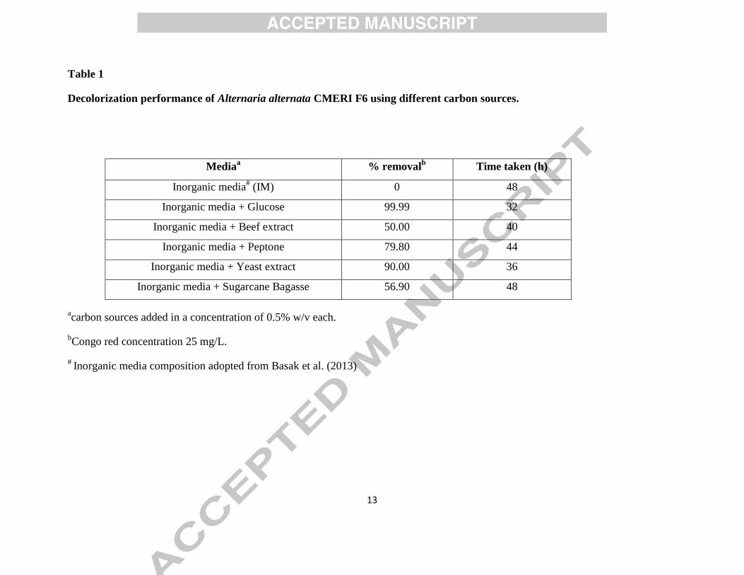
3.1 Effect of supplementation of carbon and nitrogen sources on CR decolorization 21
The decolorization performance of CR in IM supplemented with various carbon sources 22
(Table 1) was studied in 250 ml Erlenmeyer flasks containing 50 ml media at 25 oC, pH 5 and 23
1 2 3 4 5 6 7 8 9 10 11 12 13 14 15 16 17 18 19 20 21 22 23 24 25 26 27 28 29 30 31 32 33 34 35 36 37 38 39 40 41 42 43 44 45 46 47 48 49 50 51 52 53 54 55 56 57 58 59 60 61 62 63 64 65
6
120 rpm. IM supplemented with glucose and yeast extract showed 99.99% and 90% 1
decolorization of CR, respectively, indicating that glucose is the efficient carbon source for CR 2
decolorization by the fungus amongst the carbon sources taken under investigation, followed by 3
yeast extract (Table 1). Since, azo dyes generally do not act as carbon source as they are electron 4
deficient owing to presence of their azo linkage (-N=N-) (Solís et al., 2012) microbial 5
decolorization and degradation of azo dyes generally depends on the presence and type of a 6
carbon source used, because they act as electron donor for azo-dye reduction (Hou et al., 2012). 7
Following this result, a set of different media comprising combinations of yeast extract, glucose 8
and peptone were designed (Section 2.2). It was found that medium consisting of yeast extract 9
and glucose yielded better CR decolorization rate compared to any other media. Therefore, this 10
medium was selected for further decolorization experiment. 11
3.2 Effect of dye concentration on decolorization 12
Decolorization experiments were conducted with dye concentrations of 25-1000 mg/L 13
CR in yeast extract-glucose medium. As the sequence of experiments were performed for 14
acclimitization, it was found that with increasing concentrations of CR beyond 600 mg/L, the 15
amount of absorbed dye remained constant. However, the fungus was only able to retain its 16
decolorization capacity to 99.99% up to 600 mg/L CR resulting in 0.28-18.01 mg/L h in 17
decolorization rate (Fig. 1a-1d). Fig. 1b & 1d show that at 800 and 1000 mg/L concentration, CR 18
removal resulted in 78.37% and 60% final decolorization efficiency with removal rate of 10.86-19
20.21 and 10.52-16.59 mg/L h, respectively. This finding is also in accordance with previous 20
reports (Ayed et al., 2011; Saratale et al., 2013) and attributed to the fact that biosorption of dyes 21
is due to heteropolysaccharide and lipid components of the cell wall, which contain different 22
1 2 3 4 5 6 7 8 9 10 11 12 13 14 15 16 17 18 19 20 21 22 23 24 25 26 27 28 29 30 31 32 33 34 35 36 37 38 39 40 41 42 43 44 45 46 47 48 49 50 51 52 53 54 55 56 57 58 59 60 61 62 63 64 65
7
charged functional groups like carboxyl, hydroxyl, phosphate and other charged groups, causing 1
strong attractive forces between the azo dye and the cell wall (Vitor & Corso, 2008). 2
3.3 Determination of optimum temperature, pH and agitation speed for decolorization of CR 3
Adaptation of the fungus to a broader range of pH and temperature can make it more 4
suitable for dye bioremediation. Experiments showed that 99.99% decolorization of 600 mg/L 5
CR was observed at 20, 25 and 30 oC within 44, 40, and 48 h, respectively. Fig. 2a shows that 6
CR reomoval rate was maximal (6 mg/L h) at 25 oC, followed by CR removal rate at 30 (5.28 7
mg/L h) and 20 oC (4.61 mg/L h), respectively. This is attributed to the fact that (a) in adsorptive 8
decolorization, optimum temperature plays a crucial role in increased surface activity and kinetic 9
energy of the dye molecules and (b) in degradative decolorization, optimum temperature is 10
necessary for optimal activity of dye degradative enzymes (Saratale et al., 2011). 11
A. alternata CMERI F6 showed the decolorization up to nearly 99.99% in the broad 12
range of pH (3-7). Although, the fungus could effectively decolorize (89%) CR at relatively 13
higher pH (8), it showed maximal rate (6.32 mg/L h) of CR decolorization at pH 5 as can be 14
observed from Fig. 2b. It is an established fact that by controlling the [H+] concentration, the pH 15
of the medium influences degradation of a pollutant (Basak et al., 2013). Hence, the dye removal 16
efficiency of the fungus at broad range of pH makes it potent candidate for removal of CR from 17
textile watewaters that differs significantly in pH (Saratale et al., 2011). 18
At agitation speed of 150 and 120 rpm, the fungus was able to decolorize 99.99% of 600 19
mg/L CR. The CR removal rate was also maximal (5.59 mg/L h) at 150 rpm showing that 20
optimum aeration is necessary for maximal CR removal (Fig 2c). In this case, agitation increased 21
mass and oxygen transfer between cells and the medium. In addition, enzyme activity also could 22
have depended on the presence of oxygen (Saratale et al., 2011). At agitation speed of 90 and 23

1 2 3 4 5 6 7 8 9 10 11 12 13 14 15 16 17 18 19 20 21 22 23 24 25 26 27 28 29 30 31 32 33 34 35 36 37 38 39 40 41 42 43 44 45 46 47 48 49 50 51 52 53 54 55 56 57 58 59 60 61 62 63 64 65
8
180 rpm, the fungus showed relatively less decolorization performance, giving 93 and 89% 1
decolorization after 48 h of incubation, respectively, similar to Ali et al. (Ali et al., 2009). 2
3.4 Effect of heavy metals on decolorization performance 3
The presence of heavy metals in the media affected CR removal rate significantly. In 4
presence of 100 mg/L Pb(NO3)2 and CdCl2, 45 and 37% decolorization was observed within 48 5
h. However, when concentrations of both the salt were increased to 200 mg/L, CR removal rate 6
decreased drastically to 0.75 mg/L h and 1.57 mg/L h (Fig. 2d and 2e), resulting in only 16 and 7
30% CR decolorization, respectively. Presence of Hg was observed to have more severe impact 8
on CR decolorization rate. Fig. 3f shows at HgCl2 concentration of 100 and 200 mg/L, the 9
fungus showed significantly less decolorization rate of 1.32 and 0.37 mg/L h, giving only 13 and 10
6% removal of CR, respectively, similar to Pointing et al. (Pointing et al., 2000). 11
3.5 Biodecolorization and biodegradation analysis 12
Both light microscopy and scanning electron microscopy revealed the surface 13
morphology of the fungus after CR decolorization to be more amorphous in nature (Fig 1S, 14
supplementary data). This result is also in close agreement with the X-ray diffraction analysis. 15
The XRD analyses of the fungal mycelia before CR decolorization revealed that the value of 2θ 16
is 280
when the maximum count is observed, which is indicative of the amorphous in nature of 17
mycelia. The XRD of the fungal mycelia after the dye adsorption showed that the 2θ value 18
further decreased, indicating increase in amorphous nature as evident from the less sharper X-ray 19
diffraction peaks noted in Fig. 1S a and 1S b (supplementary data). 20
To investigate the possible mechanism of CR decolorization, HPLC and FTIR analyses 21
were carried out. HPLC elution profile showed three prominent peaks at retention time of 2.141, 22
2.609 and 3.235 min when metabolites separated from the sample obtained after decolorization, 23

1 2 3 4 5 6 7 8 9 10 11 12 13 14 15 16 17 18 19 20 21 22 23 24 25 26 27 28 29 30 31 32 33 34 35 36 37 38 39 40 41 42 43 44 45 46 47 48 49 50 51 52 53 54 55 56 57 58 59 60 61 62 63 64 65
9
compared to peak obtained for control dye at retention time 1.861 min. The analysis has showed 1
that the peak of control dye has shifted to 2.141 min and there is a sharp decrease in the 2
concentration of the dye (Fig. 2S c and d, supplementary data). The new peaks formed indicated 3
the production of certain metabolites, confirming the degradation of CR by A. alternata CMERI 4
F6. Similar observations have also been reported in the literature (Saratale et al., 2013). In the 5
present study, generation of three peaks in HPLC chromatogram may be consistent with 6
formation of three metabolites of CR degradation described by Xu et al. (Xu et al., 2013). 7
The FTIR spectrum of the control dye (Fig. 3S, supplementary data) showed a broad peak 8
at 3340.1 cm-1
due to stretching of bonded O-H group, a peak at 1641.13 cm-1
due to N=N 9
stretching of azo group, along with other peaks at 2158.92 cm-1
and 2340.19 cm-1
. In addition, 10
peak at 1228.43 cm-1
represented primary aromatic amines with C-N vibration. The FTIR 11
spectrum of the extracted metabolites from the decolorized sample (Fig. 4S) shows peak at 12
3292.86 cm-1
and 1641.13 cm-1
. Other peaks at 2361 cm-1
and 2152 cm-1
indicate stretch due to C13
C band. As compared to the control dye, the disappearance of peak at 1228.43 cm-1
indicates 14
the possible breakage of C-N bond and degradation of CR by the fungus. Decolorization of 15
various dyes has also been reported to have occurred through both adsorption and degradation by 16
filamentous fungi (Jadhav et al., 2010). However, the toxicity of the end product of the 17
decolorization of CR has yet to be found out. 18
3.6 Thermal and chemical stability of the bioabsorbed dye 19
When the hydrated biomass was autoclaved at 121 oC at 15psi, no cell disruption, 20
desorption and dye liberation was observed visually. Moreover, on Spectrophotometric analysis 21
negligible change in the adsorption spectrum was noticed. 22

1 2 3 4 5 6 7 8 9 10 11 12 13 14 15 16 17 18 19 20 21 22 23 24 25 26 27 28 29 30 31 32 33 34 35 36 37 38 39 40 41 42 43 44 45 46 47 48 49 50 51 52 53 54 55 56 57 58 59 60 61 62 63 64 65
10
The Thermogravimetric analysis (TG) and Differential Thermal Analysis (DTA) 1
provided information about desorption of the dye and other physical changes of the fungal 2
mycelium. Most of the weight loss of the mycelium due to pyrolysis of the fungus (after CR 3
adsorption) in inert atmosphere of Helium takes place at temperature up to 150 oC where most of 4
the moisture and volatiles are released. Beyond 150 oC there is only decomposition of 5
carbohydrates, protein and lipids and weight loss occurred mainly due to gasification (Agrawal 6
& Chakraborty, 2013). The DTA curve (Fig. 5S, supplementary data) showed that at about 80 7
oC, an exothermic effect appears resulting in the peak where the degradation of mycelium and 8
subsequent liberation of the dye has occurred completely. The strong exothermic effect signifies 9
that disintegration of the fungal mycelium and liberation of the dye requires relatively low 10
temperature in inert atmosphere. The low degradation temperature of the fungal mycelium that 11
adsorbed the dye, compared to the incineration temperature may help in removal of the dye from 12
the biomass in inert conditions after biosorption. 13
Upon treatment of dried fungal biomass with 1M HCl and 1M NaOH, the fungal 14
structure disintegrated but the dye was not released freely in the media and remained adsorbed to 15
the disintegrated fungal structures, except for 70% ethanol treatment. This can be attributed to 16
the dissolution of lipid content and dehydration of protein by ethanol which consequently 17
released the dye into the environment. However, relatively high resistance of the fungus to acid 18
or alkali treatment makes it an efficient and promising candidate for retaining adsorbed CR. 19
4. Conclusion 20
A. alternata CMERI F6 could decolorize 99.99% of 600 mg/L and 78% of 800 mg/L CR 21
within 48 h, at maximal decolorization rate of 18.01 mg/L h and 20.21 mg/L h, respectively. The 22
fungal strain proved to be an excellent candidate for removal of azo dyes within broad pH, 23
1 2 3 4 5 6 7 8 9 10 11 12 13 14 15 16 17 18 19 20 21 22 23 24 25 26 27 28 29 30 31 32 33 34 35 36 37 38 39 40 41 42 43 44 45 46 47 48 49 50 51 52 53 54 55 56 57 58 59 60 61 62 63 64 65
11
temperature and aeration range. HPLC and FTIR study suggest that dye removal by the fungus 1
can be attributed to adsorption to biomass and degradation. The thermal and chemical stability 2
analyses disclosed that the fungus can retain the adsorbed dye despite different harsh treatments 3
like high temperature and acid-alkali treatment. 4
References 5
1. Agrawal, A., Chakraborty, S. 2013. A kinetic study of pyrolysis and combustion of microalgae 6
Chlorella vulgaris using thermo-gravimetric analysis. Bioresour. Technol., 128, 72-80. 7
2. Ali, N., Hameed, A., Siddiqui, M., Ghumro, P., Ahmed, S. 2009. Application of Aspergillus 8
niger SA1 for the enhanced bioremoval of azo dyes in simulated textile effluent. Afr. J. 9
Biotechnol., 8, 3839–3845. 10
3. Ayed, L., Mahdhi, A., Cheref, A., Bakhrouf, A. 2011. Decolorization and degradation of azo 11
dye Methyl Red by an isolated Sphingomonas paucimobilis: Biotoxicity and metabolites 12
characterization. Desalination, 274, 272-277. 13
4. Basak, B., Bhunia, B., Mukherjee, S., Dey, A. 2013. Optimization of physicochemical 14
parameters for phenol biodegradation by Candida tropicalis PHB5 using Taguchi 15
Methodology. Desalin. Water Treat., DOI:10.1080/19443994.2013.770638 16
5. Hou, B., Hu, Y., Sun, J. 2012. Performance and microbial diversity of microbial fuel cells 17
coupled with different cathode types during simultaneous azo dye decolorization and 18
electricity generation. Bioresour. Technol., 111, 105-10. 19
6. Jadhav, J.P., Kalyani, D.C., Telke, A.A., Phugare, S.S., Govindwar, S.P. 2010. Evaluation of 20
the efficacy of a bacterial consortium for the removal of color, reduction of heavy metals, 21
and toxicity from textile dye effluent. Bioresour. Technol., 101(1), 165-73. 22

1 2 3 4 5 6 7 8 9 10 11 12 13 14 15 16 17 18 19 20 21 22 23 24 25 26 27 28 29 30 31 32 33 34 35 36 37 38 39 40 41 42 43 44 45 46 47 48 49 50 51 52 53 54 55 56 57 58 59 60 61 62 63 64 65
12
7. Pointing, S.B., Bucher, V.V.C., Vrijmoed, L.L.P. 2000. Dye decolorization by sub-tropical 1
basidiomycetous fungi and the effect of metals on decolorizing ability. World J. 2
Microbiol. Biotechnol., 16, 199-205. 3
8. Saratale, R.G., Gandhi, S.S., Purankar, M.V., Kurade, M.B., Govindwar, S.P., Oh, S.E., 4
Saratale, G.D. 2013. Decolorization and detoxification of sulfonated azo dye C.I. 5
Remazol Red and textile effluent by isolated Lysinibacillus sp. RGS. J. Biosci. Bioeng., 6
115(6), 658-67. 7
9. Saratale, R.G., Saratale, G.D., Chang, J.S., Govindwar, S.P. 2011. Bacterial decolorization and 8
degradation of azo dyes: A review. J Taiwan Inst. Chem. Eng., 42, 138–157. 9
10. Solís, M., Solís, A., Pérez, H.I., Manjarrez, N., Flores, M. 2012. Microbial decolouration of 10
azo dyes: A review. Process Biochem., 47, 1723–1748. 11
11. Vitor, V., Corso, C.R. 2008. Decolorization of textile dye by Candida albicans isolated from 12
industrial effluents. J. Ind. Microbiol. Biotechnol., 35(11), 1353-7. 13
12. Xu, Q., Sun, J., Hu, Y.Y., Chen, J., Li, W.J. 2013. Characterization and interactions of anodic 14
isolates in microbial fuel cells explored for simultaneous electricity generation and 15
Congo red decolorization. Bioresour. Technol., 142, 101-8. 16
13. Yu, J., Zhang, X., Tan, T. 2007. An novel immobilization method of Saccharomyces 17
cerevisiae to sorghum bagasse for ethanol production. J. Biotechnol., 129(3), 415-20. 18
19
Figure Legends 20
Figure 1: Effect of different CR concentrations on decolorization performance of the fungus. 21
Figure 2: Effect of temperature (a), pH (b), agitation speed (c) and different heavy metals (d, e, f) 22
on removal of CR by the fungus. 23
Highlights:
Optimum physicochemical parameters for congo red decolorization were
determined.
Effect of different heavy metals on congo red decolorization was evaluated.
Characterization of fungal morphology after dye removal was done using SEM and
XRD.
Congo red biodegradation analysis was done using HPLC and FTIR.
Evaluation of dye retention properties of the fungus by TG-DTA analysis.
13
Table 1
Decolorization performance of Alternaria alternata CMERI F6 using different carbon sources.
acarbon sources added in a concentration of 0.5% w/v each.
bCongo red concentration 25 mg/L.
# Inorganic media composition adopted from Basak et al. (2013)
Mediaa % removal
b Time taken (h)
Inorganic media# (IM) 0 48
Inorganic media + Glucose 99.99 32
Inorganic media + Beef extract 50.00 40
Inorganic media + Peptone 79.80 44
Inorganic media + Yeast extract 90.00 36
Inorganic media + Sugarcane Bagasse 56.90 48