CHADS2 score predicts atrial fibrillation following cardiac surgery
Upload
independentCategory
view
4download
0
3 8 | A R B E L E T A L . | M O L M E D 2 0 : 3 8 - 4 5 , 2 0 1 4
INTRODUCTIONImbalanced sympathetic-parasympa-
thetic activity has been associated withpoor cardiovascular outcome, calling foridentifying readily measurable biomark-ers of parasympathetic activity for pre-dicting future risks. Supporting this no-tion, indirect measures of cardiacparasympathetic dysfunction such as ele-
vated resting heart rate, delayed heartrate recovery after exercise and attenuatedheart rate increase during exercise haveall been shown to be independent predic-tors for adverse cardiovascular outcome(1–3). Abnormalities in these parameters(4) have been shown in diverse studypopulations to be associated with suddencardiac death (1,5) as well as all-cause
mortality (2,3,6,7), but clinically validatedbiomarkers to assess the parasympatheticsystem are not yet available. Of note, theparasympathetic neurotransmitter, acetyl-choline (ACh), is extremely labile and dif-ficult to use for clinical measurements (8).
ACh is hydrolyzed in the serum bytwo homologous enzymes with uniquefeatures: acetylcholinesterase (AChE) andbutyrylcholinestersase (BChE). BChE isthe major ACh hydrolyzing enzyme inthe circulation (9). Correspondingly, mostprevious evaluations of ACh hydrolyzingcapacity in the serum used butyrylthio-choline (BTCh), a butyrylcholine (BCh)analog as a substrate. A recent studydemonstrated a strong inverse relationbetween serum BTCh hydrolyzing activ-ity (which would negate parasympathetic
Decline in Serum Cholinesterase Activities Predicts 2-YearMajor Adverse Cardiac Events
Yaron Arbel,1* Shani Shenhar-Tsarfaty,2* Nir Waiskopf,2 Ariel Finkelstein,1 Amir Halkin,1 Miri Revivo,1
Shlomo Berliner,3 Itzhak Herz,1 Itzhak Shapira,1 Gad Keren,1 Hermona Soreq,2* and Shmuel Banai1*
1Department of Cardiology, Tel Aviv Medical Center, affiliated with the Sackler School of Medicine, Tel Aviv University, Tel Aviv,Israel; 2Department of Biological Chemistry, The Life Sciences Institute and the Edmond and Lily Safra Center of Brain Science, TheHebrew University of Jerusalem, Jerusalem, Israel; 3Internal Medicine “E,” Tel Aviv Medical Center, affiliated with the Sackler Schoolof Medicine, Tel Aviv University, Tel Aviv, Israel
Parasympathetic activity influences long-term outcome in patients with cardiovascular disease, but the underlying mecha-nism(s) linking parasympathetic activity and the occurrence of major adverse cardiovascular events (MACEs) are incompletelyunderstood. The aim of this pilot study was to evaluate the association between serum cholinesterase activities as parasympa-thetic biomarkers and the risk for the occurrence of MACEs. Cholinergic status was determined by measuring the cumulativecapacity of serum acetylcholinesterase (AChE) and butyrylcholinesterase (BChE) to hydrolyze the AChE substrate acetylthio-choline. Cholinergic status was evaluated in randomly selected patients undergoing cardiac catheterization. The patients weredivided into two groups of 100 patients in each group, with or without occurrence of MACEs during a follow-up period of 40months. Cox regression models adjusted for potential clinical, metabolic and inflammatory confounders served to evaluate as-sociation with clinical outcome. We found that patients with MACE presented lower cholinergic status and AChE values atcatheterization (1,127 ± 422 and 359 ± 153 nmol substrate hydrolyzed per minute per milliliter, respectively) than no-MACE pa-tients (1,760 ± 546 and 508 ± 183 nmol substrate hydrolyzed per minute per milliliter, p < 0.001 and p < 0.001, respectively), whoselevels were comparable to those of matched healthy controls (1,622 ± 303 and 504 ± 126 nmol substrate hydrolyzed per minuteper milliliter, respectively). In a multivariate analysis, patients with AChE or total cholinergic status values below median showedconspicuously elevated risk for MACE (hazard ratio 1.85 [95% confidence interval [CI] 1.09–3.15, p = 0.02] and 2.21 [95% CI1.22–4.00, p = 0.009]) compared with those above median, even after adjusting for potential confounders. We conclude thatparasympathetic dysfunction expressed as reduced serum AChE and AChE activities in patients compared to healthy controlscan together reflect impaired parasympathetic activity. This impairment predicts the risk of MACE up to 40 months in such pa-tients. Monitoring these parasympathetic parameters might help in the risk stratification of patients with cardiovascular disease.Online address: http://www.molmed.orgdoi: 10.2119/molmed.2013.00139
*YA, SS-T, HS, and SB contributed equally to this work.
Address correspondence to Hermona Soreq, Givat Ram, Jerusalem 91904, Israel. Phone:
+972-2-6585109; Fax: +972-2-6520258; E-mail: [email protected].
Submitted October 27, 2013; Accepted for publication December 19, 2013; Epub
(www.molmed.org) ahead of print December 24, 2013.

R E S E A R C H A R T I C L E
M O L M E D 2 0 : 3 8 - 4 5 , 2 0 1 4 | A R B E L E T A L . | 3 9
potency (8)) and long-term mortality in acohort of stable coronary artery diseasepatients (10). However, BCh is not physi-ologically available in the body and isonly hydrolyzed by BChE but not AChE.Moreover, AChE is 20-fold faster thanBChE in hydrolyzing ACh, and studiesdemonstrate a causal link between in-flammatory pathways and cholinergicsignaling (11,12). Specifically, the so-called cholinergic antiinflammatory pathway inhibits cytokine synthesis andrelease (13,14), predicting direct associa-tions between cholinesterase activitiesand inflammation.
On the basis of these considerations, wehypothesized that using the ACh analogacetylthiocholine (ATCh) as a substratethat is hydrolyzed by both enzymesmight reflect the cholinergic status andwould be physiologically more meaning-ful and practically advantageous. ATChcan better reflect the full parasympatheticpotency in inactivating ACh and mightoffer insight into the nature of its rele-vance for cardiovascular diseases.
In the present pilot study, we havetaken this approach to evaluate the asso-ciation between parasympathetic systemactivity and major adverse cardiac events(MACEs).
MATERIALS AND METHODS
Study Design and Patient SelectionThe dataset included in this study was
collected as part of the Tel Aviv Prospec-tive Angiographic Survey (TAPAS), a pro-spective, single-center registry that enrollsall patients undergoing cardiac catheteri-zation at the Tel Aviv Medical Center(15–18). The registry includes 4,500 con-secutive admitted patients and thereforecovers a variety of clinical conditionswithout inclusion/exclusion criteria. Toevaluate the effect of the parasympatheticsystem on MACE outcome, we chose twogroups of randomly selected patients ac-cording to the occurrence or absence ofMACEs at 40 months of follow-up as anested case control study. Group 1 con-sisted of 100 randomly selected patientswith MACEs (a negative outcome group),
and group 2 consisted of 100 randomlyselected patients who had no MACEs (apositive outcome group) at follow-up ofup to 40 months. Eight patients were ex-cluded because of incomplete data, and192 patients remained for the final analy-sis. In addition, an age-, sex- and bodymass index–matched control group of 264apparently healthy patients was used.Control group characteristics were previ-ously described (19). For the controlgroup, the exclusion criteria included ahistory of cerebral or cardiac events dur-ing the previous 12 months; known in-flammatory diseases, history of acutefebrile disease or infection during the pre-vious 3 months; and known malignancy,pregnancy, steroidal or nonsteroidal treat-ment (except for acetylsalicylic acid at adose of 325 mg/dL) and invasive proce-dures (surgery, catheterization and so on)during the previous 6 months.
All patients signed a written informedconsent for participation in the study,which was approved by the Tel AvivMedical Center institutional ethics com-mittee (IEC) according to the Declarationof Helsinki (IEC approvals 07-396, 00-116).
Cardiovascular Endpoints and Follow-up
An MACE was defined as follows: all-cause mortality, myocardial infarction(MI) and hemorrhagic or ischemic stroke.Occurrence of the individual componentsof the composite endpoint was ascer-tained by medical chart review and bytelephone interview at regular intervalsup to 40 months after coronary angiogra-phy. All patient files were coreviewed byan independent physician for accurateanalysis of endpoint definitions. End offollow-up for MACE outcome was de-fined as the first from the followingevents: MACE, or January 1, 2012.
Laboratory TestsAll of the participants underwent an-
giography after a night of fasting, exceptfor patients with ST-elevation MI. Arterialblood was obtained via the arterial accesspuncture site as part of the coronary an-giography procedure. Serum samples
were kept frozen at –80°C until analysis ofcholinesterase activities. To compare theeffects of AChE, BChE and cholinergicstatus changes on cardiovascular risk, weused ATCh as a substrate with or withouttetraisopropyl pyrophosphoramide (iso-OMPA, a specific BChE inhibitor), thusidentifying AChE alone, mimicking thetotal impact of ACh hydrolysis and en-abling a calculation of BChE contribution.This method has been reliably used inseveral recent studies (14,19–23).
Cholinesterase activities. In brief, totalcholinergic status is the total capacity forACh hydrolysis (that is, the summationof AChE and BChE activities).
AChE activity levels were assessed inthe serum samples by using a microtiterplate assay by measuring rates of ATCh(1 mmol/L; Sigma-Aldrich, St. Louis,MO, USA) hydrolysis after 20-min prein-cubation in the dark with 500 μmol/Liso-OMPA (Sigma-Aldrich), a specificBChE inhibitor. Enzyme activities werecalculated using the e405 absorbancevalue for 5-thio-2- nitrobenzoate pro-duced from dithionitrobenzoate reactionwith the hydrolysis product, thiocholine(13,600 [mol·L]/cm). The nonenzymaticbreakdown of substrate was subtractedfrom the total rate of hydrolysis.
Cholinergic status. Cholinergic statuswas determined by measuring the totalserum rates of ATCh hydrolysis withoutcholinesterase inhibition (14,19,21). Toeliminate any assay-by-assay variations,we reanalyzed 10 arbitrary control sam-ples in different plates and at differentmeasurement times.
Calculated BChE. Calculated BChE ac-tivity values were determined by sub-tracting AChE values from the measuredcholinergic status values.
Cholinesterase activity assays were per-formed in triplicate by a single blindedtechnician for each cohort. In an attempt tominimize intratest variations, exclude day-of-measurement effects and ascertain theauthenticity of the observed differences,we used the same reagents throughout theseries and remeasured selected sampleseach test day. Freezing and defrosting didnot affect the enzyme activities.

4 0 | A R B E L E T A L . | M O L M E D 2 0 : 3 8 - 4 5 , 2 0 1 4
A C E T Y L C H O L I N E A N D M A C E
Enzymatic stability and reproducibilitycriteria for our measurements werebased on the use of highly purifiedhuman recombinant cholinesterases asreference enzymes (24,25).
Crystalographic StructureThe crystalographic structure PDB file
of recombinant human acethyl-cholinesterase in complex with donepezil(PDB ID: 4EY7 [26]) and the X-ray struc-ture PDB file of human butyryl-cholinesterase (PDB ID: 2XQG [27]) wereobtained from the RCSB Protein DataBank (www.pdb.org) (28). One of themonomers in the crystalographic struc-ture of human BChE and all water andligand molecules besides donepezil wereextracted from the PDB files using thePyMOL Molecular Graphics System, Ver-sion 1.3 (Schrödinger LLC, New York,NY, USA). Then the two crystalographicstructures were aligned and the width ofthe active gorge site was measured usingPyMOL.
Statistical AnalysisCategorical variables were compared
by using a χ2 test and continuous vari-ables by using a t test (presented asmeans with standard deviations [SDs]) orby Kruskal Wallis/Mann-Whitney test(medians with interquartile range [IQR]).Continuous variables were tested for nor-mal distribution by using a Kolmogorov-Smirnov test and Q-Q plots.
We analyzed the distributions of the dif-ferent parasympathetic variables by usinghistograms. To evaluate the significance ofthe differences of the two principal com-ponents of the parasympathetic system forMACE, we redivided our combined co-hort including patients with and withoutMACEs into two new groups by AChEmedians and examined the clinical andlaboratory characteristics (SupplementaryTable S1). Next, we examined the effect ofboth AChE/cholinergic status medians onthe risk of the composite myocardial in-farction, stroke and all-cause mortality(MACE) endpoint. MACE was evaluatedusing univariate and multivariate Coxproportional hazard regression. Survival
curves divided by AChE medians andMACE are presented. We adjusted ourmodel for age, sex, conventional risk fac-tors (diabetes mellitus, hypertension, dys-lipidemia, peripheral vascular disease,known ischemic heart disease, prior strokeand acute coronary syndrome [ACS] sta-tus) by using highly sensitive troponin(29) and laboratory variables (estimatedglomerular filtration rate, hemoglobinA1c, triglyceride, total cholesterol, low-and high-density lipoprotein, high- sensitivity C-reactive protein (hs-CRP) andwhite blood cell counts). The influence ofadding each block to the model waschecked. A two-tailed p < 0.05 was consid-ered statistically significant. All analyseswere performed with the SPSS 19.0 soft-ware (SPSS, Chicago, IL, USA).
All supplementary materials are availableonline at www.molmed.org.
RESULTSA total of 192 patients referred for cor-
onary angiography at the Tel Aviv Med-
ical Center were included in the finalanalysis. A total of 98 patients suffered aMACE during a mean follow-up periodof 369 ± 250 d (median 411, range2–1,199). The mean age was 67.9 ±12.2 years (range 32–95); 67% of the pa-tients were males. Approximately two-thirds of the cohort underwent angiogra-phy because of acute coronary syndromeand the other one-third because of stableangina. Coronary angiography revealednormal or nonobstructive coronary ar-tery disease in 18% of the patients, andone-, two- or three-vessel coronary arterydisease was found in 21%, 25% and 36%of the patients, respectively (Supplemen-tary Table S1). During follow up, 39(44%) patients died. Seventeen of thosedeaths were due to cardiac causes.
Serum AChE, BChE and CholinergicStatus Values Show InverseAssociations with MACE
To enable in-depth interrogation of the cholinergic parameters, we analyzedthe distributions of measured AChE and
Figure 1. Decreased serum levels of AChE, BChE and cholinergic status in MACE patients.Histograms of enzymatic levels of AChE (red), BChE (yellow) and cholinergic status (green)are presented for MACE, no-MACE and age-, sex- and body mass index–matched con-trols. The MACE histograms are left-shifted, reflecting reduced enzymatic activities in allthree measurements, whereas no-MACE patients present similar distribution to that ofmatched controls. Hydrolyzed/min × mL, hydrolyzed per minute per milliliter.

R E S E A R C H A R T I C L E
M O L M E D 2 0 : 3 8 - 4 5 , 2 0 1 4 | A R B E L E T A L . | 4 1
cholinergic status values and of calculatedBChE in patients with and withoutMACEs compared with matched controls(19) without cardiac disease. These distri-butions clearly demonstrated drastic de-cline in AChE, BChE and cholinergic sta-tus values in patients with MACEscompared with those without MACEs orin healthy controls (Figure 1). The meannormalized serum AChE activity (359 ±153 nmol substrate hydrolyzed perminute per milliliter) was lower in MACEpatients than the measured levels in pa-tients without MACE (508 ± 183; p <0.001), who presented indistinguishableactivities from those of apparently healthymatched controls (504 ± 126; p = NS).Likewise, both calculated BChE activitiesand the mean cholinergic status werelower in patients with MACE (768 ± 286
and 1,127 ± 422) than in patients withoutMACE (1,252 ± 421 and 1,760 ± 546, p <0.001 for both) and apparently healthy in-dividuals (1,118 ± 217 and 1,622 ± 303).
Cardiac patients presented with awider distribution pattern of serumAChE, BChE and cholinergic status val-ues, reflecting larger individual variabil-ity in these values compared withmatched controls. Using individual en-zyme measurements demonstrated thatthe parasympathetic changes in MACEpatients reflected a mutual decline inserum AChE and BChE activities that to-gether led to reduced cholinergic status.
To evaluate the significance of the dif-ferences in these two principal compo-nents of the parasympathetic system forMACE, we redivided our combined co-hort including patients with and without
MACE into two new groups that differ bytheir serum AChE levels (SupplementaryTable S1). The two groups, above andbelow median AChE activities, were simi-lar in risk factors and clinical presentation.Nevertheless, the lower AChE group suf-fered from 68 MACEs (70%) comparedwith patients in the higher AChE group,who experienced 30 events (31.6%) (p <0.001). The crude hazard ratio for MACEwas 1.68 (95% confidence interval [CI]1.06–2.65, p = 0.026) in patients within thelower median of AChE compared with pa-tients in the higher median AChE. To com-pare AChE to cholinergic status as a pre-dictive biomarker, we further reclassifiedthe combined cohort into patients withcholinergic status values above or belowmedian. The crude hazard ratio for MACEwas 1.83 (95% CI 1.13–2.99, p = 0.015) forthe patients within the lower cholinergicstatus median compared with the patientsin the higher median. Figure 2 presentsthe hazard ratio (HR) of the two testedbiomarkers in relation to MACE.
Serum Cholinesterase Values PredictMultivariate MACEs
In the multivariate regression, the ad-dition of AChE to the model was signifi-cant (p = 0.02). After controlling for allthe risk factors detailed in Materials andMethods, the lower AChE median re-mained significantly associated with ad-verse outcome, with HR = 1.85 (95% CI1.09–3.15, p = 0.02). The addition of thecholinergic status parameter was alsohighly significant (p = 0.008). After con-trolling for all the above-mentioned riskfactors, the lower cholinergic status me-dian was still highly significantly associ-ated with adverse outcome: HR = 2.21(95% CI 1.22–4.00, p = 0.009). Figure 3demonstrates the survival curves by me-dians for AChE and cholinergic status.
In patients with troponin-positiveacute coronary syndrome, cholinergicstatus significantly predicted MACE: HR = 2.0 (95% CI 1–3.89, p = 0.05). AChEshowed a trend toward significance: HR = 1.88 (95% CI 0.99–3.6, p = 0.054).The troponin-negative ACS group wastoo small for conducting this analysis.
Figure 2. HR for MACE. Means and CIs for MACE of the cardiac catheterization group arepresented by using univariate and multivariate Cox proportional hazard regression forlower medians of AChE or total cholinergic status. Adj, adjusted model for conventionalrisk factors (see Materials and Methods).
Figure 3. Lower AChE and total cholinergic status medians predict worse outcome. Cu-mulative (Cum) survival curves for cardiac catheterization patients in the higher andlower median groups of AChE or total cholinergic status activity values are shown.

4 2 | A R B E L E T A L . | M O L M E D 2 0 : 3 8 - 4 5 , 2 0 1 4
A C E T Y L C H O L I N E A N D M A C E
Cholinergic Status Correlates withInflammation and Anemia
Higher cholinergic status and AChEvalues were associated with higher he-moglobin levels and lower hs-CRP val-ues, suggesting a protective power forcholinesterase increases against anemiaand inflammation (Figure 4). Cholinergicstatus values were directly and signifi-cantly correlated with hemoglobin levels(r = 0.35, p < 0.001) and inversely withhs-CRP values (r = –0.27, p < 0.001). Sim-ilarly, AChE measures were directly andsignificantly correlated with hemoglobinlevels (r = 0.35, p < 0.001) and inverselywith hs-CRP values (r = –0.19, p = 0.008).
The above results raised the questionof what fraction of the cholinergic statusvalue is contributed by serum AChE. A representative kinetic chart ofcholinesterase activity during reactiontime in serum samples is presented inFigure 5A. The apparent linearity andminimal deviation in ATCh hydrolysisrates (reflected in the slopes and SDs pre-sented for these curves) adds significanceto the measured values of both enzymeactivities. This analysis predictablydemonstrated a considerably faster rate
of ATCh hydrolysis in the cholinergicstatus measurement (no inhibition),which reflects the cumulative activity ofboth BChE and AChE. Although theAChE protein amounts to a small part ofthe total cholinesterase content in theserum (9), net AChE activity was about40% of the total cholinergic status value,due to its rapid hydrolysis rate of ATCh,as can been seen by the slope differences(Figure 5A). The BChE and AChE pro-
teins share 50% of their sequence and re-semble each other in their structure (Fig-ures 5B, C). However, the active sitespace is considerably wider in BChEcompared with AChE (30) (Figures 5D,E), explaining the differences observed intheir substrate specificity.
DISCUSSIONBy performing individual serum
cholinesterase activity measurements to
Figure 4. Cholinergic status values are pos-itively correlated with inflammation andanemia. Tertiles of C-reactive protein andhemoglobin are shown, with total choliner-gic status for the cardiac catheterizationgroup of patients. Note the inverse corre-lations; higher cholinergic status and AChEvalues were associated with higher hemo-globin levels and lower hs-CRP values.
Figure 5. Measurement characteristics. Kinetic curves of thiocholine release from ATCh areshown, as measured by quantifying absorbance at 405 nm for a consecutive 1,200 s in theabsence of iso-OMPA (cholinergic status) or in its presence (AChE) compared with sponta-neous thiocholine release in phosphate-buffered saline (PBS) (A). Note the linearity ofcurves and minimal SD values. The crystallographic structures of BChE (B) and AChE (C) il-lustrate the general resemblance between the two enzymes. Zoom-in to the active site ofBChE (D) and AChE (E); the narrower gouge in the active site of AChE can explain its ca-pacity to hydrolyze ACh more efficiently than BChE.
R E S E A R C H A R T I C L E
M O L M E D 2 0 : 3 8 - 4 5 , 2 0 1 4 | A R B E L E T A L . | 4 3
yield distinct AChE, BChE and choliner-gic status values, we discovered cumula-tive changes in the AChE and BChE val-ues, which shed new light on ourunderstanding of parasympathetic con-tributions to the prediction of MACEs incoronary patients. To the best of ourknowledge, this study is the first to dem-onstrate the distinct and additive valueof these biomarkers in coronary arterydisease patients.
The main distinction and advantageof our study is the use of ATCh as asubstrate, with and without the inclu-sion of a selective BChE inhibitor, com-pared with previous studies that usedthe butyrylthiocholine analog (10,31)and viewed BChE alone, neglecting thecontribution of AChE and without as-certaining specificity through the use ofa selective inhibitor. BChE is largelyproduced in the liver, but AChE is re-leased, among other sources from pe-ripheral leukocytes (32). Consequently,
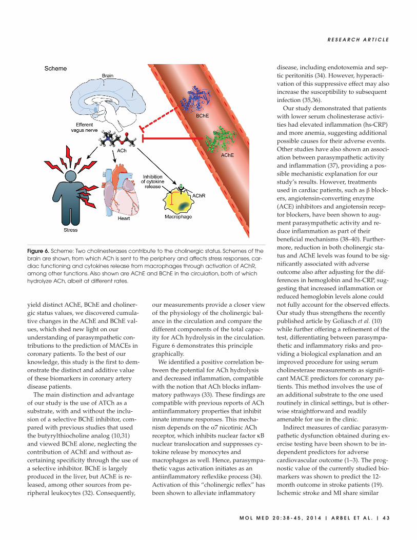
our measurements provide a closer viewof the physiology of the cholinergic bal-ance in the circulation and compare thedifferent components of the total capac-ity for ACh hydrolysis in the circulation.Figure 6 demonstrates this principlegraphically.
We identified a positive correlation be-tween the potential for ACh hydrolysisand decreased inflammation, compatiblewith the notion that ACh blocks inflam-matory pathways (33). These findings arecompatible with previous reports of AChantiinflammatory properties that inhibitinnate immune responses. This mecha-nism depends on the α7 nicotinic AChreceptor, which inhibits nuclear factor κBnuclear translocation and suppresses cy-tokine release by monocytes andmacrophages as well. Hence, parasympa-thetic vagus activation initiates as an antiinflammatory reflexlike process (34).Activation of this “cholinergic reflex” hasbeen shown to alleviate inflammatory
disease, including endotoxemia and sep-tic peritonitis (34). However, hyperacti-vation of this suppressive effect may alsoincrease the susceptibility to subsequentinfection (35,36).
Our study demonstrated that patientswith lower serum cholinesterase activi-ties had elevated inflammation (hs-CRP)and more anemia, suggesting additionalpossible causes for their adverse events.Other studies have also shown an associ-ation between parasympathetic activityand inflammation (37), providing a pos-sible mechanistic explanation for ourstudy’s results. However, treatmentsused in cardiac patients, such as β block-ers, angiotensin-converting enzyme(ACE) inhibitors and angiotensin recep-tor blockers, have been shown to aug-ment parasympathetic activity and re-duce inflammation as part of theirbeneficial mechanisms (38–40). Further-more, reduction in both cholinergic sta-tus and AChE levels was found to be sig-nificantly associated with adverseoutcome also after adjusting for the dif-ferences in hemoglobin and hs-CRP, sug-gesting that increased inflammation orreduced hemoglobin levels alone couldnot fully account for the observed effects.Our study thus strengthens the recentlypublished article by Goliasch et al. (10)while further offering a refinement of thetest, differentiating between parasympa-thetic and inflammatory risks and pro-viding a biological explanation and animproved procedure for using serumcholinesterase measurements as signifi-cant MACE predictors for coronary pa-tients. This method involves the use ofan additional substrate to the one usedroutinely in clinical settings, but is other-wise straightforward and readilyamenable for use in the clinic.
Indirect measures of cardiac parasym-pathetic dysfunction obtained during ex-ercise testing have been shown to be in-dependent predictors for adversecardiovascular outcome (1–3). The prog-nostic value of the currently studied bio-markers was shown to predict the 12-month outcome in stroke patients (19).Ischemic stroke and MI share similar
Figure 6. Scheme: Two cholinesterases contribute to the cholinergic status. Schemes of thebrain are shown, from which ACh is sent to the periphery and affects stress responses, car-diac functioning and cytokines release from macrophages through activation of AChR,among other functions. Also shown are AChE and BChE in the circulation, both of whichhydrolyze ACh, albeit at different rates.

4 4 | A R B E L E T A L . | M O L M E D 2 0 : 3 8 - 4 5 , 2 0 1 4
A C E T Y L C H O L I N E A N D M A C E
risk factors, and both stroke and MI pa-tients share decreases in serum AChE ac-tivity that may avoid excessive neuro- inflammation. Moreover, most strokepatients show increases in serum BChEthat aid to balance their cholinergic sta-tus values, whereas poststroke patientswith drastic AChE decline are at addedrisk of adverse outcome. In comparison,cardiac patients present drastically re-duced AChE, BChE and cholinergic sta-tus measures, the cumulative power ofwhich predicts subsequent MACE risks.In past studies, we demonstrated theclose correlation between our biomark-ers and parallel physiological variablessuch as resting and recovery heart rateafter stress testing (21,22,36).
Future studies will be required to fullyassess the predictive value of each ofthese parameters alone, compared withboth of them together. Other regulatorymechanisms changing cholinesterase ac-tivities may also contribute to this de-cline, such as the AChE mRNA-targetedmicroRNA-132 (41), which can blockAChE gene expression and is upregu-lated under stress (42) and in intestinalbowel disease (43), yet is drastically re-duced in the Alzheimer’s disease brain(44). Also relevant are the stress-inducedalternative splicing changes (45) and epi-genetic potentiation of AChE gene ex-pression, which may explain the long-lasting nature of such reactions (46).
LimitationsThis study has several limitations: first,
the healthy controls included in the pres-ent analysis were taken from a previousset of samples. However, over the pastseveral years, we performed extensiveAChE and cholinergic status tests on par-allel control groups of patients with simi-lar characteristics (19,21). Thus, our datawere validated in a large number of vol-unteers, all of whom showed activities inthe same range. Also, the current cohortwas relatively small, but randomly se-lected patients with and without MACEshowed significant differences. Addition-ally, we did not measure other variablesassociated with parasympathetic activity
such as heart rate variability or heart ratevalues. However, we demonstrated theclose correlation between our biomarkersand parallel physiological variables inpast studies (21,22). And lastly, Figure 6represents the current knowledge aboutthe two cholinesterases (AChE andBChE) and their contribution to thecholinergic status and its effect on theheart and inflammatory profile of MACEpatients. Future study will be needed toobtain a more detailed perspective of thescheme shown in Figure 6.
CONCLUSIONIn our pilot study, parasympathetic
dysfunction expressed as decreased cu-mulative capacity of serum AChE andBChE to hydrolyze ATCh predicts long-term MACEs even after adjusting for po-tential clinical, metabolic and inflamma-tory confounders. Future studies areneeded to evaluate these parasympa-thetic parameters and their risk stratifica-tion capabilities of patients with cardio-vascular disease.
DISCLOSUREThe authors declare that they have no
competing interests as defined by Molecu-lar Medicine, or other interests that mightbe perceived to influence the results anddiscussion reported in this paper.
REFERENCES1. Jouven X, et al. (2005) Heart-rate profile during
exercise as a predictor of sudden death. N. Engl.J. Med. 352:1951–8.
2. Cole CR, Blackstone EH, Pashkow FJ, Snader CE,Lauer MS. (1999) Heart-rate recovery immedi-ately after exercise as a predictor of mortality. N. Engl. J. Med. 341:1351–7.
3. Leeper NJ, et al. (2007) Prognostic value of heartrate increase at onset of exercise testing. Circula-tion. 115:468–74.
4. Lahiri MK, Kannankeril PJ, Goldberger JJ. (2008)Assessment of autonomic function in cardiovas-cular disease: physiological basis and prognosticimplications. J. Am. Coll. Cardiol. 51:1725–33.
5. Adabag AS, et al. (2008) Relation of heart rate pa-rameters during exercise test to sudden deathand all-cause mortality in asymptomatic men.Am. J. Cardiol. 101:1437–43.
6. Arena R, Guazzi M, Myers J, Peberdy MA. (2006)Prognostic value of heart rate recovery in patientswith heart failure. Am. Heart J. 151:851.e7–13.
7. Savonen KP, et al. (2008) Chronotropic incompe-tence and mortality in middle-aged men withknown or suspected coronary heart disease. Eur.Heart J. 29:1896–902.
8. Soreq H, Seidman S. (2001) Acetylcholinesterase:new roles for an old actor. Nat. Rev. Neurosci.2:294–302.
9. Loewenstein-Lichtenstein Y, et al. (1995) Geneticpredisposition to adverse consequences of anti-cholinesterases in “atypical” BCHE carriers. Nat.Med. 1:1082–5.
10. Goliasch G, et al. (2012) Routinely available bio-markers improve prediction of long-term mortal-ity in stable coronary artery disease: the Viennaand Ludwigshafen Coronary Artery Disease(VILCAD) risk score. Eur. Heart J. 3:2282–9.
11. Metz CN, Tracey KJ. (2005) It takes nerve todampen inflammation. Nat. Immunol. 6:756–7.
12. Shaked I, et al. (2009) MicroRNA-132 potentiatescholinergic anti-inflammatory signaling by tar-geting acetylcholinesterase. Immunity. 31:965–73.
13. Borovikova LV, et al. (2000) Vagus nerve stimula-tion attenuates the systemic inflammatory re-sponse to endotoxin. Nature. 405:458–62.
14. Ofek K, et al. (2007) Cholinergic status modula-tions in human volunteers under acute inflam-mation. J. Mol. Med. (Berlin). 85:1239–1251.
15. Arbel Y, et al. (2013) Impact of estimatedglomerular filtration rate on vascular disease ex-tent and adverse cardiovascular events in pa-tients without chronic kidney disease. Can. J. Car-diol. 29:1374–81, 2013
16. Steinvil A, et al. (2012) The development of ane-mia of inflammation during acute myocardial in-farction. Int. J. Cardiol. 156:160–4.
17. Arbel Y, et al. (2012) Prevalence and predictors ofslow flow in angiographically normal coronaryarteries. Clin. Hemorheol. Microcirc. 52:5–14.
18. Arbel Y, et al. (2012) Neutrophil/lymphocyteratio is related to the severity of coronary arterydisease and clinical outcome in patients under-going angiography. Atherosclerosis. 225:456–60.
19. Ben Assayag E, et al. (2010) Serum cholinesteraseactivities distinguish between stroke patients andcontrols and predict 12-month mortality. Mol.Med. 16:278–86.
20. Bryk B, et al. (2005) Inherited and acquired inter-actions between ACHE and PON1 polymor-phisms modulate plasma acetylcholinesterase andparaoxonase activities. J. Neurochem. 92:1216–27.
21. Canaani J, et al. (2010) Serum AChE activitiespredict exercise heart rate parameters of asymp-tomatic individuals. Neurosci. Med. 1:43–9.
22. Shenhar-Tsarfaty S, et al. (2011) Butyrylcholinesteraseinteractions with amylin may protect pancreaticcells in metabolic syndrome. J. Cell. Mol. Med.15:1747–56.
23. Shenhar-Tsarfaty S, Ben Assayag E, BornsteinNM, Berliner S, Soreq H. (2011) Post-strokecholinergic biomarkers [response]. Science [Inter-net]. [cited 2014 Jan 7]. Available from: http://www. sciencemag.org/content/334/6052/101/reply#sci_el_16350

R E S E A R C H A R T I C L E
M O L M E D 2 0 : 3 8 - 4 5 , 2 0 1 4 | A R B E L E T A L . | 4 5
24. Waiskopf N, Shweky I, Lieberman I, Banin U,Soreq H. (2011) Quantum dot labeling of butyryl-cholinesterase maintains substrate and inhibitorinteractions and cell adherence features. ACSChem. Neurosci. 2:141–50.
25. Waiskopf N, Shweky I, Lieberman I, Banin U,Soreq H. (2011) Quantum Dot Labeling of Bu-tyrylcholinesterase Maintains Substrate and In-hibitor Interactions and cell Adherence Features.ACS Chem. Neurosci. 2:141–50.
26. Cheung J, et al. (2012) Structures of human acetyl-cholinesterase in complex with pharmacologicallyimportant ligands. J. Med. Chem. 55:10282–6.
27. Wandhammer M, et al. (2011) Structural study ofthe complex stereoselectivity of human butyryl-cholinesterase for the neurotoxic V-agents. J. Biol.Chem. 286:16783–9.
28. Berman HM, et al. (2000) The protein data bank.Nucl. Acids Res. 28:235–42.
29. Hamm CW, et al. (2011) ESC Guidelines for themanagement of acute coronary syndromes in pa-tients presenting without persistent ST-segmentelevation: The Task Force for the management ofacute coronary syndromes (ACS) in patients pre-senting without persistent ST-segment elevationof the European Society of Cardiology (ESC).Eur. Heart J. 32:2999–3054.
30. Gilson MK, et al. (1994) Open “back door” in amolecular dynamics simulation of acetyl-cholinesterase. Science. 263:1276–8.
31. Calderon-Margalit R, Adler B, Abramson JH,Gofin J, Kark JD. (2006) Butyrylcholinesterase ac-tivity, cardiovascular risk factors, and mortalityin middle-aged and elderly men and women inJerusalem. Clin. Chem. 52:845–52.
32. Grisaru D, et al. (2006) Hydrolytic and nonenzy-matic functions of acetylcholinesterase comodu-late hemopoietic stress responses. J. Immunol.176:27–35.
33. Rosas-Ballina M, et al. (2011) Acetylcholine- synthesizing T cells relay neural signals in avagus nerve circuit. Science. 334:98–101.
34. Tracey KJ. (2010) Understanding immunity re-quires more than immunology. Nat. Immunol.11:561–4.
35. Meisel C, Meisel A. (2011) Suppressing immuno-suppression after stroke. N. Engl. J. Med. 365:2134–6.
36. Shenhar-Tsarfaty S, Berliner S, Bornstein NM,Soreq H. (2013) Cholinesterases as biomarkers forparasympathetic dysfunction and inflammation- related disease. J. Mol. Neurosci. 2013, November 20[Epub ahead of print].
37. Kon H, et al. (2006) Association of decreased varia-tion of R-R interval and elevated serum C-reactiveprotein level in a general population in Japan. Int.Heart J. 47:867–76.
38. Bibevski S, Dunlap ME. (2011) Evidence for im-paired vagus nerve activity in heart failure. HeartFail. Rev. 16:129–35.
39. Binkley PF, et al. (1993) Sustained augmentation ofparasympathetic tone with angiotensin-convertingenzyme inhibition in patients with congestiveheart failure. J. Am. Coll. Cardiol. 21:655–61.
40. Du XJ, Cox HS, Dart AM, Esler MD. (1998) De-pression of efferent parasympathetic control ofheart rate in rats with myocardial infarction: ef-fect of losartan. J. Cardiovasc. Pharmacol. 31:937–44.
41. Shaked I, et al. (2009) MicroRNA-132 potentiatescholinergic anti-inflammatory signaling by tar-geting acetylcholinesterase. Immunity. 31:965–73.
42. Shaltiel G, et al. (2013) Hippocampal microRNA-132 mediates stress-inducible cognitive deficitsthrough its acetylcholinesterase target. BrainStruct. Funct. 218:59–72.
43. Maharshak N, et al. MicroRNA-132 modulatescholinergic signaling in inflammatory bowel dis-ease. Inflammatory Bowel Diseases. In press
44. Lau P, et al. Alteration of the microRNA networkduring the progression of Alzheimer’s disease.EMBO Mol. Med. 5:1613–34.
45. Kaufer D, Friedman A, Seidman S, Soreq H.(1998) Acute stress facilitates long-lastingchanges in cholinergic gene expression. Nature.393:373–7.
46. Sailaja BS, Cohen-Carmon D, Zimmerman G,Soreq H, Meshorer E. (2012) Stress-induced epi-genetic transcriptional memory of acetyl-cholinesterase by HDAC4. Proc. Natl. Acad. Sci.U. S. A. 109:E3687–95.
Copyright © 2022 FDOKUMEN