Debra Jayne Clayton - University of Glasgow
290
Analysis of the role of fimbriae in the virulence of Salmonella enterica in poultry Thesis submitted for the degree of Doctor of Philosophy By Debra Jayne Clayton Institute for Animal Health, Compton, Berkshire and School of Veterinary Medicine University of Glasgow 2008
-
Upload
khangminh22 -
Category
Documents
-
view
0 -
download
0
Transcript of Debra Jayne Clayton - University of Glasgow

Analysis of the role of
fimbriae in the virulence of
Salmonella enterica in
poultry Thesis submitted for the degree of
Doctor of Philosophy
By
Debra Jayne Clayton
Institute for Animal Health, Compton, Berkshire
and
School of Veterinary Medicine
University of Glasgow
2008

2
Abstract
Salmonella is a Gram-negative bacterium that consists of two species; S. enterica and
S. bongori. The species S. enterica can be further divided into 6 subspecies and
subspecies I is predominantly associated with disease in warm blooded animals and
contains over 2,500 antigenically distinct serovars. Each serovar is >90% identical at
the DNA level but can infect a different range of hosts and cause different diseases.
Poultry are an important reservoir of entry of Salmonella into the human food chain
owing to the contamination of their eggs and meat. The molecular mechanisms
underlying colonisation of food producing animals with Salmonella are unknown.
Fimbrial genes encode proteinaceous surface exposed appendages which have been
shown to mediate adhesion of bacterial cells but the precise role for fimbriae in the
carriage and virulence of Salmonella is poorly defined.
The purpose of this study was to annotate and characterise the fimbrial genes of the
poultry-associated S. enterica serovars Enteritidis and Gallinarum and relate this role to
host-specificity. The availability of the genome sequences of several strains of S.
enterica allowed a comparison of the sequence, location and repertoire of fimbrial genes
and although no unique fimbrial genes were identified all serovars possessed a unique
repertoire. The host-specific serovars contain a higher number of pseudogenes within
fimbrial operons than the ubiquitous serovars and the rate of attrition of fimbrial genes
was 3-4 fold higher than the genomic mean. Such gene decay may partially explain the
narrowing of host-range of the host-restricted and host-specific serovars.
Polymorphisms that may alter transcription were identified along with targets that may
be associated with phase variation of the fimbrial genes.
Lambda red-mediated homologous recombination was used to construct a panel of S.
Enteritidis P125109 and S. Gallinarum 287/91 strains lacking major fimbrial subunit
3
genes which were examined in vitro and in vivo. Several fimbrial subunits played a role
in the adherence to and invasion of different cell lines in different growth conditions and
the role appeared to be serovar-specific. A mutation in the steA gene impaired
interactions with different cell lines in vitro but this phenotype was found to be due to a
polar effect on genes downstream of steA. The majority of fimbrial subunits played no
significant role in the colonisation of the alimentary tract in an established chicken
model. Mutation of the stcA gene resulted in the greatest degree of attenuation in vivo
of all of the fimbrial mutants examined. This phenotype was trans-complemented and
was not the result of a polar or second-site defect thereby fulfilling molecular Koch’s
postulates. The stcA genes therefore play a significant role in the colonisation of the
chicken caeca.

4
Contents
Title page 1
Abstract 2
Table of Contents 4
List of Figures 10
List of Tables 13
Acknowledgements 15
Declaration 16
Publications 17
List of Abbreviations 18
Chapter 1 – Introduction 21
1.1.1. General introduction 22
1.1.2. Classification 22
1.1.3. Host-specificity 23
1.1.4. Host-specific serovars 24
1.1.5. Host-restricted serovars 25
1.1.6. Ubiquitous serovars 26
1.2.1. Salmonella genome sequences 30
1.3.1. Control and prevention of Salmonella infections in poultry 31
1.4.1. Immunity and avian host resistance 34
1.4.2. Avian gastrointestinal tract 37
1.4.3. Avian reproductive tract 40
1.5.1. Establishment of Salmonella infection in the avian host 41
1.5.2. Establishment of Salmonella infections in the mammalian host 43
1.6.1. Virulence factors 45
1.6.2. Plasmids 45
1.6.3. Flagella 47
1.6.4. Salmonella pathogenicity islands SPIs 48
1.6.5. Type three secretion systems T3SS 51
1.6.6. Lipopolysaccharide LPS 53
1.7.1. Fimbriae 54
1.7.2. Fimbrial operons and structure 55
5
1.7.3. Biofilms 59
1.7.4. Fimbriae typing 59
1.7.5. S. Enteritidis fimbriae 60
1.7.6. E. coli fimbriae 61
1.7.7. The fim operon 61
1.7.8. Long polar fimbriae (lpf) 64
1.7.9. Csg fimbriae 65
1.7.10. Sef fimbriae 67
1.7.11. Bcf fimbriae 69
1.7.12. Saf fimbriae 69
1.7.13. Other fimbriae 70
1.8.1. Phase Variation 71
1.9.1. Rationale for the project 73
Thesis Aims 75
Chapter 2 – Materials and Methods 76
2.1. Strains, plasmids, reagents and media 77
2.1.1. Strains 77
2.1.2. Plasmids 79
2.1.3. Media and reagents 80
2.2. In silico tools 84
2.2.1. Source of sequences 84
2.2.2. Glimmer 84
2.2.3. ClustalW 84
2.2.4. Pfam 85
2.2.5. HMMER 85
2.2.6. BLAST 85
2.2.7. Artemis/ACT 86
2.2.8. Perl 86
2.2.9. Neural network promoter prediction 86
2.2.10. Tandem repeats finder 87
2.3. Molecular technique 88
2.3.1. Extraction of genomic DNA 88
2.3.2. Isolation of plasmid DNA 88
6
2.3.3. Restriction endonuclease digestion of genomic DNA 89
2.3.4. Agarose gel electrophoresis 89
2.3.5. Southern blotting 90
2.3.6. Dot blotting 91
2.3.7. Primer design for amplification of major fimbrial subunits and
chloramphenicol gene probes
91
2.3.8. Digoxigenin (DIG) labelled probes 93
2.3.9. Hybridisation of fimbrial probe to membrane 94
2.3.10. Detection of DIG-labelled probes 96
2.4. Construction and confirmation of fimbrial mutants 97
2.4.1. Construction of fimbrial mutants by λ Red mutagenesis 97
2.4.2. Preparation of electrocompetent cells 98
2.4.3. Transformation of electrocompetent cells 99
2.4.4. Verification of transformants 99
2.4.5. Generation of amplicons for mutagenesis of major fimbrial subunits 100
2.4.6. Confirmation of fimbrial mutation position 101
2.4.7. Transduction of mutations between strains using bacteriophage
P22/int
103
2.5. In vitro analysis of fimbrial mutants 104
2.5.1. Growth kinetics 104
2.5.2. Adhesion and invasion assay of fimbrial mutants 104
2.5.3. Validation of adherence assay 107
2.5.4. Confocal microscopy 108
2.5.5. Confirmation of mutants by antibody-mediated detection of fimbrial
proteins
108
2.5.6. Removal of the antibiotic cassette from fimbrial mutants 110
2.5.7. Trans-complementation of fimbrial mutants 111
2.5.8. Transformation of chemically-competent cells 112
2.5.9. Analysis of recombinant plasmids 112
2.5.10. Trans-complementation in vitro 113
2.6. In vivo analysis of fimbrial mutants 114
2.6.1. Experimental inoculation of chickens 114
2.6.2. Trans-complementation of a fimbrial mutant in vivo 116

7
2.7.1. Statistics 116
Chapter 3 - In silico analysis 117
3.1. Introduction 118
3.2. Aims 121
3.3. Identification and comparative analysis of fimbrial loci 122
3.3.1. Identification of fimbrial genes 122
3.3.2. Molecular detection of fimbrial loci 123
3.3.3. Conservation of the repertoire of fimbrial loci in the same serovars 125
3.3.4. Conservation of fimbrial genes 127
3.3.5. Operon organisation 130
3.3.6. Comparisons of flanking regions 136
3.3.7. Comparison of intergenic regions 137
3.3.8. Plasmid-encoded fimbriae 140
3.3.9. Identification of Pfam domains in the fimbrial genes of S. Enteritidis
and S. Gallinarum
141
3.3.10. Analysis of fimbrial loci for traits associated with phase variation 142
3.3.11. Correlation of fimbrial repertoire and virulence of the strains
examined
145
3.4. Discussion 146
Chapter 4 – Construction and characterisation of fimbrial mutants 149
4.1. Introduction 150
4.2. Aims 152
4.3. Lambda red mutagenesis 153
4.4. Selection and confirmation of fimbrial mutants 155
4.5. Transduction of mutations using bacteriophage P22HT/int 159
4.6. Verification of insertion mutants by Southern blotting 160
4.7. Antibody mediated detection of fimbrial proteins 163
4.8. Growth kinetics 166
4.9. Removal of the chloramphenicol antibiotic resistant cassette 168
4.10. Trans-complementation of the fimbrial mutations of S. Enteritidis
P125109
173
4.11. Discussion 175

8
Chapter 5 – Characterisation of fimbrial mutants in vitro 179
5.1. Introduction 180
5.2. Aims 182
5.3.1. Adhesion to and invasion of chick kidney cells (CKC) 183
5.3.2. Adhesion to and invasion of human epithelial cells (HEp-2) 188
5.3.3. Adhesion to and invasion of HD11 cells 192
5.4. Validation of adherence and invasion assay 196
5.4.1. Quantitative confirmation of cell association 196
5.4.2. Visual confirmation of cell association by confocal laser scanning
microscopy
197
5.5.1. Confirmation of the phenotype of fimbrial mutants 199
5.5.2. Confirmation of the steA::cat fimbrial mutant phenotype 199
5.5.3. Trans-complementation using the entire ste operon 201
5.6. Discussion 203
Chapter 6 – Characterisation of fimbrial mutants in vivo 209
6.1. Introduction 210
6.2. Aims 212
6.3. Pilot experiment 213
6.4. Caecal colonisation of S. Enteritidis fimbrial mutants 216
6.5. Confirmation of the phenotype of an S. Enteritidis P125109 ΔstcA::cat
mutant
223
6.6. Comparison of the colonisation of other enteric and systemic sites by
ΔstcA::cat of S. Enteritidis P125109 and S1400
224
6.7. Removal of the chloramphenicol cassette 228
6.8. Trans-complementation of the S. Enteritidis P125109 ΔstcA::cat
fimbrial mutant
228
6.9. Discussion 231
Chapter 7 – General Discussion 238
Chapter 8 – Bibliography 247
Chapter 9 – Appendices 276
Appendix 2.1. Perl Script to extract and draw specific regions of the genome
with CDS, Pfam domains and miscellaneous features labelled
277
Appendix 3.1. Putative homo-polymeric tracts in the S. Enteritidis P125109 281

9
genome sequence
Appendix 6.1. Bacterial counts of S. Enteritidis wild-type and fimbrial
mutants at 3, 7 and 10 days post oral inoculation of 18-day-old Rhode Island
Red chickens
288

10
List of Figures
Chapter 1
Figure 1.1. Non-typhoidal Salmonella cases in humans in England and Wales 27
Figure 1.2. Digestive tract of a 12 week old bird 38
Figure 1.3. The female avian reproductive tract 41
Figure 1.4. KEGG Diagram for the Type III secretion system 52
Chapter 2
Figure 2.1. λ Red recombinase-mediated integration of linear PCR prodcuts to
disrupt chromosomal genes
98
Chapter 3
Figure 3.1. A flow diagram describing the methodology used to identify and
analyse Salmonella fimbrial loci in silico
120
Figure 3.2. Schematic representation of the repertoire and relative genomic
location of the fimbrial operons in strains representing different S. enterica
serovars
124
Figure 3.3. Schematic representation of the repertoire and relative genomic
location of the fimbrial operons in strains of the same S. enterica serovar
126
Figure 3.4. Organisation of the fimbrial operon of S. Enteritidis P125109 fimbriae 132
Figure 3.5. Schematic representation of the flanking gene regions of predicted
fimbrial operons
136
Figure 3.6. Polymorphisms detected in the intergenic regions in fimbrial operons
of S. enterica serovars
137
Figure 3.7. Gene organisation and % G+C content of the plasmid-encoded
fimbriae
141
Chapter 4
Figure 4.1. The sequence and location of primers targeting the major fimbrial
subunit of stcA
154
Figure 4.2. Location of primers for PCR validation of mutant strains 155
Figure 4.3. Predicted sequence of the insertion of the pKD3 derived
chloramphenicol resistance cassette in ΔstcA::cat
157
Figure 4.4. Construction and validation of ΔstcA::cat fimbrial mutant of S.
Enteritidis P125109
159

11
Figure 4.5. Southern blot analysis of HindIII fragments from fimbrial mutants of
S. Enteritidis using the chloramphenicol resistance cassette
162
Figure 4.6. Antibody-mediated detection of FimA in S. Enteritidis S1400 wild-
type and ΔfimA::cat mutant strains
165
Figure 4.7. Antibody-mediated detection of CsgA in S. Enteritidis P125109 and
S. Gallinarum 287/91 wild-type and ΔcsgA::cat mutant strains
165
Figure 4.8. Growth curves of S. Enteritidis P125109 and S1400 wild-type and
fimbrial mutant strains in LB at 37 ºC
167
Figure 4.9. The predicted sequence of the ΔsteA::cat fimbrial mutant 169
Figure 4.10. The predicted sequence of the ΔsteA fimbrial locus after FLP-
mediated excision of the chloramphenicol resistance cassette
171
Figure 4.11. PCR confirmation of FLP-mediated excision of the chloramphenicol
resistance cassette
172
Chapter 5
Figure 5.1. Confocal microscopy confirming adherence of S. Enteritidis P125109
wild-type to different cells
198
Chapter 6
Figure 6.1. Kinetics of intestinal colonisation and systemic translocation of S.
Enteritidis P125109 in 18-day-old Rhode Island Red chickens at intervals post-
oral inoculation
215
Figure 6.2. Total caecal load of S. Enteritidis P125109 wild-type and fimbrial
mutant strains at intervals after oral inoculation of 18-day-old Rhode Island Red
chickens
217
Figure 6.3. Total caecal load of S. Enteritidis S1400 wild-type, ΔfimA, ΔsteA and
ΔsafA mutant strains at intervals after oral inoculation of 18-day-old Rhode
Island Red chickens
219
Figure 6.4. Total caecal load of S. Enteritidis S1400 wild-type, and ΔstcA::cat
strains at intervals after oral inoculation of 18-day-old Rhode Island Red chickens
224
Figure 6.5a. Colonisation of enteric and systemic sites by S. Enteritidis S1400
wild-type and ΔstcA::cat fimbrial mutant strans at intervals after oral inoculation
of 18-day-old Rhode Island Red chickens.
226
Figure 6.5b. Colonisation of enteric and systemic sites by S. Enteritidis P125109
wild-type and ΔstcA::cat fimbrial mutant strans at intervals after oral inoculation
227

12
of 18-day-old Rhode Island Red chickens.
Figure 6.8. Trans-complementation of the stcA fimbrial mutant 229
13
List of Tables
Chapter 1
Table 1.1. Fimbrial assembly pathways 56
Chapter 2
Table 2.1. Salmonella enterica strains used in this study 77
Table 2.2. Plasmids used in this study 79
Table 2.3. Primer sequences for amplification of major fimbrial subunit and
chloramphenicol cassette gene probes
92
Table 2.4. Hybridisation temperatures of DIG-labelled probes 95
Table 2.5. Forward and reverse primers for lambda Red mutagenesis 100
Table 2.6. Primer sequences for confirmation of the location of fimbrial mutants 102
Table 2.7. Primer sequences for cloning 111
Chapter 3
Table 3.1. Conservation of the nucleotide sequences of S. Enteritidis strain
P125109 fimbrial genes across strains representing other S. enterica serovars
128
Table 3.2. Putative Dam methylation sites in the fimbrial loci of S. Enteritidis 144
Chapter 4
Table 4.1. Table of predicted sizes of PCR products for S. Enteritidis S1400 and
S. Gallinarum 287/91 mutants
156
Table 4.2. Predicted sizes of the HindIII restriction fragments of S. Enteritidis
P125109 fimbrial mutants containing the chloramphenicol resistance cassette
163
Chapter 5
Table 5.1. The log10 values of bacterial counts of adherence and invasion of S.
Enteritidis wild-type and fimbrial mutant strains to CKC
185
Table 5.2. The log10 values of bacterial counts of adherence and invasion of S.
Gallinarum 287/91 wild-type and fimbrial mutant strains to CKC
187
Table 5.3. The log10 values of bacterial counts of adherence and invasion of S.
Enteritidis wild-type and fimbrial mutant strains to HEp-2 cells
189
Table 5.4. The log10 values of bacterial counts of adherence and invasion of S.
Gallinarum 287/91 wild-type and fimbrial mutant strains to HEp-2 cells
191
Table 5.5. The log10 values of bacterial counts of invasion of S. Enteritidis wild- 193
14
type and fimbrial mutant strains to HD11 cells
Table 5.6. The log10 values of bacterial counts of invasion of S. Gallinarum
287/91 wild-type and fimbrial mutants to HD11 cells
194
Table 5.7. Summary of the phenotype of fimbrial subunit mutants in assays for
adherence to and invasion of CKC, HEp-2 and HD11 cells
195
Table 5.8. Quantification of Salmonella cell association 197
Table 5.9 Effect of the removal of the chloramphenicol resistance cassette from
S. Enteritidis Δ steA::cat mutant on adherence to and invasion of CKC cells
200
Table 5.10. Effect of the removal of the chloramphenicol resistance cassette from
S. Enteritidis ΔsteA::cat fimbrial mutant on adherence to and invasion of HEp-2
cells
200
Table 5.11. Trans-complementation of S. Enteritidis S1400 Δ steA::cat using the
ste operon in CKC adherence and invasion assay
202
Chapter 6
Table 6.1.The P values obtained from an F test analysis of bacterial counts of S.
Enteritidis fimbrial mutant strains from the liver, spleen and ileum at various time
points post-oral inoculation
222

15
Acknowledgements
I would like to thank Professor Mark Stevens for taking on this project half way
through and for providing direction and focus. I would also like to thank Professor Paul
Barrow and Dr. Michael Jones who had the foresight to obtain funding for this project
and initiate the project.
I am grateful to everyone who has contributed to this project within the mammalian
enterics and avian enterics research groups at I.A.H., Compton. In particular, a huge
thanks to Scott Hulme who taught me all the basics and more and a big thank you to
Alison Bowen, Tony Buckley and Victoria Deacon along with the staff of the
experimental animal house at Compton for all their help with the animal work.
I would also like to thank all my friends and my family especially my husband
Richard who has always been there for me and supported me in completing my thesis.
He would never let me quit and always believed I could do it.

16
Declaration
I, Debra Jayne Clayton declare that the work presented herein represents my own
work, except where acknowledged and has not been previously submitted for a higher
degree at any university. I agree to grant access and to permit copies to be made for
other libraries or individuals without my specific authorisation.
Signed …………………………………………………..

17
Publications
Work from this thesis has been previously presented as follows:
1. D.J. Taylor, M Watson, M. Stevens, E. Morgan, N. Thomson, P. Barrow, M.
Jones and M Woodward. Analysis of the repertoire of fimbrial genes in Salmonella and
Escherichia coli. Poster presented at the I3S Conference, St. Malo, May 2005.
2. D.J. Clayton. Analysis of the repertoire and function of fimbrial operons in
Salmonella, presented at the Society of General Microbiology conference, York,
September 2006.
3. Thomson N.R., Clayton D.J., Windhorst D., Davidson S., Vernikos G., Churcher
C, Quail M.A., Stevens M.P., Jones M.A., Lord A., Woodward J., Arrowsmith C.,
Norbertczak H., Rabbinowitch E., Barrow P.A., Maskell M., Humphreys T, Roberts M.,
Parkhill J., Dougan G. Comparative genome analysis of Salmonella enteritidis PT4 and
Salmonella gallinarum 287/91 provides insights into host adaptation in zoonotic
bacterial pathogens. (in press)
4. Clayton D.J., Hulme S.D., Bowen A.J., Buckley A.M., Deacon V.L., Watson
M., Barrow P.A. and Stevens M.P. Analysis of the role of fimbriae in colonisation of
the chicken intestines by Salmonella enterica serovar Enteritidis P125109.

18
List of Abbreviations
% Percent
ºC Degrees Celsius
α Alpha
β Beta
λ Lambda
Ω Ohms
ACT Artemis comparison tool
A600 Absorbance at 600 nm
BCIP/NBT 5-bromo-4-chloro-3-indolylphosphate nitro blue tetrazolium
bcf Bovine colonisation factor
bp Base pair
BLAST Basic local alignment sequencing tool
BSA Bovine serum albumin
cfu Colony forming units
CKC Chick kidney cells
CO2 Carbon dioxide
CTAB Cetyl trimethylammonium bromide
DC Dendritic cells
DNA Deoxyribonucleic acid
dNTPs Deoxynucelotides
dpi Days post-infection
EDTA ethylenediaminetetraacetic acid
EHEC Enterohaemorrhagic E. coli
ELISA Enzyme linked immunosorbent assay
FAE Follicle associated epithelium
FRT Flippase recombinase target
G Gram
g Gravity
GALT Gut-associated lymphoid tissue
h Hours
HEp-2 Human epithelial cells
HMM Hidden markov models
19
IAH Institute for Animal Health
IFN Interferon
IL Interleukin
IPTG Isopropyl-beta-D-thiogalactopyranoside
kb Kilo bases
kDa Kilo Dalton
LB Luria-bertani
LD50 50 % lethal dose
lpf Long polar fimbriae
LPS Lipopolysaccharide
M Molar
Min Minutes
MHC Major histocompatibility complex
mg Milligram
MgCl2 Magnesium chloride
ml Millilitre
MLEE Multilocus enzyme electrophoresis
mm Millimetre
mM Millimolar
MRHA Mannose resistant haemagglutination
MSHA Mannose sensitive haemagglutination
nm Nanometres
NaCl Sodium chloride
NNPP Neural network promoter prediction
NK Natural killer cells
P Pseudogenes
PAMP Pathogen-associated molecular patterns
PBS Phosphate-buffered saline
pef Plasmid-encoded fimbriae
PM Necropsy
PMN Polymorphonuclear leukocytes
Rpm Revolutions per minute
RNA Ribonucleic acid

20
rRNA Ribosomal ribonucleic acid
RT-PCR Real-time PCR
s Seconds
SCV Salmonella-containing vacuole
SDS Sodium dodecyl sulphate
sef Salmonella-encoded fimbriae
SEF Salmonella Enteritidis fimbriae
SEM Standard error of the mean
SPI Salmonella pathogenicity island
SSC Saline sodium citrate
SPF Specific pathogen free
STM Signature tagged mutagenesis
T3SS Type three secretion system
TAE Tris acetate and EDTA buffer
tcf Typhi colonisation factor
TE Tris/ EDTA buffer
Thyb Hybridisation temperature
TLR Toll like receptor
Tm Melting temperature
TNF Tumour necrosis factor
TSAP Thermosenstitive alkaline phosphatase
μg Microgram
μl Micro litre
μM Micrometre
UV Ultra violet
V Volts
VLA Veterinary Laboratories Agency

21
Chapter 1
Introduction

22
1.1.1. General introduction
Salmonella are Gram-negative, rod-shaped, facultative anaerobic bacteria that belong
to the Enterobacteriaceae family and cause a variety of diseases in a range of hosts.
Salmonella infections are a significant cause of morbidity and mortality in livestock and
humans and a greater understanding of the molecular mechanisms needed to cause
disease is required.
1.1.2. Classification
The genus Salmonella consists of two species: S. bongori, which causes disease
mainly in cold-blooded animals and S. enterica, which infects warm-blooded animals.
It is believed that Escherichia coli and Salmonella originally diverged from a common
ancestor 120-160 million years ago (Ochman and Wilson, 1987). S. bongori is the most
divergent of Salmonella when attempting to group in evolutionary terms and appears to
have evolved separately from Salmonella enterica. Different techniques have been used
to define the phylogenetic relationships between Salmonella, such as multi-locus
enzyme electrophoresis (MLEE), DNA hybridisation and micro-array analysis, and
suggests that S. bongori is more closely related to E. coli (Boyd et al., 1996, Christensen
et al., 1998, Reeves et al., 1989, Porwollik et al., 2002). Phylogenetic relationships
based on rRNA sequences place S. bongori evolutionary closer to E. coli when
examining 16s rRNA and with S. enterica when examining 23s rRNA (Christensen et
al., 1998). S. bongori is thought to be the ancestral species from which S. enterica
evolved and it is rarely associated with human disease. S. bongori and S. enterica both
possess Salmonella pathogenicity island (SPI)-1, but (SPI)-2 is not present in S. bongori
and it is believed that the addition of SPI-2 allowed Salmonella to cause systemic

23
disease in a range of hosts (Ochman and Groisman, 1996, Ochman et al., 1996),
(reviewed in (Hensel, 2000).
S. enterica consists of seven subspecies: I enterica, II salamae, IIIa arizonae, IIIb
diarizonae, IV houteriae, VI indica and subspecies VII (unnamed) (Boyd et al., 1996,
Reeves et al., 1989). S. enterica are further subdivided into over 2,400 serovars and the
majority (60 %) belong to subspecies I with only a small number of these serovars being
responsible for 99 % of Salmonella infections in humans and warm-blooded animals
(Popoff et al., 2004, Chan et al., 2003). The serovars are characterised by their
antigenic properties using the Kaufmann-White scheme (LeMinor and Popoff, 1987,
Popoff and LeMinor, 2001) which is maintained and updated by the World Health
Organisation (WHO). Salmonella are classified by somatic lipopolysaccharide (O)
antigens and then by the presence of specific flagellar (H) antigens, followed by the
phase of the capsular polysaccharide (Vi) antigen. The Vi antigen is a virulence-
associated capsular polysaccharide (Looney and Steigbigel, 1986) and is present in a
limited number of Salmonella enterica serovars including S. Dublin, S. Typhi and S.
Paratyphi C.
1.1.3. Host specificity
The S. enterica serovars of subspecies I can be further divided into three broad
groups based on host-range and the type of disease they produce in healthy, outbred,
adult individuals of a given species (reviewed in (Uzzau et al., 2000). The serovars may
be host-specific, causing a severe systemic disease in one healthy adult host; host-
restricted, causing a systemic disease in a limited range of hosts; or ubiquitous, causing
disease ranging from mild enteritis to a severe systemic infection in a wide range of

24
hosts. The severity of the disease is dependent upon both the serovar and the host, and
identification of the genetic factors influencing host adaptation would provide an insight
into the mechanisms of host-specificity.
1.1.4. Host-specific serovars
Host-specific serovars are able to cause a severe often fatal systemic disease in only
one host. S. enterica serovar Typhi is a host-specific serovar that causes typhoid fever
in humans resulting in an estimated 21.6 million cases globally every year with a 1 %
fata rate (Crump et al., 2004). S. Typhi has no natural animal reservoir and spreads by
human-to-human transmission resulting in bloody diarrhoea, fever and abdominal
cramps for 4 to 7 days (reviewed in (Pang et al., 1995). S. Typhimurium infection of
inbred mice results in murine typhoid fever and is most commonly used as a model for
typhoid fever.
Host-specific serovars are also associated with severe disease in animals, for
example Salmonella enterica serovar Gallinarum is a non-motile bacterium that only
infects avian hosts causing Fowl Typhoid, a septicaemic disease with a mortality rate as
high as 90 % in two-week-old birds (Barrow et al., 1987b, Smith, 1955). Chicks
infected with S. Gallinarum by the oral route within a few days of hatch rarely survive,
symptoms include: poor growth, weakness, laboured breathing and the yolk sac will
often be found to be colonised. In mature birds, symptoms include a decrease in food
consumption, droopy and ruffled feathers, shrunken combs and green and yellow
diarrhoea. Necropsy examinations reveal lesions in the lung, heart and gizzard,
hepatomegaly and splenomegaly and the liver and spleen appear green or bronzed
(reviwed in (Pomeroy, 1991). The factors that dictate the host-specificity of these

25
serovars are currently undefined but are believed to be due to the ability of the
Salmonella to multiply in tissues particularly those of the reticuloendothelial system at
least in chickens and mice (Barrow et al., 1994).
Several vaccines have been developed for the protection of chickens from Fowl
typhoid. The most widely used and commercially available is the live attenuated 9R
vaccine, developed in the 1950s (Smith, 1956). The attenuation has not been fully
characterized and is believed to be due in part to the semi-rough nature of this strain.
The 9R vaccine can reduce mortality from 95-100 % down to 0-5 % (Lee et al., 2005).
Plasmid cured derivatives of S. Gallinarum 9 also were able to reduce mortality to 0 %
but only for the large virulence plasmid, the same effect was not seen with other smaller
plasmids (Barrow et al., 1987b). A crp deletion in S. Gallinarum transduced from S.
Typhimurium resulted in the complete attenuation of S. Gallinarum in ileal loop models
and in chickens (Rosu et al., 2007).
1.1.5. Host-restricted serovars
Host-restricted serovars preferentially cause disease in one host but can occasionally
cause infection in other hosts. S. enterica serovar Choleraesuis primarily infects pigs
causing a systemic infection (swine paratyphoid), but occasionally infects humans. Pigs
infected with S. Choleraesuis develop a range of symptoms; the lungs, spleen and liver
increase in size, septicaemia and severe pneumonia may develop and there is an acute
inflammatory response (Wilcock et al., 1976). In humans, S. Choleraesuis causes fever,
gastroenteritis, bacteraemia and extra-intestinal localised infections in many organs but
is only fatal in those people with an underlying condition or immuno-compromised
individuals (Cheng-Hsun et al., 2006, Lee et al., 2002, Chiu et al., 2004).

26
1.1.6. Ubiquitous serovars
Ubiquitous serovars of Salmonella are able to cause disease in a range of hosts and
the severity of the disease depends upon host parameters including species, genetics,
age at challenge and imunity. Salmonella enterica serovar Enteritidis usually causes an
enteric infection in man and during 1988-1998 S. Enteritidis resulted in over 25,000
reported cases of acute diarrhoel illness in humans per year in England and Wales with
symptoms including fever, diarrhoea and abdominal cramps (Health Protection Agency,
2005). Although S. Enteritidis usually causes acute self-limiting gastroenteritis it can be
fatal in people with underlying conditions due to kidney failure or septicaemia
(Shibusawa et al., 1997). At the time of writing S. Enteritidis is the predominant cause
of non-typhoidal salmonellosis in England and Wales as shown in Figure 1.1. It is
believed, that many cases go unreported and that Figure 1.1 represents an underestimate
of the actual number of cases of Salmonella infections.

27
Figure 1.1. Non-typhoidal Salmonella cases in humans in England and Wales
Data obtained from the Health Protection Agency, 2005.
0
5000
10000
15000
20000
25000
30000
35000
1981
1982
1983
1984
1985
1986
1987
1988
1989
1990
1991
1992
1993
1994
1995
1996
1997
1998
1999
2000
2001
2002
2003
2004
2005
*
Year
Cas
es S. Typhimurium
S. EnteritidisOther serotypesTotal salmonellas
The number of cases of S. Enteritidis infections in humans increased dramatically
during the late 1980s however in the late 1990s the number of cases started to decrease
dramatically due to the introduction of the Lion Quality Code of Practice as denoted by
the arrow in Figure 1.1. All laying hens were vaccinated against Salmonella and all
eggs now have a best before date stamped on them. It is not acceptable to vaccinate
broiler chickens as the may introduce live Salmonella into the human food chain. The
decrease in the the number of cases of Salmonella from broiler chickens may be due to
an increased awareness of Salmonella, improved husbandry, better care in the handling,
preparation and cooking of chickens or periodically strains emerge causing epidemics
and other strains disappear often for unknown reasons.
The broad host range of the ubiquitous serovars frequently results in their
transmission between animals and from animals to humans either by direct or indirect
contact via the food chain. As S. Enteritidis is the most commonly isolated Salmonella

28
from humans, the route of infection through the food chain would appear to be from
chickens or eggs. The Food Standards Agency (FSA) reported that during 2003, 0.3 %
of eggs in the U.K. were infected with Salmonella mostly due to the importation of
eggs, and 78 % of these were infected with S. Enteritidis (FSA, 2004). Infected adult
chickens can often be without symptoms but S. Enteritidis can still be isolated from the
liver and spleen and a lifelong infection can be established in the reproductive tissue
which is easily transmitted to the rest of the flock (reviewed in (Saeed, 1999, Hopper
and Mawer, 1988).
Young chicks are more severely affected by S. Enteritidis infections than adult
chickens and symptoms may include pericarditis, necrosis of the liver, anorexia,
depression, drowsiness, dehydration and white diarrhoea (Gorham et al., 1994, McIlroy
et al., 1989). During necropsy examination, chicks often have an enlarged liver and
spleen, infected and firm yolk sac, dilation of the pericardial sac and microscopic or
gross lesions (Gorham et al., 1994). Morbidity and mortality are common within the
first 24 hours of life (McIlroy et al., 1989).
S. Enteritidis is the Salmonella enterica serovar most commonly isolated from eggs,
yet the virulence factors that allow S. Enteritidis to colonise eggs at a higher frequency
than other serovars are currently unknown but have been proposed to be in the ability of
S. Enteritidis to survive low pH, low temperatures and low availability of iron (Kang et
al., 2006). It has been identified that the majority (92%) of contaminated eggs in the
UK contain a 38 MDa plasmid that is present at a much lowere frequency in S.
Enteritidis strains identified fom humans or chickens (Threlfall et al., 1994), whereas
the presence of a 54 kb plasmid appeared to play no role in egg colonisation of PT4
strains (Halavatkar and Barrow, 1993). In part this may be due to its ability to survive
in albumin better than other serovars (Clavijo et al., 2006, Keller et al., 1997).

29
Contamination of the egg contents is thought to occur through infected ovaries or
contact with faeces (Henzler et al., 1994). The precise mechanisms are still unclear but
it has been suggested that trans-ovarian infection may occur prior to shell formation
(reviewed in (Pang et al., 1995, Saeed, 1999, Keller et al., 1997). S. Enteritidis was
identified in the albumin and yolk in comparable numbers (Gast and Beard, 1990)
despite it being reported that S. Enteritidis is unable to multiply as well in the egg
albumin (Cogan et al., 2004, Gast and Holt, 2000b). Differences in infection rate of
eggs were observed with different strains of S. Enteritidis (Gast and Beard, 1990). A
signature-tagged mutagenesis (STM) screen carried out in S. Enteritidis identified
several genes required for survival in albumin and indicated a requirement for iron
acquisition. However of the genes impicated in survival in egg albumin only two of
these were unique to S. Enteritidis (Clavijo et al., 2006).
S. enterica serovar Typhimurium is also a ubiquitous serovar. Most studies carried
out on the pathogenesis of Salmonella infections use S. Typhimurium in mice as a
model for typhoid fever in humans, a systemic and often fatal infection (reviewed in
(Zhang et al., 2003). In healthy adult mice infected with S. Typhimurium acute colitis
develops (Harrington et al., 2007). Most mice develop a systemic infection similar to
typhoid fever in humans including typhoid nodules and deep tissue colonisation occurs
(Collins, 1974, Collins et al., 1966) and in immuno-compromised mice can develop
meningitis and neurological problems (Wickham et al., 2007). In calves and pigs, S.
Typhimurium infection is localised to the intestine and mesenteric lymph nodes and
results in diarrhoea that may be severe and life threatening if untreated (reviewed in
(Wallis and Barrow, 2005).
In humans, S. Typhimurium infection results in gastroenteritis, abdominal cramps
and fever but is not usually fatal. S. Typhimurium can also be isolated from chickens

30
and in experimentally inoculated day-old-chicks a systemic disease may be seen that
results in a high rate of mortality. In adult birds, S. Typhimurium mostly causes
asymptomatic infections although shedding is persistent (Barrow et al., 1988, Barrow et
al., 1987a). S. Typhimurium is the most prevalent serovar isolated from livestock
particularly pigs whereas the incidence of Salmonella from cattle and sheep is
comparatively low (Davies and Wray, 1996, Davies et al., 2004).
1.2.1. Salmonella genome sequences
The genome sequences of several strains of Salmonella ranging in host-specificity
are now available. These include the ubiquitous serovar Typhimurium, strain LT2
(McClelland et al., 2001), the host-restricted serovar Choleraesuis, strain SC-B67 (Chiu
et al., 2005), and Typhi strains CT18 (Parkhill et al., 2001) and Ty2 (Deng et al., 2003).
At the time of writing, complete but unpublished genomes are also available for serovar
Typhimurium strains SL1344 and DT104, Enteritidis phage type 4 strain P125109,
Gallinarum strain 287/91 and S. bongori (http://www.sanger.ac.uk/Projects/Salmonella).
The genome sequences of some Salmonella serovars have been available for several
years but linking genotype to phenotype is a difficult and labour intensive task. A
comparison of the genome sequences of the two S. Typhi strains, CT18 and Ty2 has
shown that both contain over 200 pseudogenes but differ in prophages, insertions,
deletions and distribution of pseudogenes (Parkhill et al., 2001, Deng et al., 2003). By
comparison the ubiquitous serovar S. Typhimurium LT2 possesses only 39 pseudogenes
(McClelland et al., 2001). The loss of genes may be the key to the reduction of the
number of niches or hosts available but the virulence factors relating to host-specificity
are currently unknown.
31
1.3.1. Control and prevention of Salmonella infections in poultry
Controlling Salmonella infections in the avian host is difficult due to the large
number of Salmonella serovars able to colonise poultry, the size of industrial chicken
flocks and the range of niches that some of these serovars can be isolated from
including, feed, litter, environment and nest boxes (Bains and MacKenzie, 1974). The
task can be made more difficult by the fact that chick fluff can remain contaminated up
to four years after an outbreak (Miura et al., 1964). Despite this, the avian-specific
serovar S. Gallinarum has been eradicated from chickens in the U.K. and countries with
a developed poultry industry via a test-and-slaughter policy (Rabsch et al., 2000).
Poultry infections with S. Enteritidis and other serovars remains a problem and
several control strategies have been developed. Husbandry plays a key role in
controlling Salmonella numbers in chickens. The cleansing and disinfecting of poultry
units decreased the number of Salmonella isolated but did not eliminate it, largely due
to transfer via rodents, over dilution and inconsistent application of disinfectants
(Davies and Wray, 1996).
The vaccination of chicken flocks is mandatory in several countries but is not always
successful if good husbandry is not present and occurrence of Salmonella can still be as
high as 63 % (Davison et al., 1999, Davies and Breslin, 2004). Several vaccines are
currently in production that are either killed or non-characterised live-attenuated
vaccines. There are only a few licensed live vaccines that are available including
Nobilis SG9R an attenuated derivative of S. Gallinarum 9 that provides cross-immunity
to S. Enteritidis, TALOVAC or TADSvacE and TADSvacT which are produced by
chemical mutagenesis (Lohmann Animal Health)(Gantois et al., 2006) and offer
protection against S. Enteritidis and an avirulent live delta cya delta crp S. Typhimurium
strain showed long term protection against Salmonella (Hassan and Curtiss, 1997) .

32
Several inactivated vaccines have also ben developed including Nobilis Salenvac T
protects against both S. Enteritidis and S. Typhimurium. An aroA and a separate hilA
mutants of S. Enteritidis has successfully been used to provide significant protection
against an oral infection in chickens (Cooper et al., 1992, Bohez et al., 2008). Salenvac,
an inactivated iron-restricted S. Enteritidis vaccine also showed a significant decrease in
the numbers of Salmonella isolated from experimentally infected laying hens
(Woodward et al., 2002) and other vaccines have been developed against S.
Typhimurium in the avian host and are decreasing colonisation numbers of Salmonella
but not eradicating it (Clifton-Hadley et al., 2002). It has been proposed that avirulent
strains produce a cellular immunity whereas killed vaccines provide an humoral
response which is more beneficial but may have a shorter effect (Curtiss et al., 1993).
However other groups have found a long term protective effect of live vaccines (Hassan
and Curtiss, 1997).
The number of Salmonellae isolated from the avian host can also be reduced by a
change in diet as the feed can have anticoccidial and antimicrobial properties (Bailey et
al., 1988). Medium chain fatty acids act as an antibacterial agent against Salmonella
and fermented feed improves the barrier of the crop and gizzard by increasing the
concentration of lactic and acetic acid resulting in a decrease in Salmonella isolated
from the anterior parts of the gastrointestinal tract. Mannose-oligonucleotides in the
feed also decrease Salmonella numbers in the caeca (Heres et al., 2003, Van Immerseel
et al., 2006, Fernandez et al., 2002). Colonisation of the chicken gut may be inhibited
by gut microflora, a phenomenon known as competitive exclusion which can include
obligate and facultative anaerobic bacteria. The absence of gut microflora allows
Salmonella to colonise at any point along the gastrointestinal tract (Soerjadi et al.,
1982). The caecal contents from an uninfected adult chicken can be given orally, to

33
day-old-chicks which results in decreased numbers of Salmonella colonising the caeca
(Soerjadi et al., 1982). A mixture of organisms in gut microflora provides better
protection against S. Typhimurium in 2-day-old-chicks than gut microflora composed of
either E. coli or Lactobacillus alone (Baba et al., 1991). Several studies have been
carried out to examine the presence of other bacteria competing with S. Enteritidis
including Bacillus subtilis (La Ragione and Woodward, 2003) and Lactobacillus
salivarius (Zhang et al., 2007). Competitive exclusion can reduce Salmonella shedding
and prevalence in the environment as well as reduce contamination of processing plants
(Davies and Breslin, 2003). Yogurt has also been shown to promote an earlier immune
response to Salmonella than other microbiota resulting in lower numbers of S.
Enteritidis in the livers and spleens (Tayeb et al., 2007, Avila et al., 2006). Several
commercial competive exclusion agents are now available with varying degrees of
success (Ferreira et al., 2003).
Bacteriophages can be included in the gut content to target and kill bacteria without
the use of antibiotics. Bacteriophages have been shown to reduce but not eliminate the
number of S. Enteritidis PT4 and S. Typhimurium in chickens (Atterbury et al., 2007).
Salmonella can become resistant to bacteriophages and higher doses of phage resulte in
a higher rate of resistance (Fiorentin et al., 2005, Atterbury et al., 2007). Treatment
with Salmonella-specific bacteriophages resulted in a decrease of Salmonella in the
caeca in experimentally inoculated chickens. A 4 log decrease was observed with S.
Enteritidis and a 2 log difference with S. Typhimurium (Atterbury et al., 2007).
Treatment of the chicken skin with bacteriophages also resulted in a decrease in
Salmonella numbers and was correlated with the multiplicity of infection of the
bacteriophage applied (Goode et al., 2003). The use of certain antibiotics, whilst no
34
longer allowed in many countries, has been shown to actually increase the severity of S.
Enteritidis infections in chickens (Manning et al., 1994).
In addition to these strategies breeding for heritable resistance to Salmonella may be
feasible. In-bred lines of birds exhibiting heritable differences in resistance to several
different Salmonella serovars have been defined and understanding the factors that
mediate protection will be key to developing a generation of Salmonella-resistant
chickens (Bumstead and Barrow, 1993).
1.4.1. Immunity and avian host resistance
Salmonella enter the host and pass through the epithelial cell layer which acts as a
barrier and an initiator of the innate immune response. Lymphocyte populations in the
gut are dependent upon age, genetics and diet and consist of NK cells, B cells and
heterophils all of which are consistently present in low numbers (reviewed in (Beal et
al., 2006b). Lymphoid tissues are poorly organised and characterised in the chicken,
distinct Peyer’s patches similar to those in mammals become visible after hatch and
increase in numbers (Befus et al., 1980). Within the avian Peyer’s patch lies
lymphoepithelium with M cells. M cells are an efficient site for entry of
microorganisms and often possess irregular microvilli distinguishing them from other
epithelial cells. The M cells possess vacuoles that maybe involved in pinocytosis
(Befus et al., 1980). M cells more efficiently transfer antigens from the lumen than
other absorptive cells (Bockman et al., 1983).
Salmonella are first detected in the host by polymorphonuclear leukocytes (PMN)
such as monocytes or heterophils which immediately engulf the bacteria (Henderson et
al., 1999). Heterophil numbers increase rapidly in response to challenge with invasive

35
S. Enteritidis strains but not with non-invasive strains (Ziprin, 1997). Heterophils are
efficient bacteriocidal cells which express a range of toll-like receptors (TLR) and are
able to kill non-opsonised cells (Kogut et al., 2005b). Heterophils are also capable of
producing rapid increases in Th1 cytokines after phagocytosis (Stabler et al., 1994).
The role of NK cells and macrophages is reviewed elsewhere (Beal et al., 2006b).
Salmonella are detected by receptors for pathogen-associated molecular patterns
(PAMPs) such as TLRs that most cells possess. Activation of TLRs induces a cascade
of events leading to a pro-inflammatory response e.g. IL-1, IL-6 and IL-8 (Kogut et al.,
2005c, Stabler et al., 1994) and the production of cytokines (reviewed in (Kaiser et al.,
2005). Chickens constitutively express multiple TLRs which may be activated by
lipopolysaccharide (LPS), peptidoglycan, flagella or CpG and it is likely that other
factors are involved (Kogut et al., 2005a, Higgs et al., 2006). A different collection of
TLRs exist in the avian host compared to the mammalian host.
Experimental S. Enteritidis and S. Typhimurium infections in CKCs produce a strong
inflammatory response, in contrast S. Gallinarum does not produce an inflammatory
response and results in systemic disease, possible arriving at the systemic sites by
stealth (Kaiser et al., 2000). S. Gallinarum lacks flagella and is thought to be related to
the ability of S. Gallinarum to avoid a TLR5-induced proinflammatory response (Iqbal
et al., 2005).
The clearance of S. Typhimurium from the gut has been shown to be age-dependent
and relies upon the development of a Th1 T cell response and IFN γ and an increased
expression of CD4+ and CD8+ T cells (Withanage et al., 2004, Beal et al., 2004a).
Gallinacins are antimicrobial peptides that increase in response to S. Enteritidis
infection and in older birds may contribute to the host immune response (Yoshimura et
al., 2006).

36
A primary infection with S. Typhimurium or S. Enteritidis induces a strong
lymphocyte proliferation and an increase in the production of antibodies of the IgM,
IgG and IgA class (Beal et al., 2006c). B cells are produced by the avian host in the
Bursa of Fabricius and an antibody response occurs in chicks as young as 2-weeks-old.
Older birds have a much stronger and more rapid antibody response than young chicks.
However surgical bursectomy during embryo development resulting in B cell deficient
birds has indicated that B cells are not essential for the clearance of a Salmonella
infection or enhanced clearance after secondary challenge, despite the fact that infection
induces high titres of specific antibody (Beal et al., 2006a, Beal et al., 2005). The
clearance may be due to a general increase of the entire immune response including the
enteric T cell response and not just due to B cells (Beal et al., 2004b). The onset of
sexual maturity has been implicated in the increase of S. Pullorum in the reproductive
tract and is correlated with a fall in T cell responses during a short period of around 3
weeks (Wigley et al., 2005).
Dendritic cells (DC) are a direct link between the innate and the adaptive immune
systems. DCs are professional antigen-presenting cells which present bacterial antigens
to T cells. S. Typhimurium avoids lysosomal degradation and thus prevents activation
of the adaptive immune response by DCs (Tobar et al., 2006). In vitro, IFNγ-activated
macrophages use alternative pathways to present Salmonella to CD8+ cells by the
loading of an antigenic peptide to major histocompatibility complex class I (MHC-I)
molecules on macrophage cell surfaces. Salmonella engulfed into B cells cannot use
this pathway and this provides a survival advantage (Rosales-Reyes et al., 2005).
Inbred lines of birds show variability in their susceptibility to Salmonella and this has
been correlated with the production of cytokines and chemokines. The macrophages in
chicken lines that are resistant to Salmonella produce more chemokines and cytokines

37
than those that are susceptible in both S. Typhimurium and S. Gallinarum infections
(Wigley et al., 2006). The genetic differences between the birds is currently poorly
understood (reviewed in (Wigley, 2004), but it has been proposed that it is due to
variation in specific TLRs (Leveque et al., 2003).
1.4.2. Avian gastrointestinal tract
The avian digestive system extends from the beak to the external orifice of the cloaca
and consists of the buccal cavity, pharynx, oesophagus, crop, stomach consisting of two
components; proventriculus and gizzard, intestine, caeca and cloaca and Salmonella
passes through these if it gains entry via the oral route (Figure 1.2).

38
The avian digestive tract is shorter than that in mammals, food retention time is
lower and nutrients are less efficiently absorbed (Turk, 1982, Carlson, 1982). The
mouth consists of the beak, tongue, salivary glands and pharynx. The oesophagus is a
Figure 1.2. Digestive tract of a 12-week-old chicken.
1, crop; 2, esophagus; 3, gizzard; 4, duodenum; 5, pancreas; 6, liver; 7, jejenum; 8,
ileum; 9, colon; 10, caecal tonsils; 11, caeca; 12, rectum; 13, cloaca. Scale is in
centimeters. (Denbow, 2000). Gastrointestinal anatomy and physiology”, in Sturkie’s
Avian Physiology, fifth edition, edited by G. C. Whittow, p. 300.

39
muscular tube that draws the bird's food further into the body and ends in a specialised
storage organ called the crop (Ramel, 2006). The crop leads into the stomach, which
can be divided into two sections, the proventriculus and the gizzard. The proventriculus
consists of a thickened mucosa, and circular and longitudinal muscle layers. It is
sometimes called the glandular stomach and produces a relatively large volume of
digestive juices e.g. pepsin and hydrochloric acid (Ramel, 2006). A valve, the pylorus,
is located between the gizzard and the duodenum and controls the passage of food
further into the digestive tract (Ramel, 2006). The proximal portion of the intestine is U
shaped around the pancreas and is called the duodenum. The portion of the intestine
distal to the duodenum is the jejunum and anterior to the caeca is the ileum (Turk,
1982). From the upper duodenum to the lower ileum the mucosa decreases in thickness,
the villi become smaller and the crypts increase in depth. The interior surface of the
intestine is folded to increase the surface area and goblet cells are scattered over the
surface of the villi and produce a mucus secretion (Turk, 1982). Immediately post-
hatch, at the end of the jejunum is the yolk sac or Merkels diverticulum.
The end of the ileum is marked by a circular ring of muscle or sphincter projecting
into the lumen, the ileo-caecal valve (Turk, 1982). The caeca are a pair of blind-ended
tubular structures that lie along the ileum and are folded at midpoint. They are
approximately 12-16 cm long in adult chickens and the proximal end of the caeca
possess large numbers of mucus producing goblet cells (Turk, 1982). The caeca is the
site of highest colonisation and Salmonella persist here longer than any other part in the
gut (Fanelli et al., 1971). The caecal tonsils occupy the first 4-10 mm of the proximal
region the caeca and are lymphomyeloid tissue that appear as an enlargement of the
caeca and have germinal centres (Glick et al., 1978). Peyer’s patches are located at the
distal end of the caecal tonsil (Jeurissen et al., 1994). The bursa is located dorsally to

40
the cloaca as a shallow sac and is a central lymphoid organ for B cell proliferation
(Jeurissen et al., 1994). The large intestine or colon is short, (5-8 cm) and leads from
the ileo-caecal junction to the cloaca, a posterior opening that is also known as the vent.
1.4.3. Avian reproductive tract
Egg contamination by Salmonella is primarily due to S. Enteritidis and occasionally
S. Typhimurium (Keller et al., 1997). S. Enteritidis is currently the only serovar that
causes frequent human infection from contaminated eggs. S. Enteritidis has the
advantage over other serovars in its ability to colonise the vaginal tissue of hens and the
reproductive tract including the ovaries and oviduct (Okamura et al., 2001a, Okamura et
al., 2001b). The mechanisms underlying egg colonisation of Salmonella are poorly
understood and several methods have been proposed.
The infection of the eggs can occur via a descending infection from the ovarian
tissue or an ascending infection from the cloaca (Keller et al., 1995). S. Enteritidis
infection of the ovary and oviduct tissue can lead to contamination of the egg prior to
the formation of the shell via vertical transmission to the egg or albumin from the
reproductive tract (reviewed (Guard-Petter, 2001), Figure 1.3 shows the close proximity
of the cloaca and the vagina (Snoeyenbos et al., 1969). It has been suggested that
factors within the egg control the pathogen (Keller et al., 1995).
Alternatively the outside of the egg becomes contaminated with faecal matter and as
it cools a vacuum forms, pulling in the bacteria (Keller et al., 1995). It has also been
proposed that semen may serve as a vehicle for transmission of S. Enteritidis to egg
samples (Reiber et al., 1995). In vitro analysis of egg colonisation indicated S.
Enteritidis colonises the preovulatory follicles at different stages of development by

41
interacting with the ovarian granulose cells (Thiagarajan et al., 1994). In several reports
the albumin is usually colonised rather than the yolk (Humphrey, 1994) but other
studies have found the opposite (Gast and Holt, 2000a).
Figure 1.3. The female avian reproductive tract.
(http://www.iacuc.arizona.edu/training/poultry/species.html#body)
1.5.1. Establishment of Salmonella infection in the avian host
Salmonellae usually enter the avian host by the faecal-oral route and colonise the
gastro-intestinal tract. Following oral inoculation Salmonella isolation is highest from
the caeca and it persists here longer than at any other part of the intestine in both young
and adult chickens (Barrow et al., 1988, Williams, 1978, Fanelli et al., 1971). Bacteria
must adhere to the epithelial cells if they are to avoid clearance by the slow continuous
42
flow of the intestinal contents as the caeca empty 2-4 times per day. The host and
bacterial factors mediating adherence are ill-defined however, glycosphingolipids
(GSL) in the plasma membrane of avian cells are used by the SEF21 Salmonella
fimbriae (discussed later) as receptors on the epithelial surfaces of the chicken intestine
(Li et al., 2003b, Li et al., 2003a), other mechanisms or receptors for adherence may
also exist.
S. Enteritidis has been visualized in the intestinal mucosa following experimental
infection of 1-day-old chickens and the uptake of Salmonella was associated with
evaginations or membrane ruffles on the epithelial cell surface and the bacteria were
enclosed within membrane-bound vacuoles (Turnbull and Richmond, 1978). These
lysosomal vacuoles appear abnormal but rupture releasing Salmonella back into the
lumen (Popiel and Turnbull, 1985).
S. Gallinarum causes a systemic disease in the avian host and preferentially invades
the caecal tonsils via the gut-associated lymphoid tissue (GALT) during the early stages
of infection rather than through non-phagocytic cells and high bacterial counts are found
in the caecal tonsils (Chadfield et al., 2003). However the caecal tonsils do not mature
in the chick until two to three weeks post hatch, therefore S. Gallinarum is likely to use
more than one route of infection (Bar-Shira et al., 2003). S. Gallinarum has no
advantage over other serovars in terms of survival or replication but is able to cause a
systemic disease in chickens (Chadfield et al., 2003). S. Typhimurium has been
demonstrated to be more invasive than S. Gallinarum which does not reflect the disease
(Chadfield et al., 2003). The host-specific serovars are able to colonise the distal
alimentary canal and the differences between the host-specific and ubiquitous serovars
is in their ability to translocate, survive and replicate in specific organs such as the liver
and spleen (Barrow et al., 1994). The ubiquitous serovars induce a pro-inflammatory
43
response by increasing the production of IL-6, a cytokine whereas S. Gallinarum does
not induce an inflammatory response or increase levels of IL-6 (Kaiser et al., 2000).
1.5.2. Establishment of Salmonella infections in the mammalian host
The study of Salmonella infections has primarily taken place in mammalian hosts.
Screening of signature-tagged transposon mutants of S. Typhimurium in cattle, pigs and
mice revealed that different factors were required for colonisation in different hosts
(Morgan et al., 2004, Carnell et al., 2007, Tsolis et al., 1999). Therefore, observations
made in one species cannot necessarily be extrapolated to other hosts.
Visualisation of the interaction of S. Typhimurium with cultured epithelial cells was
undertaken and the transient induction of surface-expressed appendages was seen which
appear to be required for internalisation of Salmonella but rapidly disappear (Ginocchio
et al., 1994). The presence of these appendages was associated with the inv operon and
mutations within this operon resulted in their absence (Ginocchio et al., 1994). The
production of these appendages has been associated with induced fluid secretion both in
the presence and absence of mucosal damage.
The mammalian intestinal epithelium is interspersed with specialised antigen-
sampling cells or M cells, which are clustered within Peyer’s patches. In the mouse
model, the primary route of infection for S. Typhimurium is via the M cells and is often
accompanied by M cell destruction (Jones et al., 1994, Clark et al., 1998). Type three
secretion system (T3SS)-1 promotes the invasion of M cells and enterocytes by
inducing polymerised actin cytoskeleton rearrangements or membrane ruffles leading to
uptake (Clark et al., 1994, Watson et al., 1995). S. Typhi and S. Typhimurium are
capable of entering the murines intestinal epithelium via M cells, however S. Typhi does

44
not destroy the epithelium like S. Typhimurium and S. Gallinarum enters the murine
Peyer’s patch epithelium at a much lower frequency than other serovars proposing that
other routes of infection exist (Pascopella et al., 1995).
In the bovine intestines, S. Typhimurium interacts with the lymphoid follicle-
associated epithelium (FAE) within 5 minutes and M cells can be observed to form
ruffles that engulf the bacteria. Within 15 minutes the bacteria were seen adhering to
enterocytes and were taken up in vacuoles, after 60 minutes there was no further
interaction between the FAE and the bacteria, with most bacteria residing in an ill-
defined cell population in the lamina propria but are no longer interacting within the
enterocytes after 60 minutes (Frost et al., 1997).
In the bovine ligated ileal loop model no evidence was found to support the presence
of Peyer’s patches or M cells as a mode of dissemination to distal organs as enteric and
systemic serovars invaded M cells and enterocytes to the same extent (Bolton et al.,
1999, Paulin et al., 2002). Few bacteria that enter the epithelium progress beyond the
lamina propria. Salmonella serovars that are able to cause a systemic infection such as
S. Dublin in calves and S. Choleraesuis in pigs are able to persist and translocate from
the ileal lymph nodes to systemic sites via the efferent lymphatics unlike Salmonella
serovars that result in enteric infections (Paulin et al., 2002, Pullinger et al., 2007). The
net replication of S. Choleraesuis in pigs was lower than that of S. Typhimurium in the
intestinal mucosa and S. Choleraesuis induced expression of lower amounts of pro-
inflammatory cytokines e.g. IL-8. IL-18, TNFα than S. Typhimurium (Paulin et al.,
2007). S. Typhimurium is confined to the intestines, possibly because it replicates
rapidly and induces a stronger host immune response than S. Choleraesuis suggesting
that systemic serovars may disseminate via a strategy of stealth (Paulin et al., 2007,
Paulin et al., 2002).
45
The ability of enteropathogenic Salmonella to recruit inflammatory cells and induce
secretory responses is one of the main features of enteritis. The interaction between
Salmonella and the epithelial cells trigger an inflammatory response resulting in fluid
secretion. This is an indication of innate immunity as a direct or indirect consequence
of the deployment of T3SS-1 to invade enterocytes and M cells (Wallis et al., 1986,
Jones et al., 1998). Invasive Salmonella can also directly penetrate the epithelial layer
in a process that is independent of T3SS-1 and is believed to be mediated by DC and M
cells. The DCs are capable of opening up tight junctions allowing further influx of
Salmonella (Vazquez-Torres et al., 1999, Watson et al., 1998, Niess and Reinecker,
2005).
1.6.1. Virulence factors
The symptoms of a Salmonella infection vary depending on the host and bacterial
virulence factors. Many Salmonella virulence factors have only recently been identified
and include virulence plasmids, flagella and fimbriae, which are briefly discussed below
(reviewed in (van Asten and van Dijk, 2005). Attempts have been made to identify
virulence genes by targeted and genome wide mutagenesis and screening for attenuation
in animal models (Lawley et al., 2006, Morgan et al., 2004, Carnell et al., 2007, Hensel
et al., 1995, Shea et al., 1996, Tsolis et al., 1999, Turner et al., 1998). These screens
have reinforced the roles for the virulence factors discussed below.
1.6.2. Plasmids
Many of the serovars of S. enterica subspecies I carry a low copy number virulence
plasmid which varies in size from 50 to 100 kb (Rychlik et al., 2006, Rotger and

46
Casadesus, 1999). All S. enterica virulence plasmids contain a 7.8 kb conserved region
consisting of 5 genes within the spv operon, a recent acquisition to the Salmonella
genome probably after Salmonella speciation (Boyd and Hartl, 1998). This operon is
required for bacterial multiplication within the reticuloendothelial system and to trigger
systemic disease by non-typhoidal Salmonellae in susceptible hosts (Rotger and
Casadesus, 1999, Haneda et al., 2001). The virulence plasmids also contain a range of
other genes such as tra, fimbrial genes and rck genes which maybe involved in various
stages of infection (Rotger and Casadesus, 1999).
The virulence plasmids of S. Gallinarum and S. Pullorum are able to restore full
virulence in plasmid-cured strains of the same Salmonella serovars (Barrow and Lovell,
1989). The removal of this plasmid from S. Pullorum results in reduction in mortality in
chickens from 70 % to 0 % (Barrow and Lovell, 1988) and the removal of the large
plasmid in S. Gallinarum prevented Fowl Typhoid, suggesting that the plasmids may be
a key virulence factor (Williams Smith and Tucker, 1980, Barrow et al., 1987b). S.
Dublin strains that carry a virulence plasmid are highly virulent in calves resulting in a
systemic and fatal disease whereas plasmid-free strains only cause mild diarrhoea. The
S. Dublin virulence plasmid has been shown to mediate persistence at systemic sites in
cattle (Wallis et al., 1995).
The virulence plasmids of some Salmonella carry fimbrial genes (plasmid-encoded
fimbriae pef). In S. Typhimurium, a 90 kb virulence plasmid has been shown to confer
increased virulence in mice by promoting the spread of infection after invasion of the
intestinal epithelium (Friedrich et al., 1993). In S. Gallinarum pef is not present on the
virulence plasmid instead it has 3 genes with homology to the K88 fimbrial genes of E.
coli (Rychlik et al., 2006).

47
1.6.3. Flagella
A flagellum is a filamentous projection composed of subunits of a single protein that
is helically arranged and rotates, driving motility. Mutations within the flagella genes
of S. Typhimurium result in reduced virulence in mice and decreased survival in
macrophages (Schmitt et al., 2001, Carsiotis et al., 1984). The flagella are a major
component in triggering a pro-inflammatory response in S. Typhimurium (Zeng et al.,
2003, Iqbal et al., 2005).
Flagella mutants exhibit reduced enteropathogenicity in a bovine ligated ileal loop
model of infection (Schmitt et al., 2001). The expression of the flagella may enable
bacteria to penetrate the host epithelial cells and provide an advantage in invasion and
colonisation (Carsiotis et al., 1984). Flagella have been implicated in chemotaxis,
however these characteristics are not required for full virulence in some serovars since
S. Gallinarum and S. Pullorum do not possess flagella and are non-motile but are still
able to cause a systemic disease in the avian host (Iqbal et al., 2005, Barrow and Lovell,
1989).
TLR-5-mediated detection of flagellin is believed to be important in the control of
Salmonellosis in chickens and it has been proposed that an absence of flagellin may
allow strains to evade the innate immune response at mucosal surfaces. The
significance of flagella in bacterial pathogenesis of Salmonella is reviewed elsewhere
(Penn and Luke, 1992). Aflagellate S. Enteritidis strains have a decreased invasion rate
and produce fewer ruffles than the wild-type in both avian and human cell lines (La
Ragione et al., 2003).

48
1.6.4. Salmonella Pathogenicity Islands (SPI)
Pathogenicity islands are found in Gram-negative and Gram-positive bacteria. SPIs
are defined regions of the Salmonella chromosome that contain a collection of genes
whose roles have been implicated in pathogenicity and have often been obtained though
horizontal gene transfer indicated by the differing % G+C content to the rest of the
genome (Groisman and Ochman, 1996). SPI’s are often near to tRNA or insertion
sequences representing target sites for integration (Hansen-Wester and Hensel, 2001,
Hacker et al., 1997). At the time of writing 14 pathogenicity islands have been
identified in S. enterica and the distribution is variable across the serovars (McClelland
et al., 2001, Parkhill et al., 2001, Morgan, 2007). Many SPIs are capable of acting
synergistically for example effector proteins encoded within SPI-5 are translocated by
T3SS-1 encoded by SPI-1 and SPI-2 (Knodler et al., 2002, Hansen-Wester and Hensel,
2001).
A 40 kb region of the chromosome consisting of invasion genes was identified as
Salmonella pathogenicity island 1 (SPI-1) and encodes a T3SS-1 (Mills et al., 1995). In
S. Typhimurium the % G+C content of SPI-1 is 45.9 %, much lower than the genomic
average for S. Typhimurium LT2 of 52 % (McClelland et al., 2001). Invasion of
intestinal epithelial cells is dependent on a functional T3SS-1 of SPI-1 causing the
cytoskeleton rearrangements (Zhou and Galan, 2001). Mutation of S. Typhimurium
SPI-1 results in a reduction in systemic virulence in 1-week-old birds and 4-week-old
calves but only a moderate reduction in virulence of mice (Morgan et al., 2004, Tsolis et
al., 1999). SPI-1 is not essential in S. Gallinarum or S. Pullorum infections in chickens
(Jones et al., 2001, Wigley et al., 2002, Tsolis et al., 1999). However, SPI-1 mutations
decrease the bacterial levels both in the gastrointestinal tract and at systemic sites in 2-
49
week-old-birds infected with S. Typhimurium, these mutants were later shown to be
instable (Jones et al., 2001, Jones et al., 2007).
A second pathogenicity island has been identified; SPI-2, a 40 kb region which has a
low % G+C content indicating that it too has been acquired through horizontal gene
transfer and its function is reviewed in detail elsewhere (Waterman and Holden, 2003).
SPI-2 is not present in S. bongori and may have been acquired after S. enterica and S.
bongori diverged (Ochman and Groisman, 1996). SPI-2 encodes a second T3SS-2 that
is functionally distinct from the T3SS-1 of SPI-1. (Shea et al., 1996, Yap et al., 2001,
Hensel, 2000, Chakravortty et al., 2005). S. Gallinarum in the avian host requires a
functional SPI-2 to possess full virulence, to survive within macrophages and to
translocate to systemic sites (Jones et al., 2001). SPI-2 was also required for systemic
and enteric infection of S. Dublin in calves and S. Typhimurium in mice (Shea et al.,
1996, Bispham et al., 2001).
SPI-1 and SPI-2 are the most widely studied pathogenicity islands and whilst the
screening of signature-tagged mutants of S. Typhimurium has indicated pivotal roles for
SPI-1 and SPI-2 encoded T3SSs in the colonisation of cattle (Morgan et al., 2004) and
pigs (Carnell et al., 2007), the same mutations had only a minor effect on the
colonisation of chickens. Further studies have identified a role for SPI-1 and SPI-2 in
systemic virulence of S. Typhimurium in chicks but is age dependent (Jones et al.,
2007). There is little information is known about the other 12 pathogenicity islands but
a brief description of the islands is given below (reviewed in (Morgan, 2007).
SPI-3 was originally identified through examination of the selC locus and was later
extended to cover a 34 kb mosaic region (Blanc-Potard and Groisman, 1997,
McClelland et al., 2001). Four genes within SPI-3 are required for colonisation of

50
either chickens, calves or both (Morgan et al., 2004). SPI-4 was defined through
genome sequencing of S. Typhimurium LT2 (Wong et al., 1998) and the sequence was
confirmed by Morgan et al., 2004 and consists of six ORFs, siiA-F (McClelland et al.,
2001). SPI-4 encodes a Type 1 secretion system which is required for colonisation of
calves but not chickens or pigs by S. Typhimurium (Morgan et al., 2007, Carnell et al.,
2007). The expression of SPI-4 genes is co-regulated with SPI-1 invasion genes by a
global regulator (Gerlach et al., 2007a). SPI-5 is a small pathogenicity island and
consists of 5 novel genes that contribute to enteric but not systemic infections in calves
(Wood et al., 1998). SPI-6 contains the saf fimbrial operon which has been implicated
with the colonisation of the porcine intestines but not calves or chickens (Carnell et al.,
2007). The typhi colonisation fimbriae (tcf) is also found on SPI-6 in S. Typhi (Morgan
et al., 2004, Carnell et al., 2007, Parkhill et al., 2001). The deletion of the SPI-6 region
resulted in a decrease in the ability of S. Typhimurium to enter eukaryotic cells but no
difference in virulence in mice was detected (Folkesson et al., 2002). SPI-7 of S. Typhi
encodes the Vi antigen and a type IV pilus for attachment to epithelial cells (Zhang et
al., 2000). It consists of a mosaic structure and has inserted in the tRNA pheU site
(Parkhill et al., 2001, Pickard et al., 2003). SPI-8 and SPI-9 were identified from the
genome sequencing of S. Typhi CT18 and whilst little is known on the function of SPI-
8 other than it encodes for bacteriocins, SPI-9 has a similar genetic organisation to SPI-
4 and contains 4 genes exhibiting homology to genes in SPI-4 (Parkhill et al., 2001).
SPI-10 was first identified through the genome sequencing of S. Typhi and contains the
sef fimbrial operon (Parkhill et al., 2001), which influences virulence in mice and
internalisation into macrophages (Edwards et al., 2000). SPI-11 and SPI-12 were both
identified from the complete genome sequencing of S. Choleraesuis SC-B67 (Chiu et

51
al., 2005) and SPI-13 and SPI-14 were identified at the same time and play a role in
colonisation of day-old chicks with S. Gallinarum (Shah et al., 2005).
The acquisition of discrete genomic islands each conferring novel properties may
have contributed to the evolution of virulence in Salmonella. The high proportion of
attenuating mutations in SPI’s indicates that they may play a role in determining the
outcome of infection (Hensel et al., 1995, Carnell et al., 2007, Lawley et al., 2006,
Morgan et al., 2004, Shea et al., 1996, Tsolis et al., 1999).
1.6.5. Type three secretion systems (T3SS)
T3SS often play key roles in the virulence of Gram-negative pathogens of humans,
animals and plants and are encoded on pathogenicity islands reviewed in (Hansen-
Wester and Hensel, 2001). The invasion of S. Typhimurium into the intestinal epithelial
cells is mediated by T3SS and the expression of T3SS proteins is tightly regulated and
is often influenced by environmental conditions such as iron (Ellermeier and Slauch,
2008). They consist of a multi-protein complex to translocate proteins from the
bacterial cytoplasm into the host-cell (reviewed in (Galan and Wolf-Watz, 2006). The
translocation of proteins into the host cell occus via a needle complex and may consist
of over 20 proteins, summarised in Figure 1.4. The structure of the T3SS-1 needle
complex has been defined (Marlovits et al., 2004).

52
Figure 1.4. KEGG Diagram for Type III secretion system
Once inside the cell, these proteins inhibit or activate cellular processes for the benefit
of the bacteria, the proteins have been identified to function in different ways including
inner membrane proteins (InvA, SpaP, SpaQ, SpaR), outer membrane proteins (InvG,
PrgH, PrgK), energy transduction (InvC), regulation (InvF, HilA), transcription factors
and secretory proteins (InvJ, EaeB) reviewed in (Suarez and Russmann, 1998). T3SS-1
is required for invasion by injecting effector proteins that reorganise the actin
cytoskeleton inducing membrane ruffles whereas T3SS-2 influences intracellular
survival by modulating the trafficking of Salmonella containing vacuoles.

53
Although they function in different stages of infection the molecular mechanisms are
very similar and it is presumably the differences in the repertoire and function of the
substrates of each system that dictates their respective roles (reviewed (Hansen-Wester
and Hensel, 2001, Hueck, 1998). It has been proposed that mutations in genes encoding
for one T3SS can directly impact on a different T3SS (Deiwick et al., 1998).
T3SS-1 plays a key role in the induction of inflammatory responses for the
penetration of the bovine intestinal mucosa (Watson et al., 1995, Jones et al., 1998).
T3SS-1 also plays a role in the colonisation of S. Typhimurium in pigs and calves but
not in chickens (Carnell et al., 2007, Morgan et al., 2004).
1.6.6. Lipopolysaccharide (LPS)
The LPS is an essential part of the outer membrane of Gram-negative bacteria. The
outermost hydrophilic portion of the LPS, the O side chain, is composed of repeating
oligosaccharides, the type, order and number of which differ between different serovars.
The core region connects the O polysaccharide with the hydrophobic lipid A antigen in
the outer leaflet of the bacterial outer membrane. The O antigen is the major heat-stable
antigen and lipid A is the primary agent responsible for the endotoxicity of Gram-
negative bacteria (Murray, 1986). The LPS is a major virulence determinant and is
associated with complement resistance, induction of immune responses and resistance
to macrophage engulfment, and may play a role in bacterial adherence to epithelial cells
(Craven, 1994, Turner et al., 1998). In S. Enteritidis, the chain length and glycosylation
of the O antigen structure plays a major role in invasion and colonisation of the hen
ovaries and oviduct and enhances the persistence of Salmonella in the avian intestinal

54
tract (Guard-Petter et al., 1996), however it is difficult to separate the role of LPS per se
from its role in insertion, folding and stability of membrane proteins.
S. Typhimurium LPS directly inoculated into 3-week-old broiler chickens resulted in
an increase in temperature and a decrease in the body weight. The liver weight
increased whilst the bursa of Fabricius decreased in weight indicating that LPS has a
profound effect on the chicken (Xie et al., 2000). The LPS plays a role in colonisation
of chickens but the role is less important in young birds (Carroll et al., 2004). An LPS
defective mutant of S. Typhimurium did not persist in the caeca to the same extent as
the wild-type in day old chicks (Craven, 1994). Screening of random transposon
mutants of S. Typhimurium F98 in chickens revealed a role for 9 genes in the
colonisation of 3-week-old chicks that lacked a component of the LPS (Turner et al.,
1998). Screening of STM revealed that LPS also contributes to colonisation of the
intestines of calves, chickens, pigs and mice (Morgan et al., 2004, Craven, 1994,
Carnell et al., 2007, Hensel et al., 1995, Lawley et al., 2006, Tsolis et al., 1999).
1.7.1. Fimbriae
Fimbriae are surface-expressed filamentous appendages of bacteria that are
composed primarily of protein subunits called fimbrins and are present in all members
of the Enterobacteriaceae family (reviewed in (Clegg and Gerlach, 1987, Thorns,
1995). In total approximately 20 fimbrial operons have been identified in the genome
sequences of Salmonella (Chiu et al., 2005, McClelland et al., 2001, Parkhill et al.,
2001). It has been proposed that multiple insertions of fimbrial operons contribute to
the ability of Salmonella to cause disease in a range of hosts and fimbriae may
determine the cell type invaded (Baumler et al., 1996a). Fimbriae have been proposed

55
to mediate adhesion to intestinal epithelial surfaces (reviewed in (Baumler et al., 1996a,
Ernst et al., 1990, Wilson et al., 2000). In S. Enteritidis, virulent strains isolated from
different sources were able to adhere to glass and formed visible filaments, whereas the
avirulent strains did not adhere or produce filaments, indicating that virulence may be
associated with the production of filaments (Solano et al., 1998).
1.7.2. Fimbrial operons and structure
Fimbriae are composed of repeats of the major subunit (fimbrin) that translocates to
the cell surface in a process requiring several accessory proteins (Fernandez and
Berenguer, 2000, Thanassi and Hultgren, 2000). Fimbriae can polymerise by
hydrophobic or hydrophilic interactions to form either thick rigid structures, thin
flexible filaments or a combination of both (Collinson et al., 1996). Different systems
exist in Gram-positive bacteria which will not be covered here (reviewed in (Telford et
al., 2006, Scott and Zahner, 2006). The expression and adhesion of fimbriae is
dependent on motility and temperature (Jones and Richardson, 1981, Old et al., 1986).
The surface-exposed fimbrial subunits are diverse and many, although not all
fimbrial operons contain a predicted major fimbrial subunit protein domain within them
which is more variable than the chaperone and usher protein domains (Choudhury et al.,
1999, Edwards et al., 2002, Sauer et al., 1999). Several fimbrial operons contain minor
fimbrial subunits and these can often be adhesive molecules such as FimH (Kisiela et
al., 2006) or secondary fimbrial structures such as SefD (Clouthier et al., 1994). In all
fimbrial operons except csg, a major fimbrial subunit is located at the 5΄ end and only in
some fimbrial operons do multiples of the fimbrial subunit exist which are often
referred to as minor fimbrial subunits.

56
Fimbrial assembly has been separated into four distinct pathways (Table
1.1)(reviewed in (Soto and Hultgren, 1999). Salmonella fimbriae are known to utilise
two of these pathways; the Csg encoded curli fimbriae are assembled via the extra
cellular-nucleation pathway (Hultgren et al., 1991).
Table 1.1. Fimbrial assembly pathways
Reviewed in (Soto and Hultgren, 1999)
Pathway Description Diagram
Chaperone-
usher
Requires two specialised
classes of protein, a
periplasmic
immunoglobulin like
chaperone and an outer
membrane usher. The
majority of fimbriae are
assembled in this way
(Thanassi et al., 1998).
Diagram shows the
assembly of P pili from E.
coli.

57
General
secretion
Requires the interaction of
several proteins (Hobbs
and Mattick, 1993).
Prepilin is processed by the
peptidase which cleaves the
leader peptide from the N-
terminus of the subunits.
The mature subunit is
assembled by the inner
membrane assembly
complex. The adhesion is
incorporated at the end of
the tip.
Diagram shows type IV
pilus assembly in N.
gonorrhoeae.
Extra cellular
Nucleation-
Precipitation
The main component of
curli is secreted across the
outer membrane (Hammar
et al., 1996)
Diagram shows the
assembly of curli from E.
coli.

58
Alternative
Pathway
Requires a specialised set
of periplasmic chaperones
distinct from those of the
chaperone-usher pathway
(Soto and Hultgren, 1999).
The chaperone forms
complexes with the main
components of the pilus.
Diagram shows assembly
of CS1 pili from E. coli.
The cell surface-bound nucleator primes the polymerisation of secreted curlin by a
nucleator-presenting cell and is extended by addition of monomers to the free end
(Hammar et al., 1996). All other fimbriae are assembled by the chaperone-usher
pathway which allows the association of adhesive multi-subunit fibers on the bacterial
cell surface.
The usher protein facilitates the translocation of the subunits across the outer
membrane to the surface. The immunoglobulin-like periplasmic chaperone stabilises
the subunit by donating a strand to complete a fold within the substrate via a mechanism
termed donor strand complementation (Sauer et al., 1999, Choudhury et al., 1999). The
complementary beta strand is then replaced by an N-terminal extension of the next
incoming subunit (Remaut et al., 2006). The chaperone consists of two conserved
protein domains or Pfam domains, an N- and C-terminal protein domain and the usher
contains a single Pfam domain. These domains are highly conserved in ushers and
chaperones in all Enterobacteriaceae.
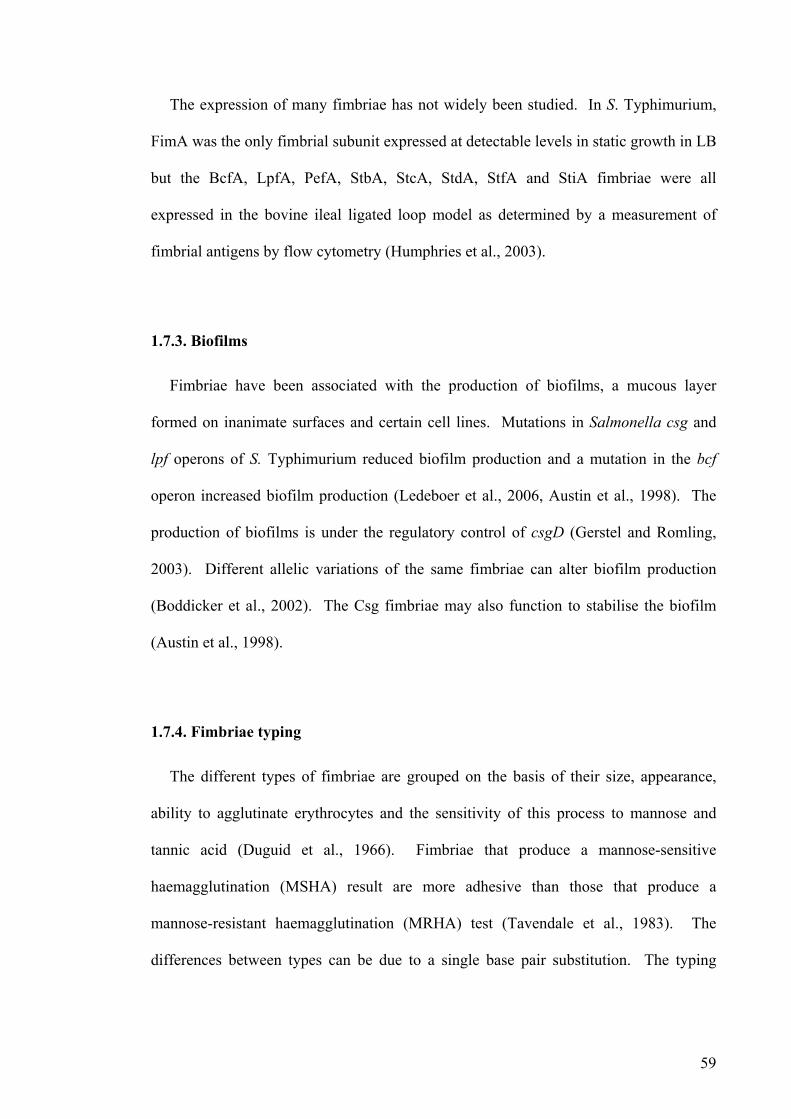
59
The expression of many fimbriae has not widely been studied. In S. Typhimurium,
FimA was the only fimbrial subunit expressed at detectable levels in static growth in LB
but the BcfA, LpfA, PefA, StbA, StcA, StdA, StfA and StiA fimbriae were all
expressed in the bovine ileal ligated loop model as determined by a measurement of
fimbrial antigens by flow cytometry (Humphries et al., 2003).
1.7.3. Biofilms
Fimbriae have been associated with the production of biofilms, a mucous layer
formed on inanimate surfaces and certain cell lines. Mutations in Salmonella csg and
lpf operons of S. Typhimurium reduced biofilm production and a mutation in the bcf
operon increased biofilm production (Ledeboer et al., 2006, Austin et al., 1998). The
production of biofilms is under the regulatory control of csgD (Gerstel and Romling,
2003). Different allelic variations of the same fimbriae can alter biofilm production
(Boddicker et al., 2002). The Csg fimbriae may also function to stabilise the biofilm
(Austin et al., 1998).
1.7.4. Fimbriae typing
The different types of fimbriae are grouped on the basis of their size, appearance,
ability to agglutinate erythrocytes and the sensitivity of this process to mannose and
tannic acid (Duguid et al., 1966). Fimbriae that produce a mannose-sensitive
haemagglutination (MSHA) result are more adhesive than those that produce a
mannose-resistant haemagglutination (MRHA) test (Tavendale et al., 1983). The
differences between types can be due to a single base pair substitution. The typing

60
system does not reflect any sequence differences or similarities and will not be referred
to in this study.
1.7.5. S. Enteritidis fimbriae
S. Enteritidis produces fimbriae that are morphologically indistinguishable from the
fimbriae of all other Enterobacteriaceae but the fimbrial proteins are named after the
protein subunit size. They share homology with fimbriae from other Salmonella
serovars which have a different nomenclature (Muller et al., 1991, Feutrier et al., 1986).
Primarily SEF14, SEF17 and SEF21 have been studied.
The SEF14 gene cluster consists of 3 genes sefABC and are composed of repeating
protein subunits of 14 kDa and has only relatively recently been acquired (Thorns et al.,
1990). The structural gene (sefA) encoding for SEF14 has been identified and
sequenced and has a limited distribution across Salmonella (Turcotte and Woodward,
1993). SEF17 comprises protein subunits of 17 kDa with a unique N-terminal sequence
and are compromised of polymerised CsgA proteins. There is a high degree of
similarity between SEF17 and CsgA of E. coli (Collinson et al., 1996). SEF21 refers to
the product of the fimA gene (Muller et al., 1991). The FimA protein of S. Enteritidis in
the rat was distributed throughout the gut mainly in the large intestine and lumina
contents. Purified FimA was isolated in the gut in a similar pattern to that found with
whole Salmonella (Naughton et al., 2001).
Throughout this thesis, the Salmonella fimbrial genes will be referred to by name
rather than the size of the protein formed. The best characterised fimbrial loci in
Salmonella are the fim, lpf, csg, and sef operons. The genetics, assembly and role of
fimbriae encoded by these operons are reviewed below.

61
1.7.6 E. coli fimbriae
The complete genome sequence of E. coli K-12 MG1655 has been available for several
years and different types of fimbriae have been identified and in some cases a role has
been determined for adhesion and colonisation of chickens (Blattner et al., 1997).
The stg fimbriae from avian pathogenic E. coli (APEC) play a significant role in the
colonisation of the avian respiratory tract (Lymberopoulos et al., 2006). The type 1
fimbriae have been shown to play an important role in the colonisation of avain
pathogenic E. coli in breeders whereas the P and S fimbriae were not required for
bacterial adherence. The the absence of P fimbriae promotes bacterial adhesion
(Mellata et al., 2003),(Monroy et al., 2005). The presence of fimbriae and flagellar on
the cell surface of E. coli O87:K80 play a major role in colonisation, invasion and
persistence in chickens (La Ragione et al., 2000).
Several other bacterial species also possess fimbriae including P pili, type 1 fimbriae,
tpe IV pili, curli and CS1 pili. These different type of fimbriae have been mainly
characterised in different strains of E. Coli but are also present in several other species
of the Enterobactericeae family including Neisseria, Klebsiella pneumonia and Yersinia
enterocolitica reviewed (Soto 1999).
1.7.7. The fim operon
The fim operon consists of 9 genes in two operons, fimA, fimI, fimC, fimD, fimH, and
fimF in one operon and fimZ, fimY and fimW in a convergently transcribed second
operon (McClelland et al., 2001). Downstream of fimA is the fimI gene, which also
encodes a fimbrin protein. There is a similar genetic organisation between fimA and
fimI and the fimI gene may have originated from a duplication of fimA (Rossolini et al.,

62
1993). The fimA gene is adjacent to the folD gene and the fimA-folD intergenic region
of Salmonella encompasses a junctional site of genetic rearrangement probably due to
chromosomal relocation, suggesting that the fim operon was gained through horizontal
gene transfer. The fim operon does not have regulatory genes directly upstream as in E.
coli (Rossolini et al., 1994). In Salmonella the fim operon is in a different chromosomal
location to the homolog in E. coli and the fimWYZ regulatory operon has a lower %
G+C content than the genomic mean (Boyd and Hartl, 1999). The fimA promoter is
always orientated so that transcription can occur (Clegg et al., 1996), in marked contrast
to the location of the fimA promoter to an invertible element subject to recombinase-
mediated inversion and thus phase-variable expression of Type 1 fimbriae in E. coli
(Abraham et al., 1985).
The fimC gene encodes a 26 kDa polypeptide which contains a chaperone domain
similar to the chaperone papD in E. coli. The chaperones are highly conserved and may
fold and function in a similar way (Bonci et al., 1997). The fimD gene encodes an usher
and in E. coli the protein was found to be located in the outer membrane and when
produced on its own has deleterious effects on growth (Klemm and Christiansen, 1990).
A fimD mutation in S. Enteritidis resulted in no adherence in vitro or in chickens and
resulted in prolonged bacteraemia and reduced egg shell contamination, it was also
incapable of adhering to the isthmal secretions which are involved in generating fibers
of the egg shell membrane (De Buck et al., 2004, De Buck et al., 2003). The FimD
protein is involved in the export and assembly of FimA (De Buck et al., 2004). The
fimH gene encodes an adhesin (Jones et al., 1995). The FimD, FimC and FimH proteins
form a complex to protect the usher from degradation in vivo due to a conformational
change in the usher (Saulino et al., 1998). The cloning and sequencing of the fimH gene
from different S. Typhimurium strains resulted in a range of products being
63
characterised that differed in their ability to adhere to HEp-2 cells (Boddicker et al.,
2002). A mutation in fimH can produce non-fimbriate, non-adhesive Salmonella and it
appears that the adhesive properties of Fim are due to both the fimbrial shaft and the
fimbrial tip (Thankavel et al., 1999, Duncan et al., 2005, Hancox et al., 1997).
Recombinant FimH adhesins of type 1 fimbriae of S. Gallinarum and S. Pullorum do not
bind to mannose oligosaccharides or to HT29 human colon carcinoma cells due to a
single base pair mutation of FimH at position 78; resulting in a isoleucine to threonine
substitution (Kisiela et al., 2005). Differences seen between the low and high adhesive
properties of the type 1 fimbriae of two serovars, S. Enteritidis and S. Typhimurium,
are partially due to only 4 amino acid substitutions in FimH and the differences in
adherence between two strains of S. Typhimurium was due to only 2 amino acid
substitutions (Kisiela et al., 2006). FimH is also required for efficient interactions with
DCs and mutations in fimH result in an impaired ability of S. Typhimurium to bind and
be internalised (Guo et al., 2007).
The fim operon has 4 regulatory genes downstream of the first fim operon; fimZ,
fimY, fimU and fimW and the expression of FimA is controlled at the transcriptional
level by the products of fimZ, a transcriptional activator which works in co-operation
with fimY, a positive regulator (Tinker et al., 2001, Yeh et al., 1995, Tinker and Clegg,
2000). FimZ may be a DNA-binding protein and plays a role in its own expression
along with FimY (Yeh et al., 2002). The increased expression of FimZ results in a
hyper-fimbriated non-motile Salmonella in soft agar (Clegg and Hughes, 2002). FimZ
is the molecular connection between flagella and fimbriae (Clegg and Hughes, 2002).
The fimW gene is a negative regulator of Type 1 fimbriae expression and mutations in
this gene result in a 4-8 fold increase in the number of fimbriae produced (Tinker et al.,
2001). The fimW gene is initiated from its own promoter and the encoded protein may

64
function by interfering with FimZ activation of FimA expression (Tinker et al., 2001).
The FimU is an activator of FimY and a mutation in the fimU gene results in an
afimbriate phenotype (Tinker and Clegg, 2001). A fimU mutant in S. Enteritidis
resulted in a decrease in expression of both FimA and SefA (Clouthier et al., 1998b).
Chickens infected with S. Enteritidis not expressing fimA were less intensively
colonised and had less faecal shedding than the wild-type in laying hens (Thiagarajan et
al., 1996) but the same mutation appears to play no role in the colonisation of non-
laying chickens (Rajashekara et al., 2000).
Mutations in fim of S. Enteritidis resulted in a decrease in adherence and invasion of
cultured epithelial cells (Dibb-Fuller et al., 1999) and in S. Typhimurium a decreased
attachment of HeLa cells (Baumler et al., 1996a). In vivo in combination with other
fimbrial genes fim of S. Typhimurium plays a role in the colonisation of mice and
production of murine typhoid fever (van der Velden et al., 1998, Lockman and Curtiss,
1992).
1.7.8. Long polar fimbriae (lpf)
In S. Typhimurium, the lpf operon contains 5 genes lpfA, lpfB, lpfC, lpfD and lpfE
and the operon has entered the Salmonella genomes early as it has a scattered phylogeny
and has been lost from many serovars (Baumler and Heffron, 1995, Baumler et al.,
1997a). The genes flanking the lpf operon in S. Typhimurium have homology to the
flanking genes in enterohaemorrhagic Escherichia coli (EHEC), and the lpf operon has
inserted into a similar chromosomal position (Torres et al., 2002). EHEC has 2 lpfC
genes, encoding proteins of 40.2 kDa and 17.8 kDa respectively and S. Typhimurium
has 1 lpfC gene encoding a protein of 94.4 kDa suggesting a merge of two genes in

65
Salmonella or a separation in EHEC (Torres et al., 2002). The EHEC lpf operon is 60
% identical to that of S. Typhimurium. The lpfA and lpfE genes in E. coli are
translationally coupled as are the lpfA and lpfC genes, it is unknown if this translational
coupling occurs in Salmonella (Torres et al., 2002).
A mutation in the lpfC gene of S. Typhimurium resulted in a 5-fold increase in the
median lethal dose of mice, a decrease in the numbers of bacteria isolated from the
Peyer’s patches, mesenteric lymph nodes, liver and spleen as well as impaired
destruction of M cells (Baumler et al., 1996b). The lpf operon of S. Typhimurium was
also required for adherence to and invasion of HEp-2 cells (Baumler et al., 1996a).
Multiple fimbrial mutations including the lpf operon resulted in an 26-fold increase in
the LD50 of orally inoculated mice with S. Typhimurium implying a synergistic effect
(van der Velden et al., 1998). Genome-wide and targeted mutagenesis have implicated
Lpf fimbriae in Salmonella pathogenesis in mice and the major fimbrial subunit is up-
regulated in S. Typhimurium in a ligated bovine ileal ligated loops, suggesting a role in
virulence or colonisation (Humphries et al., 2003, van der Velden et al., 1998, Weening
et al., 2005, Lawley et al., 2006).
1.7.9. Csg fimbriae
The Csg fimbriae are also known as the Agf fimbriae or thin aggregative fimbriae
(tafi) and in S. Enteritidis are known as SEF17. Throughout this thesis they will be
referred to as Csg or curli fimbriae. The csg fimbrial operon contains 7 genes in two
operons, csgB, csgA and csgC in one operon and csgD, csgE, csgF and csgG in a
divergently transcribed operon. The csg operon is believed to be the oldest fimbriae as
it is present and highly conserved in E. coli (Baumler et al., 1997a, Romling et al.,

66
1998a). The Csg fimbriae are assembled via the nucleator-dependent assembly pathway
and are the only Salmonella fimbriae currently identified that assemble in this way
(Hammar et al., 1996). The expression of the curli fimbriae is temperature and pH
dependent, the majority of strains of S. Enteritidis examined express curli when grown
at 18-30 ºC, however for some strains of S. Enteritidis expression only occurs at 37 ºC
or 42 ºC (Dibb-Fuller et al., 1997). Csg were also produced by some strains at 20 ºC but
not at 37 ºC and only at pH 6.18 or above (Walker et al., 1999).
The csgBAC operon encodes thin aggregative fimbriae which are fibrous and
polymeric and are composed of repeats of the major subunit CsgA (White et al., 2001).
The CsgA is the subunit and CsgB is the surface-exposed nucleator protein (Gerstel and
Romling, 2003). CsgA and CsgB share 51 % amino acid sequence and dimers form
between CsgA-CsgA, CsgB-CsgB and CsgA-CsgB producing a highly rigid
multicellular stable structure which is controlled at the level of transcription by csgD
(Romling et al., 1998b);(White et al., 2001). The CsgA fimbrin is a pathogen-
associated molecular pattern (PAMP) and triggers the hosts innate immune response
causing inflammation (Tukel et al., 2005). A mutation in csgBA of S. Typhimurium
resulted in an inflammatory response, a decrease in the fluid accumulation, and a
decrease in the IL-8 production in macrophages in streptomycin-pretreated mice (Tukel
et al., 2005). The curli of S. Typhimurium can also bind flagellin and the bound flagella
may modulate immune responses in this manner (Rochon and Romling, 2006).
The expression of the curlin protein is dependent on the starvation-induced CsgD
protein, a positive transcriptional activator of the LuxR family. CsgD has an N-terminal
receiver domain, a helix-turn-helix motif and a DNA-binding motif at the C-terminus
(Gerstel et al., 2003). The expression of the CsgD protein results in altered transcription
of 24 novel genes and the csgD gene is itself controlled by a variety of regulators,

67
including a transcriptional activator upstream (Brombacher et al., 2006, Gerstel and
Romling, 2003). The csgD gene regulates two distinct pathways, both of which
contribute to multicellular morphology (Romling et al., 2000).
The CsgC protein is localised to the periplasm and a mutation of csgC results in the
production of fibers of 20 nm, much larger than the usual curli which are usually 5-7 nm
which results in an increase in the surface hydrophobicity. The csgC and csgE genes
are important for extracellular assembly of curli fibers (Gibson et al., 2007). CsgG is an
outer membrane lipoprotein and is required for the secretion of CsgA and CsgB
(Loferer et al., 1997). In E. coli the csgE, csgF and csgG each play a role in the
formation of csgA (Hammar et al., 1995) and in S. Typimurium the csgEFG function as
accessory genes, although their precise role is unclear, they appear to contribute to long-
term systemic infection in mice (Lawley et al., 2006, Gerstel et al., 2003).
Multiple fimbrial mutations including the csg operon resulted in an 26-fold increase
in the LD50 of orally inoculated mice with S. Typhimurium and implies that the Csg
fimbriae have a synergistic effect (van der Velden et al., 1998). Curli-deficient S.
Enteritidis strains were able to reproduce more rapidly inside less eggs than the curli-
expressing S. Enteritidis (Cogan et al., 2004). Mutations in curli of S. Enteritidis delay
the colonisation of the spleen and liver of chickens for the first 24 hours post-infection
(Dibb-Fuller and Woodward, 2000).
1.7.10. Sef fimbriae
The Salmonella-encoded fimbriae or sef operon consists of five genes four co-
transcribed genes (sefA, sefB, sefC and sefD) and a regulatory gene sefR (or sefE) which
is homologous to an araC-like positive transcriptional activator (Edwards et al., 2001,

68
Collighan and Woodward, 2001, Clouthier et al., 1993). The SefA protein assembles to
form a thin filamentous fimbrin structure, SefB is a chaperone, SefC is an outer
membrane usher and SefD is the tip-located adhesin (Clouthier et al., 1993, Edwards et
al., 2001). SefB and SefC cannot be expressed in the absence of SefA due to
translational coupling and SefC has a predicted signal sequence (Clouthier et al., 1993).
The sef operon has a much lower % G+C content than the genomic mean and it has a
scattered presence across the Salmonella serovars implying that it may have been
acquired through horizontal gene transfer (Edwards et al., 2001). Further to support this
is the presence of an insertion like element adjacent to the operon (Collighan et al.,
2000).
Expression of Sef was optimal during growth in late exponential phase and was
repressed during stationary phase (Edwards et al., 2001). Sef expression appears to be
regulated by fimU (Clouthier et al., 1998b). The Sef protein of S. Enteritidis mediate
adhesion to inanimate objects at low temperatures but the adhesion is lost at 37 ºC
(Woodward et al., 2000), however Sef expression was detected by ELISA only at 37 ºC
and at pH 4.77 and above, showing strain-to-strain variation (Walker et al., 1999).
S. Enteritidis contains the sef fimbrial operon which influences virulence in mice and
internalisation into macrophages (Edwards et al., 2000). Polar mutations affecting the
entire sef operon of S. Enteritidis resulted in a decrease in virulence in mice of 1000-
fold. A non-polar mutation affecting only sefA had no effect on virulence but a non-
polar sefD mutant resulted in a severe virulence defect in mice. The product of sefD
influences efficient uptake by macrophages (Edwards et al., 2000). A mutation in the
sef operon of S. Enteritidis results in lower numbers of bacteria being isolated from the
livers and a faster clearance rate from the spleen in chickens (Rajashekara et al., 2000).
The SefA protein has potential vaccine properties as a recombinant plasmid containing

69
SefA given orally to day-old-chickens reduced the colonisation of an experimental
challenge of S. Enteritidis (Lopes et al., 2006).
The SefA protein is insoluble in most detergents and in SDS can depolymerise into
monomers, dimers and other multimers. SefA fimbrins multimerise through the N-
terminus and undergo changes before assembling into fibers. Once the fibers are
formed the subunits are held in contact with each other by hydrophobic interactions
(Clouthier et al., 1998a).
1.7.11. Bcf fimbriae
The bovine colonisation fimbriae are encoded by the bcf operon which consists of
eight genes in one large polycistronic unit bcfA, bcfB, bcfC, bcfD, bcfE, bcfF, bcfG and
bcfH. It is located between the dnaJ and nhaA in S. Typhi and between uvrB and yphK
in S. Typhimurium on a 30 kb region absent from E. coli K-12 (Townsend et al., 2001).
The BcfA fimbriae of S. Typhimurium has been found to be up-regulated in the bovine
ileal ligated loop model as detected by flow cytometry (Humphries et al., 2003).
Genome-wide and targeted mutagenesis have implicated the bcf genes in S.
Typhimurium pathogenesis in mice (van der Velden et al., 1998, Weening et al., 2005,
Lawley et al., 2006). However, although the bcf operon was identified as being required
for colonisation in mice it appears to play no role in calves and the nomenclature may
therefore be inappropriate (Tsolis et al., 1999).
1.7.12. Saf fimbriae
Salmonella atypical fimbriae (saf) comprise a non-fimbrial adhesin and are encoded
by four genes (safA, safB, safC and safD). In S. Typhi safE also exists downstream of

70
safA (Parkhill et al., 2001). The SafB protein is a chaperone, SafC an usher and SafD is
an adhesin that is similar to AFA adhesins of E. coli (Folkesson et al., 1999). The saf
operon is located on a large region that is absent from E. coli K-12 (Folkesson et al.,
1999, Folkesson et al., 2002).
The SafB and SafD proteins form a complex fimbrial adhesin, which is sensitive to
low pH and enzymatic degradation and can induce different immune responses in mice
depending on the route of administration (Strindelius et al., 2004). The safA gene has a
more heterogeneous sequence than the safB and safC genes. A transposon mutation in
the saf operon of S. Typhimurium resulted in a decrease in colonisation of pigs but not
in chickens or calves (Carnell et al., 2007, Morgan et al., 2004).
1.7.13. Other fimbriae
With the completion of the genome sequences of S. Typhimurium LT2 and S. Typhi
CT18 (McClelland et al., 2001, Parkhill et al., 2001) several other fimbrial loci were
identified for the first time. These have been designated st followed by a letter from a-g
and little is known of their role or the assembly process.
The sta operon has orthologues in E. coli K-12 (yadCKLM), as does the stc operon
(yehDCBA). The stb, stg, std, ste and sth operons are all absent in E. coli K-12
(Townsend et al., 2001). The stf-encoded fimbriae have been identified in S.
Typhimurium and are absent from S. Typhi and S. bongori. The stf operon is flanked by
fhuB and hemL whereas in E. coli these genes are adjacent to each other indicating that
it is likely a genomic insertion (Emmerth et al., 1999). The stg fimbrial operon is
encoded in the glmS-pstS intergenic region in S. Typhi and in some E. coli strains. In S.
Typhi, the stgC gene contains a pseudogene but it appears that the gene products of the
71
stg operon are important as in its absence a decrease in adherence and an increase in
macrophage uptake is seen (Forest et al., 2007). The stj fimbrial operon has been
identified in S. Typhimurium LT2 but consists of only 2 genes with homology to a
chaperone and an usher, it is unknown if it is functional (McClelland et al., 2001). The
tcf fimbrial operon is present in S. Typhi and has similarity to the coo operon encoding
the CS1 fimbrial adhesin in E. coli and the operon consists of 4 genes tcfA, tcfB, tcfC
and tcfD (Folkesson et al., 1999). The tcf operon is not unique to S. Typhi as was once
believed (Townsend et al., 2001).
The role of the st(a-g) fimbriae is poorly defined and it has been proposed that they
may carry out several subtle roles at different points during infection (reviewed in
(Baumler et al., 1997b). The stc and sth operons of S. Typhimurium have been
implicated in contributing to gastrointestinal colonisation and long-term systemic
disease in mice (Lawley et al., 2006), the stb, stc, std and sth of S. Typhimurium are
required for intestinal persistence in mice (Weening et al., 2005) and the stbB and sthC
genes are required for colonisation in chickens but not calves (Morgan et al., 2004).
1.8.1. Phase Variation
Phase variation refers to the reversible switch between all-or-nothing expression of a
factor and is different from gene regulation as the change is heritable and reversible in a
given population and often occurs at a rate of 1 change per 103 generations (reviewed in
(van der Woude and Baumler, 2004, Casadesus and Low, 2006). In other Gram-
negative bacteria phase variation is known to be mediated by recombination (e.g.
FimBE-mediated inversion of a 314 bp segment containing the fimA promoter in E. coli
(Abraham et al., 1985, Klemm, 1986), epigenetic regulation dependent on Dam

72
methylation (e.g. control of Pap pili in uropathogenic E. coli (Blyn et al., 1990)) or
slipped-strand mispairing between homo- or hetero-polymeric tracts during DNA
replication (e.g. assembly and maturation of Neisserial pilin (reviewed in (Meyer and
van Putten, 1989, van Belkum et al., 1998).
In Salmonella evidence exists for phase variable expression of Type I fimbriae which
is not the same as the phase variation that exist for Type I fimbriae of E. coli (Abraham
et al., 1985, Old and Duguid, 1970, Swenson and Clegg, 1992) and long polar fimbriae
(Norris et al., 1998). Further, epigenetic regulation of the expression of pef fimbrial
genes in S. Typhimurium by Dam methylation has been described (Nicholson and Low,
2000) and transcription of std fimbrial genes have been observed to be repressed in a S.
Typhimurium Dam methylase mutant (Balbontin et al., 2006).
Phase variation may be sensitive to environmental stimulus, acid conditions have
been reported to stimulate Dam methylation of the GATC sites in Salmonella
(Nicholson and Low, 2000), and in E. coli temperature and growth media have been
implicated in altering phase variation (Gally et al., 1993, Clegg et al., 1996). The
change in response to the environment may allow a proportion of the bacterial
population to survive sudden changes in the environment (Norris et al., 1998).
Phase variation of the lpf operon has also been proposed to be a mechanism to evade
cross immunity between serotypes by changing the antigenic properties of the cell
surface. If a host has encountered the Lpf fimbriae of S. Typhimurium it will select
against S. Enteritidis that is also expressing the Lpf fimbriae, but if Lpf expression can
be switched off then S. Enteritidis can colonise the same host, indicting that Lpf cannot
be essential for colonisation (Nicholson and Baumler, 2001, Norris and Baumler, 1999).

73
Most Salmonella cells are richly fimbriated but a proportion of the cells are always in
the off phase. After passage of S. Typhimurium in LB broth for 120 generations, 96 %
of the cells were carrying lpf in the on-phase; however after 500 generations on an LB
solid media plate only 2 % were in the on phase. The differences were due to changes
in the on-to-off and off-to-on rate of phase variation and the starting culture (Kingsley et
al., 2002). The genetic mechanisms underlying such regulation are ill-defined and it
remains unclear if other Salmonella fimbriae are subject to phase variable expression.
1.9.1. Rationale for the project
Given the important role of fimbriae in adherence and colonisation of different hosts
and given that Salmonella serovars vary both in virulence and tissue tropism it is a
reasonable hypothesis that these differences are due to fimbriae.
The repertoire of fimbrial operons in different Salmonella serovars has been
examined using different techniques. Micro-array hybridisation studies were used to
separate core regions of the genome using decision trees (Anjum et al., 2005),
comparative genomic hybridisation to micro-arrays and Southern hybridisation have
produced results that contain ambiguous data (Porwollik et al., 2004, van Asten and van
Dijk, 2005) and analysis of strain variation of the same serovars indicated differences
(Boyd et al., 2003). The availability of several whole genome sequences of Salmonella
strains has made it possible to carry out the first large-scale genome comparison of the
sequence and repertoire of Salmonella enterica fimbrial operons in host-specific and
ubiquitous serovars in fast and efficient manner (Chiu et al., 2005, McClelland et al.,
2001, Parkhill et al., 2001). With the recent sequencing of the genome of an S.
Enteritidis PT4 strain and given the importance of such strains in human health, this

74
project will include a comprehensive analysis of its fimbriae in vitro and in vivo, to
attempt to link genotype to phenotype.

75
Thesis Aims
To analyse the repertoire sequence and chromosomal location of fimbrial genes
within the genome sequences of several strains of Salmonella using in silico
techniques.
To mutate all chromosomal fimbriae in S. enterica serovars Enteritidis and
Gallinarum
To characterise the fimbrial mutants in vitro using cell adherence and invasion
assays, microscopy and protein expression analysis.
To characterise the fimbrial mutants in vivo in the avian host.
To verify the phenotype/s produced using trans-complementation techniques to
fulfill molecular Kochs postulates.

76
Chapter 2
Materials and Methods
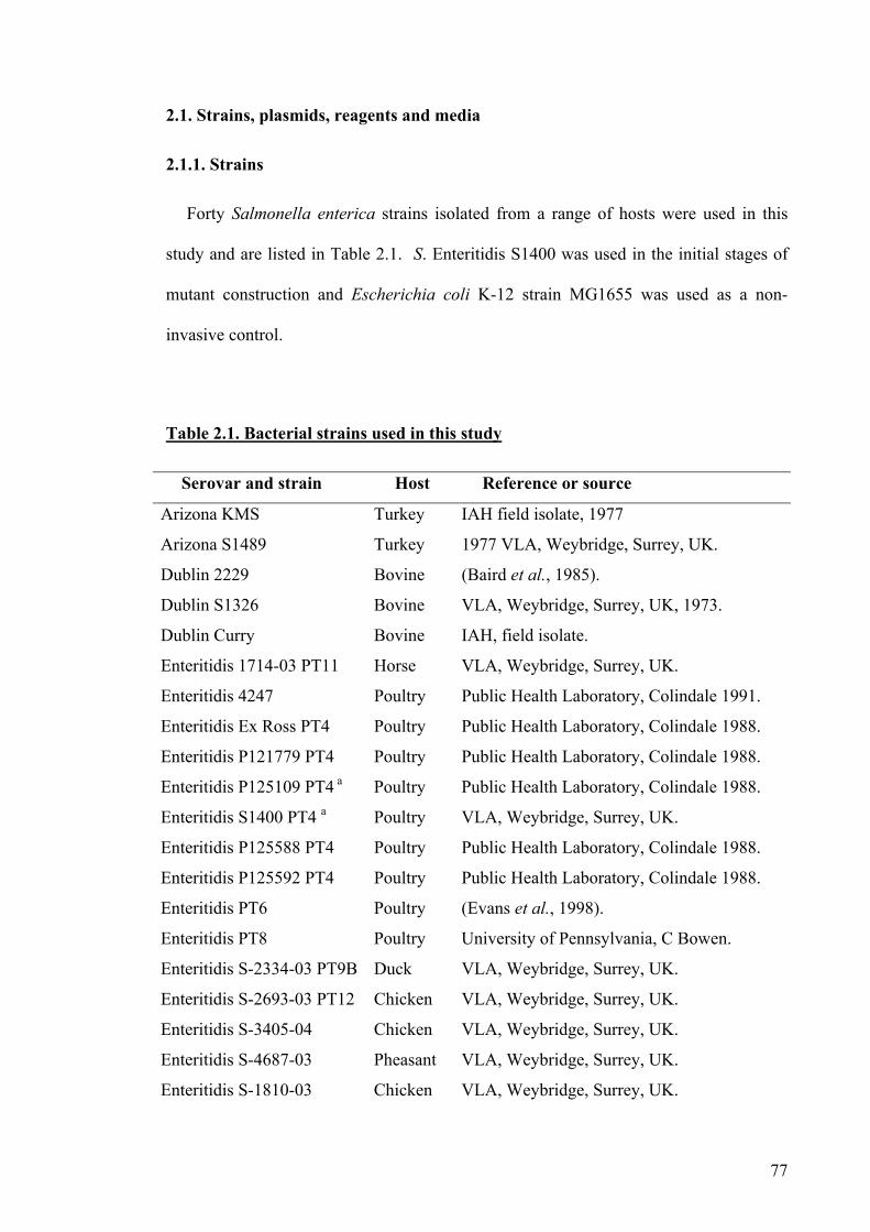
77
2.1. Strains, plasmids, reagents and media
2.1.1. Strains
Forty Salmonella enterica strains isolated from a range of hosts were used in this
study and are listed in Table 2.1. S. Enteritidis S1400 was used in the initial stages of
mutant construction and Escherichia coli K-12 strain MG1655 was used as a non-
invasive control.
Table 2.1. Bacterial strains used in this study
Serovar and strain Host Reference or source
Arizona KMS Turkey IAH field isolate, 1977
Arizona S1489 Turkey 1977 VLA, Weybridge, Surrey, UK.
Dublin 2229 Bovine (Baird et al., 1985).
Dublin S1326 Bovine VLA, Weybridge, Surrey, UK, 1973.
Dublin Curry Bovine IAH, field isolate.
Enteritidis 1714-03 PT11 Horse VLA, Weybridge, Surrey, UK.
Enteritidis 4247 Poultry Public Health Laboratory, Colindale 1991.
Enteritidis Ex Ross PT4 Poultry Public Health Laboratory, Colindale 1988.
Enteritidis P121779 PT4 Poultry Public Health Laboratory, Colindale 1988.
Enteritidis P125109 PT4 a Poultry Public Health Laboratory, Colindale 1988.
Enteritidis S1400 PT4 a Poultry VLA, Weybridge, Surrey, UK.
Enteritidis P125588 PT4 Poultry Public Health Laboratory, Colindale 1988.
Enteritidis P125592 PT4 Poultry Public Health Laboratory, Colindale 1988.
Enteritidis PT6 Poultry (Evans et al., 1998).
Enteritidis PT8 Poultry University of Pennsylvania, C Bowen.
Enteritidis S-2334-03 PT9B Duck VLA, Weybridge, Surrey, UK.
Enteritidis S-2693-03 PT12 Chicken VLA, Weybridge, Surrey, UK.
Enteritidis S-3405-04 Chicken VLA, Weybridge, Surrey, UK.
Enteritidis S-4687-03 Pheasant VLA, Weybridge, Surrey, UK.
Enteritidis S-1810-03 Chicken VLA, Weybridge, Surrey, UK.

78
Enteritidis S-3688-04 Chicken VLA, Weybridge, Surrey, UK.
Enteritidis S-523-04 Chicken VLA, Weybridge, Surrey, UK.
Enteritidis S-7016-03 Pig VLA, Weybridge, Surrey, UK.
Enteritidis S-4850-04 Bovine VLA, Weybridge, Surrey, UK.
Gallinarum 1026 Chicken Athens, 1979.
Gallinarum 287/91 a Poultry Angelo Berchiere, Brazil.
Gallinarum 72/80 Poultry University of Nairobi, Tony Harris, 1986.
Gallinarum 8338/1 Chicken Christensen/Olsen, Denmark, 1993.
Gallinarum 95/80 Chicken University of Nairobi, Tony Harris, 1986.
Gallinarum 9 Poultry (Barrow and Lovell, 1989).
Pullorum 2249 Chicken IAH, field isolate.
Pullorum 449/87 Chicken VLA, Weybridge, Surrey, UK.
Pullorum 5078 Chicken (Li et al., 1993).
Typhimurium 333 PT49 Human Public Health Laboratory, London, 1985.
Typhimurium SL1344 Bovine (Wray and Sojka, 1978).
Typhimurium 68/67 PT56 Bovine IAH, field isolate, 1967.
Typhimurium bangor PT44 Bovine Public Health Laboratory, Colindale, 1978
Typhimurium F98 PT14 Chicken (Barrow and Lovell, 1989)
Typhimurium S1622
PT104
Chicken VLA, Weybridge, Surrey, UK, 1984.
Typhi aroCD Lab.
strain
Steve Chadfield, 1999.
E. coli K-12 MG1655 Standard laboratory strain due to suitablitly for genetic
rearrangements (Zinder and Lederberg, 1952) a These strains are those used to make fimbrial mutants. All other strains were used
in dot blot analysis.
All strains were confirmed at the outset using anti-Salmonella serum O9 for S.
Enteritidis, S. Dublin, S. Pullorum and S. Gallinarum, O4 for S. Typhimurium and H
antigens were used to distinguish between the different O9 groups according to the
Kauffmann-white scheme. All Salmonella serovars were confirmed to possess an intact
LPS by the use of acriflavine-HCl as detailed in section 2.4.4.

79
2.1.2. Plasmids
Several plasmids used in this study for the construction of fimbrial mutants and for
trans-complementation are listed in Table 2.2.
Table 2.2. Plasmids used in this study
Plasmid Details/ Description Source Reference
pKD46 Temperature-sensitive origin of
replication. Ampicillin resistance
gam, bet and exo genes under
arabinose-inducible promoter.
Barry Wanner (Datsenko and
Wanner, 2000)
pKD3 Derivative of pSC140, used as
PCR template to amplify FRT
flanked chloramphenicol
resistance cassette for mutant
construction.
Barry Wanner (Datsenko and
Wanner, 2000)
pACYC177 Derivative of P15A, cloning
vector, ampicillin and kanamycin
resistance
New England
Biolabs (NEB)
(Chang and
Cohen, 1978)
pCP20 Temperature-sensitive origin of
replication. Ampicillin and
chloramphenicol resistance.
Expresses FLP recombinase.
Barry Wanner (Cherepanov and
Wackernagel,
1995)
pCR®4Blunt-
TOPO
Vector for topoisomerase-
mediated cloning of blunt-ended
amplicons or fragments,
ampicillin and kanamycin
resistant.
Invitrogen

80
2.1.3. Media and reagents
All media were obtained from Microbiological Services, IAH (supplied by Sigma,
Difco or Oxoid) and were sterilised by autoclaving prior to use. Unless otherwise stated
all strains were cultivated in Luria-Bertani (LB) broth in a Unitron INFORS HT
incubator at 37 ºC and 130 revolutions per minute (rpm).
LB broth: 25 g LB broth base (Miller # 244610) comprising 10 g peptone, 140.5 g
yeast extract and 10 g NaCl dissolved in 1 litre of sterilised water.
LB agar: 25g LB broth base (Miller # 244510) and 15 g bacto agar dissolved in 1 litre
of sterilised water.
Antibiotic supplements: LB agar or broth was supplemented with antibiotics (Sigma)
to a final concentration of chloramphenicol cat. # C0378 (25 µg/ml), ampicillin cat #
A9518 (100 µg/ml), novobiocin cat # N1628 (1 µg/ml) and nalidixic acid cat # N4382
(20 µg/ml) where appropriate. Typhi aroCD mutant was grown in LB broth
supplemented with 10 mg/ml tyrosine and 1 mg/ml each of tryptophan, phenylalanine,
p-aminobenzoic acid (PABA) and di-hydroxybenzoic acid (DAB).
Brilliant green agar: 58.0 g Brilliant green agar base (Difco #228530) comprising 10
g protease peptone, 3 g yeast extract, 10 g lactose, 10 g saccharose, 5 g NaCl, 20 g agar,
12.5 mg brilliant green and 0.08 g phenol red dissolved in 1 litre of sterilised water.
SOC: 31 g SOC powder (Q-BIO gene # 3031-012) comprising 20 g tryptone, 5 g yeast
extract, 0.5 g NaCl, 5 g MgSO4.7H20 and 20 mM glucose dissolved in 1 litre of SQW
water.
DMEM: Dulbecco’s minimal essential medium supplemented with 3 g/litre of sodium
bicarbonate with L-glutamine(Sigma # D7777).

81
EMEM: Eagles minimal essential medium supplemented with 2 g/litre of sodium
bicarbonate with L-glutamine (Sigma # M0268)
RPMI 1640: Supplemented with 2 g/litre of sodium bicarbonate with L-glutamine
(Sigma # R6504).
Phenol:chloroform:isoamyl alcohol 24:1: (Sigma # P3803)
CTAB: 10 % (w/v) hexa-decyl-trimethyl ammonium bromide in sterilised water (Sigma
# 83935).
Isopropanol: (VWR #133)
Ethanol: (VWR #101077Y)
EDTA: Ethylenediaminetetraacetic acid disodium salt (Sigma # E7889)
Proteinase K: (Sigma #P6556)
RNase: (#R4875)
Paraformaldehyde: (Sigma # 158127)
Vectashield: (Vector labs #H1400)
Formic acid: (VWR #83634)
5-bromo-4-chloro-3-indolyl phosphate/Nitro blue tetrazolium (BCIP/NBT): (Sigma
# B5655)
Zero blunt TOPO PCR cloning kit: – Salt solution (1.2 M sodium chloride and 0.06
M magnesium chloride) and pCR4Blunt TOPO vector (Invitrogen #K286020)
Herculase: (Stratagene #600310) comprising pfu DNA polymerase, Archae Maxx
polymerase and Taq 2000.
TOP10 E. coli cells: (Invitrogen # C664-11)

82
IPTG – isopropyl-beta-D-thiogalactopyranoside: (Invitrogen #15520019)
Ligafast T4 DNA ligase: (Promega # 9PIM822)
Agarose: (Sigma # A9539)
Ethidium bromide: 10 mg/ml (Biorad #161-0433)
1 kb DNA ladder: (Invitrogen # 10787-026)
Hydrochloric acid: (VWR # 190686W)
Sodium hydroxide: (Sigma # 30620)
Sodium chloride: (Sigma # S7653)
Tri-sodium citrate: (Sigma #32320)
Trizma hydrochloride: (Sigma # T5941)
dNTPs: (Promega #U1240)
Sodium dodecyl sulphate: (Sigma # L4390)
DIG Easy Hyb granules: (Roche # 11796895001)
DIG Wash and block buffer set: (Roche # 11585762001)
Tween 20: (Sigma # P9046)
Anti-digiocigenin-AP: (Roche # 11093274910)
PCR DIG labelling kit: (Roche # 11585550910)
CSPD disodium-3 (4-methoxyspiro-[1,2-dioxetane-
3,2(5’chloro)tricycle[3.3.1.1]decan]-4-yl: (Roche # 11755633001)
Foetal bovine serum: (Autogen #318A)
Triton 100: (Sigma # 234729)

83
Haemacolour stains: (Merck # 1.11661.0001)
ClaI, DpnI, HindIII: (NEB # RO197L, #RO176L, #RO104L)
Saline: 0.9 % (w/v) sodium chloride in sterilised water.
TE buffer: 10 mM Tris-Cl (pH 7.5) and 1 mM EDTA in sterilised water (made in
house).
SSC: 3 M NaCl and 0.3 M sodium citrate in sterilised water (Invitrogen #15557044).
TAE: 400 mM Tris-acetate and 10 mM EDTA in sterilised water (Invitrogen #15558-
042). Sodium acetate
Loading buffer: 250 mg bromophenol blue in 33 ml of 150 mM Tris (pH 7.6), glycerol
and 7 ml of water.
PCR Taq polymerase: – comprising 10x PCR buffer, 1.5 mM Magnesium chloride,
Taq DNA polymerase: ( Invitrogen #10342-020)
Anti-Salmonella O serum: Wellcome, diagnostics Dartford UK
Anti-rabbit-Ig Alexa568: (Invitrogen # A10037)
Phalloidin flouroscein-isothiocyanate-conjugate: (Invitrogen # F432)
Gentamycin: 50 mg/ml (Sigma #G1397)
Permanent stocks of all strains and plasmids were maintained as stationary phase
cultures in LB broth supplemented with 20 % (v/v) glycerol and stored at -70 ºC. For
growth, the strains were streaked from permanent stocks to single colonies on LB agar
plates supplemented with antibiotics where appropriate and incubated for 16-18 h at 37
°C statically. Liquid cultures were inoculated using a streak of colonies and incubated
as described above. All Salmonella strains were confirmed by the use of serology
testing as described in Section 2.4.4.

84
2.2. In silico tools
2.2.1. Source of sequences
The complete genome sequences of Salmonella enterica serovar Enteritidis PT4
strain P125109, S. Gallinarum strain 287/91, S. Typhimurium SL1344, S. Typhimurium
DT104, S. Typhi CT18 and S. bongori 12419 were produced by the Pathogen
Sequencing Unit at the Wellcome Trust Sanger Institute, UK
[http://www.sanger.ac.uk/Projects/Salmonella/]. Published genome sequences were
obtained from the National Center for Biotechnology Information (NCBI). The genome
sequences are described with their RefSeq curated accession numbers; S. Typhi Ty2
NC_004631 (Deng et al., 2003), S. Choleraesuis SC-B67 NC_006905 (Chiu et al.,
2005), S. Typhimurium LT2 NC_003197 (McClelland et al., 2001), Escherichia coli K-
12 MG1655 NC_000913 (Riley et al., 2006) and Escherichia coli O157:H7 EDL933
NC_002695 (Hayashi et al., 2001).
2.2.2. Glimmer
The genome sequence of S. Gallinarum 287/91 was unannotated and Glimmer was
used to predict coding regions in the genome sequence (Delcher et al., 1999). Glimmer
uses an input training set of sequences with known genes in this case from E. coli and
other sequenced strains of Salmonella and selects the putative gene predictions based on
this training set.
2.2.3. ClustalW
ClustalW 1.83 [http://www.ebi.ac.uk/clustalw](Pearson and Lipman, 1988,
Thompson et al., 1994) was used to align the protein sequences of the known fimbrial

85
proteins and identify polymorphisms. Alignments for the ushers, chaperones, and
regulatory proteins as identified from the literature were generated for each group.
2.2.4. Pfam
Pfam version 17.0 (Bateman et al., 2000) was used to search the translated genome
sequence in all 6 reading frames of S. Enteritidis P125109 and S. Gallinarum 287/91 to
identify conserved domains within putative fimbrial proteins.
2.2.5. HMMER
HMMER version 3.3.2 (http://hmmer.wustl.edu) was used to generate a Hidden
Markov Model (HMM) from a ClustalW alignment of known fimbrial protein
sequences. The HMM allows variation, so at any one position the amino acid could be
one of a selection if it has occurred within the training set, rather than fixed. The HMM
was used to search the genome sequence of S. Enteritidis P125109 for fimbriae-
associated Pfam domains.
2.2.6. BLAST
Basic Local Alignment Sequencing Tool (BLASTp version 2.2.11
(http://www.ncbi.nlm.nih.gov/BLAST/ (Altschul et al., 1997) compared protein
sequences with the Swiss-Prot version 5.2 database and Uniprot (Bairoch and Apweiler,
1996, Bairoch et al., 2005). BLAST allows partial alignments at the beginning, middle
or end of a sequence and takes into consideration the length of the sequence being
searched. All putative fimbrial gene products were confirmed using this search tool.

86
The closer the E-value or output value to zero the less likely the match occurred by
chance. Default parameters were used.
2.2.7. Artemis/ACT
Artemis is a DNA sequence visualisation and annotation tool and was used to
visualise genome sequences used in this study (Rutherford et al., 2000). Sequences
were viewed in a graphical and interactive format and multiple lines of information
were presented in a single context. Artemis can use annotation directly from EMBL
(Baker et al., 2000) and GenBank (Benson et al., 2000).
ACT (Artemis Comparison Tool) is an extension of the Artemis program and allows
comparison of two or more genomes simultaneously (Carver et al., 2005). The S.
Enteritidis P125109 and S. Gallinarum 287/91 genome sequences were compared with
the published genome sequences using ACT version 4 (Rutherford et al., 2000).
2.2.8. Perl
Perl and Bioperl [http://www.bioperl.org] were used to write a script to visualise
sections of the genome (Appendix 2.1). The script was written to use Artemis and
extract gene features of a specific region, Pfam domains and % G+C of the extracted
region.
2.2.9. Neural Network Promoter Prediction (NNPP)
The NNPP v 2.2 (Reese, 2001) identifies promoters by training a neural network on
known promoters in a training set from E. coli. The program was used to search for

87
promoters within the genome sequence of S. Enteritidis P125109. A threshold level of
0.8 was set allowing 60 % of promoters to be identified with a 0.4 % false positive hit
rate.
2.2.10. Tandem Repeats Finder
The tandem repeats finder program (Benson, 1999) was used to analyse DNA repeat
sequences in the genome sequence of S. Enteritidis P125109. A detection component
finds candidate tandem repeats and the analysis component produced alignments of
these candidates. This programme is unique in its ability to find tandem repeats without
the need to specify the pattern or size. All default parameters were used to identify
homopolymeric tracts in S. Enteritidis P125109.

88
2.3. Molecular techniques
2.3.1. Extraction of genomic DNA
A 1.5 ml aliquot of a 10 ml overnight culture of Salmonella was harvested by
centrifugation at approximately 10,000 g for 15 minutes (min) at room temperature and
the pellet was resuspended in 567 µl of TE buffer. To this 30 µl of 10 % (w/v) sodium
dodecly sulphate (SDS) and 3 µl of 20 mg/ml of proteinase K was added and mixed by
inversion before being incubated for 1-2 h at 37 °C statically to lyse the bacteria. The
cell wall debris, protein and polysaccharides were bound by the addition of 100 µl of 5
M sodium chloride and 80 µl of CTAB and incubated statically for 10 min at 65 °C, 750
µl of phenol:chloroform:isoamyl alcohol (25:24:1) was then added, mixed by inversion
and the suspension centrifuged at 10,000 g for 40 min at room temperature. The
aqueous phase was removed and the process repeated three times. The DNA was
precipitated by the addition of 450 µl of isopropanol and harvested by centrifugation at
approximately 10,000 g for 10 min at room temperature. The DNA was purified by the
addition of 400 µl of 70 % (v/v) ethanol and the DNA was harvested by centrifugation
at approximately 10,000 g for 5 min at room temperature. The pellet was air-dried and
resuspended in 50 µl distilled sterile water. Any contaminating RNA was removed by
the addition of RNase at a final concentration of 10 µg/ml. The DNA was stored at -20
ºC (Manfioletti and Schneider, 1988).
2.3.2. Isolation of plasmid DNA
All plasmids were extracted using a QIAGEN Plasmid Midi kit, following the
manufacturers’ instructions using a modified alklaine lysis method (version July 2004).
Briefly bacterial cells were harvested and lysed under alkaline conditions and

89
neutralised. The DNA was adsorbed onto silica in the presence of high salt, salts and
endonucleases were washed and removed. The plasmid was eluted and the DNA was
purified by isopropanol precipitation and ethanol washing.
2.3.3. Restriction endonuclease digestion of genomic DNA
The concentration of genomic DNA was measured at 260 nm using a DNA
spectrophotometer (Ultrospec 2100 pro, Amersham) and the concentration (mg/ml)
calculated (A260 x 50 x dilution factor). DNA (20 µg) was cleaved in 20 µl of the
appropriate enzyme buffer and 2 µl of enzyme at 10 units/µl, in a total volume of 200 µl
of water at 37 ºC for 16-18 h. The digest was cleaned using 200 µl of
phenol:chloroform:isoamyl alcohol (25:24:1), centrifuged at 10,000 g for 3 min at room
temperature, followed by recovery of the aqueous layer. One-tenth final volume of 3 M
sodium acetate (pH 5.2) was added along with at least 2 volumes of ice-cold absolute
ethanol to precipitate the DNA. The samples were incubated at -20 ºC for a minimum
of 15 min and the DNA harvested by centrifuguation at approximately 10,000 g for 15
min at room temperature. The DNA was washed in 500 µl of 70 % ethanol (v/v) and re-
suspended in 12 µl of water and 2 µl of 10 x loading buffer (250 mg bromophenol blue
in 33 ml of 150 mM Tris (pH 7.6), 60 ml glycerol and 7 ml water);(Sambrook et al.,
1989).
2.3.4. Agarose gel electrophoresis
PCR products and DNA digests were separated and visualised using horizontal slab
gel electrophoresis with a 0.8 % (w/v) agarose gel unless otherwise stated. For a 0.8 %
gel, 0.8 g of agarose was added to 100 ml of 1 x TAE buffer (Sigma) and heated until

90
the agarose had dissolved. Once cooled, ethidium bromide (BioRad) was added at a
final concentration of 1 µg/ml and the gel was poured into a gel tray with a comb and
allowed to set. DNA samples were loaded onto the gel using a 1 x loading buffer. The
DNA was separated by applying a constant voltage of 80 V and visualised under UV
light. The image was captured using a UVP gel documentation system (Sony). A 1 kb
DNA ladder (Promega) was always included on the gel as a reference of size.
2.3.5. Southern blotting
DNA fragments were separated on a 1 % agarose gel as described in Section 2.3.4
and the DNA on the gel was depurinated in 250 mM hydrochloric acid for 10 min. The
DNA was denatured in a mixture of 0.5 M sodium hydroxide and 1.5 M sodium
chloride for 15 min then rinsed in sterile water and the denaturation step was repeated.
The DNA was neutralised in a mixture of 1.5 M sodium chloride and 0.5 M Tris (pH
7.5) for 15 min at room temperature then rinsed with water and repeated. The DNA was
equilibrated in 2 x SSC (Invitrogen) for 10 min before being transferred using a
membrane pump system (BiometraR) to Hybond-N membrane according to the
manufacturers’ instructions (Amersham). Briefly, the gel was placed on top of the
Hybond-N membrane which sat on top of Whatmann paper pre-soaked in 2 x SSC. A
vacuum was applied to draw the DNA across the gel and onto the membrane. The DNA
was fixed to the membrane by exposure to UV light for 3 min whilst the membrane was
still wet (Southern, 1975).

91
2.3.6. Dot blotting
Genomic DNA (5 µg) was spotted directly onto Hybond-N membrane and allowed to
dry at room temperature. The membrane was placed DNA side up on Whatmann paper
soaked in a mixture of 1.5 M NaCl and 0.5 M NaOH for 5 min to denature the DNA.
The membrane was then placed on Whatmann paper soaked in 0.5 M Tris (pH 7.4) for
30 seconds followed by 5 min on Whatmann paper soaked in a mixture of 1.5 M NaCl
and 0.5 M Tris (pH 7.4). The membrane was allowed to dry for 30 min before the DNA
was fixed by exposure to UV light for 3 min.
2.3.7. Primer design for amplification of major fimbrial subunits and
chloramphenicol gene probes
The genome sequence of S. Enteritidis P125109 was used as the template for
designing primers and the sequences of S. Typhimurium LT2 and S. Typhi CT18 were
used when genes were not present in S. Enteritidis P125109 and are shown in Table 2.3.

92
Table 2.3. Primer sequences for amplification of major fimbrial subunit and
chloramphenicol cassette gene probes
Gene Name Sequence (5′-3′) csgA F TTCGCAGCAATCGTAGTTTC csgA
csgAR TAATACTGGTTAGCCGTGGCGT
lpfA F TTTTGCTCTGTCTGCTCTCGCTG lpfA
lpfA R AAGTCCACTTCTGCGTTACCGTAACCA
stfA F GTTGCTGCCGCACTGGTTATGGGTG stfA
stfA R ACAGATAGCTGATCGTGAAGTTTACGGTGC
sefA F GCGTAAATCAGCATCT sefA
sefA R CTGAACGTAGAAGGTCGCAGT
stbA F ATGATCACAGGCTCGCTTCTTGTCTC stbA
stbA R ACGAAACGGCGTATTGTAGGGTGGCA
stcA F CGTTCACTTATTGCTGCTTCTG stcA
stcA R CCGTCATCGTCAGTACAGATTC
fimA F ATGACCTCTACTATTGCGAGTCTGA fimA
fimA R TTATTCGTATTTCATGATAAAGGTG
bcfA F AAAAGCCTGTACTAGCATTAATGGT bcfA
bcfA R TCAGGAATAAACCATGCTAAATGTC
steA F ATGAAGTCATCTCATTTTTGTAAAC steA
steA R TTACAGGTAAGAGATAGTGACGTTG
safA F GTGGTTATTCAAATGAAAAGCATAA safA
safA R TTAAGGCTGATATCCCACTACGTCT
stdA F GTGCTTCGTTTAACACCAGGCGTTT stdA
stdA R TCACAGGTATTTCAGGGTGTAGGTG
sthA F ATGTTTAATAAGAAAATTATCATC sthA
sthA R ACGAAACGGTATACGTAACCTGAGT
stiA F ATGAAACTCTCCTTAAAAACACTC stiA
stiA R TCAGTTATATTGCAGATAGAATGTT
tcfA F AATTTTAAAGATACTCTTCCCGGGGT tcfA
tcfA R TTACTTTCCGGCTGCTGTTAAATCCA
staA F ATGAAAAAAGCGATTTTAGCTGCCG staA
staA R TTACTGGTAAGTAAAGGTATACATT
stjB F GTGAAGTATTTAAAACTGCCGCTAT stjB
stjB R TCATTGACAGACTCCCTTTGCCGTT
stgA F AAACTGAATTTAATTGCCAGCGCTCT stgA
stgA R TTATTTTTGGTATTCGACAGTGAAC
Chl F TTCAGCTGGATATTACGG Chl
cassette Chl R ATCGCAGTACTGTTG

93
The genome sequences were visualised in Artemis and primer sequences were
selected of approximately 20 base pairs (bp) in length either directly at the 5′ and 3′
ends of the gene of interest or 5 bp into the downstream of the start and stop codons and
were obtained from Sigma-Aldrich. The primer sequences for the chloramphenicol
cassette were obtained from (Datsenko and Wanner, 2000). All oligonucleotides were
resuspended in sterile water at a concentration of 100 mM and maintained at –20 ºC.
The start and stop codons are underlined in Table 2.3.
2.3.8. Digoxigenin (DIG) labelled probes
The primers listed in Table 2.3 were used in a PCR reaction incorporating DIG-
labelled dUTP to prepare probes to each fimbrial gene and the chloramphenicol cassette
using S. Enteritidis P125109, S. Typhi aroCD, S. Typhimurium SL1344 or pKD3 as a
template. This was carried out according to the manufacturers’ instructions (June 2006,
PCR DIG probe synthesis kit, Roche Applied Science, U.K.). The PCR mixture
consisted of the following; final concentrations 1 x PCR buffer (Invitrogen), 1.5 mM
magnesium chloride (Invitrogen), 0.2 mM of each dNTP including dUTP (Roche
Applied Science, U.K.), 0.5 µM of each primer (Sigma), template genomic DNA (2 µg),
Taq DNA polymerase 0.5 µl and made up to 50 µl with distilled water.
The reaction was set at
1. Initial denaturation 95 oC for 2 min
Followed by 30 cycles of :-
2. Denaturation at 95 oC denaturation for 30 seconds
3. Annealing at 55 oC for 1 min
4. Elongation at 72 oC for 1 min
5. One final elongation step at 72 ºC for 7 min

94
PCRs were carried out in a GeneAmp PCR system 2700 thermocycler from Applied
Biosystems and the PCR products were analysed using agarose gel electrophoresis as
described in Section 2.3.4.
2.3.9. Hybridisation of fimbrial probe to membrane
The optimal hybridisation temperatures were calculated for each of the probes and
are shown in Table 2.4. The hybridisation temperature (Thyb) is dependent upon the
melting temperature of the probe (Tm) and is set 20-25 ºC lower.
The melting temperatures of the probe can be calculated;
Tm = 49.82 + 0.41 x (%G+C) - 600/l where l is the length of the probe and % G+C is
the GC content of the probe. Thyb = Tm-(20-25 ºC)

95
Table 2.4. Hybridisation temperatures of DIG-labelled probes
Probe target Hybridisation temperature (oC) staA 47 stbA 45 stcA 44 stdA 46 steA 47 stfA 46 stgA 47 sthA 47 stiA 48 stjA 47 lpfA 44 tcfA 46 fimA 49 safA 43 sefA 48 csgA 47 bcfA 47
Chloramphenicol cassette 41
The DIG Easy Hyb Granules (Roche Applied Science, U.K.) were dissolved in
sterile water as described in manufacturers’ guidelines (version 1 July 2003), and pre-
heated to the appropriate hybridisation temperature as calculated in Table 2.4. The
membrane was incubated at the appropriate temperature with 20 ml of the hybridisation
solution for 15-30 min with agitation. The labelled probe was denatured by heating to
100 oC for 5 min and cooling on ice and 10 µl was then added to 20 ml of the dissolved
DIG Easy Hyb Granules. The pre-hybridisation solution was replaced with the probe
and hybridisation solution and the membrane was incubated for a minimum of 6 h with
constant agitation at the appropriate temperature specified in Table 2.4.

96
2.3.10. Detection of DIG-labelled probes
The hybridisation solution was replaced with low stringency buffer (2 x SSC and 0.1
% (w/v) SDS) and incubated for 2 x 15 min at room temperature with agitation. Two
high stringency washes (0.5 x SSC and 0.1 % (w/v) SDS) were carried out for 15 min
each with agitation at 68 °C. The membrane was washed for 15 min with washing
buffer (0.1 M maleic acid and 3-5 % Tween 20) at room temperature and then incubated
with blocking buffer (10 % blocking solution provided within the kit and 0.1 M maleic
acid) for 30 min with agitation at room temperature (All reagents from DIG wash and
block kit, Roche, Applied Science, U.K.). Anti-digoxigenin-AP (Roche, UK), was
added to 20 ml of fresh blocking solution and the membrane was incubated for 30 min
at room temperature with agitation. The membrane was washed and equilibriated in
detection buffer (1 M NaCl and Tris-HCl (pH 9.5)). The membrane was placed DNA
side up in a developing folder and disodium-3(4-methoxyspiro-[1,2-dioxetane-3,2-
(5’chloro)tricyclo[3.3.1.1]decan]-4-yl) phenyl phosphate (CSPD; Roche) was added
dropwise following the manufacturers’ guidelines (version 3, June 2000) and was
incubated for 5 min at room temperature. The excess CSPD was squeezed out and the
developing folder was sealed and incubated for 10 min at 37 °C. The membrane was
exposed to a X-ray film for 5-15 min and the image captured using a X-ograph machine
X4 Compact, from Imaging Systems (as described in manufacturers’ manual version 1,
March 2003).

97
2.4. Construction and confirmation of fimbrial mutants
2.4.1. Construction of fimbrial gene mutants by λ Red mutagenesis
This method allows for mutagenesis of chromosomal genes in a single step by
homologous recombination of linear PCR products harbouring an insertion of an
antibiotic resistance cassette in the gene of interest. Recombination requires the pKD46
helper plasmid which encodes the bacteriophage λ, gam, bet and exo recombinase
functions that are under the control of an arabinose inducible promoter. The helper
plasmid can be selected for with ampicillin and cured after recombination by virtue of
its temperature-sensitive origin of replication. Once the helper plasmid is present, the
linear PCR products containing the chloroamphenicol cassette can recombine with the
gene of interest resulting in its inactivation. Where polar effects are anticipated the
antibiotic resistance gene inserted in the gene of interest may be excised by transient
expression of flippase (FLP) recombinase which catalyses the reaction between flippase
recognition target (FRT) sites flanking the cassette leaving an in frame 84 nucleotide
‘scar’. The process is summarised in Figure 2.1.

98
Figure 2.1. λ Red recombinase-mediated integration of linear PCR products to
disrupt chromosomal genes
Image taken from (Datsenko and Wanner, 2000).
2.4.2. Preparation of electrocompetent cells
LB broth was inoculated with the strain of interest and incubated as appropriate. The
culture was diluted 1:100 into 100 ml of LB broth and grown at 130 rpm (standard
grwoth condition to aerate the bacteria) at the appropriate temperature until the A600nm
was approximately 0.5, where the pKD46 plasmid was present 10 mM L-arabinose was
added to induce expression of the bacteriophage λ genes. The bacterial cells were
harvested by centrifugation at approximately 2000 g for 15 min at 4 oC and the pellet
was washed twice in 20 ml ice-cold sterile water and once in 20 ml of 10 % (v/v)

99
glycerol. The bacterial cells were resuspended in 500 µl of 10 % (v/v) glycerol
(Sambrook et al., 1989).
2.4.3. Transformation of electocompetent cells
The pKD46 plamid was extracted as described in Section 2.3.2. A range of volumes
of the plasmid or linear PCR products (Section 2.4.5) were mixed with
electrocompetent bacterial cells (50 µl) (Section 2.4.2) and incubated on ice for 10 min.
The electroporation was carried out in a 0.2 mm cuvette using a Biorad gene pulser and
pulse controller at 200 Ω, 25 µF, 2.5 kV, with a time constant of approximately 4. The
bacteria were recovered in 1 ml of SOC with appropriate antibiotic and incubated at 37
°C unless the plasmid is temperature-sensitive. The recovered bacteria (100 µl) were
plated on LB agar plates with appropriate antibiotics and incubated for 16-18 h at 37 °C,
unless the plasmid is temperature sensistive.
2.4.4. Verification of transformants
All colonies were checked for the presence of intact lipopolysaccharide (LPS),
during electroporation bacteria lacking LPS take up plasmid DNA at a higher frequency
than those with intact LPS. A colony was resuspended in 5 µl PBS and 2 µl acriflavin,
HCl 5 % (w/v) was added; aggregation indicates the absence of the LPS. To confirm
the colonies were S. Enteritidis or S. Gallinarum, the process was repeated but anti-
Salmonella O9 serum was added instead of acriflavin HCl and the anti-H serum was
used to distinguish between S. Enteritidis and S. Gallinarum. Agglutination indicates
the presence of specific O or H antigens. The presence of the plasmid was confirmed
by the re-isolation and digestion of the plasmid as described in Sections 2.3.2 and 2.3.3.

100
2.4.5. Generation of amplicons for mutagenesis of major fimbrial subunits
Forward and reverse primers were designed with 40 bp homology extensions to the
major fimbrial subunit gene of S. Enteritidis P125109 (40 bp 3′ of start codon and 5′ of
stop codon, underlined) some primers were designed 1-5 bp downsteam of the start or
stop codon.
Table 2.5. Forward and reverse primers for lambda Red mutagenesis
Primer Name
Primer sequence (5′- 3′)
stbAFmut ATGTCTATGAAAAAATATTTAGCAATGATCACAGGCTCGCTGTGTAGGCTGGAGCTGCTTCG
stbARmut TTATTTATACGAAACGGCGTATTGTAGGGTGGCAGCGACTCATATGAATATCCTCCTTA
stcAFmut ATGAAACGTTCACTTATTGCTGCTTCTGTATTGTCTGCTGTGTGTAGGCTGGAGCTGCTTCG GCTGCTGATGAAGATATGGGGGAATTAAAAATAAACGGTGTGTAGGCTGGAGCTGCTTCG
stcARmut TTAATCAGTTAATACCGTCATCGTCAGTACAGATTCAACACATATGAATATCCTCCTTA AGTATTTTCAACGGCGGCATAGCGGGCAGAAAAGTTCAGGGTCATATGAATATCCTCCTT
stdAFmut GTGCTTCGTTTAACACCAGGCGTTTATTATTCATACGAATTGTGTAGGCTGGAGCTGCTTCG
stdARmut TCACAGGTATTTCAGGGTGTAGGTGACGGATGCGTTGAAGCATATGAATATCCTCCTAA
steAFmut ATGAAGTCATCTCATTTTTGTAAACTGGCAGTAACTGCATGTGTAGGCTGGAGCTGCTTCG
steARmut TTACAGGTAAGAGATAGTGACGTTGGCGGCGCTGCTGAACATATGAATATCCTCCTTA
stfAFmut ATGAATACAGCAGTAAAAGCTGCGGTTGCTGCCGCACTGGTGTGTAGGCTGGAGCTGCTTCG
stfARmut TTACAGATAGCTGATCGTGAAGTTTACGGTGCTGCTGAATCATATGAATATCCTCCTTA
sthAFmut ATGTTTAATAAGAAAATTATCATCCTGGCAATGTTAACTTGTGTAGGCTGGAGCTGCTTCG
sthARmut TTACTGATACGAAACGGTATACGTAACCTGAGTGCTAACACATATGAATATCCTCCTTA
stiAFmut ATGAAACTCTCCTTAAAAACACTCACTGTGGCACTGCCGTGTGTAGGCTGGAGCTGCTTCG
stiARmut TCAGTTATATTGCAGATAGAATGTTGCGGTTGCATCGACCCATATGAATATCCTCCTTA
bcfAFmut ATGAAAAAGCCTGTACTAGCATTAATGGTCTCTGCCATTGTGTGTAGGCTGGAGCTGCTTC
bcfARmut TCAGGAATAAACCATGCTAAATGTCGCCGTCGCGGTAACCATATGAATATCCTCCTTA
csgAFmut ATGAAACTTTTAAAAGTGGCAGCATTCGCAGCAATCGTAGTTGTGTAGGCTGGAGCTGCTTCG GACTCAACGTTGAGCATTTATCAGTACGGTTCCGCTAACGCTGTGTAGGCTGGAGCTGCTTCG
csgARmut TTAATACTGGTTAGCCGTGGCGTTGTTGCCAAAACCAACCCATATGAATATCCTCCTTA AAAACCAACCTGACGCACCATTACGCTGGAATCAGATGCCATATGAATATCCTCCTT
lpfAFmut ATGGAGTTTTTAATGAAAAAGGTTGTTTTTGCTCTGTCTGTGTGTAGGCTGGAGCTGCTTCG
lpfARmut TTATTCGTAGGACAGGTTGAAGTCACTTCTGCGTTACCGCATATGAATATCCTCCTTA
fimAFmut ACCTCTACTATTGCGAGTCTGATGTTTGTCGCTGGCGCATGTGTAGGCTGGAGCTGCTTCG
fimARmut TTATTCGTATTTCATGATAAAGGTGGCGTCGGCATTAGCCTGCATATGAATATCCTCCTTA
sefAFmut ATGCGTAAATCAGCATCTGCAGTAGCAGTTCTTGCTTTAATGTGTAGGCTGGAGCTGCTTCG
sefARmut GTTTTGATACTGCTGAACGTAGAAGGTCGCAGTGGGTCCATTTCATATGAATATCCTCCTTA

101
safAFmut GTGGTTATTCAAATGAAAAGCATAAAAAAATTGATTATCGTGTGTAGGCTGGAGCTGCTTCG TGAAAAGCATAAAAAAATTGATTATCGCAAGTGCGTTGAGTGTGTAGGCTGGAGCTGCTT
safARmut TTAAGGCTGATATCCCACTACGTCTACAGTTATTGGGTACCATATGAATATCCTCCTTA CACTACGTCTAAAGTTATTGGGTACGTGTCAGCTGTGACATTCTGCATATGAATGTCCTCCTT
NB: bold In some cases alternative primers were designed to S. Gallinarum 287/91
The pKD3 plasmid containing the chloramphenicol resistant cassette was used as a
template for PCR and a 20 bp region homologous sequence to the pKD3 plasmid was
inluded in the primer design to permit amplification of the chloramphenicol resistant
cassette. The PCR was carried as described in Section 2.3.8 except the initial
denaturation step was for 5 min and the dNTPs were unlabelled. All oligonucleotides
were resuspended in sterile water at a concentration of 100 mM and maintained at –20
ºC.
2.4.6. Confirmation of fimbrial mutations position
Primers were designed to regions 50 bp upstream or downstream of the targeted
mutation and these were used in combination with primers designed within the
chloramphenicol resistant cassette in the putative mutants (Table 2.6). The PCR was
carried as described in Section 2.3.8 except the initial denaturation step was for 5 min
and the dNTPs were unlabelled. All PCR products were visualised on a 0.8 % agarose
gel as described in Section 2.3.4.

102
Table 2.6. Primer sequences for confirmation of the location of fimbrial mutants
Primer combination
Predicted size of PCR product (bp)
Sequence (5′-3′)
bcfAFOR + C1 633 TGCACTATCCGCAACGATATATTT
bcfAREV + C2 507 TAAAATACGCTTTCGCGATCGGTCGGT
csgAFOR + C2 173 CAAGGAGCAATAAAGTATGCATAATTT
csgAREV + C1 302 CAGCAGTTGTAGTGCAGAAACAGTCGCATA
lpfAFOR + C2 867 TTAGTTACGCGCTGTGTCAA
lpfAREV + C1 288 ATCCAATACCCACCTCTATACACTCCA
fimAFOR +C1 807 AACCTCAGATCGCACCTGCTGC
fimAREV + C2 429 ATGCCGACATGACGCCAGACC
sefAFOR + C1 373 CTATTAATGGGGATGTTGTGTAA
sefAREV + C2 946 CTAATAATCTCTTATAATTTC
safAFOR + C1 701 TGAGACTCTCTCATTGGAGCGCT
safAREV + C2 597 AATTGAGGTCAAGGGTCGCGCC
stbAFOR + C2 887 TTAATGGTGGGGGACATCGTA
stbAREV + C1 295 TTATTTTTACCACTCCATAAGCACGAA
stcAFOR + C2 179 CACAAGCCAGGCATAATGCAATCATC
stcAREV + C1 377 ACATTGCGATAACTTCCTGTCTATGAGAA
stdAFOR + C2 587 GCTGTACCGTACCTGACTGTC
stdAREV + C1 714 TGTTTTTAAATTTCATCCGCGAAG
steAFOR + C1 739 TACGACAACGCCTATATAATA
steAREV + C2 600 AGCAGCGTGGAGTGTCCCAGGTCAGC
stfAFOR + C1 283 CATATAAACATGGGGTATTGATGA
stfAREV + C2 155 GGCTGGCATCATCTTTAACA
sthAFOR + C1 584 GCGTTGATTTTGTTAATGC
sthAREV + C2 704 GAAAGCTCACGATTTGAGATCAAC
stiAFOR + C2 385 TTTGGCCGACAACACACTATG
stiAREV + C1 661 GTAAATCAGCTTAAATTCCG
C1 - TTATACGCAAGGCGACAAGG
C2 - GATCTTCCGTCACAGGTAGG
103
The primers were designed using the genome sequence of S. Enteritidis P125109 and
were obtained from Sigma. All oligonucleotides were resuspended in sterile water at a
concentration of 100 mM and maintained at –20 ºC.
2.4.7. Transduction of mutations between strains using bacteriophage P22/int
Bacteriophage P22 packages approximately 40 kb of the hosts DNA and injects this
foreign DNA into a new bacterial cell (Zinder and Lederberg, 1952, Casjens and
Hayden, 1988). This provides an efficient means of transduction of marked mutations
between bacteria and reduces the likelihood that any secondary defects that may have
occurred will explain observed phenotypes. Each of the fimbrial mutants was used as a
donor strain and grown in 10 ml LB broth statically for 16-18 h at 37 ºC. Phage P22
lysate was added at an approximate multiplicity of infection of 1:1 and the bacteria were
incubated further at 130 rpm for 16 h at 37 ºC. The bacteria were centrifuged at 2000 g
for 15 min at room temperature and the supernatant was syringe-filtered through a 0.45
µm membrane to remove all bacteria, leaving a sterile filtrate containing the phage. The
number of plaque-forming units of the phage were counted by flooding an LB agar plate
with a wild-type recipient strain, which was dried statically for 2 h at 37 ºC, and 20 µl of
10 x serial dilutions of the phage were plated and incubated for 18 h at 37 ºC. The
plaques were counted and plaque-forming units per millilitre were calculated.
The recipient strain or archived strain was grown at 130 rpm for 16-18 h at 37 °C and
harvested at approximately 2000 g for 15 min at room temperature. The bacteria were
resuspended with transducing phage at a multiplicity of infection of approximately 0.8
and incubated statically for 25 min at 37 ºC. Colonies were selected on LB agar plates
with chloramphenicol (25 µg/ml) and were purified by examining single colonies for

104
signs of phage such as jagged edges and selecting those that did not appear to possess
phage. Each colony was streaked on LB agar plates with chloramphenicol (25 µg/ml) to
single colonies and incubated for 16 h at 37 ºC. This was repeated a minimum of five
times until no phage-infected bacteria were seen. All colonies were also screened with
the primers in Table 2.6 as described in Section 2.4.6 and for the presence of LPS as
described in Section 2.4.4.

105
2.5. In vitro analysis of fimbrial mutants
2.5.1. Growth kinetics
To ensure that the growth rate of the Salmonella strains was not affected by the
introduction of a mutation, the growth rates of wild-type and mutant strains were
measured. The strains were grown overnight and diluted 1:1000 in LB broth. The
growth rate was assessed over a period of 18 h at 37 ºC with readings every 30 min
using a BioscreenC, real-time spectrophotometer from Thermo®. For each wild-type or
mutants strain 3 replicates were carried out on 3 individual days.
2.5.2. Adhesion and invasion assay of fimbrial mutants
Adherence and invasion assays were carried out essentially as described (La Ragione
et al., 2000, Clark et al., 1998). Salmonella wild-type and fimbrial mutant strains and E.
coli K-12 were grown in 10 ml LB broth at 130 rpm for 16-18 h at 37 ºC and statically
for 16-18 h at 25 °C. The shaking cultures were diluted 1:100 in 10 ml of LB broth and
were grown at 130 rpm for a further 4 h at 37 ºC to grow the bacteria to log phase; the
statically grown cultures were used immediately. The absorbance of the cultures was
measured at A600 nm using a ThermoSpectronic Helios gamma (ThermoR) and
appropriate volumes were added to 5 ml of pre-warmed media to give a multiplicity of
infection (MOI) of approximately 5:1 and an MOE of 3:1.
106
Optical density 0.16-0.20 add 0.70 ml
0.21-0.25 add 0.65 ml
0.26-0.30 add 0.60 ml
0.31-0.35 add 0.55 ml
0.36-0.40 add 0.50 ml
0.41-0.45 add 0.45 ml
0.46-0.50 add 0.425 ml
0.51-0.55 add 0.40 ml
0.56-0.60 add 0.375 ml
Typically the A600 of the mutants and respective parent in a given experiment were
comparable, however S. Gallinarum and S. Enteritidis grown for the same duration often
exhibited markedly different A600. Serial dilutions of the 5 ml cultures were carried out
and 100 µl of a range of dilutions was plated on LB agar plates. This was done in
triplicate and the plates were incubated statically for 16-18 h at 37 ºC. The colonies
were counted and the number of colony forming units (cfu) per millilitre was calculated
to ensure that the dilutions resulted in comparable numbers of viable bacteria being
used.
Three cell lines were used; chick kidney cells (CKC), human epithelial cells (HEp-2)
and the chicken macrophage-like cell line HD11. All cells were obtained from the
Microbiological services department at the IAH and were seeded at 5x10⁵ cells/well in
appropriate growth media containing 5 % foetal bovine serum albumin and non-
essential amino acids. Prior to infection, the cell media was replaced with 900 µl fresh
pre-warmed media.

107
A 1:10 dilution of the 5 ml cultures was added to each well and the cells were
returned for 15 min to a 5 % CO2, 37 ºC incubator. As S. Gallinarum bacteria are non-
motile the plates were centrifuged at approximately 100 g for 3-5 min at room
temperature before being incubated.
The inoculum was removed and for the adhesion assay the cells were washed 6 times
in 1 ml pre-warmed phosphate-buffered saline (PBS). For the invasion assay
gentamicin was added at a final concentration of 100 µg/ml and the cells were incubated
for 1 h at 37 ºC, 5 % CO2 and then washed 3 times with pre-warmed PBS. In both
assays, 1 % (v/v) Triton was added for 15 min to disrupt the monolayer. Ten-fold serial
dilutions were carried out and 100 µl was plated onto LB agar plates that were then
incubated for 16-18 h at 37 °C. The colonies were counted and the number of colony
forming units per millilitre calculated (cfu/ml).
2.5.3. Validation of adherence assay
All cell lines were grown in wells on coverslips, (1 cm diameter, poly-L-lysine
coated to attract cells). The same methods were used as described in Section 2.5.2,
except at the point where Triton-X-100 was added, 1 ml of 4 % (w/v) paraformaldehyde
in PBS was added to fix the cells. The coverslip was removed from the well with a
hypodermic needle and fine forceps and was stained using HaemacolorTM reagents
(Merck); the coverslip was placed in fixant solution for 5 s, red staining eosin solution
for 3 s and in methylene blue solution for 6 s. The excess dye was removed by rinsing
in PBS and excess fluid was removed by blotting on a paper towel. The coverslips were
placed cell side down on mounting media on a slide and 20 fields of view per slide were
108
examined, ensuring that views were taken from the edges and the middle of the
coverslip. The numbers of bacteria attached to cells or to the coverslips were counted.
2.5.4. Confocal microscopy
Alternatively after paraformaldehyde treatment as described in Section 2.5.3, the
cells were rinsed twice in PBS and permeabilised with 0.5 % Triton-X-100 for 15 min.
Non-specific binding sites were blocked with 0.5 % (w/v) bovine serum albumin
(BSA). The coverslips were then covered with 50 µl of a primary antibody, anti-
Salmonella O9 serum 1:200 in BSA for 1 h at 37 ºC and then rinsed in PBS. The
secondary antibody, anti-rabbit-Ig Alexa568 was diluted 1:100 in BSA and 50 µl was
added to the coverslip for a further 1 h at 37 °C and rinsed. Phalloidin fluoroscein-
isothiocyanate-conjugate was added and incubated at room temperature for 15 min to
stain F-actin and the coverslip was rinsed with PBS before mounting on a slide with
Vectashield®. The slides were viewed and the image captured using a confocal
microscope and PC.
2.5.5. Confirmation of mutants by antibody-mediated detection of fimbrial
proteins
To confirm that selected mutations abolished expression of the targeted protein,
antibody-mediated detection of fimbrial proteins was used to detect differences between
the wild-type strain and mutant strains under identical growth conditions used in the in
vitro assays. Antibodies specific to S. Enteritidis FimA, SefA and CsgA were a kind
gift from R.M LaRagione, Veterinary Laboratories Agency, Weybridge. These
antibodies were raised against a purified fimbrial subunit in mice. Due to the auto-

109
aggregative nature of selected fimbrial proteins, a dot blot approach was taken to
analyse expression of the protein.
The fimbrial protein preparation was carried out as previously described (Collinson
et al., 1993). Briefly, a 10 ml culture of S. Enteritidis wild-type or fimbrial mutant was
grown at 130 rpm for 24 h at 37 ºC or statically for 36 h at 25 ºC. For detection of
FimA and SefA, the 10 ml culture was pelleted by centrifugation at 4000 g for 15 min at
room temperature and resuspended in 1 ml of PBS. The culture was washed in sterile
water by centrifugation at 4000 g for 15 m at room temperature. The washed pellet was
resuspended in 100 µl of PBS and boiled for 10 min. To detect the expression of the
CsgA protein, the bacteria were harvested, washed twice in water and resuspended in 90
% (v/v) formic acid. The insoluble CsgA was clarified and resuspended in loading
buffer supplemented with 0.2 M glycine (pH 2).
Hybond-ECL membrane (Amersham) was used and 15 µl of the final culture was
spotted directly onto the membrane and allowed to air dry for 1 h. The membrane was
blocked for 1 h in a mixture of 1 % (w/v) skimmed milk and 0.1 % (v/v) Tween 20 in
PBS (blocking solution) at room temperature. The membrane was washed for 5 min, 3
times in PBS and 0.1 % (v/v) Tween 20 (washing buffer). The primary antibody for the
fimbrial subunit protein was diluted 1:100 in blocking solution and added onto the
membrane for 1 h at room temperature and a second wash step was carried out. The
secondary antibody anti-mouse Ig conjugated to alkaline phosphatase and produced in
goat was diluted 1:10,000 in blocking solution and incubated with the membrane for 1 h
and a third wash step was carried out as before. The developing solution 5-Bromo-4-
chloro-3-indolyl phosphate/ Nitro blue tetrazolium (BCIP/NBT) (Sigma) was added
until the antibody was developed and the reaction was stopped by the addition of water.
The membrane was allowed to air dry and a digital image collected.

110
2.5.6. Removal of the antibiotic cassette from fimbrial mutants
The pCP20 plasmid was extracted from E. coli DH5α using a QIAGEN Midi kit as
described in Section 2.3.2. This plasmid encodes FLP recombinase which is thermally
induced and promotes recombination between the FRT sites flanking the pKD3-derived
chloramphenicol cassette (Figure 2.1, step 4). The plasmid was electroporated into the
mutant strains with selection for ampicillin resistance at 30 ºC for 16-18 h (described in
Sections 2.4.2, 2.4.3 and 2.4.4). The colonies were grown in 10 ml of LB broth at 43 ºC
for 16-18 h to induce FLP and plated on LB agar plates for a further 16-18 h to select
for bacterial cells that have lost both the chloramphenicol cassette and the pCP20
plasmid.
Colonies were streaked on LB agar plates to create a master plate and were
simultaneously plated on LB agar plates with ampicillin (100 µg/ml) and LB agar plates
with chloramphenicol (25 µg/ml). Colonies that did not grow on chloramphenicol and
ampicillin supplemented agar plates were selected for further analysis. Primers flanking
the mutation were used to screen for the absence of the chloramphenicol cassette and
fimbrial gene, resulting in a smaller fragment size (Datsenko and Wanner, 2000).

111
2.5.7. Trans-complementation of fimbrial mutants
Primers were designed to the gene or operon of interest and when required sites for
specific restriction enzymes were introduced (Table 2.7).
Table 2.7. Primer sequences for cloning
Name Primer sequence (5′-3′)
steoperonfor ATGAAGTCATCTCATTTTTGT
steoperonrev TTACTGATATTCAAAACTCACTGT
stcAfor ATATATATCGATAAGGGTTAATAACTCTTAACAA
stcArev ATATATATCGATGATTGTTAATCAGTTAATA
The bold section highlights the ClaI restriction enzyme site. The start and stop codon are underlined. the stcA primers were
designed downstream of the start and stop codons. All primers were designed using the genome sequence of S. Enteritidis P125109
and were obtained from Sigma. All oligonucleotides were resuspended in sterile water at a concentration of 100 mM and maintained
at –20 ºC.
The cloning of the steA gene was carried out using primers in Table 2.3 and PCR
conditions as described in Section 2.3.8 without labelled dNTPs. Due to the large size
of the amplicons (for the ste operon), a long-range PCR reaction was carried out.
Hercules (Stratagene) uses a mix of polymerases, pfu DNA polymerase, Archae Maxx
polymerase and Taq 2000. For long-range PCR, the reaction conditions were as
described in Section 2.3.8 but the elongation stage was increased to 1 min per kilobase
of region to be amplified and 35 cycles were carried out.
The PCR product for stcA was amplified using a proof reading enzyme pfu
(Promega) and all PCR reactions were carried out in a GeneAmp PCR system 2700
thermocycler from Applied Biosystems. PCR products were visualised on a 0.8 %
agarose gel as described in Section 2.3.4.

112
The PCR products were cloned using a Zero Blunt® TOPO® PCR cloning kit
(Invitrogen), according to the manufacturers’ instructions. Briefly, 4 µl of the PCR
products, 1 µl of salt solution (1.2 M NaCl and 0.06 M MgCl2) and 1 µl of pCR4Blunt-
TOPO vector were mixed together for 5 min at room temperature and then stored on ice.
2.5.8. Transformation of chemically-competent cells
Two microlitres of the cloning reactions generated in Section 2.5.6 were added to
chemically-competent one-shot TOP10 E. coli cells (Invitrogen), which had been
slowly thawed on ice. The mixture was incubated on ice for 5-30 min. The cells were
heat-shocked for 30 s at 42 ºC statically and immediately transferred to ice. To this 250
µl of room temperature SOC medium was added and incubated at 130 rpm for 1 h at 37
ºC, and 100 µl of the transformants were plated on LB agar plates with selection for the
plasmid-encoded antibiotic resistance and incubated at 37 °C for 18 h. Up to 10
colonies were analysed further.
2.5.9. Analysis of recombinant plasmids
The plasmids containing the inserts were extracted as described in Section 2.3.2 and
digested in a 50 µl restriction digest for 1 h at 37 ºC that included 1 unit of enzyme, 1 x
buffer and 10 µg DNA. Plasmids with the inserts were electroporated into the strain of
interest as described in Section 2.4.2, 2.4.3 and 2.4.4.

113
2.5.10. Trans-complementation in vitro
pCR4Blunt-steA with inserts in the sense (forward) and antisense (reverse)
orientation were used in the in vitro adherence and invasion assay as described in
Section 2.5.2. Wild-type, mutant, and trans-complemented strains were supplemented
with isopropyl-beta-D-thiogalactopyranoside 0.5 mM (IPTG) (Invitrogen) to induce the
expression of cloned genes which are under control of the lac operon. The LB broth
was supplemented where appropriate with antibiotics to ensure the plasmid was
maintained during growth.

114
2.6. In vivo analysis of fimbrial mutants
2.6.1. Experimental inoculation of chickens
Day-old specific pathogen-free (SPF) Rhode Island Red chickens were obtained
from the Poultry Production Unit at IAH Compton. All animal experiments were
carried out under project license number 30/1998 with the approval of the local ethical
review committee. Fifteen birds were housed in each cage with water and food
provided ad libitum. Four cages were used in each experiment and incorporated a wild-
type control as it was not possible to evaluate all fimbrial mutants simultaneously owing
to the constraints on space and time. The cages were separated by plastic sheeting to
ensure faeces from one cage did not drop onto another.
All birds were dosed using a gavage needle with 0.1 ml of adult gut flora on day of
hatch. The gut flora was prepared by the extraction and culturing of the caecal contents
from an SPF adult Light Sussex chicken in LB broth in stationary conditions. The broth
was confirmed to be free of E. coli, Salmonella or Campylobacter by plating on
MacConkey plates, brilliant green plates and Campy blood-free agar plates respectively
for 16-18 h at 37 ºC and for Campylobacter 48 h at 37 ºC in microaerophilic conditions
(10 % CO2 and 5 % O2) in nitrogen. Each gut flora was sub-cultured and incubated at
37 ºC statically for 16-18 h before use. The same batch of gut flora was used for the S.
Enteritidis P125109, a different batch was used for S. Enteritidis S1400 experiments and
another batch was used for the trans-complementation studies, to use gut flora that was
no older than 3 months.
At 18-days-old, each bird was dosed with a gavage needle and 0.3 ml of a culture of
either S. Enteritidis S1400 or S. Enteritidis P125109 wild-type or mutant strains that had
been grown for 16-18 h at 37 ºC, 130 rpm. All wild-type and mutant strains were

115
nalidixic acid resistant and this was used to aid recovery. Ten-fold serial dilutions of
the inoculum were carried out and 20 µl of a range of dilutions was plated on brilliant
green agar plates supplemented with nalidixic acid (20 µg/ml) and novobiocin (1 µg/ml)
S. Enteritidis P125109 and S. Gallinarum 287/91 were also novobiocin resistant. This
was done in triplicate and the plates were incubated for 16-18 h at 37 ºC. The pink
colonies were counted and the number of colony-forming units per millilitre was
calculated. One caeca from each group of 5 wild-type birds was checked for
Salmonella using O9 anti-Salmonella serum as described in Section 2.4.4 and was also
plated on LB plates supplemented with chloramphenicol to confirm hte absence of any
mutant contamination.
At 3, 7 and 10 days post-infection groups of 5 birds from each cage were killed by
cervical dislocation of the neck. A pilot experiment had shown that this number of
birds was sufficient to give an accurate and reliable assessment of bacterial counts
whilst allowing for bird to bird variation. Samples of the spleen, liver, caecal contents,
caecal wall, ileal contents and ileal wall were taken at necropsy examination with sterile
forceps and scissors. The samples were weighed and a 1:10 dilution with saline was
made. The samples were homogenised with a rotary blade, which was sterilised with 70
% (v/v) ethanol between samples. Bacterial counts were determined by serial dilutions
on brilliant green agar and the number of colony-forming units per gram of tissue was
calculated. Samples that contained bacteria below the limits of detection were enriched
by addition of selenite broth 1x for 16-18 h at 37 ºC and then 100 µl was plated on
brilliant green agar to produce a positive or negative result.

116
2.6.2. Trans-complementation of a fimbrial mutant in vivo
The recombinant pCR4Blunt plasmid harbouring the stcA gene obtained in Sections
2.5.6 and the pACYC177 plasmid was purified from E. coli K-12 ER2420 (NEB) as
described in Section 2.3.2 were digested for 2 h at 37 ºC with ClaI. The two products
were ligated for 5 min at room temperature with 1 µl of LigaFast T4 DNA ligase
(Promega) and 5 µl of 2 x DNA ligase buffer to catalyse the formation of
phosphodiester bonds. Different ratios of vector and insert were used. The product was
transformed into chemically-competent E. coli as described in Section 2.5.7 and the
orientation was confirmed by a restriction digest as described in Section 2.5.8.
The plasmids containing the confirmed inserts were extracted as described in Section
2.3.2 and electroporated into the S. Enteritidis P125109 strain that were mutated and
lacked the chloramphenicol resistance cassette (Section 2.4.2, 2.4.3 and 2.4.4). S.
Enteritidis P125109 wild-type, ΔstcA::cat, ΔstcA and the trans-complemented strains
were screened in vivo as described in Section 2.6.1.
2.7.1. Statistics
Statistical analysis was carried out on both the in vitro and in vivo data in the same
manner using the Statistical Analysis System SAS, version 9 (Newnan and Lavelle,
1998). The data were log10 transformed and a generalised linear model was constructed
using the least square means. Significant differences were measured by using an F-test
analysis with data taken as repeated measurements and the standard error of the mean
was calculated, P values of less than 0.05 were considered significant.

117
Chapter 3
In silico analysis of
Salmonella fimbrial loci

118
3.1. Introduction
Salmonella enterica subspecies I is a facultative intracellular pathogen of animals
and humans and as yet, the genetic basis of the differential virulence, tissue tropism and
host range of S. enterica serovars is poorly defined. The genome sequences of several
strains of Salmonella are available including S. Enteritidis P125109, S. Gallinarum
287/91, S. Typhimurium LT2, S. Typhi CT18, S. Choleraesuis SC-B67, S. Bongori
12419, E. coli O157:H7 EDL933 and E. coli K-12 MG1655. Comparative analysis has
indicated that the narrowing of host range of S. Typhi is associated with gene decay
(Deng et al., 2003) as has been described for other host-restricted pathogens including
Burkholderia mallei (Holden et al., 2004, Nierman et al., 2004) and Mycobacterium
leprae (Cole et al., 2001), compared to their broad host-range counterparts. The precise
genetic basis of host-restriction and the role of residual genes in colonisation and
pathogenesis are not understood.
Fimbriae are proteinaceous surface-exposed structures that can adhere to abiotic and
biotic surfaces and interact with host cells in vitro and in vivo (Boddicker et al., 2002,
Woodward et al., 2000, Rajashekara et al., 2000, Edwards et al., 2000, Dibb-Fuller and
Woodward, 2000, Baumler et al., 1996a, Thiagarajan et al., 1996). Several fimbrial loci
have been implicated in Salmonella pathogenesis by genome-wide and targeted
mutagenesis as described in Chapter 1. Thus, I sought to determine if variations in the
repertoire, sequence or organisation of fimbrial operons among S. enterica serovars
could be correlated with host-specificity using a range of in silico approaches. At the
time of writing, there is no literature available that compares the complete fimbrial
repertoire of Salmonella strains and by defining the repertoire, sequence and
organisation of fimbrial operons and considering their role in colonisation and

119
pathogenesis, it may be possible to partially explain the host-specificity of S. enterica
serovars.
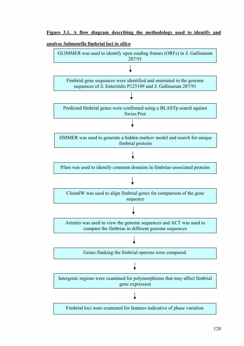
120
Figure 3.1. A flow diagram describing the methodology used to identify and
analyse Salmonella fimbrial loci in silico
GLIMMER was used to identify open reading frames (ORFs) in S. Gallinarum 287/91
Genes flanking the fimbrial operons were compared.
Fimbrial gene sequences were identified and annotated in the genome sequences of S. Enteritidis P125109 and S. Gallinarum 287/91
Artemis was used to view the genome sequences and ACT was used to compare the fimbriae in different genome sequences
ClustalW was used to align fimbrial genes for comparison of the gene sequence
Pfam was used to identify common domains in fimbriae-associated proteins
HMMER was used to generate a hidden markov model and search for unique fimbrial proteins
Predicted fimbrial genes were confirmed using a BLASTp search against Swiss-Prot
Intergenic regions were examined for polymorphisms that may affect fimbrial gene expression
Fimbrial loci were examined for features indicative of phase variation

121
3.2. Aims
To identify and annotate all fimbrial loci in sequenced strains of Salmonella
enterica.
To determine if the repertoire and/or sequence of fimbrial loci correlates with
host-specificity.
To examine the genomic insertion sites of fimbrial loci and relate this to the
phylogeny of the strains.
To identify conservation or polymorphisms in the coding and non-coding
regions of the fimbrial loci that may affect their function or expression.
To identify features indicative of phase variation.
To identify targets for mutagenesis to probe the role of fimbrial loci in
Salmonella pathogenesis.
122
3.3. Identification and comparative analysis of fimbrial loci
3.3.1. Identification of fimbrial genes
At the outset of this project the genome sequence of S. Gallinarum 287/91 was in a
raw format consisting of 2 contigs, chromosome and plasmid, and no predicted genes.
Glimmer was used to predict coding regions in the entire genome of S. Gallinarum
287/91. The fimbrial loci of S. Enteritidis P125109 and S. Gallinarum 287/91 were
identified by BLASTp searches against SwissProt/Uni-prot. The Artemis Comparison
Tool (ACT) was used to compare the genome sequences of S. Enteritidis P125109 and
S. Gallinarum 287/91 to the fully annotated genome sequences of S. Typhimurium LT2,
S. Typhi CT18, S. Choleraesuis SC-B67, S. Bongori 12419, E. coli O157:H7 EDL933
and E. coli K-12 MG1655. The relative chromosomal positions of fimbrial operons in
the Salmonella strains are shown in Figure 3.2. This is the first time a large-scale
comparison has been carried out on the repertoire of fimbrial genes at the genomic
level.
The analysis revealed differences in the number of fimbrial loci but no single locus
directly correlated with host-specificity. The sta, stg and tcf operons were specific to
serovars causing human systemic illness, however they are absent in serovars Enteritidis
and Typhimurium, which infect humans and induce enteritis. The bcf, sth and csg
operons are present in all the Salmonella strains examined, although the genes within
the csg operon of both S. Gallinarum 287/91 and S. Enteritidis P125109 are part of a
large region that has undergone an inversion relative to the other Salmonella strains
(Figure 3.2 large arrows). In E. coli O157:H7 and S. bongori 12419, the lpf operon
exists in duplicate compared to the single copy in the other Salmonella strains, yet it is
absent in E coli K-12. Interestingly, the fim operon of Salmonella also shares structural,
chromosomal and sequence identity with the sfm operon of E. coli K-12 and O157, not

123
the fim operon of these strains. The ste operon is missing from S. Typhimurium but the
remnants and flanking regions of ste are present implying that the ste operon has been
lost.
3.3.2. Molecular detection of fimbrial loci
Molecular techniques were used to examine the full repertoire of fimbrial operons in
40 different strains of Salmonella. Despite several attempts to detect conservation by
DNA hybridisation (Southern blots and dot blots) all were unsuccessful. The results
were unreliable, ambiguous and produced false positives, owing to the high degree of
homology between fimbrial loci. As the complete genome sequences were available for
some of the strains used it it easy to identify false positives, primarily staA. Studies in
silico identified staA as present in S. Typhi but both Southern and dot blots showed it to
be present in S. Enteritidis P125109. This introduces an element of doubt over all of the
data and reliable conclusions can not be made. Other groups have attempted to detect
the entire repertoire of fimbrial operons using comparative genomic hybridisation to
microarrays has also been reported to be unreliable for detecting the full complement of
fimbriae in different strains of Salmonella as ambiguous data was generated (Porwollik
et al., 2004, van Asten and van Dijk, 2005).
Hybridisation to the chaperone or usher will always result in cross-hybridisation to
other fimbriae dur to the very large highly conserved Pfam domain contained within the
chaperone and usher. The najor fimbrial subunit in many cases also contains a Pfam
domain but it is not as conserved as the chaperone and usher but it some cases may still
be allowing cross-hybridisation to occur.

124
Figure 3.2. Schematic representation of the repertoire and relative genomic
location of the fimbrial operons in strains representing different S. enterica
serovars
Each coloured block represents a distinct fimbrial operon encoded in the sense (top) or anti-
sense (bottom) orientation. Boxes of the same colour represent divergently transcribed operons.
A diagonal line through the block indicates that at least one gene in the operon is a predicted
pseudogene. All genomes are aligned relative to their predicted origin (not to scale).
12419

125
3.3.3. Conservation of the repertoire of fimbrial loci in the same serovars
To confirm the strains are representative of the serovars in respect of their
complement of predicted fimbrial loci, ACT was used to compare the genome
sequences of S. Typhi strains, CT18 and Ty2 and three strains of S. Typhimurium; LT2,
SL1344 and DT104, (the two latter strains were un-annotated). The presence or
absence of fimbrial loci was determined and summarised in Figure 3.3.
These comparisons revealed striking conservation of repertoire and sequence of the
fimbrial genes (Figure 3.3). In S. Typhimurium, all fimbrial genes are 99 % identical to
each other and the repertoire of the three sequenced strains is identical. In S. Typhi
CT18 and Ty2, the repertoire of operons is identical but the occurrence of pseudogenes
is different. In S. Typhi Ty2, the fimI gene is intact whereas stcC and stbC genes are
pseudogenes in addition to those pseudogenes predicted in CT18. The stj operon is
present in all 3 strains of S. Typhimurium but not in any other serovars and appears
truncated, as it only encodes a putative chaperone and usher proteins. No other
Salmonella strain examined possessed a fimbrial operon at the same chromosomal
position but a putative fimbrial operon was identified in E. coli K-12 in the same
position.

126
Figure 3.3. Schematic representation of the repertoire and relative genomic location
of the fimbrial operons in strains of the same S. enterica serovars
Each coloured block represents a distinct fimbrial operon encoded in the sense (top) or anti-
sense (bottom) orientation. Boxes of the same colour on either strand represent divergently
transcribed operons. A diagonal line through the block indicates that at least one gene in the
operon is a predicted pseudogene. All genomes are aligned relative to their predicted origin (not
to scale).

127
3.3.4. Conservation of fimbrial genes
The conservation of the fimbrial loci amongst the S. enterica strains examined in
Figure 3.2 was compared by ACT. At the nucleic acid level, 56 out of 71 fimbrial
genes examined possessed ≥95 % sequence identity as shown in Table 3.1. These
include all of the genes of the fimbrial operons sti, stb, fim, csg and lpf, implying that
their function may also be conserved.
The host-specific strains of S. Typhi CT18 and S. Gallinarum 287/91 possessed the
highest number of predicted fimbrial pseudogenes 11 out of 68 fimbrial genes (based on
the presence of at least one stop codon in the predicted gene co-ordinates). In S. Typhi
CT18 8 out of 56 fimbrial genes were predicted to be pseudogenes including sefD, sefR,
sefA, bcfC, fimI, steA, sthC, csgD, bcfG and sthE contained internal stop codons, as did
stiC, stfF, safC, stbC, stcC, lpfC, sefD, sefC, sthB, sthC and sthE in S. Gallinarum
287/91. In S. Choleraesuis SC-B67, only bcfC and lpfC contain internal stop codons
and there were no predicted pseudogenes in the fimbrial operons of the other serovars
examined. Where a pseudogene exists in more than one strain, there was no evidence
that the same residue was always mutated.
The percentage of fimbrial genes that are pseudogenes in S. Typhi CT18, Ty2 and S.
Gallinarum 287/91 was found to be 14 %, 16 % and 16 % respectively compared to the
total genomic mean of 4 % of predicted genes in S. Typhi CT18 and Ty2 and 7 % in S.
Gallinarum 287/91. This may imply that selection has occurred for loss of fimbrial
function in host-restricted serovars, or that mutations have accumulated as the fimbriae
are no longer required in the niche occupied. The pseudogenes are merely predicted
and it remains possible that rest of the operon may still function and contribute to the
assembly or expression of distally-encoded fimbriae (Forest et al., 2007).

128
Table 3.1. Conservation of the nucleotide sequences of S. Enteritidis strain
P125109 fimbrial genes across strains representing other S. enterica serovars
SE SG STm ST SC Gene n n % n % n % n % bcfA 543 543 99 543 99.63 543 98.89 543 99.63 bcfB 687 688 97.2 687 99.85 687 98.98 687 79.18 bcfC 2622 2622 99 2622 99.24 2622p 98.36 2622p 99.05 bcfD 1008 1008 100 1008 99.31 1008 98.12 1008 98.71 bcfE 546 546 100 546 98.35 546 97.62 546 98.90 bcfF 519 519 99 519 99.81 519 97.88 519 99.23 bcfG 732 732 100 732 99.32 705 95.63 732 99.45 bcfH 846 846 100 846 99.76 846 100 846 99.53 stiA 540 540 99 540 99.23 X X 540 99.63 stiB 684 684 97 684 98.97 X X 684 98.97 stiC 2547 2547p 99.8 2547 99.72 X X 2547 99.37 stiH 1080 1080 99 1080 99.35 X X 1080 99.17 stfA 561 561 99 561 98.57 X X 561 99.15 stfC 2658 2658 99 2658 99.59 X X 2658 99.47 stfD 753 753 99 753 99.47 X X 753 99.47 stfE 513 513 98 513 99.02 X X 513 98.83 stfF 477 477p 85 477 99.58 X X 477 92.66 stfG 531 531 98 531 96.99 X X 531 91.34 safA 510 495 51.2 513 81.48 495 68.63 513 81.18 safB 738 741 83.5 738 87.12 741 81.10 738 95.94 safC 2511 2482p 50.8 2511 98.85 2511 98.49 2511 98.73 safD 471 471 93.6 471 96.39 471 96.60 471 95.54 stbA 537 537 100 537 98.88 537 98.69 537 99.26 stbB 762 762 99 762 99.21 762 98.03 762 98.95 stbC 2562 2559p 99 2562 99.22 2562 98.83 2562 99.53 stbD 1326 1326 99 1326 99.02 1326 98.87 1326 99.25 stbE 759 756 99 759 97.76 759 98.95 760 98.82 fimA 558 556 98.6 558 98.75 555 97.83 555 99.95 fimI 534 534 100 534 98.32 534p 99.06 534p 98.50 fimC 693 693 98 693 98.99 693 98.70 X X fimD 2613 2613 99 2613 99.00 2613 98.81 2619 99.00 fimH 1008 1008 99 1008 98.41 1008 97.62 1008 98.81 fimF 519 519 100 519 99.23 519 98.07 519 99.81 fimZ 633 633 98 633 100 633 98.74 634 99.53 fimY 723 723 98 723 98.20 723 98.20 723 97.79 fimW 597 596 99 597 98.49 597 98.32 597 98.83 csgC 327 327 100 327 100 327 99.69 327 100 csgB 456 456 99 456 99.56 456 99.34 456 99.78 csgA 456 456 99 456 98.90 456 98.68 456 100 csgD 651 651 99 651 99.69 627 95.39 651 100 csgE 396 396 99 396 100 396 100 396 100 csgF 417 417 99 417 100 417 99.52 417 99.52
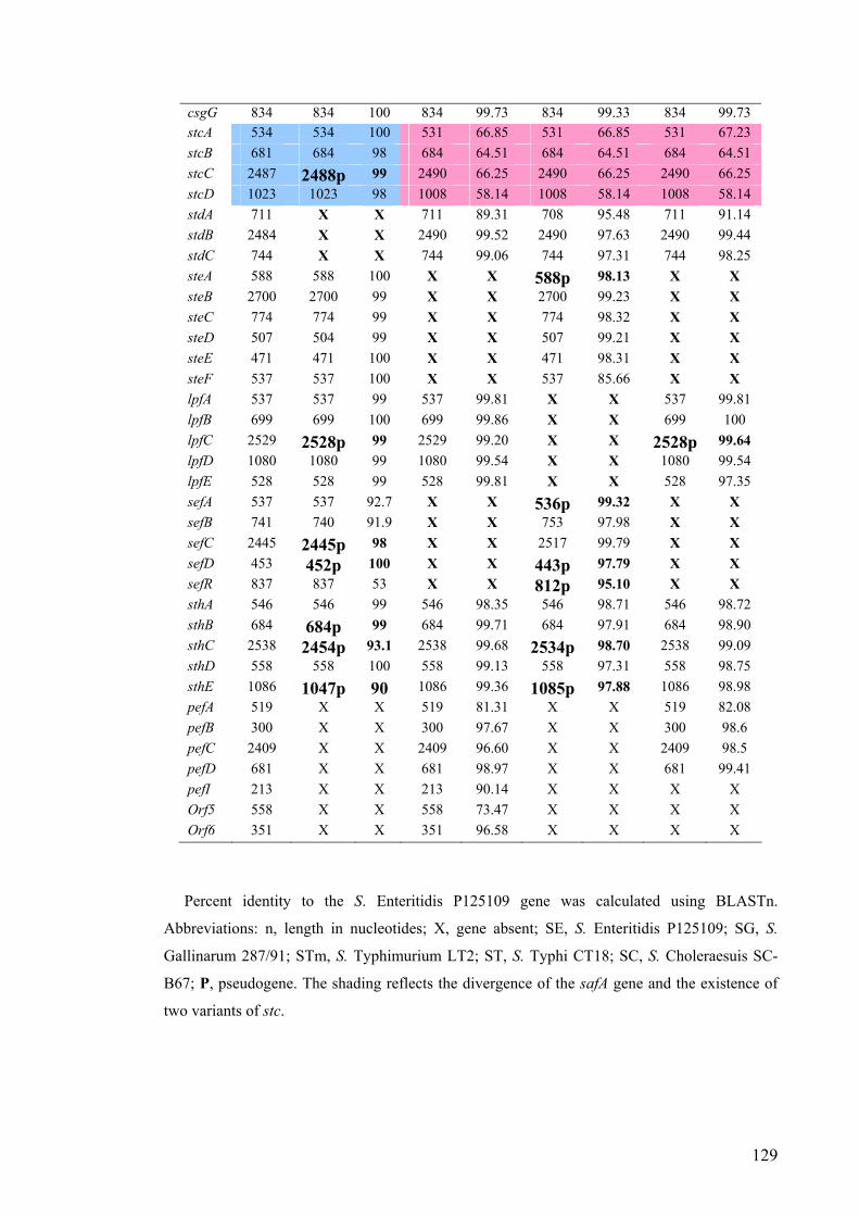
129
csgG 834 834 100 834 99.73 834 99.33 834 99.73 stcA 534 534 100 531 66.85 531 66.85 531 67.23 stcB 681 684 98 684 64.51 684 64.51 684 64.51 stcC 2487 2488p 99 2490 66.25 2490 66.25 2490 66.25 stcD 1023 1023 98 1008 58.14 1008 58.14 1008 58.14 stdA 711 X X 711 89.31 708 95.48 711 91.14 stdB 2484 X X 2490 99.52 2490 97.63 2490 99.44 stdC 744 X X 744 99.06 744 97.31 744 98.25 steA 588 588 100 X X 588p 98.13 X X steB 2700 2700 99 X X 2700 99.23 X X steC 774 774 99 X X 774 98.32 X X steD 507 504 99 X X 507 99.21 X X steE 471 471 100 X X 471 98.31 X X steF 537 537 100 X X 537 85.66 X X lpfA 537 537 99 537 99.81 X X 537 99.81 lpfB 699 699 100 699 99.86 X X 699 100 lpfC 2529 2528p 99 2529 99.20 X X 2528p 99.64 lpfD 1080 1080 99 1080 99.54 X X 1080 99.54 lpfE 528 528 99 528 99.81 X X 528 97.35 sefA 537 537 92.7 X X 536p 99.32 X X sefB 741 740 91.9 X X 753 97.98 X X sefC 2445 2445p 98 X X 2517 99.79 X X sefD 453 452p 100 X X 443p 97.79 X X sefR 837 837 53 X X 812p 95.10 X X sthA 546 546 99 546 98.35 546 98.71 546 98.72 sthB 684 684p 99 684 99.71 684 97.91 684 98.90 sthC 2538 2454p 93.1 2538 99.68 2534p 98.70 2538 99.09 sthD 558 558 100 558 99.13 558 97.31 558 98.75 sthE 1086 1047p 90 1086 99.36 1085p 97.88 1086 98.98 pefA 519 X X 519 81.31 X X 519 82.08 pefB 300 X X 300 97.67 X X 300 98.6 pefC 2409 X X 2409 96.60 X X 2409 98.5 pefD 681 X X 681 98.97 X X 681 99.41 pefI 213 X X 213 90.14 X X X X Orf5 558 X X 558 73.47 X X X X Orf6 351 X X 351 96.58 X X X X
Percent identity to the S. Enteritidis P125109 gene was calculated using BLASTn.
Abbreviations: n, length in nucleotides; X, gene absent; SE, S. Enteritidis P125109; SG, S.
Gallinarum 287/91; STm, S. Typhimurium LT2; ST, S. Typhi CT18; SC, S. Choleraesuis SC-
B67; P, pseudogene. The shading reflects the divergence of the safA gene and the existence of
two variants of stc.

130
The safA fimbrial gene exhibits the greatest variation between the serovars (Table
3.1) which has been implicated in the colonisation of the porcine intestines by S.
Typhimurium (Carnell et al., 2007). Screening of the same safA mutant in calves and
chickens did not reveal any attenuating effects (Morgan et al., 2004), implying that
Salmonella atypical fimbriae may play a host-specific role in colonisation. The saf
operon appears intact in serovars able to colonise pigs, but contains pseudogenes in the
avian- and human-specific serovars. Polymorphisms were also identified in the safA-B
intergenic region in the avian- and human-specific strains (below) and this may reflect
the fact that a porcine-specific colonisation factor is no longer needed. The safA gene
contains a conserved N- and C-terminus but the region in between is highly variable.
The remainder of the saf operon is highly conserved.
The stc fimbrial operon exists in two forms in identical chromosomal positions but
each possesses different sequences with limited sequence identity (highlighted in Table
3.1); one is highly conserved in S. Enteritidis P125109 and S. Gallinarum 287/91, and a
different variant exists in S. Typhi CT18, S. Typhimurium LT2 and S. Choleraesuis SC-
B67.
3.3.5. Operon organisation
A BioPerl script (Appendix 2.1) was written to visually display the organisation of
the complete repertoire of fimbrial loci in S. Enteritidis P125109. The size and number
of genes together with the positioning of Pfam domains is shown in Figure 3.4.
The operon diagrams demonstrate the variation that exists between the fimbrial
operons both in the number of genes within the operon and the size of the genes. The
gene number and order are conserved across all of the strains examined in which the

131
operons appear. The bcf operon comprises 8 genes and the fim operon comprises 9
genes including the regulatory genes, yet the std operon contains only 3 genes. The
function of the additional genes is unknown but implies that some loci may not encode
functional fimbriae per se but rely on distally-encoded genes for assembly. The saf, stb,
stc, std, ste, sth, bcf and sef operons are present in regions of the S. Enteritidis P125109
chromosome that are absent in E. coli K-12 (Townsend et al., 2001). Only the sef
fimbrial operon is a recent acquisition by the serogroup D Salmonella strains which is
supported by the fact that this operon has a much lower % G+C content than the rest of
the genome (Figure 3.4) (Turcotte and Woodward, 1993). The other fimbrial operons
have no major deviations in % G+C content; however a reproducibly lower % G+C
region was detected in the region 5′ of the operon in the direction of transcription
(Figure 3.3).

132
Figure 3.4. Organisation of the fimbrial operons of S. Enteritidis P125109
Arrows denote the direction of transcription. Blue arrows denote the location of
predicted Pfam domains, red arrows show predicted co-ordinates and green arrows
show miscellaneous features (to scale).
bcf (bovine colonisation factor)
stf (Salmonella Typhi F)
sti (Salmonella Typhi I)
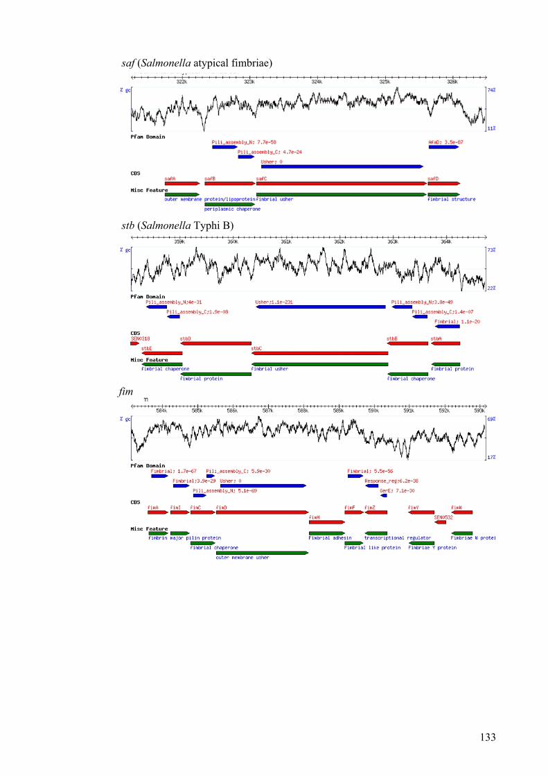
133
saf (Salmonella atypical fimbriae)
stb (Salmonella Typhi B)
fim

134
stc (Salmonella Typhi C)
ste (Salmonella Typhi E)
csg

135
std (Salmonella Typhi D)
sef (Salmonella Enteritidis fimbriae)
sth (Salmonella Typhi H)

136
3.3.6. Comparisons of flanking regions
The regions flanking predicted fimbrial operons were examined to determine if the
fimbrial genes were acquired individually, as part of the operon or larger region, or were
maintained from a common ancestor. The first coding regions 5` and 3` of each
fimbrial operon were identified and compared using ACT. The bcf, sti, stf, stb, stc, ste,
std, lpf, sef and sth fimbrial operons show complete conservation of their flanking genes
between the strains examined, implying that these operons may have been maintained
from a common ancestor.
The saf operon has a 3′ flanking gene encoding a putative xylanase (Figure 3.4) in all
serovars examined. The region 5′ of safA consists of two variants S. Enteritidis
P125109 and S. Gallinarum 287/91 possess one variant, a putative transposase 5′ of
safA, and all of the other serovars encode a putative integrase at this position,
suggesting that the saf genes may have been acquired or rearranged differently (Figure
3.5). S. Enteritidis P125109, S. Gallinarum 287/91 and S. Typhi CT18 possess a gene
encoding a hypothetical protein 5′ of the csg operon, whereas S. Choleraesuis SC-B67
and S. Typhimurium LT2 possess a gene encoding a membrane protein (Figure 3.5).
Interestingly, only S. Enteritidis P125109 and S. Gallinarum 287/91 have an inverted
csg operon relative to all other Salmonella serovars and E. coli strains examined.

137
Figure 3.5. Schematic representation of the flanking gene regions of predicted
fimbrial operons
Only those with differences are shown, all are 5΄ to 3΄.
3.3.7. Comparison of intergenic regions
The non-coding sequences of the fimbrial operons were aligned using ClustalW to
detect polymorphisms that may affect the transcription or translation of fimbrial genes.
The majority of differences existed between rather than within the S. enterica serovars
examined implying that the fimbrial operons were likely acquired as whole operons at a
similar time in evolution or were maintained from a common ancestor.
The region between the safA and safB genes varies between serovars; S. Typhi CT18
has an extra gene in the saf operon, (safE) at this position that is not present in any other
serovars examined. The regions between safE and safB in S. Typhi CT18 and the
region between safA and safB in S. Gallinarum 287/91 are both 25 bp in length and
138
identical bar 2 bases. The safA-safB intergenic region contains a putative promoter in S.
Gallinarum 287/91 and S. Typhi CT18 and it is unclear if this is required for expression
of the downstream genes (Figure 3.6). Interestingly, S. Typhimurium LT2, S.
Enteritidis P125109 and S. Choleraesuis SC-B67 also have an identical region between
safA and safB but it differs from that in S. Gallinarum 287/91 and S. Typhi CT18.
The non-coding sequence of the stc operon has two variants (Figure 3.6). In each, S.
Enteritidis P125109 and S. Gallinarum 287/91 share the same stc intergenic sequences
whereas the other serovars possess a different sequence, implying divergent evolution.
Several putative ribosomal binding sites have been predicted in the intergenic regions in
S. Choleraesuis SC-B67, S. Typhimurium LT2 and S. Typhi CT18 as highlighted in
Figure 3.6 but are absent in S. Enteritidis P125109 and S. Gallinarum 287/91. The
impact of such polymorphisms on translation of stc genes is unknown.
An insertion is present in the fimA-I intergenic region of S. Typhimurium LT2 and S.
Choleraesuis SC-B67 which is not present in S. Enteritidis P125109 and S. Gallinarum
287/91 (Figure 3.6). A ribosomal binding site has been predicted in this region of S.
Typhimurium LT2, S. Choleraesuis SC-B67 and S. Typhi CT18 genome sequences.
The fimI-C intergenic region is truncated and missing a ribosomal binding site in S.
Choleraesuis SC-B67.
The fimZ-Y and the fimY-W intergenic regions were not identical between serovars
due to point mutations. In S. Choleraesuis SC-B67, the fimY-W intergenic region has a
completely different sequence and it does not contain an additional coding region as in
all of the other serovars examined, identified as fimU. S. Typhi CT18 has more
variation in the csgE-D, steA-B and the sefB-C intergenic regions than the other
serovars, which may be due to the high degree of genome decay that has occurred in S.
Typhi relative to the other genomes examined.

139
Figure 3.6. Polymorphisms detected in the intergenic regions in fimbrial operons of
S. enterica serovars
The hyphen shows a gap in the alignment. Putative promoter regions predicted using NNPP
shown in pink. Predicted ribosomal binding sites are shown in blue. The entire intergenic
region is shown. STYM = S. Typhimurium LT2, SCHL = S. Choleraesuis SC-B67, SENT = S.
Enteritidis P125109, SGAL = S. Gallinarum 287/91 and STYP = S. Typhi CT18.

140
3.3.8. Plasmid-encoded fimbriae
The pef operon consists of 5 genes pefA, pefB, pefC, pefD and pefR (pefI) and is
identical in S. Enteritidis P125109, S. Typhimurium LT2 and S. Choleraesuis SC-B67
(Figure 3.7). The pef operon is absent in S. Gallinarum 287/91; instead, 3 genes with
homology to fimbrial genes in E. coli K88 were identified (faeH and faeI, predicted to
encode minor fimbrial subunits and faeJ). In E. coli, this operon consists of 9 genes
(faeB-J), suggesting that these 3 genes will not form a functional fimbrial operon but
may have an accessory role.
Whilst the pefI gene is absent from the pef locus in the sequenced plasmid of S.
Choleraesuis SC-B67, a pefI homologue is present 3’ of the chromosomal sef operon in
S. Enteritidis P125109 and S. Choleraesuis SC-B67. The pefI gene is also present 3’ of
sef in S. Typhi CT18 chromosome genome sequence despite it lacking a virulence
plasmid and it has been reported that other pef remnants exist at this chromosomal locus
(Bishop et al., 2005), however BLASTp analysis revealed negligible homology. The
promoter region of chromosomal and plasmid-encoded pefI differ entirely (Collighan
and Woodward, 2001), however the impact of this is unknown.
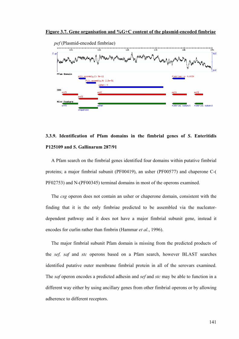
141
Figure 3.7. Gene organisation and %G+C content of the plasmid-encoded fimbriae
3.3.9. Identification of Pfam domains in the fimbrial genes of S. Enteritidis
P125109 and S. Gallinarum 287/91
A Pfam search on the fimbrial genes identified four domains within putative fimbrial
proteins; a major fimbrial subunit (PF00419), an usher (PF00577) and chaperone C-(
PF02753) and N-(PF00345) terminal domains in most of the operons examined.
The csg operon does not contain an usher or chaperone domain, consistent with the
finding that it is the only fimbriae predicted to be assembled via the nucleator-
dependent pathway and it does not have a major fimbrial subunit gene, instead it
encodes for curlin rather than fimbrin (Hammar et al., 1996).
The major fimbrial subunit Pfam domain is missing from the predicted products of
the sef, saf and stc operons based on a Pfam search, however BLAST searches
identified putative outer membrane fimbrial protein in all of the serovars examined.
The saf operon encodes a predicted adhesin and sef and stc may be able to function in a
different way either by using ancillary genes from other fimbrial operons or by allowing
adherence to different receptors.
pef (Plasmid-encoded fimbriae)

142
Several fimbriae contain two or more fimbrial subunit domains, including the bcf and
fim operons which each contain 3 fimbrial domains, and the ste, sth and lpf operons
contain 2 fimbrial domains. The bcf and stb operons also code for two putative
chaperone proteins each with both an N-terminal and C-terminal chaperone domain.
The function of the duplicated putative proteins is unknown and implies that some loci
may not encode functional fimbriae on their own but rely on distally-encoded proteins
for assembly.
3.3.10. Analysis of fimbrial loci for traits associated with phase variation
Fimbrial genes in some bacteria are subject to phase variable (on-off) expression at
the transcriptional level (Brinton, 1959, van der Woude and Baumler, 2004, Casadesus
and Low, 2006). The mechanisms behind phase variation in Salmonella are poorly
characterised. During the annotation of S. Enteritidis fimbrial operons and genome
sequence comparisons, no genes with homology to known recombinases were detected
within or proximal to fimbrial loci. Putative transposase and integrase genes associated
with DNA mobility were observed proximal to the saf, sef and fim loci. Direct or
inverted repeat sequences that may serve as substrates for recombination were not
detected in any of the fimbrial operons examined using the tandem repeats finder.
A pattern matching search was carried out for the Dam methylase target sequence
GATC within and proximal to the fimbrial operons of S. Enteritidis P125109. This
identified hundreds of potential targets, however those present from -500 to +200 bp
relative to the predicted translation initiation codon of fimbrial genes are listed in Table
3.2. This search identified the same sites in the pef gene cluster as predicted to be
methylated in S. Typhimurium (Nicholson and Low, 2000). Whereas S. Typhimurium

143
strains LT2 and SL1344 possess GATC sites at -98, -110 and -212 relative to the start of
pefB, S. Enteritidis P125109 possessed only the sites at -110 and -212 but an additional
site at +47 in pefB that is absent in the two strains of S. Typhimurium. Three potential
Dam methylation target sites were also identified upstream of the std operon (-88, -97
and -110) in S. Enteritidis P125109 and S. Typhimurium LT2. This density of GATC
sites is higher than would be predicted due to random distribution and further studies
will be required to determine the role of these in Dam-dependent repression of the std
genes as detected by microarray analysis (Balbontin et al., 2006). Predicted Dam
methylase targets were also identified upstream of the sef, sti and stf operon in S.
Enteritidis P125109, which require further investigation (Table 3.2).
Searches were also performed to detect stretches of a repeated nucleotide homo-
polymeric tract or those with a repeating unit containing different nucleotides (hetero-
polymeric tracts) that may be subject to slipped-strand mispairing to introduce frame-
shift mutations (van Belkum et al., 1998). Mispairing between such tracts occurs
during DNA replication and can result in production of a non-sense or truncated protein
genes e.g. in assembly and maturation of Neisserial pilin (Meyer and van Putten, 1989).
Homo-polymeric tracts consisting of repeats of A’s or C’s were searched on both
strands using the pattern matching function within ACT and those identified within 200
bp of the predicted translation initiation codon of a fimbrial gene are listed in Appendix
3.1. Several conserved hetero-polymeric tracts were identified using a variable tandem
repeat pattern finder (Davison et al., 1999), however only one potential tract was
identified within the stdA gene, the tract was a 6-mer GACCAT repeated 10 times.
Variation in the number of such repeats is not predicted to alter the open reading frame
resulting in premature termination; rather it may reduce the number of codons and thus
the primary sequence of the encoded product StdA.

144
Table 3.2. Putative Dam methylation sites in the fimbrial loci of S. Enteritidis
GATC genome location Relative location Fimbrial gene location
24391-24394 156 bases into bcfA bcfA 24235-24777
25564-25567 Overlapping start of bcfC bcfC 25569-28190
232155-232158 330 upstream of stf operon stfA 232485-233045
232259-232262 226 upstream of stf operon stfA 232485-233045
233275-233278 144 bases into stfC stfC 233131-235788
235893-235896 87 bases into stfD stfD 235806-236558
321303-321306 444 bases upstream of safA safA 321747-322256
2007363-2007366 190 bases into csgB csgB 2007101-2007556c
2007481-2007484 75 bases into csgB csgB 2007101-2007556c
2008355-2008358 44 bases into csgD csgD 2008311-2008961
2984272-2984275 163 bases upstream of steA steA 2984435-2985022
2984588-2984591 153 bases into steA steA 2984435-2985022
4569956-4569959 245 bases upstream of sefA sefA 4570250-4570747
4570097-4570100 153 bases upstream of sefA sefA 4570250-4570747
4570145-4570148 105 bases upstream of sefA sefA 4570250-4570747
4570397-4570400 147 bases into sefA sefA 4570250-4570747
583667-583670 69 bases into fimA fimA 583598-584155
589175-589178 2 bases into fimF fimF 589173-589691
2987904-2987907 90 bases into steC steC 2987814-2988587
4677536-4677539 135 bases into sthD sthD 4677117-4677674c
4677600-4677603 74 bases into sthD sthD 4677117-4677674c
4680139-4680142 87 bases into sthC sthC 4677692-4680229c
4681823-4681826 249 bases upstream of sthA sthA 4681029-4681574c
4681472-4681475 99 bases into sthA sthA 4681029-4681574c
3080578-3080581 88 bases upstream of stdA stdA 3079783-3080490c
3080587-3080590 97 bases upstream of stdA stdA 3079783-3080490c
3080600-3080603 110 bases upstream of stdA stdA 3079783-3080490c
3080295-3080298 195 bases into stdA stdA 3079783-3080490c
364215-364218 29 bases into stbA stbA 363708-364244c
364336-364339 92 bases upstream of stbA stbA 363708-364244c
362854-362857 4 bases upstream of stbC stbC 360340-362901c
145
210707-210710 8 bases upstream of stiA stiA 210160-210699c
211099-211102 400 bases upstream of stiA stiA 210160-210699c
210991-210994 292 bases upstream of stiA stiA 210160-210699c
14610-14613 47 bases within pefB pefB 14358-14657c
14767-14770 110 bases upstream of pefB pefB 14358-14657c
14869-14872 212 bases upstream of pefB pefB 14358-14657c
The location of the GATC site is noted below along with the fimbrial gene in close proximity.
This analysis provides merely a starting point for identifying traits associated with
phase variation by known mechanisms. It is possible that fimbrial phase variation will
occur via another unknown mechanism, which cannot be predicted.
3.3.11. Correlation of fimbrial repertoire and virulence of the strains examined
The virulence of S. Typhimurium SL1344, S. Enteritidis P125109 and S. Gallinarum
287/91 has been compared in a streptomycin-pre-treated mouse model. Each strain was
observed to induce enterocolitis at comparable levels (Suar et al., 2006). The fimbrial
loci bcf, lpf, stb, stc, std and sth have been reported to influence colonisation of the
murine intestines by S. Typhimurium (van der Velden et al., 1998, Weening et al., 2005,
Lawley et al., 2006). S. Gallinarum 287/91 lacks the std operon and has pseudogenes in
lpf, stb, stc and sth relative to S. Typhimurium SL1344, indicating that either these
fimbriae are not essential for enterocolitis in mice or their absence is compensated for
by other fimbriae. Interestingly, S. Gallinarum 287/91 was observed to invade polarised
trans-immortalized IEC cells (derived from the small intestines of a transgenic mouse)
at lower levels than S. Typhimurium SL1344 and S. Enteritidis P125109. It can be
hypothesised that this may reflect polymorphisms in the repertoire and function of

146
fimbrial operons between the strains, though the differences may be explained by other
traits.
3.4. Discussion
A comparative analysis of fimbrial operons in complete Salmonella genome
sequences available at the time of writing was performed. Fimbriae have been
implicated in virulence and tissue tropism, however no single fimbrial locus directly
correlated with the host range of the S. enterica serovars examined. Several of the
strains examined (S. Typhimurium SL1344, S. Enteritidis P125109 and S. Gallinarum
287/91) are of well-defined virulence in murine models (Suar et al., 2006) and food-
producing hosts (Carnell et al., 2007, Morgan et al., 2004), however variations in the
repertoire of fimbrial loci possessed by such strains could not be correlated with
virulence. Previous studies have indicated that fimbriae may act in concert (van der
Velden et al., 1998) and it remains to be seen if such synergy is important in host-
specificity.
Host-specific S. enterica serovars contained a higher number of predicted
pseudogenes in fimbrial loci than host-restricted serovars, and in turn ubiquitous
serovars. Gene decay is a feature of the genome of S. Typhi in general, which possesses
over 200 pseudogenes relative to S. Typhimurium (Deng et al., 2003), as well as other
host-restricted pathogens including B. mallei (Holden et al., 2004, Nierman et al., 2004,
Sebaihia et al., 2006) and M. leprae (Cole et al., 2001). The decay of genomes appears
to correlate with a reduced ability to survive in the environment and in different hosts
(Nierman et al., 2004, Holden et al., 2004). However, the observed frequency of
pseudogenes among fimbrial genes in host-specific serovars was 2-4 folds higher than
147
the genomic mean. Such genetic attrition may contribute to the inability of host-
restricted and -specific S. enterica serovars to persist in diverse animal hosts.
Salmonella has maintained a large repertoire of fimbriae suggesting their role may be
important in colonisation of diverse niches. The majority of fimbrial genes across the
serovars examined show >95% homology at the nucleic acid level when compared to
the fimbrial genes of S. Enteritidis P125109. The high conservation of the genes should
imply that their function is also conserved. However this is not always the case, in S.
Typhi CT18 the sefA gene is 99% identical to the sefA gene in S. Enteritidis P125109
but it is a pseudogene due the presence of a stop codon. S. Gallinarum has lost its
mannose-specific binding properties due to a single base pair mutation in the fimH gene
resulting in the translated adhesin being formed with a point mutation at position 78;
isoleucine to threonine (Kisiela et al., 2005, Kisiela et al., 2006).
The number of genes encoded in each fimbrial locus varies, however each locus is
highly conserved across the serovars. For loci that lack a fimbrial subunit Pfam domain
but encode a predicted usher and chaperone (e.g. saf and stc) it remains unclear if
distally-encoded genes are required for the complete assembly or function of fimbriae.
The csg operon also has no fimbrial subunit domain, however in this case it is known to
promote fimbriae assembly via a distinct nucleator-dependent pathway which occurs
extracellularly through a self-assembly process (Hammar et al., 1996).
The differences that occur within the flanking genes of the fimbrial operons as well
as in the intergenic regions of these operons may play a crucial role in the expression of
the fimbriae. These areas may assist with phase variation or efficient transcription
and/or translation of the gene. Promoter sequences are required to enable a gene to be
transcribed and the ribosomal binding sites promote efficient and accurate translation of
mRNA. The variation that occurs in these sites in the saf, stc and fim operons could

148
have huge consequences on the downstream regions. Further examination of the
expression profiles would be needed to determine the effects of these polymorphisms
e.g. detection of proteins by antibody mediated methods and transcripts by hybridisation
or RT-PCR.
Polymorphisms were identified that have the potential to affect the regulation of
fimbrial genes and transcriptomic and proteomic analysis of different S. enterica
serovars is needed to determine the impact of these. Further research is also required to
dissect the role in virulence of the fimbriae loci identified herein and to understand the
regulatory cross-talk that may co-ordinate their expression (Encheva et al., 2007).
Phase variation of fimbrial genes is common and has been described in several
bacterial species including E. coli (Abraham et al., 1985, Klemm, 1986, Blyn et al.,
1990) and in Neisseria (Meyer and van Putten, 1989, van Belkum et al., 1998)and some
of the Salmonella fimbrial loci including lpf (Norris and Baumler, 1999). Whether this
phase variation occurs across all fimbrial operons is currently unknown and if it does
occur in all fimbriae, the mechanisms behind this may vary. The mechanisms behind
phase variation are not fully understood in Salmonella.
The strains examined have evolved at different times and cause disease in different
hosts, however, given the limited number of each of the serovars examined only limited
conclusions regarding the conservation of these operons can be made as differences
have been identified amongst different strains of the same serovar (Boyd et al., 2003,
Deng et al., 2003). The remaining studies were focused on defining a role for fimbriae
through targeted mutagenesis of the predicted major fimbrial subunits and analysis of
these mutants in vitro and in vivo.

149
Chapter 4
Construction and
characterisation of fimbrial
mutants

150
4.1. Introduction
To understand the function of a gene it is a standard method to inactivate the gene
and identify any resulting phenotype. This may involve random mutagenesis of
bacterial genomes using transposons, followed by screening of individual mutants or
signature-tagged mutagenesis (STM) to produce a library of tagged mutants that can be
screened simultaneously (Hensel et al., 1995). Screening of random transposon mutants
of S. Typhimurium F98 in chickens revealed a role for 18 genes in the colonisation of 3-
day-old chicks, of these 9 were rough (lacked LPS) and 9 contained mutations in other
genes including rfaY, dksA, clpB, hupA, and sipC (Turner et al., 1998). Screening of a
library of signature-tagged transposon mutants has shown a role for S. Typhimurium
4/74 stbC, fimZ, csgD and sthB in chickens, but not in calves (Morgan et al., 2004). A
role for saf fimbriae in S. Typhimurium 4/74 has been identified in pigs but not in other
animal models (Carnell et al., 2007). Transposon insertions may have polar effects on
the expression of proximal genes and multiple insertions may occur. Therefore, a need
exists to confirm the phenotype of such mutants by construction of defined non-polar
mutants. This can be accomplished by using targeted mutagenesis techniques such as
lambda Red recombinase-mediated homologous recombination. This permits the
replacement of the target gene with an antibiotic resistance gene flanked by a target site
for flippase recombinase (FRT) (Datsenko and Wanner, 2000). Alternatively, mutants
may be constructed by introducing a defective gene on a suicide plasmid into a wild-
type strain. Some suicide replicons permit a second round of homologous
recombination by positive-selection to excise the vector. This method is commonly
used but has been reported to result in a high frequency of second-site defects (Johnson
et al., 2003).

151
The λ red mutagenesis method was used as described in Section 2.4 to determine a
phenotype of individual mutants each lacking a single major fimbrial subunit both in
vitro and in vivo. Lambda Red mutagenesis has the advantage over other targeted
mutagenesis methods in that chromosomal genes can be mutated in a single step using
linear PCR products rather than by cloning, inactivation and re-introduction of the gene
on a suicide plasmid. The chances of second site defects was reduced using
bacteroiphage P22/int mediated transduction into an archived strain. The unique
putative major fimbrial subunit of each fimbrial operon was chosen for mutation as
identified by Pfam and BLAST searches, described in Chapter 3.

152
4.2. Aims
To mutate the putative major fimbrial subunit of all chromosomally located
fimbrial operons in S. Enteritidis P125109 and S. Gallinarum 287/91.
To confirm the positions of all fimbrial mutations following transduction where
possible.
To confirm the mutations had no affect on growth.
To reduce the chances of second-site defects contributing a phenotype.

153
4.3. Lambda Red mutagenesis
The lambda Red method of mutagenesis promotes homologous recombination
between a linear PCR fragment and a targeted gene of interest and was used here to
separately introduce an antibiotic resistance cassette into each major fimbrial subunit
gene of two poultry-associated S. enterica strains. The helper plasmid, pKD46 contains
3 genes from bacteriophage lambda λ: exo, bet and gam which promote homologous
recombination. The gam gene inhibits the bacterial exonuclease activity so that the
proteins encoded by bet and exo can gain access to the DNA and promote
recombination. In pilot experiments, pKD46 failed to promote integration of a range of
linear amplicons including steA::chlR in S. Enteritidis P125109 despite the presence of
pKD46 being confirmed both by PCR amplification of regions of the plasmid and by re-
extracting the plasmid (data not shown). The reasons behind this are unknown and it
could be speculated that the presence of the plasmid has a lethal or inhibitory effect on
S. Enteritidis P125109 or S. Enteritidis P125109 contains an element in its genome that
prevents the lambda genes of pKD46 from functioning. The same stock of pKD46 has
successfully been used in S. Gallinarum 287/91 and S. Typhimurium F98 to integrate
the same linear amplicons. Therefore, an alternative strain, S. Enteritidis S1400 was
used to construct the mutants in the first instance. Other laboratories have had similar
strain specific problems but the reasons behind this are currently unknown.
Transformants containing pKD46 were selected at 30 ºC on LB agar plates
supplemented with ampicillin (100 µg/ml).
A PCR reaction was carried out using pKD3 plasmid DNA (GenBank, AY048742)
as a template to produce linear DNA for recombination using the primers listed in Table
2.5. Each PCR reaction was carried out 12 times to provide 600 µl of each PCR
product. The PCR products were concentrated using a PCR clean up kit from QIAGEN

154
and were cleaved by DpnI (2 µl) in 1 x buffer in a total 100 µl reaction with water to
remove unmethylated template DNA. The reaction was incubated statically for 3 h at
37 ºC. The digests were separated on a 1 % agarose gel as described in Section 2.3.4
and the DNA fragments excised from the gel with a clean sharp scalpel. The DNA was
purified from the agarose using a gel extraction kit from QIAGEN. An example of the
location of the primers for the stcA major fimbrial subunit is shown in Figure 4.1.
Figure 4.1. The sequence and location of primers targeting the major fimbrial
subunit of stcA
5′ATGAAACGTTCACTTATTGCTGCTTCTGTATTGTCTGCTGTATTTATGAGCGCTGGG
GTTTTTGCTGCTGATGAAGATATGGGGGAATTAAAAATAAACGGAGAAGTGGTGGGAAC
ATCCTGTACTTTCGAAGGTGCAAATAGCGCGACTATTGAATTATCCCAGGTAGGTGTTG
ATAGATTGACTGACTTAAACCCTGGCGATATATATACAGGATACACTAGCCCAGAAGCG
ATTTTAAAAGTAAGATGTACGAATACAGCTAATCCACGAATTAGTTTTAACCGTTCTCA
ATTTGTGGATAACATGCAAATCACCAAAAATAATGCTACTAATAATGGTGCGGGCTTCG
CTGTTTATCTTGATGGTATTCAGGTGAAACCGGATGAGGCGGGGAATTACACTCTGAAT
TCAAGTAAGTTTGAAAATGGTGTATATACCCTGAACTTTTCTGCCCGCTATGCCGCCGT
TGAAAATACTGTAACACCAGGTTCTGTTGAATCTGTACTGACGATGACGGTATTAACTG
ATTAA –3′
NB The entire gene is shown and the primers are highlighted in blue. The start and stop codons are underlined
The 3′ end of each primer contains a 20 nucleotide sequence to permit annealing to
the pKD3 antibiotic resistance cassette 5′ TGTGTAGGCTGGAGCTGCTTCG and 3′
ATTCCTCCTATAAGTATAC, to replace the internal portion of the gene with a
chloramphenicol resistance cassette and leave a 40 bp region either side of the insertion
behind. The cleaned and digested PCR products were electroporated into electro-
competent S. Enteritidis S1400 or S. Gallinarum 287/91 carrying the pKD46 plasmid.
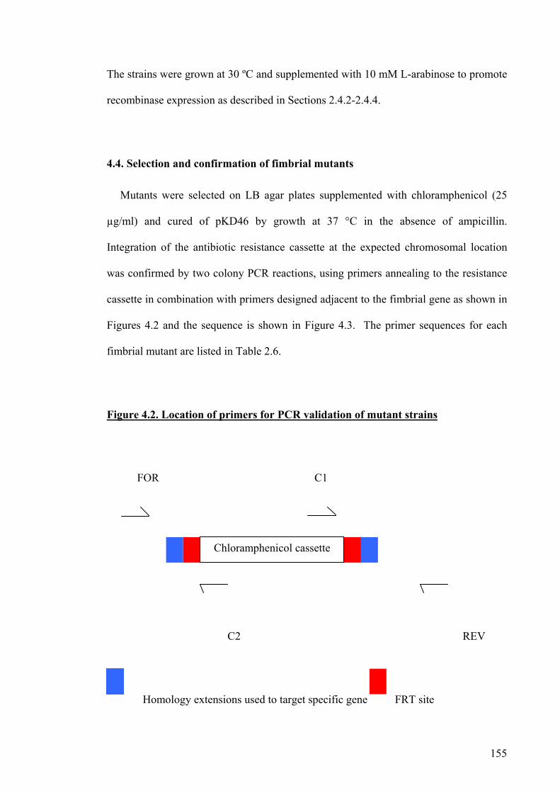
155
The strains were grown at 30 ºC and supplemented with 10 mM L-arabinose to promote
recombinase expression as described in Sections 2.4.2-2.4.4.
4.4. Selection and confirmation of fimbrial mutants
Mutants were selected on LB agar plates supplemented with chloramphenicol (25
µg/ml) and cured of pKD46 by growth at 37 °C in the absence of ampicillin.
Integration of the antibiotic resistance cassette at the expected chromosomal location
was confirmed by two colony PCR reactions, using primers annealing to the resistance
cassette in combination with primers designed adjacent to the fimbrial gene as shown in
Figures 4.2 and the sequence is shown in Figure 4.3. The primer sequences for each
fimbrial mutant are listed in Table 2.6.
Figure 4.2. Location of primers for PCR validation of mutant strains
FOR C1
C2 REV
Homology extensions used to target specific gene FRT site
Chloramphenicol cassette

156
Table 4.1. Predicted sizes of PCR products for S. Enteritidis S1400 and S.
Gallinarum 287/91 fimbrial mutants
Primer PCR product size (base PCR product size (base bcfAFOR + C1 633 633 bcfAREV + C2 507 507 csgAFOR + C2 173 173 csgAREV + C1 302 302 lpfAFOR + C2 867 867 lpfAREV + C1 288 288 fimAFOR +C1 807 807 fimAREV + C2 429 429 sefAFOR + C1 373 373 sefAREV + C2 946 946 safAFOR + C1 701 701 safAREV + C2 597 597 stbAFOR + C2 887 887 stbAREV + C1 295 295 stcAFOR + C2 179 179 stcAREV + C1 377 377 stdAFOR + C2 587 - stdAREV + C1 714 - steAFOR + C1 739 739 steAREV + C2 600 600 stfAFOR + C1 283 283 stfAREV + C2 155 155 sthAFOR + C1 584 584 sthAREV + C2 704 704 stiAFOR + C2 385 385 stiAREV + C1 661 661
All amplicons from respective mutant strains were approximately of the sizes predicted. The stdA gene was not examined as it
is not present in the genome sequence of S. Gallinarum 287/91. PCR reactions were performed using the following primer pairs:
FOR and C2, and REV and C1. Some fimbrial mutants have the chloramphenicol resistant cassette in the opposite orientation
(Table 4.1).

157
Figure 4.3. Predicted sequence of the insertion of the pKD3-derived
chloramphenicol resistance cassette in stcA
5′-CAACAAAACATCGTGTTTACATTGCGATAACTTCCTGTCTATGAGAATTTTCGTTG
CAAGGGTTAATAACTCTTAACAAATAGAAATTACTTCATTAAGGAAGAGATTATGAAAC
GTTCACTTATTGCTGCTTCTGTATTGTCTGCTGTGTGTAGGCTGGAGCTGCTTCGAAGT
TCCTATACTTTCTAGAGAATAGGAACTTCGGAATAGGAACTTCATTTAAATGGCGCGCC
TTACGCCCCGCCCTGCCACTCATCGCAGTACTGTTGTATTCATTAAGCATCTGCCGACA
TGGAAGCCATCACAAACGGCATGATGAACCTGAATCGCCAGCGGCATCAGCACCTTGTC
GCCTTGCGTATAATATTTGCCCATGGTGAAAACGGGGGCGAAGAAGTTGTCCATATTGG
CCACGTTTAAATCAAAACTGGTGAAACTCACCCAGGGATTGGCTGAGACGAAAAACATA
TTCTCAATAAACCCTTTAGGGAAATAGGCCAGGTTTTCACCGTAACACGCCACATCTTG
CGAATATATGTGTAGAAACTGCCGGAAATCGTCGTGGTATTCACTCCAGAGCGATGAAA
ACGTTTCAGTTTGCTCATGGAAAACGGTGTAACAAGGGTGAACACTATCCCATATCACC
AGCTCACCGTCTTTCATTGCCATACGTAATTCCGGATGAGCATTCATCAGGCGGGCAAG
AATGTGAATAAAGGCCGGATAAAACTTGTGCTTATTTTTCTTTACGGTCTTTAAAAAGG
CCGTAATATCCAGCTGAACGGTCTGGTTATAGGTACATTGAGCAACTGACTGAAATGCC
TCAAAATGTTCTTTACGATGCCATTGGGATATATCAACGGTGGTATATCCAGTGATTTT
TTTCTCCATTTTAGCTTCCTTAGCTCCTGAAAATCTCGACAACTCAAAAAATACGCCCG
GTAGTGATCTTATTTCATTATGGTGAAAGTTGGAACCTCTTACGTGCCGATCAACGTCT
CATTTTCGCCAAAAGTTGGCCCAGGGCTTCCCGGTATCAACAGGGACACCAGGATTTAT
TTATTCTGCGAAGTGATCTTCCGTCACAGGTAGGCGCGCCGAAGTTCCTATACTTTCTA
GAGAATAGGAACTTCGGAATAGGAACATTCCTCCTATAAGTATACTGTTGAATCTGTAC
TGACGATGACGGTATTAACTGATTAACAATCGTTTTTTGGCGGCTAAGCTTTTAGCCGC
TTGTATCTCTTTTATAAGGTTATCGGCCATGAAATGGATGATTGCATTATGCCTGGCTT
GTGCCGCTATGCCTGCGTGGAGCGGCATTTATATATAT-3′
The primers for constructing the mutant are shown in blue and are positioned at the 5′ and 3′ ends of the stcA gene, the start and stop
codons of the stcA gene are underlined. The pink font is the chloramphenicol resistant cassette. The red text denotes the primers
within the chloramphenicol resistance cassette used to confirm the location of the mutation in combination with primers flanking the
gene shown in green. The black bold text denotes the flippase recombinase target (FRT) sites and the yellow sequences are the
primers for amplification of pKD3 chloramphenicol resistant cassette.

158
The location of all fimbrial mutants constructed in both S. Enteritidis S1400 and S.
Gallinarum 287/91 and the transduced fimbrial mutants were confirmed by the use of
PCR using primers in the combinations listed in Table 4.1 and Figure 4.2 and the
forward and reverse primers together as indicated in Figure 4.2. The PCR product sizes
were predicted from the genome sequences of S. Enteritidis P125109 and for all mutants
were approximately as predicted, Figure 4.4.

159
Figure 4.4. Construction and validation of a ΔstcA::cat fimbrial mutant of S.
Enteritidis P125109
Lane 1 is stcFOR and stcREV, lane 2 is stcFOR and C2 and lane 3 is stcREV and C1. Product sizes are predicted from Figure 4.3 to
be 1280, 179 and 377 bp respectively.
4.5. Transduction of mutations using bacteriophage P22HT/int
It is possible during homologous recombination that secondary unwanted
recombination events may have occurred (Datsenko and Wanner, 2000). To avoid such
second-site defects major fimbrial subunit mutations were transduced into the archived
strain or an alternative strain by bacteriophage P22 HT/int transduction as described in
Section 2.4.7. General transduction was first discovered in S. Typhimurium, genetic
material from pLT-22 could be transferred to S. Typhimurium LT2, (pLT-22 is now
known as P22) (Zinder and Lederberg, 1952). Generalised transduction relies on the
endonuclease activity of bacteriophage P22 and the accidental packaging of linear DNA
1 2 3
1000 bp 750 bp
250 bp

160
fragments into phage capsids instead of approximately 40 kb of phage genome.
Infection of a susceptible Salmonella strain by such particles leads to insertion of linear
fragments into the bacterial cell, which may then replace endogenous sequences by
homologous recombination.
Within the two serovars examined, S. Gallinarum and S. Enteritidis, 25 fimbrial
mutations were constructed and confirmed and 20 of the mutations were transduced into
clean or archived strains. The fimbrial mutants that were made in S. Enteritidis S1400
were transduced into S. Enteritidis P125109 and those made in S. Gallinarum 287/91
were transduced into the archived stock of this strain. Five fimbrial mutants could not
be transduced despite multiple attempts. Interestingly, of these 5 fimbrial mutations
there are only 3 genes involved fimA, steA and safA. The fimA, steA and safA could not
be transduced between the two strains of S. Enteritidis, and the fimA and safA mutants
of S. Gallinarum 287/91 could not be transduced into the archived strain. The reasons
for this are unclear. All transduced mutants were confirmed by PCR as described in
Section 4.4 and were shown to possess intact LPS as described in Section 2.4.4.
4.6. Verification of insertion mutants by Southern blotting
Bacteriophage P22 transduction is capable of transducing approximately 40 kb of
chromosomal DNA (Vander Byl and Kropinski, 2000, Casjens and Hayden, 1988)
which may potentially include the fimbrial mutation and approximately 40 kb 5′ or 3′.
The transferred region may contain sequences that are only present in one strain of S.
Enteritidis and not in another since the sequence flanking the S. Enteritidis S1400
fimbrial loci is unknown. The introduction of these unique regions may alter the
phenotype of the recipient and therefore DNA was extracted from the fimbrial mutants

161
of both strains of S. Enteritidis used (the three that would not transduce were omitted) as
described in Section 2.3.1. Genomic DNA digests were carried with HindIII and the
digests were resolved on a 1 % agarose gel. The DNA was denatured before being
transferred to a membrane by vacuum transfer for 1 h as described in Section 2.3.5.
A DIG-labelled probe targeted to the chloramphenicol resistance cassette was
amplified by PCR using pKD3 as the template DNA and incorporating DIG-labelled
dNTPs. The primer sequences are listed in Table 2.3 and PCR conditions are as
described in Section 2.3.8. The membrane was hybridised with the DIG-labelled probe
and detected as described in Sections 2.3.9 and 2.3.10 to determine if the
chloramphenicol cassette localised to HindIII fragments of the expected size in the S.
Enteritidis S1400 λ Red mutants and the S. Enteritidis P125109 transductants.

162
Figure 4.5. Southern blot analysis of HindIII fragments from fimbrial mutants of
S. Enteritidis using the chloramphenicol cassette as a probe
S. Enteritidis S1400 mutants S. Enteritidis P125109 mutants
The DIG-labelled probe was observed to hybridise in part to a high molecular weight
product migrating at a similar position to intact genomic DNA despite having being
digested for 16-18 h. In the case of stbA, bcfA and stdA the expected fragment size is >
16 kb and it is not possible this as it can not be separated from intact DNA. Despite the
large size of the bcf fragment it appears to have separated from the intact DNA as a
second band is visible but it is too large to accurately size. The stcA, stfA, sthA, stiA,
csgA, lpfA and sefA fragment sizes were predicted by NEB cutter to be in a sizeable
range (Table 4.2). The fragment sizes for S. Enteritidis P125109 stcA, stfA, sthA, csgA,
lpfA and sefA are approximately as expected Table 4.2. The size of the hybridising
species for stiA was much too large and this may be due to incomplete digestion. In
several cases, the prominent hybridising fragment in the two strains examined was
approximately the same size for each fimbrial mutant. The exceptions are stc and sef
that hybridise to different size fragments in the two strains, suggesting strain-strain
genetic variation in or adjacent to the fimbrial genes resulting in different fragment
sizes. However, the predicted fragment sizes for stc and sef are as predicted in S.
Enteritidis P125109.
stb stc std stf sth sti bcf csg lpf sef stb stc std stf sth sti bcf csg lpf sef
8576 bp 4899 bp
2799 bp
1953 bp

163
Table 4.2. Predicted sizes of the HindIII restriction fragments of S. Enteritidis
P125109 fimbrial mutants containing the chloramphenicol resistance cassette
Mutant HindIII size of fragment (bp)
Predicted size Actual size
stbA 19495 >8576
stcA 4533 3639-4899
stdA 16236 >8576
stfA 8572 ~8576
sthA 8509 ~8576
stiA 3227 >8576
bcfA 41270 >8576
csgA 3045 2799-3639
lpfA 5858 4899-6106
sefA 5012 4899-6106
Fragment size predictions were made using NEB cutter (Vincze et al., 2003) and the genome sequence of S. Enteritidis
P125109. Actual sizes are from the known size of markers that migrate to a similar position.
4.7. Antibody mediated detection of fimbrial proteins
To confirm that the mutation of the fimbrial genes abolished expression of the major
fimbrial subunit as expected, Western blots were attempted on three of the fimbrial
mutants SefA, CsgA and FimA owing to the availability of subunit-specific antibodies.
Due to the auto-aggregative nature of some fimbrial subunits (Eisenstein et al., 1983,
Collinson et al., 1991, Humphries et al., 2005) conventional Western blot analysis
proved extremely difficult as the Csg fimbrial subunits would not enter the SDS-PAGE
gel as previously reported (Collinson et al., 1991, Humphries et al., 2005). Therefore, a
dot blot approach was undertaken. The protein was prepared from LB-grown cultures
amplified at two different temperatures as detailed in Section 2.5.4, and 15 μl was

164
applied directly onto Hybond-ECL membrane (Amersham) and dried for 1 h at room
temperature. The membrane was blocked (1 % (w/v) skimmed milk and 0.1 % (v/v)
Tween 20 in PBS) and detected as for a conventional Western blot as described in
Section 2.5.4. The results are shown in Figure 4.6. Despite repeated attempts, the SefA
antibody did not detect any protein in S. Enteritidis P125109, S. Enteritidis S1400 or S.
Gallinarum 287/91 wild-type strains despite having been reported to be capable of
doing so (Woodward et al., 2000, Walker et al., 1999, Collinson et al., 1993). As the
ΔfimA::cat mutant could not be transduced into S. Enteritidis P125109, the phenotype
of this mutant was examined in the validated S1400 strain. The FimA antibody detected
protein in wild-type S. Enteritidis S1400 grown at 25 ºC and at 37 ºC here and in other
strains of Salmonella (Humphries et al., 2003). No FimA protein was detected in the
S1400 ΔfimA::cat strain as expected or in S. Gallinarum 287/91 wild-type. The CsgA
antibody detected protein only at 37 ºC in both S. Gallinarum 287/91 wild-type and S.
Enteritidis P125109 wild-type but no protein was detected at 25 ºC. This may be due to
temperature-sensitive regulation of expression of the CsgA protein as indicated in other
studies (Dibb-Fuller et al., 1997, Walker et al., 1999, Woodward et al., 2000). The
ΔcsgA::cat mutant did not express the CsgA protein as expected under the two
conditions studied. Expression of many fimbriae has not been detected in vitro but
expression is induced in vivo which may depend upon host factors (Humphries et al.,
2003).
The expression of fimbriae in the wild-type strains was not examined either by
electron microscopy due to the lack of specific antibodies for specific fimbriae (which
would have made it extremely difficult to identify individual fimbriae) nor by
haemagglutination which was have tols us that type 1 fimbriae were being expressed, a
phenomenon that does not occur in S. Gallinaurum (Kisiela et al., 2006). Only 2 strains

165
of S. Enteritidis were examined and 1 strain of S. Gallinarum and their fimbrial
expression may not be typical of all strains of that serovar.
Figure 4.6. Antibody-mediated detection of FimA in S. Enteritidis S1400 wild-type
and ΔfimA::cat mutant strains
In S. Gallinarum 287/91 no protein was detected (not shown).
Figure 4.7. Antibody-mediated detection of CsgA in S. Enteritidis P125109 and S.
Gallinarum 287/91 wild-type and ΔcsgA::cat mutant strains
At 25 ºC, CsgA was not detected in either S. Enteritidis P125109 or S. Gallinarum 287/91 wild-
type strains (not shown).
25 °C S. Enteritidis S1400 37 ºC S. Enteritidis S1400
S. Enteritidis P125109 37 ºC S. Gallinarum 287/91 37 ºC
Wild-type ΔfimA::cat wild-type ΔfimA::cat
WT ΔcsgA::cat WT ΔcsgA::cat

166
4.8. Growth kinetics
To ensure that the introduction of a chloramphenicol resistance cassette into the
major fimbrial subunit genes did not have an adverse effect on bacterial growth,
replication kinetics of all fimbrial mutants were compared with the parent strain.
The growth curves of the fimbrial mutants in S. Enteritidis P125109 and S1400 are
comparable to the respective wild-type strain in LB at 37 ºC. The steA, safA and fimA
mutants are compared to S. Enteritidis S1400 as they could not be transduced into S.
Enteritidis P125109, all other fimbrial mutants are compared to S. Enteritidis P125109
wild-type strain. Although minor variation occurs the same general kinetics of
replication are seen across the mutants, which all reach approximately the same density
after 18 hours.

167
S . Enteritidis wild type and fimbrial mutant growth rates
0.00
0.10
0.20
0.30
0.40
0.50
0.60
0.70
0.80
0.0 1.0 2.0 3.0 4.0 5.0 6.0 7.0 8.0 9.0 10.0
11.0
12.0
13.0
14.0
15.0
16.0
17.0
18.0
Time (hours)
OD
600
nm
S1400WT stb stc std ste stf sthstibcfcsgfim lpf saf sef
Figure 4.8. Growth curves of S. Enteritidis P125109 and S1400 wild-type and fimbrial mutant strains in LB at 37 ºC
S1400 refers to wild-type S. Enteritidis S1400 and WT refers to wild-type S. Enteritidis P125109
Using a B
ioscreenC real-tim
e spectrophotometer to record the A
600 at 30 min intervals during grow
th in LB at
37 ºC as described in Section 2.5.1. The results are for the average of three w
ells on three independent days,
only minor variation occurred. The error bars are not show
n for ease of read of the graph. The cultures were
168
4.9. Removal of chloramphenicol resistance cassette
The chloramphenicol resistance cassette was removed from the fimbrial mutation of
ΔsteA:cat and ΔstcA:cat, to confirm the phenotype was due to the loss of the major
fimbrial subunit and not due to polar effects on the expression of downstream genes
caused by the presence of the chloramphenicol resistance cassette. This removal was
carried out as described in Section 2.5.5, by transient expression of flippase
recombinase from a temperature-sensitive plasmid. The predicted sequences of the
ΔsteA:cat region with and without the chloramphenicol resistance cassette are shown in
Figures 4.9 and 4.10 (Datsenko and Wanner, 2000).

169
Figure 4.9. The predicted sequence of the ΔsteA::cat fimbrial mutant
5′-CGTATTATTCTTAACCATTCACGCACAGAGATACTACGACAACGCCTATATAATAA
AATATATTGTTAACAGGCGTTGAATGCTACCTTTCCCGTATAACTTTAAAATTATTAAT
CGATACACAACAATATAATATATCATATAACTAATTTATTAAAAAACAGATATTTGCAC
CAGGCATTATAAATAACATCAATTAAGTAAAAAAATTATGCAAACACTATAAGCCTCCC
CCCCCCAAAGAGCCTTCCCTTTCAAAAAAAATAAATTATTTCACTTTTACGGAAAAACG
AGTAGCATGCACGCCAGTTTAATATTCAAAAAAGACCGCTTTTTTGTTTTTTCTGGAAC
ATACATGAAATAAATATATTGAAAGTATTACATATAATATTCTATGAGCAGGTACGATC
ATTCAGCTCATAAGAATATACTCATAAAAATGTAGAAATATAATATTTTTATTATGACC
TATTTTTTACCCAAAGCCCGCAGCATGGCTCTATGCCGCATATCCCTTGGTATACGCGT
ACAAAATGTTGCCATAAAACACGTTAATTAAGGATAACACGATGAAGTCATCTCATTTT
TGTAAACTGGCAGTAACTGCATGTGTAGGCTGGAGCTGCTTCAAGTTCCTATACTTTCT
AGAGAATAGGAACTTCGGAATAGGAACTTCATTTAAATGGCGCGCCTTACGCCCCGCCC
TGCCACTCATCGCAGTACTGTTGTATTCATTAAGCATCTGCCGACATGGAAGCCATCAC
AAACGGCATGATGAACCTGAATCGCCAGCGGCATCAGCACCTTGTCGCCTTGCGTATAA
TATTTGCCCATGGTGAAAACGGGGGCGAAGAAGTTGTCCATATTGGCCACGTTTAAATC
AAAACTGGTGAAACTCACCCAGGGATTGGCTGAGACGAAAAACATATTCTCAATAAACC
CTTTAGGGAAATAGGCCAGGTTTTCACCGTAACACGCCACATCTTGCGAATATATGTGT
AGAAACTGCCGGAAATCGTCGTGGTATTCACTCCAGAGCGATGAAAACGTTTCAGTTTG
CTCATGGAAAACGGTGTAACAAGGGTGAACACTATCCCATATCACCAGCTCACCGTCTT
TCATTGCCATACGTAATTCCGGATGAGCATTCATCAGGCGGGCAAGAATGTGAATAAAG
GCCGGATAAAACTTGTGCTTATTTTTCTTTACGGTCTTTAAAAAGGCCGTAATATCCAG
CTGAACGGTCTGGTTATAGGTACATTGAGCAACTGACTGAAATGCCTCAAAATGTTCTT
TACGATGCCATTGGGATATATCAACGGTGGTATATCCAGTGATTTTTTTCTCCATTTTA
GCTTCCTTAGCTCCTGAAAATCTCGACAACTCAAAAAATACGCCCGGTAGTGATCTTAT
TTCATTATGGTGAAAGTTGGAACCTCTTACGTGCCGATCAACGTCTCATTTTCGCCAAA
AGTTGGCCCAGGGCTTCCCGGTATCAACAGGGACACCAGGATTTATTTATTCTGCGAAG
TGATCTTCCGTCACAGGTAGGCGCGCCGAAGTTCCTATACTTTCTAGAGAATAGGAACT
TCGGAATAGGTAAGGAGGATATTCATATGTTCAGCAGCGCCGCCAACGTCACTATCTCT
TACCTGTAATTCAGTTAACAGAGTCACGACGATAATACCCTGCCGCTTTAGGTAGGGTA
TTCCTGTTGAGTTTTATTGCGGAATATATTTGATGAATAACACATGGAAAAGTGTTCTT
TGCCCAATAGCGTGCGGAGTGGGAATGCTTCTAAGCCTCTCCCCTTATAGCGCGTCAGG
CAAAGACATCGAATTTAATACCGATTTCCTCGATGTAAAAAATCGCGATAACGTTAACA

170
TTGCACAGTTTTCTCGTAAGGGTTTTATTCTGCCAGGCGTCTACCTTTTACAAATTAAA
ATTAACGGACAGACTCTGCCGCAGGAATTTCCTGTTAACTGGGTTATTCCAGAACATGA
TCCACAAGGAAGTGAGGTTTGCGCAGAACCAGAATTAGTTACGCAATTGGGTATAAAGC
CGGAACTCGCGGAAAAACTCGTCTGGATAACGCACGGTGAACGACAATGTCTGGCGCCA
GATTCACTGAAAGGCATGGATTTTCAGGCTGACCTGGGACACTCCACGCTGCTG- 3′
The green sequences indicate the primers flanking the gene of interest (Table 2.6), the blue and orange sequences were used to
construct the original mutant at the 5΄ and 3΄ ends of the gene of interest, the orange sequence was used to amplify the pKD3-
derived chloramphenicol cassette and the blue has homology to the steA gene for homologous recombination (Table 2.3). The start
and stop codons of the gene are underlined. The bold font shows the flippase recombinase target (FRT) site and the pink sequence
is the chloramphenicol resistance cassette.

171
Figure 4.10. The predicted sequence of the ΔsteA fimbrial locus after FLP-
mediated excision of the chloramphenicol resistance cassette
5′ -CGTATTATTCTTAACCATTCACGCACAGAGATACTACGACAACGCCTATATAATA
AAATATATTGTTAACAGGCGTTGAATGCTACCTTTCCCGTATAACTTTAAAATTATTAA
TCGATACACAACAATATAATATATCATATAACTAATTTATTAAAAAACAGATATTTGCA
CCAGGCATTATAAATAACATCAATTAAGTAAAAAAATTATGCAAACACTATAAGCCTCC
CCCCCCCAAAGAGCCTTCCCTTTCAAAAAAAATAAATTATTTCACTTTTACGGAAAAAC
GAGTAGCATGCACGCCAGTTTAATATTCAAAAAAGACCGCTTTTTTGTTTTTTCTGGAA
CATACATGAAATAAATATATTGAAAGTATTACATATAATATTCTATGAGCAGGTACGAT
CATTCAGCTCATAAGAATATACTCATAAAAATGTAGAAATATAATATTTTTATTATGAC
CTATTTTTTACCCAAAGCCCGCAGCATGGCTCTATGCCGCATATCCCTTGGTATACGCG
TACAAAATGTTGCCATAAAACACGTTAATTAAGGATAACACGATGAAGTCATCTCATTT
TTGTAAACTGGCAGTAACTGCATGTGTAGGCTGGAGCTGCTTCAAGTTCCTATACTTTC
TAGAGAATAGGAACTTCGGAATAGGTAAGGAGGATATTCATATGTTCAGCAGCGCCGCC
AACGTCACTATCTCTTACCTGTAATTCAGTTAACAGAGTCACGACGATAATACCCTGCC
GCTTTAGGTAGGGTATTCCTGTTGAGTTTTATTGCGGAATATATTTGATGAATAACACA
TGGAAAAGTGTTCTTTGCCCAATAGCGTGCGGAGTGGGAATGCTTCTAAGCCTCTCCCC
TTATAGCGCGTCAGGCAAAGACATCGAATTTAATACCGATTTCCTCGATGTAAAAAATC
GCGATAACGTTAACATTGCACAGTTTTCTCGTAAGGGTTTTATTCTGCCAGGCGTCTAC
CTTTTACAAATTAAAATTAACGGACAGACTCTGCCGCAGGAATTTCCTGTTAACTGGGT
TATTCCAGAACATGATCCACAAGGAAGTGAGGTTTGCGCAGAACCAGAATTAGTTACGC
AATTGGGTATAAAGCCGGAACTCGCGGAAAAACTCGTCTGGATAACGCACGGTGAACGA
CAATGTCTGGCGCCAGATTCACTGAAAGGCATGGATTTTCAGGCTGACCTGGGACACTC
CACGCTGCTG-3′
The green sequences indicate the primers flanking the gene of interest (Table 2.6), the blue and orange sequences were used to
construct the original mutant at the 5΄ and 3΄ ends of the gene of interest, the orange sequence was used to amplify the pKD3
derived chloramphenicol cassette and the blue has homology to the steA gene for homologous recombination (Table 2.3). The start
and stop codons of the gene are underlined. The bold font shows the flippase recombinase target (FRT) site.

172
The removal of the chloramphenicol resistance cassette was confirmed by negative
selection on LB agar plates supplemented with chloramphenicol and by PCR using the
primers in Table 2.6 flanking the gene of interest (Figure 4.10). The removal of the
chloramphenicol resistance cassette leaves an 81 nucleotide scar.
Figure 4.11. PCR confirmation of FLP-mediated excision of the chloramphenicol
resistance cassette
Lane 1 is wild-type S. Enteritidis S1400, Lane 2 is the steA mutant with the chloramphenicol resistance cassette removed (ΔsteA)
and Lane 3 is the steA mutant with chloramphenicol resistance cassette present (ΔsteA::cat). Although some non-specific
amplification has occurred the most intense band is approximately the same size as predicted. The sizes of the products were
predicted from Figures 4.9 and 4.10 and the genome sequence of S. Enteritidis P125109 wild-type, wild-type (1639 bp), ΔsteA
(1233 bp) and ΔsteA::cat (2163 bp).
1 2 32000 1500 1000
1 2 3
Wild
-type
Δs
teA
Δste
A::c
at.

173
4.10. Trans-complementation of the fimbrial mutation of S. Enteritidis P125109
For in vitro trans-complementation the entire steA-E operon was cloned using the
primers in Table 2.7 designed to the 5΄ and 3΄ ends of the steA-E operon and a Hercules
kit was used to carry out a long-range PCR due to the large size of the operon
(Stratagene). A PCR reaction was carried out as described in Section 2.5.6 to amplify
the whole operon and the PCR product was ligated with the TOPO pCR4Blunt cloning
vector and transformed into E. coli strain TOP10f by heat-shock as described in
Sections 2.5.7 and 2.5.8. Plasmids were obtained and restriction digests confirmed the
orientation of the inserts. Two plasmids were selected, one with the operon transcribed
from the lac promoter and the other not expressed. These were both electroporated into
the ΔsteA::cat mutant and the in vitro assays for adhesion and invasion were repeated.
All cultures were supplemented with 0.5 mM of IPTG to induce operon expression
during growth and in the cell media and the strains carrying the plasmid were grown
with ampicillin (100 µg/ml) to maintain the plasmid.
For in vivo trans-complementation studies the stcA gene was amplified by PCR from
S. Enteritidis P125109 genomic DNA using pfu, proof-reading DNA polymerase and
the stcAfor and stcArev primers listed in Table 2.7. Each of the primers has a ClaI
restriction enzyme site (shown in bold) and as many restriction enzymes will not cut
efficiently close to the end of DNA fragments, the site was preceded by a 5΄ spacer
region. The primers target the ORF of the entire gene and do not include any flanking
regions. The amplified PCR product was cloned into pCR-4Blunt as described in
Section 2.5.6 and transformed into chemically-competent E. coli TOP10f by heat shock
at 42 ºC for 30 seconds. The presence of an insert of the expected size was confirmed
by PCR using the primers stcAF and stcAR listed in Table 2.3. Recombinant plasmids
were verified by digestion with ClaI. The ClaI insert was then separated on a 1 %

174
agarose gel and extracted from the gel using a QIAGEN Gel extraction kit for
subcloning.
The pACYC177 plasmid was chosen for in vivo trans-complementation studies as it
has been shown to have a negligible impact on invasion of cultured cells and the
virulence of S. Typhimurium compared to other cloning vectors (Knodler et al., 2005).
The pACYC177 plasmid was purified from E. coli K-12 ER2420 as described in
Section 2.3.2 and digested with ClaI and 1.5 µl of Thermosensitive alkaline phosphatase
(TSAP) to prevent self-ligation of the plasmid. All restriction enzymes and TSAP were
heat-inactivated at 74 ºC for 15 min. The digested plasmid and insert were ligated using
a 5:1 ratio of vector to insert and T4 DNase ligase and then transformed into TOP10
chemically-competent E. coli (Invitrogen). The orientation of the insert was confirmed
by a restriction digest using the SphI restriction enzyme. Plasmids with inserts in the
sense orientations will be expressed from the promoter whereas plasmids with the insert
in the antisense orientation are predicted not to express the stcA gene. Plasmids with
the insert in both orientations were electroporated into S. Enteritidis P125109
ΔstcA::cat.
175
4.11. Discussion
Despite repeated attempts, the fimbrial genes within the sequenced strain of S.
Enteritidis P125109 could not be directly mutated by λ Red mutagenesis. The helper
plasmid, pKD46 failed to mediate integration of a range of linear amplicons despite its
presence being confirmed both by PCR and plasmid extraction. The reasons for this are
unclear though other laboratories have reported strain-, gene- and serovar-specific
difficulties of this kind with the same system. S. Enteritidis S1400 was therefore chosen
as a surrogate host strain as it has already been shown to be an efficient coloniser of the
chicken alimentary tract (Robertson et al., 2003, Allen-Vercoe et al., 1999).
All of the chromosomally-located fimbrial operons of S. Enteritidis S1400 and S.
Gallinarum 287/91 were mutated, and 20 out of 25 mutants were transduced into clean
genetic backgrounds, summarised in Table 4.3.
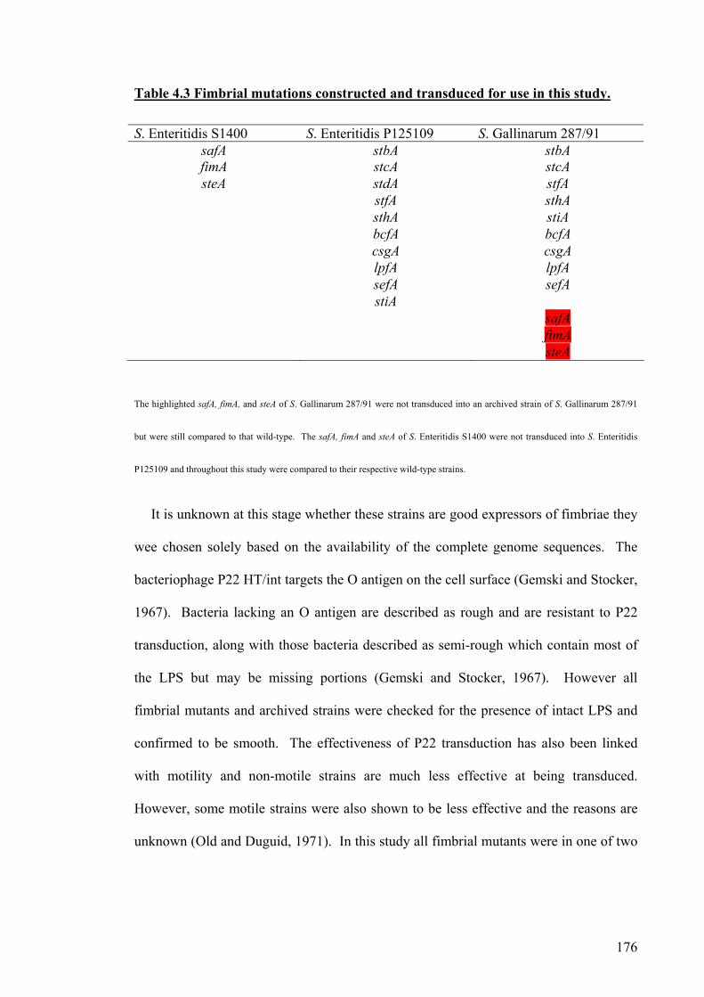
176
Table 4.3 Fimbrial mutations constructed and transduced for use in this study.
S. Enteritidis S1400 S. Enteritidis P125109 S. Gallinarum 287/91safA stbA stbA fimA stcA stcA steA stdA stfA
stfA sthA sthA stiA bcfA bcfA csgA csgA lpfA lpfA sefA sefA stiA safA fimA steA
The highlighted safA, fimA, and steA of S. Gallinarum 287/91 were not transduced into an archived strain of S. Gallinarum 287/91
but were still compared to that wild-type. The safA, fimA and steA of S. Enteritidis S1400 were not transduced into S. Enteritidis
P125109 and throughout this study were compared to their respective wild-type strains.
It is unknown at this stage whether these strains are good expressors of fimbriae they
wee chosen solely based on the availability of the complete genome sequences. The
bacteriophage P22 HT/int targets the O antigen on the cell surface (Gemski and Stocker,
1967). Bacteria lacking an O antigen are described as rough and are resistant to P22
transduction, along with those bacteria described as semi-rough which contain most of
the LPS but may be missing portions (Gemski and Stocker, 1967). However all
fimbrial mutants and archived strains were checked for the presence of intact LPS and
confirmed to be smooth. The effectiveness of P22 transduction has also been linked
with motility and non-motile strains are much less effective at being transduced.
However, some motile strains were also shown to be less effective and the reasons are
unknown (Old and Duguid, 1971). In this study all fimbrial mutants were in one of two

177
strains and both strains were capable of being transduced. The reasons behind the lack
of transduction for specific genes are unknown.
All 25 fimbrial mutations were confirmed by PCR and the fimbrial mutants of S.
Enteritidis have no obvious effect on the growth rate during batch culture in rich
medium. Analysis of the expression of the fimbrial proteins (where antibodies were
available) could not detect the FimA protein from wild-type S. Gallinarum 287/91. This
may be due to the growth conditions being unsuitable for expression in this strain or
only small amounts of the protein may be produced which are below the limits of
detection for this technique. Alternatively the antibody maybe specific for S. Enteritidis
despite the fimA gene of S. Enteritidis P125109 and S. Gallinarum 287/91 being highly
conserved (98 %) as shown in Table 3.1 and FimA was detected in S. Enteritidis S1400
wild-type.
The results for the CsgA protein showed that both S. Enteritidis P125109 and S.
Gallinarum 287/91 expressed the protein at 37 ºC but not at 25 ºC. It has been shown
that CsgA is expressed at 25 ºC in certain strains of S. Enteritidis but not others
(Clouthier et al., 1998b) and it is likely that there may be inter-serovar variation in the
expression of CsgA (Dibb-Fuller et al., 1997, Walker et al., 1999). At the time of
writing, there is limited literature regarding the expression of fimbriae in S. Gallinarum
and this is the first report of CsgA being expressed by S. Gallinarum 287/91 under these
conditions. Antibodies were also obtained for SefA but this protein could not be
detected in either wild-type strain under the two growth conditions, which is likely to be
due to the sensitivity of the method or could be indicative of strain variation. SefA has
been detected in a different strain of S. Enteritidis at 37 ºC but not at 25 ºC (Clouthier et
al., 1998b). Expression of fimbriae has also been shown to be depedent upon pH
(Walker et al., 1999) and on host-pathogen interactions (Humphries et al., 2003).

178
Southern blots were used to identify differences in the two S. Enteritidis strains used in
this study and to determine if genomic rearrangements had occurred proximal to the
insertions. Southern blots were not conclusive in this respect. A single point mutation
anywhere in the genome sequence could cause different restriction digest patterns that
would result in an increase or decrease in the size of the fragment containing the
fimbrial genes. Alternatively, insertions in the genome may have occurred that do not
alter the restriction digest patterns but do result in the transfer of unique sequences.
Mutations in the stc and sef operons produced different fragment sizes patterns but the
correct fragment size was obtained in 6 out of 10 fimbrial mutants in the sequenced
strain of S. Enteritidis P125109 including stc and sef. The differences in the size of the
fragment obtained may be due to differences in this region in S. Enteritidis S1400. In S.
Enteritidis S1400, the P22 fragment may have included only one of the restriction
enzyme sites and the other enzyme site is absent in S. Enteritidis S1400 resulting in
different size fragments. The role of the major fimbrial subunits mutated herein in
adherence to cultured cells and intestinal colonisation in chickens will be discussed in
the following chapters.

179
Chapter 5
Characterisation of
fimbrial mutants in vitro

180
5.1. Introduction
Salmonellae usually enter the avian host by the faecal-oral route and colonise the
gastro-intestinal tract. Adherence to and colonisation of the gut epithelium is key to the
survival and invasion of Salmonella if it is to avoid clearance by the intestinal flow.
The S. Enteritidis FimA fimbrial subunit has been shown to adhere to specific
glycosphingolipid (GSL) receptors on the epithelial surface of chicken oviductal
mucosal cells and it is possible that other fimbriae may adhere to receptors at other sites
(Li et al., 2003b, Li et al., 2003a). The role of fimbriae in adherence has been studied
using several in vitro techniques such as immortalised cell lines, primary cell lines and
explant models and have indicated a role for several fimbriae in adherence.
Mutations in the lpfC fimbrial gene of S. Typhimurium 4252 resulted in the reduction
of adherence to and invasion of HEp-2 cells. A fimA fimbrial mutant of S.
Typhimurium resulted in no difference in adherence to or invasion of HEp-2 cells but
was deficient in the ability to attach to and invade HeLa cells (Baumler et al., 1996a).
This study suggests that adherence is a pre-requisite for invasion and implies that
different fimbriae target different cells. Mutations affecting the csgA and the fimA
fimbrial genes of different strains of S. Enteritidis decreased the association with and
invasion of cultured epithelial cells including Int-407 and Caco-2 cells and histological
examination of the csgA mutants indicated a role in localised aggregated adhesion
(Dibb-Fuller et al., 1999). Mutations of the sefA fimbrial gene did not result in any
difference in adherence to or invasion of Int-407, Caco-2 cells, HEp-2 or HeLa cells
(Ogunniyi et al., 1997, Thorns et al., 1996, Dibb-Fuller et al., 1999).
The adherence to certain cell lines such as HEp-2 cells and HeLa cells has been
shown to vary not only between different fimbriae but also between different allelic
variations of fimH in S. Typhimurium (Boddicker et al., 2002, Hancox et al., 1997). S.

181
Gallinarum and S. Pullorum expressing FimA from S. Typhimurium exhibited an
increase in adherence to HEp-2 cells of 10-20 fold and an increase in invasion by 20-60
fold (Wilson et al., 2000). Invasion of HEp-2 cells by S. Typhimurium is also increased
in anaerobic growth conditions (Ernst et al., 1990).
Mutations in pefA, lpfA, fimA, csgA and sefA genes of S. Enteritidis S1400 played no
role in adherence to chick gut explants (Allen-Vercoe and Woodward, 1999b). In an
organ culture model the lpfC fimbrial mutant of S. Typhimurium adhered in much lower
numbers to murine Peyers patches than the wild-type but there was no difference in the
association with the villous intestine epithelium in an organ culture model (Baumler et
al., 1996b).
Salmonella have also been shown to adhere to abiotic surfaces and only virulent
strains of S. Enteritidis adhered to glass tubes and formed visible filaments (Solano et
al., 1998). S. Enteritidis can adhere to inanimate surfaces at both 37 ºC and 25 ºC. The
expression of fimbriae is dependent on the growth phase, the growth media and the
temperature of growth as detected by ELISA and flow cytometry, (Edwards et al., 2001,
Dibb-Fuller et al., 1997, Woodward et al., 2000, Humphries et al., 2003).
The choice of in vitro model, the strain, serovar and growth conditions all appear to
be important in characterising fimbriae and the adhesive properties of many of the
fimbriae have not been studied in vitro.
182
5.2. Aims
To investigate the role of S. Enteritidis P125109 and S. Gallinarum 287/91
fimbriae in adherence to and invasion of cultured cell lines
To microscopically confirm the adherence and invasion of cells by Salmonella
To determine if fimbriae play conserved roles between serovar and cell types
To confirm selected mutant phenotypes by trans-complementation

183
5.3.1. Adhesion to and invasion of chick kidney cells (CKC)
In chickens, S. Gallinarum 287/91 causes a systemic infection whereas S. Enteritidis
P125109 is mostly restricted to the alimentary tract causing an enteric infection. It was
of interest to determine if the difference in disease outcome of these strains depends
upon their ability to adhere to and subsequently invade different cell lines. Chick
kidney epithelial cells (CKC), a primary cell line that was used to provide a model for
adherence and invasion and was always grown to a confluent monolayer. Transposon
mutants of S. Typhimurium SR11 that were shown to exhibit a decrease in adherence
and invasion in this model were later shown to be attenuated in a chick oral inoculation
model (Lee et al., 1996). The CKCs were obtained from uninfected Rhose Island Red
outbred chickens as a primary cell line so the entire kidney was removed from a 2-3
week old bird and the kidneys were trypsonised (Kaiser et al., 2000). The primary cell
line will be a mixture of kidney cells and were not characterised.
During the construction and bacteriophage transduction of the fimbrial mutants
(Chapter 4), the ΔsafA::cat, ΔfimA::cat and ΔsteA::cat fimbrial mutants could not be
transduced between the two S. Enteritidis strains used, therefore throughout this study
all fimbrial mutants were compared to their respective parent wild-type strain, either
P125109 or S1400. All wild-type and fimbrial mutant strains were cultured under two
growth conditions, 25 ºC static and 37 ºC with agitation to enable a comparison of
different condtions across all of the cell lines examined and all assays were carried out
at 37 ºC. It has been shown that in S. Enteritidis expression of CsgA, FimA and SefA
fimbriae is dependent on temperature (Woodward et al., 2000, Walker et al., 1999,
Humphries et al., 2003). Differences in the expression of S. Enteritidis CsgA fimbriae
were also detected at 42 ºC (Dibb-Fuller et al., 1997). This was not examined herein
184
despite it being the clocal temperature for a chicken model as it would not have been
possible to culture and maintain mammalian cell lines.
The adhesion and invasion assays were performed as described in Section 2.5.2,
mannose was not used in any assay in this study. Briefly, the optical density of the
culture of the wild-type or mutant strains was measured and bacterial numbers were
adjusted accordingly to give an MOI of 5:1 and an MOE of 3:1. Each mutant and wild-
type strain was separately added to three wells on 3 individual days and the assay was
carried out in two replica plates. Bacterial counts were taken of the inocula to confirm
the number of bacteria used. After 15 minutes, gentamicin was added to one plate to
kill all non-internalised bacteria and the other plate was untreated to enable a count of
the number of bacteria that were cell-associated. Triton 1 % (v/v) was added to both
plates and ten-fold serial dilutions were carried out on LB agar plates to calculate the
number of bacteria cell-associated. The data for all mutants, cell lines and growth
conditions are shown separately in the following sections but will be summarised later
in Table 5.7.
A non-invasive control strain, E. coli K-12 MG1655 was included and did not invade
CKCs or did so below the limit of detection of 100 viable bacteria. Mutation of fimbrial
genes in S. Enteritidis had only subtle effects on adherence and invasion of CKC and
significant differences were only seen when the bacteria were grown at 25 ºC statically.
The S. Enteritidis fimbrial mutants, ΔsteA::cat (P=0.021), ΔlpfA::cat (P=0.037),
ΔsafA::cat (P=0.014) and ΔsefA::cat (P=0.002) invaded in significantly lower numbers
than the wild-type and the ΔstbA::cat (P=0.004), ΔsteA::cat (P=0.007), and ΔstiA::cat
(P=0.005) mutants adhered in significantly lower numbers than the wild-type. The
adherence and invasion results for all S. Enteritidis fimbrial mutants are shown in Table
5.1.

185
Table 5.1. The log10 values of bacterial counts of adherence and invasion of S.
Enteritidis wild-type and fimbrial mutant strains to CKC
Grown statically at 25 ºC Grown with shaking at 37 ºC
Strain Invasion P value Adher
-ence
P value Invasio
n
P value Adher
-ence
P
value
WT 2.89 4.10 3.46 3.87
K-12 0 <0.0001 2.76 <0.0001 0 <0.0001 2.98 0.227
stbA 2.93 0.933 3.47 0.004 3.86 0.531 4.36 0.504
stcA 3.24 0.279 3.84 0.200 4.45 0.132 4.73 0.245
stdA 2.99 0.762 3.86 0.236 4.12 0.306 4.47 0.416
stfA 2.93 0.921 3.89 0.315 4.18 0.271 4.62 0.306
sthA 2.62 0.362 3.77 0.106 4.5 0.116 4.28 0.575
stiA 2.75 0.625 3.49 0.005 3.49 0.962 3.56 0.673
bcfA 2.68 0.477 3.83 0.185 4.47 0.125 4.74 0.239
csgA 2.49 0.191 3.95 0.459 3.26 0.757 3.72 0.839
sefA 2.39 0.002 3.73 0.074 4.2 0.255 4.78 0.217
lpfA 2.23 0.037 4.04 0.563 3.44 0.384 4.74 0.250
S1400 3.07 4.16 4.07 3.89
fimA 2.96 0.705 3.79 0.135 4.39 0.157 4.63 0.299
steA 2.33 0.021 3.39 0.0007 3.24 0.253 4.11 0.759
safA 2.27 0.014 3.81 0.103 3.51 0.441 4.84 0.196
SEM 0.217 0.144 0.452 0.512
F-test analysis was carried out using SAS, P values below 0.05 were considered significant and
are highlighted. In Table 5.3 and Figure 5.2, WT refers to S. Enteritidis P125109 wild-type,
S1400 refers to S. Enteritidis S1400 and K-12 refers to E. coli K-12 MG1655. SEM indicates
the standard error of the mean as all results were the mean of triplicate experiments. fimA, steA
and safA were compared to S. Enteritidis S1400 wild-type as they could not be transduced into
S. Enteritidis P125109. All other mutants were compared with S. Enteritidis P125109.
186
The fimbrial mutants of S. Gallinarum 287/91 were examined using the same assay
as for S. Enteritidis except as S. Gallinarum 287/91 is non-motile, the cells were
centrifuged after the addition of the inoculum at 100 g for 3 min at room temperature.
The S. Gallinarum 287/91 fimbrial mutants played a more significant role in adherence
to and invasion of CKCs than S. Enteritidis. At 25 ºC, the S. Gallinarum ΔsteA::cat,
ΔlpfA::cat and ΔsafA::cat mutants did not display a significant reduction in invasion
compared to the wild-type unlike the results for S. Enteritidis. Interestingly, the
mutations of ΔsefA::cat (P<0.0001) and ΔsthA::cat (P<0.0001) of S. Gallinarum 287/91
reduced invasion to undetectable levels (Table 5.2).
At 37 ºC, the ΔsefA::cat (P<0.0001) fimbrial mutant also invaded in numbers below
the limit of detection, surprisingly the ΔstbA::cat (P=0.045), ΔstcA::cat (P=0.036) and
ΔbcfA::cat (P=0.037) fimbrial mutants showed a significant increase in the number of
bacteria invading CKC. The ΔfimA::cat, ΔstiA::cat and ΔlpfA::cat mutations also
increased invasion of CKCs but not significantly but the same fimbriae produced a
significant increase in adherence compared to the wild-type (P<0.001 for both) (Table
5.2). The ΔsafA::cat and ΔsteA::cat mutants of S. Gallinarum 287/91 did not show any
significant differences in adhesion or invasion in either growth condition compared to
the wild-type unlike in S. Enteritidis.
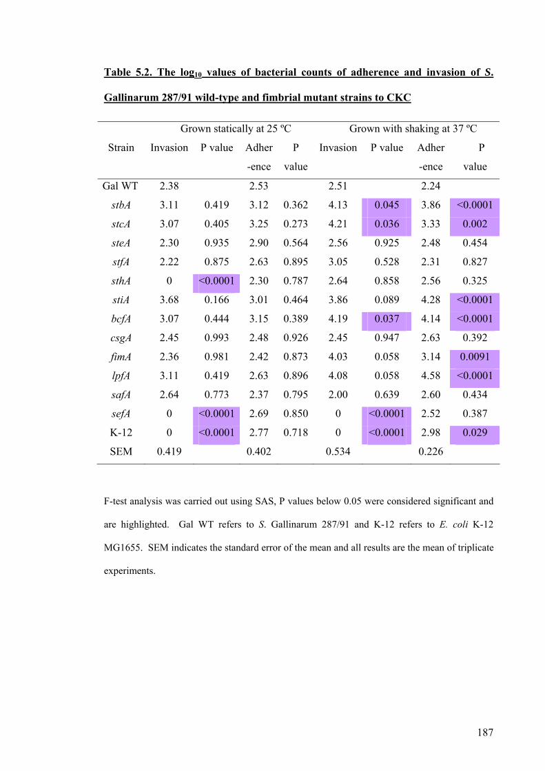
187
Table 5.2. The log10 values of bacterial counts of adherence and invasion of S.
Gallinarum 287/91 wild-type and fimbrial mutant strains to CKC
Grown statically at 25 ºC Grown with shaking at 37 ºC
Strain Invasion P value Adher
-ence
P
value
Invasion P value Adher
-ence
P
value
Gal WT 2.38 2.53 2.51 2.24
stbA 3.11 0.419 3.12 0.362 4.13 0.045 3.86 <0.0001
stcA 3.07 0.405 3.25 0.273 4.21 0.036 3.33 0.002
steA 2.30 0.935 2.90 0.564 2.56 0.925 2.48 0.454
stfA 2.22 0.875 2.63 0.895 3.05 0.528 2.31 0.827
sthA 0 <0.0001 2.30 0.787 2.64 0.858 2.56 0.325
stiA 3.68 0.166 3.01 0.464 3.86 0.089 4.28 <0.0001
bcfA 3.07 0.444 3.15 0.389 4.19 0.037 4.14 <0.0001
csgA 2.45 0.993 2.48 0.926 2.45 0.947 2.63 0.392
fimA 2.36 0.981 2.42 0.873 4.03 0.058 3.14 0.0091
lpfA 3.11 0.419 2.63 0.896 4.08 0.058 4.58 <0.0001
safA 2.64 0.773 2.37 0.795 2.00 0.639 2.60 0.434
sefA 0 <0.0001 2.69 0.850 0 <0.0001 2.52 0.387
K-12 0 <0.0001 2.77 0.718 0 <0.0001 2.98 0.029
SEM 0.419 0.402 0.534 0.226
F-test analysis was carried out using SAS, P values below 0.05 were considered significant and
are highlighted. Gal WT refers to S. Gallinarum 287/91 and K-12 refers to E. coli K-12
MG1655. SEM indicates the standard error of the mean and all results are the mean of triplicate
experiments.

188
5.3.2. Adhesion to and invasion of human epithelial cells (HEp-2)
Salmonella gains entry into the host via epithelial cells as discussed in Chapter 1 and
the invasion and replication of Salmonella and other species has been characterised
using HEp-2 human lung carcinoma cells in vitro (Small et al., 1987, Lee et al., 1992,
Ledeboer et al., 2006, La Ragione et al., 2003, Boddicker et al., 2002). The role of
fimbriae in adherence to HEp-2 cells has been correlated with attenuation detected in
vivo and it was predicted that using HEp-2 cells may also allow the role of other
fimbriae to be determined and provide targets for in vivo analysis (Thorns et al., 1996,
Baumler et al., 1996a). HEp-2 cells were first established in the 1950s (Moore et al.,
1955) from tumors produced in irradiated cortisonised rats after injection with
epidermoid cancer tissue from the larynx of a human (Toolan, 1954).
The in vitro adhesion and invasion assays were carried out as described in Section
2.5.2 at 37 ºC in a 5 % CO2 incubator. The majority of S. Enteritidis fimbrial mutants
showed no significant difference in invasion of or adherence to HEp-2 cells (Table 5.3).
During growth at 25 ºC, the ΔsteA::cat (P<0.0001), ΔfimA::cat (P<0.0001) and
ΔsafA::cat (P<0.0001) mutants of S. Enteritidis showed a significant decrease in ability
to invade HEp-2 cells and the ΔfimA::cat (P<0.0001) and ΔsafA::cat (P=0.020) fimbrial
mutants also showed a significant decrease in adherence to HEp-2 cells. The
ΔfimA::cat mutant strain could not be obtained at detectable limits at 25 ºC from
adhesion or invasion assays but could be detected at 37 ºC (Table 5.3). At 37 ºC, the
ΔsafA::cat (P=0.006) and ΔsteA::cat (P= 0.004) fimbrial mutations of S. Enteritidis
S1400 resulted in a significant increase in adherence compared to the wild-type.
Interestingly, all three of these mutants were in S. Enteritidis S1400 and could not be
transduced into clean backgrounds.
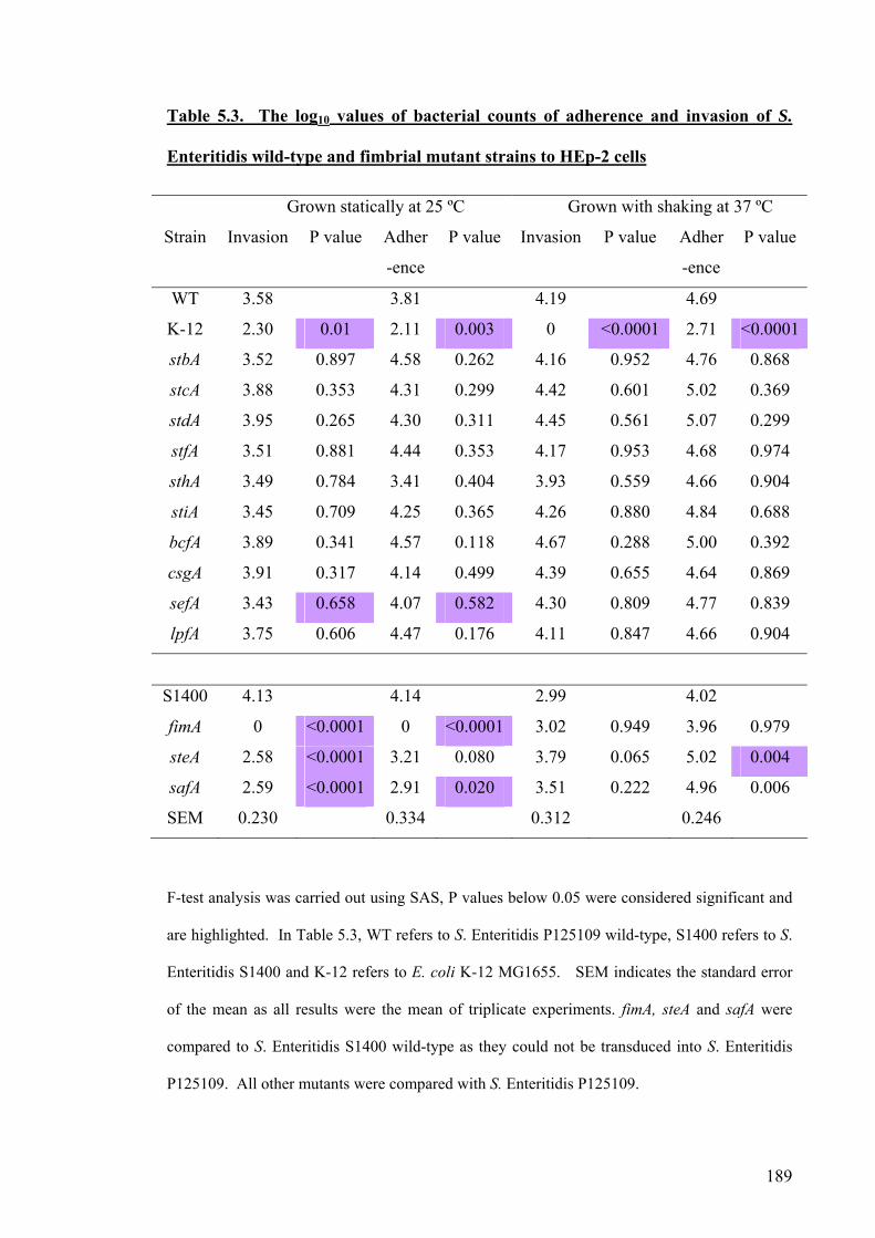
189
Table 5.3. The log10 values of bacterial counts of adherence and invasion of S.
Enteritidis wild-type and fimbrial mutant strains to HEp-2 cells
Grown statically at 25 ºC Grown with shaking at 37 ºC
Strain Invasion P value Adher
-ence
P value Invasion P value Adher
-ence
P value
WT 3.58 3.81 4.19 4.69
K-12 2.30 0.01 2.11 0.003 0 <0.0001 2.71 <0.0001
stbA 3.52 0.897 4.58 0.262 4.16 0.952 4.76 0.868
stcA 3.88 0.353 4.31 0.299 4.42 0.601 5.02 0.369
stdA 3.95 0.265 4.30 0.311 4.45 0.561 5.07 0.299
stfA 3.51 0.881 4.44 0.353 4.17 0.953 4.68 0.974
sthA 3.49 0.784 3.41 0.404 3.93 0.559 4.66 0.904
stiA 3.45 0.709 4.25 0.365 4.26 0.880 4.84 0.688
bcfA 3.89 0.341 4.57 0.118 4.67 0.288 5.00 0.392
csgA 3.91 0.317 4.14 0.499 4.39 0.655 4.64 0.869
sefA 3.43 0.658 4.07 0.582 4.30 0.809 4.77 0.839
lpfA 3.75 0.606 4.47 0.176 4.11 0.847 4.66 0.904
S1400 4.13 4.14 2.99 4.02
fimA 0 <0.0001 0 <0.0001 3.02 0.949 3.96 0.979
steA 2.58 <0.0001 3.21 0.080 3.79 0.065 5.02 0.004
safA 2.59 <0.0001 2.91 0.020 3.51 0.222 4.96 0.006
SEM 0.230 0.334 0.312 0.246
F-test analysis was carried out using SAS, P values below 0.05 were considered significant and
are highlighted. In Table 5.3, WT refers to S. Enteritidis P125109 wild-type, S1400 refers to S.
Enteritidis S1400 and K-12 refers to E. coli K-12 MG1655. SEM indicates the standard error
of the mean as all results were the mean of triplicate experiments. fimA, steA and safA were
compared to S. Enteritidis S1400 wild-type as they could not be transduced into S. Enteritidis
P125109. All other mutants were compared with S. Enteritidis P125109.

190
In S. Gallinarum 287/91, the ΔsteA::cat and ΔfimA::cat fimbrial operons played no
significant role in adherence or invasion of HEp-2 cells, in contrast to their roles in S.
Enteritidis. The ΔsafA::cat (P=0.008) mutant when grown in static growth conditions at
25 ºC, was significantly decreased in its ability to adhere to HEp-2 cells but not at 37 ºC
as shown in Table 5.4. At 37 ºC, the ΔlpfA::cat (P=0.030) and ΔbcfA::cat (P=0.039)
fimbrial mutations resulted in a significant increase in their ability to invade HEp-2 cells
and a slight but insignificant increase in adherence (Table 5.4).

191
Table 5.4. The log10 values of bacterial counts of adherence and invasion of S.
Gallinarum 287/91 wild-type and fimbrial mutant strains to HEp-2 cells
Grown statically at 25 ºC Grown with shaking at 37 ºC
Strain Invasion P value Adher
-ence
P
value
Invasion P value Adhe
rence
P
value
Gal WT 2.75 3.74 2.30 4.05
K-12 0 <0.0001 2.11 0.023 0 <0.0001 2.66 0.081
stbA 3.18 0.309 3.86 0.840 3.31 0.253 3.81 0.620
stcA 2.26 0.204 3.67 0.905 3.38 0.224 3.54 0.305
steA 2.42 0.419 3.69 0.936 3.32 0.293 3.52 0.279
stfA 2.00 0.079 2.81 0.133 2.91 0.492 3.71 0.484
sthA 3.04 0.482 3.34 0.479 2.83 0.519 3.65 0.388
stiA 2.36 0.304 3.99 0.691 3.47 0.188 4.72 0.185
bcfA 3.00 0.495 3.58 0.794 4.18 0.039 4.91 0.09
csgA 2.32 0.299 3.07 0.280 2.81 0.558 4.04 0.983
fimA 2.11 0.132 3.26 0.433 1.66 0.465 3.74 0.527
lpfA 2.31 0.245 3.55 0.757 4.29 0.030 4.77 0.153
safA 2.81 0.872 2.00 0.008 2.33 0.980 3.43 0.214
sefA 0 <0.0001 2.69 0.130 2.56 0.789 3.19 0.125
SEM 0.285 0.426 0.548 0.345
F-test analysis was carried out using SAS, P values below 0.05 were considered significant and
are highlighted. Gal WT refers to S. Gallinarum 287/91 and K-12 refers to E. coli K-12
MG1655. SEM indicates the standard error of the mean as all results were the mean of triplicate
experiments.

192
5.3.3. Adhesion to and invasion of HD11 cells
HD11 cells are a chicken-derived macrophage-like cell line, they were first derived
from chicken hematopoietic cells that had been transformed in vitro and in vivo with 7
different strains of a replicons deficient avain leukaemia viruses which resulted in the
production of a cell line with macrophage like properties (Beug et al., 1979). It was
relevant to use these cells because in the avian host S. Enteritidis infection has been seen
to involve macrophage uptake in the caecal lumen (Popiel and Turnbull, 1985). A
reduction in uptake into HD11 cells has been correlated with a decrease in virulence in
the avian host in early colonisation and this has been proposed to be mediated by a
reduction in macrophage cell death and a faster clearance rate (Bohez et al., 2006). The
relative importance of epithelial cell attachment and invasion versus macrophage-uptake
in the colonisation of the avian host are unknown. Several papers have reported
differences in uptake of Salmonella into macrophages due to various specific mutations
which have then correlated with an attenuated effect in vivo (Bohez et al., 2006,
Adriaensen et al., 2007, Amy et al., 2004). The results obtained here showed only
subtle effects of a single fimbrial gene mutation. The non-invasive E. coli K-12 control
was internalised due to the phagocytic nature of HD11 cells. It is not possible to
separate the number of bacteria present due to invasion and those present due to
engulfment by the HD11 cells. It is also difficult to capture Salmonella only adhering
to the surfaces without being engulfed, therefore only uptake data is shown. The values
obtained for uptake are also likely to be an underestimate due to HD11 cells being
capable of engulfing gentamycin and therefore killing some internalised bacteria. The
S. Enteritidis fimbrial mutants showed no significant difference when compared to the
wild-type in the uptake into HD11 cells as shown in Table 5.5.

193
Table 5.5. The log10 values of bacterial counts of uptake of S. Enteritidis wild-type
and fimbrial mutant strains into HD11 cells
Grown statically at 25 ºC Grown with shaking at 37 ºC
Strain Invasion P value Invasion P value
WT 5.19 5.84
K-12 5.03 0.506 3.69 0.020
stbA 5.19 0.984 5.80 0.821
stcA 5.03 0.506 5.49 0.163
stdA 4.94 0.296 5.59 0.172
stfA 5.23 0.897 5.85 0.887
sthA 4.86 0.178 5.49 0.228
stiA 4.98 0.377 5.78 0.272
bcfA 5.24 0.869 5.69 0.686
csgA 5.26 0.782 5.09 0.176
sefA 4.78 0.105 5.84 0.413
lpfA 5.19 0.983 5.88 0.881
SEM 0.174 0.238
S1400 3.79 5.77
fimA 4.63 0.080 5.69 0.352
steA 4.98 0.828 5.76 0.523
safA 4.67 0.121 5.59 0.345
F-test analysis was carried out using SAS, P values below 0.05 were considered significant and
are highlighted. WT refers to S. Enteritidis P125109 wild-type, S1400 refers to S. Enteritidis
S1400 and K-12 refers to E. coli K-12 MG1655. SEM indicates the standard error of the mean
and all results were from triplicate experiments. fimA, steA and safA were compared to S.
Enteritidis S1400 wild-type as they could not be transduced into S. Enteritidis P125109. All
other mutants were compared with S. Enteritidis P125109.

194
In S. Gallinarum 287/91, at 25 ºC the ΔbcfA::cat fimbrial mutants showed an
increase in invasion and the ΔfimA::cat fimbrial mutant showed a decrease in invasion
but no significant differences were seen at 37 ºC from any fimbrial mutant (Table 5.6).
Table 5.6. The log10 values of bacterial counts of invasion of S. Gallinarum 287/91
wild-type and fimbrial mutants to HD11 cells
Grown statically at 25 ºC Grown shaking at 37 ºC
Strain Invasion P value Invasion P value
Gal WT 4.08 4.78
K-12 3.79 0.674 3.68 0.252
stbA 4.77 0.319 5.08 0.757
stcA 4.90 0.239 6.06 0.186
steA 4.22 0.831 5.54 0.426
stfA 4.26 0.793 3.40 0.156
sthA 5.00 0.185 5.16 0.670
stiA 5.30 0.083 4.38 0.669
bcfA 5.53 0.040 6.02 0.202
csgA 4.13 0.943 5.61 0.388
fimA 2.23 0.014 5.06 0.773
lpfA 4.96 0.209 5.18 0.678
safA 5.33 0.078 6.37 0.103
sefA 5.05 0.214 5.61 0.388
SEM 0.482 0.668
P values below 0.05 are considered significant and are highlighted. Gal WT refers to S.
Gallinarum 287/91 and K-12 refers to E. coli K-12 MG1655. Standard error of the mean was
included, as all results were from triplicate experiments.

195
Table 5.7. Summary of the phenotype of fimbrial subunit mutants in assays for
adherence to and invasion of CKC, HEp-2 and HD11 cells
CKC cells HEp-2 cells HD11 cells
25 ºC 37 ºC 25 ºC 37 ºC 25 ºC 37 ºC
Mutant A I A I A I A I I I
stbA
stcA
stdA
steA
stfA
sthA
stiA
bcfA
csgA
fimA
lpfA
safA
sefA
Black arrows – S. Enteritidis, green arrows- S. Gallinarum, no arrows indicates no difference
when compared to the wild-type.
A- adherence, I –invasion
The ΔstdA::cat, ΔstfA::cat and ΔcsgA::cat fimbrial mutants produced no significant
diffrerences in adhesion to or invasion of any of the cell lines examined in either
serovar.

196
5.4. Validation of adherence and invasion assay
5.4.1 Quantitative confirmation of cell association
Confirmation of bacterial association with cells was carried out to confirm that the
bacterial counts obtained in the adherence and invasion assays were not due to bacteria
adhering to the plastic surfaces used for cell culture, as fimbriae have been shown to
mediate adherence to abiotic surfaces (Woodward et al., 2000).
It is considered unlikely that the bacteria adhered to plastic as differences were seen
between cell lines despite the fact that they were cultured in the same plastic trays,
implying that the differences may be due to the different cell lines used. However, to
confirm this and that the washes were successful in removing non-specifically
associated bacteria, haemacolour staining of infected CKC monolayers was carried out.
The three wild-type strains used in this study were used in the adherence and invasion
assays as previously described in Section 2.5.2 but the CKC cells were grown in the
plastic trays on a poly-L-lysine coverslip. The cells and bacteria on the coverslips were
stained using haemacolour staining as described in Section 2.5.3 and the number of cell-
associated and non-cell associated bacteria were counted in 100 fields of view from 3
independent experiments and the mean was calculated, shown in Table 5.8. The
quantification of the Salmonella showed that ≥75% of bacteria were cell-associated and
that the washes had removed the majority of non-cell-associated bacteria. The
remaining bacteria were seen floating and were not assocated with the plastic trays.
Under these assay conditions relatively few bacteria were detected by microscopy and
therefore only the wild-type strains were examined.

197
Table 5.8. Quantification of Salmonella cell-association with CKCs
Wild-type strain
Average number
of bacteria cell-
associated
Average
number of
bacteria not
cell-associated
Percentage
cell-
associated
P value
S. Enteritidis P125109 38 12 76 0.001
S. Enteritidis S1400 45 10 81 0.0061
S. Gallinarum 287/91 48 13 78 0.0006
The number of bacteria cell-associated or not cell-associated was determined by visual inspection 100 hundred fields of view, the
counts are the mean of 3 independent experiments per strain.
5.4.2. Visual confirmation of cell association by confocal laser scanning microscopy
To visually capture the counts in Section 5.4.1, S. Enteritidis P125109 wild-type was
used to infect CKC, HEp-2 and HD11 cells as in the adherence and invasion assays at
37 ºC. The coverslips were treated as described in Section 2.5.3 for examination by
confocal laser scanning microscopy. The bacterial numbers were low in relation to the
number of cells, but the majority of bacteria were cell-associated, despite the monolayer
not being confluent after treatments with bacteria, trypsine, several washed and
abitbodies. A 0.25 μm optical section was collected which indicated that the bacteria
were in contact with or surrounded by the cellular structures shown in Figure 5.1 (f-
actin, green). By scanning vertically through cells in the Z plane it was possible to find
bacteria that were apically located. Each of the cell lines were examined to rule out
differences between the cells and these images are shown in Figures 5.1. It is possible
that not all bacteria are captured by the antibody as internalised bacteria may have been

198
missed. Differences may also be seen between different serovars/strains but this was
not examined here.
Figure 5.1. Confocal microscopy showing adherence of S. Enteritidis P125109 wild-
type to different cells
The cells are shown in green and the Salmonella are shown in red, the black area is where there are no cells and no Salmonella. Giemsa staining was
not done to enumerate all bacteria and it is possible that internalised bacteria are not recognised by the primary antibody. These images are
representative of the low number of bacteria and cells that were captured after examining 3 coverslips from 3 independent experiments.
CKC HEp-2
HD11
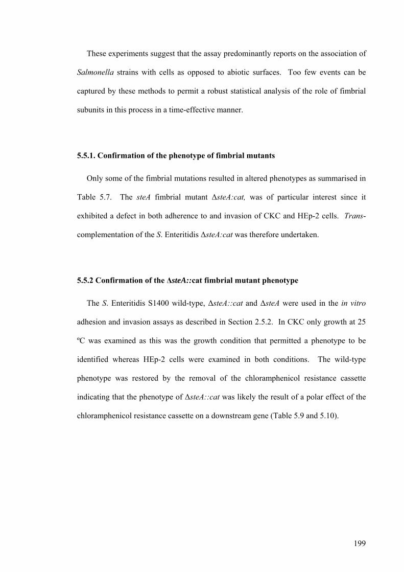
199
These experiments suggest that the assay predominantly reports on the association of
Salmonella strains with cells as opposed to abiotic surfaces. Too few events can be
captured by these methods to permit a robust statistical analysis of the role of fimbrial
subunits in this process in a time-effective manner.
5.5.1. Confirmation of the phenotype of fimbrial mutants
Only some of the fimbrial mutations resulted in altered phenotypes as summarised in
Table 5.7. The steA fimbrial mutant ΔsteA:cat, was of particular interest since it
exhibited a defect in both adherence to and invasion of CKC and HEp-2 cells. Trans-
complementation of the S. Enteritidis ΔsteA:cat was therefore undertaken.
5.5.2 Confirmation of the ΔsteA::cat fimbrial mutant phenotype
The S. Enteritidis S1400 wild-type, ΔsteA::cat and ΔsteA were used in the in vitro
adhesion and invasion assays as described in Section 2.5.2. In CKC only growth at 25
ºC was examined as this was the growth condition that permitted a phenotype to be
identified whereas HEp-2 cells were examined in both conditions. The wild-type
phenotype was restored by the removal of the chloramphenicol resistance cassette
indicating that the phenotype of ΔsteA::cat was likely the result of a polar effect of the
chloramphenicol resistance cassette on a downstream gene (Table 5.9 and 5.10).
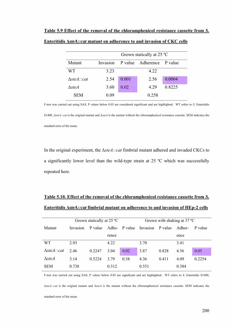
200
Table 5.9 Effect of the removal of the chloramphenicol resistance cassette from S.
Enteritidis ΔsteA::cat mutant on adherence to and invasion of CKC cells
Grown statically at 25 ºC
Mutant Invasion P value Adherence P value
WT 3.23 4.22
ΔsteA::cat 2.54 0.001 2.56 0.0004
ΔsteA 3.60 0.02 4.29 0.8225
SEM 0.09 0.258
F-test was carried out using SAS, P values below 0.05 are considered significant and are highlighted. WT refers to S. Enteritidis
S1400, ΔsteA::cat is the original mutant and ΔsteA is the mutant without the chloramphenicol resistance cassette. SEM indicates the
standard error of the mean.
In the original experiment, the ΔsteA::cat fimbrial mutant adhered and invaded CKCs to
a significantly lower level than the wild-type strain at 25 ºC which was successfully
repeated here.
Table 5.10. Effect of the removal of the chloramphenicol resistance cassette from S.
Enteritidis ΔsteA::cat fimbrial mutant on adherence to and invasion of HEp-2 cells
Grown statically at 25 ºC Grown with shaking at 37 ºC
Mutant Invasion P value Adhe-
rence
P value Invasion P value Adher-
ence
P value
WT 2.93 4.22 3.70 3.41
ΔsteA::cat 2.46 0.2247 3.04 0.02 3.87 0.828 4.56 0.05
ΔsteA 3.14 0.5224 3.79 0.38 4.36 0.411 4.09 0.2254
SEM 0.738 0.312 0.551 0.384
F-test was carried out using SAS, P values below 0.05 are significant and are highlighted. WT refers to S. Enteritidis S1400,
ΔsteA::cat is the original mutant and ΔsteA is the mutant without the chloramphenicol resistance cassette. SEM indicates the
standard error of the mean.

201
During the original experiment at 25 ºC the ΔsteA::cat showed a significant
difference in adherence and invasion of HEp-2 cells and whilst this pattern was repeated
here, it was only significant for adherence. At 37 ºC, adherence and invasion were
increased compared to the wild-type in the original experiment and here a significant
increase in adherence was seen but not in invasion (Table 5.9).
5.5.3. Trans-complementation of the entire ste operon
The region between the steA gene and the steB gene is 83 bp and it is considered
unlikely that the effect of the chloramphenicol cassette insertion may be to interrupt the
stop-start codon translation of steA and steB owing to translational coupling. It remains
feasible that the insertion impaired the transcription of genes downstream of steA and
therefore the entire operon was used for trans-complementation.

202
Table 5.11. Trans-complementation of S. Enteritidis S1400 ΔsteA::cat using the ste
operon in CKC adherence and invasion assay
Grown statically at 25 ºC
Mutant Invasion P value Adherence P value
WT 3.65 5.3
Δ steA::cat 2.71 0.089 4.07 0.032
Δ steA::cat [psteA-E fwd] 3.42 0.651 4.95 0.491
Δ steA::cat [psteA-E rev] 3.43 0.660 4.90 0.441
SEM 0.3411 0.3398
F-test was carried out using SAS, P values below 0.05 are considered significant and are highlighted. WT is S. Enteritidis
S1400 wild-type, ΔsteA::cat is the original fimbrial mutant, ΔsteA::cat [psteA-E fwd] is the mutant with a plasmid carrying the
fimbrial operon in the on orientation and ΔsteA::cat [psteA-E rev] is with the operon in the off orientation. SEM indicates the
standard error of the mean as all experiments were carried out in triplicate.
The ΔsteA::cat fimbrial mutant again showed a decrease in invasion and adherence
of CKC at 25 ºC as shown in Table 5.11. The presence of the steA-E operon on a
TOPO pCR4Blunt plasmid in either orientation partially restored adherence and
invasion to wild-type levels. It is therefore likely that the expression of the steA-E
operon does not strictly require the lac promoter of the cloning vector and that the genes
may be transcribed from internal operon promoter/s or cryptic plasmid promoters. The
data confirmed the adherence and invasion defect of the original ΔsteA::cat mutant but
suggests that genes encoded downstream of steA rather than steA alone are required for
the full effect.
203
5.6. Discussion
The purpose of this study was to characterise a role for fimbrial subunits in vitro in
relation to adherence or invasion of different cell lines. Several fimbrial subunits in S.
Enteritidis showed no role at all as evidenced by the phenotypes of the ΔstcA::cat,
ΔstdA::cat, ΔstfA::cat, ΔsthA::cat, ΔbcfA::cat and ΔcsgA::cat mutants. The
ΔstdA::cat, ΔstfA::cat and ΔcsgA::cat encoded fimbrial subunits played no significant
role in either S. Gallinarum or S. Enteritidis. One cannot preclude the possibility that
these fimbriae may play a role in other growth conditions, at different temperatures,
different cell lines or may require in vivo host conditions to be expressed as has been
previously indicated (Humphries et al., 2003). The expression of the fimbriae in cell
culture media was not examined and may play an important role but a comparison to the
wild-type strain was always carried out. Therefore any differences observed would be
due to the differences in phenotype of the strain which may or may not be due to the
expression of fimbriae in different media or at a different temperature. Salmonella were
only in cell culture media for 15 minutes and would not have time for one complete
round of replication, making it unlikely that the cell culure media or change in
temperature will alter fimbriae expression. Differences in fimbrial expression under
different growth conditions has been shown previously for SefA, FimA and CsgA and
both temperature and pH play a role in their production (Walker et al., 1999, Woodward
et al., 2000, Dibb-Fuller et al., 1997). The expression of these 3 fimbriae are known in
some strains to occur under the conditions examined herein, however for many fimbriae
the conditions under which expression occurs is unknown and appears to vary greatly
from in vitro to in vivo (Dibb-Fuller et al., 1997, Humphries et al., 2003, Woodward et
al., 2000, Walker et al., 1999).

204
Using CKC in the in vitro assays showed that the ΔlpfA::cat, ΔsteA::cat, ΔsafA::cat
and ΔsefA::cat fimbrial mutants of S. Enteritidis were impaired in their ability to invade
at 25 ºC and the ΔsteA::cat, ΔstbA::cat and ΔstiA::cat were impaired in their adherence
to CKC at 25 ºC but no significant differences were seen at 37 ºC. As CKC are a
primary chicken epithelial cell, it was anticipated that fimbriae would play a more
significant role at the higher temperature, as the core temperature of the avian host is 42
ºC. It may be that other environmental conditions that would be present in the natural
host are missing and this may be masking the full phenotype of the fimbrial mutants.
The role of S. Gallinarum fimbriae are poorly characterised and limited information
exists in the literature on the role of its fimbriae in vitro or in vivo. In CKC cells, the
ΔsthA::cat and ΔsefA::cat fimbrial mutants of S. Gallinarum were significantly
decreased in their ability to invade at 25 ºC. At 37 ºC the ΔstbA::cat, ΔstcA::cat,
ΔstiA::cat, ΔbcfA::cat, ΔfimA::cat and ΔlpfA::cat fimbrial subunit mutations all showed
significant increases in adherence and the ΔstbA::cat, ΔstcA::cat and ΔbcfA::cat
fimbrial mutations all resulted in an increase in invasion. The ΔsefA::cat fimbrial
mutant showed a decrease in invasion at both temperatures. Interestingly the stb, sti and
lpf fimbrial operons of S. Gallinarum 287/91 contain frame-shift mutations in their
putative usher gene. Mutation of the predicted fimbrial subunits of these operons still
produces a phenotype despite this. Therefore the pseudogene may not be required for
assembly of the fimbriae and other fimbrial genes may substitute for their function. The
loss of a functional major fimbrial subunit may result in single or multiple fimbriae to
function in its place and a mutation in the major fimbrial subunit of one operon, may
still allow other components of the same operon to be produced. The precise
mechanisms behind such proposed compensation systems are unclear and may vary
between the fimbrial operons. It is also possible that a fimbrial mutation results in the

205
up-regulation of non-fimbrial adhesins, such as SiiE whose expression is related to
activation of Salmonella invasion genes (Gerlach et al., 2007b).
In S. Enteritidis, the ΔsteA::cat and ΔsafA::cat fimbrial mutations increased
adherence at 37 ºC to HEp-2 cells. At 25 ºC, the ΔfimA::cat and ΔsafA::cat fimbrial
mutants exhibited decreased adhesion to HEp-2 cells and ΔsteA::cat, ΔfimA::cat and
ΔsafA::cat mutations resulted in a decrease in the number of bacteria invading HEp-2
cells. Interestingly, all of these mutants were made in S. Enteritidis S1400 and could
not be transduced and the phenotype may be the result of secondary recombination
events, but was not seen in CKC.
In S. Gallinarum, the ΔsafA::cat fimbrial mutant exhibited a decrease in adherence to
HEp-2 cells at 25 ºC. The saf operon contains a frame-shift mutation in the usher gene,
suggesting it uses a different usher or ancillary genes to function fully. This has also
been demonstrated in the stg fimbriae found in S. Typhi, the stg usher is a predicted
pseudogene yet the stg operon still functions (Forest et al., 2007).
A fimA mutant of S. Typhimurium was previously shown to adhere to HEp-2 cells in
comparable numbers to the wild-type (Baumler et al., 1996a)Rajasheakera and at 37 ºC
this concurred with the data obtained here. However, at 25 ºC in S. Enteritidis, the fimA
mutant showed a significant decrease in adherence and invasion which has not
previously been identified (Baumler et al., 1996a). It has been previously shown that
sefA fimbrial mutations do not affect the adherence or invasion of HEp-2 cells
(Ogunniyi et al., 1997, Thorns et al., 1996) as was also identified from the results
herein. Mutations of the lpfC gene reduce the invasion and adherence of S.
Typhimurium which was not seen in these studies possible due to the fact that in
literature the lpfC gene was mutated not the major subunit (Baumler et al., 1996a).
206
Despite the phagocytic nature of HD11 cells, the ΔbcfA::cat and ΔlpfA::cat fimbrial
mutants of S. Gallinarum 287/91 showed an increase in invasion of HD11 cells but no
difference was seen with S. Enteritidis fimbrial mutants, concurring with earlier
observations (Rajashekara et al., 2000). However, disrupting genes downstream of the
sefA major fimbrial subunit in a polar manner has been shown to decrease the bacterial
uptake of S. Typhi into macrophages (Edwards et al., 2000). During the assays the
centrifugation of the bacteria to the cells may have resulted in serovar differences
becoming more apparent in one serovar than another.
Both haemacolour staining and quantitation along with confocal imaging were used
to visually confirm the adherence of bacteria to the cells and to confirm that the washes
were effective and that the majority (>75%) of bacteria were associated with cells and
remained behind after washing to be counted.
The phenotype of ΔsteA::cat mutant of S. Enteritidis was independently confirmed
but further studies showed that the phenotype was an indirect effect caused by the
presence of the chloramphenicol resistance cassette since FLP-mediated excision of the
cassette reversed the effect. It is likely that the chloramphenicol resistance cassette had
a polar effect on the ste operon affecting the steB, steC, steD and steE genes. The steA
and steB genes are separated by 83 bp and are unlikely to be translationally coupled. It
is unclear whether all of the phenotypes identified were a result of the mutation of the
major fimbrial subunits or a consequence of polar effects on other genes within the
fimbrial operon or other genes. It has been reported in the literature that a sefA non-
polar mutation had no effect on virulence whereas a polar mutant showed reduced
virulence (Edwards et al., 2000). To confirm the phenotypes of all other fimbrial
mutants a non-polar mutation would have to be constructed using the Flippase
207
recombinase. The effects of polar mutations could also be examined using RT-PCR or
where possible Western blots to determine which genes are expressed.
Complementing the S. Enteritidis S1400 ΔsteA::cat polar mutation with the steA-E
operon resulted in the partial return of wild-type characteristics. However, the plasmid
that possessed the operon in the antisense orientation still partially restored cell-
association to the same extent as the plasmid with the operon in the sense orientation.
This implies that the operon may be expressed from internal promoters or transcribed
from extraneous plasmid promoters.
The differences in adherence to different cells, in different growth conditions has
been demonstrated for S. Typhimurium and S. Enteritidis but is not reported in S.
Gallinarum (Ernst et al., 1990, Woodward et al., 2000, Dibb-Fuller et al., 1997). S.
Gallinarum showed a role for some fimbriae that was not seen in S. Enteritidis and may
be because of serovar-specific traits related to the genomic attrition that was seen in the
host-specific serovars, resulting in reduced redundancy in the repertoire of fimbriae
(Chapter 3). There are less functional genes in the host-specific serovars than in the
ubiquitous serovars and therefore less compensation systems exist and the phenotype of
any one gene is more pronounced. A mucous secreting cell line HT29 has also been
used to study the role of fimbriae in adhesion in E. Coli and Salmonella (La Ragione et
al., 2000, Rajashekara et al., 2000) and is sensitive enough to detect differences that
occur as a result of single base pair substituitions (Kisiela et al., 2006).
It has also been proposed that fimbrial adherence is mediated by allelic variation or
point mutations within fimbriae which could help in part to explain serovar differences
(Kisiela et al., 2005, Boddicker et al., 2002, Hancox et al., 1997). The stbA and stcA
genes are identical in S. Gallinarum and S. Enteritidis, but the phenotype of mutants in

208
these genes are different which may be due to variation in the sequence of the rest of the
operon or other ancillary genes (Table 3.1).
This chapter highlights the potential for strain-, serovar- and host cell-specific effects
in the role of fimbriae during adherence and invasion. To better define their role
emphasis was next placed on the role of fimbrial subunits of S. Enteritidis in
colonisation of the alimentary tract of chickens.

209
Chapter 6
Characterisation of
fimbrial mutants in vivo

210
6.1. Introduction
Targeted and genome-wide mutagenesis of Salmonella has revealed roles for
selected fimbriae in the colonisation of different hosts. The lpf, fim, bcf, stb, stc, std, sth
and csg fimbriae of S. Typhimurium are required for long-term systemic carriage in
mice (van der Velden et al., 1998, Weening et al., 2005, Lawley et al., 2006, Edwards et
al., 2000, Tsolis et al., 1999). The saf fimbriae have been shown to play a role in
colonisation of pigs (Carnell et al., 2007) and in the avian host the stbC, csgD and sthB
fimbrial genes of S. Typhimurium have been implicated in colonisation (Morgan et al.,
2004, Turner et al., 1998). The majority of studies involving S. Enteritidis fimbriae in
chickens has shown a weak or undetectable role for those fimbriae examined (Allen-
Vercoe and Woodward, 1999a, Rajashekara et al., 2000). A fimD mutant of S.
Enteritidis produced prolonged bacteremia and decreased egg shell colonisation but
exhibited no difference in gut colonisation (De Buck et al., 2003).
The majority of studies to date have involved the use of S. Typhimurium despite S.
Enteritidis being the prevalent non-typhoidal Salmonella isolated from humans in the
England and Wales (Figure 1.1). To define the role of S. Enteritidis fimbriae in
colonisation of the avian alimentary tract, an established chicken colonisation model
was used. In chickens, S. Enteritidis, was shown to preferentially colonise the caeca and
was primarily isolated from the lumen of the caeca (Fanelli et al., 1971, Barrow et al.,
1988). During initial infection S. Enteritidis can be isolated from the liver, spleen and
caeca at 1-week post-infection but by 4-weeks post-infection only the caeca is still
colonised (Gast and Holt, 1998, Bohez et al., 2006) and our own studies have indicated
that the caeca is most commonly colonised (personal communication). Obviously the
length of persistence and type of colonisation can depend on several factors and
therefore a pilot experiment was to be carried out. Although in vitro assays are useful in

211
predicting the roles of certain genes it is not possible to mimic the natural hosts’ in vivo
conditions. An out-bred line of Rhode Island Red chickens were used to provide a
model that was as close to natural populations as possible.

212
6.2. Aims
To produce a valid model to study the role of S. Enteritidis fimbriae in vivo
To characterise the role of S. Enteritidis fimbrial subunit genes in the
colonisation of chickens
To confirme any phenotype identified by trans-complementation

213
6.3. Pilot experiment
The design and execution of experimental infection studies with Salmonella wild-
type and mutant strains is described in Section 2.6. A pilot experiment was carried out,
as although the colonisation of S. Enteritidis in chickens is widely reported in the
literature there is limited information on the colonisation of chickens with the
sequenced strain of S. Enteritidis P125109 (Atterbury et al., 2007) and at the ouset of
this study data was only available for colonisation of this strain in mice (Suar et al.,
2006). The pilot experiment aimed to define the kinetics (magnitude and duration) of
intestinal colonisation and systemic translocation (if any) and to gain an assessment of
variance between birds. This experiment allowed time intervals to be identified at
which adequate numbers of bacteria can be recovered to permit a robust statistical
analysis of the effect of fimbrial subunit mutations. Groups of 15 SPF birds were
gavage dosed with 0.1 ml of adult gut flora to standardise the gut contents at day of
hatch. At 18-days-old each bird was orally dosed with 0.3 ml of approximately 5x10⁸
cfu/ml wild-type or fimbrial mutant as used in other experiments (Bohez et al., 2006).
Five birds from each group were killed by cervical dislocation of the neck and necropsy
examinations were performed at 1, 3 and 7 days post-infection (dpi) to enable
assessment of early adhesion in colonisation of the chicken. The liver (caudate lobe),
spleen, caecal contents, caecal wall, ileal contents and ileal (prior to the ileo-caecal
junction) were recovered aseptically and diluted 1:10 in saline for homogenisation. A
rotary blade was used to homogenise the samples and ten-fold serial dilutions were
carried out. As each sample was diluted 1:10 for homogenisation and 20 µl of this was
plated in triplicate, the theoretical limits of detection are log10 2.2 colony forming units
per gram (cfu/g).

214
For some samples bacterial counts were below the limits of detection and therefore
enrichment was used. The entire sample was grown overnight at 37 ºC in 1 x selenite
broth and then plated on brilliant green agar plates supplemented with nalidixic acid (20
μg/ml) and novobiocin (1 μg/ml) (S. Enteritidis P125109 and S. Gallianrum 287/91
were both nalidixic acid and novabiocin resistant, S. Enteritidis S1400 was only
nalidixic acid resistant). This results in a qualitative rather than a quantitative count but
was given an arbitrary figure of log101 as the sample diluted 10-1 must have contained at
least one viable organism. The enrichments may have been improved by increasing the
time of enrichment or the temeperature (Huhtanen and Naghski, 1972, June et al.,
1995).

215
Figure 6.1. Kinetics of intestinal colonisation and systemic translocation of S.
Enteritidis P125109 wild-type in 18-day-old Rhode Island Red chickens at
intervals post-oral inoculation
0
1
2
3
4
5
6
7
Liver Spleen CaecalContents
Caecal Wall Ileal Contents Ileal Wall
Site of colonisation
log
10 c
fu/g
WT day 1 WT day 3 WT day 7
The green line indicates the limit of detection (log10 2.2). Where counts are reported below the limits of detection this is due to either enrichment
or the recovery of counts above the limits of detection in some but not all birds in a cohort such that the average appears below the limits of detection.
The error bars show the standard error of the mean and provide a measure of bird-to-bird variation. As early liver, spleen and ileum samples yielded
bacterial numbers approaching the limits of detection, subsequent samples were taken from 3 dpi instead of 24 hours post-infection and at 7 and 10 dpi.
The caecal mucosa and contents were colonised with ≥ 104 cfu/g for the duration of
the experiment peaking in the caecal contents at almost 106 cfu/g. This compares to the
lower loads in the ileum and in systemic sites as expected as most studies identify the
caeca as containing the highest bacterial load (Gast and Holt, 1998, Fanelli et al., 1971,
Barrow et al., 1988).
The magnitude and duration of colonisation of enteric and systemic sites by S.
Enteritidis S1400 in chickens has been widely reported in the literature and was
E
E
EE

216
therefore not examined here (Carroll et al., 2004, Allen-Vercoe and Woodward, 1999a,
Woodward et al., 1996, Cooper et al., 1992).
6.4. Caecal colonisation by S. Enteritidis fimbrial subunit mutants
In studies to define the role of S. Enteritidis fimbriae, four groups of fifteen birds
were used. One group was always a control and was dosed with the wild-type strain
and three different fimbrial mutants were separately used to inoculate the 3 other
groups. Each of the fimbrial mutants was compared to their respective wild-type strain.
Each group of unsexed SPF birds were housed in separate cages and provided with food
and water ab litium and were gavage dosed at 18 days old (Section 2.6). This would
ensure that the chickens would develop an immune response as B cell responses are
capable at 14 days old and caecal tonsils are mature (Beal 2006, Bar-Shira 2003). At 3,
7 and 10 dpi, 5 birds from each group were killed. The liver (caudate lobe), spleen,
caecal contents, caecal wall, ileal contents and ileal (prior to the ileo-caecal junction)
were taken as described in Section 2.6 and homogenised. Since attenuation of defined
and random mutants has been detected by examining bacterial numbers in the caeca,
data on the role of S. Enteritidis fimbriae in intestinal colonisation are evaluated in the
following sections against this criterion. Owing to the difficulty in physically
separating contents from mucosa bacterial counts from the caecal wall and caecal counts
were combined to give a total caecal load; these are displayed in Figures 6.2-6.3. Each
tissue sample was homogenised, serially diluted and plated on brilliant green agar plates
supplemented with nalidixic acid (20 µg/ml) and novobiocin (1 µg/ml) in triplicate.
Values represent the mean ± the standard error of the mean and an F-test statistical
analysis was carried out. P values < 0.05 were considered significant and are marked on
the figures with an asterix.

217
*
Figure 6.2. Total caecal load of S. Enteritidis P125109 wild-type and fimbrial
mutant strains at intervals after oral inoculation of 18-day-old Rhode Island Red
chickens
ΔstbA::cat P values at 7 dpi = 0.0081 and
at 10 dpi = 0.03
0
1
2
3
4
5
6
7
8
0 2 4 6 8 10 12days post-infection
log
10 c
fu/g
WT stbA
ΔstcA::cat P values at 3 dpi = 0.0006 and
at 7 dpi = 0.0002
0
1
2
3
4
5
6
7
8
0 2 4 6 8 10 12days post-infection
log
10 c
fu/g
WT stcA
ΔstdA::cat
0
1
2
3
4
5
6
7
8
0 2 4 6 8 10 12days post-infection
log
10 c
fu/g
WT stdA
ΔstiA::cat
0
1
2
3
4
5
6
7
8
0 2 4 6 8 10 12days post-infection
log
10 c
fu/g
WT sti
ΔstfA::cat
0
1
2
3
4
5
6
7
0 2 4 6 8 10 12days post-infection
log
10 c
fu/g
WT stf
ΔsthA::cat
0
1
2
3
4
5
6
7
8
0 2 4 6 8 10 12days post-infection
log
10 c
fu/g
WT sth
* * *

218
ΔbcfA::cat P values at 7 dpi = 0.04
0
1
2
3
4
5
6
7
8
0 2 4 6 8 10 12days post-infection
log
10 c
fu/g
WT bcf
ΔcsgA::cat
0
1
2
3
4
5
6
7
8
0 2 4 6 8 10 12days post-infection
log
10 c
fu/g
WT csg
ΔlpfA::cat
0
1
2
3
4
5
6
7
8
0 2 4 6 8 10 12days post-infection
log
10 c
fu/g
WT lpf
ΔsefA::cat
0
1
2
3
4
5
6
7
8
0 2 4 6 8 10 12days post-infection
log
10 c
fu/g
WT sef
The green line indicates the limit of detection; all samples below this line were obtained via enrichment. The data reflect the
total bacterial count from the caecal wall and caecal contents taken from five birds at each time point for each mutant. The error
bars denote the standard error of the mean (SEM). An F-test analysis was carried out and P values below 0.05 were considered
significant and are marked with an astrix.
*

219
Figure 6.3. Total caecal load of S. Enteritidis S1400 wild-type, ΔfimA::cat,
ΔsteA::cat and ΔsafA::cat mutant strains at intervals after oral inoculation of 18-
day-old Rhode Island Red chickens
ΔfimA::cat
0
1
2
3
4
5
6
7
8
0 2 4 6 8 10 12days post-infection
log
10 c
fu/g
WT fim
ΔsafA::cat
0
1
2
3
4
5
6
7
8
0 2 4 6 8 10 12days post-infection
log
10 c
fu/g
WT saf
ΔsteA::cat P value at 10 dpi = 0.0034
0
1
2
3
4
5
6
7
8
0 2 4 6 8 10 12days post-infection
log
10 c
fu/g
WT ste
S. Enteritidis S1400 was used in this experiment because these mutations could not be transduced into S. Enteritidis P125109. The examination of
ΔfimA::cat and ΔsafA::cat mutations were carried out in the same experiment whereas the ΔsteA::cat mutant was examined in a different experiment
but still in comparison with age matched birds inoculated with the wild-type at the same time.
Figures 6.2-6.3 show the limited variation that occurs between the fimbrial mutants
and the respective wild-type strain. The csgA, sefA and fimA have previously shown no
role in the colonisation of the chicken and data obtained here agrees with this
(Rajashekara et al., 2000). In Figure 6.2, the ΔstbA::cat fimbrial mutant was reliably
recovered from the chicken caeca at lower levels than the wild-type although the
difference only became significant at 7 and 10 dpi with P values of 0.0081 and 0.03
*

220
respectively. The ΔstcA::cat fimbrial mutant showed a significant difference in
colonisation of the caeca at 3 and 7 dpi with P values of 0.0006 and 0.0002 respectively,
implying that the stcA gene may play a role in the initial stages of infection and stbA in
later stages of infection. The ΔbcfA::cat fimbrial mutant resulted in statistically lower
numbers of bacteria being recovered in the caeca only at 7 dpi with a P value of 0.04.
In Figures 6.3, it is of interest to note the lower colonisation rates of the caeca of S.
Enteritidis S1400 wild-type compared with S. Enteritidis P125109 wild-type. This
difference may have arisen due to the composition of gut-flora which was different
between the cohorts or due to strain variation and the data reinforce the need to use a
wild-type strain in all groups of challenged birds. In Figure 6.3, it can be seen that the
ΔsteA::cat fimbrial mutant is recovered in lower numbers from the caeca only at 10 dpi
compared to the wild-type (P = 0.0034). The ΔfimA::cat and ΔsafA::cat fimbrial
mutants persisted in the caeca in greater numbers than S. Enteritidis S1400 wild-type at
10 dpi. However, the numbers of wild-type bacteria in the caeca are below the limits of
detection and it would be unsafe to draw conclusions on the role of fimA and safA in the
absence of further experiments.
During each experiment the liver, spleen, and ileum were also processed and
bacterial counts were determined, an F-test analysis was carried out on all data and the P
values are shown in Table 6.1. All bacterial counts are recorded in Appendix 6.1.
Although several significant differences were identified at sites other than the caeca, the
majority were below the limits of detection as indicated by E in Table 6.1 and therefore
cannot be reliably used for comparison. Several fimbrial subunit mutants displayed
significant differences to the wild-type only at one organ at one time point e.g.
ΔstfA::cat and ΔsthA::cat or at only one site across different times e.g. ΔsteA::cat in the

221
ileum, whereas other fimbrial mutants’ were attenuated at more than one organ or at
more than one time point (Table 6.1).
Both the ΔbcfA::cat and the ΔlpfA::cat fimbrial mutant strains were recovered in the
ileum below the limits of detection at 3 and 7 dpi, despite all other sites being colonised
in comparable numbers to the wild-type. The ΔsefA::cat mutation resulted in a decrease
in the numbers of bacteria recovered from the ileal wall and contents at both 3 and 10
dpi and from the liver and spleen at 7 dpi. The ΔsafA::cat mutant was reduced in the
spleen at all time points and in the ileal contents at 3 dpi and the ΔfimA::cat was lower
in the spleen at 3 and 7 dpi and in the ileal contents at 3 dpi. The ΔstcA::cat mutant was
recovered from the spleen at 10 dpi at significantly lower numbers than the wild-type
and the ΔstdA::cat fimbrial mutant at 7 dpi was found in significantly greater numbers
in the ileal wall and ileal contents with P values of 0.05 and 0.03, respectively. The
ΔstiA::cat produced significant differences to the wild-type at 3 and 10 dpi but not at 7
dpi.
The ΔstbA::cat and ΔcsgA::cat mutant phenotypes are not shown in Table 6.1 as
there were no significant differences in the P values at any of the sites examined.
Despite a significant difference occurring for the ΔstbA::cat mutant in the caeca.

222
Table 6.1. P values obtained from an F-test analysis of bacterial counts of S.
Enteritidis fimbrial mutant strains from the liver, spleen and ileum at various time
points post-oral inoculation
The E represents those samples which were obtained via enrichment. The bacterial
counts are shown in Appendix 6.1.
Day 3 Liver Spleen Ileal contents Ileal wall sefA 0.063 0.638 E 0.001 E 0.02 steA 1 0.599 E 0.0004 0.125 stcA 0.091 0.893 0.061 0.111 stdA 0.178 0.319 0.976 0.961 sthA 0.291 E 0.035 0.280 0.054 stiA 0.670 0.020 0.240 E0.046 stfA 0.600 0.910 0.010 0.849 bcfA 0.670 0.190 E 0.010 E 0.020 lpfA 0.590 0.780 E 0.005 E 0.030 fimA 0.640 E 0.030 E 0.026 0.390 safA 0.350 E 0.019 E 0.007 0.350
Day 7 Liver Spleen Ileal contents Ileal wall sefA E <0.0001 E 0.0003 0.541 0.821 steA E 0.0012 0.153 E 0.0058 E 0.0006 stcA 0.661 0.922 0.248 0.881 stdA 0.622 0.311 0.052 0.031 sthA 0.840 0.449 0.890 0.828 stiA 0.410 0.101 0.750 0.687 stfA 0.070 0.269 0.930 0.674 bcfA 0.384 E 0.015 E 0.002 E 0.001 lpfA 0.384 0.082 E 0.014 0.115 fimA 0.550 E 0.002 0.690 0.115 safA 0.120 E 0.010 0.240 1.000
Day 10 Liver Spleen Ileal contents Ileal wall
sefA 0.981 0.0071 0.0017 0.0162 steA 0.169 0.951 E 0.0064 0.287 stcA 0.091 0.031 0.116 0.015 stdA 0.178 0.877 0.692 0.841 sthA 0.960 0.610 0.360 0.402 stiA 0.010 0.870 0.660 0.580 stfA 0.770 0.316 0.550 0.145 bcfA 0.709 0.850 0.760 0.770 lpfA 0.676 0.550 0.175 0.180 fimA 0.440 0.212 0.370 0.270 safA E 0.660 E 0.020 0.520 E 0.007

223
The phenotypes obtained in vivo do not correlate with the results obtained in vitro in
Chapter 5. In vivo, the ΔstcA::cat and ΔstbA::cat mutants are attenuated in their ability
to colonise the caeca. In vitro the ΔstbA::cat mutation resulted in a decrease in the
adherence to CKC only 25 ºC but interestingly, no attenuating phenotype was identified
for the ΔstcA::cat mutant in vitro.
Whilst phenotypes were detected for fimbrial mutants in respect of adherence
(ΔsteA::cat, ΔstiA::cat, ΔfimA::cat ΔsafA::cat, ΔstbA::cat) and invasion (ΔlpfA::cat,
ΔsafA::cat, ΔsefA::cat, ΔfimA::cat, ΔsteA::cat), the same mutants did not exhibit
attenutation in chickens.
6.5. Confirmation of the phenotype of an S. Enteritidis P125109 ΔstcA::cat mutant
phenotype
The ΔstcA::cat fimbrial mutant of S. Enteritidis P125109 produced the strongest
phenotype in terms of magnitude and the time intervals at which attenuation was
detected and was therefore chosen for further analysis. To confirm that the stcA
contributes to colonisation, an independent ΔstcA::cat fimbrial mutant of S. Enteritidis
S1400 was screened using the same experimental design as above relative to the parent
strain as described in Section 6.3. The total bacterial load in the caeca is shown in
Figure 6.4.

224
Figure 6.4. Total caecal load of S. Enteritidis S1400 wild-type and ΔstcA::cat
strains at intervals after oral inoculation of 18-day-old Rhode Island Red chickens
0
1
2
3
4
5
6
7
8
0 2 4 6 8 10 12days post-infection
log
10 c
fu/g
WT stc
Despite the lower number of bacteria in the caeca of birds infected with S. Enteritidis S1400 wild-type compared with S.
Enteritidis P125109 wild-type, the ΔstcA::cat of S. Enteritidis S1400 was recovered at approximately 2 logs cfu/g lower than the
wild-type strains at 3 and 7 dpi (P<0.05), however caecal loads were comparable for the wild-type and ΔstcA::cat mutant at 10 dpi.
6.6. Comparison of the colonisation of other enteric and systemic sites by the
ΔstcA::cat mutants of S. Enteritidis P125109 and S1400
As a significant difference was seen with the ΔstcA::cat mutant in the caeca,
bacterial counts from other sites were examined and are shown in Figure 6.5. With S.
Enteritidis S1400 wild-type and ΔstcA::cat mutant strains bacterial recoveries from the
liver, spleen, ileal contents and ileal wall bacterial counts approached the limits of
detection by direct plating, indicated by the green line in Figure 6.5a.
* *
P= 0.0135 P= 0.0088

225
For the S. Enteritidis P125109 wild-type and ΔstcA::cat mutant, there is little
difference between the bacterial counts in the liver, spleen and ileal contents Figure
6.5b. In this background an estimate of viable bacteria per gram of tissue could be
derived by direct plating and therefore may be considered a more reliable indicator of
bacterial load. Recoveries of the S. Enteritidis P125109 ΔstcA::cat mutant from the
ileal contents and wall at 3 dpi were higher than the wild-type (P values of 0.06 and 0.1
respectively) but by 7 and 10 dpi comparable numbers of bacteria could be recovered
from this site.

226
Figure 6.5a. Colonisation of enteric and systemic sites by S. Enteritidis S1400 wild-
type and ΔstcA::cat fimbrial mutant strains at intervals after oral inoculation of
18-day-old Rhode Island Red chickens
-1
0
1
2
3
4
5
6
Liver Spleen CaecalContents
Caecal Wall IlealContents
Ileal Wall
Site of colonisation
log
10 c
fu/g
stc day 3 stc day 7 stc day 10 WT day 3 WT day 7 WT day 10
The green line indicates the limit of detection; all samples below this line were obtained via enrichment. N = 5. The error bars are
standard error of the mean SEM. F test analysis was carried out and P values below 0.05 were considered significant and are
marked with an astrix. At 3 dpi in the caecal contents the P = 0.0075 and at 7 dpi P = 0.0078 and in the caecal wall at 3 dpi P =
0.0078 and at 7 dpi in the caecal contents the P value is 0.045.
*
**
*
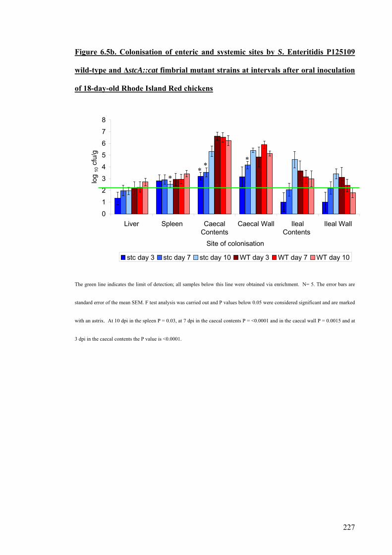
227
Figure 6.5b. Colonisation of enteric and systemic sites by S. Enteritidis P125109
wild-type and ΔstcA::cat fimbrial mutant strains at intervals after oral inoculation
of 18-day-old Rhode Island Red chickens
0
1
2
3
4
5
6
7
8
Liver Spleen CaecalContents
Caecal Wall IlealContents
Ileal Wall
Site of colonisation
log
10 c
fu/g
stc day 3 stc day 7 stc day 10 WT day 3 WT day 7 WT day 10
The green line indicates the limit of detection; all samples below this line were obtained via enrichment. N= 5. The error bars are
standard error of the mean SEM. F test analysis was carried out and P values below 0.05 were considered significant and are marked
with an astrix. At 10 dpi in the spleen P = 0.03, at 7 dpi in the caecal contents P = <0.0001 and in the caecal wall P = 0.0015 and at
3 dpi in the caecal contents the P value is <0.0001.
**
*
*

228
6.7. Removal of the chloramphenicol resistant cassette.
The chloramphenicol resistance cassette was removed from the ΔstcA::cat fimbrial
mutant to investigate if the phenotype was due to the mutation of the major fimbrial
subunit or due to polar effects on the expression of downstream genes. This was carried
out by transient expression of the flippase recombinase from plasmid pCP20 as
described in Section 2.5.5 and Chapter 4, Figure 4.3 (Datsenko and Wanner, 2000).
The removal of the chloramphenicol resistance cassette was confirmed by negative
selection on LB agar supplemented with chloramphenicol and by PCR using the
stcAFOR and stcAREV primers flanking the gene of interest.
6.8. Trans-complementation of the S. Enteritidis P125109 ΔstcA::cat fimbrial
mutant
The stcA gene was cloned on to the pACYC177 plasmid in both orientations, on and
off and both were electroporated into S. Enteritidis P125109 ΔstcA::cat. S. Enteritidis
P125109 wild-type, ΔstcA::cat mutant, ΔstcA mutant, ΔstcA::cat [pstcAfwd] and
ΔstcA::cat [pstcArev] were cultured for 16-18 hours at 37 ºC and those strains carrying
the plasmid were supplemented with ampicillin to maintain the plasmid during growth.
These cultures were used to orally inoculate 18-day-old Rhode Island Red chickens,
with approximately 5x10⁹ cfu/ml. Necropsy examinations were performed at 1 and 3
dpi as the ΔstcA::cat phenotype was most pronounced at early time points and plasmid
stability may be an issue owing to the absence of antibiotic selection in vivo.

229
Figure 6.6. Trans-complementation of the ΔstcA::cat fimbrial mutant
0
1
2
3
4
5
6
7
8
0 1 2 3 4days post-infection
log 1
0 cfu
/g
WT ΔstcA::cat ΔstcA [PstcAfwd] [PstcAfrev]
WT is the S. Enteritidis P125109 wild-type, the green line indicates the limit of detection via direct plating. The error bars represent the standard
error of the mean. Each point is the average of 5 birds per group.
The removal of the chloramphenicol resistance cassette from S. Enteritidis P125109
ΔstcA::cat mutant did not significantly alter the total caecal load at either time point (P
values were 0.27 and 0.648). The caecal load of both the ΔstcA::cat and ΔstcA were
approximately two orders of magnitude lower than the parent strain at both time points
indicating the original phenotype is not likely due to polar effects of the insertion. The
ΔstcA::cat mutant exhibited the same degree of attenuation as observed in previous
experiments. At 1 dpi P = <0.0001 and at 3 dpi P = 0.0002 (Figure 6.2 and 6.5).
Introduction of pstcArev into the ΔstcA::cat mutant resulted in total caecal counts
that that were comparable to the ΔstcA fimbrial mutants both with and without the
chloramphenicol resistance cassette and was significantly different to the wild-type at 1

230
dpi (P= 0.0005) and 3 dpi (P= 0.024). Introduction of the pACYC177-derived plasmid
containing the ΔstcA::cat [pstcAfwd] partially restored the ability of the mutant strain to
colonise the caeca at both time points. The inability of the pstcAfwd replicon to fully
restore colonisation to wild-type levels may reflect differences in the expression level of
the fimbrial subunit and/or the fitness cost of maintaining the plasmid since the
pstcArev exerted a slightly inhibitory effect at least at 1 dpi. The stability of the
plasmid was not examined in vivo herein but has previously been examined in mice
with no effect on the virulence of the strain examined (Knodler et al., 2005).

231
6.9. Discussion
There are limited studies available that use S. Enteritidis P125109 to examine the
colonisation of chickens (Atterbury et al., 2007) and the role of candidate virulence
factors and it was used here to link phenotype to genotype. Therefore, it was important
that the pilot experiment was carried out to confirm that this strain of S. Enteritidis was
able to colonise birds of the age and breed used in this study at a level that permited a
robust statistical analysis. Chapter 3, highlighted differences that can occur between
two strains of the same serovar and it cannot be assumed that because one strain of S.
Enteritidis colonises the avian alimentary tract that all strains will be able to do so at a
comparable level.
S. Enteritidis P125109 colonised the caeca at levels that were anticipated for S.
Enteritidis in outbred birds of this age (Atterbury et al., 2007) and S. Enteritidis S1400
colonised at lower levels than P125109 but these were consistent with the levels
reported in the literature (Carroll et al., 2004). Swabbing was not undertaken as an
initial colonisation rate was to be assessed not the rate of clearance as would have been
measured with swabbing.
The role of several major fimbrial subunit genes (fimA, sefA and csgA), has been
examined in vivo and the data obtained here cocncurred with previous studies as no
differences were seen in the colonisation of the caeca (Rajashekara et al., 2000). In
previous studies a mutation in the sefA gene of S. Enteritidis resulted in reduced
numbers of bacteria in the liver and a faster clearance rate from the spleen. This
phenotype was not observed here however bacterial recoveries from these sites were
close to the limits of detection by direct plating (Rajashekara et al., 2000).

232
In STM screens, a role for the stbC, sthB and csgD fimbrial genes was inferred from
the attenuation of S. Typhimurium miniTn5Km2 mutants in 14-day-old Light Sussex
chickens (Morgan et al., 2004). However, single defined mutants were not made for
these genes to confirm the phenotype and no role for the sthA or csgA genes was
identified in this study. The role of the remaining fimbrial genes annotated herein in the
avian host has not been previously described in the literature.
The ΔstbA::cat mutant of S. Enteritidis P125109 exhibited a significant reduction in
the total caecal load at 7 and 10 dpi compared to the parent strain (P = 0.0081 and 0.03
respectively). The stbA gene is unlikely to be involved in the initial colonisation but
may be required for long term carriage of Salmonella. Further studies using non-polar
mutations and trans-complementation strains are required to confidently establish a role
for Stb encoded fimbriae in the avian host.
The ΔstcA::cat mutant exhibited a significant reduction in bacterial numbers at 3 and
7 dpi (P = 0.0006 and 0.0002). Several fimbrial mutants exhibited a reduction in
colonisation compared with the wild-type at sites other than the caeca, however in many
cases the bacterial counts were below the limits of detection by direct plating making it
difficult to assess a phenotype with confidence.
Several studies have suggested that fimbriae play a role in the adhesion and invasion
of specific cells and in vitro studies within this project (Chapter 5) have indicated that
the lpfA, steA, safA, sefA, stiA and fimA genes influence adherence to or invasion of
specific cell lines. The differences identified in vitro did not correlate with differences
identified in vivo. Thus, mutants that exhibited attenuation in chickens stbA, stcA and
bcfA showed wild-type levels of adhesion and invasion in CKC, HEp-2 and HD11 cells.
This highlights the importance of assessing mutant phenotypes in relevant animal
models wherever feasible.

233
The majority of fimbrial subunits played either a very subtle role or no role at all in
the colonisation of the caeca (P values greater than 0.05). However, more statistically
significant differences may have been identified if bird-to-bird variation was decreased.
All chickens were orally given 0.1 ml of adult gut-flora on the day of hatch to provide
comparable microbiota and competitition in the gut for adherence sites as would be seen
in a natural infection. The microfloras composition was not confirmed by FISH or
DGGE but with two different gut microflora and two different strains of S. Enteritidis
the same attenuating phenotype was seen.
Inbred chicken lines of defined heritable resistance or susceptibility to Salmonella
could be used in future studies; however it was considered that out-bred birds provided
an industry-relevant model in which to evaluate the role of fimbriae. The use of
bicarbonate could also be used to neutralise the stomach acid and allow a higher
proportion of bacteria to enter the host and subtle differences may become pronounced,
unfortunately the side effct of this is that some chickens can receive an extremely high
dose of Salmonella (personnal communication R.M. LaRagione).
In the mouse model, it has been proposed that a compensation mechanism exists,
whereby a single fimbriae may compensate for loss of another. Deletions in the lpf, pef,
fim and csg operons individually only moderately impair mouse virulence, but a
quadruple mutation resulted in a 26-fold increase in the LD50 and a reduced ability to
colonise the intestinal lumen for S. Typhimurium (van der Velden et al., 1998). Given
the repertoire of S. Enteritidis P125109 fimbrial operons there is no reason no believe
that such functional redundancy does not also apply to the chicken model and further
studies with strains harbouring multiple mutations may be warranted. The use of
microarray analysis of to examine the expression of genes in vivo in a fimbrial mutated

234
strain may indicate areas of compensation provided probes are used to discriminate
between fimbrial loci in the absence of cross-hybridisation.
A mutation in the stcA fimbrial subunit gene produced the most attenuating
phenotype of all of the S. Enteritidis fimbrial mutants examined in vivo, yet no
phenotype was identified in vitro for the same mutant. As the same pattern of
colonisation of the caeca was seen with both strains of S. Enteritidis it is unlikely that
secondary mutations have occurred that produced a comparable phenotype in two
independently constructed mutants. It should be noted that the region flanking the
ΔstcA::cat mutation yielded two different restriction fragment sizes in S. Enteritidis
S1400 and S. Enteritidis P125109 in Southern blots (Chapter 4). The predicted size was
based on the genome sequence of S. Enteritidis P125109 and the actual fragment size
seen in this strain was approximately the same size. The removal of the
chloramphenicol resistance cassette did not alter the phenotype seen in vivo and it is
unlikely that a polar effect on the expression of downstream genes has occurred.
Introduction of stcA on a plasmid in an orientation that permits its expression partially
restored the colonisation phenotype of the S. Enteritidis P125109 ΔstcA::cat mutant,
fulfilling molecular Kochs postulate.
Interestingly, the stc operon nucleotide sequence exists as 2 different variants
consisting of different DNA sequences (Table 3.1). S. Enteritidis P125109 and S.
Gallinarum 287/91 possess one sequence which is highly conserved between these two
serovars with all genes being >99% identical. S. Typhi CT18, S. Typhimurium LT2 and
S. Choleraesuis SC-B67 possess a different nucleotide sequence that is also highly
conserved >99%. However, there is limited identity (58-66 %) between these two
variants. S. Gallinarum 287/91 also possesses a predicted pseudogene in the chaperone
of this operon yet all other serovars appear to maintain a functional gene. It would be of

235
interest to examine the S. Gallinarum 287/91 stcA gene to determine if the operon plays
a role in virulence and mediates an important phenotype even with a putative
pseudogene. It would also be of interest to examine a serovar possessing the different
sequence or to clone the different sequence into S. Enteritidis P125109 to confirm if the
phenotype is due to the stc operon sequence or whether the variation in sequence
produces the same or different effects, as point mutations have been shown to play
significant roles in vitro (Boddicker et al., 2002). S. Typhimurium strains carrying
deletions in the stc operon were recovered at significantly reduced numbers from the
faeces of mice (Weening et al., 2005) but have not been studied in chickens.

236
Chapter 7
General
discussion

237
The aim of this study was to annotate and characterise fimbrial genes of the poultry
associated ubiquitous Salmonella enterica serovars Enteritidis and host-specific S.
Gallinarum. This enabled the entire genome sequence to be analysed and allowed the
potential for a link between phenotype and genotype to be established. This was done
to determine whether the repertoire or sequence of the fimbrial operons influences the
outcome of infection and may help explain the differential tissue and host tropisms of
serovars of S. enterica. Three approaches were used; in silico analysis of genome
sequences available at the time of writing, in vitro analysis of defined wild-type,
fimbrial mutant and trans-complemented strains and in vivo analysis of the phenotype
of such strains in a chicken model of infection. In this way, this study compares and
correlates genotype to phenotype and the two strains used were selected solely for this
purpose as the strains were previously uncharacterised.
The in silico analysis compared the genome sequences of strains representing
ubiquitous, host-restricted and host-specific Salmonella serovars. The in silico analysis
included mainly one strain from each serovar and it is uknown if these strains are
representative of a particular serovar. It must be considered that conclusions made from
the genome sequences are limited.
Using a range of bioinformatic tools, primarily ACT and Artemis thirteen fimbrial
operons were identified in S. Enteritidis P125109 and twelve in S. Gallinarum 287/91.
Although all of the fimbrial loci in S. Enteritidis P125109 were predicted to encode
intact genes, eight of the identified operons in S. Gallinarum were predicted to contain
pseudogenes. Whilst no single fimbrial locus was associated with host-specificity, the
mutational attrition seen in the fimbrial operons may account for the restricted host
range available to host-restricted and host-specific serovars. The genomes sequences of
S. Typhi CT18 and S. Gallinarum 287/91 contain a large number of pseudogenes

238
throughout accounting for 4-7 % of the genome whilst in fimbrial operons this increases
to 14-16 % and is unlikely to be the result of a random distribution of pseudogenes.
Genome decay has been associated with the host restriction of agents other than
Salmonella and it is possible that the loss of functional fimbriae may partially restrict
the niches that may be colonised (Holden et al., 2004, Nierman et al., 2004, Sebaihia et
al., 2006).
Having predicted putative major fimbrial subunit genes in both S. Enteritdis P125109
and S. Gallinarum 287/91 a total of 25 defined mutants were constructed using the
lambda Red system for homologous recombination in these two strains. (Datsenko and
Wanner, 2000). The location of all fimbrial mutations was confirmed by PCR using
primers flanking the targeted gene. Where possible, mutations were transduced using
bacteriophage P22 HT/int into an archived strain to eliminate the effects of any rare
second-site defects. Five fimbrial mutations could not be transduced, therefore fimA,
safA, and steA remained in S. Enteritidis S1400 and were compared to this wild-type
strain throughout this study. The bacteriophage P22 HT/int can also move 40 kb of
DNA and this may vary between the two strains of S. Enteritidis used.
All working strains were maintained on LB agar slopes whereas archive strains were
stored in LB glycerol stocks at -70 ºC. This ensures no cross-contamination and no
changes such as spontaneous mutations will have occurred to the bacteria at this
temperature for long periods of time. The growth kinetics of the fimbrial mutants were
also analysed and the insertion of the chloramphenicol resistance cassette was not found
to adversely affect growth.
The validated fimbrial mutants were used for adherence and invasion assays with
CKC, HEp-2 and HD11 cell lines. These in vitro assays indicated that the S. Enteritidis
stbA, steA, stiA, lpfA, safA and sefA encoded fimbrial subunits play a role in adherence

239
to and invasion of CKC only at 25 ºC and the steA, fimA and safA fimbrial subunits play
a role in adherence to and invasion of HEp-2 cells. In S. Gallainrum more fimbriae
played a significant role than in S. Enteritidis; the stbA, stcA, stiA, bcfA, sthA, fimA, lpfA
and sefA genes influenced adherence to and invasion of CKC, safA and sefA influenced
adherence to and invasion of HEp-2 cells and bcfA, fimA and lpfA played a role in the
uptake into HD11 cells.
In previous studies, the sefA, csgA and fimA genes have been widely studied in vitro
in S. Enteritidis and S. Typhimurium. In S. Enteritidis, fimA was not required for
adherence to chick gut explant, or adherence to HEp-2 cells (Allen-Vercoe and
Woodward, 1999b, Baumler et al., 1996a) but in S. Typhimurium fimA was involved in
adherence to and invasion of HeLa cells, HEp-2 cells and porcine enterocytes (Baumler
et al., 1996a, Baumler et al., 1997b, Dibb-Fuller et al., 1999, Ernst et al., 1990). In this
study, fimA played a role in S. Enteritidis infection of HEp-2 cells and in S. Gallinarum
infection of CKC, indicating that strain-, serovar- and cell-line specific effects may
exist. The sefA encoded fimbrial subunit of S. Enteritidis has previously been reported
not to be required for adherence to chick gut explant or cultured epithelial cells (Dibb-
Fuller et al., 1999, Allen-Vercoe and Woodward, 1999b), in contrast to the findings
here where it played a significant role in adherence to CKC in both serovars examined
and in S. Gallinarum sefA also played a role in adherence or invasion of HEp-2 cells.
The differences may be due to the cell-line examined. The csgA fimbrial subunit of S.
Enteritidis played no role in adherence to chick gut explant or invasion of cultured
epithelial cells (Allen-Vercoe and Woodward, 1999b, Dibb-Fuller et al., 1999) which
agrees with this study. The remainder of the fimbrial loci have not previously been
assigned roles in the litereature in vitro adherence or invasion and this report is likely to
be the first to do so.

240
These studies identified a role for the S. Enteritidis ste operon in adherence and
invasion. The removal of the chloramphenicol resistance cassette from the ΔsteA::cat
mutant resulted in the restoration of the wild-type characteristics which indicated that
the phenotype was due to polar effects on the genes downstream of the steA gene. The
entire ste operon was used to trans-complement the ΔsteA::cat mutant and resulted in
the partial restoration of the wild-type, therefore fulfilling molecular Kochs postulates.
Mutations in the same fimbrial operon in the host-specific and the ubiquitous
serovars influenced adherence and invasion to differing extents when compared to their
respective wild-type strains. Despite many of the S. Gallinarum fimbrial operons
containing predicted pseudogenes, significant decreases and increases in adherence and
invasion were still apparent compared with the wild-type (Table 5.7). This suggests that
the presence of a pseudogene does not always affect the functionality of the other genes
encoded in the same operon and has been previously shown for the stg fimbriae of S.
Typhi (Forest et al., 2007). This may indicate that the products encoded by such loci
contribute to the assembly of fimbriae partially encoded by other distal loci or may
induce the expression of other genes (Gerlach et al., 2007b).
It is unclear why differences exist between the role of the same fimbriae of S.
Gallinarum 287/91 and S. Enteritidis P125109, but maybe due to the method used or
point mutations between the fimbriae as has been previously demonstrated (Boddicker
et al., 2002). The expression of fimA from S. Typhimurium in S. Gallinarum increased
adherence to mammalian cells (Wilson et al., 2000). The serovar differences could be
due to different co-operation systems existing because of a difference in the repertoire
of fimbrial operons. A fimbrial mutation in one serovar results in the up-regulation of
several fimbrial operons or other genes. It has been previously shown that fimU of S.
Enteritidis is capable of regulating the expression of two fimbrial subunits in distinct

241
regions of the genome, FimA and SefA (Clouthier et al., 1998b) and the CsgD promoter
in S. Typhimurium is under the control of a global regulator which can have wider
implications for the expression of a range of genes, including the production of O
antigens and components of T3SS (Gerstel et al., 2003, Gibson et al., 2006).
Differences observed in vitro may have been due to the growth conditions. The
expression of S. Enteritidis and S. Gallinarum 287/91 curli, Fim and Sef fimbriae has
been shown in this study to depend upon the growth conditions and the regulation may
subtley differ between strains and serovars. There is no published data on fimbriae
expression in S. Gallinarum (Edwards et al., 2001, Dibb-Fuller et al., 1997, Walker et
al., 1999, Humphries et al., 2003, Woodward et al., 2000).
It is possible that during bacteriophage transduction unwanted traits were transferred
as whilst the S. Gallinarum 287/91 archived strain could be used, two different S.
Enteritidis strains were used as recipients of the mutagenic cassette at the λ Red and
transduction stages. Therefore the phenotypes seen in S. Enteritidis P125109 could be
due to additional genomic material being moved between the two strains. During
transduction the timing was limited so that only one round of bacterial replication could
occur. This ensures that only one insert is encorporated into the bacteria. The P22
phage were grown prior to use in S. Enteritidis or S. Gallinarum wild-type cultures to
ensure that they contained only components of the Salmonella genome.
However, the most significant phenotype both in vitro and in vivo was successfully
trans-complemented and this would need to be carried out to confirm all fimbrial
mutant phenotypes. The in vitro analysis was carried out at 37 ºC not at 42 ºC (the
clocal temperature of a chicken) and it is therefore clear that the cell culture assays will
not mimic this aspect of the host environment. It would not have been possible to

242
compare the HEp-2 cell lines with the CKC and HD11 as the human epithelial cell line
will not tolerate a high temperature.
Colonisation of chickens with the fimbrial mutants and their cognate wild-type
strains showed that the stbA and stcA fimbrial mutants of S. Enteritidis were
significantly attenuated in colonisation of the chicken caeca. This suggests that stcA
plays a role in the initial colonisation of the chicken and stbA may be important during
later stages of infection. Interestingly, stcA was identified by in silico analysis using
ClustalW as occurring in two different variations at the nucleotide level in different
serovars and it may be that variation in sequence that influences the in vivo phenotype.
The ΔstcA::cat was not found to be important for adherence or invasion of any of the
cell lines examined. This shows that the data obtained in vitro cannot be used to give an
accurate indication as to the events in vivo in contrast to other reports (Baumler et al.,
1996a, Baumler et al., 1996b). The expression of fimbriae has been previously shown
to vary considerably in vitro to in vivo (Humphries et al., 2003) and this may partially
explain the differences. One cannot preclude the possibility that the fimbrial mutations
elicit subtle effects on colonisation that cannot be detected owing to animal-animal
variability and the sensitivity of bacterial detection. One way to reduce variation may
be to examine mutant phenotypes in co-infection studies and to derive a competive
index relative to the parent strain and/or to construct multiple mutants. It would be of
interest to examine other stc variants e.g. in S. Typhimurium (Turner et al., 1998,
Morgan et al., 2004). Competitive index (CI) studies were considered where both the
wild-type and the mutant strain are given to the chicken and they compete against each
other. This idea was dismissed due to several problems that have been highlighted in
our laboratory and others. For competitive index studies to be successful high doses of
bacteria are required along with a high number of animals to reduce the variation that

243
occurs in order to produce an accurate CI value. It is possible that in vivo the mutant
strain maybe complemented by the wild-type, reviewed (Beuzon and Holden, 2001).
None of the S. Enteritidis fimbrial mutants showed any difference in their growth
kinetics in vitro in LB, implying that the differences seen in the caeca are likely due to
differences in their ability to colonise and survive in the alimentary tract as opposed to
defects in fitness. However, the different rates of growth were not measured in different
media e.g. minimal media supplemented with or without appropriate nutrients. A
growth curve in these conditions may have produced different results as LB is nutrient
rich. Bacteria in the gut are likely to be exposed at some point to a lack of nutrients and
the different fimbrial mutations may have produced different growth rates. This may
also have shown a direct or indirect role for fimbriae in the utilisation of specific
nutrients.
Those fimbriae expressed in vitro and not expressed in vivo may be phase variable
and this has been proposed as a mechanism to explain the degree of functional
redundancy that occurs in the fimbrial operons of Salmonella (Humphries et al., 2001).
In S. Typhimurium the lpf operon undergoes phase variation to avoid cross immunity.
Different serovars of Salmonella express the same fimbriae which would prevent them
colonising the same host therefore one fimbriae is switched off and others are switched
on or up-regulated (Nicholson and Baumler, 2001). The differences in the role of
fimbriae in vivo compared with in vitro may be partially due to phase variation dicatated
by the environmental conditions in vivo (Norris et al., 1998).
Several in vivo screens have implicated fimbriae in the colonisation of mice. The
stcC, stcD and fimY of S. Typhimurium have been implicated in a microarray-based
negative selection screen as being involved in the colonisation of mice along with bcfD,
safB, fimI and csgA in long-term infections (Lawley et al., 2006). The lpf and fim

244
operons of S. Typhimurium have been implicated in mouse colonisation and several
fimbriae appear to act in a synergistic manner (van der Velden et al., 1998) as well as
the lpf, bcf, stb,stc, std and sth fimbrial operons (Weening et al., 2005). In chickens
much fewer fimbriae have been proposed to be involved in colonisation. In a STM
screen of S. Typhimurium in vivo the stbC, csgD and sthB were implicated in
colonisation of chickens (Morgan et al., 2004) although single defined mutants or trans-
complementation were not carried out. Several in vivo screens using random mutants of
S. Typhimurium did not implicate any of the major fimbrial subunits as playing a role in
the colonisation of the chicken as was identified in this study using targeted mutants
(Morgan et al., 2004, Turner et al., 1998). It is important to note however that some
phenotypes may be age-dependent. In several reports fimA plays no role in the
colonisation of chickens by S. Enteritidis (Allen-Vercoe et al., 1999, Rajashekara et al.,
2000, Thorns et al., 1996) however in laying hens, fimA appears to have an effect on
colonisation of the gut (Thiagarajan et al., 1996). For S. Pullorum infections it has been
shown that a drop in immunity occurs at sexual maturity (Wigley et al., 2005)The
differences maybe due to the fact in older birds there will be a more developed gut flora
to compete with fimbriae for specific receptors, the birds may have been pre-exposed to
Salmonella and/or specific fimbriae and the birds immune system will be more
developed (Beal et al., 2005, Beal et al., 2006b).
Further potential exists for strain-specific effects as studies of S. Enteritidis in mice
found that in one strain sefA played a role in colonisation and yet in a different strain of
the same serovar sefA did not play any role (Ogunniyi et al., 1997).
The fimbrial proteins, in particular the StcA protein may be a target for vaccine
development as it could generate a significant immune response and is surface
expressed. S. Typhimurium fimbriae serve as antigens during infection in mice

245
(Humphries et al., 2005) and the SefA fimbrial proteins has elicited an immune
response against S. Enteritidis in chickens (Lopes et al., 2006). Salmonella fimbriae
may have a potential role in future vaccine development (Thorns, 1995).
Although this study indicated a role for the stcA gene in colonisation of chickens
experimental infections are artificial, involving a one-off high does which is unlikely to
occur in nature. Chickens are more likely to be exposed to lower doses of Salmonella
over a longer period of time and although the oral route of infection is believed to
mimic the natural infection, it would be of interest to examine if other fimbriae play a
more significant role in colonisation if administered via other routes of infection e.g.
aerosol transmission or intra-cloacally particularly in relation to oviduct and egg
colonisation where it is a challenge to examine oviduct colonisation due to the rare
occurrence in artificial models.
For at least one of the fimbrial genes (stcA) I have defined a significant role in the
colonisation of chickens and a role for the ste operon has been identified in vitro. Both
of these phenotypes have been partially restored by the presence of the gene or operon
on a plasmid there by confirming the phenotype. This project has combined sequence
and function analysis which is an important step in the post-genomic era.
The next step within this project would to mutate more than one fimbrial operon in a
strain and to carry out a systematic approach to examine different combinations of
fimbriae, in different aged birds and different species. Although several parts of the gut
were examined only the caeca proved to be reliably colonised at a level that permits a
robust statistical analysis to detect attenuation. Analysis of the in vivo expression levels
with the fimbrial mutants either singularly or with multiple mutants would provide an
insight into the compensation systems, regulatory systems and the degree of cross-talk
that exists within fimbrial operons. Microarray analysis of S. Typhimurium gene
246
expression in macrophages has indicated that fimA expression is downregulated whilst
csgA is upregulated, the relevance of this in vivo is currently unknown (Eriksson et al.,
2003). Further research into the role of fimbriae would also benefit from the production
of a complete set of antibodies for all fimbrial subunits which would allow detection of
fimbriae via Western blots, confocal microscopy and electron microscopy to examine
their morphology. Significant potential also exists for analysis of the large
commendium of microarray data that exists at Instiute for Food Research relating to
Salmonella gene expression under many different environmental conditions and in the
absence of several global regulators. This may then allow for analysis of components
that are involved in the regulation of fimbriae expression. To develop further the idea
that fimbriae could be used as vaccines we would need to examine the host immune
response triggered by purified fimbrial subunits or possible other components of
fimbriae.

247
Chapter 8
Bibliography

248
ABRAHAM, J. M., FREITAG, C. S., CLEMENTS, J. R. & EISENSTEIN, B. I. (1985) An invertible element of DNA controls phase variation of type 1 fimbriae of Escherichia coli. Proceedings of the Natural Academy of Science U S A, 82, 5724-5727.
ADRIAENSEN, C., DE GREVE, H., TIAN, J. Q., DE CRAEYE, S., GUBBELS, E., EECKHAUT, V., VAN IMMERSEEL, F., DUCATELLE, R., KUMAR, M. & HERNALSTEENS, J. P. (2007) A live Salmonella enterica serovar Enteritidis vaccine allows serological differentiation between vaccinated and infected animals. Infection and Immunity, 75, 2461-2468.
ALLEN-VERCOE, E., SAYERS, A. R. & WOODWARD, M. J. (1999) Virulence of Salmonella enterica serotype Enteritidis aflagellate and afimbriate mutants in a day-old chick model. Epidemiology and Infection, 122, 395-402.
ALLEN-VERCOE, E. & WOODWARD, M. J. (1999a) Colonisation of the chicken caecum by afimbriate and aflagellate derivatives of Salmonella enterica serotype Enteritidis. Veterinary Microbiology, 69, 265-275.
ALLEN-VERCOE, E. & WOODWARD, M. J. (1999b) The role of flagella, but not fimbriae, in the adherence of Salmonella enterica serotype Enteritidis to chick gut explant. Journal of Medical Microbiology, 48, 771-780.
ALTSCHUL, S. F., MADDEN, T. L., SCHAFFER, A. A., ZHANG, J., ZHANG, Z., MILLER, W. & LIPMAN, D. J. (1997) Gapped BLAST and PSI-BLAST: a new generation of protein database search programs. Nucleic Acids Research, 25, 3389-3402.
AMY, M., VELGE, P., SENOCQ, D., BOTTREAU, E., MOMPART, F. & VIRLOGEUX-PAYANT, I. (2004) Identification of a new Salmonella enterica serovar Enteritidis locus involved in cell invasion and in the colonisation of chicks. Research in Microbiology 155, 543-552.
ANJUM, M. F., MAROONEY, C., FOOKES, M., BAKER, S., DOUGAN, G., IVENS, A. & WOODWARD, M. J. (2005) Identification of core and variable components of the Salmonella enterica subspecies I genome by microarray. Infection and Immunity, 73, 7894-7905.
ATTERBURY, R. J., VAN BERGEN, M. A., ORTIZ, F., LOVELL, M. A., HARRIS, J. A., DE BOER, A., WAGENAAR, J. A., ALLEN, V. M. & BARROW, P. A. (2007) Bacteriophage therapy to reduce Salmonella colonization of broiler chickens. Applied and Environmental Microbiology, 73, 4543-4549.
AUSTIN, J. W., SANDERS, G., KAY, W. W. & COLLINSON, S. K. (1998) Thin aggregative fimbriae enhance Salmonella enteritidis biofilm formation. FEMS Microbiology Letters, 162, 295-301.
AVILA, L. A., NASCIMENTO, V. P., SALLE, C. T. & MORAES, H. L. (2006) Effects of probiotics and maternal vaccination on Salmonella enteritidis infection in broiler chicks. Avian Disease, 50, 608-612.
BABA, E., NAGAISHI, S., FUKATA, T. & ARAKAWA, A. (1991) The role of intestinal microflora on the prevention of Salmonella colonisation in gnotobioitc chickens. Poultry Science, 70, 1902-1907.
BAILEY, J. S., BLANKENSHIP, L. C., STERN, N. J., COX, N. A. & MCHAN, F. (1988) Effect of anticoccidial and antimicrobial feed additives on prevention of Salmonella colonization of chicks treated with anaerobic cultures of chicken feces. Avian Disease, 32, 324-329.

249
BAINS, B. S. & MACKENZIE, M. A. (1974) Transmission of Salmonella through an integrated poultry organisation. Poultry Science, 53, 1114-1118.
BAIRD, G. D., MANNING, E. J. & JONES, P. W. (1985) Evidence for related virulence sequences in plasmids of Salmonella dublin and Salmonella typhimurium. Journal of General Microbiology, 131, 1815-1823.
BAIROCH, A. & APWEILER, R. (1996) The SWISS-PROT protein sequence data bank and its new supplement TREMBL. Nucleic Acids Research, 24, 21-25.
BAIROCH, A., APWEILER, R., WU, C. H., BARKER, W. C., BOECKMANN, B., FERRO, S., GASTEIGER, E., HUANG, H., LOPEZ, R., MAGRANE, M., MARTIN, M. J., NATALE, D. A., O'DONOVAN, C., REDASCHI, N. & YEH, L. S. (2005) The Universal Protein Resource (UniProt). Nucleic Acids Research, 33, D154-9.
BAKER, W., VAN DEN BROEK, A., CAMON, E., HINGAMP, P., STERK, P., STOESSER, G. & TULI, M. A. (2000) The EMBL nucleotide sequence database. Nucleic Acids Research, 28, 19-23.
BALBONTIN, R., ROWLEY, G., PUCCIARELLI, M. G., LOPEZ-GARRIDO, J., WORMSTONE, Y., LUCCHINI, S., GARCIA-DEL PORTILLO, F., HINTON, J. C. & CASADESUS, J. (2006) DNA adenine methylation regulates virulence gene expression in Salmonella enterica serovar Typhimurium. Journal of Bacteriology, 188, 8160-8168.
BAR-SHIRA, E., SKLAN, D. & FRIEDMAN, A. (2003) Establishment of immune competence in the avian GALT during the immediate post-hatch period. Developmental and Comparative Immunology, 27, 147-157.
BARROW, P. A., HUGGINS, M. B. & LOVELL, M. A. (1994) Host specificity of Salmonella infection in chickens and mice is expressed in vivo primarily at the level of the reticuloendothelial system. Infection and Immunity, 62, 4602-4610.
BARROW, P. A., HUGGINS, M. B., LOVELL, M. A. & SIMPSON, J. M. (1987a) Observations on the pathogenesis of experimental Salmonella typhimurium infection in chickens. Research in Veterinary Science 42, 194-199.
BARROW, P. A. & LOVELL, M. A. (1988) The association between a large molecular mass plasmid and virulence in a strain of Salmonella pullorum. Journal of General Microbiology, 134 ( Pt 8), 2307-2316.
BARROW, P. A. & LOVELL, M. A. (1989) Functional homology of virulence plasmids in Salmonella gallinarum, S. pullorum, and S. typhimurium. Infection and Immunity, 57, 3136-3141.
BARROW, P. A., SIMPSON, J. M. & LOVELL, M. A. (1988) Intestinal colonisation in the chicken by food poisoning Salmonella serotypes; microbial characteristics associated with faecal excretion. Avian Pathology, 17, 571-588.
BARROW, P. A., SIMPSON, J. M., LOVELL, M. A. & BINNS, M. M. (1987b) Contribution of Salmonella gallinarum large plasmid toward virulence in fowl typhoid. Infection and Immunity, 55, 388-392.
BATEMAN, A., BIRNEY, E., DURBIN, R., EDDY, S. R., HOWE, K. L. & SONNHAMMER, E. L. (2000) The Pfam protein families database. Nucleic Acids Research, 28, 263-266.
BAUMLER, A. J., GILDE, A. J., TSOLIS, R. M., VAN DER VELDEN, A. W., AHMER, B. M. & HEFFRON, F. (1997a) Contribution of horizontal gene transfer and deletion events to development of distinctive patterns of fimbrial operons during evolution of Salmonella serotypes. Journal of Bacteriology, 179, 317-322.

250
BAUMLER, A. J. & HEFFRON, F. (1995) Identification and sequence analysis of lpfABCDE, a putative fimbrial operon of Salmonella typhimuirum. Journal of Bacteriology, 177, 2087-2097.
BAUMLER, A. J., TSOLIS, R. M. & HEFFRON, F. (1996a) Contribution of fimbrial operons to attachment to and invasion of epithelial cell lines by Salmonella typhimurium. Infection and Immunity, 64, 1862-1865.
BAUMLER, A. J., TSOLIS, R. M. & HEFFRON, F. (1996b) The lpf fimbrial operon mediates adhesion of Salmonella typhimurium to murine Peyer's patches. Proceedings of the Natural Academy of Science U S A, 93, 279-283.
BAUMLER, A. J., TSOLIS, R. M. & HEFFRON, F. (1997b) Fimbrial adhesins of Salmonella typhimurium. Role in bacterial interactions with epithelial cells. Advances in Experimental Medicine and Biology, 412, 149-158.
BEAL, R. K., POWERS, C., DAVISON, T. F., BARROW, P. A. & SMITH, A. L. (2006a) Clearance of enteric Salmonella enterica serovar Typhimurium in chickens is independent of B-cell function. Infection and Immunity, 74, 1442-1444.
BEAL, R. K., POWERS, C., DAVISON, T. F. & SMITH, A. L. (2006b) Immunological development of the avian gut IN PERRY, G. C. (Ed.) Avian gut function in health and disease. CAB international
BEAL, R. K., POWERS, C., WIGLEY, P., BARROW, P. A., KAISER, P. & SMITH, A. L. (2005) A strong antigen-specific T-cell response is associated with age and genetically dependent resistance to avian enteric salmonellosis. Infection and Immunity, 73, 7509-7516.
BEAL, R. K., POWERS, C., WIGLEY, P., BARROW, P. A. & SMITH, A. L. (2004a) Temporal dynamics of the cellular, humoral and cytokine responses in chickens during primary and secondary infection with Salmonella enterica serovar Typhimurium. Avian Pathology, 33, 25-33.
BEAL, R. K., WIGLEY, P., POWERS, C., BARROW, P. A. & SMITH, A. L. (2006c) Cross-reactive cellular and humoral immune responses to Salmonella enterica serovars Typhimurium and Enteritidis are associated with protection to heterologous re-challenge. Veterinary Immunology and Immunopathology, 114, 84-93.
BEAL, R. K., WIGLEY, P., POWERS, C., HULME, S. D., BARROW, P. A. & SMITH, A. L. (2004b) Age at primary infection with Salmonella enterica serovar Typhimurium in the chicken influences persistence of infection and subsequent immunity to re-challenge. Veterinary Immunology and Immunopathology, 100, 151-164.
BEFUS, A. D., JOHNSTON, N., LESLIE, G. A. & BIENENSTOCK, J. (1980) Gut-associated lymphoid tissue in the chicken. I. Morphology, ontogeny, and some functional characteristics of Peyer's patches. Journal of Immunology, 125, 2626-2632.
BENSON, D. A., KARSCH-MIZRACHI, I., LIPMAN, D. J., OSTELL, J., RAPP, B. A. & WHEELER, D. L. (2000) GenBank. Nucleic Acids Research, 28, 15-18.
BENSON, G. (1999) Tandem repeats finder: a program to analyze DNA sequences. Nucleic Acids Research, 27, 573-580.
BEUG, H., VON KIRCHBACH, A., DODERLEIN, G., CONSCIENCE, J. F. & GRAF, T. (1979) Chicken hematopoietic cells transformed by seven strains of defective avian leukemia viruses display three distinct phenotypes of differentiation. Cell, 18, 375-390.

251
BEUZON, C. R. & HOLDEN, D. W. (2001) Use of mixed infections with Salmonella strains to study virulence genes and their interactions in vivo. Microbes and Infection, 3, 1345-1352.
BISHOP, A. L., BAKER, S., JENKS, S., FOOKES, M., GAORA, P. O., PICKARD, D., ANJUM, M., FARRAR, J., HIEN, T. T., IVENS, A. & DOUGAN, G. (2005) Analysis of the hypervariable region of the Salmonella enterica genome associated with tRNA(leuX). Journal of Bacteriology, 187, 2469-2482.
BISPHAM, J., TRIPATHI, B. N., WATSON, P. R. & WALLIS, T. S. (2001) Salmonella pathogenicity island 2 influences both systemic salmonellosis and Salmonella-induced enteritis in calves. Infection and Immunity, 69, 367-377.
BLANC-POTARD, A. B. & GROISMAN, E. A. (1997) The Salmonella selC locus contains a pathogenicity island mediating intramacrophage survival. Embo Journal, 16, 5376-5385.
BLATTNER, F. R., PLUNKETT, G., 3RD, BLOCH, C. A., PERNA, N. T., BURLAND, V., RILEY, M., COLLADO-VIDES, J., GLASNER, J. D., RODE, C. K., MAYHEW, G. F., GREGOR, J., DAVIS, N. W., KIRKPATRICK, H. A., GOEDEN, M. A., ROSE, D. J., MAU, B. & SHAO, Y. (1997) The complete genome sequence of Escherichia coli K-12. Science, 277, 1453-1474.
BLYN, L. B., BRAATEN, B. A. & LOW, D. A. (1990) Regulation of pap pilin phase variation by a mechanism involving differential dam methylation states. Embo Journal, 9, 4045-4054.
BOCKMAN, D. E., BOYDSTON, W. R. & BEEZHOLD, D. H. (1983) The role of epithelial cells in gut-associated immune reactivity. Annals of the New York Academy of Science, 409, 129-144.
BODDICKER, J. D., LEDEBOER, N. A., JAGNOW, J., JONES, B. D. & CLEGG, S. (2002) Differential binding to and biofilm formation on, HEp-2 cells by Salmonella enterica serovar Typhimurium is dependent upon allelic variation in the fimH gene of the fim gene cluster. Molecular Microbiology, 45, 1255-1265.
BOHEZ, L., DEWULF, J., DUCATELLE, R., PASMANS, F., HAESEBROUCK, F. & VAN IMMERSEEL, F. (2008) The effect of oral administration of a homologous hilA mutant strain on the long-term colonization and transmission of Salmonella Enteritidis in broiler chickens. Vaccine, 26, 372-378.
BOHEZ, L., DUCATELLE, R., PASMANS, F., BOTTELDOORN, N., HAESEBROUCK, F. & VAN IMMERSEEL, F. (2006) Salmonella enterica serovar Enteritidis colonization of the chicken caecum requires the HilA regulatory protein. Veterinary Microbiology, 116, 202-210.
BOLTON, A. J., OSBORNE, M. P., WALLIS, T. S. & STEPHEN, J. (1999) Interaction of Salmonella choleraesuis, Salmonella dublin and Salmonella typhimurium with porcine and bovine terminal ileum in vivo. Microbiology, 145 ( Pt 9), 2431-2441.
BONCI, A., CHIESURIN, A., MUSCAS, P. & ROSSOLINI, G. M. (1997) Relatedness and phylogeny within the family of periplasmic chaperones involved in the assembly of pili or capsule-like structures of gram-negative bacteria. J Mol Evol, 44, 299-309.
BOYD, E. F. & HARTL, D. L. (1998) Salmonella virulence plasmid. Modular acquisition of the spv virulence region by an F-plasmid in Salmonella enterica subspecies I and insertion into the chromosome of subspecies II, IIIa, IV and VII isolates. Genetics, 149, 1183-1190.

252
BOYD, E. F. & HARTL, D. L. (1999) Analysis of the type 1 pilin gene cluster fim in Salmonella: its distinct evolutionary histories in the 5' and 3' regions. Journal of Bacteriology, 181, 1301-1308.
BOYD, E. F., PORWOLLIK, S., BLACKMER, F. & MCCLELLAND, M. (2003) Differences in gene content among Salmonella enterica serovar typhi isolates. Journal of Clinical Microbiology, 41, 3823-3828.
BOYD, E. F., WANG, F. S., WHITTAM, T. S. & SELANDER, R. K. (1996) Molecular genetic relationships of the salmonellae. Applied and Environmental Microbiology, 62, 804-808.
BRINTON, C. C., JR. (1959) Non-flagellar appendages of bacteria. Nature, 183, 782-786.
BROMBACHER, E., BARATTO, A., DOREL, C. & LANDINI, P. (2006) Gene expression regulation by the curli activator CsgD protein: Modulation of cellulose biosynthesis and control of negative determinants for microbial adhesion. Journal of Bacteriology, 188, 2027-2037.
BUMSTEAD, N. & BARROW, P. (1993) Resistance to Salmonella gallinarum, S. pullorum, and S. enteritidis in inbred lines of chickens. Avian Disease, 37, 189-193.
CARLSON, H. C. (1982) Defense mechanisms of the avian gastrointestinal tract. Poultry Science, 61, 1268-1278.
CARNELL, S. C., BOWEN, A., MORGAN, E., MASKELL, D. J., WALLIS, T. S. & STEVENS, M. P. (2007) Role in virulence and protective efficacy in pigs of Salmonella enterica serovar Typhimurium secreted components identified by signature-tagged mutagenesis. Microbiology, 153, 1940-1952.
CARROLL, P., LA RAGIONE, R. M., SAYERS, A. R. & WOODWARD, M. J. (2004) The O-antigen of Salmonella enterica serotype Enteritidis PT4: a significant factor in gastrointestinal colonisation of young but not newly hatched chicks. Veterinary Microbiology, 102, 73-85.
CARSIOTIS, M., WEINSTEIN, D. L., KARCH, H., HOLDER, I. A. & O'BRIEN, A. D. (1984) Flagella of Salmonella typhimurium are a virulence factor in infected C57BL/6J mice. Infection and Immunity, 46, 814-818.
CARVER, T. J., RUTHERFORD, K. M., BERRIMAN, M., RAJANDREAM, M. A., BARRELL, B. G. & PARKHILL, J. (2005) ACT: the Artemis Comparison Tool. Bioinformatics, 21, 3422-3423.
CASADESUS, J. & LOW, D. (2006) Epigenetic gene regulation in the bacterial world. Microbiology and Molecular Biology Reviews 70, 830-856.
CASJENS, S. & HAYDEN, M. (1988) Analysis in vivo of the bacteriophage P22 headful nuclease. Journal of Molecular Biology, 199, 467-474.
CHADFIELD, M. S., BROWN, D. J., AABO, S., CHRISTENSEN, J. P. & OLSEN, J. E. (2003) Comparison of intestinal invasion and macrophage response of Salmonella Gallinarum and other host-adapted Salmonella enterica serovars in the avian host. Veterinary Microbiology, 92, 49-64.
CHAKRAVORTTY, D., ROHDE, M., JAGER, L., DEIWICK, J. & HENSEL, M. (2005) Formation of a novel surface structure encoded by Salmonella Pathogenicity Island 2. Embo Journal, 24, 2043-2052.
CHAN, K., BAKER, S., KIM, C. C., DETWEILER, C. S., DOUGAN, G. & FALKOW, S. (2003) Genomic comparison of Salmonella enterica serovars and Salmonella bongori by use of an S. enterica serovar typhimurium DNA microarray. Journal of Bacteriology, 185, 553-563.

253
CHANG, A. C. & COHEN, S. N. (1978) Construction and characterization of amplifiable multicopy DNA cloning vehicles derived from the P15A cryptic miniplasmid. Journal of Bacteriology, 134, 1141-1156.
CHENG-HSUN, C., CHIH-HSEIN, C., SHUN, C., LIN-HUI, S. & TZOU-YIEN, L. (2006) Salmonella enterica serotype Choleraesuis infections in pediatric patients Pediatrics 117, 1193-1196.
CHEREPANOV, P. P. & WACKERNAGEL, W. (1995) Gene disruption in Escherichia coli: TcR and KmR cassettes with the option of Flp-catalyzed excision of the antibiotic-resistance determinant. Gene, 158, 9-14.
CHIU, C. H., SU, L. H. & CHU, C. (2004) Salmonella enterica serotype Choleraesuis: epidemiology, pathogenesis, clinical disease, and treatment. Clinical Microbiological Review, 17, 311-322.
CHIU, C. H., TANG, P., CHU, C., HU, S., BAO, Q., YU, J., CHOU, Y. Y., WANG, H. S. & LEE, Y. S. (2005) The genome sequence of Salmonella enterica serovar Choleraesuis, a highly invasive and resistant zoonotic pathogen. Nucleic Acids Research, 33, 1690-1698.
CHOUDHURY, D., THOMPSON, A., STOJANOFF, V., LANGERMANN, S., PINKNER, J., HULTGREN, S. J. & KNIGHT, S. D. (1999) X-ray structure of the FimC-FimH chaperone-adhesin complex from uropathogenic Escherichia coli. Science, 285, 1061-1066.
CHRISTENSEN, H., NORDENTOFT, S. & OLSEN, J. E. (1998) Phylogenetic relationships of Salmonella based on rRNA sequences. International Journal of Systematic Bacteriology, 48 Pt 2, 605-610.
CLARK, M. A., HIRST, B. H. & JEPSON, M. A. (1998) Inoculum composition and Salmonella pathogenicity island 1 regulate M-cell invasion and epithelial destruction by Salmonella typhimurium. Infection and Immunity, 66, 724-731.
CLARK, M. A., JEPSON, M. A., SIMMONS, N. L. & HIRST, B. H. (1994) Preferential interaction of Salmonella typhimurium with mouse Peyer's patch M cells. Res Microbiol, 145, 543-552.
CLAVIJO, R. I., LOUI, C., ANDERSEN, G. L., RILEY, L. W. & LU, S. (2006) Identification of genes associated with survival of Salmonella enterica serovar Enteritidis in chicken egg albumen. Applied and Environmental Microbiology, 72, 1055-1064.
CLEGG, S. & GERLACH, G. F. (1987) Enterobacterial fimbriae. Journal of Bacteriology, 69, 934-938.
CLEGG, S., HANCOX, L. S. & YEH, K. S. (1996) Salmonella typhimurium fimbrial phase variation and FimA expression. Journal of Bacteriology, 178, 542-545.
CLEGG, S. & HUGHES, K. T. (2002) FimZ is a molecular link between sticking and swimming in Salmonella enterica serovar Typhimurium. Journal of Bacteriology 184, 1209-1213.
CLIFTON-HADLEY, F. A., BRESLIN, M., VENABLES, L. M., SPRIGINGS, K. A., COOLES, S. W., HOUGHTON, S. & WOODWARD, M. J. (2002) A laboratory study of an inactivated bivalent iron restricted Salmonella enterica serovars Enteritidis and Typhimurium dual vaccine against Typhimurium challenge in chickens. Veterinary Microbiology, 89, 167-179.
CLOUTHIER, S. C., COLLINSON, S. K. & KAY, W. W. (1994) Unique fimbriae-like structures encoded by sefD of the SEF14 fimbrial gene cluster of Salmonella enteritidis. Molecular Microbiology, 12, 893-901.

254
CLOUTHIER, S. C., COLLINSON, S. K., LIPPERT, D., AUSIO, J., WHITE, A. P. & KAY, W. W. (1998a) Periplasmic and fimbrial SefA from Salmonella enteritidis. Biochimica et Biophysica Acta, 1387, 355-368.
CLOUTHIER, S. C., COLLINSON, S. K., WHITE, A. P., BANSER, P. A. & KAY, W. W. (1998b) tRNA(Arg) (fimU) and expression of SEF14 and SEF21 in Salmonella enteritidis. Journal of Bacteriology, 180, 840-845.
CLOUTHIER, S. C., MULLER, K. H., DORAN, J. L., COLLINSON, S. K. & KAY, W. W. (1993) Characterization of three fimbrial genes, sefABC, of Salmonella enteritidis. Journal of Bacteriology, 175, 2523-2533.
COGAN, T. A., JORGENSEN, F., LAPPIN-SCOTT, H. M., BENSON, C. E., WOODWARD, M. J. & HUMPHREY, T. J. (2004) Flagella and curli fimbriae are important for the growth of Salmonella enterica serovars in hen eggs. Microbiology, 150, 1063-1071.
COLE, S. T., EIGLMEIER, K., PARKHILL, J., JAMES, K. D., THOMSON, N. R., WHEELER, P. R., HONORE, N., GARNIER, T., CHURCHER, C., HARRIS, D., MUNGALL, K., BASHAM, D., BROWN, D., CHILLINGWORTH, T., CONNOR, R., DAVIES, R. M., DEVLIN, K., DUTHOY, S., FELTWELL, T., FRASER, A., HAMLIN, N., HOLROYD, S., HORNSBY, T., JAGELS, K., LACROIX, C., MACLEAN, J., MOULE, S., MURPHY, L., OLIVER, K., QUAIL, M. A., RAJANDREAM, M. A., RUTHERFORD, K. M., RUTTER, S., SEEGER, K., SIMON, S., SIMMONDS, M., SKELTON, J., SQUARES, R., SQUARES, S., STEVENS, K., TAYLOR, K., WHITEHEAD, S., WOODWARD, J. R. & BARRELL, B. G. (2001) Massive gene decay in the leprosy bacillus. Nature, 409, 1007-1011.
COLLIGHAN, R. J., WALKER, S. L. & WOODWARD, M. J. (2000) Sequence analysis and distribution in Salmonella enterica serovars of IS3-like elements. International Journal of Medical Microbiology, 290, 619-626.
COLLIGHAN, R. J. & WOODWARD, M. J. (2001) The SEF14 fimbrial antigen of Salmonella enterica serovar Enteritidis is encoded within a pathogenicity islet. Veterinary Microbiology, 80, 235-245.
COLLINS, F. M. (1974) Vaccines and cell-mediated immunity. Bacteriology Review, 38, 371-402.
COLLINS, F. M., MACKANESS, G. B. & BLANDEN, R. V. (1966) Infection-immunity in experimental salmonellosis. Journal of Experimental Medicine, 124, 601-619.
COLLINSON, S. K., CLOUTHIER, S. C., DORAN, J. L., BANSER, P. A. & KAY, W. W. (1996) Salmonella enteritidis agfBAC operon encoding thin, aggregative fimbriae. Journal of Bacteriology, 178, 662-667.
COLLINSON, S. K., DOIG, P. C., DORAN, J. L., CLOUTHIER, S., TRUST, T. J. & KAY, W. W. (1993) Thin, aggregative fimbriae mediate binding of Salmonella enteritidis to fibronectin. Journal of Bacteriology, 175, 12-18.
COLLINSON, S. K., EMODY, L., MULLER, K. H., TRUST, T. J. & KAY, W. W. (1991) Purification and characterization of thin, aggregative fimbriae from Salmonella enteritidis. Journal of Bacteriology, 173, 4773-4781.
COOPER, G. L., VENABLES, L. M., NICHOLAS, R. A., CULLEN, G. A. & HORMAECHE, C. E. (1992) Vaccination of chickens with chicken-derived Salmonella enteritidis phage type 4 aroA live oral Salmonella vaccines. Vaccine, 10, 247-254.
255
CRAVEN, S. E. (1994) Altered colonizing ability for the ceca of broiler chicks by lipopolysaccharide-deficient mutants of Salmonella typhimurium. Avian Disease, 38, 401-408.
CRUMP, J. A., LUBY, S. P. & MINTZ, E. D. (2004) The global burden of typhoid fever. Bulletin of the World Health Organisation 82, 346-353.
CURTISS, R., 3RD, KELLY, S. M. & HASSAN, J. O. (1993) Live oral avirulent Salmonella vaccines. Veterinary Microbiology, 37, 397-405.
DATSENKO, K. A. & WANNER, B. L. (2000) One-step inactivation of chromosomal genes in Escherichia coli K-12 using PCR products. Proceedings of the Natural Academy of Science U S A, 97, 6640-6645.
DAVIES, R. & BRESLIN, M. (2004) Observations on Salmonella contamination of eggs from infected commercial laying flocks where vaccination for Salmonella enterica serovar Enteritidis had been used. Avian Pathology, 33, 133-144.
DAVIES, R. H. & BRESLIN, M. F. (2003) Observations on the distribution and persistence of Salmonella enterica serovar enteritidis phage type 29 on a cage layer farm before and after the use of competitive exclusion treatment. British Poulttry Science, 44, 551-557.
DAVIES, R. H., DALZIEL, R., GIBBENS, J. C., WILESMITH, J. W., RYAN, J. M., EVANS, S. J., BYRNE, C., PAIBA, G. A., PASCOE, S. J. & TEALE, C. J. (2004) National survey for Salmonella in pigs, cattle and sheep at slaughter in Great Britain (1999-2000). Journal of Applied Microbiology, 96, 750-760.
DAVIES, R. H. & WRAY, C. (1996) Studies of contamination of three broiler breeder houses with Salmonella enteritidis before and after cleansing and disinfection. Avian Disease, 40, 626-633.
DAVISON, S., BENSON, C. E., HENZLER, D. J. & ECKROADE, R. J. (1999) Field observations with Salmonella enteritidis bacterins. Avian Disease, 43, 664-669.
DE BUCK, J., VAN IMMERSEEL, F., HAESEBROUCK, F. & DUCATELLE, R. (2004) Effect of type 1 fimbriae of Salmonella enterica serotype Enteritidis on bacteraemia and reproductive tract infection in laying hens. Avian Pathology, 33, 314-320.
DE BUCK, J., VAN IMMERSEEL, F., MEULEMANS, G., HAESEBROUCK, F. & DUCATELLE, R. (2003) Adhesion of Salmonella enterica serotype Enteritidis isolates to chicken isthmal glandular secretions. Veterinary Microbiology, 93, 223-233.
DEIWICK, J., NIKOLAUS, T., SHEA, J. E., GLEESON, C., HOLDEN, D. W. & HENSEL, M. (1998) Mutations in Salmonella pathogenicity island 2 (SPI2) genes affecting transcription of SPI1 genes and resistance to antimicrobial agents. Journal of Bacteriology, 180, 4775-4780.
DELCHER, A. L., HARMON, D., KASIF, S., WHITE, O. & SALZBERG, S. L. (1999) Improved microbial gene identification with GLIMMER. Nucleic Acids Research, 27, 4636-4641.
DENBOW, D. M. (2000) Sturkie’s Avian Physiology Academic Press DENG, W., LIOU, S. R., PLUNKETT, G., 3RD, MAYHEW, G. F., ROSE, D. J.,
BURLAND, V., KODOYIANNI, V., SCHWARTZ, D. C. & BLATTNER, F. R. (2003) Comparative genomics of Salmonella enterica serovar Typhi strains Ty2 and CT18. Journal of Bacteriology, 185, 2330-2337.
DIBB-FULLER, M., ALLEN-VERCOE, E., WOODWARD, M. J. & THORNS, C. J. (1997) Expression of SEF17 fimbriae by Salmonella enteritidis. Letters in Applied Microbiology, 25, 447-452.

256
DIBB-FULLER, M. P., ALLEN-VERCOE, E., THORNS, C. J. & WOODWARD, M. J. (1999) Fimbriae- and flagella-mediated association with and invasion of cultured epithelial cells by Salmonella enteritidis. Microbiology, 145 ( Pt 5), 1023-1031.
DIBB-FULLER, M. P. & WOODWARD, M. J. (2000) Contribution of fimbriae and flagella of Salmonella enteritidis to colonization and invasion of chicks. Avian Pathology, 29, 295-304.
DUGUID, J. P., ANDERSON, E. S. & CAMPBELL, I. (1966) Fimbriae and adhesive properties in Salmonellae. Journal of Pathology and Bacteriology, 92, 107-138.
DUNCAN, M. J., MANN, E. L., COHEN, M. S., OFEK, I., SHARON, N. & ABRAHAM, S. N. (2005) The distinct binding specificities exhibited by enterobacterial type 1 fimbriae are determined by their fimbrial shafts. Journal of Biological Chemistry, 280, 37707-37716.
EDWARDS, R. A., MATLOCK, B. C., HEFFERNAN, B. J. & MALOY, S. R. (2001) Genomic analysis and growth-phase-dependent regulation of the SEF14 fimbriae of Salmonella enterica serovar Enteritidis. Microbiology, 147, 2705-2715.
EDWARDS, R. A., OLSEN, G. J. & MALOY, S. R. (2002) Comparative genomics of closely related Salmonellae. Trends in Microbiology, 10, 94-99.
EDWARDS, R. A., SCHIFFERLI, D. M. & MALOY, S. R. (2000) A role for Salmonella fimbriae in intraperitoneal infections. Proceedings of the Natural Academy of Science U S A, 97, 1258-1262.
EISENSTEIN, B. I., CLEMENTS, J. R. & DODD, D. C. (1983) Isolation and characterization of a monoclonal antibody directed against type 1 fimbriae organelles from Escherichia coli. Infection and Immunity, 42, 333-340.
ELLERMEIER, J. R. & SLAUCH, J. M. (2008) Fur regulates expression of the Salmonella pathogenicity island 1 type III secretion system through HilD. Journal of Bacteriology 190, 476-486.
EMMERTH, M., GOEBEL, W., MILLER, S. I. & HUECK, C. J. (1999) Genomic subtraction identifies Salmonella typhimurium prophages, F-related plasmid sequences, and a novel fimbrial operon, stf, which are absent in Salmonella typhi. Journal of Bacteriology, 181, 5652-5661.
ENCHEVA, V., WAIT, R., BEGUM, S., GHARBIA, S. E. & SHAH, H. N. (2007) Protein expression diversity amongst serovars of Salmonella enterica. Microbiology, 153, 4183-4193.
ERIKSSON, S., LUCCHINI, S., THOMPSON, A., RHEN, M. & HINTON, J. C. (2003) Unravelling the biology of macrophage infection by gene expression profiling of intracellular Salmonella enterica. Molecular Microbiology 47, 103-118.
ERNST, R. K., DOMBROSKI, D. M. & MERRICK, J. M. (1990) Anaerobiosis, type 1 fimbriae, and growth phase are factors that affect invasion of HEp-2 cells by Salmonella typhimurium. Infection and Immunity, 58, 2014-2016.
EVANS, M. R., LANE, W. & RIBEIRO, C. D. (1998) Salmonella enteritidis PT6: another egg-associated salmonellosis? Emerging Infectious Diseases, 4, 667-669.
FANELLI, M. J., SADLER, W. W., FRANTI, C. E. & BROWNELL, J. R. (1971) Localization of Salmonellae within the intestinal tract of chickens. Avian Disease, 15, 366-375.

257
FERNANDEZ, F., HINTON, M. & VAN GILS, B. (2002) Dietary mannan-oligosaccharides and their effect on chicken caecal microflora in relation to Salmonella Enteritidis colonization. Avian Pathology, 31, 49-58.
FERNANDEZ, L. A. & BERENGUER, J. (2000) Secretion and assembly of regular surface structures in Gram-negative bacteria. FEMS Microbiology Reviews, 24, 21-44.
FERREIRA, A. J., FERREIRA, C. S., KNOBL, T., MORENO, A. M., BACARRO, M. R., CHEN, M., ROBACH, M. & MEAD, G. C. (2003) Comparison of three commercial competitive-exclusion products for controlling Salmonella colonization of broilers in Brazil. Journal of Food Protection 66, 490-492.
FEUTRIER, J., KAY, W. W. & TRUST, T. J. (1986) Purification and characterization of fimbriae from Salmonella enteritidis. Journal of Bacteriology, 168, 221-227.
FIORENTIN, L., VIEIRA, N. D. & BARIONI, W., JR. (2005) Oral treatment with bacteriophages reduces the concentration of Salmonella Enteritidis PT4 in caecal contents of broilers. Avian Pathology, 34, 258-263.
FOLKESSON, A., ADVANI, A., SUKUPOLVI, S., PFEIFER, J. D., NORMARK, S. & LOFDAHL, S. (1999) Multiple insertions of fimbrial operons correlate with the evolution of Salmonella serovars responsible for human disease. Molecular Microbiology, 33, 612-622.
FOLKESSON, A., LOFDAHL, S. & NORMARK, S. (2002) The Salmonella enterica subspecies I specific centisome 7 genomic island encodes novel protein families present in bacteria living in close contact with eukaryotic cells. Research in Microbiology 153, 537-545.
FOREST, C., FAUCHER, S. P., POIRIER, K., HOULE, S., DOZOIS, C. M. & DAIGLE, F. (2007) Contribution of the stg fimbrial operon of Salmonella enterica serovar Typhi during interaction with human cells. Infection and Immunity, 75, 5264-5271.
FRIEDRICH, M. J., KINSEY, N. E., VILA, J. & KADNER, R. J. (1993) Nucleotide sequence of a 13.9 kb segment of the 90 kb virulence plasmid of Salmonella typhimurium: the presence of fimbrial biosynthetic genes. Molecular Microbiology, 8, 543-558.
FROST, A. J., BLAND, A. P. & WALLIS, T. S. (1997) The early dynamic response of the calf ileal epithelium to Salmonella typhimurium. Veterinary Pathology, 34, 369-386.
FSA (2004) Report of the survey of Salmonella contamination of UK produced shell eggs on retail sale
GALAN, J. E. & WOLF-WATZ, H. (2006) Protein delivery into eukaryotic cells by type III secretion machines. Nature, 444, 567-573.
GALLY, D. L., BOGAN, J. A., EISENSTEIN, B. I. & BLOMFIELD, I. C. (1993) Environmental regulation of the fim switch controlling type 1 fimbrial phase variation in Escherichia coli K-12: effects of temperature and media. Journal of Bacteriology, 175, 6186-6193.
GANTOIS, I., DUCATELLE, R., TIMBERMONT, L., BOYEN, F., BOHEZ, L., HAESEBROUCK, F., PASMANS, F. & VAN IMMERSEEL, F. (2006) Oral immunisation of laying hens with the live vaccine strains of TAD Salmonella vac E and TAD Salmonella vac T reduces internal egg contamination with Salmonella Enteritidis. Vaccine, 24, 6250-5.
GAST, R. K. & BEARD, C. W. (1990) Production of Salmonella enteritidis-contaminated eggs by experimentally infected hens. Avian Disease, 34, 438-446.

258
GAST, R. K. & HOLT, P. S. (1998) Persistence of Salmonella enteritidis from one day of age until maturity in experimentally infected layer chickens. Poultry Science, 77, 1759-1762.
GAST, R. K. & HOLT, P. S. (2000a) Deposition of phage type 4 and 13a Salmonella enteritidis strains in the yolk and albumen of eggs laid by experimentally infected hens. Avian Disease, 44, 706-710.
GAST, R. K. & HOLT, P. S. (2000b) Influence of the level and location of contamination on the multiplication of Salmonella enteritidis at different storage temperatures in experimentally inoculated eggs. Poultry Science, 79, 559-563.
GEMSKI, P., JR. & STOCKER, B. A. (1967) Transduction by bacteriophage P22 in nonsmooth mutants of Salmonella typhimurium. Journal of Bacteriology 93, 1588-1597.
GERLACH, R. G., JACKEL, D., GEYMEIER, N. & HENSEL, M. (2007a) Salmonella pathogenicity island 4-mediated adhesion is coregulated with invasion genes in Salmonella enterica. Infection and Immunity, 75, 4697-4709.
GERLACH, R. G., JACKEL, D., STECHER, B., WAGNER, C., LUPAS, A., HARDT, W. D. & HENSEL, M. (2007b) Salmonella Pathogenicity Island 4 encodes a giant non-fimbrial adhesin and the cognate type 1 secretion system. Cell Microbiology, 9, 1834-1850.
GERSTEL, U., PARK, C. & ROMLING, U. (2003) Complex regulation of csgD promoter activity by global regulatory proteins. Molecular Microbiology, 49, 639-654.
GERSTEL, U. & ROMLING, U. (2003) The csgD promoter, a control unit for biofilm formation in Salmonella typhimurium. Research in Microbiology, 154, 659-667.
GIBSON, D. L., WHITE, A. P., RAJOTTE, C. M. & KAY, W. W. (2007) AgfC and AgfE facilitate extracellular thin aggregative fimbriae synthesis in Salmonella Enteritidis. Microbiology, 153, 1131-1140.
GIBSON, D. L., WHITE, A. P., SNYDER, S. D., MARTIN, S., HEISS, C., AZADI, P., SURETTE, M. & KAY, W. W. (2006) Salmonella produces an O-antigen capsule regulated by AgfD and important for environmental persistence. Journal of Bacteriology, 188, 7722-7730.
GINOCCHIO, C. C., OLMSTED, S. B., WELLS, C. L. & GALAN, J. E. (1994) Contact with epithelial cells induces the formation of surface appendages on Salmonella typhimurium. Cell, 76, 717-724.
GLICK, B., HOLBROOK, K. A., OLAH, I., PERKINS, W. D. & STINSON, R. (1978) A scanning electron microscope study of the caecal tonsil: the identification of a bacterial attachment to the villi of the caecal tonsil and the possible presence of lymphatics in the caecal tonsil. Poultry Science, 57, 1408-1416.
GOODE, D., ALLEN, V. M. & BARROW, P. A. (2003) Reduction of experimental Salmonella and Campylobacter contamination of chicken skin by application of lytic bacteriophages. Applied and Environmental Microbiology 69, 5032-5036.
GORHAM, S. L., KADAVIL, K., VAUGHAN, E., LAMBERT, H., ABEL, J. & PERT, B. (1994) Gross and microscopic lesions in young chickens experimentally infected with Salmonella enteritidis. Avian Disease, 38, 816-821.
GROISMAN, E. A. & OCHMAN, H. (1996) Pathogenicity islands: bacterial evolution in quantum leaps. Cell, 87, 791-794.
GUARD-PETTER, J. (2001) The chicken, the egg and Salmonella enteritidis. Environmental Microbiology, 3, 421-430.
GUARD-PETTER, J., KELLER, L. H., RAHMAN, M. M., CARLSON, R. W. & SILVERS, S. (1996) A novel relationship between O-antigen variation, matrix

259
formation, and invasiveness of Salmonella enteritidis. Epidemiology and Infections 117, 219-231.
GUO, A., LASARO, M. A., SIRARD, J. C., KRAEHENBUHL, J. P. & SCHIFFERLI, D. M. (2007) Adhesin-dependent binding and uptake of Salmonella enterica serovar Typhimurium by dendritic cells. Microbiology, 153, 1059-1069.
HACKER, J., BLUM-OEHLER, G., MUHLDORFER, I. & TSCHAPE, H. (1997) Pathogenicity islands of virulent bacteria: structure, function and impact on microbial evolution. Molecular Microbiology, 23, 1089-1097.
HALAVATKAR, H. & BARROW, P. A. (1993) The role of a 54-kb plasmid in the virulence of strains of Salmonella enteritidis of phage type 4 for chickens and mice. Journal of Medical Microbiology, 38, 171-176.
HAMMAR, M., ARNQVIST, A., BIAN, Z., OLSEN, A. & NORMARK, S. (1995) Expression of two csg operons is required for production of fibronectin- and congo red-binding curli polymers in Escherichia coli K-12. Molecular Microbiology, 18, 661-670.
HAMMAR, M., BIAN, Z. & NORMARK, S. (1996) Nucleator-dependent intercellular assembly of adhesive curli organelles in Escherichia coli. Proceedings of the Natural Academy of Science U S A, 93, 6562-6566.
HANCOX, L. S., YEH, K. S. & CLEGG, S. (1997) Construction and characterization of type 1 non-fimbriate and non-adhesive mutants of Salmonella typhimurium. FEMS Immunology and Medical Microbiology, 19, 289-296.
HANEDA, T., OKADA, N., NAKAZAWA, N., KAWAKAMI, T. & DANBARA, H. (2001) Complete DNA sequence and comparative analysis of the 50-kilobase virulence plasmid of Salmonella enterica serovar Choleraesuis. Infection and Immunity, 69, 2612-2620.
HANSEN-WESTER, I. & HENSEL, M. (2001) Salmonella pathogenicity islands encoding type III secretion systems Microbes and Infection, 3, 549-559.
HARRINGTON, L., SRIKANTH, C. V., ANTONY, R., SHI, H. N. & CHERAYIL, B. J. (2007) A role for natural killer cells in intestinal inflammation caused by infection with Salmonella enterica serovar Typhimurium. FEMS Immunology and Medical Microbiology, 51, 372-380.
HASSAN, J. O. & CURTISS, R., 3RD (1997) Efficacy of a live avirulent Salmonella typhimurium vaccine in preventing colonization and invasion of laying hens by Salmonella typhimurium and Salmonella enteritidis. Avian Disease, 41, 783-791.
HAYASHI, T., MAKINO, K., OHNISHI, M., KUROKAWA, K., ISHII, K., YOKOYAMA, K., HAN, C. G., OHTSUBO, E., NAKAYAMA, K., MURATA, T., TANAKA, M., TOBE, T., IIDA, T., TAKAMI, H., HONDA, T., SASAKAWA, C., OGASAWARA, N., YASUNAGA, T., KUHARA, S., SHIBA, T., HATTORI, M. & SHINAGAWA, H. (2001) Complete genome sequence of enterohemorrhagic Escherichia coli O157:H7 and genomic comparison with a laboratory strain K-12. DNA Research, 8, 11-22.
HENDERSON, S. C., BOUNOUS, D. I. & LEE, M. D. (1999) Early events in the pathogenesis of avian Salmonellosis. Infection and Immunity, 67, 3580-3586.
HENSEL, M. (2000) Salmonella pathogenicity island 2. Molecular Microbiology, 36, 1015-1023.
HENSEL, M., SHEA, J. E., GLEESON, C., JONES, M. D., DALTON, E. & HOLDEN, D. W. (1995) Simultaneous identification of bacterial virulence genes by negative selection. Science, 269, 400-403.
260
HENZLER, D. J., EBEL, E., SANDERS, J., KRADEL, D. & MASON, J. (1994) Salmonella enteritidis in eggs from commercial chicken layer flocks implicated in human outbreaks. Avian Disease, 38, 37-43.
HERES, L., WAGENAAR, J. A., KNAPEN, F. & URLINGS, B. A. P. (2003) Passage of Salmonella through the crop and gizzard of broiler chickens fed with fermented liquid feed. Avian Pathology, 32, 173-181.
HIGGS, R., CORMICAN, P., CAHALANE, S., ALLAN, B., LLOYD, A. T., MEADE, K., JAMES, T., LYNN, D. J., BABIUK, L. A. & O'FARRELLY, C. (2006) Induction of a novel chicken Toll-like receptor following Salmonella enterica serovar Typhimurium infection. Infection and Immunity, 74, 1692-1698.
HOBBS, M. & MATTICK, J. S. (1993) Common components in the assembly of type 4 fimbriae, DNA transfer systems, filamentous phage and protein-secretion apparatus: a general system for the formation of surface-associated protein complexes. Molecular Microbiology 10, 233-243.
HOLDEN, M. T., TITBALL, R. W., PEACOCK, S. J., CERDENO-TARRAGA, A. M., ATKINS, T., CROSSMAN, L. C., PITT, T., CHURCHER, C., MUNGALL, K., BENTLEY, S. D., SEBAIHIA, M., THOMSON, N. R., BASON, N., BEACHAM, I. R., BROOKS, K., BROWN, K. A., BROWN, N. F., CHALLIS, G. L., CHEREVACH, I., CHILLINGWORTH, T., CRONIN, A., CROSSETT, B., DAVIS, P., DESHAZER, D., FELTWELL, T., FRASER, A., HANCE, Z., HAUSER, H., HOLROYD, S., JAGELS, K., KEITH, K. E., MADDISON, M., MOULE, S., PRICE, C., QUAIL, M. A., RABBINOWITSCH, E., RUTHERFORD, K., SANDERS, M., SIMMONDS, M., SONGSIVILAI, S., STEVENS, K., TUMAPA, S., VESARATCHAVEST, M., WHITEHEAD, S., YEATS, C., BARRELL, B. G., OYSTON, P. C. & PARKHILL, J. (2004) Genomic plasticity of the causative agent of melioidosis, Burkholderia pseudomallei. Proceedings of the Natural Academy of Science U S A, 101, 14240-14245.
HOPPER, S. A. & MAWER, S. (1988) Salmonella enteritidis in a commercial layer flock. Veterinary Record, 123, 351.
HUECK, C. J. (1998) Type III secretion systems in bacterial pathogens of animals and plants. Molecular Biology Reviews 62, 379-433.
HUHTANEN, C. N. & NAGHSKI, J. (1972) Effect of type of enrichment and duration of incubation on salmonella recovery from meat-and-bone meal. Applied Microbiology, 23, 578-585.
HULTGREN, S. J., NORMARK, S. & ABRAHAM, S. N. (1991) Chaperone-assisted assembly and molecular architecture of adhesive pili. Annual Review of Microbiology, 45, 383-415.
HUMPHREY, T. J. (1994) Contamination of egg shell and contents with Salmonella enteritidis: a review. International Journal of Food Microbiology, 21, 31-40.
HUMPHRIES, A., DERIDDER, S. & BAUMLER, A. J. (2005) Salmonella enterica serotype Typhimurium fimbrial proteins serve as antigens during infection of mice. Infection and Immunity, 73, 5329-5338.
HUMPHRIES, A. D., RAFFATELLU, M., WINTER, S., WEENING, E. H., KINGSLEY, R. A., DROLESKEY, R., ZHANG, S., FIGUEIREDO, J., KHARE, S., NUNES, J., ADAMS, L. G., TSOLIS, R. M. & BAUMLER, A. J. (2003) The use of flow cytometry to detect expression of subunits encoded by 11 Salmonella enterica serotype Typhimurium fimbrial operons. Molecular Microbiology, 48, 1357-1376.

261
HUMPHRIES, A. D., TOWNSEND, S. M., KINGSLEY, R. A., NICHOLSON, T. L., TSOLIS, R. M. & BAUMLER, A. J. (2001) Role of fimbriae as antigens and intestinal colonization factors of Salmonella serovars. FEMS Microbiology Letters, 201, 121-125.
IQBAL, M., PHILBIN, V. J., WITHANAGE, G. S., WIGLEY, P., BEAL, R. K., GOODCHILD, M. J., BARROW, P., MCCONNELL, I., MASKELL, D. J., YOUNG, J., BUMSTEAD, N., BOYD, Y. & SMITH, A. L. (2005) Identification and functional characterization of chicken toll-like receptor 5 reveals a fundamental role in the biology of infection with Salmonella enterica serovar typhimurium. Infection and Immunity, 73, 2344-2350.
JEURISSEN, S. H. M., VERVELDE, L. & JANSE, E. M. (1994) Structure and function of lymphoid tissues of the chicken. Poultry Science Review, 5, 183-207.
JOHNSON, J. R., LOCKMAN, H. A., OWENS, K., JELACIC, S. & TARR, P. I. (2003) High-frequency secondary mutations after suicide-driven allelic exchange mutagenesis in extraintestinal pathogenic Escherichia coli. Journal of Bacteriology, 185, 5301-5305.
JONES, B. D., GHORI, N. & FALKOW, S. (1994) Salmonella typhimurium initiates murine infection by penetrating and destroying the specialized epithelial M cells of the Peyer's patches. Journal of Experimental Medicine 180, 15-23.
JONES, C. H., PINKNER, J. S., ROTH, R., HEUSER, J., NICHOLES, A. V., ABRAHAM, S. N. & HULTGREN, S. J. (1995) FimH adhesin of type 1 pili is assembled into a fibrillar tip structure in the Enterobacteriaceae. Proceedings of the Natural Academy of Science U S A, 92, 2081-2085.
JONES, G. W. & RICHARDSON, L. A. (1981) The attachment to, and invasion of HeLa cells by Salmonella typhimurium: the contribution of mannose-sensitive and mannose-resistant haemagglutinating activities. Journal of General Microbiology, 127, 361-370.
JONES, M. A., HULME, S. D., BARROW, P. A. & WIGLEY, P. (2007) The Salmonella pathogenicity island 1 and Salmonella pathogenicity island 2 type III secretion systems play a major role in pathogenesis of systemic disease and gastrointestinal tract colonization of Salmonella enterica serovar Typhimurium in the chicken. Avian Pathology, 36, 199-203.
JONES, M. A., WIGLEY, P., PAGE, K. L., HULME, S. D. & BARROW, P. A. (2001) Salmonella enterica serovar Gallinarum requires the Salmonella pathogenicity island 2 type III secretion system but not the Salmonella pathogenicity island 1 type III secretion system for virulence in chickens. Infection and Immunity, 69, 5471-5476.
JONES, M. A., WOOD, M. W., MULLAN, P. B., WATSON, P. R., WALLIS, T. S. & GALYOV, E. E. (1998) Secreted effector proteins of Salmonella dublin act in concert to induce enteritis. Infection and Immunity, 66, 5799-5804.
JUNE, G. A., SHERROD, P. S., HAMMACK, T. S., AMAGUANA, R. M. & ANDREWS, W. H. (1995) Relative effectiveness of selenite cystine broth, tetrathionate broth, and Rappaport-Vassiliadis medium for the recovery of Salmonella from raw flesh and other highly contaminated foods: precollaborative study. Journal AOAC International, 78, 375-380.
KAISER, P., POH, T. Y., ROTHWELL, L., AVERY, S., BALU, S., PATHANIA, U. S., HUGHES, S., GOODCHILD, M., MORRELL, S., WATSON, M., BUMSTEAD, N., KAUFMAN, J. & YOUNG, J. R. (2005) A genomic analysis of chicken cytokines and chemokines. Journal of Interferon and Cytokine Research 25, 467-484.

262
KAISER, P., ROTHWELL, L., GALYOV, E. E., BARROW, P. A., BURNSIDE, J. & WIGLEY, P. (2000) Differential cytokine expression in avian cells in response to invasion by Salmonella typhimurium, Salmonella enteritidis and Salmonella gallinarum. Microbiology, 146 Pt 12, 3217-3226.
KANG, H., LOUI, C., CLAVIJO, R. I., RILEY, L. W. & LU, S. (2006) Survival characteristics of Salmonella enterica serovar Enteritidis in chicken egg albumen. Epidemiology and Infection, 134, 967-976.
KELLER, L. H., BENSON, C. E., KROTEC, K. & ECKROADE, R. J. (1995) Salmonella enteritidis colonization of the reproductive tract and forming and freshly laid eggs of chickens. Infection and Immunity, 63, 2443-2449.
KELLER, L. H., SCHIFFERLI, D. M., BENSON, C. E., ASLAM, S. & ECKROADE, R. J. (1997) Invasion of chicken reproductive tissues and forming eggs is not unique to Salmonella enteritidis. Avian Disease, 41, 535-539.
KINGSLEY, R. A., WEENING, E. H., KEESTRA, A. M. & BAUMLER, A. J. (2002) Population heterogeneity of Salmonella enterica serotype Typhimurium resulting from phase variation of the lpf operon in vitro and in vivo. Journal of Bacteriology, 184, 2352-2359.
KISIELA, D., LASKOWSKA, A., SAPETA, A., KUCZKOWSKI, M., WIELICZKO, A. & UGORSKI, M. (2006) Functional characterization of the FimH adhesin from Salmonella enterica serovar Enteritidis. Microbiology, 152, 1337-1346.
KISIELA, D., SAPETA, A., KUCZKOWSKI, M., STEFANIAK, T., WIELICZKO, A. & UGORSKI, M. (2005) Characterization of FimH adhesins expressed by Salmonella enterica serovar Gallinarum biovars Gallinarum and Pullorum: reconstitution of mannose-binding properties by single amino acid substitution. Infection and Immunity, 73, 6187-6190.
KLEMM, P. (1986) Two regulatory fim genes, fimB and fimE, control the phase variation of type 1 fimbriae in Escherichia coli. Embo Journal, 5, 1389-1393.
KLEMM, P. & CHRISTIANSEN, G. (1990) The fimD gene required for cell surface localization of Escherichia coli type 1 fimbriae. Molecular and General Genetics, 220, 334-338.
KNODLER, L. A., BESTOR, A., MA, C., HANSEN-WESTER, I., HENSEL, M., VALLANCE, B. A. & STEELE-MORTIMER, O. (2005) Cloning vectors and fluorescent proteins can significantly inhibit Salmonella enterica virulence in both epithelial cells and macrophages: implications for bacterial pathogenesis studies. Infection and Immunity, 73, 7027-7031.
KNODLER, L. A., CELLI, J., HARDT, W. D., VALLANCE, B. A., YIP, C. & FINLAY, B. B. (2002) Salmonella effectors within a single pathogenicity island are differentially expressed and translocated by separate type III secretion systems. Molecular Microbiology, 43, 1089-1103.
KOGUT, M. H., HE, H. & KAISER, P. (2005a) Lipopolysaccharide binding protein/CD14/ TLR4-dependent recognition of Salmonella LPS induces the functional activation of chicken heterophils and up-regulation of pro-inflammatory cytokine and chemokine gene expression in these cells. Animal Biotechnology, 16, 165-181.
KOGUT, M. H., IQBAL, M., HE, H., PHILBIN, V., KAISER, P. & SMITH, A. (2005b) Expression and function of Toll-like receptors in chicken heterophils. Developmental and Comparative Immunology, 29, 791-807.
KOGUT, M. H., ROTHWELL, L. & KAISER, P. (2005c) IFN-gamma priming of chicken heterophils upregulates the expression of proinflammatory and Th1 cytokine mRNA following receptor-mediated phagocytosis of Salmonella

263
enterica serovar enteritidis. Journal of Interferon and Cytokine Research 25, 73-81.
LA RAGIONE, R. M., COOLEY, W. A., VELGE, P., JEPSON, M. A. & WOODWARD, M. J. (2003) Membrane ruffling and invasion of human and avian cell lines is reduced for aflagellate mutants of Salmonella enterica serotype Enteritidis. International Journal of Medical Microbiology, 293, 261-272.
LA RAGIONE, R. M., COOLEY, W. A. & WOODWARD, M. J. (2000) The role of fimbriae and flagella in the adherence of avian strains of Escherichia coli O78:K80 to tissue culture cells and tracheal and gut explants. Journal of Medical Microbiology, 49, 327-338.
LA RAGIONE, R. M. & WOODWARD, M. J. (2003) Competitive exclusion by Bacillus subtilis spores of Salmonella enterica serotype Enteritidis and Clostridium perfringens in young chickens. Veterinary Microbiology, 94, 245-256.
LAWLEY, T. D., CHAN, K., THOMPSON, L. J., KIM, C. C., GOVONI, G. R. & MONACK, D. M. (2006) Genome-wide screen for Salmonella genes required for long-term systemic infection of the mouse. PLoS Pathogens, 2, e11.
LEDEBOER, N. A., FRYE, J. G., MCCLELLAND, M. & JONES, B. D. (2006) Salmonella enterica serovar Typhimurium requires the Lpf, Pef, and Tafi fimbriae for biofilm formation on HEp-2 tissue culture cells and chicken intestinal epithelium. Infection and Immunity, 74, 3156-3169.
LEE, C. A., JONES, B. D. & FALKOW, S. (1992) Identification of a Salmonella typhimurium invasion locus by selection for hyperinvasive mutants. Proceedings of the Natural Academy of Science U S A, 89, 1847-1851.
LEE, C. Y., CHIU, C. H., CHUANG, Y. Y., SU, L. H., WU, T. L., CHANG, L. Y., HUANG, Y. C. & LIN, T. Y. (2002) Multidrug-resistant non-typhoid Salmonella infections in a medical center. Journal of Microbiology Immunology and Infection, 35, 78-84.
LEE, M. D., CURTISS, R. & PEAY, T. (1996) The effect of bacterial surface structures on the pathogenesis of Salmonella typhimurium infection in chickens Avian Disease, 40, 28-36.
LEE, Y. J., MO, I. P. & KANG, M. S. (2005) Safety and efficacy of Salmonella gallinarum 9R vaccine in young laying chickens. Avian Pathology, 34, 362-326.
LEMINOR, L. & POPOFF, M. Y. (1987) Antigenic formulas of the Salmonella serovars. WHO Collaborating Centre for Reference and Research on Salmonella Paris Institut Pasteur
LEVEQUE, G., FORGETTA, V., MORROLL, S., SMITH, A. L., BUMSTEAD, N., BARROW, P., LOREDO-OSTI, J. C., MORGAN, K. & MALO, D. (2003) Allelic variation in TLR4 is linked to susceptibility to Salmonella enterica serovar Typhimurium infection in chickens. Infection and Immunity, 71, 1116-1124.
LI, J., SMITH, N. H., NELSON, K., CRICHTON, P. B., OLD, D. C., WHITTAM, T. S. & SELANDER, R. K. (1993) Evolutionary origin and radiation of the avian-adapted non-motile Salmonellae. Journal of Medical Microbiology, 38, 129-139.
LI, W., WATARAI, S. & KODAMA, H. (2003a) Identification of glycosphingolipid binding sites for SEF21-fimbriated Salmonella enterica serovar Enteritidis in chicken oviductal mucosa. Veterinary Microbiology, 93, 73-78.

264
LI, W., WATARAI, S. & KODAMA, H. (2003b) Identification of possible chicken intestinal mucosa receptors for SEF21-fimbriated Salmonella enterica serovar Enteritidis. Veterinary Microbiology, 91, 215-229.
LOCKMAN, H. A. & CURTISS, R., 3RD (1992) Isolation and characterization of conditional adherent and non-type 1 fimbriated Salmonella typhimurium mutants. Molecular Microbiology, 6, 933-945.
LOFERER, H., HAMMAR, M. & NORMARK, S. (1997) Availability of the fibre subunit CsgA and the nucleator protein CsgB during assembly of fibronectin-binding curli is limited by the intracellular concentration of the novel lipoprotein CsgG. Molecular Microbiology, 26, 11-23.
LOONEY, R. J. & STEIGBIGEL, R. T. (1986) Role of the Vi antigen of Salmonella typhi in resistance to host defense in vitro. Journal of Laboratory and Clinical Medicine, 108, 506-516.
LOPES, V. C., VELAYUDHAN, B. T., HALVORSON, D. A. & NAGARAJA, K. V. (2006) Preliminary evaluation of the use of the sefA fimbrial gene to elicit immune response against Salmonella enterica serotype Enteritidis in chickens. Avian Disease, 50, 185-190.
LYMBEROPOULOS, M. H., HOULE, S., DAIGLE, F., LEVEILLE, S., BREE, A., MOULIN-SCHOULEUR, M., JOHNSON, J. R. & DOZOIS, C. M. (2006) Characterization of Stg fimbriae from an avian pathogenic Escherichia coli O78:K80 strain and assessment of their contribution to colonization of the chicken respiratory tract. Journal of Bacteriology, 188, 6449-6459.
MANFIOLETTI, G. & SCHNEIDER, C. (1988) A new and fast method for preparing high quality lambda DNA suitable for sequencing. Nucleic Acids Research, 16, 2873-2884.
MANNING, J. G., HARGIS, B. M., HINTON, A., JR., CORRIER, D. E., DELOACH, J. R. & CREGER, C. R. (1994) Effect of selected antibiotics and anticoccidials on Salmonella enteritidis cecal colonization and organ invasion in Leghorn chicks. Avian Disease, 38, 256-261.
MARLOVITS, T. C., KUBORI, T., SUKHAN, A., THOMAS, D. R., GALAN, J. E. & UNGER, V. M. (2004) Structural insights into the assembly of the type III secretion needle complex. Science, 306, 1040-1042.
MCCLELLAND, M., SANDERSON, K. E., SPIETH, J., CLIFTON, S. W., LATREILLE, P., COURTNEY, L., PORWOLLIK, S., ALI, J., DANTE, M., DU, F., HOU, S., LAYMAN, D., LEONARD, S., NGUYEN, C., SCOTT, K., HOLMES, A., GREWAL, N., MULVANEY, E., RYAN, E., SUN, H., FLOREA, L., MILLER, W., STONEKING, T., NHAN, M., WATERSTON, R. & WILSON, R. K. (2001) Complete genome sequence of Salmonella enterica serovar Typhimurium LT2. Nature, 413, 852-856.
MCILROY, S. G., MCCRACKEN, R. M., NEILL, S. D. & O'BRIEN, J. J. (1989) Control, prevention and eradication of Salmonella enteritidis infection in broiler and broiler breeder flocks. Veterinary Record, 125, 545-548.
MELLATA, M., DHO-MOULIN, M., DOZOIS, C. M., CURTISS, R., 3RD, LEHOUX, B. & FAIRBROTHER, J. M. (2003) Role of avian pathogenic Escherichia coli virulence factors in bacterial interaction with chicken heterophils and macrophages. Infection and Immunity, 71, 494-503.
MEYER, T. F. & VAN PUTTEN, J. P. (1989) Genetic mechanisms and biological implications of phase variation in pathogenic neisseriae. Clinical Microbiology Reviews, 2 Suppl, S139-45.

265
MILLS, D. M., BAJAJ, V. & LEE, C. A. (1995) A 40 kb chromosomal fragment encoding Salmonella typhimurium invasion genes is absent from the corresponding region of the Escherichia coli K-12 chromosome. Molecular Microbiology, 15, 749-759.
MIURA, S., SATO, G. & MIYAMAE, T. (1964) Occurence and survival of Salmonella organisms in hatcher chick fluff from commercial hatcheries Avian Disease, 8, 546-554.
MONROY, M. A., KNOBL, T., BOTTINO, J. A., FERREIRA, C. S. & FERREIRA, A. J. (2005) Virulence characteristics of Escherichia coli isolates obtained from broiler breeders with salpingitis. Comparative Immunology Microbiology and Infectious Diseases, 28, 1-15.
MOORE, A. E., SABACHEWSKY, L. & TOOLAN, H. W. (1955) Culture characteristics of four permanent lines of human cancer cells. Cancer Research, 15, 598-602.
MORGAN, E. (2007) Salmonella pathogenicity islands IN RHEN, M., MASKELL, D., MASTROENI, P. & THRELFALL, J. (Eds.) Salmonella, Molecular biology and pathogenesis Norfolk, Horizon bioscience
MORGAN, E., BOWEN, A. J., CARNELL, S. C., WALLIS, T. S. & STEVENS, M. P. (2007) SiiE is secreted by the Salmonella enterica serovar Typhimurium pathogenicity island 4-encoded secretion system and contributes to intestinal colonization in cattle. Infection and Immunity, 75, 1524-1533.
MORGAN, E., CAMPBELL, J. D., ROWE, S. C., BISPHAM, J., STEVENS, M. P., BOWEN, A. J., BARROW, P. A., MASKELL, D. J. & WALLIS, T. S. (2004) Identification of host-specific colonization factors of Salmonella enterica serovar Typhimurium. Molecular Microbiology, 54, 994-1010.
MULLER, K. H., COLLINSON, S. K., TRUST, T. J. & KAY, W. W. (1991) Type 1 fimbriae of Salmonella enteritidis. Journal of Bacteriology, 173, 4765-4772.
MURRAY, M. J. (1986) Salmonella: virulence factors and enteric Salmonellosis. Journal of American Veterinary and Medicine Association, 189, 145-147.
NAUGHTON, P. J., GRANT, G., SOJKA, M., BARDOCZ, S., THORNS, C. J. & PUSZTAI, A. (2001) Survival and distribution of cell-free SEF 21 of Salmonella enterica serovar Enteritidis in the stomach and various compartments of the rat gastrointestinal tract in vivo. Journal of Medical Microbiology, 50, 1049-1054.
NEWNAN, D. G. & LAVELLE, J. P., ENGINEERING ECONOMIC ANALYSIS, AUSTIN, TEXAS: ENGINEERING PRESS. (1998) Engineering Economic Analysis. Engineering Press. .
NICHOLSON, B. & LOW, D. (2000) DNA methylation-dependent regulation of pef expression in Salmonella typhimurium. Molecular Microbiology, 35, 728-742.
NICHOLSON, T. L. & BAUMLER, A. J. (2001) Salmonella enterica serotype Typhimurium elicits cross-immunity against a Salmonella enterica serotype Enteritidis strain expressing LP fimbriae from the lac promoter. Infection and Immunity, 69, 204-212.
NIERMAN, W. C., DESHAZER, D., KIM, H. S., TETTELIN, H., NELSON, K. E., FELDBLYUM, T., ULRICH, R. L., RONNING, C. M., BRINKAC, L. M., DAUGHERTY, S. C., DAVIDSEN, T. D., DEBOY, R. T., DIMITROV, G., DODSON, R. J., DURKIN, A. S., GWINN, M. L., HAFT, D. H., KHOURI, H., KOLONAY, J. F., MADUPU, R., MOHAMMOUD, Y., NELSON, W. C., RADUNE, D., ROMERO, C. M., SARRIA, S., SELENGUT, J., SHAMBLIN, C., SULLIVAN, S. A., WHITE, O., YU, Y., ZAFAR, N., ZHOU, L. &

266
FRASER, C. M. (2004) Structural flexibility in the Burkholderia mallei genome. Proceedings of the Natural Academy of Science U S A, 101, 14246-14251.
NIESS, J. H. & REINECKER, H. C. (2005) Lamina propria dendritic cells in the physiology and pathology of the gastrointestinal tract. Current Opinion in Gastroenterology, 21, 687-691.
NORRIS, T. L. & BAUMLER, A. J. (1999) Phase variation of the lpf operon is a mechanism to evade cross-immunity between Salmonella serotypes. Proceedings of the Natural Academy of Science U S A, 96, 13393-13398.
NORRIS, T. L., KINGSLEY, R. A. & BUMLER, A. J. (1998) Expression and transcriptional control of the Salmonella typhimurium Ipf fimbrial operon by phase variation. Molecular Microbiology, 29, 311-320.
OCHMAN, H. & GROISMAN, E. A. (1996) Distribution of pathogenicity islands in Salmonella spp. Infection and Immunity, 64, 5410-5412.
OCHMAN, H., SONCINI, F. C., SOLOMON, F. & GROISMAN, E. A. (1996) Identification of a pathogenicity island required for Salmonella survival in host cells. Proceedings of the Natural Academy of Science U S A, 93, 7800-7804.
OCHMAN, H. & WILSON, A. C. (1987) Evolution in bacteria: evidence for a universal substitution rate in cellular genomes. Journal of Molecular Evolution, 26, 74-86.
OGUNNIYI, A. D., KOTLARSKI, I., MORONA, R. & MANNING, P. A. (1997) Role of SefA subunit protein of SEF14 fimbriae in the pathogenesis of Salmonella enterica serovar Enteritidis. Infection and Immunity, 65, 708-717.
OKAMURA, M., KAMIJIMA, Y., MIYAMOTO, T., TANI, H., SASAI, K. & BABA, E. (2001a) Differences among six Salmonella serovars in abilities to colonize reproductive organs and to contaminate eggs in laying hens. Avian Disease, 45, 61-69.
OKAMURA, M., MIYAMOTO, T., KAMIJIMA, Y., TANI, H., SASAI, K. & BABA, E. (2001b) Differences in abilities to colonize reproductive organs and to contaminate eggs in intravaginally inoculated hens and in vitro adherences to vaginal explants between Salmonella enteritidis and other Salmonella serovars. Avian Disease, 45, 962-971.
OLD, D. C. & DUGUID, J. P. (1970) Selective outgrowth of fimbriate bacteria in static liquid medium. Journal of Bacteriology, 103, 447-456.
OLD, D. C. & DUGUID, J. P. (1971) Selection of fimbriate transductants of Salmonella typhimurium dependent on motility. Journal of Bacteriology 107, 655-658.
OLD, D. C., ROY, A. I. & TAVENDALE, A. (1986) Differences in adhesiveness among type 1 fimbriate strains of Enterobacteriaceae revealed by an in vitro HEp2 cell adhesion model. Journal of Applied Bacteriology, 61, 563-568.
PANG, T., BHUTTA, Z. A., FINLAY, B. B. & ALTWEGG, M. (1995) Typhoid fever and other Salmonellosis: a continuing challenge. Trends in Microbiology, 3, 253-255.
PARKHILL, J., DOUGAN, G., JAMES, K. D., THOMSON, N. R., PICKARD, D., WAIN, J., CHURCHER, C., MUNGALL, K. L., BENTLEY, S. D., HOLDEN, M. T., SEBAIHIA, M., BAKER, S., BASHAM, D., BROOKS, K., CHILLINGWORTH, T., CONNERTON, P., CRONIN, A., DAVIS, P., DAVIES, R. M., DOWD, L., WHITE, N., FARRAR, J., FELTWELL, T., HAMLIN, N., HAQUE, A., HIEN, T. T., HOLROYD, S., JAGELS, K., KROGH, A., LARSEN, T. S., LEATHER, S., MOULE, S., O'GAORA, P., PARRY, C., QUAIL, M., RUTHERFORD, K., SIMMONDS, M., SKELTON, J., STEVENS, K., WHITEHEAD, S. & BARRELL, B. G. (2001) Complete
267
genome sequence of a multiple drug resistant Salmonella enterica serovar Typhi CT18. Nature, 413, 848-852.
PASCOPELLA, L., RAUPACH, B., GHORI, N., MONACK, D., FALKOW, S. & SMALL, P. L. (1995) Host restriction phenotypes of Salmonella typhi and Salmonella gallinarum. Infection and Immunity, 63, 4329-4335.
PAULIN, S. M., JAGANNATHAN, A., CAMPBELL, J., WALLIS, T. S. & STEVENS, M. P. (2007) Net replication of Salmonella enterica serovars Typhimurium and Choleraesuis in porcine intestinal mucosa and nodes is associated with their differential virulence. Infection and Immunity, 75, 3950-3960.
PAULIN, S. M., WATSON, P. R., BENMORE, A. R., STEVENS, M. P., JONES, P. W., VILLARREAL-RAMOS, B. & WALLIS, T. S. (2002) Analysis of Salmonella enterica serotype-host specificity in calves: avirulence of S. enterica serotype gallinarum correlates with bacterial dissemination from mesenteric lymph nodes and persistence in vivo. Infection and Immunity, 70, 6788-6797.
PEARSON, W. R. & LIPMAN, D. J. (1988) Improved tools for biological sequence comparison. Proceedings of the Natural Academy of Science U S A, 85, 2444-2448.
PENN, C. W. & LUKE, C. J. (1992) Bacterial flagellar diversity and significance in pathogenesis. FEMS Microbiology Letters, 79, 331-336.
PICKARD, D., WAIN, J., BAKER, S., LINE, A., CHOHAN, S., FOOKES, M., BARRON, A., GAORA, P. O., CHABALGOITY, J. A., THANKY, N., SCHOLES, C., THOMSON, N., QUAIL, M., PARKHILL, J. & DOUGAN, G. (2003) Composition, acquisition, and distribution of the Vi exopolysaccharide-encoding Salmonella enterica pathogenicity island SPI-7. Journal of Bacteriology, 185, 5055-5065.
POMEROY, B. S. A. N., K.V. (1991) Fowl Typhoid. IN CALNEK, B. W. (Ed.) Diseases of Poultry. ninth edition ed. Iowa, Iowa State University Press.
POPIEL, I. & TURNBULL, P. C. (1985) Passage of Salmonella enteritidis and Salmonella thompson through chick ileocecal mucosa. Infection and Immunity, 47, 786-792.
POPOFF, M. Y., BOCKEMUHL, J. & GHEESLING, L. L. (2004) Supplement 2002 (no. 46) to the Kauffmann-White scheme. Research in Microbiology 155, 568-570.
POPOFF, M. Y. & LEMINOR, L. (2001) Antigenic formulas of the Salmonella serovars, eighth revision WHO Collaborating Centre for Reference and Research on Salmonella Paris Institut Pasteur
PORWOLLIK, S., BOYD, E. F., CHOY, C., CHENG, P., FLOREA, L., PROCTOR, E. & MCCLELLAND, M. (2004) Characterization of Salmonella enterica subspecies I genovars by use of microarrays. Journal of Bacteriology, 186, 5883-5898.
PORWOLLIK, S., WONG, R. M. & MCCLELLAND, M. (2002) Evolutionary genomics of Salmonella: gene acquisitions revealed by microarray analysis. Proceedings of the Natural Academy of Science U S A, 99, 8956-8961.
PULLINGER, G. D., PAULIN, S. M., CHARLESTON, B., WATSON, P. R., BOWEN, A. J., DZIVA, F., MORGAN, E., VILLARREAL-RAMOS, B., WALLIS, T. S. & STEVENS, M. P. (2007) Systemic translocation of Salmonella enterica serovar Dublin in cattle occurs predominantly via efferent lymphatics in a cell-free niche and requires Type III secretion system 1 (T3SS-1) but not T3SS-2. Infection and Immunity 75, 5191-5199.

268
RABSCH, W., HARGIS, B. M., TSOLIS, R. M., KINGSLEY, R. A., HINZ, K. H., TSCHAPE, H. & BAUMLER, A. J. (2000) Competitive exclusion of Salmonella enteritidis by Salmonella gallinarum in poultry. Emerging Infectious Disease, 6, 443-448.
RAJASHEKARA, G., MUNIR, S., ALEXEYEV, M. F., HALVORSON, D. A., WELLS, C. L. & NAGARAJA, K. V. (2000) Pathogenic role of SEF14, SEF17, and SEF21 fimbriae in Salmonella enterica serovar enteritidis infection of chickens. Applied and Environmental Microbiology, 66, 1759-1763.
RAMEL, G. (2006) Avian anatomy and morphology. http://www.earthlife.net/birds/anatomy.html.
REESE, M. G. (2001) Application of a time-delay neural network to promoter annotation in the Drosophila melanogaster genome. Computional Chemistry, 26, 51-56.
REEVES, M. W., EVINS, G. M., HEIBA, A. A., PLIKAYTIS, B. D. & FARMER, J. J., 3RD (1989) Clonal nature of Salmonella typhi and its genetic relatedness to other Salmonellae as shown by multilocus enzyme electrophoresis, and proposal of Salmonella bongori comb. nov. Journal of Clinical Microbiology, 27, 313-320.
REIBER, M. A., CONNER, D. E. & BILGILI, S. F. (1995) Salmonella colonization and shedding patterns of hens inoculated via semen. Avian Disease, 39, 317-322.
REMAUT, H., ROSE, R. J., HANNAN, T. J., HULTGREN, S. J., RADFORD, S. E., ASHCROFT, A. E. & WAKSMAN, G. (2006) Donor-strand exchange in chaperone-assisted pilus assembly proceeds through a concerted beta strand displacement mechanism. Molecular Cell, 22, 831-842.
RILEY, M., ABE, T., ARNAUD, M. B., BERLYN, M. K., BLATTNER, F. R., CHAUDHURI, R. R., GLASNER, J. D., HORIUCHI, T., KESELER, I. M., KOSUGE, T., MORI, H., PERNA, N. T., PLUNKETT, G., 3RD, RUDD, K. E., SERRES, M. H., THOMAS, G. H., THOMSON, N. R., WISHART, D. & WANNER, B. L. (2006) Escherichia coli K-12: a cooperatively developed annotation snapshot--2005. Nucleic Acids Research, 34, 1-9.
ROBERTSON, J. M., MCKENZIE, N. H., DUNCAN, M., ALLEN-VERCOE, E., WOODWARD, M. J., FLINT, H. J. & GRANT, G. (2003) Lack of flagella disadvantages Salmonella enterica serovar Enteritidis during the early stages of infection in the rat. Journal of Medical Microbiology, 52, 91-99.
ROCHON, M. & ROMLING, U. (2006) Flagellin in combination with curli fimbriae elicits an immune response in the gastrointestinal epithelial cell line HT-29. Microbes and Infection 8, 2027-2033.
ROMLING, U., BIAN, Z., HAMMAR, M., SIERRALTA, W. D. & NORMARK, S. (1998a) Curli fibers are highly conserved between Salmonella typhimurium and Escherichia coli with respect to operon structure and regulation. Journal of Bacteriology, 180, 722-731.
ROMLING, U., ROHDE, M., OLSEN, A., NORMARK, S. & REINKOSTER, J. (2000) AgfD, the checkpoint of multicellular and aggregative behaviour in Salmonella typhimurium regulates at least two independent pathways. Molecular Microbiology, 36, 10-23.
ROMLING, U., SIERRALTA, W. D., ERIKSSON, K. & NORMARK, S. (1998b) Multicellular and aggregative behaviour of Salmonella typhimurium strains is controlled by mutations in the agfD promoter. Molecular Microbiology, 28, 249-264.

269
ROSALES-REYES, R., ALPUCHE-ARANDA, C., RAMIREZ-AGUILAR MDE, L., CASTRO-EGUILUZ, A. D. & ORTIZ-NAVARRETE, V. (2005) Survival of Salmonella enterica serovar Typhimurium within late endosomal-lysosomal compartments of B lymphocytes is associated with the inability to use the vacuolar alternative major histocompatibility complex class I antigen-processing pathway. Infection and Immunity, 73, 3937-3944.
ROSSOLINI, G. M., MUSCAS, P., CHIESURIN, A. & SATTA, G. (1993) Analysis of the Salmonella fim gene cluster: identification of a new gene (fimI) encoding a fimbrin-like protein and located downstream from the fimA gene. FEMS Microbiology Letters, 114, 259-265.
ROSSOLINI, G. M., MUSCAS, P., CHIESURIN, A. & SATTA, G. (1994) fimA-folD genes linkage in Salmonella identifies a putative junctional site of chromosomal rearrangement in the enterobacterial genome. FEMS Microbiology Letters, 119, 321-328.
ROSU, V., CHADFIELD, M. S., SANTONA, A., CHRISTENSEN, J. P., THOMSEN, L. E., RUBINO, S. & OLSEN, J. E. (2007) Effects of crp deletion in Salmonella enterica serotype Gallinarum. Acta Veterinaria Scandinavica, 49, 14-21.
ROTGER, R. & CASADESUS, J. (1999) The virulence plasmids of Salmonella. International Microbiology 2, 177-184.
RUTHERFORD, K., PARKHILL, J., CROOK, J., HORSNELL, T., RICE, P., RAJANDREAM, M. A. & BARRELL, B. (2000) Artemis: sequence visualization and annotation. Bioinformatics, 16, 944-945.
RYCHLIK, I., GREGOROVA, D. & HRADECKA, H. (2006) Distribution and function of plasmids in Salmonella enterica. Veterinary Microbiology 112, 1-10.
SAEED, A. M. (1999) Salmonella enterica Serovar Enteritidis in Humans and Animals, Iowa, Iowa State University Press.
SAMBROOK, J., MANIATIS, T. & FRITSCH, E. (1989) Molecular Cloning, a laboratory manual IN FORD, N. (Ed.) Second ed. Cold Spring Harbour Cold Spring Harbour Laboratory Press
SAUER, F. G., FUTTERER, K., PINKNER, J. S., DODSON, K. W., HULTGREN, S. J. & WAKSMAN, G. (1999) Structural basis of chaperone function and pilus biogenesis. Science, 285, 1058-1061.
SAULINO, E. T., THANASSI, D. G., PINKNER, J. S. & HULTGREN, S. J. (1998) Ramifications of kinetic partitioning on usher-mediated pilus biogenesis. Embo Journal 17, 2177-2185.
SCHMITT, C. K., IKEDA, J. S., DARNELL, S. C., WATSON, P. R., BISPHAM, J., WALLIS, T. S., WEINSTEIN, D. L., METCALF, E. S. & O'BRIEN, A. D. (2001) Absence of all components of the flagellar export and synthesis machinery differentially alters virulence of Salmonella enterica serovar Typhimurium in models of typhoid fever, survival in macrophages, tissue culture invasiveness, and calf enterocolitis. Infection and Immunity, 69, 5619-5625.
SCOTT, J. R. & ZAHNER, D. (2006) Pili with strong attachments: Gram-positive bacteria do it differently. Molecular Microbiology, 62, 320-330.
SEBAIHIA, M., PRESTON, A., MASKELL, D. J., KUZMIAK, H., CONNELL, T. D., KING, N. D., ORNDORFF, P. E., MIYAMOTO, D. M., THOMSON, N. R., HARRIS, D., GOBLE, A., LORD, A., MURPHY, L., QUAIL, M. A., RUTTER, S., SQUARES, R., SQUARES, S., WOODWARD, J., PARKHILL, J. & TEMPLE, L. M. (2006) Comparison of the genome Sequence of the poultry pathogen Bordetella avium with those of B. bronchiseptica, B. pertussis, and B.

270
parapertussis reveals extensive diversity in surface structures associated with host interaction. Journal of Bacteriology, 188, 6002-6015.
SHAH, D. H., LEE, M. J., PARK, J. H., LEE, J. H., EO, S. K., KWON, J. T. & CHAE, J. S. (2005) Identification of Salmonella gallinarum virulence genes in a chicken infection model using PCR-based signature-tagged mutagenesis. Microbiology, 151, 3957-3968.
SHEA, J. E., HENSEL, M., GLEESON, C. & HOLDEN, D. W. (1996) Identification of a virulence locus encoding a second type III secretion system in Salmonella typhimurium. Proceedings of the Natural Academy of Science U S A, 93, 2593-2597.
SHIBUSAWA, N., ARAI, T., HASHIMOTO, K., HASHIMOTO, Y., YAHAGI, K., MATSUMOTO, J., SUZUKI, Y. & KONDOH, T. (1997) Fatality due to severe Salmonella enteritis associated with acute renal failure and septicemia. Internal Medicine, 36, 750-753.
SMALL, P. L., ISBERG, R. R. & FALKOW, S. (1987) Comparison of the ability of enteroinvasive Escherichia coli, Salmonella typhimurium, Yersinia pseudotuberculosis, and Yersinia enterocolitica to enter and replicate within HEp-2 cells. Infection and Immunity, 55, 1674-1679.
SMITH, H. W. (1955) Observations on experimental fowl typhoid. Journal of Comparative Pathology, 65, 37-54.
SMITH, H. W. (1956) The immunity to Salmonella gallinarum infection in chickens produced by live cultures of members of the Salmonella genus. Journal of Hygeine (Lond), 54, 433-439.
SNOEYENBOS, G. H., SMYSER, C. F. & VAN ROEKEL, H. (1969) Salmonella infections of the ovary and peritoneum of chickens. Avian Disease, 13, 668-670.
SOERJADI, A. S., RUFNER, R., SNOEYENBOS, G. H. & WEINACK, O. M. (1982) Adherence of Salmonellae and native gut microflora to the gastrointestinal mucosa of chicks. Avian Disease, 26, 576-584.
SOLANO, C., SESMA, B., ALVAREZ, M., HUMPHREY, T. J., THORNS, C. J. & GAMAZO, C. (1998) Discrimination of strains of Salmonella enteritidis with differing levels of virulence by an in vitro glass adherence test. Journal of Clinical Microbiology 36, 674-678.
SOTO, G. E. & HULTGREN, S. J. (1999) Bacterial adhesins: common themes and variations in architecture and assembly. Journal of Bacteriology, 181, 1059-1071.
SOUTHERN, E. M. (1975) Detection of specific sequences among DNA fragments separated by gel electrophoresis. Journal of Molecular Biology, 98, 503-517.
STABLER, J. G., MCCORMICK, T. W., POWELL, K. C. & KOGUT, M. H. (1994) Avian heterophils and monocytes: phagocytic and bactericidal activities against Salmonella enteritidis. Veterinary Microbiology, 38, 293-305.
STRINDELIUS, L., FOLKESSON, A., NORMARK, S. & SJOHOLM, I. (2004) Immunogenic properties of the Salmonella atypical fimbriae in BALB/c mice. Vaccine, 22, 1448-1456.
SUAR, M., JANTSCH, J., HAPFELMEIER, S., KREMER, M., STALLMACH, T., BARROW, P. A. & HARDT, W. D. (2006) Virulence of broad- and narrow-host-range Salmonella enterica serovars in the streptomycin-pretreated mouse model. Infection and Immunity, 74, 632-644.
SUAREZ, M. & RUSSMANN, H. (1998) Molecular mechanisms of Salmonella invasion: the type III secretion system of the pathogenicity island 1. International Microbiology, 1, 197-204.

271
SWENSON, D. L. & CLEGG, S. (1992) Identification of ancillary fim genes affecting fimA expression in Salmonella typhimurium. Journal of Bacteriology, 174, 7697-7704.
TAVENDALE, A., JARDINE, C. K., OLD, D. C. & DUGUID, J. P. (1983) Haemagglutinins and adhesion of Salmonella typhimurium to HEp2 and HeLa cells. Journal of Medical Microbiology, 16, 371-380.
TAYEB, I. T., NEHME, P. A., JABER, L. S. & BARBOUR, E. K. (2007) Competitive exclusion against Salmonella Enteritidis in layer chickens by yoghurt microbiota: impact on egg production, protection and yolk-antibody and cholesterol levels. Journal of Applied Microbiology, 102, 1330-1336.
TELFORD, J. L., BAROCCHI, M. A., MARGARIT, I., RAPPUOLI, R. & GRANDI, G. (2006) Pili in gram-positive pathogens. Nature Review Microbiology, 4, 509-519.
THANASSI, D. G. & HULTGREN, S. J. (2000) Assembly of complex organelles: pilus biogenesis in gram-negative bacteria as a model system. Methods, 20, 111-126.
THANASSI, D. G., SAULINO, E. T. & HULTGREN, S. J. (1998) The chaperone/usher pathway: a major terminal branch of the general secretory pathway. Current Opinion in Microbiology, 1, 223-231.
THANKAVEL, K., SHAH, A. H., COHEN, M. S., IKEDA, T., LORENZ, R. G., CURTISS, R., 3RD & ABRAHAM, S. N. (1999) Molecular basis for the enterocyte tropism exhibited by Salmonella typhimurium type 1 fimbriae. Journal of Biological Chemistry, 274, 5797-5809.
THIAGARAJAN, D., SAEED, A. M. & ASEM, E. K. (1994) Mechanism of transovarian transmission of Salmonella enteritidis in laying hens. Poultry Science, 73, 89-98.
THIAGARAJAN, D., THACKER, H. L. & SAEED, A. M. (1996) Experimental infection of laying hens with Salmonella enteritidis strains that express different types of fimbriae. Poultry Science, 75, 1365-1372.
THOMPSON, J. D., HIGGINS, D. G. & GIBSON, T. J. (1994) CLUSTAL W: improving the sensitivity of progressive multiple sequence alignment through sequence weighting, position-specific gap penalties and weight matrix choice. Nucleic Acids Research, 22, 4673-4680.
THORNS, C. J. (1995) Salmonella fimbriae: novel antigens in the detection and control of Salmonella infections. British Veterinary Journal, 151, 643-658.
THORNS, C. J., SOJKA, M. G. & CHASEY, D. (1990) Detection of a novel fimbrial structure on the surface of Salmonella enteritidis by using a monoclonal antibody. Journal of Clinical Microbiology, 28, 2409-2414.
THORNS, C. J., TURCOTTE, C., GEMMELL, C. G. & WOODWARD, M. J. (1996) Studies into the role of the SEF14 fimbrial antigen in the pathogenesis of Salmonella enteritidis. Microbial Pathogenicity, 20, 235-246.
THRELFALL, E. J., HAMPTON, M. D., CHART, H. & ROWE, B. (1994) Use of plasmid profile typing for surveillance of Salmonella enteritidis phage type 4 from humans, poultry and eggs. Epidemiology and Infection, 112, 25-31.
TINKER, J. K. & CLEGG, S. (2000) Characterization of FimY as a coactivator of type 1 fimbrial expression in Salmonella enterica serovar Typhimurium. Infection and Immunity, 68, 3305-3313.
TINKER, J. K. & CLEGG, S. (2001) Control of FimY translation and type 1 fimbrial production by the arginine tRNA encoded by fimU in Salmonella enterica serovar Typhimurium. Molecular Microbiology, 40, 757-768.

272
TINKER, J. K., HANCOX, L. S. & CLEGG, S. (2001) FimW is a negative regulator affecting type 1 fimbrial expression in Salmonella enterica serovar typhimurium. Journal of Bacteriology, 183, 435-442.
TOBAR, J. A., CARRENO, L. J., BUENO, S. M., GONZALEZ, P. A., MORA, J. E., QUEZADA, S. A. & KALERGIS, A. M. (2006) Virulent Salmonella enterica serovar Typhimurium evades adaptive immunity by preventing dendritic cells from activating T Cells. Infection and Immunity, 74, 6438-6448.
TOOLAN, H. W. (1954) Transplantable human neoplasms maintained in cortisone-treated laboratory animals: H.S. No. 1; H.Ep. No. 1; H.Ep. No. 2; H.Ep. No. 3; and H.Emb.Rh. No. 1. Cancer Research, 14, 660-666.
TORRES, A. G., GIRON, J. A., PERNA, N. T., BURLAND, V., BLATTNER, F. R., AVELINO-FLORES, F. & KAPER, J. B. (2002) Identification and characterization of lpfABCC'DE, a fimbrial operon of enterohemorrhagic Escherichia coli O157:H7. Infection and Immunity, 70, 5416-5427.
TOWNSEND, S. M., KRAMER, N. E., EDWARDS, R., BAKER, S., HAMLIN, N., SIMMONDS, M., STEVENS, K., MALOY, S., PARKHILL, J., DOUGAN, G. & BAUMLER, A. J. (2001) Salmonella enterica serovar Typhi possesses a unique repertoire of fimbrial gene sequences. Infection and Immunity, 69, 2894-2901.
TSOLIS, R. M., TOWNSEND, S. M., MIAO, E. A., MILLER, S. I., FICHT, T. A., ADAMS, L. G. & BAUMLER, A. J. (1999) Identification of a putative Salmonella enterica serotype typhimurium host range factor with homology to IpaH and YopM by signature-tagged mutagenesis. Infection and Immunity, 67, 6385-6393.
TUKEL, C., RAFFATELLU, M., HUMPHRIES, A. D., WILSON, R. P., ANDREWS-POLYMENIS, H. L., GULL, T., FIGUEIREDO, J. F., WONG, M. H., MICHELSEN, K. S., AKCELIK, M., ADAMS, L. G. & BAUMLER, A. J. (2005) CsgA is a pathogen-associated molecular pattern of Salmonella enterica serotype Typhimurium that is recognized by Toll-like receptor 2. Molecular Microbiology, 58, 289-304.
TURCOTTE, C. & WOODWARD, M. J. (1993) Cloning, DNA nucleotide sequence and distribution of the gene encoding the SEF14 fimbrial antigen of Salmonella enteritidis. Journal of General Microbiology, 139, 1477-1485.
TURK, D. E. (1982) Symposium: The avian gastrointestinal tract and digestion. The anatomy of the avian tract as related to feed utilisation. Poultry Science, 61, 1225-1244.
TURNBULL, P. C. & RICHMOND, J. E. (1978) A model of Salmonella enteritis: the behaviour of Salmonella enteritidis in chick intestine studies by light and electron microscopy. British Journal of Experimental Pathology, 59, 64-75.
TURNER, A. K., LOVELL, M. A., HULME, S. D., ZHANG-BARBER, L. & BARROW, P. A. (1998) Identification of Salmonella Typhimurium genes required for colonization of the chicken alimentary tract and for virulence in newly hatched chicks. Infection and Immunity, 66, 2099-2106.
UZZAU, S., BROWN, D. J., WALLIS, T., RUBINO, S., LEORI, G., BERNARD, S., CASADESUS, J., PLATT, D. J. & OLSEN, J. E. (2000) Host adapted serotypes of Salmonella enterica. Epidemiology and Infections, 125, 229-255.
VAN ASTEN, A. J. & VAN DIJK, J. E. (2005) Distribution of "classic" virulence factors among Salmonella spp. FEMS Immunology and Medical Microbiology, 44, 251-259.
273
VAN BELKUM, A., SCHERER, S., VAN ALPHEN, L. & VERBRUGH, H. (1998) Short-sequence DNA repeats in prokaryotic genomes. Microbiology and Molecular Biology Review 62, 275-293.
VAN DER VELDEN, A. W., BAUMLER, A. J., TSOLIS, R. M. & HEFFRON, F. (1998) Multiple fimbrial adhesins are required for full virulence of Salmonella typhimurium in mice. Infection and Immunity, 66, 2803-2808.
VAN DER WOUDE, M. W. & BAUMLER, A. J. (2004) Phase and antigenic variation in bacteria. Clinical Microbiology Review, 17, 581-611.
VAN IMMERSEEL, F., RUSSELL, J. B., FLYTHE, M. D., GANTOIS, I., TIMBERMONT, L., PASMANS, F., HAESEBROUCK, F. & DUCATELLE, R. (2006) The use of organic acids to combat Salmonella in poultry: a mechanistic explanation of the efficacy. Avian Pathology, 35, 182-188.
VANDER BYL, C. & KROPINSKI, A. M. (2000) Sequence of the genome of Salmonella bacteriophage P22. Journal of Bacteriology, 182, 6472-6481.
VAZQUEZ-TORRES, A., JONES-CARSON, J., BAUMLER, A. J., FALKOW, S., VALDIVIA, R., BROWN, W., LE, M., BERGGREN, R., PARKS, W. T. & FANG, F. C. (1999) Extraintestinal dissemination of Salmonella by CD18-expressing phagocytes. Nature, 401, 804-808.
VINCZE, T., POSFAI, J. & ROBERTS, R. J. (2003) NEBcutter: A program to cleave DNA with restriction enzymes. Nucleic Acids Research, 31, 3688-3691.
WALKER, S. L., SOJKA, M., DIBB-FULLER, M. & WOODWARD, M. J. (1999) Effect of pH, temperature and surface contact on the elaboration of fimbriae and flagella by Salmonella serotype Enteritidis. Journal of Medical Microbiology, 48, 253-261.
WALLIS, T. S. & BARROW, P. A. (2005) Salmonella epidemiology and pathogenesis in food-producing animals. . EcoSal. ASM Press
WALLIS, T. S., PAULIN, S. M., PLESTED, J. S., WATSON, P. R. & JONES, P. W. (1995) The Salmonella dublin virulence plasmid mediates systemic but not enteric phases of salmonellosis in cattle. Infection and Immunity, 63, 2755-2761.
WALLIS, T. S., STARKEY, W. G., STEPHEN, J., HADDON, S. J., OSBORNE, M. P. & CANDY, D. C. (1986) The nature and role of mucosal damage in relation to Salmonella typhimurium-induced fluid secretion in the rabbit ileum. Journal of Medical Microbiology, 22, 39-49.
WATERMAN, S. R. & HOLDEN, D. W. (2003) Functions and effectors of the Salmonella pathogenicity island 2 type III secretion system. Cell Microbiology, 5, 501-511.
WATSON, P. R., GALYOV, E. E., PAULIN, S. M., JONES, P. W. & WALLIS, T. S. (1998) Mutation of invH, but not stn, reduces Salmonella-induced enteritis in cattle. Infection and Immunity, 66, 1432-1438.
WATSON, P. R., PAULIN, S. M., BLAND, A. P., JONES, P. W. & WALLIS, T. S. (1995) Characterization of intestinal invasion by Salmonella typhimurium and Salmonella dublin and effect of a mutation in the invH gene. Infection and Immunity, 63, 2743-2754.
WEENING, E. H., BARKER, J. D., LAARAKKER, M. C., HUMPHRIES, A. D., TSOLIS, R. M. & BAUMLER, A. J. (2005) The Salmonella enterica serotype Typhimurium lpf, bcf, stb, stc, std, and sth fimbrial operons are required for intestinal persistence in mice. Infection and Immunity, 73, 3358-3366.
WHITE, A. P., COLLINSON, S. K., BANSER, P. A., GIBSON, D. L., PAETZEL, M., STRYNADKA, N. C. & KAY, W. W. (2001) Structure and characterization of

274
AgfB from Salmonella enteritidis thin aggregative fimbriae. Journal of Molecular Biology, 311, 735-749.
WICKHAM, M. E., BROWN, N. F., PROVIAS, J., FINLAY, B. B. & COOMBES, B. K. (2007) Oral infection of mice with Salmonella enterica serovar Typhimurium causes meningitis and infection of the brain. BMC Infectious Diseases, 7, 65.
WIGLEY, P. (2004) Genetic resistance to Salmonella infection in domestic animals. Research in Veterinary Science, 76, 165-169.
WIGLEY, P., HULME, S., ROTHWELL, L., BUMSTEAD, N., KAISER, P. & BARROW, P. (2006) Macrophages isolated from chickens genetically resistant or susceptible to systemic salmonellosis show magnitudinal and temporal differential expression of cytokines and chemokines following Salmonella enterica challenge. Infection and Immunity, 74, 1425-1430.
WIGLEY, P., HULME, S. D., POWERS, C., BEAL, R. K., BERCHIERI, A., JR., SMITH, A. & BARROW, P. (2005) Infection of the reproductive tract and eggs with Salmonella enterica serovar pullorum in the chicken is associated with suppression of cellular immunity at sexual maturity. Infection and Immunity 73, 2986-2990.
WIGLEY, P., JONES, M. A. & BARROW, P. A. (2002) Salmonella enterica serovar Pullorum requires the Salmonella pathogenicity island 2 type III secretion system for virulence and carriage in the chicken. Avian Pathology, 31, 501-506.
WILCOCK, B. P., ARMSTRONG, C. H. & OLANDER, H. J. (1976) The significance of the serotype in the clinical and pathological features of naturally occurring porcine Salmonellosis. Canadian Journal of Comparative Medicine, 40, 80-88.
WILLIAMS, J. E. (1978) Recent literature on detection of Salmonellae in poultry production flocks: A critical review. Avian Pathology, 7, 1-14.
WILLIAMS SMITH, H. & TUCKER, J. F. (1980) The virulence of Salmonella strains for chickens: their excretion by infected chickens. Journal of Hygiene (Lond), 84, 479-488.
WILSON, R. L., ELTHON, J., CLEGG, S. & JONES, B. D. (2000) Salmonella enterica serovars gallinarum and pullorum expressing Salmonella enterica serovar typhimurium type 1 fimbriae exhibit increased invasiveness for mammalian cells. Infection and Immunity 68, 4782-4785.
WITHANAGE, G. S., KAISER, P., WIGLEY, P., POWERS, C., MASTROENI, P., BROOKS, H., BARROW, P., SMITH, A., MASKELL, D. & MCCONNELL, I. (2004) Rapid expression of chemokines and proinflammatory cytokines in newly hatched chickens infected with Salmonella enterica serovar typhimurium. Infection and Immunity, 72, 2152-2159.
WONG, K. K., MCCLELLAND, M., STILLWELL, L. C., SISK, E. C., THURSTON, S. J. & SAFFER, J. D. (1998) Identification and sequence analysis of a 27-kilobase chromosomal fragment containing a Salmonella pathogenicity island located at 92 minutes on the chromosome map of Salmonella enterica serovar typhimurium LT2. Infection and Immunity, 66, 3365-3371.
WOOD, M. W., JONES, M. A., WATSON, P. R., HEDGES, S., WALLIS, T. S. & GALYOV, E. E. (1998) Identification of a pathogenicity island required for Salmonella enteropathogenicity. Molecular Microbiology, 29, 883-891.
WOODWARD, M. J., ALLEN-VERCOE, E. & REDSTONE, J. S. (1996) Distribution, gene sequence and expression in vivo of the plasmid encoded fimbrial antigen of Salmonella serotype Enteritidis. Epidemiology and Infection, 117, 17-28.
WOODWARD, M. J., GETTINBY, G., BRESLIN, M. F., CORKISH, J. D. & HOUGHTON, S. (2002) The efficacy of Salenvac, a Salmonella enterica subsp.
275
Enterica serotype Enteritidis iron-restricted bacterin vaccine, in laying chickens. Avian Pathology, 31, 383-392.
WOODWARD, M. J., SOJKA, M., SPRIGINGS, K. A. & HUMPHREY, T. J. (2000) The role of SEF14 and SEF17 fimbriae in the adherence of Salmonella enterica serotype Enteritidis to inanimate surfaces. Journal of Medical Microbiology, 49, 481-487.
WRAY, C. & SOJKA, W. J. (1978) Experimental Salmonella typhimurium infection in calves. Research in Veterinary Science, 25, 139-143.
XIE, H., RATH, N. C., HUFF, G. R., HUFF, W. E. & BALOG, J. M. (2000) Effects of Salmonella typhimurium lipopolysaccharide on broiler chickens. Poultry Science 79, 33-40.
YAP, L. F., LOW, S., LIU, W., LOH, H., TEO, T. P. & KWANG, J. (2001) Detection and screening of Salmonella enteritidis-infected chickens with recombinant flagellin. Avian Disease, 45, 410-415.
YEH, K. S., HANCOX, L. S. & CLEGG, S. (1995) Construction and characterization of a fimZ mutant of Salmonella typhimurium. Journal of Bacteriology, 177, 6861-6865.
YEH, K. S., TINKER, J. K. & CLEGG, S. (2002) FimZ binds the Salmonella typhimurium fimA promoter region and may regulate its own expression with FimY. Microbiology and Immunology, 46, 1-10.
YOSHIMURA, Y., OHASHI, H., SUBEDI, K., NISHIBORI, M. & ISOBE, N. (2006) Effects of age, egg-laying activity, and Salmonella-inoculation on the expressions of gallinacin mRNA in the vagina of the hen oviduct. Journal of Reproduction and Development 52, 211-218.
ZENG, H., CARLSON, A. Q., GUO, Y., YU, Y., COLLIER-HYAMS, L. S., MADARA, J. L., GEWIRTZ, A. T. & NEISH, A. S. (2003) Flagellin is the major proinflammatory determinant of enteropathogenic Salmonella. Journal of Immunology, 171, 3668-3674.
ZHANG, G., MA, L. & DOYLE, M. P. (2007) Salmonellae reduction in poultry by competitive exclusion bacteria Lactobacillus salivarius and Streptococcus cristatus. Journal of Food Protection, 70, 874-878.
ZHANG, S., KINGSLEY, R. A., SANTOS, R. L., ANDREWS-POLYMENIS, H., RAFFATELLU, M., FIGUEIREDO, J., NUNES, J., TSOLIS, R. M., ADAMS, L. G. & BAUMLER, A. J. (2003) Molecular pathogenesis of Salmonella enterica serotype Typhimurium-induced diarrhea. Infection and Immunity, 71, 1-12.
ZHANG, X. L., TSUI, I. S., YIP, C. M., FUNG, A. W., WONG, D. K., DAI, X., YANG, Y., HACKETT, J. & MORRIS, C. (2000) Salmonella enterica serovar typhi uses type IVB pili to enter human intestinal epithelial cells. Infection and Immunity, 68, 3067-3073.
ZHOU, D. & GALAN, J. (2001) Salmonella entry into host cells: the work in concert of type III secreted effector proteins. Microbes and Infection, 3, 1293-1298.
ZINDER, N. D. & LEDERBERG, J. (1952) Genetic exchange in Salmonella. Journal of Bacteriology, 64, 679-699.
ZIPRIN, R. L. (1997) Heterophil response to intraperitoneal challenge with invasion-deficient Salmonella enteritidis and Salmonella-immune lymphokines. Avian Disease, 41, 438-441.

276
Chapter 9
Appendices

277
Appendix 2.1. Perl Script to extract and draw specific regions of the genome with
CDS, Pfam domains and miscellaneous features labelled
use lib "/usr/local/bioperl-1.4";
use Bio::SeqIO;
use Bio::Graphics;
my $gbk = $ARGV[0];
my $start = $ARGV[1];
my $stop = $ARGV[2];
my $in = Bio::SeqIO->new(-file => "$gbk", -format => "embl");
my $seq = $in->next_seq;
my @features = $seq->all_SeqFeatures;
my @GENES = grep [$_->primary_tag eq 'gene'] @features;
my @CDS = grep [$_->primary_tag eq 'CDS'] @features;
my @MF = grep [$_->primary_tag eq 'misc_feature'] @features;
my $panel = &get_panel($start,$stop);
print $panel->png();
sub get_panel
my $start = shift;
my $stop = shift;
my $panel = Bio::Graphics::Panel->new(-key_style => between,
-offset => $start,
-length => $stop - $start + 1,
-width => 800
);
# draw the genome arrow

278
my $full_length = Bio::SeqFeature::Generic->new(-start => $start,
-end => $stop,
);
$panel->add_track($full_length,
-glyph => 'arrow',
-label => 1,
-tick => 2,
-fgcolor => 'black',
-double => 1,
-label => 1,
-font => 'gdSmallFont',
);
# draw the gene features
$panel->add_track(\@GENES,
-glyph => 'transcript2',
-description => undef,
-bump => 1,
-font => 'gdSmallFont',
-fontcolor => 'red',
-font2color => 'blue',
-bgcolor => 'blue',
-fgcolor => 'black',
-key => "Genes",
-label => \&gene_label,
);
# draw the CDS features
$panel->add_track(\@CDS,
-glyph => 'transcript2',
-description => undef,
-bump => 1,
-font => 'gdSmallFont',
-fontcolor => 'red',

279
-font2color => 'blue',
-bgcolor => 'red',
-fgcolor => 'black',
-key => "CDS",
-label => \&gene_label,
);
# draw the misc_features
$panel->add_track(\@MF,
-glyph => 'transcript2',
-bump => 1,
-font => 'gdSmallFont',
-fontcolor => 'red',
-font2color => 'blue',
-bgcolor => 'green',
-fgcolor => 'black',
-key => "Misc Feature",
-description => \&mf_label,
);
return $panel;
sub gene_label
my $feature = shift;
my @notes;
foreach (qw(gene locus_tag))
next unless $feature->has_tag($_);
@notes = $feature->each_tag_value($_);
last;
$notes[0];
sub mf_label
my $feature = shift;

280
my @notes;
foreach (qw(note))
next unless $feature->has_tag($_);
@notes = $feature->each_tag_value($_);
last;
$notes[0];

281
Appendix 3.1. Putative homo-polymeric tracts in the S. Enteritidis P125109
genome sequence
Homo-polymeric repeats non-divisible by 3 at the beginning of a fimbrial gene were
noted. Only A’s and C’s were searched for as obviously homo-polymeric tracts
consisting of T’s or G’s will complement those identified from A’s and C’s. The region
of the homo-polymeric tracts were noted along with the gene they were present in.
Repeats Location Gene 5A 24238-24242 bcfA 24235-24777
4A 24339-24342 bcfA 24235-24777
8A 24881-24888 bcfB 24878-25564
4A 24897-24900 bcfB 24878-25564
7A 25047-25053 bcfB 24878-25564
4C 25627-25630 bcfC 25569-28190
4C 25664-25667 bcfC 25569-28190
4A 25582-25585 bcfC 25569-28190
4A 25675-25678 bcfC 25569-28190
4A 25737-25740 bcfC 25569-28190
4A 28194-28197 bcfD 28191-29198
4A 28204-28207 bcfD 28191-29198
5A 28266-28270 bcfD 28191-29198
4A 28327-28330 bcfD 28191-29198
6A 28357-28362 bcfD 28191-29198
5A 29221-29225 bcfE 29199-29744
4A 29887-29890 bcfF 29760-30278
4A 29928-29931 bcfF 29760-30278
4A 29963-29966 bcfF 29760-30278
4C 30380-30383 bcfG 30244-30975
5A 30283-30287 bcfG 30244-30975
4A 30443-30446 bcfG 30244-30975
4A 31056-31059 bcfH 31039-31884
5A 31231-31235 bcfH 31039-31884
4C 31130-31133 bcfH 31039-31884
4A 232499-232502 stfA 232485-233045
4A 232580-232583 stfA 232485-233045
4A 232903-232906 stfA 232485-233045
6A 233143-233148 stfC 233131-235788
4A 233204-233207 stfC 233131-235788

282
4A 233287-233290 stfC 233131-235788
4C 233313-233317 stfC 233131-235788
4C 236087-236090 stfD 235806-236558
5C 236111-236115 stfD 235806-236558
4A 235817-235820 stfD 235806-236558
4A 235840-235843 stfD 235806-236558
4A 235849-235852 stfD 235806-236558
5A 235947-235951 stfD 235806-236558
4C 236766-236769 stfE 236577-237089
5C 237115-237119 stfF 237086-237562
7A 237089-237095 stfF 237086-237562
4A 237242-237245 stfF 237086-237562
4A 237568-237571 stfG 237562-238092
5A 237651-237655 stfG 237562-238092
5A 237720-237724 stfG 237562-238092
4C 237610-237613 stfG 237562-238092
4A 321762-321765 safA 321747-322256
7A 321770-321776 safA 321747-322256
5A 321831-321835 safA 321747-322256
4A 321886-321889 safA 321747-322256
4A 322343-322346 safB 322340-323077
4A 322354-322357 safB 322340-323077
4A 322359-322362 safB 322340-323077
4A 322536-322539 safB 322340-323077
4C 323209-323212 safC 323101-325611
5C 323235-323239 safC 323101-325611
4A 325642-325645 safD 325633-326103
4A 325657-325660 safD 325633-326103
5A 325727-325731 safD 325633-326103
4C 325753-325756 safD 325633-326103
4C 210492-210495 stiA 210160-210699 c
5C 210643-210647 stiA 210160-210699 c
4A 210646-210467 stiA 210160-210699 c
4A 210518-210522 stiA 210160-210699 c
5A 210564-210568 stiA 210160-210699 c
4A 210581-210584 stiA 210160-210699 c
5A 210681-210685 stiA 210160-210699 c
5A 209876-209880 stiB 209429-210112 c
5A 210014-210018 stiB 209429-210112 c
4C 209917-209920 stiB 209429-210112 c

283
4A 209225-209228 stiC 206856-209414 c
4A 209281-209284 stiC 206856-209414 c
4A 209375-209378 stiC 206856-209414 c
4A 209393-209396 stiC 206856-209414 c
4A 206676-206679 stiH 205768-206847 c
6A 206726-206731 stiH 205768-206847 c
4A 206808-206811 stiH 205768-206847 c
4C 206721-206724 stiH 205768-206847 c
5A 358925-358929 stbE 358286-359044 c
4A 359019-359022 stbE 358286-359044 c
4A 360199-360202 stbD 359010-360335 c
4A 360219-360222 stbD 359010-360335 c
5A 360327-360331 stbD 359010-360335 c
4C 360429-360432 stbD 359010-360335 c
5C 362787-362791 stbC 360340-362901 c
4A 362758-362761 stbC 360340-362901 c
4A 362771-362774 stbC 360340-362901 c
4A 362809-362812 stbC 360340-362901 c
6A 363492-363497 stbB 362885-363646 c
4A 363549-363552 stbB 362885-363646 c
5A 363599-363603 stbB 362885-363646 c
4A 363640-363643 stbB 362885-363646 c
4C 364035-364038 stbA 363708-364244 c
4C 364148-364151 stbA 363708-364244 c
4A 364043-364046 stbA 363708-364244 c
6A 364233-364238 stbA 363708-364244 c
4C 584255-584258 fimI 584229-584762
4A 584263-584266 fimI 584229-584762
4A 584820-584823 fimC 584806-584762
4A 584993-584996 fimC 584806-584762
6A 585014-585019 fimC 584806-584762
7C 584877-584883 fimC 584806-584762
5A 585658-585662 fimD 585529-588141
5A 585704-585708 fimD 585529-588141
5A 588198-588202 fimH 588156-589163
4A 588261-588264 fimH 588156-589163
4A 588280-588283 fimH 588156-589163
5A 588328-588332 fimH 588156-589163
4C 588185-588188 fimH 588156-589163
4A 589189-589192 fimF 589173-589691

284
4A 589210-589213 fimF 589173-589691
4A 589286-589289 fimF 589173-589691
4A 589249-589352 fimF 589173-589691
4A 590267-590270 fimZ 589737-590369 c
5A 590293-590297 fimZ 589737-590369 c
4A 590305-590308 fimZ 589737-590369 c
4A 591477-591480 fimY 590973-591695 c
4A 591485-591488 fimY 590973-591695 c
4A 592690-592693 fimW 592187-592783 c
4A 592729-592732 fimW 592187-592783 c
4A 592755-592758 fimW 592187-592783 c
5C 2006388-2006392 csgC 2006216-2006542 c
5A 2006366-2006370 csgC 2006216-2006542 c
4A 2007045-2007048 csgA 2006604-2007059 c
4C 2006980-2006983 csgA 2006604-2007059 c
4A 2007362-2007365 csgB 2007101-2007556 c
4A 2007461-2007464 csgB 2007101-2007556 c
5A 2007552-2007556 csgB 2007101-2007556 c
4A 2008424-2008427 csgD 2008311-2008961
4A 2008477-2008480 csgD 2008311-2008961
6A 2009422-2009427 csgF 2009388-2009804
5A 2009505-2009509 csgF 2009388-2009804
4C 2009489-2009492 csgF 2009388-2009804
4C 2009546-2009549 csgF 2009388-2009804
4C 2009886-2009889 csgG 2009831-2010664
4C 2009921-2009924 csgG 2009831-2010664
4C 2010287-2010290 csgG 2009831-2010664
4A 2009847-2009850 csgG 2009831-2010664
4A 2009859-2009862 csgG 2009831-2010664
4A 2010041-2010044 csgG 2009831-2010664
5A 2247332-2247336 stcD 2246326-2247348 c
4A 2247342-2247345 stcD 2246326-2247348 c
4A 2249732-2249735 stcC 2247364-2249850 c
5A 2249818-2249822 stcC 2247364-2249850 c
4A 2249843-2249846 stcC 2247364-2249850 c
5A 2250365-2250369 stcB 2249879-2250559 c
6A 2250458-2250463 stcB 2249879-2250559 c
5C 2251070-2251074 stcA 2250621-2251154 c
4C 2251097-2251100 stcA 2250621-2251154 c
5A 2251061-2251065 stcA 2250621-2251154 c

285
4A 2251092-2251095 stcA 2250621-2251154 c
5A 2984449-2984453 steA 2984435-2985022
5C 2984559-2984563 steA 2984435-2985022
4C 2985172-2985175 steB 2985102-2987801
4A 2985120-2985123 steB 2985102-2987801
6A 2985227-2985232 steB 2985102-2987801
4A 2985258-2985261 steB 2985102-2987801
4A 2985275-2985278 steB 2985102-2987801
4A 2985295-2985298 steB 2985102-2987801
4A 2985306-2985309 steB 2985102-2987801
4A 2987831-2987834 steC 2987814-2988587
5A 2987923-2987927 steC 2987814-2988587
4A 2987935-2987938 steC 2987814-2988587
5A 2987961-2987965 steC 2987814-2988587
4A 2988631-2988634 steD 2988607-2989113
5A 2988687-2988692 steD 2988607-2989113
4A 2989131-2989134 steE 2989128-2989598
4A 2989153-2989156 steE 2989128-2989598
4A 2989256-2989259 steE 2989128-2989598
4A 3076986-3076990 stdC 3076396-3077139 c
4A 3077109-3077113 stdC 3076396-3077139 c
4A 3077111-3077114 stdC 3076396-3077139 c
5A 3077132-3077136 stdC 3076396-3077139 c
4C 3079526-3079529 stdB 3077180-3079663 c
4A 3079621-3079624 stdB 3077180-3079663 c
4A 3079657-3079660 stdB 3077180-3079663 c
4A 3080259-3080262 stdA 3079783-3080490 c
4A 3080352-3080355 stdA 3079783-3080490 c
4A 3080397-3080400 stdA 3079783-3080490 c
4A 3080414-3080417 stdA 3079783-3080490 c
4A 3080446-3080449 stdA 3079783-3080490 c
5C 3080227-3080232 stdA 3079783-3080490 c
5A 3703808-3703812 lpfE 3703495-3704022 c
4A 3703834-3703837 lpfE 3703495-3704022 c
4A 3703886-3703889 lpfE 3703495-3704022 c
4A 3704969-3704972 lpfD 3704028-3705107 c
4A 3705024-3705027 lpfD 3704028-3705107 c
4A 3705053-3705056 lpfD 3704028-3705107 c
5A 3705097-3705102 lpfD 3704028-3705107 c
4C 3705048-5705051 lpfD 3704028-3705107 c

286
4C 3707567-3707570 lpfC 3705125-3707653 c
5A 3708169-3708173 lpfB 3707676-3708374 c
4A 3708227-3708230 lpfB 3707676-3708374 c
4A 3708309-3708312 lpfB 3707676-3708374 c
4A 3708316-3708319 lpfB 3707676-3708374 c
5A 3708811-3708815 lpfA 3708459-3708995 c
5A 3708966-3708970 lpfA 3708459-3708995 c
5A 3708976-3708980 lpfA 3708459-3708995 c
5A 3708985-3708989 lpfA 3708459-3708995 c
4A 4570876-4570880 sefB 4570869-4571609
5A 4570908-4570912 sefB 4570869-4571609
7A 4570914-4570920 sefB 4570869-4571609
7A 4570950-4570956 sefB 4570869-4571609
5A 4570978-4570982 sefB 4570869-4571609
4A 4571632-4571635 sefC 4571626-4574070
4A 4571654-4571657 sefC 4571626-4574070
4A 4571684-4571684 sefC 4571626-4574070
4A 4571748-4571751 sefC 4571626-4574070
4A 4571814-4571817 sefC 4571626-4574070
4C 4574231-4574234 sefD 4574067-4574513
5A 4574110-4574114 sefD 4574067-4574513
6A 4574129-4574134 sefD 4574067-4574513
4A 4574175-4574178 sefD 4574067-4574513
6A 4574191-4574196 sefD 4574067-4574513
4A 4574220-4574223 sefD 4574067-4574513
8A 4574255-4574261 sefD 4574067-4574513
7A 4575220-4575226 sefR 4574550-4575362 c
5A 4575254-4575258 sefR 4574550-4575362 c
5A 4575301-4575305 sefR 4574550-4575362 c
4A 4575316-4575319 sefR 4574550-4575362 c
4A 4575336-4575339 sefR 4574550-4575362 c
5A 4575341-4575345 sefR 4574550-4575362 c
8A 4575349-4575356 sefR 4574550-4575362 c
4C 4676973-4676976 sthE 4675991-4677076 c
4C 4677516-4677519 sthD 4677117-4677674 c
4A 4680069-4680072 sthC 4677692-4680229 c
4A 4680078-4680081 sthC 4677692-4680229 c
4A 4680114-4680117 sthC 4677692-4680229 c
4A 4680134-4680137 sthC 4677692-4680229 c
4A 4680158-4680161 sthC 4677692-4680229 c

287
7C 4680062-4680068 sthC 4677692-4680229 c
4C 4680205-4680208 sthC 4677692-4680229 c
4C 4680775-4680778 sthB 4680275-4680958 c
4A 4680867-4680870 sthB 4680275-4680958 c
5A 4681389-4681393 sthA 4681029-4681574 c
4A 4681481-4681484 sthA 4681029-4681574 c
4A 4681559-4684562 sthA 4681029-4681574 c

288
Appendix 6.1. Bacterial counts of S. Enteritidis wild-type and fimbrial mutants 3, 7
and 10 days post oral-inoculation of 18-day-old Rhode Island Red chickens
Values below log10 2.2 are below the levels of detection by direct plating
Liver Spleen Caecal Contents
Caecal Wall
Ileal Contents
Ileal Wall
WT day 3 2.2 2.92 6.61 4.85 3.68 3.118 WT day 7 2.27 2.96 6.52 5.88 3.15 2.42 WT day 10 2.72 3.42 6.24 5.15 2.99 1.8 stdA day 3 2.14 2.14 6.25 5.5 3.71 3.06
stdA day 7 1.96 3.54 6.29 5.41 P 0.05 4.78
P 0.034.3
stdA day 10 2.11 3.49 6.23 5.46 2.61 1.68
stbA day 3 1.4 2.25 P 0.0075
5.13 4.82 2.56 3.03
stbA day 7 2.46 3.12 P 0.002
4.48 5.01 2.84 2.37
stbA day 10 1.85 3.39 P 0.05 4.98 4.76 1.41 2.11
stcA day 3 1.32 2.8 P <0.0001
3.2 3.16 1 1
stcA day 7 1.98 2.91 P <0.0001
3.54 P 0.0015
4.15 2.07 2.2
stcA day 10 1.95 P 0.03 2.52 5.32 5.41 4.61 3.41
WT day 3 1.370 2.940 6.180 4.170 1.530 3.130 WT day 7 2.380 3.090 5.180 4.960 2.010 2.430 WT day 10 1.640 3.420 3.770 3.840 1.560 1.000
sthA day 3 2.040 P 0.03
1.750 5.620 5.400 1.000 1.420 sthA day 7 2.480 3.370 6.130 5.470 1.890 2.650
sthA day 10 1.670 3.160 4.550 P 0.005 4.720 1.000 1.850
stiA day 3 1.640 P 0.02
2.870 P 0.03
4.180 3.370 1.000 P0.04
1.000 stiA day 7 1.930 2.470 5.290 4.590 1.740 2.060
stiA day 10 P 0.01
3.680 3.340 P 0.0037
5.330 P 0.001 4.950 1.300 1.470
stfA day 3 1.700 1.610 P 0.04
4.390 4.210 P 0.01 2.830 3.280
stfA day 7 1.380 2.650 4.470 4.050 2.080 2.070 stfA day 10 1.420 2.910 4.500 4.220 1.920 2.340

289
WT day 3 1.44 1.14 5.14 4.62 3.38 2.84 WT day 7 1.34 2.8 6.22 4.628 4.13 3.51 WT day 10 1.139 2.08 6.67 5.84 2.18 2.28
bcfA day 3 1.68 2.2 5.96 5.26 P 0.01 1.33
P 0.02 1.03
bcfA day 7 1 P 0.015
1 4.7 P 0.04 5.68
P 0.002 0.61
P 0.0014 0.4
bcfA day 10 1.34 2.19 5.8 5.31 2.54 2 csgA day 3 1 1.82 5.92 5.27 4.34 3.92 csgA day 7 1.68 2.92 5.15 4.29 3.91 2.62 csgA day 10 1.34 3.27 6.26 5.67 2.07 1.5
lpfA day 3 1.14 1.36 5.87 5.58 P 0.005 0.98
P 0.03 1.24
lpfA day 7 1 4.03 5.34 5.19 1.6 P 0.014 2.16
lpfA day 10 1.37 2.44 5.51 5.21 3.89 3.62 WT day 3 0.4 1.78 5.73 4.94 2.74 1.88 WT day 7 0.8 1.96 6.15 5.32 1.77 1.31 WT day 10 1.72 2.19 6.22 5.13 1.52 0.2
sefA day 3 1 2.14 6.14 5.35 P 0.001 0.6
P 0.02 0.2
sefA day 7 P <0.0001 2.59
P 0.0003 3.2 5.96 4.64 2.38 1.16
sefA day 10 1.71 P 0.0071
3.82 6.29 5.29 P 0.017 4.48
P 0.016 2.69
S1400 day 3 0.4 1.04 5.32 4.92 3.23 4.37 S1400 day 7 0.2 0.6 5.97 5.51 3.92 3.79 S1400 day 10 1 1.24 6.48 5.53 4.37 3.26
steA day 3 0.4 0.64 5.04 4.87 P 0.0004 0.84 1.9
steA day 7 1.24 1 6.39 5.83 P 0.006
0.8 P0.0006 0.84
steA day 10 0.4 1.21 P 0.0035 3.96
P 0.038 3.94
P 0.006 1.9 2.23
S1400 day 3 1.86 2.39 2.14 3.19 3.07 1.97 S1400 day 7 1.36 1.93 2.77 2.17 0.9838 0.8 S1400 day 10 1.42 1 1.24 1.58 1.2074 0.5037
fimA day 3 2.13 1 3.33 2.59 P 0.02 1.2 1.18
fimA day 7 1 P 0.002
1 2.36 2.6 1.24 0.2
fimA day 10 1 1.76 2.09 P 0.002 3.45 1.207 0
safA day 3 1.3 0.8 2.38 2.06 P 0.006
0.7 1.1
safA day 7 2.35 P 0.01 1.45 3.56 3.08 0.2 0.8
safA day 10 1.66 P 0.02 2.42
P 0.008 3.43
P 0.0002 4.11 1.68 1.88

290
S1400 day 3 1.48 1.65 4.66 4 0.84 1.08 S1400 day 7 1 1.3 4.58 4.37 0.64 1.1 S1400 day 10 1 1.84 2.86 3.76 1.47 0.4
stcA day 3 2.28 3.06 P 0.0075 1.43
P 0.0126 2.327 2.08 1.73
stcA day 7 1.48 1.44 P 0.0078 1.69
P 0.044 2.44 0.9 1.14
stcA day 10 P 0.0048
1 0.6 2.32 3.28 0.7 0.4