Dear Author Here are the proofs of your article. You can submit your corrections online, via e-mail...
18
After online publication, subscribers (personal/institutional) to this journal will have access to the complete article via the DOI using the URL: If you would like to know when your article has been published online, take advantage of our free alert service. For registration and further information, go to: . Due to the electronic nature of the procedure, the manuscript and the original figures will only be returned to you on special request. When you return your corrections, please inform us, if you would like to have these documents returned. Dear Author Here are the proofs of your article. • You can submit your corrections online, via e-mail or by fax. • For online submission please insert your corrections in the online correction form. Always indicate the line number to which the correction refers. • You can also insert your corrections in the proof PDF and email the annotated PDF. • For fax submission, please ensure that your corrections are clearly legible. Use a fine black pen and write the correction in the margin, not too close to the edge of the page. • Remember to note the journal title, article number, and your name when sending your response via e-mail or fax. • Check the metadata sheet to make sure that the header information, especially author names and the corresponding affiliations are correctly shown. • Check the questions that may have arisen during copy editing and insert your answers/corrections. • Check that the text is complete and that all figures, tables and their legends are included. Also check the accuracy of special characters, equations, and electronic supplementary material if applicable. If necessary refer to the Edited manuscript. • The publication of inaccurate data such as dosages and units can have serious consequences. Please take particular care that all such details are correct. • Please do not make changes that involve only matters of style. We have generally introduced forms that follow the journal’s style. • Substantial changes in content, e.g., new results, corrected values, title and authorship are not allowed without the approval of the responsible editor. In such a case, please contact the Editorial Office and return his/her consent together with the proof. • If we do not receive your corrections within 48 hours, we will send you a reminder. • Your article will be published Online First approximately one week after receipt of your corrected proofs. This is the official first publication citable with the DOI. Further changes are, therefore, not possible. • The printed version will follow in a forthcoming issue. Please note http://www.link.springer.com http://dx.doi.org/10.1007/s11368-013-0823-y
Transcript of Dear Author Here are the proofs of your article. You can submit your corrections online, via e-mail...

After online publication, subscribers (personal/institutional) to this journal will haveaccess to the complete article via the DOI using the URL:
If you would like to know when your article has been published online, take advantageof our free alert service. For registration and further information, go to:
.
Due to the electronic nature of the procedure, the manuscript and the original figureswill only be returned to you on special request. When you return your corrections,please inform us, if you would like to have these documents returned.
Dear Author
Here are the proofs of your article.
• You can submit your corrections online, via e-mail or by fax.
• For online submission please insert your corrections in the online correction form.
Always indicate the line number to which the correction refers.
• You can also insert your corrections in the proof PDF and email the annotated PDF.
• For fax submission, please ensure that your corrections are clearly legible. Use a fine
black pen and write the correction in the margin, not too close to the edge of the page.
• Remember to note the journal title, article number, and your name when sending your
response via e-mail or fax.
• Check the metadata sheet to make sure that the header information, especially author
names and the corresponding affiliations are correctly shown.
• Check the questions that may have arisen during copy editing and insert your
answers/corrections.
• Check that the text is complete and that all figures, tables and their legends are included.
Also check the accuracy of special characters, equations, and electronic supplementary
material if applicable. If necessary refer to the Edited manuscript.
• The publication of inaccurate data such as dosages and units can have serious
consequences. Please take particular care that all such details are correct.
• Please do not make changes that involve only matters of style. We have generally
introduced forms that follow the journal’s style.
• Substantial changes in content, e.g., new results, corrected values, title and authorship are
not allowed without the approval of the responsible editor. In such a case, please contact
the Editorial Office and return his/her consent together with the proof.
• If we do not receive your corrections within 48 hours, we will send you a reminder.
• Your article will be published Online First approximately one week after receipt of your
corrected proofs. This is the official first publication citable with the DOI. Further
changes are, therefore, not possible.
• The printed version will follow in a forthcoming issue.
Please note
http://www.link.springer.com
http://dx.doi.org/10.1007/s11368-013-0823-y

AUTHOR'S PROOF!
Metadata of the article that will be visualized in OnlineFirst
1 Article Title Morphological changes induced by heav y metals in dandelion
(Taraxacum officinale Web.) growing on mine soils
2 Article Sub- Title
3 Article Copyright -Year
Springer-Verlag Berlin Heidelberg 2013(This will be the copyright line in the final PDF)
4 Journal Name Journal of Soils and Sediments
5
Corresponding
Author
Family Name Maleci
6 Particle
7 Given Name Laura
8 Suffix
9 Organization University of Florence
10 Division Department of Biology
11 Address Via P.A. Micheli, 3, Florence 50121, Italy
12 e-mail [email protected]
13
Author
Family Name Buffa
14 Particle
15 Given Name Gabriella
16 Suffix
17 Organization Ca’Foscari University of Venice
18 Division Department of Environmental Sciences,Informatics and Statistics
19 Address Dorsoduro 2137, Venice 30123, Italy
20 e-mail
21
Author
Family Name Wahsha
22 Particle
23 Given Name Mohammad
24 Suffix
25 Organization The University of Jordan—Aqaba Branch
26 Division Marine Science Station
27 Address Aqaba, Jordan
28 e-mail
29 Author Family Name Bini

AUTHOR'S PROOF!
30 Particle
31 Given Name Claudio
32 Suffix
33 Organization Ca’Foscari University of Venice
34 Division Department of Environmental Sciences,Informatics and Statistics
35 Address Dorsoduro 2137, Venice 30123, Italy
36 e-mail
37
Schedule
Received 24 August 2013
38 Revised
39 Accepted 26 November 2013
40 Abstract Purpose: Heavy metal accumulation produces significantphysiological and biochemical responses in vascular plants. Plantsgrowing on abandoned mine sites are of particular interest, sincethey are genetically tolerant to high metal concentrations. In thiswork, we examined the effect of heavy metals (HMs) on themorphology of T. officinale growing in pots with mine soils, with thefollowing objectives: (1) to determine the evolution of HMconcentration in leaves and roots over 3 years of cultivation; (2) tohighlight possible damage at anatomical and cytological level.Materials and methods: Wild specimens of Taraxacum officinaleWeb., with their soil clod, were gathered from three sites withdifferent contamination levels by heavy metals (Cd, Cr, Cu, Fe, Pb,Zn) in the abandoned Imperina Valley mine (Northeast Italy). Acontrol plant was also gathered from a non-contaminated sitenearby. Plants were cultivated in pots at the botanical garden ofthe University of Florence (HBF), and appeared macroscopically notaffected by toxic signals (reduced growth, leaf necrosis) possiblyinduced by soil HM concentration. Leaves and roots taken at thesame growing season were observed by light microscopy andtransmission electron microscopy.Results and discussion: Light microscopy observations show aclear difference in the cellular organisation of non-contaminatedand contaminated samples. The unpolluted samples present awell-organised palisade tissue and spongy photosyntheticparenchyma. Samples from contaminated sites, instead, present apalisade parenchyma less organised, and a reduction of leafthickness proportional to HM concentration. The poor structuralorganisations, and the reduced foliar thickness of the contaminatedplants, are related to soil contamination. Differences in rootmicromorphology concern the cortical parenchyma. Moreover, allthe samples examined present mycorrhiza. Ultrastructureobservations of the parenchyma cells show mitochondrial structurealteration, with lacking or reduced cristae of the internal membraneat increasing metal content. Instead, chloroplast organisation doesnot present significant differences, particularly in number and

AUTHOR'S PROOF!
compartmentalization of thylakoids.Conclusions: Although macromorphology does not presentevidence of phytotoxicity, the recorded observations of themicromorphological characteristics of leaves and roots, show asuffering state of the plants, strictly related to HM content. Leachingreduced partly the HM content of the soil, therefore decreasingtheir phytotoxic effect. A gradual restoration of leaf organisationsuggests that somewhat resil ience occurred in plants. Moreover, thepresence of stress-tolerant mycorrhizal fungi could contribute toreduce metal toxicity.
41 Keywordsseparated by ' - '
Heavy metals - Mine soils - Plant morphology - Taraxacumofficinale - Ultrastructure
42 Foot noteinformation
Responsible editor: Jaume Bech

AUTHOR'S PROOF!
UNCORRECTEDPROOF
1
23 POTENTIALLY HARMFUL ELEMENTS IN SOIL-PLANT INTERACTIONS
4 Morphological changes induced by heavy metals5 in dandelion (Taraxacum officinale Web.) growing6 on mine soils
8 Laura Maleci & Gabriella Buffa & Mohammad Wahsha &
9 Claudio Bini10
11
12 Received: 24 August 2013 /Accepted: 26 November 201313 # Springer-Verlag Berlin Heidelberg 2013
14 Abstract15 Purpose Heavy metal accumulation produces significant16 physiological and biochemical responses in vascular plants.17 Plants growing on abandoned mine sites are of particular18 interest, since they are genetically tolerant to high metal19 concentrations. In this work, we examined the effect of heavy20 metals (HMs) on the morphology of T. officinale growing in21 pots with mine soils, with the following objectives: (1) to22 determine the evolution of HM concentration in leaves and23 roots over 3 years of cultivation; (2) to highlight possible24 damage at anatomical and cytological level.25 Materials and methods Wild specimens of Taraxacum26 officinale Web., with their soil clod, were gathered from three27 sites with different contamination levels by heavy metals (Cd,28 Cr, Cu, Fe, Pb, Zn) in the abandoned Imperina Valley mine29 (Northeast Italy). A control plant was also gathered from a30 non-contaminated site nearby. Plants were cultivated in pots at31 the botanical garden of the University of Florence (HBF), and32 appeared macroscopically not affected by toxic signals (re-33 duced growth, leaf necrosis) possibly induced by soil HM34 concentration. Leaves and roots taken at the same growing35 season were observed by light microscopy and transmission36 electron microscopy.
37Results and discussion Light microscopy observations show a38clear difference in the cellular organisation of non-39contaminated and contaminated samples. The unpolluted40samples present a well-organised palisade tissue and spongy41photosynthetic parenchyma. Samples from contaminated42sites, instead, present a palisade parenchyma less organised,43and a reduction of leaf thickness proportional to HM concen-44tration. The poor structural organisations, and the reduced45foliar thickness of the contaminated plants, are related to soil46contamination. Differences in root micromorphology concern47the cortical parenchyma. Moreover, all the samples examined48present mycorrhiza. Ultrastructure observations of the paren-49chyma cells show mitochondrial structure alteration, with50lacking or reduced cristae of the internal membrane at in-51creasing metal content. Instead, chloroplast organisation does52not present significant differences, particularly in number and53compartmentalization of thylakoids.54Conclusions Although macromorphology does not present55evidence of phytotoxicity, the recorded observations of the56micromorphological characteristics of leaves and roots, show57a suffering state of the plants, strictly related to HM content.58Leaching reduced partly the HM content of the soil, therefore59decreasing their phytotoxic effect. A gradual restoration of60leaf organisation suggests that somewhat resilience occurred61in plants. Moreover, the presence of stress-tolerant mycorrhi-62zal fungi could contribute to reduce metal toxicity.
63Keywords Heavymetals .Mine soils . Plantmorphology .
64Taraxacum officinale . Ultrastructure
651 Introduction
66Trace elements are ordinarily present in rocks, sediments and67soils, but locally may be concentrated in rocks as ore bodies68and generally dispersed in the environment through pollution
Responsible editor: Jaume Bech
L. Maleci (*)Department of Biology, University of Florence, Via P.A. Micheli, 3,50121 Florence, Italye-mail: [email protected]
G. Buffa : C. BiniDepartment of Environmental Sciences, Informatics and Statistics,Ca’Foscari University of Venice, Dorsoduro 213730123 Venice, Italy
M. WahshaMarine Science Station, The University of Jordan—Aqaba Branch,Aqaba, Jordan
J Soils SedimentsDOI 10.1007/s11368-013-0823-y
JrnlID 11368_ArtID 823_Proof# 1 - 04/12/2013

AUTHOR'S PROOF!
UNCORRECTEDPROOF
69 as a consequence of mining the ores (Davies 1987; Alloway70 1995). Environmental threats arise when ores are mined,71 milled and smelted, and a certain amount of potentially harm-72 ful elements (PHEs) is released in the surrounding areas and to73 waterways. Depending on the nature of the waste rock and74 tailings, a wide dispersion of these PHEs both in solution and75 in particulate form is possible (Sivri et al. 2010).76 Industrial extraction of metals from ore minerals usually77 results in large amounts of waste materials which often contain78 elevated concentrations of PHEs such as As, Cd, Cr, Cu, Pb, Zn79 (Helios-Rybicka 1996; Lee et al. 2001; Navarro et al. 2008).80 These elements can be transported, dispersed in the environ-81 ment and accumulated in plants, and then may pass through the82 food chain to human people as the final consumer, causing83 serious health problems as intoxication, lead poisoning,84 mercurialism and also cancer (Bernard 1995; Steinnes 2009).85 The metal-enriched areas, therefore, represent an ideal nat-86 ural laboratory where to study the processes in order to provide87 descriptive models of the interactions between PHEs, the88 pedosphere, the biosphere and the hydrosphere. As stated by89 Preeti and Tripathi (2011), there is a direct relationship between90 chemical characteristics of soil, heavy metals concentration and91 morphological and biochemical responses of plants. Indeed, it92 is well known that PHEs may have toxic effects on living93 organisms (microbes, plants and animals, including humans):94 decline of soil fertility and yield depression (Zhao et al. 2011),95 decrease inmicrobial activity (Kucera et al. 2008), limited plant96 growth and root elongation (Kidd et al. 2009), reduction of the97 meristematic zone (Giuliani et al. 2008; Lösch 2004), damaged98 epidermal cells, plasmolysis, reduced chlorophyll and carotene99 production (Lopareva-Pohu et al. 2011).100 In the last decades, the assessment of soil contamination101 has been extensively carried out through plant analysis (Ernst102 1996; Zupan et al. 2003; Bini 2010 and references therein).103 There are plants that can survive in a metal-enriched environ-104 ment, and other plants that can accumulate metals in their105 aerial parts; both are genetically tolerant to heavy metals. The106 first ones are diffused in abandoned mine sites or in naturally107 metal-enriched soils (e.g. serpentine soils); they are good108 (passive accumulative) bioindicators for large scale and local109 soil contamination (Baker 1981; Baker and Brooks 1989;110 Bargagli 1993; Zupan et al. 1995, 2003; Poschenrieder et al.111 2001). The second ones are bioaccumulator plants, and have112 been proposed recently as tools to clean up contaminated soils113 by the environmental friendly technique of phytoremediation114 (Adriano et al. 1995; Baker et al. 2000; Bini 2009). A third115 group of plants do not tolerate metals, and are referred to as116 excluder (Baker 1981).117 Although plants are metal-tolerant or bioaccumulators,118 heavy metals interfere with their metabolism (Lopareva-119 Pohu et al. 2011), and several morphological modifications120 were observed in their structure and ultrastructure121 (Mangabeira et al. 2001; Sarret et al. 2001;Maleci et al. 2001).
122A large part of the phytotoxic effects of heavy metals have123been assessed in laboratory studies on seeds germination or on124plantlets (Mangabeira et al. 2001; Li et al. 2005; Preeti and125Tripathi 2011). On the contrary, few contributions have been126published on full-scale experiments and field observations127carried out on wild plants (Madejon et al. 2002; Yoon et al.1282006; Unterbrunner et al. 2007; Llugany et al. 2009; Fontana129et al. 2010; Bini et al. 2010; Wahsha et al. 2012b).130Previous studies of our research group (Bini et al. 2000,1312011; Fontana et al. 2010, 2011a, b; Wahsha et al. 2012a, b)132investigated the heavy metal concentration of soils developed133from mine waste material. Attention was focused on the134toxicity and influence of heavy metals on wild plants growing135on those contaminated soils, and on the metal uptake by both136known and unreported metal-tolerant plant species.137The plant selected for this study was dandelion (Taraxacum138officinale Web.), a species well known for his tolerance to139heavy metals (Królak 2003; Zupan et al. 2003; Rosselli et al.1402006). T. officinale is a very common plant, easy to identify141and greatly adaptable to different geo-morpho-pedological142and climatic conditions (Keane et al. 2001; Malawska and143Wilkomirski 2001). Moreover, it is commonly collected to be144eaten as a fresh salad or boiled vegetable; it is reported in145European Pharmacopoeia (2006), and used in traditional med-146icine as hepatoprotector and diuretic. Therefore, when grown147on heavily contaminated soils, it may be potentially harmful if148introduced in dietary food or used as medicine.149Our preliminary results (Bini et al. 2012) confirmed that150T. officinale is a species tolerant to high metal concentrations,151and suggested to use it as a bioindicator plant. Metals accumu-152lated preferentially in roots, but leaves also proved to be accu-153mulator organs, showing only little morphological damage.154In this work, we report the results of the chemical content155and its effect on the morphology of T. officinale plants grow-156ing on soils with different contamination levels from the mine157area of Imperina Valley (North East Italy), with the following158objectives:
159– To determine heavy metal (HM) concentration in leaves160and roots161– To highlight possible damage at anatomical and cytolog-162ical level163– To assess the possible plant resilience
1642 Materials and methods
1652.1 Site description
166The Imperina Valley mining area is located in the Dolomites167mountain district (NE Italy, Fig. 1), with an altitude ranging168between 543 and 990 m above sea level. The geological
J Soils Sediments
JrnlID 11368_ArtID 823_Proof# 1 - 04/12/2013

AUTHOR'S PROOF!
UNCORRECTEDPROOF
169 substrate consists of rocks of the metamorphic basement (Pre-170 Permian), in tectonic contact with dolomite rocks (Dolomia171 Principale, Upper Triassic). The exploitation area is located172 along the tectonic contact; it consists of a deposit of mixed173 sulphides (Fe–Cu–Pb–Zn), composed primarily of cuprifer-174 ous pyrite, pyrite and chalcopyrite, with minor amounts of175 other metallic minerals. Waste dump materials are dispersed176 over a large area in the territory, and contain relatively high177 amounts of toxic metals, with these average values: Cu=178 1.3%, Pb=0.2%, Zn=1%, Cd=8 mg kg−1, Cr=75 mg kg−1,179 Ni=62 mg kg−1. The recorded metal amounts confirm the180 waste composition to be determined by weathering products181 of primary minerals (cupriferous pyrite, sphalerite, galena),182 where Cr and Ni are present in traces. Mining activity in the183 investigated areas dates back at least to the Middle Ages and184 flourished in the nineteenth and twentieth centuries, until final185 closure in 1962.186 Full information on the geological and environmental set-187 ting is available in Fontana et al. (2010) and references188 therein.
189 2.2 Soil sampling and laboratory analyses
190 Three contaminated areas, each with two sites (1–2, riverbed191 upstream; 3–4, roasting area; 5–6, permanent meadow down-192 stream) and a non-contaminated site over dolomite (back-193 ground control), were selected (Fig. 1) and sampled according194 to the procedures described by Hood and Jones (1997) and195 Margesin and Schinner (2005).196 In the period between spring–summer 2011, soil pits were197 opened and described following Italian national guidelines
198(Costantini 2007). All locations were sampled for topsoil (0–19930 cm). Afterwards, soil samples were recovered to the labora-200tory for routine and geochemical analyses. For the analysis of201pseudo-total metal content, 0.2 g of soil samples was subjected202to a complete digestion in the microwave (model 1600-Ethos,203Milestone) in closed containers made of Teflon. The break-204down was performed in 5 mL of aqua regia (37% HCl+65%205HNO3, 1:3) and 1 mL of 48% HF, and then 1 mL of cold206supersaturated H3BO3 was added. Two standard certified ref-207erence materials (Soil 5 from the International Atomic Energy208Agency and MESS3 from National Research Council Canada)209were analyzed as a part of the quality control.210Full information on field sampling and laboratory methods211is available in Wahsha et al. (2012a, b).
2122.3 Plant sampling
213At four of the previously selected sites (sites 2, 4, 6, and214control), during summer 2011, T. officinale specimens were215sampled with their corresponding soil clod. Plants of the216different sites, at gathering, presented normal appearance,217and did not evidence particular diversities in shape and di-218mension of the leaves. The plants with their natural substrate219were transferred and cultivated in pots (12 cm, one specimen220for each pot, three replicates) at the Botanical Garden of the221University of Florence, under the supervision of the authors.
2222.3.1 Chemical analysis
223Plant samples were rinsed gently with tap and distilled water224to remove the adhering soil, then were divided into leaves and
Fig. 1 LocationQ1 of the studiedarea and sampling sites (2 , 4 , 6 ,c) of Imperina Valley. c controlsite, m metamorphic basement, pphyllite, d dolomite
J Soils Sediments
JrnlID 11368_ArtID 823_Proof# 1 - 04/12/2013

AUTHOR'S PROOF!
UNCORRECTEDPROOF
225 roots, dried in ventilated oven at 80 °C for 2 days. Dried226 tissues were grinded in an agate mill (<100 μm). Plant sam-227 ples (0.5 g) were digested in an acid mixture of 5 mL 65%228 HNO3 and 3 mL 30% H2O2 in open vessels on the hot plate,229 followed by filtration with filter cellulose Whatman n. 42.230 After digestion, plant samples were analyzed for PHEs. The231 concentration of metals (Cd, Cr, Cu, Pb, Zn, Fe) was deter-232 mined by inductively coupled plasma–optical emission spec-233 troscopy (ICP–OES, Perkin Elmer model 5300DV) according234 to the method reported by Margesin and Schinner (2005).235 Analyses were carried out at gathering (June, 2011), and236 repeated at the corresponding vegetative stage, during June,237 2013.
238 2.3.2 Microscope observations: Optical Microscopy239 and Transmission Electron Microscopy
240 Microscope observations were carried out in autumn, 2011,241 and summer, 2012 and 2013. Small pieces of leaves (taken in242 the middle part of the leaf length), and thin roots, with prom-243 inent absorbing function, were withdrawn from plants of each244 contaminated site (Adriano et al. 1995; Ashraf et al. 2011;245 Baker and Brooks 1989) and from the control. Fresh material246 was pre-fixed in 2.5% glutaraldehyde in 0.1 M phosphate247 buffer at a pH 6.8, post-fixed in 2% OsO4 in the same buffer,248 then dehydrated and embedded in Spurr’s epoxy resin. Semi-249 thin sections of embedded material were stained with250 Toluidine Blu, and observed with a Leitz Light Microscope251 for a general overview of the leaf and root morphology.252 Ultrathin sections were stained with uranyl acetate and subse-253 quently with lead citrate to observe leaf ultrastructure.254 Observations were performed with a Philips EM 300 trans-255 mission electron microscope.
256 2.4 Data analysis
257 Statistical analysis of metal content in soils and plants was258 based on ANOVA and is presented as means±S.D. The sta-259 tistical significance was declaredwhen p value was equal to or260 less than 0.05. Statistical analyses were performed using261 Sigma Stat statistical software version 3.5.
262 3 Results
263 3.1 Soil characters and heavy metals accumulation in soils
264 The soils of the study area, except control soil, are classified as265 Spolic Technosols (Wahsha et al. 2012b). Soils are shallow,266 sandy loam in texture and typically unsaturated with respect to267 water; they have low cation exchange capacity and relatively268 high hydraulic conductivity. Organic carbon content is highly269 variable, ranging between 4 and 12 g kg−1 (33 g kg−1 at the
270control site, Mollisol over dolomite), with the lowest values at271the most contaminated sites. The pH value is 7.5 at site 2,2725.3 at site 4, and 7.6 at site 6, while the control pH is 7.8, the273range depending on the lithology of parent material. Full274description of soil data is reported in Fontana et al. (2010).275The average concentrations of Cd, Cr, Cu, Pb, Zn, Fe in the276soils examined are reported in Table 1. All the studied sites,277out of the non-contaminated one, are strongly enriched in278metals. The total concentrations of most of the investigated279metals (Cd, Cu, Pb, Zn and Fe) in the soil samples were280significantly higher than those of the non-contaminated site,281and almost all above the toxicity threshold according to the282Italian Legislative Decree (D.L. 152/2006). Chromium con-283centration, instead, is below the regulatory limits, and consid-284erably higher in non-contaminated soil than at contaminated285sites. Site 4 is the most contaminated, presenting very high286metal concentrations (Cd up to 4.35 mg kg−1, Cu up to2874,100 mg kg−1, Pb up to 14,150 mg kg−1, Zn up to2882,700 mg kg−1), with Fe up to 58% in the roasting area.289Sites 2 and 6 too are highly contaminated by Cu, Pb, Zn, Fe,290while Cd concentration is slightly above the non-291contaminated site, and Cr is well below the control value292and under the detection limit at site 6.293Univariate statistics show that there is a positive linear294correlation, significant at p <0.05, between Pb, Cu, Zn and295Fe (Cu/Pb 0.867; Pb/Zn 0.616; Cu/Zn 0.688; Cu/Fe 0.933).296This is consistent with the composition of ore deposits found297in the Imperina Valley, as chalcopyrite (CuFeS2), sphalerite298(ZnS) and galena (PbS). Instead, Cr is negatively correlated299with Cu (−0.847), Pb (−0.816), Zn (−0.604) and Fe (−0.754).300Iron presents a significant positive correlation with Pb (Fe/Pb3010.734). Furthermore, Fe it is not significantly correlated with302Cd, as expected considering the counteracting geochemical303behaviour of the two elements.
3043.2 Heavy metals accumulation in T. officinale plants
305The concentrations of heavy metals in roots and leaves of306dandelion plants collected in Imperina Valley are presented in307Table 2. Data show that at contaminated sites this plant is able308to accumulate metals, out of Cd, at much higher concentra-309tions than at the unpolluted site, and above the toxicity thresh-310old indicated by Kabata-Pendias (2011); metal accumulation311in shoots was proportional to soil metal content; therefore, the312greatest metal amount was found at the most contaminated site313(sample 4).314Cadmium concentrations in both shoots and roots of plants315from sites 2, 4, 6, are below the control value (up to3161.46 mg kg−1), and below the phytotoxicity threshold, with317site 2 presenting the highest value (1.05 mg kg−1).318Chromium concentrations in plants from contaminated319sites are slightly higher than the detection limit320(1.00 mg kg−1), and very low in comparison to the
J Soils Sediments
JrnlID 11368_ArtID 823_Proof# 1 - 04/12/2013

AUTHOR'S PROOF!
UNCORRECTEDPROOF
321 phytotoxicity threshold reported by Kabata-Pendias (2011),322 consistently with concentration levels recorded in soil (see323 Table 1).324 Copper and lead concentrations in shoots present much325 higher levels (Cu up to 64 mg kg−1; Pb up to 193 mg kg−1)326 than at the non-contaminated site. However, Cu is nearly327 equally accumulated in shoots and roots, while Pb is slightly328 more abundant in shoots than in roots.329 Zinc levels in T. officinale shoots from sites 2 and 6 fall330 within the normal range (27–150 mg kg−1) given by Kabata-331 Pendias (2011), while plants from site 4 present Zn concen-332 trations slightly above the normal values (189 mg kg−1).333 Iron concentrations in leaves show a wide range of varia-334 tion, with the highest level up to 890 mg kg−1 at site 4.335 However, this metal is not considered toxic unless at very336 high concentration: above 1,000 mg kg−1 according to337 Kabata-Pendias (2011).338 Heavy metals in roots of T. officinale present lower con-339 centrations with respect to leaves, with the exception of Cu at340 site 4. This suggests the ability of this plant to translocate most341 metals from roots to shoots. This is particularly evident for Fe342 and Zn (two- to threefold), which are essential micronutrients,
343while Pb and especially Cu are less prone (0.20 and 0.0 fold,344respectively) to translocate from roots.345Leaves of dandelion taken directly from the assisted pots at346University of Florence (HBF) during June, 2013 were ana-347lyzed for heavy metals following the previously described348procedure. Roots could not be sampled, to avoid death of349plants. The results obtained are shown in Table 3.350A comparison of HM concentrations in leaves between the351two sampling periods indicates that metal uptake and transfer352to leaves decreased significantly (at p <0.05). Not all the353metals, however, presented the same decreasing trend: Cd,354Cr, Cu and Fe contents decreased by one- to fivefolds, while355Pb content decreased up to 100-folds at sites 2 and 6, and up to356tenfolds at site 4; Zn content decreased by tenfolds at all357contaminated sites. It is likely that leaching with rain and tap358water decreased the metal content in soils (data not shown),359and consequently in plants.
3603.3 LM observations
361Light microscope observations were primarily carried out in362October 2011, few months after plants were gathered (i.e. the
t1:1 Table 1 Heavy metals concentration in soils of Imperina Valley
t1:2 Sampling site Cd Cr Cu Pb Zn Fe
t1:3 2 0.85±0.5 i 14±3 i 2,822±40 i 11,280±37 i 1,096±11 i 320,437±178 i
t1:4 4 4.35±1.1 i 31±2 i 4,098±36 i 14,147±95 i 2,717±13 i 578,632±229 i
t1:5 6 0.98±0.6 i < DLa 1,894±35 i 12,124±56 i 2,513±20 i 47,571±287 i
t1:6 Control 0.32±0.2 141±4 105±6 39±2 95±7 37,984±328
t1:7 R. L.b – 150 120 200 150 –
t1:8 C.S.T.Cc 3–8 75–100 60–125 100–400 70–400 1,000d
The letter i following ±S.D. indicates significant difference at p<0.05 according to ANOVA. Adapted from Wahsha et al. 2012a, b
Cd, Cr, Cu, Pb, Zn, Fe are expressed as milligrams per kilogram. All the values are mean of five replicates±S.D.a Less than the detection limitb Residential limits in the Italian legislation (D.L. 152/2006, Annex 5)c Critical soil total concentration (range of values above which toxicity is considered to be possible) (Source: Alloway 1995)d Source: Kabata-Pendias 2011
t2:1 Table 2 Concentration of heavy metals in Taraxacum (milligrams per kilogram dry weight)
t2:2 Sampling site Cd Cr Cu Pb Zn Fe
t2:3 Leaves Roots Leaves Roots Leaves Roots Leaves Roots Leaves Roots Leaves Roots
t2:4 2 1.00±0.1 1.05±0.1 3.22±0.7 1.00±0.0 64±4 58±4 120±8 99.8±1 101±28 67±8 620±4 187±22
t2:5 4 0.69±0.1 0.17±0.1 3.67±0.4 1.71±0.2 49±3 50±2 193±7 142±7 189±17 71±6 890±14 320±24
t2:6 6 0.39±0.2 0.34±0.3 2.58±0.1 0.95±0.3 45±2 40±4 147±2 118 ±2 115±8 59±2 490±19 167±14
t2:7 Control 1.46±0.3 1.33±0.4 <DLa < DL a 9±1 7±1 3.32±1 1.41±2 44±9 33±4 470±24 108±27
Sampling period during June 2011. All the values are mean of five replicates±S.D.a Below detection limit
J Soils Sediments
JrnlID 11368_ArtID 823_Proof# 1 - 04/12/2013

AUTHOR'S PROOF!
UNCORRECTEDPROOF
363 leaves are those grown during spring and summer in the364 different sites). As clearly evidenced in Fig. 2, the thickness
365and especially the organisation of the leaf blade is different in366the samples observed. Indeed, control samples present a well-
t3:1 Table 3 Concentration of heavymetals in Taraxacum (milligramsper kilogram dry weight)
Sampling period during June2013. All the values are mean offive replicates±S.D.a Below detection limit
t3:2 Sampling site Cd Cr Cu Pb Zn Fet3:3 Leaves Leaves Leaves Leaves Leaves Leaves
t3:4 2 0.64±0.1 1.36±0.5 11.13±3 1.26±0.7 27.18±18 189±14
t3:5 4 0.66±0.1 1.81±0.5 12.25±3 10.80±7 40.41±14 636±18
t3:6 6 0.32±0.4 1.11±0.4 14.89±2 3.14±0.9 40.42±8 312±14
t3:7 Control 0.84±0.3 <DLa 8.29±1 1.99±1 22.95±9 147±24
Fig. 2 Transverse section (LM)of the leaf lamina (bar 100 μm).a Control plant in 2011; b controlplant in 2013; c plant from site 2in 2011; d plant from site 2 in2013; e plant from site 4 in 2011;f plant from site 4 in 2013; g plantfrom site 6 in 2011; h plant fromsite 6 in 2013
J Soils Sediments
JrnlID 11368_ArtID 823_Proof# 1 - 04/12/2013

AUTHOR'S PROOF!
UNCORRECTEDPROOF
367 developed photosynthetic parenchyma organised in an evident368 palisade and spongy parenchyma (Fig. 2a). In plants taken369 from polluted soils, the photosynthetic parenchyma is less370 developed (i.e. the leaf is less thick), it is constituted371 of a lower cell number and scarcely organised372 (Fig. 2c, e, g). Yet, plants from site 4, the most polluted373 one, show a photosynthetic parenchyma with small374 roundish cells and large intercellular spaces (Fig. 2e).375 Moreover, chloroplasts appear less numerous and small-376 er in comparison to the control. Plants from sites 6 and377 2 show the absence of an actual palisade and spongy378 parenchyma, but the cellular organisation (shape, distri-379 bution and intercellular spaces) tends towards an ar-380 rangement similar to that of the control plant381 (Fig. 2c, g), particularly in plants from site 2, the less382 polluted.383 A new series of observations were set up on leaves of the384 early vegetative phase, in June, 2012 and 2013. Small differ-385 ences in the leaves micromorphology in the 2 years were386 observed; therefore, a unique observation set is reported387 (Fig. 2b, d, f, h). It is noteworthy to point out that both these388 observations were carried out on new leaves born in spring,389 after winter dormancy, on plants irrigated with tap water or/390 and by rain. As clearly evidenced in Fig. 2, the lamina thick-391 ness of the different samples is more or less the same in all the392 plants observed and similar to the control. The parenchyma393 cells present a better organisation in comparison to that of394 2011. Indeed, the cells present intercellular spaces thinner on395 the adaxial side of the leaf and larger on the abaxial side396 (Fig. 2b, d, f, h). However, these cells have still a roundish397 shape and present smaller and less numerous chloroplasts in398 comparison to the control (Fig. 3a, b).399 Themibrid of the leaf appears well developed in the control400 (Fig. 3c) with numerous vascular bundles of different dimen-401 sions. The central part of the mibrid is constituted of a large402 wide space. In sample 4 (Fig. 3d) the shape appears different,403 but the dimensions are almost similar. The differences concern404 the vascular bundles, which are reduced in number (one large405 and two small bundles), and the absence of the empty central406 part, although several large intercellular spaces are evident. In407 samples from less contaminated soil (samples 2, 6) interme-408 diate development between themost contaminated plant (sam-409 ple 4) and control is recorded.410 Primary roots, with a moderately developed cortical re-411 serve parenchyma, were examined. All the samples observed412 present externally a well-developed mycorrhizal fungi sheath413 (Fig. 3e, f, g, h). Differences between control and polluted414 samples concern, in particular, the cortical parenchyma that in415 the control is compact with few and small intercellular spaces.416 The polluted samples present parenchyma cells with larger417 dimensions in comparison to the control and numerous large418 intercellular spaces, particularly developed in samples 4 and 6,419 the more polluted ones.
4203.4 TEM observations
421Transmission electron microscopy (TEM) observations were422performed on leaves, primarily in October 2011, coincident423with light microscopy (LM) observations. In all the samples424observed, the photosynthetic parenchyma presents normal425cells bearing numerous chloroplasts with thylakoids426intergrana and grana, similar to the control (Fig. 4a). Instead,427the mitochondria appear strongly damaged in samples gath-428ered at the most contaminated sites, cristae are few in sample 6429(Fig. 4d) or lacking at all in sample 4 (Fig. 4c). In sample 2,430grown on less polluted soil, mitochondria appear normal431(Fig. 4e).432Observations carried out in June, 2012 and 2013 show433somewhat recalcitrant ultrastructure in all samples examined.434The damage observed in the mitochondria disappeared; in-435deed, these organelles present normal cristae in all the exam-436ined samples (Fig. 5a, b, c, d). No particular differences were437observed in the root ultrastructure of the control and those of438all the contaminated sites.
4394 Discussion
4404.1 HM in soils and plants
441In this study, we have examined the toxic effect of selected442HM on T. officinale , and its detoxification and resilience.443Consistently with previous reports regarding the effects of444heavy metals on plants (Simon et al. 1996; Malawska and445Wilkomirski 2001; Zupan et al. 2003; Li et al. 2005; Savinov446et al. 2007; Bini et al. 2012), our results show that HM447concentration in soils affects their concentration in448T. officinale tissues, in a proportional way with respect to449the contamination level of different sites, the most con-450taminated being site 4. It is worthy to note that the451extremely high content of iron is at site 4 (up to 58%),452to which corresponds the highest Fe level in leaves and453roots. Site 4 is a proper roasting area, forming a true454iron pan, quite difficult to penetrate by plants, and has455an acidic reaction (pH=5.3). The acidic environment en-456hances metal solubility, and therefore their transfer from soil457to plants, although there was no apparent visual phytotoxicity,458as reported also by Giordani et al. (2012) in plants grown with459iron nanoparticles.460Metals accumulate in leaves more than in roots, which461qualifies dandelion as an indicator plant, as proposed by462Baker (1981). The metal translocation ability, expressed by463the ratio of metal concentration in shoots and roots (Malik464et al. 2010), in almost all the investigated samples, presents465positive translocation factors (TF>1) (data not shown), indi-466cating that T. officinale does not present a barrier effect against467metals. Conversely, other studies (Mangabeira et al. 2001;
J Soils Sediments
JrnlID 11368_ArtID 823_Proof# 1 - 04/12/2013
AUTHOR'S PROOF!
UNCORRECTEDPROOF
468 Kabata-Pendias and Mukherjee 2007; Bini et al. 2008) report469 some root barrier for non-essential metals (e.g. Cd, Cr, Pb)470 uptake, suggesting some exclusion strategy by plants. Yet, as471 reported by Memon et al. (2001), plant species that have no472 exclusion mechanism in the roots absorb and translocate473 metals and accumulate them in their shoots, especially in474 leaves, without showing any toxicity symptoms, via a sort of475 internal resistance or accumulation mechanism.
476Concerning cadmium, it is interesting to note that its con-477centration in both leaves and roots from the unpolluted site is478higher than that from contaminated sites. The different soil479parent material is likely to regulate the uptake of Cd as Ca-480substituted element in dolomite at the control site, as reported481by Dubois et al. (1998) in soils from Switzerland.482The differential HM content decrease in leaves collected in4832013 in comparison to 2011 (up to 100-folds for Pb) suggests
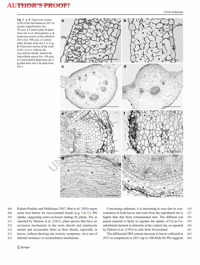
Fig. 3 a , b Transverse section(LM) of the leaf lamina in 2013 atgreater magnification (bar50 μm). a Control plant; b plantfrom site 4 (ch chloroplasts). c , dtransverse section of the mibrid in2013 (bar 500 μm). c Controlplant; d plant from site 4. e , f , g ,h Transverse section of the roots(LM); arrows indicate themycorrhizal sheath, asterisk theintercellular spaces (bar 100 μm).e Control plant f plant from site 2;g plant from site 4; h plant fromsite 6
J Soils Sediments
JrnlID 11368_ArtID 823_Proof# 1 - 04/12/2013

AUTHOR'S PROOF!
UNCORRECTEDPROOF
Fig. 4 Ultrastructure ofparenchyma cells (TEM); arrowsindicate the mitochondrial cristae(bar 1 μm); a Control plant; bplant from site 2; c plant from site4; d plant from site 6
Fig. 5 Ultrastructure ofparenchyma cells (TEM) showingthe restored internal membrane ofmitochondria. Same samples as inFig. 4. a bar 1 μm; b , c , d bar0.5 μm
J Soils Sediments
JrnlID 11368_ArtID 823_Proof# 1 - 04/12/2013
AUTHOR'S PROOF!
UNCORRECTEDPROOF
484 that metals are leached with different pathways, according to485 their available fraction in the soil. Metals with the highest486 concentrations in 2011 leaves (Pb, Zn) were decreased to a487 major extent in comparison to the less concentrated ones.488 Lead was the easiest to be removed, as indicated by its strong489 decrease in 2013 leaves. Despite their less abundant content,490 Cd and Cr presented a minor decrease, which suggests some491 exclusion strategy for these non-essential elements. Copper492 and iron concentrations were relatively high (Cu up to493 15 mg kg−1, Fe up to 636 mg kg−1) in 2013 leaves, with a494 less than fivefold decrease in comparison to 2011, in agree-495 ment with their essential/critical role as micronutrients, and do496 not determine any visual symptoms of phytotoxicity, suggest-497 ing a Cu–Fe tolerance (Abreu et al. 2008).
498 4.2 Plant morphology
499 In this study, any evident variation of the macromorphology500 was recorded in T. officinale by HM excess; indeed, all the501 plants examined (control and samples 2, 4, 6), even consider-502 ing the range of species variability, present similar morpholo-503 gy and dimensions. Consistently, any apparent toxicity symp-504 toms were visible in numerous accumulator plants, as reported505 by Memon et al. (2001). However, we observed a little differ-506 ence in leaves colour (a less intense green colour in specimens507 of contaminated sites in comparison to control). These state-508 ments are consistent with findings byQ2 Kupper et al. (1998),509 who noted that in water plants transition metals such as Cd,510 Cu, Ni, Pb, Zn may substitute for Mg in the chlorophyll511 molecule, thus reducing the photosynthetic function, which512 results in colour change (i.e. chloroplasts number decrease,513 and consequently the chlorophyll production). It is likely that514 analogous process would occur in our studied plants.515 A modified growth of leaves and roots, instead, was re-516 cently observed byAshraf et al. (2011) in several plants grown517 on mine tailings. Although the leaf morphology of518 T. officinale does not present particular differences among519 the specimens of various sites, at microscopic level morpho-520 logical and histological changes were observed. The actual521 thickness measured by microscopy (and which is not appre-522 ciable with the visual observation), is different in the observed523 samples (Fig. 2), and decreases with increasing HM content of524 plants. The different leaf thickness is consistent with the525 reduced, or even lacking, photosynthetic parenchyma normal-526 ly structured as palisade on the adaxial leaf surface, and527 spongy parenchyma on the abaxial surface, as observed in528 control plants.529 In the mibrid, the reduced number of vascular bundles530 (proportional to the contamination level), in comparison to531 control, have, as a consequence, less water availability for532 leaves, which certainly influences the plant metabolic activity.533 Vacuoles constitute the cell compartment where HM, taken534 up by active transport systems, are deposited; in vacuoles,
535plants, according to a metal-adaptive strategy, isolate HM,536thus inhibiting any interference with the plant metabolic reac-537tions ( Memon et al. 2001; Schutzendubel and Polle 2002).538At relatively low metal concentrations, metals isolated in539cell walls and vacuoles, are confined in roots, and therefore it540is unlike to relate HM contents in soil with those in leaves, as541reported by Rosselli et al. (2006). It is possible that plants542grown at site 2 (the less contaminated) deposited all HM in543cell walls and in vacuoles, since no macroscopic neither544cellular deformations were observed. With higher soil metal545contents, plants are able to translocate metals in the aerial546parts, particularly in leaves (Keunen et al. 2011). In our study,547plants from the most contaminated sites (samples 4, 6) pre-548sented HM contents in leaves related to those of the corre-549sponding soils (with the exception of Cu which probably has a550less effective transporter). The metal amount transported to551leaves provokes cell morphology modification, as shown in552figures above. Yet, as observed in samples 4 and 6, the cell553metal content is responsible for serious deformations of mito-554chondria, the organelles performing aerobic respiration.555Keunen et al. (2011) reported that PHEs provoke an increase556in reactive oxygen species (ROS) production, and a reduction557of mitochondria respiration functions. Similarly,558Karuppanapandian et al. (2011) found that ROS gener-559ated under stress conditions provoke cell damage in560various cellular compartments, including chloroplasts,561mitochondria, endoplasmic reticulum and plasma mem-562branes. Our morphological study ascertained mitochon-563dria cristae reduction in sample 6, and even lack in sample5644, confirming recent physiological observations by565Karuppanapandian et al. (2011), Keunen et al. (2011) and566Lopareva-Pohu et al. (2011).567Micromorphological studies on plant cells subjected to HM568stress are nearly lacking. In a recent paper, Zhao et al. (2011)569report the effects on the morphology and ultrastructure of570tomato plants subjected to lead-induced stress. Besides a571reduction in size of different parts (fruits, leaves, stems and572roots), thinner cell walls, swollen and deformed chloroplasts573are recorded at ultrastructural level. Reduction in thickness574and dimensions of leaf blade has been recorded also in soy-575bean by Weryszko-Chmielewska and Chwil (2005). More576recently, Giordani et al. (2012), in an experimental work on577metal nanoparticles (NP) application to tomato seed-578lings, found clear effects at both morphological and579genetic level (e.g. root hair formation, epidermal cells580outgrowth). In our study, no deformation was observed in581chloroplasts; however, in sample 6, and in sample 4 in partic-582ular, chloroplasts are smaller and numerically less than in the583control (Fig. 3a, b).584Considering the HM–plants relationships, and their adap-585tion to contaminated environment, it is important to remind586that in geophytes like Taraxacum complete detoxification587occurs at the end of the vegetative season, when plants loose
J Soils Sediments
JrnlID 11368_ArtID 823_Proof# 1 - 04/12/2013

AUTHOR'S PROOF!
UNCORRECTEDPROOF
588 leaves, which accumulate on the ground, and after winter589 dormancy, during the subsequent spring season, a new leaf590 generation may start again with metal deposition in vacuoles.591 It is worthy to note also that all the investigated samples,592 including the control plants, resulted to being mycorrhized.593 Several studies have dealt with a possible alleviation of metal594 toxicity by mycorrhization, but only few presented evi-595 dence of such effects (Peterson et al. 2004 and596 references therein). In our samples, the PHEs effect is597 certainly attenuated, since there is a hindered access of598 metals to the root surface, which suggests the metal-induced599 stress response to be significantly reduced, as stated also by600 Schutzendubel and Polle 2002.
601 5 Conclusions
602 – Soil analysis shows that PHEs are released from mine603 tailing because of low pH and low available water capac-604 ity; part of their bioavailable fraction is taken up by605 Taraxacum and transferred from roots to shoots, as it is606 common in indicator plants.607 – The study shows that there is a relationship between high608 metal content in Taraxacum plants and their modified609 morphology. PHEs do not determine an evident modifi-610 cation of the macromorphology. However, the leaf thick-611 ness decreases, and the absence of a regularly structured612 cellular organisation is likely related to the soil contami-613 nation degree.614 – It is likely that a 2-year leaching reduced partly the HM615 content of the soil, therefore decreasing their phytotoxic616 effect. A gradual restoration of leaf organisation suggests617 that, somewhat, resilience occurred in plants.618 – Since all samples, including control, present mycorrhized619 roots, it is suggested that stress-tolerant mycorrhizal fungi620 could contribute to reduce metal sorption. A comparison621 with non-mycorrhized plants could clarify the effective622 role of mycorrhizal fungi.623 – Data which is generated through this study could be624 helpful in detecting the lethal levels of heavy metals for625 particular plant species, their tolerance and remediation626 capacity.627 – The presence of heavy metals in dandelion has two con-628 sequences: a beneficial consequence, because the land629 affected by anthropogenic pollution may be restored630 by natural way; a fatal consequence, because both631 humans and animals feed dandelion, and, if plants are632 contaminated, through this way toxic metals enter the633 food chain.
634 Acknowledgments The authors wish to thank Corrado Tani, Pietro Di635 Falco and Flavia Visin for technical assistance.
636References 637
638Abreu MM, Tavares MT, Batista MJ (2008) Potential use of Erica639avandulensis and Erica australis in phytoremediation of sulphide640mine environments: Sao Domingos, Portugal. J Geochem Explor64196:210–222642Adriano DC, Chlopecka A, KaplandDI, Clijsters H, Vangrosvelt J (1995)643Soil contamination and remediation philosophy, science and tech-644nology. In: Prost R (ed) Contaminated soils. INRA, Paris, pp 466–645504646Alloway BJ (1995) Heavy metals in soils. Blackie, London, p 368647Ashraf M-L, Maah MJ, Yusoff I (2011) Heavy metals accumulation in648plants growing in ex tin mining catchment. Int J Environ Sci Tech6498(2):401–416650Baker AJM (1981) Accumulators and excluders strategies in the response651of plants to heavy metals. J Plant Nutr 3:643–654652Baker AMJ, Brooks RR (1989) Terrestrial higher plants which653hyperaccumulate metallic elements—a review of their distribution,654ecology and phytochemistry. Biorecovery 1:81–126655Baker A, Mc Grath S, Reeves R, Smith J (2000) Metal hyperaccumulator656plants: a review of the ecology and physiology of a biological657resource for phytoremediation of metal-polluted soils. In: Terry N,658Banuelos G (eds) Phytoremediation of contaminated soils. Lewis,659London, pp 85–107660Bargagli R (1993) Plant leaves and lichens as biomonitors of naturals or661anthropogenic emissions of mercury. In Markert B (ed) Plants as662Biomonitors. Weinheim WCH, pp 468-484663Bernard AM (1995) Effects of heavy metals in the environment on the664human health. In: Prost R (ed) Contaminated soils. INRA, Paris, pp66521–34666Bini C (2009) Soil restoration: remediation and valorisation of contami-667nated soils. In: Manual of Methods for Soil and Land Evaluation668(E.A.C. Costantini edit), Science, Enfield, 137–160. (ISBN 978-1-66957808-571-2)670Bini C (2010) From soil contamination to land restoration. In:671Contaminated soils: environmental impact, disposal and treatment672(R.V. Steinberg edit.), Nova, New York, 97-137 (ISBN 978-1-67360741-791-0)674Q3Bini C (2012) Environmental impact of abandoned mine waste: a review.675Nova, New York, p 92676Bini C, Casaril S, Pavoni B (2000) Fertility gain and heavy metal677accumulation in plants and soils. Toxicol Environ Chem 77:131–678142679Bini C, Maleci L, Romanin A (2008) The chromium issue in soils680of the leather tannery district in Italy. J Geochem Explor 96(2–3):681194–202682Bini C, Fontana S, Wahsha M (2010) Land contamination by mine683dumps, plant toxicity and restoration perspectives by684phytoremediation. Int J Environ Qual EQA 4:173–180, Tipografia685Fanti, Imola. ISBN 10: 88-901261-7-5686Bini C, Fontana S,WahshaM (2011) Environmental impact of PTEs (Cu,687Fe, Pb, Zn) from mixed sulphides mines in Italy. Proc. XI ICOBTE,688I, 505-506. Florence, July, 3–7, 2011689Bini C,WahshaM, Fontana S,Maleci L (2012) Effect of heavymetals on690morphological characteristics of Taraxacum officinale Web growing691on mine soils in NE Italy. J Geochem Explor 123:101–108692Costantini EAC (2007) Soil survey methods and information of soil data.693S.E.L.C.A, Firenze (in Italian)694D.L.—Legislation Act no 152/2006. Official Gazette n.88, 14/04/2006—695Supplement no 96 (in Italian)696Davies BE (1987) Consequences of environmental contamination by lead697mining in Wales. Hydrobiologia 149:213–220698Dubois JP, Okopnik F, Benitez N, Vedy JC (1998) Origin and spatial699variability of cadmium in some soils of the Swiss Jura. Proc. 16th700IUSS Congress, Montpellier. Symposia 25:1–8
J Soils Sediments
JrnlID 11368_ArtID 823_Proof# 1 - 04/12/2013

AUTHOR'S PROOF!
UNCORRECTEDPROOF
701 Ernst WHO (1996) Bioavailability of heavy metals and decontamination702 of soils by plants. Appl Geochem 11:163–167703 European Pharmacopoeia (2006)—VI ed., Council of Europe, Strasburg704 Fontana S, Wahsha M, Bini C (2010) Preliminary observations on heavy705 metal contamination in soils and plants of an abandoned mine in706 Imperina Valley (Italy). Agrochimica 4:218–231, LIV707 Fontana S, Bini C, Wahsha M, Bullo M (2011a) Heavy metals contam-708 ination in soils and their transfer to common wheat (Triticum709 aestivum L.): a case study. Geophys Res Abstr 13:1751710 Fontana S, Bini C, Wahsha M, Bullo M (2011b) PTEs in agroecosystems711 and implications for the food chain. Proc. XI ICOBTE, I, 507–508.712 Florence, July, 3–7, 2011713 Giordani T, Fabrizi A, Guidi L, Natali L, Giunti G, Ravasi F, Cavallini A,714 Pardossi A (2012) Response of tomato plants exposed to treatment715 with nano particles. Int J Environ Qual 8:27–38716 Giuliani C, Pellegrino F, Tirillini B,Maleci L (2008)Micromorphological717 and chemical characterization of Stachys recta subsp serpentini718 (Fiori) Arrigoni in comparison to S. recta subsp. recta719 (Lamiaceae). Flora 203:376–385720 Helios-Rybicka E (1996) Impact of mining and metallurgical industries721 on the environment in Poland. Appl Geochem 11(1–2):3–11722 Hood TM, Jones BJ Jr (1997) Soil and plant analysis in sustainable723 agriculture and environment. Marcel Dekker, New York, p 877724 Kabata-Pendias A (2011) Trace elements in soils and plants, 3rd edn.725 CRC, Boca Raton, p 365726 Kabata-Pendias A, Mukherjee AB (2007) Trace elements from soil to727 human. Springer, Berlin, p 550728 Karuppanapandian T, Moon J, Kim C, Manoharan K, Kim W (2011)729 Reactive oxygen species in plants: their generation, signal transduc-730 tion, and scavenging mechanisms. Aust J Crop Sci 5(6):709–725731 Keane B, Collier MH, Shann JR, Rogstad SH (2001) Metal content of732 dandelion (Taraxacum officinale) leaves in relation to soil contam-733 ination and airborne particulate matter. Sci Total Environ 281:63–78734 Keunen E, Remans T, Bohler S, Vangronsveld J, Cuypers A (2011)735 Metal-induced oxidative stress and plant mitochondria. Int J Mol736 Sci 12:6894–6918737 Kidd P, Barcelo J, Bernal MP, Navari Izzo F, Poschenrieder C, Shilev S,738 Clemente R, Monterroso C (2009) Trace element behaviour at the739 root-soil interface: implications in phytoremediation. Environ Exp740 Bot 67:243–259741 Królak E (2003) Accumulation of Zn, Cu, Pb and Cd by dandelion742 (Taraxacum officinale Web.) in environments with various degrees743 of metallic contamination. Pol J Environ Stud 12(6):713–721744 Kucera T, Horáková H, Šonská A (2008) Toxic metal ions in photoauto-745 trophic organisms. Photosynthetica 46:481–489746 Lee CG, Chon H, Jung MC (2001) Heavy metal contamination in the747 vicinity of the Daduk Au–Ag–Pb–Zn mine in Korea. Appl748 Geochem 16:1377–1386749 Li WJ, Khan MA, Yamaguchi S, Kamiya Y (2005) Effects of heavy750 metals on seed germination and early seedling growth of751 Arabidopsis thaliana. Plant Growth Regul 46:45–50752 Llugany M, Lombini A, Dinelli E, Poschenrieder C, Barcelo J (2009)753 Transfer of selected mineral nutrients and trace elements in the host–754 hemiparasite association, Cistus–Odontides lutea , growing on and755 off metal-polluted sites. Plant Biol 11:170–178756 Lopareva-PohuA,Verdin A, GarçonG, Sahraoui AL, Pourrut B, Debiane757 D, Waterlot C, Laruelle F, Bidar G, Douay F, Shirali P (2011)758 Influence of fly ash aided phytostabilisation of Pb, Cd and Zn highly759 contaminated soils on Lolium perenne and Trifolium repens metal760 transfer and physiological stress. Environ Pollut 159:1721–1729761 Lösch R (2004) Plant mitochondrial respiration under the influence of762 heavy metals. In: Prasad (ed) Heavy metal stress in plants. From763 biomolecules to ecosystems, 2nd edn. Springer, Berlin, Germany, pp764 182–200765 Madejon P, Murillo JM, Marañon T, Cabrera F, Lopez R (2002)766 Bioaccumulation of As, Cd, Cu, Fe and Pb in wild grasses affected
767by the Aznalcollar mine spill (SW Spain). Sci Total Environ 290:768105–120769Malawska M, Wilkomirski B (2001) An analysis of soil and plant770(Taraxacum officinale) contamination with heavy metals and poly-771cyclic aromatic hydrocarbons (PAHs) in the area of the railway772junction Ilawa Glòwna, Poland. Water Air Soil Pollut 127:339–349773Maleci L, Bini C, Paolillo A (2001) Chromium (III) uptake byCalendula774arvensis L. and related phytotoxicity. Proc. VI ICOBTE, Guelph,775On., 384 (abstract)776Malik RN, Husain SZ, Nazir I (2010) Heavy metal contamination and777accumulation in soil and wild plant species from industrial area of778Islamabad, Pakistan. J Bot 42(1):291–301779Mangabeira P, Almeida AA,MielkeM, Gomes FP,Mushrifah I, Escaig F,780Laffray D, Severo MI, Oliveira AH, Galle P (2001) Ultrastructural781investigations and electron probe X-ray microanalysis of chromium-782treated plants. Proc. VI ICOBTE, Guelph, 555 (abstract)783Margesin R, Schinner F (2005)Manual for soil analysis—monitoring and784assessing soil bioremediation, 1st edn. Springer, Berlin, Germany, p785359786Memon AR, Aktoprakligul D, Zdemur A, Vertii A (2001) Heavy metal787accumulation and detoxification mechanisms in plants. Turk J Bot78825:111–121789Navarro MC, Pérez-Sirvent C, Martínez-Sánchez MJ, Vidal J, Tovar PJ,790Bech J (2008) Abandoned mine sites as a source of contamination791by heavy metals. A case study in a semi-arid zone. J Geochem792Explor 96:183–193793Peterson RL, Massicotte HB, Melville LH (2004) Mycorrhizas: anatomy794and cell biology. NRC Research, Ottawa795Poschenrieder C, Bech J, Llugany M, Pace A, Fenes E, Barcelo J (2001)796Copper in plant species in a copper gradient in Catalonia (North East797Spain) and their potential for phytoremediation. Plant Soil 230:247–798256799Preeti P, Tripathi AK (2011) Effect of heavy metals onmorphological and800biochemical characteristics of Albizia procera (Roxb.) Benth. seed-801lings. Int J Environ Sci 1(5):1009802Rosselli W, Rossi M, Sasu I (2006) Cd, Cu and Zn contents in the leaves803of Taraxacum officinale . Snow Landsc Res 80(3):361–366804Sarret G, Vangronsveld J, Roux M, Coves J, Manceau A (2001)805Bioaccumulation of metal in plants and microorganisms studied by806electron microscopy and EXAFS spectroscopy. Proc. VI ICOBTE,807Guelph, 131 (abstract)808Savinov AB, Kurganova LN, Shekunov YI (2007) Lipid peroxidation809rates in Taraxacum officinale Wigg. and Vicia cracca L. from810biotopes with different levels of soil pollution with heavy metals.811Russ J Ecol 38(3):174–180812Schutzendubel A, Polle A (2002) Plant responses to abiotic stresses:813heavy metal-induced oxidative stress and protection by814mycorrhization. J Exp Bot 53(372):1351–1365815Simon L, Martin HW, Adriano DC (1996) Chicory (Cichorium intybus816L.) and dandelion (Taraxacum officinale Web.) as phytoindicators817of cadmium contamination. Water Air Soil Pollut 91(3–4):351–362818Sivri Y, Munoz M, Sappin-Didier V, Riotte J, Denaix L, de Parceval P,819Destrigneville C, Dupré B (2010) Multimetallic contamination from820Zn-ore smelter: solid speciation and potential mobility in riverine821floodbank soils of the upper Lot River (SW France). Eur J Mineral82222:679–691823Steinnes E (2009) Soils and geomedicine. Environ Geochem Health 31:824523–535825Unterbrunner R, Puschenreiter M, Sommer P, Wieshammer G, Tlustos P,826Zupan M, Wenzel WW (2007) Heavy metal accumulation in trees827growing on contaminated sites in Central Europe. Environ Pollut828148:107–114829Wahsha M, Bini C, Fontana S, Zilioli D, Wahsha A (2012a) Toxicity830assessment of contaminated soils from a mining area in northeast831Italy by using lipid peroxidation assay. J Geochem Explor 113:112–832117
J Soils Sediments
JrnlID 11368_ArtID 823_Proof# 1 - 04/12/2013
AUTHOR'S PROOF!
UNCORRECTEDPROOF
833 Wahsha M, Bini C, Argese E, Minello F, Fontana S, Wahsheh H (2012b)834 Heavy metals accumulation in willows growing on Spolic835 Technosols from the abandoned Imperina Valley mine in Italy. J836 Geochem Explor 123:19–24837 Weryszko-Chmielewska E, Chwil M (2005) Lead-induced histological838 and ultrastructural changes in the leaves of soybean (Glycine max839 (L.) Merr.). Soil Sci Plant Nutr 51(2):203–212840 Yoon J, Cao X, Zhou Q, Ma LQ (2006) Accumulation of Pb, Cu, and Zn841 in native plants growing on a contaminated Florida site. Sci Total842 Environ 368:456–464
843Zhao S, Ye X, Zheng J (2011) Lead-induced changes in plant morphol-844ogy, cell ultrastructure, growth and yields of tomato. Afr J845Biotechnol 10(50):10116–10124846Zupan M, Hudnik V, Lobnik F, Kadunc V (1995) Accumulation of Pb,847Cd, Zn from contaminated soil to various plants and evaluation of848soil remediation with indicator plant (Plantago lanceolata). In:849Prost R (ed) Contaminated soils. INRA, Paris, pp 325–335850Zupan M, Kralj T, Grcman H, Hudnik V, Lobnik F (2003) The accumu-851lation of Cd, Zn, Pb in Taraxacum officinale and Plantago852lanceolata from contaminated soils. Proc VII ICOBTE, Uppsala Sv.
853
J Soils Sediments
JrnlID 11368_ArtID 823_Proof# 1 - 04/12/2013

AUTHOR'S PROOF!
UNCORRECTEDPROOF
AUTHOR QUERIES
AUTHOR PLEASE ANSWER ALL QUERIES.
Q1. Figure 1 contains poor quality of text. Please provide replacement. Otherwise, please advise ifokay to proceed with the figure as is.
Q2. Kupper et al. (1998) was cited in the text but was not found in the reference list. Please provide thenecessary information.
Q3. Bini (2012) was not cited anywhere in the text. Please provide a citation. Alternatively, delete theitem from the list.