What Lies Beneath A dissertation submit - eScholarship
258
UNIVERSITY OF CALIFORNIA SAN DIEGO Incorporating Germline Variants into Cancer Analyses: What Lies Beneath A dissertation submitted in partial satisfaction of the requirements for the degree Doctor of Philosophy in Biomedical Sciences by Alexandra Ray Buckley Committee in charge: Professor Nicholas J. Schork, Chair Professor Arshad Desai, Co-Chair Professor Vineet Bafna Professor Hannah Carter Professor Olivier Harismendy Professor Elizabeth Winzeler 2018
-
Upload
khangminh22 -
Category
Documents
-
view
2 -
download
0
Transcript of What Lies Beneath A dissertation submit - eScholarship

UNIVERSITY OF CALIFORNIA SAN DIEGO
Incorporating Germline Variants into Cancer Analyses:What Lies Beneath
A dissertation submitted in partial satisfaction of therequirements for the degree
Doctor of Philosophy
in
Biomedical Sciences
by
Alexandra Ray Buckley
Committee in charge:
Professor Nicholas J. Schork, ChairProfessor Arshad Desai, Co-ChairProfessor Vineet BafnaProfessor Hannah CarterProfessor Olivier HarismendyProfessor Elizabeth Winzeler
2018

Copyright
Alexandra Ray Buckley, 2018
All rights reserved.

The dissertation of Alexandra Ray Buckley is approved,
and it is acceptable in quality and form for publication
on microfilm and electronically:
Co-Chair
Chair
University of California San Diego
2018
iii

DEDICATION
To my family, for listening to me endlessly complain
To my friends, for the laughs, food, and beer
iv

EPIGRAPH
And the haters gonna hate, hate, hate, hate, hate
Baby, I’m just gonna shake, shake, shake, shake, shake
I shake it off
Shake it off
v

TABLE OF CONTENTS
Signature Page . . . . . . . . . . . . . . . . . . . . . . . . . . . . . . . . . . . . . . iii
Dedication . . . . . . . . . . . . . . . . . . . . . . . . . . . . . . . . . . . . . . . . . iv
Epigraph . . . . . . . . . . . . . . . . . . . . . . . . . . . . . . . . . . . . . . . . . v
Table of Contents . . . . . . . . . . . . . . . . . . . . . . . . . . . . . . . . . . . . . vi
List of Figures . . . . . . . . . . . . . . . . . . . . . . . . . . . . . . . . . . . . . . ix
List of Tables . . . . . . . . . . . . . . . . . . . . . . . . . . . . . . . . . . . . . . . xi
Acknowledgements . . . . . . . . . . . . . . . . . . . . . . . . . . . . . . . . . . . . xiii
Vita . . . . . . . . . . . . . . . . . . . . . . . . . . . . . . . . . . . . . . . . . . . . xvi
Abstract of the Dissertation . . . . . . . . . . . . . . . . . . . . . . . . . . . . . . . xvii
Chapter 1 The Value of Germline Variation in Cancer Research . . . . . . . . . 11.1 Background . . . . . . . . . . . . . . . . . . . . . . . . . . . . . 1
1.1.1 Germline Predisposition to Cancer . . . . . . . . . . . . 41.1.2 Heritability of Somatic Phenotypes . . . . . . . . . . . . 71.1.3 Heritability of DNA Damage Response . . . . . . . . . . 81.1.4 Somatic Molecular Phenotypes . . . . . . . . . . . . . . . 91.1.5 Tumor Immune Phenotypes, Cell Composition, and Metas-
tasis . . . . . . . . . . . . . . . . . . . . . . . . . . . . . 151.1.6 Other Reasons to Consider Germline Variants in Cancer
Studies . . . . . . . . . . . . . . . . . . . . . . . . . . . . 171.2 Overview and Organization of Dissertation . . . . . . . . . . . . 201.3 Acknowledgements . . . . . . . . . . . . . . . . . . . . . . . . . 22
Chapter 2 Pan-Cancer Analysis Reveals Technical Artifacts in TCGA GermlineVariant Calls . . . . . . . . . . . . . . . . . . . . . . . . . . . . . . . 232.1 Abstract . . . . . . . . . . . . . . . . . . . . . . . . . . . . . . . 232.2 Background . . . . . . . . . . . . . . . . . . . . . . . . . . . . . 242.3 Methods . . . . . . . . . . . . . . . . . . . . . . . . . . . . . . . 27
2.3.1 Cohort . . . . . . . . . . . . . . . . . . . . . . . . . . . . 272.3.2 Germline Variant Calling . . . . . . . . . . . . . . . . . . 272.3.3 PCA and Self-Report Ancestry Validation . . . . . . . . 282.3.4 Annotation and BAM metrics . . . . . . . . . . . . . . . 292.3.5 Realignment Comparison . . . . . . . . . . . . . . . . . . 292.3.6 WGA Enriched Indels . . . . . . . . . . . . . . . . . . . 30
vi

2.3.7 Homopolymer Indel Analyses . . . . . . . . . . . . . . . 312.3.8 Chimera Read Analysis . . . . . . . . . . . . . . . . . . . 312.3.9 Repeated Samples . . . . . . . . . . . . . . . . . . . . . . 322.3.10 Indel Filter Methods . . . . . . . . . . . . . . . . . . . . 322.3.11 Statistical Methods . . . . . . . . . . . . . . . . . . . . . 33
2.4 Results . . . . . . . . . . . . . . . . . . . . . . . . . . . . . . . . 342.4.1 Technical Heterogeneity in TCGA WXS Data Generation 342.4.2 Impact of Technical Heterogeneity on Loss of Function
Variants . . . . . . . . . . . . . . . . . . . . . . . . . . . 372.4.3 Characterizing WGA Artifacts . . . . . . . . . . . . . . . 422.4.4 Filtering Artifactual LOF Variant Calls . . . . . . . . . . 482.4.5 Consequences of Technical Artifacts on Genetic Associa-
tions . . . . . . . . . . . . . . . . . . . . . . . . . . . . . 502.5 Discussion . . . . . . . . . . . . . . . . . . . . . . . . . . . . . . 532.6 Acknowledgements . . . . . . . . . . . . . . . . . . . . . . . . . 57
Chapter 3 Exome-Wide Analysis of Bi-allelic Alterations Identifies a Lynch Phe-notype in the Cancer Genome Atlas . . . . . . . . . . . . . . . . . . . 583.1 Abstract . . . . . . . . . . . . . . . . . . . . . . . . . . . . . . . 583.2 Background . . . . . . . . . . . . . . . . . . . . . . . . . . . . . 593.3 Methods . . . . . . . . . . . . . . . . . . . . . . . . . . . . . . . 61
3.3.1 Data Acquisition . . . . . . . . . . . . . . . . . . . . . . 613.3.2 Variant Annotation and Filtering . . . . . . . . . . . . . 623.3.3 Somatic Methylation . . . . . . . . . . . . . . . . . . . . 623.3.4 Loss of Heterozygosity . . . . . . . . . . . . . . . . . . . 633.3.5 Gene Set Enrichment Analysis . . . . . . . . . . . . . . . 633.3.6 Mutational Signature Analysis . . . . . . . . . . . . . . . 643.3.7 Statistical Analyses . . . . . . . . . . . . . . . . . . . . . 64
3.4 Results . . . . . . . . . . . . . . . . . . . . . . . . . . . . . . . . 663.4.1 MMR Pathway is Frequently Affected by Bi-allelic Alter-
ation . . . . . . . . . . . . . . . . . . . . . . . . . . . . . 663.4.2 Landscape of Germline and Somatic Alteration of DNA
Damage Repair Pathways . . . . . . . . . . . . . . . . . 673.4.3 Individuals in TCGA Exhibit Lynch Syndrome Charac-
teristics . . . . . . . . . . . . . . . . . . . . . . . . . . . 693.4.4 Using MSI-H to Reclassify Variants of Unknown Significance 733.4.5 Missense Alterations Exhibit an Attenuated Lynch Phe-
notype . . . . . . . . . . . . . . . . . . . . . . . . . . . . 773.4.6 Mono-allelic Germline Alteration has Little Effect on So-
matic MSI . . . . . . . . . . . . . . . . . . . . . . . . . . 793.4.7 Methylation of SHPRH Associated with Somatic MSI . . 793.4.8 Mono-allelic Germline Alterations not Associated with
Mutational Signatures . . . . . . . . . . . . . . . . . . . 82
vii

3.4.9 Cancer Predisposition Syndromes in TCGA . . . . . . . 833.5 Discussion . . . . . . . . . . . . . . . . . . . . . . . . . . . . . . 863.6 Acknowledgements . . . . . . . . . . . . . . . . . . . . . . . . . 90
Chapter 4 Rare Variant Phasing Using Paired Tumor:Normal Sequence Data . . 924.1 Abstract . . . . . . . . . . . . . . . . . . . . . . . . . . . . . . . 924.2 Background . . . . . . . . . . . . . . . . . . . . . . . . . . . . . 934.3 Methods . . . . . . . . . . . . . . . . . . . . . . . . . . . . . . . 95
4.3.1 Data Acquisition . . . . . . . . . . . . . . . . . . . . . . 954.3.2 Variant Annotation and Filtering . . . . . . . . . . . . . 964.3.3 Implementation of VAF Phasing . . . . . . . . . . . . . . 964.3.4 Comparison To Other Phasing Methods . . . . . . . . . . 974.3.5 HMMvar Annotation and Compound Heterozygosity Anal-
ysis . . . . . . . . . . . . . . . . . . . . . . . . . . . . . . 984.3.6 Statistical Analyses . . . . . . . . . . . . . . . . . . . . . 99
4.4 Results . . . . . . . . . . . . . . . . . . . . . . . . . . . . . . . . 994.4.1 Phasing with Variant Allele Frequency . . . . . . . . . . 994.4.2 VAF Phasing is Concordant with Other Methods . . . . 1064.4.3 Application of VAF Phasing to Cancer Predisposition . . 108
4.5 Discussion . . . . . . . . . . . . . . . . . . . . . . . . . . . . . . 1114.6 Acknowledgements . . . . . . . . . . . . . . . . . . . . . . . . . 114
Chapter 5 The Upshot . . . . . . . . . . . . . . . . . . . . . . . . . . . . . . . . 1165.1 Discussion . . . . . . . . . . . . . . . . . . . . . . . . . . . . . . 116
5.1.1 Batch Effects in Public Datasets . . . . . . . . . . . . . . 1165.1.2 Germline Variants and Tumor Phenotypes in TCGA . . . 1175.1.3 Germline Variant Phasing in Cancer Samples . . . . . . . 1195.1.4 Tumors are Like Onions . . . . . . . . . . . . . . . . . . 1205.1.5 Germline Variation, What is it Good For? . . . . . . . . 122
5.2 Future Directions . . . . . . . . . . . . . . . . . . . . . . . . . . 1235.2.1 The Upshot . . . . . . . . . . . . . . . . . . . . . . . . . 127
Appendix A Supplemental Material: Pan-Cancer Analysis Reveals Technical Arti-facts in TCGA Germline Variant Calls . . . . . . . . . . . . . . . . . 128
Appendix B Supplemental Material: Exome-Wide Analysis of Bi-allelic AlterationsIdentifies a Lynch Phenotype in the Cancer Genome Atlas . . . . . . 150
Appendix C Supplemental Material: Rare Variant Phasing Using Paired Tumor:NormalSequence Data . . . . . . . . . . . . . . . . . . . . . . . . . . . . . . . 176
Bibliography . . . . . . . . . . . . . . . . . . . . . . . . . . . . . . . . . . . . . . . 201
viii

LIST OF FIGURES
Figure 1.1: The Environment Shapes Growth . . . . . . . . . . . . . . . . . . . . . 3
Figure 2.1: Technical Covariates in TCGA WXS Samples . . . . . . . . . . . . . . 35Figure 2.2: WGA Increases LOF Indel Burden . . . . . . . . . . . . . . . . . . . . 40Figure 2.3: Characteristics of Variants in WGA Samples . . . . . . . . . . . . . . . 43Figure 2.4: Comparison of Indel Filters . . . . . . . . . . . . . . . . . . . . . . . . 49Figure 2.5: Association Between LOF Burden and Cancer Type . . . . . . . . . . 52
Figure 3.1: Frequency of Germline and Somatic Alterations in Cancer-RelevantPathways . . . . . . . . . . . . . . . . . . . . . . . . . . . . . . . . . . 68
Figure 3.2: Genetic and Clinical Characteristics of MSI-H Individuals . . . . . . . 71Figure 3.3: Identification of Potential Pathogenic Lynch Syndrome Variants . . . . 75Figure 3.4: Germline, Somatic, and Epigenetic Associations with MSI . . . . . . . 80Figure 3.5: Cancer Predisposition Syndromes in TCGA . . . . . . . . . . . . . . . 84
Figure 4.1: Overview of VAF Phasing Method . . . . . . . . . . . . . . . . . . . . 101Figure 4.2: Using Duplicated Normal Samples to Identify SCNAs . . . . . . . . . . 105Figure 4.3: Comparison of Phasing Methods . . . . . . . . . . . . . . . . . . . . . 107Figure 4.4: Leveraging Phase to Identify Cancer Predisposing Germline Variation . 111
Figure 5.1: Tumors are Like Onions . . . . . . . . . . . . . . . . . . . . . . . . . . 121
Figure A.1: Technical Covariates of Cohort . . . . . . . . . . . . . . . . . . . . . . 129Figure A.2: Number of Processing Workflows . . . . . . . . . . . . . . . . . . . . . 130Figure A.3: Variant Call Discordance Between NewAlign and OldAlign . . . . . . . 131Figure A.4: Discordance With BAM Realignment . . . . . . . . . . . . . . . . . . . 132Figure A.5: PCA of Common Variants . . . . . . . . . . . . . . . . . . . . . . . . . 133Figure A.6: LOF SNV and Indel Burden . . . . . . . . . . . . . . . . . . . . . . . . 134Figure A.7: LOF Indel Burden in NewAlign Cohort . . . . . . . . . . . . . . . . . 135Figure A.8: Coverage and Read Depth in WGA Samples . . . . . . . . . . . . . . . 135Figure A.9: Frequently Inserted and Deleted Bases of WGA Indels . . . . . . . . . 136Figure A.10:Discordance Between Repeated WXS Samples . . . . . . . . . . . . . . 137Figure A.11: Proposed Mechanism of Artifactual Indel Generation . . . . . . . . . . 138Figure A.12:Distribution of Indel Sequence BLAST Hits . . . . . . . . . . . . . . . 139Figure A.13: Individual LOF Indel Burden Across Filtering Methods . . . . . . . . . 140Figure A.14: LOF Indel Burden in WGA Samples Across Filtering Methods . . . . . 141Figure A.15:G/C Homopolymer Content of Genes Shared Between OV and LAML 142Figure A.16: LOF SNV Logistic Regression Analysis . . . . . . . . . . . . . . . . . . 143
Figure B.1: Calling Somatic Methylation Status . . . . . . . . . . . . . . . . . . . 151Figure B.2: Example LOH Events . . . . . . . . . . . . . . . . . . . . . . . . . . . 152Figure B.3: Genes Frequently Affected by Germline:Somatic Alteration . . . . . . . 153
ix

Figure B.4: Association Between Germline LOF Burden and Cancer Type . . . . . 154Figure B.5: Germline and Somatic LOF in PMS2 . . . . . . . . . . . . . . . . . . . 155Figure B.6: Mutational Signature Analysis of Germline:Somatic MMR Alteration . 156Figure B.7: Mono-allelic Germline MMR Variation Not Associated With MSI . . . 157Figure B.8: Association Testing Between Genomic Alteration and MSI Burden . . 158Figure B.9: SHPRH Methylation in Uterine Cancer . . . . . . . . . . . . . . . . . 159Figure B.10: SHPRH Expression in Normal Tissues . . . . . . . . . . . . . . . . . . 160Figure B.11: Mutational Signature Analysis of MLH1 and SHPRH Methylation . . 161Figure B.12: Co-Occurrence Testing for SHPRH Methylation . . . . . . . . . . . . . 162Figure B.13: Mutational Signature Analysis of BRCA1/2 Carriers . . . . . . . . . . 163Figure B.14: Mutational Signature Analysis of DDR Pathway Alteration . . . . . . 164Figure B.15: Association Between Age and Damaging Germline Variants . . . . . . 165
Figure C.1: Schematic of ∆ VAF Changes in Cancer . . . . . . . . . . . . . . . . . 177Figure C.2: Example ∆ VAF Data . . . . . . . . . . . . . . . . . . . . . . . . . . . 178Figure C.3: Example VCF-CBS Data . . . . . . . . . . . . . . . . . . . . . . . . . 179Figure C.4: VAF-CBS Segment Metrics . . . . . . . . . . . . . . . . . . . . . . . . 180Figure C.5: ∆ VAF Data From a Contaminated Sample . . . . . . . . . . . . . . . 181Figure C.6: Discordance Between Phasing Methods . . . . . . . . . . . . . . . . . . 182Figure C.7: Sample Metrics that Affect ∆ VAF Phasing . . . . . . . . . . . . . . . 183Figure C.8: Phasing Performance on Rare Variants . . . . . . . . . . . . . . . . . . 184Figure C.9: Fraction of Phased Variants Visualized by Chromosome . . . . . . . . 185Figure C.10: Comparison Between TCGA and VAF-CBS SCNAs . . . . . . . . . . . 186Figure C.11:Method Used to Calculate Pairwise Error . . . . . . . . . . . . . . . . 187Figure C.12: Discordance Between VAF and 10X Genomics Phasing . . . . . . . . . 188Figure C.13: Fraction of Compound Heterozygosity Events Phased . . . . . . . . . . 189Figure C.14: Association Between Age and Damaging Germline Variants . . . . . . 190Figure C.15: Association Between Age and BRCA1/2 Germline Variants . . . . . . 191Figure C.16: Assumptions of VAF Phasing Model . . . . . . . . . . . . . . . . . . . 192
x

LIST OF TABLES
Table 2.1: ANOVA of LOF Variant Burden . . . . . . . . . . . . . . . . . . . . . . 41Table 2.2: Characteristics of WGA Indels . . . . . . . . . . . . . . . . . . . . . . . 47Table 2.3: Variant Filter Metrics . . . . . . . . . . . . . . . . . . . . . . . . . . . . 50
Table 3.1: Bi-allelic Germline:Somatic MMR Alteration . . . . . . . . . . . . . . . 78
Table A.1: Composition of the Pan-Cancer Cohort . . . . . . . . . . . . . . . . . . 144Table A.2: Coverage of the Six TCGA Capture Kits . . . . . . . . . . . . . . . . . 145Table A.3: K-means Cluster Membership of HapMap Samples . . . . . . . . . . . . 145Table A.4: K-means Cluster Membership of TCGA Samples . . . . . . . . . . . . . 146Table A.5: GC Content of the Sequence Surrounding WGA Indels . . . . . . . . . 146Table A.6: Allele Frequency of Homopolymer Indels . . . . . . . . . . . . . . . . . 146Table A.7: Frequency of BLAST Match for WGA Indels . . . . . . . . . . . . . . . 147Table A.8: ANOVA of LOF Indel Burden Using Different Filters . . . . . . . . . . 148Table A.9: Correlation Between LOF Indels and Homopolymer Tracts . . . . . . . 149
Table B.1: Association Between MSI and MMR Alteration . . . . . . . . . . . . . 165Table B.2: Association Between Age and MMR Alteration . . . . . . . . . . . . . . 166Table B.3: Germline Variants Pathogenic for Lynch Syndrome . . . . . . . . . . . . 166Table B.4: Germline Variants of Unknown Significance for Lynch Syndrome . . . . 167Table B.5: Modeling a Germline:Somatic Interaction for L-MMR Genes . . . . . . 169Table B.6: Association Between MSI and MMR Germline:Somatic Alteration . . . 170Table B.7: Association Between Age and MMR Germline:Somatic Altertion . . . . 170Table B.8: Association Between MSI and Mono-allelic Germline MMR Variants . . 171Table B.9: Association Between MSI and MMR Alteration Including Confounders . 172Table B.10: Association Between MSI and Alterations CorrelatedWith SHPRH Methy-
lation . . . . . . . . . . . . . . . . . . . . . . . . . . . . . . . . . . . . . 174Table B.11: Association Between MSI and SHPRH Expression . . . . . . . . . . . . 174Table B.12: Association Between Age and Known Predisposing Germline Variants . 175Table B.13: Association Between Age and Predicted Predisposing Germline Variants 175
Table C.1: Possible Contaminated Normal Tissue Samples . . . . . . . . . . . . . . 193Table C.2: Discordance Between VAF Phasing and Other Methods . . . . . . . . . 194Table C.3: Factors That Influence VAF Phasing . . . . . . . . . . . . . . . . . . . 195Table C.4: Discordance Between VAF Phasing with TCGA SCNA and Other Methods195Table C.5: Discordance Between VAF and 10X Genomics Phasing . . . . . . . . . 196Table C.6: Features of VAF Phasing Errors . . . . . . . . . . . . . . . . . . . . . . 196Table C.7: Association Between Age and Compound Heterozygosity . . . . . . . . 197Table C.8: Association Between Age and Non-Compensatory Variants . . . . . . . 197Table C.9: Association Between Age and Non-Compensatory Variants, ClinVar Sam-
ples Removed . . . . . . . . . . . . . . . . . . . . . . . . . . . . . . . . 197Table C.10: Association Between Age and Non-Compensatory Variants in BRCA1/2 198
xi

Table C.11: Germline Non-Compensatory Variants in BRCA1/2 . . . . . . . . . . . 199
xii

ACKNOWLEDGEMENTS
They say it takes a villiage to raise a child, and I feel it took a village to get me
through my PhD. First and foremost I’d like to thank my advisor, Nik Schork. He took a
chance on me as a runaway grad student and gave me the opportunity and the guidance
I needed to metamorphosize from a biologist to a data scientist. My labmates from the
Schork lab, for endless help during the aforementioned metamorphosis. Particularly Kris
Standish for teaching me everything I know about GATK, R, and baseball and Danjuma
Quarless for his perpetually spot-on life advice. Arshad Desai and Tracy Handel, for
believing in me when it felt like no one else did. Olivier Harismendy and Hannah Carter
for being excellent committee members, collaborators, and mentors. Barry Demchak, for
being the best sys admin a grad student could ask for. Rebecca Boumil and Wayne Frankel,
for ignitng my interest in research and helping me get into grad school. The BMS program
and staff for creating such a welcoming and supportive home for me in San Diego. Finally,
the two MacBooks that gave their lives for the making of this thesis. Their sacrifice will
not be forgotten.
Chapter 1 is being prepared for publication under the title, "Germline Variation
in Cancer, What is it Good For?". Authors included Alexandra R. Buckley and Nicholas
J. Schork. AB and NJS wrote manuscript. The dissertation author was the primary
researcher and author on this manuscript.
Chapter 2 was previously published in BMC Genomics in June 2017 under the
title, "Pan-cancer analysis reveals technical artifacts in TCGA germline variant calls".
Authors included Alexandra R. Buckley, Kristopher A. Standish, Kunal Bhutani, Trey
Ideker, Roger S. Lasken, Hannah Carter, Olivier Harismendy, and Nicholas J. Schork.
NJS designed and supervised the research. ARB executed pipelines, analyzed data, and
drafted the manuscript. KAS helped assemble pipelines and assisted with statistical anal-
xiii

ysis. ARB, KAS, and RSL designed figures. ARB, KAS, KB, RSL, OH, and HC designed
experiments. NJS, OH, and HC helped write the manuscript. TI set up high performance
computing infrastructure. All authors read and approved the final manuscript. The dis-
sertation author was the primary researcher and author on this manuscript.
Chapter 3 is under review for publication in BMC Genome Medicine under the
title, "Exome-Wide Analysis of Bi-allelic Alterations Identifies a Lynch Phenotype in the
Cancer Genome Atlas". Authors included Alexandra R. Buckley, Trey Ideker, Hannah
Carter, Olivier Harismendy, and Nicholas J. Schork. NJS designed and supervised the re-
search. NJS designed and supervised the research. ARB performed the statistical analysis,
prepared the figures and tables, and drafted the manuscript. ARB, OH, and HC designed
experiments. NJS, OH, and HC assisted writing the manuscript. TI set up high perfor-
mance computing infrastructure. All authors read and approved the final manuscript. We
would like to thank Bethany Buckley for her assistance in obtaining ClinVar annotations
and interpreting germline variants, and Barry Demchak for his assistance with managing
data and setting up analysis pipelines on NRNB. The dissertation author was the primary
researcher and author on this manuscript.
Chapter 4 is being prepared for publication under the title, "Rare Variant Phas-
ing Using Paired Tumor:Normal Sequence Data". Authors included Alexandra R. Buck-
ley, Trey Ideker, Hannah Carter, Jonathan Keats, and Nicholas J. Schork. NJS de-
signed and supervised the research. ARB executed pipelines, analyzed data, and drafted
the manuscript. ARB and HC designed experiments. NJS and HC helped write the
manuscript. TI set up high performance computing infrastructure. All authors read and
approved the final manuscript. The dissertation author was the primary researcher and
author on this manuscript.
This work was funded in part by the National Institute Of General Medical Sciences
xiv

of the National Institutes of Health under Award Number T32GM008666, the Translational
Genomics Research Institute (TGen), and a gift from the San Diego Cancer Research
Institute. NJS and his lab are also supported in part by National Institutes of Health
Grants UL1TR001442 (CTSA), U24AG051129, U19G023122, as well as a contract from
the Allen Institute for Brain Science. All computing was done using the National Resource
for Network Biology (NRNB) P41 GM103504. All primary data were accessed from The
Cancer Genome Atlas Research Network (cancergenome.nih.gov).
xv

VITA
2011 - B.S. Bachelor of Science, NeuroscienceUniversity of Scranton, Scranton, PA
2018 - Ph.D. Doctor of Philosophy, Biomedical SciencesBiomedical Sciences Graduate ProgramUniversity of California San Diego
PUBLICATIONS
Asinof SK, Sukoff Rizzo SJ, Buckley AR, Beyer BJ, Letts VA, Frankel WN, BoumilRM.Independent Neuronal Origin of Seizures and Behavioral Comorbidities in an AnimalModel of a Severe Childhood Genetic Epileptic Encephalopathy. PLoS Genetics
Buckley AR, Standish KA, Bhutani K, Ideker T, Lasken RS, Carter H, Harismendy O,Schork NJ.Pan-Cancer Analysis Reveals Technical Artifacts in The Cancer Genome Atlas(TCGA) Germline Variant Calls. BMC Genomics
Buckley AR, Ideker T, Carter H, Harismendy O, Schork NJ. Exome-wide Analysis ofBi-allelic Alterations Identifies a Lynch Phenotype in The Cancer Genome Atlas (TCGA).Under Review
Buckley AR, Keats J, Ideker T, Carter H, Schork NJ. Rare Variant Phasing Using PairedTumor:Normal Sequence Data. In Preparation
xvi

ABSTRACT OF THE DISSERTATION
Incorporating Germline Variants into Cancer Analyses:What Lies Beneath
by
Alexandra Ray Buckley
Doctor of Philosophy in Biomedical Sciences
University of California San Diego, 2018
Professor Nicholas J. Schork, ChairProfessor Arshad Desai, Co-Chair
Cancer results from the progressive accumulation of genetic alterations that drive
uncontrolled cell growth. The genetic alterations present in a cancer cell originate from two
sources: 1) inherited, or germline, variants present in every cell of the body and 2) acquired,
or somatic, mutations specific to tumor cells. These two sources of genetic alterations have
largely been studied separately: germline variants for their role in cancer risk and somatic
mutations for their role in shaping somatic phenotypes. Only recently have these two
fields intersected, most notably by the observation that germline BRCA1/2 variants not
xvii

only predispose to cancer but also influence the mutational profile of the resultant tumors.
The degree to which germline variation influences somatic phenotypes in sporadic cancer
remains unclear. We propose that similar to how the climate of a region influences the
local flora and fauna, germline variation in genes mediating processes such as DNA damage
repair, immune response, and drug metabolism, shapes tumor development.
In this work, we study germline variation in 9,099 individuals from the Cancer
Genome Atlas (TCGA) with the goal of identifying associations between germline vari-
ants and somatic phenotypes and determining what, if any, value is added by integrating
germline variants into cancer analyses. A hindrance to this type of study was a lack of
publicly available germline variant calls from individuals with cancer. To address this, we
developed and implemented a variant calling pipeline to generate a high quality germline
variant dataset from TCGA data. Accurately assessing the contribution of germline vari-
ants to somatic phenotypes requires models that account for both germline and somatic
sources of genetic alterations. We integrated germline variation and somatic mutation,
epigenetic modification, and copy number alteration data to identify genetic factors that
underlie variation in two somatic phenotypes: microsatellite instability and somatic mu-
tational signatures. We further describe a novel method to phase germline variants that
leverages unique properties of paired somatic and germline sequence data, and demonstrate
the value of including phase information into germline analyses of cancer. Overall, this
study illustrates that integration of germline and somatic data can reveal novel biological
and methodological insights.
xviii

Chapter 1
The Value of Germline Variation in
Cancer Research
1.1 Background
It is recognized that cancer results from a progressive accumulation of damaging
genetic alterations that drive cells to grow unchecked [1, 2]. In addition to these acquired
somatic mutations that drive to tumorigenesis, all cells of the body, including all cells of
the tumor, posses inherited germline genetic variants. In this light, the cancer cell genome
can be envisioned as two distinct layers: 1. alterations that are inherited and 2. alterations
accumulated during somatic cell replication. These two genomic ’layers’ have largely been
studied separately: somatic mutations to understand how tumors grow and take on certain
molecular characteristics, and germline variants to understand heritable risk for cancer.
This disconnect between these two sources of genomic alterations is evident from the fact
that it has only recently been recommended that joint analysis of paired tumor:normal
sequence data is essential for accurate genomic interpretation [3].
Incorporating germline variants into genomic profiling of cancer patients can aid
1

in identifying cancers that have a heritable origin and provide a deeper understanding
of tumor phenotypes in both inherited and sporadic cancers. As germline variants and
somatic mutations are both present in tumor cells, they in theory have the same potential to
shape tumor phenotypes. Germline variants have been demonstrated to influence somatic
phenotypes both independently and through interactions with other somatic mutations
[4, 5]. They can also influence the course of disease in a cell-extrinsic manner by influencing
how permissive the host environment is toward tumor growth. Similar to how the climate
of a region influences the local flora and fauna, there is potential for heritable variation in
genes mediating processes such as angiogenesis, immune response, and drug metabolism,
to shape individual tumor development (Figure 1.1).
The ability of germline variants to shape tumor phenotypes has been increasingly
recognized by the cancer genomics community. For example, it has recently been proposed
that germline variants can act as "co-oncogenes", or genetic alterations that are not suffi-
cient to induce cancer on their own, but can complement acquired somatic mutations [4].
This can be seen as an extension of the Knudson two-hit hypothesis, which focused on
combined germline and somatic bi-allelic alterations in the same gene, to include germline
and somatic alterations co-occurring not necessarily in the same gene, but rather in the
same functional pathways [6, 7]. Here, we will build on this idea and more broadly describe
the importance of integrating germline variation into cancer analyses. To gain additional
perspective, we summarize the current evidence that germline variation modulates cancer
risk and shapes somatic phenotypes with an emphasis on somatic phenotypes derived from
next generation sequencing (NGS) data and germline variants in the DNA damage repair
pathway.
2

ATTCTTCTGGA TTTGCTAATGC GCGCAATCGAT TGCTACTGAAA
ATACTGCTAGA TTTGCTAATGC GCTCGATCGAT TGCTACTGATA
ATTCTGCCGGA TTTGCTAATGT TCTCACTCGAT TGCTACTGATA
A
B
Figure 1.1: The Environment Shapes Growth. (A) The climate of a regioninfluences the composition of the local flora and fauna. (B) Similarly, we proposethat germline genetic variants shape the host environment and influence tumorcharacteristics.
3

1.1.1 Germline Predisposition to Cancer
It has been estimated that 4-7 genetic alterations are required to transform a nor-
mal cell to a fully malignant cell [7, 8]. The origins of these genetic alterations have only
now been the subject of intense scrutiny. The idea that a germline variant could serve as
an initiating genetic alteration was proposed first by Nordling and then Knudson [6, 7].
In Knudson’s ’two-hit’ model, an individual inherits a single damaging allele (’hit’) in a
gene, and at a probability related to the somatic mutation rate, acquires a secondary al-
teration of the remaining wild type allele (’two-hit’) in a subset of cells that go on to form
a tumor. Genes that follow this ’two-hit’ bi-allelic inactivation mechanism typically are
haplosufficient: a single genetic ’hit’ has no phenotypic effect, dual inactivation is required
to drive tumor formation. Inheriting damaging variation in certain genes effectively in-
creases cancer risk by decreasing the number of somatic alterations required to drive a cell
to malignancy.
The germline variants observed by Knudson that are known to drive overt risk
for cancer are highly damaging and relatively rare. Naturally, highly damaging germline
variants that lead to early onset cancer decrease reproductive success and will be selected
against in the population [9]. Study of rare and damaging variants that predispose to can-
cer has led to the description of a number of hereditary cancer predisposition syndromes,
each associated with a specific spectrum of cancer types and characteristic clinical features.
Cancer predisposition syndromes can result from germline alteration of a single gene, such
as Li-Fraumeni and the TP53 gene, or from germline alteration of a functional pathway,
such as Lynch syndrome and the mismatch repair (MMR) pathway [10]. Most of the causal
genetic alterations for these syndromes follow the ’two-hit’ model of dual germline and so-
matic inactivation of the predisposing [6]. However, there is also evidence for heritable
syndromic predisposition to cancer that doesn’t follow this mechanism, such as dominant
negative TP53 alleles [8] or activating mutations seen in RASopathies [11]. While these
4

syndromes demonstrate a clear association between germline variants and increased cancer
risk, they account for only 5-10% of the total incidence of cancer [10].
The ability of germline variants to increase risk outside of these known predisposi-
tion syndromes is debated [12, 13]. The estimated heritability of sporadic cancer from twin
studies was recently estimated to be 33% [14]. Importantly, there is significant heritability
even when excluding highly penetrant germline variants in BRCA1 and BRCA2, which
are more common in the population than known cancer syndrome predisposition variants
[15, 16]. Damaging germline variation in genes implicated in cancer predisposition is rare
in sporadic cancer datasets [17]. Studies estimate 8-11% of adult cancers [18, 19] and
8% of pediatric cancers [20, 21] harbor a likely pathogenic germline variant in a cancer
predisposition gene. While this is higher than what was observed in control cohorts, where
1% of patients carried a likely pathogenic germline variant, it still suggests that much of
the heritable component of cancer risk is unexplained [21]. It is unlikely that this ’missing
heritability’ will be largely explained by new high-risk cancer predisposition genes. For
example, it has been suggested that it is highly unlikely that more genes exist that increase
risk for breast cancer to the same degree as BRCA1/2 [22].
In terms of genetic factors that underlie susceptibility to cancer, to date genome-
wide association studies (GWAS) have identified over 430 associations between common
variants and cancer risk [23]. The influence of these loci individually are generally small;
however, multiple susceptibility loci can be combined into a polygenic risk score that can
identify individuals with high risk [24]. In colorectal cancer, individuals in the top 1%
of a polygenic risk score distribution have a 2.9 fold increased risk over the population
median [23]. Often the mechanism by which common, non-cancer syndrome-related SNPs
identified using GWAS increase cancer risk is unclear, and frequently these SNPs are in
noncoding regions. Many noncoding GWAS SNPs, both in the context of cancer and other
diseases, have been shown to act as ’eQTLS’ that impact the expression of genes that con-
5

tribute to molecular pathophysiology [25, 26]. For example, multiple cancer susceptibility
SNPs increase expression of cancer-relevant genes in normal tissue, such as PSCA expres-
sion in bladder cancer [27], SMAD7 expression in colon cancer [28], and TERT expression
in melanoma [29]. This suggests common variants identified by GWAS can modulate can-
cer risk through perturbations in expression of genes involved in oncogenic transformation.
The ability of germline variants to influence cancer risk has been shown to be
context dependent, both for high-risk cancer predisposition syndrome variants and for
low-risk GWAS susceptibility loci. A germline variant may increase cancer risk only in
certain tissues, in certain genomic contexts, or following certain carcinogen exposures.
Many cancer predisposition genes are integral components of the DNA damage repair
pathway, yet they only increase risk for specific cancer types [17]. It has not been fully
elucidated how ubiquitously expressed genes can cause tissue-specific patterns of cancer.
Similarly, the majority of GWAS loci predispose to a specific cancer type, they don’t
increase risk for cancer generally [23]. Together, this suggests that the effect of cancer
predisposing germline variants varies across tissue types. This type of interaction was
demonstrated for the 8q24 loci, which increases risk for prostate, colon, and breast cancer.
Cancer-type specific risk is achieved through tissue-specific 3D genome looping interactions
with a nearby oncogene MYC [30]. The effects of germline variants can be modified by
other genetic alteration elsewhere in the genome. Genetic modifier effects that delay cancer
onset have been described for variants pathogenic for both Lynch [31, 32] and Li-Fraumeni
syndrome [33]. Similarly, a SNP in RAD51C has been shown to increase cancer risk
in BRCA1/2 pathogenic variant carriers [34]. Finally, lifestyle factors can modulate the
effect of germline variants; for example, polymorphisms in carcinogen metabolizing genes
are associated with bladder cancer risk in the context of cigarette smoking [35]. As these
examples demonstrate, the association between germline variation and cancer risk can be
indirect and modulated by other factors.
6

1.1.2 Heritability of Somatic Phenotypes
If heritable genetic variation can shape tumor development, it follows that geneti-
cally similar individuals should develop phenotypically similar tumors. Two studies have
tested this hypothesis using individuals who developed multiple independent cancers. The
first examined multiple distinct kidney tumors arising in individuals carrying pathogenic
germline variants in the VHL gene. They observed that all tumors acquired clonal chro-
mosome 3p loss and somatic mutation of the PI3K signaling pathway, but that each tumor
acquired different specific genetic alterations, suggesting convergent evolution to a similar
somatic phenotype [36]. The second examined high multiplicity squamous cell carcinomas
(SCCs) in organ transplant patients and observed more similar somatic copy number pro-
files in tumors arising in the same individual than in tumors across the cohort [37]. While
these studies offer insight into how genetic similarity correlates with somatic phenotypic
similarity, both study somewhat unique cases of individuals with an extreme predisposition
to cancer.
Studies of monozygotic and dizygotic twins have offered insight into the heritabil-
ity of cancer risk, but few twin studies have incorporated molecular profiling of tumor
characteristics [16, 14]. Somatic phenotypes have been studied in infant twins with con-
cordant leukemia, a rare phenomenon that occurs through intraplacental transfer of tumor-
initiating cells between monozygotic twins [38]. Due to this unique method of tumor initi-
ation, both twins share a common driving genetic alteration; however, the development of
further genetic alteration in the tumors varied between twins. In one case it was reported
that similar copy number alterations occurred in both twins [39], and another that there
was little similarity [40]. Thus, evidence is inconclusive, and again confounded by the fact
that these studies focus on an atypical form of cancer.
Phenotypic differences due to strain background in genetic mouse models are com-
mon [41]. Mouse models of cancer can provide insight into the role of genetic background
7

in cancer development. TP53 knockout models of Li-Fraumeni show different rates and
types of tumors depending on mouse strain background [42]. Similarly, while all strain
backgrounds of APC mutant models of FAP develop colon polyps, the number varies con-
siderably. This effect was mapped to a strain-specific frameshift insertion in PLA2G2A
[41, 43]. These examples demonstrate how naturally occurring genetic variation can mod-
ify the course of cancer in mouse models. Similarly, engineered genetic variation can alter
somatic phenotypes. NF1 deficient models of neurofibromatosis only develop tumors when
NF1 is deleted in Schwann cells and hemizygous in non-neoplastic cells: mice with NF1
deletion in Schwann cells and a wild type NF1 genetic background don’t develop cancer
[44]. Fewer tumor infiltrating immune cells were observed in mice with a wild type NF1
background, indicating that genetic differences in the host environment, particular host
cells that interact with cancer cells, play a key role in the development of neurofibromas.
Both limited studies in humans and extensive work in mouse models demonstrate that
genetic background can alter tumor phenotypes and cancer progression.
1.1.3 Heritability of DNA Damage Response
Defects in DNA repair are closely linked with the development of cancer, as ev-
idenced by the fact that many predisposition genes are involved in the DNA damage
response (DDR) [45, 17]. A typical somatic cell acquires tens of thousands of DNA lesions
per day, giving a cell ample opportunity to acquire transforming mutations should these
lesions not be faithfully repaired [46]. Defects in DDR pathways can leave a signature
pattern of somatic alterations in a tumor, as evidenced by high levels of microsatellite
instability (MSI) in MMR defective tumors [47, 48] and the well-described homologous
recombination deficient (HRD) genomic rearrangement pattern seen in BRCA1/2 defec-
tive tumors [49]. Thus, defective DDR can both increase cancer risk and determine the
molecular phenotype of a tumor.
8

The ability to repair DNA lesions has been demonstrated to vary between individ-
uals and is heritable [50, 9, 51]. A common method used to estimate DDR heritability
involves isolating lymphocytes from twin pairs and unrelated individuals, exposing them
to a DNA damaging agent, and assaying the degree of damage accumulated. Using this ap-
proach, multiple DDR phenotypes have been shown to be heritable, including: irradiation
(IR)-induced apoptosis and cell cycle delay [52, 53], IR-induced micronuclei burden [54],
basal micronuclei burden [54], and bleomycin-induced chromatid breaks [55]. In some in-
stances, specific genetic variants have been associated with DDR defects, but these studies
were conducted using a limited number of candidate genetic variants [56].
It has been proposed that defects in DDR could be used as an intermediate phe-
notype to predict cancer risk [54]. Heritability estimates of DDR are higher than the
heritability estimates of cancer, characteristic of an intermediate phenotype. In line with
this reasoning, lymphocytes isolated from cancer patients show DDR defects following
mutagen exposure [57, 58]. Many of these studies are a retrospective case control design
between healthy individuals and cancer patients, thus it is impossible to distinguish if the
DDR defect is constitutional or due to the cancer. However, one study included unaf-
fected family members and observed a stepwise increase in DDR defects, as measured by
the comet assay and micronuclei burden, between healthy controls, unaffected family mem-
bers, and affected patients [59]. It is intuitive to think of DDR defects influencing cancer
risk and phenotypes, but there is also potential for robust DDR to prevent cancer. For
example, ’Super p53’ mice with an extra copy of TP53 under endogenous transcriptional
control show decreased cancer incidence [60].
1.1.4 Somatic Molecular Phenotypes
Novel analytical methods have been developed to dissect a number of molecular
phenotypes from paired tumor:normal HTS data, such as mutational signatures [61, 62],
9

MSI [47, 48], and burden of tumor infiltrating immune cells [63, 64]. Many of these pheno-
types exhibit a high degree of variability between cancer types and even between individual
tumors within a single cancer type. While a large fraction of this variability can be ex-
plained by somatic alterations, a number of associations between germline variants and
somatic molecular phenotypes have been discovered. Below we will summarize these find-
ings, with an emphasis on DDR pathways and somatic mutation, copy number alteration,
and methylation phenotypes.
As somatic mutation of DDR genes has been associated with characteristic patterns
of somatic base substitutions and the ability to repair DNA is heritable, it follows logi-
cally that germline variation in DDR genes can also influence somatic mutational patterns.
Rare germline variants in DDR genes have been associated with an overall increase in the
number of somatic mutations [65]. Interestingly, a GWAS approach has also identified
a common haplotype at 11q22 that is also associated with increased somatic mutation
burden that does not seem to be driven by a DDR gene [19]. The strongest evidence that
germline variants can shape the type of somatic mutations a tumor acquires comes from
the study of somatic mutational signatures. Mutational signature analysis is a method that
uses the profile of somatic base substitutions and flanking bases to identify patterns that
reflect an underlying mutational processes, or ’signature’ [61, 62]. Pathogenic germline
variants in BRCA1/2 are associated with an increased number of somatic mutations pro-
duced by mutational signature 3. However, this association requires bi-allelic germline and
somatic BRCA1/2 alteration [66, 67, 68, 19]. In contrast, mono-allelic damaging germline
variants the in homologous recombination genes PALB2, FANCD2, and FANCM have
been associated with signature 3 [65, 69, 68]. Mono-allelic variants in PALB2 in particu-
lar have been shown to cause DDR defects in cell lines and primary lymphocyte models
[70]. In what contexts germline variants require bi-allelic alteration to affect change in
somatic mutational profile remains an open question. For example, there is conflicting
10

evidence that mono-allelic alteration of mismatch repair can increase somatic MSI burden
[71, 72].(cite our paper). Outside of DDR genes, associations have been found between
common variants in the APOBEC3 region and signatures 2 and 13 [73, 19], and between
rare variants in MDB4 and signature 1 [19]. An intriguing study showed that germline
HLA type can restrict the type of somatic mutations observed both at the individual and
population level [74]. Using germline HLA type and predicted neoantigen peptide binding
efficiencies, the authors show that the most frequently observed somatic mutations are
those that produce a neoantigen epitope predicted to be poorly presented by HLA. While
HLA type does not alter somatic mutational processes, it can indirectly influence the final
somatic mutational profile of a tumor by determining what somatic mutations are more
likely to escape detection by the immune system.
We have discussed somatic mutations in terms of physical changes to DNA and
mutational phenotypes as patterns of base pair substitutions across the genome. Somatic
mutations can also be analyzed in terms of the genes and functional pathways altered
in the tumor. It has been proposed that inherited variation can act as a ’co-oncogene’
to complement acquired somatic mutations, both at the single gene level in a ’two-hit’
mechanism, and at the functional pathway level [4]. Hanahan and Weinberg have described
’hallmark’ functional processes that confer oncogeneic potential to cancer cells and are
frequently somatically altered [75]. For each ’hallmark’ process, there are different paths
a tumor can take to acquire oncogenic traits. For example, cells can become resistant
to death through somatic upregulation of anti-apoptotic factors or somatic upregulation
pro-survival signals. The choice of what path a tumor takes, and therefore what genes are
somatically altered, could be influenced by germline variants that alter protein function
or expression of ’hallmark’ pathway genes. The average individual carries 85 heterozygous
and 35 homozygous loss of function (LOF) germline variants [76], and considerably more
variants predicted to be damaging or alter gene expression. Should this hypothesis be true,
11

it is expected that there will be a relationship between the germline variants a person carries
and what somatic mutations they acquire. Indeed, co-occurrence and mutual exclusivity
between specific germline variants and somatic mutations has been shown [4, 77, 65, 78].
A haplotype at the 19p13.3 locus has been associated with PTEN somatic mutations [77],
and two common SNPs have been associated with PIK3CA mutation, possibly via acting
as cis-eQTLs and increasing expression of MAP3K1 and SETD9 [78]. ATM germline
truncations and TP53 somatic mutations were found to be mutually exclusive, supporting
the idea that germline dysregulation of the apoptotic pathway obviates the need for somatic
mutation of the pathway [65]. Finding meaningful relationships between specific germline
and somatic alterations is difficult as most damaging alterations are rare, and there are
an incredible number of pairwise hypotheses that could be tested. One approach is to bin
alterations by gene or by functional pathway. It has been shown that leveraging known
biological network data to smooth somatic mutation profile produces clusters of individuals
that are predictive of overall survival [79]. Further, most BRCA1/2 germline carriers fell
within the same ’network-smoothed’ cluster. This suggests that germline alterations can
influence somatic mutation of both specific genes and the overall profile of pathways that
are somatically altered.
Somatic copy number alterations (SCNAs) are common in tumors, with approx-
imately 90% of solid tumors exhibiting some degree of aneuploidy [80]. As mentioned
above, SCNA profile has been found to be similar in multiple tumors originating in the
same individual or twins, suggesting that inherited variation can shape the pattern of
SCNAs a tumor acquires [37, 36, 39, 40]. The most frequently studied SCNA phenotype
is ’BRCAness’, a somatic phenotype identified in BRCA1/2 germline pathogenic variant
carriers characterized by loss of heterozygosity (LOH) events, large-scale transitions, and
a distinct somatic mutation pattern described by mutational signature 3 [49, 62]. Us-
ing similar methodology as mutational signature analysis, a rearrangement signature has
12

been defined that describes the BRCAness phenotype [66]. The term BRCAness is not
limited to tumors arising in germline BRCA1/2 carriers, associations between other so-
matic alterations in the HR pathway and a BRCAness somatic profile have been identified
[49]. There is suggestive evidence that non-BRCA1/2 hereditary breast cancer, called
’BRCAX’, also have a distinct SCNA phenotype; however this study was limited due to
small sample size [81]. Chromothripsis is a distinctive SCNA event whereby chromosomes
undergo catastrophic ’shattering’ that results in massive rearrangements. An association
between pathogenic Li-Fraumeni germline variants and somatic chromothripsis was found
in pediatric medulloblastoma, with suggestive evidence that the same association exists in
other Li-Fraumeni cancers [82]. Thus, there is evidence that both inherited variation as a
whole and specific pathogenic germline variants can influence SCNA profile of tumors.
DNA methylation is a common epigenetic mechanism of gene silencing in tumors.
The most direct association between germline variants and somatic methylation is con-
stitutional epimutation of MLH1 seen in some Lynch syndromes cases. Germline SNPs
in MLH1 regulatory elements have been shown to induce mosaic methylation of MLH1
in somatic tissues [83]. Similarly, a germline MGMT promoter SNP is associated with
somatic methylation of MGMT in colorectal cancers [84]. The exact mechanism of how
germline variants in gene regulatory elements can cause aberrant methylation remains to
be elucidated. At a broad level, germline MTHFR variants known to decrease MTHFR en-
zymatic activity have been associated with the CpG island methylator phenotype (CIMP)
in colorectal cancer [85]. The overall landscape of methylation in a tumor has been used
to improve classification of brain tumor subtypes over traditional histopathology clas-
sification [86]. Using similar methods, new associations between germline variants and
methylation patterns may be found, similar to the associations between BRCA1/2 and
SCNA profiles. Thus far it has been shown that germline variation can influence somatic
methylation by rending a specific locus more liable to DNA methylation, or by altering
13

carbon metabolizing pathways.
A number of somatic gene expression signatures have been identified to characterize
tumors, particularly to identify cancer subtypes with differential overall survival. For ex-
ample, the PAM50 gene expression signature can predict survival in breast cancer [87], and
GBM expression subtypes were defined that predict overall survival [88]. There is some
evidence that germline variants can alter overall somatic expression profile. Studies of BR-
CAX families have shown that expression-derived tumor subtypes were more similar within
families than between unrelated individuals [89]. However, expression-derived signatures
are sensitive to tumor heterogeneity, as differing gene expression among heterogeneous cell
types is lost in bulk tumor RNA profiling. It has been shown using multiregion tumor
sampling that a single tumor can exhibit all GBM expression subtypes [90]. Similar intra-
tumoral heterogeneity of a prognostic expression signature was also shown in multiregion
sampling of kidney cancer [91]. Germline variants have been shown to alter somatic gene
expression at the single gene level as cis-eQTLs [92]. A study of paired tumor and normal
expression data in colorectal cancer revealed that heritable inter-individual variation in
gene expression is largely conserved between tumor and normal samples [93]. A study
in breast cancer estimated that 1.2% of the variation in somatic gene expression is due
to germline cis-eQTLs, with somatic copy number alterations and methylation explaining
another 7.3% and 3.3% respectively [25]. This indicates that germline variants influence
global somatic gene expression profiles, in accordance to what was observed in BRCAX
familial cancers. Further, a subset eQTLs alter gene expression only in the tumor [93].
These tumor-specific eQTLs have the potential to act as germline driver events that are
only activated during oncogenic transformation. While gene expression is a challenging
somatic phenotype to study, there is evidence that germline variants acting as eQTLs
influence individual differences in somatic expression.
Similar to gene expression subtyping, some cancer types are classified into subgroups
14

based on histopathology and expression of a few key genes. For example, presence or
absence of the estrogen receptor differentiates the two main subtypes of breast cancer.
It is well described that BRCA1 pathogenic germline variant carriers are more likely to
develop ER- breast cancers whereas BRCA2 carriers are more likely to develop ER+ [94].
Interestingly, there are no observed histological or molecular differences between BRCA1
and BRCA2 carriers in ovarian cancer [94, 95]. While the relationship between germline
variants and cancer subtypes is most well understood in breast cancer, two different regions
of the 5p15 locus have been associated with two lung cancer subtypes: squamous cell
carcinoma and adenocarcinoma [96]. From the study of cancer predisposition syndromes,
it is known that germline variation can influence the tissue affected by cancer. This work
demonstrates that germline variation can also influence the histopathological subtype.
1.1.5 Tumor Immune Phenotypes, Cell Composition, and Metas-
tasis
The importance of the immune system in cancer is increasing being recognized, as
evidenced by the addition of ’avoiding immune destruction’ to the hallmarks of cancer [75]
and the great interest in immunotherapy [97]. Evidence that the host immune system plays
an important role in cancer development comes from studies of immunocompromised indi-
viduals, which show that these individuals are at a higher risk for some cancer types [97].
Interestingly, it has been shown that melanoma can be transferred from an organ donor
in remission to an immunosuppressed receipient [98]. It is speculated that the donor’s
immune system can keep the cancer in a dormant state, but once in the immune-depleted
recipient environment, the cancer could grow and spread. While in these instances the
host’s immune state is influenced by immunosuppressive medication, not inherited varia-
tion, it suggests that host differences in immune response can affect tumor development.
Methods exist to determine the composition of infiltrating immune cell types using
15

bulk tumor gene expression profile [64], and to determine infiltrating T cell abundance by
quantifying the number of sequencing reads that correspond to rearranged T cell receptors
[63]. These methods have allowed for a more robust quantification of the immune cell
environment in large public datasets such as the Cancer Genome Atlas (TCGA) [99]. In
an extensive study of the immune component of tumors from 33 cancer types, evidence
was found that genetic ancestry can influence PD-L1 expression, a key target of check-
point immunotherapy, and the type and abundance of tumor infiltrating lymphocytes [99].
While specific germline variants were not implicated in these associations, it suggests there
is potential for inherited variation in immune-related pathways to influence the immune
cell composition of the tumor. A pair of studies on the rs351855 germline polymorphism
in the FGFR gene exquisitely highlights how a specific germline variant can alter immune
infiltration, and how a single variant can have both cell-intrinsic and cell-extrinsic effects
on tumor development [100, 101]. In the first study, it was shown that this polymorphism
creates a novel STAT3 binding site that enhances STAT3 signaling and cell-intrinsic tu-
mor growth in a transgenic mouse model carrying a homozygous rs351855 polymorphism
[101]. In a follow-up study, it was additionally shown that this polymorphism alters the
balance of CD8+ T cells and T regulatory cells systemically, ultimately resulting in fewer
infiltrating T cells in the tumors of transgenic homozygous rs351855 mice [100]. Another
interesting avenue of investigation in tumor immunology is to understand the genetic
determinants of immunotherapy response. An estimated 9% of patients have accelerated
progression in response to immunotherapy, termed ’hyperprogressors’ [102]. Thus far there
are few genetic markers that can identify which patients may have a negative response to
immunotherapy. It would be interesting to examine heritable variation in host immune
response as a potential explanatory factor.
Associations between inherited variation and other, less easily quantified tumor
characteristics have been reported. It is now well recognized that heterotypic interactions
16

between tumor cells and host stromal cells play an important role in influencing tumor
growth [75]. A fact that is often overlooked when pursuing this line of study is that host
stromal cells vary between individuals, and this host:tumor interaction may vary depend-
ing on heritable characteristics of the host cells. This has been demonstrated in prostate
cancer, where polymorphisms in ASPN are associated with an increased risk of metastatic
disease [103]. While the exact mechanism is unclear, ASPN is highly expressed in cancer
associated fibroblasts (CAFs), and a mouse model where CAFs are engineered to overex-
press ASPN risk alleles showed more metastases. It has been proposed that metastasis
is a stochastic process [2]; however, this finding suggests certain host environments may
be more hospitable to metastatic cells. Host differences in angiogenesis have also been
implicated in tumor development. Mice engineered to lack endogenous inhibitors of an-
giogenesis show enhanced angiogenesis and faster tumor growth [104]. Interestingly, these
mice show no phenotype without tumor induction, demonstrating that germline variation
that produces no overt systemic phenotype can influence tumor growth. Finally, a sugges-
tive association between germline variation in the ADAMTSL1 gene and overall survival
have been found in breast cancer, but further study is required to confirm this finding and
determine a mechanism [105].
1.1.6 Other Reasons to Consider Germline Variants in Cancer
Studies
Incorporating germline variants into the analysis of cancer samples can be informa-
tive not only for tumor phenotyping but also for personalized therapy. It is known that
germline variation can alter metabolism of common chemotherapeutics and the propensity
to have an adverse event in response to therapy [106]. For example, genetic variation in
CYP2D6 alters the metabolism of the prodrug tamoxifen to the active metabolite endox-
ifen. It has been demonstrated that there are significant differences in plasma concentration
17

of endoxifen based on CYP2D6 genotype [107]. Increasing tamoxifen dose in low metabo-
lizers abrogates this difference without a significant change in adverse events. While it has
not been shown that increased plasma endoxifen correlates with improved response, these
results suggest germline CYP2D6 genotype may be useful tool when deciding tamoxifen
dosing in breast cancer. Study of immortalized lymphoblastoid cell lines from 14 families
estimated that the heritability of response to 29 common chemotherapeutic agents ranged
from 0.06 - 0.64 [108]. Much like in the case of DDR capacity, this study suggests that it
is possible to identify specific germline variants that underlie drug response. The authors
suggest the need for large-scale studies of chemotherapy response to identify pharmacologic
QTLs (pQTLs).
In a similar vein, germline variants may help inform the choice of chemotherapeutic
agent for an individual’s cancer. The breast and ovarian cancers of BRCA1/2 pathogenic
germline variant carriers exhibit high sensitivity to drugs that induce replication fork col-
lapse and DNA double strand breaks, such as platinum agents and PARP inhibitors [49].
These drugs induce DNA damage that would normally be repaired via HR; however, in
HRD BRCA1/2 cancers this onslaught of genetic mutation overwhelms the cell’s capacity
to repair leading to apoptosis or general loss of cell fitness. The increased drug sensitivity
of BRCA1/2 breast cancer is also associated with a greater overall survival [109]. It is
speculated that the improved survival is due to the fact that BRCA1/2 carriers harbor
clonal BRCA1/2 haploinsufficiency, making the development of drug-resistant subclones
less likely. This idea is supported by the observation that somatic back mutation of
germline BRCA1/2 variants is a common mechanism of platinum resistance [110]. Inter-
estingly, platinum sensitivity was also observed in ovarian cancer patients carrying germline
defects in other HR genes, suggesting this phenomenon is not limited to BRCA1/2 [109].
PARP inhibitors are commonly used as a maintenance therapy following platinum-based
chemotherapy in germline BRCA1/2 carriers. The use of PARP inhibitors in BRCA1/2
18
carriers represents the first targeted treatment for an inherited cancer disorder [49]. There
is also potential to target chemopreventive agents using germline carrier status, such as the
use of daily aspirin in Lynch syndrome patients [111]. Tailoring treatment to pathogenic
germline variants gives a unique opportunity to target a genetic alteration that is clonal
in the tumor, as well as design chemoprevention strategies for those at high cancer risk.
Another often overlooked consideration when studying germline variation is the
importance of phase information [112]. Humans have two copies of every chromosome,
one inherited maternally and the other paternally. In typical HTS experiments germline
variants are not phased, or assigned to a homologous chromosome of origin. It is impossible
to fully interpret the pathogenicity of multiple heterozygous variants within a gene region
without resolving what variants lie in the same gene copy (cis) vs. those in opposite copies
(trans). For example, deleterious germline variation in a single copy of a MMR gene results
in Lynch syndrome and adult onset cancer [113]; however, deleterious germline variation in
both copies of a MMR gene is known as bi-allelic mismatch repair deficiency (bMMRD) and
leads to pediatric cancer [114]. Phase information is undoubtedly important in situations
where two highly damaging variants are present in the same gene region; however, it can
also be important for variants that are not obviously pathogenic. For example, if an
individual carries a single pathogenic variant and a cis-eQTL that affects the expression
of the relevant gene in the same gene region, two scenarios could occur: the individual
overexpresses the altered gene copy, or the individual overexpresses the WT gene copy. The
difference could have important biological implications and would be overlooked without
phase information. Further, in the situation where multiple missense variants exist in
the same gene, which is common for many large DNA damage repair proteins such as
BRCA1/2, having certain combinations of variants in cis may have a different functional
effect than would be expected from individual variant scores. Currently few tools exist to
computationally model these types of genetic interactions [115]. It is important to include
19

phase information when incorporating germline variants into cancer studies, particularly
given the fact that many cancer susceptibility loci have been shown to modulate expression
of local genes.
1.2 Overview and Organization of Dissertation
In this work we aim to further the understanding of how genetic germline vari-
ants shape the course of cancer development and the development of tumor phenotypes.
While there have been numerous studies on the relationship between germline variants and
somatic phenotypes in the context of cancer predisposition syndromes, mainly cancers as-
sociated with pathogenic BRCA1/2 alleles, this relationship is less understood in sporadic
cancers. We note that since the initation of this project in 2014, interest in germline vari-
ation in cancer has grown rapidly, resulting in a number of publications that we described
in the previous section.
To address this question, we utilized the Cancer Genome Atlas (TCGA). TCGA
currently represents the largest dataset containing paired tumor:normal sequence data
from cancer patients, with data from over 10,000 individuals representing 33 cancer types
[116]. While raw germine sequence data is available from TCGA, germline variant calls
are not, largely due to patient privacy concerns. Therefore, in order to study the germline
we first had to call germline variants from the raw sequence data. We selected a cohort of
9,099 individuals with paired tumor:normal whole exome sequencing (WXS) data for our
study. We describe our experience calling germline variants and our subsequent discovery
of a technical artifact in chapter 2. At the time of publication, our data represented the
largest set of germline variant calls in a cancer cohort [117].
After stringent quality control of the germline variants calls, we next tested for
associations between rare, damaging germline variants and somatic phenotypes. We chose
20

to focus on microsatellite instability (MSI) and somatic mutational signatures [61] for a
number of reasons: 1) these phenotypes are quantifiable and easily extracted from tumor
sequencing data, 2) these phenotypes are highly variable within and between cancer types,
3) there is strong underlying biological mechanism between DNA damage repair pathway
defects and manifestation of these phenotypes. In chapter 3 we explore the relationship
between germline, somatic, and epigenetic alteration of DNA damage repair (DDR) genes
and these somatic phenotypes. We surprisingly found evidence of heritable cancer predis-
position syndromes in TCGA, a dataset widely thought to represent sporadic adult-onset
cancer. We describe individuals in TCGA that exhibit characteristics of Lynch syndrome
and identify novel potentially pathogenic Lynch syndrome variants.
In the course of identifying loss of heterozygosity (LOH) events, I made the obser-
vation that unique properties of paired tumor:nomral sequence data could be exploited to
phase germline variants. Briefly, changes in variant allele frequency (VAF) between the
normal and tumor sample in regions of somatic copy number alteration can be used to
assign germline variants to their homologous chromosome of origin. Chapter 4 describes
this approach, which we call VAF phasing. We benchmarked VAF phasing against other
phasing methods and performed a phase-informed analysis of germline variants in cancer
predisposition genes.
We conclude in chapter 5 with a discussion of our results in the context of the
current knowledge of the role of germline variants in determining somatic phenotypes.
We identify limitations of the datasets currently used to investigate these questions and
propose directions for future research in the field.
21

1.3 Acknowledgements
Chapter 1 is being prepared for publication under the title, "Germline Variation
in Cancer, What is it Good For?". Authors included Alexandra R. Buckley and Nicholas
J. Schork. AB and NJS wrote manuscript. The dissertation author was the primary
researcher and author on this manuscript.
22

Chapter 2
Pan-Cancer Analysis Reveals Technical
Artifacts in TCGA Germline Variant
Calls
2.1 Abstract
Background: Cancer research to date has largely focused on somatically acquired
genetic aberrations. In contrast, the degree to which germline, or inherited, variation
contributes to tumorigenesis remains unclear, possibly due to a lack of accessible germline
variant data. Here we called germline variants on 9,618 cases from The Cancer Genome
Atlas (TCGA) database representing 31 cancer types.
Results: We identified batch effects affecting loss of function (LOF) variant calls
that can be traced back to differences in the way the sequence data were generated both
within and across cancer types. Overall, LOF indel calls were more sensitive to technical
artifacts than LOF Single Nucleotide Variant (SNV) calls. In particular, whole genome
amplification of DNA prior to sequencing led to an artificially increased burden of LOF
23

indel calls, which confounded association analyses relating germline variants to tumor type
despite stringent indel filtering strategies. The samples affected by these technical artifacts
include all acute myeloid leukemia and practically all ovarian cancer samples.
Conclusions: We demonstrate how technical artifacts induced by whole genome
amplification of DNA can lead to false positive germline-tumor type associations and
suggest TCGA whole genome amplified samples be used with caution. This study draws
attention to the need to be sensitive to problems associated with a lack of uniformity in
data generation in TCGA data.
2.2 Background
Cancer research to date has largely focused on genetic aberrations that occur specif-
ically in tumor tissue. This is not without reason as tumor formation is driven to a great
degree by somatically-acquired changes [2]. However, the degree to which germline, or in-
herited, DNA variants contribute to tumorigenesis is unknown. While it has been clearly
demonstrated that germline variation increases cancer risk in overt and rare familial cancer
predisposition syndromes, the contribution of germline to more common and sporadic can-
cer risk is unclear and highly debated [2, 10]. It is likely that inherited germline variation
in fundamental molecular processes, such as DNA repair, can create a more permissive en-
vironment for tumorigenesis and shape tumor growth in some individuals [55, 50, 51]. It is
also likely that variation in the host germline genome can act synergistically with acquired
somatic mutations to shape the way in which tumors grow and ultimately manifest.
There is a growing interest in better understanding the contribution of germline
variation to cancer risk and tumor phenotypes [65, 21]. The most extensive pan-cancer
germline study to date identified associations between deleterious germline variation in
known cancer predisposing genes and both age of onset and somatic mutation burden [65].
24

Lu et. al demonstrated that inherited variants can increase risk of developing cancer, as
well as influence tumor growth and overall phenotypic features. Similar results were found
in a study of bialleleic mismatch repair deficiency (bMMRD). It is known that bMMRD
predisposes to childhood cancer, but it was further demonstrated that acquisition of so-
matic mutations in polymerase genes (POLE, POLD1 ) led to a hypermutated phenotype
in childhood brain tumors [118]. This demonstrates a synergistic interaction between
germline variation and somatic mutation. A comprehensive study of breast cancer whole
genomes identified a somatic copy number profile signature associated with BRCA1 inac-
tivation [66]. Interestingly, this profile was associated with either inactivation of BRCA1
in the tumor via mutation or promoter hypermethylation, or via inherited germline vari-
ants. This shows that somatic mutation and germline variation can both influence tumor
phenotype.
We chose to use the whole exome sequence (WXS) data from TCGA to investi-
gate the role of germline variation in shaping tumor phenotypes. TCGA is an attractive
dataset for this purpose as there are paired tumor normal data for many cancer types.
We took a pan-cancer approach for two reasons: 1. increased sample size and therefore
increased power to detect associations of small effect size; and 2. cancers of disparate
origin may share common features which would be overlooked in a cancer type-specific
analysis [119]. For example, germline mutations in BRCA1/2 are most commonly studied
in breast and ovarian cancer, but have also been shown to increase risk for stomach and
prostrate cancer[120]. Further, germline BRCA2 mutations have been associated with a
distinct somatic mutational phenotype and an overall increased somatic mutation burden
in both prostrate and breast cancer [121, 65, 66]. To our knowledge, a comprehensive
germline analysis of all cancer types available in TCGA has not been performed. Thus
other cross-cancer germline associations likely remain to be discovered.
In an ideal dataset, a single protocol should be used for processing all samples.
25

Unfortunately, this is unrealistic in large public datasets like TCGA in which samples are
collected over time and across many data centers. Since its inception in 2005, TCGA has
collected data on 11,000 patients from 20 collaborating institutions and generated sequence
data from 3 sequencing centers [116]. Differences in sample collection and processing
across centers could lead to batch effects, or variation in the data due to a technical
factor that masks relevant biological variation [122]. Problems with batch effects can
be amplified when analyzing samples across TCGA, since the number of methods used
to collect samples increases with the number of cancer types. The Pan-Cancer Analysis
Project has recognized this and aims to generate a high quality dataset of 12 TCGA cancer
types, taking care to identify and minimize technical artifacts [119].
While extensive curated somatic data are available from TCGA, germline informa-
tion is currently only available in raw form, under controlled access. Therefore, we first had
to develop and execute a variant calling pipeline on the raw normal tissue sequence data.
As a main goal of our variant calling analysis is to create a cohesive, pan-cancer dataset,
we chose to use the Genome Analysis Toolkit (GATK) joint calling approach [123, 124].
Joint calling is a strategy for variant calling in which read data are shared across samples,
in contrast to single sample calling where genotype decisions are made based on reads
from a single sample only. There are three major advantages of this approach: the ability
to distinguish sites that are homozygous reference vs. those that have insufficient data
to make a call, increased sensitivity to detect variant sites that are poorly covered in any
individual sample but well covered when the cohort is considered as a whole, and the abil-
ity to use GATK’s statistical modeling approach to variation filtration, known as ’variant
quality score recalibration’ (VQSR).
Here we describe our experience calling germline variants from a large cohort of
TCGA normal tissue WXS samples spanning 31 cancer types. Specifically, we were in-
terested in cataloguing sources of heterogeneity in sample preparation, identifying batch
26

effects in our variant calls, and determining methods to reduce or control for technical
noise. Our finding reveals a critical artifact introduced by preparation of DNA samples
through whole genome amplification, leading to false positive LOF indels. The study there-
fore highlights the importance of quality control at all stages of the variant calling process
and suggest that pan-cancer analysis with TCGA data be approached with caution.
2.3 Methods
2.3.1 Cohort
Approval for access to TCGA case sequence and clinical data were obtained from the
database of Genotypes and Phenotypes (dbGaP). We selected a total of 9,618 normal tissue
DNA samples with whole exome sequence data (Supplementary Table A.1). We limited
analysis to samples sequenced with Illumina technology and aligned to the GRCh37/hg19
reference genome.
2.3.2 Germline Variant Calling
Aligned sequence data for normal samples in BAM file format and the accompa-
nying metadata was downloaded from CGhub [125]. Individual samples were matched
with the target regions for the exome capture kit used to generate the sequence data, and
variant calling was limited to these target regions +/- 100 bp. SNVs and small indels
were identified using the GATK v.3.5/v.3.4 best practices pipeline and a joint calling ap-
proach [123, 124]. The GATK pipeline includes two preprocessing steps to improve the
quality of the BAM file. Local realignment of reads is performed in regions containing
indels, and base quality scores are recalibrated to minimize known sources of score bias.
’HaplotypeCaller’ was run on individual samples in gVCF output mode, producing an in-
termediate single sample gVCF to be used for joint genotyping. Running this pipeline on
27

a single BAM from CGhub took approximately 15 compute hours and produced a 100MB
gVCF. Individual gVCFs were combined in groups of 100 and the final joint genotyping
step was performed by chromosome on all 9,618 samples as a single cohort. Following
this joint genotyping step, all future analysis was limited to the intersection of all exome
kit capture regions. The intersection of the kits covered 27 MB and 97.7% of Gencode
v19 exons (Supplementary Table A.2) [126]. GATK VQSR was run separately for SNVs
and indels. VQSR learns from variant quality annotations using variants overlapping with
vetted resources such as dbSNP and 1000 genomes as a truth set. VQSR filters are defined
by the percentage of truth variants that pass filter, termed truth sensitivity (TS). For the
initial analysis, SNVs were filtered at VQSR TS 99.5% and indels at VQSR TS 99.0%, as
suggested by GATK documentation.
2.3.3 PCA and Self-Report Ancestry Validation
PCA was performed jointly on the filtered pan-cancer VCF and HapMap genotype
data from 1,184 individuals using PLINK v1.90b3.29 [127, 128]. Multiallelic sites, rare
variants (< 1% AF), and sites with missing values were excluded from the pan-cancer
VCF. A final variant set of 4,376 SNPs was obtained by taking the union of the pan-
cancer and HapMap variant calls, requiring 100% genotyping rate across all samples. To
assess accuracy of self-report ancestry from TCGA clinical data, principle component
(PC) loadings of TCGA samples and HapMap samples were compared. HapMap samples
were clustered on PC 1 and PC 2 using the R package ’flexclust’ and K-means clustering
with k=4 to roughly approximate the four major TCGA self-reported ancestry categories
(White, Asian, Black, and Hispanic) (Supplementary Table A.3) [129]. TCGA samples
were assigned to one of these four clusters using the predict function and PC 1 and PC 2
loadings (Supplementary Table A.4). Comparing self-reported ancestry to HapMap cluster
membership showed 4% of TCGA samples had inaccurate self-reported ancestry.
28

2.3.4 Annotation and BAM metrics
Putative LOF variants, defined here as stop-gained, nonsense, frameshift, and splice
site disrupting, were identified using the LOFTEE plugin for VEP and Ensembl release
85 [130]. LOFTEE assigns confidence to loss of function annotations based on position
of variant in the transcript, proximity to canonical splice sites, and conservation of the
putative LOF allele across primates. For our analysis we used default LOFTEE filter
setting and only included high confidence predicted LOF variants. A variant was called
LOF if it received a high confidence LOF prediction in any Ensembl transcript.
Predicted variant effects were obtained using Annovar v.2014Jul14 [131]. Annovar
returns a single prediction for each variant position, collapsing across transcripts and
reporting the most damaging variant prediction.
Allele frequencies were obtained from ExAC v0.3.1 and used for comparison to our
cohort [76].
We quantified capture efficiency in this analysis as the percentage of capture target
area covered by at least 20 X read depth (denoted C20X). Sequence depth information
was obtained on BAMs downloaded from CGhub using GATK ’DepthOfCoverage’ and the
corresponding exon capture bed file to define coverage intervals. Gene level read depth
information was obtained from a 5113 BAM files using GATK ’DepthOfCoverage’ and
a RefSeq exon coordinate file obtained from UCSC’s table browser [132, 133]. For the
gene level depth analysis, files were downloaded from GDC legacy archive to preserve the
original sequence alignment [134].
2.3.5 Realignment Comparison
To assess the effect of heterogeneous alignment protocols on variant calls, we re-
aligned the raw sequence data for a subset of our cohort. We chose 345 samples to represent
29

a large range of sample preparation variation present in the TCGA BAM files. Reads were
stripped from the BAM to generate a FASTQ file using samtools v.0.1.18 bam2fq [135].
The FASTQ was realigned to GRCh37 using BWA MEM v.0.7.12 (with parameters -t 3
-p -M) and duplicates were marked using Picard v.1.131 [135, 136]. From this point the
realigned BAM file was processed through the same GATK pipeline described above to
produce individual gVCFs. To directly compare the effect of realignment, we generated a
VCF for the 345 realigned samples (NewAlign) and for the same 345 samples processed
without the realignment step (OldAlign). We were unable to run GATK indel VQSR on
a cohort of this size, thus we filtered both VCFs with GATK SNV VQSR TS 99.5 and
GATK indel hardfilters (settings QD > 2, FS < 200, ReadPosRankSum > -20). We cal-
culated discordance between alignment pipelines as the percent discordant variant calls:
1- (intersection of variant calls/ union of variant calls). Variant calls were matched by
position and alternate base, disregarding zygosity.
2.3.6 WGA Enriched Indels
Indel allele counts were obtained for n=614 WGA and n=9,004 DNA samples sep-
arately. For each indel site, we obtained a contingency table of the number observed
alternate allele counts vs number reference allele counts in DNA vs WGA samples. Refer-
ence allele counts were calculated as (2 * the number of samples) - alternate allele count. A
one-way Fisher’s exact test was used to define indels with allele counts enriched in WGA
samples. A threshold of p < 0.063 was used to define WGA enrichment. This cutoff
corresponds to the p value of a one-way Fisher’s exact test for a singleton present only
in WGA samples. Using this method we define n=5,654 WGA-enriched and n=34,880
non-enriched indels.
30

2.3.7 Homopolymer Indel Analyses
To determine if indels occurred within homopolymer sequences, we obtained the
GRCh37 reference sequence +/- 10 base pairs from each indel start position. The only
indels considered for homopolymer analysis were those that were single base insertions
or deletions or multi base insertions or deletions of the same base. All indels used for
homopolymer analysis were < 15 bp in length. An indel was labeled as a homopolymer +
indel if a sequential repeat of the inserted/deleted base/s occurred within +/- 1 bp of the
indel start position. Using this method we labeled every indel in the pan-cancer VCF as
homopolymer +/-. The GC content of the region +/- 10 bp of each indel was additionally
determined as number G,C bases/ total number of bases.
Homopolymer content by gene was determined using RefSeq coding exon defini-
tions and the GRCh37 reference sequence [133]. For this analysis a homopolymer region
was defined as four or more sequential repeats of a single base pair. For each gene, the
sequence of all coding exon regions was scanned for homopolymer sequences. Sum totals
of number of homopolymers of each type (A,T,C,G) were obtained. G/C and A/T ho-
mopolymers were considered together by summing single base homopolymer counts. To
compare homopolymer content across genes of different sizes, these counts were divided
by the total number of base pairs in the gene’s coding region to obtain the homopolymer
count per exonic basepair.
2.3.8 Chimera Read Analysis
We define large indels as those with an inserted or deleted sequence >= 15 base
pairs in length. We identify n=1,418 WGA-enriched and n=2,301 non-enriched large
indels. The inserted or deleted sequence for each indel was aligned to the GRCh37 reference
genome using ncbi-blast-2.6.0+ (with parameters -reward 1 -outfmt 6 -num_alignments
31

1 -max_hsps 3) [137]. For insertions, the match with the highest predicted similarly was
retained. For deletions, the best match excluding the actual deleted reference sequence
was retained. For all indels with a BLAST hit, the distance between the start position
BLAST hit and the indel start position was determined. Indels with BLAST hits > 10
kB away from the indel start position were excluded from this analysis, as MDA chimera
artifacts act predominantly within a 10kB proximal region [138].
2.3.9 Repeated Samples
A subset of individuals in our cohort have multiple germline DNA WXS samples.
This cohort of 9,618 samples represents 9,099 unique individuals; 1,012 of the normal WXS
samples were obtained from 492 individuals (2-5 samples per individual). The repeated
samples all represent germline DNA from the individual, but differ in terms of sample
preparation, sequencing, and processing. Percent discordance between repeated samples
was calculated as described above. One sample (TCGA-BH-A0BQ) was removed from
future analysis due to a high discordance between two high coverage DNA samples. We
suspect a sample label mismatch. For association testing, we selected one the sample with
the highest coverage that was not whole genome amplified, leaving 9,098 samples.
2.3.10 Indel Filter Methods
To assess different indel filtering methods, indels were extracted from the raw pan-
cancer VCF using GATK ’SelectVariants’. Multialleleic sites containing both SNPs and
indels were included in the indel VCF. Four filter methods were tested on the pan-cancer
indel VCF: GATK VQSR TS 90.0, TS 95.0, TS 99.0, and GATK Hardfilter. GATK
VQSR and Hardfilter filters were applied using the modules ’ApplyRecalibration’ and
’VariantFiltration’ respectively (Hardfilter settings QD > 2, FS < 200, ReadPosRankSum
> -20). Indels were additionally identified using Varscan v.2.3.9 (with parameters –p-value
32

0.1 –strand-filter 1) on BAMs downloaded directly from CGhub with no preprocessing
[139]. Single sample indel VCFs were generated using Varscan for all 9,618 samples in our
cohort.
2.3.11 Statistical Methods
To detect contribution of technical factors to LOF variant burden Type II ANOVA
was performed using the R package ’car’ [140]. To determine the percent variance explained
by technical factors the sum of squared error for each factor was divided by the total sum
of squared error. To create 95% confidence intervals for non-normally distributed data,
we used the R package ’boot’ [141]. The mean for each of 1,000 bootstrap samples was
calculated and a confidence interval was constructed using the boot.ci function with type
set to ’basic’.
To detect association between germline gene LOF status and cancer type, we used
an ’one vs. rest’ approach. For each cancer type, a binary (’dummy’) vector was created
indicating whether each individual had the given cancer type (1) or another cancer type
(0). For sex specific cancers, only individuals of the same gender were compared. LOF
variants with AF < 0.05 were binned by individual by gene to generate on individual
LOF variant count for each gene. Genes were only included in our analysis if at least two
individuals in the cohort had germline LOF variants in the gene. For each cancer type and
each gene we used a logistic regression to test association between germline LOF variant
burden and cancer type. Our regression model took the form: glm( cancer type indicator
~variant burden + race + age ). To discover significant gene-cancer type associations we
obtained the p value of the β coefficient for the variant burden term and used a Bonferroni
cutoff of 1.61 X 10⁻⁷ to account for multiple testing (31 cancer types x ~10,000 genes).
33

2.4 Results
2.4.1 Technical Heterogeneity in TCGA WXS Data Generation
We obtained TCGA WXS data from CGhub in the form of reads aligned to the
human reference genome (BAM files) [125]. From the BAM files and available metadata we
identified seven technical sources of variation in the way the sequence data were generated:
tissue source of normal DNA, exome capture kit, whole genome amplification of DNA
prior to sequencing (WGA), sequencing center, sequencing technology, BWA version, and
capture efficiency (C20X) (Supplementary Figure A.1). We found substantial variation
existed within and between cancer types with respect to these technical factors (Figure
2.1). Some of these technical factors were found to be highly associated with cancer type,
such as use of Illumina Genome Analyzer II and ovarian cancer (OV), while others exhibited
no clear relationship with cancer type, such as use of solid normal tissue as opposed to
blood as a source of normal DNA. Relationships existed between pairs of technical factors
as well, such as the Broad Institute’s exclusive use of a custom Agilent exome capture kit.
All possible combinations of the first six technical factors produce 1,152 unique workflows,
of which only 44 were used to generate the TCGA data. This further demonstrates that
relationships exist between technical factors. Of the 31 cancer types examined, only uveal
melanoma (UVM) and testicular germ cell tumors (TCGT) had a uniform workflow for
all samples (Supplementary Figure A.2). These observations highlight the substantial
heterogeneity in data generation across TCGA and importantly even within cancer types.
34

Figure 2.1: Technical Covariates in TCGA WXS Samples. For eachcovariate and cancer type, color represents the fraction of total samples. Fractionof total samples sums to 1 for each covariate and cancer type. Red indicateshigher heterogeneity. Year first published included for context.
35

The technical factors can ultimately be divided into two categories: those that can
be modified during processing of the sequence data (BWA version, target regions of a
capture kit), and those that cannot be modified computationally (source of normal DNA,
WGA, center, technology, capture efficiency). Six exome capture kits ranging in size from
33-64 MB were used to capture normal DNA for sequencing (Supplementary Table A.2).
As the goal of our variant calling pipeline was obtain a uniform set of variants across
samples, we chose to restrict analysis to the intersection of the capture regions. The area
hereby excluded consists largely of exon flanking regions. The intersection covers 97.7%
of Gencode exons, thus for the purposes of studying protein-coding variation using the
intersection of the kits leads to minimal loss of data (Supplementary Table A.2) [126]. It
has been shown that differences in capture efficiency and sample preparation protocols
between exome kits can affect variant calls, even in regions common between kits [142].
Therefore, despite using the common capture region, the use of multiple capture kits may
still introduce artifacts.
To assess the effect of heterogeneous BWA alignments on variant calls, we called
variants on 345 of the TCGA normal samples either using the provided BAM (OldAlign)
or stripping and realigning reads to GRCh37 using BWA MEM v.0.7.12 (NewAlign). The
overall raw discordance rates between the two sets of variants was 5%, which is in the
expected range for different alignment protocols (Supplementary Figure A.3) [143]. Indel
calls were noticeably more discordant, consistent with the specific challenges and notorious
variability of indel calling [144]. Interestingly, the discordance rate was correlated with
BWA version used to generate the BAM file in CGhub, with older versions displaying more
discordance. This effect can largely be reduced by applying VQSR filters, which decreases
overall discordance from 5% to 3% (Supplementary Figure A.4). Greater discordance
between variant calling pipelines has been observed in repetitive regions of the genome,
and in accordance with this we reduce overall discordance to 1.7% with the removal of
36

repetitive regions from analysis (Supplementary Figure A.3) [145]. As no set of true
positive variants exists for TCGA samples, we cannot determine whether realigning BAM
files produces more accurate calls. Given the computational cost of realignment, and that
discordance can be mitigated by filtering variants and masking repetitive regions of the
genome, we proceeded with variant calling using the provided BAM files.
Functional annotation of the 1,093,501 variants in the final VCF predicted 625,365
missense; 371,754 silent; 24,455 nonsense; 2,968 splice site; 553 stoploss; 46,280 frameshift
indels and 22,126 in-frame indels in 9,618 samples. For initial quality control we performed
principal component analysis (PCA) to identify the most significant sources of variation in
the variant calls. PCA on common variants showed that the first two principal components
stratified samples by self-reported race and ethnicity, indicating that the largest source of
variation is ethnic background and not technical factors (Supplementary Figure A.5). To
assess the quality of the calls, we measured the fraction of variants also present in the
ExAC database [76]. We expect a high degree of overlap between our calls and ExAC, as
the ExAC v0.3.1 dataset includes germline variants from 7,601 TCGA individuals. Overall
88.56% of the variant calls were present in ExAC, with SNVs showing higher overlap than
indels (89.91% vs. 53.94%). Based on these results, we concluded the variant calls were
free of overt technical artifacts and proceeded to the next stage of analysis.
2.4.2 Impact of Technical Heterogeneity on Loss of Function Vari-
ants
There is great interest in understanding how inherited impaired functionality of
cancer-relevant pathways shapes tumor phenotypes, as has been previously demonstrated
for bMMRD and BRCA1 germline mutations [65, 66, 118]. To identify germline variation
likely to disrupt function of genes, we used VEP and LOFTEE to predict LOF variants
in this cohort [130]. We observed a median 150 LOF per sample across our entire cohort,
37

consistent with the ExAC findings (Figure 2.2A). [76]. However, two cancer types, acute
myeloid leukemia (LAML) and OV deviate significantly from this expected value, with
individuals with these cancers having up to 500 LOF germline variants. This suggests an
artifact was manifesting in rare LOF variants that was not identified by PCA on common
variants. Notably this effect is specific to LOF indels, in contrast to LOF SNVs that are
distributed more uniformly across cancer types (Supplementary Figure A.6).
We used Analysis of Variance (ANOVA) to assess the contribution of each technical
factor to individual LOF variant burden. Initial analysis showed that source of normal
control DNA and sequencing technology were not significantly associated with LOF variant
burden, and that capture kit was highly collinear with sequencing center. Therefore,
we limited subsequent analysis to sequencing center, BWA version, WGA, and C20X. It
is known that LOF variant burden varies between ethnic groups, thus we include self-
reported race as a covariate in this analysis as a reference point for expected variation [76].
All technical factors combined explain less than 1% of the variance in LOF SNV burden,
indicating SNVs are largely unaffected by technical variation. In contrast, 59% of variation
in LOF indel burden was explained by technical factors, with WGA alone explaining over
50% (Table 2.1).
WGA samples have a higher LOF variant burden with a median 201 LOF variants
per WGA sample. Four cancer types contain samples that underwent WGA: colon ade-
nocarcinoma (COAD) (26% WGA), rectum adenocarcinoma (READ) (33% WGA), OV,
(92% WGA) and LAML (100% WGA) (Figure 2.1). Analyzing cancer types containing
both amplified and non-amplified DNA samples, we observed that WGA samples had
a significantly higher LOF variant burden (Figure 2.2B), further suggesting that WGA
rather than cancer type is the main source of bias. The cohort contains 13 individuals
with both amplified and non-amplified DNA samples. We observed a 1.5 fold increase in
LOF variant burden in amplified samples relative to non-amplified samples from the same
38

individuals (p = 0.0002 by paired Wilcoxon Signed Rank test) (Figure 2.2C), suggesting
that WGA prior to sequencing leads to an artificially inflated number of predicted LOF
variants.
39

A
B C
Figure 2.2: WGA Increases LOF Indel Burden. (A) LOF variant burdenincludes both SNV and indels. Red line indicates expected LOF burden fromExAC (155). (B) Individual LOF variant burden in cancers with WGA samplesplotted by WGA status. *= Wilcoxon rank sum test p < 0.05, ** = Wilcoxonrank sum test p < 0.001. (C) Individual LOF variant burden in n=13 samplesthat have both DNA and WGA samples available. ** = Wilcoxon paired ranksum test p < 0.001.
40

Table 2.1: ANOVA of LOF Variant Burden. Variance in LOF SNV andindel burden explained by technical covariates. Sum. Sq., Sum of Squares; Df,Degrees of Freedom; % Var. Exp., Percent variance explained by each factor(factor Sum. Sq./total Sum. Sq.)
41

To determine whether our choice not to realign BAM files contributed to the ob-
served WGA effect, we calculated LOF variant burden in our NewAlign and OldAlign
cohort using the same protocol. Realignment of the sequence data with BWA MEM
increased the number of LOF calls per individual but overall LOF burden was highly cor-
related (Pearson R2 = 0.95) (Supplemental Figure A.7). WGA explained a significant
amount of variance in LOF variant burden in both NewAlign and OldAlign samples (Sup-
plemental Figure A.7). Thus we can conclude that realignment does not remove WGA
artifacts observed in our variant calling pipeline.
2.4.3 Characterizing WGA Artifacts
Having demonstrated that WGA is associated with increased LOF variant burden,
we sought to characterize WGA samples more deeply. We observe that WGA samples have
an excess of LOF indels while LOF SNV burden appears unaffected, as expected from the
ANOVA results (Figure 2.3A). Interestingly, WGA samples had fewer variants overall, due
more variable coverage depth over the capture regions (Figure 2.3B, Supplementary Figure
A.8). Read depth was highly variable across genes in WGA samples with an average depth
of 165 X and standard deviation of 140 X (Supplementary Figure A.8). As a consequence of
this variable coverage, an average of 27 genes per sample had 0 coverage in WGA samples
(Figure 2.3C).
42

Figure 2.3: Characteristics of Variants in WGA Samples. (A) IndividualLOF indel burden vs. individual LOF SNV burden. Color indicates WGA status.(B) Total number of variant calls plotted by WGA status. (C) Number of geneswith 0 read depth across 16,824 genes. (D) Fraction of insertions and deletionsin n=5,654 WGA-enriched and n=34,880 non-enriched indels. Shading indicatesLOF status. (E) Size in base pairs of WGA-enriched and non-enriched indels.(F) Density plot showing distribution of insertion and deletion size for WGA-enriched and non-enriched indels. (G) Individual burden of LOF indels for allindels, homopolymer + indels, indels 15 base pairs or longer, and other indels.Color indicates WGA status. Indel burden calculated using GATK VQSR TS99filter.
43

D
G
E
A B C
F
44

As indel variant calls are the source of inflated LOF variant burden in WGA sam-
ples, we next determined which indels are enriched in WGA samples using a one-way
Fisher’s exact test. While it is impossible to distinguish errors from true indels defini-
tively at this scale, indels that are a found at a significantly higher frequency in WGA
samples relative to DNA samples are good candidates to be errors. The majority of WGA-
enriched indels are insertions, and the ratio of insertions to deletions is skewed relative
to non-enriched indel sites (Figure 2.3D). Further, 75% of WGA-enriched indels are LOF
relative to 60% of non-enriched indels (Figure 2.3D). Upon examining the size of the in-
dels in base pairs, we noticed that WGA-enriched insertions were larger than non-enriched
insertions and their size distribution deviated from what is expected for coding indels (Fig-
ure 2.3E,F). The length of indels in coding regions is frequently a multiple of three base
pairs, due to natural selection acting to maintain the reading frame [146]. WGA-enriched
insertions did not show this expected distribution, and thus are more likely to be LOF
frameshift indels. As previously reported, LOF variants are enriched for sequencing er-
rors, supporting our hypothesis that the excess LOF indels in WGA samples are technical
artifacts [147].
We observe that the local sequence context surrounding WGA-enriched insertions
has a higher GC content, and that G and C insertions are twice as frequent in WGA-
enriched insertions than non-enriched insertions (Supplementary Figure A.9 and Table
A.5). This observation prompted us to look for homopolymer repeats in the sequence
surrounding WGA-enriched indels. WGA-enriched indels occur in homopolymer repeats
more frequently than non-enriched indels (Table 2.2). Further, indels that occur in ho-
mopolymer regions had an increased allele frequency in WGA samples relative to indels
not in homopolymer regions, indicating that homopolymer indels are also more recurrent
in WGA samples (Supplementary Table A.6). We observe that WGA-enriched indels are
larger on average and are frequently in homopolymer regions, but that these two charac-
45

teristics are mutually exclusive. To better resolve the contribution of each of these indel
types to WGA technical artifacts, we define three distinct categories of indels: homopoly-
mer +, large, and all other indels (Table 2.2). Calculating individual LOF indel burden
for each of these categories shows that the increased LOF indel burden observed in WGA
samples is due to an excess of LOF homopolymer + indels (Figure 2.3G).
The pan-cancer cohort contains 492 individuals with multiple germline WXS sam-
ples. Presumably, variants that are not concordant between repeated samples on the same
individual are errors, and thus we used genotype discordance as a surrogate measure for
variant calling error. In addition to the 13 individuals with paired normal WXS samples
with and without amplification (denoted WGA:DNA), 44 individuals have paired normal
WXS samples where both samples have been amplified (denoted as WGA:WGA) and 435
are paired samples without amplification (denoted DNA:DNA). We calculated genotype
discordance between all repeated samples for SNVs and indels separately and observed a
stepwise increase in discordance with amplification of one or both samples. This effect
was most apparent in indels, with a median 59.9% indel discordance between repeated
WGA:WGA samples (Supplementary Figure A.10). Calculating indel discordance using
the indel categories previous defined reveals that discordance between WGA samples is
highest for homopolymer + indels, lower for large indels, and similar to DNA samples for
other indels (Supplementary Figure A.10). This demonstrates that WGA errors manifest
as small indels in homopolymer regions and large indels with no clear sequence context
bias.
46

Table 2.2: Characteristics of WGA Indels. Fraction of WGA-enriched andnon-enriched indels in three indel categories. Homopolymer indels: indels with a4 or more single base repeat directly proximal to the indel; Large indels: indelswith 15 or more inserted or deleted bases. Other indels: all indels that don’t fitone of the previous criteria.
WGA by multiple displacement amplification (MDA) is known to create chimeric
DNA rearrangements, which manifest in the sequence data as reads with sequence from
noncontiguous portions of the reference genome (Supplementary Figure A.11).[138]. To
determine if chimeric reads were responsible for the large indels in WGA samples, we
used BLAST to align the inserted and deleted sequences of large indels to the reference
genome [137]. We observe that 86% of WGA-enriched large insertion sequences have a
BLAST match, whereas only 10% WGA-enriched large deletions and non-enriched large
indels have a BLAST match (Supplementary Table A.7). Further, the BLAST matches for
WGA-enriched insertions were predominantly within 2 kb of the indel start position which
is in accordance with the mechanism of MDA chimeric rearrangements (Supplementary
Figure A.12). Thus, the large indels we observe in WGA samples can be explained by
known MDA artifacts (Supplementary Figure A.11). Small indels in homopolymer regions
may occur by the same mechanism, as it has been shown that the majority of MDA chimeric
junctions occur in regions of short complimentary sequence [138]. The small homopolymer
indel errors may also be due to known difficulties of calling indels in homopolymer regions,
47

which is exacerbated with amplification [148].
2.4.4 Filtering Artifactual LOF Variant Calls
We next sought an appropriate filter to remove artifactual LOF variant calls in
WGA samples. As SNV calls were largely robust to technical artifacts, we focused on
filtering indels specifically (Supplementary Figure A.6). We used two strategies available
from GATK: 1) Statistical model filtering using VQSR with increasing stringency cutoffs
(99%, 95%, 90%), and 2) Heuristic filtering (Hardfilter) based on fixed thresholds (QD >
2, FS < 200, ReadPosRankSum > -20), for a total of four filtering approaches [124]. The
four filters varied in stringency, resulting in a median individual LOF indel burden ranging
from 53-98 across methods (Figure 2.4A and Supplementary Figure A.13). To assess the
efficiency of each filter to remove technical artifacts, we performed an ANOVA analysis
as described in Figure 2.2 for each filtering approach, including the initial filter (GATK
VQSR 99) as a reference (Figure 2.4B). VQSR 90 and VQSR 95 reduced technical artifacts
to a similar degree, whereas VQSR 99 and Hardfilters performed poorly (Supplementary
Figure A.14A and Table A.8).
48

B
A
Figure 2.4: Comparison of Indel Filters. (A) Individual LOF indel burdenfor all indel filter methods in order of decreasing stringency. (B) Percent ofvariation in individual LOF indel burden explained by technical covariates foreach filter method.
Variant filtering is a balance between removing likely false positive signal while
retaining true positive signal. Using VQSR 99 we observe an individual LOF variant
burden similar to that reported in the ExAC database, while all other methods produce
lower LOF burden than expected (Supplementary Figure A.14B) [76]. Therefore, while
more stringent filtering approaches can reduce technical artifacts, they do so at the cost
49

of losing likely true positive indels. Without a way to manually validate a large number
of rare indel variant calls, it is impossible to exactly measure false positives rates for our
filter approaches. Instead, we once again used the repeated samples in our cohort to
identify likely true positives (indels concordant between repeated samples) and likely false
positives (indels discordant between repeated samples). We assessed filter quality using
three measures: the fraction of discordant indels removed by the filter, the fraction of
concordant indels removed by the filter, and the fraction of indels overlapping the ExAC
database. The stringency of each filter was measured as the total number of LOF indel
sites and the median individual indel LOF burden when each filter was applied (Table
2.3).
Table 2.3: Variant Filter Metrics.
2.4.5 Consequences of Technical Artifacts on Genetic Associa-
tions
To determine how sensitive association results are to filtering method, we tested for
association between germline LOF variant burden and cancer type using different filtering
approaches. We took an ’one vs. rest’ approach with our samples using all cancers except
50

the cancer of interest as a control. Thus, we tested for enrichment of LOF germline
variants in one cancer type as compared to other cancers, which is different than other
studies that have used control cohorts [65]. Our rationale for using this approach was to
minimize heterogeneity that would be introduced by including control samples collected in
different studies. We chose to highlight the results only from OV for two reasons. First, it
is established that BRCA1/2 germline variants are enriched in OV so the OV - BRCA1/2
association can be used as a positive control, and second virtually all OV samples have
been amplified and are confounded with WGA artifacts [149, 65, 150].
Quantile-quantile plots from logistic association tests for three indel filter methods
are shown in Figure 2.5A. It was immediately apparent that our initial filtering approach
(VQSR 99) produced an excess of significant associations even above a strict Bonferroni
multiple hypothesis correction (Figure 2.5B). True associations are mixed with false associ-
ations due to WGA artifacts in LOF indel calls. Increasing the stringency of indel filtering
reduced noise due to technical artifacts while retaining a putative true positive BRCA1/2
association signal. Stringent filtering removes noise at the cost of reducing potential signal,
as evidenced by the decreased number of genes that can be tested for association. This
inflation in significant associations was only observed in cancers containing WGA samples,
and persisted, albeit to a far lesser extent, even with the most stringent filter (Figure
2.5B). Supporting the idea that some of the associations in WGA cancer types are false,
only two of the significant genes (BRCA1/2 ) in OV and none in LAML are genes where
germline variation is known to be associated with cancer risk [17].
51

A B
Figure 2.5: Association Between LOF Burden and Cancer Type.(A) Quantile-quantile plots from logistic regression association testing be-tween germline LOF burden and ovarian cancer for three indel filter methods.n=number of genes tested. Red line indicates significant cutoff and red points in-dicate associations significant p < 1.61 x 10⁻⁷. BRCA1/2 associations highlighted.(B) Number of significant cancer type - gene associations in each cancer type forthree indel filter methods. Color indicates cancer types with WGA samples.
52

We observe that an unusually high fraction of significantly associated genes were
shared between LAML and OV, with 69%, 55%, and 25% of significant genes shared for
VQSR filters TS99, TS95, and TS90, respectively. Having demonstrated that LOF indels
occur at a high allele frequency in homopolymer regions in WGA samples, we calculated
the number of homopolymer regions in these shared genes. We observe that shared genes
have a higher G/C homopolymer content compared to all genes tested (Supplementary
Figure A.15). Further we see a stronger correlation between LOF indel burden and ho-
mopolymer content in WGA samples than in DNA samples (Supplementary Table A.9).
Taken together, we can conclude that the high fraction of shared genes between LAML
and OV is driven by high allele frequency LOF indels in homopolymer regions. LOF indel
calls are more prone to batch effects than LOF SNVs, therefore we repeated the associa-
tion test limiting to LOF SNVs only. While this reduces the excess number of significant
associations, the analysis was underpowered to detect the true positive BRCA1/2 - OV
association (Supplementary Figure A.16). These results demonstrate that technical arti-
facts can lead to spurious associations and highlight the difficulty of correcting for artifacts
in a pan-cancer analysis when technical factors are highly correlated with the phenotype
being tested (Figure 2.1).
2.5 Discussion
We identified sources of technical variation in LOF variant calls from TCGA germline
WXS data. Overall SNV calls were more robust to technical factors than indel calls. We
found the strongest association between amplification of DNA prior to sequencing and an
excess of LOF indel calls. Other factors tested were found to be significantly associated
with both LOF SNV and LOF Indel burden, but explain little of the total variance in
LOF variant burden when appropriate filters are applied (Table 2.1 and Fig 2.4B). The
53

factor explaining the most technical variation in total LOF variant calls after filtering is
capture efficiency (C20X). It is likely that poor coverage over common capture regions,
perhaps due to the different capture technologies used, decreased the ability to assign
genotypes in some samples. Joint calling distinguishes sites with insufficient coverage to
make a genotype call from those with adequate coverage for calling a homozygous reference
genotype. Therefore, while C20X is a significant factor in the simple burden analyses per-
formed here, a more sophisticated burden testing approach that can accommodate missing
genotype values should mitigate this technical artifact.
Difficulty producing reliable variant calls in WGA exome samples has been pre-
viously reported [142, 151]. Inaccurate read alignment has been identified as a main
contributor to spurious calls in WGA samples. However, even with an alignment protocol
optimized for WGA samples it is still estimated that 7% of variant calls in WGA samples
are artifactual [142]. Previous work comparing amplified and non-amplified DNA obtained
from the same biological sample report higher variant call discordance in indels compared
to SNVs, similar to what we observe [151].These studies conclude that overall concordance
between amplified and non-amplified samples is satisfactory; however, neither examined
the impact of WGA on deleterious variants. Here we have demonstrated that errors intro-
duced by WGA manifest as rare frameshift indels that are difficult to distinguish from true
rare deleterious variation. We further demonstrated that the WGA indel errors we ob-
serve are in accordance with known errors and biases that occur due to MDA, and provide
a mechanism by which MDA chimeric reads lead to erroneous indel calls (Supplemental
Figure A.11). In addition to drawing attention to batch effects in TCGA sequence data,
our study also provides valuable insight into potential pitfalls of calling indels in sequence
data generated from MDA.
Simultaneous to our investigation, the genomic data commons (GDC) has called
somatic mutations on TCGA tumor sequence data using four different pipelines and dis-
54

covered an excess of insertion mutations in tumor samples with amplified DNA [152]. This
validates our findings in the orthogonal process of somatic mutation calling. Further, GDC
only reports this observation for the MuTect2 pipeline, which combines aspects of the orig-
inal MuTect algorithm and GATK’s ’HaplotypeCaller’ [153]. As WGA artifacts have thus
far only been observed in GATK-derived variant callers, it is possible that these artifacts
are specific to the GATK pipeline. An alternate method of variant calling could reduce
or eliminate WGA errors, but this issue is still problematic as GATK is one of the most
commonly used variant callers for large datasets such as ExAC and gnomAD [76].
While joint calling is the approach recommended by GATK, with the exception of
one paper from our lab exploring the impact of genetic background on joint calling, to our
knowledge there has not been a published systematic comparison of joint calling vs. single
sample calling with GATK on a gold standard dataset to quantify the advantages of joint
calling [154]. GATK’s joint calling approach is not without problems. Greater accuracy for
the group as a whole comes at the cost of loss of singleton variants from any given sample.
Another complicating factor unique to joint called samples are multi-allelic sites, or sites
where multiple alternate alleles are found in the population genotyped. Relatively few
sites in our VCF were multi-allelic (3%, or 30,620 sites), but these sites contain 4,947 high-
confidence LOF variants (11% of all LOF variants), indicating the importance of correct
multi-allelic site parsing. Multi-allelic sites additionally pose a problem when filtering
reliable from unreliable variants. With current tools for filtering VCFs, it is only possible
to filter at the site level, meaning at multi-allelic sites all alleles will either be included
or excluded by the filter. Further, in the version of GATK used for this analysis (v3.5),
quality annotations for a site are calculated using all alternate reads without distinguishing
between alleles. Therefore it is possible for low quality alternate alleles to pass filter at
multi-allelic sites if high quality alternate alleles are present at the same site.
Our work shows that amplification of DNA prior to sequencing resulted in an excess
55

of predicted damaging indel variants. In our dataset, we find that using VQSR TS90 can
eliminate the significant association between WGA and LOF indel burden, but it appears
false associations persist in our association analyses (Figure 2.5B, Supplementary Table
A.8). Thus, we find removal of WGA samples to be the only option to fully eliminate
batch effects in our dataset. It is possible WGA indel artifacts could be eliminated in
WGA samples using a different variant calling approach perhaps sensitive to MDA induced
errors. The GDC has worked to optimize MuTect2 parameters for WGA samples, and their
methods could potentially be applied to germline variant calling [152]. We suggest that
variant calling in these samples should be handled with extra care.
TCGA is often thought of as a single dataset, but due to differences in sample
collection and processing across the participating sites, should be thought of as a collection
of studies. While we focused on the germline WXS sequence data, it is likely that batch
effects are present in other data types. This has been recognized by the Pan-Cancer
TCGA effort, although it is less often acknowledged in papers published on one or few
cancer types [119]. There is heterogeneity even within cancer types in terms of sample
preparation, such as in COAD and READ where roughly a third of germline WXS samples
were prepared using WGA. Batch effects present in TCGA data can potentially confound
even single cancer type analyses if not properly addressed. In terms of pan-cancer analysis,
the correlation between certain technical factors and cancer types confounds analyses that
use cancer type as the phenotype of interest, as we demonstrated in Figure 2.5. We note
that since the initiation of our analysis, the raw TCGA sequence data have moved to the
GDC [134]. The GDC has realigned the sequence to the current reference genome (GRCh38
.d1.vd1) using a standardized pipeline to harmonize the BAM file. Although this will
eliminate one source of variation (BWA version), it only serves to remind researchers how
sensitive data analyses might be to non-standardized data collection protocols, especially
in the context of the TCGA data, as our study makes clear. Analyses of large, extant data
56

sets will continue to grow and impact biomedical research, with many in the community
committed to pointing out the need for care in interpreting the results and impact of those
analyses [155, 156, 122].
2.6 Acknowledgements
Chapter 2 was previously published in BMC Genomics in June 2017 under the
title, "Pan-cancer analysis reveals technical artifacts in TCGA germline variant calls".
Authors included Alexandra R. Buckley, Kristopher A. Standish, Kunal Bhutani, Trey
Ideker, Roger S. Lasken, Hannah Carter, Olivier Harismendy, and Nicholas J. Schork.
NJS designed and supervised the research. ARB executed pipelines, analyzed data, and
drafted the manuscript. KAS helped assemble pipelines and assisted with statistical anal-
ysis. ARB, KAS, and RSL designed figures. ARB, KAS, KB, RSL, OH, and HC designed
experiments. NJS, OH, and HC helped write the manuscript. TI set up high perfor-
mance computing infrastructure. All authors read and approved the final manuscript.
The dissertation author was the primary researcher and author on this manuscript. ARB
is supported in part by the National Institute Of General Medical Sciences of the National
Institutes of Health under Award Number T32GM008666, the Translational Genomics Re-
search Institute (TGen), and a gift from the San Diego Cancer Research Institute. NJS and
his lab are also supported in part by National Institutes of Health Grants UL1TR001442
(CTSA), U24AG051129, U19G023122, as well as a contract from the Allen Institute for
Brain Science. All computing was done using the National Resource for Network Biology
(NRNB) P41 GM103504. All primary data were accessed from The Cancer Genome Atlas
Research Network (cancergenome.nih.gov).
57

Chapter 3
Exome-Wide Analysis of Bi-allelic
Alterations Identifies a Lynch
Phenotype in the Cancer Genome Atlas
3.1 Abstract
Background: Damaging germline BRCA1/2 variants do not influence tumor mu-
tation profile unless the remaining copy of BRCA1/2 is somatically altered. Whether
combined germline and somatic bi-allelic alterations are universally required for germline
variation to influence tumor mutational profile is unclear. Here we performed an exome-
wide analysis of the frequency and functional effect of bi-allelic alterations in The Cancer
Genome Atlas (TCGA).
Methods: We integrated germline variant, somatic mutation, somatic methyla-
tion, and somatic copy number loss data from 7,790 individuals from TCGA to identify
germline and somatic bi-allelic alterations in all coding genes. We used linear models to
test for association between mono- and bi-allelic alterations and somatic microsatellite
58

instability (MSI) and somatic mutational signatures.
Results: We discovered significant enrichment of bi-allelic alterations in mismatch
repair (MMR) genes, and identified 6 bi-allelic carriers with elevated microsatellite insta-
bility (MSI), consistent with Lynch syndrome. In contrast, we find little evidence of an
effect of mono-allelic germline variation on MSI. Using MSI burden and bi-allelic alter-
ation status, we reclassify two variants of unknown significance in MSH6 as potentially
pathogenic for Lynch syndrome. Extending our analysis of MSI to a set of 127 DNA dam-
age repair (DDR) genes, we identified a novel association between methylation of SHPRH
and MSI burden.
Conclusions: We find that bi-allelic alterations are infrequent in TCGA, but most
frequently occur in BRCA1/2 and MMR genes. Our results support the idea that bi-allelic
alteration is required for germline variation to influence tumor mutational profile. Overall,
we demonstrate that integrating germline, somatic, and epigenetic alterations provides
new understanding of somatic mutational profiles.
3.2 Background
In rare familial cancer, inherited variation can both increase cancer risk and influ-
ence the molecular landscape of a tumor. For example, Lynch syndrome is characterized by
an increased cancer risk and increased burden of somatic microsatellite instability (MSI)
[10, 72]. The study of this phenomenon has been recently extended to sporadic cancers.
For example, carriers of pathogenic mutations in BRCA1/2 have both increased cancer risk
and molecular evidence of homologous recombination deficiency in their tumors [15, 62].
Novel sequencing and analytical methods can be used to reveal a myriad of molecular
phenotypes in the tumor, such as mutational signatures, rearrangement signatures, MSI,
and infiltrating immune cell content [61, 47, 48, 64, 66]. A number of novel associations
59

between these molecular somatic phenotypes and germline variants have recently been dis-
covered. Rare variants in BRCA1/2 have been associated with mutational signature 3, a
novel rearrangement signature, and an overall increased mutational burden [65, 66, 69, 67].
Common variants in the APOBEC3 region have been associated with the corresponding
APOBEC deficient mutational signature, and a haplotype at the 19p13.3 locus has been
associated with somatic mutation of PTEN [77, 73]. In addition, interestingly, distinct
squamous cell carcinomas (SCCs) arising in the same individual have a more similar so-
matic copy number profile than SCCs that occur between individuals [37]. Taken together,
these results demonstrate that both common and rare germline variation can influence the
somatic phenotype of sporadic cancers.
Similar to the two-hit mechanism of inactivation of tumor suppressor genes in fa-
milial cancer syndromes described by Nordling and then Knudson decades ago, germline
and somatic bi-allelic alteration of BRCA1/2 is required to induce somatic mutational
signature 3, a single germline ’hit’ is not sufficient [6, 7, 69, 67]. Whether a secondary ’hit’
is universally required for germline variation to influence somatic phenotype is currently
unclear. Here we address this question using the Cancer Genome Atlas (TCGA) dataset.
TCGA is the most comprehensive resource of germline and somatic variation to enable
this analysis, as it contains paired tumor and normal sequence data and a number of other
molecular somatic phenotypes for 33 cancer types [119]. In contrast with previous studies
of TCGA germline variation that focused on specific cancer types or candidate genes, we
performed an exome-wide analysis to identify genes affected by both germline and somatic
alterations (referred to as bi-allelic alteration) and study their association with somatic
phenotypes [77, 65, 69, 67, 88]. Specifically, we conducted an integrated study of all genetic
factors that contribute to somatic MSI burden and identified 6 individuals with charac-
teristics consistent with Lynch syndrome: bi-allelic alteration of a MMR gene, elevated
somatic MSI and an earlier age of cancer diagnosis.
60

3.3 Methods
3.3.1 Data Acquisition
Approval for access to TCGA case sequence and clinical data were obtained from
the database of Genotypes and Phenotypes (project #8072: Integrated analysis of germline
and somatic perturbation as it relates to tumor phenotypes). WXS germline variant calls
from 8,542 individuals were obtained using GATK v3.5 as described previously [117].
Samples prepared using whole genome amplification (WGA) were excluded from analysis
due to previous identification of technical artifacts in both somatic and germline variant
calls in WGA samples [117, 152]. Somatic mutation calls obtained using MuTect2 were
downloaded from GDC as mutation annotation format (MAF) files [134]. Raw somatic
sequence data was downloaded from the genomic data commons (GDC) in BAM file format
aligned to the hg19 reference genome. Normalized somatic methylation beta values from
the Illumina 450 methylation array for the probes most anti-correlated with gene expression
were downloaded from Broad Firehose (release stddata__2016_01_28, file extension:
min_exp_corr ). A total of 7,790 samples and 28 cancer types had germline, somatic, and
methylation data available.
Segmented SNP6 array data were downloaded from Broad Firehose (release std-
data__2016_01_28, file extension: segmented_scna_hg19). Segments with an esti-
mated fold change value <= 0.9, which corresponds to a single chromosome loss in 20%
of tumor cells, were considered deletions. RNAseq RSEM abundance estimates normal-
ized by gene were downloaded from Broad Firehose (release 2016_07_15, file extension:
RSEM_genes_normalized). MSI calls from 5,931 TCGA WXS samples were obtained
from previous work done by Hause et. al [48]. MSI is expressed as the percentage of
microsatellite regions that display somatic instability. Aggregate allele frequencies and
allele frequencies in 7 ancestry groups (African, Admixed American, East Asian, Finnish,
61

non-Finnish European, South Asian, and other) were obtained from ExAC v3.01 [76].
Gene-level expression data from normal tissues was downloaded from the GTEx portal
(V7, file extension: RNASeQCv1.1.8_gene_tpm) [157].
3.3.2 Variant Annotation and Filtering
Raw variant calls were filtered using GATK VQSR TS 99.5 for SNVs and TS 95.0 for
indels. Additionally, indels in homopolymer regions, here defined as 4 or more sequential
repeats of the same nucleotide, with a quality by depth (QD) score < 1 were removed.
Putative germline and somatic loss-of-function (LOF) variants were identified using
the LOFTEE plugin for VEP and Ensembl release 85 [130]. LOFTEE defines LOF variants
as stop-gained, nonsense, frameshift, and splice site disrupting. Default LOFTEE settings
were used and only variants receiving a high confidence LOF prediction were retained. It
was further required that LOF variants have an allele frequency < 0.05 in all ancestry
groups represented in ExAC. For somatic mutations, LOFTEE output with no additional
filters was used. Gene level, CADD score, and ClinVar annotations were obtained using
ANNOVAR and ClinVar database v.20170905 [158]. A germline variant was determined to
be pathogenic using ClinVar annotations if at least half of the contributing sources rated
the variant "Pathogenic" or "Likely Pathogenic". Li-Fraumeni variant annotations were
obtained from the IARC-TP53 database [159, 160, 131]. Pfam protein domain annotations
used in lollipop plots were obtained from Ensembl biomart [161, 162].
3.3.3 Somatic Methylation
For each gene, the methylation probe that was most anti-correlated with gene ex-
pression was obtained from Broad firehose and used for all subsequent analyses. Methyla-
tion calls were performed for each gene and each cancer type independently. For each gene,
the beta value of the chosen methylation probe was converted to a Z-score within each
62

cancer type. Individuals with a Z-score >= 3 were considered hyper methylated (M=1)
and all others were considered non-methylated (M=0). To determine if methylation calls
were associated with reduced somatic gene expression, a linear model of the form log10(Eij)
~Ci + Mij was used, where Eij denotes expression of gene j in tumor i, Ci denotes cancer
type of sample i, and Mij denotes binary methylation status of gene j in sample i. Only
genes where methylation calls were nominally associated with decreased gene expression
were retained. Using this process we identified 863,798 methylation events affecting 11,744
genes.
3.3.4 Loss of Heterozygosity
To assess loss of heterozygosity (LOH) for a given heterozygous germline variant, the
somatic allele frequency of the germline variant was obtained from the somatic BAM files
using samtools mpileup v1.3.1 (SNPs) or varscan v2.3.9 (indels) [139, 135]. Any germline
variant that was not observed in the tumor was excluded from further analysis. A one-way
Fisher’s exact test comparing reference and alternate read counts was performed to test
for allelic imbalance between the normal and tumor sample. Only sites with a nominally
significant (p < 0.05) increase in the germline allelic fraction were retained. To confirm
that the observed allelic imbalance was due to somatic loss of the WT allele and not due
to somatic amplification of the damaging allele, we required that the region be deleted
in the tumor based on TCGA CNV data (fold change value <= 0.9). Loci that had a
significant Fisher’s exact test but were not located in a somatic deletion were considered
’allelic imbalance’ (AI). Using this method, we observed 3,418 LOH events in 1,672 genes.
3.3.5 Gene Set Enrichment Analysis
Gene set enrichment analysis was performed using the fgsea R package and the fol-
lowing parameters: minSize=3, maxSize=500, nperm=20,000 and the canonical pathways
63

gene set from MsigDB (c2.cp.v5.0.symbols.gmt) [163, 164]. Genes were ranked according
to the fraction of germline LOF variants that acquired a second somatic alteration (number
bi-allelic alterations/number germline LOF variants). Genes with fewer than 3 germline
LOF variants in the entire cohort were excluded from this analysis to reduce noise.
3.3.6 Mutational Signature Analysis
To identify somatic mutational signatures, counts for each of 96 possible somatic
substitutions +/- 1 bp context were obtained for all tumor samples. For each sample,
mutational signatures were identified using the DeconstructSigs R package, which uses a
non-negative least squares regression to estimate the relative contributions of previously
identified signatures to the observed somatic mutation matrix [165]. DeconstructSigs was
run with default normalization parameters and relative contributions were estimated for
the 30 mutational signatures in COSMIC [166].
To estimate significance of association between germline variants and somatic mu-
tational signature burden we employed both a pan-cancer Wilcoxon rank sum test and a
permutation-based approach to ensure that significance was due to germline variant status
and not cancer type. For the permutation approach, the pairing between germline variant
status and mutational signature profile was shuffled 10,000x. A Wilcoxon rank sum test
was run for each permutation to obtain a null distribution for the test statistic. P-values
were determined for each signature as the fraction of permutations with a Wilcoxon test
statistic greater than or equal to the observed data.
3.3.7 Statistical Analyses
Principal Component Analysis (PCA) was performed on common (allele frequency
> 0.01) germline variants using PLINK v1.90b3.29 and the first two principal components
obtained from this analysis were used to control for ancestry in all of the regression models
64

we fit to the data [127]. G*Power 3.1 was used to perform a power calculation for the
contribution of damaging germline variants to somatic MSI [167]. The following parameters
were used: α error probability = 0.05, power = 0.80, effect size = 6.83e-4, number of
predictors = 20. To assess potential co-occurrence of SHPRH methylation with alterations
in other genes, individuals were grouped according to presence (+) or absence (-) of SHPRH
methylation. A one-way Fisher’s exact test was used to test for an abundance of another
alteration of interest in SHPRH methylation positive individuals vs. SHPRH methylation
negative individuals. Individuals with > 5,000 somatic mutations were excluded from these
analyses to exclude potential confounding due to somatic hypermutation.
To test for association between genetic alteration and somatic MSI burden, a linear
model of the form log10(Mi) ~Gij + Sij + Meij + Xi was used, where Mi denotes somatic
MSI burden of sample i, Gij, Sij, and Meij are binary indicators for germline, somatic, and
methylation alteration status of gene j in sample i, and Xi represents a vector of covariates
for sample i (cancer type, PC1, PC2). All analyses using somatic MSI data were performed
on a maximum of n = 4,997 individuals. To test for association between germline alteration
and age of diagnosis a linear model of the form Ai ~Gij + Xi was used where Ai denotes
age of diagnosis for sample i, Gij, is a binary indicator for germline alteration status of
gene j in sample i, and Xi represents a vector of covariates for sample i (cancer type, PC1,
PC2). All analyses using age of diagnosis were performed on a maximum of n = 8,913
individuals.
65

3.4 Results
3.4.1 MMR Pathway is Frequently Affected by Bi-allelic Alter-
ation
To find events most likely to influence a somatic phenotype, we limited our analysis
to alterations predicted to be highly disruptive. We therefore only considered loss-of-
function (LOF) germline variants, LOF somatic mutations, epigenetic silencing of genes via
DNA hyper-methylation, and somatic loss of heterozygosity (LOH) events that select for
a germline LOF allele (see methods and Supplementary Figure B.1 and B.2). In total, we
analyzed 7,790 individuals with germline variant, somatic mutation, and methylation data
available, corresponding to 95,601 germline LOF variants, 225,257 somatic LOF mutations,
and 863,798 somatic methylation events (Figure 3.1). Using this data, we were able to
determine the frequency of three types of germline bi-allelic alterations: 1) germline LOF
and somatic LOF (germline:somatic), 2) germline LOF and somatic epigenetic silencing
(germline:methylation), and 3) germline LOF with somatic LOH.
Surprisingly, we found a low incidence of bi-allelic alterations, with only 4.0% of
all germline LOF variants acquiring a secondary somatic alteration via any mechanism. We
observed 198 germline:somatic events (0.02% of all germline LOF), 433 germline:methylation
events (0.04%), and 3,279 LOH events (3.4%). To determine whether bi-allelic alterations
affect specific biological processes, we ranked genes by the frequency of bi-allelic alteration
and performed a gene set enrichment analysis (GSEA) using 1,330 canonical pathway gene
sets [163, 164]. The only association significant beyond a multiple hypothesis correction
was an enrichment of germline:somatic alterations in the KEGG mismatch repair (MMR)
pathway (q = 0.0056) (Supplementary Figure B.3). To ensure that the lack of enriched
pathways wasn’t due to our strict definition of somatic damaging events, we repeated the
analysis including all somatic mutations with a CADD score >= 20. Though this increased
66

the number of germline:somatic alterations (376, 0.039%), no additional significantly en-
riched pathways were found. Similarly we repeated the analysis using a less restrictive
definition of LOH, referred to as ’allelic imbalance’ (AI), that accommodates other mecha-
nisms such as copy neutral LOH (see methods). We again observed more AI events (7,920,
8.2%), but no additional pathways were significantly enriched.
3.4.2 Landscape of Germline and Somatic Alteration of DNA
Damage Repair Pathways
Having shown that MMR genes frequently harbor bi-allelic alterations, we next in-
vestigated the frequency of germline, somatic, and epigenetic alterations in a panel of 210
DNA damage repair (DDR) genes. While germline variation in DDR genes has previously
been studied, only a few studies have considered specific DDR pathway information. DDR
genes were assigned to 8 gene sets using pathway information: direct repair, translesion
synthesis, mismatch repair, Fanconi anemia, non-homologous end joining, base excision
repair, homologous recombination, and nucleotide excision repair [45]. We also examined
3 additional cancer-relevant gene sets: oncogenes, tumor suppressors, and cancer predis-
position genes [17, 2]. For each gene set and cancer type, we calculated the fraction of
individuals with bi-allelic, germline, somatic, or epigenetic alteration of any gene in the
gene set (Figure 3.1).
67

A B
Figure 3.1: Frequency of Germline and Somatic Alterations in Cancer-Relevant Pathways.(A-B) Circos plots displaying the individual-level frequencyof alterations for each cancer type in DNA damage repair pathways (A) or onco-genes, tumor suppressors, and cancer predisposition genes (B). Individuals weregrouped into four mutually exclusive categories based on the type of alterationsobserved in the gene set: Bi-allelic: combined germline and somatic alteration ofthe same gene; Mixed: germline and somatic alteration of different genes in theset; Germ: germline alterations only; Som: somatic alterations only (mutation ormethylation). The height of each bar represents the fraction of individuals in eachalteration category. The black arrows highlight cancer types with bi-allelic mis-match repair alterations. Gene sets are ranked according to size moving clockwise.Pathway abbreviations and size: DR: direct repair (N = 3 genes); TLS: transle-sion synthesis (N = 19); MMR: mismatch repair (N = 27); FA: Fanconi anemia (N= 34); NHEJ: non-homologous end joining (N = 37); BER: base excision repair(N = 43); HR: homologous recombination (N = 53); NER: nucleotide excisionrepair (N = 70); OG: oncogenes (N = 54); TS: tumor suppressors (N = 71); PRE:predisposition genes (N = 144). There are 382 unique genes total and gene setsare not mutually exclusive.
Consistent with previous studies, the fraction of individuals carrying germline LOF
was low for both DDR genes and cancer-relevant gene sets (Figure Figure 3.1) [65]. Over-
all, 16% of individuals carried a germline LOF in any of the genes interrogated, with 5%
carrying a germline LOF in a known predisposition gene. For each gene set, we tested for
68

overabundance of germline LOF carriers in each cancer type vs. all other cancer types.
We discovered associations between breast cancer and germline alteration of the Fanconi
anemia and tumor suppressor gene set, which are likely driven by BRCA1/2 germline vari-
ants (Supplementary Figure B.4A). We expanded our analysis to include known pathogenic
missense variants from the ClinVar database and discovered additional significant associa-
tions between pheochromocytoma and paraganglioma (PCPG) and both the predisposition
and oncogene sets (Supplementary Figure B.4B) [158]. This association is driven by the
known PCPG predisposition genes SDHB and RET that have been previously reported
[168]. Loss of heterozygosity in these PCPG individuals was frequently observed (77% of
SDHB germline carriers), consistent with SDHB acting via a tumor suppressor mecha-
nism [169]. We conclude that there is no cancer type in TCGA that harbors an excess of
damaging germline variants in DDR or cancer-relevant genes, with the exception of the
well-described predisposition syndrome genes BRCA1/2, SDHB, and RET.
3.4.3 Individuals in TCGA Exhibit Lynch Syndrome Character-
istics
We found that the MMR pathway was significantly enriched for germline:somatic
alterations. This association was driven by six individuals who carry a germline:somatic
alteration of a MMR gene. In five individuals, the gene affected was a known Lynch
syndrome gene (MLH1, MSH2, MSH6, and PMS2 ), which we will refer to as L-MMR genes
[72]. The remaining individual carried a germline:somatic alteration of MSH5 (Figure
3.2A, red arrow). While MSH5 is not known to be a Lynch syndrome gene, we included
this individual in further analyses of MMR germline:somatic alteration carriers. Four of
the germline:somatic alteration carriers have uterine cancer (UCEC) and two have colon
cancer (COAD), cancer types characteristic of Lynch syndrome (Figure 3.1A, arrows) [113].
This prompted us to investigate the molecular and clinical phenotype of germline:somatic
69

alteration carriers to determine if they are consistent with Lynch syndrome characteristics.
While germline:somatic alteration of MMR genes in TCGA has been previously described,
detailed somatic phenotyping of these individuals has not been performed [47]. Using
previously published MSI data, we investigated the fraction of microsatellite loci that
exhibit instability in the tumor (somatic MSI burden) of individuals carrying alterations
in MMR genes [48]. Figure 3.2A shows germline, somatic, and epigenetic alteration status
of L-MMR genes for all individuals classified as MSI high (MSI-H) by Hause et.al, with bi-
allelic mutation carriers grouped to the left. Interestingly, only 76% of MSI-H individuals
have an alteration (germline LOF, somatic LOF, or hyper-methylation) of an MMR gene,
indicating that some of the variation in somatic MSI is not explained by the genetic
alterations investigated.
70

Figure 3.2: Genetic and Clinical Characteristics of MSI-H Individuals.(A) CoMut plot displaying germline, somatic, and epigenetic events in L-MMRgenes (bottom 4 rows - number of affected individuals in parenthesis) for 217 MSI-H individuals (columns). The top histogram represents MSI burden expressed asthe fraction of possible microsatellite sites that are unstable. Age of diagnosis wasconverted to a Z-score using the mean and standard deviation age for each cancertype. Cancer types with fewer than 5 MSI-H individuals are labeled "Other" andinclude bladder, head and neck, kidney, glioma, lung, liver, prostate, stomach,and rectal cancer. The type of genetic alteration is indicated by color and bi-allelicevents are indicated by a black box. Individuals with bi-allelic (germline:somatic)MMR mutations are grouped to the left. The red arrow highlights an individualwith bi-allelic alteration in MSH5 (not an L-MMR gene). (B) Somatic MSIburden in 4,997 TCGA individuals grouped by type of MMR pathway alteration.Categories are the same as those described in Figure 3.1: Bi-allelic: combinedgermline and somatic alteration of the same gene; Mixed: germline and somaticalteration of different genes in the set; Germ: germline alterations only; Som:somatic alterations only (mutation or methylation). Individuals with bi-allelicalteration occurring via germline:somatic and germline:methylation mechanismsare displayed separately. The number of individuals in each category is indicatedin parentheses.
71

A
B
72

Using a linear model controlling for cancer type, we found that the six individuals
with germline:somatic MMR alterations were diagnosed on average 14 years earlier (p =
0.0041) and have 2.8 fold higher somatic MSI (p = 3.95e-15) than individuals with any
other type of MMR pathway alteration (Figure 3.2B, Supplemental Tables B.1 and B.2).
Of the five individuals with germline:somatic alteration of a L-MMR gene, four carried
a germline LOF variant that is known to be pathogenic for Lynch syndrome, and one
carried a LOF variant MSH6 (p.I855fs) not present in ClinVar (Supplementary Table B.3).
This MSH6 variant in located in the same exon as another known pathogenic frameshift
variant, suggesting it likely predisposes to Lynch syndrome (Supplementary Table B.4).
While a diagnosis of Lynch syndrome requires clinical family history data not available in
TCGA, the carriers were diagnosed at an earlier age and exhibit increased somatic MSI
characteristic of Lynch syndrome. We note that this result would have gone unnoticed in an
analysis of somatic MSI using interaction terms to model bi-allelic alteration at the single
gene level, highlighting the value of grouping genes by biological pathway (Supplementary
Table B.5). Interestingly, we observed the identical nonsense mutation in PMS2 (p.R628X)
in two individuals, once as an inherited variant and once as an acquired somatic mutation
(Supplementary Figure B.5). This overlap between clinically-relevant germline variants
and somatic mutations suggests that, in some instances, the origin of a mutation is less
important than its functional effect.
3.4.4 Using MSI-H to Reclassify Variants of Unknown Signifi-
cance
Given the large effect of germline:somatic LOF mutations on somatic MSI, we next
asked whether germline:somatic missense mutations produced a similar phenotype. We
expanded our analysis to include missense variants known to be pathogenic for Lynch
syndrome from ClinVar. We identified one individual with bi-allelic alteration of MSH2
73

involving a pathogenic missense germline variant (p.S554N) and a somatic LOF mutation
(Supplementary Table B.3). Including missense somatic mutations with a CADD score
>= 20 led to the identification of one individual with bi-allelic alteration of PMS2 in-
volving a germline LOF variant (p.R563X) and a secondary somatic missense mutation
(Supplementary Table B.4).
We observed a number of missense germline variants in L-MMR genes not present
in ClinVar, which we consider variants of unknown significance (VUS). We reasoned that
the phenotype of elevated somatic MSI and germline:somatic L-MMR mutation could be
used to identify germline VUS likely to be pathogenic for Lynch syndrome. Using 212 indi-
viduals classified as MSI-H, we identified 74 individuals with a damaging somatic mutation
in a L-MMR gene (Figure 3.3A) [48]. Of the individuals with L-MMR somatic mutations,
37 have a germline missense variant in the somatically mutated gene. To identify variants
most likely to be damaging, we retained only those with a minor allele frequency < 0.005
in all ancestry groups represented in ExAC. Three individuals met the criteria of having an
MSI-H phenotype and a bi-allelic L-MMR mutation involving a likely damaging missense
germline variant. One was the previously identified MSH2 p.S554N variant carrier, the
others carried two VUS: MSH2 (p.P616R) and MSH6 (p.F432C) (Supplementary Table
B.4).
74

Figure 3.3: Identification of Potential Pathogenic Lynch SyndromeVariants. (A) Analysis workflow: 212 individuals with MSI-H classification weredichotomized based on presence of germline:somatic mutation of a L-MMR gene.Individuals carrying germline:somatic mutations were further subdivided by al-lele frequency of the candidate germline variant in ExAC. Pink boxes indicate theuse of somatic data, blue boxes integrate somatic and germline data. Numbers inparenthesis refer to number of individuals that fulfill the box criteria. Individualsthat carry bi-allelic alterations are labeled according to ClinVar significance ofthe germline variant. VUS: variant of unknown significance. (B-C) Somatic MSIburden (B) and age of diagnosis (C) of individuals who carry germline:somaticmutations in a MMR gene. Individuals were grouped by MMR gene mutationtype: None: no alteration; Germ: germline LOF variants only; Som: somaticLOF mutations only; Bi-Miss: bi-allelic alteration including a missense muta-tion; Bi-LOF, bi-allelic alteration via dual LOF mutations. Age was converted toa Z-score using the mean and standard deviation age of diagnosis for each cancertype. ** = p < 0.001, * = p < 0.01; p-values were determined using a linearmodel to predict somatic MSI burden while accounting for cancer type.
75

MSI-H (212)
No Somatic L-MMR Mutation
(138, 65%)
Somatic L-MMR Mutation (74, 35%)
Bi-allelic Mutation (37, 17.5%)
Mono-allelic Mutation (37, 17.5%)
AF >= 0.005 (34, 16% Benign)
AF < 0.005 (2 VUS; 1 Pathogenic)
Other MMR Alteration (88, 41%)
a
b c
76
Closer investigation of the MSH6 p.F432C variant showed that other amino acid
substitutions at the same residue were classified as pathogenic in ClinVar (Supplementary
Table B.4). Further, the individual carrying the MSH6 p.F432C variant had an earlier age
of diagnosis (Z = -1.03), suggesting this variant is pathogenic. In contrast, the individual
carrying the MSH2 p.P616R variant had an older age of diagnosis (Z = 1.20), suggesting
this variant is not pathogenic. While validation is required to confirm pathogenicity of this
variant as well as the previously mentioned MSH6 p.I855fs, we offer genetic and clinical
evidence that these variants may predispose to Lynch syndrome, as well as show evidence
suggesting that MSH2 p.P616R is likely benign.
3.4.5 Missense Alterations Exhibit an Attenuated Lynch Pheno-
type
Taken together, we have identified ten individuals with germline:somatic MMR
alterations, six of which carry a germline variant that is known to be pathogenic for
Lynch syndrome (Table 3.1). With this in mind, we asked whether individuals with
germline:somatic LOF mutations have a more severe phenotype than those with combined
LOF and missense mutations. Bi-allelic alteration carriers were divided into two groups:
those with germline and somatic LOF mutations (Bi-LOF, n = 6) and those with missense
germline variants or missense somatic mutations (Bi-Miss, n = 4). We found that both
Bi-LOF (p = 2.78e-15) and Bi-Miss (p = 1.01e-10) groups have significantly elevated MSI
(Figure 3.3B and Supplementary Table B.6). Bi-Miss and Bi-LOF have a median 1.50 and
2.35 fold higher somatic MSI compared to individuals with somatic MMR alteration alone,
demonstrating a synergistic effect between germline variants and somatic mutations. Sim-
ilarly, both Bi-LOF and Bi-Miss groups had significantly higher contribution of mutational
signature 6, a signature associated with mismatch repair defects (Supplementary Figure
B.6) [61]. In contrast, only Bi-LOF individuals were diagnosed at an earlier age (Figure
77

3.3C and Supplementary Table B.7). These results show that any damaging bi-allelic
MMR alterations are sufficient to induce a MSI-H tumor phenotype, but only bi-allelic
alterations via dual LOF mutation are associated with an earlier age of diagnosis.
Table 3.1: Bi-allelic Germline:Somatic MMR Alteration. Number ofindividuals affected by three types of germline:somatic alterations in MMR genes.LOF = Loss of function variant, MISS = missense variant, * = individual carriesa ClinVar pathogenic germline variant
78

3.4.6 Mono-allelic Germline Alteration has Little Effect on So-
matic MSI
Having shown that combined germline LOF and missense somatic mutations are
sufficient to cause elevated MSI, we hypothesized that damaging germline variation in
the absence of somatic mutation could also increase somatic MSI. To maximize power we
expanded our analysis to include all MMR genes as well as two different categories of
damaging germline variation: known (ClinVar) and predicted (CADD >= 30) pathogenic.
Individuals with any somatic alterations in MMR genes were excluded from this analysis
to get an accurate estimate of the effect of damaging germline variation alone. There were
no significant association between damaging germline variation in the MMR pathway and
somatic MSI burden (Supplementary Figure B.7 and Table B.8). Known variants showed
the strongest effect (0.02 fold increase in MSI burden), and this was largely driven by
MLH3 p.V741F, a variant with conflicting reports of pathogenicity that is carried by 195
individuals. From this we conclude that the effect of damaging germline variation without
concomitant somatic mutation on somatic MSI is small.
3.4.7 Methylation of SHPRH Associated with Somatic MSI
We observe that 24% of MSI-H individuals have no alteration of an MMR gene,
suggesting that there is variation in somatic MSI burden due to factors outside of known
MMR genes (Figure 3.3B) [170]. To investigate this further, we extended the search to all
DDR genes. We separately assessed the contribution of germline LOF, somatic LOF, and
somatic methylation to somatic MSI burden using a gene level linear model. Somatic LOF
frameshift mutations that overlap with microsatellite loci were removed from this analysis,
as we were unable to determine the direction of causality between these mutations and
overall MSI burden (Supplementary Figure B.8 and Table B.9). Additionally, the MMR bi-
79

allelic alteration carriers were excluded from this analysis to obtain an accurate assessment
of mono-allelic germline variation. The results of this analysis are summarized in Figure
3.4. Consistent with the lack of association between damaging MMR germline variants and
somatic MSI, we found no significant association at the single gene level between germline
LOF and somatic MSI (Figure 3.4A).
A B C
D E F
Figure 3.4: Germline, Somatic, and Epigenetic Associations with MSI.(A-C) Volcano plots of gene-level association testing between germline LOF (A)somatic LOF (B) and somatic methylation (C) and somatic MSI burden. A totalof 127 DDR genes were tested in 4,987 individuals. Red dotted line representsBonferroni significance cutoff. (D) Somatic expression of MLH1 and SHPRH inindividuals with somatic methylation. ** = p < 0.001 as determined using alinear model to predict gene expression while accounting for cancer type. (E-F) Somatic SHPRH expression is significantly reduced (E, Wilcox p = 0.0018)and somatic MSI is significantly increased (F, Wilcox p = 0.0067) in uterinetumors with SHPRH methylation. TPM = transcripts per million. The numberof individuals in each category is indicated in parentheses.
We found that somatic mutation of MLH1 and MSH2 and somatic methylation of
80

MLH1 were associated with increased MSI burden, confirming what has been previously
reported (Figure 3.4B,C) [170]. In addition, we discovered a novel association between
methylation of SHPRH and elevated somatic MSI (p = 1.19e-16) (Figure 3.4C). SHPRH
is a E3 ubiquitin-protein ligase and a member of the translesion synthesis pathway, a
pathway that enables DNA replication to traverse regions of DNA damage via special-
ized polymerases [171]. Methylation of SHPRH was associated with a 16% decrease in
gene expression in a pan-cancer analysis (Figure 3.4D). We observed that methylation of
SHPRH has the strongest effect both on SHPRH expression and somatic MSI burden
in uterine cancer (Figure 3.4E,F and Supplementary Figure B.9). Interestingly, SHPRH
expression is highest in normal ovarian and uterine tissues among 23 tissues examined,
suggesting a specific function for SHPRH in these organs (Supplementary Figure B.10)
[157]. Methylation of MLH1 and SHPRH are both associated with mutational signature
6, with a stronger association in uterine cancer (Supplementary Figure B.11).
To confirm that SHPRH methylation is the likely causal factor influencing somatic
MSI, we performed a co-occurrence analysis to find other somatic events correlated with
SHPRH methylation (Supplementary Figure B.12). There were a large number of somatic
events significantly correlated with SHPRH methylation, including somatic MMR muta-
tions; however, we found that SHPRH methylation remains a significant determinant of
somatic MSI even after accounting for other somatic MMR alterations (Supplementary
Table B.10). Furthermore, we found a significant, albeit weaker, association between so-
matic expression of SHPRH and MSI burden, indicating that SHPRH methylation likely
affects MSI burden via silencing of SHPRH (Supplementary Table B.11).
81

3.4.8 Mono-allelic Germline Alterations not Associated with Mu-
tational Signatures
It was previously reported that bi-allelic alteration of BRCA1/2 is associated with
somatic mutational signature 3 [69]. As a result, we hypothesized that bi-allelic alterations
in other DDR pathways may also be associated with known mutational signatures. We
first attempted to replicate the BRCA1/2 association, but surprisingly found high levels of
mutational signature 3 in individuals carrying mono-allelic damaging germline BRCA1/2
variation. However, when we considered AI events to be bi-allelic alterations, we no longer
found a significant association between mono-allelic BRCA1/2 alterations and somatic
mutational signature 3 (Supplementary Figure B.13). In contrast to individuals with
BRCA1/2 LOH, we suspect that individuals with AI have sub-clonal BRCA1/2 loss,
which would explain the lower levels of signature 3 observed. Thus, we demonstrate that
variability in LOH calling method can lead to conflicting results.
We next tested for association between 30 somatic mutational signatures from COS-
MIC and germline bi-allelic alteration in six DDR pathways with more than five individuals
carrying bi-allelic alteration (FA, MMR, HR, BER, NHEJ, and TLS) (Supplementary Fig-
ure B.14A) [166]. The only significant association uncovered (FDR < 15%) was between
Fanconi anemia and signature 3, which was driven by the known association between
BRCA1/2 alterations and signature 3. We found that when we include all bi-allelic alter-
ations in MMR genes, there was not a significant association with signature 6. This was due
to the inclusion of germline:methylation events. Limiting our analyses to germline:somatic
events led to an association that was statistically significant after multiple hypothesis
correction (Supplementary Figure B.6). This suggests that the mechanism of secondary
somatic alteration modulates the effect of germline variation on somatic phenotype. We re-
peated this analysis expanding to include individuals with mono-allelic germline alteration
82

in DDR pathways (Supplementary Figure B.14B). We found no significant associations,
consistent with the idea that bi-allelic alteration is required for the germline to alter so-
matic mutational phenotypes.
3.4.9 Cancer Predisposition Syndromes in TCGA
While TCGA is generally thought to represent sporadic adult onset cancers, our
work as well as that of others has shown evidence suggesting that some individuals in
TCGA have hereditary cancer predisposition syndromes. Known pathogenic variation
in SDHB/RET, BRCA1/2 and MMR genes is thought to be responsible for a subset
of pheochromocytoma and paraganglioma, breast, ovarian, colon, and uterine cancers in
TCGA [47, 168, 172, 69]. Another relatively common cancer syndrome that predisposes
to cancer types found in TCGA is Li-Fraumeni syndrome (LFS), which arises due to
inherited variation in TP53 [10]. Using the IARC-TP53 variant database, we identified
38 individuals carrying a potential LFS variant. Interestingly, aside from bi-allelic MMR
alteration, we observed that pathogenic germline variation in cancer predisposition genes
was not associated with an earlier age of diagnosis. To explore this further, we divided
individuals into two groups: those who developed the cancer type expected given the
predisposition gene altered and those with another cancer type. Using this approach, we
found significant associations between germline alteration status and age of diagnosis for
the expected cancer type (Figure 3.5A and Supplementary Table B.12). This suggests
that predisposition syndromes can lead to an earlier age of onset in a specific spectrum of
cancers, but have no significant effect on other cancer types.
83

Figure 3.5: Cancer Predisposition Syndromes in TCGA. (A) Age of diag-nosis for MMR germline:somatic alteration carriers and individuals carrying Clin-Var pathogenic or LOF germline variation in BRCA1, BRCA2, TP53, SDHB, andRET. Age was converted to a Z-score using the mean and standard deviation ageof diagnosis for each cancer type. The expected cancer types for each gene set are:MMR, colon, uterine, and stomach; BRCA1/2, breast cancer; TP53, adrenal cor-tical carcinoma, glioma, glioblastoma, breast cancer, and sarcoma; SDHB/RET,pheochromocytoma and paraganglioma. All MMR germline:somatic alterationcarriers have the expected cancer type. The number of individuals in each cat-egory is displayed in parentheses. (B) Age of diagnosis for individuals carryingClinVar pathogenic or LOF germline variation in genes described in (A) (’known’)compared to a set of 75 other cancer predisposing genes (’possible’). ** p < 0.001,* = p < 0.05, . p < 0.1. p values were determined using a linear model to predictage of onset while accounting for cancer type.
84

A
B
85

To determine if damaging germline variation in other predisposition genes was
associated with earlier age of diagnosis, we examined 75 cancer predisposition genes not
included in the previous analysis. We found no significant association between germline
alteration status and age of diagnosis in any of these additional genes (Supplementary
Figure B.15 and Table B.13). To increase power, we examined these additional genes in
aggregate as a gene set ("possible") and compared this gene set to the genes we examined
previously ("known", BRCA1, BRCA2,MLH1,MSH2,MSH5,MSH6, PMS2, SDHB, RET,
and TP53 ). The known gene set was associated with an earlier age of diagnosis, but the
possible gene set was not (Figure 3.5B). It is possible that using biological knowledge
to group genes or cancer types in a meaningful way could increase power and find new
associations. However, we believe much of the variation in age of diagnosis due to germline
variation lies in genes associated with prevalent cancer predisposition syndromes.
3.5 Discussion
We present an analysis of cancer exomes that integrates germline variation, somatic
mutation, somatic LOH, and somatic methylation. To our knowledge, our study is the first
exome-wide analysis of the prevalence of bi-allelic alterations across the full spectrum of
cancer types represented in TCGA, and one of the first to integrate somatic methylation
data for a large number of genes. Of all gene sets and bi-allelic alteration mechanism
examined, we only discovered a significant enrichment of combined germline and somatic
LOF mutations in the MMR pathway. Bi-allelic alteration of the MMR pathway has been
previously reported; however, the individuals harboring these alterations weren’t studied
in detail [47]. While a diagnosis of Lynch syndrome cannot be made without a family
history, we identified 10 individuals with bi-allelic alteration in an MMR gene, elevated
somatic MSI burden, and in individuals with bi-allelic LOF mutations, earlier age of cancer
86

diagnosis.
The genes harboring bi-allelic alterations by our analyses are predominantly those
that are less frequently mutated in Lynch syndrome: MSH6 and PMS2. Similarly, only
20% of the proposed Lynch individuals have colon cancer, the classic Lynch presentation.
Thus, it is possible that what we observe is not bona fide Lynch syndrome, but an atten-
uated form of the disease [31, 113]. The median age of cancer onset in TCGA is 60, thus
the individuals in TCGA carrying cancer predisposing variants may have genetic modifier
mechanisms that delay cancer onset and severity. Interestingly, proposed mechanisms of
genetic compensation delaying cancer onset have been described previously both for Lynch
syndrome and Li-Fraumeni syndrome [33, 32]. We observed six individuals carrying a po-
tentially pathogenic germline variant in a L-MMR gene (2 ClinVar pathogenic, 4 LOF) who
did not acquire a second somatic mutation and don’t have elevated somatic MSI burden.
This is not unexpected as the penetrance of Lynch syndrome variants is often incomplete
[72]. We observed that any damaging germline:somatic alteration is sufficient to induce
elevated somatic MSI, but only individuals with Bi-LOF mutation have an earlier age of
diagnosis. This observation is consistent with the previously proposed idea that bi-allelic
MMR mutation is likely not the tumor-initiating event but instead acts to accelerate tumor
growth (Figure 3.3B,C) [72]. Given our observations, we propose that the less damaging
Bi-Miss mutations could lead to slower tumor growth than Bi-LOF mutations.
Recently, Polak et. al demonstrated that somatic mutational signature 3 and
BRCA1/2 LOH bi-allelic inactivation could be used to reclassify BRCA1/2 germline vari-
ants that were previously considered VUS [69]. Here we provide another example of how
somatic phenotype data can be used to reclassify germline VUS. We identify two novel
potentially damaging Lynch syndrome variants in MSH6. Of note, the ClinVar pathogenic
Lynch predisposing MSH2 variant was not present in the ANNOVAR ClinVar database
despite being reported in ClinVar, highlighting the importance of manual curation of po-
87

tentially pathogenic variants. Further experimental validation of these variants is required.
Germline MMR variants can be used to guide therapy and monitoring for patients at risk.
For example, the risk of colorectal cancer can be reduced in individuals carrying pathogenic
germline MMR variants using a daily aspirin regimen [111, 17]. Distinguishing between
sporadic cancer and cancer driven by inherited variation is important both for treatment of
the individual as well as for informing relatives who may carry the same inherited predis-
position. The novel variants we discovered could increase the knowledge base of variants
that predispose to cancer.
A large portion of population-level variation in MSI is not easily explained by
germline, somatic, or epigenetic alteration in DDR genes. This could be due to our mod-
eling approach, our strict criteria for defining damaging events, copy number events we
did not analyze, measurement error in the evaluation of the MSI phenotype, or the lim-
ited focus on DDR genes. Despite these constraints, we successfully identified a novel
association between methylation of SHPRH and somatic MSI burden, with a particularly
strong effect in uterine cancer where SHPRH methylated individuals exhibit a 2.4 fold in-
crease in somatic MSI burden. This finding is particularly interesting as outside of MLH1,
there is little evidence of other epigenetic alterations associated with somatic MSI burden
[173, 174]. Knockdown of SHPRH in yeast has previously been shown to increase DNA
breaks and genomic instability [175]. To our knowledge, SHPRH has not been directly
associated with MSI and therefore should motivate further biological validation of this
result.
The lack of significant GSEA hits from the exome-wide bi-allelic alteration analysis
suggests that there are few novel genes to be found using TCGA that fit the two-hit
inactivation model proposed by Nording and Knudson [6, 7]. However, we show how even
with the same raw data, differences in methodology used to determine bi-allelic alteration,
specifically calling LOH events in BRCA1/2, can lead to conflicting results. Therefore, it
88

is possible that more sophisticated methods may discover novel genes frequently affected
by bi-allelic alteration. Outside of bi-allelic alteration, we find that mono-allelic damaging
germline variation has little effect on somatic MSI burden. This is not entirely surprising,
as there is conflicting evidence on the effect of MMR haploinsufficiency on mutation rates
[71, 113]. Using the effect size of known pathogenic MMR variants, we performed a power
calculation and estimated that 11,482 individuals (6,485 more than our analysis) would be
required to detect the association between mono-allelic damaging germline MMR variants
and somatic MSI (see methods). We further found no significant association between
mono-allelic damaging germline variants and somatic mutational signatures. Our analysis
suggests that the contribution of mono-allelic germline variation to somatic mutational
phenotypes is likely to be small.
In addition to individuals with potential Lynch syndrome, we identified individuals
who carry germline variants that reportedly predispose to Li-Fraumeni spectrum cancers
as well as pheochromocytoma and paraganglioma. While the number of individuals who
carry these variants is small, in some cases their phenotype is extreme enough to con-
found analyses, as we saw with somatic MSI (Supplementary Figure B.8B and Table B.9).
It is important that studies using TCGA as a sporadic cancer control remove potential
confounding cases [176]. These individuals may have escaped previous notice due to the
fact that many did not develop the cancer type expected based on their germline predis-
position. This confirms what is known about predisposition syndromes: that a variant
can predispose to one cancer type but have no significant effect on the course of disease
of another cancer type [17]. Variable penetrance could explain why some individuals will
not acquire the cancer type they are predisposed toward, but ’bad luck’ or environmental
exposures may lead them to develop another sporadic cancer [13, 12].
The goal of this study was to assess the ability of germline mono-allelic and germline
and somatic combined bi-allelic alterations to alter somatic molecular phenotypes. We
89

observed that combined germline and somatic alteration of MMR genes had a synergistic
effect on somatic MSI burden, but germline alteration alone showed no effect. We later
showed that germline variation in known cancer predisposition genes only led to an earlier
age of diagnosis only in a subset of cancer types. From these observations, we conclude
that germline variation has the ability to influence both somatic phenotypes and cancer
development, but often this ability is dependent on other somatic alterations or tissue
type specific processes. Our work highlights the importance of integrating germline and
somatic data to identify bi-allelic alterations when testing for associations between germline
variants and somatic phenotypes.
In this study we intended to characterize sporadic adult-onset cancers, but in the
course of our analyses, we identified individuals that likely have rare cancer predisposition
syndromes. Our results and observations shed important light on the issue of incidental
findings, not just in the TCGA, but with any dataset with paired germline variant and
phenotype data. We have taken care to be sensitive in our reporting of the data for patient
privacy and followed precedents set by others using the TCGA germline data. We believe
it will be important moving forward to have a set standard for reporting germline variation,
especially given the recent surge of interest in germline variation in cancer.
3.6 Acknowledgements
Chapter 3 is under review for publication in BMC Genome Medicine under the
title, "Exome-Wide Analysis of Bi-allelic Alterations Identifies a Lynch Phenotype in the
Cancer Genome Atlas". Authors included Alexandra R. Buckley, Trey Ideker, Hannah
Carter, Olivier Harismendy, and Nicholas J. Schork. NJS designed and supervised the re-
search. NJS designed and supervised the research. ARB performed the statistical analysis,
prepared the figures and tables, and drafted the manuscript. ARB, OH, and HC designed
90

experiments. NJS, OH, and HC assisted writing the manuscript. TI set up high perfor-
mance computing infrastructure. All authors read and approved the final manuscript. The
dissertation author was the primary researcher and author on this manuscript. ARB is
supported in part by the National Institute Of General Medical Sciences of the National
Institutes of Health under Award Number T32GM008666 and as a TGen scholar at the Uni-
versity of California, San Diego. NJS and his lab are also supported in part by National In-
stitutes of Health Grants UL1TR001442 (CTSA), U24AG051129, and U19G023122. (note
that the content of this manuscript is solely the responsibility of the authors and does
not necessarily represent the official views of the NIH). All computing was done using the
National Resource for Network Biology (NRNB) P41 GM103504. All primary data were
accessed from The Cancer Genome Atlas Research Network (cancergenome.nih.gov). We
would like to thank Bethany Buckley for her assistance in obtaining ClinVar annotations
and interpreting germline variants, and Barry Demchak for his assistance with managing
data and setting up analysis pipelines on NRNB.
91

Chapter 4
Rare Variant Phasing Using Paired
Tumor:Normal Sequence Data
4.1 Abstract
Background: In standard high throughput sequencing analysis, genetic variants
are not assigned to a homologous chromosome of origin. Haplotype phasing can reveal
information important for understanding the relationship between genetic variants and
biological phenotypes. For example, in genes that carry multiple heterozygous missense
variants, phasing resolves whether one or both gene copies are altered. Here, we present
a novel approach to phasing variants that takes advantage of unique properties of paired
tumor:normal sequencing data from cancer studies.
Results: VAF phasing uses changes in variant allele frequency (VAF) between
tumor and normal data in regions of somatic chromosomal gain or loss to phase germline
variants. We apply multiple phasing methods to 6,180 samples from the Cancer Genome
Atlas (TCGA) and demonstrate that VAF phasing is highly concordant with other stan-
dard phasing methods, and can phase an average of 33% more variants than other read-
92

backed phasing methods. Using variant annotation tools designed to score gene haplotypes,
we find a suggestive association between carrying multiple missense variants in a single
copy of a cancer predisposition gene and earlier age of cancer diagnosis.
Conclusions: VAF phasing exploits unique properties of tumor genetics and paired
tumor:normal sequence data to increase the number of germline variants that can be phased
over standard read-backed methods in paired tumor:normal samples. Our phase-informed
association testing results call attention to the need to develop more tools for assessing
the joint effect of multiple genetic variants.
4.2 Background
Humans have two copies of every chromosome, one inherited maternally and the
other paternally. Studying genetic variants in the context of their haplotype, termed
diplomics, can yield important biological insights [112]. Assigning genetic variants to their
homologous chromosome of origin is called phasing. There are three main strategies for
phasing unrelated individuals using next generation sequencing (NGS) data: population-
based, which relies on population linkage disequilibrium structure, laboratory-based, which
relies on physical isolation of homologous chromosome segments, and read-backed, which
relies on paired-end sequencing reads that span multiple heterozygous loci [177, 112].
Each method comes with a cost: population-based methods perform poorly on rare and de
novo variants and at phasing distances greater than a haplotype block, laboratory-based
methods require sample preparation which can be costly or impractical depending on the
source of input DNA, and read-backed methods generally can only phase a fraction of
possible variants at distances limited by read and insert size.
Here we present VAF phasing, a method that uses changes in variant allele frequency
(VAF) between paired tumor and normal samples in regions of somatic chromosomal copy
93

loss or gain to phase germline variants. Similar to read-backed approaches, VAF phasing
is unaffected by population allele frequency and can be run on existing NGS data. Unlike
read-backed approaches, VAF phasing is not limited by read and insert size, and can phase
over long distances including whole chromosomes. VAF phasing is limited to regions of
somatic copy number alteration (SCNA); however, SCNAs are widespread in cancer and
approximately 90% of solid tumors exhibit some degree of aneuploidy [80]. The concept of
using allelic ratios of heterozygous germline variants to infer somatic copy number changes
is the basis for many SCNA detection algorithms [178, 179]. However, using this data to
infer the phase of germline variants has not been widely implemented. While a similar
method of using SCNAs to phase variants exists, VAF phasing is a more simple approach
that can be run without training data using only read counts from paired tumor:normal
NGS [180].
There is growing interest in the role of germline variation in increasing cancer
risk and influencing molecular tumor phenotypes [77, 20, 65, 69]. Many cancer-relevant
genes, such as DNA damage repair genes, are large and often contain multiple missense
variants [45, 17]. Phasing damaging heterozygous variants in these genes is important
to determine whether an individual carries variants in both homologous copies of the
gene, termed compound heterozygosity, or carries multiple variants in a single copy. The
biological consequences of compound heterozygosity is exemplified by cancer predisposition
syndromes involving deleterious germline alteration of the mismatch repair (MMR) genes
[10]. Germline compound heterozygosity of a MMR gene is associated with bi-allelic
mismatch repair deficiency (bMMRD) and childhood onset cancer [114, 181], whereas
mono-allelic germline altertion is associated with Lynch syndrome and adult onset cancer
[113]. In the event a gene harbors multiple missense variants in a single copy, it is possible
the combined effect of these variants on protein structure and function is different than
the predicted effect of each variant independently. Further, non-coding variants can act
94

as eQTLs and alter expression of a single gene copy [157, 93]. Resolving which gene copy
is under regulation by proximal eQTLs can provide important information, particularly
if one gene carryies inactivaing or dominant negative alleles [112]. Therefore, resolving
the phase of both coding and non-coding variants in a gene region can provide important
insight into the biological consequences of germline variation.
We apply VAF phasing to 6,180 whole exome sequencing (WXS) samples from the
Cancer Genome Atlas (TCGA), and benchmark VAF phasing against two read-backed
methods: HapCUT2 and phASER, one population-based method: SHAPEIT, and one
laboratory-based method: 10X Genomics sequencing [182, 183, 184, 185]. VAF phasing
is highly concordant with all phasing methods assessed up to at distances of 10 Mb. We
demonstrate the value of phase information by testing for association between germline
variation in cancer predisposition genes and age of cancer diagnosis. We find suggestive
evidence that carrying sets of non-compensatory missense variants in the same gene copy
is associated with an earlier age of cancer diagnosis.
4.3 Methods
4.3.1 Data Acquisition
Approval for access to TCGA case sequence and clinical data were obtained from
the database of Genotypes and Phenotypes (project #8072: Integrated analysis of germline
and somatic perturbation as it relates to tumor phenotypes). WXS germline variant calls
from 8,542 individuals were obtained using GATK v3.5 as described previously [117].
Samples prepared using whole genome amplification (WGA) were excluded from analysis
due to previous identification of technical artifacts in both somatic and germline variant
calls in WGA samples [117]. Raw somatic WXS sequence data and somatic RNA-seq
data was downloaded from the legacy archive of the genomic data commons (GDC) in
95

BAM file format aligned to the hg19 reference genome [134]. Segmented SNP6 array data
were downloaded from Broad Firehose (release stddata__2016_01_28, file extension:
segmented_scna_hg19). Aggregate allele frequencies and allele frequencies in 7 ancestry
groups (African, Admixed American, East Asian, Finnish, non-Finnish European, South
Asian, and other) were obtained from ExAC v3.01 [76]. Clinical biospecimen histology
slide data for tumor purity measurements was downloaded from GDC.
4.3.2 Variant Annotation and Filtering
Raw variant calls were filtered using GATK VQSR TS 99.5 for SNVs and TS 95.0
for indels. Putative germline loss-of-function (LOF) variants were identified using the
LOFTEE plugin for VEP and Ensembl release 85 [130]. Only germline LOF variants with
an AF < 0.05 in all ancestry groups represented in ExAC were used in the age of diagnosis
association analyses. Gene, CADD score, and ClinVar annotations were obtained using
ANNOVAR and ClinVar database v.20170905[158]. A germline variant was determined to
be pathogenic using ClinVar annotations if at least half of the contributing sources listed
the variant "Pathogenic" or "Likely Pathogenic".
4.3.3 Implementation of VAF Phasing
Somatic reference and alternate read counts for germline variants were obtained
from the germline VCF and somatic BAM files using samtools mpileup v1.3.1 (SNPs) or
varscan v2.3.9 (indels) [139, 135]. Germline variants not present in the somatic sequence
data were excluded from further analysis. A two-way Fisher’s exact test comparing ref-
erence and alternate read counts was performed on variants within significant segments
to test for deviation in VAF between the normal and tumor sample. Only sites with a
nominally significant (p < 0.05) change in VAF between tumor and normal sample were
considered for phasing. Circular binary segmentation (CBS) was performed on absolute ∆
96

VAF values, calculated as abs(somatic VAF - germline VAF), of all heterozygous germline
variants using the R package ’PSCBS’, a process we refer to as VAF-CBS [186]. Smoothing
of gaps between heterozygous sites was implemented using the function ’findLargeGaps’
and setting ’minLength’ to the values of 0.5, 1, 2, or 3 Mb. For all segments containing >
1 variants, the mean absolute ∆ VAF of all variants was calculated. Significant segments
were determined either directly using a region-specific or a hard cutoff null model. In
the region-specific model, the absolute ∆ VAF of each segment identified by VAF-CBS
is compared to the absolute ∆ VAF of the same genomic region in n = 416 paired nor-
mal replicate samples. Segments with an absolute ∆ VAF in the 90th percentile were
considered significant and to represent true SCNA. In the cutoff model, a hard cutoff of
absolute ∆ VAF >= 0.14 was used to identify significant segments. Within each segment,
significant variants were assigned to a chromosome of origin using the sign of ∆ VAF, such
that all variants with an increasing VAF are assigned to one chromosome and all variants
with a decreasing VAF are assigned to the other. For analyses using TCGA SCNA calls,
segments with an estimated fold change value < 0.9 or > 1.1, which corresponds to a single
chromosome loss or gain in 20% of tumor cells, were considered significant. VAF phasing
was applied to a total of 6,180 TCGA samples with tumor WXS, normal WXS, somatic
RNA-seq, and evidence of SCNA burden > 0.
4.3.4 Comparison To Other Phasing Methods
HapCUT2 was run with default parameters using germline WXS BAM files from
GDC and single sample VCFs of germline variant calls generated as described previously
[184]. PhASER was run with the parameters –mapq 255, –baseq 10, and –paired_end 1
[182]. The HLA region was blacklisted with the –blacklist option and indels were excluded
from analysis. Phaser was run on somatic RNA-seq BAM files and single sample germline
VCFs. For SHAPEIT phasing, the germline VCF from the full cohort of 8,542 individ-
97

uals from TCGA was converted to PLINK bed format, excluding multiallelic sites [183].
SHAPEIT was run with default parameters on the full cohort with the genetic HapMap
phase II recombination map provided by SHAPEIT specified with the -M parameter.
To determine overall discordance between two methods, phase blocks in common
between both methods were found. Within a common block, the number of variants
with disagreeing phase orientation by the two methods as well as the total number of
variants phased in common were counted. Discordance was calculated as: 1 - (the number
of concordant phased variants / number of phased variants in common) (Supplemental
Figure C.11). To obtain pairwise discordance and features of individual phase pairs, all
unique pairwise combinations of variants were identified within each common phase block.
Pairs with disagreeing phase orientation were considered discordant (Supplemental Figure
C.11). Additional features calculated for each phase pairs were: Minimum Read Depth =
lowest read depth of phase pair, Segment Size = size of VAF-CBS segment in base pairs,
Segment Abs. ∆ VAF = absolute ∆ VAF of the VAF-CBS segment, ∆ ∆ VAF = difference
in ∆ VAF between the phase pair variants, Pair Distance = distance between phase pair
variants in base pairs, ∆ Allele Frequency = difference in allele frequency between phase
pair variants, Minimum Allele Frequency = lowest allele frequency of phase pair variants.
4.3.5 HMMvar Annotation and Compound Heterozygosity Anal-
ysis
HMMvar v.1.1.0 was used to jointly assess the functional effect of multiple cis-
phased nonsynonymous variants [115]. HMMvar is a method that uses a hidden markov
model computed from multiple sequence alignment of homologous proteins to predict the
effect of multiple nonsynonmyous coding variants in a gene based on amino acid con-
servation of the variant set. For each gene and each individual, a gene variant set was
constructed using phased heterozygous and homozygous nonsynonymous variants. For
98

each gene, the RefSeq standard transcript or the longest coding transcript was used to
calculate HMMvar scores. HMMvar scores were calculated for individual variants and for
variant sets. Compensatory variant sets were defined as those with a set score <= min (in-
dividual variant scores) - 1.5 * (max (individual variant scores) - min (individual scores)),
non-compensatory variant sets were defined as those with a set score >= max (individual
variant scores) + 1.5 * (max (individual variant scores) - min (individual scores)).
For identifying compound heterozygosity events, variants with a CADD score >=
15 were considered damaging. Compound heterozygosity events were defined at the gene
level as possessing two damaging variants in trans configuration (one variant in each copy).
Cis damaging events were defined as possessing two damaging variants in cis configuration
(two variants in one copy).
4.3.6 Statistical Analyses
Principal Component Analysis (PCA) was performed on common (AF > 0.01)
germline variants using PLINK v1.90b3.29 and the first two principal components obtained
from this analysis were used to control for ancestry in all of the regression models we fit
to the data [127]. To test for association between germline alteration and age of diagnosis
a linear model of the form A ~Gij + Xi was used where A denotes age of diagnosis, Gij, is
a binary indicator for germline alteration status of gene j in sample i, and Xi represents a
vector of covariates for sample i (cancer type, PC1, PC2).
4.4 Results
4.4.1 Phasing with Variant Allele Frequency
In regions of SCNA sequencing reads will be skewed toward the homologous chro-
mosome that is amplified, or in the case of deletions, the chromosome that is retained
99

(Supplementary Figure C.1). It follows that the VAF of heterozygous germline variants
in the tumor will also deviate from the expected value of 0.5 dependent on the homolo-
gous chromosome of origin. The difference in VAF between tumor and normal sequencing
samples, which we refer to as ∆ VAF, can be used to phase germline variants in regions
of SCNA (Figure 4.1A). Germline variants that lie on the same homologous chromosome
(cis phase) will have ∆ VAF values of similar magnitude and direction, whereas variants
that lie on opposite homologous chromosomes (trans phase) will have ∆ VAF values of
similar magnitude but opposite direction. Importantly, phasing with ∆ VAF requires iden-
tifying regions of contiguous SCNA. Therefore, the VAF phasing method has two steps:
1) determine which heterozygous germline variants have significantly deviant ∆ VAF to
confidently phase and 2) identify the coordinates of SCNAs (Figure 4.1B).
To identify variants with deviant ∆ VAF, a Fisher’s exact test was performed
for each germline heterozygous variant comparing reference and alternate read counts
between tumor and normal samples. Only variants with a nominally significant p-value
were considered for phasing. A number of methods exist to detect SCNAs from SNP array
or NGS data, many of which use differences in signal intensity or read depth between
normal and tumor samples to identify SCNA regions [179, 187]. Similarly, we reasoned that
absolute ∆ VAF could be used to identify SCNAs, as within a single SCNA the absolute
∆ VAF of heterozygous germline variants should contiguous and of a similar magnitude
(Supplementary Figure C.2). While this approach does not distinguish amplifications from
deletions, for the purposes of phasing germline variants only the coordinates of SCNAs are
of interest. We applied circular binary segmentation (CBS), a method to partition the
genome into segments with similar values, using absolute ∆ VAF as input, a method we
refer to as VAF-CBS [186]. While any SCNA calling method could be used to identify
SCNAs coordinates for VAF phasing, we sought to provide a method that could be run
entirely on paired tumor:normal reference and alternate read count data.
100

Figure 4.1: Overview of VAF Phasing Method. (A) The left panel il-lustrates two heterozygous germline SNVs in trans phase with the chromosomecarrying SNV1 somatically amplified. In the normal sample, both SNVs have aVAF of 0.5. In the tumor sample, SNV1 is overrepresented in the sequence data(VAF = 0.75) and SNV2 is underrepresented (VAF = 0.25). The difference inVAF between the tumor and normal sample, which we refer to as ∆ VAF, indi-cates that the VAF of SNV1 is increased (∆ VAF = 0.25) and that the VAF ofSNV2 is decreased (∆ VAF = -0.25) in the somatic sample. For a pair of vari-ants, somatic changes VAF in opposite directions suggest that the variants lie ondifferent homologous chromosomes. (B) The right panel illustrates two heterozy-gous germline SNVs in cis phase with the chromosome carrying both SNV1 andSNV2 somatically amplified. In this case, both variants have an increased VAF(∆ VAF = 0.25). For a pair of variants, somatic changes VAF in the same direc-tion suggest that the variants lie on the same homologous chromosome. (C) TheVAF phasing pipeline has two steps: a Fisher’s exact test to identify sites withsignificant ∆ VAF, and circular binary segmentation (CBS) on ∆ VAF values toidentify SCNA regions.
101

Copy Number
Sequencing Reads
VAF
Phase
Normal Tumor Normal Tumor
Trans Phase Cis Phase
0.5 0.5 0.5 0.5 0.75 0.75
Reference Reads Alternate Reads 2 2 3 1
2 2 1 3
0.75 0.25
2 2 3 3
2 2 1 1
Δ VAF A = 0.75 - 0.5 = 0.25
A = SNV 1 G = SNV 2
G = 0.25 - 0.5 = - 0.25 G = 0.25 - 0.5 = 0.25 A = 0.75 - 0.5 = 0.25
G A
G A A A
A G A G A G A G
G G
A A
G
A A A
A A
A A A
G G
G G G
A G A G
A B
Tumor : Normal Read Counts
CBS on Δ VAF
Fisher’s Exact Test
Phased Variants
C
102

Identifying SCNA breakpoints using WXS data is difficult due to the sparse cover-
age of the genome, and this problem is exacerbated when only using heterozygous variants
as informative data points [179]. In an effort to account for this known difficulty, we
tested multiple values of a smoothing parameter that allows the CBS algorithm to join
distant data points with similar ∆ VAF values (see methods, Supplementary Figure C.3).
Increasing the smoothing distance resulted in longer predicted SCNA segments (Supple-
mentary Figure C.3). This allows for longer range phasing, however, it also carries the
risk of missing SCNA breakpoints in regions not covered by exome capture. Increased
smoothing distance also resulted in fewer segments carrying a single heterozygous variant
and more variants able to be phased overall (Supplementary Figure C.4). To balance the
assumptions made by smoothing with the increased phasing capacity, we used a value of
1 Mb for future analyses.
Changes in VAF between normal and tumor samples may be due to a biased read
sampling or low read depth, not a physical change in chromosomal copy number in the
tumor. To determine a threshold to identify true SCNA segments above background
noise, we utilized duplicated normal WXS samples. A subset of individuals in TCGA
have multiple normal WXS samples, typically a blood and normal tissue sample. As there
should be no CNAs in duplicated normal samples from the same individual, we used these
samples to derive a null distribution of read sampling noise (Figure 4.2A). Interestingly, we
identified seven duplicated normal samples with strong evidence of CNAs (Supplementary
Figure C.5). Given that the CNA regions observed in paired normal:normal samples
were also observed in paired tumor:normal samples, we suspect this observation is due to
tumor contamination of normal tissue and excluded these samples from further analysis
(Supplementary Table C.1). We ran VAF-CBS on duplicated normal samples from 416
individuals and observed 95% of segments identified have a mean absolute ∆ VAF value
< 0.14 (Figure 4.2B). Therefore, we expect using a hard cutoff mean absolute ∆ VAF of
103

0.14 to call SCNA segments would result in a 5% error rate. In an alternate approach, the
mean absolute ∆ VAF of each segment identified by VAF-CBS was compared to the mean
absolute ∆ VAF of the same genomic region in the duplicated normal samples, generating
a null distribution for that specific genomic region. While this method has the advantage
of accounting for region-specific read sampling noise, it is likely only applicable for samples
within the TCGA cohort. We refer to these methods as "hard cutoff" and "region-specific"
and use the region-specific null model for future analyses.
Similarly, we used duplicated normal samples to confirm that the assumptions of
the Fisher exact test were not violated. Indeed, the Fisher p-values for all heterozygous
germline variants in duplicated normal samples followed the expected distribution, with a
median 6% of heterozygous loci significant at a p < 0.05 cutoff (Figure 4.2C). In contrast, a
median 17% of heterozygous loci were significant in paired tumor:normal samples (Figure
4.2D,E). By requiring that a variant both have a nominally significant p-value and be in
a VAF-CBS SCNA region to be considered for phasing, we further reduce false positives
due to read sampling noise. Applying VAF phasing with the hard cutoff null model to the
duplicated normal samples, we observe only 0.3% of variants erroneously meet criteria for
phasing.
104

Normal Blood
Normal Tissue
Copy Number
Sequencing Reads
0.5 0.5 0.25 0.75 VAF
G G A A
A A A A
A A A
G G G
G G
A B
C D E
Figure 4.2: Using Duplicated Normal Samples to Identify SCNAs. (A)The expectation in diploid regions is that that the VAF of heterozygous SNVswill be 0.5; however, due to read sampling error, VAF greater or less than 0.5is frequently observed. Duplicated normal samples in TCGA can be used asa null model to estimate how often read sampling error resembles an SCNAevent by chance. (B) Distribution of mean segment absolute ∆ VAF for 249,471segments identified from n = 416 duplicated normal samples. Segments wereidentified using VAF-CBS with a smoothing parameter of 1 Mb. The solid linerepresents the 95% percentile (absolute ∆ VAF = 0.14). QQ plots showing p-values obtained from a Fisher’s exact test on tumor and normal read counts foran example sample (A) paired tumor:normal tissue, (B) tumor:normal blood, (C)normal tissue:normal blood.
105

4.4.2 VAF Phasing is Concordant with Other Methods
We ran VAF phasing on 6,180 TCGA samples using a range of smoothing param-
eters and both region-specific and hard cutoff null model approaches for SCNA identifi-
cation. As there is no gold standard phasing dataset with paired tumor:normal sequence
data, we assessed accuracy of our phase calls by comparing to TCGA germline phase calls
generated by HapCUT2, phASER, and SHAPEIT (see methods) [182, 183, 184]. We ob-
served a median 99% concordance between VAF phasing and both HapCUT2 and phASER
(Supplementary Table C.2). Samples with poor concordance were largely those with few
variants phased in common between methods (Supplementary Figure C.6). Choice of
smoothing parameter did not have a large effect on concordance, however concordance
was lower when using the hard cutoff null model (Supplementary Table C.2).
There is considerable SCNA burden in TCGA samples, allowing VAF phasing to
phase a median 1,276 variants per sample (Supplementary Figure C.7). The addition
of VAF phasing to HapCUT2 and phASER increased the cumulative number of variants
phased by 33% on average, and VAF phasing phased a median 942 variants not accessible
to other methods (Figure 4.3 A,B). We observed similar results when restricting to rare
variants (Supplementary Figure C.8). The number of variant phased by VAF phasing is
variable between samples and across genomic regions (Supplementary Figure C.9). We
performed linear regression to identify factors underlying the performance of VAF phasing
and found that the number of variants phased by VAF phasing is largely determined
by CNV burden and estimated tumor sample purity (Supplementary Table C.3). We
compared the performance of VAF phasing using SCNA calls derived from VAF-CBS vs.
SCNA calls from TCGA SNP6 array data. A median 63% of variants were phased using
both methods of SCNA identification (Supplementary Figure C.10). However, the variants
uniquely phased using VAF-CBS had higher concordance with HapCUT2 and phASER,
suggesting that VAF-CBS provides more accurate phase calls (Supplementary Table C.4).
106

A B
C D
Figure 4.3: Comparison of Phasing Methods. Comparison of VAF phasingto read backed, population based, and laboratory phasing methods. (A) Thefraction of germline heterozygous variants phased by HapCUT2 alone, HapCUT2and phASER, and by HapCUT2, phASER, and VAF in n = 6,180 samples. (B)The fraction of germline variants phased that are unique to each method. (C)Pairwise discordance between VAF phasing and SHAPEIT for n = 6,263 samplesas a function of distance and allele frequency. Pairs of variants were binnedaccording to distance between the variants in base pairs and binned according tominimum allele frequency of the variant pair. Colors represent allele frequencybins. Solid lines represent the mean discordance, dotted lines are mean += 2s.e.m. (D) Pairwise discordance between VAF phasing and 10X Genomics phasingfor the COLO829 cell line as a function of distance and allele frequency.
107
We measured long range phasing performance using SHAPEIT phase calls and
a pairwise approach to measuring phase accuracy (see methods, Supplementary Figure
C.11). VAF phasing and SHAPEIT are highly concordant up to approximately 10kb (Fig-
ure 4.3C). At distances larger than 10kb discordance between VAF phasing and SHAPEIT
sharply increases, likely due to the fact that median haplotype block size in humans is
45 kb [188]. As expected, discordance was also higher for very rare and singleton vari-
ants, which are not amenable to phasing using population-based methods. To validate
VAF phasing in a separate dataset, we performed 10X Genomics phasing on COLO829,
a tumor:normal pair of cell lines generated from a melanoma patient [189, 185]. Overall
concordance between VAF phasing and 10X Genomics phasing was 96% (Supplementary
Table C.5). Pairwise discordance was largely unaffected by allele frequency and distance
up to approximately 10 Mb (Figure 4.3D). Supporting our previous finding that choice of
VAF-CBS smoothing parameter doesn’t significantly impact phasing accuracy, we observe
that discordance between VAF phasing and 10X Genomics phasing is similar for smoothing
values between 0.5-2 Mb (Supplementary Figure C.12, Supplementary Table C.5). Finally,
to assess what genomic features of variants are associated with VAF phasing errors, we ex-
amined all phased variant pairs in the COLO829 sample. We observe that read depth and
magnitude of ∆ VAF change between variants are the features most strongly associated
with phase errors (Supplementary Table C.6).
4.4.3 Application of VAF Phasing to Cancer Predisposition
In genes that carry multiple variants, phase information disambiguates whether a
single copy or both copies of the gene are altered. To demonstrate the value of phasing for
biological analysis, we performed a phase-informed analysis relating germline variants in
a set of 114 cancer predisposition genes to age of cancer diagnosis [17]. We first identified
compound heterozygosity events, which we defined as carrying a variant with a CADD
108

score >= 15 in both copies of a gene [160]. Using all read backed phasing methods com-
bined, we were able to phase resolve 50% of all possible compound heterozygosity events
exome-wide, and identified a total of 54,284 compound heterozygosity events in 4,873
genes (Supplementary Figure C.13). As we found few compound heterozygosity events
for any single gene, we categorized individuals into four hierarchical mutually exclusive
groups based on type of alteration in the predisposition gene set: those carrying a com-
pound heterozygosity event (Trans), those with two or more phased CADD damaging
variants in the same gene copy (Cis), those carrying mono-allelic ClinVar pathogenic or
loss-of-function variants (ClinVar/LOF), and those carrying mono-allelic CADD damaging
variants (CADD). We found no association between carrying a compound heterozygosity
event in a cancer predisposition gene and age of cancer diagnosis (Figure 4.4A, Supple-
mentary Table C.7).
We next asked whether carrying multiple missense variants in a single gene copy
may be more deleterious than carrying a single variant. Variant scoring tools such as
CADD scores are not designed to address this question, as they score variants indepen-
dently [160]. Instead we used HMMvar, a variant scoring tool that assesses the collective
effect of multiple missense variants and identifies sets of variants that collectively have a
different score than expected based on single variant scores [115]. HMMvar identifies both
compensatory variant sets, which collectively are less damaging than independently, and
non-compensatory variant sets, which collectively are more damaging than independently.
Similar to the previous analysis, we categorized individuals into four hierarchical mutually
exclusive groups based on type of alteration in the predisposition gene set: those carry-
ing a non-compensatory variant set (Non-Compensatory), those carrying a compensatory
variant set (Compensatory), those carrying mono-allelic ClinVar pathogenic or loss-of-
function variants (ClinVar/LOF), and those carrying mono-allelic CADD damaging vari-
ants (CADD). We found a significant association between carrying a non-compensatory
109

variant set in a predisposition gene and an earlier age of cancer diagnosis (Figure 4.4B,
Supplementary Table C.8). However, this may be in part due to six individuals who
carry both a non-compensatory variant set and a ClinVar/LOF germline variant in differ-
ent predisposition genes. Removing these samples reduces this association below nominal
significance (Supplementary Figure C.14, Table C.9). BRCA1/2 is one of the most fre-
quently studied cancer predisposition genes [190, 10, 15]. Limiting analysis to BRCA1/2
identified three non-compensatory variant sets and a suggestive, but not significant, as-
sociation between carrying a BRCA1/2 non-compensatory variant set and earlier age of
diagnosis (Supplementary Figure C.15, Table C.10). Interestingly, all variants in the pre-
dicted BRCA1/2 non-compensatory variant sets are individually predicted to be benign
in ClinVar (Supplementary Table C.11). Our results are suggestive that multiple missense
variants that appear benign based on individual variant scores may collectively contribute
to cancer predisposition.
110

A B
Figure 4.4: Leveraging Phase to Identify Cancer Predisposing GermlineVariation. Association between germline compound heterozygosity events andnon-compensatory cis variants and age of diagnosis. (A-B) Age of cancer diagnosisZ-score in n = 6,093 TCGA individuals grouped by type of germline alteration ina set of 144 cancer predisposition genes. (A) Individuals were grouped into fourhierarchical mutually exclusive groups based on compound heterozygosity sta-tus: those carrying a compound heterozygosity event (Trans), those with two ormore phased CADD damaging variants in the same gene copy (Cis), those carry-ing mono-allelic ClinVar pathogenic or loss-of-function variants (ClinVar/LOF),and those carrying mono-allelic CADD damaging variants (CADD). (B) Indi-viduals were grouped using HMMvar cis variant scores: those carrying a non-compensatory variant set (Non-Compensatory), those carrying a compensatoryvariant set (Compensatory), those carrying mono-allelic ClinVar pathogenic orloss-of-function variants (ClinVar/LOF), and those carrying mono-allelic CADDdamaging variants (CADD). The number of samples is shown in parentheses. *= p < 0.05; p-values were determined using a linear model to predict age ofdiagnosis while accounting for cancer type.
4.5 Discussion
A major assumption of VAF phasing is that SCNAs detected using VAF-CBS orig-
inate from a single germline homologous chromosome. This assumption is based off pre-
vious work showing somatic amplifications are predominantly mono-allelic [180, 191]. If
VAF-CBS SCNA calls represent copy number alterations of similar magnitude from both
111

homologs, phase switch errors will occur (Supplementary Figure C.16). Given our high
concordance with 10X Genomics long-range phasing, we believe our mono-allelic SCNA
assumption is largely valid. Due to the sparse genomic coverage of the WXS data used
in this study, we primarily apply VAF phasing to phase variants within a single gene.
However, VAF phasing could be applied to paired tumor:normal whole genome sequencing
(WGS) data, such as the 2,800 WGS samples in PCAWG, to potentially phase up to entire
chromosomes [19]. While the goal in developing VAF phasing was to create a straightfor-
ward and specific approach, our model could be improved to incorporate uncertainty and
increase sensitivity. Heterozygous variants in SCNA regions with a non-significant Fisher’s
exact test could be given an estimated phase confidence score based on read count and
population haplotype data. From a sample preparation perspective, VAF phasing could
be improved by better isolation of tumor from normal tissue and with deeper sequencing
depth.
VAF phasing can be used to extract more value from the numerous existing paired
tumor:normal datasets. Phasing germline variants from individuals with cancer is not
only of interest to understanding cancer predisposition, as we demonstrated, but also to
population genetics as a whole. Phased germline variants obtained from cancer data can
serve as a reference dataset of phased gene haplotypes. The human leukocyte antigen
(HLA) locus is of great importance to many diseases, including autoimmune diseases,
infection, and cancer [192]. The complex nature and high degree of polymorphism of this
region makes phasing difficult [193]. In the TCGA samples we examined, 3,651 individuals
have SCNA of chromosome 6p spanning the major histocompatibility locus (MHC) region,
including 602 ethnically-diverse samples. VAF phasing could potentially be incorporated
into existing HLA phasing methods to facilitate phasing of this region and increase the
knowledge base of known HLA haplotypes. HLA typing of cancer patients has become
increasing important in personalized immunotherapy [194]. VAF phasing could potentially
112

also be leveraged for patients with chromosome 6 SCNA to better estimate individual HLA
haplotypes.
Using phase information we identified compound heterozygosity events and sets
of missense variants in the same homologous gene copy predicted to negatively interact.
We found no significant association between carrying a compound heterozygosity event
in a cancer predisposition gene and age of cancer diagnosis; however, we found that in-
dividuals carrying non-compensatory missense variant sets had a significantly earlier age
of diagnosis. While it seems counterintuitive that alteration of both gene copies is less
deleterious than alteration of a single copy, it’s likely that our definition of compound het-
erozyosity included missense variants that don’t fully disrupt gene function. Using a more
strict threshold to identify damaging variants, we observe few individuals carrying com-
pound heterozygosity events, presumably because dual inactivation of cancer predisposing
genes would result in childhood onset cancer [114]. We noted that different predisposi-
tion genes were preferentially affected by compound heterozygosity vs. those affected by
non-compensatory missense events, which may be in part due to selection against dual
alteration of specific genes key for survival. Further, using VAF phasing we are unable to
resolve all possible compound heterozygosity events, thus we likely underestimated of the
effect of compound heterozygosity.
We identified 42 missense variant sets in 20 predisposition genes predicted to
have a non-compensatory effect on protein function. We investigated non-compensatory
BRCA1/2 missense variants in detail and noted that all were independently annotated
as benign in ClinVar. This could indicate that there is a negative interaction between
variants or that some of the variants are miss-annotated as benign in ClinVar. While there
are a tremendous number of tools aimed at predicting the functional effect of individual
missense variants, few methods exist that predict the effect of multiple missense variants
simultaneously [195, 160, 196, 115]. There is some evidence in cardiovascular disorders
113

that multiple missense variants in a gene are more deleterious than single variants [197].
However, as it is not routine to assess the potential for negative interactions between multi-
ple missense variants in a gene, the importance is likely underestimated. High throughput
in vitro assays have been used to predict the effect of 2,000 amino acid substitutions on
BRCA1 E3 ubiquitin ligase activity [198]. Similar approaches could be used to assess the
joint effect of multiple missense variants in a protein.
In this study we present a simple method to phase germline variants in preexisting
tumor:normal sequencing datasets. We demonstrate VAF phasing is highly concordant
with two read-backed and one laboratory-based phasing method, and that the addition of
VAF phasing to existing read-backed methods increased the number of variants phased by
an average of 33%. VAF phasing performs well on common and rare variants and at long
distances, with the potential to phase up to entire chromosome lengths with WGS data.
We identified individuals from TCGA that carry multiple missense variants in a single
gene copy predicted to collectively be more deleterious than independently, and show that
carrying one of these non-compensatory variant sets in a cancer predisposition gene is
associated with an earlier age of cancer diagnosis. Our work demonstrates the biological
relevance of phasing germline variants in cancer and highlights the need for better scoring
tools to account for multiple variants in a single gene copy.
4.6 Acknowledgements
Chapter 4 is being prepared for publication under the title, "Rare Variant Phas-
ing Using Paired Tumor:Normal Sequence Data". Authors included Alexandra R. Buck-
ley, Trey Ideker, Hannah Carter, Jonathan Keats, and Nicholas J. Schork. NJS de-
signed and supervised the research. ARB executed pipelines, analyzed data, and drafted
the manuscript. ARB and HC designed experiments. NJS and HC helped write the
114

manuscript. TI set up high performance computing infrastructure. All authors read
and approved the final manuscript. ARB is supported in part by the National Institute
Of General Medical Sciences of the National Institutes of Health under Award Number
T32GM008666, the Translational Genomics Research Institute (TGen), and a gift from the
San Diego Cancer Research Institute. NJS and his lab are also supported in part by Na-
tional Institutes of Health Grants UL1TR001442 (CTSA), U24AG051129, U19G023122, as
well as a contract from the Allen Institute for Brain Science. All computing was done using
the National Resource for Network Biology (NRNB) P41 GM103504. All primary data
were accessed from The Cancer Genome Atlas Research Network (cancergenome.nih.gov).
115

Chapter 5
The Upshot
5.1 Discussion
5.1.1 Batch Effects in Public Datasets
While the TCGA is an incredible resource, it is not without problems. TCGA
samples were collected over the course of five years using a variety of sample collection and
preparation techniques. For example, the germline WXS samples were collected using 44
different sequencing workflows. This renders TCGA data sensitive to batch effects, as we
demonstrated in chapter 2 with our discovery of a technical artifact in germline indel calls
due to whole genome amplification (WGA) of DNA samples prior to sequencing. While the
TCGA pan-cancer working group is aware of such issues and together with the genomic
data commons (GDC) is attempting to ameliorate batch effects, to our knowledge, our
paper was one of the first to draw attention to the magnitude of variability in sample
preparation procedures and possible implications of batch effects in TCGA [119, 134, 117].
Despite an in-depth analysis of the characteristics and possible mechanisms underlying
WGA indel artifacts, we were unable to identify a method that could satisfactorily filter
out artifacts while retaining true variation. As frameshift coding indels are a major source
116

of pathogenic variation, this is a severe limitation on how WGA samples from TCGA can
be utilized. While we only examined germline sequence data, as noted, GDC has also
observed similar artifacts in the somatic mutation data [152]. Virtually all ovarian cancer
samples were prepared using WGA, making deleterious germline variation in these samples
difficult to interpret. Despite this, papers have been published using the TCGA ovarian
cancer data [149].
In our work, we describe methods to reduce errors due to technical artifacts when
working with public data. First, we describe a method of group-calling for WXS germline
variants that minimizes batch effects and demonstrate the effectiveness of different variant
filtering approaches provided by GATK at reducing technical artifacts [123]. Second, we
describe a ’one vs. rest’ approach for pan-cancer analysis. TCGA is frequently used in a
’case vs. control’ study design where variant allele frequencies from TCGA are compared to
a non-cancer dataset such as ExAC [76]. Using germline variant calls from another dataset
only serves to introduce more heterogeneity and risk of artifactual findings. Overall, this
work draws attention to the difficulty of working with heterogeneous public datasets, and
demonstrates the types of errors than can occur if batch effects aren’t accounted for in
association studies. I hope that our work will prompt other researchers using the TCGA
data to carefully asses how they utilize the germline data, and motivate more homogenous
sample collection for future genomics datasets.
5.1.2 Germline Variants and Tumor Phenotypes in TCGA
Our study of the effect of germline variation on somatic phenotypes in chapter 3
led to unanticipated discoveries. The most striking finding was evidence that at least
six individuals in TCGA likely have Lynch syndrome. We drew this conclusion based on
the fact that these individuals have ClinVar pathogenic or predicted deleterious germline
variants in known Lynch syndrome genes, secondary bi-allelic somatic mutations, earlier
117

age of cancer diagnosis, and elevated somatic microsatellite instability (MSI). This finding
prompted us to investigate the frequency of bi-allelic alteration exome-wide. It seems
probable that bi-allelic alteration would occur in sporadic cancer, perhaps not as the
driver event as is seen in cancer predisposition syndromes, but as a compliment to other
somatic drivers. We found bi-allelic alteration to be extremely rare, and enriched in
known pathways (mismatch repair genes and BRCA1/2 ), suggesting that novel genes
acting through a ’two-hit’ mechanism won’t be discovered using TCGA data. We went on
to categorize other pathogenic germline variants and identified a total of ~60 individuals
likely to have a cancer predisposition syndrome. This was surprising to me, as TCGA is
often thought of, and utilized as [176], a sporadic cancer dataset.
We found no evidence that mono-allelic germline variation could alter somatic MSI
or somatic mutational signatures. As detailed in the introduction, other studies have
shown associations between mono-allelic germline variants and somatic phenotypes. Our
lack of significant findings is likely due to a number of factors. First, out study design was
limited to highly deleterious loss of function (LOF) germline variants. From preliminary
testing on the known association between deleterious BRCA1/2 germline variants and
age of cancer diagnosis, we found that we were unable to detect the effects of non-LOF
variants. While LOF variants are more likely to have a large effect, they are rare. It is
possible that associations between mono-allelic germline variants and somatic phenotypes
will be discovered by incorporating both rare and common germline variants. However,
using variant scoring tools to identifying which common variants are functional is still an
difficult problem. Second, it is possible that MSI is less regulated by germline variation
than other somatic phenotypes. Third, it seems probable that germline variants will play a
larger role in pediatric cancer, as pediatric tumors typically have fewer somatic mutations
[20]. This also facilities study of the effect of germline variants on somatic phenotypes, as
fewer confounding somatic mutations need to be accounted for in the model. Finally, as
118

detailed in chapter 2, TCGA is heterogeneous dataset. It is possible that some true signal
is lost amidst technical noise in both the germline and somatic sequence data.
In the course of this analysis we stumbled across an interesting ethical dilemma. In
datasets with paired germline and phenotypic data, it is near possible to diagnosis individ-
uals with genetic diseases, as we demonstrated with Lynch syndrome. While the TCGA
data is entirely de-identified to researchers, it would be possible for the individuals who
contributed their data to TCGA to identify themselves based on data reported in a publi-
cation, especially if the reported genetic disease is rare. Further, family members of TCGA
participants may question whether their relative, and possibly themselves, carry genetic
risk variants. We have taken great care in our reporting of our findings to anonymize the
data; however, our experience draws attention to the need for set standards for reporting
germline findings. With the surge of interest in germline variation in cancer, and genomics
in general, it is becoming increasingly apparent that the balance between protecting pa-
tient privacy while also collecting the type of paired genotype:phenotype data needed to
advance genomic medicine is a difficult problem that needs to be addressed.
5.1.3 Germline Variant Phasing in Cancer Samples
In chapter 4 we describe a novel method for phasing germline variants in cancer
samples with paired tumor:normal sequence data. While a similar approach has been
proposed, this approach is more complicated and requires more data to estimate param-
eters [180]. Further, it was not implemented on a large dataset or benchmarked against
other phasing approaches. We demonstrate that VAF phasing is highly concordant with
standard read-backed, population-based, and laboratory-based phasing methods, and can
phase an average of 33% more variants than read-backed approaches. We propose VAF
phasing be used as an add-on to cancer informatics pipelines to extract more potentially
useful information out of pre-existing datasets.
119

With WXS sequence data, phase information allows for the discovery of compound
heterozygosity events and genes that carry multiple missense variants. We used HMMvar,
a variant scoring method that jointly scores multiple missense variants occurring in the
same gene, to identify sets of variants that produce a more deleterious effect on gene
function than would be expected from their independent scores (non-compensatory sets)
[115]. We found suggestive evidence that carrying a non-compensatory germline variant
set in a cancer predisposition gene is associated with an earlier age of cancer diagnosis.
While the functional analyses of this study were limited, it suggests that with better tools
to jointly predict the effect of multiple variants within a gene region, novel biological
associations will be uncovered.
5.1.4 Tumors are Like Onions
From my extensive analysis of the TCGA genomics data, I have come to think of a
tumor like an onion (Figure 5.1). At the core are germline variants, present in every cell of
the body including every cell in the tumor. As the tumor develops, layers of complexity in
the form of other genetic alterations, like somatic mutations, methylation, or copy number
changes, are added. It is only through integrating all of these layers of genetic information
that the origin, phenotype, and trajectory of a tumor will be fully understood.
In most instances, somatic genetic alterations likely have a greater impact on so-
matic phenotypes than germline variants. Therefore, it is important to build models that
account for somatic alterations when studying the relationship between germline variants
and somatic phenotypes in order to avoid misattributing significance to germline variants.
Incorporating somatic alterations into these models is particularly difficult when studying
a phenotype such as overall somatic mutation burden, as the probability of having any
specific gene mutated will be correlated with overall mutation burden. We encountered
this problem when looking for relationships between somatic frameshift mutations and
120

MSI burden, and attempted to reduce this correlation between predictor and response by
filtering indels in microsatellite regions. Future studies would benefit from more sophisti-
cated models that can account for ’chicken or the egg’ type relationships between somatic
mutations and somatic phenotypes.
Germline Variation Somatic Mutation
Epigenetic Modification Copy Number Alterations
Clonal Heterogeneity Rearrangements
Figure 5.1: Tumors are Like Onions. Multiple layers of genomic altertionscreate the final phenotype of a tumor. As germline variants are present in allcells of the tumor, they represent the core of the onion. Other genetic altertionsacquired during tumorigenesis add layers of complexity. Only by integrating allgenomic perturbations will the etiology, phenotype, and trajectory of a tumor befully understood.
121

A recent study of TCGA germline data described an association between deleterious
germline variants in PMS2 and earlier age of cancer diagnosis, and an association between
variants in MSH6 and somatic mutation burden [65]. I believe this is a motivating example
of how an incorrect model can lead to misattribution of significance. In my own analysis
of the TCGA, I identified an association between combined germline and somatic bi-allelic
alteration of PMS2 and MSH6 and age of diagnosis and MSI burden, likely due to Lynch
syndrome. I suspect this study did not integrate somatic and germline information to
identify bi-allelic alterations. As a result, they claim there is a direct association between
germline variants and somatic phenotypes, whereas I believe this association is not direct,
but mediated through a secondary somatic bi-allelic alteration.
5.1.5 Germline Variation, What is it Good For?
It has recently been suggested that heritable factors play a small role in the devel-
opment of sporadic cancer, and that the majority of cancer incidence can be attributed to
replicative mutations that occur during cell division [12, 13]. Even in cancers that show a
strong heritable component, the risk of developing cancer in a specific organ is influenced
by tissue turnover rates. For example, in familial adenomatous polyposis (FAP) the risk
of colorectal is much higher than the risk of duodenal cancer, which correlates with the
much higher rate of cell division in the colon vs. the duodenum [13]. While this may seem
to discount the importance of heritable factors in the development of cancer, this find-
ing is not incongruent with what is known about context-dependent associations between
germline variants and cancer risk. It seems likely that germline variants do contribute to
risk of sporadic adult-onset cancer, but that their contribution is modulated by factors
such as tissue turnover, environmental factors, and the genetic background (i.e., ancestry
and polygenic profile) of an individual. Moreover, germline variants not only modulate
cancer risk, but can also shape tumor phenotypes. Even if the development of cancer is
122

determined entirely by chance mutations and ’bad luck’, other factors such as tumor pro-
gression, metastatic potential, druggability, and response to therapy may be influenced by
germline variants. As we have made clear, it is quite likely that inherited genetic variation
can also influence the course of tumor development and the phenotypic features exhibited
by tumors.
Germline variants have been shown to influence a number of somatic phenotypes
including: mutation profile, copy number profile, methylation, gene expression, immune
cell infiltration, and metastatic potential. Both rare germline variants that disrupt gene
function and common variants that modulate gene expression have been implicated in
shaping tumor phenotypes. Often these germline variant:somatic phenotype associations
depend on specific contexts, such as a secondary somatic bi-allelic mutation, the cell of
origin, or tumor specific changes in transcriptional regulation. The mechanisms by which
the germline can alter somatic phenotypes have been shown to be cell-intrinsic by increas-
ing mutagenic or oncogenic potential of the cancer cells themselves, or cell-extrinsic by
altering host stromal or immune cell interaction with the tumor. Many of the associations
have been found in breast cancer and involved BRCA1/2 and other HR genes. It is unclear
whether germline variants in other pathways and other cancer types have a smaller effect,
or if they are simply less thoroughly studied, although we believe the latter.
5.2 Future Directions
This study focused on coding variation from the TCGA WXS data. TCGA also
contains roughly 2,000 paired tumor:normal whole genome sequencing (WGS) samples. It
would be relatively straightforward to apply our GATK germline variant calling pipeline
and our VAF phasing method to these samples to obtain phased WGS germline variants.
Using WGS data, one could perform a phase-informed analysis of germline variants that
123

integrates eQTLs and missense variants at the gene level. Combining noncoding variants
that regulate gene expression and coding variants that perturb gene function allows for the
identification of genes with allele-specific expression favoring either the WT or perturbed
allele. This additional information about how germline variants function as a haplotype
may uncover novel mechanisms by which germline variants function in cancer. For exam-
ple, certain missense germline variants may only promote a somatic phenotype when in
cis with an eQTL that increases their expression. In addition to TCGA, other large pub-
lic cancer datasets are now available. As mentioned in the previous discussion, studying
the relationship between germline variants and somatic phenotypes in a pediatric cancer
dataset may yield more results. It would be interesting to investigate this question in a
cohort of both pediatic and adult cancers. GATK’s latest pipeline makes group genotyp-
ing rapid and scalable, such that our germline variant calls from TCGA could easily be
combined with germline calls from a pediatric dataset while minimzing batch effects [123].
In this study we investigated MSI and mutational signatures as somatic phenotypes
that may be modulated by germline variation. There are a large number of other pheno-
types that can be extracted from tumor sequencing and array data. We have discussed
many in the introduction; however, I believe immune phenotypes are the most exciting
direction for future research. Specifically, the type and abundance of infiltrating immune
cells as well as markers of immune reponse. Immunotherapy is a promising new avenue
of cancer treatment, and a better understanding of what tumors are vulnerable to im-
mune attack would be immensely beneficial to patients. The recently published work on
immuno phenotyping of TCGA samples identified possible relationships between genetic
background and different tumor immune characteristics [99]; however, specific germline
variants were not implicated. Recently work in transgenic mouse models suggests that a
germline polymorphism common in the human population can regulate immune cell in-
filtration [100]. Together these results suggest that germline variants can shape immune
124

phenotypes, and novel biology remains to be discovered.
Network based stratification of tumors using somatic mutations has produced mean-
ingful clustering of patients that can predict survival [79]. It may be possible to improve
this stratification by incorporating salient germline variants into the network smoothing
algorithm. As we demonstrated with pathogenic variants in PMS2, the same functional
pathogenic alteration can occur as a somatic mutation or an inherited variant (Supplemen-
tary Figure B.5). For some genetic alterations, the need to distinguish between somatic
vs. germline origin, as is currently dogma in cancer genomics, may be unnecessary. While
germline variants are largely studied as mediators of cancer risk, they are genetic variants
present in all tumor cells with the same potential to affect somatic phenotypes as somatic
mutations. Overall, I believe better integration of the germline and somatic ’layers’ of the
cancer genome will be beneficial to understanding of molecular somatic phenotypes.
The analyses discussed above could be performed on pre-existing datasets. A prob-
lem with the current public cancer datasets is a relative dearth of clinical and phenotype
data. For example, in TCGA many samples lack information about important clinical
covariates such as smoking history and clinical outcomes such as response to treatment.
As we demonstrated in our work in chapter 3, a number of patients appear to have a
heritable predisposition to cancer, but the TCGA clinical data available on the samples
had no record of cancer predisposition genetic testing. Some tumor phenotypes of interest,
for example the degree of vascularization or metastatic spread of a tumor, are difficult to
quantify from HTS data. Further, most public cancer datasets generate data from a sin-
gle tumor biopsy, which will not accurately represent the full repertoire of intra-tumoral
heterogeneity that exists within many cancer types. The ability to detect associations
between the germline and somatic characteristics depends on accurate quantification of
somatic phenotypes and important clinical covariates. Therefore, novel discoveries may
require deeper phenotyping of tumor samples and better integration of medical records
125

into public cancer datasets.
Large-scale analyses have identified a number of genetic variants associated with
variation in gene expression (eQTLs) or protein levels (pQTLs) [157]. As shown in the
introduction, the ability to repair DNA in response to damaging agents is heritable [51].
Similar QTL analyses could be performed to identify the specific germline variants that
underlie heritable variation in DNA damage response (DDR) activity, which could be
called drQTL. Identifying drQTLs would require paired sequencing and DDR response
measurements from a diverse population of individuals, similar to the GTEx project [157].
A possible source of this information would be the lymphoblastoid cell lines available from
HapMap samples, which have germline variant data available and could be subjected to
a high-throughput DDR assay [128]. If common drQTL are identified, DDR could serve
as an intermediate phenotype for association analyses similar to TWAS [199]. With only
germline variants, DDR could be imputed in cohorts of patients where DDR was not
measured and used as to predict cancer risk or specific somatic phenotypes.
Monozygotic and dizygotic twins have been extensively used to determine the her-
itability of cancer risk [14, 16]; however, these studies generally don’t include somatic
phenotyping of the tumors. If germline variation does influence the growth of a tumor and
the resultant somatic phenotype, cancers that arise in concordant twins should be more
phenotypically similar than those arising in unrelated individuals. In other words, twin
studies could also be utilized to estimate the heritability of somatic phenotypes. To our
knowledge, no study has been undertaken to formally test this hypothesis. While obtain-
ing sufficient numbers of twins concordant for cancer would be difficult, tools like GCTA
could be applied to data from large public cancer datasets to estimate the heritability of
somatic phenotypes [200].
126

5.2.1 The Upshot
While our work did not identify novel associations between germline variants and
somatic phenotypes, we provide methods and suggestions for future research on germline
variation in cancer. We provide a best practices approach to analyzing the germline vari-
ant data in TCGA, identified potential confounding Lynch syndrome samples in TCGA,
describe an approach to modeling relationships between germline variants and somatic
phenotypes that accounts for other somatic alterations, and developed a method for phas-
ing variants in that takes advantage of unique properties of paired tumor:normal data. It
has been clearly demonstrated that germline variants can influence somatic phenotypes,
the question remaining for future research is to what degree and in what contexts. We
believe that both creative analysis of existing datasets as well as the generation of larger,
more homogenous cancer datasets will yield novel insights.
127

Appendix A
Supplemental Material: Pan-Cancer
Analysis Reveals Technical Artifacts in
TCGA Germline Variant Calls
This appendix contains supplemental figures for the work described in Chapter 2
of this document: "Pan-Cancer Analysis Reveals Technical Artifacts in TCGA Germline
Variant Calls". Each figure is referred to in the main text of the chapter and a brief
description of each figure is given here.
128

Figure A.1: Technical Covariates of Cohort. The distribution of the sevenidentified technical covariates for n=9618 TCGA WXS samples. Capture effi-ciency is measured as percentage of capture target area covered by at least 20 Xread depth (denoted C20X).
129

Figure A.2: Number of Processing Workflows. The number of uniquecombinations of six technical factors (sequencing center, normal tissue, WGA,BWA version, capture kit, and sequencing technology) per cancer type.
130

Figure A.3: Variant Call Discordance Between NewAlign and OldAlign(n=345). For filtered condition SNPs were filtered using GATK VQSR TS 99.5and indels using GATK hardfilter. For filtered + RepeatMask condition variantsin UCSC tracks RepeatMasker and Segmental Dups were excluded.
131

A B
Figure A.4: Discordance With BAM Realignment. (A) Raw VCF discor-dance between NewAlign and OldAlign samples plotted by BWA version. (B)Filtered VCF discordance between NewAlign and OldAlign samples plotted byBWA version. SNVs were filtered at GATK VQSR TS 99.5, indels with GATKHardfilter.
132

A B
C
Figure A.5: PCA of Common Variants.(A) Principal components (PC) 1and 2 from joint pan-cancer and HapMap analysis, HapMap samples are coloredby population. (B) Same data as A, TCGA samples are colored by self-reportancestry. (C) Percent total variance explained by the top 10 PCs.
133

A
B
Figure A.6: LOF SNV and Indel Burden. (A) Individual LOF SNV burdenplotted by cancer type. (B) Individual LOF indel burden plotted by cancer type.
134

A B
Figure A.7: LOF Indel Burden in NewAlign Cohort. (A) Number of LOFindels per individual in NewAlign and OldAlign pipelines. There were a median 8more LOF indels in the NewAlign pipeline. Overall individual LOF indel burdenwas highly correlated between pipelines (Pearson R2 = 0.947). (B) Percent ofvariation in individual LOF indel burden explained by technical covariates asassessed by ANOVA.
A B C
Figure A.8: Coverage and Read Depth in WGA Samples. (A) C20Xplotted by WGA status. (B) Mean read depth per individual across 16,824 genesfor n=446 WGA samples and n=4,667 DNA samples. (C) Standard deviation inread depth per individual across 16,824 genes.
135

Figure A.9: Frequently Inserted and Deleted Bases of WGA Indels.The ten most frequent inserted or deleted base pairs for WGA-enriched and non-enriched indels. The height of the bar indicates frequency of each insertion ordeletion relative to all insertions or deletions in that indel set.
136

A B
C
Figure A.10: Discordance Between Repeated WXS Samples. Discor-dance between repeated samples of n=492 individuals plotted by WGA status.DNA:DNA = all samples are DNA, WGA:DNA = at least one sample is WGA,WGA:WGA= all samples are WGA. Discordance was calculated separately forSNVs (A) and indels(B). (C) Indel discordance on the same samples calculatedseparately for hompolymer + indels, indels 15 base pairs or longer, and otherindels.
137

5’
5’
3’
3’ 5’
5’
3’
3’
A
B
Reference Genome C
G / GGTGCTCACTGTC
GGC CCG
Figure A.11: Proposed Mechanism of Artifactual Indel Generation.(A) Branching during MDA creates free 3’ termini that can anneal to proximal 5’strands with complementary sequence, generating chimera events. These eventsmost frequently occur within a 10 kB window. (B) Chimera events manifestin sequence data as reads containing sequence from two noncontinuous regionsof the reference genome. Here we demonstrate a chimera read formed by aninverted rearrangement with a deletion. (C) Chimeric reads can be discardedduring multiple stages of variant calling, including initial alignment of reads to thegenome, GATK’s indel realignment step, or GATK’s ’HaplotypeCaller’ pairHMMrealignment. We observe that chimeric reads that persist to the final stages ofvariant calling resemble insertions of varying sizes.
138

Figure A.12: Distribution of Indel Sequence BLAST Hits. For WGA-enriched and non-enriched large insertions and deletions with BLAST matches,the location of BLAST matches are shown. Indel start position is 0, n=1,113WGA-enriched insertions, n=11 WGA-enriched deletions, n=69 Non-enrichedinsertions, n=175 Non-enriched deletions.
139

Figure A.13: Individual LOF Indel Burden Across Filtering Methods.The black line represents the median LOF indel burden in the most stringentfilter condition (VQSR 90) for comparison.
140
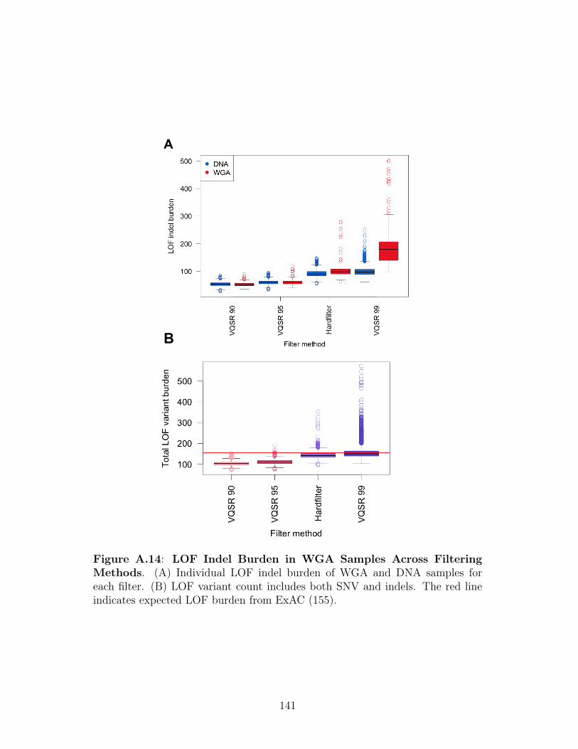
A
B B
Figure A.14: LOF Indel Burden in WGA Samples Across FilteringMethods. (A) Individual LOF indel burden of WGA and DNA samples foreach filter. (B) LOF variant count includes both SNV and indels. The red lineindicates expected LOF burden from ExAC (155).
141

Figure A.15: G/C Homopolymer Content of Genes Shared BetweenOV and LAML. The number of G/C hompolymer regions normalized by codingexon length in base pairs plotted for all genes and for genes that were significantp < 1.61 x 10⁻⁷ by logistic regression for both OV and LAML. Significant genesshared between OV and LAML under three different indel filter conditions (VQSRTS99, TS95, TS90) are plotted for comparison.
142

A B B
Figure A.16: LOF SNV Logistic Regression Analysis. (A) Quantile-quantile plot from logistic regression association testing between germline LOFSNV burden and OV. n=number of genes tested. Red line indicates significantcutoff and red points indicate associations significant p < 1.61 x 10⁻⁷. BRCA1/2associations highlighted. (B) Number of genes significant p < 1.61 x 10⁻⁷ by logis-tic regression for all cancer types. Color indicates cancer types containing WGAsamples.
143

Table A.1: Composition of the Pan-Cancer Cohort.
144

Table A.2: Coverage of the Six TCGA Capture Kits. Size and overlapwith Gencode exons for the six capture kits used to collect TCGA normal DNAsamples.
Table A.3: K-means Cluster Membership of HapMap Samples.
145

Table A.4: K-means Cluster Membership of TCGA Samples.
Table A.5: GC Content of the Sequence Surrounding WGA Indels.Mean GC content of the sequence surrounding WGA-enriched and non-enrichedindels. C.I. = Confidence interval for mean estimate derived from 1,000 bootstrapsamples.
Table A.6: Allele Frequency of Homopolymer Indels. Mean allele fre-quency of indels in homopolymer regions in DNA and WGA samples. C.I. =Confidence interval for mean estimate derived from 1,000 bootstrap samples.
146

Table A.7: Frequency of BLAST Match for WGA Indels. Total numberof large indels and fraction of large indels with a BLAST hit in WGA-enrichedand non-enriched indel sets. Large indels are indels >= 15 base pairs. BLASThits are defined as a BLAST match +/- 10 kB from the indel start position.
147

Table A.8: ANOVA of LOF Indel Burden Using Different Filters. Vari-ance in LOF indel burden explained by technical covariates for each indel filteringapproach. Sum. Sq., Sum of Squares; Df, Degrees of Freedom.
148

Table A.9: Correlation Between LOF Indels and Homopolymer Tracts.Spearman correlation between LOF indel burden and homopolymer content. LOFindel allele counts were calculated for each gene separately for WGA and DNAsamples. Both allele counts and homopolymer region counts were normalized bygene by dividing by coding exon length in base pairs.
149

Appendix B
Supplemental Material: Exome-Wide
Analysis of Bi-allelic Alterations
Identifies a Lynch Phenotype in the
Cancer Genome Atlas
This appendix contains supplemental figures for the work described in Chapter
3 of this document: "Exome-Wide Analysis of Bi-allelic Alterations Identifies a Lynch
Phenotype in the Cancer Genome Atlas". Each figure is referred to in the main text of
the chapter and a brief description of each figure is given here.
150

A
B C
Figure B.1: Calling Somatic Methylation Status. (A) MLH1 expression isdecreased in methylated samples. Cancer types with 5+ methylated samples areshown. TPM = transcripts per million. (B) Expression of MLH1 vs. methylationbeta value. Both expression values (in transcripts per million) and methylationbeta values were converted to Z-scores using the mean and standard deviationfor each cancer type. The red line indicates the cutoff used to call methylation.(C) Expression of MLH1 vs. methylation beta value in colon cancer samplesonly. Beta values are from the methylation probe cg13846866, which was mostanti-correlated with MLH1 expression in colon cancer. The red line indicates thecutoff used to call methylation.
151

ATM 11:108183151 G / T
BRCA1 17:41209079 T / TG
BRCA2 13:32911297 TAAAC / T
Nor
mal
Tu
mor
A B C
Figure B.2: Example LOH Events. IGV snapshots of paired tumor:normalBAM files with a somatic LOH event for ATM (A), BRCA1 (B), and BRCA2 (C).The germline locus subject to LOH is highlighted with a box. Screenshots weretaken with downsampling settings of max read count 20 per 200 base window.
152

Figure B.3: Genes Frequently Affected by Germline:Somatic Al-teration. Barplot showing gene level frequency of germline:somatic alter-ation with KEGG mismatch repair pathway genes highlighted in red. Frac-tion germline:somatic alteration was calculated for each gene as number ofgermline:somatic alterations/number of germline LOF variants. Only genes with> 2 germline LOF variants in the cohort were included in the analysis. Signifi-cance was calculated using fgsea and is adjusted for multiple hypothesis testing.
153

A B
Figure B.4: Association Between Germline LOF Burden and CancerType. QQ plot of Fisher exact p-values of the association between 11 DDRpathways and 28 cancer types using LOF variants only (A) or LOF and ClinVarpathogenic variants (B). Only samples determined to be of European descentusing PCA were used (n = 7,734). Red indicates significance above a Bonferronithreshold. (A) Significant hits: breast cancer:tumor suppressor pathway (p =9.69e-5 ), breast cancer:Fanconi anemia pathway (p = 7.42e-6 ). (B) Significanthits: pheochromocytoma and paraganglioma:cancer predisposition genes (p =1.18e-9), breast cancer:Fanconi anemia pathway (p = 3.89e-7), pheochromocytomaand paraganglioma:oncogenes (p = 2.30e-6).
154

Figure B.5: Germline and Somatic LOF in PMS2 . Both germline andsomatic LOF mutations can alter the same position. Lollipop plot indicating theamino acids altered in two samples with PMS2 bi-allelic alteration. GermlineLOF variants are represented by solid lines, somatic LOF mutations by dashedlines. The stopgain mutation highlighted by an arrow, p.R628X, is inherited inone individual and acquired in the other. PFAM domain abbreviations: HATPasec 3 = Histidine kinase, DNA gyrase B, and HSP90-like ATPase; MutL C = MutLC terminal dimerization domain.
155

A
C
B
Figure B.6: Mutational Signature Analysis of Germline:Somatic MMRAlteration. (A) Fraction of mutations attributed to mutational signature 6 plot-ted by type of germline:somatic MMR alteration. Individuals were grouped byMMR gene mutation type: NONE, no alteration; GERM, germline LOF variantsonly; SOM, somatic LOF mutations only; MISS, bi-allelic alteration including amissense mutation; LOF, bi-allelic alteration via dual LOF mutation. Wilcox p =0.00023, 0.0063, and 0.00096; permutation p = 0.0002, 0.011, and 0.002 for SOM,MISS, and LOF respectively. (B) Fraction of combined germline:somatic alter-ation carriers mutational profile attributed to the mutational signatures availablein COSMIC. (C) Mutational profile of all germline:somatic alteration carrierscombined.
156

Figure B.7: Mono-allelic Germline MMR Variation Not AssociatedWith MSI. Somatic MSI burden for individuals with MMR germline variantsbut no somatic alteration of the MMR pathway. NONE = no alteration of theMMR pathway, LOF = germline LOF variant in an MMR gene, LOF + CLIN= germline LOF variant or ClinVar pathogenic variant in an MMR gene, LOF+ CADD = germline LOF variant or variant with a CADD score >= 30 in anMMR gene. The number of samples in each category is displayed in parentheses.
157

A
B
Figure B.8: Association Testing Between Genomic Alteration and MSIBurden. (A) The same analysis as Figure 3.4, but including all somatic LOFmutations. (B) The same analysis as Figure 3.4, but including germline:somaticMMR alteration carriers.
158

Figure B.9: SHPRH Methylation in Uterine Cancer. Expression of SH-PRH vs. methylation beta value in uterine cancer samples only. Beta valuesare from the methylation probe cg00571935, which was most anti-correlated withSHPRH expression in uterine cancer. The red line indicates the cutoff used tocall methylation.
159

A
B
Figure B.10: SHPRH Expression in Normal Tissues. Expression of SH-PRH (A) or GAPDH (B) in 23 normal tissue types represented in GTEx. TPM= transcripts per million.
160

A B C
Figure B.11: Mutational Signature Analysis of MLH1 and SHPRHMethylation. (A) Fraction of mutations attributed to mutational signature 6plotted byMLH1 methylation status. Wilcox p = 3.882e-15, permutation p < 1e-4.(B) Fraction of mutations attributed to mutational signature 6 plotted by SHPRHmethylation status. Wilcox p = 0.041, permutation p = 0.0264. (C) Fraction ofmutations attributed to mutational signature 6 plotted by SHPRH methylationstatus in uterine cancer samples only. Wilcox p = 3.405e-5, permutation p < 1e-4.
161

A B
C
Figure B.12: Co-Occurrence Testing for SHPRH Methylation. QQ plotsof co-occurrence testing between SHPRH methylation and germline LOF variants(A), somatic LOF mutations (B), and somatic methylation (C) in 9,484 genesand n = 8,087 samples. Red indicates 5% FDR. Somatic LOF mutations in 213genes and somatic methylation of 3,694 genes significantly co-occur with SHPRHmethylation. Of these co-occurring genes, 8 are MMR pathway genes (MLH3,PCNA, POLD3, RFC1, RFC5, RPA3, MLH3, and PMS2 ).
162

Figure B.13: Mutational Signature Analysis of BRCA1/2 Carriers.Fraction of mutations attributed to mutational signature 3 plotted by BRCA1/2status. Single = germline LOF or ClinVar pathogenic variant only, SOM = bi-allelic alteration via somatic mutation, AI = allelic imbalance, LOH = loss ofheterozygosity. Wilcoxon rank sum test p = 2.246e-4, 6.981e-6, 2.343e-7; permu-tation p < 1e-4, for SOM, AI, and LOH respectively.
163

A
B
Figure B.14: Mutational Signature Analysis of DDR Pathway Alter-ation. A Wilcoxon rank sum test was used to test for differences in somatic mu-tational signature burden between individuals carrying DDR alterations vs. thosewithout. (A) Heatmap of p values for the association between bi-allelic alterationin 6 DDR pathways and 30 mutational signatures in COSMIC. (B) Heatmap ofp values for the association between mono-allelic alteration in 8 DDR pathwaysand 30 mutational signatures in COSMIC.
164

Figure B.15: Association Between Age and Damaging Germline Vari-ants. Volcano plot of association testing between germline LOF and pathogenicClinVar variant carrier status and age of diagnosis for 85 cancer predispositiongenes in n = 8,913 samples.
Table B.1: Association Between MSI and MMR Alteration. P-values, βestimates, and standard errors determined using a linear model.
165

Table B.2: Association Between Age and MMR Alteration. P-values, βestimates, and standard errors determined using a linear model.
Table B.3: Germline Variants Pathogenic for Lynch Syndrome. ClinVarannotations: P = pathogenic, Num. Sub. = number of sources submitting toClinVar.
166

Table B.4: Germline Variants of Unknown Significance for Lynch Syn-drome. ClinVar annotations: P = pathogenic, NA = not in ClinVar, Num. Sub.= number of sources submitting to ClinVar.
167

168

Table B.5: Modeling a Germline:Somatic Interaction for L-MMRGenes. P-values, β estimates, and standard errors determined using a linearmodel.
169

Table B.6: Association Between MSI and MMR Germline:Somatic Al-teration. P-values, β estimates, and standard errors determined using a linearmodel.
Table B.7: Association Between Age and MMR Germline:Somatic Al-tertion. P-values, β estimates, and standard errors determined using a linearmodel.
170

Table B.8: Association Between MSI and Mono-allelic Germline MMRVariants. P-values, β estimates, and standard errors determined using a linearmodel.
171

Table B.9: Association Between MSI and MMR Alteration IncludingConfounders. MSI linear model results using unfiltered somatic mutations andwith germline:somatic MMR alteration carriers included. P-values, β estimates,and standard errors determined using a linear model.
172

173

Table B.10: Association Between MSI and Alterations Correlated WithSHPRH Methylation. P-values, β estimates, and standard errors determinedusing a linear model.
Table B.11: Association Between MSI and SHPRH Expression. P-values, β estimates, and standard errors determined using a linear model.
174

Table B.12: Association Between Age and Known PredisposingGermline Variants. Association between MMR, BRCA1/2, SDHB/RET, andTP53 germline variant carrier status and age of diagnosis. P-values, β estimates,and standard errors determined using a linear model.
Table B.13: Association Between Age and Predicted PredisposingGermline Variants. P-values, β estimates, and standard errors determinedusing a linear model.
175

Appendix C
Supplemental Material: Rare Variant
Phasing Using Paired Tumor:Normal
Sequence Data
This appendix contains supplemental figures for the work described in Chapter 3
of this document: "Rare Variant Phasing Using Paired Tumor:Normal Sequence Data".
Each figure is referred to in the main text of the chapter and a brief description of each
figure is given here.
176

Amp Del
1 Ratio M:P
Maternal Paternal
SNV VAF 0.5 0
Normal
Sequencing Reads
0.5
A = SNV
A
A A
A A A A
A
0.5 0.25
0.5 0.75 0.5
A B C
Figure C.1: Schematic of ∆ VAF Changes in Cancer. In regions of SCNA,somatic sequencing reads are skewed toward the chromosomes that are physicallymore abundant in the sample. Here we illustrate the maternal chromosome inblue and the paternal in green. (A) The expectation in diploid regions is thatthe sequencing reads will originate from the maternal and paternal chromosomein a 0.5 ratio and that the VAF of heterozygous SNVs will be 0.5. (B) Theexpectation in regions of amplification is that sequencing reads will be skewedtoward the amplified chromosome and that the VAF of heterozygous germlineSNVs will change from 0.5. In this illustration the SNV lies on the non-amplifiedchromosome and therefore the VAF decreases to 0.25. (C) The expectation inregions of deletion is that sequencing reads will be skewed toward the non-deletedchromosome. In this illustration the SNV lies on the deleted chromosome andtherefore the VAF decreases to 0.
177

A
B
Figure C.2: Example ∆ VAF Data. ∆ VAF changes in SCNA regions forchromosome 2 of sample TCGA-Y8-A8RZ. (A) ∆ VAF for 812 germline het-erozygous variants. (B) Absolute ∆ VAF for the same variants as (A). SCNAsegments identified using TCGA data are shown as black lines, SCNA segmentsidentified using VAF-CBS are shown in red. Color indicates p-value obtainedfrom a Fisher’s exact test on tumor and normal read counts: gray: p >= 0.05,purple: p < 0.05.
178

A
C D
B
Figure C.3: Example VCF-CBS Data. VAF-CBS segments identified usingdifferent smoothing parameters for a SCNA region in chromosome 2 of sampleTCGA-Y8-A8RZ. VAF-CBS segments are shown as red lines and the total numberof segments in the region is shown in the upper right corner. Color indicates Fisherexact test p-value using tumor and normal read counts: gray: p >= 0.05, purple:p < 0.05. Four different smoothing parameters were tested: (A) 0.5 megabases(Mb), (B) 1 Mb, (C) 2 Mb, (D) 3 Mb.
179

A B C
Figure C.4: VAF-CBS Segment Metrics. SCNA segment metrics from n =6,158 samples using four different VAF-CBS smoothing parameters: 0.5 Mb, 1Mb, 2 Mb, and 3 Mb. (A) Mean size in base pairs of segments identified. (B)Fraction of segments identified that contain more than one heterozygous variant.(C) Fraction of all germline heterozygous variants that can be phased.
180

A B C
D E F
Figure C.5: ∆ VAF Data From a Contaminated Sample. Example dataillustrating absolute ∆ VAF changes in (A) paired normal tissue:normal blood,(B) normal tissue:tumor, and (C) normal blood:tumor for chromosome 7 of sampleTCGA-V5-AASX. Example data illustrating absolute ∆ VAF changes in (D)paired normal tissue:normal blood, (E) normal tissue:tumor, and (F) normalblood:tumor for chromosome 7 of sample TCGA-Y8-A8RZ. Color indicates p-value obtained from a Fisher’s exact test on tumor and normal read counts: gray:p >= 0.05, purple: p < 0.05. Segments with an absolute ∆ VAF >= 0.14 arered.
181

Figure C.6: Discordance Between Phasing Methods. (A) Discordancebetween VAF phasing and HapCUT2 for n = 6,180 samples. Number of commonsites are number of sites phased by both methods. (B) Discordance between VAFphasing and phASER for n = 6,180 samples.
182

A
B
Figure C.7: Sample Metrics that Affect ∆ VAF Phasing. (A) Percent ofthe genome involved in a SCNA event for n = 8,542 TCGA samples across 29cancer types. Fraction of the genome calculated as: total length in base pairs ofTCGA SCNA regions (predicted FC > 1.1 or < 0.9) / 3e09 (B) Percent tumornuclei for the same samples as (A). Percent tumor nuclei values were obtainedfrom TCGA biospecimen histology slide data. Samples with missing values wereimputed to cancer type median.
183

A B
Figure C.8: Phasing Performance on Rare Variants. (A) The fraction ofrare (allele frequency <= 0.01) germline heterozygous variants phased by Hap-CUT2 alone, HapCUT2 and phASER, and by HapCUT2, phASER, and VAFin n = 6,180 samples. The addition of VAF phasing increased the number ofphased variants by 98%. (B) The fraction of rare germline variants phased thatare unique to each method.
184

A
B
Figure C.9: Fraction of Phased Variants Visualized by Chromosome.(A) Fraction of heterozygous germline variants phased using VAF phasing forn = 100 samples. Cancer type is indicated by color: BRCA = Breast InvasiveCarcinoma, STAD = Stomach Adenocarcinoma, UCEC = Uterine Corpus En-dometrial Carcinoma. Samples with more than 33% of the genome involved in anSCNA are indicated by gray bars. Samples with a histology slide based purity >80% are indicated by black bars. (B) Fraction of heterozygous germline variantsphased using HapCUT2 for the same samples as (A).
185

Figure C.10: Comparison Between TCGA and VAF-CBS SCNAs. Com-paring number of variants phased using TCGA SCNA segments vs. VAF-CBSsegmentation. COMMON = variants phased by both methods, VAF-CBS =variants phased only using VAF-CBS segmentation, CNV = variants phased onlyusing TCGA CNV calls. A median 63% of variants were phased by both methodsin n = 6,180 samples.
186

A
T
C G
1000 bp
Pair Distance Minimum Allele Frequency
Mismatch
A : T 100 0.01 0 A : C 700 0.5 1 A : G 1000 0.01 0 T : C 600 0.01 1 T : G 900 0.01 0 C : G 300 0.01 1
A
T C
G
Distance AF Pairwise Discordance
< 500 < 0.1 0.5 >= 500 < 0.1 0.33 < 500 > 0.1 NA
>= 500 > 0.1 1
Method 1 Method 2
C G
rare variant common variant
Maternal Paternal
A
C D
Phase 1 Phase 2 1|0 1|0 0|1 0|1 1|0 0|1 1|0 1|0
Overall Discordance: 0.25
B
Figure C.11: Method Used to Calculate Pairwise Error. (A) Exampleof a shared phase segment showing phase calls from two methods. Rare variants(allele frequency <= 0.01) are shown in red. (B) The overall discordance wascalculated as the fraction of discordant phase calls within a block. (C) Tableshowing all pairwise phase pairs from the segment in (A). Distance between pairsis calculated as distance in base pairs between the variants. Minimum allelefrequency is the smaller allele frequency of the two variants. Error is a binaryvariable that indicated whether the two variants are in the same orientation. (D)Example showing how pairwise error was binned by distance and allele frequencyas in Figure 4.3. For each category the mean error was calculated.
187

Figure C.12: Discordance Between VAF and 10X Genomics Phasing.Pairwise discordance for the COLO829 cell line as a function of distance and allelefrequency. Colors represent different values of the smoothing parameter.
188

Figure C.13: Fraction of Compound Heterozygosity Events Phased].Fraction was calculated for each individual as: the number of genes with multipleCADD >= 15 variants phased / the number of genes with multiple CADD >=15 germline variants. H = HapCUT2 only, H+P = HapCUT2 and phASER,H+P+V = HapCUT2, phASER, and VAF phasing.
189

Figure C.14: Association Between Age and Damaging Germline Vari-ants. Age of cancer diagnosis Z-score in n = 6,093 TCGA individuals groupedby type of germline alteration in a set of 144 cancer predisposition genes. Groupsare the same as Figure 4.4B with the exception of six individuals carrying both anon-compensatory variant set and a ClinVar/LOF variant that are grouped sep-arately (Both). * = p < 0.05, . = p < 0.1; p-values were determined using alinear model to predict age of diagnosis while accounting for cancer type.
190

Figure C.15: Association Between Age and BRCA1/2 Germline Vari-ants. Age of cancer diagnosis Z-score in 6,093 TCGA individuals grouped bytype of germline alteration in BRCA1 or BRCA2. (B) Individuals were groupedusing HMMvar cis variant scores: CADD = individuals carrying a germline vari-ant with a CADD score >= 15, ClinVar/LOF = individuals carrying a ClinVarpathogenic or LOF germline variant, Non-Compensatory = individuals carryingmultiple nonsynonymous variants in a gene predicted to be more deleterious col-lectively than independently The number of samples is shown in parentheses. *= p < 0.05, . = p < 0.1; p-values were determined using a linear model to predictage of diagnosis while accounting for cancer type.
191

Normal Tumor
A T G C
A T G C
A T G C G C G C
G C G C A T
A T
A T G C
Variant VAF Δ VAF Abs. Δ VAF Phase A 0.25 -0.25 0.25 1|0
T 0.25 -0.25 0.25 1|0
C 0.75 0.25 0.25 0|1
G 0.75 0.25 0.25 0|1
Variant VAF Δ VAF Abs. Δ VAF Phase A 0.75 0.25 0.25 0|1
T 0.75 0.25 0.25 0|1
C 0.75 0.25 0.25 0|1
G 0.75 0.25 0.25 0|1
A
B
Figure C.16: Assumptions of VAF Phasing Model. VAF phasing assumesthat SCNA segments originate from a single homologous chromosome. (A) Underthis assumption, ∆ VAF can be used to correctly phase variants. (B) Should aSCNA segment result from equal amplification of both homologous chromosomes,using ∆ VAF to phase will result in switch errors. In this example, amplificationof both homologous chromosomes results in all variants being assigned to thesame chromosome incorrectly.
192

Table C.1: Possible Contaminated Normal Tissue Samples. TCGA bar-codes of suspected contaminated normal tissue samples.
193

Table C.2: Discordance Between VAF Phasing and Other Methods.Null model refers to VAF phasing using duplicated samples directly to determinesignificant VAF-CBS segments, cutoff refers to using a absolute ∆ VAF cutoff of0.14 to determine significant VAF-CBS segments.
194

Table C.3: Factors That Influence VAF Phasing.Factors that influencenumber of variants phased per sample by VAF phasing. P-values, β estimates,and standard errors determined using a linear model.
Table C.4: Discordance Between VAF Phasing with TCGA SCNAand Other Methods. Mean discordance between VAF phasing, HapCUT2and phASER in n = 6,180 samples either using VAF-CBS segments or TCGASCNA calls.
195

Table C.5: Discordance Between VAF and 10X Genomics Phasing.
Table C.6: Features of VAF Phasing Errors. Features of incorrect VAFphasing pairs from COLO829. Features were calculated for all possible pairs ofphased heterozygous germline variants from COLO829 (see Methods).
196

Table C.7: Association Between Age and Compound Heterozygosity.P-values, β estimates, and standard errors determined using a linear model.
Table C.8: Association Between Age and Non-Compensatory Variants.P-values, β estimates, and standard errors determined using a linear model.
Table C.9: Association Between Age and Non-Compensatory Variants,ClinVar Samples Removed. P-values, β estimates, and standard errors deter-mined using a linear model.
197

Table C.10: Association Between Age and Non-Compensatory Variantsin BRCA1/2 . P-values, β estimates, and standard errors determined using alinear model.
198

Table C.11: Germline Non-Compensatory Variants in BRCA1/2
199

200

Bibliography
[1] Eric R. Fearon and Bert Volgestein. A Genetic Model for Colorectal tumorogenesis.Cell, 61:759–767, 1990.
[2] Bert Vogelstein, Nickolas Papadopoulos, Victor E Velculescu, Shibin Zhou, Luis ADiaz, and Kenneth W Kinzler. Cancer genome landscapes. Science, 340(6127):1546–1558, 2013.
[3] Sian Jones, Valsamo Anagnostou, Karli Lytle, Sonya Parpart-Li, Monica Nesselbush,David R Riley, Manish Shukla, Bryan Chesnick, Maura Kadan, Eniko Papp, Kevin GGalens, Derek Murphy, Theresa Zhang, Lisa Kann, Mark Sausen, Samuel V Angiuoli,Luis A Diaz, and Victor E Velculescu. Personalized genomic analyses for cancermutation discovery and interpretation. Science Translational Medicine, 7(283), 2015.
[4] Divyansh Agarwal, Christoph Nowak, Nancy R Zhang, Lajos Pusztai, and ChristosHatzis. Functional germline variants as potential co-oncogenes. npj Breast Cancer,3(1):46, 2017.
[5] Heather S Feigelson, Katrina A.B. Goddard, Celine Hollombe, Sharna R Tingle,Elizabeth M Gillanders, Leah E Mechanic, and Stefanie A Nelson. Approaches tointegrating germline and tumor genomic data in cancer research. Carcinogenesis,35(10):2157–2163, 2014.
[6] Alfred G Knudson. Mutation and Cancer: Statistical Study of Retinoblastoma.Proceedings of the National Academy of Sciences, 68(4):820–823, 1971.
[7] C. O. Nordling. A new theory on the cancer-inducing mechanism. British Journalof Cancer, 7(1):68–72, mar 1953.
[8] Manuela Santarosa and Alan Ashworth. Haploinsufficiency for tumour suppressorgenes: When you don’t need to go all the way. Biochimica et Biophysica Acta -Reviews on Cancer, 1654(2):105–122, 2004.
[9] Steven A. Frank. Genetic predisposition to cancer - Insights from population genetics.Nature Reviews Genetics, 5(10):764–772, oct 2004.
201

[10] Judy E Garber and Kenneth Offit. Hereditary Cancer Predisposition Syndromes. JClin Oncol, 23(2):276–292, 2005.
[11] Katherine A Rauen. The RASopathies. Annual Review of Genomics and HumanGenetics, 14(1):355–369, 2013.
[12] Cristian Tomasetti, Lu Li, and Bert Vogelstein. Stem cell divisions, somatic muta-tions, cancer etiology, and cancer prevention. Science, 355(6331):1330–1334, 2017.
[13] Cristian Tomasetti and Bert Vogelstein. Variation in cancer risk among tissues canbe explained by the number of stem cell divisions. Science, 347(6217):78–81, 2015.
[14] Lorelei A. Mucci, Jacob B. Hjelmborg, Jennifer R. Harris, Kamila Czene, David J.Havelick, Thomas Scheike, Rebecca E. Graff, Klaus Holst, Soren Moller, Robert H.Unger, Christina McIntosh, Elizabeth Nuttall, Ingunn Brandt, Kathryn L. Penney,Mikael Hartman, Peter Kraft, Giovanni Parmigiani, Kaare Christensen, MarkkuKoskenvuo, Niels V. Holm, Kauko Heikkila, Eero Pukkala, Axel Skytthe, Hans OlovAdami, Jaakko Kaprio, Julia Isaeva, Thomas Nilsen, Tellervo Korhonen, and UlrichHalekoh. Familial risk and heritability of cancer among twins in nordic countries.JAMA - Journal of the American Medical Association, 315(1):68–76, jan 2016.
[15] J. Hall, M. Lee, B Newman, J. Morrow, L. Anderson, B Huey, and M. King. Linkageof early-onset familial breast cancer to chromosome 17q21. Science, 250(4988):1684–1689, dec 1990.
[16] Paul Lichtenstein, Niels V. Holm, Pia K. Verkasalo, Anastasia Iliadou, JaakkoKaprio, Markku Koskenvuo, Eero Pukkala, Axel Skytthe, and Kari Hemminki. En-vironmental and heritable factors in the causation of cancer analyses of cohorts oftwins from Sweden, Denmark, and Finland. New England Journal of Medicine,343(2):78–85, 2000.
[17] Nazneen Rahman. Realizing the promise of cancer predisposition genes. Nature,505(7483):302–308, 2014.
[18] Kuan-lin Huang, R. Jay Mashl, Yige Wu, Deborah I. Ritter, Jiayin Wang, ClaraOh, Marta Paczkowska, Sheila Reynolds, Matthew A. Wyczalkowski, Ninad Oak,Adam D. Scott, Michal Krassowski, Andrew D. Cherniack, Kathleen E. Houlahan,Reyka Jayasinghe, Liang-Bo Wang, Daniel Cui Zhou, Di Liu, Song Cao, Young WonKim, Amanda Koire, Joshua F. McMichael, Vishwanathan Hucthagowder, Tae-BeomKim, Abigail Hahn, Chen Wang, Michael D. McLellan, Fahd Al-Mulla, Kimberly J.Johnson, Olivier Lichtarge, Paul C. Boutros, Benjamin Raphael, Alexander J. Lazar,Wei Zhang, Michael C. Wendl, Ramaswamy Govindan, Sanjay Jain, David Wheeler,Shashikant Kulkarni, John F. Dipersio, Juri Reimand, Funda Meric-Bernstam, KenChen, Ilya Shmulevich, Sharon E. Plon, Feng Chen, Li Ding, Samantha J. Caesar-Johnson, John A. Demchok, Ina Felau, Melpomeni Kasapi, Martin L. Ferguson, Car-olyn M. Hutter, Heidi J. Sofia, Roy Tarnuzzer, Zhining Wang, Liming Yang, Jean C.
202

Zenklusen, Jiashan (Julia) Zhang, Sudha Chudamani, Jia Liu, Laxmi Lolla, RashiNaresh, Todd Pihl, Qiang Sun, Yunhu Wan, Ye Wu, Juok Cho, Timothy DeFreitas,Scott Frazer, Nils Gehlenborg, Gad Getz, David I. Heiman, Jaegil Kim, Michael S.Lawrence, Pei Lin, Sam Meier, Michael S. Noble, Gordon Saksena, Doug Voet,Hailei Zhang, Brady Bernard, Nyasha Chambwe, Varsha Dhankani, Theo Knijnen-burg, Roger Kramer, Kalle Leinonen, Yuexin Liu, Michael Miller, Sheila Reynolds,Ilya Shmulevich, Vesteinn Thorsson, Wei Zhang, Rehan Akbani, Bradley M. Broom,Apurva M. Hegde, Zhenlin Ju, Rupa S. Kanchi, Anil Korkut, Jun Li, Han Liang,Shiyun Ling, Wenbin Liu, Yiling Lu, Gordon B. Mills, Kwok-Shing Ng, Arvind Rao,Michael Ryan, Jing Wang, John N. Weinstein, Jiexin Zhang, Adam Abeshouse,Joshua Armenia, Debyani Chakravarty, Walid K. Chatila, Ino de Bruijn, Jian-jiong Gao, Benjamin E. Gross, Zachary J. Heins, Ritika Kundra, Konnor La, MarcLadanyi, Augustin Luna, Moriah G. Nissan, Angelica Ochoa, Sarah M. Phillips,Ed Reznik, Francisco Sanchez-Vega, Chris Sander, Nikolaus Schultz, Robert Sheri-dan, S. Onur Sumer, Yichao Sun, Barry S. Taylor, Jioajiao Wang, Hongxin Zhang,Pavana Anur, Myron Peto, Paul Spellman, Christopher Benz, Joshua M. Stuart,Christopher K. Wong, Christina Yau, D. Neil Hayes, Joel S. Parker, Matthew D.Wilkerson, Adrian Ally, Miruna Balasundaram, Reanne Bowlby, Denise Brooks, Re-becca Carlsen, Eric Chuah, Noreen Dhalla, Robert Holt, Steven J.M. Jones, Katay-oon Kasaian, Darlene Lee, Yussanne Ma, Marco A. Marra, Michael Mayo, Richard A.Moore, Andrew J. Mungall, Karen Mungall, A. Gordon Robertson, Sara Sadeghi,Jacqueline E. Schein, Payal Sipahimalani, Angela Tam, Nina Thiessen, Kane Tse,Tina Wong, Ashton C. Berger, Rameen Beroukhim, Andrew D. Cherniack, CarrieCibulskis, Stacey B. Gabriel, Galen F. Gao, Gavin Ha, Matthew Meyerson, Steven E.Schumacher, Juliann Shih, Melanie H. Kucherlapati, Raju S. Kucherlapati, StephenBaylin, Leslie Cope, Ludmila Danilova, Moiz S. Bootwalla, Phillip H. Lai, Dennis T.Maglinte, David J. Van Den Berg, Daniel J. Weisenberger, J. Todd Auman, SaianandBalu, Tom Bodenheimer, Cheng Fan, Katherine A. Hoadley, Alan P. Hoyle, Stu-art R. Jefferys, Corbin D. Jones, Shaowu Meng, Piotr A. Mieczkowski, Lisle E. Mose,Amy H. Perou, Charles M. Perou, Jeffrey Roach, Yan Shi, Janae V. Simons, TaraSkelly, Matthew G. Soloway, Donghui Tan, Umadevi Veluvolu, Huihui Fan, ToshinoriHinoue, Peter W. Laird, Hui Shen, Wanding Zhou, Michelle Bellair, Kyle Chang,Kyle Covington, Chad J. Creighton, Huyen Dinh, HarshaVardhan Doddapaneni,Lawrence A. Donehower, Jennifer Drummond, Richard A. Gibbs, Robert Glenn,Walker Hale, Yi Han, Jianhong Hu, Viktoriya Korchina, Sandra Lee, Lora Lewis, WeiLi, Xiuping Liu, Margaret Morgan, Donna Morton, Donna Muzny, Jireh Santibanez,Margi Sheth, Eve Shinbrot, Linghua Wang, Min Wang, David A. Wheeler, Liu Xi,Fengmei Zhao, Julian Hess, Elizabeth L. Appelbaum, Matthew Bailey, Matthew G.Cordes, Li Ding, Catrina C. Fronick, Lucinda A. Fulton, Robert S. Fulton, CyriacKandoth, Elaine R. Mardis, Michael D. McLellan, Christopher A. Miller, Heather K.Schmidt, Richard K. Wilson, Daniel Crain, Erin Curley, Johanna Gardner, KevinLau, David Mallery, Scott Morris, Joseph Paulauskis, Robert Penny, Candace Shel-ton, Troy Shelton, Mark Sherman, Eric Thompson, Peggy Yena, Jay Bowen, Julie M.
203

Gastier-Foster, Mark Gerken, Kristen M. Leraas, Tara M. Lichtenberg, Nilsa C.Ramirez, Lisa Wise, Erik Zmuda, Niall Corcoran, Tony Costello, Christopher Hov-ens, Andre L. Carvalho, Ana C. de Carvalho, Jose H. Fregnani, Adhemar Longatto-Filho, Rui M. Reis, Cristovam Scapulatempo-Neto, Henrique C.S. Silveira, Daniel O.Vidal, Andrew Burnette, Jennifer Eschbacher, Beth Hermes, Ardene Noss, RosySingh, Matthew L. Anderson, Patricia D. Castro, Michael Ittmann, David Hunts-man, Bernard Kohl, Xuan Le, Richard Thorp, Chris Andry, Elizabeth R. Duffy,Vladimir Lyadov, Oxana Paklina, Galiya Setdikova, Alexey Shabunin, Mikhail Tavo-bilov, Christopher McPherson, Ronald Warnick, Ross Berkowitz, Daniel Cramer,Colleen Feltmate, Neil Horowitz, Adam Kibel, Michael Muto, Chandrajit P. Raut,Andrei Malykh, Jill S. Barnholtz-Sloan, Wendi Barrett, Karen Devine, JordonnaFulop, Quinn T. Ostrom, Kristen Shimmel, Yingli Wolinsky, Andrew E. Sloan,Agostino De Rose, Felice Giuliante, Marc Goodman, Beth Y. Karlan, Curt H. Hage-dorn, John Eckman, Jodi Harr, Jerome Myers, Kelinda Tucker, Leigh Anne Zach,Brenda Deyarmin, Hai Hu, Leonid Kvecher, Caroline Larson, Richard J. Mural,Stella Somiari, Ales Vicha, Tomas Zelinka, Joseph Bennett, Mary Iacocca, BrendaRabeno, Patricia Swanson, Mathieu Latour, Louis Lacombe, Bernard Tetu, AlainBergeron, Mary McGraw, Susan M. Staugaitis, John Chabot, Hanina Hibshoosh,Antonia Sepulveda, Tao Su, Timothy Wang, Olga Potapova, Olga Voronina, Lau-rence Desjardins, Odette Mariani, Sergio Roman-Roman, Xavier Sastre, Marc-HenriStern, Feixiong Cheng, Sabina Signoretti, Andrew Berchuck, Darell Bigner, EricLipp, Jeffrey Marks, Shannon McCall, Roger McLendon, Angeles Secord, AlexisSharp, Madhusmita Behera, Daniel J. Brat, Amy Chen, Keith Delman, Seth Force,Fadlo Khuri, Kelly Magliocca, Shishir Maithel, Jeffrey J. Olson, Taofeek Owonikoko,Alan Pickens, Suresh Ramalingam, Dong M. Shin, Gabriel Sica, Erwin G. VanMeir, Hongzheng Zhang, Wil Eijckenboom, Ad Gillis, Esther Korpershoek, LeendertLooijenga, Wolter Oosterhuis, Hans Stoop, Kim E. van Kessel, Ellen C. Zwarthoff,Chiara Calatozzolo, Lucia Cuppini, Stefania Cuzzubbo, Francesco DiMeco, Gae-tano Finocchiaro, Luca Mattei, Alessandro Perin, Bianca Pollo, Chu Chen, JohnHouck, Pawadee Lohavanichbutr, Arndt Hartmann, Christine Stoehr, Robert Stoehr,Helge Taubert, Sven Wach, Bernd Wullich, Witold Kycler, Dawid Murawa, MaciejWiznerowicz, Ki Chung, W. Jeffrey Edenfield, Julie Martin, Eric Baudin, GlennBubley, Raphael Bueno, Assunta De Rienzo, William G. Richards, Steven Kalka-nis, Tom Mikkelsen, Houtan Noushmehr, Lisa Scarpace, Nicolas Girard, MartaAymerich, Elias Campo, Eva Gine, Armando Lopez Guillermo, Nguyen Van Bang,Phan Thi Hanh, Bui Duc Phu, Yufang Tang, Howard Colman, Kimberley Evason,Peter R. Dottino, John A. Martignetti, Hani Gabra, Hartmut Juhl, Teniola Ak-eredolu, Serghei Stepa, Dave Hoon, Keunsoo Ahn, Koo Jeong Kang, Felix Beuschlein,Anne Breggia, Michael Birrer, Debra Bell, Mitesh Borad, Alan H. Bryce, Erik Cas-tle, Vishal Chandan, John Cheville, John A. Copland, Michael Farnell, ThomasFlotte, Nasra Giama, Thai Ho, Michael Kendrick, Jean-Pierre Kocher, Karla Kopp,Catherine Moser, David Nagorney, Daniel O’Brien, Brian Patrick O’Neill, TusharPatel, Gloria Petersen, Florencia Que, Michael Rivera, Lewis Roberts, Robert Small-
204

ridge, Thomas Smyrk, Melissa Stanton, R. Houston Thompson, Michael Torbenson,Ju Dong Yang, Lizhi Zhang, Fadi Brimo, Jaffer A. Ajani, Ana Maria Angulo Gonza-lez, Carmen Behrens, Jolanta Bondaruk, Russell Broaddus, Bogdan Czerniak, BitaEsmaeli, Junya Fujimoto, Jeffrey Gershenwald, Charles Guo, Alexander J. Lazar,Christopher Logothetis, Funda Meric-Bernstam, Cesar Moran, Lois Ramondetta,David Rice, Anil Sood, Pheroze Tamboli, Timothy Thompson, Patricia Troncoso,Anne Tsao, Ignacio Wistuba, Candace Carter, Lauren Haydu, Peter Hersey, Va-lerie Jakrot, Hojabr Kakavand, Richard Kefford, Kenneth Lee, Georgina Long, Gra-ham Mann, Michael Quinn, Robyn Saw, Richard Scolyer, Kerwin Shannon, AndrewSpillane, Jonathan Stretch, Maria Synott, John Thompson, James Wilmott, Hik-mat Al-Ahmadie, Timothy A. Chan, Ronald Ghossein, Anuradha Gopalan, Dou-glas A. Levine, Victor Reuter, Samuel Singer, Bhuvanesh Singh, Nguyen Viet Tien,Thomas Broudy, Cyrus Mirsaidi, Praveen Nair, Paul Drwiega, Judy Miller, Jen-nifer Smith, Howard Zaren, Joong-Won Park, Nguyen Phi Hung, Electron Kebe-bew, W. Marston Linehan, Adam R. Metwalli, Karel Pacak, Peter A. Pinto, MarkSchiffman, Laura S. Schmidt, Cathy D. Vocke, Nicolas Wentzensen, Robert Worrell,Hannah Yang, Marc Moncrieff, Chandra Goparaju, Jonathan Melamed, Harvey Pass,Natalia Botnariuc, Irina Caraman, Mircea Cernat, Inga Chemencedji, Adrian Clipca,Serghei Doruc, Ghenadie Gorincioi, Sergiu Mura, Maria Pirtac, Irina Stancul, Di-ana Tcaciuc, Monique Albert, Iakovina Alexopoulou, Angel Arnaout, John Bartlett,Jay Engel, Sebastien Gilbert, Jeremy Parfitt, Harman Sekhon, George Thomas,Doris M. Rassl, Robert C. Rintoul, Carlo Bifulco, Raina Tamakawa, Walter Urba,Nicholas Hayward, Henri Timmers, Anna Antenucci, Francesco Facciolo, GianlucaGrazi, Mirella Marino, Roberta Merola, Ronald de Krijger, Anne-Paule Gimenez-Roqueplo, Alain Piche, Simone Chevalier, Ginette McKercher, Kivanc Birsoy, GeneBarnett, Cathy Brewer, Carol Farver, Theresa Naska, Nathan A. Pennell, DanielRaymond, Cathy Schilero, Kathy Smolenski, Felicia Williams, Carl Morrison, Jef-frey A. Borgia, Michael J. Liptay, Mark Pool, Christopher W. Seder, Kerstin Junker,Larsson Omberg, Mikhail Dinkin, George Manikhas, Domenico Alvaro, Maria Con-siglia Bragazzi, Vincenzo Cardinale, Guido Carpino, Eugenio Gaudio, David Chesla,Sandra Cottingham, Michael Dubina, Fedor Moiseenko, Renumathy Dhanasekaran,Karl-Friedrich Becker, Klaus-Peter Janssen, Julia Slotta-Huspenina, Mohamed H.Abdel-Rahman, Dina Aziz, Sue Bell, Colleen M. Cebulla, Amy Davis, Rebecca Du-ell, J. Bradley Elder, Joe Hilty, Bahavna Kumar, James Lang, Norman L. Lehman,Randy Mandt, Phuong Nguyen, Robert Pilarski, Karan Rai, Lynn Schoenfield, KellySenecal, Paul Wakely, Paul Hansen, Ronald Lechan, James Powers, Arthur Tischler,William E. Grizzle, Katherine C. Sexton, Alison Kastl, Joel Henderson, Sima Porten,Jens Waldmann, Martin Fassnacht, Sylvia L. Asa, Dirk Schadendorf, Marta Couce,Markus Graefen, Hartwig Huland, Guido Sauter, Thorsten Schlomm, Ronald Simon,Pierre Tennstedt, Oluwole Olabode, Mark Nelson, Oliver Bathe, Peter R. Carroll,June M. Chan, Philip Disaia, Pat Glenn, Robin K. Kelley, Charles N. Landen, JoannaPhillips, Michael Prados, Jeffry Simko, Karen Smith-McCune, Scott VandenBerg,Kevin Roggin, Ashley Fehrenbach, Ady Kendler, Suzanne Sifri, Ruth Steele, Anto-
205

nio Jimeno, Francis Carey, Ian Forgie, Massimo Mannelli, Michael Carney, BrendaHernandez, Benito Campos, Christel Herold-Mende, Christin Jungk, Andreas Un-terberg, Andreas von Deimling, Aaron Bossler, Joseph Galbraith, Laura Jacobus,Michael Knudson, Tina Knutson, Deqin Ma, Mohammed Milhem, Rita Sigmund,Andrew K. Godwin, Rashna Madan, Howard G. Rosenthal, Clement Adebamowo,Sally N. Adebamowo, Alex Boussioutas, David Beer, Thomas Giordano, Anne-MarieMes-Masson, Fred Saad, Therese Bocklage, Lisa Landrum, Robert Mannel, KathleenMoore, Katherine Moxley, Russel Postier, Joan Walker, Rosemary Zuna, MichaelFeldman, Federico Valdivieso, Rajiv Dhir, James Luketich, Edna M. Mora Pinero,Mario Quintero-Aguilo, Carlos Gilberto Carlotti, Jose Sebastiao Dos Santos, RafaelKemp, Ajith Sankarankuty, Daniela Tirapelli, James Catto, Kathy Agnew, ElizabethSwisher, Jenette Creaney, Bruce Robinson, Carl Simon Shelley, Eryn M. Godwin,Sara Kendall, Cassaundra Shipman, Carol Bradford, Thomas Carey, Andrea Had-dad, Jeffey Moyer, Lisa Peterson, Mark Prince, Laura Rozek, Gregory Wolf, RayleenBowman, Kwun M. Fong, Ian Yang, Robert Korst, W. Kimryn Rathmell, J. LeighFantacone-Campbell, Jeffrey A. Hooke, Albert J. Kovatich, Craig D. Shriver, JohnDiPersio, Bettina Drake, Ramaswamy Govindan, Sharon Heath, Timothy Ley, BrianVan Tine, Peter Westervelt, Mark A. Rubin, Jung Il Lee, Natalia D. Aredes, andArmaz Mariamidze. Pathogenic Germline Variants in 10,389 Adult Cancers. Cell,173(2):355–370.e14, apr 2018.
[19] Sebastian M. Waszak, Grace Tiao, Bin Zhu, Tobias Rausch, Francesc Muyas,Bernardo Rodriguez-Martin, Raquel Rabionet, Sergei Yakneen, Georgia Escaramis,Yilong Li, Natalie Saini, Steven A. Roberts, German M. Demidov, Esa Pitkanen,Olivier Delaneau, Jose M. Heredia-Genestar, Joachim Weischenfeldt, Suyash S.Shringarpure, Jieming Chen, Hidewaki Nakagawa, Ludmil B. Alexandrov, OliverDrechsel, L. Jonathan Dursi, Ayellet V. Segre, Erik Garrison, Serap Erkek, NinaHabermann, Lara Urban, Ekta Khurana, Andy Cafferkey, Shuto Hayashi, SeiyaImoto, Lauri A. Aaltonen, Eva G. Alvarez, Adrian Baez-Ortega, Matthew Bailey,Mattia Bosio, Alicia L. Bruzos, Ivo Buchhalter, Carlos D. Bustamante, Claudia Cal-abrese, Anthony DiBiase, Mark Gerstein, Aliaksei Z. Holik, Xing Hua, Kuan-linHuang, Ivica Letunic, Leszek J. Klimczak, Roelof Koster, Sushant Kumar, MikeMcLellan, Jay Mashl, Lisa Mirabello, Steven Newhouse, Aparna Prasad, Gun-nar Raetsch, Matthias Schlesner, Roland Schwarz, Pramod Sharma, Tal Shmaya,Nikos Sidiropoulos, Lei Song, Hana Susak, Tomas Tanskanen, Marta Tojo, David C.Wedge, Mark Wright, Ying Wu, Kai Ye, Venkata D. Yellapantula, Jorge Zamora,Atul J. Butte, Gad Getz, Jared Simpson, Li Ding, Tomas Marques-Bonet, ArcadiNavarro, Alvis Brazma, Peter Campbell, Stephen J. Chanock, Nilanjan Chatterjee,Oliver Stegle, Reiner Siebert, Stephan Ossowski, Olivier Harismendy, Dmitry A.Gordenin, Jose M. C. Tubio, Francisco M. De La Vega, Douglas F. Easton, XavierEstivill, Jan Korbel, PCAWG Germline Working Group, and ICGC/TCGA Pan-Cancer Analysis of Whole Genomes Net. Germline determinants of the somaticmutation landscape in 2,642 cancer genomes. bioRxiv, page 208330, nov 2017.
206

[20] Susanne N. Grobner, Barbara C. Worst, Joachim Weischenfeldt, Ivo Buchhalter,Kortine Kleinheinz, Vasilisa A. Rudneva, Pascal D. Johann, Gnana Prakash Bal-asubramanian, Maia Segura-Wang, Sebastian Brabetz, Sebastian Bender, BarbaraHutter, Dominik Sturm, Elke Pfaff, Daniel Hubschmann, Gideon Zipprich, MichaelHeinold, Jurgen Eils, Christian Lawerenz, Serap Erkek, Sander Lambo, SebastianWaszak, Claudia Blattmann, Arndt Borkhardt, Michaela Kuhlen, Angelika Eggert,Simone Fulda, Manfred Gessler, Jenny Wegert, Roland Kappler, Daniel Baumhoer,Stefan Burdach, Renate Kirschner-Schwabe, Udo Kontny, Andreas E. Kulozik, Diet-mar Lohmann, Simone Hettmer, Cornelia Eckert, Stefan Bielack, Michaela Nathrath,Charlotte Niemeyer, Gunther H. Richter, Johannes Schulte, Reiner Siebert, FrankWestermann, Jan J. Molenaar, Gilles Vassal, Hendrik Witt, Marc Zapatka, BirgitBurkhardt, Christian P. Kratz, Olaf Witt, Cornelis M.Van Tilburg, Christof M.Kramm, Gudrun Fleischhack, Uta Dirksen, Stefan Rutkowski, Michael Fruhwald,Katja Von Hoff, Stephan Wolf, Thomas Klingebiel, Ewa Koscielniak, Pablo Land-graf, Jan Koster, Adam C. Resnick, Jinghui Zhang, Yanling Liu, Xin Zhou, Angela J.Waanders, Danny A. Zwijnenburg, Pichai Raman, Benedikt Brors, Ursula D. We-ber, Paul A. Northcott, Kristian W. Pajtler, Marcel Kool, Rosario M. Piro, Jan O.Korbel, Matthias Schlesner, Roland Eils, David T.W. Jones, Peter Lichter, LukasChavez, and Stefan M. Pfister. The landscape of genomic alterations across child-hood cancers. Nature, 555(7696):321–327, feb 2018.
[21] Jinghui Zhang, Michael F. Walsh, Gang Wu, Michael N. Edmonson, Tanja A. Gru-ber, John Easton, Dale Hedges, Xiaotu Ma, Xin Zhou, Donald A. Yergeau, Mark R.Wilkinson, Bhavin Vadodaria, Xiang Chen, Rose B. McGee, Stacy Hines-Dowell,Regina Nuccio, Emily Quinn, Sheila A. Shurtleff, Michael Rusch, Aman Patel,Jared B. Becksfort, Shuoguo Wang, Meaghann S. Weaver, Li Ding, Elaine R. Mardis,Richard K. Wilson, Amar Gajjar, David W. Ellison, Alberto S. Pappo, Ching-HonPui, Kim E. Nichols, and James R. Downing. Germline Mutations in PredispositionGenes in Pediatric Cancer. New England Journal of Medicine, 373(24):2336–2346,2015.
[22] Douglas F Easton. How many more breast cancer predisposition genes are there?Breast cancer research, 1(1):14–7, 1999.
[23] Amit Sud, Ben Kinnersley, and Richard S Houlston. Genome-wide association stud-ies of cancer: Current insights and future perspectives. Nature Reviews Cancer,17(11):692–704, 2017.
[24] Paul D.P. Pharoah, Antonis C Antoniou, Douglas F Easton, and Bruce A.J. Ponder.Polygenes, Risk Prediction, and Targeted Prevention of Breast Cancer. New EnglandJournal of Medicine, 358(26):2796–2803, 2008.
[25] Qiyuan Li, Ji-Heui Seo, Barbara Stranger, Aaron McKenna, Itsik Pe’er, ThomasLaFramboise, Myles Brown, Svitlana Tyekucheva, and Matthew L Freedman. Inte-
207

grative eQTL-Based Analyses Reveal the Biology of Breast Cancer Risk Loci. Cell,152(3):633–641, jan 2013.
[26] Dan L. Nicolae, Eric Gamazon, Wei Zhang, Shiwei Duan, M. Eileen Dolan, andNancy J. Cox. Trait-associated SNPs are more likely to be eQTLs: Annotation toenhance discovery from GWAS. PLoS Genetics, 6(4):e1000888, apr 2010.
[27] Y.-P. Fu, Indu Kohaar, Nathaniel Rothman, Julie Earl, Jonine D Figueroa, Yuan-qing Ye, Nuria Malats, Wei Tang, Luyang Liu, Montserrat Garcia-Closas, BrianMuchmore, Nilanjan Chatterjee, Mcanthony Tarway, Manolis Kogevinas, PatriciaPorter-Gill, Dalsu Baris, Adam Mumy, Demetrius Albanes, Mark P Purdue, AmyHutchinson, Alfredo Carrato, A. Tardon, Consol Serra, R. Garcia-Closas, JosepLloreta, Alison Johnson, Molly Schwenn, Margaret R Karagas, Alan Schned, W RyanDiver, Susan M Gapstur, Michael J Thun, Jarmo Virtamo, Stephen J Chanock,Joseph F Fraumeni, Debra T Silverman, Xifeng Wu, Francisco X Real, and LudmilaProkunina-Olsson. Common genetic variants in the PSCA gene influence gene ex-pression and bladder cancer risk. Proceedings of the National Academy of Sciences,109(13):4974–4979, 2012.
[28] Alan M Pittman, Silvia Naranjo, Emily Webb, Peter Broderick, Esther H Lips, TomVan Wezel, Hans Morreau, Kate Sullivan, Sarah Fielding, Philip Twiss, JayaramVijayakrishnan, Fernando Casares, Mobshra Qureshi, Jose Luis Gomez-Skarmeta,and Richard S Houlston. The colorectal cancer risk at 18q21 is caused by a novelvariant altering SMAD7 expression. Genome Research, 19(6):987–993, 2009.
[29] Franklin W. Huang, Eran Hodis, Mary Jue Xu, Gregory V. Kryukov, Lynda Chin,and Levi A. Garraway. Highly recurrent TERT promoter mutations in humanmelanoma. Science, 339(6122):957–959, 2013.
[30] Nasim Ahmadiyeh, Mark M Pomerantz, Chiara Grisanzio, Paula Herman, Li Jia,Vanessa Almendro, Housheng Hansen He, Myles Brown, Shirley Liu, Matt Davis,Jennifer L Caswell, Christine A Beckwith, Adam Hills, Laura MacConaill, Gerhard ACoetzee, Meredith M Regan, and Matthew L Freedman. 8q24 prostate, breast, andcolon cancer risk loci show tissue-specific long-range interaction with MYC. PNAS,107(21):9742–8746, 2010.
[31] Cristina Bozzao, Patrizia Lastella, and Alessandro Stella. Anticipation in LynchSyndrome: Where We Are Where We Go. Current Genomics, 12(7):451–465, nov2011.
[32] Bente A Talseth-Palmer, Juul T Wijnen, Desma M Grice, and Rodney J Scott.Genetic modifiers of cancer risk in Lynch syndrome: A review. Familial Cancer,12(2):207–216, jun 2013.
[33] Hany Ariffin, Pierre Hainaut, Anna Puzio-Kuter, Soo Sin Choong, Adelyne Sue LiChan, Denis Tolkunov, Gunaretnam Rajagopal, Wenfeng Kang, Leon Li Wen Lim,
208

Shekhar Krishnan, Kok-Siong Chen, Maria Isabel Achatz, Mawar Karsa, JannahShamsani, Arnold J Levine, and Chang S Chan. Whole-genome sequencing analysisof phenotypic heterogeneity and anticipation in Li-Fraumeni cancer predispositionsyndrome. Proceedings of the National Academy of Sciences of the United States ofAmerica, 111(43):15497–501, 2014.
[34] Antonis C Antoniou, Olga M Sinilnikova, Jacques Simard, Melanie Leone, Mar-tine Dumont, Susan L Neuhausen, Jeffery P Struewing, Dominique Stoppa-Lyonnet,Laure Barjhoux, David J Hughes, Isabelle Coupier, Muriel Belotti, Christine Las-set, Valerie Bonadona, Yves-Jean Bignon, Timothy R Rebbeck, Theresa Wagner,Henry T Lynch, Susan M Domchek, Katherine L Nathanson, Judy E Garber, JeffreyWeitzel, Steven A Narod, Gail Tomlinson, Olufunmilayo I Olopade, Andrew Godwin,Claudine Isaacs, Anna Jakubowska, Jan Lubinski, Jacek Gronwald, Bohdan Gorski,Tomasz Byrski, Tomasz Huzarski, Susan Peock, Margaret Cook, Caroline Baynes,Alexandra Murray, Mark Rogers, Peter A Daly, Huw Dorkins, Rita K Schmut-zler, Beatrix Versmold, Christoph Engel, Alfons Meindl, Norbert Arnold, DieterNiederacher, Helmut Deissler, Amanda B Spurdle, Xiaoqing Chen, Nicola Waddell,Nicole Cloonan, Tomas Kirchhoff, Kenneth Offit, Eitan Friedman, Bella Kaufmann,Yael Laitman, Gilli Galore, Gad Rennert, Flavio Lejbkowicz, Leon Raskin, Irene LAndrulis, Eduard Ilyushik, Hilmi Ozcelik, Peter Devilee, Maaike P.G. Vreeswijk,Mark H Greene, Sheila A Prindiville, Ana Osorio, Javier Benitez, Michal Zikan,Csilla I Szabo, Outi Kilpivaara, Heli Nevanlinna, Ute Hamann, Francine Durocher,Adalgeir Arason, Fergus J Couch, Douglas F Easton, and Georgia Chenevix-Trench.RAD51 135G>C Modifies Breast Cancer Risk among BRCA2 Mutation Carriers:Results from a Combined Analysis of 19 Studies. The American Journal of HumanGenetics, 81(6):1186–1200, 2007.
[35] Montserrat Garcia-Closas, Nathaniel Rothman, Jonine D Figueroa, LudmilaProkunina-Olsson, Summer S Han, Dalsu Baris, Eric J. Jacobs, Nuria Malats, Im-maculata De Vivo, Demetrius Albanes, Mark P Purdue, Sapna Sharma, Yi Ping Fu,Manolis Kogevinas, Zhaoming Wang, Wei Tang, Adonina Tardon, Consol Serra, Al-fredo Carrato, Reina Garcia-Closas, Josep Lloreta, Alison Johnson, Molly Schwenn,Margaret R Karagas, Alan Schned, Gerald Andriole, Robert Grubb, Amanda Black,Susan M Gapstur, Michael Thun, William Ryan Diver, Stephanie J Weinstein, JarmoVirtamo, David J Hunter, Neil Caporaso, Maria Teresa Landi, Amy Hutchinson,Laurie Burdett, Kevin B Jacobs, Meredith Yeager, Joseph F Fraumeni, Stephen JChanock, Debra T Silverman, and Nilanjan Chatterjee. Common genetic polymor-phisms modify the effect of smoking on absolute risk of bladder cancer. CancerResearch, 73(7):2211–2220, apr 2013.
[36] Rosalie Fisher, Stuart Horswell, Andrew Rowan, Maximilian P Salm, Elza C.de Bruin, Sakshi Gulati, Nicholas McGranahan, Mark Stares, Marco Gerlinger,Ignacio Varela, Andrew Crockford, Francesco Favero, Virginie Quidville, FabriceAndre, Carolina Navas, Eva Gronroos, David Nicol, Steve Hazell, David Hrouda,
209

Tim O’Brien, Nik Matthews, Ben Phillimore, Sharmin Begum, Adam Rabinowitz,Jennifer Biggs, Paul A Bates, Neil Q. McDonald, Gordon Stamp, Bradley Spencer-Dene, James J Hsieh, Jianing Xu, Lisa Pickering, Martin Gore, James Larkin, andCharles Swanton. Development of synchronous VHL syndrome tumors reveals con-tingencies and constraints to tumor evolution. Genome biology, 15(8):433, 2014.
[37] Amy M. Dworkin, Katie Ridd, Dianne Bautista, Dawn C. Allain, O. Hans Iwenofu,Ritu Roy, Boris C. Bastian, and Amanda Ewart Toland. Germline Variation Controlsthe Architecture of Somatic Alterations in Tumors. PLoS Genetics, 6(9):e1001136,sep 2010.
[38] Mel F Greaves, Ana Teresa Maia, Joseph LWiemels, and Anthony M Ford. Leukemiain twins: Lessons in natural history. Blood, 102(7):2321–2333, oct 2003.
[39] Ana Teresa Maia, Anthony M Ford, G. Reza Jalali, Christine J Harrison, G. MalcolmTaylor, Osborn B Eden, and Mel F Greaves. Molecular tracking of leukemogenesisin a triplet pregnancy. Blood, 98(2):478–482, 2001.
[40] G. Cazzaniga, F. W. van Delft, L. Lo Nigro, A. M. Ford, J. Score, I. Iacobucci,E. Mirabile, M. Taj, S. M. Colman, A. Biondi, and M. Greaves. Developmentalorigins and impact of BCR-ABL1 fusion and IKZF1 deletions in monozygotic twinswith Ph+ acute lymphoblastic leukemia. Blood, 118(20):5559–5564, nov 2011.
[41] Bruce A Hamilton and Benjamin D Yu. Modifier genes and the plasticity of geneticnetworks in mice, 2012.
[42] Michele Harvey, M J McArthur, Charles A Montgomery, A Bradley, and L a Done-hower. Genetic background alters the spectrum of tumors that develop in p53-deficient mice. The FASEB journal : official publication of the Federation of Amer-ican Societies for Experimental Biology, 7(10):938–943, 1993.
[43] Melina MacPhee, Kenneth P Chepenik, Rebecca A Liddell, Kelly K Nelson, Linda DSiracusa, and Arthur M Buchberg. The secretory phospholipase A2 gene is a candi-date for the Mom1 locus, a major modifier of ApcMin-induced intestinal neoplasia.Cell, 81(6):957–966, 1995.
[44] Yuan Zhu, Pritam Ghosh, Patrick Charnay, Dennis K Burns, and Luis F Parada.Neurofibromas in NF1: Schwann cell origin and role of tumor microenvironment.Science, 296:920–922, 2002.
[45] Laurence H Pearl, Amanda C Schierz, Simon E Ward, Bissan Al-Lazikani, andFrances M G Pearl. Therapeutic opportunities within the DNA damage response.Nature Reviews Cancer, 15(3):166–180, 2015.
[46] Stephen P Jackson and Jiri Bartek. The DNA-damage response in human biologyand disease. Nature, 461(7267):1071–1078, 2009.
210

[47] Isidro Cortes-Ciriano, Sejoon Lee, Woong Yang Park, Tae Min Kim, and Peter JPark. A molecular portrait of microsatellite instability across multiple cancers. Na-ture Communications, 8, 2017.
[48] Ronald J Hause, Colin C Pritchard, Jay Shendure, and Stephen J Salipante. Clas-sification and characterization of microsatellite instability across 18 cancer types.Nature Medicine, 22(11):1342–1350, oct 2016.
[49] Christopher J Lord and Alan Ashworth. BRCAness revisited. Nature Reviews Can-cer, 16(2):110–120, 2016.
[50] Marianne Berwick and Paolo Vineis. Markers of DNA repair and susceptibility tocancer in humans: an epidemiologic review. Journal of the National Cancer Institute,92(11):874–897, jun 2000.
[51] Harvey W Mohrenweiser, David M Wilson, and Irene M Jones. Challenges andcomplexities in estimating both the functional impact and the disease risk associatedwith the extensive genetic variation in human DNA repair genes. Mutation research,526(1-2):93–125, 2003.
[52] R S Camplejohn, S Hodgson, N Carter, B S Kato, and T D Spector. Heritability ofDNA-damage-induced apoptosis and its relationship with age in lymphocytes fromfemale twins. British Journal of Cancer, 95(4):520–524, aug 2006.
[53] Paul Finnon, Naomi Robertson, Sylwia Dziwura, Claudine Ravy, Wei Zhang, LizAinsbury, Jaakko Kaprio, Christophe Badie, and Simon Bouzer. Evidence for sig-nificant heritability of apoptotic and cell cycle responses to ionising radiation. HumGenet, 123:485–493, 2008.
[54] Harald Surowy, Antje Rinckleb, Manuel Luedeke, Madeleine Stuber, Anna Wecker,Dominic Varga, Christiane Maier, Josef Hoegel, and Walther Vogel. Heritability ofbaseline and induced micronucleus frequencies. Mutagenesis, 26(1):111–117, 2011.
[55] Jacqueline Cloos, Eline J.C. Nieuwenhuis, Dorret I. Boomsma, Dirk J. Kuik, Mar-ianne L.T. Van Der Sterre, Fre Arwert, Gordon B. Snow, and Boudewijn J.M.Braakhuis. Inherited susceptibility to bleomycin-induced chromatid breaks in cul-tured peripheral blood lymphocytes. Journal of the National Cancer Institute,91(13):1125–1130, jul 1999.
[56] Sara Gutierrez-Enriquez, Marie Fernet, Thilo Dork, Michael Bremer, AnthonyLauge, Dominique Stoppa-Lyonnet, Norman Moullan, Sandra Angele, and JanetHall. Functional Consequences of ATM Sequence Variants for Chromosomal Ra-diosensitivity. Genes Chromosomes and Cancer, 40(2):109–119, 2004.
[57] M R Spitz, J J Fueger, S Halabi, S P Schantz, D Sample, and T C Hsu. Muta-gen sensitivity in upper aerodigestive tract cancer: a case-control analysis. Cancerepidemiology, biomarkers & prevention, 2(4):329–33, jul 1993.
211

[58] X Wu, J Gu, Y Patt, M Hassan, M R Spitz, R P Beasley, and L Y Hwang. Mutagensensitivity as a susceptibility marker for human hepatocellular carcinoma. Cancerepidemiology, biomarkers & prevention : a publication of the American Associationfor Cancer Research, cosponsored by the American Society of Preventive Oncology,7(7):567–70, jul 1998.
[59] N. Rajeswari, Y. R. Ahuja, U. Malini, S. Chandrashekar, N. Balakrishna, K. V. Rao,and Ashok Khar. Risk assessment in first degree female relatives of breast cancerpatients using the alkaline Comet assay. Carcinogenesis, 21(4):557–561, 2000.
[60] Isabel Garcia-Cao, Marta Garcia-Cao, Juan Martin-Caballero, Luis M. Criado, PeterKlatt, Juana M. Flores, Jean Claude Weill, Maria A. Blasco, and Manuel Serrano.’Super p53’ mice exhibit enhanced DNA damage response, are tumor resistant andage normally. EMBO Journal, 21(22):6225–6235, 2002.
[61] Ludmil B. Alexandrov, Serena Nik-Zainal, David C. Wedge, Samuel A.J.R. Apari-cio, Sam Behjati, Andrew V. Biankin, Graham R. Bignell, Niccolo Bolli, Ake Borg,Anne Lise Børresen-Dale, Sandrine Boyault, Birgit Burkhardt, Adam P. Butler, Car-los Caldas, Helen R. Davies, Christine Desmedt, Roland Eils, Jorunn Erla Eyfjord,John A. Foekens, Mel Greaves, Fumie Hosoda, Barbara Hutter, Tomislav Ilicic, San-drine Imbeaud, Marcin Imielinsk, Natalie Jager, David T.W. Jones, David Jonas,Stian Knappskog, Marcel Koo, Sunil R. Lakhani, Carlos Lopez-Otin, Sancha Martin,Nikhil C. Munshi, Hiromi Nakamura, Paul A. Northcott, Marina Pajic, Elli Papaem-manuil, Angelo Paradiso, John V. Pearson, Xose S. Puente, Keiran Raine, Man-asa Ramakrishna, Andrea L. Richardson, Julia Richter, Philip Rosenstiel, MatthiasSchlesner, Ton N. Schumacher, Paul N. Span, Jon W. Teague, Yasushi Totoki, An-drew N.J. Tutt, Rafael Valdes-Mas, Marit M. Van Buuren, Laura Van ’T Veer, AnneVincent-Salomon, Nicola Waddell, Lucy R. Yates, Jessica Zucman-Rossi, P. AndrewFutreal, Ultan McDermott, Peter Lichter, Matthew Meyerson, Sean M. Grimmond,Reiner Siebert, Elias Campo, Tatsuhiro Shibata, Stefan M. Pfister, Peter J. Camp-bell, and Michael R. Stratton. Signatures of mutational processes in human cancer.Nature, 500(7463):415–421, 2013.
[62] Serena Nik-Zainal, Ludmil B Alexandrov, David C Wedge, Peter Van Loo, Christo-pher D Greenman, Keiran Raine, David Jones, Jonathan Hinton, John Marshall,Lucy A Stebbings, Andrew Menzies, Sancha Martin, Kenric Leung, Lina Chen,Catherine Leroy, Manasa Ramakrishna, Richard Rance, King Wai Lau, Laura JMudie, Ignacio Varela, David J McBride, Graham R Bignell, Susanna L Cooke,Adam Shlien, John Gamble, Ian Whitmore, Mark Maddison, Patrick S Tarpey, He-len R Davies, Elli Papaemmanuil, Philip J Stephens, Stuart McLaren, Adam PButler, Jon W Teague, Goran Jonsson, Judy E Garber, Daniel Silver, PenelopeMiron, Aquila Fatima, Sandrine Boyault, Anita Langerod, Andrew Tutt, John W.M.Martens, Samuel A.J.R. Aparicio, Ake Borg, Anne Vincent Salomon, Gilles Thomas,Anne Lise Borresen-Dale, Andrea L Richardson, Michael S Neuberger, P Andrew
212

Futreal, Peter J Campbell, and Michael R Stratton. Mutational processes moldingthe genomes of 21 breast cancers. Cell, 149(5):979–993, may 2012.
[63] Scott D. Brown, Lisa A. Raeburn, and Robert A. Holt. Profiling tissue-resident Tcell repertoires by RNA sequencing. Genome Medicine, 7(1):125, dec 2015.
[64] Aaron M Newman, Chih Long Liu, Michael R Green, Andrew J Gentles, WeiguoFeng, Yue Xu, Chuong D Hoang, Maximilian Diehn, and Ash A Alizadeh. Ro-bust enumeration of cell subsets from tissue expression profiles. Nature Methods,12(5):453–457, may 2015.
[65] Charles Lu, Mingchao Xie, Michael C. Wendl, Jiayin Wang, Michael D. McLellan,Mark D.M. Leiserson, Kuan Lin Huang, Matthew A. Wyczalkowski, Reyka Jayas-inghe, Tapahsama Banerjee, Jie Ning, Piyush Tripathi, Qunyuan Zhang, BeifangNiu, Kai Ye, Heather K. Schmidt, Robert S. Fulton, Joshua F. McMichael, PragBatra, Cyriac Kandoth, Maheetha Bharadwaj, Daniel C. Koboldt, Christopher A.Miller, Krishna L. Kanchi, James M. Eldred, David E. Larson, John S. Welch,Ming You, Bradley A. Ozenberger, Ramaswamy Govindan, Matthew J. Walter,Matthew J. Ellis, Elaine R. Mardis, Timothy A. Graubert, John F. Dipersio, Timo-thy J. Ley, Richard K. Wilson, Paul J. Goodfellow, Benjamin J. Raphael, Feng Chen,Kimberly J. Johnson, Jeffrey D. Parvin, and Li Ding. Patterns and functional im-plications of rare germline variants across 12 cancer types. Nature Communications,6:10086, dec 2015.
[66] Serena Nik-Zainal, Helen Davies, Johan Staaf, Manasa Ramakrishna, DominikGlodzik, Xueqing Zou, Inigo Martincorena, Ludmil B Alexandrov, Sancha Martin,David CWedge, Peter Van Loo, Young Seok Ju, Marcel Smid, Arie B Brinkman, San-dro Morganella, Miriam R Aure, Ole Christian Lingjærde, Anita Langerød, MarkusRingner, Sung Min Ahn, Sandrine Boyault, Jane E Brock, Annegien Broeks, AdamButler, Christine Desmedt, Luc Dirix, Serge Dronov, Aquila Fatima, John A Foekens,Moritz Gerstung, Gerrit K.J. Hooijer, Se Jin Jang, David R Jones, Hyung Yong Kim,Tari A King, Savitri Krishnamurthy, Hee Jin Lee, Jeong Yeon Lee, Yilong Li, StuartMcLaren, Andrew Menzies, Ville Mustonen, Sarah O’Meara, Iris Pauporte, XavierPivot, Colin A Purdie, Keiran Raine, Kamna Ramakrishnan, F. German Rodriguez-Gonzalez, Gilles Romieu, Anieta M Sieuwerts, Peter T Simpson, Rebecca Shep-herd, Lucy Stebbings, Olafur A Stefansson, Jon Teague, Stefania Tommasi, IsabelleTreilleux, Gert G. Van Den Eynden, Peter Vermeulen, Anne Vincent-Salomon, LucyYates, Carlos Caldas, Laura Van t. Veer, Andrew Tutt, Stian Knappskog, BenitaKiat Tee Tan, Jos Jonkers, Ake Borg, Naoto T. Ueno, Christos Sotiriou, Alain Viari,P Andrew Futreal, Peter J Campbell, Paul N Span, Steven Van Laere, Sunil RLakhani, Jorunn E Eyfjord, Alastair M Thompson, Ewan Birney, Hendrik G Stun-nenberg, Marc J Van De Vijver, John W.M. Martens, Anne Lise Børresen-Dale,Andrea L Richardson, Gu Kong, Gilles Thomas, and Michael R Stratton. Land-scape of somatic mutations in 560 breast cancer whole-genome sequences. Nature,534(7605):47–54, 2016.
213

[67] Nadeem Riaz, Pedro Blecua, Raymond S. Lim, Ronglai Shen, Daniel S. Higginson,Nils Weinhold, Larry Norton, Britta Weigelt, Simon N. Powell, and Jorge S. Reis-Filho. Pan-cancer analysis of bi-allelic alterations in homologous recombination DNArepair genes. Nature Communications, 8(1):857, dec 2017.
[68] Nicola Waddell, Marina Pajic, Ann Marie Patch, David K. Chang, Karin S. Kassahn,Peter Bailey, Amber L. Johns, David Miller, Katia Nones, Kelly Quek, Michael C.J.Quinn, Alan J. Robertson, Muhammad Z.H. Fadlullah, Tim J.C. Bruxner, Ange-lika N. Christ, Ivon Harliwong, Senel Idrisoglu, Suzanne Manning, Craig Nourse,Ehsan Nourbakhsh, Shivangi Wani, Peter J. Wilson, Emma Markham, Nicole Cloo-nan, Matthew J. Anderson, J. Lynn Fink, Oliver Holmes, Stephen H. Kazakoff, Con-rad Leonard, Felicity Newell, Barsha Poudel, Sarah Song, Darrin Taylor, Nick Wad-dell, Scott Wood, Qinying Xu, Jianmin Wu, Mark Pinese, Mark J. Cowley, Hong C.Lee, Marc D. Jones, Adnan M. Nagrial, Jeremy Humphris, Lorraine A. Chantrill, Ve-nessa Chin, Angela M. Steinmann, Amanda Mawson, Emily S. Humphrey, Emily K.Colvin, Angela Chou, Christopher J. Scarlett, Andreia V. Pinho, Marc Giry-Laterriere, Ilse Rooman, Jaswinder S. Samra, James G. Kench, Jessica A. Pettitt,Neil D. Merrett, Christopher Toon, Krishna Epari, Nam Q. Nguyen, Andrew Bar-bour, Nikolajs Zeps, Nigel B. Jamieson, Janet S. Graham, Simone P. Niclou, RolfBjerkvig, Robert Grutzmann, Daniela Aust, Ralph H. Hruban, Anirban Maitra,Christine A. Iacobuzio-Donahue, Christopher L. Wolfgang, Richard A. Morgan,Rita T. Lawlor, Vincenzo Corbo, Claudio Bassi, Massimo Falconi, Giuseppe Zam-boni, Giampaolo Tortora, Margaret A. Tempero, Anthony J. Gill, James R. Esh-leman, Christian Pilarsky, Aldo Scarpa, Elizabeth A. Musgrove, John V. Pearson,Andrew V. Biankin, and Sean M. Grimmond. Whole genomes redefine the muta-tional landscape of pancreatic cancer. Nature, 518(7540):495–501, feb 2015.
[69] Paz Polak, Jaegil Kim, Lior Z Braunstein, Rosa Karlic, Nicholas J Haradhavala,Grace Tiao, Daniel Rosebrock, Dimitri Livitz, Kirsten Kubler, Kent W Mouw,Atanas Kamburov, Yosef E Maruvka, Ignaty Leshchiner, Eric S Lander, Todd RGolub, Aviad Zick, Alexandre Orthwein, Michael S Lawrence, Rajbir N Batra, Car-los Caldas, Daniel A Haber, Peter W Laird, Hui Shen, Leif W Ellisen, Alan DD’Andrea, Stephen J Chanock, William D Foulkes, and Gad Getz. A mutationalsignature reveals alterations underlying deficient homologous recombination repairin breast cancer. Nature Genetics, 49(10):1476–1486, aug 2017.
[70] Jenni Nikkila, Ann Christin Parplys, Katri Pylkas, Muthiah Bose, Yanying Huo,Kerstin Borgmann, Katrin Rapakko, Pentti Nieminen, Bing Xia, Helmut Pospiech,and Robert Winqvist. Heterozygous mutations in PALB2 cause DNA replicationand damage response defects. Nature Communications, 4:2578, oct 2013.
[71] Mary I Coolbaugh-Murphy, Jing Ping Xu, Louis S. Ramagli, Brian C Ramagli,Barry W Brown, Patrick M Lynch, Stanley R Hamilton, Marsha L Frazier, andMichael J Siciliano. Microsatellite instability in the peripheral blood leukocytes ofHNPCC patients. Human Mutation, 31(3):317–324, mar 2010.
214

[72] Henry T. Lynch and Thomas Smyrk. Hereditary nonpolyposis colorectal cancer(Lynch syndrome). An updated review. Cancer, 78(6):1149–1167, sep 1996.
[73] Candace D Middlebrooks, A Rouf Banday, Konichi Matsuda, Krizia Ivana Udquim,Olusegun O Onabajo, Ashley Paquin, Jonine D Figueroa, Bin Zhu, Stella Koutros,Michiaki Kubo, Taro Shuin, Neal D Freedman, Manolis Kogevinas, Nuria Malats,Stephen J Chanock, Montserrat Garcia-Closas, Debra T Silverman, Nathaniel Roth-man, and Ludmila Prokunina-Olsson. Association of germline variants in theAPOBEC3 region with cancer risk and enrichment with APOBEC-signature mu-tations in tumors. Nature Genetics, 48(11):1330–1338, sep 2016.
[74] Rachel Marty, Saghar Kaabinejadian, David Rossell, Michael J Slifker, Joris van deHaar, Hatice Billur Engin, Nicola de Prisco, Trey Ideker, William H Hildebrand,Joan Font-Burgada, and Hannah Carter. MHC-I Genotype Restricts the OncogenicMutational Landscape. Cell, 171(6):1272–1283.e15, 2017.
[75] Douglas Hanahan and Robert A Weinberg. Hallmarks of cancer: The next genera-tion. Cell, 144(5):646–674, mar 2011.
[76] Monkol Lek, Konrad J Karczewski, Eric V Minikel, Kaitlin E Samocha, Eric Banks,Timothy Fennell, Anne H. O’Donnell-Luria, James S. Ware, andrew J Hill, Beryl BCummings, Taru Tukiainen, Daniel P Birnbaum, Jack A. Kosmicki, Laramie EDuncan, Karol Estrada, Fengmei Zhao, James Zou, Emma Pierce-Hoffman, JoanneBerghout, David N. Cooper, Nicole Deflaux, Mark DePristo, Ron Do, Jason Flan-nick, Menachem Fromer, Laura Gauthier, Jackie Goldstein, Namrata Gupta, DanielHowrigan, Adam Kiezun, Mitja I. Kurki, Ami Levy Moonshine, Pradeep Natarajan,Lorena Orozco, Gina M. Peloso, Ryan Poplin, Manuel A. Rivas, Valentin Ruano-Rubio, Samuel A. Rose, Douglas M Ruderfer, Khalid Shakir, Peter D Stenson,Christine Stevens, Brett P Thomas, Grace Tiao, Maria T. Tusie-Luna, Ben Weis-burd, Hong Hee Won, Dongmei Yu, David M Altshuler, Diego Ardissino, MichaelBoehnke, John Danesh, Stacey Donnelly, Roberto Elosua, Jose C. Florez, Stacey B.Gabriel, Gad Getz, Stephen J. Glatt, Christina M. Hultman, Sekar Kathiresan,Markku Laakso, Steven McCarroll, Mark I. McCarthy, Dermot McGovern, RuthMcPherson, Benjamin M Neale, Aarno Palotie, shaun M Purcell, Danish Sale-heen, Jeremiah M. Scharf, Pamela Sklar, Patrick F. Sullivan, Jaakko Tuomilehto,Ming T. Tsuang, Hugh C. Watkins, James G. Wilson, Mark J Daly, and Daniel G.MacArthur. Analysis of protein-coding genetic variation in 60,706 humans. Nature,536(7616):285–291, 2016.
[77] Hannah Carter, Rachel Marty, Matan Hofree, Andrew M Gross, James Jensen, Kath-leen M Fisch, Xingyu Wu, Christopher Deboever, Eric L Van Nostrand, Yan Song,Emily Wheeler, Jason F Kreisberg, Scott M Lippman, Gene W Yeo, J Silvio Gutkind,and Trey Ideker. Interaction landscape of inherited polymorphisms with somaticevents in cancer. Cancer Discovery, 7(4):410–423, apr 2017.
215

[78] Roberto Puzone and Ulrich Pfeffer. SNP variants at the MAP3K1/SETD9 locus5q11.2 associate with somatic PIK3CA variants in breast cancers. European Journalof Human Genetics, 25(3):384–387, mar 2017.
[79] Matan Hofree, John P Shen, Hannah Carter, Andrew Gross, and Trey Ideker.Network-based stratification of tumor mutations. Nature Methods, 10(11):1108–1118,2013.
[80] Duane A Compton. Mechanisms of aneuploidy. Current Opinion in Cell Biology,23(1):109–113, feb 2011.
[81] M. A. Didraga, E. H. Van Beers, S. A. Joosse, K. I.M. Brandwijk, R. A. Oldenburg,L. F.A. Wessels, F. B.L. Hogervorst, M. J. Ligtenberg, N. Hoogerbrugge, S. Verhoef,P. Devilee, and P. M. Nederlof. A non-BRCA1/2 hereditary breast cancer sub-groupdefined by aCGH profiling of genetically related patients. Breast Cancer Researchand Treatment, 2011.
[82] Tobias Rausch, David T W Jones, Marc Zapatka, Adrian M Stu Tz, Thomas Zichner,JoachimWeischenfeldt, Natalie Ja, Marc Remke, David Shih, Paul A Northcott, ElkePfaff, Jelena Tica, Qi Wang, Luca Massimi, Hendrik Witt, Sebastian Bender, Sab-rina Pleier, Huriye Cin, Cynthia Hawkins, Christian Beck, Andreas Von Deimling,Volkmar Hans, Benedikt Brors, Roland Eils, Wolfram Scheurlen, Jonathon Blake,Vladimir Benes, Andreas E Kulozik, Olaf Witt, Dianna Martin, Cindy Zhang, Rin-nat Porat, Diana M Merino, Jonathan Wasserman, Nada Jabado, Adam Fontebasso,Lars Bullinger, Jan Koster, Jan J Molenaar, Rogier Versteeg, Marcel Kool, Uri Ta-bori, David Malkin, Andrey Korshunov, Michael D Taylor, Peter Lichter, Stefan MPfister, and Jan O Korbel. Genome Sequencing of Pediatric Medulloblastoma LinksCatastrophic DNA Rearrangements with TP53 Mutations. Cell, 148:59–71, 2012.
[83] Megan P Hitchins, Robert W Rapkins, Chau To Kwok, Sameer Srivastava,Justin J.L. Wong, Levon M Khachigian, Patsie Polly, Jack Goldblatt, and Robyn LWard. Dominantly Inherited Constitutional Epigenetic Silencing of MLH1 in aCancer-Affected Family Is Linked to a Single Nucleotide Variant within the 5’UTR.Cancer Cell, 20(2):200–213, 2011.
[84] Shuji Ogino, Aditi Hazra, Gregory J Tranah, Gregory J Kirkner, Takako Kawasaki,Katsuhiko Nosho, Mutsuko Ohnishi, Yuko Suemoto, Jeffrey A Meyerhardt, David JHunter, and Charles S Fuchs. MGMT germline polymorphism is associated withsomatic MGMT promoter methylation and gene silencing in colorectal cancer. Car-cinogenesis, 28(9):1985–1990, 2007.
[85] Aditi Hazra, Charles S Fuchs, Takako Kawasaki, Gregory J Kirkner, David J Hunter,and Shuji Ogino. Germline polymorphisms in the one-carbon metabolism pathwayand DNA methylation in colorectal cancer. Cancer Causes and Control, 21(3):331–345, 2010.
216

[86] David Capper, David T.W. Jones, Martin Sill, Volker Hovestadt, Daniel Schrimpf,Dominik Sturm, Christian Koelsche, Felix Sahm, Lukas Chavez, David E Reuss, An-nekathrin Kratz, Annika K Wefers, Kristin Huang, Kristian W Pajtler, LeonilleSchweizer, Damian Stichel, Adriana Olar, Nils W Engel, Kerstin Lindenberg,Patrick N Harter, Anne K Braczynski, Karl H Plate, Hildegard Dohmen, Boyan KGarvalov, Roland Coras, Annett Holsken, Ekkehard Hewer, Melanie Bewerunge-Hudler, Matthias Schick, Roger Fischer, Rudi Beschorner, Jens Schittenhelm, OriStaszewski, Khalida Wani, Pascale Varlet, Melanie Pages, Petra Temming, DietmarLohmann, Florian Selt, Hendrik Witt, Till Milde, Olaf Witt, Eleonora Aronica, Fe-lice Giangaspero, Elisabeth Rushing, Wolfram Scheurlen, Christoph Geisenberger,Fausto J Rodriguez, Albert Becker, Matthias Preusser, Christine Haberler, RolfBjerkvig, Jane Cryan, Michael Farrell, Martina Deckert, Jurgen Hench, StephanFrank, Jonathan Serrano, Kasthuri Kannan, Aristotelis Tsirigos, Wolfgang Bruck,Silvia Hofer, Stefanie Brehmer, Marcel Seiz-Rosenhagen, Daniel Hanggi, VolkmarHans, Stephanie Rozsnoki, Jordan R Hansford, Patricia Kohlhof, Bjarne W Kris-tensen, Matt Lechner, Beatriz Lopes, Christian Mawrin, Ralf Ketter, Andreas Ku-lozik, Ziad Khatib, Frank Heppner, Arend Koch, Anne Jouvet, Catherine Keohane,Helmut Muhleisen, Wolf Mueller, Ute Pohl, Marco Prinz, Axel Benner, Marc Zap-atka, Nicholas G Gottardo, Pablo Hernaiz Driever, Christof M Kramm, Hermann LMuller, Stefan Rutkowski, Katja Von Hoff, Michael C Fruhwald, Astrid Gnekow,Gudrun Fleischhack, Stephan Tippelt, Gabriele Calaminus, Camelia Maria Mono-ranu, Arie Perry, Chris Jones, Thomas S Jacques, Bernhard Radlwimmer, MarcoGessi, Torsten Pietsch, Johannes Schramm, Gabriele Schackert, Manfred Westphal,Guido Reifenberger, Pieter Wesseling, Michael Weller, Vincent Peter Collins, Ing-mar Blumcke, Martin Bendszus, Jurgen Debus, Annie Huang, Nada Jabado, Paul ANorthcott, Werner Paulus, Amar Gajjar, Giles W Robinson, Michael D Taylor,Zane Jaunmuktane, Marina Ryzhova, Michael Platten, Andreas Unterberg, Wolf-gang Wick, Matthias A Karajannis, Michel Mittelbronn, Till Acker, Christian Hart-mann, Kenneth Aldape, Ulrich Schuller, Rolf Buslei, Peter Lichter, Marcel Kool,Christel Herold-Mende, David W Ellison, Martin Hasselblatt, Matija Snuderl, Se-bastian Brandner, Andrey Korshunov, Andreas Von Deimling, and Stefan M Pfister.DNA methylation-based classification of central nervous system tumours. Nature,555(7697):469–474, 2018.
[87] Philip S Bernard, Joel S Parker, Michael Mullins, Maggie C.U. Cheung, SamuelLeung, David Voduc, Tammi Vickery, Sherri Davies, Christiane Fauron, Xiaping He,Zhiyuan Hu, John F. Quackenhush, Inge J Stijleman, Juan Palazzo, J. S. Matron,Andrew B Nobel, Elaine Mardis, Torsten O Nielsen, Matthew J Ellis, and Charles MPerou. Supervised risk predictor of breast cancer based on intrinsic subtypes. Journalof Clinical Oncology, 27(8):1160–1167, mar 2009.
[88] Roel G.W. Verhaak, Katherine A. Hoadley, Elizabeth Purdom, Victoria Wang, YuanQi, Matthew D. Wilkerson, C. Ryan Miller, Li Ding, Todd Golub, Jill P. Mesirov,Gabriele Alexe, Michael Lawrence, Michael O’Kelly, Pablo Tamayo, Barbara A.
217

Weir, Stacey Gabriel, Wendy Winckler, Supriya Gupta, Lakshmi Jakkula, Heidi S.Feiler, J. Graeme Hodgson, C. David James, Jann N. Sarkaria, Cameron Bren-nan, Ari Kahn, Paul T. Spellman, Richard K. Wilson, Terence P. Speed, Joe W.Gray, Matthew Meyerson, Gad Getz, Charles M. Perou, and D. Neil Hayes. In-tegrated Genomic Analysis Identifies Clinically Relevant Subtypes of GlioblastomaCharacterized by Abnormalities in PDGFRA, IDH1, EGFR, and NF1. Cancer Cell,17(1):98–110, jan 2010.
[89] Martin J Larsen, Mads Thomassen, Qihua Tan, Anne-Vibeke Lænkholm, MartinBak, Kristina P Sørensen, Mette Klarskov Andersen, Torben A Kruse, and Anne-Marie Gerdes. RNA profiling reveals familial aggregation of molecular subtypes innon-BRCA1/2 breast cancer families. BMC Medical Genomics, 7(1):9, dec 2014.
[90] Andrea Sottoriva, Inmaculada Spiteri, Sara G M Piccirillo, Anestis Touloumis, V Pe-ter Collins, John C Marioni, Christina Curtis, Colin Watts, Simon Tavare, andS Tavare. Intratumor heterogeneity in human glioblastoma reflects cancer evolution-ary dynamics. Proc Natl Acad Sci U S A, 110(10):4009–4014, 2013.
[91] Marco Gerlinger, Andrew J Rowan, Stuart Horswell, James Larkin, David Endes-felder, Eva Gronroos, Pierre Martinez, Nicholas Matthews, Aengus Stewart, PatrickTarpey, Ignacio Varela, Benjamin Phillimore, Sharmin Begum, Neil Q McDonald,Adam Butler, David Jones, Keiran Raine, Calli Latimer, Claudio R Santos, MahrokhNohadani, Aron C Eklund, Bradley Spencer-Dene, Graham Clark, Lisa Pickering,Gordon Stamp, Martin Gore, Zoltan Szallasi, Julian Downward, P. Andrew Futreal,and Charles Swanton. Intratumor Heterogeneity and Branched Evolution Revealedby Multiregion Sequencing. New England Journal of Medicine, 366(10):883–892,2012.
[92] Jing Gong, Shufang Mei, Chunjie Liu, Yu Xiang, Youqiong Ye, Zhao Zhang, JingFeng, Renyan Liu, Lixia Diao, An Yuan Guo, Xiaoping Miao, and Leng Han. Pan-canQTL: Systematic identification of cis-eQTLs and trans-eQTLs in 33 cancer types.Nucleic Acids Research, 46(D1):D971–D976, 2018.
[93] Halit Ongen, Claus L Andersen, Jesper B Bramsen, Bodil Oster, Mads H Ras-mussen, Pedro G Ferreira, Juan Sandoval, Enrique Vidal, Nicola Whiffin, Alexan-dra Planchon, Ismael Padioleau, Deborah Bielser, Luciana Romano, Ian Tomlinson,Richard S Houlston, Manel Esteller, Torben F Orntoft, and Emmanouil T Dermitza-kis. Putative cis-regulatory drivers in colorectal cancer. Nature, 512(1):87–90, 2014.
[94] Nasim Mavaddat, Daniel Barrowdale, Irene L Andrulis, Susan M Domchek, DianaEccles, Heli Nevanlinna, Susan J Ramus, Amanda Spurdle, Mark Robson, MarkSherman, Anna Marie Mulligan, Fergus J Couch, Christoph Engel, Lesley McGuf-fog, Sue Healey, Olga M Sinilnikova, Melissa C Southey, Mary Beth Terry, DavidGoldgar, Frances O’Malley, Esther M John, Ramunas Janavicius, Laima Tihomirova,Thomas V.O. Hansen, Finn C Nielsen, Ana Osorio, Alexandra Stavropoulou, Javier
218

Benitez, Siranoush Manoukian, Bernard Peissel, Monica Barile, Sara Volorio, Bar-bara Pasini, Riccardo Dolcetti, Anna Laura Putignano, Laura Ottini, Paolo Radice,Ute Hamann, Muhammad U Rashid, Frans B Hogervorst, Mieke Kriege, Rob B. VanDer Luijt, Susan Peock, Debra Frost, D Gareth Evans, Carole Brewer, Lisa Walker,Mark T Rogers, Lucy E Side, Catherine Houghton, Jo Ellen Weaver, Andrew KGodwin, Rita K Schmutzler, Barbara Wappenschmidt, Alfons Meindl, Karin Kast,Norbert Arnold, Dieter Niederacher, Christian Sutter, Helmut Deissler, DorotehaGadzicki, Sabine Preisler-Adams, Raymonda Varon-Mateeva, Ines Schonbuchner,Heidrun Gevensleben, Dominique Stoppa-Lyonnet, Muriel Belotti, Laure Barjhoux,Claudine Isaacs, Beth N Peshkin, Trinidad Caldes, Miguel De Al Hoya, CarmenCanadas, Tuomas Heikkinen, Paivi Heikkila, Kristiina Aittomaki, Ignacio Blanco,Conxi Lazaro, Joan Brunet, Bjarni A Agnarsson, Adalgeir Arason, Rosa B Barkar-dottir, Martine Dumont, Jacques Simard, Marco Montagna, Simona Agata, EmmaD’Andrea, Max Yan, Stephen Fox, Timothy R Rebbeck, Wendy Rubinstein, NadineTung, Judy E Garber, Xianshu Wang, Zachary Fredericksen, Vernon S Pankratz, No-ralane M Lindor, Csilla Szabo, Kenneth Offit, Rita Sakr, Mia M Gaudet, Christian FSinger, Muy Kheng Tea, Christine Rappaport, Phuong L Mai, Mark H Greene, AnnaSokolenko, Evgeny Imyanitov, Amanda Ewart Toland, Leigha Senter, Kevin Sweet,Mads Thomassen, Anne Marie Gerdes, Torben Kruse, Maria Caligo, Paolo Are-tini, Johanna Rantala, Anna Von Wachenfeld, Karin Henriksson, Linda Steele, Su-san L Neuhausen, Robert Nussbaum, Mary Beattie, Kunle Odunsi, Lara Sucheston,Simon A Gayther, Kate Nathanson, Jenny Gross, Christine Walsh, Beth Karlan,Georgia Chenevix-Trench, Douglas F Easton, and Antonis C Antoniou. Pathologyof breast and ovarian cancers among BRCA1 and BRCA2 mutation carriers: Resultsfrom the consortium of investigators of modifiers of BRCA1/2 (CIMBA). CancerEpidemiology Biomarkers and Prevention, 21(1):134–147, jan 2012.
[95] Yi Kan Wang, Ali Bashashati, Michael S Anglesio, Dawn R Cochrane, Diljot SGrewal, Gavin Ha, Andrew McPherson, Hugo M Horlings, Janine Senz, Leah MPrentice, Anthony N Karnezis, Daniel Lai, Mohamed R Aniba, Allen W Zhang,Karey Shumansky, Celia Siu, Adrian Wan, Melissa K McConechy, Hector Li-Chang,Alicia Tone, Diane Provencher, Manon De Ladurantaye, Hubert Fleury, AikouOkamoto, Satoshi Yanagida, Nozomu Yanaihara, Misato Saito, Andrew J Mungall,Richard Moore, Marco A Marra, C. Blake Gilks, Anne Marie Mes-Masson, Jessica NMcAlpine, Samuel Aparicio, David G Huntsman, and Sohrab P Shah. Genomic con-sequences of aberrant DNA repair mechanisms stratify ovarian cancer histotypes.Nature Genetics, 49(6):856–864, 2017.
[96] Maria N. Timofeeva, Rayjean J. Hung, Thorunn Rafnar, David C. Christiani,John K. Field, Heike Bickeboller, Angela Risch, James D. Mckay, Yufei Wang,Juncheng Dai, Valerie Gaborieau, John Mclaughlin, Darren Brenner, Steven A.Narod, Neil E. Caporaso, Demetrius Albanes, Michael Thun, Timothy Eisen,H. Erich Wichmann, Albert Rosenberger, Younghun Han, Wei Chen, Dakai Zhu,Margaret Spitz, Xifeng Wu, Mala Pande, Yang Zhao, David Zaridze, Neonilia
219

Szeszenia-dabrowska, Jolanta Lissowska, Peter Rudnai, Eleonora Fabianova, DanaMates, Vladimir Bencko, Lenka Foretova, Vladimir Janout, Hans E. Krokan,Maiken Elvestad Gabrielsen, Frank Skorpen, Lars Vatten, Inger NjØlstad, ChuChen, Gary Goodman, Mark Lathrop, Simone Benhamou, Tonu Vooder, Krist-jan Valk, Mari Nelis, Andres Metspalu, Olaide Raji, Ying Chen, John Gosney,Triantafillos Liloglou, Thomas Muley, Hendrik Dienemann, Gudmar Thorleifsson,Hongbing Shen, Kari Stefansson, Paul Brennan, Christopher I. Amos, Richard Houl-ston, and Maria Teresa Landi. Influence of common genetic variation on lung cancerrisk: Meta-analysis of 14,900 cases and 29,485 controls. Human Molecular Genetics,21(22):4980–4995, nov 2012.
[97] Robert D Schreiber, Lloyd J Old, and Mark J Smyth. Integrating Immunity’s Rolesin Cancer Suppression and Promotion. Science, 331(6024):1565–1570, 2014.
[98] Dirk C Strauss and J. Meirion Thomas. Transmission of donor melanoma by organtransplantation. The Lancet Oncology, 11(8):790–796, 2010.
[99] Vesteinn Thorsson, David L. Gibbs, Scott D. Brown, Denise Wolf, Dante S. Bortone,Tai-Hsien Ou Yang, Eduard Porta-Pardo, Galen F. Gao, Christopher L. Plaisier,James A. Eddy, Elad Ziv, Aedin C. Culhane, Evan O. Paull, I.K. Ashok Sivakumar,Andrew J. Gentles, Raunaq Malhotra, Farshad Farshidfar, Antonio Colaprico, Joel S.Parker, Lisle E. Mose, Nam Sy Vo, Jianfang Liu, Yuexin Liu, Janet Rader, VarshaDhankani, Sheila M. Reynolds, Reanne Bowlby, Andrea Califano, Andrew D. Cher-niack, Dimitris Anastassiou, Davide Bedognetti, Arvind Rao, Ken Chen, Alexan-der Krasnitz, Hai Hu, Tathiane M. Malta, Houtan Noushmehr, Chandra SekharPedamallu, Susan Bullman, Akinyemi I. Ojesina, Andrew Lamb, Wanding Zhou, HuiShen, Toni K. Choueiri, John N. Weinstein, Justin Guinney, Joel Saltz, Robert A.Holt, Charles E. Rabkin, Alexander J. Lazar, Jonathan S. Serody, Elizabeth G.Demicco, Mary L. Disis, Benjamin G. Vincent, Llya Shmulevich, Samantha J.Caesar-Johnson, John A. Demchok, Ina Felau, Melpomeni Kasapi, Martin L. Fer-guson, Carolyn M. Hutter, Heidi J. Sofia, Roy Tarnuzzer, Zhining Wang, LimingYang, Jean C. Zenklusen, Jiashan (Julia) Zhang, Sudha Chudamani, Jia Liu, LaxmiLolla, Rashi Naresh, Todd Pihl, Qiang Sun, Yunhu Wan, Ye Wu, Juok Cho, Tim-othy DeFreitas, Scott Frazer, Nils Gehlenborg, Gad Getz, David I. Heiman, JaegilKim, Michael S. Lawrence, Pei Lin, Sam Meier, Michael S. Noble, Gordon Saksena,Doug Voet, Hailei Zhang, Brady Bernard, Nyasha Chambwe, Varsha Dhankani,Theo Knijnenburg, Roger Kramer, Kalle Leinonen, Yuexin Liu, Michael Miller,Sheila Reynolds, Ilya Shmulevich, Vesteinn Thorsson, Wei Zhang, Rehan Akbani,Bradley M. Broom, Apurva M. Hegde, Zhenlin Ju, Rupa S. Kanchi, Anil Korkut, JunLi, Han Liang, Shiyun Ling, Wenbin Liu, Yiling Lu, Gordon B. Mills, Kwok-ShingNg, Arvind Rao, Michael Ryan, Jing Wang, John N. Weinstein, Jiexin Zhang, AdamAbeshouse, Joshua Armenia, Debyani Chakravarty, Walid K. Chatila, Ino de Bruijn,Jianjiong Gao, Benjamin E. Gross, Zachary J. Heins, Ritika Kundra, Konnor La,Marc Ladanyi, Augustin Luna, Moriah G. Nissan, Angelica Ochoa, Sarah M. Phillips,
220

Ed Reznik, Francisco Sanchez-Vega, Chris Sander, Nikolaus Schultz, Robert Sheri-dan, S. Onur Sumer, Yichao Sun, Barry S. Taylor, Jioajiao Wang, Hongxin Zhang,Pavana Anur, Myron Peto, Paul Spellman, Christopher Benz, Joshua M. Stuart,Christopher K. Wong, Christina Yau, D. Neil Hayes, Joel S. Parker, Matthew D.Wilkerson, Adrian Ally, Miruna Balasundaram, Reanne Bowlby, Denise Brooks, Re-becca Carlsen, Eric Chuah, Noreen Dhalla, Robert Holt, Steven J.M. Jones, Katay-oon Kasaian, Darlene Lee, Yussanne Ma, Marco A. Marra, Michael Mayo, Richard A.Moore, Andrew J. Mungall, Karen Mungall, A. Gordon Robertson, Sara Sadeghi,Jacqueline E. Schein, Payal Sipahimalani, Angela Tam, Nina Thiessen, Kane Tse,Tina Wong, Ashton C. Berger, Rameen Beroukhim, Andrew D. Cherniack, CarrieCibulskis, Stacey B. Gabriel, Galen F. Gao, Gavin Ha, Matthew Meyerson, Steven E.Schumacher, Juliann Shih, Melanie H. Kucherlapati, Raju S. Kucherlapati, StephenBaylin, Leslie Cope, Ludmila Danilova, Moiz S. Bootwalla, Phillip H. Lai, Dennis T.Maglinte, David J. Van Den Berg, Daniel J. Weisenberger, J. Todd Auman, SaianandBalu, Tom Bodenheimer, Cheng Fan, Katherine A. Hoadley, Alan P. Hoyle, Stu-art R. Jefferys, Corbin D. Jones, Shaowu Meng, Piotr A. Mieczkowski, Lisle E. Mose,Amy H. Perou, Charles M. Perou, Jeffrey Roach, Yan Shi, Janae V. Simons, TaraSkelly, Matthew G. Soloway, Donghui Tan, Umadevi Veluvolu, Huihui Fan, ToshinoriHinoue, Peter W. Laird, Hui Shen, Wanding Zhou, Michelle Bellair, Kyle Chang,Kyle Covington, Chad J. Creighton, Huyen Dinh, HarshaVardhan Doddapaneni,Lawrence A. Donehower, Jennifer Drummond, Richard A. Gibbs, Robert Glenn,Walker Hale, Yi Han, Jianhong Hu, Viktoriya Korchina, Sandra Lee, Lora Lewis, WeiLi, Xiuping Liu, Margaret Morgan, Donna Morton, Donna Muzny, Jireh Santibanez,Margi Sheth, Eve Shinbrot, Linghua Wang, Min Wang, David A. Wheeler, Liu Xi,Fengmei Zhao, Julian Hess, Elizabeth L. Appelbaum, Matthew Bailey, Matthew G.Cordes, Li Ding, Catrina C. Fronick, Lucinda A. Fulton, Robert S. Fulton, CyriacKandoth, Elaine R. Mardis, Michael D. McLellan, Christopher A. Miller, Heather K.Schmidt, Richard K. Wilson, Daniel Crain, Erin Curley, Johanna Gardner, KevinLau, David Mallery, Scott Morris, Joseph Paulauskis, Robert Penny, Candace Shel-ton, Troy Shelton, Mark Sherman, Eric Thompson, Peggy Yena, Jay Bowen, Julie M.Gastier-Foster, Mark Gerken, Kristen M. Leraas, Tara M. Lichtenberg, Nilsa C.Ramirez, Lisa Wise, Erik Zmuda, Niall Corcoran, Tony Costello, Christopher Hov-ens, Andre L. Carvalho, Ana C. de Carvalho, Jose H. Fregnani, Adhemar Longatto-Filho, Rui M. Reis, Cristovam Scapulatempo-Neto, Henrique C.S. Silveira, Daniel O.Vidal, Andrew Burnette, Jennifer Eschbacher, Beth Hermes, Ardene Noss, RosySingh, Matthew L. Anderson, Patricia D. Castro, Michael Ittmann, David Hunts-man, Bernard Kohl, Xuan Le, Richard Thorp, Chris Andry, Elizabeth R. Duffy,Vladimir Lyadov, Oxana Paklina, Galiya Setdikova, Alexey Shabunin, Mikhail Tavo-bilov, Christopher McPherson, Ronald Warnick, Ross Berkowitz, Daniel Cramer,Colleen Feltmate, Neil Horowitz, Adam Kibel, Michael Muto, Chandrajit P. Raut,Andrei Malykh, Jill S. Barnholtz-Sloan, Wendi Barrett, Karen Devine, JordonnaFulop, Quinn T. Ostrom, Kristen Shimmel, Yingli Wolinsky, Andrew E. Sloan,Agostino De Rose, Felice Giuliante, Marc Goodman, Beth Y. Karlan, Curt H. Hage-
221

dorn, John Eckman, Jodi Harr, Jerome Myers, Kelinda Tucker, Leigh Anne Zach,Brenda Deyarmin, Hai Hu, Leonid Kvecher, Caroline Larson, Richard J. Mural,Stella Somiari, Ales Vicha, Tomas Zelinka, Joseph Bennett, Mary Iacocca, BrendaRabeno, Patricia Swanson, Mathieu Latour, Louis Lacombe, Bernard Tetu, AlainBergeron, Mary McGraw, Susan M. Staugaitis, John Chabot, Hanina Hibshoosh,Antonia Sepulveda, Tao Su, Timothy Wang, Olga Potapova, Olga Voronina, Lau-rence Desjardins, Odette Mariani, Sergio Roman-Roman, Xavier Sastre, Marc-HenriStern, Feixiong Cheng, Sabina Signoretti, Andrew Berchuck, Darell Bigner, EricLipp, Jeffrey Marks, Shannon McCall, Roger McLendon, Angeles Secord, AlexisSharp, Madhusmita Behera, Daniel J. Brat, Amy Chen, Keith Delman, Seth Force,Fadlo Khuri, Kelly Magliocca, Shishir Maithel, Jeffrey J. Olson, Taofeek Owonikoko,Alan Pickens, Suresh Ramalingam, Dong M. Shin, Gabriel Sica, Erwin G. VanMeir, Hongzheng Zhang, Wil Eijckenboom, Ad Gillis, Esther Korpershoek, LeendertLooijenga, Wolter Oosterhuis, Hans Stoop, Kim E. van Kessel, Ellen C. Zwarthoff,Chiara Calatozzolo, Lucia Cuppini, Stefania Cuzzubbo, Francesco DiMeco, Gae-tano Finocchiaro, Luca Mattei, Alessandro Perin, Bianca Pollo, Chu Chen, JohnHouck, Pawadee Lohavanichbutr, Arndt Hartmann, Christine Stoehr, Robert Stoehr,Helge Taubert, Sven Wach, Bernd Wullich, Witold Kycler, Dawid Murawa, MaciejWiznerowicz, Ki Chung, W. Jeffrey Edenfield, Julie Martin, Eric Baudin, GlennBubley, Raphael Bueno, Assunta De Rienzo, William G. Richards, Steven Kalka-nis, Tom Mikkelsen, Houtan Noushmehr, Lisa Scarpace, Nicolas Girard, MartaAymerich, Elias Campo, Eva Gine, Armando Lopez Guillermo, Nguyen Van Bang,Phan Thi Hanh, Bui Duc Phu, Yufang Tang, Howard Colman, Kimberley Evason,Peter R. Dottino, John A. Martignetti, Hani Gabra, Hartmut Juhl, Teniola Ak-eredolu, Serghei Stepa, Dave Hoon, Keunsoo Ahn, Koo Jeong Kang, Felix Beuschlein,Anne Breggia, Michael Birrer, Debra Bell, Mitesh Borad, Alan H. Bryce, Erik Cas-tle, Vishal Chandan, John Cheville, John A. Copland, Michael Farnell, ThomasFlotte, Nasra Giama, Thai Ho, Michael Kendrick, Jean-Pierre Kocher, Karla Kopp,Catherine Moser, David Nagorney, Daniel O’Brien, Brian Patrick O’Neill, TusharPatel, Gloria Petersen, Florencia Que, Michael Rivera, Lewis Roberts, Robert Small-ridge, Thomas Smyrk, Melissa Stanton, R. Houston Thompson, Michael Torbenson,Ju Dong Yang, Lizhi Zhang, Fadi Brimo, Jaffer A. Ajani, Ana Maria Angulo Gonza-lez, Carmen Behrens, Jolanta Bondaruk, Russell Broaddus, Bogdan Czerniak, BitaEsmaeli, Junya Fujimoto, Jeffrey Gershenwald, Charles Guo, Alexander J. Lazar,Christopher Logothetis, Funda Meric-Bernstam, Cesar Moran, Lois Ramondetta,David Rice, Anil Sood, Pheroze Tamboli, Timothy Thompson, Patricia Troncoso,Anne Tsao, Ignacio Wistuba, Candace Carter, Lauren Haydu, Peter Hersey, Va-lerie Jakrot, Hojabr Kakavand, Richard Kefford, Kenneth Lee, Georgina Long, Gra-ham Mann, Michael Quinn, Robyn Saw, Richard Scolyer, Kerwin Shannon, AndrewSpillane, Onathan Stretch, Maria Synott, John Thompson, James Wilmott, Hik-mat Al-Ahmadie, Timothy A. Chan, Ronald Ghossein, Anuradha Gopalan, Dou-glas A. Levine, Victor Reuter, Samuel Singer, Bhuvanesh Singh, Nguyen Viet Tien,Thomas Broudy, Cyrus Mirsaidi, Praveen Nair, Paul Drwiega, Judy Miller, Jen-
222

nifer Smith, Howard Zaren, Joong-Won Park, Nguyen Phi Hung, Electron Kebe-bew, W. Marston Linehan, Adam R. Metwalli, Karel Pacak, Peter A. Pinto, MarkSchiffman, Laura S. Schmidt, Cathy D. Vocke, Nicolas Wentzensen, Robert Worrell,Hannah Yang, Marc Moncrieff, Chandra Goparaju, Jonathan Melamed, Harvey Pass,Natalia Botnariuc, Irina Caraman, Mircea Cernat, Inga Chemencedji, Adrian Clipca,Serghei Doruc, Ghenadie Gorincioi, Sergiu Mura, Maria Pirtac, Irina Stancul, Di-ana Tcaciuc, Monique Albert, Iakovina Alexopoulou, Angel Arnaout, John Bartlett,Jay Engel, Sebastien Gilbert, Jeremy Parfitt, Harman Sekhon, George Thomas,Doris M. Rassl, Robert C. Rintoul, Carlo Bifulco, Raina Tamakawa, Walter Urba,Nicholas Hayward, Henri Timmers, Anna Antenucci, Francesco Facciolo, GianlucaGrazi, Mirella Marino, Roberta Merola, Ronald de Krijger, Anne-Paule Gimenez-Roqueplo, Alain Piche, Simone Chevalier, Ginette McKercher, Kivanc Birsoy, GeneBarnett, Cathy Brewer, Carol Farver, Theresa Naska, Nathan A. Pennell, DanielRaymond, Cathy Schilero, Kathy Smolenski, Felicia Williams, Carl Morrison, Jef-frey A. Borgia, Michael J. Liptay, Mark Pool, Christopher W. Seder, Kerstin Junker,Larsson Omberg, Mikhail Dinkin, George Manikhas, Domenico Alvaro, Maria Con-siglia Bragazzi, Vincenzo Cardinale, Guido Carpino, Eugenio Gaudio, David Chesla,Sandra Cottingham, Michael Dubina, Fedor Moiseenko, Renumathy Dhanasekaran,Karl-Friedrich Becker, Klaus-Peter Janssen, Julia Slotta-Huspenina, Mohamed H.Abdel-Rahman, Dina Aziz, Sue Bell, Colleen M. Cebulla, Amy Davis, Rebecca Du-ell, J. Bradley Elder, Joe Hilty, Bahavna Kumar, James Lang, Norman L. Lehman,Randy Mandt, Phuong Nguyen, Robert Pilarski, Karan Rai, Lynn Schoenfield, KellySenecal, Paul Wakely, Paul Hansen, Ronald Lechan, James Powers, Arthur Tischler,William E. Grizzle, Katherine C. Sexton, Alison Kastl, Joel Henderson, Sima Porten,Jens Waldmann, Martin Fassnacht, Sylvia L. Asa, Dirk Schadendorf, Marta Couce,Markus Graefen, Hartwig Huland, Guido Sauter, Thorsten Schlomm, Ronald Simon,Pierre Tennstedt, Oluwole Olabode, Mark Nelson, Oliver Bathe, Peter R. Carroll,June M. Chan, Philip Disaia, Pat Glenn, Robin K. Kelley, Charles N. Landen, JoannaPhillips, Michael Prados, Jeffry Simko, Karen Smith-McCune, Scott VandenBerg,Kevin Roggin, Ashley Fehrenbach, Ady Kendler, Suzanne Sifri, Ruth Steele, Anto-nio Jimeno, Francis Carey, Ian Forgie, Massimo Mannelli, Michael Carney, BrendaHernandez, Benito Campos, Christel Herold-Mende, Christin Jungk, Andreas Un-terberg, Andreas von Deimling, Aaron Bossler, Joseph Galbraith, Laura Jacobus,Michael Knudson, Tina Knutson, Deqin Ma, Mohammed Milhem, Rita Sigmund,Andrew K. Godwin, Rashna Madan, Howard G. Rosenthal, Clement Adebamowo,Sally N. Adebamowo, Alex Boussioutas, David Beer, Thomas Giordano, Anne-MarieMes-Masson, Fred Saad, Therese Bocklage, Lisa Landrum, Robert Mannel, KathleenMoore, Katherine Moxley, Russel Postier, Joan Walker, Rosemary Zuna, MichaelFeldman, Federico Valdivieso, Rajiv Dhir, James Luketich, Edna M. Mora Pinero,Mario Quintero-Aguilo, Carlos Gilberto Carlotti, Jose Sebastiao Dos Santos, RafaelKemp, Ajith Sankarankuty, Daniela Tirapelli, James Catto, Kathy Agnew, ElizabethSwisher, Jenette Creaney, Bruce Robinson, Carl Simon Shelley, Eryn M. Godwin,Sara Kendall, Cassaundra Shipman, Carol Bradford, Thomas Carey, Andrea Had-
223

dad, Jeffey Moyer, Lisa Peterson, Mark Prince, Laura Rozek, Gregory Wolf, RayleenBowman, Kwun M. Fong, Ian Yang, Robert Korst, W. Kimryn Rathmell, J. LeighFantacone-Campbell, Jeffrey A. Hooke, Albert J. Kovatich, Craig D. Shriver, JohnDiPersio, Bettina Drake, Ramaswamy Govindan, Sharon Heath, Timothy Ley, BrianVan Tine, Peter Westervelt, Mark A. Rubin, Jung Il Lee, Natalia D. Aredes, andArmaz Mariamidze. The Immune Landscape of Cancer. Immunity, 48(0):1–19, apr2018.
[100] Daniel Kogan, Alexander Grabner, Christopher Yanucil, and Christian Faul. Stat3-enhancing germline mutations contribute to tumor-extrinsic immune evasion. TheJournal of Clinical Investigation, 128(5):1867–1872, 5 2018.
[101] Vijay K Ulaganathan, Bianca Sperl, Ulf R Rapp, and Axel Ullrich. Germline vari-ant FGFR4 p.G388R exposes a membrane-proximal STAT3 binding site. Nature,528(7583):570–574, 2015.
[102] Shumei Kato, Aaron Goodman, Vighnesh Walavalkar, Donald A Barkauskas, An-drew Sharabi, and Razelle Kurzrock. Hyperprogressors after immunotherapy: Anal-ysis of genomic alterations associated with accelerated growth rate. Clinical CancerResearch, 23(15):4242–4250, 2017.
[103] Paula J Hurley, Debasish Sundi, Brian Shinder, Brian W Simons, Robert M Hughes,Rebecca M Miller, Benjamin Benzon, Sheila F Faraj, George J Netto, Ismael AVergara, Nicholas Erho, Elai Davicioni, R Jeffrey Karnes, Guifang Yan, CharlesEwing, Sarah D. Isaacs, David M Berman, Jennifer R Rider, Kristina M Jordahl,Lorelei A Mucci, Jessie Huang, Steven S An, Ben H Park, William B Isaacs, LuigiMarchionni, Ashley E Ross, and Edward M Schaeffer. Germline variants in asporinvary by race, modulate the tumor microenvironment, and are differentially associatedwith metastatic prostate cancer. Clinical Cancer Research, 22(2):448–458, jan 2016.
[104] M Streit, L Riccardi, P Velasco, L F Brown, T Hawighorst, P Bornstein, M Detmar,Jack Lawler, Mark Kieran, Amish Shah, and Raghu Kalluri. Thrombospondin-2: apotent endogenous inhibitor of tumor growth and angiogenesis. Proceedings of theNational Academy of Sciences of the United States of America, 96(26):14888–93, dec1999.
[105] Latha Kadalayil, Sofia Khan, Heli Nevanlinna, Peter A Fasching, Fergus J Couch,John L Hopper, Jianjun Liu, Tom Maishman, Lorraine Durcan, Sue Gerty, CarlBlomqvist, Brigitte Rack, Wolfgang Janni, Andrew Collins, Diana Eccles, andWilliam Tapper. Germline variation in ADAMTSL1 is associated with prognosisfollowing breast cancer treatment in young women. Nature Communications, 8(1),2017.
[106] Howard L. McLeod. Cancer pharmacogenomics: Early promise, but concerted effortneeded. Science, 340(6127):1563–1566, 2013.
224

[107] William J Irvin, Christine MWalko, Karen EWeck, Joseph G Ibrahim, Wing K Chiu,E Claire Dees, Susan G Moore, Oludamilola A Olajide, Mark L Graham, Sean TCanale, Rachel E Raab, Steven W Corso, Jeffrey M Peppercorn, Steven M Ander-son, Kenneth J Friedman, Evan T Ogburn, Zeruesenay Desta, David A Flockhart,Howard L. McLeod, James P Evans, and Lisa A Carey. Genotype-guided tamox-ifen dosing increases active metabolite exposure in women with reduced CYP2D6metabolism: A multicenter study. Journal of Clinical Oncology, 29(24):3232–3239,2011.
[108] Eric J Peters, Alison Motsinger-Reif, Tammy M Havener, Lorraine Everitt,Nicholas E Hardison, Venita G Watson, Michael Wagner, Kristy L Richards, Mike AProvince, and Howard L McLeod. Pharmacogenomic characterization of US FDA-approved cytotoxic drugs. Pharmacogenomics, 12(10):1407–1415, 2011.
[109] Kathryn P Pennington, Tom Walsh, Maria I Harrell, Ming K Lee, Christopher CPennil, Mara H Rendi, Anne Thornton, Barbara M Norquist, Silvia Casadei, Alexan-der S Nord, Kathy J Agnew, Colin C Pritchard, Sheena Scroggins, Rochelle L Garcia,Mary Claire King, and Elizabeth M Swisher. Germline and somatic mutations inhomologous recombination genes predict platinum response and survival in ovarian,fallopian tube, and peritoneal carcinomas. Clinical Cancer Research, 20(3):764–775,feb 2014.
[110] Elizabeth M. Swisher, Wataru Sakai, Beth Y. Karlan, Kaitlyn Wurz, Nicole Urban,and Toshiyasu Taniguchi. Secondary BRCA1 mutations in BRCA1-mutated ovariancarcinomas with platinum resistance. Cancer Research, 68(8):2581–2586, aug 2008.
[111] John Burn, John C Mathers, and D. Tim Bishop. Chemoprevention in Lynch syn-drome. Familial Cancer, 12(4):707–718, 2013.
[112] Ryan Tewhey, Vikas Bansal, Ali Torkamani, Eric J Topol, and Nicholas J Schork.The importance of phase information for human genomics. Nature Reviews Genetics,12(3):215–223, 2011.
[113] Henry T. Lynch, Carrie L. Snyder, Trudy G. Shaw, Christopher D. Heinen, andMegan P. Hitchins. Milestones of Lynch syndrome: 1895-2015. Nature ReviewsCancer, 15(3):181–194, 2015.
[114] Rebecca J. Allred and Wade Samowitz. Constitutional mismatch repair-deficiencysyndrome. Pathology Case Reviews, 18(2):79–80, may 2013.
[115] Mingming Liu, Layne T. Watson, and Liqing Zhang. Predicting the combined effectof multiple genetic variants. Human genomics, 9(1):18, dec 2015.
[116] Katarzyna Tomczak, Patrycja Czerwinska, and Maciej Wiznerowicz. Review TheCancer Genome Atlas (TCGA): an immeasurable source of knowledge. ContemporaryOncology, 1A(1A):68–77, 2015.
225

[117] Alexandra R. Buckley, Kristopher A. Standish, Kunal Bhutani, Trey Ideker, Roger S.Lasken, Hannah Carter, Olivier Harismendy, and Nicholas J. Schork. Pan-canceranalysis reveals technical artifacts in TCGA germline variant calls. BMC Genomics,18(1):458, dec 2017.
[118] Adam Shlien, Brittany B Campbell, Richard de Borja, Ludmil B Alexandrov, DanieleMerico, David Wedge, Peter Van Loo, Patrick S Tarpey, Paul Coupland, Sam Be-hjati, Aaron Pollett, Tatiana Lipman, Abolfazl Heidari, Shriya Deshmukh, amaAvitzur, Bettina Meier, Moritz Gerstung, Ye Hong, Diana M Merino, Manasa Ra-makrishna, Marc Remke, Roland Arnold, Gagan B Panigrahi, Neha P Thakkar,Karl P Hodel, Erin E Henninger, A Yasemin Goksenin, Doua Bakry, George SCharames, Harriet Druker, Jordan Lerner-Ellis, Matthew Mistry, Rina Dvir, RonaldGrant, Ronit Elhasid, Roula Farah, Glenn P Taylor, Paul C Nathan, Sarah Alexan-der, Shay Ben-Shachar, Simon C Ling, Steven Gallinger, Shlomi Constantini, PeterDirks, Annie Huang, Stephen W Scherer, Richard G Grundy, Carol Durno, MelyssaAronson, Anton Gartner, M Stephen Meyn, Michael D Taylor, Zachary F Pursell,Christopher E Pearson, David Malkin, P Andrew Futreal, Michael R Stratton, EricBouffet, Cynthia Hawkins, Peter J Campbell, and Uri Tabori. Combined heredi-tary and somatic mutations of replication error repair genes result in rapid onset ofultra-hypermutated cancers. Nature Genetics, 47(3), 2015.
[119] Cancer Genome Atlas Research Network, John N Weinstein, Eric A Collisson, Gor-don B Mills, Kenna R Mills Shaw, Brad A Ozenberger, Kyle Ellrott, Ilya Shmulevich,Chris Sander, and Joshua M Stuart. The Cancer Genome Atlas Pan-Cancer analysisproject. Nature genetics, 45(10):1113–20, oct 2013.
[120] Helen Cavanagh and Katherine M A Rogers. The role of BRCA1 and BRCA2mutations in prostate, pancreatic and stomach cancers. Hereditary cancer in clinicalpractice, 13(1):16, 2015.
[121] Brennan Decker, Danielle M. Karyadi, Brian W. Davis, Eric Karlins, Lori S. Till-mans, Janet L. Stanford, Stephen N. Thibodeau, and Elaine A. Ostrander. BiallelicBRCA2 Mutations Shape the Somatic Mutational Landscape of Aggressive ProstateTumors. The American Journal of Human Genetics, 98(5):818–829, may 2016.
[122] Jeffrey T. Leek, Robert B. Scharpf, Hector Corrada Bravo, David Simcha, Ben-jamin Langmead, W. Evan Johnson, Donald Geman, Keith Baggerly, and Rafael A.Irizarry. Tackling the widespread and critical impact of batch effects in high-throughput data. Nature Reviews Genetics, 11(10):733–739, oct 2010.
[123] Mark A DePristo, Eric Banks, Ryan Poplin, Kiran V Garimella, Jared R Maguire,Christopher Hartl, Anthony A Philippakis, Guillermo del Angel, Manuel A Rivas,Matt Hanna, Aaron McKenna, Tim J Fennell, Andrew M Kernytsky, Andrey YSivachenko, Kristian Cibulskis, Stacey B Gabriel, David Altshuler, and Mark J Daly.
226

A framework for variation discovery and genotyping using next-generation DNAsequencing data. Nature Genetics, 43(5):491–498, apr 2011.
[124] Geraldine A. Auwera, Mauricio O. Carneiro, Christopher Hartl, Ryan Poplin,Guillermo del Angel, Ami Levy Moonshine, Tadeusz Jordan, Khalid Shakir, DavidRoazen, Joel Thibault, Eric Banks, Kiran V. Garimella, David Altshuler, StaceyGabriel, and Mark A. DePristo. From fastq data to high-confidence variant calls:The genome analysis toolkit best practices pipeline. Current Protocols in Bioinfor-matics, 43(1):11.10.1–11.10.33, 2013.
[125] Christopher Wilks, Melissa S. Cline, Erich Weiler, Mark Diehkans, Brian Craft,Christy Martin, Daniel Murphy, Howdy Pierce, John Black, Donavan Nelson, BrianLitzinger, Thomas Hatton, Lori Maltbie, Michael Ainsworth, Patrick Allen, LindaRosewood, Elizabeth Mitchell, Bradley Smith, Jim Warner, John Groboske, HaifangTelc, Daniel Wilson, Brian Sanford, Hannes Schmidt, David Haussler, and DanielMaltbie. The Cancer Genomics Hub (CGHub): overcoming cancer through thepower of torrential data. Database : the journal of biological databases and curation,2014(0), sep 2014.
[126] Jennifer Harrow, Adam Frankish, Jose M. Gonzalez, Electra Tapanari, MarkDiekhans, Felix Kokocinski, Bronwen L. Aken, Daniel Barrell, Amonida Zadissa,Stephen Searle, If Barnes, Alexandra Bignell, Veronika Boychenko, Toby Hunt,Mike Kay, Gaurab Mukherjee, Jeena Rajan, Gloria Despacio-Reyes, Gary Saun-ders, Charles Steward, Rachel Harte, Michael Lin, Cedric Howald, Andrea Tanzer,Thomas Derrien, Jacqueline Chrast, Nathalie Walters, Suganthi Balasubramanian,Baikang Pei, Michael Tress, Jose Manuel Rodriguez, Iakes Ezkurdia, Jeltje VanBaren, Michael Brent, David Haussler, Manolis Kellis, Alfonso Valencia, AlexandreReymond, Mark Gerstein, Roderic Guigo, and Tim J. Hubbard. GENCODE: Thereference human genome annotation for the ENCODE project. Genome Research,22(9):1760–1774, sep 2012.
[127] Christopher C Chang, Carson C Chow, Laurent CAM Tellier, Shashaank Vattikuti,Shaun M Purcell, and James J Lee. Second-generation PLINK: Rising to the chal-lenge of larger and richer datasets. GigaScience, 4(1):7, dec 2015.
[128] John W. Belmont, Paul Hardenbol, Thomas D. Willis, Fuli Yu, Huanming Yang,Lan Yang Ch’Ang, Wei Huang, Bin Liu, Yan Shen, Paul Kwong Hang Tam, Lap CheeTsui, Mary Miu Yee Waye, Jeffrey Tze Fei Wong, Changqing Zeng, Qingrun Zhang,Mark S. Chee, Luana M. Galver, Semyon Kruglyak, Sarah S. Murray, Arnold R.Oliphant, Alexandre Montpetit, Fanny Chagnon, Vincent Ferretti, Martin Leboeuf,Michael S. Phillips, Andrei Verner, Shenghui Duan, Denise L. Lind, Raymond D.Miller, John Rice, Nancy L. Saccone, Patricia Taillon-Miller, Ming Xiao, AkihiroSekine, Koki Sorimachi, Yoichi Tanaka, Tatsuhiko Tsunoda, Eiji Yoshino, David R.Bentley, Sarah Hunt, Don Powell, Houcan Zhang, Ichiro Matsuda, YoshimitsuFukushima, Darryl R. Macer, Eiko Suda, Charles Rotimi, Clement A. Adebamowo,
227

Toyin Aniagwu, Patricia A. Marshall, Olayemi Matthew, Chibuzor Nkwodimmah,Charmaine D.M. Royal, Mark F. Leppert, Missy Dixon, Fiona Cunningham, Ar-davan Kanani, Gudmundur A. Thorisson, Peter E. Chen, David J. Cutler, Carl S.Kashuk, Peter Donnelly, Jonathan Marchini, Gilean A.T. McVean, Simon R. Myers,Lon R. Cardon, Andrew Morris, Bruce S. Weir, James C. Mullikin, Michael Feolo,Mark J. Daly, Renzong Qiu, Alastair Kent, Georgia M. Dunston, Kazuto Kato, NorioNiikawa, Jessica Watkin, Richard A. Gibbs, Erica Sodergren, George M. Weinstock,Richard K. Wilson, Lucinda L. Fulton, Jane Rogers, Bruce W. Birren, Hua Han,Hongguang Wang, Martin Godbout, John C. Wallenburg, Paul L’Archeveque, GuyBellemare, Kazuo Todani, Takashi Fujita, Satoshi Tanaka, Arthur L. Holden, Fran-cis S. Collins, Lisa D. Brooks, Jean E. McEwen, Mark S. Guyer, Elke Jordan, Jane L.Peterson, Jack Spiegel, Lawrence M. Sung, Lynn F. Zacharia, Karen Kennedy,Michael G. Dunn, Richard Seabrook, Mark Shillito, Barbara Skene, John G. Stew-art, David L. Valle, Ellen Wright Clayton, Lynn B. Jorde, Aravinda Chakravarti,Mildred K. Cho, Troy Duster, Morris W. Foster, Maria Jasperse, Bartha M. Knop-pers, Pui Yan Kwok, Julio Licinio, Jeffrey C. Long, Pilar Ossorio, Vivian Ota Wang,Charles N. Rotimi, Patricia Spallone, Sharon F. Terry, Eric S. Lander, Eric H. Lai,Deborah A. Nickerson, Goncalo R. Abecasis, David Altshuler, Michael Boehnke,Panos Deloukas, Julie A. Douglas, Stacey B. Gabriel, Richard R. Hudson, Thomas J.Hudson, Leonid Kruglyak, Yusuke Nakamura, Robert L. Nussbaum, Stephen F.Schaffner, Stephen T. Sherry, Lincoln D. Stein, and Toshihiro Tanaka. The interna-tional HapMap project. Nature, 426(6968):789–796, dec 2003.
[129] Friedrich Leisch. A Toolbox for K-Centroids Cluster Analysis. Computational Statis-tics and Data Analysis, 51(2):526–544, 2006.
[130] William McLaren, Laurent Gil, Sarah E. Hunt, Harpreet Singh Riat, Graham R.S.Ritchie, Anja Thormann, Paul Flicek, and Fiona Cunningham. The Ensembl VariantEffect Predictor. Genome Biology, 17(1):122, dec 2016.
[131] Kai Wang, Mingyao Li, and Hakon Hakonarson. ANNOVAR: Functional annotationof genetic variants from high-throughput sequencing data. Nucleic Acids Research,38(16):e164–e164, sep 2010.
[132] D. Karolchik. The UCSC Table Browser data retrieval tool. Nucleic Acids Research,32(90001):493D–496, jan 2004.
[133] Nuala A. O’Leary, Mathew W. Wright, J. Rodney Brister, Stacy Ciufo, Diana Had-dad, Rich McVeigh, Bhanu Rajput, Barbara Robbertse, Brian Smith-White, DansoAko-Adjei, Alexander Astashyn, Azat Badretdin, Yiming Bao, Olga Blinkova, Vy-acheslav Brover, Vyacheslav Chetvernin, Jinna Choi, Eric Cox, Olga Ermolaeva,Catherine M. Farrell, Tamara Goldfarb, Tripti Gupta, Daniel Haft, Eneida Hatcher,Wratko Hlavina, Vinita S. Joardar, Vamsi K. Kodali, Wenjun Li, Donna Maglott,Patrick Masterson, Kelly M. McGarvey, Michael R. Murphy, Kathleen O’Neill,Shashikant Pujar, Sanjida H. Rangwala, Daniel Rausch, Lillian D. Riddick, Conrad
228

Schoch, Andrei Shkeda, Susan S. Storz, Hanzhen Sun, Francoise Thibaud-Nissen,Igor Tolstoy, Raymond E. Tully, Anjana R. Vatsan, Craig Wallin, David Webb,Wendy Wu, Melissa J. Landrum, Avi Kimchi, Tatiana Tatusova, Michael DiCuccio,Paul Kitts, Terence D. Murphy, and Kim D. Pruitt. Reference sequence (RefSeq)database at NCBI: Current status, taxonomic expansion, and functional annotation.Nucleic Acids Research, 44(D1):D733–D745, jan 2016.
[134] Robert L. Grossman, Allison P. Heath, Vincent Ferretti, Harold E. Varmus, Dou-glas R. Lowy, Warren A. Kibbe, and Louis M. Staudt. Toward a Shared Vision forCancer Genomic Data. New England Journal of Medicine, 375(12):1109–1112, sep2016.
[135] H. Li, B. Handsaker, A. Wysoker, T. Fennell, J. Ruan, N. Homer, G. Marth,G. Abecasis, R. Durbin, and 1000 Genome Project Data Processing Subgroup. TheSequence Alignment/Map format and SAMtools. Bioinformatics, 25(16):2078–2079,aug 2009.
[136] Broad Institute. Picard tools, 2018. Accessed: 2016/09/27; version 1.131.
[137] Stephen F. Altschul, Warren Gish, Webb Miller, Eugene W. Myers, and David J.Lipman. Basic local alignment search tool. Journal of Molecular Biology, 215(3):403–410, oct 1990.
[138] Roger S Lasken and Timothy B Stockwell. Mechanism of chimera formation duringthe Multiple Displacement Amplification reaction. BMC Biotechnology, 7(1):19, apr2007.
[139] Daniel C. Koboldt, Qunyuan Zhang, David E. Larson, Dong Shen, Michael D. McLel-lan, Ling Lin, Christopher A. Miller, Elaine R. Mardis, Li Ding, and Richard K. Wil-son. VarScan 2: Somatic mutation and copy number alteration discovery in cancerby exome sequencing. Genome Research, 22(3):568–576, mar 2012.
[140] John Fox and Sanford Weisberg. An R Companion to Applied Regression. Sage,Thousand Oaks CA, second edition, 2011.
[141] Angelo Canty and B. D. Ripley. boot: Bootstrap R (S-Plus) Functions, 2017. Rpackage version 1.3-20.
[142] Brandi L. Cantarel, Yunping Lei, Daniel Weaver, Huiping Zhu, Andrew Farrell,Graeme Benstead-Hume, Justin Reese, and Richard H. Finnell. Analysis of archivedresidual newborn screening blood spots after whole genome amplification. BMCGenomics, 16(1):602, dec 2015.
[143] Sohyun Hwang, Eiru Kim, Insuk Lee, and Edward M. Marcotte. Systematic com-parison of variant calling pipelines using gold standard personal exome variants.Scientific Reports, 5(1):17875, nov 2015.
229

[144] Mohammad Shabbir Hasan, Xiaowei Wu, and Liqing Zhang. Performance evaluationof indel calling tools using real short-read data. Human genomics, 9(1):20, aug 2015.
[145] Steve Laurie, Marcos Fernandez-Callejo, Santiago Marco-Sola, Jean Remi Trotta,Jordi Camps, Alejandro Chacon, Antonio Espinosa, Marta Gut, Ivo Gut, SimonHeath, and Sergi Beltran. From Wet-Lab to Variations: Concordance and Speed ofBioinformatics Pipelines for Whole Genome and Whole Exome Sequencing. HumanMutation, 37(12):1263–1271, dec 2016.
[146] Ryan E. Mills, W. Stephen Pittard, Julienne M. Mullaney, Umar Farooq, Todd H.Creasy, Anup A. Mahurkar, David M. Kemeza, Daniel S. Strassler, Chris P. Ponting,Caleb Webber, and Scott E. Devine. Natural genetic variation caused by smallinsertions and deletions in the human genome. Genome Research, 21(6):830–839,jun 2011.
[147] D. G. MacArthur, S. Balasubramanian, A. Frankish, N. Huang, J. Morris, K. Walter,L. Jostins, L. Habegger, J. K. Pickrell, S. B. Montgomery, C. A. Albers, Z. D.Zhang, D. F. Conrad, G. Lunter, H. Zheng, Q. Ayub, M. A. DePristo, E. Banks,M. Hu, R. E. Handsaker, J. A. Rosenfeld, M. Fromer, M. Jin, X. J. Mu, E. Khurana,K. Ye, M. Kay, G. I. Saunders, M.-M. Suner, T. Hunt, I. H. A. Barnes, C. Amid,D. R. Carvalho-Silva, A. H. Bignell, C. Snow, B. Yngvadottir, S. Bumpstead, D. N.Cooper, Y. Xue, I. G. Romero, J. Wang, Y. Li, R. A. Gibbs, S. A. McCarroll, E. T.Dermitzakis, J. K. Pritchard, J. C. Barrett, J. Harrow, M. E. Hurles, M. B. Gerstein,C. Tyler-Smith, and Chris Tyler-Smith. A Systematic Survey of Loss-of-FunctionVariants in Human Protein-Coding Genes. Science, 335(6070):823–828, feb 2012.
[148] Han Fang, YiyangWu, Giuseppe Narzisi, Jason A. O’Rawe, Laura T Jimenez Barron,Julie Rosenbaum, Michael Ronemus, Ivan Iossifov, Michael C Schatz, and Gholson JLyon. Reducing INDEL calling errors in whole genome and exome sequencing data.Genome Medicine, 6(10):89, dec 2014.
[149] Krishna L Kanchi, Kimberly J Johnson, Charles Lu, Michael D. McLellan,Mark D.M. Leiserson, Michael C Wendl, Qunyuan Zhang, Daniel C Koboldt,Mingchao Xie, Cyriac Kandoth, Joshua F. McMichael, Matthew A Wyczalkowski,David E Larson, Heather K Schmidt, Christopher A Miller, Robert S Fulton, Paul TSpellman, Elaine R Mardis, Todd E Druley, Timothy A Graubert, Paul J Goodfel-low, Benjamin J Raphael, Richard K Wilson, and Li Ding. Integrated analysis ofgermline and somatic variants in ovarian cancer. Nature Communications, 5, 2014.
[150] T. Walsh, S. Casadei, M. K. Lee, C. C. Pennil, A. S. Nord, A. M. Thornton, W. Roeb,K. J. Agnew, S. M. Stray, A. Wickramanayake, B. Norquist, K. P. Pennington, R. L.Garcia, M.-C. King, and E. M. Swisher. Mutations in 12 genes for inherited ovarian,fallopian tube, and peritoneal carcinoma identified by massively parallel sequencing.Proceedings of the National Academy of Sciences, 108(44):18032–18037, nov 2011.
230

[151] Amit R Indap, Regina Cole, Christina L Runge, Gabor T Marth, and MichaelOlivier. Variant discovery in targeted resequencing using whole genome amplifiedDNA. BMC Genomics, 14(1):468, jul 2013.
[152] NCI Genomic Data Commons. Mutect2 insertion artifacts, 2016. Accessed:2016/12/08.
[153] Kristian Cibulskis, Michael S Lawrence, Scott L Carter, Andrey Sivachenko, DavidJaffe, Carrie Sougnez, Stacey Gabriel, Matthew Meyerson, Eric S Lander, and GadGetz. Sensitive detection of somatic point mutations in impure and heterogeneouscancer samples. Nature Biotechnology, 31(3):213–219, mar 2013.
[154] Kristopher A. Standish, Tristan M. Carland, Glenn K. Lockwood, Wayne Pfeiffer,Mahidhar Tatineni, C Chris Huang, Sarah Lamberth, Yauheniya Cherkas, CarrieBrodmerkel, Ed Jaeger, Lance Smith, Gunaretnam Rajagopal, Mark E. Curran, andNicholas J. Schork. Group-based variant calling leveraging next-generation super-computing for large-scale whole-genome sequencing studies. BMC Bioinformatics,16(1):304, dec 2015.
[155] Alex A.T. Bui and John Darrell Van Horn. Envisioning the future of вҐҶbig data’biomedicine. Journal of Biomedical Informatics, 69:115–117, may 2017.
[156] Xiuzhen Huang, Steven F Jennings, Barry Bruce, Alison Buchan, Liming Cai,Pengyin Chen, Carole L Cramer, Weihua Guan, Uwe KK Hilgert, Hongmei Jiang,Zenglu Li, Gail McClure, Donald F McMullen, Bindu Nanduri, Andy Perkins, BhanuRekepalli, Saeed Salem, Jennifer Specker, Karl Walker, Donald Wunsch, DonghaiXiong, Shuzhong Zhang, Yu Zhang, Zhongming Zhao, and Jason H Moore. Big data- A 21st century science Maginot Line? No-boundary thinking: Shifting from thebig data paradigm. BioData Mining, 8(1):7, jun 2015.
[157] John Lonsdale, Jeffrey Thomas, Mike Salvatore, Rebecca Phillips, Edmund Lo, Sa-boor Shad, Richard Hasz, Gary Walters, Fernando Garcia, Nancy Young, BarbaraFoster, Mike Moser, Ellen Karasik, Bryan Gillard, Kimberley Ramsey, Susan Sulli-van, Jason Bridge, Harold Magazine, John Syron, Johnelle Fleming, Laura Siminoff,Heather Traino, Maghboeba Mosavel, Laura Barker, Scott Jewell, Dan Rohrer, DanMaxim, Dana Filkins, Philip Harbach, Eddie Cortadillo, Bree Berghuis, Lisa Turner,Eric Hudson, Kristin Feenstra, Leslie Sobin, James Robb, Phillip Branton, GregKorzeniewski, Charles Shive, David Tabor, Liqun Qi, Kevin Groch, Sreenath Nam-pally, Steve Buia, Angela Zimmerman, Anna Smith, Robin Burges, Karna Robin-son, Kim Valentino, Deborah Bradbury, Mark Cosentino, Norma Diaz-Mayoral,Mary Kennedy, Theresa Engel, Penelope Williams, Kenyon Erickson, Kristin Ardlie,Wendy Winckler, Gad Getz, David DeLuca, Daniel MacArthur, Manolis Kellis,Alexander Thomson, Taylor Young, Ellen Gelfand, Molly Donovan, Yan Meng,George Grant, Deborah Mash, Yvonne Marcus, Margaret Basile, Jun Liu, Jun Zhu,Zhidong Tu, Nancy J Cox, Dan L Nicolae, Eric R Gamazon, Hae Kyung Im, Anuar
231

Konkashbaev, Jonathan Pritchard, Matthew Stevens, Timothee Flutre, XiaoquanWen, Emmanouil T Dermitzakis, Tuuli Lappalainen, Roderic Guigo, Jean Monlong,Michael Sammeth, Daphne Koller, Alexis Battle, Sara Mostafavi, Mark McCarthy,Manual Rivas, Julian Maller, Ivan Rusyn, Andrew Nobel, Fred Wright, AndreyShabalin, Mike Feolo, Nataliya Sharopova, Anne Sturcke, Justin Paschal, James MAnderson, Elizabeth L Wilder, Leslie K Derr, Eric D Green, Jeffery P Struewing,Gary Temple, Simona Volpi, Joy T Boyer, Elizabeth J Thomson, Mark S Guyer,Cathy Ng, Assya Abdallah, Deborah Colantuoni, Thomas R Insel, Susan E Koester,A Roger Little, Patrick K Bender, Thomas Lehner, Yin Yao, Carolyn C Compton,Jimmie B Vaught, Sherilyn Sawyer, Nicole C Lockhart, Joanne Demchok, and He-len F Moore. The Genotype-Tissue Expression (GTEx) project. Nature Genetics,45(6):580–585, may 2013.
[158] Melissa J Landrum, Jennifer M Lee, George R Riley, Wonhee Jang, Wendy S Ru-binstein, Deanna M Church, and Donna R Maglott. ClinVar: public archive of rela-tionships among sequence variation and human phenotype. Nucleic acids research,42(Database issue):D980–5, jan 2014.
[159] Liacine Bouaoun, Dmitriy Sonkin, Maude Ardin, Monica Hollstein, Graham Byrnes,Jiri Zavadil, and Magali Olivier. TP53 Variations in Human Cancers: New Lessonsfrom the IARC TP53 Database and Genomics Data. Human Mutation, 37(9):865–876, sep 2016.
[160] Martin Kircher, Daniela M Witten, Preti Jain, Brian J. O’roak, Gregory M Cooper,and Jay Shendure. A general framework for estimating the relative pathogenicity ofhuman genetic variants. Nature Genetics, 46(3):310–315, 2014.
[161] Robert D. Finn, Penelope Coggill, Ruth Y. Eberhardt, Sean R. Eddy, Jaina Mis-try, Alex L. Mitchell, Simon C. Potter, Marco Punta, Matloob Qureshi, AmaiaSangrador-Vegas, Gustavo A. Salazar, John Tate, and Alex Bateman. The Pfamprotein families database: Towards a more sustainable future. Nucleic Acids Re-search, 44(D1):D279–D285, jan 2016.
[162] Daniel R. Zerbino, Premanand Achuthan, Wasiu Akanni, M. Ridwan Amode, DanielBarrell, Jyothish Bhai, Konstantinos Billis, Carla Cummins, Astrid Gall, Car-los Garcia Giron, Laurent Gil, Leo Gordon, Leanne Haggerty, Erin Haskell, ThibautHourlier, Osagie G. Izuogu, Sophie H. Janacek, Thomas Juettemann, Jimmy KiangTo, Matthew R. Laird, Ilias Lavidas, Zhicheng Liu, Jane E. Loveland, Thomas Mau-rel, William McLaren, Benjamin Moore, Jonathan Mudge, Daniel N. Murphy, Vic-toria Newman, Michael Nuhn, Denye Ogeh, Chuang Kee Ong, Anne Parker, MateusPatricio, Harpreet Singh Riat, Helen Schuilenburg, Dan Sheppard, Helen Sparrow,Kieron Taylor, Anja Thormann, Alessandro Vullo, BrandonWalts, Amonida Zadissa,Adam Frankish, Sarah E. Hunt, Myrto Kostadima, Nicholas Langridge, Fergal J.Martin, Matthieu Muffato, Emily Perry, Magali Ruffier, Dan M. Staines, Stephen J.
232

Trevanion, Bronwen L. Aken, Fiona Cunningham, Andrew Yates, and Paul Flicek.Ensembl 2018. Nucleic Acids Research, 46(D1):D754–D761, nov 2018.
[163] Alexey Sergushichev. An algorithm for fast preranked gene set enrichment analysisusing cumulative statistic calculation. bioRxiv, page 060012, jun 2016.
[164] Aravind Subramanian, Pablo Tamayo, Vamsi K Mootha, Sayan Mukherjee, Ben-jamin L Ebert, Michael A Gillette, Amanda Paulovich, Scott L Pomeroy, Todd RGolub, Eric S Lander, and Jill P Mesirov. Gene set enrichment analysis: Aknowledge-based approach for interpreting genome-wide expression profiles. Pro-ceedings of the National Academy of Sciences, 102(43):15545–15550, oct 2005.
[165] Rachel Rosenthal, Nicholas McGranahan, Javier Herrero, Barry S. Taylor, andCharles Swanton. deconstructSigs: Delineating mutational processes in single tu-mors distinguishes DNA repair deficiencies and patterns of carcinoma evolution.Genome Biology, 17(1):31, dec 2016.
[166] Simon A. Forbes, David Beare, Harry Boutselakis, Sally Bamford, Nidhi Bindal,John Tate, Charlotte G. Cole, Sari Ward, Elisabeth Dawson, Laura Ponting, Ray-mund Stefancsik, Bhavana Harsha, Chai Yin Kok, Mingming Jia, Harry Jubb,Zbyslaw Sondka, Sam Thompson, Tisham De, and Peter J. Campbell. COSMIC:somatic cancer genetics at high-resolution. Nucleic Acids Research, 45(D1):D777–D783, jan 2017.
[167] Franz Faul, Edgar Erdfelder, Albert-Georg Lang, and Axel Buchner. G*Power 3: Aflexible statistical power analysis program for the social, behavioral, and biomedicalsciences. Behavior Research Methods, 39(2):175–191, may 2007.
[168] Lauren Fishbein, Ignaty Leshchiner, Vonn Walter, Ludmila Danilova, A. GordonRobertson, Amy R. Johnson, Tara M. Lichtenberg, Bradley A. Murray, Hans K.Ghayee, Tobias Else, Shiyun Ling, Stuart R. Jefferys, Aguirre A. de Cubas, BrandonWenz, Esther Korpershoek, Antonio L. Amelio, Liza Makowski, W. Kimryn Rath-mell, Anne Paule Gimenez-Roqueplo, Thomas J. Giordano, Sylvia L. Asa, Arthur S.Tischler, Rehan Akbani, Adrian Ally, Laurence Amar, Antonio L. Amelio, HarindraArachchi, Sylvia L. Asa, Richard J. Auchus, J. Todd Auman, Robert Baertsch,Miruna Balasundaram, Saianand Balu, Detlef K. Bartsch, Eric Baudin, ThomasBauer, Allison Beaver, Christopher Benz, Rameen Beroukhim, Felix Beuschlein,Tom Bodenheimer, Lori Boice, Jay Bowen, Reanne Bowlby, Denise Brooks, RebeccaCarlsen, Suzie Carter, Clarissa A. Cassol, Andrew D. Cherniack, Lynda Chin, JuokCho, Eric Chuah, Sudha Chudamani, Leslie Cope, Daniel Crain, Erin Curley, Lud-mila Danilova, Aguirre A. de Cubas, Ronald R. de Krijger, John A. Demchok, TimoDeutschbein, Noreen Dhalla, David Dimmock, Winand N.M. Dinjens, Tobias Else,Charis Eng, Jennifer Eschbacher, Martin Fassnacht, Ina Felau, Michael Feldman,Martin L. Ferguson, Ian Fiddes, Lauren Fishbein, Scott Frazer, Stacey B. Gabriel,Johanna Gardner, Julie M. Gastier-Foster, Nils Gehlenborg, Mark Gerken, Gad
233

Getz, Jennifer Geurts, Hans K. Ghayee, Anne Paule Gimenez-Roqueplo, Thomas J.Giordano, Mary Goldman, Kiley Graim, Manaswi Gupta, David Haan, StefanieHahner, Constanze Hantel, David Haussler, D. Neil Hayes, David I. Heiman, Kather-ine A. Hoadley, Robert A. Holt, Alan P. Hoyle, Mei Huang, Bryan Hunt, Carolyn M.Hutter, Stuart R. Jefferys, Amy R. Johnson, Steven J.M. Jones, Corbin D. Jones,Katayoon Kasaian, Electron Kebebew, Jaegil Kim, Patrick Kimes, Theo Knijnen-burg, Esther Korpershoek, Eric Lander, Michael S. Lawrence, Ronald Lechan, Dar-lene Lee, Kristen M. Leraas, Antonio Lerario, Ignaty Leshchiner, Tara M. Lichten-berg, Pei Lin, Shiyun Ling, Jia Liu, Virginia A. LiVolsi, Laxmi Lolla, Yair Lotan, Yil-ing Lu, Yussanne Ma, Nicole Maison, Liza Makowski, David Mallery, Massimo Man-nelli, Jessica Marquard, Marco A. Marra, Thomas Matthew, Michael Mayo, TchaoMeatchi, Shaowu Meng, Maria J. Merino, Ozgur Mete, Matthew Meyerson, Piotr A.Mieczkowski, Gordon B. Mills, Richard A. Moore, Olena Morozova, Scott Morris,Lisle E. Mose, Andrew J. Mungall, Bradley A. Murray, Rashi Naresh, Katherine L.Nathanson, Yulia Newton, Sam Ng, Ying Ni, Michael S. Noble, Fiemu Nwariaku,Karel Pacak, Joel S. Parker, Evan Paul, Robert Penny, Charles M. Perou, Amy H.Perou, Todd Pihl, James Powers, Jennifer Rabaglia, Amie Radenbaugh, Nilsa C.Ramirez, Arjun Rao, W. Kimryn Rathmell, Anna Riester, Jeffrey Roach, A. Gor-don Robertson, Sara Sadeghi, Gordon Saksena, Sofie Salama, Charles Saller, GeorgeSandusky, Silviu Sbiera, Jacqueline E. Schein, Steven E. Schumacher, Candace Shel-ton, Troy Shelton, Margi Sheth, Yan Shi, Juliann Shih, Ilya Shmulevich, Janae V.Simons, Payal Sipahimalani, Tara Skelly, Heidi J. Sofia, Artem Sokolov, Matthew G.Soloway, Carrie Sougnez, Josh Stuart, Charlie Sun, Teresa Swatloski, Angela Tam,Donghui Tan, Roy Tarnuzzer, Katherine Tarvin, Nina Thiessen, Leigh B. Thorne,Henri J. Timmers, Arthur S. Tischler, Kane Tse, Vlado Uzunangelov, Anouk vanBerkel, Umadevi Veluvolu, Ales Vicha, Doug Voet, Jens Waldmann, Vonn Walter,Yunhu Wan, Zhining Wang, Tracy S. Wang, Joellen Weaver, John N. Weinstein,Dirk Weismann, Brandon Wenz, Matthew D. Wilkerson, Lisa Wise, Tina Wong,Christopher Wong, Ye Wu, Liming Yang, Tomas Zelinka, Jean C. Zenklusen, Ji-ashan (Julia) Zhang, Wei Zhang, Jingchun Zhu, Franck Zinzindohoue, Erik Zmuda,Karel Pacak, Katherine L. Nathanson, and Matthew D. Wilkerson. ComprehensiveMolecular Characterization of Pheochromocytoma and Paraganglioma. Cancer Cell,31(2):181–193, feb 2017.
[169] Nelly Burnichon, Jean-Jacques Briere, Rossella Libe, Laure Vescovo, Julie Riviere,Frederique Tissier, Elodie Jouanno, Xavier Jeunemaitre, Paule Benit, AlexanderTzagoloff, Pierre Rustin, Jerome Bertherat, Judith Favier, and Anne-Paule Gimenez-Roqueplo. SDHA is a tumor suppressor gene causing paraganglioma. Human Molec-ular Genetics, 19(15):3011–3020, aug 2010.
[170] Bo Liu, Nicholas C. Nicolaides, Sanford Markowitz, James K.V. Willson, Ramon E.Parsons, Jin Jen, Nickolas Papadopolous, Paivi Peltomaki, Albert de la Chapelle,Stanley R. Hamilton, Kenneth W. Kinzler, and Bert Vogelstein. Mismatch repair
234

gene defects in sporadic colorectal cancers with microsatellite instability. NatureGenetics, 9(1):48–55, jan 1995.
[171] Scott D McCulloch and Thomas A Kunkel. The fidelity of DNA synthesis by eukary-otic replicative and translesion synthesis polymerases. Cell Research, 18(1):148–161,jan 2008.
[172] Cyriac Kandoth, Michael D. McLellan, Fabio Vandin, Kai Ye, Beifang Niu, CharlesLu, Mingchao Xie, Qunyuan Zhang, Joshua F. McMichael, Matthew A. Wycza-lkowski, Mark D.M. Leiserson, Christopher A. Miller, John S. Welch, Matthew J.Walter, Michael C. Wendl, Timothy J. Ley, Richard K. Wilson, Benjamin J. Raphael,and Li Ding. Mutational landscape and significance across 12 major cancer types.Nature, 502(7471):333–339, oct 2013.
[173] Julie M Cunningham, Eric R Christensen, David J Tester, Cheong Yong Kim,Patrick C Roche, Lawrence J Burgart, and Stephen N Thibodeau. Hypermethy-lation of the hMLH1 promoter in colon cancer with microsatellite instability. CancerResearch, 58(15):3455–3460, aug 1998.
[174] M Esteller, M Toyota, M Sanchez-Cespedes, G Capella, M A Peinado, D N Watkins,J P Issa, D Sidransky, S B Baylin, and J G Herman. Inactivation of the DNA repairgene O6-methylguanine-DNA methyltransferase by promoter hypermethylation isassociated with G to A mutations in K-ras in colorectal tumorigenesis. Cancerresearch, 60(9):2368–71, may 2000.
[175] Akira Motegi, Raman Sood, Helen Moinova, Sanford D Markowitz, Pu Paul Liu, andKyungjae Myung. Human SHPRH suppresses genomic instability through proliferat-ing cell nuclear antigen polyubiquitination. Journal of Cell Biology, 175(5):703–708,dec 2006.
[176] Mev Dominguez-Valentin, Christina Therkildsen, Srinivas Veerla, Mats Jonsson,Inge Bernstein, Ake Borg, and Mef Nilbert. Distinct gene expression signaturesin lynch syndrome and familial colorectal cancer type x. PloS one, 8(8):e71755,2013.
[177] Sharon R Browning and Brian L Browning. Haplotype phasing: existing methodsand new developments. Nature Publishing Group, 12, 2011.
[178] Peter Van Loo, Silje H Nordgard, O. C. Lingjaerde, Hege G Russnes, Inga H Rye,Wei Sun, Victor J Weigman, Peter Marynen, Anders Zetterberg, Bjørn Naume,Charles M Perou, A.-L. Borresen-Dale, and Vessela N Kristensen. Allele-specificcopy number analysis of tumors. Proceedings of the National Academy of Sciences,107(39):16910–16915, sep 2010.
[179] Fatima Zare, Michelle Dow, Nicholas Monteleone, Abdelrahman Hosny, and SheidaNabavi. An evaluation of copy number variation detection tools for cancer usingwhole exome sequencing data. BMC Bioinformatics, 18(1):286, may 2017.
235

[180] Ninad Dewal, Yang Hu, Matthew L Freedman, Thomas LaFramboise, and ItsikPe’Er. Calling amplified haplotypes in next generation tumor sequence data. GenomeResearch, 22(2):362–374, feb 2012.
[181] Katharina Wimmer, Christian P Kratz, Hans F.A. Vasen, Olivier Caron, Chrys-telle Colas, Natacha Entz-Werle, Anne Marie Gerdes, Yael Goldberg, Denisa Ilen-cikova, Martine Muleris, Alex Duval, Noemie Lavoine, Clara Ruiz-Ponte, Irene Slavc,Brigit Burkhardt, and Laurence Brugieres. Diagnostic criteria for constitutional mis-match repair deficiency syndrome: Suggestions of the European consortium ’Carefor CMMRD’ (C4CMMRD). Journal of Medical Genetics, 51(6):355–365, 2014.
[182] Stephane E Castel, Pejman Mohammadi, Wendy K Chung, Yufeng Shen, and TuuliLappalainen. Rare variant phasing and haplotypic expression from RNA sequencingwith phASER. Nature Communications, 7, 2016.
[183] Olivier Delaneau, Jonathan Marchini, Gil A. McVean, Peter Donnelly, GertonLunter, Jonathan L. Marchini, Simon Myers, Anjali Gupta-Hinch, Zamin Iqbal,Iain Mathieson, Andy Rimmer, Dionysia K. Xifara, Angeliki Kerasidou, ClaireChurchhouse, Olivier Delaneau, David M. Altshuler, Stacey B. Gabriel, Eric S.Lander, Namrata Gupta, Mark J. Daly, Mark A. DePristo, Eric Banks, GauravBhatia, Mauricio O. Carneiro, Guillermo del Angel, Giulio Genovese, Robert E.Handsaker, Chris Hart, Steven A. McCarroll, James C. Nemesh, Ryan E. Poplin,Stephen F. Schaffner, Khalid Shakir, Pardis C. Sabeti, Sharon R. Grossman, ShervinTabrizi, Ridhi Tariya, Heng Li, David Reich, Richard M. Durbin, Matthew E. Hurles,Senduran Balasubramaniam, John Burton, Petr Danecek, Thomas M. Keane, AnjaKolb-Kokocinski, Shane McCarthy, James Stalker, Michael Quail, Qasim Ayub,Yuan Chen, Alison J. Coffey, Vincenza Colonna, Ni Huang, Luke Jostins, AylwynScally, Klaudia Walter, Yali Xue, Yujun Zhang, Ben Blackburne, Sarah J. Lind-say, Zemin Ning, Adam Frankish, Jennifer Harrow, Chris Tyler-Smith, Gonalo R.Abecasis, Hyun Min Kang, Paul Anderson, Tom Blackwell, Fabio Busonero, Chris-tian Fuchsberger, Goo Jun, Andrea Maschio, Eleonora Porcu, Carlo Sidore, AdrianTan, Mary Kate Trost, David R. Bentley, Russell Grocock, Sean Humphray, Ter-ena James, Zoya Kingsbury, Markus Bauer, R. Keira Cheetham, Tony Cox, MichaelEberle, Lisa Murray, Richard Shaw, Aravinda Chakravarti, Andrew G. Clark, AlonKeinan, Juan L. Rodriguez-Flores, Francisco M. De La Vega, Jeremiah Degen-hardt, Evan E. Eichler, Paul Flicek, Laura Clarke, Rasko Leinonen, Richard E.Smith, Xiangqun Zheng-Bradley, Kathryn Beal, Fiona Cunningham, Javier Herrero,William M. McLaren, Graham R. S. Ritchie, Jonathan Barker, Gavin Kelman, Eu-gene Kulesha, Rajesh Radhakrishnan, Asier Roa, Dmitriy Smirnov, Ian Streeter,Iliana Toneva, Richard A. Gibbs, Huyen Dinh, Christie Kovar, Sandra Lee, LoraLewis, Donna Muzny, Jeff Reid, Min Wang, Fuli Yu, Matthew Bainbridge, DannyChallis, Uday S. Evani, James Lu, Uma Nagaswamy, Aniko Sabo, Yi Wang, JinYu, Gerald Fowler, Walker Hale, Divya Kalra, Eric D. Green, Bartha M. Knop-pers, Jan O. Korbel, Tobias Rausch, Adrian M. Sttz, Charles Lee, Lauren Grif-
236

fin, Chih-Heng Hsieh, Ryan E. Mills, Marcin von Grotthuss, Chengsheng Zhang,Xinghua Shi, Hans Lehrach, Ralf Sudbrak, Vyacheslav S. Amstislavskiy, MatthiasLienhard, Florian Mertes, Marc Sultan, Bernd Timmermann, Marie-Laure Yaspo,Marie-Laure Sudbrak, Ralf Herwig, Elaine R. Mardis, Richard K. Wilson, LucindaFulton, Robert Fulton, George M. Weinstock, Asif Chinwalla, Li Ding, David Dool-ing, Daniel C. Koboldt, Michael D. McLellan, John W. Wallis, Michael C. Wendl,Qunyuan Zhang, Gabor T. Marth, Erik P. Garrison, Deniz Kural, Wan-Ping Lee,Wen Fung Leong, Alistair N. Ward, JiantaoWu, Mengyao Zhang, Deborah A. Nicker-son, Can Alkan, Fereydoun Hormozdiari, Arthur Ko, Peter H. Sudmant, Jeanette P.Schmidt, Christopher J. Davies, Jeremy Gollub, Teresa Webster, Brant Wong, Yip-ing Zhan, Stephen T. Sherry, Chunlin Xiao, Deanna Church, Victor Ananiev, ZinaidaBelaia, Dimitriy Beloslyudtsev, Nathan Bouk, Chao Chen, Robert Cohen, CharlesCook, John Garner, Timothy Hefferon, Mikhail Kimelman, Chunlei Liu, John Lopez,Peter Meric, Yuri Ostapchuk, Lon Phan, Sergiy Ponomarov, Valerie Schneider, Eu-gene Shekhtman, Karl Sirotkin, Douglas Slotta, Hua Zhang, Jun Wang, XiaodongFang, Xiaosen Guo, Min Jian, Hui Jiang, Xin Jin, Guoqing Li, Jingxiang Li, YingruiLi, Xiao Liu, Yao Lu, Xuedi Ma, Shuaishuai Tai, Meifang Tang, Bo Wang, Guang-biao Wang, Honglong Wu, Renhua Wu, Ye Yin, Wenwei Zhang, Jiao Zhao, MeiruZhao, Xiaole Zheng, Lachlan J.M. Coin, Lin Fang, Qibin Li, Zhenyu Li, HaoxiangLin, Binghang Liu, Ruibang Luo, Haojing Shao, Bingqiang Wang, Yinlong Xie, ChenYe, Chang Yu, Hancheng Zheng, Hongmei Zhu, Hongyu Cai, Hongzhi Cao, YeyangSu, Zhongming Tian, Huanming Yang, Ling Yang, Jiayong Zhu, Zhiming Cai, JianWang, Marcus W. Albrecht, Tatiana A. Borodina, Adam Auton, Seungtai C. Yoon,Jayon Lihm, Vladimir Makarov, Hanjun Jin, Wook Kim, Ki Cheol Kim, SrikanthGottipati, Danielle Jones, David N. Cooper, Edward V. Ball, Peter D. Stenson, BretBarnes, Scott Kahn, Kai Ye, Mark A. Batzer, Miriam K. Konkel, Jerilyn A. Walker,Daniel G. MacArthur, Monkol Lek, Mark D. Shriver, Carlos D. Bustamante, SimonGravel, Eimear E. Kenny, Jeffrey M. Kidd, Phil Lacroute, Brian K. Maples, AndresMoreno-Estrada, Fouad Zakharia, Brenna Henn, Karla Sandoval, Jake K. Byrnes,Eran Halperin, Yael Baran, David W. Craig, Alexis Christoforides, Tyler Izatt, Ah-met A. Kurdoglu, Shripad A. Sinari, Nils Homer, Kevin Squire, Jonathan Sebat,Vineet Bafna, Kenny Ye, Esteban G. Burchard, Ryan D. Hernandez, Christopher R.Gignoux, David Haussler, Sol J. Katzman, W. James Kent, Bryan Howie, An-dres Ruiz-Linares, Emmanouil T. Dermitzakis, Tuuli Lappalainen, Scott E. Devine,Xinyue Liu, Ankit Maroo, Luke J. Tallon, Jeffrey A. Rosenfeld, Leslie P. Michelson,Andrea Angius, Francesco Cucca, Serena Sanna, Abigail Bigham, Chris Jones, FredReinier, Yun Li, Robert Lyons, David Schlessinger, Philip Awadalla, Alan Hodgkin-son, Taras K. Oleksyk, Juan C. Martinez-Cruzado, Yunxin Fu, Xiaoming Liu, Mo-miao Xiong, Lynn Jorde, David Witherspoon, Jinchuan Xing, Brian L. Browning,Iman Hajirasouliha, Ken Chen, Cornelis A. Albers, Mark B. Gerstein, Alexej Abyzov,Jieming Chen, Yao Fu, Lukas Habegger, Arif O. Harmanci, Xinmeng Jasmine Mu,Cristina Sisu, Suganthi Balasubramanian, Mike Jin, Ekta Khurana, Declan Clarke,Jacob J. Michaelson, Chris OSullivan, Kathleen C. Barnes, Neda Gharani, Lor-
237

raine H. Toji, Norman Gerry, Jane S. Kaye, Alastair Kent, Rasika Mathias, Pilar N.Ossorio, Michael Parker, Charles N. Rotimi, Charmaine D. Royal, Sarah Tishkoff,Marc Via, Walter Bodmer, Gabriel Bedoya, Gao Yang, Chu Jia You, Andres Garcia-Montero, Alberto Orfao, Julie Dutil, Lisa D. Brooks, Adam L. Felsenfeld, Jean E.McEwen, Nicholas C. Clemm, Mark S. Guyer, Jane L. Peterson, Audrey Duncanson,Michael Dunn, Leena Peltonenz, and Leena Peltonenz. Integrating sequence and ar-ray data to create an improved 1000 Genomes Project haplotype reference panel.Nature Communications, 5:3934, jun 2014.
[184] Peter Edge, Vineet Bafna, and Vikas Bansal. HapCUT2: Robust and accurate haplo-type assembly for diverse sequencing technologies. Genome Research, 27(5):801–812,may 2017.
[185] Yulia Mostovoy, Michal Levy-Sakin, Jessica Lam, Ernest T Lam, Alex R Hastie,Patrick Marks, Joyce Lee, Catherine Chu, Chin Lin, Zeljko Dzakula, Han Cao,Stephen A Schlebusch, Kristina Giorda, Michael Schnall-Levin, Jeffrey D Wall, andPui-Yan Kwok. A hybrid approach for de novo human genome sequence assemblyand phasing. Nature Methods, 13(7):587–590, jul 2016.
[186] Adam B. Olshen, E. S. Venkatraman, Robert Lucito, and Michael Wigler. Circu-lar binary segmentation for the analysis of array-based DNA copy number data.Biostatistics, 5(4):557–572, oct 2004.
[187] Min Zhao, Qingguo Wang, Quan Wang, Peilin Jia, and Zhongming Zhao. Com-putational tools for copy number variation (CNV) detection using next-generationsequencing data: features and perspectives. BMC bioinformatics, 14 Suppl 1(Suppl11):S1, sep 2013.
[188] Victor Guryev, Bart M. G. Smits, Jose van de Belt, Mark Verheul, Norbert Hubner,and Edwin Cuppen. Haplotype Block Structure Is Conserved across Mammals. PLoSGenetics, 2(7):e121, 2006.
[189] David W Craig, Sara Nasser, Richard Corbett, Simon K Chan, Lisa Murray,Christophe Legendre, Waibhav Tembe, Jonathan Adkins, Nancy Kim, ShukmeiWong, Angela Baker, Daniel Enriquez, Stephanie Pond, Erin Pleasance, Andrew JMungall, Richard A Moore, Timothy McDaniel, Yussanne Ma, Steven J.M. Jones,Marco A Marra, John D Carpten, and Winnie S Liang. A somatic reference standardfor cancer genome sequencing. Scientific Reports, 6:24607, apr 2016.
[190] Douglas F Easton, Amie M Deffenbaugh, Dmitry Pruss, Cynthia Frye, Richard JWenstrup, Kristina Allen-Brady, Sean V Tavtigian, Alvaro N.A. Monteiro, Edwin SIversen, Fergus J Couch, and David E Goldgar. A Systematic Genetic Assessmentof 1,433 Sequence Variants of Unknown Clinical Significance in the BRCA1 andBRCA2 Breast Cancer Predisposition Genes. The American Journal of HumanGenetics, 81(5):873–883, 2007.
238

[191] Thomas LaFramboise, Barbara A. Weir, Xiaojun Zhao, Rameen Beroukhim, ChengLi, David Harrington, William R. Sellers, and Matthew Meyerson. Allele-specificamplification in cancer revealed by SNP array analysis. PLoS Computational Biology,1(6):0507–0517, 2005.
[192] C Andrew Stewart, Roger Horton, Richard J.N. Allock, Jennifer L Ashurst, Alexey MAtrazhev, Penny Coggill, Ian Dunham, Simon Forbes, Karen Halls, Joanna M.M.Howson, Sean J Humphray, Sarah Hunt, Andrew J Mungall, Kazutoyo Osoegawa,Sophie Palmer, Anne N Roberts, Jane Rogers, Sarah Sims, Yu Wang, Laurens GWilming, John F. Elliot, Pieter J de Jong, Stephen Sawcer, John A Todd, JohnTrowsdale, and Stephan Beck. Complete MHC haplotype sequencing for commondisease gene mapping. Genome Research, 14(6):1176–1187, jun 2004.
[193] Kazuyoshi Hosomichi, Timothy A Jinam, Shigeki Mitsunaga, Hirofumi Nakaoka, andIturo Inoue. Phase-defined complete sequencing of the HLA genes by next-generationsequencing. BMC Genomics, 14(1):355, may 2013.
[194] Diego Chowell, Luc G T Morris, Claud M Grigg, Jeffrey K Weber, Robert M Sam-stein, Vladimir Makarov, Fengshen Kuo, Sviatoslav M Kendall, David Requena,Nadeem Riaz, Benjamin Greenbaum, James Carroll, Edward Garon, David M Hy-man, Ahmet Zehir, David Solit, Michael Berger, Ruhong Zhou, Naiyer A Rizvi, andTimothy A Chan. Patient HLA class I genotype influences cancer response to check-point blockade immunotherapy. Science (New York, N.Y.), 359(6375):582–587, dec2018.
[195] Ivan A Adzhubei, Steffen Schmidt, Leonid Peshkin, Vasily E Ramensky, AnnaGerasimova, Peer Bork, Alexey S Kondrashov, and Shamil R Sunyaev. A method andserver for predicting damaging missense mutations. Nature Methods, 7(4):248–249,apr 2010.
[196] Prateek Kumar, Steven Henikoff, and Pauline C Ng. Predicting the effects of codingnon-synonymous variants on protein function using the SIFT algorithm. NatureProtocols, 4(7):1073–1081, jul 2009.
[197] M. Kelly and C. Semsarian. Multiple Mutations in Genetic Cardiovascular Disease:A Marker of Disease Severity? Circulation: Cardiovascular Genetics, 2(2):182–190,apr 2009.
[198] Lea M Starita, David L Young, Muhtadi Islam, Jacob O Kitzman, JustinGullingsrud, Ronald J Hause, Douglas M Fowler, Jeffrey D Parvin, Jay Shendure,and Stanley Fields. Massively parallel functional analysis of BRCA1 RING domainvariants. Genetics, 200(2):413–422, jun 2015.
[199] Alexander Gusev, Arthur Ko, Huwenbo Shi, Gaurav Bhatia, Wonil Chung, BrendaW J H Penninx, Rick Jansen, Eco J C de Geus, Dorret I Boomsma, Fred A Wright,Patrick F Sullivan, Elina Nikkola, Marcus Alvarez, Mete Civelek, Aldons J Lusis,
239

Terho Lehtimaki, Emma Raitoharju, Mika Kahonen, Ilkka Seppala, Olli T Raitakari,Johanna Kuusisto, Markku Laakso, Alkes L Price, Paivi Pajukanta, and BogdanPasaniuc. Integrative approaches for large-scale transcriptome-wide association stud-ies. Nature Genetics, 48(3):245–252, mar 2016.
[200] Jian Yang, S Hong Lee, Michael E Goddard, and Peter M Visscher. GCTA: atool for genome-wide complex trait analysis. American journal of human genetics,88(1):76–82, jan 2011.
240