David_Squarre.pdf - HUSCAP
78
Instructions for use Title Molecular epidemiological study of African trypanosomiasis and piroplasmosis at the interface of human-wildlife- livestock populations in Zambia Author(s) Squarre, David Citation 北海道大学. 博士(感染症学) 乙第7143号 Issue Date 2021-09-24 DOI 10.14943/doctoral.r7143 Doc URL http://hdl.handle.net/2115/83510 Type theses (doctoral) File Information David_Squarre.pdf Hokkaido University Collection of Scholarly and Academic Papers : HUSCAP
-
Upload
khangminh22 -
Category
Documents
-
view
4 -
download
0
Transcript of David_Squarre.pdf - HUSCAP

Instructions for use
Title Molecular epidemiological study of African trypanosomiasis and piroplasmosis at the interface of human-wildlife-livestock populations in Zambia
Author(s) Squarre, David
Citation 北海道大学. 博士(感染症学) 乙第7143号
Issue Date 2021-09-24
DOI 10.14943/doctoral.r7143
Doc URL http://hdl.handle.net/2115/83510
Type theses (doctoral)
File Information David_Squarre.pdf
Hokkaido University Collection of Scholarly and Academic Papers : HUSCAP
Molecular epidemiological study of African
trypanosomiasis and piroplasmosis at the interface of
human-wildlife-livestock populations in Zambia
(ザンビアの人間-家畜-野生動物共存領域における
アフリカトリパノソーマ症およびピロプラズマ症の
分子疫学研究)
David Squarre
International Institute for Zoonosis Control Hokkaido University, Japan
2021

1

2
Table of Contents List of abbreviation............................................................................................................... 3 List of figures......................................................................................................................... 4 List of tables.......................................................................................................................... 5 List of publications related to this dissertation.................................................................... 6 1 General introduction 1.1 Ecosystems and diseases of the human-wildlife-livestock interface ....................... 7 1.2 Trypanosomes .......................................................................................................... 8 1.3 Piroplasmas............................................................................................................... 13 2 Chapter 1: Human African trypanosomiasis in the Kafue national park, Zambia 2.1 Introduction.............................................................................................................. 16 2.2 Materials and methods............................................................................................. 19 2.2.1 Case history............................................................................................................... 19 2.2.2 Diagnostics................................................................................................................ 19 2.3 Results ...................................................................................................................... 21 2.3.1 Treatment................................................................................................................. 21 2.4 Discussion................................................................................................................. 22 3 Chapter 2: Diversity of trypanosomes in wildlife of the Kafue ecosystem, Zambia 3.1 Introduction.............................................................................................................. 24 3.2 Materials and methods............................................................................................. 27 3.2.1 Study location and sample collection ....................................................................... 29 3.2.2 Ethical clearance....................................................................................................... 29 3.2.3 DNA extraction.......................................................................................................... 29 3.2.4 ITS1-PCR and species confirmation by MinION sequencing.................................. 29 3.2.5 SRA-PCR and sequencing analysis........................................................................... 32 3.3 Results ...................................................................................................................... 34 3.3.1 Prevalence and polymorphism of trypanosome infection in wild animals............... 34 3.3.2 Diversity of SRA gene sequences.............................................................................. 39 3.4 Discussion................................................................................................................. 41 4. Chapter 3: Investigation of the Piroplasm diversity circulating in wildlife
and cattle of the greater Kafue ecosystem, Zambia 4.1 Introduction.............................................................................................................. 45 4.2 Materials and methods............................................................................................ 47 4.2.1 Sample collection and DNA extraction..................................................................... 47 4.2.2 RLB-PCR amplification and library preparation....................................................... 50 4.2.3 Amplicon sequencing and bioinformatic analysis..................................................... 52 4.2.4 Phylogenetic analyses .............................................................................................. 54 4.3. Results ...................................................................................................................... 55 4.3.1 Detection of piroplasm parasite by PCR and taxonomical annotation..................... 55 4.4. Discussion................................................................................................................. 60 5. General summary..................................................................................................... 63 6. Acknowledgements................................................................................................... 65 7. Reference.................................................................................................................. 66 8. Summary in Japanese............................................................................................... 75
3
List of abbreviation AAT African Animal Trypanosomiasis ASV Amplicon Sequence Variants BLAST Basic Local Alignment Tool CATT Card Agglutination Test for Trypanosomiasis CFT Complement fixation Test CNS Central Nervous System CSF Cerebral Spinal Fluid DNPW Department of National Parks and Wildlife ECF East Coast Fever EDTA Ethylenediaminetetraacetic Acid ERES Excellence in Research Ethics and Science FBC Full Blood Count gHAT Gambiense Human African Trypanosomiasis GMA Game Management Area GPS Global Positioning System HAT Human African Trypanosomiasis HEP Human Erythrocyte Parasite ITS Internal Transcribed Spacer KNP Kafue National Parks LAMP Loop-mediated Isothermal Amplification NECT Nifurtimox–Eflornithine Combination Therapy NGS Next Generation Sequencing NTD Neglected Tropical Disease OTU Operational Taxonomic Unit PCR Polymerase Chain Reaction PCV Packed Cell Volume rHAT Rhodesiense Human African Trypanosomiasis RLB Reverse Line Blotting rRNA Ribosomal Ribonucleic Acid SRA Serum Resistance Associated VHF Very High Frequency VSG Variant Surface Glycoprotein WBC White Blood Cell WHO World Health Organization ZAWA Zambia Wildlife Authority
4
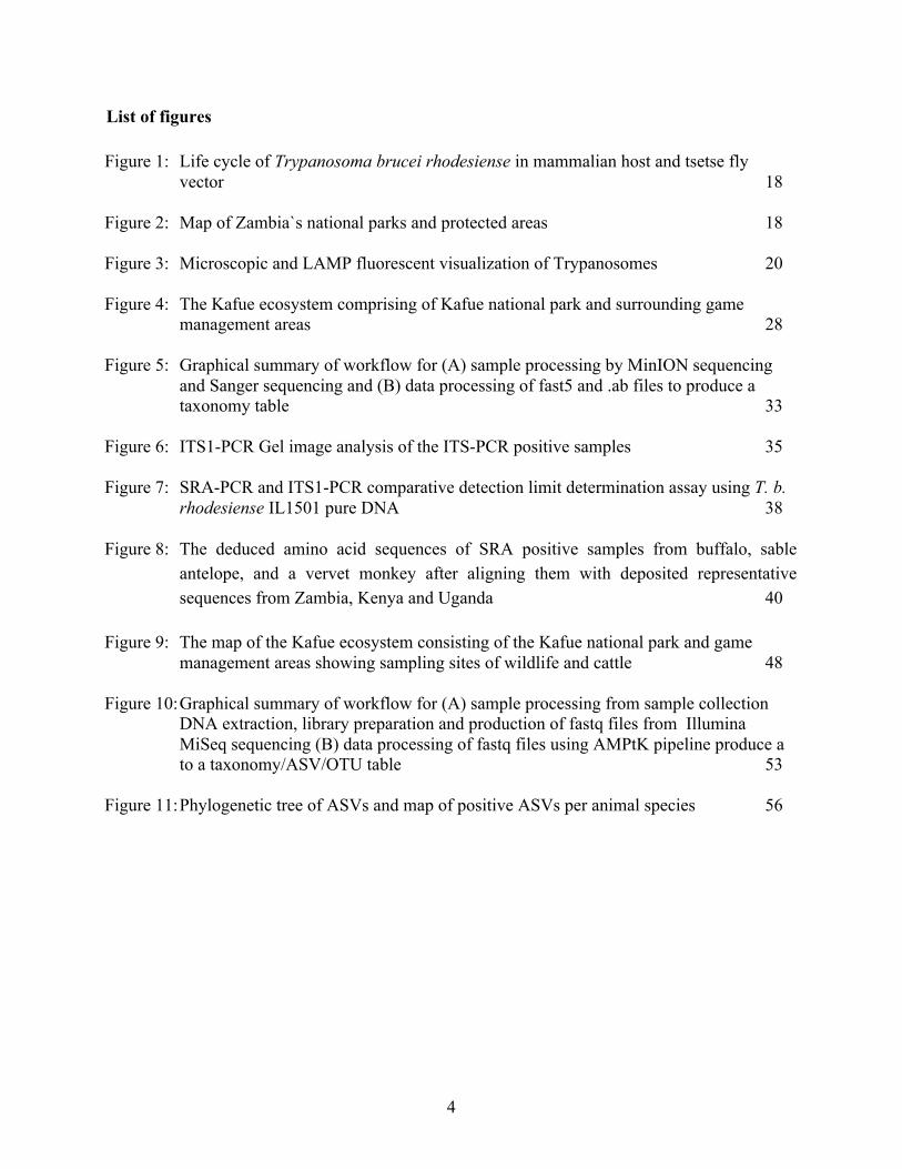
List of figures Figure 1: Life cycle of Trypanosoma brucei rhodesiense in mammalian host and tsetse fly
vector 18 Figure 2: Map of Zambia`s national parks and protected areas 18 Figure 3: Microscopic and LAMP fluorescent visualization of Trypanosomes 20 Figure 4: The Kafue ecosystem comprising of Kafue national park and surrounding game
management areas 28 Figure 5: Graphical summary of workflow for (A) sample processing by MinION sequencing
and Sanger sequencing and (B) data processing of fast5 and .ab files to produce a taxonomy table 33
Figure 6: ITS1-PCR Gel image analysis of the ITS-PCR positive samples 35 Figure 7: SRA-PCR and ITS1-PCR comparative detection limit determination assay using T. b.
rhodesiense IL1501 pure DNA 38 Figure 8: The deduced amino acid sequences of SRA positive samples from buffalo, sable
antelope, and a vervet monkey after aligning them with deposited representative sequences from Zambia, Kenya and Uganda 40
Figure 9: The map of the Kafue ecosystem consisting of the Kafue national park and game
management areas showing sampling sites of wildlife and cattle 48 Figure 10: Graphical summary of workflow for (A) sample processing from sample collection
DNA extraction, library preparation and production of fastq files from Illumina MiSeq sequencing (B) data processing of fastq files using AMPtK pipeline produce a to a taxonomy/ASV/OTU table 53
Figure 11: Phylogenetic tree of ASVs and map of positive ASVs per animal species 56

5
List of tables Table 1: Table showing primers and primers sequencies 30 Table 2: The MinION sequence results of ITS-PCR amplicons 36 Table 3: Summary of ITS1-PCR/NGS and SRA-PCR/NGS analysis and diagnosis of
trypanosome in mammalian wildlife species in Kafue National Park 37 Table 4: Detection of heamoparasites in wildlife species and cattle from the Kafue ecosystem
using RLB-PCR 49 Table 5: Primers used for piroplasm parasite etection 51 Table 6: Diversity of piroplasmas detected in wildlife and cattle samples collected from the
Kafue ecosystem 57

6
List of publications related to this dissertation
Contents of chapter 1 have been published in the PLoS Neglected Tropical Diseases.
Squarre, D., Kabongo, I., Munyeme, M., Mumba, C., Mwasinga, W., Hachaambwa, L., Sugimoto, C., Namangala, B., 2016. Human African Trypanosomiasis in the Kafue National Park, Zambia. PloS Negl. Trop. Dis. 10, e0004567.
Contents of chapter 2 have been published in the International Journal for Parasitology: Parasites and Wildlife.
Squarre, D., Hayashida, K., Gaithuma, A., Chambaro, H., Kawai, N., Moonga, L., Namangala, B., Sugimoto, C., Yamagishi, J. 2020. Diversity of trypanosomes in wildlife of the Kafue ecosystem, Zambia. Int. J. Parasitol. Parasites Wildl. 12, 34-41.
Contents of chapter 3 have been published in Parasites and Vectors.
Squarre, D.*, Nakamura, Y.*, Hayashida, K., Kawai, N., Chambaro, H., Namangala, B., Sugimoto, C., Yamagishi, J. 2020. Investigation of the piroplasm diversity circulating in wildlife and cattle of the greater Kafue ecosystem, Zambia. Parasit. Vectors 13, 599. *David Squarre and Yukiko Nakamura contributed equally to this work.
7
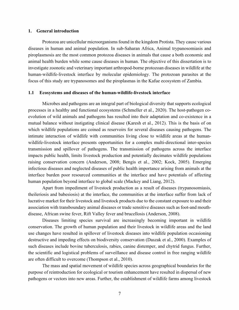
1. General introduction
Protozoa are unicellular microorganisms found in the kingdom Protista. They cause various diseases in human and animal population. In sub-Saharan Africa, Animal trypanosomiasis and piroplasmosis are the most common protozoa diseases in animals that cause a both economic and animal health burden while some cause diseases in human. The objective of this dissertation is to investigate zoonotic and veterinary important arthropod-borne protozoan diseases in wildlife at the human-wildlife-livestock interface by molecular epidemiology. The protozoan parasites at the focus of this study are trypanosomes and the piroplasmas in the Kafue ecosystem of Zambia.
1.1 Ecosystems and diseases of the human-wildlife-livestock interface
Microbes and pathogens are an integral part of biological diversity that supports ecological processes in a healthy and functional ecosystems (Schmeller et al., 2020). The host-pathogen co-evolution of wild animals and pathogens has resulted into their adaptation and co-existence in a mutual balance without instigating clinical disease (Karesh et al., 2012). This is the basis of on which wildlife populations are coined as reservoirs for several diseases causing pathogens. The intimate interaction of wildlife with communities living close to wildlife areas at the human-wildlife-livestock interface presents opportunities for a complex multi-directional inter-species transmission and spillover of pathogens. The transmission of pathogens across the interface impacts public health, limits livestock production and potentially decimates wildlife populations raising conservation concern (Anderson, 2008; Bengis et al., 2002; Kock, 2005). Emerging infectious diseases and neglected diseases of public health importance arising from animals at the interface burden poor resourced communities at the interface and have potentials of affecting human population beyond interface to global scale (Mackey and Liang, 2012).
Apart from impediment of livestock production as a result of diseases (trypanosomiasis, theileriosis and babesiosis) at the interface, the communities at the interface suffer from lack of lucrative market for their livestock and livestock products due to the constant exposure to and their association with transboundary animal diseases or trade sensitive diseases such as foot-and mouth-disease, African swine fever, Rift Valley fever and brucellosis (Anderson, 2008).
Diseases limiting species survival are increasingly becoming important in wildlife conservation. The growth of human population and their livestock in wildlife areas and the land use changes have resulted in spillover of livestock diseases into wildlife population occasioning destructive and impeding effects on biodiversity conservation (Daszak et al., 2000). Examples of such diseases include bovine tuberculosis, rabies, canine distemper, and chytrid fungus. Further, the scientific and logistical problems of surveillance and disease control in free ranging wildlife are often difficult to overcome (Thompson et al., 2010).
The mass and spatial movement of wildlife species across geographical boundaries for the purpose of reintroduction for ecological or tourism enhancement have resulted in dispersal of new pathogens or vectors into new areas. Further, the establishment of wildlife farms among livestock

8
areas has created ex situ patches of human-wildlife-livestock interface with varying potential to maintain and transmit infectious diseases at this interface (Bengis et al., 2002). This has created an anthropogenic driven dispersal and vortex of pathogens and their vectors.
1.2 Trypanosomes
Trypanosomes are flagellated unicellular parasitic organisms belonging to the superkindom of Eukaryota (Schoch et al., 2020). Their classification is as follows:
Superkingdom Eukaryota Phylum Euglenozoa Class Kinetoplastea Order Trypanosomatida Family Trypanosomastidae Genus Trypanosoma Through their mechanism of transmission by the vector, trypanosomes are generally
divided into two groups of Stercolaria and Salivaria (Haag et al., 1998; Hoare, 1966).
Stercolarian trypanosomes are transmitted through the hindgut of the vector through fecal contamination. These include trypanosomes of the subgenus Schizotrypanum, Megatrypanum, and Herpetosoma. The pathogenic species of Trypasoma cruzi which causes Chagas disease in south America belong to the subgenus Schizotrypanum. Also, a non-pathogenic species of Trypanosoma theileri which has worldwide distribution in domestic and wild ruminants belongs to the stercolarian subgenus of Megatrypanum. Salivarian trypanosomes are transmitted via the anterial station of the vector during bloodmeal feeding. The salivarian trypanosomes are divided into four subgenera of Duttonella, Nannomonas, Pycnomonas, and Trypanozoon. The specie of Trypanosoma vivax belongs to the subgenus Duttonella while the species of Tr. congolense, Tr. godfreyi, and Tr. simiae belong to the subgenus Nannomonas. Trypanosoma suis belongs to the subgenus of Pycomonas. The fourth subgenus of Trypanozoon consists of three species of Trypansoma equipedum, Tr. evansi and Tr. brucei. Further, Tr. brucei consists of three important subspecies of which Tr. b. rhodesiense and Tr. b. gambiense cause infections in humans while Tr. b. brucei causes infection in animals.
African trypanosomes are transmitted by a dipteran vector of the genus Glossina (tsetse fly) and thus have a confined distribution to that of tsetse flies in sub-Saharan Africa. However, in addition to tsetse flies, Tr. vivax and Tr. evansi are also transmitted mechanically by other hematophagous biting insects such as horseflies (genus: Tabanus) and stable flies (genus: Stomoxys). In addition, Tr. euiperdum is venereal transmitted in equine species. Due to non-reliance on tsetse fly vector for transmission, these three Trypanosoma species have a worldwide distribution beyond sub-Saharan Africa (Baral, 2010).
9
African trypanosomiasis
African trypanosomiasis is a vector-borne disease that is caused by a protozoan parasite of genus Trypanosoma and infects both human and animals. It occurs in sub-Saharan Africa following an extant vector tsetse fly distribution that is maintained by ecological elements that supports the survival of both parasite and vector (Baral, 2010). The burden of trypanosomiasis on public health and livestock production has resulted in significant socioeconomic impact in sub-Saharan Africa. The infection in human is referred to as Human african trypanosomiasis (HAT) and the African animal trypanosomiasis (AAT) refers to infection in animals (Kristjanson et al., 1999). HAT is an infectious neglected tropical disease (NTD) caused by two trypanosome subspecies of Tr. b. gambiense and Tr. b. rhodesiense. Although these two parasites are considered to generally cause HAT, they cause two specific and separate pathologies with distinct epidemiology, pathogenesis, clinical presentation, severity and treatment regime (Kennedy and Rodgers, 2019; Steverding, 2008). Trypanosoma b. gambiense infection (gHAT) causes a chronic disease and is found in western and central Africa. It is predominantly an anthropogenic disease with a minor role played by animal reservoirs. It accounts for over 98% of all reported HAT cases. On the other hand, Tr. b. rhodesiense infection (rHAT) generally causes an acute disease and is found in southern and eastern Africa. The livestock and wildlife reservoirs play a major role in disease occurrence (WHO, 2015a). Clinical signs of HAT are generally divided into two stages following the progression of the disease. Non-specific or non-pathognomonic signs of headache, chancre, intermittent pyrexia, pruritus, lymphadenopathies, weakness, asthenia, anemia, cardiac disorders, endocrine disturbances, musculoskeletal pains and hepatosplenomegaly are characteristic in the first or early stage. The progression of the parasite from blood and lymph into the central nervous system (CNS) leads to the second or late stage which is marked by neurologic symptoms such as sleep disorders, seizures, coma and eventual death. The febrile and neurologic symptoms of HAT are similar to many other diseases and misdiagnosis is not uncommon. The rate of progression from early stage to late stage marks a major difference in clinical symptoms of gHAT and rHAT. In rHAT the rate of progression is relatively acute The recently developed oral monotherapy drug fexinidazole and nifurtimox–eflornithine combination therapy (NECT) for the treatment of both early stage and late stage gHAT has shown to be effective in diminishing the parasite reservoir in humans and reducing transmission (WHO, 2019). On the other hand, staging in rHAT by demonstrating the presence or absence of the parasite in the CNS is important in determining the choice of therapeutic drugs based on the ability to cross the blood brain barrier and the associated adverse side effects (Kennedy, 2013; Mwanakasale and Songolo, 2011). AAT is an important infectious disease with an enormous economic and production impact in cattle, goats and sheep. It is caused by a single or mixed infection of Trypanosoma species of

10
Tr. congolense, Tr. vivax and Tr. brucei. Similar to HAT, the clinical signs of AAT are varied and non-specific and usually misdiagnosed for tick-borne infection. Anemia, pyrexia, loss of condition, and lethargy are the common symptoms seen in AAT (Morrison et al., 2016). Life cycle of African trypanosomes
The life cycle of trypanosomes involves biological processes and stages in both the vector
and mammalian hosts where the parasite undergoes several distinct changes in morphology and metabolic process due to differences in available nutrients and host response (Vigneron et al., 2020). The vector, tsetse fly inoculates the infective metacyclic trypanosomes (trypomastigotes) into a mammalian host during feeding. The parasite evades the host immune response by expressing antigenic variation through the inherent large repertoire of Variant Surface Glycoprotein (VSG). They multiply by binary fission and are carried and distributed throughout the host by the blood and lymphatic system. In the blood, the trypanosomes are heterogeneous and characterized by the proliferative long slender shaped and non-proliferative short stumpy shaped form. The stumpy shaped form is pre-adapted to establish and survive in the tsetse fly midgut following a blood meal by the vector (Matthews et al., 2004). The parasite in the vector’s midgut transform into procyclic trypomastigotes which undergoes differentiation where the blood adapted VSG surface protein coat is lost and the tsetse midgut adaptive procyclin surface protein is acquired (Roditi and Liniger, 2002). The trypanosomes replicate in the midgut and move as immatures via the foregut and proboscis to the salivary gland, while successively undergoing through different stages before maturing again into the mammalian‑infective metacyclic form (Sharma et al., 2008). The life cycle of trypanosomes is summarized in Figure 1.

11
Figure 1: Life cycle of Trypanosoma brucei rhodesiense in mammalian host and tsetse fly vector. The metacyclic form of the trypanosomes are inoculated in to the blood stream of mammalian host (1) and undergoes morphological changes of long slender (2) and short stumpy forms (3). The short stumpy form then enters the midgut of the vector following feeding (4) and they transform into procyclic trypomastigotes. They further differentiate into epimastigotes (5) that migrate to the proboscis to transform again into mammalian infective metacyclic trypomastigotes (6). (image obtained from Stijlemans et al, 2017 under the terms of the creative common attribution license.
12
Diagnosis and control of trypanosomiasis
Several trypanosome diagnostic approaches based on parasitological technique, immunological assay, and molecular methods have been utilized and developed over the years. Microscopy is the most common and basic diagnostic method utilized for the detection of trypanosomes through blood wet smears, Giemsa-stained thick and thin blood smears. Hematocrit and buffy coat technique are also employed to concentrate the parasite in low parasitemia to increase chances of parasite visualization. The general limitation of microscopy is low sensitivity in low parasitemia. Further, microscopy has an inherent constraint on species differentiation in mixed infection.
Immunological methods have been utilized especially in the diagnosis of gHAT and Tr. equiperdum (dourine in equine species). Card agglutination test for trypanosomiasis (CATT) is widely applied to screen gHAT on the basis of detection of antibodies to the Tr. b. gambiense VSG LiTat 1.3 antigen. complement fixation test (CFT) is a well-established method in screening Tr. equiperdum in horses. The general drawback on the use and development of immunologic methods in diagnosis of trypanosomiasis are sensitivity, cross reactions, field practicability, and costs (Eisler et al., 2004).
The wide spread application of molecular methods to diagnose trypanosomes is based on its ability to accurately detect and definitively identify specific species or sub-species of trypanosomes even in mixed infection. The potency of molecular methods hinges on elevated sensitivity to detect conserved DNA or RNA markers/genes and amplify them for sequence analysis coupled with bioinformatic tools (Gaithuma et al., 2019; Njiru et al., 2004). This proficiency facilitates the generation of useful epidemiological data that result into the application of specific and effective disease control strategy. The downside of molecular methods is the requirement of highly specialized human resource, costly equipment and laborious processes. However, field friendly, less cost-prohibitive platform such as loop-mediated isothermal amplification (LAMP) and portable sequencer such as MinION by Oxford Nanopore technologies, continues to be developed for the use in resource poor environment (Marsela et al., 2020; Yamagishi et al., 2017).
Control of trypanosomiasis is largely focused on the elimination or reduction in abundance of the vector tsetse fly with the aim of diminishing the exposure to human and livestock. However, specific control measures of HAT are based on passive and active surveillance for early detection of infection and prudent case management through staging and application of appropriate treatment protocol (Simarro et al., 2008). Vector control measures employed in the control AAT indirectly benefit HAT control especially in rHAT where animal reservoirs play an import role in maintenance and transmission of Tr. b. rhodesiense. In the past, the measures applied included vegetation clearing, elimination of wildlife reservoirs, erection of wildlife barriers/fences and aerial spraying. These methods were discontinued due to conflict and being inconsistent with environmental, ecological and conservation benchmarks (Hocking et al., 1963). The current
13
methods are hinged on the use of insecticides in tsetse targets, baited traps, ground spraying, aerial spraying and animal cleansing. Other methods include tsetse fly sterilization technique and chemotherapy to livestock with clinical infection.
1.3 Piroplasmas
Piroplasmas are intracellular heamoprotozoan parasite of the order piroplasmida which is
characterized by two genera of Theileria and Babesia that cause animal diseases of theileriosis and babesiosis, respectively. These diseases are vector-borne and are transmitted by various genera of Ixodid ticks including Riphicephalus, Amblyomma, Hyalomma, Ixodes, Dermacentor and Haemaphysalis. The infections in domestic and wild vertebrates result in economic losses due to poor production, outbreaks, and mortalities. The increasing interaction of wildlife with the vector and livestock justifies the investigation of diversity of piroplasma in the wildlife reservoir population. The taxonomic classification of piroplasma is shown below:
Superkingdom Eukaryota Phylum Apicomplexa Class Aconoidasida Order Piroplasmida Family (i) Bebesiidae and
(ii) Theileriidae Genus (i) Babesia and
(ii) Theileria Some species of Babesia causes diseases in human and a wide range of domestic animals.
Human babesiosis is zoonotic and an emerging infectious disease caused by Babesia microti (Vannier et al., 2008). Bovine babesiosis is one of the major tick-borne diseases impeding production in cattle resulting from typically Babesia bovis and B. bigemina infection. Equine piroplasmosis is caused by Babesia caballi/Theileria equi while canine babesiosis also referred as biliary fever is caused by Babesia gibsoni, B. canis, B. rossi and B. vogeli. Pathology of babesiosis is based on the parasite exclusive infection to erythrocytes. This erythrocyte destruction by the parasite leads to the clinical picture of anemia and jaundice (Bock et al., 2004). The over production of pro-inflammatory cytokines by macrophages results in vascular permeability, oedma, vascular collapse and endothelial damage (Brown and Palmer, 1999).
Theileria species are diverse but generally are categorized into two groups of schizont “transforming” and “non-transforming” Theileria. The schizont “transforming” group is characterized by inducement of indefinite lymphoproliferative syndrome in leucocyte resulting in severe pathology by destruction of host lymphoid system. The production of pro-inflammatory cytokines adds to the severity of the pathology through damaging to the lung tissue by increased capillary permeability, endothelial damage and oedema. The schizont transforming Theileria also referred to as malignant Theileria include Theileria parva, Th. annulate, and Th. lestoquardi. The
14
species of Th. parva, and Th. annulate are extremely pathogenic to cattle while Th. lestoquardi causes serious clinical disease in goats and sheep (Shiels et al., 2006). The schizont “non-transforming” Theileria also referred to as “benign” Theileria include Th. mutans, Th. verifera, and Th. orientalis which mainly causes anemia (Sugimoto and Fujisaki, 2002).
Infections of T. mutans and T. verifera are not associated with severe disease in cattle and are thus referred to as less pathogenic Theileria. However, their presence in co-infection with Th. parva are known to moderate the severe clinical outcomes of Th. parva (Woolhouse et al., 2015). Life cycle
The life cycle of piroplasmas follows distinct stages of development in both the tick vector
and the animal host that undergoes the general phases of sporogony, merogony and gamogony. There are specific differences in the life cycle of the Babesia and Theileria parasites (Mehlhorn and Schein, 1985).
The inoculation of the Babesia sporozoites by the tick vectors into the vertebrate hosts results in the invasion of erythrocytes. The intra-erythrocytic sporozoites transform into trophozoites that develop into merozoites by the process of merogony. Gamogony begins when the merozoites are transformed into gametocytes which continues to develop in the gut of the tick vectors following feeding to form zygotes. The kinetes are developed from zygotes and multiply by schizogony to transform into sporoblasts which are the precursor for the development of sporozoites. Unique to Babesia is that, in parallel, the kinetes infect the ovaries where they also develop into sporozoites. This phase is important for Babesia as it provides the trait of transovarian transmission (Bock et al., 2004; Potgieter and Els, 1977).
Theileria sporozoites are introduced into the vertebrate host by the tick vector during feeding. The sporozoites enter leukocyte where they differentiate into schizonts and initiate a lymphoproliferative stage which give rise to merozoites (merogony). The merozoites mature into piroplasms in the erythrocyte. In non-transforming species of Theileria such as Th. mutans, Th. verifera, and Th. orientalis, the schizonts do not divide in the lymphocyte and all the multiplication takes place in the erythrocytes. The piroplasms are taken up by the tick during feeding. The piroplasms in the gut of the tick transform to form zygote that develop into sporoblast in the salivary glands as precursor for sporozoites.

15
Piroplasmosis control
The control of piroplasmosis relies on the combination of control strategies based on vector control, chemotherapy, livestock movement control and vaccination. The application of acaricides on livestock is the most common method despite its association with rising acaricide resistance in ticks and environmental pollution. Even though this limitation is related its costs, treatment against piroplasmosis is effective especially when applied early. In the case for Theileria parva, calf immunization by infection with live sporozoites and simultaneous treatment with long acting antibiotic has extensively been applied with measurable success (Bishop et al., 2020). Different approaches to theileriosis vaccination has been used with varying success. The use of Muguga cocktail vaccines consisting of three (Muguga, Kiambu 5, and Serengeti) stocks, has been used to provide broad cross protection on heterologous Th. parva challenge. However there is also criticism to the Muguga cocktail approach since it has a potential to introduce and spread foreign strains of Th. parva contained in the cocktail vaccine to a local cattle and vector population. On the other hand, local vaccine produced from a local strain is also available, which provides specific and homologous protection against the locally circulating Theileria challenge (Geysen et al., 1999). However, the production capacity and applicable areas are still limited.
16
Chapter 1 Human African Trypanosomiasis in the Kafue national park, Zambia
2.1 Introduction
HAT is among the twenty NTDs listed by the World Health Organization (WHO) targeted for elimination as a public health problem (WHO, 2021). It is caused by Tr. b. rhodesiense (eastern and southern Africa) or Tr. b. gambiense (western and central Africa) and is transmitted through the bite of an infected tsetse fly (Glossina species) (WHO, 2012). The tsetse flies acquire their infections from humans or animals harboring the human pathogenic parasites (Wamwiri and Changasi, 2016). The disease is endemic in tropical and subtropical Africa (Steverding, 2008), where it afflicts low-income populations (WHO, 2012). Whereas Tr. b. rhodesiense causes acute HAT (Bentivoglio et al., 2014; Brun et al., 2010), Tr. b. gambiense causes a more chronic form of the disease (Bentivoglio et al., 2014). Although HAT has been re-emerging in most of the old foci within sub-Saharan Africa since the 1970s, with Tr. b. gambiense accounting for more than 98% of the reported cases (Stich et al., 2012), the latest WHO report suggests that the number of new cases have been reduced (WHO, 2015a). In the year 2009, after continued control efforts, the number of cases of HAT reported dropped below 10,000 for the first time in 50 years. This decline in number of cases has continued with 977 new cases reported in 2018 down from 2,164 case reported in 2016 (Franco et al., 2020). However, the estimated number of actual cases is higher and the estimated population at risk is 54 million people. Despite such progress, only a fraction of the population at risk for contracting HAT in sub-Saharan Africa is under surveillance and relatively few cases are diagnosed annually (Namangala et al., 2012; WHO, 2015b). In particular, there is considerable underdiagnosis of rHAT in sub-Saharan Africa, including Zambia, mainly due to lack of HAT surveillance and control programmes (Odiit et al., 2004; Sindato et al., 2008).
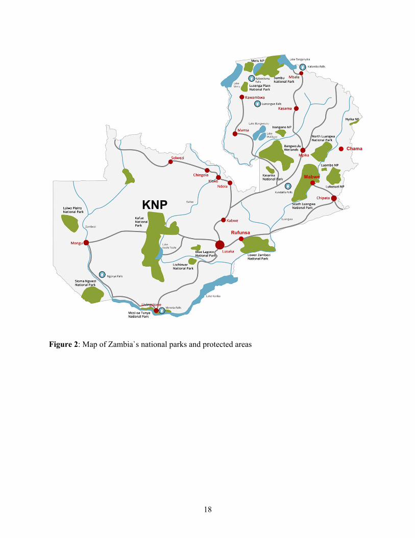
Historically, epidemics of rHAT were reported from the northern and southern regions of the Luangwa valley and the Kafue river basin in the 1960s and early 1970s (Buyst, 1974). According to WHO (WHO, 2015a), Zambia reports <100 new HAT cases annually, mainly from the old foci in the tsetse-infested Luangwa river valley, including the Chama, Mpika, Chipata, Mambwe, and, recently, Rufunsa districts (Figure 2), where the disease is re-emerging (Mulenga et al., 2015; Mwanakasale et al., 2013; Mwanakasale and Songolo, 2011).
The Kafue National Park (KNP) and its surrounding Game Management Areas (GMAs) form the Kafue ecosystem, which is a vast and continuous wildlife conservation area located in the central part of Zambia and rich in biodiversity of high biomass. It is a pristine ecosystem that supports a wide variety of undisturbed flora and fauna of important conservation status (Zambia Wildlife Authority, 2011a). The area also supports the communities that live there by harnessing the benefits from ecotourism and ecosystem services (Siamudaala et al., 2009). Importantly, it has abundant wildlife and tsetse flies.
17
The Kafue ecosystem has in the past reported cases and epidemics of HAT (Mwima, 2001; Zambia Wildlife Authority, 2011a). The Primitive Methodist Church of England established Nkala Mission in 1893, which was later abandoned in 1930 because of tsetse flies and sleeping sickness (Zambia Wildlife Authority, 2011a). Today Nkala lies in the heart of the Kafue ecosystem. Another focus, Itumbi Safari Camp, which was opened in 1958 in the KNP, was closed down in 1959 due to severe cases of sleeping sickness (Mwima, 2001). This demonstrates the historical presence of HAT in the Kafue ecosystem. However, for over 50 years now no reports or notable incidences of HAT have been recorded in the area. Based on this fact, it has been assumed that the area was devoid of HAT despite the obvious presence of tsetse flies. However, in 2015, a HAT case was reported for the first time in 50 years from KNP, 16 kilometers away from Itumbi Safari Camp. In this chapter, detailed information of the case is described.
18
Figure 2: Map of Zambia`s national parks and protected areas Figure 2: Map of Zambia`s national parks and protected areas
Rufunsa
Mabwe
Chama
KNP

19
2.2 Materials and methods 2.2.1 Case history
A 47-year-old man from KNP was hospitalized at the Care for Business Medical Center and Hospital in Lusaka, Zambia, with initial complaints of frequent episodes of headache, fever, dizziness, body malaise, and erythematous skin rashes. The patient reported being bitten multiple times by tsetse flies and other biting arthropods. The history of the patient revealed that he owned a wildlife safari lodge in KNP, about 16 kilometers from the Itumbi Safari Camp. 2.2.2 Diagnostics
Blood collected from the patient was subjected to full blood count, blood biochemistry, rapid diagnostic tests for malaria (Immuno Chromatographic Test and blood slide), typhoid (IgG/IgM) and tick fever (Weil-Felix test). Further microscopic examination of Giemsa-stained thin buffy coat smears was done following centrifugation of the blood. A loop-mediated isothermal amplification (LAMP) analysis for HAT was conducted by incubation at 64°C for 30 min as
described by Hayashida (Hayashida et al., 2015). The cerebral spinal fluid (CSF) was also
collected for microscopic analysis.
2.3 Results
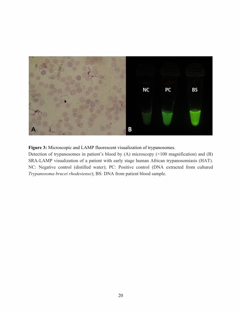
The microscopic examination of the blood slides revealed the presence of trypanosomes in the blood as shown in Figure 3A. The LAMP analysis confirmed the trypanosomes to be Tr. b. rhodesiense which was positive for specific human serum resistance associated (SRA) gene (Figure 3B). The SRA gene is unique to Tr. b. rhodesiense and provides for the identification of the parasite. The results from the rapid diagnostic tests for malaria, typhoid and tick fever were all negative. All parameters of full blood count (FBC) were within normal ranges although the packed
cell volume (PCV) was 42% on the lower margin of the normal range (42%-54%). The biochemical tests results were as follows: alanine aminotransferase 114.3 U/L, bilirubin 82.7 μmol/L, creatinine 42.1 μmol/L and urea 2.78 mmol/L.
20
Figure 3: Microscopic and LAMP fluorescent visualization of trypanosomes. Detection of trypanosomes in patient’s blood by (A) microscopy (×100 magnification) and (B) SRA-LAMP visualization of a patient with early stage human African trypanosomiasis (HAT). NC: Negative control (distilled water); PC: Positive control (DNA extracted from cultured Trypanosoma brucei rhodesiense); BS: DNA from patient blood sample.
21
2.3.1 Treatment
Although the patient had a normal PCV value of 42%, he was critically ill. He was progressively getting weaker, having episodes of unconsciousness, and eventually became comatose. He was placed on assisted breathing ventilator as he could not breathe on his own. The presence of Tr. b. rhodesiense, which was only in the patient’s blood and not in the CSF, coupled with ≤5 white blood cells (WBCs)/mm3 in the CSF, signified early-stage HAT, which was treated with suramin (Brun et al., 2010) along with supportive therapy. A suramin dose of 1 gram dissolved in 5 ml of sterile water was intravenously administered slowly on days 1, 3, 5, 14, and 21. The patient showed a tremendous improvement on the second day by returning to normal consciousness and was thus removed from the ventilator. He appeared brighter and more alert and was responsive to treatment. Before and after each treatment, a sample of blood was collected. Thin buffy coat slides were regularly examined by microscopy to track the course of the disease. His blood was cleared of all parasites after the fifth day of treatment and all clinical signs receded. The patient was hospitalized for observation until all treatment was completed. Other people that were in his company during his stay in the area were screened using microscopy and LAMP and were all negative.
22
2.4 Discussion
Despite KNP having historical presence of HAT (Mwima, 2001; Zambia Wildlife Authority, 2011a), no new cases were recorded for more than 50 years. This may be attributed to several reasons, including non-surveillance of HAT in the area and undetected HAT mortalities through misdiagnosis with other febrile conditions, such as malaria, tuberculosis and HIV/AIDS (Mwanakasale and Songolo, 2011; Namangala et al., 2012). Although most cases of re-emerging HAT in Zambia are mainly reported from Luangwa River Valley and, to a lesser extent, Zambezi River Valley (Hayashida et al., 2015; Mulenga et al., 2015; Mwanakasale et al., 2013; Namangala et al., 2012; Simarro et al., 2011), the patient described herein had no travel history to any of those foci, but was only bitten by tsetse flies from within KNP, strongly suggesting that he contracted the disease from that area. The operational area of the patient is just 16 kilometers from the Itumbi Safari Camp (old focus), which was closed down in 1959 due to severe cases of HAT (Mwima, 2001). This further highlights the continuous risk for park rangers, hunters, tourists, tourism facility operators, and the surrounding population who may become infected, as wildlife in this protected area are niches for HAT (Anderson et al., 2015). The infections at this stage are probably the result of an ecological disturbance that forces an encounter between an infected fly and humans.
The occurrence and distribution of tsetse fly in the Kafue ecosystem form a patchy mosaic of areas with varying degrees of fly densities ranging from none/low to some with very high densities (Zambia Wildlife Authority, 2011a). The area provides a suitable habitat and competent wildlife reservoir that support a thriving vector population. The main objective of protecting this conservation area is to preserve its wildlife and biodiversity by limiting and controlling the anthropogenic activity that directly or indirectly affects and threatens the biodiversity integrity (Zambia Wildlife Authority, 2011a). Tsetse flies are also protected as part of this biodiversity despite being known vectors that transmit HAT. By reason of the fly being protected in conservation areas and its habitat being adequately preserved and undisturbed, tsetse flies have flourished and further maintained the circulation of the parasite(s) they transmit. It can also be said that biodiversity protection, to an extent, reduces the ease of spread of HAT by hindering the effortless encounter of humans with high tsetse fly-infested areas and thus prevents disease transmission.
Human clinical case diagnosis should start with the recognition of the endemic presence of the disease in the area or ecoregion. Through this case, the presence of HAT in the area has been demonstrated. It is therefore recommended that all febrile conditions with a clinical picture resembling septicemia or malaria should have HAT on top of its differential diagnostic list. This is important because early detection and treatment is key to case management (Mwanakasale and Songolo, 2011). HAT progresses through distinct clinical stages that invariably lead to death if left untreated (WHO, 2015a). In the present case, although there was no invasion of the CNS, the reported HAT was diagnosed late in light of the rapid progression of the disease due to absence of historical incidence or recognized/established presence of the disease in the area. This is further
23
demonstrated by the lack of therapeutic drugs and incidences recorded from the local health care facility. It was, however, diagnosed by considering travel history and recollected multiple tsetse fly bites. This report further underscores the importance of accurate diagnosis in the management of HAT. Thus, although the patient had deteriorated to a comatose state with assisted ventilation, therapeutic intervention with suramin provided a complete cure of the disease.
It is important to not only reinforce local health care facilities in areas with demonstrable risk of HAT with relatively quick diagnostic tools such as LAMP and microscopy in addition to therapeutic drugs to avert non-detected HAT-related deaths but also provide the correct and timely treatment of diagnosed cases. Mitigation of HAT in Zambia has primarily occurred through passive detection and treatment (Mwanakasale and Songolo, 2011). Control of HAT requires a multi-sectorial approach by establishing effective coordination of various effective strategies with wildlife managers/ecologists, tsetse biologists, medics, veterinarians, and the media (Mwima, 2001) in a model of the one health approach.
HAT is re-emerging in Zambia’s old foci, mainly in Luangwa and, to a lesser extent, Zambezi River Valleys, as is the case with other sub-Saharan African countries. This chapter described a case of HAT originating from KNP after about 50 years from the last documented case of the disease. This diagnosis is a further reminder for the need of continuous surveillance of HAT in the area. I envisage that this work will stimulate further research to investigate the prevalence of the human-infective trypanosome species in tsetse flies and wildlife from KNP using user-friendly, specific, and sensitive tests to determine the associated risks of contracting HAT by the local inhabitants, park rangers, tourists, and hunters. The next chapter of this thesis seeks to describe the possible reservoirs and diversity of trypanosomes among wildlife community in the Kafue ecosystem.

24
Chapter 2 Diversity of trypanosomes in wildlife of the Kafue ecosystem, Zambia
3.1 Introduction
The findings from chapter 1 demonstrated the presence of rHAT in the KNP through the diagnosis of T. b. rhodesiense in a human subject from the area. This is an important finding as it provides evidence of the presence of rHAT in KNP considering that there had been no reported cases of rHAT in the area for almost 50 years. This chapter seeks to investigate the diversity of trypanosomes circulating in wildlife reservoirs and further explore if KNP is a re-emerging foci of rHAT.
Apart from T. b. rhodesiense that causes rHAT, other trypanosomes such as Tr. b. brucei, Tr. congolense, Tr. simiae, and Tr. vivax causes nagana or AAT in livestock (Simukoko et al., 2011). Other species, such as Tr. godfreyi have unknown pathogenicity, while Tr. theileri is non-pathogenic. Trypanosoma theileri can also be spread by other species of biting flies besides tsetse fly. The inherent foci and circulation of rHAT and nagana in Zambia follow an endemic vector distribution mostly in conservation areas and surrounding areas (Van den Bossche et al., 2010). Wildlife in these conservation areas serve as animal reservoirs for rHAT. Current conservation strategies aimed at increasing wildlife populations in conservation areas (Department of National Parks and Wildlife, 2018; Government of Zambia, 2015; Ministry of National Development Planning, 2017) favor the enrichment of circulating parasites through the elaborate wildlife/tsetse fly interactions.
Conservation areas preserve and protect the environment and important ecological/biodiversity hotspots that maintain ecosystem services (Fanin et al., 2018). Interactions of vectors that transmit the parasite, an abundance of diverse wildlife reservoirs and an accommodating ecology play important roles in sylvatic transmission dynamics and the sustained circulation of the parasite (Auty et al., 2016). Encroachment of human developments and migration of people and their livestock into conservation areas can create, extend or intensify the scale of the existing interface within conservation areas (Bengis et al., 2002; Mweempwa et al., 2015; Stoddard et al., 2009). This has led to increasingly frequent encounters between the vector and human communities, facilitating the spillover of infection from wildlife reservoirs into the human populations and livestock. More than any other diseases, trypanosomiasis is closely associated with the conservation of biodiversity (Anderson et al., 2015).
Blood meal analysis has been used to identify tsetse fly host preferences and ascertain reservoir communities. The two major species found in KNP are Glossina morsitans centralis and G. pallidipes. Although host preferences are highly dependent on host availability, suids and bovids are considered probable favorite host for G. morsitans and G. pallidipes (Clausen et al., 1998; Leak, 1999). Suids, bovids, and primates have been also reported to be blood meal sources for G. morsitans morsitans in Zambia (Gaithuma et al., 2020; Okiwelu, 1977).
25
Molecular identification of trypanosome species and subspecies is often based on PCR amplification of ribosomal RNA sequences of the Internal Transcribed Spacer 1 (ITS1) of the small ribosomal subunit of 18S and 5.8S (Njiru et al., 2004). Recently developed primers and Next Generation Sequencing (NGS) using unique barcodes have been shown to be more sensitive methods of identifying trypanosomes (Gaithuma et al., 2019). However, the subgenus Trypanozoon has the same amplicon size of the ITS1 product, thus very difficult to identify the important human infective Tr. b. rhodesiense using ITS1-PCR. In order to identify Tr. b. rhodesiense, the SRA gene which is unique to this subspecies is targeted for amplification and is the basis for its identification by PCR. The SRA gene is expressed on the surface of Tr. b. rhodesiense and confers trypanolytic resistance to human serum making it human infective (Gibson et al., 2002).
Although efforts to eliminate of gHAT are making progresses, rHAT elimination is proving difficult due to the presence of wildlife and domestic reservoirs. The host range and distribution of reservoir populations should be considered in further studies. Cases of rHAT in Zambia, have traditionally been recorded in Luangwa Valley and the Lower Zambezi ecosystem (Munang’andu et al., 2012). Recently, the KNP recorded the first human case of rHAT after almost half a century (Squarre et al., 2016 and Chapter1). Historically, the KNP has been neglected as a potential focus for rHAT despite the widespread presence of the vectors. The area has been considered devoid of the parasite due to a lack of compelling data on the presence, abundance, and diversity of the circulating parasites, particularly in wildlife reservoir populations.
The Kafue ecosystem is a vast conservation area covering approximately 68,000 km2. It comprises two types of protected areas; the national park itself and GMAs that serve as a buffers around the park. The KNP is a reserve set aside for nature and biodiversity conservation. Only activities such as photographic tourism that pose a minimal risk of disturbance or threat to the landscape, fauna, and flora are sanctioned. Undertakings or land use activities that do not conform to or promote the intrinsic value of the park, such as human settlement, hunting, agriculture/livestock, mining, or logging, are not permitted in the confines of the KNP (Zambia Wildlife Authority, 2011b). However, the nine GMAs surrounding the park allow the proximate cohabitation of wildlife and people. Anthropogenic activities, such as human settlement, hunting, agriculture, infrastructure development, and fishing, are permissible and have been streamlined in land use plans that integrate and optimize wildlife conservation and sustainable socio-economic utilization of natural resources by the communities that live in the GMAs (Zambia Wildlife Authority, 2013a and 2013b). The presence and co-existence of wildlife, tsetse flies, humans, and their livestock makes GMAs a typical human-wildlife-livestock-tsetse fly interface areas in distinct contrast to the national park, which is characterized by an elaborate wildlife-tsetse fly interaction zone. Problems involving trypanosomiasis associated with the interface in the Kafue ecosystem were realized decades ago, as demonstrated by the closure of the Itumbi safari camp in 1956 due to sleeping sickness. In 1972, the tsetse control services cleared vegetation, eliminated wildlife, conducted aerial insecticide spraying, and constructed a game fence/barrier in the Nkhala area on
26
the southeastern border of the KNP to address trypanosomiasis problems arising from the interface and interrupted the interaction of wildlife with tsetse flies with communities and their livestock (Clarke, 1974; Mwima, 2001; Steel and Glendhil, 1982). This chapter is aimed at characterization of the nature of trypanosomes circulating in the wildlife reservoir community in the KNP and the potential risk of spillovers to human and livestock populations via wildlife. The study employed a ITS1-PCR system (Gaithuma et al., 2019) to detect all the African trypanosomes coupled with MinION NGS system. MinION is a transportable and affordable sequencing device designed for field use, that is applicable to epidemiological studies.

27
3.2 Materials and methods
3.2.1 Study location and sample collection
Sample from wild animals were collected in 2017 and 2018 in the KNP. The Kafue
ecosystem is a large conservation area located in central Zambia (between 14°03²S and 16°43²S
and 25°13²E and 26°46²E) comprising the 22,400 km2 of parkland (Zambia Wildlife Authority, 2011a) and 45,406 km2 of GMAs surrounding the park (Zambia Wildlife Authority, 2013c, 2013d, 2013e, 2013f, 2013a, 2013b, 2013g, 2013h). Blood samples were opportunistically collected from wild animals immobilized or captured for the purpose of (i) clinical interventions, (ii) placement of very high frequency (VHF) / global positioning system (GPS) collars to track spatial movements, and (iii) translocations to other wildlife estates within Zambia. Immobilization of the animals followed protocols and methods as described by Kock and Burroughs (Kock and Burroughs, 2012) and La Grange (La Grange, 2006).
Venous blood samples were aseptically collected by venipuncture using 5 ml syringes and sterile 18G or 21G needles via the jugular or ear veins following chemical immobilization and physical restraint. Blood samples were collected in Ethylenediaminetetraacetic acid (EDTA) tubes from 248 free-ranging wild animals comprising ten mammalian wildlife species. Immediately after the collection, the samples were placed in a portable refrigerator at a temperature of 4℃ and later transported to the laboratory, where they were stored at −80oC until analysis. All wild animals immobilized in 2017 and 2018 were included in this study. The GPS positions were recorded for all sampling locations. All samples were collected between the months of May and September of each year. ArcView implemented in ArcGIS was used to make spatial illustrations of the sampling point distribution on the map presented in Figure 4.

28
Figure 4: The Kafue ecosystem comprising of the KNP and surrounding GMAs. The black spots indicate sampling points and the red spots indicate areas where rHAT was detected.
29
3.2.2 Ethical clearance
The blood samples used in this study were collected from free-ranging wild animals in the KNP with the authority from and permits issued by the Department of National Parks and Wildlife, Zambia (TJ/NPW/8/27/1). Ethical clearance for this work was obtained from the Excellence in Research Ethics and Science (ERES) Converge IRB in Zambia (Ref. No. 2019-Jul-010).
3.2.3 DNA extraction
Genomic DNA was extracted from the whole blood samples using a DNA isolation kit for
mammalian blood (Roche Applied Science, Indianapolis, USA). A 200 µL sample of DNA was eluted in Eppendorf tubes and stored at −80 °C until further analysis.
3.2.4 ITS1-PCR and species confirmation by MinION sequencing
A modified ITS1-PCR described by Gaithuma (Gaithuma et al., 2019), was used to identify
and distinguish clinically infective trypanosome species and subspecies. The PCR reaction was
mixed in a 10 µL scale comprised 5.0 µL of Ampdirect plus buffer (Shimadzu, Kyoto, Japan), 0.05
µL of BioTaq HS (Bioline, London, UK), 0.2 µL of 2% dimethyl sulfoxide (DMSO), 2.25 µL of
nuclease free water, 2 µL of eluted DNA as a template, and 0.25 µL each of 10 µM AITS primers as described by Gaithuma (Table 1). Amplification conditions involved an initial denaturation step at 95 °C for 10 min followed by 40 cycles of denaturation at 94 °C for 30 s, annealing at 57°C for 1 min, an extension step of 72 °C for 2 min, and a final extension at 72 °C for 10 min. The PCR products were loaded onto 1.5% agarose gel containing GelRed nucleic acid stain (Biotium, Fremont, CA, USA) and the separated products were visualized under ultraviolet (UV) light in a transilluminator.

30
Table 1: Table showing primers and primers sequencies Primer name
Primer sequence 5` - 3`
illumina AITS-F
a
ACACTCTTTCCCTACACGACGCTCTTCCGATCTNNCGAAAGTTCACCGATATTGC
illumina AITS-R
a
GTGACTGGAGTTCAGACGTGTGCTCTTCCGATCTNNAGGAAGCCAAGTCATCCATC
NITS_F [index1-12]-CGGAAGTTCACCGATATTGC NITS_R [index1-12]-AGGAAGCCAAGTCATCCATC index1 CTATACAGCATGAG Index2 AGAGTCTAGCTAGC Index3 TGCGACACATGTGA Index4 GACTATGCAGTGCA Index5 ACGCGTGCATCTAC Index6 TCGAGTAGTCTCAG Index7 GTATCATGTCAGCA Index8 AGCTAGTAGCTACT Index9 CGAGACGATACTCT Index10 TAGATGCTCGCGAG index11 GCTACGCTGAGTAG index12 TCTCAGCGCAGTGA SRA Fb ATAGTGACAAGATGCGTACTCAACGC SRA Rb AATGTGTTCGAGTACTTCGGTCACGCT
a (Gaithuma et al., 2019) b (Radwanska et al., 2002)
31
Samples that exhibited a positive ITS1-PCR were validated by MinION sequencing. For multiplex sequences, in-house unique index sequences were added at both terminals by PCR. The
index-PCR reaction was conducted by adding 1/60 dilution of the first PCR product into a 10 µL reaction mix with the index primers. The reaction and thermocycler conditions were same as the above, except that amplification was for 15 cycles.
PCR amplicons after indexing were pooled to 12, and each pool was subjected to library construction using a ligation sequencing kit and native barcoding kit 1D (SQK-LSK109 and EXP-NBD103, respectively; Oxford Nanopore Technologies, Oxford, UK) according to the manufacturer’s instruction. Sequencing was conducted using FLO-MIN106 (Oxford Nanopore Technologies). The obtained fast5 was converted to fastq and de-barcoded by Albacore (Oxford Nanopore Technologies). After de-indexing was performed by custom scripts based on the alignment score from the obtained sequences and indexed primers using LAST (Kielbasa et al., 2011), the obtained de-multiplexed sequences were then examined by Basic Local Alignment Tool (BLAST) against a nucleotide database to confirm the infected species. The best hit sequence from BLASTn analysis was retrieved.

32
3.2.5 SRA-PCR and sequencing analysis
To detect human infective Tr. b. rhodesiense, PCR amplifying partial 284 base pairs (bp)
of the SRA gene (Radwanska et al., 2002) was conducted by adding 2 µL of a DNA template to a
10 µL reaction mix comprising of 0.05 µL of BioTaq HS, 5 µL of Ampdirect Plus buffer, 2.55 µL
nuclease free water and 0.2 µL each of the SRA primers (Table 1). The thermocycler conditions consisted of an initial denaturation at 95°C for 10 min and 40 cycles of denaturation at 94°C for 30 s, annealing at 60 °C for 1 min, and extension at 72°C for 2 min, and final extension at 72 °C for 5 min. The PCR products were loaded onto a 2% agarose gel stained with GelRed nucleic acid stain and visualized in a UV transilluminator. The PCR products were purified by ExoSAP-IT (GE Healthcare/USB, USA) following the manufacturer’s instructions. Purified PCR products were sequenced using BigDye Terminator version 3.1 (Thermo Fisher Scientific, Foster city, CA, USA) on an automated capillary sequencer (Thermo Fisher Scientific 3130 Genetic Analyzer; Thermo Fisher Scientific Japan Ltd., Tokyo, Japan). Obtained sequences were analyzed by Molecular Evolutionary Genomics Analysis version 7 (MEGA7) (Kumar et al., 2016) and aligned with the three reference SRA sequences from Uganda (AF097331), Zambia (AJ345058) and Kenya (AJ345057) using MEGA7. A summary of workflow for both sample and data processing is illustrated in Figure 5.

Figure 5: Graphical summary of workflow for sample processing by DNA extraction, ITS/SRA-PCR and sequencing by both MinION sequencing and Sanger sequencing (left side) and data processing of fast5 and ab1 files to produce a taxonomy table (right side).
3.3 Results
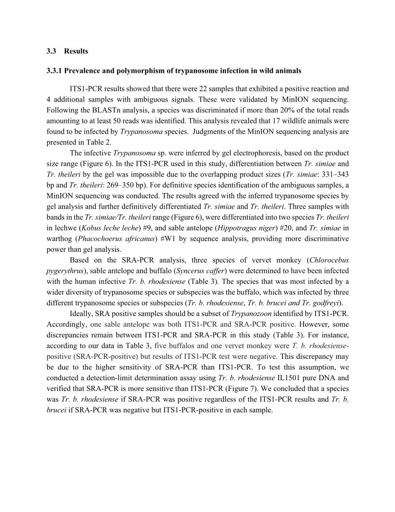
3.3.1 Prevalence and polymorphism of trypanosome infection in wild animals
ITS1-PCR results showed that there were 22 samples that exhibited a positive reaction and 4 additional samples with ambiguous signals. These were validated by MinION sequencing. Following the BLASTn analysis, a species was discriminated if more than 20% of the total reads amounting to at least 50 reads was identified. This analysis revealed that 17 wildlife animals were found to be infected by Trypanosoma species. Judgments of the MinION sequencing analysis are presented in Table 2.
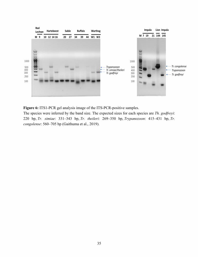
The infective Trypanosoma sp. were inferred by gel electrophoresis, based on the product size range (Figure 6). In the ITS1-PCR used in this study, differentiation between Tr. simiae and Tr. theileri by the gel was impossible due to the overlapping product sizes (Tr. simiae: 331–343 bp and Tr. theileri: 269–350 bp). For definitive species identification of the ambiguous samples, a MinION sequencing was conducted. The results agreed with the inferred trypanosome species by gel analysis and further definitively differentiated Tr. simiae and Tr. theileri. Three samples with bands in the Tr. simiae/Tr. theileri range (Figure 6), were differentiated into two species Tr. theileri in lechwe (Kobus leche leche) #9, and sable antelope (Hippotragus niger) #20, and Tr. simiae in warthog (Phacochoerus africanus) #W1 by sequence analysis, providing more discriminative power than gel analysis.
Based on the SRA-PCR analysis, three species of vervet monkey (Chlorocebus pygerythrus), sable antelope and buffalo (Syncerus caffer) were determined to have been infected with the human infective Tr. b. rhodesiense (Table 3). The species that was most infected by a wider diversity of trypanosome species or subspecies was the buffalo, which was infected by three different trypanosome species or subspecies (Tr. b. rhodesiense, Tr. b. brucei and Tr. godfreyi).
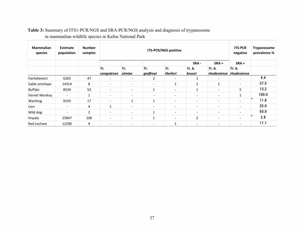
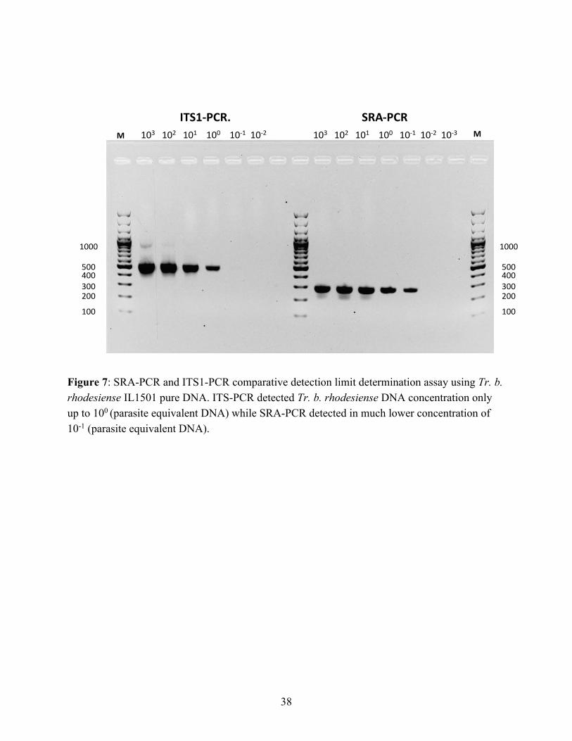
Ideally, SRA positive samples should be a subset of Trypanozoon identified by ITS1-PCR. Accordingly, one sable antelope was both ITS1-PCR and SRA-PCR positive. However, some discrepancies remain between ITS1-PCR and SRA-PCR in this study (Table 3). For instance, according to our data in Table 3, five buffalos and one vervet monkey were T. b. rhodesiense-positive (SRA-PCR-positive) but results of ITS1-PCR test were negative. This discrepancy may be due to the higher sensitivity of SRA-PCR than ITS1-PCR. To test this assumption, we conducted a detection-limit determination assay using Tr. b. rhodesiense IL1501 pure DNA and verified that SRA-PCR is more sensitive than ITS1-PCR (Figure 7). We concluded that a species was Tr. b. rhodesiense if SRA-PCR was positive regardless of the ITS1-PCR results and Tr. b. brucei if SRA-PCR was negative but ITS1-PCR-positive in each sample.
35
Figure 6: ITS1-PCR gel analysis image of the ITS-PCR-positive samples. The species were inferred by the band size. The expected sizes for each species are Th. godfreyi: 220 bp, Tr. simiae: 331–343 bp, Tr. theileri: 269–350 bp, Trypanozoon: 415–431 bp, Tr. congolense: 560–705 bp (Gaithuma et al., 2019).
100200300400
1000
500
100200300400
1000
500
Tr. godfreyiTr. simiae/theileriTrypanozoon
TrypanozoonTr. godfreyi
Tr. congolense
Red Lechwe Hartebeest Sable Buffalo Warthog
M 9 10 12 14 16 20 27 34 39 44 W1 W3
Impala Lion Impala
M 7 19 21 144 145

36
Table 2: The MinION sequence results of ITS-PCR amplicons. ID # Wildlife species Total reads
obtained a Hit reads b (%) Trypanosome
species identification by MinION
9 Red lechwe 556 289 (52.0%) Tr. theileri 10 Hartebeest 8,830 7062 (80.0%) Tr. godfreyi 12 Hartebeest 467 278 (59.5%) Tr. brucei 14 Hartebeest 3,138 2515 (80.1%) Tr. godfreyi 16 Sable antelope 65 54 (83.1%) Tr. brucei 20 Sable antelope 6,319 3,762 (59.5%) Tr. theileri 27 Sable antelope 93 55 (59.1%) Tr. brucei 34 Buffalo 302 93 (30.8%) Tr. brucei 39 Buffalo 547 439 (76.5%) Tr. godfreyi 44 Buffalo 2,336 1956 (83.7%) Tr. godfreyi W1 Warthog 447 136/181
(30.4%/40.5%) Tr. godfreyi/Tr. simiae
W3 Warthog 571 412 (72.2%) Tr. godfreyi 7 Impala 482 454 (94.2%) Tr. brucei 19 Impala 24,213 11272 (46.6%) Tr. godfreyi 21 Impala 139 110 (79.1%) Tr. brucei 144 Lion 8,221 7,886 (46.6%) Tr. congolense 149 Wild dog 1,047 822 (78.5%) Tr. godfreyi
a The total read number obtained after de-indexing. b The obtained reads were blasted against BLASTn database, and the read number of the top hit are shown
37
Table 3: Summary of ITS1-PCR/NGS and SRA-PCR/NGS analysis and diagnosis of trypanosome in mammalian wildlife species in Kafue National Park
Mammalianspecies
Estimatepopulation
Numbersamples
ITS-PCRnegative
Trypanosomeprevalence %
SRA - SRA + SRA +Tr.congolense
Tr.simiae
Tr.godfreyi
Tr.theileri
Tr. b.brucei
Tr. b.rhodesiense
Tr. b.rhodesiense
Hartebeeest 6265 47 - - 2 - 1 - - 6.4
Sable antelope 14314 8 - - - 1 1 1 - 37.5
Buffalo 8534 53 - - 1 - 1 - 5 13.2
Vervet Monkey - 1 - - - - - - 1 100.0
Warthog 9143 17 - 1 1 - - - - 11.8
Lion - 4 1 - - - - - - 25.0
Wild dog - 2 - - 1 - - - - 50.0
Impala 25847 106 - - 1 - 2 - - 2.8
Red Lechwe 12290 9 - - - 1 - - - 11.1
ITS-PCR/NGS positive
38
Figure 7: SRA-PCR and ITS1-PCR comparative detection limit determination assay using Tr. b. rhodesiense IL1501 pure DNA. ITS-PCR detected Tr. b. rhodesiense DNA concentration only up to 100 (parasite equivalent DNA) while SRA-PCR detected in much lower concentration of 10-1 (parasite equivalent DNA).
ITS1-PCR. SRA-PCR103 102 101 100 10-1 10-2 103 102 101 100 10-1 10-2 10-3
100
200300400500
1000
100
200300400500
1000
M M
39
3.3.2 Diversity of SRA gene sequences
The results reveal a discernible substitution of the guanine for adenine at position 370 on the SRA sequence (Z37159.2) obtained from the vervet monkey. Another substitution of adenine for guanine at position 461 was observed on two SRA sequences from two buffalos. All substitutions on the SRA nucleotide sequences altered the codons, resulting in variation in the translated amino acid sequences. The amino acid alanine was substituted by threonine, glycine by aspartic acid and isoleucine by leucine in the sequence from the vervet monkey, buffalos and sable antelopes, respectively, resulting in divergence from the reference sequences from Uganda, Zambia, and Kenya (Figure 8). The obtained divergent partial SRA nucleotide sequences obtained are available at GenBank under the accession number MN635739 (vervet monkey), MN635743 and MN635744 (buffalo) and MN635738 (sable antelope).

40
Figure 8: The amino acid sequences of SRA positive samples from buffalo, sable antelope, and vervet monkey after aligning them with deposited representative sequences from Zambia, Kenya, and Uganda showed variation in the SRA amino acid sequences of Tr. b. rhodesiense at positions 124, 150 and 154. (Gene Bank accession numbers: 43Buffalo = MN635743, 46Buffalo = MN635744, 39Buffalo = MN635742, 36Buffalo = MN635741, 38Buffalo = MN635740, 90VervetMonkey = MN635739, 20Sable = MN635738)
41
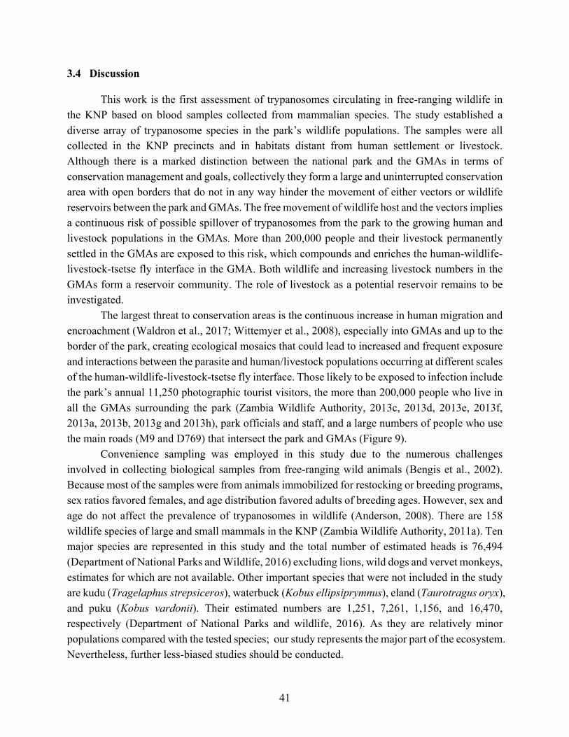
3.4 Discussion This work is the first assessment of trypanosomes circulating in free-ranging wildlife in the KNP based on blood samples collected from mammalian species. The study established a diverse array of trypanosome species in the park’s wildlife populations. The samples were all collected in the KNP precincts and in habitats distant from human settlement or livestock. Although there is a marked distinction between the national park and the GMAs in terms of conservation management and goals, collectively they form a large and uninterrupted conservation area with open borders that do not in any way hinder the movement of either vectors or wildlife reservoirs between the park and GMAs. The free movement of wildlife host and the vectors implies a continuous risk of possible spillover of trypanosomes from the park to the growing human and livestock populations in the GMAs. More than 200,000 people and their livestock permanently settled in the GMAs are exposed to this risk, which compounds and enriches the human-wildlife-livestock-tsetse fly interface in the GMA. Both wildlife and increasing livestock numbers in the GMAs form a reservoir community. The role of livestock as a potential reservoir remains to be investigated.
The largest threat to conservation areas is the continuous increase in human migration and encroachment (Waldron et al., 2017; Wittemyer et al., 2008), especially into GMAs and up to the border of the park, creating ecological mosaics that could lead to increased and frequent exposure and interactions between the parasite and human/livestock populations occurring at different scales of the human-wildlife-livestock-tsetse fly interface. Those likely to be exposed to infection include the park’s annual 11,250 photographic tourist visitors, the more than 200,000 people who live in all the GMAs surrounding the park (Zambia Wildlife Authority, 2013c, 2013d, 2013e, 2013f, 2013a, 2013b, 2013g and 2013h), park officials and staff, and a large numbers of people who use the main roads (M9 and D769) that intersect the park and GMAs (Figure 9). Convenience sampling was employed in this study due to the numerous challenges involved in collecting biological samples from free-ranging wild animals (Bengis et al., 2002). Because most of the samples were from animals immobilized for restocking or breeding programs, sex ratios favored females, and age distribution favored adults of breeding ages. However, sex and age do not affect the prevalence of trypanosomes in wildlife (Anderson, 2008). There are 158 wildlife species of large and small mammals in the KNP (Zambia Wildlife Authority, 2011a). Ten major species are represented in this study and the total number of estimated heads is 76,494 (Department of National Parks and Wildlife, 2016) excluding lions, wild dogs and vervet monkeys, estimates for which are not available. Other important species that were not included in the study are kudu (Tragelaphus strepsiceros), waterbuck (Kobus ellipsiprymnus), eland (Taurotragus oryx), and puku (Kobus vardonii). Their estimated numbers are 1,251, 7,261, 1,156, and 16,470, respectively (Department of National Parks and wildlife, 2016). As they are relatively minor populations compared with the tested species; our study represents the major part of the ecosystem. Nevertheless, further less-biased studies should be conducted.

42
A total of six trypanosome species or subspecies (Tr. b. rhodesiense, Tr. godfreyi, Tr. b. brucei, Tr. congolense, Tr. simiae, and Tr. theileri) were detected using a combination of molecular techniques of ITS1-PCR/NGS and SRA-PCR/Sanger sequence analyses. ITS1-PCR/NGS is more sensitive and offers greater accuracy when diagnosing a wide range of trypanosomes comparing with conventional ITS1-PCR gel analysis, which produces relatively imprecise identification of trypanosomes based on band size (Gaithuma et al., 2019). However, the former method cannot discriminate among Trypanozoon subspecies due to the species’s highly conserved genome (Cuypers et al., 2017), and thus SRA-PCR and Sanger sequence analysis were conducted in addition to ITS1-PCR/NGS to identify the important human infective trypanosome Tr. b. rhodesiense. The discrepancies observed in this study between ITS1-PCR and SRA-PCR can be attributed to the high sensitivity of SRA-PCR relative to ITS1-PCR, as demonstrated in the detection-limit determination assay (Figure 7). The low and persistent phases of parasitemia frequently seen in wildlife (Laohasinnarong et al., 2015; Van Den Bossche et al., 2005) can be problematic in detecting the parasites.
Trypanosomes that cause disease in livestock were also detected, including Tr. congolense , Tr. b. brucei, and Tr. simiae. The free-ranging livestock in the GMAs would usually and instantaneously share and access the same pools of resources, such as water and pasture, facilitating the exchange of diseases including trypanosomiasis. Generally, nagana is a major hindrance to livestock production in tsetse-inhabited areas and wildlife reservoirs most likely compounds this problem.
In the last 50 years, no specific rHAT surveillance in KNP has been undertaken to diagnose the disease or demonstrate its presence. Because rHAT is not pathognomonic and its symptoms are similar to those of many other febrile conditions, it is likely that the disease has been misdiagnosed and masked by malaria and other common diseases. The recent focus on HIV/AIDS, malaria, and tuberculosis, and the widespread deviation from the routine use of microscopy due to increased use of rapid detection test kits for diseases such as malaria, may have hampered the proper diagnosis of rHAT (Mwanakasale and Songolo, 2011; Namangala et al., 2012).
This study revealed substantial infection rates of Tr. b. rhodesiense (12.5% in sable antelopes and 9.4% in buffalos) in the wildlife population of the KNP and further supports the recent diagnosis of Tr. b. rhodesiense in an adult male patient from the KNP using LAMP, which demonstrated the presence of rHAT in the KNP (Squarre et al., 2016). The outcomes and results of this study confirm the presence of rHAT in the KNP and further confirm that the KNP is a genuine neglected and re-emerging focus of rHAT. The first step to control this disease is to acknowledge its presence and the potential risk it presents. Based on the results reported here, it is recommended that the already existing and accessible health facilities should be bolstered with the capacity to diagnose and treat rHAT within and around the KNP (Holmes, 2015).
Buffalo had the highest infection rates of Tr. b. rhodesiense in this study. The gregarious nature of buffalo in the KNP leads to the formation of large herds of 20 to 200 animals. Their ability to traverse different habitats combined with the presence of the disease vector could explain
43
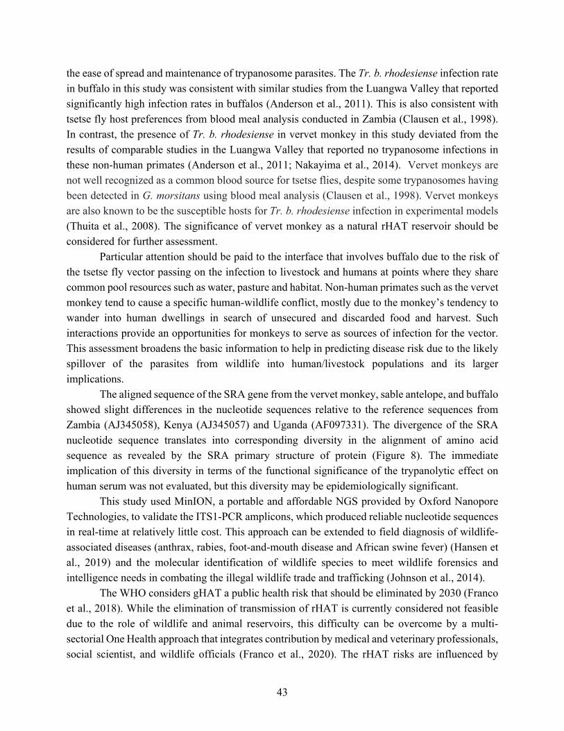
the ease of spread and maintenance of trypanosome parasites. The Tr. b. rhodesiense infection rate in buffalo in this study was consistent with similar studies from the Luangwa Valley that reported significantly high infection rates in buffalos (Anderson et al., 2011). This is also consistent with tsetse fly host preferences from blood meal analysis conducted in Zambia (Clausen et al., 1998). In contrast, the presence of Tr. b. rhodesiense in vervet monkey in this study deviated from the results of comparable studies in the Luangwa Valley that reported no trypanosome infections in these non-human primates (Anderson et al., 2011; Nakayima et al., 2014). Vervet monkeys are not well recognized as a common blood source for tsetse flies, despite some trypanosomes having been detected in G. morsitans using blood meal analysis (Clausen et al., 1998). Vervet monkeys are also known to be the susceptible hosts for Tr. b. rhodesiense infection in experimental models (Thuita et al., 2008). The significance of vervet monkey as a natural rHAT reservoir should be considered for further assessment.
Particular attention should be paid to the interface that involves buffalo due to the risk of the tsetse fly vector passing on the infection to livestock and humans at points where they share common pool resources such as water, pasture and habitat. Non-human primates such as the vervet monkey tend to cause a specific human-wildlife conflict, mostly due to the monkey’s tendency to wander into human dwellings in search of unsecured and discarded food and harvest. Such interactions provide an opportunities for monkeys to serve as sources of infection for the vector. This assessment broadens the basic information to help in predicting disease risk due to the likely spillover of the parasites from wildlife into human/livestock populations and its larger implications.
The aligned sequence of the SRA gene from the vervet monkey, sable antelope, and buffalo showed slight differences in the nucleotide sequences relative to the reference sequences from Zambia (AJ345058), Kenya (AJ345057) and Uganda (AF097331). The divergence of the SRA nucleotide sequence translates into corresponding diversity in the alignment of amino acid sequence as revealed by the SRA primary structure of protein (Figure 8). The immediate implication of this diversity in terms of the functional significance of the trypanolytic effect on human serum was not evaluated, but this diversity may be epidemiologically significant.
This study used MinION, a portable and affordable NGS provided by Oxford Nanopore Technologies, to validate the ITS1-PCR amplicons, which produced reliable nucleotide sequences in real-time at relatively little cost. This approach can be extended to field diagnosis of wildlife-associated diseases (anthrax, rabies, foot-and-mouth disease and African swine fever) (Hansen et al., 2019) and the molecular identification of wildlife species to meet wildlife forensics and intelligence needs in combating the illegal wildlife trade and trafficking (Johnson et al., 2014).
The WHO considers gHAT a public health risk that should be eliminated by 2030 (Franco et al., 2018). While the elimination of transmission of rHAT is currently considered not feasible due to the role of wildlife and animal reservoirs, this difficulty can be overcome by a multi-sectorial One Health approach that integrates contribution by medical and veterinary professionals, social scientist, and wildlife officials (Franco et al., 2020). The rHAT risks are influenced by

44
wildlife distribution, habitat management and land-use. A more holistic ecological approach should be advanced (WHO, 2015b).
In summary, the molecular tests employed in this study revealed that trypanosomes parasites are circulating in wildlife reservoirs while human and livestock populations in and around the Kafue ecosystem expand. Human infective/zoonotic trypanosome, T. b. rhodesiense was also detected further suggesting that the KNP is a neglected and re-emerging focus of rHAT.

45
4. Chapter 3 Investigation of the piroplasm diversity circulating in wildlife and cattle of the greater Kafue ecosystem, Zambia
4.1 Introduction
Piroplasmida is an order of intracellular heamoprotozoan parasites that belong to the
phylum Apicomplexa. The species of genera Theileria and Babesia cause clinical disease in vertebrate hosts including domestic and wild animals (Allsopp et al., 1994; Schreeg et al., 2016). The parasites are transmitted by vectors of ixodid ticks and cause considerable socio-economic impact on livestock production in sub-Saharan Africa, threatening livelihoods and food security (Bishop et al., 2004). The Theileria and Babesia genera are consists of a wide diversity of species and genotypes (Criado-fornelio et al., 2003; Criado-Fornelio et al., 2004).
Wildlife plays an important role in the circulation, maintenance and evolution of these parasites. African buffalos (S. caffer) for example, are reservoirs of buffalo-derived T. parva that causes theileriosis or corridor disease in cattle (Kock et al., 2014; Morrison et al., 2020). This disease is transmitted from buffalo to cattle and not between cattle, because cattle acutely die before piroplasms emerge or infect new ticks (Uilenberg, 1999; Yusufmia et al., 2010). Conversely, in East coast fever (ECF) caused by T. parva circulating among cattle population, some infected cattle survive due to immune response and occasional chemotherapy, and then becoming asymptomatic carriers and leading to the continuous spread of the parasite among cattle (Fandamu, 2005).
Although Theileria is by far the most important piroplasma causing considerable effect on livestock production, Babesia also cause a wide range of infectious diseases in domestic animals. Redwater in cattle, canine babesiosis, and equine piroplasmosis are caused by Babesia bigemina/B. bovis, B. canis, and B. caballi/Theileria equi, respectively. Several wildlife species are natural hosts of a wide diversity of piroplasma that are pathogenic and non-pathogenic to domestic animals.
The Kafue ecosystem, measuring 68,000 km2 in size, is a large conservation area in central Zambia. It is composed of the KNP and nine adjacent GMAs that act as a buffer to the national park. The national park is host to numerous wildlife species and particularly is devoid of human settlements and livestock. The GMAs that immediately surround the park are notably characteristic of wildlife cohabiting with communities and their livestock, thus forming a wildlife-livestock interface area (Bandyopadhyay and Tembo, 2010; Kock, 2005). The potential for likely spillover of arthropod-borne pathogens such as piroplasmas from wildlife to livestock occurs when a common ecological niche is shared in the presence of a competent vector (Munang’andu et al., 2009). In addition to the interface in conservation areas, the growing game ranching industry in Zambia has integrated wildlife and livestock farming, creating widespread patches of ex-situ wildlife-livestock interface areas across the country. The primary source of wildlife for stocking

46
game ranches is conservation areas such as the greater Kafue ecosystem. This is likely to spread parasites and create a vortex of piroplasm parasites across the country.
Highlighting comprehensive piroplasm parasite community composition including cryptic species/genotype diversity of circulating parasites in wildlife, livestock and vector population is essential to understand disease ecology and to prepare optimized counter measures. Reservoir of important pathogens and their transmission path will be illustrated by this approach. Pathogen-pathogen interaction under mixed infection which may cause discriminated manifestation is another interest. Understanding the parasite community also has implication on the choice of assays to adopt in control options such as calf immunization, and in epidemiology studies of piroplasm infections (Eygelaar et al., 2015; Glidden et al., 2019; Hemmink et al., 2018; Muleya et al., 2012; Pienaar et al., 2014). It provides basic information for the selection of live or recombinant vaccines to be used in a specific area as well (Geysen et al., 1999).
To investigate the parasite diversity, deep amplicon sequencing of the 18S rRNA V4 hyper-variable region by NGS technology has been developed (Bishop et al., 2015; Chaudhry et al., 2019; Glidden et al., 2019; Hemmink et al., 2018). The scheme has been adopted for cattle (Bishop et al., 2015), African buffalo (Glidden et al., 2019; Hemmink et al., 2018), Asian buffalo, cattle and sheep (Chaudhry et al., 2019). I also adopted the scheme and expanded the target to whole wildlife in this study to investigate and illustrate a diversity of piroplasm community in wildlife and cattle in a discrete geographical region of the greater Kafue ecosystem of Zambia.
47
4.2 Materials and methods
4.2.1 Sample collection and DNA extraction
The geographical area of sampling and the method used in collecting the wildlife samples
are the same as described in chapter 2 (Figure 9). In addition to the 248 samples described in chapter 2, additional 4 sitatunga and 1 baboon samples were added to bring the total to 253 wildlife samples. Extra 232 blood samples were collected from cattle in the interface between the Nkhala GMA and the surrounding communities areas in Zambia’s Itezhi-Tezhi district between April and May, 2019 (Table 4). Primarily these samples were collected for a study on genetic diversity of trypanosomes in cattle population and tsetse fly (Nakamura et al., 2021).
The wildlife samples for this study were collected under the ethical approval and permission as stated in chapter 2. Additional ethical clearance for the study in cattle was obtained from ERES Converge IRB (Ref. No. 2019-Feb-081)
From each blood sample collected, genomic DNA was extracted using the DNA Isolation Kit for Mammalian Blood for wild animal samples and QuickGene DNA whole blood kit S (Kurabo, Osaka, Japan) for cattle samples as per manufacturer’s protocol. A final volume of 200
µL of DNA was eluted in tubes and stored at -80 °C until analysis.
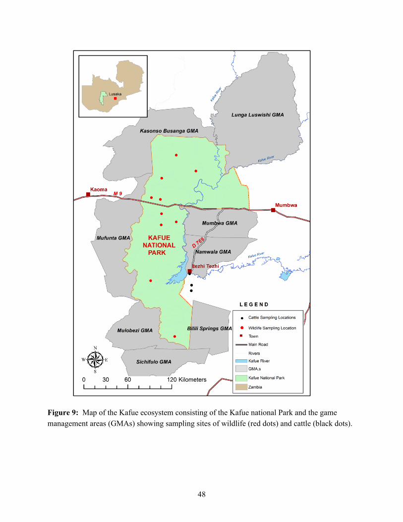
48
Figure 9: Map of the Kafue ecosystem consisting of the Kafue national Park and the game management areas (GMAs) showing sampling sites of wildlife (red dots) and cattle (black dots).

49
Table 4: Detection of heamoparasites in wildlife species and cattle from the Kafue ecosystem using RLB-PCR and Miseq sequence.
Species sampled
Number
of
samples
Positive
number of
RLB-PCR
Positive rate of
RLB-PCR (%)
Impala (Aepyceros melampus) 106 65 61
Hartebeest (Alcelaphus buselaphus) 47 19 40
Sable antelope (Hippotragus niger) 8 8 100
Lion (Panthera leo) 4 1 25
Wild dog (Lycaon pictus) 2 2 100
Sitatunga (Tragelaphus spekii) 4 3 74
Buffalo (Syncerus caffer) 53 33 62
Lechwe (Kobus leche leche) 9 0 0
Cheetah (Acinonyx jubatus) 1 0 0
Ververt monkey (Chlorocebus pygerythrus) 1 0 0
Baboon (Papio ursinus) 1 0 0
Warthog (Phacochoerus africanus) 17 0 0
Cattle (Bos taurus) 232 147 63
Total 483 278 58

50
4.2.2 RLB-PCR amplification and illumina library preparation
Amplification of the V4 hypervariable region of the 18S rRNA gene was obtained by piroplasma specific RLB-PCR using primers RLB-F and RLB-R (Table 5) (Gubbels et al., 1999).
A reaction mix of 10 µL containing 5.0 µL Ampdirect plus buffer (Shimadzu, Kyoto, Japan), 3.95
µL PCR grade water, 0.05 µL Bio Taq HS, 0.5 µL DNA template and 0.25 µL each of the RLB primers. The thermocycler conditions were 94 °C for 10 min denaturation and 40 cycles of 94 °C for 1 min, annealing at 50 °C for 1 minute, extension at 72 °C for 1.5 min and final extension at 72 °C for 10 min.
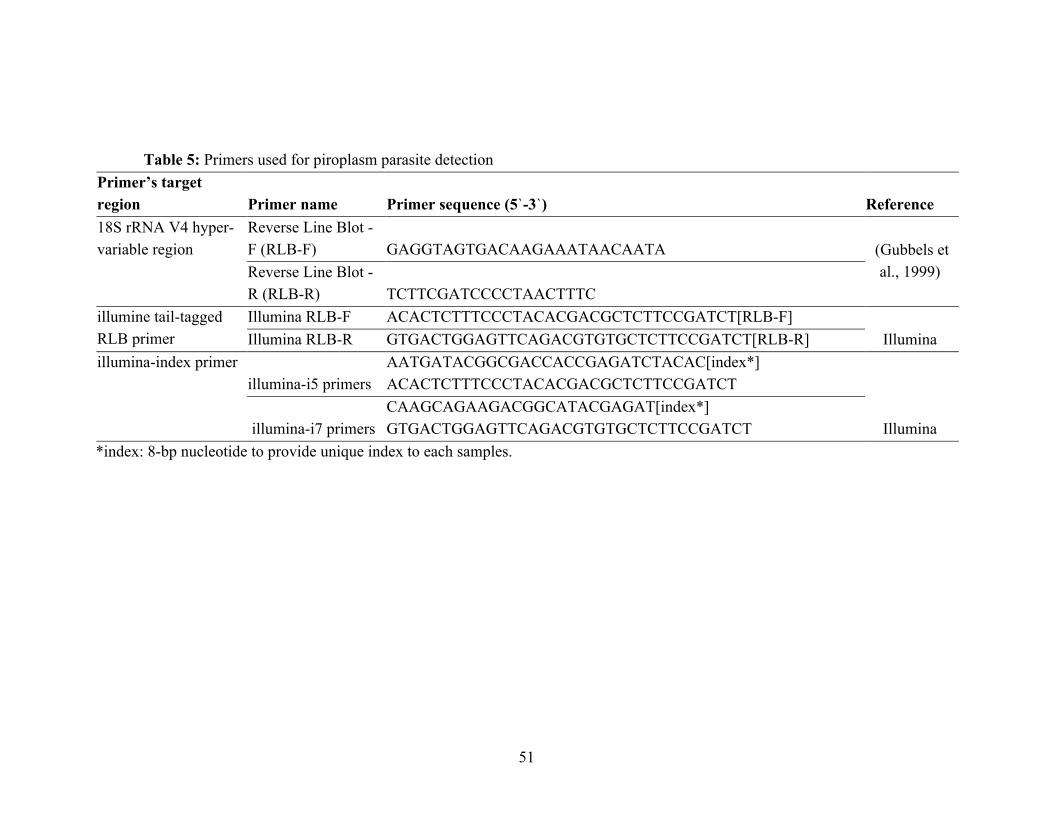
51
Table 5: Primers used for piroplasm parasite detection Primer’s target region Primer name Primer sequence (5`-3`) Reference 18S rRNA V4 hyper-variable region
Reverse Line Blot - F (RLB-F) GAGGTAGTGACAAGAAATAACAATA (Gubbels et
al., 1999) Reverse Line Blot - R (RLB-R) TCTTCGATCCCCTAACTTTC
illumine tail-tagged RLB primer
Illumina RLB-F ACACTCTTTCCCTACACGACGCTCTTCCGATCT[RLB-F] Illumina Illumina RLB-R GTGACTGGAGTTCAGACGTGTGCTCTTCCGATCT[RLB-R]
illumina-index primer illumina-i5 primers
AATGATACGGCGACCACCGAGATCTACAC[index*] ACACTCTTTCCCTACACGACGCTCTTCCGATCT
Illumina illumina-i7 primers CAAGCAGAAGACGGCATACGAGAT[index*] GTGACTGGAGTTCAGACGTGTGCTCTTCCGATCT
*index: 8-bp nucleotide to provide unique index to each samples.

52
The second PCR adding illumina tail was conducted using 100 times diluted 1st PCR
amplicons as template. The reaction volume of 10 µL comprised the same volumes of reagents as
the first RLB-PCR but instead replaced the primer with 10 µM illumina tail-tagged RLB primers
(Table 5). The thermocycler conditions were the same as the first PCR except amplification was
set at 12 cycles.
The illumina tail-tagged amplicons from the second PCR were then diluted 50 times and 1
µL was added to a 20 µL reaction mixture for index PCR. The other reagents included 4 µL of 5
x buffer, 1.4 µL MgCl2 (25mM), 0.5 µL 10mM dNTP mix, 1 µL mixed illumina-index primer
(Table 5), 11.975 µL nuclease-free water and 0.125 µL KAPA Taq Extra (Takara, Japan). The
indexing PCR was run with thermocycler condition of 95 °C initial denaturation for 5 min followed
by 15 cycles of 92 °C for 30 sec, 45 °C for 30 sec and 72 °C for 30 sec and final extension at 72
°C for 15 min. The obtained amplicons were quantified by agarose gel electrophoresis, pooled,
then purified using Wizard SV Gel and PCR Clean-Up system (Promega, Madison, WI, USA).
4.2.3 Amplicon sequencing and bioinformatic analysis
The RLB-PCR amplicon library was sequenced with the MiSeq (Chaudhry et al., 2019;
Glidden et al., 2019) using a 300 bp paired-end sequencing protocol and the MiSeq sequencing
reagent kits v3 (Illumina, Hayward, CA, USA) with 25% PhiX DNA spike-in control according to
the manufacturer’s instructions. Quality control and filtering were conducted with Trimmomatic
(Bolger et al., 2014) using the following parameter; TRAILING:20, SLIDINGWINDOW:4:15 and
MINLEN:36. Concatenation between forward and reverse reads and primer trimming were
conducted with AMPtk (Palmer et al., 2018), allowing minimum merged length of 400 bp. Primer
sequences to be trimmed were GAGGTAGTGACAAGAAATAACAATA and
TCTTCGATCCCCTAACTTTC for forward and reverse reads, respectively.
A set of amplicon sequence variants (ASVs) was generated by DADA2 and LULU in the AMPtk
package using the default parameters. The obtained sequences were annotated based on sequence
homology with the BLAST, and non-redundant nucleotide database by NCBI using -
max_target_seqs 1, -perc_identity 70, -qcov_hsp_perc 70 and -evalue 1e-20 as a set of
parameters (Altschul et al., 1990). ASVs which assigned to other than Alveolata organisms were
filttered. Operational taxonomic units (OTUs) was further generated by clustering the ASVs
using usearch (Edgar, 2010) with 99% identity as clustering threshold. Observed ASVs in each
sample were filtered out if number of the assigned reads were less than 1% of the total number of
assigned reads. A graphical summary of sample and all data processing are shown in Figure 10

53
Figure 10: Graphical summary of workflow for (left side) sample processing from sample collection, DNA extraction, library preparation and production of fastq files from Illumina MiSeq sequencing and (right side) data processing of fastq files using AMPtK pipeline produce a taxonomy/ASV/OTU table.
54
4.2.4 Phylogenetic analyses
Phylogenetic relationship among ASVs were analyzed using Neighbor-Joining method (Saitou and Nei, 1987) implemented in MEGA X (Kumar et al., 2018). The evolutionary distances were computed using the maximum composite likelihood method (Tamura et al., 2018) and default parameters with 10,000 bootstraps. Visualization and annotation were conducted using iTOL v5.5 (Letunic and Bork, 2019). Each clade was annotated based on sequence identity obtained by the BLAST analysis.
55
4.3. Results 4.3.1 Detection of piroplasm parasite by PCR and taxonomical annotation
Out of 253 sampled wild animals, 61% (65/106) of impalas, 40% (19/47) of hartebeests, 62% (33/53) of buffalos, 75% (3/4) of sitatungas, 25% (1/4) of lions and all the sable antelope (8/8) and wild dogs (2/2) were positive for piroplasmosis by RLB-PCR. In case of cattle, 63% (147/232) were RLB-PCR positive (Table 4). Of the 12 wildlife species sampled and screened, 7 species were infected by piroplasm parasites (Table 4). All the positive amplicons were subjected to sequence analysis to identify their taxonomic classification. In total, 2.80 million raw reads had been obtained from 278 PCR positive samples, then merged into 2.46 million contigs.
A total of 45 ASVs of the V4 hyper-variable region of the 18S rRNA gene were obtained from both wildlife species and cattle sampled from the greater Kafue ecosystem (Table 6). The taxonomic assignment of ASV using BLASTn resulted into the identification of four genera, Theileria, Babesia, Hepatozoon and Colpodella, which consisted of 11, 3, 2 and one known species and 36, 6, 2 and one ASVs, respectively (Table 6).
In the phylogenetic analysis, we observed both Theileria and Babesia clade (Figure 11). The Theileria clade consisted of subclade for Th. velifera, Th. mutans, Th. parva and Th. taurotragi.
The Th. velifera subclade consisted of seven ASVs and sequence identity to Th. velifera was 98.7% to 100% (Table 6), suggesting all of these ASV belong to Th. velifera. The subclade was further divided into two groups based on sequence identity. One was ASV6, 29 and 55 which were detected only in buffalo, while ASV7, 64 and 92 were detected only in cattle while ASV1 was detected in cattle and impala (Figure 11).
A similar correlation among sequence identity and hosts was observed in the Th. mutans clade. ASV26, 42 and 60 were buffalo specific and ASV3, 5, 16 were detected from both buffalo and cattle. All of them had more than 99.5% identity to the reference sequences of Th. mutans (Figure 11 and Table 6).
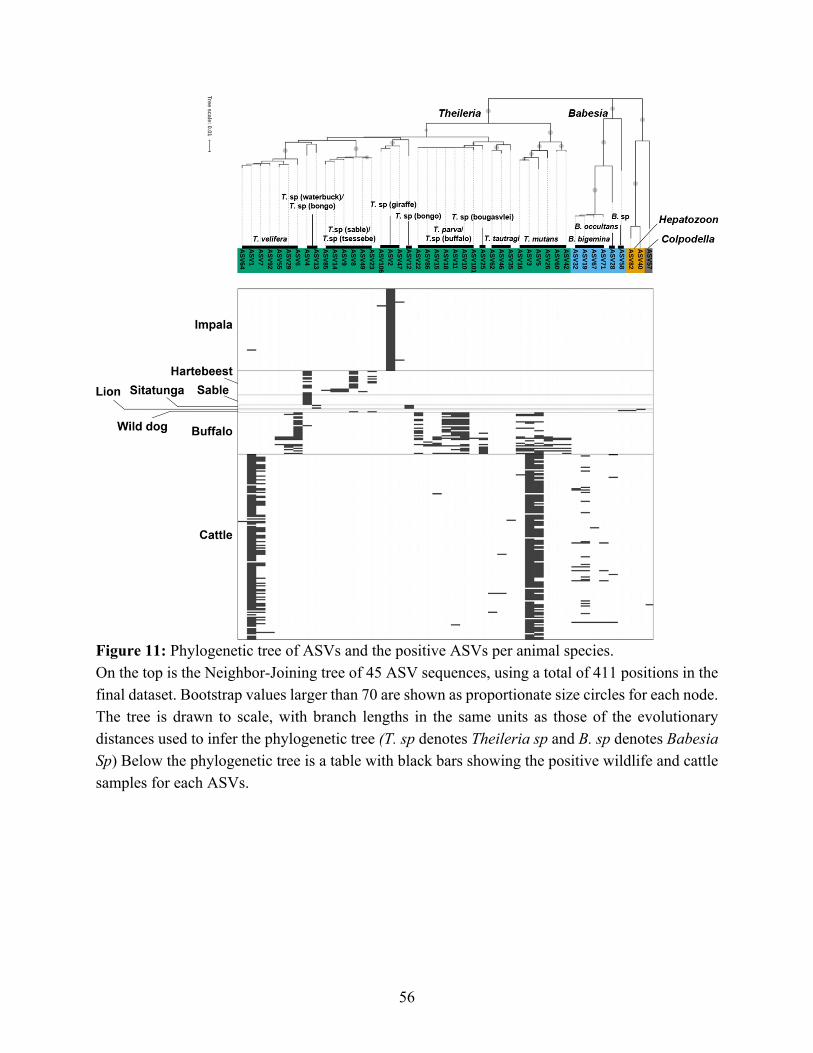
56
Figure 11: Phylogenetic tree of ASVs and the positive ASVs per animal species. On the top is the Neighbor-Joining tree of 45 ASV sequences, using a total of 411 positions in the final dataset. Bootstrap values larger than 70 are shown as proportionate size circles for each node. The tree is drawn to scale, with branch lengths in the same units as those of the evolutionary distances used to infer the phylogenetic tree (T. sp denotes Theileria sp and B. sp denotes Babesia Sp) Below the phylogenetic tree is a table with black bars showing the positive wildlife and cattle samples for each ASVs.
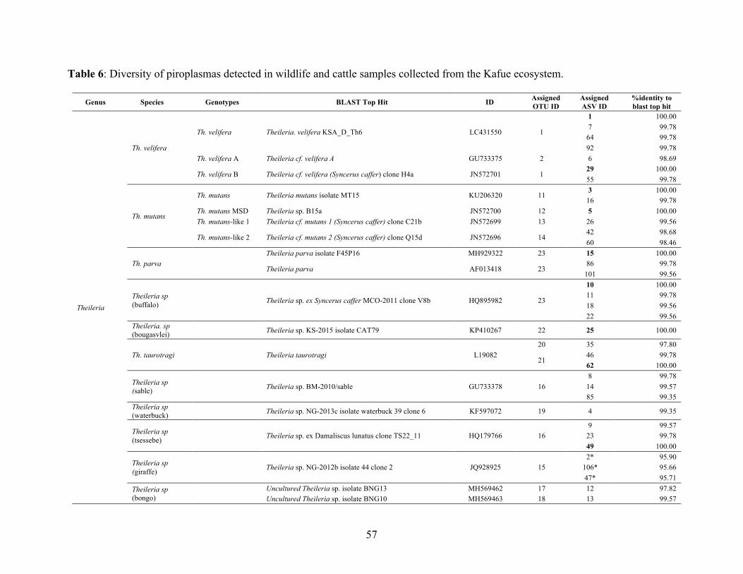
57
Table 6: Diversity of piroplasmas detected in wildlife and cattle samples collected from the Kafue ecosystem.
Genus Species Genotypes BLAST Top Hit ID Assigned OTU ID
Assigned ASV ID
%identity to blast top hit
Theileria
Th. velifera
Th. velifera Theileria. velifera KSA_D_Th6 LC431550 1
1 100.00
7 99.78
64 99.78
92 99.78
Th. velifera A Theileria cf. velifera A GU733375 2 6 98.69
Th. velifera B Theileria cf. velifera (Syncerus caffer) clone H4a JN572701 1 29 100.00
55 99.78
Th. mutans
Th. mutans Theileria mutans isolate MT15 KU206320 11 3 100.00
16 99.78
Th. mutans MSD Theileria sp. B15a JN572700 12 5 100.00
Th. mutans-like 1 Theileria cf. mutans 1 (Syncerus caffer) clone C21b JN572699 13 26 99.56
Th. mutans-like 2 Theileria cf. mutans 2 (Syncerus caffer) clone Q15d JN572696 14 42 98.68
60 98.46
Th. parva Theileria parva isolate F45P16 MH929322 23 15 100.00
Theileria parva AF013418 23
86 99.78
101 99.56
Theileria sp (buffalo)
Theileria sp. ex Syncerus caffer MCO-2011 clone V8b HQ895982 23
10 100.00 11 99.78 18 99.56
22 99.56
Theileria. sp (bougasvlei) Theileria sp. KS-2015 isolate CAT79 KP410267 22 25 100.00
Th. taurotragi
Theileria taurotragi L19082
20 35 97.80
21 46 99.78
62 100.00
Theileria sp (sable)
Theileria sp. BM-2010/sable GU733378 16
8 99.78 14 99.57
85 99.35
Theileria sp (waterbuck) Theileria sp. NG-2013c isolate waterbuck 39 clone 6 KF597072 19 4 99.35
Theileria sp (tsessebe)
Theileria sp. ex Damaliscus lunatus clone TS22_11 HQ179766 16
9 99.57 23 99.78
49 100.00
Theileria sp (giraffe)
Theileria sp. NG-2012b isolate 44 clone 2 JQ928925 15
2* 95.90 106* 95.66
47* 95.71
Theileria sp (bongo)
Uncultured Theileria sp. isolate BNG13 MH569462 17 12 97.82
Uncultured Theileria sp. isolate BNG10 MH569463 18 13 99.57
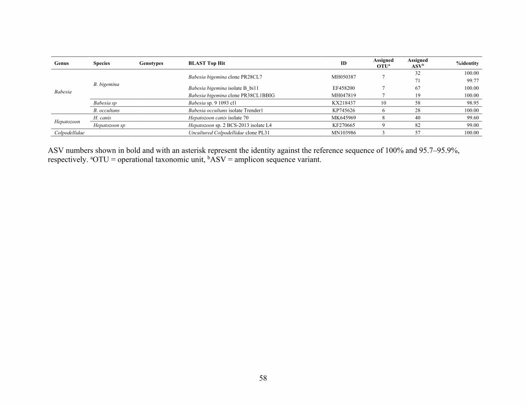
58
Genus Species Genotypes BLAST Top Hit ID Assigned
OTUa Assigned
ASVb %identity
Babesia B. bigemina
Babesia bigemina clone PR28CL7 MH050387 7
32 100.00 71 99.77 Babesia bigemina isolate B_bi11 EF458200 7 67 100.00
Babesia bigemina clone PR38CL1BBIG MH047819 7 19 100.00
Babesia sp Babesia sp. 9 1093 cl1 KX218437 10 58 98.95
B. occultans Babesia occultans isolate Trender1 KP745626 6 28 100.00
Hepatozoon H. canis Hepatozoon canis isolate 70 MK645969 8 40 99.60
Hepatozoon sp Hepatozoon sp. 2 BCS-2013 isolate L4 KF270665 9 82 99.00
Colpodellidae Uncultured Colpodellidae clone PL31 MN103986 3 57 100.00
ASV numbers shown in bold and with an asterisk represent the identity against the reference sequence of 100% and 95.7–95.9%,
respectively. aOTU = operational taxonomic unit, bASV = amplicon sequence variant.

59
Interestingly, most of the observed ASVs in the Th. parva clade were detected only in
buffalo except ASV15 (Th. parva) and ASV11 (Theileria. sp buffalo) which were detected in both buffalo and cattle. ASV25 showed 100% identity to Theileria sp. (bougasvlei) but was adjacent to the Th. parva clade (Figure 11, Table 6).
Theileria taurotragi was detected in four cattle. ASV46 and 62 had more than 99.8% identity to a Th. taurotragi reference sequence but ASV35 had 97.8% identity implying this can be categorized in a different genotype (Table 6).
There were two additional clades in Theileria (Figure 11). One consisted of ASV8, 9, 14, 23, 49 and 85. It was almost exclusively detected in hartebeest even though ASV8 and 23 were also detected from a wild dog. They showed high identity to Theileria spp. without species name. The other consisted of ASV2, 47, and 106, which were detected in impala. ASV4 was detected from both hartebeest and sable antelope but also found in a buffalo. ASV12 and 13 were detected from sitatunga.
The Babesia clade consisted of subclade for B. bigemina and B. occultans. ASVs in the B. bigemina subclade showed more than 99.8% identity to B. bigemina reference sequences and were only detected in cattle. Babesia occultans was also detected only in cattle (Figure 11, Table 6).
A Hepatozoon canis sequence was detected from a lion and another Hepatozoon sp. was detected from a wild dog. Interestingly, a Colpodellidae sequence, ASV57, was also detected from cattle (Figure 11). Although this study sought to understand the diversity of piroplasms in wildlife and cattle, it is important to state that mixed infection were observed but did not endevor to analyse the degree of mixed infections in the population.
60
4.4. Discussion We applied the deep amplicon sequencing scheme to investigate piroplasm communities (Chaudhry et al., 2019; Glidden et al., 2019) to investigate those in wildlife and cattle of the Kafue ecosystem including Kafue national park and surrounding wildlife-livestock interface area in Zambia. Despite the wildlife and cattle being sampled in consecutive years of 2018 and 2019 respectively, this may not affect the comparison and interpretation of the results on the diversity of piroplasm parasites obtained, because fundamental change of biodiversity in functional ecosystem like Kafue would take a long period.
Our data show that 45 (Amplicon Sequence Variants (ASVs) and 23 species consisting of 4 genera (Babesia, Theileria, Hepatozoon and Colpodella) were detected. Among the 45 ASVs, 14 were identical to public sequences. However, 28 ASVs demonstrated percentage identity of 95.7-99.8%, suggesting that novel genotypes may also exist. ASV2, 47, and 106 presented 95.7-95.9% identity to Theileria sp. suggesting possible novel Theileria sp. or undeposited sequences of known Theileria sp. (astarisk in Table 6). Within Theileria species, 36 ASVs were detected (Table 6). As an important natural reservoir host, buffalo had a diversity of 18 Theileria ASVs, which was highest compared to other wildlife species. Importantly, three ASVs of Th. parva (OTU23 comprising ASV15, 86, and 101) were obtained from buffalo, providing important epidemiological insight for cattle in the area in terms of corridor disease transmission. This finding is consistent with a previous report from a serological study involving buffalos (Munang’andu et al., 2009). Indeed, Th. parva ASV15 was detected in cattle, suggesting possible spillover of Th. parva from buffalo to domestic cattle. The presence of Thelieria sp. (buffalo) (49%; 26 of 53) and Thelieria sp. (bougasvlei) (19%; 10 of 53) in buffalo (Table 6) is of diagnostic importance as it affects the accurate detection of Th. parva in mixed infections when performing hybridization PCR assay (Pienaar et al., 2011). In addition to buffalo, this study found Theileria sp (buffalo) circulating in cattle population (0.4%; 1 of 232). These results support the observations and findings from studies conducted in Kenya which also identified Theileria sp (buffalo) from cattle, suggesting that Theileria sp (buffalo) infection in cattle occurs in the field where buffalo and cattle share pasture (Bishop et al., 2015; Githaka et al., 2014). Nevertheless, more knowledge on the infection epidemiology and pathogenicity to cattle will be required. The presence of T. taurotragi circulating in cattle population is consistent with findings in other similar studies (Mans et al., 2011) .The characterization of parasite community and molecular ecology raises awareness on the consequences and limitations of specific diagnostic tests and requires further cautions for the interpretation of the results used for diagnostics or surveillance in a specified area. Babesia was predominantly observed in cattle but also detected in wild dogs. Babesia bigemina (10.3%; 24 of 232) and B. occultans (1.7%; 4 of 232) were the only species detected in cattle (Table 6), of which B. bigemina is a pathogenic parasite to cattle causing a clinical disorder of babesiosis, also known as redwater. These findings are similar to other comparable studies in
61
southern Africa where the presence of Babesia in cattle and wild animals, particularly buffalo, was assessed (Mans et al., 2016).
To the best of our knowledge, the detection of B. ocultans in cattle is the first report in Zambia. Babesia ocultans is known to be non-pathogenic in cattle. Although its distribution is considered to be endemic to sub-Saharan Africa, its detection in cattle in Europe has been reported (Aktas and Ozubek, 2015). In this study, the specific vectors, impact on cattle, diagnostic consequence in Babesia mixed infection or implication of infection to wildlife were not evaluated.
Is has been discussed that enzootic stability (herd immunity) does play a role in averting clinical disease of babesiosis especially if it is well managed by strategic tick control and monitoring/keeping parasite inoculation within thresholds (Bock et al., 2004; Rikhotso et al., 2005). Our developed method can also be used as a tool for monitoring parasite community in cattle in the field.
Despite not being classified in the order of piroplasmida but Eucoccidiorida, Apicomplexan species of H. canis and Hepatozoon sp. were detected in African lion and wild dog samples, respectively. Divergent to other arthropod-borne parasites transmitted through the vector’s salivary glands at the time of feeding, Hepatozoon are transmitted to the carnivore host exclusively by ingestion of infected vectors (ticks) during grooming (Greene, 2012; Smith, 1996). They cause subclinical infection in wild carnivores and clinical infection in domestic dogs (Cunningham and Yabsley, 2012). Previous studies on free ranging wild carnivores in Zambia have indicated the widespread presence of Hepatozoon sp. in lions (Williams et al., 2014). This highlights the considered epidemiologic role of wild carnivores as sylvatic reservoir of Hepatozoon and presents the risks of likely spillover of Hepatozoon infections to domestic carnivores in the interface area.
Genus Colpodella are part of Alveolata organisms that are originally known to be free-living. However, recent studies have revealed the parasitic nature of Colpodella sp. as a Human Erythrocyte Parasite (HEP) that has lately been reported from China to cause relapsing fevers and neurological symptoms in humans (Jiang et al., 2018; Yuan et al., 2012). Furthermore, the detection of Colpodella sp. in ticks suggests that this parasite may potentially be transmitted by tick vector(s) (Jiang et al., 2018). We detected a Colpodella sequence from one of the cattle samples, with the sequence identity of 79.6% with the reported human cases (GQ411073; Colpodella sp. HEP). The sequence detected from our cattle sample showed perfect match (100% identity) to GenBank MN103986 (Colpodellidae clone PL31), reported in raccoon dog in Poland (Solarz et al., 2020). Thus, the detection of Colpodella sp. from cattle sample implies to support those findings that some of the Colpodella species are associated with vertebrates, and possibly cause disease. It is largely undetermined what vector is involved, how the parasite is maintained, and the risk that the cattle may pose for human infection. It would be important to determine the zoonotic scale of Colpodella infection to rule out incidental infections.
Identification of multiple infection is the other advantage of the deep amplicon sequencing scheme (Chaudhry et al., 2019). It is known that the African buffalo is simultaneously infected
62
with multiple species of Theileria (Mans et al., 2015). Besides, most of the cattle were also co-infected with Th. velifera and Th. mutans. It is reported that co-infection of multiple Theileria species in cattle results in dramatically different pathological disorders compare to single-species infections (Woolhouse et al., 2015). Further studies with expanded sample size might demonstrate similar interactions in wildlife as well. This is particularly important since Zambia’s cattle population stronghold is in Itezhi-Tezhi district which is adjacent to the KNP. This is cardinal as accurate diagnosis and effective control (vaccinations) of piroplasm parasites need to take the parasite community data into account. Hartebeest also tended to be co-infected with Theileria spp. In contrast, the Impalas were mainly infected with Theileria spp. isolated from giraffe but hardly co-infected with other piroplasmas.
The identification of tick-borne pathogens in wildlife and cattle population in the study area supports the apparent presence of the known tick vectors implicated in their transmission. Particular Theileria species are known to be transmitted by specific ixodid tick species of Rhipicephalus appendiculatus, R. zambeziensis and Amblyomma variegatum, while Babesia species are transmitted by R. microplus, R. decororatus and R. evertsi (Makala et al., 2003; Simuunza et al., 2011). The tick species associated with transmission of Hepatozoon species is the Rhipicephalus sanguineus sensu lato (s.l.) (Qiu et al., 2018). In order to identify the unknown vectors of some of the parasites described in this study, there is need to conduct tick piroplasm metagenomic analysis. This would further illustrate the piroplasm parasite eco-cycle more precisely.
In conclusion, molecular epidemiology based on the strength of knowledge of cryptic parasite community and diversity is essential in the control of piroplasmosis. Mapping of the piroplasm parasites in all major livestock landscapes beyond the Kafue ecosystem using metagenomic approach may benefit the piroplasmosis control in Zambia through high resolution data to precisely guide diagnosis, vaccination and movement controls.
63
5. General Summary The intimate interaction of human, their livestock and wildlife populations at the human-wildlife-livestock interface poses opportunities for a complex multi-directional inter-specie transmission of pathogens of zoonotic and veterinary importance. The transmission of these pathogens across the interface impacts public health, limits livestock production and potentially decimates wildlife populations raising conservation concern. Trypanosomiasis and piroplasmosis are important parasitic diseases that humper livestock production at the interface. The presence of wildlife reservoirs play an important role in the transmission of these two diseases. Using molecular tools of NGS, this thesis aims to investigate the diversity of trypanosomes and piroplasmas circulating in wildlife reservoir populations at the interface. The description of cryptic parasite communities in wildlife populations at the interface is important to provide data to counter disease transmission and pre-empty specific infectious disease control measures. This approach is also important to identify the precise local strain or genotypes for vaccine development and roll-out especially in the control of piroplasmosis. Apart from the impact that these two diseases have on livestock, the study also reveals the circulation of zoonotic trypanosomes in wildlife population of the Kafue ecosystem. The first chapter of this thesis describes a human clinical case of HAT caused by a vector-borne parasite of Tr. b. rhodesiense. The parasite was diagnosed on microscopy and confirmed with the Tr. b. rhodesiense-specific human SRA gene LAMP analysis. This is the first case of HAT in the Kafue national park after 50 years of non-HAT cases in the area despite the presence of the vector and reservoir. This diagnosis reveals that the Kafue national park is an emerging foci for HAT.
Following the demonstration of human infection of Tr. b. rhodesiense in the Kafue national park in chapter one, the second chapter investigated the prevalence and diversity of circulating trypanosomes in wildlife reservoir population in the Kafue ecosystem. Various trypanosomes circulating in different mammalian wildlife species were detected by applying a high throughput ITS1-PCR/MinION sequencing method in combination with SRA-PCR/Sanger sequencing method. The prevalence of trypanosomes in hartebeest, sable antelope, buffalo, warthog, impala and lechwe were 6%, 37%, 13%, 11%, 2% and 11%, respectively. A total of six trypanosomes species or subspecies were detected in the wildlife examined, including Tr. b. brucei, Tr. godfreyi, Tr. congolense, Tr. simiae and Tr. theileri. Importantly human infective Tr. b. rhodesiense was detected in buffalo, sable antelope, and vervet monkey. The results from this chapter reaffirmed that the Kafue ecosystem is a genuine neglected and re-emerging foci for HAT.
The third chapter looks at the piroplasmas circulating in wildlife and cattle of the Kafue ecosystem and their potential spillover from wildlife to cattle. To investigate piroplasm diversity in wildlife and cattle population, PCR was utilized to amplify the 18S rRNA V4 hyper-variable region and meta-barcoding strategy using illumina MiSeq sequencing platform and amplicon sequence variant (ASV) based bioinformatics pipeline to generate high resolution data which
64
discriminate sequences down to a single nucleotide difference. A parasite community of 45 ASVs corresponding to 23 species consisting of 4 genera of Babesia, Theileria, Hepatozoon and Colpodella, were identified in wildlife and cattle population from the study area. Theileria species were detected in buffalo, impala, hartebeest, sable antelope, sitatunga, wild dog and cattle. In contrast, Babesia species were only observed in cattle and wild dog. These results demonstrate possible spillover of these hemoprotozoan parasites from wildlife to cattle
population in the wildlife-livestock interface. These results illustrated the diversity of piroplasma and the specificity of their hosts. The deep amplicon sequencing of the 18S rRNA V4 hyper-variable region for wildlife was informative enough to speculate possible ecological cycle including transmission from wildlife to domestic animals in the Kafue ecosystem. The application of this approach to reveal the diversity of parasite community may contribute to the establishment of appropriate and effective disease control strategies.

65
6. Acknowledgements
I would like to convey my gratitude to my supervisors Prof. Junya Yamagishi, Prof. Boniface Namangala and Prof. Kyoko Hayashida for their sound guidance, inspiration and oversight at every stage of this research endeavor and for the insightful counsel that enriched my academic research experience. My profound appreciation goes to Prof. Chihiro Sugimoto for the longtime collaboration, support and encouragement to undertake this research study. I am thankful to all the laboratory members and staff in the Division of Collaboration and Education at the International institute for Zoonosis Control (Hokkaido University) and at the school of veterinary medicine (University of Zambia) for their advice, assistance and companionship during the course of this research work. The staff of the department of national parks and wildlife, Zambia especially those in the wildlife veterinary unit were extremely helpful in facilitating sampling permits and field collection of wildlife samples. I am sincerely grateful to the Japanese society for promotion of science (JSPS) for the scholarship that enabled me to conduct this scholarly work. I would like to thank my family for their support and understanding especially my wife and the kids (Thoko and Micah) for allowing me to divert my attention from them to this work. I thus dedicate this work to you.
66
7. Reference Aktas, M., Ozubek, S., 2015. Molecular and parasitological survey of bovine piroplasms in the
black sea region, Including the first report of babesiosis associated with Babesia divergens in Turkey. J. Med. Entomol. 52, 1344–1350.
Allsopp, M.T.E.P., Cavalier-Smith, T., Waal, D.T.D.E., Allsopp, B.A., 1994. Phylogeny and evolution of the piroplasms. Parasitology 108, 147–152.
Altschul, S.F., Gish, W., Miller, W., Myers, E.W., Lipman, D.J., 1990. Basic local alignment search tool. J. Mol. Biol. 215, 403–410.
Anderson, N.E., 2008. Investigation into the ecology of trypanosomiasis in the Lungawa valley, Zambia. PhD thesis, University of Edinburgh.
Anderson, N.E., Mubanga, J., Fevre, E.M., Picozzi, K., Eisler, M.C., Thomas, R., Welburn, S.C., 2011. Characterisation of the wildlife reservoir community for human and animal trypanosomiasis in the Luangwa valley, Zambia. PLoS Negl. Trop. Dis. 5, e1211.
Anderson, N.E., Mubanga, J., Machila, N., Atkinson, P.M., Dzingirai, V., Welburn, S.C., 2015. Sleeping sickness and its relationship with development and biodiversity conservation in the Luangwa valley, Zambia. Parasites and Vectors 8, 1–14.
Auty, H., Morrison, L.J., Torr, S.J., Lord, J., 2016. Transmission dynamics of rhodesian sleeping sickness at the interface of wildlife and livestock areas. Trends Parasitol. 32, 608–621.
Bandyopadhyay, S., Tembo, G., 2010. Household consumption and natural resource management around national parks in Zambia. J. Nat. Resour. Policy Res. 2, 39–55.
Baral, T.N., 2010. Immunobiology of African trypanosomes: Need of alternative interventions. J. Biomed. Biotechnol. 2010, 1–24
Bengis, R.G., Kock, R.A., Fischer, J., 2002. Infectious animal diseases : The wildlife / livestock interface Wildlife. Sci. Tech. Rev. OIE 21, 53–65.
Bentivoglio, M., Cavalheiro, E.A., Kristensson, K., Patel, N.B. 2014. Neglected tropical diseases and conditions of the nervous system. Chapter 9. Springer, New York.
Bishop, R., Musoke, A., Morzaria, S., Gardner, M., Nene, V., 2004. Theileria: Intracellular protozoan parasites of wild and domestic ruminants transmitted by ixodid ticks. Parasitology 129, S271–S283.
Bishop, R.P., Hemmink, J.D., Morrison, W.I., Weir, W., Toye, P.G., Sitt, T., Spooner, P.R., Musoke, A.J., Skilton, R.A., Odongo, D.O., 2015. The African buffalo parasite Theileria sp. (buffalo) can infect and immortalize cattle leukocytes and encodes divergent orthologues of Theileria parva antigen genes. Int. J. Parasitol. Parasites Wildl. 4, 333–342.
Bishop, R.P., Odongo, D., Ahmed, J., Mwamuye, M., Fry, L.M., Knowles, D.P., Nanteza, A., Lubega, G., Gwakisa, P., Henning, P., Isaiah, C., 2020. A review of recent research on Theileria parva : Implications for the infection and treatment vaccination method for control of East Coast fever. Transbound. Emerg. Dis. 67, 56–67.
Bock, R., Jackson, L., De Vos, A., Jorgensen, W., 2004. Babesiosis of cattle. Parasitology 129, S247–269
Bolger, A.M., Lohse, M., Usadel, B., 2014. Trimmomatic: a flexible trimmer for Illumina sequence data. Bioinformatics 30, 2114–2120.
Brown, W.C., Palmer, G.H., 1999. Designing blood-stage vaccines against Babesia bovis and B. bigemina. Parasitol. Today 15, 275–281.
Brun, R., Blum, J., Chappuis, F., Burri, C., 2010. Human African trypanosomiasis. Lancet 375, 148–159.

67
Buyst, H., 1974. The epidemiology, clinical features, treatment and history of sleeping sickness on the northern edge of the Luangwa fly belt. Med. J. Zambia 4, 2–12.
Chaudhry, U., Ali, Q., Rashid, I., Shabbir, M.Z., Ijaz, M., Abbas, M., Evans, M., Ashraf, K., Morrison, I., Morrison, L., Sargison, N.D., 2019. Development of a deep amplicon sequencing method to determine the species composition of piroplasm haemoprotozoa. Ticks Tick. Borne. Dis. 10, 101276
Clarke, J.E., 1974. Department of Wildlife, Fisheries and National parks Annual report for the year 1973. Lusaka
Clausen, P.H., Adeyemi, I., Bauer, B., Breloeer, M., Salchow, F., Staak, C., 1998. Host preferences of tsetse (Diptera: Glossinidae) based on bloodmeal identifications. Med. Vet. Entomol. 12, 169–180.
Criado-Fornelio, A., Gónzalez-Del-Río, M.A., Buling-Saraña, A., Barba-Carretero, J.C., 2004. The ‘expanding universe’ of piroplasms. Vet. Parasitol. 119, 337–345.
Criado-fornelio, A., Martinez-marcos, A., Buling-Saraña, A., Barba-Carretero, J.C., 2003. Molecular studies on Babesia, Theileria and Hepatozoon in southern Europe Part II. Phylogenetic analysis and evolutionary history. Vet. Parasitol. 114, 173–194.
Cunningham, M.W., Yabsley, M.J., 2012. Primer on Tick-Borne Diseases in Exotic Carnivores. In: Fowler’s Zoo and Wild Animal Medicine Current Therapy, Volume 7. Elsevier Inc., Missouri
Cuypers, B., Van Den Broeck, F., Van Reet, N., Meehan, C.J., Cauchard, J., Wilkes, J.M., Claes, F., Goddeeris, B., Birhanu, H., Dujardin, J.C., Laukens, K., Büscher, P., Deborggraeve, S., 2017. Genome-wide SNP analysis reveals distinct origins of Trypanosoma evansi and Trypanosoma equiperdum. Genome Biol. Evol. 9, 1990–1997.
Daszak, P., Cunningham, A.A., Hyatt, A.D., 2000. Emerging infectious diseases of wildlife - Threats to biodiversity and human health. Science 287, 443–449.
Department of National Parks and wildlife, 2016. Report on the 2015 aerial census of elephants and other large mammals in Zambia. Chilanga.
Department of National Parks and Wildlife, 2018. National Parks and Wildlife Policy. Lusaka Dobbelaere, D.A.E., Mckeever, D.J., 2002. Theileria. Chapter 7. Springer, Boston. Edgar, R.C., 2010. Search and clustering orders of magnitude faster than BLAST. Bioinformatics
26, 2460–2461. Eygelaar, D., Jori, F., Mokopasetso, M., Sibeko, K.P., Collins, N.E., Vorster, I., Troskie, M.,
Oosthuizen, M.C., 2015. Tick-borne haemoparasites in African buffalo (Syncerus caffer) from two wildlife areas in northern Botswana. Parasit. Vectors 8, 1–11.
Fandamu, P., 2005. Transmission and infection dynamics of theileriosis in southern Zambia: The effect of environmental and host factors. PhD thesis, Ghent University.
Fanin, N., Gundale, M.J., Farrell, M., Ciobanu, M., Baldock, J.A., Nilsson, M.C., Kardol, P., Wardle, D.A., 2018. Consistent effects of biodiversity loss on multifunctionality across contrasting ecosystems. Nat. Ecol. Evol. 2, 269–278.
Franco, J.R., Cecchi, G., Priotto, G., Paone, M., Diarra, A., Grout, L., Matttioli, R.C., Argaw, D., 2018. Monitoring the elimination of human African trypanosomiasis: Update to 2016. PLoS Negl. Trop. Dis. 12, e0006890.
Franco, J.R., Cecchi, G., Priotto, G., Paone, M., Diarra, A., Grout, L., Simarro, P.P., Zhao, W., Argaw, D., 2020. Monitoring the elimination of human African trypanosomiasis at continental and country level: Update to 2018. PLoS Negl. Trop. Dis. 14, 1–18.
Gaithuma, A., Yamagishi, J., Hayashida, K., Kawai, N., Namangala, B., Sugimoto, C., 2020.

68
Blood meal sources and bacterial microbiome diversity in wild- caught tsetse flies. Sci. Rep. 10, 1–10.
Gaithuma, A.K., Yamagishi, J., Martinelli, A., Hayashida, K., Kawai, N., Marsela, M., Sugimoto, C., 2019. A single test approach for accurate and sensitive detection and taxonomic characterization of trypanosomes by comprehensive analysis of internal transcribed spacer 1 amplicons. PLoS Negl. Trop. Dis. 13, 1–20.
Geysen, D., Bishop, R., Skilton, R., Dolan, T.T., Morzaria, S., 1999. Molecular epidemiology of Theileria parva in the field. Trop. Med. Int. Heal. 4, 21–27.
Gibson, W., Backhouse, T., Griffiths, A., 2002. The human serum resistance associated gene is ubiquitous and conserved in Trypanosoma brucei rhodesiense throughout East Africa. Infect. Genet. Evol. 1, 207–214.
Githaka, N., Konnai, S., Bishop, R., Odongo, D., Lekolool, I., Kariuki, E., Gakuya, F., Kamau, L., Isezaki, M., Murata, S., Ohashi, K., 2014. Identification and sequence characterization of novel Theileria genotypes from the waterbuck (Kobus defassa) in a Theileria parva-endemic area in Kenya. Vet. Parasitol. 202, 180–193.
Glidden, C.K., Koehler, A. V., Hall, R.S., Saeed, M.A., Coppo, M., Beechler, B.R., Charleston, B., Gasser, R.B., Jolles, A.E., Jabbar, A., 2019. Elucidating cryptic dynamics of Theileria communities in African buffalo using a high-throughput sequencing informatics approach. Ecol. Evol. 10, 70–80.
Government of Zambia, 2015. Wildlife Act of 2015, No. 14, Government Gazette, Lusaka. Greene, C., 2012. Infectious Diseases of The Dog and Cat. Chapter 74. Elsevier Saunders,
Missouri. Gubbels, J.M., De Vos, A.P., Van Der Weide, M., Viseras, J., Schouls, L.M., De Vries, E.,
Jongejan, F., 1999. Simultaneous detection of bovine Theileria and Babesia species by reverse line blot hybridization. J. Clin. Microbiol. 37, 1782–1789.
Haag, J., O’hUigin, C., Overath, P., 1998. The molecular phylogeny of trypanosomes: evidence for an early divergence of the Salivaria. Mol. Biochem. Parasitol. 91, 37–49.
Hansen, S., Dill, V., Shalaby, M.A., Eschbaumer, M., Böhlken-fascher, S., Ho, B., Czerny, C., Abd, A., Wahed, E., 2019. Serotyping of foot-and-mouth disease virus using oxford nanopore sequencing. J. Virol. Methods 263, 50–53.
Hayashida, K., Kajino, K., Hachaambwa, L., Namangala, B., Sugimoto, C., 2015. Direct Blood Dry LAMP: A Rapid, Stable, and Easy Diagnostic Tool for Human African Trypanosomiasis. PLoS Negl. Trop. Dis. 9, 1–14.
Hemmink, J.D., Sitt, T., Pelle, R., Klerk-lorist, L. De, Shiels, B., Toye, P.G., Morrison, W.I., Weir, W., 2018. Ancient diversity and geographical sub-structuring in African buffalo Theileria parva populations revealed through metagenetic analysis of antigen-encoding loci. Int. J. Parasitol. 48, 287–296.
Hoare, C.A., 1966. The classification of mammalian trypanosomes. Ergeb. Mikrobiol. Immunitatsforsch. Exp. Ther. 39, 43–57
Hocking, K.S., Lamerton, J.F., Lewis, E.A., 1963. Tsetse-fly control and eradication. Bull. World Health Organ. 28, 811–823.
Holmes, P., 2015. On the Road to Elimination of Rhodesiense Human African Trypanosomiasis: First WHO Meeting of Stakeholders. PLoS Negl. Trop. Dis. 9, 1–3.
Jiang, J., Jiang, R., Chang, Q., Zheng, Y., Fricken, E. Von, Cao, W., 2018. Potential novel tick-borne Colpodella species parasite infection in patient with neurological symptoms case report. PLoS Negl. Trop. Dis. 12, e0006546
69
Johnson, R.N., Wilson-wilde, L., Linacre, A., 2014. Current and future directions of DNA in wildlife forensic science. Forensic Sci. Int. Genet. 10, 1–11.
Karesh, W.B., Dobson, A., Lloyd-Smith, J.O., Lubroth, J., Dixon, M.A., Bennett, M., Aldrich, S., Harrington, T., Formenty, P., Loh, E.H., MacHalaba, C.C., Thomas, M.J., Heymann, D.L., 2012. Ecology of zoonoses: Natural and unnatural histories. Lancet 380, 1936–1945.
Kennedy, P.G.E., 2013. Clinical features, diagnosis, and treatment of human African trypanosomiasis (sleeping sickness). Lancet Neurol. 12, 186–194.
Kennedy, P.G.E., Rodgers, J., 2019. Clinical and neuropathogenetic aspects of human African trypanosomiasis. Front. Immunol. 10, 1–11.
Kielbasa, S.M., Wan, R., Sato, K., Horton, P., Frith, M.C., 2011. Adaptive seeds tame genomic sequence comparison. Genomic Res. 487–493.
Kock, M.D., Burroughs, R.E.J., 2012. Chemical and physical restraint of wild animals: a training and field manual for African species. IWVS, Greyton.
Kock, R., Kock, M., Garine-wichatitsky, M. De, Chardonnet, P., Caron, A., 2014. Livestock and buffalo (Syncerus caffer) interfaces in Africa: ecology of disease transmission and implications for conservation and development. In: Melletti, M., Burton, J. (Eds.), Ecology, Evolution and Behaviour of Wild Cattle Implications for Conservation. University of Cambridge Press.
Kristjanson, P.M., Swallow, B.M., Rowlands, G.J., Kruska, R.L., De Leeuw, P.N., 1999. Measuring the costs of African animal trypanosomosis, the potential benefits of control and returns to research. Agric. Syst. 59, 79–98.
Kumar, S., Stecher, G., Li, M., Knyaz, C., Tamura, K., 2018. MEGA X: molecular evolutionary genetics analysis across computing platforms. Mol. Biol. Evol. 35, 1547–1549.
Kumar, S., Stecher, G., Tamura, K., 2016. MEGA7 : Molecular evolutionary genetics analysis version 7 . 0 for bigger datasets. Mol. Evol. Genet. Anal. 33, 1870–1874.
La Grange, M., 2006. The capture, care, and management of wildlife. Van Schaik, Pretoria. Laohasinnarong, D., Goto, Y., Asada, M., Nakao, R., Hayashida, K., Kajino, K., Kawazu, S.I.,
Sugimoto, C., Inoue, N., Namangala, B., 2015. Studies of trypanosomiasis in the Luangwa Valley, north-eastern Zambia. Parasit.Vectors 8, 1–8.
Leak, S.G.A., 1999. Tsetse biology and ecology: their role in the epidemiology and control of trypanosomiasis. Chapter 14. CABI Publishing, Oxfordshire.
Letunic, I., Bork, P., 2019. Interactive Tree of Life (iTOL) v4: Recent updates and new developments. Nucleic Acids Res. 47, 256–259.
Mackey, T.K., Liang, B.A., 2012. Threats from emerging and re-emerging neglected tropical diseases (NTDs). Infect. Ecol. Epidemiol. 2, 18667.
Makala, L.H., Mangani, P., Fujisaki, K., Nagasawa, H., 2003. The current status of major tick borne diseases in Zambia. Vet. Res. 34, 27–45.
Mans, B.J., Pienaar, R., Latif, A.A., 2015. A review of Theileria diagnostics and epidemiology. Int. J. Parasitol. Parasites Wildl. 4, 104–118.
Mans, B.J., Pienaar, R., Latif, A.A., Potgieter, F.T., 2011. Diversity in the 18S SSU rRNA V4 hyper-variable region of Theileria spp. in Cape buffalo (Syncerus caffer) and cattle from southern Africa. Parasitology 138, 766–779.
Mans, B.J., Pienaar, R., Ratabane, J., Pule, B., Latif, A.A., 2016. Investigating the diversity of the 18S SSU rRNA hyper-variable region of Theileria in cattle and Cape buffalo (Syncerus caffer) from southern Africa using a next generation sequencing approach. Ticks Tick. Borne. Dis. 7, 869–879.

70
Marsela, M., Hayashida, K., Nakao, R., Chatanga, E., Gaithuma, A.K., Naoko, K., Musaya, J., Sugimoto, C., Yamagishi, J., 2020. Molecular identification of trypanosomes in cattle in Malawi using PCR methods and nanopore sequencing: Epidemiological implications for the control of human and animal trypanosomiases. Parasite 27, 1–14
Matthews, K.R., Ellis, J.R., Paterou, A., 2004. Molecular regulation of the life cycle of African trypanosomes. Trends Parasitol. 20, 40–47.
Maudlin, I., Holmes, P., Miles, M., 2004. The Trypanosomiases. Chapter 14. CAB International, Oxfordshire.
Mehlhorn, H., Schein, E., 1985. The Piroplasms: Life cycle and sexual stages. In: Advances in Parasitology. Academic press, London.
Ministry of National Development Planning, 2017. 7th National Development Plan 2017 - 2021. Government Printer, Lusaka.
Morrison, L.J., Vezza, L., Rowan, T., Hope, J.C., 2016. Animal African Trypanosomiasis: Time to increase focus on clinically relevant parasite and host species. Trends Parasitol. 32, 599–607.
Morrison, W.I., Hemmink, J.D., Toye, P.G., 2020. Theileria parva: a parasite of African buffalo, which has adapted to infect and undergo transmission in cattle. Int. J. Parasitol. 50, 403–412.
Mulenga, G.M., Likwa, R.N., Namangala, B., 2015. Assessing the capacity to diagnose human African trypanosomiasis among health care personnel from Chama and Mambwe districts of eastern Zambia. BMC Res. Notes 8, 1–7.
Muleya, W., Namangala, B., Simuunza, M., Nakao, R., Inoue, N., Kimura, T., Ito, K., Sugimoto, C., Sawa, H., 2012. Population genetic analysis and sub-structuring of Theileria parva in the northern and eastern parts of Zambia. Parasit. Vectors 5, 1–14
Munang’andu, H.M., Siamudaala, V., Matandiko, W., Mulumba, M., Nambota, A., Munyeme, M., Mutoloki, S., Nonga, H., 2009. Detection of Theileria parva antibodies in the African buffalo (Syncerus caffer) in the livestock-wildlife interface areas of Zambia. Vet. Parasitol. 166, 163–166.
Munang’andu, H.M., Siamudaala, V., Munyeme, M., Nalubamba, K.S., 2012. A review of ecological factors associated with the epidemiology of wildlife trypanosomiasis in the Luangwa and Zambezi valley ecosystems of Zambia. Interdiscip. Perspect. Infect. Dis. 2012, 1–13.
Mwanakasale, V., Songolo, P., 2011. Disappearance of some human African trypanosomiasis transmission foci in Zambia in the absence of a tsetse fly and trypanosomiasis control program over a period of forty years. Trans. R. Soc. Trop. Med. Hyg. 105, 167–172.
Mwanakasale, V., Songolo, P., Daka, V., 2013. Challenges in the control of Human African Trypanosomiasis in the Mpika district of Zambia. BMC Res. Notes 6, 4–10.
Mweempwa, C., Marcotty, T., De Pus, C., Penzhorn, B.L., Dicko, A.H., Bouyer, J., De Deken, R., 2015. Impact of habitat fragmentation on tsetse populations and trypanosomosis risk in Eastern Zambia. Parasit. Vectors 8, 1–10.
Mwima, H.K., 2001. A brief history of Kafue national park, Zambia. Koedoe 44, 57–72. Nakamura, Y., Hayashida, K., Delesalle, V., Qiu, Y., Omori, R., Simuunza, M., Sugimoto, C.,
Namangala, B., Yamagishi, J., 2021. Genetic Diversity of African Trypanosomes in Tsetse Flies and Cattle From the Kafue Ecosystem. Front. Vet. Sci. 8, 599815
Nakayima, J., Hayashida, K., Nakao, R., Ishii, A., Ogawa, H., Nakamura, I., Moonga, L., Hang’ombe, B.M., Mweene, A.S., Thomas, Y., Orba, Y., Sawa, H., Sugimoto, C., 2014. Detection and characterization of zoonotic pathogens of free-ranging non-human primates

71
from Zambia. Parasit. Vectors 7, 1–7. Namangala, B., Hachaambwa, L., Kajino, K., Mweene, A.S., Hayashida, K., Simuunza, M.,
Simukoko, H., Choongo, K., Chansa, P., Lakhi, S., Moonga, L., Chota, A., Ndebe, J., Nsakashalo-Senkwe, M., Chizema, E., Kasonka, L., Sugimoto, C., 2012. The use of Loop-mediated Isothermal Amplification (LAMP) to detect the re-emerging Human African Trypanosomiasis (HAT) in the Luangwa and Zambezi valleys. Parasit. Vectors 5, 1–5.
Njiru, Z.K., Constantine, C.C., Guya, S., Crowther, J., Kiragu, J.M., Thompson, R.C.A., Davila, A.M.R., 2004. The use of ITS1 rDNA PCR in detecting pathogenic African trypanosomes. Parasitol. Res. 95, 186–192.
Odiit, M., Coleman, P.G., McDermott, J.J., Fèvre, E.M., Welburn, S.C., Woolhouse, M.E.J., 2004. Spatial and temporal risk factors for the early detection of Trypanosoma brucei rhodesiense sleeping sickness patients in Tororo and Busia districts, Uganda. Trans. R. Soc. Trop. Med. Hyg. 98, 569–576.
Okiwelu, S.N., 1977. Host preference and trypanosome infection rates of Glossina morsitans morsitans westwood in the Republic of Zambia. Ann. Trop. Med. Parasitol. 71, 101–107.
Osofsky, S.A., 2005. conservation and development interventions at the wildlife/livestock interface: Implications for wildlife, livestock and human health. Chapter 1. IUCN, Cambridge.
Palmer, J.M., Jusino, M.A., Banik, M.T., Lindner, D.L., 2018. Non-biological synthetic spike-in controls and the AMPtk software pipeline improve mycobiome data. PeerJ 6, e4925
Pienaar, R., Latif, A.A., Thekisoe, O.M.M., Mans, B.J., 2014. Geographic distribution of Theileria sp. (buffalo) and Theileria sp. (bougasvlei) in Cape buffalo (Syncerus caffer) in southern Africa: implications for speciation. Parasitology 141, 411–424.
Pienaar, R., Potgieter, F.T., Latif, A.A., Thekisoe, O.M.M., Mans, B.E.N.J., 2011. Mixed Theileria infections in free-ranging buffalo herds : implications for diagnosing Theileria parva infections in Cape buffalo (Syncerus caffer). Parasitology 138, 884–895.
Potgieter, F., Els, H., 1977. The fine structure of intra-erythrocytic stages of Babesia bigemina. Onderstepoort J. Vet. Res. 44, 157–168.
Qiu, Y., Kaneko, C., Kajihara, M., Ngonda, S., Simulundu, E., Muleya, W., Thu, M.J., Hang’ombe, M.B., Katakura, K., Takada, A., Sawa, H., Simuunza, M., Nakao, R., 2018. Tick-borne haemoparasites and Anaplasmataceae in domestic dogs in Zambia. Ticks Tick. Borne. Dis. 9, 988–995.
Radwanska, M., Chamekh, M., Vanhamme, L., Claes, F., Magez, S., Magnus, E., De Baetselier, P., Büscher, P., Pays, E., 2002. The serum resistance-associated gene as a diagnostic tool for the detection of Trypanosoma brucei rhodesiense. Am. J. Trop. Med. Hyg. 67, 684–690.
Rikhotso, B.O., Stoltsz, W.H., Bryson, N.R., Sommerville, J.E.M., 2005. The impact of 2 dipping systems on endemic stability to bovine babesiosis and anaplasmosis in cattle in 4 communally grazed areas in Limpopo province, South Africa. J. S. Afr. Vet. Assoc. 76, 217–223.
Roditi, I., Liniger, M., 2002. Dressed for success: The surface coats of insect-borne protozoan parasites. Trends Microbiol. 10, 128–134.
Saitou, N., Nei, M., 1987. The neighbor-joining method: a new method for reconstructing phylogenetic trees. Mol. Biol. Evol. 4, 406–425.
Schmeller, D.S., Courchamp, F., Killeen, G., 2020. Biodiversity loss, emerging pathogens and human health risks. Biodivers. Conserv. 29, 3095–3102.
Schoch, C.L., Ciufo, S., Domrachev, M., Hotton, C.L., Kannan, S., Khovanskaya, R., Leipe, D., McVeigh, R., O’Neill, K., Robbertse, B., Sharma, S., Soussov, V., Sullivan, J.P., Sun, L.,
72
Turner, S., Karsch-Mizrachi, I., 2020. NCBI Taxonomy: A comprehensive update on curation, resources and tools. Database 2020, 1–21.
Schreeg, M.E., Marr, H.S., Tarigo, J.L., Cohn, L.A., Bird, D.M., Scholl, E.H., Levy, M.G., Wiegmann, B.M., Birkenheuer, A.J., 2016. Mitochondrial genome sequences and structures aid in the resolution of piroplasmida phylogeny. PLoS One 11, 1–27.
Sharma, R., Peacock, L., Gluenz, E., Gull, K., Gibson, W., Carrington, M., 2008. Asymmetric cell division as a route to reduction in cell length and change in cell morphology in trypanosomes. Protist 159, 137–151.
Shiels, B., Langsley, G., Weir, W., Pain, A., McKellar, S., Dobbelaere, D., 2006. Alteration of host cell phenotype by Theileria annulata and Theileria parva: Mining for manipulators in the parasite genomes. Int. J. Parasitol. 36, 9–21.
Siamudaala, V.M., Nyirenda, V.R., Saiwana, L.M., 2009. Effectiveness of law enforcement on wildlife crimes in the Kafue ecosystem in Zambia. Zambia Wildlife Authority, Chilanga.
Simarro, P.P., Diarra, A., Ruiz Postigo, J.A., Franco, J.R., Jannin, J.G., 2011. The Human African Trypanosomiasis control and surveillance programme of the World Health Organization 2000–2009: The Way Forward. PLoS Negl. Trop. Dis. 5, e1007.
Simarro, P.P., Jannin, J., Cattand, P., 2008. Eliminating Human African Trypanosomiasis: Where do we stand and what comes next? PLoS Med. 5, e55.
Simukoko, H., Marcotty, T., Vercruysse, J., Van den Bossche, P., 2011. Bovine trypanosomiasis risk in an endemic area on the eastern plateau of Zambia. Res. Vet. Sci. 90, 51–54.
Simuunza, M., Weir, W., Courcier, E., Tait, A., Shiels, B., 2011. Epidemiological analysis of tick-borne diseases in Zambia. Vet. Parasitol. 175, 331–342.
Sindato, C., Kibona, S.N., Nkya, G.M., Mbilu, T.J., Manga, C., Kaboya, J.S., Rawille, F., 2008. Challenges in the diagnosis and management of sleeping sickness in Tanzania: a case report. Tanzan. J. Health Res. 10, 177–181.
Smith, T.G., 1996. The Genus Hepatozoon (Apicomplexa: Adeleina ). J. Parasitol. 82, 565–585. Solarz, W., Najberek, K., Wilk-Woźniak, E., Biedrzycka, A., 2020. Raccoons foster the spread of
freshwater and terrestrial microorganisms—Mammals as a source of microbial eDNA. Divers. Distrib. 26, 453–459.
Squarre, D., Kabongo, I., Munyeme, M., Mumba, C., Mwasinga, W., Hachaambwa, L., Sugimoto, C., Namangala, B., 2016. Human African Trypanosomiasis in the Kafue national park, Zambia. PLoS Negl. Trop. Dis. 10, e0004567 Steel, W.S., Glendhil, J.A., 1982. A Survey of the distribution of Glossina spp and factors
influencing their control in the Territoty of Northern Rhodesia (Zambia). Dept of veterinary and tsetse control services, Lusaka.
Steverding, D., 2008. The history of African trypanosomiasis. Parasit. Vectors 1, 1–8. Stich, A., Abel, P.M., Krishna, S., 2012. Human African Trypanosomiasis clinical review. Br.
Med. J. 325, 203–206. Stijlemans, B., De Baetselier, P., Caljon, G., Van Den Abbeele, J., Van Ginderachter J.A.,
Magez S., 2017. Nanobodies as tools to understand, diagnose, and treat African Trypanosomiasis. Front Immunol. 8, 724.
Stoddard, S.T., Morrison, A.C., Vazquez-Prokopec, G.M., Soldan, V.P., Kochel, T.J., Kitron, U., Elder, J.P., Scott, T.W., 2009. The role of human movement in the transmission of vector-borne pathogens. PLoS Negl. Trop. Dis. 3, e481
Tamura, K., Tao, Q., Kumar, S., 2018. Theoretical foundation of the reltime method for estimating divergence times from variable evolutionary rates. Mol. Biol. Evol. 35, 1770–1782.
73
Thompson, R.C.A., Lymbery, A.J., Smith, A., 2010. Parasites, emerging disease and wildlife conservation. Int. J. Parasitol. 40, 1163–1170.
Thuita, J.K., Kagira, J.M., Mwangangi, D., Matovu, E., Turner, C.M.R., Masiga, D., 2008. Trypanosoma brucei rhodesiense Transmitted by a Single Tsetse Fly Bite in Vervet Monkeys as a Model of Human African Trypanosomiasis. PLoS Negl. Trop. Dis. 2, e238.
Uilenberg, G., 1999. Immunization against diseases caused by Theileria parva: A review. Trop. Med. Int. Heal. 4, A12–A20.
Van Den Bossche, P., Ky-Zerbo, A., Brandt, J., Marcotty, T., Geerts, S., De Deken, R., 2005. Transmissibility of Trypanosoma brucei during its development in cattle. Trop. Med. Int. Heal. 10, 833–839.
Van den Bossche, P., Rocque, S. de La, Hendrickx, G., Bouyer, J., 2010. A changing environment and the epidemiology of tsetse-transmitted livestock trypanosomiasis. Trends Parasitol. 26, 236–243.
Vannier, E., Gewurz, B., Krause, P.J., 2008. Human babesiosis. Infect. Dis. Clin. North Am. 22, 197–208.
Vigneron, A., O’Neill, M.B., Weiss, B.L., Savage, A.F., Campbell, O.C., Kamhawi, S., Valenzuela, J.G., Aksoy, S., 2020. Single-cell RNA sequencing of Trypanosoma brucei from tsetse salivary glands unveils metacyclogenesis and identifies potential transmission blocking antigens. Proc. Natl. Acad. Sci. 117, 2613–2621.
Waldron, A., Miller, D.C., Redding, D., Mooers, A., Kuhn, T.S., Nibbelink, N., Roberts, J.T., Tobias, J.A., Gittleman, J.L., 2017. Reductions in global biodiversity loss predicted from conservation spending. Nature 551, 364–367.
Wamwiri, F.N., Changasi, R.E., 2016. Tsetse Flies ( Glossina ) as Vectors of Human African Trypanosomiasis: A Review . Biomed Res. Int. 2016, 1–8.
WHO, 2021. Ending the neglect to attain the sustainable development goals: A road map for neglected tropical diseases 2021 - 2030 overview. World Health Organisation, Geneva WHO, 2019. WHO interim guidelines for the treatment of gambiense human African
trypanosomiasis., World Health Organisation, Geneva WHO, 2015a. Investing To Overcome The Global Impact Of Neglected Tropical Diseases Third
WHO Report On Neglected Tropical Diseases. World Health Organisation, Geneva WHO, 2015b. Report of the first WHO stakeholders meeting on rhodesiense human African
trypanosomiasis. World Health Organisation, Geneva WHO, 2012. Research priorities for Chagas disease, human African trypanosomiasis and
leishmaniasis. World Health Organisation, Geneva Williams, B.M., Berentsen, A., Shock, B.C., Teixiera, M., Dunbar, M.R., Matthew, B.S., Yabsley,
M.J., 2014. Prevalence and diversity of Babesia, Hepatozoon, Ehrlichia, and Bartonella in wild and domestic carnivores from Zambia, Africa. Parasitol. Res. 113, 911–918.
Wittemyer, G., Elsen, P., Bean, W.T., Burton, A.C.O., Brashares, J.S., 2008. Accelerated Human Population Growth at Protected Area Edges 321, 123–127.
Woolhouse, M.E.J., Thumbi, S.M., Jennings, A., Chase-Topping, M., Callaby, R., Kiara, H., Oosthuizen, M.C., Mbole-Kariuki, M.N., Conradie, I., Handel, I.G., Poole, E.J., Njiiri, E., Collins, N.E., Murray, G., Tapio, M., Auguet, O.T., Weir, W., Morrison, W.I., Kruuk, L.E.B., De Bronsvoort, C.B.M., Hanotte, O., Coetzer, K., Toye, P.G., 2015. Co-infections determine patterns of mortality in a population exposed to parasite infection. Sci. Adv. 1, 1–10
Yamagishi, J., Runtuwene, L.R., Hayashida, K., Mongan, A.E., Thi, L.A.N., Thuy, L.N., Nhat, C.N., Limkittikul, K., Sirivichayakul, C., Sathirapongsasuti, N., Frith, M., Makalowski, W.,
74
Eshita, Y., Sugano, S., Suzuki, Y., 2017. Serotyping dengue virus with isothermal amplification and a portable sequencer. Sci. Rep. 7, 1–10.
Yuan, C.L., Keeling, P.J., Krause, P.J., Horak, A., Bent, S., Rollend, L., Hua, X.G., 2012. Colpodella spp.– like Parasite Infection in woman, China. Emerg. Infect. Dis. 18, 125–127
Yusufmia, S.B.A.S., Collins, N.E., Nkuna, R., Troskie, M., Van Den Bossche, P., Penzhorn, B.L., 2010. Occurrence of Theileria parva and other haemoprotozoa in cattle at the edge of Hluhluwe-iMfolozi Park, KwaZulu-Natal, South Africa. J. S. Afr. Vet. Assoc. 81, 45–49.
Zambia Wildlife Authority, 2013a. Mumbwa Game Management Area General Management Plan 2013-2023. ZAWA, Chilanga.
Zambia Wildlife Authority, 2013b. Namwala Game Management Area General Management Plan 2013-2023. ZAWA, Chilanga.
Zambia Wildlife Authority, 2013c. Nkala Game Management Area General Management Plan 2013-2023. ZAWA, Chilanga.
Zambia Wildlife Authority, 2013d. Mufunta Game Management Area General Management Plan 2013-2023. ZAWA, Chilanga.
Zambia Wildlife Authority, 2013e. Kasonso Busanga Game Management Area General Management Plan 2013-2023. ZAWA, Chilanga.
Zambia Wildlife Authority, 2013f. Lunga Luswishi Game Management Area General Management Plan 2013-2023. ZAWA, Chilanga.
Zambia Wildlife Authority, 2013g. Sichifulo Game Management Area General Management Plan 2013-2023. ZAWA, Chilanga.
Zambia Wildlife Authority, 2013h. Mulobezi Game Management Area General Management Plan 2013-2023. ZAWA, Chilanga.
Zambia Wildlife Authority, 2011a. Kafue National Park General Management Plan 2011-2020. ZAWA, Chilanga.
Zambia Wildlife Authority, 2011b. Kafue National Park General Management Plan 2011 - 2020. ZAWA, Chilanga
75
8. Summary in Japanese 人間、野生動物、家畜が共存する領域では、それらの集団が密接に関わり合っている
ため、人獣共通感染症や獣医学的に重要な疾病の病原体が特定の2種間だけでなく多
種間内で多段伝播する可能性がある。各々の領域内に留まっていたこれらの病原体が
、共存領域内での伝播により境界線を越えて伝播すると、畜産業への被害や野生動物
個体群が壊滅する可能性といった家畜衛生上や環境保全上の懸念が生じる。トリパノ
ソーマ症とピロプラズマ症は畜産を阻害する重要な寄生虫疾患であり、境界領域にお
ける自然宿主との接触を通じて感染拡大が生じる可能性がある。そこで本研究では、
次世代シーケンサーを用いた遺伝子解析により、境界領域の野生動物を自然宿主とし
て循環するトリパノソーマ原虫とピロプラズマ原虫の多様性について調査した。これ
は、家畜の感染症対策を行う際の基礎的知見として欠かすことができない。さらに、
ピロプラズマ症の制御においてワクチンの開発や使用の際、各々の地域で流行する原
虫株や遺伝子型を特定し、効率の良い対策を実施するためにも重要である。生態系保
全の観点からは、カフエ国立公園の野生動物集団における人獣共通感染症トリパノソ
ーマ原虫の感染状況についても明らかにした。 本論文の第1章では、Trypanosoma brucei rhodesienseによって引き起こされたヒトアフ リカトリパノソーマ症(HAT)の臨床例を報告した。患者抹梢血に存在するトリパノソーマ原虫が顕微鏡により確認され、さらにT. b. rhodesiense特異的なSRA遺伝子をL AMP法で検出することで、その感染が明らかとなった。カフエ国立公園では、ベクターであるツェツェバエや感染野生動物が存在するにもかかわらずHAT症例の報告は長い間なく、本症例は50年ぶりの報告であった。本研究により、カフエ国立公園がHATの再興流行地であることが改めて確認された。 第2章ではカフエ国立公園の野生動物におけるトリパノソーマ原虫の保有率と遺伝的多様性を調査した。異なる哺乳類野生動物種で循環している様々なトリパノソーマ原虫
を、ITS1-PCRとMinIONシーケンスを組み合わせることでハイスループットに検出し 、特にヒト感染性を有するT. b. rhodesienseは、その特異的なマーカーであるSRA遺 伝子のSangerシーケンスにより確認した。ハーテビースト,セーブルアンテロープ,バッファロー,イボイノシシ,インパラ,レーチェにおけるトリパノソーマ原虫の保
有率は,それぞれ6.4%,37.2%,13.2%,11.8%,2.8%,11.1%であった。調査対象となった野生動物からは、T. b. brucei、T. godfreyi、T. congolense、T. simiae、T. theileriなど、合計6種類のトリパノソーマ原虫が検出された。重要な知見として、T. b. rhodesienseがバッファロー、セーブルアンテロープ、バーベットモンキーから、それぞれ9.4%(5/53)、12.5%(1/8)、100.0%(1/1)の保有率で検出された。本章の結果より、カフエ国立公園がHATの流行地であることが野生動物の感染状況からも確認された。 第3章では、カフエ国立公園の野生動物とウシに循環しているピロプラズマ原虫の、野生動物からウシへの移入の可能性について考察した。野生動物とウシの集団における
ピロプラズマの多様性を調査するために、PCRを用いて18S rRNA V4可変領域を増幅

76
し、MiSeqで配列取得を行った。一塩基多型を識別可能な高解像度の解析パイプラインを採用することで、Babesia、Theileria、Hepatozoon、Colpodellaの4属23種に対応する45個のamplicon sequence variant(ASV)を同定した。Theileria種は,バッファロー, インパラ,ハーテビースト,セーブルアンテロープ,シタトゥンガ,ワイルドドッグ
,ウシから検出された。一方,Babesia種はウシとワイルドドッグからのみ検出された。これらの結果から,ピロプラズマの多様性とその宿主特異性が示されたと共に、野
生動物と家畜の境界領域において,これらの原虫が野生動物(特にバッファロー)か
らウシ集団へと波及する可能性が示された。この手法を用いて境界領域における野生
動物および家畜に感染する寄生虫の多様性を明らかにすることで、適切かつ効果的な
疾病管理戦略の確立に貢献することが期待される。