Cytotoxic effects of intra and extracellular zinc chelation on human breast cancer cells
11
UNCORRECTED PROOF 1 2 Cytotoxic effects of intra and extracellular zinc chelation on human 3 breast cancer cells 4 Mohammad Hashemi a, ⁎ ,1 , Saeid Ghavami a,b , Mehdi Eshraghi b , Evan P. Booy b , Marek Los b,c, ⁎ ,1 5 a Department of Clinical Biochemistry, School of Medicine, Zahedan University of Medical Sciences, Zahedan, Iran 6 b Manitoba Institute of Cell Biology, and Department of Biochemistry and Medical Genetics, Univ. Manitoba, Winnipeg, Canada 7 c Department of Human Anatomy and Cell Science, Univ. Manitoba, Winnipeg, Canada 8 Received 4 January 2006; received in revised form 2 November 2006; accepted 6 November 2006 9 Abstract 10 Zinc is an essential trace element with cofactor functions in a large number of proteins of intermediary metabolism, hormone secretion 11 pathways, immune defence mechanisms, and as a cofactor of transcription factors it is also involved in the control of gene expression. Our study 12 demonstrates that the modulation of intra and extracellular zinc alone is sufficient to induce metabolic changes or even apoptosis in two model 13 human breast cancer cell lines MCF-7 and MDA-MB468. Treatment of breast cancer cells with different concentrations of a cell membrane 14 permeable zinc chelator, N,N,N′,N′-tetrakis(2-pyridylmethyl)ethylenediamine (TPEN) and the membrane impermeable zinc chelator, 15 diethylenetriaminepentacetic acid, (DTPA) resulted in a significant increase of cell death. Features of apoptosis, such as chromatin condensation 16 and nuclear fragmentation accompanied the DTPA and TPEN-induced cell death. A significant increase in the activity of caspase-9 was observed 17 in both cell lines; whereas, caspase-3 activity was only increased in MDA-MB468 cells since caspase-3 is not expressed in MCF-7 cells. Caspase- 18 8 activation was negligible in both cell lines. Addition of Zn 2+ or Cu 2+ prevented DTPA and TPEN-induced cytotoxicity, indicating that both 19 bivalent cations can be replaced functionally to a certain extent in our experimental system. Interestingly, addition of Ca 2+ , or Mg 2+ had no effect. 20 The antioxidant N-Acetyl–L-Cysteine inhibited the cytotoxic effect of DTPA and TPEN, indicating that oxidative stress is the likely mediator of 21 Zn-deficiency-related cell death. 22 © 2006 Published by Elsevier B.V. 23 24 Keywords: Apoptosis; Breast cancer; Caspase; Cu/Zn-dismutase; Diethylenetriaminepentacetic acid; N,N,N′,N′-tetrakis(2-pyridylmethyl)ethylenediamine 25 26 1. Introduction 27 Cell death is essential for the homeostasis of multicellular 28 organisms, as well as embryonic and post-embryonic develop- 29 ment(Hashemi et al., 2005; Hashemi and Kroczak, 2005; Los 30 et al., 1997, 1999). Zinc serves many essential functions in 31 mammalian cells and is an important micronutrient involved in 32 structural and regulatory functions (Berg and Shi, 1996; Vallee 33 and Falchuk, 1993). Zinc interacts with zinc-binding motifs, so 34 called “zinc finger” domains, and thereby acts as a cofactor for 35 several hundred enzymes (Heximer and Forsdyke, 1993; Klug, 36 1999; Prasad, 1991; Sukegawa and Blobel, 1993). Additionally, 37 many transcription factors contain zinc fingers that facilitate 38 interaction with DNA. Zinc ions also specifically bind to 39 various membrane receptors, transporters, and channels, 40 thereby modulating their activity (Huang, 1997). Hence, it is 41 not very surprising that severe depletion of zinc has profound 42 effects on cell physiology. Consistently, cellular zinc shortage 43 induced by zinc-deficient medium or cell permeable zinc 44 chelators, has been shown to result in cell dysfunction 45 (Kennedy et al., 1994; Mares-Perlman et al., 1996; McClain 46 et al., 1985; Prasad, 1985) as well as apoptosis induction in 47 various cell types (Adler et al., 1999; Ahn et al., 1998; Martin European Journal of Pharmacology xx (2006) xxx – xxx + MODEL EJP-63991; No of Pages 11 www.elsevier.com/locate/ejphar ⁎ Corresponding authors. Los is to be contacted at Manitoba Institute of Cell Biology ON6010-675 McDermot Ave. University of Manitoba, Winnipeg, MB, Canada, R3E 0V9. Tel.: +1 204 787 2294 (office), 1403 (lab), 4108 (lab); fax: +1 204 787 2190. Hashemi, Department of Clinical Biochemistry, School of Medicine, Zahedan University of Medical Sciences, Zahedan, I. R. Iran. Tel.: +98 917 3640366; fax: +98 541 2427913. E-mail addresses: [email protected], [email protected] (M. Hashemi), [email protected] (M. Los). 1 Both authors share senior authorship. 0014-2999/$ - see front matter © 2006 Published by Elsevier B.V. doi:10.1016/j.ejphar.2006.11.010 ARTICLE IN PRESS Please cite this article as: Hashemi, M. et al. Cytotoxic effects of intra and extracellular zinc chelation on human breast cancer cells. Eur. J. Pharmacol. (2006), doi:10.1016/j.ejphar.2006.11.010
-
Upload
independent -
Category
Documents
-
view
2 -
download
0
Transcript of Cytotoxic effects of intra and extracellular zinc chelation on human breast cancer cells
1
2
3
4
5
6
7
8
9
10111213141516171819202122
2324
25
26
27
28
29
30
31
ogy xx (2006) xxx–xxx
+ MODEL
EJP-63991; No of Pages 11
www.elsevier.com/locate/ejphar
ARTICLE IN PRESS
European Journal of Pharmacol
OOF
Cytotoxic effects of intra and extracellular zinc chelation on humanbreast cancer cells
Mohammad Hashemi a,⁎,1, Saeid Ghavami a,b, Mehdi Eshraghi b, Evan P. Booy b, Marek Los b,c,⁎,1
a Department of Clinical Biochemistry, School of Medicine, Zahedan University of Medical Sciences, Zahedan, Iranb Manitoba Institute of Cell Biology, and Department of Biochemistry and Medical Genetics, Univ. Manitoba, Winnipeg, Canada
c Department of Human Anatomy and Cell Science, Univ. Manitoba, Winnipeg, Canada
Received 4 January 2006; received in revised form 2 November 2006; accepted 6 November 2006
RECTEDPAbstract
Zinc is an essential trace element with cofactor functions in a large number of proteins of intermediary metabolism, hormone secretionpathways, immune defence mechanisms, and as a cofactor of transcription factors it is also involved in the control of gene expression. Our studydemonstrates that the modulation of intra and extracellular zinc alone is sufficient to induce metabolic changes or even apoptosis in two modelhuman breast cancer cell lines MCF-7 and MDA-MB468. Treatment of breast cancer cells with different concentrations of a cell membranepermeable zinc chelator, N,N,N′,N′-tetrakis(2-pyridylmethyl)ethylenediamine (TPEN) and the membrane impermeable zinc chelator,diethylenetriaminepentacetic acid, (DTPA) resulted in a significant increase of cell death. Features of apoptosis, such as chromatin condensationand nuclear fragmentation accompanied the DTPA and TPEN-induced cell death. A significant increase in the activity of caspase-9 was observedin both cell lines; whereas, caspase-3 activity was only increased in MDA-MB468 cells since caspase-3 is not expressed in MCF-7 cells. Caspase-8 activation was negligible in both cell lines. Addition of Zn2+ or Cu2+ prevented DTPA and TPEN-induced cytotoxicity, indicating that bothbivalent cations can be replaced functionally to a certain extent in our experimental system. Interestingly, addition of Ca2+, or Mg2+ had no effect.The antioxidant N-Acetyl–L-Cysteine inhibited the cytotoxic effect of DTPA and TPEN, indicating that oxidative stress is the likely mediator ofZn-deficiency-related cell death.© 2006 Published by Elsevier B.V.
RRKeywords: Apoptosis; Breast cancer; Caspase; Cu/Zn-dismutase; Diethylenetriaminepentacetic acid; N,N,N′,N′-tetrakis(2-pyridylmethyl)ethylenediamine
32
33
34
35
36
37
38
NCO1. Introduction
Cell death is essential for the homeostasis of multicellularorganisms, as well as embryonic and post-embryonic develop-ment(Hashemi et al., 2005; Hashemi and Kroczak, 2005; Loset al., 1997, 1999). Zinc serves many essential functions inmammalian cells and is an important micronutrient involved in
U 3940
41
42
43
44
45
46
47
⁎ Corresponding authors. Los is to be contacted at Manitoba Institute of CellBiology ON6010-675 McDermot Ave. University of Manitoba, Winnipeg, MB,Canada, R3E 0V9. Tel.: +1 204 787 2294 (office), 1403 (lab), 4108 (lab); fax:+1 204 787 2190. Hashemi, Department of Clinical Biochemistry, School ofMedicine, Zahedan University of Medical Sciences, Zahedan, I. R. Iran. Tel.:+98 917 3640366; fax: +98 541 2427913.
E-mail addresses: [email protected], [email protected](M. Hashemi), [email protected] (M. Los).1 Both authors share senior authorship.
0014-2999/$ - see front matter © 2006 Published by Elsevier B.V.doi:10.1016/j.ejphar.2006.11.010
Please cite this article as: Hashemi, M. et al. Cytotoxic effects of intra and extracedoi:10.1016/j.ejphar.2006.11.010
structural and regulatory functions (Berg and Shi, 1996; Valleeand Falchuk, 1993). Zinc interacts with zinc-binding motifs, socalled “zinc finger” domains, and thereby acts as a cofactor forseveral hundred enzymes (Heximer and Forsdyke, 1993; Klug,1999; Prasad, 1991; Sukegawa and Blobel, 1993). Additionally,many transcription factors contain zinc fingers that facilitateinteraction with DNA. Zinc ions also specifically bind tovarious membrane receptors, transporters, and channels,thereby modulating their activity (Huang, 1997). Hence, it isnot very surprising that severe depletion of zinc has profoundeffects on cell physiology. Consistently, cellular zinc shortageinduced by zinc-deficient medium or cell permeable zincchelators, has been shown to result in cell dysfunction(Kennedy et al., 1994; Mares-Perlman et al., 1996; McClainet al., 1985; Prasad, 1985) as well as apoptosis induction invarious cell types (Adler et al., 1999; Ahn et al., 1998; Martin
llular zinc chelation on human breast cancer cells. Eur. J. Pharmacol. (2006),
C
48
49
50
51
52
53
54
55
56
57
58
59
60
61
62
63
64
65
66
67
68
69
70
71
72
73
74
75
76
77
78
79
80
81
82
83
84
85
86
87
88
89
90
91
92
93
94
95
96
97
98
99
100
101
102
103
104
105
106
107
108
109
110
111
112
113
114
115
116
117
118
119
120
121
122
123
124
125
126
127
128
129
130
131
132
133
134
135
136
137
138
139
140
141
142
143
144
145
146
147
2 M. Hashemi et al. / European Journal of Pharmacology xx (2006) xxx–xxx
ARTICLE IN PRESS
UNCO
RRE
et al., 1991; McCabe et al., 1993; Rudolf and Cervinka, 2004;Treves et al., 1994; Truong-Tran et al., 2000b; Zalewski et al.,1993).
The impaired regulation of physiological cell death signal-ling may contribute to malignant transformation. Furthermore,recent observations suggest that this process might occur or beenhanced in the presence of zinc ions, since they are able tosuppress apoptosis by interfering with several molecular targetsin numerous cell types (Fukamachi et al., 1998; Perry et al.,1997). We have previously shown that calprotectin (S100A8/S100A9) and an extracellular Zn2+ ion chelator, DTPA(diethylenetriaminepentacetic acid), induced apoptotic deathof colon cancer cell lines (Ghavami et al., 2004). Furthermore,several drugs have bivalent cation chelating activity (Behrooziet al., 1996; Bergan et al., 2001). Thus, the purpose of this workwas to study the biological mechanisms and significance of thedepletion of labile endogenous Zn2+ stores in the cell. One ofthe main problems in breast cancer treatment is the occurrenceof estrogen receptor (ER) negative tumors that are resistant toanti-hormone therapy. To reveal possible ER-related differencesin response to Zn-shortage, two human breast cancer cell linesare used as a model, MCF-7 (ER+) and MDA-MB468 (ER−). Inaddition, caspase-3 is not expressed in MCF-7 cell line;therefore, this study will clarify the role of caspase-3 in zincion depletion-induced cell death.
2. Materials and methods
2.1. Materials and reagents
TPEN (N,N,N′,N′-tetrakis(2-pyridylmethyl)ethylenedia-mine), DTPA, Zinquin, culture media and related compoundswere purchased from Sigma Co. (USA). Cell culture plasticware was obtained from Nunc Co. (Denmark), caspase-3 and -8colorimetric assay kits were from Sigma (Germany). Caspase-9colorimetric assay kit (Cat. no. BF10100) was obtained fromR&D systems Co. (USA). Monoclonal mouse–anti-cytochromec antibody was from Santa Cruz Biotechnologies (USA).
2.2. Cell culture
MDA-MB468 and MCF-7 cells were incubated at 37 °C in ahumidified incubator with 5% CO2 and 95% air.
2.3. Cytotoxicity assay
To evaluate the cytotoxic effect of DTPA and TPEN on thebreast cancer cell lines; MTT (3-(4,5-dimethyl-2-thiazolyl)-2,5-diphenyl-2H-tetrazolium bromide) colorimetric assay wasapplied (Ghavami et al., 2005a). Briefly, asynchronouslygrowing cells (1.5104 cells/ml) were transferred into 96-wellculture plates and incubated for 24 h. The culture medium wasreplaced by fresh medium containing different concentrations ofDTPA and TPEN, and incubated for 24, 48 and 72 h. Then, theMTT assay was performed and cell viability was calculatedusing the equation: (mean OD of treated cells /mean OD ofcontrol cells)×100.
Please cite this article as: Hashemi, M. et al. Cytotoxic effects of intra and extracedoi:10.1016/j.ejphar.2006.11.010
TEDPR
OOF
2.4. Measurement of apoptosis by flow cytometry
Apoptosis was measured using the Nicoletti method(Barczyk et al., 2005; Maddika et al., 2005). Briefly, cellsgrown in 12 well plates were treated with DTPA (0–100 μM),and TPEN (0–20 μM) for the indicated time periods, afterscraping, cells were harvested by centrifugation at 800 g, 4 °C,for 5 min. The cells were washed once with PBS, and thenresuspended in a hypotonic PI lysis buffer (1% sodium citrate,0.1% Triton X-100, 0.5 mg/ml RNase A, 40 μg/ml propidiumiodide). Cell nuclei were then incubated for 30 min at 30 °C andthe nuclei were subsequently analyzed by flow cytometry.Nuclei to the left of the G1 peak containing hypodiploid DNAwere considered to be apoptotic.
2.5. Analysis of cellular morphology
Cellular morphology was assessed as previously described inour laboratory using Hoechst 33258 fluorescence staining(Ghavami et al., 2005a), and inverted microscope (Micros,Austria).
2.6. Quantification of intracellular labile Zn (II) by Zinquinfluorescence
Zinquin was used to estimate the intracellular zinc concen-trations as described previously (Ghavami et al., 2004). Briefly,after treatment with TPEN or DTPA for 24 h, 106 cells wereincubated in PBS containing 1 mg/ml ovalbumin and 25 μMZinquin for 30 min. After 30 min at room temperature, the cellswere transferred into fluorimetry grade cuvettes, and thefluorescence was measured at excitation/emission wavelengthsof 365/490 nm in a Shimadzu RF 5000 spectrofluorimeter.
2.7. Mitochondrial transmembrane potential (Ψm) analysis
To measure the Ψm of our model cell lines, the fluorescentprobe JC-1 (5,5,6,6-tetrachloro-1,1,3,3-tetraethylbenzimidazolecarbocyanide iodide) was used. JC-1 exists as a monomer at lowvalues of Ψm (green fluorescence), while it forms aggregates athigh Ψm (red fluorescence). Thus, mitochondria with normalΨm concentrate JC-1 into aggregates (red fluorescence), butwith de-energized or depolarized mitochondria, JC-1 formsmonomers (green fluorescence). Briefly, MCF-7 and MDA-MB468 cells treated with different concentrations of DTPA andTPEN for 16 h (5×10⁵) were collected by scraping, washed inphosphate buffered saline (PBS; pH=7.4), and incubated for15 min at 37 °C with 2.5 μg/ml JC-1. Cells were pelleted at400 g for 5 min in room temperature, washed in PBS, andanalyzed by flow cytometry using Coulter EPICS-XL (Coulter,Miami, FL, USA). The analyzer threshold was adjusted on theforward light scatter channel to exclude most of the sub-cellulardebris. Photomultiplier settings were adjusted to detect JC-1monomer fluorescence signals on the filter 1 (FL1) detector(green fluorescence, 530 nm) and JC-1 aggregate fluorescencesignals on the FL2 detector (red fluorescence, 590 nm). Meanfluorescence intensity values for FL1 and FL2, expressed as
llular zinc chelation on human breast cancer cells. Eur. J. Pharmacol. (2006),

148
149
150
151
152
153
154
155
156
157
158
159
160
161
162
163
164
165
3M. Hashemi et al. / European Journal of Pharmacology xx (2006) xxx–xxx
ARTICLE IN PRESS
relative linear fluorescence channels, were obtained for allexperiments. In each experiment, at least 15,000 events wereanalyzed.
2.8. Cell fractionation
Cytoplasmic and mitochondrial fractions were separated bydifferential centrifugation (Barczyk et al., 2005; Maddika et al.,2005). Briefly, the cells were treated with DTPA (50 μM) andTPEN (10 μM), then harvested and washed once with PBS after
UNCO
RREC
166
167
168
169
170
171
172
173
174
175
176
177
178
179
180
181
182
183
184
185
186
187
188
189
190
191
192
193
Please cite this article as: Hashemi, M. et al. Cytotoxic effects of intra and extracedoi:10.1016/j.ejphar.2006.11.010
TEDPR
OOF
the indicated time points. The cells were resuspended for 5 minon ice in a lysis buffer: 10 mM Tris–HCl (pH 7.8), 1% NonidetP-40, 10 mM mercaptoethanol, 0.5 mM PMSF, 1 mg/mlaprotinin and 1 mg/ml leupeptin. In some experiments, an equalamount of distilled water was added to the cells in order toincrease the cell lysis. Cells were then sheared by passing themthrough a 22-gauge needle. The nuclear fraction was recoveredby centrifugation at 600 g for 5 min, and the ‘low-speed’supernatant was centrifuged at 10,000 g for 30 min to obtain themitochondrial fraction (pellet) and the cytosolic fraction(supernatant). The mitochondrial fraction was further lysed inthe buffer: 10 mM Tris (pH 7.4), 150 mM NaCl, 1% Triton X-100, 5 mM EDTA (pH 8.0).
2.9. Immunoblotting
The release of cytochrome c was detected by immunoblot-ting. The cells were treated with DTPA (50 μM) and TPEN(10 μM) for the indicated time periods and the extracts wereprepared as described above. Protein (30 μg) was separated bydenaturing SDS-PAGE and then transferred onto nylonmembranes. The membranes were blocked in 5% non-fatdried milk in TBS and then incubated overnight with theprimary antibodies at 4 °C. The blots were then incubated withthe corresponding secondary antibodies conjugated with HRP atroom temperature for 1 h. Visualization was carried out byenhanced chemiluminescence (ECL) detection (Amersham-Pharmacia Biotech).
2.10. Caspase-3, -8, and -9 activation assays
Caspase-3 (using DEVD-pNA as substrate), caspase-8 (using Ac-IETD-pNA as substrate) and caspase-9 (usingLEHD-pNA as substrate) colorimetric assay kits were used toinvestigate the activity of these caspases in the treated MCF-7and MDA-MB468 cells. Briefly, to estimate caspase-3 and -8activities, cells were lysed by incubation with cell lysis bufferon ice for 15 min and then centrifuged at 20,000 g for 10 min at4 °C. For caspase-9 activation assay, cells were lysed byincubation with cell lysis buffer on ice for 10 min and thencentrifuged at 10,000 g for 1 min at 4 °C. Enzymatic reactionswere carried out in a 96-well flat-bottom microplate. To each
Fig. 1. The toxic effect of DTPA (cell impermeable) and TPEN (cell permeable)zinc ion chelators on breast cancer cell lines. Effect of DTPA and TPEN on thegrowth of MCF-7 (A, C) and MDA-MB468 (B, D) breast cancer cell lines. Thecells were treated with different concentrations of DTPA and TPEN for 24, 48and 72 h, and the viability was assessed by MTT assay. DTPA inducedsignificant cytotoxic effect in MCF-7 (A) at concentration ≥80 μM in 24 h(Pb0.05), this effect was observed at concentration ≥40 μM (Pb0.001) in 48and 72 h. In MDA-MB468 (B), DTPA induced significant cytotoxic effect atconcentration ≥40 μM in 24 h (Pb0.01), this effect was observed at allconcentrations (Pb0.001) in 48 and 72 h. TPEN induced significant cytotoxiceffect in MCF-7 (C) at concentration≥7.5 μM in 24 h (Pb0.01),≥5 μM in 48 h(Pb0.001) and all concentrations in 72 h. In MDA-MB468 cells (D), TPENinduced significant cytotoxicity at concentration ≥5 μM in 24 h (Pb0.001) andin 24 and 48 h, cytotoxicity was observed at all concentrations (Pb0.001).Results are expressed as percentage of corresponding control and represent themean±S.D. of six repeats.
llular zinc chelation on human breast cancer cells. Eur. J. Pharmacol. (2006),
CTED
PROO
F
194
195
196
197
198
199
200
201
202
203
204
205
206
207
208
209
210
211
212
213
214
215
216
217
218
219
220
221
222
223
224
225
226
227
228
229
230
231
232
233
234
235
236
237
238
239
240
241
242
243
Fig. 2. Apoptotic cell death in MCF-7 and MDA-MB468 cells induced byextracellular (DTPA) and intracellular (TPEN) zinc ion chelators. The cells weretreated with different concentration of DTPA (A, B) and TPEN (C, D) for 24 h and48 h. Apoptotic cell death was then detected by flow cytometry (Nicoletti method).DTPA induced significant apoptotic effect inMCF-7 (A) at concentration≥40μMin 24 h (Pb0.05) and significant apoptosis was observed at all concentrations in48 h (Pb0.001). DTPA induced significant apoptosis in MDA-MB468 (B) at allconcentrations in all indicated times (Pb0.001). TPEN induced significantapoptotic effect in MCF-7 (C) (Pb0.01) and MDA-MB468 (D) (Pb0.001) at allconcentrations in all indicated times. Results are expressed as percentage ofapoptotic cells, and represent the mean±S.D. of four independent experiments.
4 M. Hashemi et al. / European Journal of Pharmacology xx (2006) xxx–xxx
ARTICLE IN PRESS
UNCO
RRE
reaction sample 5, 25, and 50 μl of cell lysate (100–200 μg totalprotein) were added for caspase-3, -8, and -9 respectively.Additional controls, one free from cell lysate and the otherlacking substrate as well as caspase-3 and -8 positive controlswere included. Protein content was estimated by Bradfordmethod (Bradford, 1976). The activities were expressed asnmol/min/mg protein.
2.11. Statistical analysis
The results were expressed as the mean±S.D. and statisticaldifferences were evaluated by one-way analysis of variancebetween groups (ANOVA) followed by Tukey's post hoc test.Statistical analyses were performed using the software packageSPSS version 11. The Pb0.05 was considered significant.
3. Results
3.1. Both, intra and extracellular zinc chelators induce cell death
To determine the effect of DTPA and TPEN on cell viabilitywe have tested the effect of zinc depletion by the MTTassay. Asit is shown in Fig. 1, treatment of MCF-7 and MDA-MB468cells with DTPA and TPEN resulted in a significant amount ofcell death that was both time and dose-dependent. However,both cell lines displayed a marked difference in their sensitivitytoward both agents as far as time-course and effective doses arecompared. The treatment of MCF-7 cells with the cellimpermeable Zn chelator, DTPA, resulted in significantlyreduced cell viability at concentrations higher than 40 μMwithin 48 and 72 h. At 24 h, the cytotoxicity was not significantat any tested dose (Fig. 1A). In comparison, cells treated withDTPA showed significant cell death at concentrations higherthan 60 μM within 24 h. At 48 and 72 h, cytotoxicity wassignificant at a concentration higher than 20 μM (Fig. 1B).
Treatment of MCF-7 cells with the cell permeable Znchelator TPEN resulted in significant cell death at a concen-tration much lower than used for the cell-membrane-imperme-able DTPA. Thus, TPEN was already toxic at concentrationshigher than 10 μM (24 h treatment), 5 μM (48 h treatment), and2.5 μM after 72 h of treatment, respectively (Fig. 1C). The sametrend could be observed for the MDA-MB468 cells. The TPEN-treated MDA-MB468 showed significant cell death at concen-trations higher than 5 μM at 24 h. At 48 and 72 h thecytotoxicity was significant at all doses tested (Fig. 1D). For celldeath type confirmation, MCF-7 and MDA-MB468 cells thatwere treated with DTPA and TPEN have been examined usingflow cytometry, by Nicoletti method, which detects apoptosis-typical hypodiploid nuclei. Percentage of apoptotic cell deathhas been shown in Fig. 2.
3.2. Zn chelator induced apoptotic cell morphology
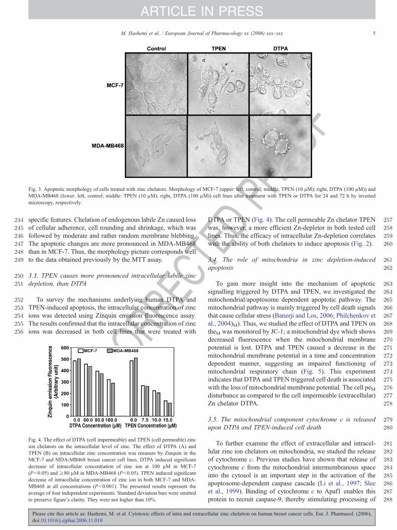
The cell morphology of treated and untreated cells wasexamined by phase contrast microscopy (Fig. 3). In order toconfirm the nature of changes promoted by TPEN and DTPA inMCF-7 and MDA-MB468 cells, we followed several apoptosis-
Please cite this article as: Hashemi, M. et al. Cytotoxic effects of intra and extracellular zinc chelation on human breast cancer cells. Eur. J. Pharmacol. (2006),doi:10.1016/j.ejphar.2006.11.010
ROOF
244
245
246
247
248
249
250
251
252
253
254
255
256
257
258
259
260
261
262
263
264
265
266
267
268
269
Fig. 3. Apoptotic morphology of cells treated with zinc chelators. Morphology of MCF-7 (upper: left, control; middle, TPEN (10 μM); right, DTPA (100 μM)) andMDA-MB468 (lower: left, control; middle: TPEN (10 μM); right, DTPA (100 μM)) cell lines after treatment with TPEN or DTPA for 24 and 72 h by invertedmicroscopy, respectively.
5M. Hashemi et al. / European Journal of Pharmacology xx (2006) xxx–xxx
ARTICLE IN PRESS
REC
specific features. Chelation of endogenous labile Zn caused lossof cellular adherence, cell rounding and shrinkage, which wasfollowed by moderate and rather random membrane blebbing.The apoptotic changes are more pronounced in MDA-MB468than in MCF-7. Thus, the morphology picture corresponds wellto the data obtained previously by the MTT assay.
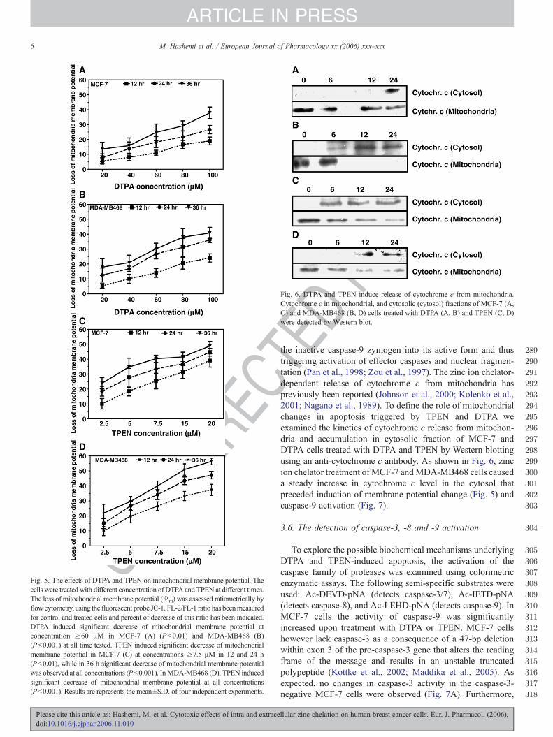
3.3. TPEN causes more pronounced intracellular labile zincdepletion, than DTPA
To survey the mechanisms underlying human DTPA andTPEN-induced apoptosis, the intracellular concentration of zincions was detected using Zinquin emission fluorescence assay.The results confirmed that the intracellular concentration of zincions was decreased in both cell lines that were treated with
UNCO
R 270
271
272
273
274
275
276
277
278
279
280
281
282
283
284
285
286
287
288
Fig. 4. The effect of DTPA (cell impermeable) and TPEN (cell permeable) zincion chelators on the intracellular level of zinc. The effect of DTPA (A) andTPEN (B) on intracellular zinc concentration was measure by Zinquin in theMCF-7 and MDA-MB468 breast cancer cell lines. DTPA induced significantdecrease of intracellular concentration of zinc ion at 100 μM in MCF-7(Pb0.05) and ≥80 μM in MDA-MB468 (Pb0.05). TPEN induced significantdecrease of intracellular concentration of zinc ion in both MCF-7 and MDA-MB468 at all concentrations (Pb0.001). The presented results represent theaverage of four independent experiments. Standard deviation bars were omittedto preserve figure's clarity. They were not higher than 10%.
Please cite this article as: Hashemi, M. et al. Cytotoxic effects of intra and extracedoi:10.1016/j.ejphar.2006.11.010
TEDP
DTPA or TPEN (Fig. 4). The cell permeable Zn chelator TPENwas, however, a more efficient Zn-depletor in both tested celllines. Thus, the efficacy of intracellular Zn-depletion correlateswith the ability of both chelators to induce apoptosis (Fig. 2).
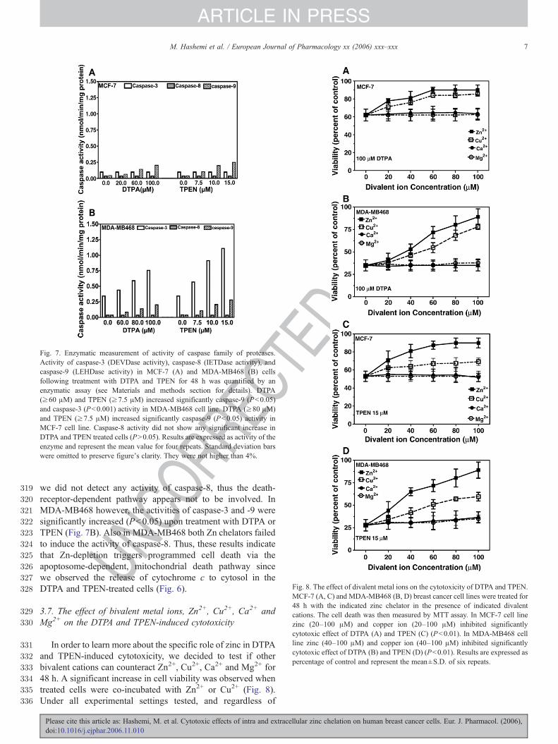
3.4. The role of mitochondria in zinc depletion-inducedapoptosis
To gain more insight into the mechanism of apoptoticsignalling triggered by DTPA and TPEN, we investigated themitochondrial/apoptosome dependent apoptotic pathway. Themitochondrial pathway is mainly triggered by cell death signalsthat cause cellular stress (Banerji and Los, 2006; Philchenkov etal., 2004)M). Thus, we studied the effect of DTPA and TPEN ontheM was monitored by JC-1; a mitochondrial dye which showsdecreased fluorescence when the mitochondrial membranepotential is lost. DTPA and TPEN caused a decrease in themitochondrial membrane potential in a time and concentrationdependent manner, suggesting an impaired functioning ofmitochondrial respiratory chain (Fig. 5). This experimentindicates that DTPA and TPEN triggered cell death is associatedwith the loss of mitochondrial membrane potential. The cell peMdisturbance as compared to the cell impermeable (extracellular)Zn chelator DTPA.
3.5. The mitochondrial component cytochrome c is releasedupon DTPA and TPEN-induced cell death
To further examine the effect of extracellular and intracel-lular zinc ion chelators on mitochondria, we studied the releaseof cytochrome c. Previous studies have shown that release ofcytochrome c from the mitochondrial intermembranous spaceinto the cytosol is an important step in the activation of theapoptosome-dependent caspase cascade (Li et al., 1997; Sleeet al., 1999). Binding of cytochrome c to Apaf1 enables thisprotein to recruit caspase-9, thereby stimulating processing of
llular zinc chelation on human breast cancer cells. Eur. J. Pharmacol. (2006),
UNCO
RREC
DPR
OOF
289
290
291
292
293
294
295
296
297
298
299
300
301
302
303
304
305
306
307
308
309
310
311
312
313
314
315
316
317
318
Fig. 5. The effects of DTPA and TPEN on mitochondrial membrane potential. Thecells were treated with different concentration of DTPA and TPEN at different times.The loss of mitochondrial membrane potential (Ψm) was assessed ratiometrically byflow cytometry, using the fluorescent probe JC-1. FL-2/FL-1 ratio has beenmeasuredfor control and treated cells and percent of decrease of this ratio has been indicated.DTPA induced significant decrease of mitochondrial membrane potential atconcentration ≥60 μM in MCF-7 (A) (Pb0.01) and MDA-MB468 (B)(Pb0.001) at all time tested. TPEN induced significant decrease of mitochondrialmembrane potential in MCF-7 (C) at concentrations ≥7.5 μM in 12 and 24 h(Pb0.01), while in 36 h significant decrease of mitochondrial membrane potentialwas observed at all concentrations (Pb0.001). InMDA-MB468 (D), TPEN inducedsignificant decrease of mitochondrial membrane potential at all concentrations(Pb0.001). Results are represents the mean±S.D. of four independent experiments.
Fig. 6. DTPA and TPEN induce release of cytochrome c from mitochondria.Cytochrome c in mitochondrial, and cytosolic (cytosol) fractions of MCF-7 (A,C) and MDA-MB468 (B, D) cells treated with DTPA (A, B) and TPEN (C, D)were detected by Western blot.
6 M. Hashemi et al. / European Journal of Pharmacology xx (2006) xxx–xxx
ARTICLE IN PRESS
Please cite this article as: Hashemi, M. et al. Cytotoxic effects of intra and extracedoi:10.1016/j.ejphar.2006.11.010
TEthe inactive caspase-9 zymogen into its active form and thustriggering activation of effector caspases and nuclear fragmen-tation (Pan et al., 1998; Zou et al., 1997). The zinc ion chelator-dependent release of cytochrome c from mitochondria haspreviously been reported (Johnson et al., 2000; Kolenko et al.,2001; Nagano et al., 1989). To define the role of mitochondrialchanges in apoptosis triggered by TPEN and DTPA weexamined the kinetics of cytochrome c release from mitochon-dria and accumulation in cytosolic fraction of MCF-7 andDTPA cells treated with DTPA and TPEN by Western blottingusing an anti-cytochrome c antibody. As shown in Fig. 6, zincion chelator treatment of MCF-7 andMDA-MB468 cells causeda steady increase in cytochrome c level in the cytosol thatpreceded induction of membrane potential change (Fig. 5) andcaspase-9 activation (Fig. 7).
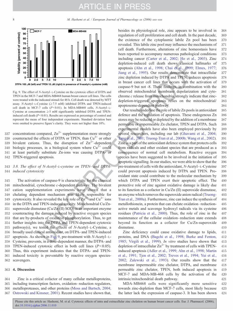
3.6. The detection of caspase-3, -8 and -9 activation
To explore the possible biochemical mechanisms underlyingDTPA and TPEN-induced apoptosis, the activation of thecaspase family of proteases was examined using colorimetricenzymatic assays. The following semi-specific substrates wereused: Ac-DEVD-pNA (detects caspase-3/7), Ac-IETD-pNA(detects caspase-8), and Ac-LEHD-pNA (detects caspase-9). InMCF-7 cells the activity of caspase-9 was significantlyincreased upon treatment with DTPA or TPEN. MCF-7 cellshowever lack caspase-3 as a consequence of a 47-bp deletionwithin exon 3 of the pro-caspase-3 gene that alters the readingframe of the message and results in an unstable truncatedpolypeptide (Kottke et al., 2002; Maddika et al., 2005). Asexpected, no changes in caspase-3 activity in the caspase-3-negative MCF-7 cells were observed (Fig. 7A). Furthermore,
llular zinc chelation on human breast cancer cells. Eur. J. Pharmacol. (2006),
RECT
EDPR
OOF
319
320
321
322
323
324
325
326
327
328
329
330
331
332
333
334
335
336
Fig. 7. Enzymatic measurement of activity of caspase family of proteases.Activity of caspase-3 (DEVDase activity), caspase-8 (IETDase activity), andcaspase-9 (LEHDase activity) in MCF-7 (A) and MDA-MB468 (B) cellsfollowing treatment with DTPA and TPEN for 48 h was quantified by anenzymatic assay (see Materials and methods section for details). DTPA(≥60 μM) and TPEN (≥7.5 μM) increased significantly caspase-9 (Pb0.05)and caspase-3 (Pb0.001) activity in MDA-MB468 cell line. DTPA (≥80 μM)and TPEN (≥7.5 μM) increased significantly caspase-9 (Pb0.05) activity inMCF-7 cell line. Caspase-8 activity did not show any significant increase inDTPA and TPEN treated cells (PN0.05). Results are expressed as activity of theenzyme and represent the mean value for four repeats. Standard deviation barswere omitted to preserve figure's clarity. They were not higher than 4%.
Fig. 8. The effect of divalent metal ions on the cytotoxicity of DTPA and TPEN.MCF-7 (A, C) and MDA-MB468 (B, D) breast cancer cell lines were treated for48 h with the indicated zinc chelator in the presence of indicated divalentcations. The cell death was then measured by MTT assay. In MCF-7 cell linezinc (20–100 μM) and copper ion (20–100 μM) inhibited significantlycytotoxic effect of DTPA (A) and TPEN (C) (Pb0.01). In MDA-MB468 cellline zinc (40–100 μM) and copper ion (40–100 μM) inhibited significantlycytotoxic effect of DTPA (B) and TPEN (D) (Pb0.01). Results are expressed aspercentage of control and represent the mean±S.D. of six repeats.
7M. Hashemi et al. / European Journal of Pharmacology xx (2006) xxx–xxx
ARTICLE IN PRESS
UNCO
R
we did not detect any activity of caspase-8, thus the death-receptor-dependent pathway appears not to be involved. InMDA-MB468 however, the activities of caspase-3 and -9 weresignificantly increased (Pb0.05) upon treatment with DTPA orTPEN (Fig. 7B). Also in MDA-MB468 both Zn chelators failedto induce the activity of caspase-8. Thus, these results indicatethat Zn-depletion triggers programmed cell death via theapoptosome-dependent, mitochondrial death pathway sincewe observed the release of cytochrome c to cytosol in theDTPA and TPEN-treated cells (Fig. 6).
3.7. The effect of bivalent metal ions, Zn2+, Cu2+, Ca2+ andMg2+ on the DTPA and TPEN-induced cytotoxicity
In order to learn more about the specific role of zinc in DTPAand TPEN-induced cytotoxicity, we decided to test if otherbivalent cations can counteract Zn2+, Cu2+, Ca2+ and Mg2+ for48 h. A significant increase in cell viability was observed whentreated cells were co-incubated with Zn2+ or Cu2+ (Fig. 8).Under all experimental settings tested, and regardless of
Please cite this article as: Hashemi, M. et al. Cytotoxic effects of intra and extracellular zinc chelation on human breast cancer cells. Eur. J. Pharmacol. (2006),doi:10.1016/j.ejphar.2006.11.010
C337
338
339
340
341
342
343
344
345
346
347
348
349
350
351
352
353
354
355
356
357
358
359
360
361
362
363
364
365
366
367
368
369
370
371
372
373
374
375
376
377
378
379
380
381
382
383
384
385
386
387
388
389
390
391
392
393
394
395
396
397
398
399
400
401
402
403
404
405
406
407
408
409
410
411
412
413
414
415
416
417
418
419
420
421
422
423
424
Fig. 9. The effect of N-Acetyl–L-Cysteine on the cytotoxic effect of DTPA andTPEN in the MCF-7 andMDA-MB468 human breast cancer cell lines. The cellswere treated with the indicated stimuli for 48 h. Cell death was detected by MTTassay. N-Acetyl–L-Cysteine (≥7.5 mM) inhibited DTPA and TPEN-inducedcell death in MCF-7 cells (Pb0.01). In MDA-MB468 cells, N-Acetyl–L-Cysteine at concentration ≥5 mM significantly inhibited DTPA and TPEN-induced cell death (Pb0.01). Results are expressed as percentage of control andrepresent the mean of four independent experiments. Standard deviation barswere omitted to preserve figure's clarity. They were not higher than 10%.
8 M. Hashemi et al. / European Journal of Pharmacology xx (2006) xxx–xxx
ARTICLE IN PRESS
UNCO
RRE
concentrations compared, Zn2+ supplementation more stronglycounteracted the effects of DTPA or TPEN, than Cu2+ or otherbivalent cations. Thus, the disruption of Zn2+-dependentbiologic processes, in a biological system where Cu2+ couldat least partially substitute Zn-ions was causing DTPA- orTPEN-triggered apoptosis.
3.8. The effect of N-Acetyl–L-cysteine on TPEN- and DTPA-induced cytotoxicity
The activation of caspase-9 is characteristic for the classicalmitochondrial, cytochrome c-dependent pathway. The bivalentcation supplementation experiments have shown that achelating dependent mechanism was likely responsible forcytotoxicity. It also revealed the key role of Zn2+ and Cu2+ ionsin the DTPA and TPEN-induced toxicity. Mitochondrial Cu/Zn-superoxide dismutase (Cu/Zn (II) SOD) is an important enzymecounteracting the damage induced by reactive oxygen speciesthat are by-products of oxidative phosphorylation. Thus, to getfurther insights into the DTPA and TPEN-dependent cytotoxicpathway(s), we tested the effects of N-Acetyl–L-Cysteine, abroadly used clinical antioxidant, on DTPA- and TPEN-inducedapoptosis. As shown in Fig. 9, pre-treatment with N-Acetyl–L-Cysteine, prevents, in a dose-dependent manner, the DTPA- andTPEN-induced cytotoxic effect in both cell lines (Pb0.05).Thus, this experiment indicates that the DTPA- and TPEN-induced toxicity is preventable by reactive oxygen species-scavengers.
4. Discussion
Zinc is a critical cofactor of many cellular metalloproteins,including transcription factors, oxidation–reduction regulators,metalloproteases, and other proteins (Moss and Bartsch, 2004;Ostrowski and Kinsner, 2001). Recent studies have shown that,
Please cite this article as: Hashemi, M. et al. Cytotoxic effects of intra and extracedoi:10.1016/j.ejphar.2006.11.010
TEDPR
OOF
besides its physiological role, zinc appears to be involved inregulation of cell proliferation and cell death. In the past decade,the existence of the cytoplasmic labile Zn pool has beenrevealed. This labile zinc pool may influence the mechanisms ofcell death. Furthermore, alterations of zinc homeostasis havebeen reported to accompany numerous pathological conditionsincluding cancer (Carter et al., 2002; Ho et al., 2003). Zincdepletion-induced cell death shows classical hallmarks ofapoptosis (Ahn et al., 1998; Chai et al., 1999; Elmes, 1977;Jiang et al., 1995). Our results demonstrate that intracellularzinc depletion induced by DTPA and TPEN, induces apoptosisin breast cancer cell lines that occurs with the activation ofcaspase-9 but not -8. These findings, in combination with theobserved mitochondrial membrane depolarization and cyto-chrome c release from mitochondria, strongly indicate that zinc-depletion-triggered apoptosis relies on the mitochondrial/apoptosome-dependent pathway.
Our results underline the role of labile Zn pools in antioxidantdefence and the regulation of apoptosis. These endogenous Znstores may be reduced or depleted by the addition of a membranepermeable or impermeable Zn chelator, TPEN and DTPA. Suchexperimental models have also been employed previously byseveral researchers, including our lab (Ghavami et al., 2004;Tang et al., 2001; Truong-Tran et al., 2000b; Wang et al., 2002).Zinc is a part of the antioxidant defence system that protects cellsfrom radicals and other oxidant species that are produced as aconsequence of normal cell metabolism. Reactive oxygenspecies have been suggested to be involved in the initiation ofapoptotic signalling. In our studies, wewere able to show that thepre-treatment of cells with the antioxidant,N-Acetyl–L-Cysteinecould prevent apoptosis induced by DTPA and TPEN. Pro-oxidant state could contribute to the molecular mechanism bywhich DTPA and TPEN exert their apoptotic effect. Theprotective role of zinc against oxidative damage is likely dueto its function as a cofactor in Cu/Zn (II) superoxide dismutase,an enzymewhich removes the superoxide anion radical (Truong-Tran et al., 2000a). Furthermore, zinc can induce the synthesis ofmetallothionein, a protein that can chelate oxidation–reduction-active metals and scavenge hydroxyl radicals via its cysteineresidues (Patricia et al., 2000). Thus, the role of zinc in themaintenance of the cellular oxidation–reduction state extendsbeyond its function as a cofactor for Cu/Zn-superoxidedismutase.
Zinc deficiency could cause oxidative damage to lipids,proteins, and DNA (Bagchi et al., 1998; Burke and Fenton,1985; Virgili et al., 1999). In vitro studies have shown thatdepletion of intracellular Zn2+ by treatment of cells with TPEN-induced apoptosis (Adler et al., 1999; Ahn et al., 1998; Martinet al., 1991; Tjen et al., 2002; Treves et al., 1994; Yui et al.,2002; Zalewski et al., 1993). Our results show that themembrane impermeable zinc chelator, DTPA, and membranepermeable zinc chelator, TPEN, both induced apoptosis inMCF-7 and MDA-MB-468 cells by the activation of theintrinsic/mitochondrial death pathway.
MDA-MB468 cells were significantly more sensitivetowards zinc-depletion than MCF-7 cells, most likely becausethe latter lack the expression of caspase-3. It has been shown
llular zinc chelation on human breast cancer cells. Eur. J. Pharmacol. (2006),
425
426
427
428
429
430
431
432
433
434
435
436
437
438
439
440
441
442
443
444
445
446
447
448
449
450
451
452
453
454
455
456
457
458
459
460
461
462
463
464
465
466
467
468
469
470
471
472
473
474
475476477
478479480481482483484485486487488489490491492493494495496497498499500501502503504505506507508509510511512513514515516517518519520521522523524525526527528529530531532533534535536537538539540541542543544545
9M. Hashemi et al. / European Journal of Pharmacology xx (2006) xxx–xxx
ARTICLE IN PRESS
UNCO
RREC
recently that pro-caspase-3 is stabilised in the presence of zincions, either directly through binding to Zn2+ (Hyun et al., 2000;Marini et al., 2001) or indirectly through the effect of zinc onoxidation–reduction-controlled processes (Strasser et al., 2000).Thus, Zn-depletion may have a direct effect on the activation ofcaspase-3.
Intracellular zinc depletion causes a significant cellular stressby itself, since this bivalent cation is critical for the function ofseveral transcription factors and enzymes. Cellular stress isknown to activate the mitochondrial/apoptosome-dependentdeath pathway (Brouckaert et al., 2005; Ghavami et al., 2005b,2004; Krzemieniecki et al., 2006). Moreover, mitochondriarepresent the cellular compartment responsible for generation ofenergy in the form of ATP. In addition, they may activateapoptosis by releasing cytochrome c (Barczyk et al., 2005),along with other pro-apoptotic proteins, thereby activatingcaspase-9 and caspase-3, the major apoptosis executionenzymes (Maddika et al., 2005; Mignotte and Vayssiere,1998). In addition, recent studies indicate that mobilization ofendogenous labile Zn pools might interfere with normalfunctioning of mitochondria, and it may be responsible foractivation of apoptosis (Sensi et al., 2003). Thus, consistentwith the above reports, we found that chelation of intracellularlabile zinc pools leads to the activation of the apoptotic cascadethat is accompanied by the loss of mitochondrial transmem-brane potential and caspase-9 activation.
In summary, we show that one of the major negative effectsof Zn ion insufficiency in the cell is the inhibition of Cu/Zn-superoxide dismutase and, in consequence, an antioxidant-preventable induction of programmed cell death. Thus, Zn-deficiency may have serious health consequences that areevoked not only directly, by the lack of Zn as a cofactor formetalloproteins, but also indirectly as an effect of oxidativestress within the cell. The oxidative stress may cause damage tovarious cellular organelles as well as DNA-strand breaks withthe subsequent activation of cell death programs (Booy et al.,2005; Pour-Jafari et al., 2005). Furthermore, since “smallmolecules” (Hauff et al., 2005; Kroczak et al., in press;Mendoza et al., 2005) have superior pharmacokinetic featuresas compared to proteins and other macro-molecules, manipula-tion of the bioavailability of “trace metals” like zinc, that oftenfunction as cofactors, may prove to be a new promising way ofpharmacologic modulation of metabolic processes.
Acknowledgements
M.L. thankfully acknowledges the support by the CFI-Canada Research Chair program, PCRFC-CCMF, MMSF,MHRC, and CIHR-foundation-financed programs. E.P.B.thankfully acknowledges the support by the CIHR. The salaryof S.G. has been supported by the MHRC and CCMF.
References
Adler, M., Shafer, H., Hamilton, T., Petrali, J.P., 1999. Cytotoxic actions of theheavy metal chelator TPEN on NG108-15 neuroblastoma-glioma cells.Neurotoxicology 20, 571–582.
Please cite this article as: Hashemi, M. et al. Cytotoxic effects of intra and extracedoi:10.1016/j.ejphar.2006.11.010
TEDPR
OOF
Ahn, Y.H., Kim, Y.H., Hong, S.H., Koh, J.Y., 1998. Depletion of intracellularzinc induces protein synthesis-dependent neuronal apoptosis in mousecortical culture. Exp. Neurol. 154, 47–56.
Bagchi, D., Vuchetich, P.J., Bagchi, M., Tran, M.X., Krohn, R.L., Ray, S.D.,Stohs, S.J., 1998. Protective effects of zinc salts on DTPA-induced hepaticand brain lipid peroxidation, glutathione depletion, DNA damage andperitoneal macrophage activation in mice. Gen. Pharmacol. 30, 43–50.
Banerji, S., Los, M., 2006. Important differences between topoisomerase-I andII targeting agents. Cancer Biol. Ther. 5, 965–966.
Barczyk, K., Kreuter, M., Pryjma, J., Booy, E.P., Maddika, S., Ghavami, S.,Berdel, W.E., Roth, J., Los, M., 2005. Serum cytochrome c indicates invivo-apoptosis and it can serve as a prognostic marker during cancer therapy.Int. J. Cancer. 114, 167–173.
Behroozi, S.J., Kim, W., Dannaldson, J., Gates, K.S., 1996. 1,2-dithiolan-3-one1-oxides: a class of thiol-activated DNA-cleaving agents that are structurallyrelated to the natural product leinamycin. Biochemistry 35, 1768–1774.
Berg, J.M., Shi, Y., 1996. The galvanization of biology: a growing apprecia-tionfor the roles of zinc. Science 271, 1081–1085.
Bergan, T., Klaveness, J., Aasen, A.J., 2001. Chelating agents. Chemotherapy47, 10–14.
Booy, E.P., Kadkhoda, K., Johar, D., Bay, G.H., Los, M., 2005. The immunesystem, involvement in neurodegenerative diseases, ageing and cancer. Curr.Med. Chem. Anti-Inflamm. Anti-Allerg. Agents 4, 349–353.
Bradford, M.M., 1976. A rapid and sensitive method for the quantities ofmicrogram of protein utilizing the principle of protein-dye binding. Anal.Biochem. 72, 248–254.
Brouckaert, G., Kalai, M., Saelens, X., Vandenabeele, P., 2005. Apoptoticpathways and their regulation. In: Los, M., Gibson, S.B. (Eds.), ApoptoticPathways as Target for Novel Therapies in Cancer and Other Diseases.Springer Academic Press, New York.
Burke, J.P., Fenton, M.R., 1985. Effect of a zinc-deficient diet on lipidperoxidation in liver and tumor subcellular membranes. Proc. Soc. Exp.Biol. Med. 179, 187–191.
Carter, J.E., Truong-Tran, A.Q., Grosser, D., Ho, L., Ruffin, R.E., Zalewski, P.D.,2002. Involvement of redox events in caspase activation in zinc-depletedairway epithelial cells. Biochem. Biophys. Res. Commun. 297, 1062–1070.
Chai, F., Truong-Tran, A.Q., Ho, L.H., Zalewski, P.D., 1999. Regulation ofcaspase activation and apoptosis by cellular zinc fluxes and zinc deprivation:a review. Immunol. Cell Biol. 77, 272–278.
Elmes, M.E., 1977. Apoptosis in the small intestine of zinc-deficient and fastedrats. J. Pathol. 123, 219–223.
Fukamachi, Y., Karasaki, Y., Sugiura, T., Itoh, H., Abe, T., Yamamura, K.,Higashi, K., 1998. Zinc suppresses apoptosis of U937 cells induced byhydrogen peroxide through an increase of the Bcl-2/Bax ratio. Biochem.Biophys. Res. Commun. 246, 364–369.
Ghavami, S., Kerkhoff, C., Los, M., Hashemi, M., Sorg, C., Karami-Tehrani, F.,2004. Mechanism of apoptosis induced by S100A8/A9 in colon cancer celllines: the role of ROS and the effect of metal ions. J. Leukoc. Biol. 76,169–175.
Ghavami, S., Barczyk, K., Maddika, S., Vogl, T., Steinmüller, L., Pour-Jafari,H., Evans, J.A., Los, M., 2005a. Monitoring of programmed cell death invivo and in vitro,— new and old methods of cancer therapy assessment. In:Los, M., Gibson, S.B. (Eds.), Apoptotic Pathways as Target for NovelTherapies in Cancer and Other Diseases. Springer Science+BusinessMedia,Inc., New York, pp. 323–341.
Ghavami, S., Hashemi, M., Kadkhoda, K., Alavian, S.M., Bay, G.H., Los, M.,2005b. Apoptosis in liver diseases— detection and therapeutic applications.Med. Sci. Monit. 11, RA337–RA345.
Hashemi, M., Kroczak, T.J., 2005. Apoptosis and autoimmune disease. Curr.Med. Chem. Anti-Inflamm. Anti-Allerg. Agents 4, 429–437.
Hashemi, M., Karami-Tehrani, F., Ghavami, S., Maddika, S., Los, M., 2005.Adenosine and deoxyadenosine induces apoptosis in oestrogen receptor-positive and negative human breast cancer cells via the intrinsic pathway.Cell Prolif. 38, 269–285.
Hauff, K., Zamzow, C., Law, W.J., de Melo, J., Kennedy, K., Los, M., 2005.Peptide-based approaches to treat asthma, arthritis, other autoimmunediseases and pathologies of the central nervous system. Arch. Immunol.Ther. Exp. 53, 308–320.
llular zinc chelation on human breast cancer cells. Eur. J. Pharmacol. (2006),
C
546547548549550551552553554555556557558559560561562563564565566567568569570571572573574575576577578579580581582583584585586587588589590591592593594595596597598599600601602603604605606607608609610611612613
614615616617618619620621622623624625626627628629630631632633634635636637638639640641642643644645646647648649650651652653654655656657658659660661662663664665666667668669670671672673674675676677678679680681
10 M. Hashemi et al. / European Journal of Pharmacology xx (2006) xxx–xxx
ARTICLE IN PRESS
UNCO
RRE
Heximer, S.P., Forsdyke, D.R., 1993. A human putative lymphocyte G0/G1switch gene homologous to a rodent gene encoding a zinc-bindingpotential transcription factor. DNA Cell Biol. 12, 73–88.
Ho, E., Courtemanche, C., Ames, B.N., 2003. Zinc deficiency induces oxidativeDNA damage and increases p53 expression in human lung fibroblasts. J. Nutr.133, 2543–2548.
Huang, E.P., 1997. Metal ions and synaptic transmission: think zinc. Proc. Natl.Acad. Sci. U. S. A. 94, 13386–13387.
Hyun, H.J., Sohn, J., Ahn, Y.H., Shin, H.C., Koh, J.Y., Yoon, Y.H., 2000.Depletion of intracellular zinc induces macromolecule synthesis andcaspase-dependent apoptosis of cultured retinal cells. Brain Res. 869,39–48.
Jiang, S., Chow, S.C., McCabe, J.M.J., Orrenius, S., 1995. Lack of Ca2+
involvement in thymocyte apoptosis induced by chelation of intracellularZn2+. Lab. Invest. 73, 111–117.
Johnson, V.L., Ko, S.C., Holmstrom, T.H., Eriksson, J.E., Chow, S.C., 2000.Effector caspases are dispensable for the early nuclear morphologicalchanges during chemical-induced apoptosis. J. Cell Sci. 113 (Pt 17),2941–2953.
Kennedy, C.J., Rakoczy, P.E., Robertson, T.A., Papadimitriou, J.M., Constable,I.J., 1994. Kinetic studies on phagocytosis and lysosomal digestion of rodouter segments by human retinal pigment epithelial cells in vitro. Exp. CellRes. 210, 209–214.
Klug, A., 1999. Zinc finger peptides for the regulation of gene expression. J.Mol.Biol. 293, 215–218.
Kolenko, V.M., Uzzo, R.G., Dulin, N., Hauzman, E., Bukowski, R., Finke, J.H.,2001. Mechanism of apoptosis induced by zinc deficiency in peripheralblood T lymphocytes. Apoptosis 6, 419–429.
Kottke, T.J., Blajeski, A.L.,Meng, X.W., Svingen, P.A., Ruchaud, S.,Mesner, P.W.,Boerner, S.A.J., Samejima, K., Henriquez, V., Chilcote, T.J., Lord, J., Salmon,M., Earnshaw, W.C., Kaufmann, S.H., 2002. Lack of correlation betweencaspase activation and caspase activity assays in paclitaxeltreatedMCF-7 breastcancer cells. J. Biol. Chem. 277, 804–815.
Kroczak, T.J., Baran, J., Pryjma, J.S.,M.,Rashedi, I., Hernandez,E.,Alberti, E.M., S.,Los, M., (in press). The emerging importance of DNA mapping and othercomprehensive screening techniques as tools to identify new drug targets and as amean of (cancer) therapy personalization. Expert. Opin. Ther. Targets.
Krzemieniecki, K., Szpyt, E., Rashedi, I., Gawron, K., Los, M., 2006. Targetingof solid tumors and blood malignancies by antibody-based therapies —EGFR-pathway as an example. Centr. Eur. J. Biol. 1, 167–182.
Li, P., Nijhawan, D., Budihardjo, I., Srinivasula, S.M., Ahmad, M., Alnemri, E.S.,Wang, X., 1997. Cytochrome c and dATP-dependent formation of Apaf-1/caspase-9 complex initiates an apoptotic protease cascade. Cell 91, 479–489.
Los, M., Herr, I., Friesen, C., Fulda, S., Schulze-Osthoff, K., Debatin, K.M.,1997. Cross-resistance of CD95-and drug-induced apoptosis as a conse-quence of deficient activation of caspases (ICE/Ced-3 proteases). Blood 90,3118–3129.
Los, M., Wesselborg, S., Schulze-Osthoff, K., 1999. The role of caspases indevelopment, immunity, and apoptotic signal transduction: lessons fromknockout mice. Immunity 10, 629–639.
Maddika, S., Booy, E.P., Johar, D., Gibson, S.B., Ghavami, S., Los, M., 2005.Cancer-specific toxicity of apoptin is independent of death receptors butinvolves the loss of mitochondrial membrane potential and the release ofmitochondrial cell death mediators by a Nur77-dependent pathway. J. CellSci. 118, 4485–4493.
Mares-Perlman, J.A., Klein, R., Klein, B.E., Greger, J.L., Brady, W.E., Palta,M., Ritter, L.L., 1996. Association of zinc and antioxidant nutrients withage-related maculopathy. Arch. Ophthalmol. 114, 991–997.
Marini, M., Frabetti, F., Canaider, S., Dini, L., Falcieri, E., Poirier, G.G., 2001.Modulation of caspase-3 activity by zinc ions and by the cell redox state.Exp. Cell Res. 266, 323–332.
Martin, S.J., Mazdai, G., Strain, J.J., Cotter, T.G., Hannigan, B.M., 1991.Programmed cell death (apoptosis) in lymphoid and myeloid cell linesduring zinc deficiency. Clin. Exp. Immunol. 83, 338–343.
McCabe Jr., M.J., Jiang, S.A., Orrenius, S., 1993. Chelation of intracellular zinctriggers apoptosis in mature thymocytes. Lab. Invest. 69, 101–110.
McClain, C.J., Kasarskis Jr., E.J., Allen, J.J., 1985. Functional consequences ofzinc deficiency. Prog. Food Nutr. Sci. 9, 185–226.
Please cite this article as: Hashemi, M. et al. Cytotoxic effects of intra and extracedoi:10.1016/j.ejphar.2006.11.010
TEDPR
OOF
Mendoza, F.J., Espino, P., Cann, C.L., Bristow, N., McCrea, K., Los, M., 2005.Anti-tumor chemotherapy utilizing peptide-based approaches — apoptoticpathways, kinases and proteasome as targets. Arch. Immunol. Ther. Exp. 53,47–60.
Mignotte, B., Vayssiere, J.L., 1998. Mitochondria and apoptosis. Eur. J. Biochem.252, 1–15.
Moss, M.L., Bartsch, J.W., 2004. Therapeutic benefits from targeting of ADAMfamily members. Biochemistry 43, 7227–7235.
Nagano, T., Hirano, T., Hirobe, M., 1989. Superoxide dismutase mimics basedon iron in vivo. J. Biol. Chem. 264, 9243–9249.
Ostrowski, K., Kinsner, A., 2001. Inhibition of angiogenesis in the treatment oftumors. Arch. Immunol. Ther. Exp. 49, 27–31.
Pan, G., Humke, E.W., Dixit, V.M., 1998. Activation of caspases triggered bycytochrome c in vitro. FEBS Lett. 426, 151–154.
Patricia, I.O., Michael, S., Paola, C.Z., Carl, L.K., 2000. Zinc deficiency inducesoxidative stress and AP-1 activation in 3T3 cells. Free Radic. Biol. Med. 28,1091–1099.
Perry, D.K., Smyth,M.J., Stennicke, H.R., Salvesen, G.S., Duriez, P., Poirier, G.G.,Hannun, Y.A., 1997. Zinc is a potent inhibitor of the apoptotic protease,caspase-3. A novel target for zinc in the inhibition of apoptosis. J. Biol. Chem.272, 18530–18533.
Philchenkov, A., Zavelevich, M., Kroczak, T.J., Los, M., 2004. Caspases andcancer: mechanisms of inactivation and new treatment modalities. Exp.Oncol. 26, 82–97.
Pour-Jafari, H., Ghavami, S., Maddika, S., 2005. Mitochondrial physiology andtoxicity (mitotoxicity); importance for cancer, programmed cell death andthe immune system. Curr. Med. Chem. Anti-Inflamm. Anti-Allerg. Agents4, 439–451.
Prasad, A.S., 1985. Laboratory diagnosis of zinc deficiency. J. Am. Coll. Nutr. 4,591–598.
Prasad, A.S., 1991. Discovery of human zinc deficiency and studies in anexperimental human model. Am. J. Clin. Nutr. 53, 403–412.
Rudolf, E., Cervinka, M., 2004. Depletion of endogenous zinc stores inducesoxidative stress and cell death in human melanoma cells. Acta Medica(Hradec Kralove) 47, 91–96.
Sensi, S.L., Ton-That, D., Sullivan, P.G., Jonas, E.A., Gee, K.R., Kaczmarek, L.K.,Weiss, J.H., 2003. Modulation of mitochondrial function by endogenous Zn2+
pools. Proc. Natl. Acad. Sci. U. S. A. 100, 6157–6162.Slee, E.A., Harte, M.T., Kluck, R.M.,Wolf, B.B., Casiano, C.A., Newmeyer, D.D.,
Wang, H.G., Reed, J.C., Nicholson, D.W., Alnemri, E.S., Green, D.R., Martin,S.J., 1999. Ordering the cytochrome c-initiated caspase cascade: hierarchicalactivation of caspases-2, 3, 6, 7, 8, and 10 in a caspase-9-dependent manner.J. Cell Biol. 144, 281–292.
Strasser, A., O'Connor, L., Dixit, V.M., 2000. Apoptosis signaling. Annu. Rev.Biochem. 69, 217–245.
Sukegawa, J., Blobel, G., 1993. A nuclear pore complex protein that containszinc finger motifs, binds DNA, and faces the nucleoplasm. Cell 72, 29–38.
Tang, Z.L., Wasserloos, K., St Croix, C.M., Pitt, B.R., 2001. Role of zinc inpulmonary endothelial cell response to oxidative stress. Am. J. Physiol.,Lung Cell. Mol. Physiol. 281, L243–L249.
Tjen, W.W., Haase, H., Biagioli, Beyersmann, M.D., 2002. Induction ofapoptosis in mammalian cells by cadmium and zinc. Environ. HealthPerspect. 110, 865–867.
Treves, S., Trentini, P.L., Ascanelli, M., Bucci, G., Di Virgilio, F., 1994.Apoptosis is dependent on intracellular zinc and independent of intracellularcalcium in lymphocytes. Exp. Cell Res. 211, 339–343.
Truong-Tran, A.Q., Ho, L.H., Chai, F., Zalewski, P.D., 2000a. Cellular zincfluxes and the regulation of apoptosis/gene-directed cell death. J. Nutr. 130,1459S–1466S.
Truong-Tran, A.Q., Ruffin, R.E., Zalewski, P.D., 2000b. Visualization of labilezinc and its role in apoptosis of primary airway epithelial cells and cell lines.Am. J. Physiol., Lung Cell. Mol. Physiol. 279, L1172–L1183.
Vallee, B.L., Falchuk, K.H., 1993. The biochemical basis of zinc physiology.Physiol. Rev. 73, 79–118.
Virgili, F., Canali, R., Figus, E., Vignolini, F., Nobili, F., Mengheri, E., 1999.Intestinal damage induced by zinc deficiency is associated with enhancedCu/Zn (II) superoxide dismutase activity in rats: effect of dexamethasone orthyroxine treatment. Free Radic. Biol. Med. 26, 1194–1201.
llular zinc chelation on human breast cancer cells. Eur. J. Pharmacol. (2006),
682683684685686687688689690
691692693694695
696
697
11M. Hashemi et al. / European Journal of Pharmacology xx (2006) xxx–xxx
ARTICLE IN PRESS
Wang, X., Fosmire, G.J., Gay, C.V., Leach Jr., R.M., 2002. Short-term zincdeficiency inhibits chondrocyte proliferation and induces cell apoptosis inthe epiphyseal growth plate of young chickens. J. Nutr. 132, 665–673.
Yui, S., Nakatani, Y., Hunter, M.J., Chazin, W.J., Yamazaki, M., 2002.Implication of extracellular zinc exclusion by recombinant humancalprotectin (MRP8 and MRP14) from target cells in its apoptosis-inducingactivity. Mediat. Inflamm. 11, 165–172.
Zalewski, P.D., Forbes, I.J., Betts, W.H., 1993. Correlation of apoptosis withchange in intracellular labile Zn(II) using zinquin [(2-methyl-8-p-toluene-
UNCO
RREC
Please cite this article as: Hashemi, M. et al. Cytotoxic effects of intra and extracedoi:10.1016/j.ejphar.2006.11.010
sulphonamido-6-quinolyloxy)acetic acid], a new specific fluorescent probefor Zn(II). Biochem. J. 296 (Pt 2), 403–408.
Zou, H., Henzel, W.J., Liu, X., Lutschg, A., Wang, X., 1997. Apaf-1, a humanprotein homologous to C. elegans CED-4, participates in cytochrome c-dependent activation of caspase-3. Cell 90, 405–413.
TEDPR
OOF
llular zinc chelation on human breast cancer cells. Eur. J. Pharmacol. (2006),