Cytological and Molecular Basics of Heredity
100
Ukraine National University of life and Environmental Sciences M.Kravchenko Department of Breeding and Genetics of Animals Veterinary Genetics Cytological and Molecular Basics of Heredity Methodical instructions to performance of laboratory works on section «Cytological Basics of Heredity», «Molecular Basics of Heredity» for students of Specialty: Veterinary medicine Specialization: «Veterinary medicine» КYIV-2010
-
Upload
khangminh22 -
Category
Documents
-
view
1 -
download
0
Transcript of Cytological and Molecular Basics of Heredity

Ukraine
National University of life and Environmental Sciences
M.Kravchenko Department of Breeding and Genetics of Animals
Veterinary Genetics
Cytological and Molecular Basics of
Heredity
Methodical instructions to performance of laboratory works on section
«Cytological Basics of Heredity», «Molecular Basics of Heredity»
for students of
Specialty: Veterinary medicine
Specialization: «Veterinary medicine»
КYIV-2010

UDC 636.082 (075.8)
Methodical instructions for studying discipline of Veterinary Genetics on the
following topics: Cytological Basics of Heredity. «Molecular Basics of Heredity»,
Mitosis. Meiosis. Structure of Chromosome. Karyotypes. Gene structure.
Transcription. Synthesis of protein.
For students of the specialty «Veterinary Medicine of Veterinary Faculty» with
the profound study of English language.
Recommended by Educational-Methodical Council of Research and Training
Institute of Veterinary Medicine of National University of Life and Environmental
Sciences.
Compilers: S.O.Kostenko, I.O.Suprun, O.H.Ponomarenko
Reviewers: V.E. Zhulai, A.D. Oliynik
The educational edition
Veterinary Genetics
CYTOLOGICAL AND MOLECULAR BASICS OF HEREDITY
Methodical instructions of laboratory works doing on section «Cytological
Basics of Hereditу», «Molecular Basics of Heredity» for students of educational-
qualification level «Bachelor» a specialty, specialization – «Veterinary medicine»
Composers: Kostenko Svitlana Oleksiivna, Suprun Irina Oleksandrivna,
Ponomarenko Oksana Hrigorivna
S.O. Kostenko is responsible for the issue.
Manager of publishing center of NULIV: A.P. Kolesnikov
Signed for publishing 7.11.10 Circulation: 100 copies.
Format: 60 х 84 1/16 № 2072
Conditional sheets: 6,9 Account sheets: 6,8
Publishing center of NULIV
03041 Kyiv, Geroyiv Oborony street, 15.
INTRODUCTION
After study of the discipline section the student should know: the structure of a
cell, its cores organellas and their participation in transfer and realization of the
hereditary information, features of mitosis and meiosis, process of gametogenesis,
structures of chromosomes and karyotypes of domestic animals. Students should be
able: to prepare cytogenetic preparations, to do karyotypic analysis of animals.
Laboratory work №1
Subject: Cytological basics of animal’s heredity.
The purpose: To get acquainted with a structure of a cell, organellas which
take part in storage and realization of the genetic information. To learn preparing
cytogenetic preparations.
The equipment: tables of cell structure, tables of a mitosis and meyosis,
monocular microscopes, scissors, a scalpel, filtering paper, dye of Gimza fixing
solution of Karnua, hypotonic solution КСl, a centrifuge, rotary test tubes, a cover
glass.
Course of work:
1. To get acquainted with a structure of a cell (using the Table).
2. To draw the scheme of a cell and to designate its organelles.
3. To fill in the table of participation organelle in storage, transfer and realization
of the genetic information.
Explanation of work:
Biologists traditionally classify all living organisms into two major groups, the
prokaryotes and the eukaryotes. A prokaryote is a unicellular organism with a
relatively simple cell structure (Fig. 1). A eukaryote has a compartmentalized cell
structure divided by intracellular membranes; eukaryotes may be unicellular or
multicellular. The sizes of eukaryote organisms are various.
All cells contain many structural units of the smaller size named organellas.
Organella is substructure of cells which carry out specific functions (Table 1).
Organella is a subcell unit which is limited by a membrane and is separated for

centrifugal separation at high speed. According to this definition, ribosome,
cytoskeleton, cytosol are not organellas, but it is possible to allocate them by the
centrifugal separation.
FIGURE 1. Cell structure
From the perspective of genetics, a major difference between prokaryotic and
eukaryotic cells is that a eukaryote has a nuclear envelope, which surrounds the
genetic material to form a nucleus and separates the DNA from the other cellular
contents. In prokaryotic cells, the genetic material is in close contact with other
components of the cell – a property that has important consequences for the way in
which genes are controlled.
Another fundamental difference between prokaryotes and eukaryotes lies in the
packaging of their DNA. In eukaryotes, DNA is closely associated with a special
class of proteins, the histones, to form tightly packed chromosomes. This complex of
DNA and histone proteins is termed chromatin, which is the stuff of eukaryotic
chromosomes.
The Plasma Membrane
An animal cell is surrounded by an outer plasma membrane. The plasma
membrane marks the boundary between the outside of the cell and the inside of the
cell. Plasma membrane integrity and its function are necessary for the life of the cell.
The plasma membrane is a phospholipid belayed with attached or embedded
proteins. The structure of a phospholipid is such that the molecule has a polar head
and nonpolar tails (Fig.1). The polar heads, being charged, are hydrophilic (water
loving) and face outward, toward the cytoplasm on one side and the tissue fluid on

the other side, where they will encounter a watery environment. The nonpolar tails
are hydrophobic (not attracted to water) and face inward toward each other, where
there is no water. When phospholipids are placed in water, they naturally form a
circular bilayer because of the chemical properties of the heads and the tails. At body
temperature, the phospholipid bilayer is a liquid; it has the consistency of olive oil,
and the proteins are able to change their position by moving laterally. The fluid-
mosaic model, a working description of membrane structure, says that the protein
molecules have a changing pattern (form a mosaic) within the fluid phospholipid
bilayer.
Cholesterol lends support to the membrane. Short chains of sugars are attached
to the outer surface of some proteinand lipid molecules (called glycoproteins and
glycolipids, respectively). It is believed that these carbohydrate chains, specific to
each cell, help mark it as belonging to a particular individual. They account for why
people have different blood types, for example. Other glycoproteins have a special
configuration that allows them to act as a receptor for a chemical messenger like
ahormone. Some plasma membrane proteins form channels through which certain
substances can enter cells; others are carriers involved in the passage of molecules
through the membrane.
Plasma Membrane Functions:
The plasma membrane keeps a cell intact. It allows only certain molecules and
ions to enter and exit the cytoplasm freely; therefore, the plasma membrane is said to
be selectively permeable. Small molecules that are lipid-soluble, such as oxygen and
carbon dioxide, can pass through the membrane easily. Certain other small
molecules, like water, are not lipid soluble, but they still freely cross the membrane.
Still other molecules and ions require the use of a carrier to enter a cell. The plasma
membrane, composed of phospholipid and protein molecules, is selectively
permeable and regulates the entrance and exit of molecules and ions into and out of
the cell
The Nucleus
The nucleus, which has a diameter of about 5 m, is a prominent structure in
the eukaryotic cell. The nucleus is of primary importance because it stores genetic
information that determines the characteristics of the body’s cells and their metabolic
functioning. Every cell contains a complex copy of genetic information, but each cell
type has certain genes, or segments of DNA, turned on, and others turned off.
Activated DNA, with RNA acting as an intermediary, specifies the sequence of
amino acids during protein synthesis. The proteins of a cell determine its structure
and the functions it can perform. When you look at the nucleus, even in an electron
micrograph, you cannot see DNA molecules but you can see chromatin.
Chromatin looks grainy, but actually it is a threadlike material that undergoes
coiling into rodlike structures called chromosomes just before the cell divides.
Chemical analysis shows that chromatin, and therefore chromosomes, contains DNA
and much protein, as well as some RNA. Chromatin is immersed in a semifluid
medium called the nucleoplasm. A difference in pH between the nucleoplasm and

the cytoplasm suggests that the nucleoplasm has different composition. Most likely,
too, when you look at an electron micrograph of a nucleus, you will see one or more
areas that look darker than the rest of the chromatin. These are nucleoli (sing.,
nucleolus) where another type of RNA, called ribosomal RNA (rRNA), is produced
and where rRNA joins with proteins to form the subunits of ribosomes. (Ribosomes
are small bodies in the cytoplasm that contain rRNA and proteins.) The nucleus is
separated from the cytoplasm by a double membrane known as the nuclear envelope.
The nuclear envelope has nuclear pores of sufficient size (100 nm) to permit the
passage of proteins into the nucleus and ribosomal subunits out of the nucleus. The
structural features of the nucleus include the following. Chromatin: DNA and
proteins Nucleolus: Chromatin and ribosomal subunits. Nuclear envelope: Double
membrane
Mitochondria
Most mitochondria (sing., mitochondrion) are between 0.5 µm and 1.0 µm in
diameter and about 7 µm in length, although the size and the shape can vary.
Mitochondria are bounded by a double membrane. The inner membrane is folded to
form little shelves called cristae, which project into the matrix, an inner space filled
with a gellike fluid. Lysosomes are produced by a Golgi apparatus, and their
hydrolytic enzymes digest macromolecules from various sources. Mitochondria are
the site of ATP (adenosine triphosphate) production involving complex metabolic
pathways. ATP molecules are the common carrier of energy in cells. A shorthand
way to indicate the chemical transformation that involves mitochondria is as follows:
Mitochondria are often called the powerhouses of the cell: just as a powerhouse burns
fuel to produce electricity, the mitochondria convert the chemical energy of glucose
products into the chemical energy of ATP molecules. In the process, mitochondria
use up oxygen and give off carbon dioxide and water.
The Golgi apparatus is named by Camillo Golgi, who discovered its presence
in cells in 1898. The Golgi apparatus consists of a stack of three to twenty slightly
curved saccules whose appearance can be compared to a stack of pancakes. In animal
cells, one side of the stack (the inner face) is directed toward the ER, and the other
side of the stack (the outer face) is directed toward the plasma membrane. Vesicles
can frequently be seen at the edges of the saccules.
The Golgi apparatus receives protein and/or lipid-filled vesicles that bud from
the ER. Some biologists believe that these fuse to form a saccule at the inner face and
that this saccule remains as a part of the Golgi apparatus until the molecules are
repackaged in new vesicles at the outer face. Others believe that the vesicles from the
ER proceed directly to the outer face of the Golgi apparatus, where processing and
packaging occur within its saccules. The Golgi apparatus contains enzymes that
modify proteins and lipids. For example, it can add a chain of sugars to proteins,
thereby making them glycoproteins and glycolipids, which are found in the plasma
membrane.
The vesicles that leave the Golgi apparatus move about the cell. Some vesicles
proceed to the plasma membrane, where they discharge their contents. Because this is
secretion, it is often said that the Golgi apparatus is involved in processing,
packaging, and secretion. Other vesicles that leave the Golgi apparatus are
lysosomes. The Golgi apparatus processes, packages, and distributes molecules about
or from the cell. It is also said to be involved in secretion.
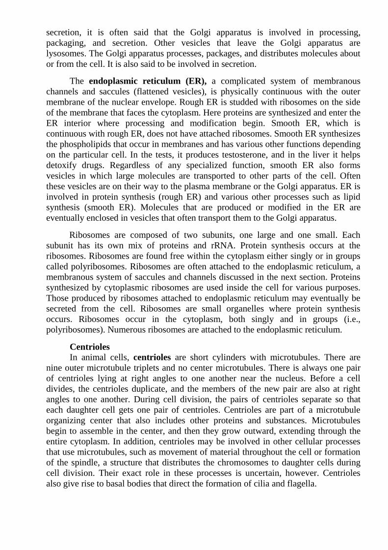
The endoplasmic reticulum (ER), a complicated system of membranous
channels and saccules (flattened vesicles), is physically continuous with the outer
membrane of the nuclear envelope. Rough ER is studded with ribosomes on the side
of the membrane that faces the cytoplasm. Here proteins are synthesized and enter the
ER interior where processing and modification begin. Smooth ER, which is
continuous with rough ER, does not have attached ribosomes. Smooth ER synthesizes
the phospholipids that occur in membranes and has various other functions depending
on the particular cell. In the tests, it produces testosterone, and in the liver it helps
detoxify drugs. Regardless of any specialized function, smooth ER also forms
vesicles in which large molecules are transported to other parts of the cell. Often
these vesicles are on their way to the plasma membrane or the Golgi apparatus. ER is
involved in protein synthesis (rough ER) and various other processes such as lipid
synthesis (smooth ER). Molecules that are produced or modified in the ER are
eventually enclosed in vesicles that often transport them to the Golgi apparatus.
Ribosomes are composed of two subunits, one large and one small. Each
subunit has its own mix of proteins and rRNA. Protein synthesis occurs at the
ribosomes. Ribosomes are found free within the cytoplasm either singly or in groups
called polyribosomes. Ribosomes are often attached to the endoplasmic reticulum, a
membranous system of saccules and channels discussed in the next section. Proteins
synthesized by cytoplasmic ribosomes are used inside the cell for various purposes.
Those produced by ribosomes attached to endoplasmic reticulum may eventually be
secreted from the cell. Ribosomes are small organelles where protein synthesis
occurs. Ribosomes occur in the cytoplasm, both singly and in groups (i.e.,
polyribosomes). Numerous ribosomes are attached to the endoplasmic reticulum.
Centrioles
In animal cells, centrioles are short cylinders with microtubules. There are
nine outer microtubule triplets and no center microtubules. There is always one pair
of centrioles lying at right angles to one another near the nucleus. Before a cell
divides, the centrioles duplicate, and the members of the new pair are also at right
angles to one another. During cell division, the pairs of centrioles separate so that
each daughter cell gets one pair of centrioles. Centrioles are part of a microtubule
organizing center that also includes other proteins and substances. Microtubules
begin to assemble in the center, and then they grow outward, extending through the
entire cytoplasm. In addition, centrioles may be involved in other cellular processes
that use microtubules, such as movement of material throughout the cell or formation
of the spindle, a structure that distributes the chromosomes to daughter cells during
cell division. Their exact role in these processes is uncertain, however. Centrioles
also give rise to basal bodies that direct the formation of cilia and flagella.

The Cytoskeleton
Several types of filamentous protein structures form a cytoskeleton that helps
maintain the cell’s shape and either anchors the organelles or assists their movement
as appropriate. The cytoskeleton includes microtubules and actin filaments Actin
filaments are long, extremely thin fibers that usually occur in bundles or other
groupings. Actin filaments have been isolated from various types of cells, especially
those in which movement occurs. Microvilli, which project from certain cells and can
shorten and extend, contain actin filaments. Actin filaments, like microtubules, can
assemble and disassemble. The cytoskeleton contains microtubules and actin
filaments. Microtubules (13 rows of tubulin protein molecules arranged to form a
hollow cylinder) and actin filaments (thin actin strands) maintain the shape of the cell
and also direct the movement of cell parts.
Microtubules are shaped like thin cylinders and are several times larger than
actin filaments. Each cylinder contains 13 rows of tubulin, a globular protein,
arranged in a helical fashion. Remarkably, microtubules can assemble and
disassemble. In many cells, the regulation of microtubule assembly is under the
control of a microtubule organizing center (MTOC), which lies near the nucleus.
Microtubules radiate from the MTOC, helping to maintain the shape of the cell and
acting as tracks along which organelles move. It is well known that during cell
division, microtubules form spindle fibers, which assist the movement of
chromosomes. Most mitochondria (sing., mitochondrion) are between 0.5 µm and
1.0 µm in diameter and about 7 µm in length, although the size and the shape can
vary. Mitochondria are bounded by a double membrane. The inner membrane is
folded to form little shelves called cristae, which project into the matrix, an inner
space filled with a gellike fluid and then mitochondria; the carbon dioxide you
breathe out is released by mitochondria. Because oxygen is involved, it is said that
mitochondria carry on cellular respiration. The matrix of a mitochondrion contains
enzymes for breaking down glucose products. ATP production then occurs at the
cristae. The protein complexes that aid in the conversion of energy are located in an
assembly-line fashion on these membranous shelves. Every cell uses a certain amount
of ATP energy to synthesize molecules, but many cells use ATP to carry out their
specialized function. For example, muscle cells use ATP for muscle contraction,
which produces movement, and nerve cells use it for the conduction of nerve
impulses, which make us aware of our environment. Mitochondria are involved in
cellular respiration, a process that provides ATP molecules to the cell.
Lysosomes, vesicles produced by the Golgi apparatus, contain hydrolytic
digestive enzymes. Sometimes macromolecules are brought into a cell by vesicle
formation at the plasma membrane. When a lysosome fuses with such a vesicle, its
contents are digested by lysosomal enzymes into simpler subunits that then enter the
cytoplasm. Even parts of a cell are digested by its own lysosomes (called
autodigestion). Normal cell rejuvenation most likely takes place in this manner, but
autodigestion is also important during development. For example, when a tadpole
becomes a frog, lysosomes digest away the cells of the tail. The fingers of a human
embryo are at first webbed, but they are freed from one another become so full of

these lysosomes that the child dies. Someday soon it may be possible to provide the
missing enzyme for these children.
Each eukaryotic species has a characteristic karyotypee (number and
morphology of chromosomes per cell). In most eukaryotic cells, there are two sets of
chromosomes. The presence of two sets is a consequence of sexual reproduction; one
set is inherited from the male parent and the other from the female parent. Each
chromosome in one set has a corresponding chromosome in the other set, together
constituting a homologous pair.
Human cells, for example, have 46 chromosomes, comprising 23 homologous
pairs. The two chromosomes of a homologous pair are usually alike in structure and
size, and each carries genetic information for the same set of hereditary
characteristics. Most cells carry two sets of genetic information; these cells are
diploid (2n). But not all eukaryotic cells are diploid: reproductive cells (such as eggs,
sperm, and spores) and even nonreproductive cells in some organisms may contain a
single set of chromosomes. Cells with a single set of chromosomes are haploid (n).
Haploid cells have only one copy of each gene. Content of DNA in diploid cell is 2c,
in haploid cell – c.
The stages of the cell division cycle (Fig. 2) are similar in most organisms. The
two basic parts of the cycle are interphase (comprising gap 1 (G1), synthesis (S), and
gap 2 (G2) and mitosis. An event essential for the propagation of genotype takes
place in the S phase (synthesis phase) because it is here that the actual replication of
the DNA of each chromosome occurs. As a result of DNA replication, each
chromosome becomes two side by side units called sister chromatids. The sister
chromatids stay attached through the action of specific adherence proteins.
Cell cycle
The cell cycle is the life story of a cell, the stages through which it passes from
one division to the next (Fig. 2). This process is critical to genetics because, through
the cell cycle, the genetic instructions for all characteristics are passed from parent to
daughter cells. A new cycle begins after a cell has divided and produced two new
cells. A new cell metabolizes, grows, and develops. At the end of its cycle, the cell
divides to produce two cells, which can then undergo additional cell cycles.
The cell cycle consists of two major phases. The first is interphase, the period
between cell divisions, in which the cell grows, develops, and prepares for cell
division. The second is M phase (mitotic phase), the period of active cell division. M
phase includes as a result of lysosomal action. Occasionally, a child is born with Tay-
Sachs disease, a metabolic disorder involving a missing or inactive lysosomal
enzyme. In these cases, the lysosomes fill to capacity with macromolecules that
cannot be broken down. The cells mitosis, the process of nuclear division, and
cytokinesis, or cytoplasmic division.
Interphase is the extended period of growth and development between cell
divisions. Although little activity can be observed with a light microscope, the cell is
quite busy: DNA is being synthesized, RNA and proteins are being produced, and
hundreds of biochemical reactions are taking place.

1. Structure and functions of cell subunites
By convention, interphase is divided into three phases:
G1, S, and G2 (see Fig 2). Interphase begins with G1(for gap 1). In G1, the cell
grows, and proteins necessary for cell division are synthesized; this phase typically
lasts several hours. There is a critical point in the cell cycle, termed the G1/S
checkpoint, in G1; after this checkpoint has been passed, the cell is committed to
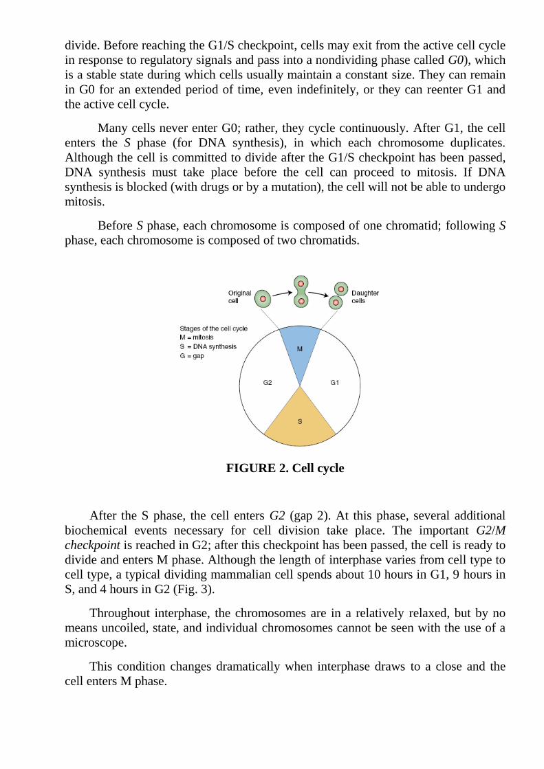
divide. Before reaching the G1/S checkpoint, cells may exit from the active cell cycle
in response to regulatory signals and pass into a nondividing phase called G0), which
is a stable state during which cells usually maintain a constant size. They can remain
in G0 for an extended period of time, even indefinitely, or they can reenter G1 and
the active cell cycle.
Many cells never enter G0; rather, they cycle continuously. After G1, the cell
enters the S phase (for DNA synthesis), in which each chromosome duplicates.
Although the cell is committed to divide after the G1/S checkpoint has been passed,
DNA synthesis must take place before the cell can proceed to mitosis. If DNA
synthesis is blocked (with drugs or by a mutation), the cell will not be able to undergo
mitosis.
Before S phase, each chromosome is composed of one chromatid; following S
phase, each chromosome is composed of two chromatids.
FIGURE 2. Cell cycle
After the S phase, the cell enters G2 (gap 2). At this phase, several additional
biochemical events necessary for cell division take place. The important G2/M
checkpoint is reached in G2; after this checkpoint has been passed, the cell is ready to
divide and enters M phase. Although the length of interphase varies from cell type to
cell type, a typical dividing mammalian cell spends about 10 hours in G1, 9 hours in
S, and 4 hours in G2 (Fig. 3).
Throughout interphase, the chromosomes are in a relatively relaxed, but by no
means uncoiled, state, and individual chromosomes cannot be seen with the use of a
microscope.
This condition changes dramatically when interphase draws to a close and the
cell enters M phase.

FIGURE 3. Cell cycle. Genetic consequences of the cell cycle
From a single cell, the cell cycle produces two cells that contain the same
genetic instructions. These two cells are identical with each other and with the cell
that gave rise to them. They are identical because DNA synthesis in S phase creates
an exact copy of each DNA molecule, giving rise to two genetically identical sister
chromatids. Mitosis then ensures that one chromatid from each replicated
chromosome passes into each new cell.
Another genetically important result of the cell cycle is that each of the cells
produced contains a full complement of chromosomes there is no net reduction or
increasing in chromosome number. Each cell also contains approximately half the
cytoplasm and organelle content of the original parental cell, but no precise
mechanism analogous to mitosis ensures that organelles are evenly divided.
Consequently, not all cells resulting from the cell cycle are identical in their
cytoplasmic content.
Control of the cell cycle
For many years, the biochemical events that controlled the progression of cells
through the cell cycle were completely unknown, but research has now revealed
many of the details of this process. Progression of the cell cycle is regulated at
several checkpoints, which ensure that all cellular components are present and in
good working order before the cell proceeds to the next stage. The checkpoints are
necessary to prevent cells with damaged or missing chromosomes from proliferating.
One important checkpoint mentioned earlier, the G1/S checkpoint, comes just before
the cell enters into S phase and replicates its DNA. When this point has been passed,
DNA replicates and the cell is committed to divide. A second critical checkpoint,
called the G2/M checkpoint, is at the end of G2, before the cell enters mitosis.
Both the G1/S and the G2/M checkpoints are regulated by a mechanism in which
two proteins interact. The concentration of the first protein, cyclin, oscillates during
the cell cycle (Fig. 3). The second protein, cyclindependent kinase (CDK), cannot
function unless it is bound to cyclin. Cyclins and CDKs are called by different names
in different organisms, but here we will use the terms applied to these molecules in
yeast.
Let’s begin by looking at the G2/M checkpoint. This checkpoint is regulated by
cyclin B, which combines with CDK to form M-phase promoting factor (MPF). After
MPF is formed, it must be activated by the addition of a phosphate group to one of
the amino acids of CDK.
Whereas the amount of cyclin B changes throughout the cell cycle, the amount
of CDK remains constant. During G1, cyclin B levels are low; so the amount of MPF
also is low. As more cyclin B is produced, it combines with CDK to form increasing
amounts of MPF. Near the end of G2, the amount of active MPF reaches a critical
level, which commits the cell to divide. The MPF concentration continues to increase,
reaching a peak in mitosis. The active form of MPF is a protein kinase, an enzyme
that adds phosphate groups to certain other proteins. Active MPF brings about many
of the events associated with mitosis, such as nuclear-membrane breakdown, spindle
formation, and chromosome condensation.
At the end of metaphase, cyclin is abruptly degraded, which lowers the amount
of MPF and, initiating anaphase, sets in motion a chain of events that ultimately
brings mitosis to a close (see Figure). Ironically, active MPF brings about its own
demise by destroying cyclin. In brief, high levels of active MPF stimulate mitosis,
and low levels of MPF bring a return to interphase conditions. A number of factors
stimulate the synthesis of cyclin B and the activation of MPF, whereas other factors
inhibit MPF.
Together these factors determine whether the cell passes through the G2/M
checkpoint and ensure that mitosis is not initiated until conditions are appropriate for
cell division. For example, DNA damage inhibits the activation of MPF; the cell is
arrested in G2 and does not undergo division.
The G1/S checkpoint is regulated in a similar manner. In fission yeast
(Shizosaccharomyces pombe), the same CDK is used, but it combines with G1
cyclins. Again, the level of CDK remains relatively constant, whereas the level of G1
cyclins increases throughout G1. When the activated CDK–G1–cyclin complex
reaches a critical concentration, proteins necessary for replication are activated and
the cell enters S phase.
In phase G0 are high differentiate cells – neuron kardiomiozites. Cells of a bone
brain, mucous of a gastrointestinal path on the contrary almost continuously share
and seldom enter into phase G0. Cells of many fabrics of an organism can turn in a
cycle of division, but is usual in a fabric the small amount of cells (except for
damages or tumoral new growths) shares.

Control questions to laboratory work №1
1. What is karyotypee?
2. How many copies of chromosomes cell does diploid number contain?
3. How many copies of chromosomes cell does haploid number contain?
4. What substance composes DNA in a haploid cell?
5. What substance composes DNA in a diploid cell?
6. What is organelle?
7. Describe features of a cell structure and functions of a kernel.
8. Give the description of mitochondrion functions.
9. Where are they located and what function do ribosomes carry out?
10. What is endoplasmic reticulum? Where is it located?
11. Where are lisosoms locating? Describe their functions.
12. To what more the common structure device Goldzhi posesses? Describe its
functions.
13. What is cytoskeleton? Describe its structure and function.
14. What is cytosol? What is it belong to?
15. Describe interphase of cells. In which periods does it divide and what occurs then?
16. How long does mitosis cycle last?
17. What is G1 phase?
18. What processes occurs in S phase?
19. What is characteristic for G2 phase?
20. What phase can pass in a terminal differentiation period?
21. What is the amitotic division? When is it observed?
Reference List:
1. Айала Ф., Кайгер Дж. Современная генетика: В 3 т. Т. 1. М.: Мир, 1987. С.
64-88.
2. Гершкович И. Генетика. М.: Наука, 1968. С. 125-161.
3. Жученко А. А., Король А. Б. Рекомбинация в эволюции и селекции. М.:
Наука, 1985. 400 с.
4. Инге-Вечтомов С. Г. Генетика с основами селекции. М.: Высш. шк., 1989.
С. 85-111.
5. Кушев В. В. Механизмы генетической рекомбинации. Л.: Наука, 1971. 97 с.
6. Лобашев М. Е. Генетика. Л.: Изд-во Ленинград, 1967. С. 116-283.
7. Синнот Э., Денн Л. Курс генетики: Теория и задачи. М.; Л.: Гос. изд-во
биол. мед. лит., 1934. С. 148-182.
8. Чадов Б. Ф., Бузыканова Г. Н. Механизм хромосомной интерференции //
Докл. АН. 1996. Т. 348. С. 407-409.
9. Allen G. E. Thomas Hunt Morgan. The man and his science. Princeton:
Princeton University Press, 1978. P. 70-71, 164-173.
10. Ashburner M. Drosophila. A laboratory handbook. Cold Spring Harbor: Cold
Spring Harbor Laboratory Press, 1989. P. 54-471.
11. Stern C. Zytologisch-genetische Untersuchungen als Beweise fur die
Morganische Theorie des Fak-torenaustauschs // Biol. Zentralbl. 1931. T. 51.
Laboratory work № 2
Subject: Mitosis and meyosis.
The purpose. To get acquainted with the basic laws of cell division and
behavior of chromosomes at mitosis and meyosis. To characterize differences and
biological value of mitosis and meyosis.
The equipment: tables of mitosis and meyosis, monocular microscopes,
cytogenetic preparations of different animals.
Course of work:
1. To get acquainted with the basic phases mitotic and meiotic divisions.
2. To sketch the scheme of mitosis and meiosis.
3. To define differences between mitosis and meiosis their biological value.
4. To consider stages of oogenesis and spermatogenesis
5. To draw conclusions.
Explanation to performance of work:
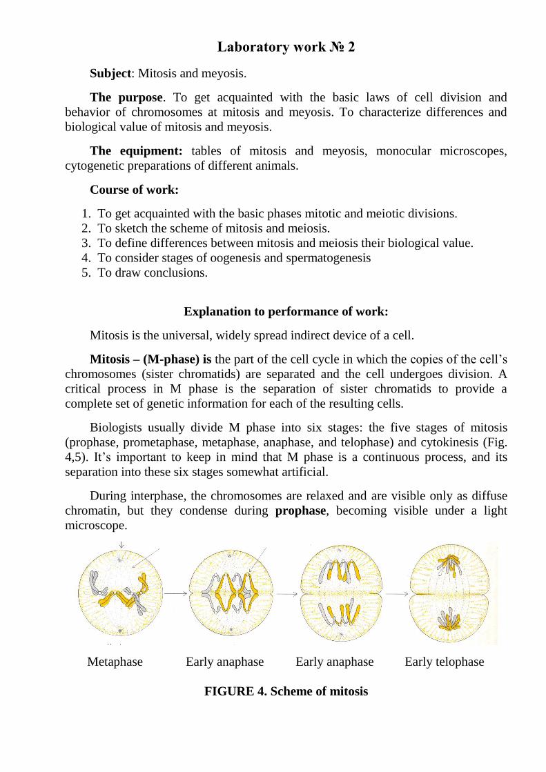
Mitosis is the universal, widely spread indirect device of a cell.
Mitosis – (M-phase) is the part of the cell cycle in which the copies of the cell’s
chromosomes (sister chromatids) are separated and the cell undergoes division. A
critical process in M phase is the separation of sister chromatids to provide a
complete set of genetic information for each of the resulting cells.
Biologists usually divide M phase into six stages: the five stages of mitosis
(prophase, prometaphase, metaphase, anaphase, and telophase) and cytokinesis (Fig.
4,5). It’s important to keep in mind that M phase is a continuous process, and its
separation into these six stages somewhat artificial.
During interphase, the chromosomes are relaxed and are visible only as diffuse
chromatin, but they condense during prophase, becoming visible under a light
microscope.
Metaphase Early anaphase Early anaphase Early telophase
FIGURE 4. Scheme of mitosis

Each chromosome possesses two chromatids because the chromosome was
duplicated in the preceding S phase. The mitotic spindle, an organized array of
microtubules that move the chromosomes in mitosis, forms. In animal cells, the
spindle grows out from a pair of centrosomes that migrate to opposite sides of the
cell. Within each centrosome is a special organelle, the centriole, which is also
composed of microtubules.
Disintegration of the nuclear membrane marks the start of prometaphase.
Spindle microtubules, which until now have been outside the nucleus, enter the
nuclear region. The ends of certain microtubules make contact with the chromosome
and anchor to the kinetochore of one of the sister chromatids; a microtubule from the
opposite centrosome then attaches to the other sister chromatid, and so each
chromosome is anchored to both of the centrosomes. The microtubules lengthen and
shorten, pushing and pulling the chromosomes about. Some microtubules extend
from each centrosome toward the center of the spindle but do not attach to a
chromosome.
During metaphase, the chromosomes arrange themselves in a single plane, the
metaphase plate, between the two centrosomes.
The centrosomes, now at opposite ends of the cell with microtubules radiating
outward and meeting in the middle of the cell, center at the spindle pole. Anaphase
begins when the sister chromatids separate and move toward opposite spindle poles.
After the chromatids have separated, each is considered a separate chromosome.
Telophase is marked by the arrival of the chromosomes at the spindle poles. The
nuclear membrane re-forms around each set of chromosomes, producing two separate
nuclei within the cell. The chromosomes relax and lengthen, once again disappearing
from view.
1. Conclucion by the major events during mitosis
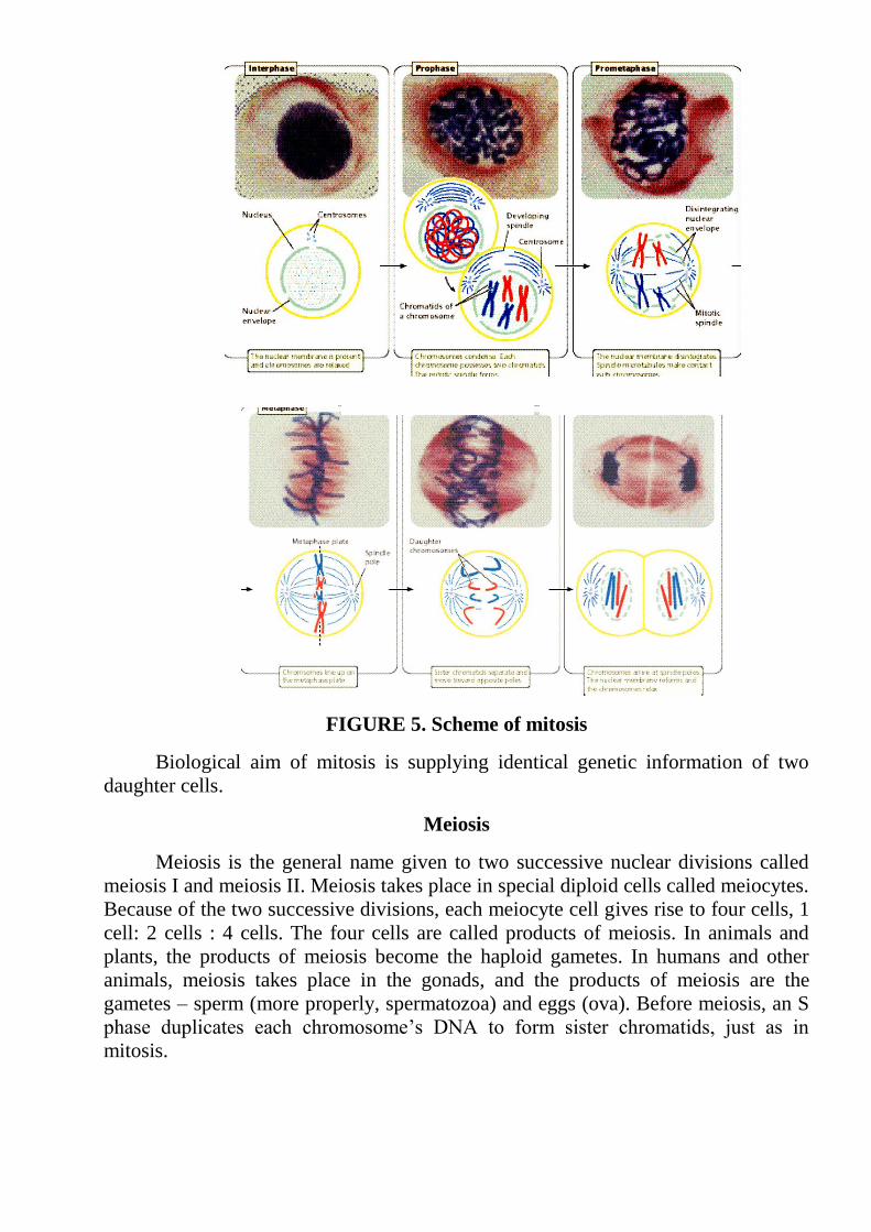
FIGURE 5. Scheme of mitosis
Biological aim of mitosis is supplying identical genetic information of two
daughter cells.
Meiosis
Meiosis is the general name given to two successive nuclear divisions called
meiosis I and meiosis II. Meiosis takes place in special diploid cells called meiocytes.
Because of the two successive divisions, each meiocyte cell gives rise to four cells, 1
cell: 2 cells : 4 cells. The four cells are called products of meiosis. In animals and
plants, the products of meiosis become the haploid gametes. In humans and other
animals, meiosis takes place in the gonads, and the products of meiosis are the
gametes – sperm (more properly, spermatozoa) and eggs (ova). Before meiosis, an S
phase duplicates each chromosome’s DNA to form sister chromatids, just as in
mitosis.
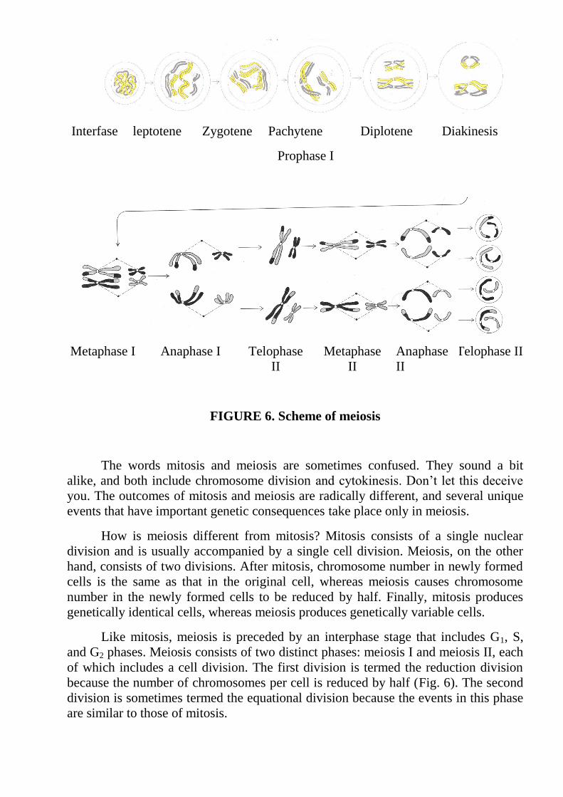
Interfase leptotene Zygotene Pachytene
Prophase I
Diplotene Diakinesis
Metaphase I Anaphase I Telophase
II
Metaphase
II
Anaphase
II
Telophase II
FIGURE 6. Scheme of meiosis
The words mitosis and meiosis are sometimes confused. They sound a bit
alike, and both include chromosome division and cytokinesis. Don’t let this deceive
you. The outcomes of mitosis and meiosis are radically different, and several unique
events that have important genetic consequences take place only in meiosis.
How is meiosis different from mitosis? Mitosis consists of a single nuclear
division and is usually accompanied by a single cell division. Meiosis, on the other
hand, consists of two divisions. After mitosis, chromosome number in newly formed
cells is the same as that in the original cell, whereas meiosis causes chromosome
number in the newly formed cells to be reduced by half. Finally, mitosis produces
genetically identical cells, whereas meiosis produces genetically variable cells.
Like mitosis, meiosis is preceded by an interphase stage that includes G1, S,
and G2 phases. Meiosis consists of two distinct phases: meiosis I and meiosis II, each
of which includes a cell division. The first division is termed the reduction division
because the number of chromosomes per cell is reduced by half (Fig. 6). The second
division is sometimes termed the equational division because the events in this phase
are similar to those of mitosis.

However, meiosis II differs from mitosis in that chromosome number has
already been halved in meiosis I, and the cell does not begin with the same number of
chromosomes as it does in mitosis.
During interphase, the chromosomes are relaxed and visible as diffuse
chromatin. Prophase I is a lengthy stage, divided into five substages (Fig. 7). In
leptotene, the chromosomes contract and become visible. In zygotene, the
chromosomes continue to condense; homologous chromosomesbegin to pair up and
begin synapsis, a very close pairing association. Each h omologous pair of synapsed
chromosomes consists of four chromatids called a bivalent or tetrad. In pachytene,
the chromosomes become shorter and thicker, and a three-part synaptonemal complex
develops between homologous chromosomes. Crossingover takes place, in which
homologous chromosomes exchange genetic information. The centromeres of the
paired chromosomes move apart during diplotene; the two homologs remain attached
at each chiasma (plural, chiasmata), which is the result of crossing over. In diakinesis,
chromosome condensation continues, and the chiasmata move toward the ends of the
chromosomes as the strands slip apart; so the homologs remained paired only at the
tips. Near the end of prophase I, the nuclear membrane breaks down and the spindle
forms.
Metaphase I is initiated when homologous pairs of chromosomes align along
the metaphase plate. A microtubule from one pole attaches to one chromosome of a
homologous pair, and a microtubule from the other pole attaches to the other member
of the pair.
Anaphase I is marked by the separation of homologous chromosomes. The
two chromosomes of a homologous pair are pulled toward opposite poles. Although
the homol homologous chromosomes separate, the sister chromatids remain attached
and travel together.
In telophase I, the chromosomes arrive at the spindle poles and the cytoplasm
divides.
The period between meiosis I and meiosis II is interkinesis, in which the
nuclear membrane re-forms around the chromosomes clustered at each pole, the
spindle breaks down, and the chromosomes relax. These cells then pass through
Prophase II, in which these events are reversed: the chromosomes recondense, the
spindle re-forms, and the nuclear envelope once again breaks down. In interkinesis in
some types of cells, the chromosomes remain condensed, and the spindle does not
break down. These cells move directly from cytokinesis into metaphase II, which is
similar to metaphase of mitosis: the individual chromosomes line up on the
metaphase plate, with the sister chromatids facing opposite poles.
In anaphase II, the kinetochores of the sister chromatids separate and the
chromatids are pulled to opposite poles. Each chromatid is now a distinct
chromosome. In telophase II, the chromosomes arrive at the spindle poles, a nuclear
envelope re-forms around the chromosomes, and the cytoplasm divides. The
chromosomes relax and are no longer visible. The major events of meiosis are
summarized.
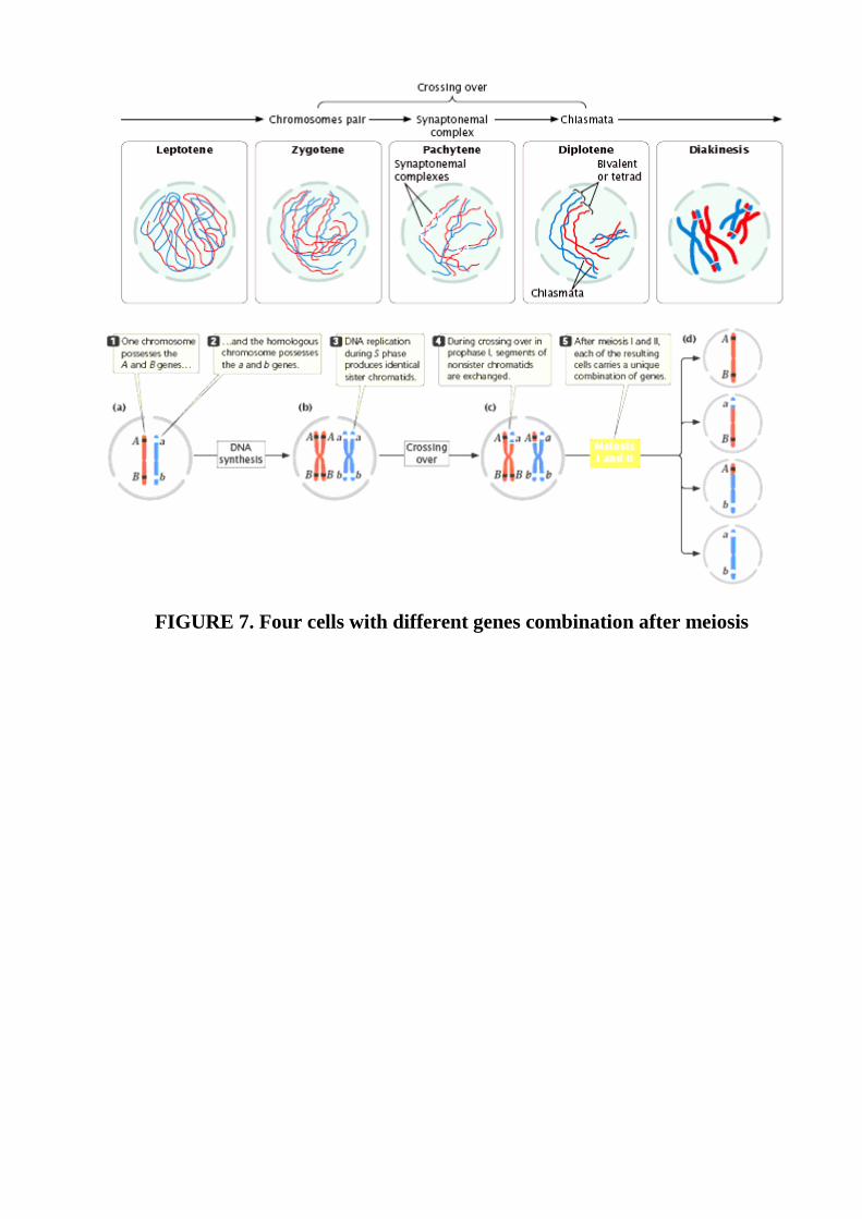
FIGURE 7. Four cells with different genes combination after meiosis
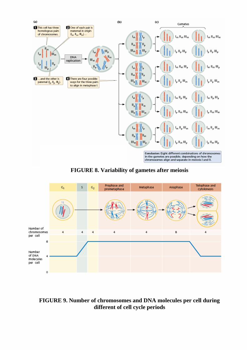
FIGURE 8. Variability of gametes after meiosis
FIGURE 9. Number of chromosomes and DNA molecules per cell during
different of cell cycle periods

2. The major events of meiosis
FIGURE 10. Comparison of meiosis and mitosis characteristics
Meiosis includes two cell divisions. In this figure, the original cell is 2n_4.

#
FIGURE 11 Comparison of meiosis and mitosis characteristics
.
Biological value of meyosis consists in a reduction of the chromosomes
number, necessary for reception of gametes and maintenance of a karyotype
constancy, genetic recombination, caused crossing-over and recombination of
chromosomes which maintain biological variety at a level of a kind and receptions of
new genetic combinations at descendants enable.
Gametogenesis
The production of gametes in a male animal (spermatogenesis) takes place in
the testes. There, diploid primordial germ cells divide mitotically to produce diploid
cells called spermatogonia (Fig. 12). Each spermatogonium can undergo repeated
rounds of mitosis, giving rise to numerous additional spermatogonia.
Alternatively, a spermatogonium can initiate meiosis and enter into prophase I.
Now called a primary spermatocyte, the cell is still diploid because the homologous
chromosomes have not yet separated. Each primary spermatocyte completes meiosis
I, giving rise to two haploid secondary spermatocytes that then undergo meiosis II,
with each producing two haploid spermatids. Thus, each primary spermatocyte
produces a total of four haploid spermatids, which mature and develop into sperm.
The production of gametes in the female (oogenesis) begins much like
spermatogenesis. Diploid primordial germ cells within the ovary divide mitotically to
produce oogonia ( FIGURE 12). Like spermatogonia, oogonia can undergo repeated
rounds of mitosis or they can enter into meiosis.
Once in prophase I, these still-diploid cells are called primary oocytes. Each
primary oocyte completes meiosis I and divides. Here the process of oogenesis begins
to differ from that of spermatogenesis. In oogenesis, cytokinesis is unequal: most of
the cytoplasm is allocated to one of the two haploid cells, the secondary oocyte. The
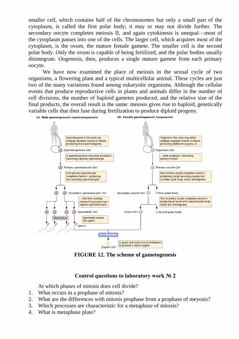
smaller cell, which contains half of the chromosomes but only a small part of the
cytoplasm, is called the first polar body; it may or may not divide further. The
secondary oocyte completes meiosis II, and again cytokinesis is unequal—most of
the cytoplasm passes into one of the cells. The larger cell, which acquires most of the
cytoplasm, is the ovum, the mature female gamete. The smaller cell is the second
polar body. Only the ovum is capable of being fertilized, and the polar bodies usually
disintegrate. Oogenesis, then, produces a single mature gamete from each primary
oocyte.
We have now examined the place of meiosis in the sexual cycle of two
organisms, a flowering plant and a typical multicellular animal. These cycles are just
two of the many variations found among eukaryotic organisms. Although the cellular
events that produce reproductive cells in plants and animals differ in the number of
cell divisions, the number of haploid gametes produced, and the relative size of the
final products, the overall result is the same: meiosis gives rise to haploid, genetically
variable cells that then fuse during fertilization to produce diploid progeny.
FIGURE 12. The scheme of gametogenesis
Control questions to laboratory work № 2
At which phases of mitosis does cell divide?
1. What occurs in a prophase of mitosis?
2. What are the differences with mitosis prophase from a prophase of meyosis?
3. Which processes are characteristic for a metaphase of mitosis?
4. What is metaphase plate?

5. When does anaphase occur?
6. What transformations in a cell happen in telofaze?
7. What kind of mitosis does polytene lead to?
8. Which stages does mitosis consist of?
9. What is meyosis?
10. Which stages does meyosis consist of? Give a short characteristic to each of
them?
11. What meyosis phases does prophase consist of?
12. When does leptotene begin and finish?
13. What are chromomeres? When are they visible?
14. What is sinaptical bouquet? What is it made from?
15. What is tetrada?
16. What processes occur during pachytene?
17. What occurs during zygotene?
18. When do chromosomes have a kind of lamp brushes?
19. What are chiasms and when are they generated?
20. What is crossing-over? When does it occur?
21. When and in what cells are synthesis of RNA and fibers active during meyosis?
22. Characterize the processes which occur during diakinesis.
23. What occur during a metaphase of the 1 meyosis? How are bivalents located?
24. What is the difference between metaphase of the 1 meyosis and a metaphase of
the 2 meyosis and mitosis?
25. What processes are characterize anaphase of the 1 meyosis? Describe it differs
from a similar phase of meyosis 2 and mitosis.
26. Characterize the telophase of the 1 meyosis. How many chromosomes and what
contents of DNA characterize a cell at this stage?
27. Which contents of DNA and quantity of chromosomes are in telophase of the 1
meyosis, and telophase of the 2 meyosis, a metaphase?
28. How does the second devision of meyosis is refer to? What is it characteristic
for?
29. What biological value of meyosis consists of? Is it differing from value of
mitosis?
30. What is gametogenesis?
31. When does a primary sexual cell of animals start to develop?
32. At what stages does spermatogenesis happen?
33. During which period of growth in spermatogenesis occur?
34. What transformation will test spermanogonies?
35. What division is share spermatocytes of the1st order? How many and what cells
are formed as a result of this division?
36. What is oogenesis?
37. Into which periods development of ovocytes happens?
38. Characterize the period of sinoptemal ways in oogenesis.
39. What does happen during protoplazmanic and trofoplazmatic growth ?
40. Characterize the period of female sexual cells maturing.
41. How long does period of ovocytes growth last?
42. When does meyosis come to the end in female sexual cells (before or after
fertilization)?
43. At what stage of maturing oocyte meyosis deploys ?
44. At what stage ovulation of oocyte happens?
45. How many and what cells does one oocyte form?
46. How many and what cells does a spermatocyte form?
Reference List:
1. Ченцов Ю.С. Общая цитология: Учебник. – М.: Изд-во МГУ, 1984. – С.
320– 336.
2. Фаллер Д.М., Шилдс Д. Молекулярная биология клетки. Руководство для
врачей. Пер. с англ.М.: БИНОМ-Пресс, 2003. – С. 131– 49.
3. Катасонов В.Я., Гомельський Б.И. Селекция рыб с основами генетики. – М:
ВО „Агропромиздат”.– 1991. – С. 14– 24.
Laboratory work № 3
Subject: The structure of chromosomes. Karyotypes of domestic animals.
The purpose. To determine morphology of chromosomes and karyotypes of
domestic animals on cytogenetic preparations and photos.
The equipment: monocular microscopes, cytogenetic preparations of different
kinds of animals, photos of chromosomes, tables of a chromosomes structure and
karyotypes of domestic animals.
Course of work:
1. To get acquainted with a structure of chromosome.
2. To consider metaphase plate on cytogenetic preparations.
3. To draw chromosomes with different morphology.
4. To construct on the basis of photos metaphase chromosomes karyogram.
5. To draw a conclusion of concerning quantity of chromosomes, their morphology
and quantity of shoulders.
Each biological species is characterized by the certain karyotype (number and
morphology of chromosomes). Karyotypes of the species is conservative (Table 4).
Overwhelming majority of individuals’ species have an identical set of chromosomes.
There are species which have chromosomal polymorphism. Change of quantity and
morphology of chromosomes - one of mechanisms of speciation. Individuals who
have different karyotypes can not give prolific descendants. It is caused by
infringements at chromosomes conjugation in a prophase of meyosis and wrong
distribution of affiliated chromosomes in anaphase. Exceptions make ginogenetic
forms which consist only from fameles.
Table 4. Karyotypes of different domestic animal species
Chromosome structure: The chromosomes of eukaryotic cells are larger and
more complex than those found in prokaryotes, but each unreplicated chromosome
nevertheless consists of a single molecule of DNA. Although linear, the DNA
molecules in eukaryotic chromosomes are highly folded and condensed; if stretched
out, some human chromosomes would be several centimeters long—thousands of
times longer than the span of a typical nucleus. To package such a tremendous length
of DNA into this small volume, each DNA molecule is coiled again and again and
tightly packed around histone proteins, forming the rod-shaped chromosomes. Most
of the time the chromosomes are thin and difficult to observe but, before cell
division, they condense further into thick, readily observed structures; it is at this
stage that chromosomes are usually studied.
A functional chromosome has three essential elements: a centromere, a pair of
telomeres, and origins of replication (FIGURE 13).
The centromere is the attachment point for spindle microtubules, which are the
filaments responsible for moving chromosomes during cell division. The centromere
appears as a constricted region that often stains less strongly than does the rest of the
chromosome. Before cell division, a protein complex called the kinetochore
assembles on the centromere, to which spindle microtubules later attach.
Kind of animals Number of chromosomes
Livestock 60
Horse 64
Pig 38
Sheep 54
Han 78
Mink 30
Rabbit 44
Cat 38
Dog 78
Monkey 48
Pigeon 80
Donkey 66
Drosophila 8
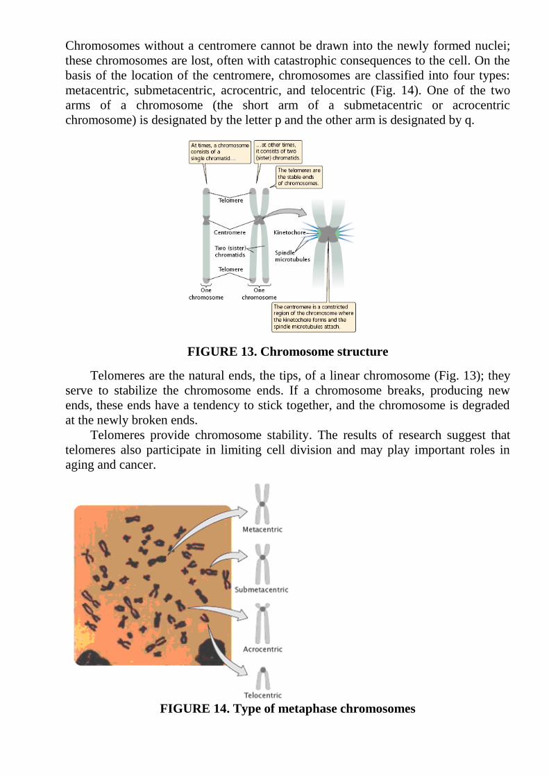
Chromosomes without a centromere cannot be drawn into the newly formed nuclei;
these chromosomes are lost, often with catastrophic consequences to the cell. On the
basis of the location of the centromere, chromosomes are classified into four types:
metacentric, submetacentric, acrocentric, and telocentric (Fig. 14). One of the two
arms of a chromosome (the short arm of a submetacentric or acrocentric
chromosome) is designated by the letter p and the other arm is designated by q.
FIGURE 13. Chromosome structure
Telomeres are the natural ends, the tips, of a linear chromosome (Fig. 13); they
serve to stabilize the chromosome ends. If a chromosome breaks, producing new
ends, these ends have a tendency to stick together, and the chromosome is degraded
at the newly broken ends.
Telomeres provide chromosome stability. The results of research suggest that
telomeres also participate in limiting cell division and may play important roles in
aging and cancer.
FIGURE 14. Type of metaphase chromosomes

Origins of replication are the sites where DNA synthesis begins; they are not
easily observed by microscopy. In preparation for cell division, each chromosome
replicates, making a copy of itself. These two initially identical copies, called sister
chromatids, are held together at the centromere. Each sister chromatid consists of a
single molecule of DNA
The morphology of a chromosome is determined by such attributes:
for definition of a chromosome morphology establish a humeral index of a
parity of greater shoulder to smaller length: І = p/q. It enables to divide chromosomes
into groups:
metacentrical chromosomes (M) - a parity of lengths of shoulders is 1 1,9;
submetacentrical chromosomes (S) – is 2 4,9;
akrocentrical(A)>is 5 –centromeres is located near one of telomeres.
Some chromosomes have additional membrane which is named secondary. If
the secondary membrane is located close up to the end of a chromosome a site which
it separates, has name satellite.
The description of kind karyotype is consists of definition of chromosomes
quantity, their morphology, subtraction of shoulders number. Chromosomes are
studied and photographed by an optical microscope. The received images of
chromosomes have number depending on their length from greater up to the least.
The organization of a chromosome has 5 levels of condensation.
The 1 level –is nucleosome. With chromosomal strings of DNA connect
nucleosomas, parts of 10 nanometers in diameter. Nucleosome is formed as a result
of four classes of the basic fibers -histons interaction: Н2А. Н2В, Н3 and Н4.
Nucleosome consists of 8 molecules histones (on 2 molecules of each kind).
Site of a double spiral of DNA forms 1 3/4 turns around of a core nucleosomes.
This site is directly connected with a core and has the constant length equal 140. In
nuclosomes linkers (connections) vary by length from 15 up to 100 and even it is
more. Thus, the twisting of DNA around nucleosoma reduces its length in 7 times.
FIGURE 15. Karyogram

FIGURE 16. The first level is nucleosoma
1–DNA;
2 - linker;
3 - nucleosoma.
2 level - the solenoid, its diameter of 25-50 nanometers, formed owing to histones
Н1. This histone (Н1) attached to linkers ends stabilizes communication nucleosoma,
that forms a spiral of higher order. Condensation of DNA in structure of the solenoid
reduces its length in 6 times.
Histone Н1
FIGURE 17. Some nucleosomas are united into the solenoid
The solenoid packed in the form of loops domens that are fixed by the internal
structure on an intranuclear albuminous skeleton. Fragments of an attachment have
received the name of SAR-of Fragments (Scaffold Assocіated Regіon the built in
places of an attachment) and MAR (Matrіx Assocіated Regіon). Owing to loops
domens the length of DNA decreases 25 times.
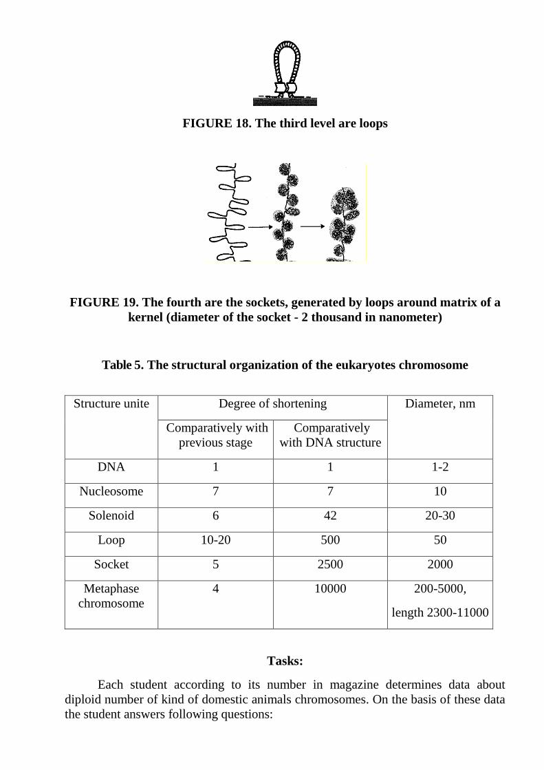
FIGURE 18. The third level are loops
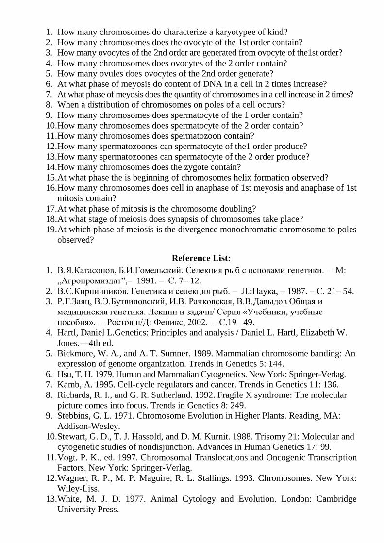
FIGURE 19. The fourth are the sockets, generated by loops around matrix of a
kernel (diameter of the socket - 2 thousand in nanometer)
Table 5. The structural organization of the eukaryotes chromosome
Structure unite Degree of shortening Diameter, nm
Comparatively with
previous stage
Comparatively
with DNA structure
DNA 1 1 1-2
Nucleosome 7 7 10
Solenoid 6 42 20-30
Loop 10-20 500 50
Socket 5 2500 2000
Metaphase
chromosome
4 10000 200-5000,
length 2300-11000
Tasks:
Each student according to its number in magazine determines data about
diploid number of kind of domestic animals chromosomes. On the basis of these data
the student answers following questions:
1. How many chromosomes do characterize a karyotypee of kind?
2. How many chromosomes does the ovocyte of the 1st order contain?
3. How many ovocytes of the 2nd order are generated from ovocyte of the1st order?
4. How many chromosomes does ovocytes of the 2 order contain?
5. How many ovules does ovocytes of the 2nd order generate?
6. At what phase of meyosis do content of DNA in a cell in 2 times increase?
7. At what phase of meyosis does the quantity of chromosomes in a cell increase in 2 times?
8. When a distribution of chromosomes on poles of a cell occurs?
9. How many chromosomes does spermatocyte of the 1 order contain?
10. How many chromosomes does spermatocyte of the 2 order contain?
11. How many chromosomes does spermatozoon contain?
12. How many spermatozoones can spermatocyte of the1 order produce?
13. How many spermatozoones can spermatocyte of the 2 order produce?
14. How many chromosomes does the zygote contain?
15. At what phase the is beginning of chromosomes helix formation observed?
16. How many chromosomes does cell in anaphase of 1st meyosis and anaphase of 1st
mitosis contain?
17. At what phase of mitosis is the chromosome doubling?
18. At what stage of meiosis does synapsis of chromosomes take place?
19. At which phase of meiosis is the divergence monochromatic chromosome to poles
observed?
Reference List:
1. В.Я.Катасонов, Б.И.Гомельский. Селекция рыб с основами генетики. – М:
„Агропромиздат”,– 1991. – С. 7– 12.
2. В.С.Кирпичников. Генетика и селекция рыб. – Л.:Наука, – 1987. – С. 21– 54.
3. Р.Г.Заяц, В.Э.Бутвиловский, И.В. Рачковская, В.В.Давыдов Общая и
медицинская генетика. Лекции и задачи/ Серия «Учебники, учебные
пособия». – Ростов н/Д: Феникс, 2002. – С.19– 49.
4. Hartl, Daniel L.Genetics: Principles and analysis / Daniel L. Hartl, Elizabeth W.
Jones.—4th ed.
5. Bickmore, W. A., and A. T. Sumner. 1989. Mammalian chromosome banding: An
expression of genome organization. Trends in Genetics 5: 144.
6. Hsu, T. H. 1979. Human and Mammalian Cytogenetics. New York: Springer-Verlag.
7. Kamb, A. 1995. Cell-cycle regulators and cancer. Trends in Genetics 11: 136.
8. Richards, R. I., and G. R. Sutherland. 1992. Fragile X syndrome: The molecular
picture comes into focus. Trends in Genetics 8: 249.
9. Stebbins, G. L. 1971. Chromosome Evolution in Higher Plants. Reading, MA:
Addison-Wesley.
10. Stewart, G. D., T. J. Hassold, and D. M. Kurnit. 1988. Trisomy 21: Molecular and
cytogenetic studies of nondisjunction. Advances in Human Genetics 17: 99.
11. Vogt, P. K., ed. 1997. Chromosomal Translocations and Oncogenic Transcription
Factors. New York: Springer-Verlag.
12. Wagner, R. P., M. P. Maguire, R. L. Stallings. 1993. Chromosomes. New York:
Wiley-Liss.
13. White, M. J. D. 1977. Animal Cytology and Evolution. London: Cambridge
University Press.

English-Ukrainian Dictionary of Key Words:
acrocentric chromosome – акроцентрична хромосома. Xромосома, центро-
мірний індекс котрої перевищує 5,0; різновид - телоцентрична хромосома
<telocentric chromosome>.
autosome, euchromosome - автосома, евхромосома. Будь-яка хромосома, яка
не є статевою.
anaphase - анафаза. Cтадія клітинного поділу (мітозу чи мейозу) між
метафазою <metaphase> та телофазою <telophase>, сестринські хроматиди
розходяться до полюсів поділу.
aneuploidy - анеуплоїдія. Наявність у клітини чи тканини кількості
хромосом, яка не дорівнює типовій для даного виду; в основі А. лежить
нерозходження хромосом; формами А. є моносомія <monosomy>, нулісомія
<nullisomy>, трисомія <trisomy>, "крайнім" варіантoм А. є поліплоїдія
<polyploidy>; термін "А." введено Г.Текхольмом в 1922.
cell cycle - клітинний цикл. Період життя клітини від закінчення одного
поділу до початку другого поділу, включає інтерфазу (періоди G1 <G1-period>,
S <S period>, G2 <G2-period>) і мітоз <mitosis> (період М), іноді виділяють в
інтерфазі період G0 (період спокою); к.ц. за тривалістю у різних організмів
значно варіює.
cell division - клітинний поділ. Форма розмноження (подвоєння) клітин;
відбувається перешнуруванням у бактерій або мітозу <mitosis>.
cell membrane, plasmalemma, cytolemma - клітинна (плазматична) мембра-
на, плазмолемма. Mембрана клітини, яка відокремлює цитоплазму від
зовнішнього середовища (у рослин) від клітинної стінки <cell wall>; К.м.
характеризується напівпроникністю, товщина 7-10 нм, як в інших біологічних
мембран, основу К.м. складає подвійний фосфоліпідний шар.
centriole - центріоль. Kлітинна органела, входить до складу клітин більшості
тварин та грибів; в багатьох випадках Ц. є елементом мітотичного апарату
<mitotic apparatus> (є циліндричним утворенням, складається з дев’яти
триплетів мікротрубочок); процес відтворення Ц. є автономним, хоча й
пов'язаним у часі з синтетичним періодом мітозу.
centromere - центромера. Ділянка моноцентричної хромосоми, в якій
сестринскі хроматиди з’єднані, які забезпечують рух хромосом до полюсів
поділу; часто прицентромірні райони генетично інертні (гетерохроматизовані);
часто в якості синоніма поняття “Ц.” використовують термін “кінетохор”
<kinetochore>, але ці елементи структурно диференційовані.
centromere separation sequence - порядок розподілу центромер. Послі-
довність разподілу центромер хромосом на початку анафази мітозу чи мейозу:
часто має невипадковий характер – наприклад у людини одними з перших
розділяються центромери хромосом 18, 2, Х, 12, 4, 5 и 17, а останніми -

центромери хромосом 1, 11, 16 и Y; порушення п.р.ц. може приводити до
неправильного розходження хромосом в анафазі і до трисомії, наприклад, за
хромосомою 18 у людини <Edwards syndrome>.
chiasma - хіазма, перехрест. Візуальний прояв кросинговеру <crossing-over>;
добре помітними Х. стають в диплотені <diplotene> (під час порушення
синаптонемального комплексу <synaptonemal complex>), коли гомологи
відштовхуються один від одного, зберігаючи зв'язок тільки в області Х. - цей
процес супроводжується терміналізацією хіазм <chiasma terminalization>.
chromatid - хроматида, напівхромосома. Oдна з двох копій реплікованої
хромосоми, з’єднаних в області центромери та візуалізованих в мітозі.
chromatin - хроматин. Hуклеопротеїдний комплекс, який складає xромосоми
евкаріотичних клітин, включає ДНК, гістони <histones> та різні негістонові
білки; термін “Х.” введено В.Флемінгом в 1880 для опису забарвлюваних
спеціальними барвниками внутрішньоядерних структур.
chromomere - хромомера. Щільно конденсована ділянка хроматинової нитки;
розкручені Х. - це петлі хромосом типу “лампових щіток” <lampbrush
chromosomes>; Х. інтенсивно зафарбовується барвниками, специфічними
стосовно ДНК.
chromosome - хромосома. Oрганела клітинного ядра еукаріотів (у прокаріотів
розміщена безпосередньо в цитоплазмі), є носієм генетичної інформації (генів),
здатна до відтворення зі збереженням структурно-функціональної
индивідуальності в ряду поколінь; основу Х. сокладає безперервна дволан-
цюгова спірально закручена (конденсована) молекула ДНК, пов’язана гістона-
ми <histones> і негістоновими білками, набір Х. (каріотип) є видоспецифічною
ознакою, для якої характерним є відносно низький рівень індивідуальної
мінливості; термін “Х” запропонований В. Вальдейером в 1888 році.
chromosome arm - плече хромосоми. Ділянка моноцентричної метафазної
хромосоми по один бік від центромери, включно з обома сестринськими
хроматидами.
chromosome arm number, “nombre fundamental”, NF - кількість
хромосомних плечей.
chromosome banding methods, banding - диференційне забарвлення хромо-
сом, бендинг. Kомплекс методів зафарбування хромосомних препаратів, що
дозволяють на основі неоднакового відношення до гетеро- та евхроматинових,
ділянок ДНК с різними АТ/ГЦ-співвідношеннями та інших особливостей
виявляти специфічну повздовжню структурованість окремих хромосом, що
дозволяє точно ідентифікувати окремі елементи каріотипу; найбільш поширені:
G-, C-, R-, Q-бендинг.
chromosome complement (set), chromotype - хромосомний набір. Cпеци-
фічний для даної особини, групи особин, виду гаплоїдний хромосомний
комплекс, часто представлений у вигляді ідіограми <idiogram>.

chromosome condensation (spiralization, contraction) - конденсація (спіра-
лізація, скорочення) хромосом. Процес ущільнення хромосом, розпочинається в
інтерфазі й достягає максимуму в метафазу; в основі К.х. лежать складні
процеси скручування (упаковки) хроматину, та процес фосфорилювання
гістона <histone> Н1, контрольованого специфічним ферментом -
гистонкіназою.
chromosome morphology - морфологія хромосом. Xарактеристика хромосом
з врахуванням співвідношення плечей у них: розрізняють метацентричні
<metacentric>, субметацентричні <submetacentric>, субтелоцентричні <subtelo-
centric>, акроцентричні <acrocentric> та телоцентричні <telocentric>
хромосоми.
chromosome number - хромосомне число. Kількість хромосом у даній
клітині або кількість хромосом в соматичних (диплоїдних) клітинах даного
організму - 2n.
chromosome painting - “розпис” хромосоми. Один із варіантів методу
гібридизації in situ <in situ hybridization>, при якому в якості зонду
використовується мічена біотином (для наступного флюорисцентного аналізу)
ДНК повної хромосомної бібліотеки <chromosome-specific library>: метод
“Р.”х. використовується для аналізу соматичних гібридних клітин,
ідентифікації хромосомних порушень, аналізу просторового розподілу
хромосом в інтерфазах та для інших цілей.
chromosome pairing, conjugation of chromosomes, synapsis, syndesis,
association – кон’югація хромосом, синапсис. Попарне зближення сестринських
хроматид гомологічних хромосом в I поділі мейозу з утворенням
взаємостабільних структу-бівалентів <bivalent>, під час якого може відбуватися
обмін генетичним матеріалом (рекомбінація, кросинговер <crossing-over>).
colchicine - колхіцин. Aлкалоїд, екстрагований з деяких лілійних рослин, є
“мітотичною отрутою”, блокує нормальну активність веретена; викорис-
товується в каріологічному аналізі та для отримання високоплоїдних форм
рослин (колхіплоїдів <colchiploid>).
crossing-over - кросинговер, обмін. Взаємний обмін ділянками гомологічних
хромосом, дає нову комбінацію алелів; механізм К. заснований на “розриві-
поєднанні” <breakage-reunion hypothesis> хроматид: К. є основою
комбінативної мінливості (рекомбінації <recombination>) і відбувається в
мейозі.
cytogenetics - цитогенетика. Галузь знань на перехресті генетики та цитології,
вивчає генетичні закономірності на клітинному (хромосомному) рівні; термін
“Ц.” запропоновано В.Саттоном в 1902.
cytokinesis - цитокінез, цитотомія. Процес розподілу материнської клітини на
дві дочірні, відбувається в телофазі мейозу або мітозу і здійснюється за рахунок
утворення або фрагмопласту <phragmoplast> (рослинної клітини), або клітинної
перетяжки (у тварин).
cytoplasm - цитоплазма.
daughter (sister) cell - дочірня (сестринська) клітина. Oдна з двох клітин, що
утворюється в результаті клітинного поділу.
daughter chromatid - дочірня хроматида. Xроматида, утворюється в
результаті мітотичного або мейотичного подвоєння хромосом.
diakinesis - діакінез. Заключний етап профази I поділу мейозу, на якому
ущільнення (спіралізація) хромосом досягає максимуму, і вони рівномірно
розподіляються в ядрі.
diploid - диплоїд. Oрганізм, клітини якого включають два гомологічних
набори хромосом (2n) <diploid number>; термін “Д.” запропонований
Е.Страсбургером в 1905.
diploid number, 2n - диплоїдне число. Oсновна характеристика каріотипу
організму (виду), визначається за кількістю хромосом в метафазі соматичного
мітозу, - наприклад, у людини в нормі 2n=46; у диплоїдного організму число 2n
характерне для всіх клітин (за відсутності індивидуальної мінливості і
хромосомного мозаїцизму <mosaicism>), окрім гамет.
diplotene, diplonema - диплотена, диплонема, стадія подвійних ниток. Cтадія
профази I поділу мейозу між пахітеною і діакінезом, характеризується
виникненням відштовхування гомологів одни від одного, що приводить до
терміналізації хіазм <chiasma terminalization>.
division, fission - поділ. Універсальна форма розмноження клітини, найбільш
поширеними формами Д. є мітоз <mitosis> та мейоз <meiosis>; також до Д.
можуть бути віднесені ті форми вегетативного розмноження, за яких
відбувається розподіл материнського організму на більш чи менш рівні
частини.
division center - центр поділу. Елемент мітотичного апарату клітин тварин,
складається з центросоми <centrosome> і центріолі <centriole>; часто замість
терміна “Ц.д.” використовують поняття “полюс веретена”.
dizygotic (fraternal) twins - різнояйцеві (двояйцеві, дизиготні, неідентичні)
близнюки. Близнюки, які утворилися при одночасному заплідненні двох чи
більше яйцеклітин; генотипи Р.б. схожі не більше, ніж генотипи братів та
сестер, хоча ідентичність середовища, в якому вони розвиваються, обумовлює
сильнішу фенотипову схожість між ними.

euchromatin - евхроматин. Активний хроматин <chromatin>, не виявляється
візуально протягом всієї інтерфази внаслідок низької щільності його
упакування, містить більшість активно транскрибованих генів, здатний зво-
ротньо перетворюватися в факультативний гетерохроматин <facultative
heterochromatin> в процесі інактивації Х-хромосоми <X-inactivation>; Е.
містить відносно більшу кількість негістонових білків порівняно з гетеро-
хроматином <heterochromatin>.
eukaryotes, eukaryotic organisms - евкаріоти. Oрганізми (вищі тварини і
рослини, гриби, одно- і багатоклітинні водорості - окрім синьо-зелених - та
найпростіші), клітини яких містять сформоване ядро; ядерна ДНК входить до
складу хромосом <chromosome>, які містять <histones> і деякі негістонові
білки, і організована у вигляді хроматину <chromatin>; термін “Е.”
запропоновано Е.Шаттоном в 1937, він уперше встановив принципові
відмінності Е. і прокаріотів <prokaryotes>; одним із найдавніших евкаріотів
визнано лямблію.
facultative heterochromatin - факультативний гетерохроматин. Гетеро-
хроматин <heterochromatin>, наявний лише в одній з гомологічних пар
хромосом, - наприклад, у інактивованої Х-хромосоми <X-inactivation> в процесі
компенсації дози Ф.х. в певних умовах здатний знову переходити в евхроматин
<euchromatin>; термін "Ф.г." запропоновано С. Брауном в 1966.
first division - перший (I) поділ [дозрівання]. Етап мейозу, який за
параметрами нагадує мітоз, але відрізняється суттєво тривалішою і складнішою
профазою, в якій відбувається рекомбінація генетичного матеріалу, а також
тим, що в ньому відбувається розходження гомологів, а не хроматид.
G0 period - період G0. Етап інтерфази <inter-phase> одразу після закінчення
мітозу, характеризується відносним спокоєм клітини (суттєво послабленим
синтезом білка), пе- редує періоду G1.
G1 period - период G1. Етап клітинного циклу (етап інтерфазы): фаза росту
(Growth), що передує періоду S <S period>.
G2 period - період G2. Етап клітинного циклу (фаза росту), починається після
реплікації ДНК (періоду S) і передує мітозу <mitosis>.
gametic meiosis - гаметичний мейоз. Mейоз, який безпосередньо приводить
до утворення одноклітинних гамет.
gametic number - гаметичне число. Гаплоїдное число хромосом.
gametic reduction - редукція гамет, редукція [числа] хромосом. Зменшення
числа хромосом наполовину порівняно з соматичним набором; Р.г. – складова
частина редукційного поділу (мейозу).
gametogenesis - гаметогенез. Процес развитку статевих клітин; у рослин Г.
представлено мікроспорогенезом <microsporogenesis> і макроспорогенезом
<macrosporogenesis>, у тварин Г. (сперматогенез <spermatogenesis> та оогенез

<oogenesis>) відбувається в спеціальних статевих органах - гонадах
(локалізований Г.) або відбувається в будь-якій ділянці тіла, як у
кишковопорожнинних та плоских червів (дифузний Г.).
genome - геном. Сукупність генів гаплоїдного набору хромосом даного виду
організмів; організм може містити в собі різні геноми якщо він виник в
результаті гібридизації; у випадку якщо організм не є алоплоїдом, термін “Г.”
використовують для позначення всієї сукупності генів (диплоїдний і т.д.), іноді
“Г.” Використовують в якості синоніму до поняття “каріотип” <karyotype>, що
є невірним; термін “Г.” запропоновано Г.Вінклером в 1920.
Goldgi apparatus (body, complex) - апарат (комплекс) Гольджi. Органела
евкаріотичної клітини, складається з щільно запакованих порожнин і мішечків;
у рослин А.Г. включає диктіосоми <dictiosome>; на відміну від ендо-
плазматичного ретикулюма <endoplasmatic reticulum> А.Г. позбавлений
рибосом; серед функцій А.Г. - модификації білків (глікозилювання,
фосфорилювання и т.п.), “грануляція” продуктів секреції, утворення лізосом
<lysosome>, синтез деяких полісахаридів, формування клітинної мембрани;
описаний К.Гольджи в 1898.
haploid - гаплоїдний. Xарактеризує індивідуума (клітину), в якого наявний
один набір хромосом (n); в нормі гаплоїдними є гамети; гаплоїдними можуть
бути особини, які утворилися в результаті індукованого гіногенезу
<gynogenesis> і т.д.
idiogram - ідеограма. Графічне зображення морфологічної структури
каріотипу <karyotypee> з врахуванням відносної довжини та співвідношення
довжини плечей хромосом, розміщення вторинних перетинок та супутніх
елементів, розподіл диференційовано забарвлених зон та ін. ознак.
interkinesis - інтеркінез. Cтадія клітинного циклу між I та II поділами мейозу,
відрізняється від інтерфази відсутністю процесу реплікації ДНК та (як правило)
процесу деспіралізації хромосом; в І. не відбувається формування ядерця
<nucleolus>; тривалість І. у різних організмів значно варіює.
interphase - інтерфаза. Етап клітинного циклу між двома послідовними
мітозами, фаза спокою клітини або ж стадія від останнього мітозу до смерті
клітини; в І. хроматин в більшості деспіралізований (на відміну від інтеркінезу
<interkinesis>); в нормі І. включає дві фази клітинного росту G1 та G2,
розділений фазою синтезу (реплікації) ДНК - S.
karyogram - каріограма, “розкладка”. Зображення диплоїдного набору хро-
мосом аналізованого об’єкту, систематизування за мікрофотографіями шляхом
підбору гомологічних пар і розподілу за морфологічними параметрами
(розміри, співвідношення хромосомних плечей, параметри диференційованого
забарвлення).
karyokinesis - каріокінез. Поділ ядра в процесі клітинного поділу.

karyotypee - каріотип. Cоматичний хромосомний набір певної особини чи
виду, як правило, характеризується високим ступенем сталості й слугує
важливою таксономічною ознакою (як інструмент каріосистематики), дає змогу
описувати нові види (наприклад, полівка Microtus subarvalis); термін “К.”
введено Г.А.Левитським в 1924.
lampbrush (lateral-loop) chromosome - хромосоми типу “лампових щіток”.
Гігантські хромосоми, які виявляють в первинних ооцитах хребетних (на стадії
диплотени <diplotene> профази I поділу мейозу) та характеризуються наявністю
численних різнорозмірних бокових петель, кожна з яких є розкрученою
хромомерою, на петлях х.т.”л.щ.” відбувається інтенсивний синтез РНК.
leptotene, leptoneme - лептотена, лептонема; стадія тонких ниток. По-
чатковий етап профази I мейозу; хромосоми візуально диференційовані, хоча
слабкоспіралізовані; в Л. формується “букет” <bouquet>.
liposome - ліпосома. Вакуоль, яка утворюється одно- або двошаровою
мембраною й заповнена лецитинами та іншими ліпідами.
loop - петля. Ділянка хромосоми типу “лампових щіток” <lampbrush chro-
mosomes.
lysosome - лізосома. Органела клітин тварин та грибів, має одношарову
мембрану, містить набір гідролітичних ферментів; Л. Утворюється в комплексі
Гольджі <Goldgi complex>; Л. вперше були описані К. де Дювом в 1955,він же
запропонував термін “Л.”.
meiosis, maturation (reduction) division - мейоз, поділ дозрівання.
Двоступеневий поділ клітин, який призводить до утворення з диплоїдних
клітин гаплоідних, що є основним етапом гаметогенезу; виділяють три типи М.:
зиготний, або початковий (у багатьох грибів і водоростей) – відбувається
одразу після запліднення і приводить до утворення гаплоїдного талому або
міцелію, гаметний, чи кінцевий (у всіх багатоклітинних тварини і у деяких
нижчих рослин) – відбувається в статевих органах і призводить до утворення
гамет, споровий, або проміжний (у вищих рослин) - відбувається перед
цвітінням і призводить до утворення гаплоїдного гаметофіту, в найпростіших
існують усі 3 типи. М. було відкрито В. Флемінгом у тварин в 1882 р. і
Е.Страсбургером у рослин в 1888 р.
metaphase - метафаза. Етап клітинного поділу, відбувається після профази
<prophase>; характеризуеться високим рівнем конденсації хроматину,
формуванням екваторіальної пластинки і прикріпленням ниток веретена до
хромосом; М. - основний етап клітинного поділу, на якому проводять
дослідження структури каріотипів.
metaphase plate - метафазна пластинка. Cкупчення хромосом у площині,
перпендикулярній осі поділу (екваторіальна площина) на стадії метафази перед
початком анафазного розходження; термін “М.п.” також використовується для
позначення скупчення хромосом на цитогенетичних препаратах.
mitosis - мітоз, непрямий поділ. Oсновний спосіб поділу евкаріотичних клітин,
забезпечує строго рівномірний розподіл реплікованих хромосом в дочірні
клітини; детальне описування М. подано Е.Страсбургером на матеріалі
рослинних клітин (1876-1879 рр.) та В.Флемінгом на тваринах (1882 р.);
тривалість М. у клітин різних типів неоднакова, в середньому 1-1,5 год.
mitotic center - мітотичний центр. Cпецифічна структура клітини, забезпечує
правильність розхождення хромосом в анафазі клітинного поділу, в більшості
евкаріотів представлена редуплікованими центріолями.
mitotic crossing-over - мітотичний (соматичний) кросинговер. Kросинговер,
який відбувається під час мітозу, відомий у різних організмів, але виражена
тенденція до соматичного синапсу (і, відповідно, до М.к.) у двокрилих комах
пов'язаний з наявністю типових політенних хромосом <polytene chromosomes>,
що є результатом синапсу великої кількості гомологів; вперше М.к. було
виявлено К.Штерном у дрозофіл.
nuclear fragmentation - фрагментація ядер, прямий поділ. Форма амітозу
<amitosis>, за якого ядро переважно ділиться на генетично різнорідні частини
шляхом перешнурування; Ф.я. характерна для старіючих дегенеративних
клітин та клітинним поділом не супроводжується.
nucleoid, prokaryon - нуклеоїд. Аналог ядра у бактерій – позбавлена
мембрани ДНК- вмісна ділянка прокаріотичної клітини (у деяких прокаріотів
може бути більше одного Н. на клітину), поділ Н. відбувається після реплікації
ДНК з ділянкою клітинної мембрани.
nucleus, cytoblast - ядро. Oрганела переважної більшості еукаріотичних
організмів, в Я. знаходиться основна частина спадкової інформації клітини
(ядерний геном), що обумовлює його провідну роль в управлінні клітинною
життєдіяльністью.
oocyte - ооцит. Жіноча статева клітина на етапах росту і дозрівання; по
завершені мітозів О. першого порядку утворюється з оогонія <oogonium>; в
фазі сповільненого росту в О. відбувається кон’югація хромосом та кро-
синговер (профаза I мейозу), у фазі швидкого (трофоплазматичного, вітело-
генезу) росту об’єм О. різко збільшується, цитоплазма О. накопичує рибосоми і
жовток, після I поділу мейозу утворюєтьтся О. другого порядку, до кінця
дозрівання мейоз може блокуватися (на стадії метафази I чи II поділу); О. може
бути заплідненим.
ovary - яєчник. Жіноча статева залоза, в якій відбувається оогенез <ooge-
nesis> (дозрівання статевих клітин - яєць); Я. утворюється з мезодермального
зародкового листка (у кишковопорожнинних з екто- або ентодерми).
ovulation - овуляція. Вихід зрілих яйцеклітин у ссавців з яєчників в
порожнину тіла; О. настає періодично або в певний сезон під впливом
“сигналів” зовнішнього середовища, а також може бути індукована штучно
(наприклад, впливом гонадотропіну).

pachytene, pachynema - пахітена, пахінема, стадія “товстих ниток”. Cтадія
профази I поділу мейозу; кон’югуючі пари хромосом представлені бівалентами
<bivalent>, які містять повністю сформовані синаптонемні комплекси
<synaptonemal complex>.
pachytene analysis - пахітенний аналіз. Процес ідентифікації мейотичних
хромосом (бівалентів) за специфікою їх структури, розміру й хромосомного
малюнка; на стадії пахітени такий аналіз найбільш ефективний.
packing - ущільнення [ДНК]. Cукупність процесів спірализації та само-
укладення дволанцюгової молекули ДНК, призводять до різкого скорочення її
абсолютної довжини.
polar body (cell) - направне (полярне) тільце. Дрібна клітина, утворюється в
процесі дозрівання ооциту після I (першого Н.т.) і II (другого Н.т.) поділів
мейозу, містить гаплоїдний набір хромосом, на кінець дегенерує; завдяки
асиметричним поділам, призводить до утворення Н.т., цитоплазма ооцита
зберігає великі розміри в оогенезі <oogenesis>.
polytene chromosome - політенна хромосома. Гігантська хромосома деяких
соматичних клітин, які пройшли процес політенізації<polyteny>; П.х. відомі в
клітинах слинних залоз, мальпігієвих судин, трихогенних і деяких ін. клітинах
комах, в клітинах рослин, грибів (род Sciara), найпростіших (інфузорії); в 1989
В.Сорса опублікував вичерпну 2-томну монографію по П.х. дрозофіл.
prokaryotes, prokaryotic organisms - прокаріоти. Oрганізми, клітини яких
позбавлені обмеженого мембраною ядра; аналогом ядра є нуклеоїд, генетична
система якого (генофор) відповідає примітивній хромосомі; мітоза у П. немає,
клітини-П. позбавлені, мітохондрій, апарата Гольджі, центріолей, а рибосоми
суттєво відрізняються від рибосом евкаріотичних клітин; П. складають окреме
царство (можливо, надцарство), включають одноклітинні (архебактерії,
евбактерії) і багатоклітинні (синьозелені водорості, чи ціанобактерії) організми;
термін “П.” запрпоновано 1937 Е.Шаттоном, який уперше сформулював
принципові розбіжності П. і евкаріотів <eukaryotes>.
prophase - профаза. Початковий етап клітинного поділу: початок розпаду
ядерної оболонки, ядерця, початок конденсації (спіралізації) хромосом,
розхождення центріолей <centriole> клітинного центру до полюсів і утворення
розміщеними довкола них фібрилами астера <aster>.
resting (uncycling) cell – клітина в стані спокою. Клітина на етапі G0 <G0
period> інтерфази; ширше П.к. – яка не поділяється, але є метаболічно
активною клітиною.
resting nucleus – ядро в спокої. Ядро клітини на стадії інтерфази.
resting stage (phase) – стадія спокою. Період клітинного циклу поза
поділом;С.п. = інтерфаза <interphase>.

RHG-banding - RHG-бендинг. Варіант метода R-бендингу <R-banding>;
включає попередню термічну обробку препаратів хромосом і зафарбування
барвником Гімза; іноді цей метод розглядають як самостійний тип
диференційного зафарбування хромосом - Т-бендинг (T – thermal).
sat-chromosome – хромосома із супутником. Xромосома, яка несе на одному
з плечей супутниковий елемент (або субтеломерну вторинну перетяжку),
зустрічаються у рослин (іноді по кілька на геном), а в каріотипах тварин
зустрічаються рідко.
satellite, trabant - супутник. Сегмент хромосоми, розміщений дистально по
відношению до вторинної перетяжки <secondary constriction>.
secondary spermatocyte - сперматоцит другого порядку. Чоловіча статева
клітина, утворюється в результаті I поділу мейозу.
sister chromatids (strands) - сестринські хроматиди. Ідентичні хроматиди
<chromatid>, утворюються в результаті репликації хромосоми і поєднані в
облатсі центромери; під час мітозу <mitosis> та II поділу мейозу <meiosis>
відбувається розподіл С.х.
sister chromosomes - сестринскі хромосоми. Ідентичні хромосоми, які
утворилися в результаті реплікації; термін “С.х.” використовується за
аналогією з терміном “сестринські хроматиди” <sister chromatids> стосовно
голоцентричних хромосом <holocentric chromosomes>.
spermatid - сперматида. Гаплоїдна клітина, утворюється в результаті II
поділу мейозу у самців; в процесі сперміогенезу С. перетворюється на
сперматозоїд.
spermatogenesis - сперматогенез. Процес перетворення диплоїдних
первинних статевих клітин самців у гаплоїдні диференційовані статеві клітини
(сперматозоїди); С. включає 4 основних етапи: розмноження (мітотичне)
сперматогоніїв, ріст сперматоцитів першого порядку на фоні тривалої профази I
поділу мейозу, мейоз з утворенням сперматоцитів другого порядку, а після II
поділу мейозу - сперматид, заключний етап С. - сперміогенез <spermiogenesis>.
spermatogonium - сперматогоній. Диплоїдна чоловіча статева клітина, яка
зазнає ряду мітотичних поділів (в середньому 3-6) на першому етапі
сперматогенезу.
spermatozoon, sperm - спермій, сперматозоїд. Зріла, цитологічно дифе-
ренційована чоловіча статева клітина, безпосередньо приймає участь у процесі
запліднення; у більшості тварин складається з головки (куди входить ядро,
мітохондрії, центріолі, акросома <acrosome>) та відносно довгого “хвоста”, що
забезпечує поступальний рух; у деяких тварин і більшості рослин С. позбавлені
джгутика (“хвоста”).
synaptonemal complex - синаптонемний комплекс. Cтруктура, утворюється
при кон’югації гомологічних хромосом в мейозі; складається з 2 паралельних

латеральных елементів <lateral elements>, що контактують із хроматином, і
медіального комплексу, вперше структура С.к. описана при електронно-
мікроскопічному дослідженні сперматоцитів саранчі в 1956 М. Мозесом і Д.
Фоукеттом.
telomere - теломер. Kінцева ділянка хромосоми, іноді багата гетеро-
хроматином <heterochromatin>, відіграє роль в збереженні цілісності
хромосоми за рахунок попередження злипання Т.
telophase - телофаза. Заключний етап клітинного поділу (мітоза та кожного з
двух поділів мейозу); в Т. Відбувається зникнення структур веретена,
утворення 2 дочірніх ядер (з утворенням ядерних оболонок - в еукаріотів),
розподіл цитоплазми на 2 частини, початок деспіралізації хроматину; Т.
переходить в інтерфазу <interphase>.
tetrad - тетрада. Група з 4 хроматид, дорівнює парі (біваленту <bivalent>)
гомологічних хромосом в профазі I поділу мейозу; також Т. - група з 4
гаплоїдних клітин, які утворюються в результаті мейозу однієї з материнських
диплоїдних клітин.
zygote – зигота; запліднене яйце - клітина, яка утворюється в результаті
злиття гамет різної статі; термін "З." запроваджено У. Бетсоном у 1902.
zygotene, zygoneme, synaptene - зиготена, зигонема, синаптема, стадія ниток,
що зливаються. Етап профази I поділу мейозу (після лептотени <leptotene> та
перед пахітеною <pachytene>), на якому розпочинається процес кон’югації
гомологічних хромосом з утворенням бівалентів (зигосом).

Laboratory work №4
Theme: Molecular Basics of Heredity
Purpose: To learn the structure of DNА and RNА, basic mechanisms of
genetic information transfering.
Equipment: tables, charts of structure of DNA, RNA, replicftive fork.
Course of work:
1. To get acquainted with structure of RNA and DNA.
2. To draw the charts of DNA and RNA structure, replikation of DNA, reverse
transcription.
3. To draw a conclusion of differences in a structure and functions of DNА and
RNА.
4. To untie tasks.
Heredity, that to pass ability the signs and features conditioned descendants, is
a transmission of genetic information from a generation to generation. The material
bases, which carriers inherited data are nucleic acids. There are two types of nucleic
acids – desoxyribonucleic acid (DNA) which is mainly in chromosomes, and
ribonucleic acid (RNA), which is both in a kernel and in the cytoplasm of cage. For
all living creatures the carrier of the inherited data is DNA, although at some viruses
the carrier of the inherited data is RNA.
Structure of DNA molecule and its replication
The molecule of desoxyribonucleic acid (DNA) shows by itself two
antiparallel polynucleodic chains of spiral involute. The monomers of DNA are
nucletoides; each of them is folded of three component parts.
DNA strand
Figure 20 DNA Structure
Nitrous
base DNA strand

There are:
1. Shugure – desoxyriboza;
2. Remain of phosphoric acid
3. One of four nitrous bases (Adenin, Thumine, Guanin, and Citozin).
Adenin Thumine
Guanin Citozin
Figure 21 Nitrous bases
Explanation of work:
The information about how, when, and where to produce each kind of protein
is carried in the genetic material, a polymer called deoxyribonucleic acid (DNA).
The three-dimensional structure of DNA consists of two long helical strands
that are coiled around a common axis, forming a double helix. DNA strands are
composed of monomers called nucleotides; these often are referred to as bases
because their structures contain cyclic organic bases. Four different nucleotides,
abbreviated A, T, C, and G, are joined end to end in a DNA strand, with the base
parts projecting out from the helical backbone of the strand. Each DNA double helix
has a simple construction: wherever there is an A in one strand there is a T in the
other, and each C is matched with a G (Fig 20).
This complementary matching of the two strands is so strong that if
complementary strands are separated, they will spontaneously zip back together in
the right salt and temperature conditions. Such hybridization is extremely useful for
detecting one strand using the other.
The genetic information carried by DNA resides in its sequence, the linear
order of nucleotides along a strand. The information-bearing portion of DNA is
divided into discrete functional units, the genes, which typically are 5000 to 100,000
nucleotides long. The genes that carry instructions for making proteins commonly
contain two parts: a coding region that specifies the amino acid sequence of a protein
and a regulatory region that controls when and in which cells the protein is made.

Cells use two processes in series to convert the coded information in DNA into
proteins.
In the first, called transcription, the coding region of a gene is copied into a
single-stranded ribonucleic acid (RNA) version of the double-stranded DNA. A
large enzyme, RNA polymerase, catalyzes the linkage of nucleotides into a RNA
chain using DNA as a template. In eukaryotic cells, the initial RNA product is
processed into a smaller messenger RNA (mRNA) molecule, which moves to the
cytoplasm. Here the ribosome, an enormously complex molecular machine
composed of both RNA and protein, carries out the second process, called
translation.
During translation, the ribosome assembles and links together amino acids in
the precise order dictated by the mRNA sequence according to the nearly universal
genetic code. All organisms have ways to control when and where their genes can be
transcribed. For instance, nearly all the cells in our bodies contain the full set of
human genes, but in each cell type only some of these genes are active, and used to
make proteins. That’s why liver cells produce some proteins that are not produced by
kidney cells, and vice versa. Moreover, many cells can respond to external signals or
changes in external conditions by turning specific genes on or off, thereby adapting
their repertoire of proteins to meet current needs. Such control of gene activity
depends on DNA-binding proteins called transcription factors, which bind to DNA
and act as switches, either activating or repressing transcription of particular genes.
The genome is packaged into chromosomes and replicated during cell division
Most of the DNA in eukaryotic cells is located in the nucleus, extensively
folded into the familiar structures we know as chromosomes. Each chromosome
contains a single linear DNA molecule associated with certain proteins. In
prokaryotic cells, most or all of the genetic information resides in a single circular
DNA molecule about a millimeter in length; this molecule lies, folded back on itself
many times, in the central region of the cell. The genome of an organism comprises
its entire complement of DNA. With the exception of eggs and sperm, every normal
human cell has 46 chromosomes. Half of these, and thus half of the genes, can be
traced back to Mom; the other half, to Dad. Every time a cell divides, a large
multiprotein replication machine, the replisome, separates the two strands of
doublehelical DNA in the chromosomes and uses each strand as a template to
assemble nucleotides into a new complementary. The outcome is a pair of double
helices, each identical to the original. DNA polymerase, which is responsible for
linking nucleotides into a DNA strand, and the many other components of the
replisome.
Five different nucleotides are used to build nucleic acids
Two types of chemically similar nucleic acids, DNA (deoxyribonucleic acid)
and RNA (ribonucleic acid), are the principal information-carrying molecules of the
cell. The monomers from which DNA and RNA are built, called nucleotides, all

have a common structure: a phosphate group linked by a phosphoester bond to a
pentose (a five-carbon sugar molecule) that in turn is linked to a nitrogen- and
carbon-containing ring structure commonly referred to as a “base”. In RNA, the
pentose is ribose; in DNA, it is deoxyribose. The bases adenine, guanine, and
cytosine are found in both DNA and RNA; thymine is found only in DNA, and
uracil is found only in RNA. Adenine and guanine are purines, which contain a pair
of fused rings; cytosine, thymine, and uracil are pyrimidines, which contain a single
ring (Figure 22).
Figure 22 Nitrous bases of DNA and RNA structures
The bases are often abbreviated A, G, C, T, and U, respectively; these same
singleletter abbreviations are also commonly used to denote the entire nucleotides in
nucleic acid polymers. In nucleotides the 1_ carbon atom of the sugar (ribose or
deoxyribose) is attached to the nitrogen at position 9 of a purine (N9) or at position 1
Figure 23 The structure of DNA and RNA sugar
of a pyrimidine (N1). The acidic character of nucleotides is due to the phosphate
group, which under normal intracellular conditions releases a hydrogen ion (H_),
leaving the phosphate negatively charged (see Fig 23). Most nucleic acids in cells are
associated with proteins, which form ionic interactions with the negatively charged
phosphates.

Cells and extracellular fluids in organisms contain small concentrations of
nucleosides, combinations of a base and a sugar without a phosphate. Nucleotides are
nucleosides that have one, two, or three phosphate groups esterified at the 5_
hydroxyl. Nucleoside monophosphates have a single esterified phosphate
diphosphates contain a pyrophosphate group: and triphosphates have a third
phosphate. The nucleoside triphosphates are used in the synthesis of nucleic acids.
Among their other functions in the cell, GTP participates in intracellular signaling
and acts as an energy reservoir, particularly in protein synthesis, and ATP, discussed
later in this chapter, is the most widely used biological energy carrier.
Figure 24 The structure of DNA and RNA. DNA consists of two polynucleotide
chaines that are antiparalel and complementary. RNA consists of a single
nucleotide chain.
These macromolecules (1) contain the information for determining the amino
acid sequence and hence the structure and function of all the proteins of a cell, (2) are
part of the cellular structures that select and align amino acids in the correct order as
a polypeptide chain is being synthesized, and (3) catalyze a number of fundamental
chemical reactions in cells, including formation of peptide bonds between amino
acids during protein synthesis.

Figure 25 End-to-end chemical orientation: the 5end has a hydroxyl or
phosphate group on the 5 carbon of its terminal sugar; the 3end usually has a
hydroxyl group on the 3 carbon of its terminal sugar
Deoxyribonucleic acid (DNA) contains all the information required to build
the cells and tissues of an organism. The exact replication of this information in any
species assures its genetic continuity from generation to generation and is critical to
the normal development of an individual. The information stored in DNA is arranged
in hereditary units, now known as genes, which control identifiable traits of an
organism. In the process of transcription, the information stored in DNA is copied
into ribonucleic acid (RNA), which has three distinct roles in protein synthesis.
Messenger RNA (mRNA) carries the instructions from DNA that specify the correct
order of amino acids during protein synthesis. The remarkably accurate, stepwise
assembly of amino acids into proteins occurs by translation of mRNA. In this
process, the information in mRNA is interpreted by a second type of RNA called
transfer RNA (tRNA) with the aid of a third type of RNA, ribosomal RNA (rRNA),
and its associated proteins. As the correct amino acids are brought into sequence by
tRNAs, they are linked by peptide bonds to make proteins. Discovery of the structure
of DNA in 1953 and subsequentelucidation of how DNA directs synthesis of RNA,
which then directs assembly of proteins – he so-called central dogma – were
monumental achievements marking the early days of molecular biology (Fig 31).
However, the simplified representation of the central dogma as DNA nRNA protein
does not reflect the role of proteins in the synthesis of nucleic acids. Moreover, as
discussed in later chapters, proteins are largely responsible for regulating gene
expression, the entire process whereby the information encoded in DNA is decoded
into the proteins that characterize various cell types.
Structure of nucleic acids
DNA and RNA are chemically very similar. The primary structures of both are
linear polymers composed of monomers called nucleotides. Cellular RNAs range in
length from less than one hundred to many thousands of nucleotides. Cellular DNA
molecules can be as long as several hundred million nucleotides. These large DNA
units in association with proteins can be stained with dyes and visualized in the light
microscope as chromosomes, so named because of their stainability.
A nucleic acid strand is a linear polymer with end-to-end directionality
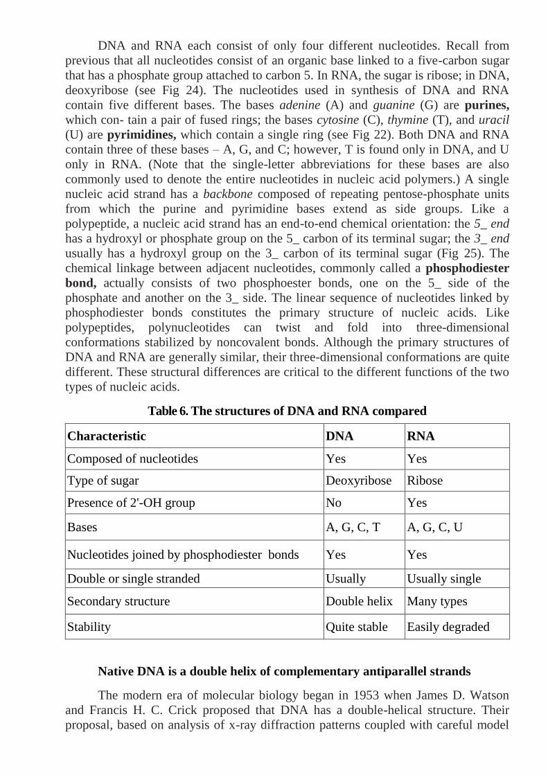
DNA and RNA each consist of only four different nucleotides. Recall from
previous that all nucleotides consist of an organic base linked to a five-carbon sugar
that has a phosphate group attached to carbon 5. In RNA, the sugar is ribose; in DNA,
deoxyribose (see Fig 24). The nucleotides used in synthesis of DNA and RNA
contain five different bases. The bases adenine (A) and guanine (G) are purines,
which con- tain a pair of fused rings; the bases cytosine (C), thymine (T), and uracil
(U) are pyrimidines, which contain a single ring (see Fig 22). Both DNA and RNA
contain three of these bases – A, G, and C; however, T is found only in DNA, and U
only in RNA. (Note that the single-letter abbreviations for these bases are also
commonly used to denote the entire nucleotides in nucleic acid polymers.) A single
nucleic acid strand has a backbone composed of repeating pentose-phosphate units
from which the purine and pyrimidine bases extend as side groups. Like a
polypeptide, a nucleic acid strand has an end-to-end chemical orientation: the 5_ end
has a hydroxyl or phosphate group on the 5_ carbon of its terminal sugar; the 3_ end
usually has a hydroxyl group on the 3_ carbon of its terminal sugar (Fig 25). The
chemical linkage between adjacent nucleotides, commonly called a phosphodiester
bond, actually consists of two phosphoester bonds, one on the 5_ side of the
phosphate and another on the 3_ side. The linear sequence of nucleotides linked by
phosphodiester bonds constitutes the primary structure of nucleic acids. Like
polypeptides, polynucleotides can twist and fold into three-dimensional
conformations stabilized by noncovalent bonds. Although the primary structures of
DNA and RNA are generally similar, their three-dimensional conformations are quite
different. These structural differences are critical to the different functions of the two
types of nucleic acids.
Table 6. The structures of DNA and RNA compared
Characteristic DNA RNA
Composed of nucleotides Yes Yes
Type of sugar Deoxyribose Ribose
Presence of 2'-OH group No Yes
Bases A, G, C, T A, G, C, U
Nucleotides joined by phosphodiester bonds Yes Yes
Double or single stranded Usually
double
Usually single
Secondary structure Double helix Many types
Stability Quite stable Easily degraded
Native DNA is a double helix of complementary antiparallel strands
The modern era of molecular biology began in 1953 when James D. Watson
and Francis H. C. Crick proposed that DNA has a double-helical structure. Their
proposal, based on analysis of x-ray diffraction patterns coupled with careful model
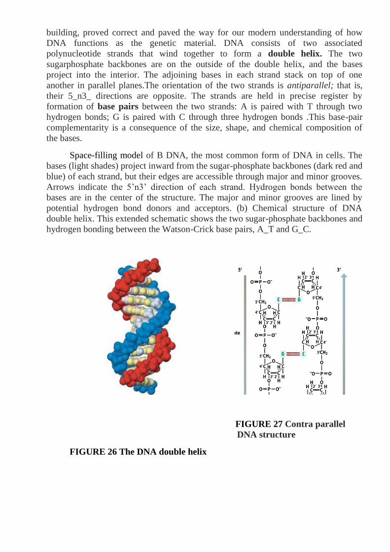
building, proved correct and paved the way for our modern understanding of how
DNA functions as the genetic material. DNA consists of two associated
polynucleotide strands that wind together to form a double helix. The two
sugarphosphate backbones are on the outside of the double helix, and the bases
project into the interior. The adjoining bases in each strand stack on top of one
another in parallel planes.The orientation of the two strands is antiparallel; that is,
their 5_n3_ directions are opposite. The strands are held in precise register by
formation of base pairs between the two strands: A is paired with T through two
hydrogen bonds; G is paired with C through three hydrogen bonds .This base-pair
complementarity is a consequence of the size, shape, and chemical composition of
the bases.
Space-filling model of B DNA, the most common form of DNA in cells. The
bases (light shades) project inward from the sugar-phosphate backbones (dark red and
blue) of each strand, but their edges are accessible through major and minor grooves.
Arrows indicate the 5’n3’ direction of each strand. Hydrogen bonds between the
bases are in the center of the structure. The major and minor grooves are lined by
potential hydrogen bond donors and acceptors. (b) Chemical structure of DNA
double helix. This extended schematic shows the two sugar-phosphate backbones and
hydrogen bonding between the Watson-Crick base pairs, A_T and G_C.
FIGURE 27 Contra parallel
DNA structure
FIGURE 26 The DNA double helix

FIGURE 28 Hydrogen copulas are between komplementarnimi nitrous bases
In natural DNA, A always hydrogen bonds with T and G with C, forming A·T
and G·C base pairs as shown in Fig 28. These associations between a larger purine
and smaller pyrimidine are often called Watson-Crick base pairs. Two polynucleotide
strands, in which all the nucleotides form such base pairs are said to be
complementary. However, in theory and in synthetic DNAs other base pairs can
form. For example, a guanine (a purine) could theoretically form hydrogen bonds
with a thymine (a pyrimidine), causing only a minor distortion in the helix. The space
available in the helix also would allow pairing between the two pyrimidines cytosine
and thymine. Although the nonstandard G·T and C·T base pairs are normally not
found in DNA, G·U base pairs are quite common in double-helical regions that form
within otherwise single-stranded RNA. Most DNA in cells is a right-handed helix.
The x-ray diffraction pattern of DNA indicates that the stacked bases are regularly
spaced 0.36 nm apart along the helix axis. The helix makes a complete turn every 3.6
nm; thus there are about 10.5 pairs per turn. This is referred to as the B form of DNA,
the normal form present in most DNA stretches in cells. On the outside of B-form
DNA, the spaces between the intertwined strands form two helical grooves of
different widths described as the major groove and the minor groove (see Fig 10). As
a consequence, the atoms on the edges of each base within these grooves are
accessible from outside the helix, forming two types of binding surfaces. DNA
binding proteins can “read” the sequence of bases in duplex DNA by contacting
atoms in either the major or the minor grooves. In addition to the major B form, three
additional DNA structures have been described. Two of these are compared to B
DNA. In very low humidity, the crystallographic structure of B DNA changes to the
A form; RNADNA and RNA-RNA helices exist in this form in cells and in vitro.
Short DNA molecules composed of alternating purinepyrimidine nucleotides
(especially Gs and Cs) adopt an alternative left-handed configuration instead of the
normal right-handed helix. This structure is called Z DNA because the bases seem to
zigzag when viewed from the side. Some evidence suggests that Z DNA may occur in

cells, although its function is unknown. Finally, a triple-stranded DNA structure is
formed when synthetic polymers of poly(A) and polydeoxy(U) are mixed in the test
tube. In addition, homopolymeric stretches of DNA composed of C and T residues in
one strand and A and G residues in the other can form a triple-stranded structure by
binding matching lengths of synthetic poly(C_T). Such structures probably do not
occur naturally in cells but may prove useful as therapeutic agents.
FIGURE 29 Models of various known DNA structures
The sugar-phosphate backbones of the two strands, which are on the outside
in all structures, are shown in red and blue; the bases (lighter shades) are oriented
inward. (a) The B form of DNA has ≈10.5 base pairs per helical turn. Adjacent
stacked base pairs are 0.36 nm apart. (b) The more compact A form of DNA has 11
base pairs per turn and exhibits a large tilt of the base pairs with respect to the helix
axis. (c) Z DNA is a left-handed double helix.
FIGURE 30 Models of various DNA structures

Table 7. Characteristics of DNA secondary structures
Characteristic A-DNA B-DNA Z-DNA
Conditions required to
produce structure 75% H2O 92% H2O
Alternating purine and
pyrimidine bases
Helix direction Right-handed Right-handed Left-handed
Average base pairs per turn 1 1 1 0 1 2
Rotation per base pair 32.7° 36°-30°
Distance between adjacent
bases
0.26 nm 0.34 nm 0.37 nm
Diameter 2.3 nm 1.9 nm 1.8 nm
Overall shape Short and wide Long and
narrow Elongated and narrow
By far the most important modifications in the structure of standard B-form
DNA come about as a result of protein binding to specific DNA sequences. Although
the multitude of hydrogen and hydrophobic bonds between the bases provide stability
to DNA, the double helix is flexible about its long axis. Unlike the _ helix in proteins,
there are no hydrogen bonds parallel to the axis of the DNA helix. This property
allows DNA to bend when complexed with a DNA-binding protein. Bending of DNA
is critical to the dense packing of DNA in chromatin, the protein-DNA complex in
which nuclear DNA occurs in eukaryotic cells.
Summery of nucleic acids structure
■ Deoxyribonucleic acid (DNA), the genetic material, carries information to specify
the amino acid sequences of proteins. It is transcribed into several types of
ribonucleic acid (RNA), including messenger RNA (mRNA), transfer RNA (tRNA),
and ribosomal RNA (rRNA), which function in protein synthesis.
■ Both DNA and RNA are long, unbranched polymers of nucleotides, which consist
of a phosphorylated pentose linked to an organic base, either a purine or pyrimidine.
■ The purines adenine (A) and guanine (G) and the pyrimidine cytosine (C) are
present in both DNA and RNA. The pyrimidine thymine (T) present in DNA is
replaced by the pyrimidine uracil (U) in RNA.
■ Adjacent nucleotides in a polynucleotide are linked by hosphodiester bonds. The
entire strand has a chemical directionality: the 5_ end with a free hydroxyl or
phosphate group on the 5_ carbon of the sugar, and the 3_ end with a free hydroxyl
group on the 3_ carbon of the sugar.
■ Natural DNA (B DNA) contains two complementary antiparallel polynucleotide
strands wound together into a regular right-handed double helix with the bases on the
in- Basic Molecular Genetic Mechanisms side and the two sugar-phosphate
backbones on the outside (see Figure 22-23). Base pairing between the strands and
hydrophobic interactions between adjacent bases in the same strand stabilize this
native structure.

■ The bases in nucleic acids can interact via hydrogen bonds. The standard Watson-
Crick base pairs are G·C, A·T (in DNA), and A·U (in RNA). Base pairing stabilizes
the native three-dimensional structures of DNA and RNA.
■ Binding of protein to DNA can deform its helical structure, causing local bending
or unwinding of the DNA molecule.
■ Heat causes the DNA strands to separate (denature). The melting temperature Tm
of DNA increases with the percentage of G·C base pairs. Under suitable conditions,
separated complementary nucleic acid strands will renature.
■ Circular DNA molecules can be twisted on themselves, forming supercoils .
Enzymes called topoisomerases can relieve torsional stress and remove supercoils
from circular DNA molecules.
■ Cellular RNAs are single-stranded polynucleotides, some of which form well-
defined secondary and tertiary structures. Some RNAs, called ribozymes, have
catalytic activity.
Transcription of protein-coding genes and formation of functional mRNA
The simplest definition of a gene is a “unit of DNA that contains the
information to specify synthesis of a single polypeptide chain or functional RNA
(such as a tRNA).” The vast majority of genes carry information to build protein
molecules, and it is the RNA copies of such protein-coding genes that constitute the
mRNA molecules of cells. The DNA molecules of small viruses contain only a few
genes, whereas the single DNA molecule in each of the chromosomes of higher
animals and plants may contain several thousand genes. During synthesis of RNA,
the four-base language of DNA containing A, G, C, and T is simply copied, or trans-
cribed, into the four-base language of RNA, which is identical except that U replaces
T. In contrast, during protein synthesis the four-base language of DNA and RNA is
translated into the 20–amino acid language of proteins. In this section we focus on
formation of functional mRNAs from protein-coding genes. A similar process yields
the precursors of rRNAs and tRNAs encoded by rRNA and tRNA genes; these
precursors are then further modified to yield functional rRNAs and tRNAs .
A Template DNA Strand Is Transcribed into a Complementary RNA Chain by
RNA Polymerase During transcription of DNA, one DNA strand acts as a template,
determining the order in which ribonucleoside triphosphate (rNTP) monomers are
polymerized to form a complementary RNA chain. Bases in the template DNA strand
base-pair with complementary incoming rNTPs, which then are joined in a
polymerization reaction catalyzed by RNA polymerase. Polymerization involves a
nucleophilic attack by the 3_ oxygen in the growing RNA chain on the phosphate of
the next nucleotide precursor to be added, resulting in formation of a phosphodiester
bond and release of pyrophosphate (PPi). As a consequence of this mechanism, RNA
molecules are always synthesized in the 5_n3_ direction. The energetics of the
polymerization reaction strongly favors addition of ribonucleotides to the growing
RNA chain because the high-energy bond between the _ and _ phosphate of rNTP

monomers is replaced by the lower-energy phosphodiester bond between nucleotides.
The equilibrium for the reaction is driven further toward chain elongation by
pyrophosphatase, an enzyme that catalyzes cleavage of the released PPi into two
molecules of inorganic phosphate. Like the two strands in DNA, the template DNA
strand and the growing RNA strand that is base-paired to it have opposite 5_n3_
directionality. By convention, the site at which RNA polymerase begins transcription
is numbered _1. Downstream denotes the direction in which a template DNA strand
is transcribed (or mRNA translated); thus a downstream sequence is toward the 3_
end relative to the start site, considering the DNA strand with the same polarity as the
transcribed RNA. Upstream denotes the opposite direction. Nucleotide positions in
the DNA sequence downstream from a start site are indicated by a positive (_) sign;
those upstream, by a negative (_) sign.
Stages in transcription To carry out transcription, RNA polymerase performs
several distinct functions, as depicted in Figure 4-10. During transcription initiation,
RNA polymerase recognizes and binds to a specific site, called a promoter, in
double-stranded DNA (step 1). Nuclear RNA polymerases require various protein
factors, called general transcription factors, to help them locate promoters and
initiate transcription. After binding to a promoter, RNA polymerase melts the DNA
strands in order to make the bases in the template strand available for base pairing
with the bases of the ribonucleoside triphosphates that it will polymerize together.
Cellular RNA polymerases melt approximately 14 base pairs of DNA around the
transcription start site, which is located on the template strand within the promoter.
Transcription initiation is considered complete when the first two ribonucleotides of
an RNA chain are linked by a phosphodiester bond. After several ribonucleotides
have been polymerized, RNA polymerase dissociates from the promoter DNA and
general transcription factors. During the stage of strand elongation, RNA polymerase
moves along the template DNA one base at a time, opening the double-stranded DNA
in front of its direction of movement and hybridizing the strands behind.
Basic molecular genetic mechanisms
One ribonucleotide at a time is added to the 3_ end of the growing (nascent) RNA
chain during strand elongation by the polymerase. The enzyme maintains a melted
region of approximately 14 base pairs, called the transcription bubble.
Approximately eight nucleotides at the 3_ end of the growing RNA strand remain
base-paired to the template DNA strand in the transcription bubble. The elongation
complex, comprising RNA polymerase, template DNA, and the growing (nascent)
RNA strand, is extraordinarily stable. For example, RNA polymerase transcribes the
longest known mammalian genes, containing ≈2 _ 106 base pairs, without
dissociating from the DNA template or releasing the nascent RNA. Since RNA
synthesis occurs at a rate of about 1000 nucleotides per minute at 37 _C, the
elongation complex must remain intact for more than 24 hours to assure continuous
RNA synthesis.
During transcription termination, the final stage in RNA synthesis, the
completed RNA molecule, or primary transcript, is released from the RNA
polymerase and the polymerase dissociates from the template DNA. Specific

sequences in the template DNA signal the bound RNA polymerase to terminate
transcription. Once released, an RNA polymerase is free to transcribe the same gene
again or another gene.
FIGURE 31
Structure of RNA polymerases The RNA polymerases of bacteria, archaea, and
eukaryotic cells are fundamentally similar in structure and function. Bacterial RNA
polymerases are composed of two related large subunits (__ and _), two copies of a
smaller subunit (_), and one copy of a fifth subunit ( ) that is not essential for
transcription or cell viability but stabilizes the enzyme and assists in the assembly of
its subunits. Archaeal and eukaryotic RNA polymerases have several additional small
subunits associated with this core complex. Schematic dia- Transcription of Protein-
Coding Genes and Formation of Functional mRNA grams of the transcription
process generally show RNA polymerase bound to an unbent DNA molecule.
However, according to a current model of the interaction between bacterial RNA
polymerase and promoter DNA, the DNA bends sharply following its entry into the
enzyme.

Figure 32 Organization of genes differs in prokaryoticand eukaryotic DNA
Having outlined the process of transcription, we now briefly consider the large-
scale arrangement of information in DNA and how this arrangement dictates the
requirements for RNA synthesis so that information transfer goes smoothly. In recent
years, sequencing of the entire genomes from several organisms has revealed not
only large variations in the number of protein-coding genes but also differences in
their organization in prokaryotes and eukaryotes.
The most common arrangement of protein-coding genes in all prokaryotes has
a powerful and appealing logic: genes devoted to a single metabolic goal, say, the
synthesis of the amino acid tryptophan, are most often found in a contiguous array in
the DNA. Such an arrangement of genes in a functional group is called an operon,
because it operates as a unit from a single promoter. Transcription of an operon
produces a continuous strand of mRNA that carries the message for a related series of
proteins. Each section of the mRNA represents the unit (or gene) that encodes one of
the proteins in the series. In prokaryotic DNA the genes are closely packed with very
few noncoding gaps, and the DNA is transcribed directly into colinear mRNA, which
then is translated into protein.This economic clustering of genes devoted to a single
metabolic function does not occur in eukaryotes, even simple ones like yeasts, which
can be metabolically similar to bacteria. Rather, eukaryotic genes devoted to a single
pathwayare most often physically separated in the DNA; indeed such genes usually
are located on different chromosomes. Each gene is transcribed from its own
promoter, producing one mRNA, which generally is translated to yield a single
polypeptide.
When researchers first compared the nucleotide sequences of eukaryotic
mRNAs from multicellular organisms with the DNA sequences encoding them, they
were surprised to find that the uninterrupted protein-coding sequence of a given
mRNA was broken up (discontinuous) in its corresponding section of DNA. They
concluded that the eukaryotic gene existed in pieces of coding sequence, the exons,
separated by non-protein-coding segments, the introns. This astonishing finding
implied that the long initial primary transcript – the RNA copy of the entire
transcribed DNA sequence – had to be clipped apart to remove the introns andthen
carefully stitched back together to produce manyeukaryotic mRNAs.

Although introns are common in multicellular eukaryotes, they are extremely
rare in bacteria.
FIGURE 33 Current model of bacterial RNA polymerase bound to a promoter
This structure corresponds to the polymerase molecule as schematically shown
in Fig 33. The DNA template and nontemplate strands are shown, respectively, as
gray and pink ribbons. Numbers indicate positions in the DNA sequence relative to
the transcription start site, with positive (+) numbers in the direction of transcription
and negative (-) numbers in the opposite direction. uncommon in many unicellular
eukaryotes such as baker’s yeast. However, introns are present in the DNA of viruses
that infect eukaryotic cells. Indeed, the presence of introns was first discovered in
such viruses, whose DNA is transcribed by host-cell enzymes.
Laboratory work №5
Subject: Basic mechanisms of the inherited information realization. Gene structure.
Transcription. Synthesis of protein.
Purpose: To learn the basic mechanisms of the inherited information realization and
its control.
Equipment: Charts of transcription, processing, splicing, and translation.
Explanation of work:
Existence of inherited information units which can be separated one from other
was guessed by Mendel in 1865 year. Gregor Mendel named them the factors of
heredity. A „gene” entered a term Iogansen in 1909 year. In spite of almost 100 years
after introduction of this term, he causes the permanent discussions of scientists.
After works of Bidl and Tatum under a term a gene was understood area of molecule
of DNA, which determines formation of one enzyme („one gene is one enzyme”).
Then this thesis was specified and took a form: „one gene is one поліпептидний
chain”.
The inherited information, written in DNA, will be realized through I-RNK to
the albumen. This way of Ô Krik named the „Central dogma of molecular biology it
was considered Long time, that an inherited information transfer in reverse direction
is impossible. In 1970 year of G.Timin and S. Muzatani opened the phenomenon of
зворотньої transcription. By the enzyme of reverse transryptaza (revertaza) in RNK-
VMISNIKH of viruses genetic information is passed from RNAS to DNA. In 1975
year of R.Dul'beko, G.Timin, D.Baltimor described the similar phenomenon in the
cages of amphibians. Coming from it, the central dogma of molecular biology can be
presented as follows:
Eukaryotic precursor mRNAs are processed to form functional mRNAs
In prokaryotic cells, which have no nuclei, translation of an mRNA into protein
can begin from the 5_ end of the mRNA even while the 3_ end is still being
synthesized by RNA polymerase. In other words, transcription and translation can
occur concurrently in prokaryotes. In eukaryotic cells, however, not only is the
nucleus separated from the cytoplasm where translation occurs, but also the primary
transcripts of protein-coding genes are precursor mRNAs (pre-mRNAs) that must
undergo several modifications, collectively termed RNA processing, to yield a
functional mRNA (see Fig 34).

This mRNA then must be exported to the • Basic Molecular Genetic
Mechanisms cytoplasm before it can be translated into protein. Thus transcription and
translation cannot occur concurrently in eukaryotic cells.
All eukaryotic pre-mRNAs initially are modified at the two ends, and these
modifications are retained in mRNAs. As the 5_ end of a nascent RNA chain emerges
from the surface of RNA polymerase II, it is immediately acted on by several
enzymes that together synthesize the 5_ cap, a 7-methylguanylate that is connected to
the terminal nucleotide of the RNA by an unusual 5_,5_ triphosphate linkage. The
cap protects an mRNA from enzymatic degradation and assists in its export to the
cytoplasm. Thecap also is bound by a protein factor required to begin translationin
the cytoplasm.Processing at the 3_ end of a pre-mRNA involves cleavage by an
endonuclease to yield a free 3_-hydroxyl group to which a string of adenylic acid
residues is added one at a time by an enzyme called poly(A) polymerase. The
resulting poly(A) tail contains 100–250 bases, being shorter in yeasts and
invertebrates than in vertebrates. Poly(A) polymerase is part of a complex of proteins
that can locate and cleave atranscript at a specific site and then add the correct
numberof A residues, in a process that does not require a template. The final step in
the processing of many different eukaryotic mRNA molecules is RNA splicing: the
Figure 34

internal cleavage of a transcript to excise the introns, followed by ligation of the
coding exons.
Figure 36 Uracil structure
Figure 35 RNA structure
The functional eukaryotic mRNAs produced by RNA processing retain noncoding
regions, referred to as 5_ and 3_ untranslated regions (UTRs), at each end. In
mammalian mRNAs, the 5_ UTR may be a hundred or more nucleotides long, and
the 3_ UTR may be several kilobases in length. Prokaryotic mRNAs also usually
have 5_ and 3_ UTRs, but these are much shorter than those in eukaryotic mRNAs,
generally containing fewer than 10 nucleotides.
Alternative RNA splicing increases the number of proteins expressed from
a single eukaryotic gene
In contrast to bacterial and archaeal genes, the vast majority of genes in higher,
multicellular eukaryotes contain multiple introns. Many proteins from higher
eukaryotes have a multidomain tertiary structure. Individual repeated protein domains
often are encoded by one exon or a small number of exons that code for identical or
nearly identical amino acid sequences. Such repeated exons are thought to have
evolved by the accidental multiple duplication of a length of DNA lying between two
sites in adjacent introns, resulting in insertion of a string of repeated exons, separated
by introns, between the original two introns. The presence of multiple introns in
many eukaryotic genes permits expression of multiple, related proteins from a single
gene by means of alternative splicing. In higher eukaryotes, alternative splicing is an
important mechanism for production of different forms of a protein, called isoforms,
by different types of cells. Fibronectin, a multidomain extracellular adhesive protein
found in mammals, provides a good example of alternative splicing. The fibronectin
gene contains numerous exons, grouped into several regions corresponding to
specific domains of the protein. Fibroblasts produce fibronectin mRNAs that contain
exons EIIIA and EIIIB; these exons encode amino acid sequences that bind tightly to
proteins in the fibroblast plasma membrane. Consequently, this fibronectin isoform
adheres fibroblasts to the extracellular matrix. Alternative splicing of the fibronectin
primary transcript in hepatocytes, the major type of cell in the liver, yields mRNAs
Nitrous
basis (A,
U, G, C)

that lack the EIIIA and EIIIB exons. As a result, the fibronectin secreted by
hepatocytes into the blood does not adhere tightly to fibroblasts or most other cell
types, allowing it to circulate. During formation of blood clots, however, the
fibrinbinding domains of hepatocyte fibronectin binds to fibrin, one of the principal
constituents of clots. The bound fibronectin then interacts with integrins on the
membranes of passing, activated platelets, thereby expanding the clot by addition of
platelets.More than 20 different isoforms of fibronectin have been identified, each
encoded by a different, alternatively spliced mRNA composed of a unique
combination of fibronectin gene exons. Recent sequencing of large numbers of
mRNAs • Basic Molecular Genetic Mechanisms isolated from various tissues and
comparison of their sequences with genomic DNA has revealed that nearly 60
percent of all human genes are expressed as alternatively spliced mRNAs. Clearly,
alternative RNA splicing greatly expands the number of proteins encoded by the
genomes of higher, multicellular organisms.
Transcription of protein-coding genes and formation of functional mRNA
key concepts:
Transcription of DNA is carried out by RNA polymerase, which adds one
ribonucleotide at a time to the 3_ end of a growing RNA chain. The sequence
of the template DNA strand determines the order in which ribonucleotides are
polymerized to form an RNA chain.
During transcription initiation, RNA polymerase binds to a specific site in
DNA (the promoter), locally melts double-stranded DNA to reveal the
unpaired template strand, and polymerizes the first two nucleotides.
During strand elongation, RNA polymerase moves along the DNA, melting
sequential segments of the DNA and adding nucleotides to the growing RNA
strand.
When RNA polymerase reaches a termination sequence in the DNA, the
enzyme stops transcription, leading to release of the completed RNA and
dissociation of the enzyme from the template DNA.
In prokaryotic DNA, several protein-coding genes commonly are clustered
into a functional region, an operon, which is transcribed from a single promoter
into one mRNA encoding multiple proteins with related functions (see Figure).
Translation of a bacterial mRNA can begin before synthesis of the mRNA is
complete.
In eukaryotic DNA, each protein-coding gene is transcribed from its own
promoter. The initial primary tran- script very often contains noncoding
regions (introns) interspersed among coding regions (exons).
Eukaryotic primary transcripts must undergo RNA processing to yield
functional RNAs. During processing, the ends of nearly all primary transcripts
from protein-coding genes are modified by addition of a 5_ cap and 3_ poly(A)
tail. Transcripts from genes containing introns undergo splicing, the removal of
the introns and joining of the exons.
The individual domains of multidomain proteins found in higher eukaryotes
are often encoded by individual exons or a small number of exons. Distinct

isoforms of such proteins often are expressed in specific cell types as the result
of alternative splicing of exons.
Table 8. Components required for protein synthesis in bacterial cells
Stage Component Function
Binding of
amino acid
to tRNA
Amino acids
tRNAs
aminoacyl-tRNA synthetase
ATP
Building blocks of proteins
Deliver amino acids to ribosomes
Attaches amino acids to tRNAs
Provides energy for binding amino
acid to tRNA
Initiation
mRNA
fMet-tRNAfMet
30S ribosomal subunit
50S ribosomal subunit
Initiation factor 1
Initiation factor 2
Initiation factor 3
Carries coding instructions
Provides first amino acid in peptide
Attaches to mRNA
Stabilizes tRNAs and amino acids
Enhances dissociation of large and
small subunits of ribosome
Binds GTP; delivers fMet-tRNAfMet
to
initiation codon
Binds to 30S subunit and prevents
association with 50S subunit
Elongation
70S initiation complex
Charged tRNAs
Elongation factor Tu
Elongation factor Ts
Elongation factor G GTP
Peptidyl transferase
Functional ribosome with A, P, and E
sites and peptidyl transferase activity
where protein synthesis takes place
Bring amino acids to ribosome and
help assemble them in order specified
by mRNA
Binds GTP and charged tRNA;
delivers charged tRNA to A site
Generates active elongation factor Tu
Stimulates movement of ribosome to
next codon
Provides energy
Creates peptide bond between amino
acids in A site and P site
Termination Release factors 1, 2, and 3 Bind to ribosome when stop codon is
reached and terminate translation
Control of gene expression in prokaryotes key concepts:
Gene expression in both prokaryotes and eukaryotes is regulated primarily by
mechanisms that control the initiation of transcription.

Binding of the subunit in an RNA polymerase to a promoter region is the first
step in the initiation of transcription in E. coli. • Basic Molecular Genetic
Mechanisms
The nucleotide sequence of a promoter determines its strength, that is, how
frequently different RNA polymerase which overlap or lie adjacent to
promoters. Binding of repressor to an operator inhibits transcription initiation.
The DNA-binding activity of most bacterial repressors is modulated by small
effector molecules (inducers). This allows bacterial cells to regulate
transcription of specific genes in response to changes in the concentration of
various nutrients in the environment.
The lac operon and some other bacterial genes also are regulated by activator
proteins that bind next to promoters and increase the rate of transcription
initiation by RNA polymerase.
The major sigma factor in E. coli is 70, but several other less abundant sigma
factors are also found, each recognizing different consensus promoter
sequences.
Transcription initiation by all E. coli RNA polymerases, except those
containing 54, can be regulated by repressors and activators that bind near the
transcription start site.
Genes transcribed by 54–RNA polymerase are regulated by activators that bind
to enhancers located ≈100 base pairs upstream from the start site. When the
activator and 54–RNA polymerase interact, the DNA between their binding
sites forms a loop.
In two-component regulatory systems, one protein acts as a sensor, monitoring
the level of nutrients or other components in the environment. Under
appropriate conditions, the phosphate of an ATP is transferred first to a
histidine in the sensor protein and then to an aspartic acid in a second protein,
the response regulator. The phosphorylated response regulator then binds to
DNA regulatory sequences, thereby stimulating or repressing transcription of
specific genes.
Three roles of RNA in translation key concepts:
Although DNA stores the information for protein synthesis and mRNA conveys the
instructions encoded in DNA, most biological activities are carried out by proteins.
The linear order of amino acids in each protein determines its three-dimensional
structure and activity. For this reason, assembly of amino acids in their correct order,
as encoded in DNA, is critical to production of functional proteins and hence the
proper functioning of cells and organisms.
Translation is the whole process by which the nucleotide sequence of an mRNA is
used to order and to join the amino acids in a polypeptide chain. In eukaryotic cells,
protein synthesis occurs in the cytoplasm, where three types of RNA molecules come
together to perform different but cooperative functions:

1. Messenger RNA (mRNA) carries the genetic information transcribed from DNA
in the form of a series of three nucleotide sequences, called codons, each of which
specifies a particular amino acid.
2. Transfer RNA (tRNA) is the key to deciphering the codons in mRNA. Each type
of amino acid has its own subset of tRNAs, which bind the amino acid and carry it to
the growing end of a polypeptide chain if the next codon in the mRNA calls for it.
The correct tRNA with its attached amino acid is selected at each step because each
specific tRNA molecule contains a three-nucleotide sequence, an anticodon, that can
base-pair with its complementary codon in the mRNA.
3. Ribosomal RNA (rRNA) associates with a set of proteins to form ribosomes.
These complex structures, which physically move along an mRNA molecule,
catalyze the assembly of amino acids into polypeptide chains. They also bind tRNAs
and various accessory proteins necessary for protein synthesis. Ribosomes are
composed of a large A molecule or molecules.
• Three Roles of RNA in Translation. These three types of RNA participate in
translation in all cells. Indeed, development of three functionally distinct RNAs was
probably the molecular key to the origin of life. How the structure of each RNA
relates to its specific task is described in this section; how the three types work
together, along with required protein factors, to synthesize proteins is detailed in the
following section. Since translation is essential for protein synthesis, the two
processes commonly are referred to interchangeably. However, the polypeptide
chains resulting from translation undergo post-translational folding and often other
changes (e.g., chemical modifications, association with other chains) that are required
for production of mature, functional proteins.
Messenger RNA carries information from DNA in a three-letter genetic code
As noted above, the genetic code used by cells is a triplet code, with every
three-nucleotide sequence, or codon, being “read” from a specified starting point in
the mRNA. Of the 64 possible codons in the genetic code, 61 specify individual
amino acids and three are stop codons. Мost amino acids are encoded by more than
one codon. Only two – methionine and tryptophan – have a single codon; at the other
extreme, leucine, serine, and arginine are each specified by six different codons. The
different codons for a given amino acid are said to be synonymous. The code itself is
termed degenerate, meaning that more than one codon can specify the same amino
acid. Synthesis of all polypeptide chains in prokaryotic and eukaryotic cells begins
with the amino acid methionine. In most mRNAs, the start (initiator) codon
specifying this aminoterminal methionine is AUG. In a few bacterial mRNAs, GUG
is used as the initiator codon, and CUG occasionally is used as an initiator codon for
methionine in eukaryotes. The three codons UAA, UGA, and UAG do not specify
amino acids but constitute stop (termination) codons that mark the carboxyl terminus
of polypeptide chains in almost all cells. The sequence of codons that runs from a
specific

FIGURE 37 Enzymes which take part in DNA replication
Basic Molecular Genetic Mechanisms start codon to a stop codon is called a
reading frame. This precise linear array of ribonucleotides in groups of three in
mRNA specifies the precise linear sequence of amino acids in a polypeptide chain
and also signals where synthesis of the chain starts and stops.
Because the genetic code is a comma-less, non-overlapping triplet code, a
particular mRNA theoretically could be translated in three different reading frames.
Indeed some mRNAs have been shown to contain overlapping information that can
be translated in different reading frames, yielding different polypeptides.
The vast majority of mRNAs, however, can be read in only one frame because
stop codons encountered in the other two possible reading frames terminate
translation before a functional protein is produced. Another unusual coding
arrangement occurs because of frame- shifting. In this case the protein-synthesizing
machinery may read four nucleotides as one amino acid and then continue reading
triplets, or it may back up one base and read all succeeding triplets in the new frame
until termination of the chain occurs. These frame shifts are not common events, but
a few dozen such instances are known.
The meaning of each codon is the same in most known organisms – a strong
argument that life on earth evolved only once. However, the genetic code has been
found to differ for a few codons in many mitochondria, in ciliated protozoans, and in
Acetabularia, a single-celled plant. As shown in Table, most of these changes involve
reading of normal stop codons as amino acids, not an exchange of one amino acid for
another. These exceptions to the general code probably were later evolutionary deve-
lopments; that is, at no single time was the code immutably fixed, although massive
changes were not tolerated once a general code began to function early in evolution.
Three roles of RNA in translation the folded structure of tRNA promotes its
decoding functions key concepts:
Translation, or decoding, of the four-nucleotide language of DNA and mRNA
into the 20–amino acid language of proteins requires tRNAs and enzymes called
aminoacyl-tRNA synthetases. To participate in protein synthesis, a tRNA molecule
must become chemically linked to a particular amino acid via a high-energy bond,

forming an aminoacyl-tRNA; the anticodon in the tRNA then base-pairs with a
codon in mRNA so that the activated amino acid can be added to the growing
polypeptide chain.
FIGURE 38 Co-operation in i RNA codon tRNA anticodon
Some 30–40 different tRNAs have been identified in bacterial cells and as
many as 50–100 in animal and plant cells. Thus the number of tRNAs in most cells is
more than the number of amino acids used in protein synthesis (20) and also differs
from the number of amino acid codons in the genetic code (61). Consequently, many
amino acids have more than one tRNA to which they can attach (explaining how
there can be more tRNAs than amino acids); in addition, many tRNAs can pair with
more than one codon (explaining how there can be more codons than tRNAs).
The function of tRNA molecules, which are 70–80 nucleotides long, depends
on their precise three-dimensional structures. In solution, all tRNA molecules fold
into a similar stem-loop arrangement that resembles a cloverleaf when drawn in two
dimensions (Figure 38). The four stems are short double helices stabilized by
Watson-Crick base pairing; three of the four stems have loops containing seven or
eight bases at their ends, while the remaining, unlooped stem contains the free 3_ and
5_ ends of the chain. The three nucleotides composing the anticodon are located at
the center of the middle loop, in an accessible position that facilitates codon-
anticodon base pairing. In all tRNAs, the 3_ end of the unlooped amino acid acceptor
stem has the sequence CCA, which in most cases is added after synthesis and
processing of the tRNA are complete. Several bases in most tRNAs also are modified
after synthesis. Viewed in three dimensions, the folded tRNA molecule has an L
shape withthe anticodon loop and acceptor stem forming the ends ofthe two arms.
Nonstandard base pairing often occurs between codons and anticodons
If perfect Watson-Crick base pairing were demanded between codons and
anticodons, cells would have to contain exactly 61 different tRNA species, one for
each codon that specifies on Basic Molecular Genetic Mechanisms amino acid. As
noted above, however, many cells contain fewer than 61 tRNAs. The explanation for
the smaller number lies in the capability of a single tRNA anticodon to recognize

more than one, but not necessarily every, codon corresponding to a given amino acid.
This broader recognition can occur because of nonstandard pairing between bases in
the so-called wobble position: that is, the third (3_) base in an mRNA codon and the
corresponding first (5_) base in its tRNA anticodon. The first and second bases of a
codon almost always form standard Watson-Crick base pairs with the third and
second bases, respectively, of the corresponding anticodon, but four nonstandard
interactions can occur between bases in the wobble position. Particularly important is
the G·U base pair, which structurally fits almost as well as the standard G·C pair.
Thus, a given anticodon in tRNA with G in the first (wobble) position can base-pair
with the two corresponding codons that have either pyrimidine (C or U) in the third
position. For example, the phenylalanine codons UUU and UUC (5_n3_) are both
recognized by the tRNA that has GAA (5_n3_) as the anticodon. In fact, any two
codons of the type NNPyr (N _ any base; Pyr _ pyrimidine) encode a single amino
acid and are decoded by a single tRNA with G in the first (wobble) position of the
anticodon.
FIGURE 39 Translation takes a place on the ribosoms due to in iRNA-codon
tRNA-anticodon co-operation
Although adenine rarely is found in the anticodon wobble position, many
tRNAs in plants and animals contain inosine
The Three Roles of RNA in Translation (I), a deaminated product of adenine,
at this position. Inosine can form nonstandard base pairs with A, C, and U. A tRNA
with inosine in the wobble position thus can recognize the corresponding mRNA
codons with A, C, or U in the third (wobble) position (see Figure 39). For this reason,
inosinecontaining tRNAs are heavily employed in translation of the synonymous
codons that specify a single amino acid. For example, four of the six codons for
leucine (CUA, CUC, CUU, and UUA) are all recognized by the same tRNA with the
anticodon 3_-GAI-5_; the inosine in the wobble position forms nonstandard base
pairs with the third base in the four codons. In the case of the UUA codon, a
nonstandard G·U pair also forms between position 3 of the anticodon and position 1
of the codon.
Ribosomes are protein-synthesizing machines
If the many components that participate in translating mRNA had to interact in
free solution, the likelihood of simultaneous collisions occurring would be so low that

thrate of amino acid polymerization would be very slow. The efficiency of translation
is greatly increased by the binding ofthe mRNA and the individual aminoacy l-tRNAs
to the mostabundant RNA-protein complex in the cell, the ribosome, which directs
elongation of a polypeptide at a rate of three to five amino acids added per second.
Small proteins of100–200 amino acids are therefore made in a minute or less.On the
other hand, it takes 2–3 hours to make the largestknown protein, titin, which is found in
muscle and containsabout 30,000 amino acid residues. The cellular machine
thataccomplishes this task must be precise and persistent.With the aid of the electron
microscope, ribosomes were first discovered as small, discrete, RNA-rich particles in
cellsthat secrete large amounts of protein. However, their role inprotein synthesis was
not recognized until reasonably pureribosome preparations were obtained. In vitro
radiolabelingexperiments with such preparations showed that radioactiveamino acids
first were incorporated into growing polypeptidechains that were associated with
ribosomes before appearing in finished chains.A ribosome is composed of three (in
bacteria) or four (in eukaryotes) different rRNA molecules and as many as 83 proteins,
organized into a large subunit and a small subunit (Figure 39, 40).
Basic Molecular Genetic Mechanism strifuged under standard conditions. The
small ribosomal subunit contains a single rRNA molecule, referred to as small rRNA.
The large subunit contains a molecule of large rRNAand one molecule of 5S rRNA,
plus an additional moleculeof 5.8S rRNA in vertebrates. The lengths of the rRNA
molecules, the quantity of proteins in each subunit, and consequently the sizes of the
subunits differ in bacterial andeukaryotic cells. The assembled ribosome is 70S in
bacteria and 80S in vertebrates. But more interesting than these differences are the
great structural and functional similaritiesbetween ribosomes from all species. This
consistency is anotherreflection of the common evolutionary origin of themost basic
constituents of living cells.The sequences of the small and large rRNAs from
severalthousand organisms are now known. Although the primarynucleotide
sequences of these rRNAs vary considerably, thesame parts of each type of rRNA
theoretically can form basepairedstem-loops, which would generate a similar
threedimensional structure for each rRNA in all organisms. Theactual three-
dimensional structures of bacterial rRNAs from Thermus thermopolis recently have
been determined by xray crystallography of the 70S ribosome. The multiple, much
smaller ribosomal proteins for the most part are associated with the surface of the
rRNAs. Although the number of protein molecules in ribosomes greatly exceeds the
number of RNA molecules, RNA constitutes about 60 percent of the mass of a
ribosome. During translation, a ribosome moves along an mRNA chain, interacting
with various protein factors and tRNAs and very likely undergoing large
conformational changes. Despite the complexity of the ribosome, great progress has
been made in determining the overall structure of bacterial ribosomes and in
identifying various reactive sites. X-ray crystallographic studies on the T.
thermophilus 70S ribosome, for instance, not only have revealed the dimensions and
overall shape of the ribosomal subunits but also have localized the positions of
tRNAs bound to the ribosome during elongation of a growing protein chain. In
addition, powerful chemical techniques such as footprinting, which is described in
Chapter 11, have been used to identify specific nucleotide sequences in rRNAs that
bind to protein or another RNA. Some 40 years after the initial discovery of

ribosomes, their overall structure and functioning during protein synthesis are finally
becoming clear, as we describe in the next section.
Three roles of RNA in translation key concepts:
Genetic information is transcribed from DNA into mRNA in the form of a
comma-less, overlapping, degenerate triplet code.
Each amino acid is encoded by one or more threenucleotide sequences
(codons) in mRNA. Each codon specifies one amino acid, but most amino
acids are encoded by multiple codons.
The AUG codon for methionine is the most common start codon, specifying
the amino acid at the NH2-terminus of a protein chain. Three codons (UAA,
UAG, UGA) functionas stop codons and specify no amino acids.
A reading frame, the uninterrupted sequence of codons in mRNA from a
specific start codon to a stop codon, is translated into the linear sequence of
amino acids in a polypeptide chain.
Decoding of the nucleotide sequence in mRNA into the amino acid sequence
of proteins depends on tRNAs and aminoacyl-tRNA synthetases.
All tRNAs have a similar three-dimensional structure that includes an acceptor
arm for attachment of a specific amino acid and a stem-loop with a three-base
anticodon sequence at its ends. The anticodon can base-pair with its
corresponding codon in mRNA.
Because of nonstandard interactions, a tRNA may base pair with more than one
mRNA codon; conversely, a particular codon may base-pair with multiple
tRNAs. In each Stepwise Synthesis of Proteins on Ribosomes case, however,
only the proper amino acid is inserted into a growing polypeptide chain.
Each of the 20 aminoacyl-tRNA synthetases recognizes a single amino acid
and covalently links it to a cognate tRNA, forming an aminoacyl-tRNA. This
reaction activates the amino acid, so it can participate in peptide bond
formation.
Both prokaryotic and eukaryotic ribosomes – the large ribonucleoprotein
complexes on which translation occurs – consist of a small and a large subunit/
Each subunit contains numerous different proteins and one major rRNA
molecule (small or large). The large subunit also contains one accessory 5S
rRNA in bacteria and two accessory rRNAs in eukaryotes (5S and 5.8S in
vertebrates).
Analogous rRNAs from many different species fold into quite similar three-
dimensional structures containing numerous stem-loops and binding sites for
proteins, mRNA, and tRNAs. Much smaller ribosomal proteins are associated
with the periphery of the rRNAs.
Translation Initiation Usually Occurs Near the First AUG Closest to the 5_ End
of an mRNA During the first stage of translation, a ribosome assembles, complexed
with an mRNA and an activated initiator tRNA, which is correctly positioned at the
start codon. Large and small ribosomal subunits not actively engaged in translation
are rthn apart by binding of two inition factors.
are kept apart by binding of two initiation factors
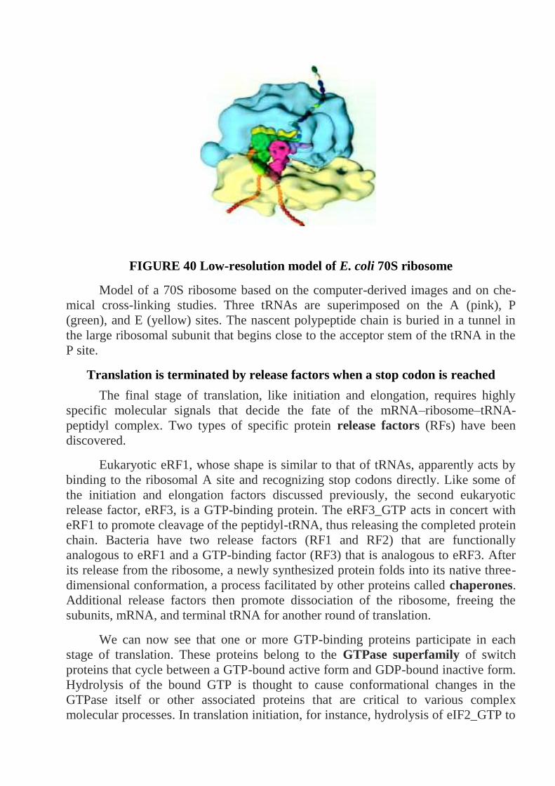
FIGURE 40 Low-resolution model of E. coli 70S ribosome
Model of a 70S ribosome based on the computer-derived images and on che-
mical cross-linking studies. Three tRNAs are superimposed on the A (pink), P
(green), and E (yellow) sites. The nascent polypeptide chain is buried in a tunnel in
the large ribosomal subunit that begins close to the acceptor stem of the tRNA in the
P site.
Translation is terminated by release factors when a stop codon is reached
The final stage of translation, like initiation and elongation, requires highly
specific molecular signals that decide the fate of the mRNA–ribosome–tRNA-
peptidyl complex. Two types of specific protein release factors (RFs) have been
discovered.
Eukaryotic eRF1, whose shape is similar to that of tRNAs, apparently acts by
binding to the ribosomal A site and recognizing stop codons directly. Like some of
the initiation and elongation factors discussed previously, the second eukaryotic
release factor, eRF3, is a GTP-binding protein. The eRF3_GTP acts in concert with
eRF1 to promote cleavage of the peptidyl-tRNA, thus releasing the completed protein
chain. Bacteria have two release factors (RF1 and RF2) that are functionally
analogous to eRF1 and a GTP-binding factor (RF3) that is analogous to eRF3. After
its release from the ribosome, a newly synthesized protein folds into its native three-
dimensional conformation, a process facilitated by other proteins called chaperones.
Additional release factors then promote dissociation of the ribosome, freeing the
subunits, mRNA, and terminal tRNA for another round of translation.
We can now see that one or more GTP-binding proteins participate in each
stage of translation. These proteins belong to the GTPase superfamily of switch
proteins that cycle between a GTP-bound active form and GDP-bound inactive form.
Hydrolysis of the bound GTP is thought to cause conformational changes in the
GTPase itself or other associated proteins that are critical to various complex
molecular processes. In translation initiation, for instance, hydrolysis of eIF2_GTP to

eIF2_GDP prevents further scanning of the mRNA once the start site is encountered
and allows binding of the large ribosomal subunit to the small subunit.
FIGURE 41 Scopes of read-out
Stepwise synthesis of proteins on ribosomes
Of the two methionine tRNAs found in all cells, only one (tRNAi Met)
functions in initiation of translation.
Each stage of translation – initiation, chain elongation, and termination –
requires specific protein factors including GTP-binding proteins that hydrolyze
their bound GTP to GDP when a step has been completed successfully.
During initiation, the ribosomal subunits assemble near the translation start
site in an mRNA molecule with the tRNA carrying the amino-terminal
methionine (Met-tRNAi Met) base-paired with the start codon.
Chain elongation entails a repetitive four-step cycle: loose binding of an
incoming aminoacyl-tRNA to the A site on the ribosome; tight binding of the
correct aminoacyl-tRNA to the A site accompanied by release of the previously
used tRNA from the E site; transfer of the growing peptidyl chain to the
incoming amino acid catalyzed by large rRNA; and translocation of the
ribosome to the next codon, thereby moving the peptidyl-tRNA in the A site to
the P site and the now unacylated tRNA in the P site to the E site.
In each cycle of chain elongation, the ribosome undergoes two conformational
changes monitored by GTP-binding proteins. The first permits tight binding of
the incoming aminoacyl-tRNA to the A site and ejection of a tRNA from the E
site, and the second leads to translocation.
Termination of translation is carried out by two types of termination factors:
those that recognize stop codons and those that promote hydrolysis of peptidyl-
tRNA .
The efficiency of protein synthesis is increased by the simultaneous translation
of a single mRNA by multiple ribosomes. In eukaryotic cells, protein-mediated
interactions. M E D I A C O N N E C T I O N S
Overview Animation: Life Cycle of an mRNA brings two ends of a
polyribosome close together, thereby promoting the rapid recycling of ribosomal
subunits, which further increases the efficiency of protein synthesis.
DNA Replication
Now that we have seen how genetic information encoded in the nucleotide
sequences of DNA is translated into the structures of proteins that perform most cell
functions, we can appreciate the necessity of the precise copying of DNA sequences
during DNA replication.
The regular pairing of bases in the double-helical DNA structure suggested to
Watson and Crick that new DNA strands are synthesized by using the existing
(parental) strands as templates in the formation of new, daughter strands
complementary to the parental strands.
This base-pairing template model theoretically could proceed either by a
conservative or a semiconservative mechanism. In a conservative mechanism, the two
daughter strands would form a new double-stranded (duplex) DNA molecule and the
parental duplex would remain intact. In a semiconservative mechanism, the parental
strands are permanently separated and each forms a duplex molecule with the
daughter strand base-paired to it.
DNA polymerases require a primer to initiate replication
Analogous to RNA, DNA is synthesized from deoxynucleoside 5_-
triphosphate precursors (dNTPs). Also like RNA synthesis, DNA synthesis always
proceeds in the 5_n3_ Basic Molecular Genetic Mechanisms direction because chain
growth results from formation of a phosphoester bond between the 3_ oxygen of a
growing strand and the _ phosphate of a dNTP. As discussed earlier, an RNA
polymerase can find an appropriate transcription start site on duplex DNA and initiate
the synthesis of an RNA complementary to the template DNA strand. In contrast,
DNA polymerases cannot initiate chain synthesis de novo; instead, they require a
short, preexisting RNA or DNA strand, called a primer, to begin chain growth. With
a primer base-paired to the template strand, a DNA polymerase adds
deoxynucleotides to the free hydroxyl group at the 3_ end of the primer as directed by
the sequence of the template strand:
Duplex DNA is unwound, and daughter strands are formed at the DNA
replication fork
In order for duplex DNA to function as a template during replication, the two
intertwined strands must be unwound, or melted, to make the bases available for base
pairing with the bases of the dNTPs that are polymerized into the newly synthesized
daughter strands. This unwinding of the parental DNA strands is by specific
helicases, beginning at unique segments in a DNA molecule called replication
origins, or simply origins. The nucleotide sequences of origins from different
organisms vary greatly, although they usually contain A_T-rich sequences. Once
helicases have unwound the parental DNA at an origin, a specialized RNA
polymerasecalled primase forms a short RNA primer complementary to the un-
wound template strands. The primer, still base-paired to its complementary DNA
strand, is then elongated by a DNA polymerase, thereby forming a new daughter
strand.
The DNA region at which all these proteins come together to carry out
synthesis of daughter strands is called the replication fork, or growing fork. As
replication proceeds, the growing fork and associated proteins move away from the
origin. As noted earlier, local unwinding of duplex DNA produces torsional stress,
which is relieved by topoisomerase I. In order for DNA polymerases to move along
and copy a duplex DNA, helicase must sequentially unwind the duplex and
topoisomerase must remove the supercoils that form. A major complication in the
operation of a DNA replication fork arises from two properties: the two strands of the

parental DNA duplex are antiparallel, and DNA polymerases (like RNA
polymerases) can add nucleotides to the growing new strands only in the 5_n3_
direction. Synthesis of one daughter strand, called the leading strand, can proceed
continuously from a single RNA primer in the 5_n 3_ direction, the same direction as
movement of the replication fork (Figure 42). The problem comes in synthesis of the
other daughter strand, called the lagging strand.
FIGURE 42 Schematic diagram of leading-strand and lagging-strand DNA
synthesis at a replication fork
Nucleotides are added by a DNA polymerase to each growing daughter strand
in the 5_n3_ direction (indicated by arrowheads). The leading strand is synthesized
continuously from a single RNA primer (red) at its 5_ end. The lagging strand is
synthesized discontinuously from multiple RNA primers that are formed periodically
as each new region of the parental duplex is unwound. Elongation of these primers
initially produces Okazaki fragments. As each growing fragment approaches the
previous primer, the primer is removed and the fragments are ligated. Repetition of
this process eventually results in synthesis of the entire lagging strand.
Because growth of the lagging strand must occur in the 5_n3_ direction,
copying of its template strand must somehow occur in the opposite direction from the
movement of the replication fork. A cell accomplishes this feat by synthesizing a new
primer every few hundred bases or so on the second parental strand, as more of the
strand is exposed by unwinding. Each of these primers, base-paired to their template
strand, is elongated in the 5_n3_ direction, forming discontinuous segments called
Okazaki fragments after their discoverer Reiji Okazaki. The RNA primer of each
Okazaki fragment is removed and replaced by DNA chain growth from the
neighboring Okazaki fragment; finally an enzyme called DNA ligase joins the
adjacent fragments.

Helicase, primase, DNA polymerases, and other proteins participate in DNA
replication
These multicomponent complexes permit the cell to carry out an ordered
sequence of events that accomplish essential cell functions. In the molecular machine
that replicates SV40 DNA, a hexamer of a viral protein called large T-antigen
unwinds the parental strands at a replication fork. All other proteins involved in SV40
DNA replication are provided by the host cell. Primers for leading and lagging
daughter-strand DNA are synthesized by a complex of primase, which synthesizes a
short RNA primer, and DNA polymerase _ (Pol _), which extends the RNA primer
with deoxynucleotides, forming a mixed RNA-DNA primer.
The primer is extended into daughter-strand DNA by DNA polymerase _ (Pol
_), which is less likely to make errors during copying of the template strand than is
Pol _. Pol _ forms a complex with Rfc (replication factor C) and PCNA (proliferating
cell nuclear antigen), which displaces the primase–Pol complex following primer
synthesis. PCNA is a homotrimeric protein that has a central hole through which the
daughter duplex DNA passes, thereby preventing the PCNA-Rfc–Pol _ complex from
dissociating from the template.
After parental DNA is separated into single-stranded templates at the
replication fork, it is bound by multiple copies of RPA (replication protein A), a
heterotrimeric protein. Binding of RPA maintains the template in a uniform
conformation optimal for copying by DNA polymerases. Bound RPA proteins are
dislodged from the parental strands by Pol _ and Pol _ as they synthesize the
complementary strands base-paired with the parental strands.
A topoisomerase associates with the parental DNA ahead of the helicase to
remove torsional stress introduced by the unwinding of the parental strands.
Ribonuclease H and FEN I remove the ribonucleotides at the 5_ ends of Okazaki
fragments; these are replaced by deoxynucleotides added by DNA polymerase _ as it
extends the upstream Okazaki fragment. Successive Okazaki fragments are coupled
by DNA ligase through standard 5_n 3_ phosphoester bonds.
DNA Replication
Each strand in a parental duplex DNA acts as a template for synthesis of a
daughter strand and remains basepaired to the new strand, forming a daughter
duplex (semiconservativ mechanism). New strands are formed in the 5_n 3_
direction.
Replication begins at a sequence called an origin. Each eukaryotic
chromosomal DNA molecule contains multiple replication origins.
DNA polymerases, unlike RNA polymerases, cannot unwind the strands of
duplex DNA and cannot initiate synthesis of new strands complementary to the
template strands.
At a replication fork, one daughter strand (the leading strand) is elongated
continuously. The other daughter strand (the lagging strand) is formed as a
series of discontinuous Okazaki fragments from primers synthesized every few
hundred nucleotides.

The ribonucleotides at the 5_ end of each Okazaki fragment are removed and
replaced by elongation of the 3_ end of the next Okazaki fragment. Finally,
adjacent Okazaki fragments are joined by DNA ligase.
Helicases use energy from ATP hydrolysis to separate the parental (template)
DNA strands. Primase synthesizesa short RNA primer, which remains base-
paired to the template DNA. This initially is extended at the 3_ end by DNA
polymerase _ (Pol _), resulting in a short (5_)RNA- (3_)DNA daughter strand.
Most of the DNA in eukaryotic cells is synthesized by Pol _, which takes over
from Pol _ and continues elongation of the daughter strand in the 5_n 3_
direction. Pol _ remains stably associated with the template by binding to Rfc
protein, which in turn binds to PCNA, a trimeric protein that encircles the
daughter duplex DNA.
DNA replication generally occurs by a bidirectional mechanism in which two
replication forks form at an origin and move in opposite directions, with both
template strands being copied at each.
Synthesis of eukaryotic DNA in vivo is regulated by controlling the activity of
the MCM helicases that initiate DNA replication at multiple origins spaced
along chromosomal DNA.
Molecular definition of a gene
In molecular terms, a gene commonly is defined as the entire nucleic acid
sequence that is necessary for the synthesis of a functional gene product (polypeptide
or RNA). According to this definition, a gene includes more than the nucleotides
encoding the amino acid sequence of a protein, referred to as the coding region. A
gene also includes all the DNA sequences required for synthesis of a particular RNA
transcript. In eukaryotic genes, transcription-control regions known as enhancers can
lie 50 kb or more from the coding region. Other critical noncoding regions in
eukaryotic genes are the sequences that specify 3_ cleavage and polyadenylation,
known as poly(A) sites, and splicing of primary RNA transcripts, known as splice
sites. Mutations in these RNA-processing signals prevent expression of a functional
mRNA and thus of the encoded polypeptide. Although most genes are transcribed
into mRNAs, which encode proteins, clearly some DNA sequences are transcribed
into RNAs that do not encode proteins (e.g., tRNAs and rRNAs). However, because
the DNA that encodes tRNAs and rRNAs can cause specific phenotypes when it is
mutated, these DNA regions generally are referred to as tRNA and rRNA genes, even
though the final products of these genes are RNA molecules and not proteins. Many
other RNA molecules described in later chapters also are transcribed from non-
protein-coding genes.
Most Eukaryotic Genes Produce Monocistronic mRNAs and Contain Lengthy
Introns
Many bacterial mRNAs are polycistronic; that is, a single mRNA molecule
(e.g., the mRNA encoded by the trp operon) includes the coding region for several
proteins that function together in a biological process. In contrast, most eukaryotic
mRNAs are monocistronic; that is, each mRNA molecule encodes a single protein.
This difference between polycistronic and monocistronic mRNAs correlates with a

fundamental difference in their translation.Within a bacterial polycistronic mRNA a
ribosomebinding site is located near the start site for each of the proteincoding
regions, or cistrons, in the mRNA. Translation initiation can begin at any of these
multiple internal sites, producing multiple proteins. In most eukaryotic mRNAs,
however, the 5_-cap structure directs ribosome binding, and translation begins at the
closest AUG start. As a result, translation begins only at this site. In many cases, the
primary transcripts of eukaryotic protein-coding genes are processed into a single
type of mRNA, which is translated to give a single type of polypeptide.
Simple and complex transcription units are found in eukaryotic genomes
The cluster of genes that form a bacterial operon comprises a single trans-
cription unit, which is transcribed from a particular promoter into a single primary
transcript. In other words, genes and transcription units often are distinguishable in
prokaryotes. In contrast, most eukaryotic genes and transcription units generally are
identical, and the two terms commonly are used interchangeably. Eukaryotic
transcription units, however, are classified into two types, depending on the fate of
the primary transcript. The primary transcript produced from a simple transcription
unit is processed to yield a single type of mRNA, encoding a single protein.
Mutations in exons, introns, and transcription-control regions all may influence
expression of the protein encoded by a simple transcription unit quite common in
multicellular organisms, the primary RNA transcript can be processed in more than
one way, leading to formation of mRNAs containing different exons. Each mRNA,
however, is monocistronic, being translated into a single polypeptide, with translation
usually initiating at the first AUG in the mRNA. Multiple mRNAs can arise from a
primary transcript in three ways).
1. Use of different splice sites, producing mRNAs with the same 5_ and 3_
exons but different internal exons.
2. Use of alternative poly(A) sites, producing mRNAs that share the same 5_
exons but have different 3_ exons.
3. Use of alternative promoters, producing mRNAs that have different 5_
exons and common 3_ exons. A gene expressed selectively in two or more types of
cells is often transcribed from distinct cell-type-specific promoters.
Examples of all three types of alternative RNA processing occur during sexual
differentiation in Drosophila . Commonly, one mRNA is produced from a complex
transcription unit in some cell types, and an alternative mRNA is made in other cell
types. For example, differences in RNA splicing of the primary fibronectin transcript
in fibroblasts and hepatocytes determines whether or not the secreted protein includes
domains that adhere to cell surfaces. The relationship between a mutation and a gene
is not always straightforward when it comes to complex transcription units. A
mutation in the control region or in an exon shared by alternative mRNAs will affect
all the alternative proteins encoded by a given complex transcription unit. On the
other hand, mutations in an exon present in only one of the alternative mRNAs will
affect only the protein encoded by that mRNA. The amino acids specified by these
codons.
FIGURE 43 Genetic cod
The codons are written 5'→3', as they appear in the mRNA. AUG is an initiation
codon; UAA, UAG, and UGA are termination codon.
Molecular definition of a Gene:
In molecular terms, a gene is the entire DNA sequence required for
synthesis of a functional protein or RNA molecule. In addition to the coding
regions (exons), a gene includes control regions and sometimes introns.
Most bacterial and yeast genes lack introns, whereas most genes in
multicellular organisms contain introns. The total length of intron sequences
often is much longer than that of exon sequences.
A simple eukaryotic transcription unit produces a single monocistronic
mRNA, which is translated into a single protein.
A complex eukaryotic transcription unit is transcribed into a primary
transcript that can be processed into two or more different monocistronic
mRNAs depending on the choice of splice sites or polyadenylation sites.
Many complex transcription units (e.g., the fibronectin gene) express one
mRNA in one cell type and an alternati ve mRNA in a different cell type.
Chromosomal organization of Genes and Noncoding DNA
Having reviewed the relation between transcription units and genes, we now consider
the organization of genes on chromosomes and the relationship of noncoding DNA
sequence to coding sequences.
Genomes of many organisms contain much nonfunctional DNA
Comparisons of the total chromosomal DNA per cell in various species first
suggested that much of the DNA in certain organisms does not encode RNA or have
any apparent regulatory or structural function. For example, yeasts, fruit flies,

chickens, and humans have successively more DNA in their haploid chromosome
sets (12; 180; 1300; and 3300 Mb, respectively), in keeping with what we perceive to
be the increasing complexity of these organisms. Yet the vertebrates with the
greatest amount of DNA per cell are amphibians, which are surely less complex than
humans in their structure and behavior. Even more surprising, the unicellular
protozoal species Amoeba dubia has 200 times more DNA per cell than humans.
Many plant species also have considerably more DNA per cell than humans have. For
example, tulips have 10 times as much DNA per cell as humans. The DNA content
per cell also varies considerably between closely related species. All insects or all
amphibians would appear to be similarly complex, but the amount of haploid DNA in
species within each of these phylogenetic classes varies by a factor of 100.
Detailed sequencing and identification of exons in chromosomal DNA have
provided direct evidence that the genomes of higher eukaryotes contain large
amounts of noncoding DNA. For instance, only a small portion of the_-globin gene
cluster of humans, about 80 kb long, encodes protein. Moreover, compared with other
regions of vertebrate DNA, the _-globin gene cluster is unusually rich in protein-
coding sequences, and the introns in globin genes are considerably shorter than those
in many human genes. In contrast, a typical 80-kb stretch of DNA from the yeast S.
cerevisiae, a single-celled contains many closely spaced protein-coding sequences
without introns and relatively much less noncoding DNA.
Approximately one-third of human genomic DNA is thought to be transcribed
into pre-mRNA precursors, but some 95 percent of these sequences are in introns,
which are removed by RNA splicing. Different selective pressures during evolution
may account, at least in part, for the remarkable difference in the amount of
nonfunctional DNA in unicellular and multicellular organisms. For example,
microorganisms must compete for limited amounts of nutrients in their environment,
and metabolic economy thus is a critical characteristic.
Protein-coding genes may be solitaryor belong to a gene family
The nucleotide sequences within chromosomal DNA can be classified on the
basis of structure and function. We will examine the properties of each class,
beginning with protein-coding genes, which comprise two groups. In multicellular
organisms, roughly 25–50 percent of the protein-coding genes are represented only
once in the haploid genome and thus are termed solitary genes. A wellstudied
example of a solitary protein-coding gene is the chicken lysozyme gene. The 15-kb
DNA sequence encoding chicken lysozyme constitutes a simple transcription unit
containing four exons and three introns. The flanking regions, extending for about 20
kb upstream and downstream from the transcription unit, do not encode any
detectable mRNAs. Lysozyme, an enzyme that cleaves the polysaccharides in
bacterial cell walls, is an abundant
Chromosomal organization of genes and noncoding DNA key concepts:
In the genomes of prokaryotes and most lower eukaryotes, which contain few
nonfunctional sequences, coding regions are densely arrayed along the geno-
mic DNA.

In contrast, vertebrate genomes contain many sequences that do not code for
RNAs or have any structural or regulatory function. Much of this nonfunc-
tional DNA is composed of repeated sequences. In humans, only about 1.5
percent of total DNA (the exons) actually encodes proteins or functional
RNAs.
Variation in the amount of nonfunctional DNA in the genomes of various
species is largely responsible for the lack of a consistent relationship between
the amount of DNA in the haploid chromosomes of an animal or plant and its
phylogenetic complexity.
Eukaryotic genomic DNA consists of three major classes of sequences: genes
encoding proteins and functional RNAs, including gene families and tandemly
repeated genes; repetitious DNA; and spacer DNA.
About half the protein-coding genes in vertebrate genomic DNA are solitary
genes, each occurring only once in the haploid genome. The remainder are
duplicated genes, which arose by duplication of an ancestral gene and
subsequent independent mutations.
Duplicated genes encode closely related proteins and generally appear as a
cluster in a particular region of DNA. The proteins encoded by a gene family
have homologous but nonidentical amino acid sequences and exhibit similar
but slightly different properties.
In invertebrates and vertebrates, rRNAs are encoded by multiple copies of
genes located in tandem arrays in genomic DNA. Multiple copies of tRNA and
histone genes also occur, often in clusters, but not generally in tandem arrays.
Simple-sequence DNA, which consists largely of quite short sequences
repeated in long tandem arrays, is preferentially located in centromeres,
telomeres, and specific locations within the arms of particular chromosomes.
The length of a particular simple-sequence tandem array is quite variable
between individuals in a species, probably because of unequal crossing over
during meiosis. Differences in the lengths of some simple-sequence tandem
arrays form the basis for DNA fingerprinting.
Tasks:
1. What is the one gene, one enzyme hypothesis? Why was this hypothesis an
important advance in our understanding of genetics?
2. What three different methods were used to help break the genetic code? What did
each reveal and what were the advantages and disadvantages of each?
3. What are isoaccepting tRNAs?
4. What is the significance of the fact that many synonymous codons differ only in
the third nucleotide position?
5. Define the following terms as they apply to the genetic code:
(a)reading frame (f ) sense codon
(b)overlapping code (g) nonsense codon
(c) nonoverlapping code (h) universal code
(d)initiation codon ( i ) nonuniversal codons
(e) termination codon

6. How is the reading frame of a nucleotide sequence set?
7. How are tRNAs linked to their corresponding amino acids?
8. What role do the initiation factors play in protein synthesis?
9. How does the process of initiation differ in bacterial and eukaryotic cells?
10. Make examples of elongation factors used in bacterial translation and explain the
played role by each factor in translation.
11. What events bring about the termination of translation?
12. Make several examples of RNA–RNA interactions that take place in protein
synthesis.
13. What are some types of posttranslational modification of proteins?
14. Explain how some antibiotics work is affecting the process of protein synthesis.
15. Compare and contrast the process of protein synthesis in bacterial and eukaryotic
cells, giving similarities and differences in the process of translation in these two
types of cells. Using the genetic code given in Figure 15.14, give the amino acids
specified by the following bacterial mRNA sequences, and indicate the amino and
carboxyl ends of the polypeptide produced.
(a) 5'-AUGUUUAAAUUUAAAUUUUGA-3'
(b) 5' - AUGUAUAUAUAUAUAUG A - 3'
(c) 5' - AUGGAUGAAAGAUUUCUCGCUUGA - 3'
(d) 5'-AUGGGUUAGGGGACAUCAUUUUGA-3'
16. A nontemplate strand on DNA has the following base sequence. What amino
acid sequence would be encoded by this sequence?
5' - ATGATACTAAGGCCC - 3'
17. The following amino acid sequence is found in a tripeptide: Met-Trp-His. Give
all possible nucleotide sequences on the mRNA, on the template strand of DNA,
and on the nontemplate strand of DNA that could encode this tripeptide.
18 How many different mRNA sequences can code for a polypeptide chain with the
amino acid sequence Met-Leu-Arg? (Be sure to include the stop codon.)
19 A series of tRNAs have the following anticodons. Consider the wobble rules
given in Table 14.2 and give all possible codons with which each tRNA can pair.
(a)5' —GGC—3'
(b)5'—AAG—3'
(c)5' —IAA—3'
(d)5'—UGG—3'
(e)5' —CAG—3'
20. An anticodon on a tRNA has the sequence 5' — GCA — 3'.
(a)What amino acid is carried by this tRNA?
(b)What would be the effect if the G in the anticodon was mutated to a U?
21. Which of the following amino acid changes could result from a mutation that
changed a single base? For each change that could result from the alteration of a

single base, determine which position of the codon (first, second, or third nucleotide)
in the mRNA must be altered for the change to result.
(a)Leu → Gln
(b) Phe → Ser
(c) Phe → Ile
(d) Pro → Ala
(e) Asn → Lys
(f) Ile → Asn
22. A synthetic mRNA added to a cell-free protein-synthesizing system produces a
peptide with the following amino acid sequence: Met-Pro-Ile-Ser-Ala. What would
be the effect on translation if the following components were omitted from the cell-
free protein-synthesizing system? What, it any, type of protein would be produced?
Explain your reasoning.
(a)initiation factor 1
(b)initiation factor 2
(c) elongation factor Tu
(d)elongation factor G
(f) release factors R1, R2, and R3
(g)ATP
(h)GTP
In what ways are spliceosomes and ribosomes similar? In what ways are they
different? Can you suggest some possible reasons for their similarities?
23 Several experiments were conducted to obtain information about how the
eukaryotic ribosome recognizes the AUG start codon. In one experiment, the gene
that codes for methionine initiator tRNA (tRNAiMet
) was located and changed. The
nucleotides that specify the anticodon on tRNAiMet
were mutated so that the anticodon
in the tRNA was 5' – CCA –3' instead of 5' – CAU –3'. When this mutated gene was
placed into a eukaryotic cell, protein synthesis took place, but the proteins produced
were abnormal. Some of the proteins produced contained extra amino acids, and
others contained fewer amino acids.
(a) What do these results indicate about and how the ribosome recognizes the starting
point for translation in eukaryotic cells? Explain your reasoning.
(b) If the same experiment had been conducted on bacterial cells, what results would
you expect?
Reference List:
Structure of Nucleic Acids
1. Dickerson, R. E. 1983. The DNA helix and how it is read. Sci. Am. 249:94–111.
2. Doudna, J. A., and T. R. Cech. 2002. The chemical repertoire of natural
ribozymes. Nature 418:222–228.
3. Kornberg, A., and T. A. Baker. 1992. DNA Replication, 2d ed. W. H. Freeman
and Company, chap. 1. A good summary of the principles
4. of DNA structure.
Transcription of Protein-Coding Genes and Formation of Functional mRNA
1. Young, B. A., T. M. Gruber, and C. A. Gross. 2002. Views of transcription
initiation. Cell 109:417–420.
Control of Gene Expression in Prokaryotes
1. Bell, C. E., and M. Lewis. 2001. The Lac repressor: a second generation of
structural and functional studies. Curr. Opin. Struc. Biol. 11:19–25.
2. Busby, S., and R. H. Ebright. 1999. Transcription activation by catabolite
activator protein (CAP). J. Mol. Biol. 293:199–213.
3. Darst, S. A. 2001. Bacterial RNA polymerase. Curr. Opin. Struc Biol. 11:155–162.
Three Roles of RNA in Translation
1. Alexander, R. W., and P. Schimmel. 2001. Domain-domain communication in
aminoacyl-tRNA synthetases. Prog. Nucleic Acid Res. Mol. Biol. 69:317–349.
2. Garrett, R. A., et al., eds. 2000. The Ribosome: Structure, Function, Antibiotics,
and Cellular Interactions. ASM Press.
3. Hatfield, D. L., and V. N. Gladyshev. 2002. How selenium has altered our
understanding of the genetic code. Mol. Cell Biol.22:3565–3576.
4. Maguire, B. A., and R. A. Zimmermann. 2001. The ribosome in focus. Cell
104:813–816.
Stepwise Synthesis of Proteins on Ribosomes
1. Gingras, A. C., R. Raught, and N. Sonenberg. 1999. eIF4 initiation factors:
effectors of mRNA recruitment to ribosomes and regulators of translation. Ann.
Rev. Biochem. 68:913–963.
2. Hellen, C. U., and P. Sarnow. 2001. Internal ribosome entry sites in eukaryotic
mRNA molecules. Genet. Devel. 15:1593–1612.
3. Kisselev, L. L., and R. H. Buckingham. 2000. Translational termination comes of
age. Trends Biochem. Sci. 25:561–566.
4. Noller, H. F., et al. 2002. Translocation of tRNA during protein synthesis. FEBS
Lett. 514:11–16.
5. Pestova, T. V., et al. 2001. Molecular mechanisms of translation initiation in
eukaryotes. Proc. Nat’l. Acad. Sci. USA 98:7029–7036.
6. Poole, E., and W. Tate. 2000. Release factors and their role as decoding proteins:
specificity and fidelity for termination of protein synthesis. Biochim. Biophys. Acta
1493:1–11.
7. Ramakrishnan, V. 2002. Ribosome structure and the mechanism of translation.
Cell 108:557–572.
8. Sonenberg, N., J. W. B. Hershey, and M. B. Mathews, eds. 2000. Translational
Control of Gene Expression. Cold Spring Harbor Laboratory Press.
DNA Replication
1. Bullock, P. A. 1997. The initiation of simian virus 40 DNA replication in vitro.
Crit. Rev. Biochem. Mol. Biol. 32:503–568.
2. Kornberg, A., and T. A. Baker. 1992. DNA Replication, 2d ed.
3. W. H. Freeman and Company eukaryotic cells. Ann. Rev. Biochem. 67:721–751.
English-Ukrainian Dictionary of Key Words:
cap, methylated cap - кеп, метильований кеп. Метильований в положенні
7 (у багатоклітинних організмів - та в положенні 2’-О) нуклеозид (гуанозин) на
5’-кінці багато мРНК еукаріотів; процес утворення До. знаходиться під
контролем гуанілилтрансферази (власне метилування <methylation> каталізує
гуанін-7-метилтрансферазою); До. (разом з поліаденіловим 3’-кінцем <poly(A) -
tail>) захищає мРНК від дії екзонуклеаз і необхідний для їх ефективної
трансляції.
central dogma - центральна догма. Основной постулат молекулярної
генетики: генетична інформація передається з молекули ДНК на мРНК
(транскрипція <transcription>), а потім з мРНК (иРНК) до білка (трансляція
<translation>); Ц.д. висунута в 1958 Ф.Кріком, що постулював безповоротність
цього двоступінчатого процесу; в даний час безповоротність першого етапу
процесу спростована (РНК-залежний синтез ДНК).
color blindness - дальтонізм. Порушення кольорового зору у людини в
області зеленого (відсутність або редукція пігменту хлоролаба), червоного
(еритролаб) і синього (цианолаб) кольору; порушення вироблення кожного з
пігментів є результатом пошкодження відповідних неалельних генів і пов'язано
з самостійним НЗЧ.
complementary RNA, cRNA - комплементарна РНК. Mолекула РНК,
синтезована на ДНК-матриці в умовах транскрипції in vitro <in vitro
transcription>.
diheterozygote = dihybrid (див.).
dihybrid, diheterozygote - дигібрид, дигетерозигота. Oсобина,
гетерозиготна за двома парами алелів.
dihybrid crossing - дигібридне схрещування. Експериментальне
схрещування особин, що аналізуються за двома ознаками фенотипу.
DNA duplex, double-stranded DNA - дволанцюжкова ДНК. Mолекула
ДНК, що складається з двох комплементарних антипаралельних ланцюгів.
DNA gyrase - ДНК-гіраза. Фермент, що відноситься до родини ДНК-
топоізомераз II <DNA topoisomerase II>, здатний вводити негативні
суперзавитки в замкнуту кільцеву молекулу ДНК; в оптимальних умовах ДНК-
Г. може утворювати близько 100 супервитків мін.; вперше ДНК-Г. описана
М.Геллертом у співавт. в 1976.
DNA lygase - ДНК-лігаза. Фермент, що каталізує утворення фосфодиефір-
ного зв'язку між 3’-гідроксилом і 5’-фосфатом сусідніх нуклеотидів в
одноланцюговому розриві полінуклеотидного ланцюга ДНК, бере участь в
процесах реплікації <replication>, репарації <repair> і рекомбінації <recom-
bination>; ДНК-л. вперше була виділена Б.Вейссом і К.Річардсоном в 1966.

DNA polymerase - ДНК-полімераза. Фермент, що каталізує процес
синтезу полінуклеотидного ланцюга ДНК з окремих нуклеотидів при викорис-
танні іншого ланцюга як матриці, а також ДНК-затравки з вільною 3’-OН-
групою; у E.coli ДНК-П. I представлена одним поліпептидом, що складається з
940 амінокислотних залишків, вперше вона була виділена А.Корнбергом в
1957; ДНК-П. II (молекулярна маса 120 кД) краще всього працює на
дволанцюжковій ДНК; ДНК-П. I і II здійснюють репараційний синтез ДНК;
ДНК-П. III - мультисубодиничний фермент, що складається з субодиниць 7
типів з сумарною молекулярною масою близько 500 кД, у вигляді холофер-
мента здійснює реплікацію хромосомної ДНК; всі 3 типи ДНК-П. мають такі,
що коректують екзонуклеазні активності; ДНК-П. прокаріот використовується
в методі полімеразної ланцюгової реакції <polymerase chain reaction>.
DNA polymerase - ДНК-полімераза. Фермент клітин савців, складається з
двох субодиниць з сумарною молекулярною масою близько 150 кД, володіє 3’-
екзонуклеазною активністю.
DNA primase - ДНК-праймаза. Фермент, що здійснює синтез РНК-
затравки для подальшого синтезу фрагментів Оказаки <Okazaki fragments>, а
також синтез РНК-затравок в процесі синтезу реплікативної форми <Replication
form> ДНК бактеріофагів; у E.coli кодується геном dnaG, її молекулярна маса
складає 60 кД; в еукаріот ДНК-П. є субодиницею ДНК-полімерази <DNA
polymerase >; на відміну від звичайних РНК-полімераз ДНК-П. здатна
використовувати як субстрат рибо-, так і дезоксирибонуклеотиди; утворює
комплекс з іншими ферментами - праймосому <primosome>.
DNA-dependent RNA polymerase - ДНК-залежна РНК-полімераза. Фер-
мент, здійснюючий синтез ДНК-залежної РНК <DNA-dependent RNA synthesis>;
у прокаріот існує 2 типи ДНК-З.РНК-п.: ДНК-праймаза <DNA primase>
каталізує синтез РНК-затравки для фрагментів Оказаки <Okazaki fragments>
при реплікації ДНК, тоді як РНК-полімераза синтезує решту клітинних РНК; у
еукаріотів всі типи клітинних РНК (мРНК <mRNA>, тРНК <tRNA>, рРНК
<rRNA>) синтезуються різними ДНК-З.РНК-п.; була відкрита у декількох еу- і
прокаріотичних організмів (зокрема, у E.coli) С.Вайсом у співавт. в 1960.
DNA-DNA hybrid - ДНК-ДНК гібрид. Дволанцюжкова молекула ДНК,
що утворилася в результаті гібридизації, зокрема ДНК, що включає ланцюги, з
різними послідовностями нуклеотидів (наприклад, що походять від різних
організмів), тоді ДНК-ДНК-г. - гетеродуплекс.
exon - екзон. Будь-яка ділянка гена, що містить інтрони <intron>, які
зберігаються в зрілій молекулі мРНК (інтрони вирізаються при процесингу
<processing>), зрідка ділянки мРНК, відповідні Е., не транслюються; різні Е.
часто кодують окремі функціональні домени <domain> поліпептидного
ланцюга; термін “Е.” запропонований У.Гілбертом в 1978.
freemartin - фримартин. Безплідна самка, одна з пари різностатевих
близнят у великої рогатої худоби; безпліддя обумовлюється гормональною
взаємодією плодів під час вагітності.

gene - ген. Транскрибована ділянка хромосоми, що кодує поліпептид,
рРНК або тРНК (функціонально найменша одиниця генетичного апарату
організму), дія Р. виявляється у фенотипі <phenotype>; Р. може мутувати з
утворенням алельних форм <alleles>, а також рекомбінувати з гомологічними
Г.; термін “Г.” введений В.Іогансеном в 1909, він часто замінюється поняттям
“Спадковий чинник” або просто “чинник”.
gene expression - експресія гена. Прояв даного гена в організмі у формі
якої-небудь специфічної для нього ознаки.
genetic code - генетичний код. Властива всім живим організмам єдина
система “запису” генетичної інформації у вигляді послідовності нуклеотидів, в
якій кожні 3 нуклеотиди (кодон) визначають одну молекулу амінокислоти;
властивості генетичної коди: триплетність (3 нуклеотида - 1 амінокислота),
неперекривність (кодони одного гена не перекриваються, хоча в даний час
відомі і гени <overlapping genes>, що перекриваються), виродженість
(кодування однієї амінокислоти декількома триплетами), однозначність (кожен
кодон кодує тільки одну амінокислоту), компактність (Г.к. не включає дрібні
пропуски між кодонами - “коми”), універсальність (хоча є і виключення).
genome size - величина генома. Кількість пар основ ДНК з розрахунку на
гаплоїдний геном; іноді (що невірно) поняття “В.г.” використовується для
позначення вагового змісту ДНК (у пікограмах на клітину) або загальної
довжини складових геному або каріотип хромосом (в цьому випадку
правильніше - загальна довжина генома, total genome length).
groove - борозенка. Елемент вторинної структури ДНК - поглиблення між
виступаючими частинами нуклеотидів; розрізняють ті, що чергуються малу
(між комплементарними підставами, тобто “усередині” молекули ДНК) і велику
Б.; в плоскому подовжньому зрізі велика і мала Б. складають крок спіралі ДНК.
Hardy-Weinberg’s law - закон Харді-Вайнберга. Підпорядкування
біноміальному розподілу частот, що зустрічається алелів диаллельного гена в
популяції, що вільно схрещується (панміктичній); при частоті аллеля A рівною
p, аллеля а - рівної q (p+q=1) частоти тієї, що зустрічається генотипів AA, Aa і
aa визначаються рівнянням p2+2pq+q2=1.
helicase - хеликаза, rep-білок. Білок, що розплітає подвійну спіраль
молекули ДНК E.coli під час реплікації <replication>.
helix-destabilizing proteins - білки, що дестабілізують спіраль. Негісто-
нові ядерні білки, що зв'язуються з ланцюгами ДНК, що розділяються, в
реплікативній вилці <Replication fork>, забезпечуючи підтримку ДНК в
деспіралізованому стані.
helix-loop-helix, helix-turn-helix - спіраль-петля-спіраль. Симетричний
елемент третинної (просторової) структури деяких ДНК- зв’язаних білків,
багато з яких бере участь в регуляції експресії генів - наприклад, рилізинг-
фактор пролактину <prolactin> складається з 56 амінокислот і включає 2
“спіралі” (у кожній по 14 залишків), сполученої “петлею” з 4 амінокислот.

heredity - спадковість. Властивість організмів забезпечувати структурну і
функціональну спадкоємність поколінь шляхом передачі біологічних ознак від
одного покоління іншому; у ряді поколінь у всіх організмів Н. - явище суворе,
безперервне, забезпечується наявністю матеріальної субстанції, що детермінує
розвиток біологічних ознак, а саме генів <gene>.
heritability - наслідуваність. Якісна характеристика генотипової
обумовленості мінливості ознаки при його передачі від покоління до покоління;
Н. (показник Н., ступінь Н.), виражена в %, визначається формулою:
G²
H² =
G² + E²
де G2 - показник генотипічної мінливості E2 - показник
модифікаційної мінливості; значення Н. конкретної ознаки, грає важливу роль
при визначенні методів практичної селекції (чим вище Н., тим ефективніше
буде масовий відбір <mass selection>).
homozygote - гомозигота. Kлітина або організм, якому властива
гомозиготність.
intron, intervening sequence - інтрон. Транскрибована ділянка гена, що не
містить кодонів і що видаляється з молекули РНК при її процесингу, в
більшості генів еукаріотів (а також у архебактерій і деяких вірусів), І.
розділяють кодуючі частини генів - екзони <exon>; І. мітохондріальних генів
(цитохромоксидаза і ін.) іноді містять відкриті рамки прочитування і кодують
структурні білки - наприклад, фермент РНК-матуразу <maturase> і деякі ін.:
аналогічні відомі випадки і еукаріотів (внутрішньогенні гени <intragenic gene>);
число їх у гені (від 0 до 50) і їх розмір (від 100 до 10000 і більше пар
нуклеотидів) значно варіюються.
lagging strand - відстаючий ланцюг. Ланцюг дочірньої ДНК, на якій
синтез комплементарного ланцюга під час реплікації здійснюється за
допомогою з'єднання фрагментів Оказаки <Okazaki fragments>.
microsatellite DNA - ДНК мікросателіта. Форма ДНК сателіта <Satellite DNA>, складена деяким числом (n>5) повторів динуклеотидів, - (ЦА)n, (АЦ)n,
(ГТ)n і т.д.; відома в геномах багатьох еукаріотів, причому у деяких з них
(наприклад, у людини, миші, великої рогатої худоби і ін.) є інтенсивний
поліморфізм по їх параметрах, а сайти їх локалізації називають локусами
варіюючого числа тандемних повторів (VNTR-локуси), які широко викорис-
товуються як маркери при картуванні структурних генів.
middle repetitious DNA - ДНК, що помірно повторюється. Hуклеотидна
послідовність (завдовжки в 100-500 нуклеотидних пар), що повторюється в
геномі 10-100 разів; У.п. ДНК виявляється у складі гетерохроматина <hetero-

chromatin>, до неї відносяться гени рРНК і тРНК тварин, деякі ін. мультигенні
родини <Multigene family>, а також мобільні генетичні елементи різної
природи.
missense codon - місенс-кодон. Mутантний кодон з новим кодуючим
сенсом - в результаті в поліпептид у відповідному місці включається інша
амінокислота, що може приводити до порушення функцій даного поліпептиду.
missense mutation - місенс-мутація. Mутація, що призводить до
утворення місенс-кодона <missense codon>.
missense suppression - місенс-супресія. Форма супресії, при якій супре-
сорною мутацією є місенс-мутація <missense mutation>; при внутрішньогенній
супресії вона може, змінюючи кодон, що мутує, забезпечувати включення в
сайт мутанта більш відповідної амінокислоти, ніж у початкових мутантів
(наприклад, в локусі триптофансинтетази М.-с. забезпечується заміною
серином аргініну, що з'явився в результаті прямої мутації замість гліцину, що і
призводить до відновлення каталітичної функції ферменту); міжгенна М.-с.
пов'язана із зміною структури тРНК.
motif - мотив. Характерна послідовність нуклеотидів в нуклеїнових
кислотах або амінокислот в поліпептидах, що часто виконує певні функції
(наприклад, ДНК- пов’язуючий М. в деяких регуляторних білках і тому
подібне); позначення “М.” зазвичай вживається в словосполученнях -
консервативний М. (послідовність, властива різним макромолекулам або
організмам), М. (мономер ділянок ДНК, що повторюються).
mRNA, messenger RNA - матрична (інформаційна) РНК, мРНК, иРНК.
Mолекула РНК, що містить інформацію про послідовність амінокислот в білку,
яка реалізується; мРНК є транскриптом гена, що кодує відповідний білок;
поліцистронні мРНК містять інформацію одночасно про декілька білків.
nascent polypeptide chain - поліпептидний ланцюг, що росте. Поліпеп-
тидний ланцюг, що знаходиться в процесі синтезу, асоційований з 50S-
субчастинкою рибосоми; Н.п.ц. завжди починається з певної амінокислоти, в
ролі якої у бактерій виступає формилметионін <Formyl methionine>, у савців -
ацетилсерин <acetyl serine>.
nascent RNA - ланцюг, що росте, РНК. Незавершена молекула РНК в
процесі синтезу, представлена 5’-кінцевою її частиною в молекулі нуклеїнової
кислоти, тобто відсутність фосфодиефірнго зв'язку між сусідніми нуклеотидами
в одному ланцюзі ДНК.
nick sealing - залік (лігування) розриву. Утворення фосфодиефірного
зв'язку між крайніми нуклеотидами сусідніх фрагментів ДНК, що з'явилася в
новосинтезованому ланцюзі, що відстає, замість фрагментів Оказаки <Okazaki
fragments>, З.р. слідує за етапом заповнення гепа <gap filling>; ширше З.р. -
відновлення фосфодиефірного зв'язку в будь-якому одноланцюговому розриві
<nick> за участю ДНК-лігази.

nick-closing enzyme = DNA topoisomerase див
non-repetitious DNA sequences - унікальні (що не повторюються)
послідовності [ДНК]. Ділянки молекули ДНК, присутня в даному геномі в
одній копії (рідко в декількох, але зазвичай не більше 10); більшість
структурних генів (за винятком тих, які складають мультигенні родини
<multigene family>) представлена У.п.; в кінетиці реасоціації ДНК У.п.
складають “повільний” компонент.
nonsense codon - нонсенс-кодон, безглуздий кодон. Kодон, що не кодує
амінокислоту, термінатор трансляції <terminating codon>; останнім часом ряд
авт. рекомендують уникати використання терміну “безглуздий кодон”, оскільки
насправді Н.-к. виконує конкретну функцію (має сенс) - термінацію синтезу
білка.
nonsense mutation - нонсенс-мутація. Tочкова мутація, що призводить до
утворення нонсенсу-кодону <Nonsense codon> і, відповідно, до передчасної
зупинки трансляції з утворенням аномального поліпептиду.
nonsense suppressor - нонсенс-супресор. Мутантий ген тРНК, що кодує
молекулу із зміненим антикодоном <anticodon>, внаслідок чого мутант тРНК
починає розпізнавати нонсенс-кодон <Nonsense codon>, що запобігає термінації
трансляціям мРНК.
nucleic acid - нуклеїнова кислота, полінуклеотид. Універсальний
біополімер, що складається з рибо- або дезоксирибонуклеозидмонофосфатів,
сполучених фосфодиефірними зв'язками, утвореними між 5’-фосфатом одного
нуклеотида і 3’-гідроксилом наступного; молекулярна маса Н.к. може досягати
1010; розрізняють (за типом вхідних цукрів) 2 основних типи Н.к. - ДНК
<DNA> і РНК <RNA>, головна роль Н.к. - зберігання і передача генетичної
інформації; термін “Н.к.” запропонований в 1889 (вперше Н.к. виявлена в
сперматоцитах лосося Ф.Мішером в 1868).
nucleotide - нуклеотид. Фосфорний ефір нуклеозида, мономер нуклеїно-
вих кислот <nucleic acid>; Н. входять в склад НАД, НАДФ, кофермента А
<Coenzyme A> і ін. біологічно активних сполук; Н. є деякі макроергічні
з'єднання - наприклад, АТФ <ATP>.
ochre mutation - охра-мутація. Oдна з трьох нонсенс-мутації <Nonsense
mutation>, що приводить до формування охра-кодону УАА, що термінує,
зазвичай обумовлює появу значної кількості коротколанцюгових білків; вперше
Пр.о.-м. описана в гені phoA у E.coli; у деяких організмів кодон УАА може
кодувати амінокислоту - наприклад, глутамін у інфузорії Stylonychia lemnae.
oligonucleotide - олігонуклеотид. Oлігомерна форма нуклеїнової кислоти,
що містить відносно невелику кількість нуклеотидів (до 20).
oligonucleotide primer - олігонуклеотидна приманка. Комплементарна
межі аналізованої ділянки генома послідовність (завдовжки в 15-25 нуклео-

тидів), використовувана в полімеразній ланцюговій реакції <Polymerase chain
reaction> або в секвенуванні ДНК за методом Сенджера.
oncogene - онкоген. Ген, експресія якого приводить до неконтрольованої
проліферації (трансформації) кліток; у О. можуть перетворюватися протоон-
когени <proto-oncogene> в результаті мутацій, наднормативної експресії і
деяких ін. механізмів.
oncogene theory (hypothesis) - теорія онкогенів. Tеорія, що пояснює
механізм виникнення злоякісних пухлин в результаті активації специфічних
генів (онкогенів) в нормальній клітині під дією онкогенного чинника; Т.о. була
підтверджена після знаходження онкогенів (гомологічних ретровірусним
онкогенам) і протионкогенів; Т.о. запропонована Р.Хюбнером і Г.Тодаро в
1969.
oncogenic (tumor) virus - онкогенний (пухлинотворний) вірус. Вірус, що
трансформує соматичні клітини в процесі інфекції, що приводить до їх
неконтрольованої проліферації; О.в. може мати неонкогенного аналога, що
з'являється в результаті втрати його геномом гена scr; до групи О.в. відносяться
і віруси, геном яких не містить онкогена (наприклад, вірус лейкозу), але які
здатні індукувати перетворення протоонкогена клітини-господаря на онкоген;
вірусна природа канцерогенезу доведена М.Коллеттом і Р.Еріксоном в 1978
(припущення про це робилися набагато раніше) - вони виявили у вірусах
саркоми Раусу онкоген scr (кодований ним білок є тирозинкіназою).
one gene - one enzyme theory - теорія “один ген - один фермент”.
Концепція, згідно якої одним геном може кодуватися тільки один фермент;
точніше це співвідношення відображено в теорії “один ген - один поліпептид”,
оскільки один фермент може бути гетерополімером і включати поліпептидні
ланцюги, що кодуються різними генами.
one gene - one polypeptide hypothesis - теорія (гіпотеза) “один ген - один
поліпептид (білок)”. Концепція, що виникла на базі теорії “один ген - один
фермент”, припускає, що кожен ген може кодувати тільки один поліпептидний
ланцюг, який, в свою чергу, може входити як субодиниця в складніший
білковий комплекс; теорія висунута Г.Бідлом і Е.Татумом в 1941 на підставі
генетико-біохімічного аналізу нейроспори, вони виявили виключення в
експериментальних умовах під дією різних мутацій кожного разу тільки одному
якому-небудь ланцюгу біохімічних реакцій (у 1958 Г.Бідл і Е.Татум були
удостоєні за ці роботи Нобелівської премії); у 80-х рр. з'явилися роботи, в яких
висловлювалися сумніви в абсолютній справедливості даної теорії у зв'язку з
відкриттям системи “два гени, - один поліпептид” <one enzyme - two genes
theory> (не виключається і система “один ген - два поліпептиди”), а також з
існуванням генів <overlapping genes, що перекриваються>; з функціональних
позицій дана теорія умовна у зв'язку із знаходженням багатофункціональних
білків <multifunctional protein>.
open reading frame, ORF - відкрита рамка зчитування. Послідовність
нуклеотидів мРНК, що не містить кодонів, що термінують; потенційно може
бути трансльована в поліпептидний ланцюг.
operator - оператор. Ділянка ДНК, розпізнана специфічними білками-
репресорами, яка негативно регулює транскрипцію структурних генів, розмір -
декілька десятків нуклеотидів; як правило, О. безпосередньо приєднується до
регульованого структурного гена (згідно моделі оперону); відомі точкові
мутації О., що ведуть до постійної (конститутивної) експресії відповідного
гена.
operon - оперон, транскриптон. Ділянка бактеріальної хромосоми, що
містить декілька структурних генів (наприклад, Lac-О. <lactose operon> E.coli
включає 3 гени), транскрибованих з утворенням однієї поліцистронної
молекули мРНК; кожен О., як правило, включає специфічний ген-оператор і
ген-регулятор, які контролюють його транскрипцію; О. фланкований специ-
фічними регуляторними послідовностями - промотором <promoter> і термі-
натором транскрипції; концепція О. розроблена Ф.Жакобом і Ж.Моно в 1961.
origin [of replication] - точка початку [реплікації]. Ділянка реплікона
(реплікуючої ділянки ДНК), в якій відбувається ініціація реплікації.
orthologic genes - ортологічні гени. Гени, що детермінують одну і ту ж
ознаку (білок), тобто гомологічні гени, що походять від гена, що входить в
геном виду, від якого отримали порівнювані види; походження О.г. (на відміну
від паралогічних генів <paralogic genes>) не пов'язане з дуплікаціями
<duplication>.
palindrome - паліндром. Ділянка дволанцюжкової молекули ДНК, обидва
ланцюги якої мають однакову послідовність нуклеотидів при прочитанні від 5’-
до 3’-кінця, тобто П. є тандемним інвертованим повтором, наприклад:
5’ А А Т Г Ц Г Ц А Т Т 3’
3’ Т Т А Ц Г Ц Г Т А А 5’
П. грають важливу роль в забезпеченні процесів термінації транскрипції
(у прокаріот П. виявлені у всіх термінаторних ділянках генів), є сайтами дії
рестриктаз <Restriction endonucleases>, а також беруть участь у ряді ін.
процесів.
plasmid - плазміда. Позахромосомний генетичний елемент, здатний до
тривалого автономного існування і редуплікації в цитоплазмі; є дволанцюж-
ковою молекулою ДНК завдовжки в 1-200 тис. пар нуклеотидів, зазвичай
кільце, хоча у деяких рослин і грибів відомі лінійні П. <linear plasmid>; до П.
відносяться різні спеціалізовані бактерійні чинники (F-фактор <F factor>, Col-
фактор і тому подібне), а також епісоми <episome>; П. виконують різноманітні
функції (статеву, лікарської стійкості і ін.) і можуть забезпечувати клітинами,
що містять їх, селективну перевагу; як правило, вони перешкоджають
проникненню в клітину ін. П. того ж типу, використовуючи механізми
поверхневого виключення і плазмідної несумісності <plasmid incompatibility>;
деякі П. можуть бути утворені дволанцюговою молекулою РНК, - наприклад,
частинки V1 і V2 дріжджів, що є чинниками-вбивцями <killer particle>; термін
“П.” введений Дж.Ледербергом у 1952.
point mutation - точкова мутація. Генна мутація <Gene mutation>, що є
заміною (в результаті транзиції <transition> або трансверсії <transversion>),
вставкою або втратою одного нуклеотида.
polyadenylation - поліаденілювання. Ферментативне приєднання
декількох залишків аденину з утворенням “поліаденільного хвоста” <poly(A)-
tail> до 3’-кінця еукаріотичної мРНК під час її процесингу перед виходом в
цитоплазму; мРНК гістонів <histones> не зазнають П. Використання
множинних сайтів поліаденілювання у попередників мРНК є додатковим
механізмом розширення кодуючого потенціалу індивідуальних генів еукаріотів.
poly(A) -tail - поліаденільний хвіст, хвіст полі-(А). Hекодована полі(А) -
послідовність еукаріотичних мРНК довжиною 10-200 нуклеотидів, що
приєднується в процесі поліаденілювання <polyadenilation>; передбачається,
що П.х. (разом з розташованим з 5’-кінця кепом <cap>) забезпечує вищу
стабільність мРНК і її захист від дії екзонуклеаз.
polycistronic message - поліцистронна мРНК. Молекула мРНК, що кодує
послідовності більш ніж одного білка; утворюється при транскрипції двох або
декількох генів, що є сусідами, входять до складу одного оперону.
polymer - полімер. Mакромолекула, побудована з низькомолекулярних,
що повторюються, з'єднань (мономерів), сполучених ковалентними зв'язками;
П. можуть бути лінійними, двовимірними або тривимірними, а також гомо-
(крохмаль <starch> і ін.) і гетерополімерами (білки і ін.), тобто включати
ідентичні або такі, що розрізняються мономери.
polymerase - полімераза. Тривіальна назва ферментів, що каталізують
утворення полінуклеотидів з мононуклеотидів; до найважливіших П.
відносяться ДНК-полімераза <DNA polymerase> і РНК-полімераза <RNA
polymerase>, що синтезують, відповідно, ДНК і РНК.
polymerase chain reaction, PCR - полімеразна ланцюгова реакція. Метод
ампліфікації in vitro за допомогою ДНК-полімерази нуклеотидних послі-
довностей з використанням олигонуклеотидних ДНК-приманок, комплемен-
тарної послідовностям протилежних ланцюгів ДНК на межах ампліфікованої
ділянки; власне П.ц.р. є серією з 3 реакцій (20-30 циклів), що циклічно
повторюються, - денатурація ДНК, відпал ДНК-приманки і синтез ДНК з
кожною з приманок назустріч один одному з використанням протилежних
ланцюгів ДНК як матриці, після закінчення кожного циклу кількість
синтезованого продукту подвоюється і відбувається збільшення кількості
аналізованою ДНК в геометричній прогресії; П.ц.р. дозволяє ампліфікувати
будь-які послідовності завдовжки до 5-6 тис. нуклеотидів, що робить можливим

використовувати її для секвенування, молекулярної ДНК-діагностики,
картування генів (як зонди для гібридизації in situ) та ін.
posttranscriptional modifications - модифікації транскрипцій поста. Всякі
зміни структури мРНК до її виходу з ядра: включає процесинг <processing>, а
також поліаденілювання <polyadenylation> і деякі ін. реакції.
posttranslational modifications – посттрансляційні модифікації. Зміна
структури білків після завершення їх синтезу рибосомами; до П.м. відносяться
відщеплення формильної групи від ініціаторного формилметіоніну (у прока-
ріот), фосфоризування, глікозилірування, окислення цистеїну при утворенні
дисульфідних зв'язків, відщеплення сигнальних послідовностей при перетво-
реннях про- <pro-sequence> і передпослідовностей <pre-sequence> та ін.
premature termination - передчасна термінація. Tермінація трансляції,
транскрипції або реплікації до повного завершення; обумовлена наявністю
нонсенсу-мутації <Nonsence mutation>, дією специфічних інгібіторів
(наприклад, пуромицина <puromycin>, дезоксирибонуклеозидтрифосфатів та
ін.) та ін. причинами.
premessenger RNA - пре-мРНК. Попередник мРНК (часто дуже великого
розміру), синтезований на матриці ДНК структурного гена в процесі
транскрипції і до виходу з ядра що зазнає транскрипції поста модифікації
<posttranscriptional modifications>.
primer DNA - ДНК-затравка. Коротка одноланцюгова молекула ДНК, що
використовується ДНК-полімеразами для ініціації синтезу ДНК, - наприклад, у
парвовірусів <parvoviruses>.
primer RNA - РНК-затравка. Oлігорибонуклеотид, що синтезується за
участю РНК-полімерази або ДНК-праймази <DNA primase>: з 5’- кінця РНК-З.
за участю ДНК-полімерази III ініціюється синтез нової молекули ДНК (або
фрагмента Оказаки <Okazaki fragment>), після чого РНК-З. відщеплюється,
розрив, що утворюється, одночасно забудовується ДНК-полімеразою I, а
одноланцюгові розриви <nick> репаруються ДНК-лігазою <DNA ligase>.
protein synthesis - білковий синтез. Cинтез поліпептидних ланцюгів в
клітині у процесі трансляції <translation>.
proto-oncogene - протионкоген. Ген, контролюючий нормальну
проліферацію клітин і здатний в результаті соматичної мутації або транспозиції
перетворюватися на онкоген <oncogene>; у нормі П. кодують протеїнкінази
(наприклад, гени родини c-src), гуанін-пов’язуючі білки (сімейство c-ras),
чинники зростання і їх рецептори.
renaturation - ренатурація. Відновлення нативної (біологічно активної)
просторової структури біополімера (білка або нуклеїнової кислоти); зокрема, Р.
Днк (після денатурації нагріванням) може відбуватися при повільному
охолоджуванні <annealing>, що використовується для отримання гібридних
гетеродуплексів.

repair replication - репаративна реплікація. Етап ексцизійної репарації
<Dark repair>, в процесі якого відбувається забудова розривів, що утворилися,
здійснювана відповідно до принципів реплікації <replication> ДНК за участю
ДНК-полімерази I.
repetitious DNA - нуклеотидна послідовність, що повторюється (ДНК).
Послідовність нуклеотидів, що міститься в хромосомній ДНК у вигляді
ідентичних копій; на підставі кінетики реассоціації ДНК розрізняють
нуклеотидні послідовності <Highly repetitous DNA> (млн. копій на геном), що
високоповторюються, а також послідовності <middle repetitious DNA, що
помірно повторюються> (десятки і сотні копій на геном).
replication, reduplication - реплікація, редуплікація, аутореплікація,
ауторепродукція. Процес самовідтворення молекул нуклеїнових кислот, що
супроводжується передачею по спадку (від клітини до клітини) точних копій
генетичної інформації; Р. здійснюється за участю набору специфічних
ферментів (хеліказа <helicase>, контролююча розплітання молекули ДНК,
ДНК-полімерази <DNA polymerase> I і III, ДНК-лігаза <DNA ligase>), прохо-
дить за напівконсервативним типом з утворенням реплікативної вилки
<replication fork>; на одному з ланцюгів <Leading strand> синтез комплемен-
тарного ланцюга безперервний, а на іншій <Lagging strand> відбувається за
рахунок утворення фрагментів Оказаки <Okazaki fragments>; Р. - високоточний
процес, частота помилок при якому не перевищує 10-9; у еукариотів Р. може
відбуватися відразу в декількох точках однієї молекули ДНК; швидкість Р. у
еукаріотів близько 100, а у бактерій - близько 1000 нуклеотидів в сек.
replicative fork, growing point - реплікативна (реплікаційна) вилка, точка
зростання. Mолекулярна форма, що утворюється материнською і двома
дочірніми дволанцюговими молекулами ДНК в процесі напівконсервативної
реплікації.
replicon - реплікон. Автономна одиниця реплікації <replication>, що
знаходиться під контролем однієї точки ініціації реплікації (реплікатора); у
прокаріот Р. представлений всім геномом, а у еукаріотів геном може включати
безліч Р.; термін “Р.” запропонований Ф.Жакобом і С.Бреннером в 1963.
restriction - рестрикція. Процес розщеплювання чужорідної молекули
ДНК під дією специфічних бактерійних ферментів - рестриктаз <restriction
endonucleases>; термін “Р.” (тобто обмеження) указує на те, що даний процес
обмежений чужорідною молекулою, тоді як ДНК клітки-господаря не
розщеплюється завдяки наявності специфічних захисних механізмів; також Р. -
обмеження можливостей вибору напрямів диференціювання тотипотентних
клітин ембріона.
restriction endonucleases - рестриктаза, рестрикційна ендонуклеаза.
Бактеріальний фермент, що розщеплює молекулу ДНК в строго специфічних
сайтах; при цьому Р., що діють на однакові послідовності нуклеотидів,
називаються ізошизомерами <isoschizomeres>, активність ізошизомерів часто
залежить від метилування <methylation> нуклеотидів в сайті рестрикції; при

цьому Р. може розщеплювати ДНК на фрагменти з тупими <Blunt ends>
(HINDIII) або з “липкими” кінцями <cohesive ends>; найменування Р. є 3-4-
буквенними абревіатурами латинської назви бактерійного штаму, з якого вони
виділені (див. Додаток 6), а римська цифра відображає хронологію відкриття
ферменту; застосування Р. дозволило різко збільшити ефективність аналізу
структури ДНК геномів різних організмів (зокрема, з використанням методу
рестрикційного картування <Restriction mapping> і виявлення поліморфізму
довжин рестрикційних фрагментів <restriction fragment length polymorphism>), а
також зробило можливим проведення робіт по генній інженерії; за відкриття Р.
і їх застосування в молекулярній генетиці В.Арбер, Х.Сміт і Д.Натанс були
удостоєні в 1978 Нобелівської премії.
restriction fragment length polymorphism, RFLP - поліморфізм довжин
рестрикційних фрагментів, ПДРФ. Мінливість розмірів фрагментів ДНК,
вищеплюваних рестриктазами <Restriction endonucleases>, обумовлена
виникненням або зміною в результаті мутацій сайтів рестрикції; у зв'язку з цим
аналіз ПДРФ дозволяє використовувати окремі алелі (внаслідок
Менделівського характеру їх успадуквання) як маркерів популяцій, а також
застосовувати їх в пренатальній і звичайній діагностиці мутацій в генах, що
обумовлюють різні НЗЧ, і визначати їх локалізацію в геномі методом
рестрикційного картування <restriction mapping>.
restriction map - рестрикційная карта. Діаграма розташування на
молекулі ДНК сайтів пізнавання рестриктазами <restriction endonucleases>;
найбільш докладні Р.к. складені для невеликих молекул ДНК, таких як
мітохондріальні <Mitochondrial DNA> та ін. геноми цитоплазми, а також
прокаріотичні хромосоми.
restriction mapping - рестрикційне картування. Визначення положення
гена на генетичній (фізичній) карті за допомогою рестриктаз <restriction
endonucleases>; полягає в отриманні фрагментів аналізованої послідовності
(гена), вирізаних різними рестриктазами і електрофоретично розділених з
подальшим зіставленням їх розмірів і визначенням відстаней на генетичній
карті; також Р.к. - визначення за допомогою рестриктаз співвідношення екзонів
і інтронів у складі гена (в цьому випадку один з варіантів - картування за
методом Берка-Шарпа <Berk-Shurp method>); роздільна здатність Р.к. - близько
20 пар нуклеотидів.
ribonucleotide - рибонуклеотид. Hуклеотид, що містить рибозу, одна з
азотистих підстав (включаючи рідкісні підстави <rare bases>) і 1 або декілька
залишків фосфорної кислоти.
ribose - рибоза. Моносахарид з групи пентоз; Р. у фуранозній (циклічній)
формі входить в склад РНК, деяких коферментів і бактерійних полісахаридів.
RNA ligase - РНК-лігаза. Фермент, що здійснює з'єднання двох молекул
РНК з утворенням фосфодиефірного зв'язку (наприклад, РНК-Л. фага Т4).
RNA polymerase, RNA synthetase - РНК-полімераза, РНК-синтетаза.
Фермент, що здійснює матричний синтез РНК з рибонуклеозидтрифосфатів;
залежно від використовуваної матриці - ДНК або РНК - розрізняють ДНК-
залежну <DNA-dependent RNA polymerase> і РНК-залежну РНК-П. <RNA-
dependent RNA polymerase>; у прокаріотів є 2 типи РНК-П.: одна з них синтезує
РНК-затравки для фрагментів Оказаки <Okazaki fragments>, а інша - решта всіх
типів РНК; у еукаріотів - 3 типи РНК-П.: РНК-П.I здійснює синтез рРНК
<Ribosomal RNA>, РНК-П.II синтезує мРНК <mRNA>, а РНК-П.III - тРНК
<Transfer RNA>, 5S-РНК та ін. невеликі РНК; активність РНК-П. може
повністю пригнічуватися деякими антибіотиками - наприклад, рифаміцином
<rifamycins> і актиноміцином D <Actinomycin D> (бактерійна РНК-П.), альфа-
аманитином <alpha amanitin> (РНК-П.II прокаріот).
RNA replicase = RNA-dependent RNA polymerase (див.).
RNA synthetase = RNA polymerase (див.).
RNA transcriptase = RNA-dependent RNA polymerase (див.).
RNAase = ribonuclease (див.).
RNA-dependent RNA polymerase, RNA transcriptase, RNA replicase -
РНК-залежна РНК-полімераза, РНКтранскриптаза, РНК-репліказа. Фермент
РНК-вмісних вірусів, що бере участь в процесах реплікації і транскрипції
генома вірусної РНК; у вірусів кору РНК-З.РНК-п. описана А.Сайфредом з
співавторами. у 1978.
RNA/DNA ratio - співвідношення РНК/ДНК. Kількісне співвідношення
змісту РНК і ДНК в клітці (тканині), як правило, позитивно корельоване із
швидкістю зростання організму; збільшення С.Рнк/днк свідчить про зростання
активності транскрипції, а також про посилення утворення рибосом.
Robertsonian translocation (rearrangement), centric fusion - Робертсо-
нівська транслокація, центричне з'єднання. Окремий випадок транслокації,
пов'язаний з перенесенням на одну хромосому ін. хромосоми повністю (тобто
злиття 2 хромосом); як правило, Р.т. характерна для одноплечих хромосом або
хромосом з дуже короткими другими плечима (ці “другі” плечі зазвичай
утворюють ацентричні фрагменти і швидко елімінуются); Р.т. практично не
змінюють структури груп зчеплення генів, тобто є відносно “нешкідливими”
для організму, - ймовірно, тому вони достатньо широко поширені і, зокрема,
можуть бути однією з причин внутрішньовидової мінливості; Р.т. зворотня,
хоча процес розділення двоплечої хромосоми на акроцентрики менш
вірогідний, ніж сама Р.т.; класичною моделлю, що пояснює механізм Р.т., є
модель “розриву-з'єднання”; один з можливих механізмів Р.т. описується
гіпотезою Хольмквіста-Дансиса <Holmquist-Dancis’s hypothesis>; явище
описане У.Робертсоном в 1911.
rolling circle model -type replication - модель кільця, що котиться,
реплікація за типом рулону, що розмотується, реплікація -типу. Процес
реплікації кільцевих молекул ДНК, характерний для багатьох бактерій і вірусів,
а також внутріклітинної органели (мітохондрій і пластид); подовження
ланцюга, що синтезується, ДНК відбувається з 5’-кінця, що прикріплюється до
клітинної мембрани, а точка реплікації розташована в місці відгалуження 5’-
нитки від початкового кільця; після закінчення декількох раундів реплікації
навколо початкової ДНК утворюється нова молекула ДНК, що містить декілька
копій початково сполучених, “голова до хвоста”, і здатна служити матрицею
для синтезу комплементарного ланцюга ДНК.
rRNA = ribosomal RNA (див.).
RT-PCR, reverse transcription-polymerase chain reaction - полімеразна
ланцюгова реакція із зворотною транскриптазою. Варіант методу полімеразної
ланцюгової реакції <Polymerase chain reaction>, модифікований для аналізу
молекул РНК, - на першому етапі методу на матриці тестованої молекули мРНК
з використанням ферменту зворотної транскриптази <Reverse transcriptase>
отримують одноноланцюгову кДНК, яку потім ампліфікують стандартним
ПЦР-методом; впровадження методу RT-PCR дозволило вивчити мРНК, при-
сутні в клітках в дуже невеликій кількості, - наприклад, мРНК білків чинників
зростання в ранніх ембріонах і др.; в даний час для цих цілей використовують
термостабільну ДНК-полімерази, що володіє зворотньо-транскриптазною
активністю (наприклад, Thermus thermophilus, що синтезується.
satellite DNA, satDNA - ДНК сателіта. Надмірна ДНК генома, як правило,
різко відрізняється зсувом співвідношення А+Т/Г+Ц (убік А+Т - “легка”
сатДНК; убік Г+Ц - “важка” сатДНК) від ін. ділянок ДНК, що міститься в
значному (105 і більш) числі повторів і, відповідно, ренатуруюча набагато
швидше за унікальні послідовності; С.ДНК може бути виділена при центри-
фугуванні в градієнті щільності хлориду цезію <Cesium chloride equilibrium
density gradient centrifugation> у вигляді додаткової (“сателітом”) по відно-
шенню до основних фракцій; як правило, C.ДНК локалізована в центромерах і
рідше - теломерах хромосом і входить до складу гетерохроматина <hetero-
chromatin>; еволюція С.ДНК може відбуватися шляхом стрибкоподібних
реплікацій <Saltatory replication> і накопичення мутацій, що чергуються.
splicing - сплайсинг. Форма процесингу попередників мРНК у еукаріотів;
в результаті С. відбувається видалення з молекули-попередника послідовностей
інтронів <intron> і ковалентне з'єднання послідовностей екзонів з утворенням
зрілих молекул мРНК.

telomerase - теломераза. Фермент групи трансфераз, який контролює
розмір, кількість і нуклеотидний склад теломер <telomere> хромосом; вперше
Т. була виділена у інфузорії Tetrahymena thermophila, в якої в макронуклеусі
<macronucleus> може міститися декілька десятків тис. теломер, Т. є складним
рибонуклеопротеїновим комплексом (РНК, що містить 159 нуклеотидів, є
матрицею для синтезу мотиву ТТГГГГ, до 100 повторів якого міститься в
кожній теломері) з молекулярною масою близько 500 кД.
telomere sequence (repeat) - теломерна послідовність (повтор). Послі-
довність нуклеотидів, специфічна для кінцевих ділянок ДНК (хромосом), як
правило, представлена численними повторами олігонуклеотидів і необхідна для
завершення реплікації кінцевих послідовностей хромосом, а також, ймовірно,
що грає захисну роль; зокрема, у хребетних висококонсервативною є Т.п.
(ТТАГГГ)n, виявлена в теломерах всіх хромосом більш ніж у 100 видів з
основних класів - риби, амфібії, рептилії, птахи, ссавці; вперше Т.п. були
описані у інфузорії Tetrahymena pyriformis (по 30-70 повторів гексануклеотида
ААЦЦЦЦ) Е.Блеберном і Дж.Галлом у 1978.
telomeric fusion - теломерне з'єднання. Варіант хромосомної перебудови,
що полягає в з'єднанні двох хромосом теломерними областями (на відміну від
Робертсоновської перебудови <Robertsonian rearrangement>) з утворенням
дицентрика; вважається, що в еволюції Т.з. відбувалися набагато рідше, ніж
центричні (Робертсоновськие) з'єднання, проте наявність Т.з. у еволюції
хромосоми 2 людини (з втратою однієї з центромер) доведено.
terminating codon - кодон, що терміну, стоп-кодон. Kодон, що визначає
закінчення (термінацію) синтезу поліпептидного ланцюга, - УАА, УАГ, УГА;
Оскільки - безглуздий (нонсенс-) кодон; крім того, кодонами, що термінують,
можуть бути (як виняток) кодони АГА і АГТ; навпаки, кодон УГА в мРНК,
транскрибованих з мітохондріального генома (окрім вищих рослин), не є таким,
що термінує, а кодує триптофан.
termination - термінація. Зупинка синтезу поліпептидного ланцюга
відбувається після досягнення кодону, що термінує, в мРНК; також Т. -
завершення синтезу РНК в процесі транскрипції <transcription> або ДНК в
процесі реплікації <replication>.
terminator - термінатор. Послідовність нуклеотидів оперону і транскри-
бований на ньому мРНК, що обумовлює припинення (термінацію) синтезу РНК.
transcription - транскрипція. Синтез РНК на матриці ДНК - перший етап
реалізації генетичної інформації; у прокаріот Т. здійснюється за участю
холофермента РНК-полімерази <RNA polymerase>, а в эукаріот є щонайменше 3
типи РНК-полімераз, які транскрибують гени різних класів.
transcription factors - чинники транскрипції. Допоміжні білки, які
допомагають РНК-полімеразам <RNA polymerase> проходження основних
етапів транскрипції (ініціацію, элонгацію і термінацію), а також забезпечують

виборчий характер транскрипції (наприклад, тканиноспецифічну експресію
генів шляхом взаємодії з энхансерами <enhancer>).
transfer (soluble) RNA, tRNA - тРНК, транспортна (розчинна) РНК.
Hизькомолекулярна молекула РНК, що виконує адапторні функції по
специфічному перенесенню амінокислот до поліпептидних ланцюгів, що
зростають, в процесі трансляції; тРНК володіють характерною вторинною
структурою у вигляді "листа конюшини", антикодон <anticodon> розташований
в антикодоновій петлі, а 5'-кінцевою підставою завжди є гуанін; у складі тРНК
є (крім основних підстав) рідкісні підстави <rare bases>; третинна
(слабоспіральна) структура нагадує латинську букву L; приєднання
амінокислоти до ЦЦА-послідовності 3'-кінця тРНК відбувається в результаті
реакції аміноацилування <aminoacylation>, причому сайт розпізнавання
аміноацил-тРНК-синтетазами локалізований поблизу дигідроуридилової петлі;
модель вторинної структури тРНК у вигляді "конюшинового листа"
запропонована Р.Холлі в співр. у 1965.
translation - трансляція. Завершальний етап реалізації генетичної
інформації - синтез поліпептидних ланцюгів рибосомами з використанням
матриці мРНК; Т. складається з етапів ініціації, реакцій аміноацилування
молекул тРНК, елонгаціі поліпептидних ланцюгів і термінації синтезу.
twisting number - коефіцієнт закрученності. Відношення загального
числа пар нуклеотидів в дволанцюговій молекулі ДНК до пар нуклеотидів, що
відводяться на 1 оберт спіралі.
twofold rotational symmetry - двостороння симетрія. Наявність
ідентичних послідовностей в ланцюгах дволанцюгової молекули ДНК при
читанні їх в одному напрямі (5' 3' або навпаки), тобто Д.с. - властивість
паліндрома <palindrome>.