
Cytolethal Distending Toxin in Isolates of Aggregatibacter actinomycetemcomitans from Ghanaian...
8
Cytolethal Distending Toxin in Isolates of Aggregatibacter actinomycetemcomitans from Ghanaian Adolescents and Association with Serotype and Disease Progression Carola Ho ¨ glund A ˚ berg 1 , Georgios Antonoglou 1 , Dorte Haubek 2 , Francis Kwamin 3 , Rolf Claesson 4 , Anders Johansson 1 * 1 Division of Molecular Periodontology, Department of Odontology, Faculty of Medicine, Umea ˚ University, Umea ˚, Sweden, 2 Section for Pediatric Dentistry, Department of Dentistry, Health, Aarhus University, Aarhus, Denmark, 3 Dental School University of Ghana, Accra, Ghana, 4 Division of Oral Microbiology, Department of Odontology, Faculty of Medicine, Umea ˚ University, Umea ˚, Sweden Abstract Background and Objectives: The cytolethal distending toxin (Cdt) is a highly conserved exotoxin that are produced by a number of Gram negative bacteria, including Aggregatibacter actinomycetemcomitans, and affects mammalian cells by inhibiting cell division and causing apoptosis. A complete cdt-operon is present in the majority of A. actinomycetemco- mitans, but the proportion of isolates that lack cdt-encoding genes (A, B and C) varies according to the population studied. The objectives of this study were to examine serotype, Cdt-genotype, and Cdt-activity in isolates of A. actinomycetemco- mitans collected from an adolescent West African population and to examine the association between the carrier status of A. actinomycetemcomitans and the progression of attachment loss (AL). Materials and Methods: A total of 249 A. actinomycetemcomitans isolates from 200 Ghanaian adolescents were examined for serotype and cdt-genotype by PCR. The activity of the Cdt-toxin was examined by DNA-staining of exposed cultured cells and documented with flow cytometry. The periodontal status of the participants was examined at baseline and at a two-year follow-up. Results: Presence of all three cdt-encoding genes was detected in 79% of the examined A. actinomycetemcomitans isolates. All these isolates showed a substantial Cdt-activity. The two different cdt-genotypes (with and without presence of all three cdt-encoding genes) showed a serotype-dependent distribution pattern. Presence of A. actinomycetemcomitans was significantly associated with progression of AL (OR = 5.126; 95% CI = [2.994–8.779], p,0.001). Conclusion: A. actinomycetemcomitans isolated from the Ghanaian adolescents showed a distribution of serotype and cdt- genotype in line with results based on other previously studied populations. Presence of A. actinomycetemcomitans was significantly associated with disease progression, in particular the b serotype, whereas the association with disease progression was not particularly related to cdt-genotype, and Cdt-activity. Citation: Ho ¨ glund A ˚ berg C, Antonoglou G, Haubek D, Kwamin F, Claesson R, et al. (2013) Cytolethal Distending Toxin in Isolates of Aggregatibacter actinomycetemcomitans from Ghanaian Adolescents and Association with Serotype and Disease Progression. PLoS ONE 8(6): e65781. doi:10.1371/ journal.pone.0065781 Editor: Jamunarani Vadivelu, University of Malaya, Malaysia Received March 4, 2013; Accepted April 28, 2013; Published June 14, 2013 Copyright: ß 2013 Ho ¨ glund A ˚ berg et al. This is an open-access article distributed under the terms of the Creative Commons Attribution License, which permits unrestricted use, distribution, and reproduction in any medium, provided the original author and source are credited. Funding: This study was supported by the Swedish National Graduate School in Odontological Science, the Research Fund (TUA), County of Va ¨sterbotten, Sweden, the Swedish Dental Association, Sweden, Ingeborg and Leo Dannins Foundation, Aarhus University Research Foundation (F-2009-SUN-1-57), funds from the Danish Dental Association (KOF and CALCIN and the dentists Kai O. Mehlsen and Espen Leth Esbensens Foundation). The funders had no role in study design, data collection and analysis, decision to publish, or preparation of the manuscript. Competing Interests: The authors have declared that no competing interests exist. * E-mail: [email protected] Introduction Colonization of bacteria that adhere to and develop biofilm on teeth and the surrounding tissues are involved in the pathogenesis of periodontitis [1,2]. Pathogens located in the subgingival biofilm release components that induce processes in the host response that can result in loss of the tooth supporting tissues [3]. More than 700 different bacterial species can be detected in samples from the subgingival plaque biofilm and other sites of the oral cavity [4]. The majority of these species can be detected in samples from both healthy and periodontally diseased subjects. Some of these species are detected in increased numbers or proportions in plaque samples from diseased subjects and have the capacity to express unique virulence factors associated with pathogenic mechanisms [3]. By use of molecular genetic tools, a biodiversity has been demonstrated in the oral microbiota. Among the periodontal pathogens, Aggregatibacter actinomycetemcomitans is often found in high PLOS ONE | www.plosone.org 1 June 2013 | Volume 8 | Issue 6 | e65781
Transcript of Cytolethal Distending Toxin in Isolates of Aggregatibacter actinomycetemcomitans from Ghanaian...
Cytolethal Distending Toxin in Isolates ofAggregatibacter actinomycetemcomitans from GhanaianAdolescents and Association with Serotype and DiseaseProgressionCarola Hoglund Aberg1, Georgios Antonoglou1, Dorte Haubek2, Francis Kwamin3, Rolf Claesson4,
Anders Johansson1*
1 Division of Molecular Periodontology, Department of Odontology, Faculty of Medicine, Umea University, Umea, Sweden, 2 Section for Pediatric Dentistry, Department of
Dentistry, Health, Aarhus University, Aarhus, Denmark, 3 Dental School University of Ghana, Accra, Ghana, 4 Division of Oral Microbiology, Department of Odontology,
Faculty of Medicine, Umea University, Umea, Sweden
Abstract
Background and Objectives: The cytolethal distending toxin (Cdt) is a highly conserved exotoxin that are produced by anumber of Gram negative bacteria, including Aggregatibacter actinomycetemcomitans, and affects mammalian cells byinhibiting cell division and causing apoptosis. A complete cdt-operon is present in the majority of A. actinomycetemco-mitans, but the proportion of isolates that lack cdt-encoding genes (A, B and C) varies according to the population studied.The objectives of this study were to examine serotype, Cdt-genotype, and Cdt-activity in isolates of A. actinomycetemco-mitans collected from an adolescent West African population and to examine the association between the carrier status ofA. actinomycetemcomitans and the progression of attachment loss (AL).
Materials and Methods: A total of 249 A. actinomycetemcomitans isolates from 200 Ghanaian adolescents were examinedfor serotype and cdt-genotype by PCR. The activity of the Cdt-toxin was examined by DNA-staining of exposed culturedcells and documented with flow cytometry. The periodontal status of the participants was examined at baseline and at atwo-year follow-up.
Results: Presence of all three cdt-encoding genes was detected in 79% of the examined A. actinomycetemcomitans isolates.All these isolates showed a substantial Cdt-activity. The two different cdt-genotypes (with and without presence of all threecdt-encoding genes) showed a serotype-dependent distribution pattern. Presence of A. actinomycetemcomitans wassignificantly associated with progression of AL (OR = 5.126; 95% CI = [2.994–8.779], p,0.001).
Conclusion: A. actinomycetemcomitans isolated from the Ghanaian adolescents showed a distribution of serotype and cdt-genotype in line with results based on other previously studied populations. Presence of A. actinomycetemcomitans wassignificantly associated with disease progression, in particular the b serotype, whereas the association with diseaseprogression was not particularly related to cdt-genotype, and Cdt-activity.
Citation: Hoglund Aberg C, Antonoglou G, Haubek D, Kwamin F, Claesson R, et al. (2013) Cytolethal Distending Toxin in Isolates of Aggregatibacteractinomycetemcomitans from Ghanaian Adolescents and Association with Serotype and Disease Progression. PLoS ONE 8(6): e65781. doi:10.1371/journal.pone.0065781
Editor: Jamunarani Vadivelu, University of Malaya, Malaysia
Received March 4, 2013; Accepted April 28, 2013; Published June 14, 2013
Copyright: � 2013 Hoglund Aberg et al. This is an open-access article distributed under the terms of the Creative Commons Attribution License, which permitsunrestricted use, distribution, and reproduction in any medium, provided the original author and source are credited.
Funding: This study was supported by the Swedish National Graduate School in Odontological Science, the Research Fund (TUA), County of Vasterbotten,Sweden, the Swedish Dental Association, Sweden, Ingeborg and Leo Dannins Foundation, Aarhus University Research Foundation (F-2009-SUN-1-57), funds fromthe Danish Dental Association (KOF and CALCIN and the dentists Kai O. Mehlsen and Espen Leth Esbensens Foundation). The funders had no role in study design,data collection and analysis, decision to publish, or preparation of the manuscript.
Competing Interests: The authors have declared that no competing interests exist.
* E-mail: [email protected]
Introduction
Colonization of bacteria that adhere to and develop biofilm on
teeth and the surrounding tissues are involved in the pathogenesis
of periodontitis [1,2]. Pathogens located in the subgingival biofilm
release components that induce processes in the host response that
can result in loss of the tooth supporting tissues [3].
More than 700 different bacterial species can be detected in
samples from the subgingival plaque biofilm and other sites of the
oral cavity [4]. The majority of these species can be detected in
samples from both healthy and periodontally diseased subjects.
Some of these species are detected in increased numbers or
proportions in plaque samples from diseased subjects and have the
capacity to express unique virulence factors associated with
pathogenic mechanisms [3].
By use of molecular genetic tools, a biodiversity has been
demonstrated in the oral microbiota. Among the periodontal
pathogens, Aggregatibacter actinomycetemcomitans is often found in high
PLOS ONE | www.plosone.org 1 June 2013 | Volume 8 | Issue 6 | e65781
numbers and proportions in plaque samples from subjects with
periodontitis, specifically of its localized aggressive form (LAP) [5].
This Gram-negative capnophilic coccobacillus is genetically
heterogeneous and comprises distinct clonal lineages that may
have different virulence potentials [6–8].
A. actinomycetemcomitans possesses a number of important
virulence factors [9]. One of them is a leukotoxin, which is a
large pore-forming protein of the RTX (repeats in toxin) family
that specifically activates and lyses human leukocytes and induces
a substantial release of IL-1b from macrophages [10]. A specific
clone (JP2) of A. actinomycetemcomitans has a significantly enhanced
expression of the leukotoxin and is strongly associated with LAP in
adolescents of African descent [11,12]. The cytolethal distending
toxin (Cdt), also an exotoxin, blocks cell cycle progression in all
types of host cells [13,14].
Seven serotypes (a–g) have been identified among A. actinomy-
cetemcomitans isolates, representing distinct clonal lineages [15–18].
There is convincing evidence of differences in serotype distribution
related to geography and/or ethnic groups [6,11,12]. A.
actinomycetemcomitans isolates from individuals in European coun-
tries are usually represented by almost equal proportions of a, b,
and c serotypes [19–21]. In contrast, several studies showed a clear
predominance of serotype c in populations living in Asia and
America [22–24]. The serotype distribution in the West African
population is unknown. The high genetic diversity in isolates of the
same serotype of A. actinomycetemcomitans [7,8] indicates that the
serotype might be a weak marker for the pathogenic potential of
an isolate. It has been suggested that serotype b of A.
actinomycetemcomitans has a higher pathogenic potential than the
other serotypes [25,26].
Cdt is a highly conserved exotoxin produced by a number of
Gram negative bacteria. It affects mammalian cells by inhibiting
cell division and causing apoptosis [27]. The active holo-toxin is
a heterotrimeric complex of CdtA, CdtB, and CdtC. CdtA and
CdtC are necessary for the secretion of the toxin, while CdtB is
responsible for the biologic activity [13]. CdtB has a sequence
homology with mammalian DNase I, indicating a critical role
for nuclease activity in host parasite interactions [28]. The Cdt
was firstly discovered in A. actinomycetemcomitans by Sugai and co-
workers in 1998 [29]. Apart from blocking cell cycle progres-
sion, Cdt also induces expression of the receptor activator of
NF-kappaB ligand (RANKL) in human periodontal fibroblasts
and lymphocytes [30,31]. RANKL is a key cytokine for bone
resorption and could therefore be associated with the patho-
genic mechanisms of periodontitis [32]. In addition, Cdt affects
the oral epithelium ex vivo and therefore might contribute to
impair the barrier function of this cell layer against invading
microbes [33].
A functional Cdt toxin requires carriage of the three genes,
cdtA, cdtB, and cdtC [27]. The genes are present in the majority
of the A. actinomycetemcomitans strains that have been isolated, but
the proportion of isolates that lack all or some of the genes
varies among the populations studied [17,22,34–38]. The cdt
genes reside in a genomic island of the variable region of the A.
actinomycetemcomitans pangenome [7]. The activity of Cdt varies
among different A. actinomycetemcomitans strains and an enhanced
Cdt-activity (cell growth inhibition .65%) was equally distrib-
uted among serotype b and c strains [22,36]. The presence of a
specific immunoreactivity to Cdt has also been studied and
assumed to be a marker for presence of Cdt-expressing A.
actinomycetemcomitans [39,40]. Interestingly, while all carriers of A.
actinomycetemcomitans exhibit neutralizing antibodies to the leuko-
toxin, the systemic immunoreactivity to Cdt is not always
capable to neutralize the toxin [40,41]. Despite substantial
evidence supporting that Cdt has an ability to function as a
virulence factor in pathogens producing the toxin [42], the
importance of Cdt in the pathogenesis of periodontal disease
remains to be understood [9]. It is unknown if the toxic action
of the Cdt may account for a part of the A. actinomycetemcomitans-
associated periodontal disease process on-going in young
populations, particularly of African descent [43–45]. Prior to
this study, the occurrence of A. actinomycetemcomitans in West
African adolescents and the potential role of the Cdt in relation
to periodontal disease has never been undertaken.
The presence of cdt-encoding genes, Cdt-activity and its
serotype distribution in A. actinomycetemcomitans isolated from
Ghanaian adolescents were determined in the present study. A
two-year prospective cohort study was undertaken to evaluate the
carrier status and selected characteristics of A. actinomycetemcomitans
in relation to progression of attachment loss (AL).
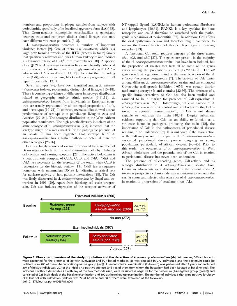
Figure 1. Flow chart overview of the study population and the detection of A. actinomycetemcomitans (Aa). At baseline, 500 adolescentswere examined for the presence of Aa with cultivation and PCR-based methods. Aa was detected in 272 individuals and the bacterium could beisolated from 200 of them (Aa cultivation-positive group (red)). A second clinical examination (follow-up) was performed after 2 years and included397 of the 500 individuals, 207 of the initially Aa-positive subjects and 148 of them from whom the bacterium had been isolated at baseline (red). Theindividuals without detectable Aa with any of the two methods used, were classified as negative for the bacterium (Aa-negative group (green)) andconsisted of 228 individuals at the baseline examination and 190 at the follow-up examination. The number of individuals that were positive for Aa byPCR, but not with cultivation (yellow), was 72 at baseline and 58 of them were examined at the follow-up.doi:10.1371/journal.pone.0065781.g001
Cdt in Aa
PLOS ONE | www.plosone.org 2 June 2013 | Volume 8 | Issue 6 | e65781
Materials and Methods
Subject RecruitmentAn adolescent West African population, described in details
previously, was examined [45]. Briefly, a random cohort of 500
school children (mean age 13.2 years; SD 61.5) was included in
the cross-sectional study, which was performed in Accra, Ghana,
in 2009 [45]. In the follow-up study, performed two years later,
397 (79.4%) of these individuals showed up for a periodontal re-
examination (Fig. 1). The drop-out individuals consisted of
children in families that moved to another area within the
follow-up period or school children who dropped out from school.
The school system had no information concerning the individuals
that had left school.
Ethical clearance for the study was obtained from the Noguchi
Memorial Institute for Medical Research, University of Ghana
(IRB 000 1276), and from the local Ethical committee of Umea
University, Sweden (Dnr 2010-188-31M). Signed consents were
received from the parents or the guardians of the children before
they entered the study.
Clinical ExaminationAll participants enrolled were given a full-mouth periodontal
examination by the same, certified periodontist with identical
procedures used at the baseline and at the follow-up examination
[45]. Attachment loss (AL) was measured at the buccal aspect of
the mesial and distal surfaces of all fully erupted permanent teeth,
which gave a potential maximum of 56 sites per individual. AL
was defined as the distance from the cemento-enamel junction
(CEJ) to the bottom of the periodontal pocket or crevice and was
calculated as the difference between two measurements (probing
pocket depth and the distance from gingival margin to CEJ).
Differences between baseline and follow-up were calculated at site
level. The disease status was established using the cut-off point of
AL $3 mm in one or more sites in the dentition. Individuals were
defined as having progressive disease if they showed $ one site
that had a progression of AL $3 mm based on data collected from
the baseline and the follow-up examinations. None of the
participants had received any periodontal treatment during the
two-year follow-up period. Very few individuals had visited a
dentist, except for treatment of pain related to the teeth.
Traditional periodontal treatment strategies are not possible to
introduce today in developing countries due to lack of dentists and
resources for funding. Until affordable alternative strategies are
available any form of periodontal treatment would be difficult to
provide. This topic is a future challenge for the present project.
Table 1. Descriptive and demographic characteristics of theexamined individuals at baseline and at the two-year follow-up examination.
Study populationBaseline(n = 500)
Follow-up(n = 397)
Demographics
Age (years) mean SD mean SD
13.2 1.53 15.0 1.39
Gender n % n %
Male 232 46.4 176 44.3
Female 268 53.6 221 55.7
SD; standard deviation.doi:10.1371/journal.pone.0065781.t001
Table 2. Descriptive and clinical characteristics of theexamined individuals (all), the study population (Aa-cultivation positive), and the reference group (Aa-negative) atbaseline (BL), and at the two-year follow-up (FU) examination.
Examined individuals
Clinical BL n = 500 FU n = 397
n % n %
N individuals (%) with sites AL $3 mm 107 21.4 156 39.2
mean SD mean SD
N of teeth (mean) with AL $3 mm 0.52 1.37 1.92 3.34
Aa-cultivation-positive
BL n = 200 FU n = 148
n % n %
N individuals (%) with sites AL $3 mm 54 27.0 82 54.4
mean SD mean SD
N of teeth (mean) with AL $3 mm 0.67 1.44 3.01 3.90
Reference group (Aa-negative)
BL n = 228 FU n = 190
n % n %
N individuals (%) with sites AL $3 mm 33 14.4 37 19.4
mean SD mean SD
N of teeth (mean) with AL $3 mm 0.34 1.28 0.89 2.63
AL; attachment loss; BL, at baseline; FU, at two-year follow-up.Aa; Aggregatibacter actinomycetemcomitans.SD; standard deviation.doi:10.1371/journal.pone.0065781.t002
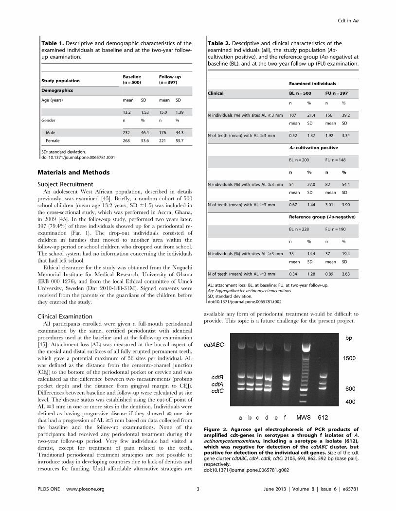
Figure 2. Agarose gel electrophoresis of PCR products ofamplified cdt-genes in serotypes a through f isolates of A.actinomycetemcomitans, including a serotype a isolate (612),which was negative for detection of the cdtABC cluster, butpositive for detection of the individual cdt genes. Size of the cdtgene cluster cdtABC, cdtA, cdtB, cdtC: 2105, 693, 862, 592 bp (base pair),respectively.doi:10.1371/journal.pone.0065781.g002
Cdt in Aa
PLOS ONE | www.plosone.org 3 June 2013 | Volume 8 | Issue 6 | e65781

Bacterial Isolates and Growth ConditionsSubgingival plaque samples from the 500 adolescents were
collected for microbiological analysis, transported, and analyzed as
described earlier [45]. In total, 792 A. actinomycetemcomitans isolates
(1–7 per subject) were collected from 200 (40%) of the 500 subjects
entered into the study at baseline. The leukotoxin promoter types
(JP2/non-JP2) of isolates was determined by PCR technique [4 ],
and the results are described in a previous study [45].
Serotyping of A. actinomycetemcomitans by PCRSuspensions of the strains were boiled for eight minutes and
centrifuged. The supernatants were used as template when the 792
isolates were serotyped by PCR. The primers used in this study
and the temperature profiles for amplification of the various genes
have previously been described, for serotyping a–e [47], and for
serotype f [17]. All isolates could be serotyped by the 6 primer
pairs (a-f). No nonserotypable was found, making it irrelevant to
consider testing for the recently described serotype g [18].
The PCR products were analysed by agarose (1.2%) gel
electrophoresis in a Tris-acetate (40 mM; pH 8.3) buffer contain-
ing 1 mM EDTA. The gel was stained with ethidium bromide and
photographed under ultraviolet light. Base pair sequences for each
of the forward and reversed primers used and gel product size is
presented in the supporting information (Table S1).
Figure 3. Histogram from flow cytometric analyses of leukocytes (HL-60 cells) exposed for 24 h to different extracts from variousisolates of A. actinomycetemcomitans. The DNA-content of each exposed HL-60 cell were determined with PI-staining and indicate cell cycle phaseof the analysed cell. The upper left panel (control) was exposed for 0.1% isoton NaCl, the upper right panel for 0.1% NaCl extract from an isolate withnon-complete cdt-genome, the lower left panel for 0.1% NaCl extract from an isolate with intact cdt-genome and the lower right panel exposed to0.1% NaCl extract from a cdt knockout mutant strain (D7SSDcdtABC).doi:10.1371/journal.pone.0065781.g003
Figure 4. Agarose gel electrophoresis of PCR productsamplified from serotypes a through f strains of A. actinomyce-temcomitans isolated from a young Ghanaian population. Size ofthe serotype–associated gel fragments (a through f, bp (base pair): 428,298, 559, 690, 211, 231.doi:10.1371/journal.pone.0065781.g004
Cdt in Aa
PLOS ONE | www.plosone.org 4 June 2013 | Volume 8 | Issue 6 | e65781
6

Cdt Genotyping of A. actinomycetemcomitans by PCRFrom the 792 serotyped and leukotoxin promoter-typed isolates,
249 isolates were selected for cdt-genotyping by PCR. One isolate
from each of the 200 subjects were used, but when more than one
serotype and/or leukotoxin promoter type (JP2 or non-JP2) were
identified in the same subject, these additional isolates were
included and summed up to 249. For the detection of the three
Cdt genes (cdtABC), two PCR-based methods were used [36]. One
of the methods was designed for detection of all three genes
(cdtABC) and revealed a 2105 base pair (bp) product. When
discrepancy between detection of cdtABC and Cdt-activity
occurred, the individual cdt genes (A, B and C, respectively) were
screened for by the second method [34]. The PCR products are
documented as described for serotyping. Primer sequences, gene
position, and gene fragment size for the cdt gene analyses, are
shown in the supporting information (Table S1).
Cdt-activity Test of A. actinomycetemcomitans in CellCultures Examined by Flow Cytometry
The 249 selected isolates were analyzed for Cdt-activity in a cell
culture assay [14]. HL-60 cells (human carcinoma leukocyte cell
line) were cultured in RPMI-1640 with 10% fetal bovine serum
(Sigma-Aldrich). A suspension of OD600 nm 2.0 was centrifuged (10
0006g, 10 min), and the supernatant added to the cultured cells.
The HL-60 cells (1 ml 56105 cells/ml) were transferred to each
well in a 24-well cell culture plate (Nunc) and mixed with 1 ml of
each of the bacterial supernatants. After 24 h of incubation the
cells were transferred to a 2 ml Eppendorf tube and washed with
PBS by centrifugation (5006g, 5 min). The cell pellet was solved
in 300 ml PBS and 900 ml ice-cold 99% ethanol and fixed for 1 h
at 4uC. Cells were washed with PBS by centrifugation and treated
with RNase (100 ml, 100 mg/ml, Sigma-Aldrich) for 15 min at
37uC. After the incubation, 400 ml of propidium iodide (Molecular
Probes, Eugene, OR, USA) in 3.8 mM sodium citrate in PBS was
added and further incubated in darkness for 1–3 h at 4uC. Cdt-
activity was determined by the ability of the bacterial supernatants
to inhibit proliferation and causing the typical accumulation of the
target cells in the G2/M-phase examined and the increased cell
size (FSC) by cell cycle analyses with flow cytometry (FACS
Calibur, Becton Dikinson; Franklin Lakes, NJ, USA). Bacterial
isolates that resulted in $50% of the target cell population in the
G2/M-phase after 24 h incubation were classified as positive for
Cdt-activity.
Statistical AnalysisData analyses were performed using SPSS 19.0 (SPSS Inc.,
Chicago, IL, USA) and STATA 8.0 (StataCorp LP., College
Station, Texas, USA). In the statistical analyses, the primary
outcome was progression of AL $3 mm in one or more sites at
subject level, based on the collection of data performed at baseline
(in November 2009) and at the follow-up (in November 2011).
Descriptive statistics were performed using mean and standard
deviation for the number of teeth with AL $3 mm per individual
and group differences assessed using a non-parametric test (Mann-
Whitney U test). The Mantel-Haenszel test was used for
comparison of the distribution between groups of individuals
harboring sites with AL $3 mm. The estimated risk associated
with progression of AL $3 mm during a two-year follow-up
period according to the carrier status of A. actinomycetemcomitans,
characteristics as serotype and Cdt-activity (negative or positive),
was evaluated by calculations of odds ratios (OR). A value of
p,0.05 was considered statistically significant. The calculation of
OR was repeated after exclusion of JP2-positive individuals, who
were defined as carriers of the JP2-genotype based on positive
plaque samples by the cultivation technique and/or by the PCR.
Results
A. actinomycetemcomitans was isolated from 200 (40%) of the
examined individuals at baseline. From these subjects, a collection
of 249 isolates was selected, one isolate from each of the 200
subjects and additional isolates included from subjects where more
than one serotype or more than one leukotoxin promoter type
could be detected in the same sample. The highly leukotoxic JP2
genotype of A. actinomycetemcomitans was found in isolates from five
(2.5%) of the individuals. All five of these individuals carried also
the non-JP2 genotypes.
Study Population and Subject RecruitmentDemographic characteristics from the baseline and the follow-
up examination of the studied individuals are shown in Table 1.
From the initially 500 examined individuals at baseline, 397 of
them (79.4%) could be identified and were available for a two-year
clinical follow-up examination (Fig. 1). Descriptive data for the
number of individuals with affected sites with AL $3 mm and the
number of affected teeth (mean) with AL $3 mm for the
examined individuals, the selected study population of A.
actinomycetemcomitans cultivation-positive individuals, and the refer-
ence group of individuals tested negative for the presence of this
bacterium, are shown in Table 2. The mean age of the A.
actinomycetemcomitans-positive individuals at baseline (n = 200) and at
follow-up (n = 148) was 13.4 (SD; 61.48) and 15.2 (SD; 61.38),
respectively, and for the A. actinomycetemcomitans-negative referents
at baseline (n = 228) and at follow up (n = 190) the corresponding
ages were 12.9 (SD; 61.43) and 14.7 (SD; 61.28), respectively.
The odds ratios (OR) for having one or more sites with AL
$3 mm was higher in the A. actinomycetemcomitans-positive group in
relation to the A. actinomycetemcomitans-negative reference group at
baseline and at follow up (OR = 2.197; 95% CI [1.355–3.561]
p = 0.001 and OR = 5.138; 95% CI [3.167–8.334] p,0.001)
(Mantel-Haenszel test) (Table 2). Also the number of affected
teeth (mean) with AL $3 mm was significantly higher among the
A. actinomycetemcomitans-positive individuals than among the A.
actinomycetemcomitans-negative reference group at both baseline
(p#0.001) and at the follow-up (p#0.001) (Mann-Whitney U test)
(Table 2). None of the examined individuals was cigarette smokers
or had diabetes.
Figure 5. Distribution of isolates with Cdt-activity among thevarious serotypes of A. actinomycetemcomitans.doi:10.1371/journal.pone.0065781.g005
Cdt in Aa
PLOS ONE | www.plosone.org 5 June 2013 | Volume 8 | Issue 6 | e65781
SerotypingSerotyping of the 249 A. actinomycetemcomitans isolates showed
presence of six different serotypes (a–f) (Fig. 2). Serotype c was the
most frequently found and was detected in 104 (42.0%) of the
isolates, while the less frequently found serotype e could be
detected in only 8 (3.2%) of the isolates. The frequency of serotype
a, b, d, and f in the examined isolates was 59 (23.7%), 47 (18.9%),
12 (4.8%), and 19 (7.6%), respectively. More than one serotype
could be detected in samples from 45 (22.5%) of the 200 subjects.
Only three individuals (1.5%) were poly-infected with three or
more serotypes.
cdt-genotype and Cdt-activityThe activity of Cdt was examined in a cell culture-based assay
by its ability to induce an accumulation of enlarged cells in the
G2/M phase of growth (Fig. 3). Cdt-activity was detected in 196
(79%) of the 249 examined isolates. These 196 isolates with Cdt-
activity also harboured all three cdt-genes (A, B and C) when
examined by PCR and afterwards visualized in an agarose gel
(Fig. 4). However, in one isolate the cdt-genes were found only by
the method used for detection of the individual three cdt-genes. It
may be possible that the method for amplifying the cdt-genes as a
complete 2100 base pair product was not optimal for this isolate
or, e.g. that a single base mutation had occurred resulting in
unsuccessful annealing of the primers (amplification of the genes).
The distribution of isolates with Cdt-activity and presence of all
three cdt-encoding genes varied among the different serotypes of A.
actinomycetemcomitans. All serotype b (n = 47), d (n = 12), and f
(n = 19) isolates were positive for Cdt-activity and the cdt-genes,
while all serotype e isolates (n = 8) lacked Cdt-activity and intact
cdt-encoding genes (Fig. 5). A total of 35 (33.7%) of the serotype c
isolates were tested negative for Cdt-activity, and for serotype a the
number of negative isolates was 8 (13.6%). Taken together, these
results showed a significant serotype-dependent pattern of cdt-
genotypes in A. actinomycetemcomitans isolated from this population
(Fig. 5). Isolates with the presence of all three cdt-encoding genes
and a substantial Cdt-activity were considered as Cdt-positive,
while the other isolates were counted as Cdt-negative.
Carrier Status of A. actinomycetemcomitans andProgression of AL
The progression of AL $3 mm at one or more sites over a two-
year follow-up period was examined in relation to the carrier
status of A. actinomycetemcomitans. There was a significantly increased
progression of AL in the A. actinomycetemcomitans-positive individuals
(OR = 5.126, 95% CI [2.994–8.779] p,0.001) in relation to that
of the A. actinomycetemcomitans-negative reference individuals
(Table 3). Exclusions of individuals with presence of the JP2-
genotype, detected by cultivation and/or PCR, resulted in for the
A. actinomycetemcomitans positive individuals (n = 130) an OR of
4.323 (95% CI [2.482–7.530], p#0.001).
Serotype and Progression of ALThe most prevalent serotypes of A. actinomycetemcomitans isolated
from the present study population were a, b, and c. In order to
examine for the importance of the serotype of the bacterium for
the progression of AL $3 mm, the individuals tested positive for
one of these three serotypes were selected from the study
population. In each group of individuals positive for any of the
three selected serotypes, the OR for progression of AL compared
to that of the A. actinomycetemcomitans-negative referents was high
(Table 3). The highest OR for progression was associated with
presence by the b serotype (OR = 7.685; 95% CI [2.835–20.830],
p,0.001), closely followed by the a serotype (OR = 6.917; 95% CI
[2.869–16.673], p,0.001), while the OR for progression was
lower, however still significant, in the individuals positive for the c
serotype (OR = 3.365; 95% CI [1.659–6.826], p,0.001). Due to
the low number of individuals carrying A. actinomycetemcomitans of
serotypes d, e, and f, these were not included in the analyses. The
corresponding results for serotypes b and c after exclusion for the
presence of the JP2-genotype strains were calculated. We also had
to consider some individuals positive for serotype c as these were
co-infected with serotype b JP2-genotype strains. For the
individuals with the presence of the b serotype A. actinomycetemco-
mitans (n = 16), the OR was 6.917 (95% CI [2.374–20.152],
p#0.001), and for the individuals with the c serotype (n = 50), the
OR was 2.964 (95% CI [1.413–6.219], p = 0.004). None of the
individuals with the presence of the a serotype of the bacterium
were in addition co-infected with the JP2-genotype.
cdt-genotype and Progression of ALPresence of Cdt-negative A. actinomycetemcomitans, as well as the
presence of Cdt-positive bacteria showed a significant association
with an increased progression of AL compared with that of the A.
actinomycetemcomitans-negative referents (Table 3). The results for
the A. actinomycetemcomitans-positive individuals (n = 148), for the
individuals (n = 123) with Cdt-positive bacteria, and for the
individuals (n = 25) with Cdt-negative bacteria were OR = 5.126
(95% CI [2.994–8.779], p#0.001); OR = 5.237 (95% CI [3.000–
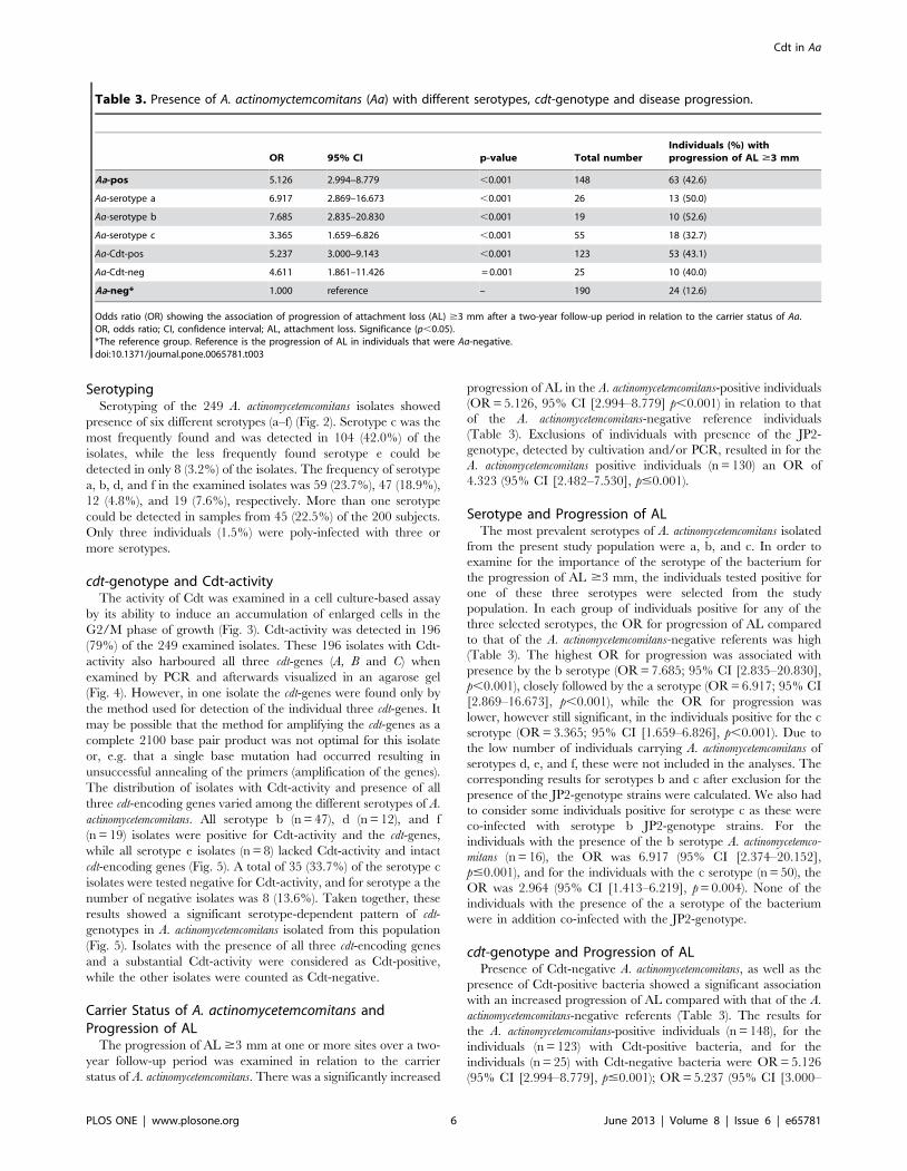
Table 3. Presence of A. actinomyctemcomitans (Aa) with different serotypes, cdt-genotype and disease progression.
OR 95% CI p-value Total numberIndividuals (%) withprogression of AL $3 mm
Aa-pos 5.126 2.994–8.779 ,0.001 148 63 (42.6)
Aa-serotype a 6.917 2.869–16.673 ,0.001 26 13 (50.0)
Aa-serotype b 7.685 2.835–20.830 ,0.001 19 10 (52.6)
Aa-serotype c 3.365 1.659–6.826 ,0.001 55 18 (32.7)
Aa-Cdt-pos 5.237 3.000–9.143 ,0.001 123 53 (43.1)
Aa-Cdt-neg 4.611 1.861–11.426 = 0.001 25 10 (40.0)
Aa-neg* 1.000 reference – 190 24 (12.6)
Odds ratio (OR) showing the association of progression of attachment loss (AL) $3 mm after a two-year follow-up period in relation to the carrier status of Aa.OR, odds ratio; CI, confidence interval; AL, attachment loss. Significance (p,0.05).*The reference group. Reference is the progression of AL in individuals that were Aa-negative.doi:10.1371/journal.pone.0065781.t003
Cdt in Aa
PLOS ONE | www.plosone.org 6 June 2013 | Volume 8 | Issue 6 | e65781
9.143], p#0.001), and OR = 4.611 (95% CI [1.861–11.426],
p = 0.001), respectively. The OR after exclusion of individuals with
presence of JP2 genotype bacteria was OR = 4.402 (95% CI
[2.472–7.837], p#0.001) for individuals (n = 108) with Cdt-
positive bacteria and OR = 3.952 (95% CI [1.501–10.409],
p = 0.005) for individuals (n = 22) with Cdt-negative bacteria.
Discussion
In the present study, we have examined a collection of A.
actinomycetemcomitans isolated from individuals included as a part of
a prospective cohort study carried out in Accra, Ghana [45]. The
proportion of individuals (79.4%) identified for the follow-up
examination after two years was considerably high in the
population chosen for this study, represented at baseline by a
group of 500 medically healthy adolescents. This is the first
microbiological study of A. actinomycetemcomitans from West-Africa,
and neither cdt-genotype and activity nor serotypes have been
reported on before based on such a collection of West-African A.
actinomycetemcomitans strains.
Six different serotypes (a-e) were detected in the collection of
249 isolates from the 200 Ghanaian individuals. In line with earlier
studies, examining isolates from Asian or American populations,
the serotype c was the most prevalent (42.0%), followed by
serotype a (23.7%), and serotype b (18.9%) [22–24]. The other
three detected serotypes d, e and f were present in relatively low
proportions being 4.8%, 7.6%, and 3.2%, respectively. This is also
in accordance with previous studies [16,21,23].
The distribution of cdt-genotypes among the analysed isolates
showed a serotype-dependent pattern that was unique for the
present collection in relation to results from previous studies [19–
24]. The present collection of A. actinomycetemcomitans is from a
representative adolescent population consisting of otherwise
healthy subjects, all living in the same geographic area located
in West-Africa. Most comparable studies have dealt with isolates
from patient cohorts with a history of periodontal disease [16,19–
23].
Cdt-activity was detected in 79% in the collection of the 249
selected isolates from 200 individuals. Previous studies, aimed at
examining the expression of Cdt, has mainly focused on the
presence of cdt-genes in bacteria isolated from periodontally
diseased individuals [22,23,36,40]. The results from these previous
studies showed a substantial variation in the proportion of isolates
that contained all three cdt-encoding genes. This may be explained
by the different geographic origins and periodontal status of the
examined individuals, but can also involve differences in the
methodology for the detection of the cdt-genes. Two studies based
on PCR-data from plaque samples have indicated a relatively low
proportion of detectable cdt-genes in the A. actinomycetemcomitans-
positive sites [21,35]. However, variations in the methodology may
explain the relatively low detection frequency of cdt-genes in these
studies. Further, the cdt-genes are located in the variable region of
the A. actinomycetemcomitans pangenome, which can explain the
variations in the detection frequency of these genes in A.
actinomycetemcomitans isolated from individuals of various origin
[7]. Results from the present study showed that all isolates with
Cdt-activity contained the three cdt-genes, while all the isolates
without Cdt-activity lacked some of the cdt-encoding genes.
In the present study, we applied a cell culture-based method for
determination of Cdt-activity that quantified cell cycle phase of the
Cdt-exposed cells [14]. This method is more specific than the
other commonly used method that demonstrates a general
inhibition of the cell proliferation or effects on cell morphology
and viability [22,36,38]. Examination of the accumulation of cells
in the G2/M phase of the cell cycle is a specific marker for Cdt-
intoxication and therefore a relevant marker for detection of Cdt-
activity [13]. The complete correlation between the presence of an
intact cdt-genome and a substantial activity of the toxin might
depend on the specific methodology selected for the Cdt-activity
analyses.
Despite substantial evidence supporting that Cdt has an ability
to function as a true virulence factor in many pathogens producing
the toxin [27,42], the importance of Cdt in the pathogenesis of
periodontal disease remains to be evaluated [9]. In our study, the
presence of A. actinomycetemcomitans was significantly associated
(p#0.001) with progression of AL over a two-year observation
period. This is in line with previous longitudinal studies examining
the relation between the presence of A. actinomycetemcomitans and
progression of AL in adolescent individuals [43,48,49]. In the
present study, both individuals colonized with Cdt-positive or Cdt-
negative bacteria were significantly more often included among
the subjects that showed progression of AL ($ 3 mm at one or
more periodontal site) than the A. actinomycetemcomitans-negative
individuals. As particularly the JP2 genotype has been strongly
associated with progression of AL, it was important for us to
eliminate this genotype from the analyses and afterwards repeat
the data analyses. Therefore, exclusion of the JP2 genotype-
positive individuals from the study population was done, but this
did not change the results substantially. Thus, data suggests that
the expression of Cdt might be of low importance for the
pathogenesis of periodontitis, although further studies are needed
to clarify whether this toxin can act and how to as a true virulence
factor.
In the present cohort of Ghanaian adolescents, the progression
of AL was examined in relation to the presence of serotype a, b, or
c of A. actinomycetemcomitans. Using the progression of AL in the A.
actinomycetemcomitans-negative individuals as the reference group,
the presence of each of these three serotypes showed a significant
association with progression of AL. Exclusion of the JP2-genotype
positive individuals did not substantially change the estimates.
Finding that the serotype b strains have the strongest relation to
disease progression (OR = 7.685), this is in line with previous
studies indicating an increased virulence in A. actinomycetemcomitans
from serotype b [25,26]. Although, the presence of A. actinomyce-
temcomitans as an important etiological factor was studied in this
cohort of Ghanaian adolescents, an effect of other periodontal
pathogens present concomitantly in the subgingival biofilm cannot
be excluded.
In conclusion, the distribution of serotypes, cdt-genotypes and
Cdt-activity of A. actinomycetemcomitans isolated from Ghanaian
adolescents, showed a pattern that was comparable with results
found in other populations. Progression of AL is mainly associated
with the presence of A. actinomycetemcomitans and appears weakly
associated with the cdt-genotype. All serotypes of A. actinomycetem-
comitans studied were related to the progression of AL, serotype b
showing the strongest association with disease progression.
Supporting Information
Table S1 Primer sequences, gene position, and genefragment size for the serotype and cdt-genotype PCRanalyses.
(DOC)
Acknowledgments
We thank Dr Gunnar Dahlen at Gotheburg University, Sweden, for
valuable support during the preparation of this manuscript.
Cdt in Aa
PLOS ONE | www.plosone.org 7 June 2013 | Volume 8 | Issue 6 | e65781
Author Contributions
Conceived and designed the experiments: DH CHA RC FK AJ.
Performed the experiments: RC GA CHA AJ. Analyzed the data: CHA
DH. Contributed reagents/materials/analysis tools: RC AJ. Wrote the
paper: AJ CHA FK GA RC DH. Logistics for the clinical examinations:
FK.
References
1. Pihlstrom BC, Michalowicz BS, Johnson DW (2005) Periodontal diseases.Lancet 366: 1809–1820.
2. Darveau RP (2010) Periodontitis: a polymicrobial disruption of host homeostasis.Nature Rev Microbiol 8: 481–490.
3. Nishihara T, Koseki T (2004) Microbial aetiology of periodontitis. Periodontol
2000 36: 14–26.
4. Paster BJ, Olsen I, Aas JA, Dewhirst FE (2006) The breadth of bacterial diversity
in the human periodontal pocket and other oral sites. Periodontol 2000 42: 80–87.
5. Zambon JJ (1985) Actinobacillus actinomycetemcomitans in human periodontal
disease. J Clin Periodontol 12: 1–20.
6. Kilian M, Frandsen EV, Haubek D, Poulsen K (2006) The etiology ofperiodontal disease revisited by population genetic analysis. Periodontol 2000
42: 158–179.
7. Kittichotirat W, Bumgarner RE, Asikainen S, Chen C (2011) Identification of
the Pangenome and Its Components in 14 Distinct Aggregatibacter actinomycetemco-
mitans Strains by Comparative Genomic Analysis. PLoS One 6: e22420.
8. Pinheiro ET, Kawamoto D, Ota-Tsuzuki C, Almeida RR, Nunes AC, et al.
(2011) Analysis of genotypic variation in genes associated with virulence in
Aggregatibacter actinomycetemcomitans clinical isolates. J Periodontal Res 46: 310–317.
9. Henderson B, Ward JM, Ready D (2010) Aggregatibacter (Actinobacillus)
actinomycetemcomitans: a triple A* periopathogen? Periodontol 2000 54: 78–105.
10. Johansson A (2011) Aggregatibacter actinomycetemcomitans Leukotoxin: A Powerful
Tool with Capacity to Cause Imbalance in the Host Inflammatory Response.
Toxins (Basel) 3: 242–259.
11. Haubek D (2010) The highly leukotoxic JP2 clone of Aggregatibacter actinomyce-
temcomitans: evolutionary aspects, epidemiology and etiological role in aggressive
periodontitis. APMIS 130: 1–53.
12. Haubek D, Poulsen K, Kilian M. (2007) Microevolution and patterns ofdissemination of the JP2 clone of Aggregatibacter (Actinobacillus) actinomycetemcomitans.
Infect Immun 75: 3080–3088.
13. Lara-Tejero M, Galan JE (2000) A bacterial toxin that controls cell cycle
progression as a deoxyribonuclease I-like protein. Science 13: 354–357.
14. Belibasakis GN, Mattsson A, Wang Y, Chen C, Johansson A (2004) Cell cyclearrest of human gingival fibroblasts and periodontal ligament cells by
Actinobacillus actinomycetemcomitans: involvement of the cytolethal distending toxin.
APMIS 112: 674–685.
15. Poulsen K, Theilade E, Lally ET, Demuth DR, Kilian M (1994) Populationstructure of Actinobacillus actinomycetemcomitans: a framework for studies of disease-
associated properties. Microbiol 140: 2049–2060.
16. Haubek D, Poulsen K, Asikainen S, Kilian M (1995) Evidence for absence in
northern Europe of especially virulent clonal types of Actinobacillus actinomyce-
temcomitans. J Clin Microbiol 33: 395–401.
17. Kaplan J, Perry MB, Maclean LL, Furgang D, Wilson ME, et al. (2002)
Structural and genetic analysis of O polysaccharide from Actinobacillus
actinomycetemcomitans serotype f. Infect Immun 69: 5375–5384.
18. Takada K, Saito M, Tsuzukibashi O, Kawashima Y, Ishida S, et al. (2010)
Characterization of a new serotype g isolate of Aggregatibacter actinomycetemcomitans.
Mol Oral Microbiol 25: 200–206.
19. Saarela M, Asikainen S, Alaluusua S, Pyhala L, Lai CH, et al. (1992) Frequencyand stability of mono- or poly-infection by Actinobacillus actinomycetemcomitans
serotypes a, b, c, d or e. Oral Microbiol Immunol 7: 277–279.
20. Lakio L, Kuula H, Dogan B, Asikainen S (2002) Actinobacillus actinomycetemcomitans
proportion of subgingival bacterial flora in relation to its clonal type. Eur J OralSci 110: 212–217.
21. Jentsch H, Cachovan G, Guentsch A, Eickholz P, Pfister W, et al. (2012)
Characterization of Aggregatibacter actinomycetemcomitans strains in periodontitispatients in Germany. Clin Oral Invest 16: 1589–1597.
22. Kawamoto D, Ando ES, Longo PL, Nunes AC, Wikstrom M, et al. (2009)Genetic diversity and toxic activity of Aggregatibacter actinomycetemcomitans isolates.
Oral Microbiol Immunol 24: 493–501.
23. Kim TS, Frank P, Eickholz P, Eick S, Kim CK (2009) Serotypes of Aggregatibacter
actinomycetemcomitans in patients with different ethnic backgrounds. J Periodontol
80: 2020–2027.
24. Chen C, Wang T, Chen W (2010) Occurrence of Aggregatibacter actinomycetemco-
mitans serotypes in subgingival plaque from United States subjects. Mol OralMicrobiol 25: 207–214.
25. Zambon JJ, Slots J, Genco RJ (1983) Serology of oral Actinobacillus
actinomycetemcomitans and serotype distribution in human periodontal disease.Infect Immun 41: 19–27.
26. Yang HW, Asikainen S, Dogan B, Suda R, Lai CH (2004) Relationship ofActinobacillus actinomycetemcomitans serotype b to aggressive periodontitis: frequency
in pure cultured isolates. J Periodontol 75: 592–599.
27. Jinadasa RN, Bloom SE, Weiss RS, Duhamel GE (2011) Cytolethal distending
toxin: a conserved bacterial genotoxin that blocks cell cycle progression, leadingto apoptosis of a broad range of mammalian cell lineages. Microbiol 157: 1851–
1875.28. Elwell CA, Dreyfus LA (2000) DNase I homologous residues in CdtB are critical
for cytolethal distending toxin-mediated cell cycle arrest. Mol Microbiol 37:
952–963.29. Sugai M, Kawamoto T, Peres SY, Ueno Y, Komatsuzawa H, et al. (1998) The
cell cycle-specific growth-inhibitory factor produced by Actinobacillus actinomyce-
temcomitans is a cytolethal distending toxin. Infect Immun 66: 5008–5019.
30. Belibasakis GN, Johansson A, Wang Y, Chen C, Kalfas S, et al. (2005) The
cytolethal distending toxin of Actinobacillus actinomycetemcomitans induces RANKLexpression by human gingival fibroblasts and periodontal ligament cells. Infect
Immun 73: 342–351.31. Belibasakis GN, Brage M, Lagergard T, Johansson A (2008) The cytolethal
distending toxin up-regulates RANKL expression in Jurkat T-cells. APMIS 116:499–506.
32. Schenkein HA (2006) Host responses in maintaining periodontal health and
determining periodontal disease. Periodontol 2000 40: 77–93.33. Damek-Poprawa M, Haris M, Volgina A, Korostoff J, DiRienzo JM (2011)
Cytolethal distending toxin damages the oral epithelium of gingival explants.J Dent Res 90: 874–879.
34. Ahmed HJ, Svensson LA, Cope LD, Latimer JL, Hansen EJ, et al. (2001)
Prevalence of cdtABC genes encoding cytolethal distending toxin amongHaemophilus ducreyi and Actinobacillus actinomycetemcomitans strains. J Med Microbiol
50: 860–864.35. Tan KS, Song KP, Ong G (2002) Cytolethal distending toxin of Actinobacillus
actinomycetemcomitans. Occurrence and association with periodontal disease.
J Periodontal Res 37: 268–272.36. Fabris AS, DiRienzo JM, Wıkstrom M, Mayer MP (2002) Detection of
cytolethal distending toxin activity and cdt genes in Actinobacillus actinomycetemco-
mitans isolates from geographically diverse populations. Oral Microbiol Immunol
17: 231–238.37. Leung WK, Ngai VK, Yau JY, Cheung BP, Tsang PW, et al. (2005)
Characterization of Actinobacillus actinomycetemcomitans isolated from young
Chinese aggressive periodontitis patients. J Periodontal Res 40: 258–268.38. Yamano R, Ohara M, Nishikubo S, Fujiwara T, Kawamoto T, et al. (2003)
Prevalence of cytolethal distending toxin production in periodontopathogenicbacteria. J Clin Microbiol 41: 1391–1398.
39. Johansson A, Buhlin K, Koski R, Gustafsson A (2005) The immunoreactivity of
systemic antibodies to Actinobacillus actinomycetemcomitans and Porphyromonas
gingivalis in adult periodontitis. Eur J Oral Sci 113: 197–202.
40. Ando ES, De-Gennaro LA, Faveri M, Feres M, DiRienzo JM, et al. (2010)Immune response to cytolethal distending toxin of Aggregatibacter actinomycetemco-
mitans in periodontitis patients. J Periodontal Res 45: 471–480.41. Brage M, Holmlund A, Johansson A (2011) Humoral immune response to
Aggregatibacter actinomycetemcomitans leukotoxin. J Periodontal Res 46: 170–175.
42. Smith JL, Bayles DO (2006) The contribution of cytolethal distending toxin tobacterial pathogenesis. Crit Rev Microbiol 32: 227–248.
43. Haubek D, Ennibi OK, Poulsen K, Vaeth M, Poulsen S, et al. (2008) Risk ofaggressive periodontitis in adolescent carriers of the JP2 clone of Aggregatibacter
(Actinobacillus) actinomycetemcomitans in Morocco: a prospective longitudinal cohort
study. Lancet 371: 237–242.44. Elamin AM, Skaug N, Ali RW, Bakken V, Albandar JM (2010) Ethnic
disparities in the prevalence of periodontitis among high school students inSudan. J Periodontol 81: 891–896.
45. Hoglund Aberg C, Kwamin F, Claesson R, Johansson A, Haubek D (2012)Presence of JP2 and non-JP2 genotypes of Aggregatibacter actinomycetemcomitans and
periodontal attachment loss in adolescents in Ghana. J Periodontol 83: 1520–
1528.46. Poulsen K, Ennibi O-K, Haubek D (2003) Improved PCR for detection of the
highly leukotoxic JP2 clone of Actinobacillus actinomycetemcomitans in subgingivalplaque samples. J Clin Microbiol 41: 4829–4832.
47. Suzuki N, Nakano Y, Yoshida Y, Ikeda D, Koga T (2001) Identification of
Actinobacillus actinomycetemcomitans serotypes by multiplex PCR. J Clin Microbiol39: 2002–2005.
48. Van der Velden U, Abbas F, Armand S, Loos BG, Timmerman MF, et al.(2006) Java project on periodontal diseases. The natural development of
periodontitis: risk factors, risk predictors and risk determinants. J ClinPeriodontol 33: 540–548.
49. Fine DH, Markowitz K, Furgang D, Fairlie K, Ferrandiz J, et al. (2007)
Aggregatibacter actinomycetemcomitans and its relationship to initiation of localizedaggressive periodontitis: Longitudinal cohort study of initially healthy adoles-
cents. J Clin Microbiol 45: 3859–3869.
Cdt in Aa
PLOS ONE | www.plosone.org 8 June 2013 | Volume 8 | Issue 6 | e65781