CT Colonography with Computer-aided Detection: Automated Recognition of Ileocecal Valve to Reduce...
22
Note: This copy is for your personal non-commercial use only. To order presentation-ready copies for distribution to your colleagues or clients, contact us at www.rsna.org/rsnarights. GASTROINTESTINAL IMAGING 1885 CT Colonography with Computer- aided Detection: Recognizing the Causes of False-Positive Reader Results 1 Computed tomography (CT) colonography is a screening modal- ity used to detect colonic polyps before they progress to colorectal cancer. Computer-aided detection (CAD) is designed to decrease errors of detection by finding and displaying polyp candidates for evaluation by the reader. CT colonography CAD false-positive results are common and have numerous causes. The relative fre- quency of CAD false-positive results and their effect on reader performance on the basis of a 19-reader, 100-case trial shows that the vast majority of CAD false-positive results were dismissed by readers. Many CAD false-positive results are easily disregarded, including those that result from coarse mucosa, reconstruction, peristalsis, motion, streak artifacts, diverticulum, rectal tubes, and lipomas. CAD false-positive results caused by haustral folds, ex- tracolonic candidates, diminutive lesions (<6 mm), anal papillae, internal hemorrhoids, varices, extrinsic compression, and flexural pseudotumors are almost always recognized and disregarded. The ileocecal valve and tagged stool are common sources of CAD false- positive results associated with reader false-positive results. Non- dismissable CAD soft-tissue polyp candidates larger than 6 mm are another common cause of reader false-positive results that may lead to further evaluation with follow-up CT colonography or optical colonoscopy. Strategies for correctly evaluating CAD polyp candi- dates are important to avoid pitfalls from common sources of CAD false-positive results. © RSNA, 2014 • radiographics.rsna.org Igor Trilisky, MD Abraham H. Dachman, MD Kristen Wroblewski, MS Michael W. Vannier, MD John M. Horne, MD Abbreviations: ICV = ileocecal valve, 3D = three-dimensional, 2D = two-dimensional RadioGraphics 2014; 34:1885–1905 Published online 10.1148/rg.347130053 Content Codes: 1 From the Department of Radiology, MC2026, University of Chicago Medical Center, 5841 S Maryland Ave, Chicago, IL 60637 (I.T., A.H.D., M.W.V.); Department of Health Stud- ies, University of Chicago, Chicago, Ill (K.W.); and Department of Medicine, Creighton Uni- versity, Omaha, Neb (J.M.H.). Presented as an education exhibit at the 2011 RSNA An- nual Meeting. Received August 28, 2012; revi- sion requested January 16, 2013, and received March 26, 2014; final version accepted April 2. For this journal-based SA-CME activity, the authors A.H.D. and K.W. have provided disclo- sures (see p 1903); all other authors, the editor, and the reviewers have disclosed no relevant re- lationships. Address correspondence to I.T. (e-mail: [email protected]). Funding: The research was supported by the National Cancer Institute. See discussion on this article by Yee (pp 1906–1907). After completing this journal-based SA- CME activity, participants will be able to: ■ List the common causes of CAD and reader false-positive results at CT colo- nography. ■ Describe the problem-solving tech- niques for evaluating a polyp candidate identified at CT colonography. ■ Discuss the benefits and drawbacks of use of CAD for reader performance in interpreting CT colonography studies. See www.rsna.org/education/search/RG. SA-CME LEARNING OBJECTIVES Introduction CT Colonography and Computer-aided Detection Computed tomography (CT) colonography is a screening modal- ity used to detect colonic polyps before they progress to colorectal cancer (1–3). Colorectal cancer screening can reduce mortality from colorectal cancer by 13%–18% (4). Current screening guidelines
-
Upload
independent -
Category
Documents
-
view
3 -
download
0
Transcript of CT Colonography with Computer-aided Detection: Automated Recognition of Ileocecal Valve to Reduce...

Note: This copy is for your personal non-commercial use only. To order presentation-ready copies for distribution to your colleagues or clients, contact us at www.rsna.org/rsnarights. G
AST
RO
INT
ESTIN
AL IM
AG
ING
1885
CT Colonography with Computer-aided Detection: Recognizing the Causes of False-Positive Reader Results1
Computed tomography (CT) colonography is a screening modal-ity used to detect colonic polyps before they progress to colorectal cancer. Computer-aided detection (CAD) is designed to decrease errors of detection by finding and displaying polyp candidates for evaluation by the reader. CT colonography CAD false-positive results are common and have numerous causes. The relative fre-quency of CAD false-positive results and their effect on reader performance on the basis of a 19-reader, 100-case trial shows that the vast majority of CAD false-positive results were dismissed by readers. Many CAD false-positive results are easily disregarded, including those that result from coarse mucosa, reconstruction, peristalsis, motion, streak artifacts, diverticulum, rectal tubes, and lipomas. CAD false-positive results caused by haustral folds, ex-tracolonic candidates, diminutive lesions (<6 mm), anal papillae, internal hemorrhoids, varices, extrinsic compression, and flexural pseudotumors are almost always recognized and disregarded. The ileocecal valve and tagged stool are common sources of CAD false-positive results associated with reader false-positive results. Non-dismissable CAD soft-tissue polyp candidates larger than 6 mm are another common cause of reader false-positive results that may lead to further evaluation with follow-up CT colonography or optical colonoscopy. Strategies for correctly evaluating CAD polyp candi-dates are important to avoid pitfalls from common sources of CAD false-positive results.
©RSNA, 2014 • radiographics.rsna.org
Igor Trilisky, MD Abraham H. Dachman, MD Kristen Wroblewski, MS Michael W. Vannier, MD John M. Horne, MD
Abbreviations: ICV = ileocecal valve, 3D = three-dimensional, 2D = two-dimensional
RadioGraphics 2014; 34:1885–1905
Published online 10.1148/rg.347130053
Content Codes: 1From the Department of Radiology, MC2026, University of Chicago Medical Center, 5841 S Maryland Ave, Chicago, IL 60637 (I.T., A.H.D., M.W.V.); Department of Health Stud-ies, University of Chicago, Chicago, Ill (K.W.); and Department of Medicine, Creighton Uni-versity, Omaha, Neb (J.M.H.). Presented as an education exhibit at the 2011 RSNA An-nual Meeting. Received August 28, 2012; revi-sion requested January 16, 2013, and received March 26, 2014; final version accepted April 2. For this journal-based SA-CME activity, the authors A.H.D. and K.W. have provided disclo-sures (see p 1903); all other authors, the editor, and the reviewers have disclosed no relevant re-lationships. Address correspondence to I.T. (e-mail: [email protected]).
Funding: The research was supported by the National Cancer Institute.
See discussion on this article by Yee (pp 1906–1907).
After completing this journal-based SA-CME activity, participants will be able to: ■ List the common causes of CAD and
reader false-positive results at CT colo-nography.
■ Describe the problem-solving tech-niques for evaluating a polyp candidate identified at CT colonography.
■ Discuss the benefits and drawbacks of use of CAD for reader performance in interpreting CT colonography studies.
See www.rsna.org/education/search/RG.
SA-CME LEARNING OBJECTIVES Introduction
CT Colonography and Computer-aided DetectionComputed tomography (CT) colonography is a screening modal-ity used to detect colonic polyps before they progress to colorectal cancer (1–3). Colorectal cancer screening can reduce mortality from colorectal cancer by 13%–18% (4). Current screening guidelines
1886 November-December 2014 radiographics.rsna.org
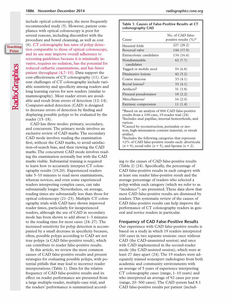
ing to the causes of CAD false-positive results (Table 2) (24). Specifically, the percentage of CAD false-positive results in each category with at least one reader false-positive result and the average percentage of readers who selected a polyp within each category (which we refer to as “incidence”) are presented. These data show that most CAD false-positive results are dismissed by readers. This systematic review of the causes of CAD false-positive results can help improve the performance of CT colonography readers in gen-eral and novice readers in particular.
Frequency of CAD False-Positive ResultsOur experience with CAD false-positive results is based on a study in which 19 readers interpreted 100 cases in two separate sessions: once without CAD (the CAD-unassisted session) and once with CAD implemented in the second-reader mode (the CAD-assisted session), which were at least 27 days apart (24). The 19 readers were ad-equately trained nonexpert radiologists from both academic and community environments with an average of 5 years of experience interpreting CT colonography cases (range, 1–10 years) and who interpreted an average of 92 cases per year (range, 20–300 cases). The CAD system had 8.1 CAD false-positive results per patient (includ-
include optical colonoscopy, the most frequently recommended study (5). However, patient com-pliance with optical colonoscopy is poor for several reasons, including discomfort with the procedure and bowel cleansing, as well as cost (6). CT colonography has rates of polyp detec-tion comparable to those of optical colonoscopy, and its use may improve overall adherence to screening guidelines because it is minimally in-vasive, requires no sedation, has the potential for reduced cathartic examinations, and has faster patient throughput (4,7–10). Data support the cost-effectiveness of CT colonography (11). Cur-rent challenges of CT colonography include vari-able sensitivity and specificity among readers and long learning curves for new readers (similar to mammography). Most reader errors are avoid-able and result from errors of detection (12–14). Computer-aided detection (CAD) is designed to decrease errors of detection by finding and displaying possible polyps to be evaluated by the reader (15–18).
CAD has three modes: primary, secondary, and concurrent. The primary mode involves an exclusive review of CAD marks. The secondary CAD mode involves reading the examination first, without the CAD marks, to avoid satisfac-tion-of-search bias, and then viewing the CAD marks. The concurrent CAD mode involves read-ing the examination normally but with the CAD marks visible. Substantial training is required to learn how to accurately interpret CT colon-ography results (19,20). Experienced readers take 5–10 minutes to read most examinations, whereas novices, and even some experienced readers interpreting complex cases, can take substantially longer. Nevertheless, on average, reading times are substantially less than those for optical colonoscopy (21–23). Multiple CT colon-ography trials with CAD have shown improved reader times, particularly for inexperienced readers, although the use of CAD in secondary mode has been shown to add about 1–3 minutes to the reading time for most cases (24–27). The increased sensitivity for polyp detection is accom-panied by a small decrease in specificity because, often, possible polyps according to CAD are not true polyps (a CAD false-positive result), which can contribute to reader false-positive results.
In this article, we review the most common causes of CAD false-positive results and pre sent strategies for evaluating possible polyps, with po-tential pitfalls that may lead to incorrect reader interpretations (Table 1). Data for the relative frequency of CAD false-positive results and its effect on reader performance are presented from a large multiple-reader, multiple-case trial, and the readers’ performance is summarized accord-
Table 1: Causes of False-Positive Results at CT colonography CAD
CauseNo. of CAD false-
positive results (%)*
Haustral folds 227 (28.2) Ileocecal valve 144 (17.9)Extracolonic candidates 134 (16.6)Nondismissible
candidates62 (7.7)
Tagged or mobile stool 55 (6.8)Diminutive lesions 42 (5.2)Coarse mucosa 33 (4.1)Rectal lesions† 33 (4.1)Artifacts‡ 31 (3.8)Flexural pseudotumor 18 (2.2)Miscellaneous§ 16 (2.0)Extrinsic compression 11 (1.4)
*Based on an analysis of 806 CAD false-positive results from a 100-case, 19-reader trial (24).†Includes anal papillae, internal hemorrhoids, and varices.‡Caused by reconstruction, peristalsis or mo-tion, high-attenuation contrast material, or streak artifact.§Includes the following categories that represent <2% of CAD false-positive results each: diverticula (n = 9), rectal tube (n = 5), and lipoma (n = 2).

RG • Volume 34 Number 7 Trilisky et al 1887
results are difficult for readers to dismiss (30,31). The number of CAD false-positive results per study has steadily decreased as algorithms for recognizing normal anatomic structures have improved, leaving polyp candidates that require reader analysis (32,33). Although the incidence of CAD false-positive results for any CAD software purchase and CT colonography data set varies, the causes are common to all software programs. Every CAD polyp candidate should be evalu-ated with a logical, systematic approach (Fig 1) (34,35). In this article, we review the causes of CAD false-positive results and give strategies for properly characterizing and dismissing them.
General Approach to Evaluating Polyp CandidatesThe specific process for interpreting a CT colon-ography examination is software and user depen-dent. The reader must be familiar with the work-station tools to accurately and efficiently interpret a study. User preference determines whether CAD is implemented in the first-, concurrent-, or sec-ond-reader mode. Usually, CT colonography soft-ware automatically segments the colon and asks the reader to verify. At this time, it is important to note whether any extracolonic gas-filled lumen is included in the segmentation (and, if possible,
ing supine and prone series) with a stand-alone performance sensitivity of 93.2% for large polyps (≥10 mm) and 91.8% for small adenomas (6–10 mm), a rate that is comparable to previous re-ports on CAD system performance (28,29).
Sensitivity of the average reader improved with use of CAD by approximately five percent-age points in each of the segment-, patient-, and polyp-level analyses, with a smaller decrease in specificity. The 806 CAD false-positive polyp candidates were reviewed and categorized ac-cording to the common causes of CAD false-positive results versus nondismissible polyp candidates by two trained residents. All 806 false-positive polyp candidates were then reviewed by a board-certified radiologist with extensive CT colonography experience. All reviewers were blinded to the readers’ performance with respect to the CAD false-positive results.
Many normal anatomic structures and le-sions that mimic polyps can be selected as a polyp candidate by CAD software and presented to the reader for further characterization. Some causes of CAD false-positive results are similar to those found without CAD, whereas others result from limitations of CAD algorithms and are often easy for readers to recognize. Previous studies reported that approximately 20% of CAD false-positive
Table 2: Rates of Reader False-Positive Results Related to CAD False-Positive Results by Cause
Cause (No.)No. of CAD FPs That
Resulted in Reader FPs (%*)No. of Reader
FPs (Incidence [%†])
Haustral folds (227) 4.0 (9) 21 (0.5)Ileocecal valve (144) 13.9 (20) 35 (1.3)Extracolonic candidates (134) 1.5 (2) 2 (0.1)Nondismissible candidates (62) 61.3 (38) 170 (14.4)Tagged or mobile stool (55) 23.6 (13) 33 (3.2)Diminutive lesions (42) 14.3 (6) 6 (0.8)Coarse mucosa (33) 0 (0) 0 (0)0iRectal lesions‡ (33) 15.2 (5) 5 (0.8)Artifacts§ (31) 0 (0) 0 (0)0iFlexural pseudotumor (18) 5.6 (1) 1 (0.3)Miscellaneous¶ (16) 0 (0) 0 (0)0iExtrinsic compression (11) 18.2 (2) 7 (3.3)Overall (806) 11.9 (96) 280 (1.8)
Note.—FPs = false-positive results.*Percentage of CAD false-positive results in a given category that resulted in at least one reader FP during the CAD-assisted session.†The average percentage of the 19 readers who selected a polyp candidate in that category during the CAD-assisted session.‡Includes anal papillae, internal hemorrhoids, and varices.§Caused by reconstruction, peristalsis or motion, high-attenuation contrast material, or streak artifact.¶Includes the following categories that represent <2% of CAD false-positive results each: diverticula (n = 9), rectal tube (n = 5), and lipoma (n = 2).

1888 November-December 2014 radiographics.rsna.org
Figure 1. Flowchart shows the general approach for evaluating a polyp candidate.
exclude it), the degree of colon distention, and whether any major artifacts are present. Many CT colonography experts initially read the study in a three-dimensional (3D) endoluminal view and navigate through the colon antegrade and retro-grade, with multiplanar two-dimensional (2D) views available for problem solving. Software tools should be used to display any regions that were not included in the field of view during navigation.
In general, the target lesion for colorectal can-cer screening is a polypoid lesion with soft-tissue attenuation or, less commonly, a flat lesion that is
at least 6 mm in diameter, does not significantly move (unless it is pedunculated), and is not a nor-mal anatomic structure. Once a polyp candidate is identified by the reader or the CAD program, the following general steps are taken (Fig 1): The intrinsic properties and context of the polyp can-didate are essential for proper characterization. Its contour and attenuation should be evaluated to exclude artifact, stool (eg, air bubbles, sharp edges, or internal tagging), lipoma (eg, fat attenuation), diverticula, and coarse mucosa. When a polyp candidate is associated with a fold, the adjacent

RG • Volume 34 Number 7 Trilisky et al 1889
Figure 2. Haustral folds. (a) Endoluminal 3D CT colonography image shows a CAD polyp candidate (blue area) on a normal haustral fold. Minimal irregularity on a convex structure can generate CAD polyp candidates that are easily disregarded by the reader. (b) Endolumi-nal 3D CT colonography image shows a CAD polyp candidate (blue area) at the site of the convergence of two normal haustral folds that is easily disregarded by the reader.
folds should be evaluated for similar thickness and nodular findings (the “neighborhood” rule), the presence of which would make the candidate most likely an artifact related to underdistention, recon-struction, peristalsis or motion, contrast material, or streak artifacts.
If suspicion remains, 2D views should be used to further characterize the finding. An in-tracolonic location should be reconfirmed on 2D multiplanar reconstructions. The contour and attenuation should be reassessed. Extrinsic compression (a result of mass effect from adja-cent structures), a flexural pseudotumor (a sharp turn in the colon), and colonic underdistention should be considered as possible causes for the finding. A search for a lesion in the same position on corresponding supine or prone views should be performed to rule out untagged mobile stool. The presence of a corresponding nonmobile le-sion should raise suspicion, although adherent untagged stool may still be a consideration. If the lesion is in the rectum, common benign entities, such as anal papillae, internal hemorrhoids, and varices, should be considered. The finding should be considered in the context of the quality of the study. For example, are numerous similar find-ings present that may result from untagged stool or poor preparation? Finally, if the lesion remains suspicious, an accurate measurement of its maxi-mum diameter should be obtained and the find-ing reported if it is 6 mm or larger.
Haustral FoldsHaustral folds are a frequent source of CAD false-positive results (Figs 2, 3). They can mark-
edly vary in their degree of difficulty to dismiss. Simple straight folds are an infrequent source of CAD polyp candidates, often at the site of converging folds or the taenia coli, and are easily dismissible once an infiltrative lesion is excluded with 2D multiplanar correlation (Figs 2a, 2b, 4). Minimally nodular folds may result from re-construction artifacts or motion and are another cause of CAD polyp candidates that may be eas-ily dismissed. Folds that are very nodular require careful evaluation to rule out an infiltrative flat lesion. Thickened folds are frequently seen in patients with a poorly distended colon or sigmoid muscular hypertrophy and can result in a CAD polyp candidate (Fig 3). By applying the so-called neighborhood rule, if most of the folds in a segment look similar, the thickened folds can be confidently dismissed. In more difficult cases in which the fold is very different from nearby folds, careful examination of 2D views should be per-formed for discrete focal or infiltrative lesions. In some cases, a lesion cannot be entirely excluded.
In our review, haustral folds were the most fre-quent source of CAD false-positive results (227 out of 806 [28.2%]). Nine of these were associ-ated with 21 reader false-positive results. Seven of these nine were selected by only one or two readers, and two were selected by six and seven readers, respectively (Fig 4).
Ileocecal ValveThe normal curved and bulbous shape of the ileocecal valve (ICV) is a frequent cause of CAD polyp candidates because it can mimic the ap-pearance of a polyp or mass (Fig 5). The 3D

1890 November-December 2014 radiographics.rsna.org
Figure 4. Normal anatomy. Axial 2D (a) and endoluminal 3D (b) CT colonography im-ages show a CAD polyp candidate (arrow in a and blue area in b) at the site of a protuber-ance at the junction of colonic folds and the taenia coli that measures approximately 7.2 mm in its maximal dimension. This is a difficult polyp candidate to disregard, with six read-ers reporting it as a polyp during the CAD-assisted session and two reporting it as a polyp during the CAD-unassisted session. The location of the polyp candidate at this junction, poor distention of the colon at this site, and the bulbous appearance of adjacent folds are most consistent with normal anatomy and a CAD false-positive result.
Figure 3. Haustral folds. (a) Endoluminal 3D CT colonography image shows a nodular thickened haustral fold that resulted in a CAD polyp candidate (blue area). This finding re-quires careful comparison with surrounding folds on 3D and 2D views and assessment for an overlying polyp or infiltrating carcinoma. The appearance of multiple convexities and similar adjacent folds suggests underdistention or normal anatomy of the sigmoid colon. (b) Coronal 2D CT colonography image shows the thickened haustral fold CAD polyp candidate (arrow). No distinct polypoid lesion is seen. Its location in the sigmoid colon and endoluminal ap-pearance suggest underdistention or muscular hypertrophy, which is common in the sigmoid colon, particularly in association with diverticulosis.
endoluminal view of the ICV correlates well with the endoscopically determined morphologic clas-sification; namely, the ICV can have a papillary appearance, a rounded protrusion with the orifice at the apex, or a labial appearance. The orifice, which is occasionally open, is frequently seen at CT colonography.
Flat masses can mimic a normal ICV; there-fore, it is important to identify the normal ICV in every examination. Its location is usually easy to ascertain in the multiple views provided on a standard CT colonography workstation. Typi-cally, it is located along the medial wall of the cecum, with a reported lateral and posterior wall

RG • Volume 34 Number 7 Trilisky et al 1891
Figure 5. Bulbous ICV in two patients. (a) Magnified coronal 2D CT colonography image obtained in the plane of the ICV, cecum, and ascending colon shows a CAD polyp candidate (arrow) that corresponds to the lips of the ICV, which, in this patient, is sufficiently bulbous to simulate a polyp candidate. No evidence of an overlying polyp or infiltrating mass is seen. The appearance of the ICV is within the normal range and should be easily dismissed by the reader. (b) Endoluminal 3D CT colonography image shows the CAD polyp candidate (blue area) in part a. Most, but not all, of the ICV surface is marked as a CAD polyp candidate. In a primary 3D read, the colon is first viewed on an endoluminal 3D image, and the 2D image is used for problem solving. Interactive 2D and 3D viewing is needed to confirm the expected location and appearance of the ICV. On 3D images, the bulbous lips of the ICV often have polypoid morphologic characteristics and require evaluation on 2D images. (c) Endoluminal 3D CT colonography image shows a bulbous ICV that generates two CAD polyp candidates (blue area), which can be reliably dismissed with 2D problem-solving images obtained to first verify that this is the location of the ICV and, second, to ensure that there is no evidence of an overlying soft-tissue polyp or infiltrating carcinoma. (d) Axial 2D magnified CT colonography image shows the CAD polyp candidate (arrow) from part c, the terminal ileum with tagged fluid, the ICV, the cecum, and the adjacent ascending colon. The lips of the ICV are bulbous and could be concerning for a polyp in any other colonic location. However, this appearance is within the normal range for the ICV.
position of 15% and 7%, respectively (36,37). However, confirmation that a CAD polyp can-didate is related to the ICV does not preclude a pathologic condition on or adjacent to the ICV. Therefore, an overlying polyp or infiltrating carcinoma must be excluded. In most cases, ex-
cluding an overlying polyp or infiltrating carci-noma is straightforward, but in some cases, nod-ularity of the ICV resulting from variable fatty infiltration or lipoma can present a diagnostic challenge (Fig 5c). The attenuation of a normal ICV varies (eg, fatty, soft-tissue, and mixed)

1892 November-December 2014 radiographics.rsna.org
Figure 7. Extracolonic location. Coronal 2D (a) and corresponding magnified endolu-minal 3D (b) CT colonography images show a CAD polyp candidate (arrow in a and blue area in b) in a segment of small bowel that could not be excluded during segmentation of the colon. Thus, the polyp candidate was displayed to the readers. Acquisition of multipla-nar problem-solving images is necessary to confirm the extracolonic location. No readers reported this finding as a polyp.
and cannot be used to distinguish a normal ICV from one that is pathologically involved (Fig 5d) (38). Furthermore, there is no consensus regarding the normal size of the valve; studies have reported a normal thickness for each ICV lip of as much as 1.5 cm and an overall maximal height of approximately 4 cm (37,39,40). On the basis of experience from air-contrast barium enemas, histologically normal valves may be large, asymmetric, or smoothly lobulated, mak-ing these characteristics generally nonspecific and unhelpful at CT colonography (37).
In our review, the ICV was the second most frequent source of CAD false-positive results (144 out of 806 [18%]). Twenty of these polyp candidates were related to 35 reader false-
positive results, for an incidence of 1.3%, 18 of which were selected by only one or two readers and one of which was selected by three read-ers. A particularly large and bulbous ICV was selected by 10 readers during the CAD-assisted session and 12 readers during the CAD-unas-sisted session (Fig 6).
Extracolonic CAD Polyp CandidatesCAD software is designed to evaluate intraco-lonic lesions; however, the segmentation of the colon may be imperfect, and extracolonic polyp candidates can be found in other gastrointes-tinal organs, such as an air-distended stomach or small bowel. Readers must be familiar with their workstation setup and know whether these
Figure 6. Endoluminal 3D CT colon-ography image shows a polyp candi-date (blue area) that corresponds to a markedly polypoid ICV. This polyp candidate is difficult to disregard be-cause polyps can occur on the ICV, the appearance of which can markedly vary. Ten readers reported this finding during the CAD-assisted session, and 12 readers reported it during the CAD-unassisted session.

RG • Volume 34 Number 7 Trilisky et al 1893
entities are automatically excluded from display once the colon is automatically segmented by the CAD software or manually segmented by the reader. Often, even a properly segmented colon can include adjacent small intestinal lu-mina, a result of gaseous communication, which can lead to additional CAD false-positive results that should be easily dismissed during problem-solving with 2D views (Fig 7). However, in cases in which adjacent redundant colon or poor colonic distention may be present, care is
Figure 8. Tagged stool. (a) Endoluminal 3D CT colonography image shows a haustral fold that was marked as a CAD polyp candidate (blue area). The haustral fold has a nodular ap-pearance due to adherent stool. Without further evaluation of the attenuation of this polyp candidate, it could be mistaken for a polyp. (b) Endoluminal 3D CT colonography image with an overlying color map shows high-attenuation (white areas) internal tagging, a find-ing consistent with tagged stool adhering to the normal haustral fold. (A soft-tissue polyp would be shaded red.) Care should be taken when evaluating attenuation on 2D images to confirm internal tagging rather than external surface tagging, which is sometimes seen with true polyps. (c) Magnified axial 2D CT colonography image shows the CAD polyp candidate (arrow). Interactive window width and level adjustment may be necessary to evaluate and confirm that the hyperattenuating material is internal tagging rather than the external surface tagging that is sometimes seen in true polyps.
required on the part of the reader to make sure these findings are truly extracolonic.
Among the 806 CAD false-positive polyp candidates in our review, 134 (17%) were ex-tracolonic, almost all of which were properly segmented either automatically by the CAD soft-ware or during manual review by the radiologist. Two extracolonic CAD false-positive results were associated with two reader false-positive results.
Tagged or Mobile StoolStool can appear polypoid and lack typical internal gas bubbles, resulting in a CAD polyp candidate, the most frequent cause of CAD false-positive re-sults in poorly cleansed colons or intentionally lim-ited or noncathartic CT colonography studies (Fig 8). It is less frequent in the setting of good bowel cleansing. Most experts recommend the routine use of fecal tagging with oral contrast material as part of the pre-imaging preparation to make many such candidates identifiable as stool. Tagging agent within the volume of the polyp candidate should be distinguished from tagging material, which may adhere to the surface of true polyps, a character-istic that is particularly common with polyps with villous histologic characteristics. This process is best accomplished with the use of interactive win-dowing of 2D images, although color maps may provide similar information (Fig 8b). In one study,

1894 November-December 2014 radiographics.rsna.org
Figure 9. Mobile stool. (a) Magnified axial “flipped” 2D CT colonography image ob-tained in a prone position shows a submerged CAD polyp candidate (arrow) that is im-possible to disregard without comparing it to the findings on supine images. Although it appears detached from the colon wall, all adjacent sections must be viewed to ascertain the absence of a stalk. (b) Magnified axial 2D CT colonography image obtained in the supine position shows the CAD polyp candidate (arrow), which is mobile to the dependent dorsal surface of the rectum. After correlating with the supine images, it is clear that this structure is mobile because it settles to the dependent region of the rectum in both the supine and prone series, a finding consistent with mobile stool. A mobile head of a pedunculated polyp must be excluded. In other potentially mobile colon segments, the possibility of bowel rota-tion should be excluded.
46% of polyps that were not touching a pool of contrast material had adherent contrast, a char-acteristic most frequently associated with polyps with villous histologic characteristics (41).
Often, nontagged stool can be identified by the presence of internal gas, low attenuation, or a mottled internal texture. Additional character-istics of stool include angulated margins. Finally, movement to the dependent surfaces between supine and prone series or distally through the colon is characteristic of mobile stool, although a mobile head of a pedunculated polyp or a mobile or peristaltic colon should be excluded (Fig 9).
In our review, tagged or mobile stool accounted for 6.8% (55 out of 806) of the CAD false-positive results. Thirteen of these polyp candidates were related to 33 reader false-positive results, for an in-cidence of 3.2%. One tagged stool CAD false-pos-itive result was selected by seven readers during the CAD-assisted session, and eight were related to reader false-positive results during the CAD-unassisted session, albeit at a lower frequency (Fig 10). Among the six moving stool CAD false-pos-itive results, three were related to six reader false-positive results during the CAD-assisted session.
Diminutive and Non - dismissible Polyp CandidatesThe target lesion for screening is an advanced adenoma, which is pathologically defined as a le-
sion that is 10 mm or larger, has villous features, or has high-grade cellular dysplasia. Polyp candi-dates that are smaller than 6 mm are often stool or hyperplastic with no malignant potential. Ade-nomatous polyps that are 5 mm or smaller have a slow growth rate and a low incidence of dysplasia, and CT colonography and conventional colonos-copy have low sensitivity for depicting them; thus, the CT colonography reporting and data system (C-RADS) recommends that these lesions not be reported. The risk for malignancy in adenoma-tous polyps that are 5 mm or smaller is estimated to be much less than 1% (42,43).
C-RADS recommends that short-term sur-veillance be considered in patients with one or two 6–9-mm polyps and no additional risk fac-tors, although there is continued debate and research relating to the appropriate polyp size and number to determine whether optical colo-n oscopy or a surveillance strategy should be rec-ommended (42–50). In this size range, the likeli-hood that a lesion contains invasive carcinoma is less than 1% (42,43).
The size of a lesion is determined by its maxi-mal dimension and excludes the stalk in pedun-culated lesions. The best view for accurately mea-suring lesions should be carefully determined; a combined 2D and 3D approach has been shown to be optimal (51). Appropriate window settings (eg, window width, 2000 HU; window level, 0

RG • Volume 34 Number 7 Trilisky et al 1895
Figure 10. Stool. (a, b) Endoluminal 3D (a) and magnified axial 2D (b) CT colonography images show a CAD polyp candidate (blue area in a and arrow in b). (c) Endoluminal 3D CT colonography image with an overlying at-tenuation-dependent color map generated with a translucency rendering tool shows the inter-nal architecture of the polyp candidate. The color map confirms internal contrast tagging of the polyp candidate, a finding characteristic of stool. Five and seven readers reported this finding as a polyp during the CAD-unassisted and CAD-assisted sessions, respectively.
HU) should be used to discriminate colon wall from mesenteric fat. Readers must be familiar with the measurement tools of the software be-ing used to correctly and reproducibly measure lesions. In general, irregularly shaped lesions may be underestimated on multiplanar views; a potential pitfall includes the calipers “falling off” the edge of a polyp (ie, depositing the cursor on a distant colon wall) on the 3D endoluminal view. Although CAD software attempts to measure the size of the polyp candidates it identifies, these measurements should not be reported because they often are incorrect. Instead, readers should carefully measure lesions manually. In our re-view, diminutive polyp candidates (those with a diameter <6 mm) accounted for 5% of the CAD false-positive results (42 out of 806), six of which resulted in six reader false-positive results during the CAD-assisted portion of the trial, for an inci-dence of 0.8% (Fig 11).
CAD soft-tissue polyp candidates that did not strictly fit into the other causes of CAD false-positive results and were 6 mm or larger were classified as nondismissible polyp candi-
dates, a category that accounted for 7.7% (62 out of 806) of the CAD false-positive results. Thirty-eight such nondismissable polyp candi-dates were selected by at least one reader, for a total of 170 reader false-positive results and an incidence of 14.4%. Of these, 33 were 6–9 mm, and five were larger than 9 mm. Eighteen were also selected during the CAD-unassisted ses-sion, albeit less frequently (usually). Four polyp candidates were selected by more than 10 read-ers during the CAD-assisted session (Fig 12).
Coarse MucosaCoarse mucosa that results from reconstruction artifacts, volume rendering, poor colonic disten-tion, extracolonic artifacts, or amorphous par-ticulate stool is rarely a diagnostic dilemma and easily dismissed in most cases (Fig 13). Coarse mucosa is more frequently seen in low-dose examinations and in thick or bony areas, such as the rectum and pelvis. The size of a nodular-appearing coarse mucosa may be overmeasured by the CAD software and presented as a polyp candidate. In our review, 4% (33 out of 806)

1896 November-December 2014 radiographics.rsna.org
Figure 12. Suspected polyp. (a, b) Magnified axial 2D (a) and endoluminal 3D (b) CT colonography images show a CAD polyp candidate (arrow in a and blue area in b). The soft-tissue polypoid structure is not secondary to a normal anatomic structure or artifact and has no stool characteristics. Its maximal diameter is 6.1 mm, meeting the target lesion size threshold. This lesion may be reported as a suspected polyp and referred for optical colonoscopy for confirmation and biopsy. Alternatively, it may be conservatively managed with short-term surveillance according to the C-RADS recommendations for a patient with fewer than three 6–9-mm lesions. This lesion was reported by 11 and eight readers in the CAD-unassisted and CAD-assisted sessions, respectively.
Figure 11. Diminutive lesion. (a, b) Endoluminal 3D (a) and magnified axial 2D (b) CT colonography images show a CAD polyp candidate (arrow) that measures approximately 4.2 mm in its maximal dimen-sion, which is below the 6-mm size criterion. During the CAD-assisted session, this polyp candidate was reported by one reader, who over-measured it as 7 mm.
of CAD false-positive results were attributed to coarse mucosa. None were related to reader false-positive results.
Rectal LesionsThe rectum ends at the anal margin, a region that may be difficult to evaluate and is a source of CAD false-positive results due to technical and anatomic factors. The rectum is not distended, and the rectal tube or balloon distorts the mu-cosa. Anatomically, it is the site of anal papillae; anterior rectal bars; and internal hemorrhoids and varices, structures that can mimic polyps.
Anal papillae are typically located at the anal verge and have a pyramidal shape in a spoke-
wheel–like arrangement (Fig 14a). (Hemorrhoids may have a similar configuration.) Anal papillae frequently resemble a polypoid lesion in terms of shape and attenuation. Hypertrophied anal papil-lae occur at the anorectal junction, are usually multiple (but can be solitary), and may be impos-sible to differentiate from polyps at CT (Fig 14b). Typically, internal hemorrhoids are broad-based, grape-like defects that surround the catheter and may change shape on supine and prone views. Classically, rectal varices cause a serpiginous im-pression on the rectal mucosa and flatten when in the prone position, a result of better rectal disten-tion. On 2D views, the presence of perirectal vas-cular structures confirms the cause.

RG • Volume 34 Number 7 Trilisky et al 1897
Figure 14. Anal papilla. (a) Magnified axial 2D CT colonography image shows a polyp candidate (arrow) at the anal verge that has a pyramidal shape in a spoke-wheel arrangement, a finding consistent with multiple anal papillae. (b) Magnified sagittal 2D CT colonography image shows the polyp candidate (arrow) adjacent to the rectal tube. On the basis of its char-acteristic location and morphologic findings, the differential diagnosis includes redundant mucosa; a solitary hypertrophied anal papilla, the most likely diagnosis; and a hemorrhoid.
Figure 13. Coarse rectal mucosa.(a, b) Endoluminal 3D CT colonography image ob-tained along the posterior rectal wall (a) and corresponding magnified sagittal 2D CT colonography image (b) show a polyp candidate with nodular-appearing coarse rectal mu-cosa (blue area in a and arrow in b), a finding that may result from reconstruction artifacts, volume rendering, or the presence of fuzzy particulate stool. This is a common appearance, particularly in low-dose examinations and in the portions of colon in the pelvis where the x-ray beam is attenuated by the pelvic bones, such as the rectum.
If the anal region has a concerning appearance, limited anoscopy or a digital rectal examination may be used to further evaluate the region (Fig 14b). CAD polyp candidates that are located near, but not at, the anal verge should be evaluated with-out taking into account their proximity to a region with frequent normal anatomic structures (35).
In our review, 4% (33 out of 806) of CAD false-positive results were attributed to one of these rectal lesions (ie, anal papillae, anterior rec-
tal bars, internal hemorrhoids, and varices). Five were related to five reader false-positive results during the CAD-assisted portion of the trial, with an incidence of 0.8%; three of the reader false-positive results were from the same reader.
Flexural PseudotumorA flexural pseudotumor is a phenomenon that can occur inside a sharp turn of the colon, where the folds of the inner aspect of the curve form a

1898 November-December 2014 radiographics.rsna.org
Figure 15. Flexural pseudotumor. Endoluminal 3D (a), magnified coronal (b), axial (c), and sagittal 2D (d) CT colonography images show a CAD polyp candidate (blue area in a and arrow in b–d) at the location of a sharp turn in the sigmoid colon. The haustral folds along the inner bend appear thickened and bulbous, a finding often referred to as a flexural pseudotumor. This example illustrates the utility of multiple projections to clearly delineate the suspicious structure. The endoluminal and axial views demonstrate that the candidate is along a sharp turn of the colon, a finding that is more difficult to appreciate on the coronal and sagittal views.
pseudo-thickening or a polypoid structure (Fig 15). Flexural pseudotumor can be easily identi-fied on axial, sagittal, and coronal reconstructions. Often, both directions of the colon can be simulta-neously seen on 3D endoluminal views, a finding that indicates a sharp turn. Care should be taken to check for true polyps at or near the site of a flexural pseudotumor. In our review, 2% (18 out of 806) of CAD false-positive results were attrib-uted to flexural pseudotumor. One was selected by a single reader during the CAD-assisted portion of the trial, for an incidence of 0.3% (Fig 16).
Extrinsic CompressionStructures adjacent to the colon—such as the bowel, blood vessels, liver, gallbladder, spleen,
psoas muscle, osseous structures, uterus, and diaphragmatic crus—can cause an impression on the colonic wall that can mimic a polypoid structure or flat lesion, particularly on 3D endo-luminal views (Figs 17, 18) (52). On 2D views obtained with conventional soft-tissue window and level settings, extrinsic compression from an adjacent structure is apparent and easily dismis-sible. In our review, 1% (11 out of 806) of CAD false-positive results were attributed to extrinsic compression. Two were related to seven reader false-positive results during the CAD-assisted portion of the trial, for an incidence of 3.3%, and three were related to reader false-positive results during the CAD-unassisted portion of the trial.

RG • Volume 34 Number 7 Trilisky et al 1899
Figure 17. Extrinsic compression. (a, b) Endoluminal 3D (a) and corresponding mag-nified axial 2D (b) CT colonography images show a CAD polyp candidate (arrow in b). On the endoluminal view, this polyp candidate is difficult to disregard and measures ap-proximately 7 mm in its maximal diameter. However, the axial view shows that an external structure, a right iliac vessel, is adjacent to the bowel wall and is extrinsically compressing the sigmoid colon lumen.
Figure 16. Flexural pseudotumor. (a, b) Magnified coronal 2D (a) and corresponding endoluminal 3D (b) CT colonography images show a CAD polyp candidate (arrow in a and blue area in b) located along the inner aspect of a sharp turn of the colon. On the 3D view, both directions of the colon are seen, a finding that confirms a flexural pseudotumor. The bulbous appearance of this turn should not be mistaken for a polyp, as one reader did during the CAD-assisted session.
Reconstruction, Peristalsis, Streak, and Metal ArtifactsMultiple regular ripples across the mucosal sur-face or folds can “fool” CAD but are usually eas-ily dismissed on 3D endoluminal views by apply-ing the neighborhood rule (Fig 19). Such ripples often result from stair-step artifacts. If symmetric findings are seen in adjacent folds, the CAD polyp candidate can be confidently dismissed. However, a discrete overlying abnormality should be ruled out by evaluating the region on 2D and additional views (eg, prone).
Electronic cleansing techniques have been de-veloped to increase patient comfort and compli-ance by replacing cathartic pre-examination bowel preparation regimens with ingestion of positive contrast material to label the fecal contents of the bowel, which can be subsequently subtracted from source CT images with postprocessing techniques. The effect of postprocessing techniques on subse-quent CAD software processing can result in an increased number of CAD polyp candidates (53).
Motion from bowel peristalsis and patient movement can potentially create soft-tissue

1900 November-December 2014 radiographics.rsna.org
Figure 18. Extrinsic compression. (a, b) Endoluminal 3D (a) and magnified axial 2D (b) CT colonography images show a CAD polyp candidate (blue area in a and arrow in b) that is secondary to extrinsic compression by the anterior tip of the spleen.
polyp candidates that are impossible to dismiss on 3D endoluminal views. Similarly, streak ar-tifacts from the interface of low-attenuation gas and high-attenuation tagged fluid or from other high-attenuation objects in the abdomen can lead to CAD polyp candidates (Fig 20). A related phenomenon was recently coined the “dense wa-terfall” sign and is associated with opacified fluid that flows from a high level to a low level relative to the examination table (54). Similarly, beam hardening and streak artifacts from metal objects in the vicinity of the colon can lead to suspicious contour abnormalities of the colonic mucosa and CAD false-positive results. In general, peristalsis, streak, and metal artifacts are easily recognizable on 2D multiplanar reconstructions. Although these artifacts are generally not confused with underlying pathologic conditions, they may po-tentially obscure relevant findings. CAD false-positive results related to these artifacts resulted in 31 (4%) CAD false-positive results and no reader false-positive results.
DiverticulumImpacted stool inside a diverticulum (ie, diver-ticular fecalith) or the neck of the diverticulum can simulate a polypoid structure and lead to
a CAD false-positive result (Fig 21). In our re-view, 1% (9 out of 806) of CAD false-positive results were related to diverticulum, and none were related to reader false-positive results. On 2D images, diverticulum classically appears as a luminal defect with a hypoattenuating center and a hyperattenuating peripheral ring. Di-verticulum is easily seen on 2D and 3D views, making it an easily avoided pitfall. An inverted diverticulum can mimic a polyp and is impos-sible to dismiss on 3D endoluminal views. How-ever, careful evaluation of 2D images may reveal mesenteric fat on the serosal side or air, findings that exclude a soft-tissue polyp.
LipomasLipomas are rare benign tumors in the gastroin-testinal tract. However, they are the second most common benign tumors of the colon (usually sig-
Figure 19. Endoluminal 3D CT colo-nography image shows a CAD polyp candidate (blue area) in the context of a symmetric, repeating rippling effect on the folds and surface of the colon. This appearance may be seen in cases of reconstruction artifact or, less com-monly, respiratory or patient motion and requires careful evaluation for any asymmetric findings.

RG • Volume 34 Number 7 Trilisky et al 1901
Figure 21. Diverticulum. (a, b) Magnified axial 2D (a) and endoluminal 3D (b) CT colonography images show a CAD polyp candidate (arrow in a and blue area in b) at the margin of the neck of a diverticulum, which causes the CAD to “see” a round surface that is concerning for a polyp.
Figure 20. Artifacts. (a, b) Magnified axial 2D supine (a) and endoluminal 3D (b) CT colon ography images show a CAD polyp candidate (arrow in a and blue area in b) that is dif-ficult to disregard on the endoluminal view. However, the 2D view shows streak artifacts from peristalsis and the interface between air and densely-tagged liquid that may obscure large pol-yps. This region should be carefully evaluated on the prone view to confidently exclude polyps.
moid) and rectum after adenomas (55). On 3D endoluminal views, lipomas are indistinguishable from any other polypoid lesion. On 2D views, they are characterized by internal fat attenuation similar to that of surrounding pericolic fat (Fig 22). CAD infrequently detects lipomas and pre-sents them as polyp candidates for review. In our review, two of the 806 CAD false-positive results were related to lipomas, and none were related to reader false-positive results.
Rectal TubeThe contour of the rectal tube often results in a CAD false-positive result that is easily dismissed
on 3D endoluminal and 2D views (Fig 23). The adjacent mucosa should be carefully evaluated to rule out an underlying or adjacent lesion.
DiscussionCAD has been shown to improve reader perfor-mance, particularly for the detection of 6–9-mm polyps. To maximize the positive predictive value of CT colonography, readers must be aware of and recognize the causes of reader and CAD false-positive results. A direct, conscientious at-tempt should be made to use the teaching points and examples herein to minimize the rate of reader false-positive results.

1902 November-December 2014 radiographics.rsna.org
Figure 23. Magnified axial 2D (a) and endoluminal 3D (b) CT colonography images show a CAD polyp candidate (arrow in a and blue area in b) on the convex contour of the rectal tube. Note the symmetric ripples of the surrounding rectal mucosal surface.
Many CAD false-positive results are easily dis-regarded, including those that result from coarse mucosa, reconstruction, peristalsis, motion, streak artifacts, diverticulum, rectal tubes, and lipomas. Of the 806 CAD false-positive results we re-viewed, only 96 (12%) were mistaken for a polyp by at least one of the 19 readers during the CAD-assisted session, most of which (49 [51%]) were selected by only one reader. Among the 96 CAD false-positive results that were selected by at least one reader during the CAD-assisted session, 43 (45%) were selected by at least one reader during the CAD-unassisted session, eighteen (42%) of which were selected by an equal or greater number of readers in the CAD-unassisted session, indicat-ing that they were difficult findings to disregard, even without the direct prompting of CAD.
These findings help explain the relatively small drop in specificity with the use of CAD compared with the moderate gains in sensitivity.
The current goal of CAD is to decrease errors of detection, not errors of characterization. By directing the readers’ focus to areas of increased suspicion, obvious polyp candidates are given a second, directed evaluation. This evaluation comes at the expense of showing several easily dismissible polyp candidates that, fortunately, have a small effect on specificity.
Frequent CAD false-positive results from haustral folds, extracolonic polyp candidates, di-minutive lesions (<6 mm), anal papillae, internal hemorrhoids, varices, and flexural pseudotumors are almost always recognized. (The incidence of related reader false-positive results is <1%.) With the neighborhood rule, haustral fold CAD false-positive results that result from reconstruction artifacts and underdistention can be recognized and dismissed. Proper and meticulous 2D local-ization is needed to exclude extracolonic polyp candidates. Careful measurement of the polyp
Figure 22. Benign lipoma. Magni-fied axial 2D CT colonography image shows a polyp candidate (arrow) with intrinsic macroscopic fat attenuation, consistent with a benign lipoma.

RG • Volume 34 Number 7 Trilisky et al 1903
candidate should be performed before the finding is reported. Knowledge of the normal anatomy and appearance of the rectal region is necessary to prevent mischaracterizing and over-reporting lesions in this region.
The categories of CAD false-positive results with the highest numbers of related single- and multireader false-positive results were stool, the ICV, and nondismissible polyp candidates (Table 2). CAD false-positive results related to stool are common and often difficult to evaluate and dis-miss. Every polyp candidate should be carefully evaluated for features of stool, including internal gas, internal tagging, sharp edges, and movement.
The morphologic characteristics of an ICV are so variable and bulbous that it is often dif-ficult to exclude a polyp. Given the inherent difficulty in evaluating the ICV and the lack of a standardized approach, it would be valuable to know the true incidence of polyps on the ICV in a screening population. Although a single report evaluating 91 consecutive cecal and ascending colon tumors reported that 15% involved the level of the ICV, the study included symptom-atic patients with 3–12-cm lesions, including mass lesions that would be evident on 2D multi-planar CT colonography images and are clearly not the target lesion for screening CT colono-graphy (56). Therefore, this data set is not generalizable to screening CT colonography. A review of more recent literature reveals a paucity of data regarding the incidence of polyps on the ICV and the rate of positive optical colonoscopy biopsy results involving the ICV. Rather, there are infrequent case reports of tumors affecting the ICV, which suggests its rarity (57). Further data are necessary to determine how best to manage suspicious ICV findings at CT colonog-raphy, given the likely low prevalence of adeno-matous polyps. When patients will be referred to endoscopy because of other suspicious findings, the endoscopist should be made aware of any suspicious ICV findings.
The management of diminutive polyps and those that are 6–9 mm plays an important role in determining the net effect of CT colonog-raphy, both with and without CAD, on patient care and costs. Nondismissible polyp candidates (6–9 mm) are frequent causes of CAD-assisted and CAD-unassisted reader false-positive re-sults. Having a 6-mm threshold increases false-positive results, with a questionable benefit in cancer detection and long-term mortality. Cur-rent C-RADS recommendations stipulate that fewer than three 6–9-mm lesions can be man-aged with short-term surveillance after discus-sion with the patient rather than immediate en-doscopy and biopsy (44–50). Additionally, there
are limitations to the use of optical colonoscopy as the reference standard, with a reported miss rate for adenomas that are 6–9 mm and 1 cm or larger as high as 13% and 6%, respectively (58–61). Although rare, extrinsic compression should be considered a possible source for polyp candidates, a question that may be resolved by careful evaluation for adjacent structures that cause mass effect on 2D views.
ConclusionsCT colonography has the potential to improve patient compliance with colon cancer screening recommendations and has been demonstrated to improve reader sensitivity in interpreting CT colonography studies. However, to maintain and improve specificity, readers must be familiar with the many sources of CAD false-positive results—including normal anatomic structures, underdistention of the colon, and stool—to recognize and properly categorize these polyp candidates. Strategies for correctly evaluating CAD polyp candidates are important to avoid pitfalls related to the most common sources of CAD false-positive results, especially the ICV and tagged stool. In a large multiple-reader, multiple-case trial, the vast majority of CAD false-positive results were dismissed by readers. As CT colonography becomes more widely im-plemented, the management of nondismissible polyp candidates and diminutive lesions will re-quire further investigation.
Disclosures of Conflicts of Interest.—A.H.D.: Activities related to the present article: cases from the clinical trial were supported by ICAD. Activities not related to the pres-ent article: disclosed no relevant relationships. Other ac-tivities: disclosed no relevant relationships. K.W.: Activi-ties related to the present article: grant from the National Cancer Institute. Activities not related to the present article: disclosed no relevant relationships. Other activities: dis-closed no relevant relationships.
References 1. Kim DH, Pickhardt PJ, Hoff G, Kay CL. Computed
tomographic colonography for colorectal screening. Endoscopy 2007;39(6):545–549.
2. Johnson CD, Harmsen WS, Wilson LA, et al. Prospective blinded evaluation of computed to-mographic colonography for screen detection of colorectal polyps. Gastroenterology 2003;125(2): 311–319.
3. Pickhardt PJ, Choi JR, Hwang I, et al. Computed tomographic virtual colonoscopy to screen for colorectal neoplasia in asymptomatic adults. N Engl J Med 2003;349(23):2191–2200.
4. Whitlock EP, Lin J, Liles E, et al. Screening for Colorectal Cancer: An Updated Systemic Review [Internet]. Rockville, Md: Agency for Healthcare Research and Quality (U.S.). http://www.ncbi.nlm .nih.gov/books/NBK35179/. Published October 2008. Accessed March 15, 2013.

1904 November-December 2014 radiographics.rsna.org
5. Klabunde CN, Lanier D, Nadel MR, McLeod C, Yuan G, Vernon SW. Colorectal cancer screening by primary care physicians: recommendations and practices—2006-2007. Am J Prev Med 2009;37(1): 8–16.
6. Centers for Disease Control and Prevention. Be-havioral Risk Factor Surveillance System Survey Data. http://apps.nccd.cdc.gov/brfss/. Published 2008. Accessed March 15, 2013.
7. Cotton PB, Durkalski VL, Pineau BC, et al. Com-puted tomographic colonography (virtual colono-scopy): a multicenter comparison with standard colonoscopy for detection of colorectal neoplasia. JAMA 2004;291(14):1713–1719.
8. Rockey DC, Paulson E, Niedzwiecki D, et al. Analy-sis of air contrast barium enema, computed tomo-graphic colonography, and colonoscopy: prospective comparison. Lancet 2005;365(9456):305–311.
9. Kim DH, Pickhardt PJ, Taylor AJ, et al. CT colo-nography versus colonoscopy for the detection of advanced neoplasia. N Engl J Med 2007;357(14): 1403–1412.
10. Pooler BD, Baumel MJ, Cash BD, et al. Screening CT colonography: multicenter survey of patient experience, preference, and potential impact on adherence. AJR Am J Roentgenol 2012;198(6): 1361–1366.
11. Knudsen AB, Lansdorp-Vogelaar I, Rutter CM, et al. Cost-effectiveness of computed tomographic colonography screening for colorectal cancer in the Medicare population. J Natl Cancer Inst 2010;102 (16):1238–1252.
12. Doshi T, Rusinak D, Halvorsen RA, Rockey DC, Suzuki K, Dachman AH. CT colonography: false-negative interpretations. Radiology 2007;244(1): 165–173.
13. Näppi JJ, Nagata K. Sources of false positives in computer-assisted CT colonography. Abdom Im-aging 2011;36(2):153–164.
14. Fidler JL, Fletcher JG, Johnson CD, et al. Under-standing interpretive errors in radiologists learning computed tomography colonography. Acad Radiol 2004;11(7):750–756.
15. Krupinski EA. What can the radiologist teach CAD: lessons from CT colonoscopy. Acad Radiol 2009;16(1):1–3.
16. Bogoni L, Cathier P, Dundar M, et al. Computer-aided detection (CAD) for CT colonography: a tool to address a growing need. Br J Radiol 2005; 78(Spec No 1):S57–S62.
17. Summers RM, Yao J, Pickhardt PJ, et al. Computed tomographic virtual colonoscopy computer-aided polyp detection in a screening population. Gastro-enterology 2005;129(6):1832–1844.
18. Yoshida H, Näppi J, MacEneaney P, Rubin DT, Dachman AH. Computer-aided diagnosis scheme for detection of polyps at CT colonography. Radio-Graphics 2002;22(4):963–979.
19. Dachman AH, Kelly KB, Zintsmaster MP, et al. Formative evaluation of standardized training for CT colonographic image interpretation by novice readers. Radiology 2008;249(1):167–177.
20. Fletcher JG, Chen MH, Herman BA, et al. Can radiologist training and testing ensure high per-formance in CT colonography? Lessons from the National CT Colonography Trial. AJR Am J Roent-genol 2010;195(1):117–125.
21. Rex DK, Bond JH, Winawer S, et al. Quality in the technical performance of colonoscopy and the con-
tinuous quality improvement process for colonos-copy: recommendations of the U.S. Multi-Society Task Force on Colorectal Cancer. Am J Gastroen-terol 2002;97(6):1296–1308.
22. Rex DK, Petrini JL, Baron TH, et al. Quality indi-cators for colonoscopy. Gastrointest Endosc 2006; 63(4 suppl):S16–S28.
23. Barclay RL, Vicari JJ, Greenlaw RL. Effect of a time-dependent colonoscopic withdrawal protocol on adenoma detection during screening colonoscopy. Clin Gastroenterol Hepatol 2008;6(10):1091–1098.
24. Dachman AH, Obuchowski NA, Hoffmeister JW, et al. Effect of computer-aided detection for CT colo-nography in a multireader, multicase trial. Radiology 2010;256(3):827–835.
25. Lawrence EM, Pickhardt PJ, Kim DH, Robbins JB. Colorectal polyps: stand-alone performance of computer-aided detection in a large asymptomatic screening population. Radiology 2010;256(3): 791–798.
26. Petrick N, Haider M, Summers RM, et al. CT colo-nography with computer-aided detection as a second reader: observer performance study. Radiology 2008;246(1):148–156.
27. Regge D, Della Monica P, Galatola G, et al. Ef-ficacy of computer-aided detection as a second reader for 6–9-mm lesions at CT colonography: multicenter prospective trial. Radiology 2013;266 (1):168–176.
28. Park HS, Kim SH, Kim JH, et al. Computer-aided polyp detection on CT colonography: comparison of three systems in a high-risk human population. Eur J Radiol 2010;75(2):e147–e157.
29. Hein PA, Krug LD, Romano VC, Kandel S, Hamm B, Rogalla P. Computer-aided detection in com-puted tomography colonography with full fecal tagging: comparison of standalone performance of 3 automated polyp detection systems. Can Assoc Radiol J 2010;61(2):102–108.
30. Yoshida H, Näppi J. CAD in CT colonography without and with oral contrast agents: progress and challenges. Comput Med Imaging Graph 2007;31 (4-5):267–284.
31. Yoshida H, Masutani Y, MacEneaney P, Rubin DT, Dachman AH. Computerized detection of colonic polyps at CT colonography on the basis of volu-metric features: pilot study. Radiology 2002;222(2): 327–336.
32. Suzuki K, Yoshida H, Näppi J, Armato SG 3rd, Dachman AH. Mixture of expert 3D massive-training ANNs for reduction of multiple types of false positives in CAD for detection of polyps in CT colonography. Med Phys 2008;35(2):694–703.
33. Suzuki K, Rockey DC, Dachman AH. CT colonog-raphy: advanced computer-aided detection scheme utilizing MTANNs for detection of “missed” polyps in a multicenter clinical trial. Med Phys 2010;37(1): 12–21.
34. Dachman AH, Lefere P, Gryspeerdt S, Morin M. CT colonography: visualization methods, interpreta-tion, and pitfalls. Radiol Clin North Am 2007;45(2): 347–359.
35. Pickhardt PJ. Differential diagnosis of polypoid lesions seen at CT colonography (virtual colo-noscopy). RadioGraphics 2004;24(6):1535–1556; discussion 1557–1559.
36. Fleischner FG, Bernstein C. Roentgen-anatomical studies of the normal ileocecal valve. Radiology 1950;54(1):43–58, illust.

RG • Volume 34 Number 7 Trilisky et al 1905
37. El-Amin LC, Levine MS, Rubesin SE, Shah JN, Kochman ML, Laufer I. Ileocecal valve: spectrum of normal findings at double-contrast barium enema examination. Radiology 2003;227(1):52–58.
38. Yitta S, Tatineny KC, Cipriani NA, Dachman AH. Characterization of normal ileocecal valve density on CT colonography. J Comput Assist Tomogr 2006;30(1):58–61.
39. Lasser EC, Rigler LG. Ileocecal valve syndrome. Gastroenterology 1955;28(1):1–16.
40. Hinkel CL. Roentgenological examination and evaluation of the ileocecal valve. Am J Roentgenol Radium Ther Nucl Med 1952;68(2):171–182.
41. O’Connor SD, Summers RM, Choi JR, Pickhardt PJ. Oral contrast adherence to polyps on CT colo-nography. J Comput Assist Tomogr 2006;30(1): 51–57.
42. Shinya H, Wolff WI. Morphology, anatomic distri-bution and cancer potential of colonic polyps. Ann Surg 1979;190(6):679–683.
43. Bond JH. Clinical relevance of the small colorectal polyp. Endoscopy 2001;33(5):454–457.
44. Zalis ME, Barish MA, Choi JR, et al. CT colonog-raphy reporting and data system: a consensus pro-posal. Radiology 2005;236(1):3–9.
45. Pickhardt PJ, Hassan C, Laghi A, et al. Small and diminutive polyps detected at screening CT colonog-raphy: a decision analysis for referral to colonoscopy. AJR Am J Roentgenol 2008;190(1):136–144.
46. Pickhardt PJ, Hassan C, Laghi A, et al. Clinical management of small (6- to 9-mm) polyps de-tected at screening CT colonography: a cost-effec-tiveness analysis. AJR Am J Roentgenol 2008;191 (5):1509–1516.
47. Winawer SJ, Zauber AG, Fletcher RH, et al. Guide-lines for colonoscopy surveillance after polypectomy: a consensus update by the US Multi-Society Task Force on Colorectal Cancer and the American Can-cer Society. CA Cancer J Clin 2006;56(3):143–159; quiz 184–185.
48. Lostumbo A, Suzuki K, Dachman AH. Flat lesions in CT colonography. Abdom Imaging 2010;35(5): 578–583.
49. Lieberman D, Moravec M, Holub J, Michaels L, Eisen G. Polyp size and advanced histology in patients undergoing colonoscopy screening: im-plications for CT colonography. Gastroenterology 2008;135(4):1100–1105.
50. Rex DK, Overhiser AJ, Chen SC, Cummings OW, Ulbright TM. Estimation of impact of American
College of Radiology recommendations on CT colonography reporting for resection of high-risk adenoma findings. Am J Gastroenterol 2009;104(1): 149–153.
51. Pickhardt PJ, Lee AD, McFarland EG, Taylor AJ. Linear polyp measurement at CT colonography: in vitro and in vivo comparison of two-dimensional and three-dimensional displays. Radiology 2005;236 (3):872–878.
52. Pickhardt PJ, Kim DH, Menias CO, Gopal DV, Arluk GM, Heise CP. Evaluation of submucosal lesions of the large intestine: part 2—nonneoplastic causes. RadioGraphics 2007;27(6):1693–1703.
53. Wi JY, Kim SH, Lee JY, Kim SG, Han JK, Choi BI. Electronic cleansing for CT colonography: does it help CAD software performance in a high-risk population for colorectal cancer? Eur Radiol 2010; 20(8):1905–1916.
54. Boyce CJ, Vetter JR, Pickhardt PJ. MDCT artifact related to the intra-scan gravitational flow of opaci-fied luminal fluid (the “dense waterfall” sign). Ab-dom Imaging 2012;37(2):292–296.
55. Bruneton JN, Quoy AM, Dageville X, Lecomte P. Lipomas of the digestive tract: review of the litera-ture apropos of 5 cases [in French]. Ann Gastroen-terol Hepatol (Paris) 1984;20(1):27–32.
56. Berk RN, Sadowsky NL, Levenson SS. Carcinoma of the cecum and ascending colon: an analysis of ninety-one cases. Am J Surg 1962;104:27–36.
57. Yörük G, Aksöz K, Buyraç Z, Unsal B, Nazli O, Ekinci N. Adenocarcinoma of the ileocecal valve: report of a case. Turk J Gastroenterol 2004;15(4): 268–269.
58. Rex DK, Cutler CS, Lemmel GT, et al. Colono-scopic miss rates of adenomas determined by back-to-back colonoscopies. Gastroenterology 1997;112 (1):24–28.
59. Ahn SB, Han DS, Bae JH, Byun TJ, Kim JP, Eun CS. The miss rate for colorectal adenoma deter-mined by quality-adjusted, back-to-back colonos-copies. Gut Liver 2012;6(1):64–70.
60. Postic G, Lewin D, Bickerstaff C, Wallace MB. Colonoscopic miss rates determined by direct comparison of colonoscopy with colon resection specimens. Am J Gastroenterol 2002;97(12): 3182–3185.
61. Heresbach D, Barrioz T, Lapalus MG, et al. Miss rate for colorectal neoplastic polyps: a prospective multicenter study of back-to-back video colonosco-pies. Endoscopy 2008;40(4):284–290.
This journal-based SA-CME activity has been approved for AMA PRA Category 1 CreditTM. See www.rsna.org/education/search/RG.

Teaching Points November-December Issue 2014
CT Colonography with Computer-aided Detection: Recognizing the Causes of False-Positive Reader ResultsIgor Trilisky, MD • Abraham H. Dachman, MD • Kristen Wroblewski, MS • Michael W. Vannier, MD • John M. Horne, MD
RadioGraphics 2014; 34:1885–1905 • Published online 10.1148/rg.347130053 • Content Codes:
Page 1886CT colonography has rates of polyp detection comparable to those of optical colonoscopy, and its use may improve overall adherence to screening guidelines because it is minimally invasive, requires no seda-tion, has the potential for reduced cathartic examinations, and has faster patient throughput (4,7–10).
Page 1892On the basis of experience from air-contrast barium enemas, histologically normal valves may be large, asymmetric, or smoothly lobulated, making these characteristics generally nonspecific and unhelpful at CT colonography (37).
Page 1894Often, nontagged stool can be identified by the presence of internal gas, low attenuation, or a mottled internal texture. Additional characteristics of stool include angulated margins.
Page 1902By directing the readers’ focus to areas of increased suspicion, obvious polyp candidates are given a sec-ond, directed evaluation. This evaluation comes at the expense of showing several easily dismissible polyp candidates that, fortunately, have a small effect on specificity.
Page 1903The categories of CAD false-positive results with the highest numbers of related single- and multireader false-positive results were stool, the ICV, and nondismissible polyp candidates (Table 2).
All in-text references underlined in blue are linked to publications on ResearchGate, letting you access and read them immediately.