Crystal Structure of Penicillin-binding Protein 1a (PBP1a) Reveals a Mutational Hotspot Implicated...
10
Crystal Structure of Penicillin Binding Protein 4 (dacB) from Escherichia coli, both in the Native Form and Covalently Linked to Various Antibiotics ² Hiroyuki Kishida, ‡ Satoru Unzai, ‡ David I. Roper, § Adrian Lloyd, § Sam-Yong Park,* ,‡ and Jeremy R. H. Tame* ,‡ Protein Design Laboratory, Yokohama City UniVersity, Suehiro 1-7-29, Tsurumi-ku, Yokohama 230-0045, Japan, and Department of Biological Sciences, UniVersity of Warwick, Gibbet Hill Road, CoVentry CV47AL, U.K. ReceiVed August 1, 2005; ReVised Manuscript ReceiVed NoVember 15, 2005 ABSTRACT: The crystal structure of penicillin binding protein 4 (PBP4) from Escherichia coli, which has both DD-endopeptidase and DD-carboxypeptidase activity, is presented. PBP4 is one of 12 penicillin binding proteins in E. coli involved in the synthesis and maintenance of the cell wall. The model contains a penicillin binding domain similar to known structures, but includes a large insertion which folds into domains with unique folds. The structures of the protein covalently attached to five different antibiotics presented here show the active site residues are unmoved compared to the apoprotein, but nearby surface loops and helices are displaced in some cases. The altered geometry of conserved active site residues compared with those of other PBPs suggests a possible cause for the slow deacylation rate of PBP4. The bacterial cell wall is a single molecule of peptidogly- can, which is essential for cell growth and survival under normal conditions. Since the enzymes involved in pepti- doglycan synthesis have no counterpart in mammalian biochemistry, they present a variety of attractive and validated targets for antibiotic design. Many natural bacte- riocidal compounds, including members of the penicillin family, also exploit the dependence of bacterial survival on the integrity of the cell wall. Penicillin derivatives remain an important class of antibiotics, but the growing problem of antibiotic resistance has led to renewed efforts to find new classes of antibacterials. Despite a great deal of research, the biological role of the various penicillin binding proteins (PBPs) 1 found in the periplasmic space of Gram-negative bacteria such as Escherichia coli remains poorly understood (1-5). Together, these proteins are responsible for pepti- doglycan synthesis, repair, and hydrolysis, maintaining a stable cell wall while allowing the bacteria to grow. PBPs are the target of the -lactam family of antibiotics which include penicillin and its derivatives. The inhibitory action of these drugs is based upon their structural similarity to the D-alanyl-D-alanine moiety present on peptidoglycan precur- sors (6). -Lactams acylate the active site serine residue, blocking further catalytic activity. Twelve PBPs have been characterized in E. coli (1). These are divided into two groups, with high and low molecular weight (MW). The high-MW PBP1a, PBP1b, PBP2, and PBP3 are bifunctional enzymes with both DD-transpeptidase and transglycosidase activities. Seven low-MW PBPs are known in E. coli, but none is essential (1). It has been concluded, on the basis of peptidoglycan composition on overexpressing PBP4, that the enzyme has both DD- endopeptidase and DD-carboxypeptidase activities (7), and was once suggested to be the endopeptidase responsible for breaking cross-links to insert new glycan chains (8). PBP4 from E. coli (encoded by dacB) is not directly related to PBP4s from Gram-positive bacteria, which are functionally equivalent to E. coli PBP5. Surprisingly, E. coli missing the genes for all the low-MW PBPs can survive and grow in rich medium, but together with PBP2 and PBP3, at least either PBP1a or PBP1b is essential, since they have the transpeptidase activity required to make peptidoglycan (1). PBP2 controls cell shape (without it, the cells become spheres instead of rods), and PBP3 is involved in septation. The remaining PBPs appear to play more specific roles; PBP5 is membrane-anchored and presumably acts to trim peptidogly- can close to the surface of the inner membrane. PBP4 is unusually sensitive to benzyl penicillin and ampicillin, which suggests an active site slightly different from those of other PBPs (9, 10). The low-MW PBPs are grouped into three classes (A, B, and C) by sequence (11, 12). PBP4, a class C PBP, shows three short sequence motifs widely found in PBPs and -lactamases, and it was suggested that PBP4 has a common ancestor with class A -lactamases, but has acquired an extra domain of 188 residues (13). PBP5 from E. coli and Streptomyces R61 are well-studied members of classes A and B, respectively (14-17), but until recently, no crystal- lographic model of a class C PBP had been determined. Recently, the structure of a DD-peptidase/PBP from Acti- nomadura R39 which is similar to PBP4 has been published (18), the first model of its class. Although a crystallization report appeared 10 years ago (19), the structure of PBP4 from E. coli has not been published. We present high- resolution structures of PBP4, both in the absence of substrate ² S.-Y.P. is supported by the ISS applied research partnership program. J.R.H.T. also thanks the Japanese Society for the Promotion of Science for financial support. * To whom correspondence should be addressed. E-mail: jtame@ tsurumi.yokohama-cu.ac.jp and [email protected]. Tele- phone: +81 (0)45 508 7228. Fax: +81 (0)45 508 7366. ‡ Yokohama City University. § University of Warwick. 1 Abbreviations: PBP, penicillin binding protein; IPTG, isopropyl -D-thiogalactopyranoside; SeMet, selenomethionine. 783 Biochemistry 2006, 45, 783-792 10.1021/bi051533t CCC: $33.50 © 2006 American Chemical Society Published on Web 12/21/2005
-
Upload
independent -
Category
Documents
-
view
1 -
download
0
Transcript of Crystal Structure of Penicillin-binding Protein 1a (PBP1a) Reveals a Mutational Hotspot Implicated...

Crystal Structure of Penicillin Binding Protein 4 (dacB) fromEscherichia coli, bothin the Native Form and Covalently Linked to Various Antibiotics†
Hiroyuki Kishida,‡ Satoru Unzai,‡ David I. Roper,§ Adrian Lloyd,§ Sam-Yong Park,*,‡ and Jeremy R. H. Tame*,‡
Protein Design Laboratory, Yokohama City UniVersity, Suehiro 1-7-29, Tsurumi-ku, Yokohama 230-0045, Japan, andDepartment of Biological Sciences, UniVersity of Warwick, Gibbet Hill Road, CoVentry CV47AL, U.K.
ReceiVed August 1, 2005; ReVised Manuscript ReceiVed NoVember 15, 2005
ABSTRACT: The crystal structure of penicillin binding protein 4 (PBP4) fromEscherichia coli, which hasboth DD-endopeptidase and DD-carboxypeptidase activity, is presented. PBP4 is one of 12 penicillinbinding proteins inE. coli involved in the synthesis and maintenance of the cell wall. The model containsa penicillin binding domain similar to known structures, but includes a large insertion which folds intodomains with unique folds. The structures of the protein covalently attached to five different antibioticspresented here show the active site residues are unmoved compared to the apoprotein, but nearby surfaceloops and helices are displaced in some cases. The altered geometry of conserved active site residuescompared with those of other PBPs suggests a possible cause for the slow deacylation rate of PBP4.
The bacterial cell wall is a single molecule of peptidogly-can, which is essential for cell growth and survival undernormal conditions. Since the enzymes involved in pepti-doglycan synthesis have no counterpart in mammalianbiochemistry, they present a variety of attractive andvalidated targets for antibiotic design. Many natural bacte-riocidal compounds, including members of the penicillinfamily, also exploit the dependence of bacterial survival onthe integrity of the cell wall. Penicillin derivatives remainan important class of antibiotics, but the growing problemof antibiotic resistance has led to renewed efforts to findnew classes of antibacterials. Despite a great deal of research,the biological role of the various penicillin binding proteins(PBPs)1 found in the periplasmic space of Gram-negativebacteria such asEscherichia coliremains poorly understood(1-5). Together, these proteins are responsible for pepti-doglycan synthesis, repair, and hydrolysis, maintaining astable cell wall while allowing the bacteria to grow. PBPsare the target of theâ-lactam family of antibiotics whichinclude penicillin and its derivatives. The inhibitory actionof these drugs is based upon their structural similarity to theD-alanyl-D-alanine moiety present on peptidoglycan precur-sors (6). â-Lactams acylate the active site serine residue,blocking further catalytic activity.
Twelve PBPs have been characterized inE. coli (1). Theseare divided into two groups, with high and low molecularweight (MW). The high-MW PBP1a, PBP1b, PBP2, andPBP3 are bifunctional enzymes with both DD-transpeptidase
and transglycosidase activities. Seven low-MW PBPs areknown in E. coli, but none is essential (1). It has beenconcluded, on the basis of peptidoglycan composition onoverexpressing PBP4, that the enzyme has both DD-endopeptidase and DD-carboxypeptidase activities (7), andwas once suggested to be the endopeptidase responsible forbreaking cross-links to insert new glycan chains (8). PBP4from E. coli (encoded by dacB) is not directly related toPBP4s from Gram-positive bacteria, which are functionallyequivalent toE. coli PBP5. Surprisingly,E. coli missing thegenes for all the low-MW PBPs can survive and grow inrich medium, but together with PBP2 and PBP3, at leasteither PBP1a or PBP1b is essential, since they have thetranspeptidase activity required to make peptidoglycan (1).PBP2 controls cell shape (without it, the cells become spheresinstead of rods), and PBP3 is involved in septation. Theremaining PBPs appear to play more specific roles; PBP5 ismembrane-anchored and presumably acts to trim peptidogly-can close to the surface of the inner membrane. PBP4 isunusually sensitive to benzyl penicillin and ampicillin, whichsuggests an active site slightly different from those of otherPBPs (9, 10).
The low-MW PBPs are grouped into three classes (A, B,and C) by sequence (11, 12). PBP4, a class C PBP, showsthree short sequence motifs widely found in PBPs andâ-lactamases, and it was suggested that PBP4 has a commonancestor with class Aâ-lactamases, but has acquired an extradomain of 188 residues (13). PBP5 from E. coli andStreptomycesR61 are well-studied members of classes Aand B, respectively (14-17), but until recently, no crystal-lographic model of a class C PBP had been determined.Recently, the structure of a DD-peptidase/PBP fromActi-nomaduraR39 which is similar to PBP4 has been published(18), the first model of its class. Although a crystallizationreport appeared 10 years ago (19), the structure of PBP4from E. coli has not been published. We present high-resolution structures of PBP4, both in the absence of substrate
† S.-Y.P. is supported by the ISS applied research partnershipprogram. J.R.H.T. also thanks the Japanese Society for the Promotionof Science for financial support.
* To whom correspondence should be addressed. E-mail: [email protected] and [email protected]. Tele-phone: +81 (0)45 508 7228. Fax:+81 (0)45 508 7366.
‡ Yokohama City University.§ University of Warwick.1 Abbreviations: PBP, penicillin binding protein; IPTG, isopropyl
â-D-thiogalactopyranoside; SeMet, selenomethionine.
783Biochemistry2006,45, 783-792
10.1021/bi051533t CCC: $33.50 © 2006 American Chemical SocietyPublished on Web 12/21/2005

and covalently linked to five different antibiotics, andcompare it to other PBP structures.
EXPERIMENTAL PROCEDURES
The coding region for mature PBP4 (dacB) ofE. coli wascloned from genomic DNA by PCR using the primersTTTGTGTCATATGGCAAATGTTGATGAGTACATTA-CTC and TTTGTGGGATCCTTAATTGTTCTGATAAA-TATCTTTATACAAACGGC. The product was then di-gested with NdeI and BamH1 and ligated into suitably cutpET21b. Sequencing a number of independent clones fromseparate PCR mixtures confirmed there is a single basechange compared to the sequence expected from the genomesequence ofE. coli K12, giving an Aspf Tyr mutation atposition 261. Native and selenomethionine-containing proteinwere expressed by standard protocols. The plasmid wasintroduced intoE. coli BL21(DE3) cells, which were grownto an optical density of 0.5 (600 nm) in LB medium at 20°C. Expression was induced by adding IPTG to a finalconcentration of 0.2 mM, and growing the cells for a further8 h. Cells were lysed by sonication in 50 mM Tris-HCl (pH8.5) containing 30 mM NaCl and 15 mg/mL lysozyme. Thelysate was centrifuged and applied to a Q-Sepharose columnin 50 mM Tris-HCl (pH 8.5) and eluted with a salt gradientup to 300 mM NaCl. The protein was then dialyzed into 10mM potassium phosphate (pH 6.8) and 300 mM NaCl andloaded onto a hydroxyapatite column (30 mL bed volume).The protein was eluted with 500 mM potassium phosphateand 500 mM NaCl. Following concentration, the protein wasgel filtered using a HiLoad 16/60 Superdex 200 columnwashed with 20 mM MES (pH 6.5). The final protein wasthen concentrated to 10 mg/mL for crystallization (15 mg/mL for the SeMet derivative protein).
Crystals were obtained by the hanging drop method usinga mother liquor of 0.1 M MES (pH 6.5) and 3-10% PEG20000. Crystals were grown at 20°C. Microseeding wasnecessary to produce single crystals of adequate size foranalysis, and produced well-ordered crystals up to 0.7 mmlong. Data collection and refinement statistics are given inTable 1. Prior to data collection, crystals were transferredto a cryoprotectant solution consisting of the same reservoirsolution containing 25% (v/v) glycerol and were flash-cooled
in liquid nitrogen. The crystals are in space groupP41212,and contain one molecule in the asymmetric unit. Crystalsof antibiotic complexes were prepared by soaking nativecrystals for 2 h with mother liquid saturated with the relevantcompound. Crystal soaking experiments using ampicillin,D-Ala-D-Ala, and D-Ala were carried out using 100 mMsubstrate for 24 h. Diffraction data were collected at PF BL5and NW12A station, PF, Tsukuba, Japan, using an ADSCQuantum 315 CCD detector. The structure of PBP4 wasinitially determined to 2.5 Å resolution by the single-wavelength anomalous dispersion (SAD) method. Diffractiondata were integrated and scaled with HKL2000 andSCALEPACK (20). General handling of the scaled data wascarried out with programs from the CCP4 suite (21). Thepositions of Se atoms were determined using SOLVE, anddensity modification was carried out with RESOLVE (22,23). The resulting electron density map was sufficiently clearto build an initial model of the structure. The model wasbuilt with TURBO-FRODO (24). Structural refinement wasperformed using X-PLOR version 3.851 (25) and REFMAC(26). Solvent molecules were placed at positions wherespherical electron density peaks were found above 1.3σ inthe 2Fo - Fc map and above 3.0σ in the Fo - Fc map andwhere stereochemically reasonable hydrogen bonds wereallowed. Structural evaluations of the final models usingPROCHECK (27) indicated that 90-93% of the residuesare in the most favorable regions of the Ramachandran plot,with no residues in disallowed regions. A summary of thedata collection and refinement statistics is given in Table 1.The models have been deposited with the Protein Data Bankas entries 2EX2 (native), 2EX6 (ampicillin bound), 2EX8(penicillin G bound), 2EX9 (penicillin V bound), 2EXA(Farom bound), and 2EXB (Flomox bound).
RESULTS AND DISCUSSION
Cloning and Crystallization.The dacB gene ofE. coli wascloned by PCR, omitting the 5′ region encoding the leaderpeptide which directs protein to the periplasm. The codingregion for the mature protein (from residue 21) was insertedinto the pET21 vector for high-level expression in thecytoplasm, permitting more than 5 mg of pure protein to beobtained from each liter of culture. Difficulties in expression
Table 1: Data Collection and Refinement Statistics
native ampicillin penicillin G penicillin V Farom Flomox
refinement resolution (Å) 50.0-1.55 50.0-1.60 50.0-1.60 50.0-1.65 50.0-1.70 50.0-1.75no. of reflections (measured/unique) 473198/75038 459562/68074 361461/68046 338643/61661 470700/58679 366163/52964completenessa (%) (overall/outer shell) 95.9/77.8 95.2/69.9 94.6/76.3 94.4/72.1 98.4/89.0 96.9/78.7Rmerge
a,b (%) (overall/outer shell) 5.4/36.4 6.0/31.1 7.7/36.1 5.7/40.4 5.4/34.4 5.3/33.0redundancy (overall) 6.3 6.8 5.3 5.5 8.0 6.9mean⟨I/σ(I)⟩ (overall) 15.3 14.9 19.1 17.8 16.5 16.3σ cutoff 0.0 0.0 0.0 0.0 0.0 0.0Rcryst
c/Rfreed (%) 20.5/23.9 21.6/25.2 20.2/22.9 20.6/24.2 19.8/22.1 20.8/26.2
rmsd for bond lengths (Å) 0.014 0.015 0.013 0.015 0.015 0.017rmsd for bond angles (deg) 1.4 1.4 1.4 1.5 1.5 1.6no. of water atoms 349 268 303 229 251 168averageB-factor (Å2) (protein/water/ligand) 30/37/* 32/37/28 31/36/28 31/38/30 29/36/42 35/38/63Ramachandran plot
residues in most favorable regions (%) 93.9 92.1 93.4 93.1 92.9 92.9residues in additional allowed regions (%) 6.1 7.9 6.6 6.9 6.9 7.1a Overall values and values for the highest-resolution shell (overall and outer shell, respectively). The highest-resolution shells from left to right
are 1.81-1.75, 1.76-1.70, 1.71-1.65, 1.66-1.60, and 1.61-1.55 Å, respectively.b Rmerge) ∑|Ii - ⟨I⟩|/∑|Ii|, whereIi is the intensity of an observationand⟨I⟩ is the mean value for that reflection. The summations are over all reflections.c Rcryst ) ∑h||Fo(h)| - |Fc(h)||/∑hFo(h), whereFo andFc arethe observed and calculated structure factor amplitudes, respectively.d Rfree was calculated with 5% of the data excluded from the refinement.
784 Biochemistry, Vol. 45, No. 3, 2006 Kishida et al.
appear to have been the major stumbling block in previousattempts to determine the crystal structure; the protein wasexpressed with an N-terminal His tag, which led to proteinaggregation in solution (19, 28). We used the strategy ofexpressing only the globular part of the protein, which hasbeen used before to enhance the yield of periplasmic proteinsin the mature, soluble form (29). Crystals were obtained byroutine screening and after optimization yielded X-raydiffraction to a resolution of 1.6 Å. Analytical ultracentrifu-gation analysis (sedimentation and equilibrium) showed theprotein forms a tightly bound dimer (data not shown). Thecrystals are in space groupP41212, with a monomer in theasymmetric unit. Since there is only one crystallographic2-fold symmetry axis, this must be the symmetry of the dimerin solution. A total surface area of 2320 Å2 per monomer isburied at the dimer interface, consistent with a tightly bounddimer.
OVerall Structure.The overall structure of the 457-residueprotein shows three distinct domains, as shown in Figure 1,with domain III embedded in domain II, itself embedded indomain I. Throughout this paper, residue numbers refer tothe full-length protein, although the crystals were grown from
the globular portion beginning with Ala 21. The monomerhas approximate dimensions of 80 Å× 65 Å × 30 Å. Fromone perspective, the CR trace of the monomer shows apassing resemblance to the British Isles, with Ireland forminga separate domain III. This region, from Cys 173 to Ala 247,is formed fromâ-strands connected by short loops. Cys 173forms a disulfide bond with Cys 159. The CR atoms of Cys173 and Ala 247 lie only 4.6 Å apart, and domain III hasfew contacts with the rest of the monomer so that it couldprobably be removed without unduly destabilizing theremainder of the structure. Despite its small size, this domainhas a well-ordered hydrophobic core, suggesting it couldform a stable globular protein on its own. It contributesroughly 1000 Å2 (40%) of the dimer interface area (Figure1b). The two copies of domain III within the dimer contacteach other only through Asp 205, which hydrogen bonds toits symmetry mate.
The bulk of the structure is built from domain I anddomain II, each having an antiparallel five-strandedâ-sheetand associatedR-helices, the chain passing twice betweenthe sheets, and the C-terminus ending close to the N-terminusin domain I. It has been suggested on the basis of sequence
FIGURE 1: Overall structure of PBP4. (a) Ribbon diagram of the monomer, showing domains I, II, and III colored green, brown, and blue,respectively.R-Helices are shown as ribbons andâ-strands as arrows. Domain I consists of residues 1-80 and 294-477, domain II residues81-172 and 248-292, and domain III residues 173-247. Domain numbering was chosen for consistency with the convention chosen forR39 (18). The active site seine (Ser 62) is colored red. (b) Dimer present in solution, one partner being colored olive and the other coloredas in panel a.
Crystal Structure of Penicillin Binding Protein 4 Biochemistry, Vol. 45, No. 3, 2006785

alignments that PBP4 is evolutionarily related toâ-lacta-mases, but carries an insertion of 188 residues shortly afterthe active site serine, Ser 62 (13). The crystal structuresupports this idea but shows the insertion is slightly longer,and consists of the 218 residues making up domains II andIII. Sequence comparison with the Protein Data Bank showsPBP4 closely matches only one entry, 1D2F, a pyridoxalphosphate-dependent enzyme with no known relation toPBPs (30). The two sequences are almost 11% identicaloverall, but with no region of special similarity. Searchingthe Protein Data Bank with DALI (21) yielded no knownprotein structures similar to PBP4 or domain III. Additionalsearches with SSM (31) also failed to find a close match tothe protein. Unusual features of the protein include the edgestrand ofâ-sheet 2 from Ser 271 to Arg 276. The chaincontinues by crossing theâ-sheet, and residues Ala 288 toSer 292 form the opposite edge of the sameâ-sheet, withthe same orientation as the previous strand.
While this paper was being prepared, a report of thestructure of DD-peptidase from Gram-positiveActinomadurasp. R39 appeared (18). This protein (“R39”) exhibits astructure very similar to that of PBP4, but is monomeric.The 400 C-terminal residues of the two proteins are 28%identical, but R39 has a signal sequence rather longer, 49residues, than the 21-residue leader peptide on PBP4. Asequence alignment of PBP4 with R39 and two other PBPsis shown in Figure 2. R39 is significantly more similar (45%identical over 453 residues) to a putative PBP fromBacillussubtilis. Overall, the structures of PBP4 and R39 are verysimilar, the main differences being found in loop regions,especially just before the final helix in the loop from Tyr448 to Pro 461 in PBP4. The regions with the highest degreeof conservation with PBP4 closely match the active site, butalso include the sequence DPTL (from Asp 95 in R39 andAsp 108 in PBP4) which forms a turn. Sauvage andcolleagues have suggested that domain II of R39 may serveto bind other components of the septation machinery (18),but no patch of conserved surface residues indicates anobvious binding site. They also implicate Trp 139 inpeptidoglycan binding by R39, a function which may beserved by Trp 153 in PBP4. Domain III also showsdifferences between the two proteins, PBP4 notably includinga short insertion from Ser 211 to Cys 217 relative to R39.Cys 217 forms a disulfide bond with Cys 234 to stabilizethis extra loop, which lies close to Phe 323 and Thr 325 inthe partner chain. Tyr 216 and Glu 218 form hydrogen bondswith Asp 334 and Arg 330, respectively, in the partnerprotein.
ActiVe Site.The active site serine is found at the bottomof an open groove on the protein surface, readily accessibleto small molecules such as ampicillin.â-Sheet 1 and domainIII lie on either side of the active site, creating a U-shapedvalley roughly 20 Å deep and 15 Å across (Figure 3). Longthin substrates appear to be able to reach the active site moreeasily than bulky ones, and one function of domain III maybe steric control of substrate access. R39 rapidly hydrolyzessmall synthetic substrates, demonstrating these can readilyenter the active site (32). The active site of PBP4 is formedby an edge strand ofâ-sheet 1 (Gly 419-Leu 421), a turnbetween two helices (Lys 305-Asn 308), and a section ofcoil (Asn 154-Ser 161). The active site serine (Ser 62) isfound at the start of helix 1. This region is well-ordered in
the electron density map even in the absence of substrate.Ala 61 has unusualφ and ψ angles, 52° and 141°,respectively, despite the remainder of the structure showingno unexpected features in the Ramachandran plot. Thedistance between the carbonyl oxygens of Ala 61 and Pro60 is only 3.0(6) Å.
Comparison with much-studied related enzymes showsPBP4 to have all the groups necessary to activate the serinehydroxyl for nucleophilic attack, including the “SXXK”,“SXN”, and “KTG” motifs characteristic of PBPs. TheSXXK motif includes the active site serine, and the lysine(Lys 65 in PBP4 and Lys 52 in R39) clearly plays animportant role in organizing the nearby residues as well asreducing the pKa of the serine hydroxyl group. Overlappingthe structures of PBP4 and R39, one can see the active sitesare highly similar with Lys 417, Thr 418, and Ser 306 sittingin locations identical to those of their counterparts in R39.These residues form a hydrogen bond network with Ser 62and Lys 65. The lysine and threonine form the highlyconserved KTG motif, and Ser 306 starts the SXN motif. Inapo-PBP4, Asn 308 forms a hydrogen bond with Lys 65 [2.6-(5) Å long] through its side chain oxygen. The importanceof the conserved Asn has been demonstrated with severalPBPs (14, 33). The precise mechanism of acylation of theactive site serine is uncertain, and may involve separatenucleophilic attack and return of a proton to the substrate,or a concerted mechanism. In either case, Lys 65 abstractsa proton from the hydroxyl group of Ser 62, and Ser 306returns a proton to the nitrogen in theâ-lactam ring.
Structures of CoValent Adducts.To observe the interactionsbetween PBP4 and different penicillin derivatives, crystalsof apo-PBP4 were soaked with five different compounds.In these structures, Ser 62 is covalently linked to the substratevia an ester linkage. With the exception of Flomox (cefapemepivoxil hydrochloride), these all yielded very clear electrondensity into which the antibiotics could be readily modeled.Electron density maps for these adducts are shown in Figure4. In the case of the ampicillin complex, Ser 420 makes twohydrogen bonds to the adduct, through its N atom to theester carbonyl and through its carbonyl to the ampicillinamide nitrogen. Asn 308 hydrogen bonds to the ampicillinamide oxygen. The benzene ring of the antibiotic contactsPhe 160 in an edge-to-face fashion. Lys 65 makes severalhydrogen bonds, with the side chains of Ser 62, Asn 308,and Ser 306, the latter also hydrogen bonding to the nitrogenof the ampicillin 5mer ring. Overlaying the structure of theprotein with and without substrate shows no significantmovements in the active site.
Although the active site shows very little movement onbinding, small shifts are found near the N- and C-termini insome of the complexes. For example, Flomox places a methylester group very close to the position of Leu 421 in the apocrystal structure. A short hairpin loop from Ser 420 to Tyr425 is pushed away from the active site, the CR atom ofGln 422 moving∼3 Å. The loop in turn presses against thelast strand of the firstâ-sheet in domain 1 (residues 438-448), disordering the loop from Ala 449 to Arg 459, andmoving the N- and C-terminalR-helices between 2 and 4 Åalong their axes. The penicillin V and penicillin G adductsdo not exhibit these movements, but residues 452-459 andthe six N-terminal residues do become disordered withpenicillin V in the binding site. Ampicillin also disorders
786 Biochemistry, Vol. 45, No. 3, 2006 Kishida et al.

the N-terminal helix without disturbing residues 449-459.None of the motions on substrate binding appears to be ableto support cooperativity between the two active sites of thedimer, which are nearly 28 Å apart, and it seems highly likelythat the subunits function independently. The rms deviationsfor the main chain atoms of the different complexescompared to apo-PBP4 are given in Table 2.
The second part of the reaction cycle, the deacylation tofree the serine from covalent attachment to the substrate, isless well understood than the acylation. Quantum mechanical
methods have been used to analyze the different rates in PBPsand â-lactamases (34). Even with the very large body ofresearch dedicated to PBP5, including kinetic analysis andcrystal structures of the native protein and site-directedmutants, and an inhibitor complex structure, Nicola andcolleagues conceded recently that a comprehensive under-standing of the PBP5 catalytic mechanism has “provenelusive” (35). They further point out that the deacylationmechanism need not necessarily be the same for peptidesubstrates andâ-lactam antibiotics, but appears to involve a
FIGURE 2: Sequence alignment of domain I of PBP4 with R39 (“DDCP_ACTSP”, SwissProt entry P39045),E. coli PBP2 (SwissProt entryP08150), andStreptococcus pneumoniaePBP2x (SwissProt entry P14677). Note that the PBP4 models described here have a single mutation,D261Y, relative to the SwissProt entry (P24228), on the protein surface roughly 35 Å from the active site. Secondary structure is indicatedwith arrows and coils forâ-strands andR-helices, respectively. Residues common to all four sequences are depicted with white letters ona red background. These residues closely match the active site motifs beginning with Ser 62, Ser 306, and Lys 417. Domains II and III donot contribute to the active site.
Crystal Structure of Penicillin Binding Protein 4 Biochemistry, Vol. 45, No. 3, 2006787

subtle network of polarizing interactions. Far less researchhas so far been published on PBP4, which is unusuallysusceptible to penicillins. In testing the effectiveness of anew penicillin derivative, Farom (faropenem sodium), Ish-iguro and colleagues found only PBP2 ofE. coli is morereadily inhibited (36). Some clues about the slow deacylationrate are found by comparing PBP4 with two class Astructures, PBP5 fromE. coli and TEM1â-lactamase.
Comparison with TEM1.â-Lactamases are enzymes whichhave evolved to hydrolyze penicillins and cephalosporins,rendering the host bacteria immune to these antibiotics.Among theâ-lactamases, class A TEM1 is one of the moststudied; crystal structures are known of both mutants andinhibitor complexes (33, 37), and electrostatic analysis hasbeen carried out to analyze the catalytic efficiency (38).Comparing PBP4 with TEM1 shows that these enzymes doshare topology, despite the failure of DALI and SSM tolocate similar structures in the Protein Data Bank. In fact,PBP4 shows almost all the secondary structures of TEM1except the firstR-helix. Mature TEM1 is 262 residues longand corresponds to domain I in PBP4. Overlaying the twostructures on only the four CR atoms of the SXXK motif(Ser 62-Lys 65 in PBP4 and Ser 70-Lys 73 in TEM1) givesa remarkably good fit over the whole CR trace of TEM1,though the loop regions are clearly less conserved and exhibita number of small insertions. Residues 79 and 294 in PBP4correspond to residues 92 and 118 in TEM1, respectively.Around the active site (Figure 5), theâ-hairpin from Ser415 to Phe 430 is also found in TEM1, and the active siteserine is found at the same point at the start of helix 1 (helix2 in TEM1). Ser 130-Asn 132 of TEM1 overlay closely onSer 306-Asn 308 of PBP4 (the SXN motif), and Lys 234matches Lys 417 of PBP4 (the KTG motif). There are,however, marked differences; the loop from Asp 355 to Leu359 in PBP4 is not present in TEM1. Also, theΩ-loop inTEM1 (residues 161-180) has no counterpart in PBP4. Thisloop carries Glu 166, which is responsible for the deacylationreaction via activation of a nearby water molecule to attacknucleophilically the intermediate ester. Deacylation is the
rate-limiting step of penicillin hydrolysis by TEM1, andreplacing Glu 166 with tyrosine reduces the deacylation rate4000-fold (39, 40). Deacylation is much slower in PBP4,and clearly cannot proceed by the same mechanism. PBP4has a serine, Ser 357, close to the active site which overlayswith Glu 166 of TEM1. Asp 312 hydrogen bonds with themain chain nitrogen atoms of Gly 356 and Ser 357, and alsoto theγ-hydroxyl of the serine. This hydroxyl oxygen lies∼7.2 Å from the ester carbon atom, and a water moleculepositioned halfway along the line between these two atomswould hydrogen bond to the side chain of Asn 308 and bein a favorable position for nucleophilic attack. PBP4 has noequivalent of Asn 170 in TEM1 which also hydrogen bondsto the nucleophilic water molecule. A small positive peakin the Fo - Fc density map of the ampicillin complexsuggests a water molecule may be very weakly held in asuitable position for attack on the acyl intermediate, but theoccupancy is very low. The other complexes show noevidence of a water molecule in this position. The lack of asuitably positioned and activated water molecule providesthe most convincing explanation for the reported slowdeacylation step, which makes ampicillin a potent inhibitorof PBP4. Clearly, further work is required to test thishypothesis, however.
Comparison with PBP5.Like PBP4, PBP5 is a low-MWPBP which plays a role in maintaining cell morphology, butwhich shows a high deacylation rate. The crystal structureof a deacylation defective engineered mutant of PBP5 fromE. coli was determined several years ago (14), and modelsof the apoprotein and inhibitor-bound wild-type protein haveappeared since (15, 35). PBP5 is membrane-anchored by anN-terminal hydrophobic leader sequence which was removedto prepare crystals of the globular domain. Comparing PBP5(PDB entry 1NZO) with PBP4 again shows the two proteinsshare topology around the active site, but PBP5 has an extraC-terminal domain from residue 263 which has no counter-part in PBP4, and does not have domains II and III of PBP4.Thus, the active site of PBP5 is more exposed than that ofPBP4, but PBP5 is restricted to the membrane in vivo, which
FIGURE 3: Molecular surface of the PBP4 monomer, colored by charge, showing the positive potential around the active site. Full colorsaturation (red for negative, blue for positive) corresponds to an electron energy of(20kBT. Lysine and arginine residues in this region areindicated, including Lys 65 and Lys 417. Ampicillin in the active site is shown as a ball-and-stick model, showing the cleft betweendomains I and III easily accommodates small antibiotics. Domain III may prevent highly cross-linked peptidoglycan from reaching theactive site.
788 Biochemistry, Vol. 45, No. 3, 2006 Kishida et al.

presumably prevents uncontrolled digestion of the pepti-doglycan layer. Overlapping the CR atoms of the SXXKmotif of the two proteins (Ser 62-Lys 65 in PBP4 and Ser44-Lys 47 in PBP5) shows the active sites retain manysimilar features (Figure 6). Lys 417 and Asn 308 of PBP4overlap Lys 213 and Asn 112 of PBP5, and the two enzymes
share a leucine residue (359 in PBP4 and 153 in PBP5) whichis not found in TEM1. There are also clear differencesbetween PBP4 and PBP5. In PBP4, the amine side chain ofLys 417 (of the KTG motif) hydrogen bonds to the side chainhydroxyl of Thr 418. In PBP5, the threonine adopts adifferent rotamer, and the lysine hydrogen bonds to Ser 110
FIGURE 4: 2mFo - DFc electron density maps covering the antibiotic moiety covalently attached to Ser 62 of PBP4. The drug moieties areshown as ball-and-stick models with carbon atoms colored green, oxygens red, nitrogens blue, and sulfurs yellow. All maps are shown atthe 1σ contour level, and beside each is a figure showing the unreacted antibiotic. Disordered parts of the molecule are colored red, as isthe covalent link to the protein: (a) ampicillin, (b) penicillin G, (c) penicillin V, (d) Farom, and (e) Flomox. Hydrophobic groups such asthe phenyl group on penicillin G and ampicillin lie close to Phe 160, and the ester carbonyl oxygen hydrogen bonds to the main chainnitrogen of Ser 420. Few other interactions are formed with the protein, except that Asn 308 hydrogen bonds to the amide carbonyl in thecase of the two penicillin derivatives and ampicillin.
Crystal Structure of Penicillin Binding Protein 4 Biochemistry, Vol. 45, No. 3, 2006789
of the SXN motif. The largest difference lies around Ser 357and Asp 312 of PBP4. As described above, these replaceGlu 166 of theΩ-loop found in TEM1, and probably explainthe very different rates of deacylation in the two enzymes.The chain trace of PBP5 is different again, placing a histidinein this position (His 151). The histidine lies on the other
side of Leu 153 from Ser 44 (the active serine of PBP5) soits role in deacylation is not clear. It has been suggested thatthe carbonyl oxygen of His 151 may promote deacylationby activating a water molecule (14). This oxygen atom lies5.1 Å from the active site serine hydroxyl of PBP5, comparedto 3.7 Å between the carbonyl oxygen of Ser 357 and the
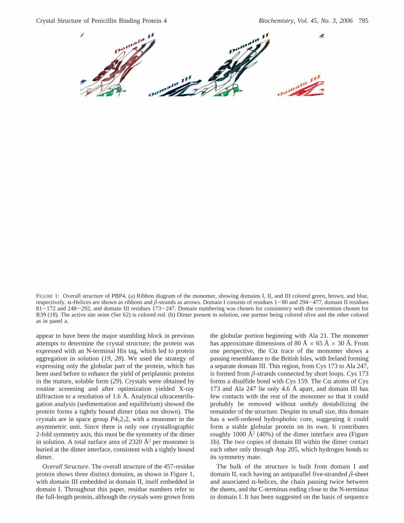
FIGURE 5: Stereo overlay of apo-PBP4 with TEM1 (PDB entry 1BTL), showing the main active site residues. The main chain atoms of Ser62-Lys 65 (PBP4) were least-squares fitted to those of Ser 70-Lys 73 in TEM1. PBP4 residues are colored by atom type and labeled inblack. TEM1 residues are colored and labeled in orange. The SXXK and SXN motifs match closely, and Ser 130 in TEM1 lies close to Ser306 in PBP4 (only the major conformer of the Ser 306 side chain is shown for clarity). PBP4 has no counterpart to Glu 166 of TEM1,however, the closest residue being Leu 359. Together with Asn 170, Glu 166 makes the active site of TEM1 much more polar than that ofPBP4. The carbonyl oxygen of Ser 357 in PBP4 points toward Ser 62, and lies∼3.7 Å from the active site serine side chain oxygen atom.The carboxyl side chain of Glu 166 in TEM1 lies slightly farther (∼4.2 Å) from its active site serine. Figures 5-7 were produced withMolscript (41) and Raster3D (42).
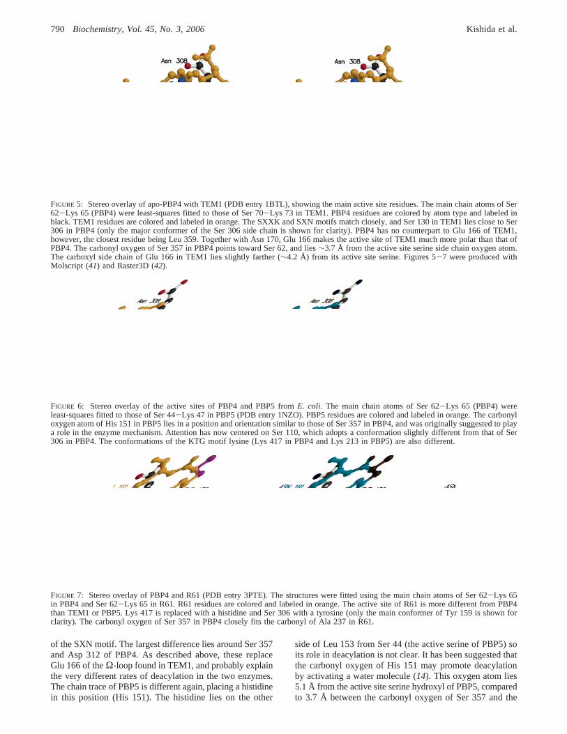
FIGURE 6: Stereo overlay of the active sites of PBP4 and PBP5 fromE. coli. The main chain atoms of Ser 62-Lys 65 (PBP4) wereleast-squares fitted to those of Ser 44-Lys 47 in PBP5 (PDB entry 1NZO). PBP5 residues are colored and labeled in orange. The carbonyloxygen atom of His 151 in PBP5 lies in a position and orientation similar to those of Ser 357 in PBP4, and was originally suggested to playa role in the enzyme mechanism. Attention has now centered on Ser 110, which adopts a conformation slightly different from that of Ser306 in PBP4. The conformations of the KTG motif lysine (Lys 417 in PBP4 and Lys 213 in PBP5) are also different.
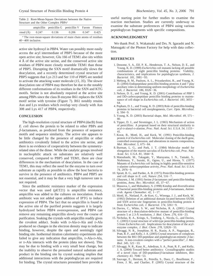
FIGURE 7: Stereo overlay of PBP4 and R61 (PDB entry 3PTE). The structures were fitted using the main chain atoms of Ser 62-Lys 65in PBP4 and Ser 62-Lys 65 in R61. R61 residues are colored and labeled in orange. The active site of R61 is more different from PBP4than TEM1 or PBP5. Lys 417 is replaced with a histidine and Ser 306 with a tyrosine (only the main conformer of Tyr 159 is shown forclarity). The carbonyl oxygen of Ser 357 in PBP4 closely fits the carbonyl of Ala 237 in R61.
790 Biochemistry, Vol. 45, No. 3, 2006 Kishida et al.
active site hydroxyl in PBP4. Water can possibly more easilyaccess the acyl intermediate of PBP5 because of the moreopen active site; however, Glu 166 of TEM1 also sits within4 Å of the active site serine, and the conserved active siteresidues of PBP4 more closely resemble TEM1 than thoseof PBP5. Disrupting the SXN motif dramatically slows thedeacylation, and a recently determined crystal structure ofPBP5 suggests that Lys 213 and Ser 110 of PBP5 are neededto activate the attacking water molecule (15, 35). The slowerdeacylation rate of PBP4 may therefore be due to the notablydifferent conformations of its residues in the SXN and KTGmotifs. Serine is not absolutely required at the active siteamong PBPs since the class B enzyme R61 replaces the SXNmotif serine with tyrosine (Figure 7). R61 notably retainsAsn and Lys residues which overlap very closely with Asn308 and Lys 417 of PBP4, respectively.
CONCLUSION
The high-resolution crystal structure of PBP4 (dacB) fromE. coli shows the protein to be related to other PBPs andâ-lactamases, as predicted from the presence of sequencemotifs and sequence similarity. The active site appears tobe little changed by the presence of ampicillin or otherantibiotics covalently linked to the active site serine, andthere is no evidence of cooperativity between the symmetry-related sites of the dimer. While the catalytic groups involvedin nucleophilic attack on substrates are relatively wellconserved, compared to PBP5 and TEM1, there are cleardifferences in the mechanism of deacylation. In the case ofTEM1, this may reflect the need for the enzyme to turn oversubstrate as rapidly as possible to allow the host bacteria tosurvive in the presence of antibiotics. PBP4 and PBP5 arenot essential, and it may be that a very high turnover rate isnot required.
Since the antibiotic resistance marker of the expressionvector that was used (pET21) is ampicillin resistance,ampicillin was added to the cultures on inoculation. Freshantibiotic was not added upon addition of IPTG to induceexpression of PBP4. The fact that no ampicillin is found inthe active site of the purified protein suggests it is largelyhydrolyzed before PBP4 overexpression, and PBP4 canremove any remaining ampicillin slowly over the course ofpurification. Soaking the crystals with ampicillin readily givesthe covalent adduct. Soaks withD-Ala-D-Ala and D-Alaproduced no changes in the electron density map to indicatebinding, however, despite the open and seemingly rigidbinding site. Isothermal titration calorimetry experiments alsofailed to give any indication that ampicillin,D-Ala-D-Ala,or D-Ala interacts with the protein (data not shown). Thismay be due to binding with a very small heat change, butthe inability to observe the presumed natural substrate andproduct in the binding site by crystal soaking implies thatadditional interactions with the peptidoglycan are requiredfor binding. The crystal structures presented here provide a
useful starting point for further studies to examine thereaction mechanism. Studies are currently underway toexamine the substrate preferences of PBP4 using variouspeptidoglycan fragments with specific compositions.
ACKNOWLEDGMENT
We thank Prof. S. Wakatsuki and Drs. N. Igarashi and N.Matsugaki of the Photon Factory for help with data collec-tion.
REFERENCES
1. Denome, S. A., Elf, P. K., Henderson, T. A., Nelson, D. E., andYoung, K. D. (1999)Escherichia colimutants lacking all possiblecombinations of eight penicillin binding proteins: Viability,characteristics, and implications for peptidoglycan synthesis,J.Bacteriol. 181, 3981-93.
2. Meberg, B. M., Paulson, A. L., Priyadarshini, R., and Young, K.D. (2004) Endopeptidase penicillin-binding proteins 4 and 7 playauxiliary roles in determining uniform morphology ofEscherichiacoli, J. Bacteriol. 186, 8326-36.
3. Nelson, D. E., and Young, K. D. (2001) Contributions of PBP 5and DD-carboxypeptidase penicillin binding proteins to mainte-nance of cell shape inEscherichia coli, J. Bacteriol. 183, 3055-64.
4. Popham, D. L., and Young, K. D. (2003) Role of penicillin-bindingproteins in bacterial cell morphogenesis,Curr. Opin. Microbiol.6, 594-9.
5. Young, K. D. (2003) Bacterial shape,Mol. Microbiol. 49, 571-80.
6. Tipper, D. J., and Strominger, J. L. (1965) Mechanism of actionof penicillins: A proposal based on their structural similarity toacyl-D-alanyl-D-alanine,Proc. Natl. Acad. Sci. U.S.A. 54, 1133-41.
7. Korat, B., Mottl, H., and Keck, W. (1991) Penicillin-bindingprotein 4 ofEscherichia coli: Molecular cloning of the dacB gene,controlled overexpression, and alterations in murein composition,Mol. Microbiol. 5, 675-84.
8. Burman, L. G., and Park, J. T. (1984) Molecular model forelongation of the murein sacculus ofEscherichia coli, Proc. Natl.Acad. Sci. U.S.A. 81, 1844-8.
9. Matsuhashi, M., Takagaki, Y., Maruyama, I. N., Tamaki, S.,Nishimura, Y., Suzuki, H., Ogino, U., and Hirota, Y. (1977)Mutants ofEscherichia colilacking in highly penicillin-sensitiveD-alanine carboxypeptidase activity,Proc. Natl. Acad. Sci. U.S.A.74, 2976-9.
10. Spratt, B. G., and Pardee, A. B. (1975) Penicillin-binding proteinsand cell shape inE. coli, Nature 254, 516-7.
11. Ghuysen, J. M. (1991) Serineâ-lactamases and penicillin-bindingproteins,Annu. ReV. Microbiol. 45, 37-67.
12. Massova, I., and Mobashery, S. (1998) Kinship and diversificationof bacterial penicillin-binding proteins andâ-lactamases,Antimi-crob. Agents Chemother. 42, 1-17.
13. Mottl, H., Nieland, P., de Kort, G., Wirenga, J. J., and Keck, W.(1992) Deletion of an additional domain located between SXXKand SXN active-site fingerprints in penicillin-binding protein 4from Escherichia coli, J. Bacteriol. 174, 3261-9.
14. Davies, C., White, S. W., and Nicholas, R. A. (2001) Crystalstructure of a deacylation-defective mutant of penicillin-bindingprotein 5 at 2.3-Å resolution,J. Biol. Chem. 276, 616-23.
15. Nicholas, R. A., Krings, S., Tomberg, J., Nicola, G., and Davies,C. (2003) Crystal structure of wild-type penicillin-binding protein5 fromEscherichia coli: Implications for deacylation of the acyl-enzyme complex,J. Biol. Chem. 278, 52826-33.
16. Silvaggi, N. R., Josephine, H. R., Kuzin, A. P., Nagarajan, R.,Pratt, R. F., and Kelly, J. A. (2005) Crystal structures of complexesbetween the R61 DD-peptidase and peptidoglycan-mimeticâ-lac-tams: A non-covalent complex with a “perfect penicillin”,J. Mol.Biol. 345, 521-33.
17. Silvaggi, N. R., Kaur, K., Adediran, S. A., Pratt, R. F., and Kelly,J. A. (2004) Toward better antibiotics: Crystallographic studiesof a novel class of DD-peptidase/â-lactamase inhibitors,Bio-chemistry 43, 7046-53.
18. Sauvage, E., Herman, R., Petrella, S., Duez, C., Bouillenne, F.,Frere, J. M., and Charlier, P. (2005) Crystal structure of the
Table 2: Root-Mean-Square Deviations between the NativeStructure and the Other Complex PBPsa
ampicillin penicillin G penicillin V Farom Flomox
rmsd (Å) 0.247 0.136 0.206 0.347 0.425a The root-mean-square deviations of main chain atoms of residues
40-400 inclusive.
Crystal Structure of Penicillin Binding Protein 4 Biochemistry, Vol. 45, No. 3, 2006791

actinomadura R39 DD-peptidase reveals new domains in penicil-lin-binding proteins,J. Biol. Chem. 29, 29.
19. Thunnissen, M. M., Fusetti, F., de Boer, B., and Dijkstra, B. W.(1995) Purification, crystallisation and preliminary X-ray analysisof penicillin binding protein 4 fromEscherichia coli, a proteinrelated to class Aâ-lactamases,J. Mol. Biol. 247, 149-53.
20. Otwinowski, Z., and Minor, W. (1997) Processing of X-raydiffraction data collected in oscillation mode,Methods Enzymol.276, 307-26.
21. Collaborative Computational Project No. 4 (1994) The CCP4suite: Programs for protein crystallography,Acta Crystallogr.D50, 760-3.
22. Terwilliger, T. C. (2003) SOLVE and RESOLVE: Automatedstructure solution and density modification,Methods Enzymol. 374,22-37.
23. Terwilliger, T. C., and Berendzen, J. (1999) Automated MAD andMIR structure solution,Acta Crystallogr. D55, 849-61.
24. Roussel, A., and Cambillau, C. (1989) inSilicon GraphicsGeometry Partners Directory, pp 77-8, Silicon Graphics, Moun-tain View, CA.
25. Brunger, A. T. (1996)X-PLOR, version 3.851, Yale UniversityPress, New Haven, CT.
26. Murshudov, G. N., Vagin, A. A., and Dodson, E. J. (1997)Refinement of macromolecular structures by the maximum-likelihood method,Acta Crystallogr. D53, 240-55.
27. Laskowski, R. A., MacArthur, M. W., Moss, D. S., and Thornton,J. M. (1993) PROCHECK: A program to check the stereochem-ical quality of protein structures,J. Appl. Crystallogr. 26, 283-91.
28. Fusetti, F., and Dijkstra, B. W. (1996) Purification and light-scattering analysis of penicillin-binding protein 4 fromEscherichiacoli, Microb. Drug Resist. 2, 73-6.
29. Heddle, J., Scott, D. J., Unzai, S., Park, S. Y., and Tame, J. R.(2003) Crystal structures of the liganded and unliganded nickel-binding protein NikA fromEscherichia coli, J. Biol. Chem. 278,50322-9.
30. Clausen, T., Schlegel, A., Peist, R., Schneider, E., Steegborn, C.,Chang, Y. S., Haase, A., Bourenkov, G. P., Bartunik, H. D., andBoos, W. (2000) X-ray structure of MalY fromEscherichia coli:A pyridoxal 5′-phosphate-dependent enzyme acting as a modulatorin mal gene expression,EMBO J. 19, 831-42.
31. Krissinel, E., and Henrick, K. (2004) Secondary-structure matching(SSM), a new tool for fast protein structure alignment in threedimensions,Acta Crystallogr. D60, 2256-68.
32. Anderson, J. W., Adediran, S. A., Charlier, P., Nguyen-Disteche,M., Frere, J. M., Nicholas, R. A., and Pratt, R. F. (2003) On the
substrate specificity of bacterial DD-peptidases: Evidence fromtwo series of peptidoglycan-mimetic peptides,Biochem. J. 373,949-55.
33. Swaren, P., Golemi, D., Cabantous, S., Bulychev, A., Maveyraud,L., Mobashery, S., and Samama, J. P. (1999) X-ray structure ofthe Asn276Asp variant of theEscherichia coliTEM-1 â-lacta-mase: Direct observation of electrostatic modulation in resistanceto inactivation by clavulanic acid,Biochemistry 38, 9570-6.
34. Gherman, B. F., Goldberg, S. D., Cornish, V. W., and Friesner,R. A. (2004) Mixed quantum mechanical/molecular mechanical(QM/MM) study of the deacylation reaction in a penicillin bindingprotein (PBP) versus in a class Câ-lactamase,J. Am. Chem. Soc.126, 7652-64.
35. Nicola, G., Peddi, S., Stefanova, M., Nicholas, R. A., Gutheil,W. G., and Davies, C. (2005) Crystal structure ofEscherichiacoli penicillin-binding protein 5 bound to a tripeptide boronic acidinhibitor: A role for Ser-110 in deacylation,Biochemistry 44,8207-17.
36. Ishiguro, M., Nishihara, T., and Tanaka, R. (2001) New orallyactive penem antibiotic: Farom,Yakugaku Zasshi 121, 915-27.
37. Jelsch, C., Mourey, L., Masson, J. M., and Samama, J. P. (1993)Crystal structure ofEscherichia coliTEM1 â-lactamase at 1.8 Åresolution,Proteins 16, 364-83.
38. Swaren, P., Maveyraud, L., Guillet, V., Masson, J. M., Mourey,L., and Samama, J. P. (1995) Electrostatic analysis of TEM1â-lactamase: Effect of substrate binding, steep potential gradientsand consequences of site-directed mutations,Structure 3, 603-13.
39. Maveyraud, L., Pratt, R. F., and Samama, J. P. (1998) Crystalstructure of an acylation transition-state analog of the TEM-1â-lactamase. Mechanistic implications for class Aâ-lactamases,Biochemistry 37, 2622-8.
40. Maveyraud, L., Saves, I., Burlet-Schiltz, O., Swaren, P., Masson,J. M., Delaire, M., Mourey, L., Prome, J. C., and Samama, J. P.(1996) Structural basis of extended spectrum TEMâ-lactamases.Crystallographic, kinetic, and mass spectrometric investigationsof enzyme mutants,J. Biol. Chem. 271, 10482-9.
41. Kraulis, P. J. (1991) MOLSCRIPT: A program to produce bothdetailed and schematic plots of protein structures,J. Appl.Crystallogr. 24, 946-50.
42. Merritt, E. A., and Bacon, D. J. (1997) Raster3D: PhotorealisticMolecular Graphics,Methods Enzymol. 277, 505-24.
BI051533T
792 Biochemistry, Vol. 45, No. 3, 2006 Kishida et al.