![The Drowsy Chaperone Program [2012] - USM Digital ...](https://static.fdokumen.com/doc/165x107/6322912f887d24588e044a66/the-drowsy-chaperone-program-2012-usm-digital-.jpg)
Cross-Monomer Substrate Contacts Reposition the Hsp90 N-Terminal Domain and Prime the Chaperone...
13
Cross-Monomer Substrate Contacts Reposition the Hsp90 N-Terminal Domain and Prime the Chaperone Activity Timothy O. Street 1 , Laura A. Lavery 1 , Kliment A. Verba 1 , Chung-Tien Lee 2 , Matthias P. Mayer 2 and David A. Agard 1,3 ⁎ 1 Department of Biochemistry and Biophysics, University of California, San Francisco, CA 94158-2517, USA 2 Zentrum für Molekulare Biologie der Universität Heidelberg (ZMBH), Im Neuenheimer Feld 282, 69120 Heidelberg, Germany 3 The Howard Hughes Medical Institute, University of California, San Francisco, CA 94158-2517, USA Received 26 August 2011; received in revised form 14 October 2011; accepted 21 October 2011 Available online 31 October 2011 Edited by F. Schmid Keywords: Hsp90 mechanism; chaperone; protein folding The ubiquitous molecular chaperone Hsp90 plays a critical role in substrate protein folding and maintenance, but the functional mechanism has been difficult to elucidate. In previous work, a model Hsp90 substrate revealed an activation process in which substrate binding accelerates a large open/ closed conformational change required for ATP hydrolysis by Hsp90. While this could serve as an elegant mechanism for conserving ATP usage for productive interactions on the substrate, the structural origin of substrate- catalyzed Hsp90 conformational changes is unknown. Here, we find that substrate binding affects an intrinsically unfavorable rotation of the Hsp90 N-terminal domain (NTD) relative to the middle domain (MD) that is required for closure. We identify an MD substrate binding region on the interior cleft of the Hsp90 dimer and show that a secondary set of substrate contacts drives an NTD orientation change on the opposite monomer. These results suggest an Hsp90 activation mechanism in which cross-monomer contacts mediated by a partially structured substrate prime the chaperone for its functional activity. © 2011 Elsevier Ltd. All rights reserved. Introduction Molecular chaperones confer stress resistance critical for survival under harsh environmental conditions and maintain protein homeostasis under normal conditions. Beyond their role in protein folding, chaperones affect protein activation and trafficking, facilitating the degradation of terminally misfolded proteins, and the formation and disassembly of macromolecular complexes. Hsp90 is a highly conserved member of the chaperone family and plays a unique role by its regulatory influence in eukaryotes via the activation of specific classes of substrates (also known as clients), such as nuclear receptors and kinases. 1 This broad regulatory influence is thought to underlie the potent influence of Hsp90 inhibitors on the growth of diverse cancer types. 2 Despite its fundamental cell biological and clinical importance, the mecha- nism by which Hsp90 stabilizes and remodels client proteins is not understood. One confounding problem is that Hsp90 is large, conformationally dynamic, and undergoes dramatic structural changes upon ATP binding and hydrolysis (Fig. 1a). 3, 4,8 Small-angle X-ray scattering (SAXS) and electron microscopy (EM) measurements have *Corresponding author. Department of Biochemistry and Biophysics, University of California, San Francisco, CA 94158-2517, USA. E-mail address: [email protected]. Abbreviations used: NTD, N-terminal domain; MD, middle domain; CTD, C-terminal domain; SAXS, small-angle X-ray scattering; EM, electron microscopy; FRET, fluorescence resonance energy transfer; HSQC, heteronuclear single quantum coherence; NOESY, nuclear Overhauser enhancement spectroscopy; NOE, nuclear Overhauser enhancement; AMPPNP, β,γ-Imidoadenosine 5′-triphosphate. doi:10.1016/j.jmb.2011.10.038 J. Mol. Biol. (2012) 415,3–15 Contents lists available at www.sciencedirect.com Journal of Molecular Biology journal homepage: http://ees.elsevier.com.jmb 0022-2836/$ - see front matter © 2011 Elsevier Ltd. All rights reserved.
Transcript of Cross-Monomer Substrate Contacts Reposition the Hsp90 N-Terminal Domain and Prime the Chaperone...

doi:10.1016/j.jmb.2011.10.038 J. Mol. Biol. (2012) 415, 3–15
Contents lists available at www.sciencedirect.com
Journal of Molecular Biologyj ourna l homepage: ht tp : / /ees .e lsev ie r.com. jmb
Cross-MonomerSubstrateContactsReposition theHsp90N-Terminal Domain and Prime the Chaperone Activity
Timothy O. Street 1, Laura A. Lavery 1, Kliment A. Verba 1,Chung-Tien Lee 2, Matthias P. Mayer 2 and David A. Agard 1, 3⁎1Department of Biochemistry and Biophysics, University of California, San Francisco, CA 94158-2517, USA2Zentrum für Molekulare Biologie der Universität Heidelberg (ZMBH), Im Neuenheimer Feld 282,69120 Heidelberg, Germany3The Howard Hughes Medical Institute, University of California, San Francisco, CA 94158-2517, USA
Received 26 August 2011;received in revised form14 October 2011;accepted 21 October 2011Available online31 October 2011
Edited by F. Schmid
Keywords:Hsp90 mechanism;chaperone;protein folding
*Corresponding author. DepartmenBiophysics, University of California,94158-2517, USA. E-mail address: agAbbreviations used: NTD, N-term
middledomain;CTD,C-terminal domX-ray scattering; EM, electron microscfluorescence resonance energy transfheteronuclear single quantum cohereOverhauser enhancement spectroscopOverhauser enhancement; AMPPNP5′-triphosphate.
0022-2836/$ - see front matter © 2011 E
The ubiquitous molecular chaperone Hsp90 plays a critical role in substrateprotein folding and maintenance, but the functional mechanism has beendifficult to elucidate. In previous work, a model Hsp90 substrate revealedan activation process in which substrate binding accelerates a large open/closed conformational change required for ATP hydrolysis by Hsp90. Whilethis could serve as an elegant mechanism for conserving ATP usage forproductive interactions on the substrate, the structural origin of substrate-catalyzed Hsp90 conformational changes is unknown. Here, we find thatsubstrate binding affects an intrinsically unfavorable rotation of the Hsp90N-terminal domain (NTD) relative to the middle domain (MD) that isrequired for closure. We identify an MD substrate binding region on theinterior cleft of the Hsp90 dimer and show that a secondary set of substratecontacts drives an NTD orientation change on the opposite monomer. Theseresults suggest an Hsp90 activation mechanism in which cross-monomercontacts mediated by a partially structured substrate prime the chaperonefor its functional activity.
© 2011 Elsevier Ltd. All rights reserved.
Introduction
Molecular chaperones confer stress resistancecritical for survival under harsh environmentalconditions and maintain protein homeostasisunder normal conditions. Beyond their role inprotein folding, chaperones affect protein activation
t of Biochemistry andSan Francisco, [email protected] domain; MD,ain; SAXS, small-angleopy; FRET,er; HSQC,nce; NOESY, nucleary; NOE, nuclear
, β,γ-Imidoadenosine
lsevier Ltd. All rights reserve
and trafficking, facilitating the degradation ofterminally misfolded proteins, and the formationand disassembly of macromolecular complexes.Hsp90 is a highly conserved member of thechaperone family and plays a unique role by itsregulatory influence in eukaryotes via the activationof specific classes of substrates (also known asclients), such as nuclear receptors and kinases.1 Thisbroad regulatory influence is thought to underlie thepotent influence of Hsp90 inhibitors on the growthof diverse cancer types.2 Despite its fundamentalcell biological and clinical importance, the mecha-nism by which Hsp90 stabilizes and remodels clientproteins is not understood.One confounding problem is that Hsp90 is large,
conformationally dynamic, and undergoes dramaticstructural changes upon ATP binding and hydrolysis(Fig. 1a).3,4,8 Small-angle X-ray scattering (SAXS) andelectron microscopy (EM) measurements have
d.

Fig. 1. Hsp90 conformational flexibility. (a) The HtpG apo crystal structure (left3) requires significant structuralplasticity to reach the closed ATP state (right4). The contacts formed in the closed conformation (red and blue spheres) aresignificantly out of alignment in the apo structure, prior to NTD rotation (middle). Shown below are the apo solutionconformations of HtpG determined by SAXS5,6 and the compact ADP state.7 (b) The NTD orientation in the Grp94 crystalstructure8 shows the nucleotide positioned far from the highly conserved arginine (spheres), whereas in the closedconformation, the ATP γ-phosphate makes a direct contact.4
4 Substrate Binding Affects Rotation of Hsp90 NTD
revealed an underlying complexity of Hsp90's con-formational dynamics.5–7,9–11 The Hsp90 monomer iscomposed of three stable domains [N-terminaldomain (NTD),middle domain (MD), andC-terminaldomain (CTD)], and conformational flexibility resultsfrom their rigid-body-like rearrangement. Under apoconditions, a weak MD/CTD interface allows for awide range of arm–arm geometries that can beinfluenced by pH and osmolyte conditions.6,12 Thisstriking flexibility has been observed for highlydiverse Hsp90 homologs7,9 and is postulated to becritically important to Hsp90's ability to recognize aremarkably diverse set of client proteins.Unlike other molecular chaperones, Hsp90 ap-
pears to prefer largely folded but nonnative states.This poses an additional practical challenge, as such
states can be difficult to populate and are prone toaggregation. Previous work introduced a well-behaved model client protein, the partially foldedbut non-aggregating protein Δ131Δ, a fragment ofStaphyloccocal Nuclease that has been studiedextensively by the protein folding community.13
Using this model client revealed that, (i) under apoconditions, Hsp90 partially closes aroundΔ131Δ; (ii)Hsp90 binds a highly structured region of Δ131Δ;(iii) and Δ131Δ accelerates a nucleotide-drivenopen/closed transition and stimulates ATP hydro-lysis by Hsp90, effectively activating the chaperoneby lowering a rate-limiting conformational barrier.Taken in the context that the ligand binding domainof GR enhances the ATPase of the human Hsp9014
and that the ribosomal subunit L2 enhances the

5Substrate Binding Affects Rotation of Hsp90 NTD
ATPase of the bacterial Hsp90,15 this suggests thatactivation of the rate-limiting Hsp90 conformationaltransition is a conserved feature of bone fide Hsp90clients, similar to Hsp70 activation by peptidesubstrates. However, the mechanism by whichsubstrate binding can drive the dramatic Hsp90open/closed transition is unknown. Indeed, aprevious low-resolution SAXS analysis13 could notdetermine whether Δ131Δ makes cross-monomercontacts as has been observed for the activatingcochaperone aha116 or solely intramonomer contactsas observed for an Hsp90–cdc37–cdk4 (chaperone–cochaperone–kinase substrate) EM reconstruction.17
The Hsp90 ATPase is slow, on the order of 0.1–1hydrolysis events per minute depending on thehomolog and conditions,18–20 and mirrors a slowconformational change from the open apo state to theclosed ATP conformation.13,21 This dramatic transi-tion involves a large change in arm–arm proximity, adomain-level change in the NTD orientation, andlocal structural changes within the NTD (lid closureover the nucleotide binding pocket, strand exchangebetween NTDs) and the MD (restructuring of thecatalytic loop).3,4,19,22 Although the relative impor-tance of these structural changes to the closure rate isnot known, the structures of the β,γ-Imidoadenosine5′-triphosphate (AMPPNP)-bound canine Grp94(the Hsp90 homolog specific to the ER) and the apobacterial Hsp90 (HtpG) suggest that the NTDrotational state plays an important role. Bothstructures exhibit an open resting state in which theNTDs are diametrically opposed, requiring a signif-icant conformational change to come into a closure-competent alignment.3,8,23 As illustrated in Fig. 1a,the required movement involves a 90° rotation and a25-Å translation of the NTD center of mass, rearran-ging ∼2000 Å2 at the MD interface. This alignsclosed-state contacts (Fig. 1a, red and blue spheres)and also repositions ATP by ∼20 Å, allowing the γ-phosphate to contact a highly conserved arginine onthe MD (R336 in HtpG) that is essential for bothclosure and ATP hydrolysis4,20,24 (Fig. 1b). Impor-tantly, a full lid closure over the nucleotide pocket,which appears to be necessary for closure,25 cannotoccur in the NTD resting state due to a significantsteric clash with the MD.8 These observationssuggest that an NTD rotation may be involved inthe timing and order of many critical steps in closureand subsequent ATP hydrolysis.Here, we use our HtpG-activating substrate to
interrogate the open/closed transition and how thisprocess is substrate catalyzed. Key questions include(i) whether substrate contacts are within a singlemonomer or across monomers, (ii) defining thesubstrate binding region on HtpG in greater detailthan could be achieved from our previous SAXSanalysis, (iii) establishing whether a single set ormultiple substrate contacts are utilized, and (iv)determining how substrate binding affects HtpG
structural dynamics, particularly at the NTD, andhow this is related to the large energetic barrier toclosure.
Results
Monitoring NTD movement by fluorescenceresonance energy transfer
HtpG can be substrate activated by acceleratingthe kinetics of a slow open/closed structuraltransition required for maximal ATP hydrolysis.Previously, closure kinetics were measured byfluorescence resonance energy transfer (FRET), inwhich opposite monomers were labeled with donorand acceptor pairs.13 Tomonitor NTD/MD rotation,we designed a FRET pair within a single monomer.We identified residues S52/D341 on the NTD/MDthat significantly change distance (22–39 Å) in theopen/closed conformations yet remain solventexposed. These sites were mutated to cysteine andlabeled with Alexa Fluor 647 and Alexa Fluor 555.Since this pair is on the same monomer, a 20× excessof unlabeled wild-type HtpG was added to ensureonly one labeled monomer per dimer.Figure 2a shows the apo and AMPPNP fluores-
cence spectra for this FRET pair. There is significantacceptor signal in the apo state, and after aprolonged incubation with AMPPNP, there is adecrease/increase in acceptor/donor signal. Similarto previous studies, these measurements wereperformed at pH 9 because, for HtpG at this pH,there is a complete conversion between the open/closed state.6 Upon addition of AMPPNP, there is aslow time-dependent loss in acceptor signal withsingle-exponential kinetics (Fig. 2b) and a rate(k=0.002 s−1) that is the same as the arm–armclosure rate measured previously.13 As a control, wedesigned a fixed-point FRET pair within the MD(residues 350 and 362), which does not changedistance in the open/closed transition and con-firmed that closure did not affect this FRET signal(Supplemental Fig. 1a).The similar rates for NTD rotation and arm–arm
closure suggest simple two-state cooperativity;however, given the large number of structuralmotions that can occur in Hsp90, this observationdoes not rule out other intermediates. Cooperativitycan only be established by the coincidence of a largenumber of structural probes. To examine thispossibility, we measured closure kinetics by SAXS.Previous work demonstrated that SAXS measure-ments can determine the conformational equilibri-um of HtpG by linear combination fitting of thescattering spectra.5,6 Given that closure is slow, it ispossible to simultaneously initiate closure on mul-tiple samples and sequentially measure scattering at

6 Substrate Binding Affects Rotation of Hsp90 NTD
different time points. The robotic sample loadingsystem at the SIBYLS beamline at the AdvancedLight Source (Berkeley, CA) allows for each mea-surement to take only ∼2 min. An advantage of thismethod is that SAXS reports on all scatteringpositions, as opposed to the limited sites measured
by FRET. Here, we find that the kinetics of closureby SAXS match well with the arm–arm closure andNTD rotation measured by FRET (SupplementalFig. 1b). Although these measurements do notindicate whether local sequential conformationalchanges occur prior to the rate-limiting step, they doindicate that the gross conformational changesassociated with HtpG closure obey simple two-state cooperativity.Given this cooperativity, we wanted to know if
NTD rotation was contributing to the high-energybarrier separating the open/closed states. In otherwords, we wanted to know whether NTD rotationcould be significantly populated in isolation on asingle monomer or whether this rotation is intrinsi-cally unfavorable and requires stabilization fromNTD dimerization contacts with the oppositemonomer. To test this question, we made the sameNTD/MD FRET pair in the monomeric NMfragment of HtpG (residues 1–495). The acceptor/donor fluorescence spectrum on the NM fragment issimilar to the corresponding spectrum on the full-length dimer (data not shown). We find that theacceptor fluorescence on the NM fragment does notundergo any net change upon addition of AMPPNPeven in the presence of Δ131Δ (Fig. 2b), demon-strating that the NTD rotation required for closurerequires stabilization from the opposite monomer.These results reveal a major energetic mismatch inthe local and global energetics associated withHsp90 closure. NTD rotation creates highly stabiliz-ing dimer contacts in the closed state at the expenseof a locally disfavored NTD/MD interface. Thissuggests that substrate binding could activate HtpGby relieving this rotational penalty.To investigate this possibility, we tested whether
substrate binding is linked to NTD rotation in theHtpG dimer. We first investigated how Δ131Δaffects AMPPNP-driven closure kinetics as moni-tored byNTD rotation. Previously, we observed that
Fig. 2. Substrate binding affects an intrinsically unfa-vorable NTD rotation required for closure. (a) FRETmeasurements within a single HtpG monomer track theNTD rotation associated with closure. This rotation isreflected in the apo (black) and AMPPNP (green)fluorescence spectra. (b) Closure kinetics are monitoredby the decrease in acceptor fluorescence and are signifi-cantly accelerated by Δ131Δ; single-exponential fits areshown in continuous lines. The same FRET pair on the NMfragment shows no change in FRET from AMPPNP. (c)Heterodimers consisting of unlabeled HtpG on one armand the NM FRET pair on the opposite arm allow for NTDrotation to be monitored under apo conditions. Δ131Δbinding changes the NTD orientation, as reflected by a lossin acceptor signal (black circles). The 30-residue peptidecorresponding to the dominant binding region on Δ131Δdoes not affect the NTD orientation (green diamonds). TheW467A heterodimer (one arm NM FRET and second armW467A, blue squares) showsminimal impact fromΔ131Δ.
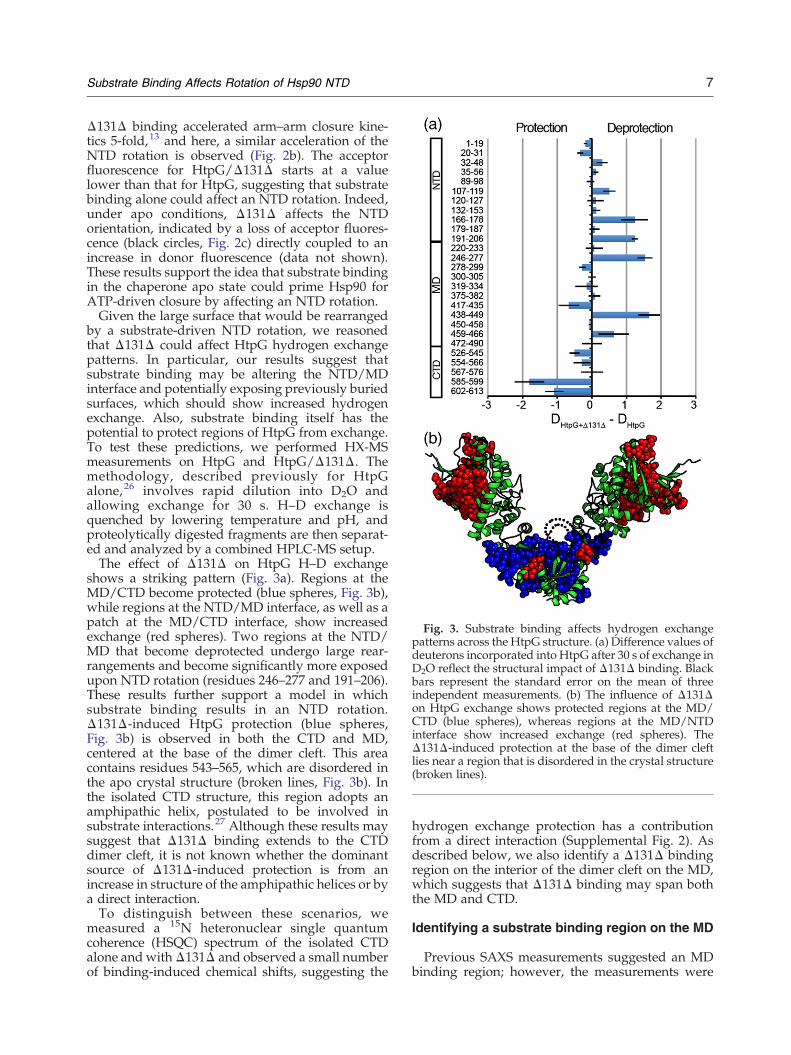
Fig. 3. Substrate binding affects hydrogen exchangepatterns across the HtpG structure. (a) Difference values ofdeuterons incorporated into HtpG after 30 s of exchange inD2O reflect the structural impact of Δ131Δ binding. Blackbars represent the standard error on the mean of threeindependent measurements. (b) The influence of Δ131Δon HtpG exchange shows protected regions at the MD/CTD (blue spheres), whereas regions at the MD/NTDinterface show increased exchange (red spheres). TheΔ131Δ-induced protection at the base of the dimer cleftlies near a region that is disordered in the crystal structure(broken lines).
7Substrate Binding Affects Rotation of Hsp90 NTD
Δ131Δ binding accelerated arm–arm closure kine-tics 5-fold,13 and here, a similar acceleration of theNTD rotation is observed (Fig. 2b). The acceptorfluorescence for HtpG/Δ131Δ starts at a valuelower than that for HtpG, suggesting that substratebinding alone could affect an NTD rotation. Indeed,under apo conditions, Δ131Δ affects the NTDorientation, indicated by a loss of acceptor fluores-cence (black circles, Fig. 2c) directly coupled to anincrease in donor fluorescence (data not shown).These results support the idea that substrate bindingin the chaperone apo state could prime Hsp90 forATP-driven closure by affecting an NTD rotation.Given the large surface that would be rearranged
by a substrate-driven NTD rotation, we reasonedthat Δ131Δ could affect HtpG hydrogen exchangepatterns. In particular, our results suggest thatsubstrate binding may be altering the NTD/MDinterface and potentially exposing previously buriedsurfaces, which should show increased hydrogenexchange. Also, substrate binding itself has thepotential to protect regions of HtpG from exchange.To test these predictions, we performed HX-MSmeasurements on HtpG and HtpG/Δ131Δ. Themethodology, described previously for HtpGalone,26 involves rapid dilution into D2O andallowing exchange for 30 s. H–D exchange isquenched by lowering temperature and pH, andproteolytically digested fragments are then separat-ed and analyzed by a combined HPLC-MS setup.The effect of Δ131Δ on HtpG H–D exchange
shows a striking pattern (Fig. 3a). Regions at theMD/CTD become protected (blue spheres, Fig. 3b),while regions at the NTD/MD interface, as well as apatch at the MD/CTD interface, show increasedexchange (red spheres). Two regions at the NTD/MD that become deprotected undergo large rear-rangements and become significantly more exposedupon NTD rotation (residues 246–277 and 191–206).These results further support a model in whichsubstrate binding results in an NTD rotation.Δ131Δ-induced HtpG protection (blue spheres,Fig. 3b) is observed in both the CTD and MD,centered at the base of the dimer cleft. This areacontains residues 543–565, which are disordered inthe apo crystal structure (broken lines, Fig. 3b). Inthe isolated CTD structure, this region adopts anamphipathic helix, postulated to be involved insubstrate interactions.27 Although these results maysuggest that Δ131Δ binding extends to the CTDdimer cleft, it is not known whether the dominantsource of Δ131Δ-induced protection is from anincrease in structure of the amphipathic helices or bya direct interaction.To distinguish between these scenarios, we
measured a 15N heteronuclear single quantumcoherence (HSQC) spectrum of the isolated CTDalone and withΔ131Δ and observed a small numberof binding-induced chemical shifts, suggesting the
hydrogen exchange protection has a contributionfrom a direct interaction (Supplemental Fig. 2). Asdescribed below, we also identify a Δ131Δ bindingregion on the interior of the dimer cleft on the MD,which suggests that Δ131Δ binding may span boththe MD and CTD.
Identifying a substrate binding region on the MD
Previous SAXS measurements suggested an MDbinding region; however, the measurements were

8 Substrate Binding Affects Rotation of Hsp90 NTD
too low resolution to determine a residue-levelsurface. To gain this insight, we next used NMRand mutagenesis. The HtpG MD (residues 231–495,31 kDa) can be purified and 15N labeled for NMR
Fig. 4. Mapping a substrate binding location on the HtpGMa dramatic simplification in the HSQC spectrum. (b and c) Bofrom Δ131Δ. (d) The MD in surface representation with a patcGly (pink). Mutation sites to test this proposed substrate bindinF390, and D476). The mutations that strongly affect substratecleft close the regions that show HX protection from Δ131Δ (marginal impact on binding are shown in light gray (D476K a
studies, but the HSQC spectrum is rather crowded(Supplemental Fig. 3a), and the residue assignmentsare unknown. Given that the structure of the MD isknown, we explored an approach utilizing selective
D. (a) The HtpGMDwith 15N-labeled Asp residues showsth peak intensity and chemical shift changes are observedh that contains Phe (yellow), Tyr (orange), Asp (red), andg region are shown on the apo state HtpG structure (W467,binding (dark gray: W467 and F390) lie within the dimerblue spheres). The charge reversal mutations that make and E369K).

Fig. 5. Cross-monomer substrate contacts are coupledto HtpG conformational changes. (a) The W467A muta-tion reduces substrate-induced conformational changes inHtpG under apo conditions as measured by SAXS. Here,the wild-type/Δ131Δ scattering distribution (blue) issignificantly more contracted than W467A/Δ131Δ(black). For reference, the HtpG spectrum in the absenceof Δ131Δ is shown in broken lines. Similarly, the W467Amutation reduces substrate-induced conformationalchanges in HtpG under AMPPNP conditions (inset).Both HtpG and Δ131Δ are at 50 μM. (b) Monomerexchange kinetics were measured by the loss of FRET thatresults from adding an excess of unlabeled HtpG. Δ131Δclearly slows exchange (red circles), whereas the peptide(green diamonds) is similar to the HtpG monomerexchange rate in the absence of substrate (blue squares).The Δ131Δ construct that lacks the 30 C-terminal residueshas only a modest affect on exchange (black crosses).
9Substrate Binding Affects Rotation of Hsp90 NTD
amino acid labeling to simplify the process ofidentifying a Δ131Δ binding location. Specifically,the Volker Doetsch laboratory introduced a methodwhereby multiple HSQC spectra are collected, eachone corresponding to a single type of amino acidbeing 15N labeled. Through counting of the numberof peaks that are affected by the substrate, it ispossible to identify one or more patches on thestructure that have the correct number of affectedresidues. Iterating this process with different labeledamino acids can identify a unique region that hascorrect surface residue composition of the bindingsite. 28 Although this method is advantageousbecause single amino acid labeling greatly simplifiesthe HSQC spectra, it only provides a predictedbinding region so the results must be independentlytested.We produced four variants of the HtpG MD
specifically 15N labeled on Asp, Phe, Tyr, and Glyresidues. These residues have an asymmetric distri-bution over the MD, suggesting the potential touniquely identify a binding region. Δ131Δ affectsboth the chemical shifts and intensities for a subsetof the labeled residues on the MD; an example withAsp is shown in Fig. 4a–c. We measured chemicalshifts and intensity changes, normalized them, anddefined their mean and standard deviation. Thepeaks that were significantly impacted werecounted by defining a significance threshold foreach amino acid type (see Methods).This process identified two Phe, three Tyr, two
Asp, and one Gly, in the predicted binding region(Supplemental Fig. 3b–i). Inspection of the MDshows a patch facing into the HtpG dimer cleft inthe apo state with this surface residue distribution(Fig. 4d), whereas the opposite face shows no suchsite (Supplemental Fig. 3j). As a test, we mutatedthree residueswithin this patch (positive predictions:W467A, F390A, and D476K) and three analogousmutations outside of this patch (negative predic-tions: W224A, F257A, and E369K). We includedcharge reversal mutations because the strong saltdependence of Δ131Δ binding suggested an electro-static contribution.Using a previously described fluorescence polar-
ization binding assay with IAEDANS-labeledΔ131Δ, we measured the binding Kd of thesevariants (wild-type HtpG has a Kd of 9 μM). Thehydrophobic truncations have significantly reducedbinding (W467A, 42 μM; F390A, 38 μM), while thenegative predictions areminimally affected (W224A,11 μM; F257A, 10 μM), which confirms that Δ131Δbinds to the HtpG interior cleft at the MD. Both thepositive and negative prediction charge reversalmutations show an intermediate reduction in bind-ing (D476K, 19 μM; E369K, 21 μM); one explanationmay be that long-range electrostatic interactionsbetween HtpG and Δ131Δ contribute to binding.This would be consistent with the significant
difference in pI between HtpG (5.1) and Δ131Δ(9.5) and also consistent with recent studies ofunfolded citrate synthase binding to Hsp90.29
However, if long-range electrostatics are playing arole, then neither mutation (D476K and E369K) is areliable test for direct binding. Therefore, we used

10 Substrate Binding Affects Rotation of Hsp90 NTD
the W467A and F390A variants to investigate therelationship between substrate binding and HtpGconformational changes.First, as a control, we tested the impact of these
mutations on Δ131Δ-induced conformationalchanges in HtpG. For reference, our previous SAXSmeasurements revealed that substrate binding iscoupled to large-scale conformational changes inHtpG under both apo and AMPPNP conditions;13
therefore, we expected that by disrupting substratebinding, these large conformational changes shouldbe reduced. The Δ131Δ-induced conformationalchanges inHtpG are visibly evident in the contractedSAXS P(r) spectrum, which reflects the combined setof scattering distances within the complex. Incontrast to wild-type HtpG, the W467A mutant hasa significantly reduced conformational change fromΔ131Δ under both apo (Fig. 5a) and nucleotideconditions (inset), confirming that the reduction insubstrate binding is directly translated in a reductionin the chaperone conformational response. TheW467A mutation itself does not affect the HtpGconformation (Supplemental Fig. 4a). Similar resultswere observed for the F390Amutation, although themutation itself resulted in a change in the conforma-tional state of HtpG (data not shown).
A secondary set of cross-monomer substratecontacts activates HtpG
A crucial mechanistic distinction concerningΔ131Δ activation of HtpG is whether the substrateactivates from within a monomer or across mono-mers and whether there are single or multiplecontacting regions of the substrate. As discussedbelow, we addressed these questions in three ways:(i) a heterodimer analysis with the W467A variantand NM FRET measurements, (ii) HtpG monomerexchange measurements, and (iii) by studyingdifferent fragments of Δ131Δ.Beyond identifying a substrate binding region in
greater detail, the W467A mutation provides anopportunity to form heterodimers of HtpG whereone arm contains the NM FRET pair, and theopposite arm is either wild-type HtpG or theW467A mutant. This type of heterodimer experi-ment has been used previously to identify cross-monomer determinants of Hsp90 hydrolysisrate20,25 and cochaperone activation.16 As discussedearlier, under apo conditions,Δ131Δ affects an NTDrotation in the wild-type heterodimer (one arm NMFRET, second arm wild-type HtpG) as seen from aconcentration-dependent loss of acceptor fluores-cence (black circles, Fig. 2c). In contrast, for theW467A heterodimer (one arm NM FRET, secondarm W467A), Δ131Δ only has a modest impact onthe NM FRET (blue squares, Fig. 2c). This resultshows that substrate binding at theMD of one arm isdirectly coupled to the NTD rotation on the opposite
arm. Since the heterodimer has a wild-type MD onthe FRET-labeled monomer, Δ131Δ should make amodest acceleration of closure by its impact on theopposing NTD. Indeed, in contrast to the 5-foldacceleration of AMPPNP-mediated closure ob-served for wild-type HtpG, Δ131Δ only acceleratesthe W467A heterodimer by a factor of 2 (Supple-mental Fig. 4b). W467A heterodimers have a similarintrinsic closure rate as the wild-type heterodimers(data not shown).A second test for cross-monomer contacts is that
Δ131Δ should slow the rate of HtpG monomerexchange. Here, we used a FRET-based assaydeveloped in the Buchner laboratory.16,21 In thisexperiment, Hsp90 heterodimers are labeled withdonor and acceptor fluorophores on opposite arms,with a resulting FRET signal that can be extinguishedby adding an excess of unlabeled wild-type Hsp90(shown schematically in Fig. 5b). Here, the loss ofacceptor fluorescence occurs because monomer ex-change randomizes fluorescently labeled monomerswith unlabeled monomers. We observe a strikingslowdown of monomer exchange kinetics in thepresence ofΔ131Δ (red circles, Fig. 5b), corroboratingthat cross-monomer substrate contacts are formed.There are two models that could explain cross-
monomer substrate contacts. The first model is thatthere is a single dominant substrate binding regionthat spans the Hsp90 monomers. The second modelis that binding is predominantly contained within amonomer with secondary substrate contacts thatspan the monomers. To discriminate between thesemodels, we investigated a limited construct thatonly contains the dominant binding region of thesubstrate. For reference, previous measurementswith Δ131Δ suggested that there was a dominantbinding region of ∼25 residues around position 100in Δ131Δ; therefore, we synthesized a 30-residuepeptide corresponding to residues 87–116 inΔ131Δ.If cross-monomer contacts are due to secondarysubstrate contacts, then this limited construct willnot rotate the NTD or slow monomer exchange.Indeed, although the peptide binds (Kd of 40 μM), itis unable to rotate the NTD (green diamonds, Fig.2c), does not change the monomer exchange rate(Fig. 5), and has a minimal impact on the closurekinetics (data not shown). SAXS measurementsunder apo conditions show that HtpG still contractsupon binding the peptide (Supplemental Fig. 4c),indicating an alteration of the MD/CTD interface.Although these results strongly suggest that
multiple regions of the substrate are required tomake cross-monomer contacts, rotate the NTD,accelerate closure, and subsequently activateHtpG, a potential confounding factor could be thatthe peptide either is misfolded or does not have asufficient level of structure to activate the chaperone.Given that previous studies on Δ131Δ demonstrat-ed that the region around residue 100 has significant

11Substrate Binding Affects Rotation of Hsp90 NTD
structure,30–32 this was a possibility we wanted toexplore in detail. Therefore, we performed nuclearOverhauser enhancement spectroscopy (NOESY)measurements (Supplemental Fig. 5a) on the pep-tide alone. We assigned the peptide using standardmethods involving total correlated spectroscopyand NOESY comparisons and a natural abundance13C–1H HSQC (see Methods). Inspection of thepattern of nonlocal nuclear Overhauser enhance-ments (NOEs) shows many i− i+3 and i− i+4 NOEsin the peptide region of the native α-helix in thewild-type structure (residues 98–107, SupplementalFig. 5b). Weak long-range NOEs suggest modesttertiary organization. Using NOE distance con-straints and dihedral constraints based on Cα/Cβ
chemical shifts with the DANGLE program,33 wedetermined an ensemble of compatible structureswith the ARIA program34 (Supplemental Fig. 5c).The peptide structure ensemble reveals a helicalregion centered on the native α-helix and adjacentN-and C-terminal loops that loosely interact. Thecentral helical region is not present in all members ofthe ensemble, indicting that the helix is a foldingequilibrium, consistent with early Δ131Δ studies.30This result shows that the dominant substrate regionrecognized by Hsp90 has a moderate level ofstructure and that the peptide is not misfolded.
Fig. 6. Model ofNTD rotation in substrate activation ofHsp90an asymmetricmechanism inwhichNTDdomain rotationmakeclosure and ATP hydrolysis. The dominant unit of structure recand is in a local folding equilibrium. However, secondary conrotation and acceleration of closure. Although the substrate isΔ131Δ and the Hsp90 long closure time, many bind and releas
Finally, we wanted to investigate the location ofthe secondary contacts on the substrate. As shownhere, Hsp90 binds to a locally structured region ofΔ131Δ around residue 100, and our results showthat secondary contacts from this region are requiredfor cross-monomer contacts that affect an NTDrotation, which primes the chaperone for ATP-driven closure. These findings are consistent withour previous NMR measurements that suggested asecondary binding site at the Δ131Δ C-terminus.13
To test whether the C-terminal region indeed makessecondary contacts, we investigated aΔ131Δ variantin which the 30 C-terminal residues are removed(residues 111–141). Indeed, this construct only re-sults in a modest slowdown of HtpG monomerexchange (Fig. 5b) and has a minimal impact on theclosure kinetics (data not shown). Although theseresults do not exclude a synergistic contributionfrom the Δ131Δ N-terminal region, secondarycontacts from C-terminal region of Δ131Δ clearlyplay a central role in Hsp90 activation.
Discussion
The Hsp90 ATPase is required for in vivofunction 35,36 and is regulated by numerous
ATPhydrolysis cycle. Substrate binding activatesHsp90 bys a significant contribution to the rate-limiting step inHsp90ognized by Hsp90 is a locally structured region (green oval)tacts outside this structured region are required for NTDshown bound for the entire cycle, given the low affinity ofe steps are likely occurring during the course of the cycle.

12 Substrate Binding Affects Rotation of Hsp90 NTD
cochaperones in eukaryotes.37 Previous work withthe model clientΔ131Δ demonstrated that substratebinding can also regulate the activity of HtpG,13
similar to reports on human Hsp90 activation by theligand binding domain of the glucocorticoidreceptor14 and an Escherichia coli ribosomal proteinL2 that activates HtpG.15 Elucidating the Hsp90functional mechanism requires an understanding ofhow the chaperone can be activated by substrates.Here, we have focused on NTD structural motions inthe HtpG conformational cycle, identifying a clientbinding region on the MD, and establishing thatmultiple regions of the substratemake cross-monomercontacts required for HtpG activation (Fig. 6).This proposed activation of HtpG by Δ131Δ has
a parallel with the mechanism of the activatingcochaperone Aha1 on the yeast Hsp90.16 Thoseauthors demonstrate that cross-monomer Aha1contacts prime Hsp90 for closure and subsequenthydrolysis, while FRET measurements suggestedthat Aha1 could affect NTD orientation.21 There isa second interesting parallel between ourHtpG/Δ131Δ findings and cochaperone-stabilizedHsp90 conformations. Recent EM measurements ofhuman Hsp90 demonstrated that the cochaperoneHop (involved in substrate loading from Hsp70)induces a partial closure of Hsp90 and fully rotatesboth NTDs into a closure-competent orientation.38
Previous measurements on Δ131Δ showed thatsubstrate binding induces a partial closure of HtpGin the apo state,13 similar to the Hop-stabilizedconformation. These similarities suggest that Hopstabilizes an Hsp90 conformation that is naturallypredisposed for substrate binding, subsequent chap-erone closure, and ATP hydrolysis.Comparison of our results with an EM reconstruc-
tion of an Hsp90–cdc37–cdk4 complex17 revealsdifferences between these substrates. Modelingsuggested the kinase substrate, cdk4, binding boththeNTD andMDon a singlemonomer, in contrast tothe cross-monomer contacts we observe. In theHsp90–cdc37–cdk4 complex, the substrate-boundNTD is in a closure-competent conformation, whilethe other monomer, bound to cdc37, is hinged away.While the primaryΔ131Δ interaction iswith theMD,one possibility is that secondary substrate contactsare made to the NTD. This would be consistent withearly studies indicating that Hsp90 has two substratebinding sites with different specificities,39 an NTDsite that can bind short unstructured peptides and aCTD/MD site that can bind partially foldedsubstrates.40 Given that our HX-MS measurementsdo not show significant Δ131Δ-induced protectionat the NTD, if Δ131Δ is contacting the NTD, thesecontacts are likely transient.Recent studies have identified that the ribosomal
subunit L2 activates HtpG, similar to the effect fromΔ131Δ; however, L2 is unable to activate the yeastHsp90 homolog.15 Interestingly, we find thatΔ131Δ
does not accelerate closure for the yeast Hsp90 butdoes accelerate closure for human TRAP1, themitochondria-specific Hsp90 homolog (T.O.S. andL.A.L., unpublished observations). Although thereis very strong evidence that the conformationalstates of Hsp90 are highly conserved, the degree towhich substrates accelerate conformational transi-tions for Hsp90 homologs appears to be variable.Numerous studies have established a conserved
Hsp90mechanism inwhich structural rearrangementsresulting in NTD dimerization organizes the catalyticmachinery required for ATP hydrolysis.4,7,21,25,41SAXS and EM studies have shown that rigid-bodymotions between the NTD/MD and the MD/CTDcan account for the wide range of Hsp90 conforma-tional states.5–7 The extreme apo state flexibility arisesfrom a weak coupling between the MD and CTD,allowing for a wide range of arm–arm geometries. Incontrast, here we find that the NTD/MD rotationrequired for closure is a high-energy state. The closedconformation, although stable, comes with a cost ofadopting an unfavorable NTD/MD interface. How-ever, our measurements do not indicate whether theNM rotation has a large kinetic barrier in addition toan unfavorable equilibrium constant, nor do ourmeasurements assess the degree to which the NMrotation is unfavorable, in terms of kilocalories permole. Also, it is not known whether Δ131Δ affects adiscrete NTD rotation or whether substrate bindingweakens the NTD/MD interface, resulting in anensemble of domain orientations. Further studies areneeded to address these questions.
Methods
HtpG, variants of HtpG, and Δ131Δ were purified asdescribed previously.5,30 A peptide corresponding toresidues 87–116 in Δ131Δ was synthesized (GenemedSynthesis), HPLC purified and confirmed by massspectrometry. A similar peptide was synthesized with aC-terminal cysteine and labeled with IAEDANS forfluorescence anisotropy measurements. Hydrogen ex-change mass spectrometry measurements26 were per-formed at pH 7.5, 25 mM Tris, 25 mM KCl, and 5 mMMgCl2. The closure, monomer exchange, and NM FRETmeasurements were performed at pH 9.0, 25 mM Tris,50 mM KCl, and 5 mM MgCl2, 25 °C.
Fluorescence measurements
Fluorescence anisotropy on IAEDANS-labeled Δ131Δwas measured on a Jobin Yvon fluorometer. Excitationand emission monochromator slits were both set to 5 nm,an integration time of 2 s, and excitation/emissionwavelengths of 340/480 nm. The NTD/MD FRET pair(S52C/D341C) was labeled with a 5-fold molar excess ofAlexa Fluor 647 and Alexa Fluor 555 (Invitrogen) for 3 h atroom temperature and quenchedwith β-mercaptoethanol.Measurements were performed with the resulting mixtureof labeled species with FRET occurring between HtpG

13Substrate Binding Affects Rotation of Hsp90 NTD
labeled with both fluorophores. HtpG heterodimers with250 nM labeled monomer and 5 μM wild-type HtpG wereformed by incubation for 30 min at 30 °C. Closure wasinitiated by 5 mM AMPPNP, either in isolation or with50 μM Δ131Δ. Excitation and emission monochromatorslits were set at 2 and 3 nm, respectively. Monomerexchange measurements were performed with a previ-ously described FRET pair at positions 62 and 341 onHtpG.13 Cross-monomer FRET was measured by formingheterodimers ( each monomer, 250 nM) by incubation for30 min at 30 °C. A 20× excess of unlabeled HtpG wasadded, and the loss of acceptor fluorescence wasmeasured at 664 nm. Addition of 25 μM Δ131Δ resultedin slower monomer exchange kinetics.
SAXS measurements
SAXS measurements, as described previously, wereperformed at the Advanced Light Source in Berkeley.5,6,20
The concentrations of HtpG, variants of HtpG, andΔ131Δwere 50 μM. To measure closure kinetics by SAXS, weinitiated closure by simultaneously adding 10 mMAMMPNP to multiple samples with a multichannelpipette. SAXS measurements were taken at varying timepoints on different samples to avoid radiation damage. Thelinear combination fitting used to determine the popula-tion of closed state has been described previously.5,6
NMR measurements
HSQC measurements were performed on a BrukerAvance 800. Fully 15N-labeled HtpG MD was produced bya 10-mL overnight starter culture, washed in M9 minimalmedium, and resuspended in M9 with 1 g/L 15Nammonium chloride and 0.5 g/L isogrow supplement(Sigma). Selectively labeled MD samples were producedby supplying all 14N amino acids except the labeled 15Namino acid. These were added in the following quantitiesin each liter ofminimalmedia (A:500mg,R:400mg,D:400mg,C:50 mg, Q:400 mg, E:650 mg, G:550 mg, H:100mg, I:230mg,L:230 mg, K:420 mg, M:250 mg, F:130 mg, P:100 mg, S:210mg, T:230 mg, Y:170 mg, V:230 mg, N:300 mg, 500 mg oftryptophan was added after autoclaving the media). NMRbuffer conditions were 25 mM 4-morpholineethanesulfonicacid (pH 6.0), 25 mM KCl, and 5 mMMgCl2.For each labeled MD sample, chemical shifts and peak
intensities were measured in ccpNMR†. For changes in thechemical shifts, shifts in the 1H dimension were normal-ized in magnitude to the shifts in 15N dimension bymultiplying each shift in 1H dimension by a ratio of meanshift changes in 15N over mean shift changes in 1H. Afterthis, an overall change in chemical shifts was determinedin two dimensions, and an overall mean change wasfound for each spectrum. This mean was subtracted fromthe chemical shift change for each particular peak, dividedby the standard deviation, and plotted to generateSupplemental Fig. 2e–h. For changes in the peak intensi-ties, a ratio of intensities of bound versus unbound for eachpeak was calculated, and a mean and standard deviation
†http://www.ccpn.ac.uk
of all ratios for a pair of spectra were found. For generationof Supplemental Fig. 2a–d, the mean ratio was subtractedfrom the ratio for each particular residue and then dividedby the standard deviation. Residues over a 1.5 σ thresholdfrom the mean for either chemical shift or intensitychanges were counted. Although the choice of 1.5 σ wasan adjustable parameter, values significantly above andbelow yielded surface residue compositions for theΔ131Δbinding site that were incompatible with the MD surface.NOESY measurements on 800 μM peptide were
performed on a Bruker Avance 800 with a 120-ms mixingtime. The spectra were processed with NMRPipe42 andanalyzed using ccpNMR. Structural ensembles werecalculated using ARIA.34
Acknowledgements
We thank Mark Kelly for help with NMR.Funding for this project was provided by theHoward Hughes Medical Institute. T.O.S. wassupported by a Damon Runyon Cancer ResearchFoundation fellowship. Many thanks to members ofthe Agard laboratory for helpful discussions.
Supplementary Data
Supplementary data to this article can be foundonline at doi:10.1016/j.jmb.2011.10.038
References
1. Young, J. C., Moarefi, I. & Hartl, F. U. (2001). Hsp90: aspecialized but essential protein-folding tool. J. CellBiol. 154, 267–273.
2. Workman, P. (2004). Combinatorial attack on multi-step oncogenesis by inhibiting the Hsp90 molecularchaperone. Cancer Lett. 206, 149–157.
3. Shiau, A. K., Harris, S. F., Southworth, D. R. & Agard,D. A. (2006). Structural analysis of E. coli hsp90 revealsdramatic nucleotide-dependent conformational rear-rangements. Cell, 127, 329–340.
4. Ali, M. M., Roe, S. M., Vaughan, C. K., Meyer, P.,Panaretou, B., Piper, P. W. et al. (2006). Crystalstructure of an Hsp90-nucleotide-p23/Sba1 closedchaperone complex. Nature, 440, 1013–1017.
5. Krukenberg, K. A., Forster, F., Rice, L. M., Sali, A. &Agard, D. A. (2008). Multiple conformations of E. coliHsp90 in solution: insights into the conformationaldynamics of Hsp90. Structure, 16, 755–765.
6. Krukenberg, K. A., Southworth, D. R., Street, T. O. &Agard, D. A. (2009). pH-dependent conformationalchanges in bacterial Hsp90 reveal a Grp94-likeconformation at pH 6 that is highly active insuppression of citrate synthase aggregation. J. Mol.Biol. 390, 278–291.
7. Southworth, D. R. & Agard, D. A. (2008). Species-dependent ensembles of conserved conformationalstates define the Hsp90 chaperone ATPase cycle. Mol.Cell, 32, 631–640.

14 Substrate Binding Affects Rotation of Hsp90 NTD
8. Dollins, D. E., Warren, J. J., Immormino, R. M. &Gewirth, D. T. (2007). Structures of GRP94–nucleotidecomplexes reveal mechanistic differences between thehsp90 chaperones. Mol. Cell, 28, 41–56.
9. Krukenberg, K. A., Bottcher, U. M., Southworth, D. R.& Agard, D. A. (2009). Grp94, the endoplasmicreticulum Hsp90, has a similar solution conformationto cytosolic Hsp90 in the absence of nucleotide. ProteinSci. 18, 1815–1827.
10. Bron, P., Giudice, E., Rolland, J. P., Buey, R. M.,Barbier, P., Diaz, J. F. et al. (2008). Apo-Hsp90 coexistsin two open conformational states in solution. Biol.Cell, 100, 413–425.
11. Onuoha, S. C., Coulstock, E. T., Grossmann, J. G. &Jackson, S. E. (2008). Structural studies on the co-chaperone Hop and its complexes with Hsp90. J. Mol.Biol. 379, 732–744.
12. Street, T. O., Krukenberg, K. A., Rosgen, J., Bolen,D. W. & Agard, D. A. (2010). Osmolyte-inducedconformational changes in the Hsp90 molecularchaperone. Protein Sci. 19, 57–65.
13. Street, T. O., Lavery, L. A. & Agard, D. A. (2011).Substrate binding drives large-scale conformationalchanges in the hsp90 molecular chaperone. Mol. Cell,42, 96–105.
14. McLaughlin, S. H., Smith, H.W.& Jackson, S. E. (2002).Stimulation of the weak ATPase activity of humanhsp90 by a client protein. J. Mol. Biol. 315, 787–798.
15. Motojima-Miyazaki, Y., Yoshida, M. & Motojima, F.(2010). Ribosomal protein L2 associates with E. coliHtpG and activates its ATPase activity. Biochem.Biophys. Res. Commun. 400, 241–245.
16. Retzlaff, M., Hagn, F., Mitschke, L., Hessling, M.,Gugel, F., Kessler, H. et al. (2010). Asymmetricactivation of the hsp90 dimer by its cochaperoneaha1. Mol. Cell, 37, 344–354.
17. Vaughan, C. K., Gohlke, U., Sobott, F., Good, V. M.,Ali, M. M., Prodromou, C. et al. (2006). Structure of anHsp90–Cdc37–Cdk4 complex. Mol. Cell, 23, 697–707.
18. Prodromou, C., Panaretou, B., Chohan, S., Siligardi,G., O'Brien, R., Ladbury, J. E. et al. (2000). The ATPasecycle of Hsp90 drives amolecular ‘clamp’ via transientdimerization of the N-terminal domains. EMBO J. 19,4383–4392.
19. Richter, K., Muschler, P., Hainzl, O. & Buchner, J.(2001). Coordinated ATP hydrolysis by the Hsp90dimer. J. Biol. Chem. 276, 33689–33696.
20. Cunningham, C. N., Krukenberg, K. A. & Agard, D.A. (2008). Intra- and intermonomer interactions arerequired to synergistically facilitate ATP hydrolysis inHsp90. J. Biol. Chem. 283, 21170–21178.
21. Hessling, M., Richter, K. & Buchner, J. (2009).Dissection of the ATP-induced conformational cycleof the molecular chaperone Hsp90. Nat. Struct. Mol.Biol. 16, 287–293.
22. Richter, K., Moser, S., Hagn, F., Friedrich, R., Hainzl,O., Heller, M. et al. (2006). Intrinsic inhibition of theHsp90ATPase activity. J. Biol. Chem. 281, 11301–11311.
23. Huai, Q., Wang, H., Liu, Y., Kim, H. Y., Toft, D. & Ke,H. (2005). Structures of the N-terminal and middledomains of E. coli Hsp90 and conformation changesupon ADP binding. Structure, 13, 579–590.
24. Meyer, P., Prodromou, C., Hu, B., Vaughan, C., Roe,S. M., Panaretou, B. et al. (2003). Structural and
functional analysis of the middle segment of hsp90:implications for ATP hydrolysis and client protein andcochaperone interactions. Mol. Cell, 11, 647–658.
25. Vaughan, C. K., Piper, P. W., Pearl, L. H. &Prodromou, C. (2009). A common conformationallycoupled ATPase mechanism for yeast and humancytoplasmic HSP90s. FEBS J. 276, 199–209.
26. Graf, C., Stankiewicz, M., Kramer, G. & Mayer, M. P.(2009). Spatially and kinetically resolved changes inthe conformational dynamics of the Hsp90 chaperonemachine. EMBO J. 28, 602–613.
27. Harris, S. F., Shiau, A. K. & Agard, D. A. (2004). Thecrystal structure of the carboxy-terminal dimeriza-tion domain of htpG, the Escherichia coliHsp90, revealsa potential substrate binding site. Structure, 12,1087–1097.
28. Reese, M. L. & Dotsch, V. (2003). Fast mapping ofprotein–protein interfaces by NMR spectroscopy.J. Am. Chem. Soc. 125, 14250–14251.
29. Wayne, N. & Bolon, D. N. (2010). Charge-rich regionsmodulate the anti-aggregation activity of Hsp90.J. Mol. Biol. 401, 931–939.
30. Alexandrescu, A. T., Abeygunawardana, C. &Shortle, D. (1994). Structure and dynamics of adenatured 131-residue fragment of staphylococcalnuclease: a heteronuclear NMR study. Biochemistry,33, 1063–1072.
31. Alexandrescu, A. T. & Shortle, D. (1994). Backbonedynamics of a highly disordered 131 residue frag-ment of staphylococcal nuclease. J. Mol. Biol. 242,527–546.
32. Wang, Y. & Shortle, D. (1995). The equilibriumfolding pathway of staphylococcal nuclease: identifi-cation of the most stable chain–chain interactions byNMR and CD spectroscopy. Biochemistry, 34,15895–15905.
33. Cheung, M. S., Maguire, M. L., Stevens, T. J. &Broadhurst, R. W. (2010). DANGLE: a Bayesianinferential method for predicting protein backbonedihedral angles and secondary structure. J. Magn.Reson. 202, 223–233.
34. Linge, J. P., Habeck, M., Rieping, W. & Nilges, M.(2003). ARIA: automated NOE assignment andNMR structure calculation. Bioinformatics, 19,315–316.
35. Obermann, W. M., Sondermann, H., Russo, A. A.,Pavletich, N. P. & Hartl, F. U. (1998). In vivo functionof Hsp90 is dependent on ATP binding and ATPhydrolysis. J. Cell Biol. 143, 901–910.
36. Panaretou, B., Prodromou, C., Roe, S. M., O'Brien, R.,Ladbury, J. E., Piper, P. W. & Pearl, L. H. (1998). ATPbinding and hydrolysis are essential to the function ofthe Hsp90 molecular chaperone in vivo. EMBO J. 17,4829–4836.
37. Pearl, L. H. & Prodromou, C. (2006). Structure andmechanism of the Hsp90 molecular chaperone ma-chinery. Annu. Rev. Biochem. 75, 271–294.
38. Southworth, D. R. & Agard, D. A. (2011). Client-loading conformation of the Hsp90 molecular chap-erone revealed in the cryo-EM structure of the humanHsp90:Hop complex. Mol. Cell, 42, 771–781.
39. Young, J. C., Schneider, C. & Hartl, F. U. (1997). Invitro evidence that hsp90 contains two independentchaperone sites. FEBS Lett. 418, 139–143.

15Substrate Binding Affects Rotation of Hsp90 NTD
40. Scheibel, T., Weikl, T. & Buchner, J. (1998). Twochaperone sites in Hsp90 differing in substratespecificity and ATP dependence. Proc. Natl Acad. Sci.USA, 95, 1495–1499.
41. Richter, K., Soroka, J., Skalniak, L., Leskovar, A.,Hessling, M., Reinstein, J. & Buchner, J. (2008).
Conserved conformational changes in the ATPasecycle of human Hsp90. J. Biol. Chem. 283, 17757–17765.
42. Delaglio, F., Grzesiek, S., Vuister, G. W., Zhu, G.,Pfeifer, J. & Bax, A. (1995). NMRPipe: a multidimen-sional spectral processing system based on UNIXpipes. J. Biomol. NMR, 6, 277–293.