coupled to RAC and stress kinase activation in TCR signaling Nonsteroidal anti-inflammatory drugs...
39
doi:10.1182/blood-2004-04-1299 Prepublished online October 28, 2004; Baldari Silvia R Paccani, Laura Patrussi, Cristina Ulivieri, Jaime L Masferrer, Mario M D'Elios and Cosima T coupled to RAC and stress kinase activation in TCR signaling Nonsteroidal anti-inflammatory drugs inhibit a FYN dependent pathway http://bloodjournal.hematologylibrary.org/site/misc/rights.xhtml#repub_requests Information about reproducing this article in parts or in its entirety may be found online at: http://bloodjournal.hematologylibrary.org/site/misc/rights.xhtml#reprints Information about ordering reprints may be found online at: http://bloodjournal.hematologylibrary.org/site/subscriptions/index.xhtml Information about subscriptions and ASH membership may be found online at: digital object identifier (DOIs) and date of initial publication. indexed by PubMed from initial publication. Citations to Advance online articles must include final publication). Advance online articles are citable and establish publication priority; they are appeared in the paper journal (edited, typeset versions may be posted when available prior to Advance online articles have been peer reviewed and accepted for publication but have not yet Copyright 2011 by The American Society of Hematology; all rights reserved. Hematology, 2021 L St, NW, Suite 900, Washington DC 20036. Blood (print ISSN 0006-4971, online ISSN 1528-0020), is published weekly by the American Society of For personal use only. on May 2, 2014. by guest bloodjournal.hematologylibrary.org From For personal use only. on May 2, 2014. by guest bloodjournal.hematologylibrary.org From
-
Upload
independent -
Category
Documents
-
view
5 -
download
0
Transcript of coupled to RAC and stress kinase activation in TCR signaling Nonsteroidal anti-inflammatory drugs...

doi:10.1182/blood-2004-04-1299Prepublished online October 28, 2004;
BaldariSilvia R Paccani, Laura Patrussi, Cristina Ulivieri, Jaime L Masferrer, Mario M D'Elios and Cosima T coupled to RAC and stress kinase activation in TCR signalingNonsteroidal anti-inflammatory drugs inhibit a FYN dependent pathway
http://bloodjournal.hematologylibrary.org/site/misc/rights.xhtml#repub_requestsInformation about reproducing this article in parts or in its entirety may be found online at:
http://bloodjournal.hematologylibrary.org/site/misc/rights.xhtml#reprintsInformation about ordering reprints may be found online at:
http://bloodjournal.hematologylibrary.org/site/subscriptions/index.xhtmlInformation about subscriptions and ASH membership may be found online at:
digital object identifier (DOIs) and date of initial publication. indexed by PubMed from initial publication. Citations to Advance online articles must include final publication). Advance online articles are citable and establish publication priority; they areappeared in the paper journal (edited, typeset versions may be posted when available prior to Advance online articles have been peer reviewed and accepted for publication but have not yet
Copyright 2011 by The American Society of Hematology; all rights reserved.Hematology, 2021 L St, NW, Suite 900, Washington DC 20036.Blood (print ISSN 0006-4971, online ISSN 1528-0020), is published weekly by the American Society of
For personal use only.on May 2, 2014. by guest bloodjournal.hematologylibrary.orgFrom For personal use only.on May 2, 2014. by guest bloodjournal.hematologylibrary.orgFrom

NONSTEROIDAL ANTI-INFLAMMATORY DRUGS INHIBIT A FYN DEPENDENT
PATHWAY COUPLED TO RAC AND STRESS KINASE ACTIVATION IN TCR
SIGNALING
Silvia ROSSI PACCANI, Laura PATRUSSI, Cristina ULIVIERI, Jaime L. MASFERRER*,
Mario Milco D'ELIOS* and Cosima T. BALDARI
Department of Evolutionary Biology, University of Siena, Siena, Via Aldo Moro 2, 53100
Siena, Italy; *Oncology Discovery Research, Pfizer Corporation, St. Louis, Missouri, USA;
*Department of Internal Medicine and Immunoallergology, University of Florence, Viale
Morgagni 85, 50134 Florence, Italy
Running head: Suppression of Fyn dependent Rac/stress kinase activation by NSAID
Corresponding author: Dr. Cosima T. Baldari, Department of Evolutionary Biology,
University of Siena, Siena, Via Aldo Moro 2, 53100 Siena, Italy. Tel. +39-0577-234400; fax
+39-0577-234476; e-mail [email protected]
This work was generously supported by the Italian Association for Cancer Research
(AIRC), Telethon (grant E.1161), the European Commission (QLK2-CT-2002-00620) and
the University of Siena (PAR).
Word count (total text/abstract): 3841/199
Scientific Section Heading: Immunobiology
Blood First Edition Paper, prepublished online October 28, 2004; DOI 10.1182/blood-2004-04-1299
Copyright © 2004 American Society of Hematology
For personal use only.on May 2, 2014. by guest bloodjournal.hematologylibrary.orgFrom
ABSTRACT
In addition to their anti-inflammatory properties, nonsteroidal anti-inflammatory drugs
(NSAID) harbour immunosuppressive activities, related to their capacity both to inhibit
cyclooxygenases (COX) and to act as PPAR ligands. We have previously shown that the
stress-activated kinase p38 is a selective target of NSAID in T cells. Here we have
investigated the effect of NSAID on the signaling pathway triggered by the T cell antigen
receptor (TCR) and leading to stress kinase activation. The results show that nonselective
and COX-1 selective NSAID also block activation of the stress kinase JNK, and that PGE2
reverses this block and enhances TCR dependent JNK activation. Analysis of the activation
state of the components upstream of p38 and JNK showed that NSAID inhibit the serine-
threonine kinase Pak1 and the small GTPase Rac, as well as the Rac specific guanine
nucleotide exchanger, Vav. Furthermore, activation of Fyn, which controls Vav
phosphorylation, is inhibited by NSAID, while activation of Lck and of the Lck-dependent
tyrosine kinase cascade is unaffacted. Accordingly, constitutively active Fyn reverses the
NSAID dependent stress kinase inhibition. The data identify COX-1 as an important early
modulator of TCR signaling and highlight a TCR proximal pathway selectively coupling
the TCR to stress kinase activation.
For personal use only.on May 2, 2014. by guest bloodjournal.hematologylibrary.orgFrom

INTRODUCTION
Cyclooxygenases (COX) catalyze the rate-limiting step in the biosynthesis of
prostaglandins (PG) and thromboxanes from arachidonic acid. As the result of sequential
reactions implicating first the cyclooxygenase and then the endoperoxidase activities of
COX, arachidonic acid is converted to PGH2, which is subsequently acted upon by specific
synthases to generate the biologically active prostanoids1,2. Notwithstanding their largely
overlapping features in structure, substrate usage and catalytic activities, the two known
COX, COX-1 and COX-2, display dramatic differences both in their expression profiles
and in the panel of cellular responses evoked. COX-1 is constitutively and ubiquitously
expressed, while COX-2 is inducibly expressed in response to proinflammatory stimuli in
monocytes, macrophages and polymorphonuclear cells. Furthermore, COX-2 is ectopically
expressed in some forms of neoplasia, including colon, breast and prostate cancer3.
Prolonged usage of nonselective nonsteroidal anti-inflammatory drugs (NSAID), which
inhibit COX activity by reversible or irreversible binding to the active site, results in
gastric ulceration and bleeding, supporting a gastroprotective and homeostatic function of
COX-1. On the other hand, a key role for COX-2 in inflammation has been established4.
This clear-cut distiction between "good" and "bad" COX has been more recently challenged
both with the generation of mice deficient for either COX-1 or COX-2, and with the
development of NSAID selective for each COX isozyme. The data suggest a complex
interplay of COX-1 and COX-2 in the control of a number of physiological and
pathophysiological functions, including inflammation and carcinogenesis5-7. The
pleiotropic and in many cases opposite activities of COX are likely to be dictated by a
number of factors, including intracellular localization and coupling to specific
For personal use only.on May 2, 2014. by guest bloodjournal.hematologylibrary.orgFrom

prostaglandin synthases, as well as by the specific cellular pattern of expression of
prostanoid receptors, which are known to trigger different intracellular signaling
pathways8.
Both COX isozymes are expressed in T lymphocytes and appear to play a role in T cell
development, activation, chemotaxis, polarization and apoptosis9-11. In the thymus COX-1
is expressed in developing (CD4-CD8- and CD4+CD8+) thymocytes, while COX-2 is
expressed in a subset of medullary stromal cells12. The role of COX isozymes in T cell
development has been elegantly addressed in COX deficient mice, as well as using
selective COX-1 and COX-2 inhibitors in fetal thymic organ cultures. Mice deficient for
COX-1 show an early block in thymic development, resulting in a dramatic reduction in
double positive thymocytes, while COX-2 deficiency selectively affects maturation of
single positive CD4+ thymocytes12. Phenocopies of COX deficiency can be obtained by
pharmacological inhibition of specific COX isozymes in the fetal thymus12, supporting a
crucial physiological role of COX in T cell development.
Although COX expression has also been documented in mature peripheral T cells, the role
of these enzymes has as yet not been fully elucidated, partly because NSAID, which have
been extensively used as tools to study the function of COX, display COX-independent
activities related to their capacity to act as agonists of the peroxisome proliferator-
activated (PPAR) family of transcription factors13. As opposed to COX-1, which is
constitutively expressed in T cells, COX-2 is inducibly expressed as an early response gene
following TCR engagement, suggesting a role for COX-2 in T cell activation14. This
possibility is supported by the inhibitory activity of COX-2 selective NSAID on T cell
For personal use only.on May 2, 2014. by guest bloodjournal.hematologylibrary.orgFrom
activation and cytokine production, which correlates with the inhibition of key
transcription factors14. We have recently shown that also COX-1 participates in the process
of T cell activation. Specifically, COX-1 is required for TCR-dependent activation of p38
stress kinase, a member of the MAP kinase family activated in response to TCR
engagement and required for T cell activation and differentiation15. Furthermore, COX-1
inhibition results in impaired COX-2 expression15, suggesting a sequential role of COX-1
and COX-2 in T cell activation. Here we address the impact of COX-1 inhibition on the
activation of the individual molecular components of the pathway triggered by the TCR
and leading to stress kinase activation. The data identify COX-1 as an early component of
the tyrosine phosphorylation cascade initiated by the TCR and highlight a TCR-proximal
pathway controlled by Fyn selectively coupled to stress kinase activation.
For personal use only.on May 2, 2014. by guest bloodjournal.hematologylibrary.orgFrom

METHODS
Cells, antibodies and reagents
Cells included the Jurkat T lymphoma line, a stably transfected reporter Jurkat line
expressing luciferase under the control of a trimer of the distal NF-AT binding site on the
human interleukin-2 gene promoter15 and the Lck-defective Jurkat variant JCaM116, as well
as peripheral blood lymphocytes from healthy donors. PBMC were isolated from whole
blood by density centrifugation on Ficoll-Paque (Amersham Biosciences, Inc.) and
subsequently depleted of macrophages by adherence. Mammalian expression vectors
encoding either F505Lck17 or F528Fyn18 and the genes encoding neomycin and hygromycin
resistance, respectively, as selectable markers, were introduced into Jurkat cells by
electroporation. Stably transfected cells were selected in medium containing 1 mg/ml G418
(Gibco BRL, Life Technologies Italia srl, Milan) or 500 µg/ml hygromycin (Sigma Italia srl,
Milan). Stably transfected cells were checked for CD3 expression using saturating amounts
of OKT3 as primary antibody and FITC-labeled anti-mouse antibodies.
Phosphospecific antibodies recognizing the phosphorylated active forms of p38, JNK,
Erk1/Erk2, Pak1 and Pyk2 were from Cell Signaling Technology (Beverly, MA). Anti-p38,
anti-Erk2 and anti-CD3ζ mAb were from Santa Cruz Biotechnology (Santa Cruz, CA), anti-
phosphotyrosine, anti-Vav, anti-ZAP-70, anti-Rac, anti-Pyk2, anti-LAT, anti-Fyn and anti-
Lck polyclonal and/or monoclonal antibodies were from Upstate Biotechnology Inc (Boston,
MA), anti-actin and anti-tubulin mAb from Amersham Pharmacia. A mAb suitable for
immunoprecipitation of tyrosine phosphorylated CD3ζ was kindly provided by M.
Banyiash. IgG antibodies from OKT3 (anti-CD3; American Type Culture Collection)
For personal use only.on May 2, 2014. by guest bloodjournal.hematologylibrary.orgFrom

hybridoma supernatants were purified on Mabtrap (Amersham Biosciences, Inc.) and
titrated by flow cytometry. Secondary unlabeled antibodies were purchased from Cappel
(Durham, NC), secondary peroxidase-labeled antibodies from Amersham Biosciences Inc.
Agarose-conjugated GST-Pak1 and purified Fyn kinase were purchased from Upstate
Biotechnology Inc.
Ibuprofen, NS-39819 and PGE2 were purchased from Sigma-Aldrich srl (Milan). sc-560 [5-
(4-chlorophenyl)-1-(4-methoxyphenyl)-3-trifluoromethylpyrazole]20 was synthesized at
Searle. Each NSAID was dissolved in DMSO and used at a concentration resulting in
maximal immunosuppression, evaluated using TCR-dependent NF-AT activation as a
read-out (ref. 15 and supplemental Fig.1 for sc-560). The JNK selective inhibitor, SP600125,
was purchased from BIOMOL Research Laboratories Inc. (Plymouth Meeting, PA). All
NSAID at the highest concentration used, as well as SP600125, were tested for lack of
toxicity by Trypan blue exclusion.
Activations, immunoprecipitations, immunoblots, in vitro binding and kinase assays,
luciferase assays
Cells were cultured overnight in low serum (0.5%) prior to treatment/activation. One hour
before to stimulation cells were tranferred to serum-free medium and added with NSAID,
PGE2 or carrier. Activations by cross-linking of mouse mAbs to TCR/CD3 in solution were
carried out as previously described by sequential binding of anti-CD3 mAb on ice and
subsequent cross-linking for 30 sec to 5 min with secondary antibodies at 37°C21.
Alternatively, andi-CD3 mAb and secondary antibodies were added simultaneously and
cross-linked for 5 min at 37°C. Cells (2x10⁶ cells/sample for analysis of total cell lysates)
were lysed in 1% (v/v) Triton X-100 in 20 mM Tris-HCl pH8, 150 mM NaCl (in the
For personal use only.on May 2, 2014. by guest bloodjournal.hematologylibrary.orgFrom

presence of 0.2 mg/ml Na orthovanadate, 1 µg/ml pepstatin, leupeptin and aprotinin and
10 mM phenyl methyl sulphonyl fluoride) and resolved by SDS-PAGE. Alternatively,
postnuclear supernatants from 2.5-5x10⁷/sample were immunoprecipitated using the
appropriate polyclonal antibodies and protein A Sepharose (Amersham Pharmacia Italia)
or agarose-conjugated anti-mouse IgG (Sigma Italia srl). Rac activity was measured by in
vitro binding assays of postnuclear supernatants from 107 cells/sample using a GST-Pak1
p21 binding domain fusion protein, which specifically pulls down GTP- bound, active
Rac22, followed by immunoblot with an anti-Rac mAb. Immunoblots were carried out
using peroxidase-labeled secondary antibodies and a chemiluminescence detection kit
(Pierce, Rockford, IL). Prestained molecular weight markers were purchased from Life
Technologies Italia srl. Each experiment was repeated 3–5 times.
In vitro autophosphorylation assays of Fyn- or Lck-specific immunoprecipitates were
carried out in 20 µl of 20 mM Tris-HCl, pH 7.4, 10 mM MgCl2, 10 mM MnCl2, 10µCi of γ-
[32 P]ATP, at room temperature for 16 min. Alternatively, Fyn activity was assayed in the
same reaction buffer added with 5µM ATP, using 10 µg acid-denatured enolase (Sigma-
Aldrich srl) per sample as exogenous substrate. The reaction products were subjected to
SDS-PAGE, transferred to nitrocellulose, and exposed to a Phosphorimager (Molecular
Dynamics, Sunnyvale, CA). The filters were subsequently probed with anti-Fyn or anti-
Lck mAb as immunoprecipitation control. The activity of purified Fyn in the presence of
sc-560 or PGE2 was evaluated as described above by in vitro kinase assays using enolase as
substrate and unlabeled ATP. Enolase phosphorylation was subsequently analysed by
immunoblot with anti-phosphotyrosine antibodies and laser densitometry (Kodak Digital
Science™ Electrophoresis Documentation and Analysis System 120).
For personal use only.on May 2, 2014. by guest bloodjournal.hematologylibrary.orgFrom

To assay NF-AT activation, reporter Jurkat cells were activated by CD3 cross-linking on a
secondary antibody-coated plate using OKT3 mAb as described15. NSAID or carrier was
added 10 min prior to activation. Cells were collected 6 h after activation and processed
for luciferase assays as described previously15. All samples were in duplicate, and each
experiment was repeated 3–5 times.
For personal use only.on May 2, 2014. by guest bloodjournal.hematologylibrary.orgFrom
RESULTS
COX-1 is required for p38 and JNK stress kinase activation
The MAP kinase family of proteins includes two subfamilies, the “classical” MAP kinases
Erk1 and Erk2, which become activated in response to mitogenic stimuli, and the stress-
activated kinases SAPK/JNK and p38, which are activated by oxidative or genotoxic
stress, as well as by mitogens23,24. Both MAP kinase subfamilies are activated following
TCR engagement and participate in the activation of key transcription factors required for
initiation of the genetic program of T cell activation25. We have previously shown that
nonselective and COX-1 selective NSAID specifically inhibit TCR-dependent p38
activation in both transformed and normal T cells without affecting the activation of Erk15.
To address the potential role of COX-1 in the activation of JNK, the effect of NSAID on
TCR-induced JNK activation was investigated. Although the costimulatory receptor CD28
can dramatically enhance JNK activation in T-cells26, engagement of the TCR in the
absence of costimulation was sufficient to trigger the activation of the 46 kDa isoform of
JNK (Fig.1A), as determined by immunoblot analysis of Jurkat T-cell lysates with
phosphospecific antibodies. Treatment of T cells with either the nonselective COX
inhibitor ibuprofen or the COX-1 selective inhibitor sc-560 resulted in a strong inhibition
of TCR-dependent JNK activation (Fig.1A). As previously reported15, p38 activation was
also inhibited (Fig.1B), while Erk activation was unaffected (Fig.1C). Consistent with the
lack of COX-2 expression in unstimulated cells14,15, no effect on either JNK or p38
For personal use only.on May 2, 2014. by guest bloodjournal.hematologylibrary.orgFrom

activation was observed in the presence of the COX-2 inhibitor NS-398 (Fig.1). Similar
results were obtained using freshly purified human peripheral blood lymphocytes (PBL)
(Fig.1D).
The block by NSAID of TCR-dependent JNK and p38 activation suggests that the products
of COX -1 activity are required for TCR signaling. In support of this notion, treatment of
Jurkat T cells with PGE2, one of the principal prostanoids produced by COX, resulted in
recovery from the block in both JNK and p38 activation by sc-560 (Fig.2). Furthermore,
PGE2 enhanced JNK and p38 phosphorylation, but not Erk phosphorylation, following
TCR engagement (Fig.1). As previously shown for p3815, inhibition of JNK affects
downstream events triggered by TCR engagement. Indeed, the JNK inhibitor, SP600125,
blocked TCR dependent activation of the transcription factor NF-AT in a dose-dependent
manner (supplemental figure 2). Hence COX-1 generated prostanoids are required for
activation of both members of the stress kinase subfamily of MAP kinases, but not of
classical MAP kinases.
COX-1 controls an early step in the stress kinase cascade
The impairment in TCR-dependent activation of both JNK and p38 by nonselective and
COX-1 selective NSAID suggests that COX-1, rather than directly modulating the activity
of stress kinases, controls a component upstream of p38 and JNK in the TCR signaling
cascade. Furthermore, since tyrosine kinase-dependent pathways leading to Erk and
p38/JNK activation diverge at the level of the small GTPases Ras and Rac, respectively27,
For personal use only.on May 2, 2014. by guest bloodjournal.hematologylibrary.orgFrom

the failure of NSAID to affect Erk activation suggests a requirement for COX-1 in the
serine-threonine cascade initiated by Rac. In T cells Rac is primarily activated by the
guanine nucleotide exchange factor Vav28. GTP-bound Rac, in addition to promoting
reorganization of the actin cytoskeleton, recruits the serine/threonine kinase Pak1, which
in turn triggers the serine/threonine kinase cascades leading to JNK and p38 activation29-
31.
The effect of COX-1 inhibition on Vav activation, as evaluated by its phosphorylation on
tyrosine residues, was determined. A significant impairment in TCR-dependent Vav
phosphorylation was observed in Jurkat cells (Fig.3A), as well as in human PBL (data not
shown) treated with the COX-1 inhibitor sc-560. A similar inhibition was observed in the
presence of ibuprofen, while the COX-2 inhibitor NS-398 did not affect Vav
phosphorylation (Fig.3A). In agreement with the decrease in Vav phosphorylation, TCR-
dependent Rac activation was impaired in cells treated with sc-560 and ibuprofen, but not
NS-398 (Fig.3B and data not shown). Furthermore, TCR-dependent Pak1 phosphorylation,
which occurs following its recruitment to GTP-bound Rac, was also reduced in sc-560 and
ibuprofen-treated cells (Fig.3C and data not shown).
COX-1 controls Fyn activation and participates in a TCR-proximal pathway selectively
coupled to stress kinase activation
Following TCR engagement, Vav is recruited to the TCR-associated signaling complex
both by SH2 domain-dependent interactions with proteins such as SLP-76 and ZAP-70,
For personal use only.on May 2, 2014. by guest bloodjournal.hematologylibrary.orgFrom

and by PH domain-dependent interactions with membrane phospholipids28. In addition, a
pool of Vav is constitutively associated to the TCR and has been proposed to participate in
an early transient phase of actin reorganization which results in
relocalization/stabilization of the TCR in lipid rafts32. Vav is activated by phosphorylation
on tyrosine residues, a process controlled by the tyrosine kinases Fyn and ZAP-7033.
Analysis of total lysates of cells treated with sc-560 showed an impairment in TCR-
dependent tyrosine phosphorylation of a subset of proteins (Fig.4A, see asterisks), which
suggests that a tyrosine kinase coupled to the TCR might be the target of COX-1.
We and others have previously shown that Lck, while essential for TCR signaling, is
partially dispensable for Vav phosphorylation32,34, suggesting that COX-1 might modulate
a TCR-proximal pathway triggered by a tyrosine kinase other than Lck and selectively
implicated in the activation of stress kinases through Vav. In support of this possibility,
despite their capacity to inhibit Vav phosphorylation, no significant effect of sc-560 (Fig.4B
and data not shown for PBL) or ibuprofen (data not shown) was observed on the
phosphorylation of CD3ζ, which crucially requires Lck16,35. Accordingly, neither ZAP-70
phosphorylation (Fig.4C), nor phosphorylation of its target, LAT (Fig.4D), were affected
by sc-560, ruling out both Lck and ZAP-70 as targets of COX-1. Furthermore, analysis of
MAP kinase activation in JCaM1 cells, a Jurkat subline defective for Lck expression,
showed that, notwithstanding a complete block in Erk activation, a weak but reproducible
activation of p38 and JNK, as well as of Pak1 and Vav, could be detected (unpublished
results and refs.32,33), indicating that Lck is at least in part dispensable for activation of
this pathway.
For personal use only.on May 2, 2014. by guest bloodjournal.hematologylibrary.orgFrom

On the other hand, Fyn plays a key role in Vav activation33,34. This function is likely to be
subserved by a pool of Fyn constitutively associated with CD3ζ36,37, which might be
responsible for initiation of a Vav-dependent pathway regulating stress kinase activation
independently of Lck and ZAP-70. The low levels of Fyn expressed in JCaM1 cells35 are in
fact likely to underlie the partial TCR-dependent activation of Vav/stress kinases
observed in these cells (unpublished results and refs.32,33). The effect of sc-560 on Fyn
activation triggered by TCR engagement was assessed by in vitro kinase assays of Fyn-
specific immunoprecipitates. As shown in figure 5A, a significant inhibition of TCR-
dependent Fyn activation was observed in the presence of sc-560 but not NS-398. Similar
results were obtained on human PBL (Fig.5A). Conversely, sc-560 did not affect TCR-
dependent activation of Lck (Fig.5B and data not shown for PBL). The notion that Fyn is
selective target of COX-1 is further supported by the finding that activation of Pyk2, a FAK
family protein tyrosine kinase associated to Fyn and activated by this kinase38, was
impaired in cells treated with sc-560 and ibuprofen (Fig.5C), but not NS-398 (data not
shown). The possibility of an allosteric inhibition of Fyn by sc-560 was ruled out by in vitro
kinase assays using purified Fyn (data not shown). In agreement with a role for COX in
the control of Fyn, treatment with PGE2 resulted in enhanced Fyn activity (Fig.5D). No
effect of PGE2 was observed in vitro kinase assays using purified Fyn (data not shown),
indicating that modulation of Fyn activity by PGE2 results from its interaction with a
specific receptor.
To further address the functional connection between COX-1 and Fyn, we generated
Jurkat T cell transfectants expressing constitutively active Fyn (F528Fyn) or Lck (F505Lck)
mutants. Immunoblot analysis of total cell lysates showed that the levels of Fyn and Lck
For personal use only.on May 2, 2014. by guest bloodjournal.hematologylibrary.orgFrom
were increased by at least 2-fold in the respective transfectants, which was paralled by an
increase in protein tyrosine phosphorylated proteins (Fig.6A). As shown in figure 6, sc-560
failed to inhibit JNK phosphorylation in cells expressing F528Fyn (Fig.6B), but not in cells
expressing F505Lck (Fig.6C), indicating that active Fyn can selectively bypass COX-1
inhibition. The results identify Fyn as an early target of COX-1 in the TCR signaling
cascade.
For personal use only.on May 2, 2014. by guest bloodjournal.hematologylibrary.orgFrom
DISCUSSION
Collectively, the data show that COX-1 participates in TCR signaling by regulating a Fyn-
dependent TCR-proximal pathway which controls the activation of stress kinases. This
pathway might involve the previously described Fyn/Fyb/SLP-76/Vav complex which is
assembled in response to TCR engagement and required for IL-2 gene transcription39. A
complex with a similar composition has been proposed to couple the Fcγ receptor to the
actin cytoskeleton in macrophages40. According to the model presented in figure 7, stress
kinases would be activated both through this complex and through a complex which
includes ZAP-70/LAT/SLP-76/Vav and which also controls the activation of Ras/MAP
kinases41. Because of the central role of Fyn in Vav phosphorylation33,34, inhibition of Fyn
by NSAID would result in impairement of stress kinase activation through both pathways,
whereas no significant effect on Erk activation would be observed. While at this stage the
mechanism underlying the modulation of Fyn activity by NSAID remains hypothetical,
the recent finding that cAMP promotes Src activation through PKA-dependent
phosphorylation of Ser1742 suggests a potential mechanism of Fyn regulation by
prostaglandin receptors coupled to adenylate cyclase. A highly conserved consensus
RXXS/T motif for PKA phosphorylation (RDGS) is indeed present in the unique N-
terminal domain of Fyn, but not of Lck. Furthermore, Fyn has been reported to be
phosphorylated on both on tyrosine and on serine residues43.
Although the immunosuppressive activity of NSAID on T cell activation might be
mediated at least in part by PPAR, which block the activity of key transcription factors
implicated in this process13,44, this is not likely to interfere with early events in TCR
For personal use only.on May 2, 2014. by guest bloodjournal.hematologylibrary.orgFrom
signaling, which are triggered within minutes following TCR engagement. A COX-
independent effect of NSAID on TCR signaling is also ruled out by the demonstration that
PGE2, one of the principal products of COX activity, effectively reverses JNK and p38
inhibition by NSAID and enhances stress kinase activation. Paradoxically, exogenous
PGE2 is a potent immunosuppressant, an activity related at least in part to its capacity to
increase the levels of intracellular cyclic AMP and interfere with IL-2 gene transcription
and cell cycle progression10. It can be hypothesized that excessive or inappropriate
stimulation of the stress kinase pathway through Fyn/Vav/Pak1 might contribute to the
immunosuppressive activity of PGE2 by interfering with early TCR signal transduction.
Although Fyn is essential for optimal T cell activation45, sustained elevation of Fyn
activity, as well as selective association of CD3ζ with Fyn, are hallmarks of T cell
anergy46,47. Increasing the levels of Fyn expression in the Lck-deficient JCaM1 cells, while
resulting in partial recovery of intracellular signaling in response to TCR engagement, is
however accompanied by reduced IL-2 production and cell growth inhibition35. These
effects correlate with anomalous CD3ζ phosphorylation and defective ZAP-70 activation, a
phenotype reminiscent of altered peptide ligand-induced anergy48,49. Furthermore, TCR
engagement by antagonistic peptides results in increased Fyn activation and Vav
phosphorylation, with little or no effect on Lck and ZAP-7034. Another potential target of
COX-1 in early Fyn-dependent signaling is Csk, which negatively controls Src kinases. Csk
has been shown to localize in lipid rafts, where it inhibits Lck activity, as the result of its
interaction with tyrosine phosphorylated PAG, a transmembrane adaptor implicated in
the negative regulation of T cell activation and a specific substrate of Fyn50,51. Furthermore,
enhancement of Csk activity by PGE2, resulting from PKA-mediated Csk phosphorylation,
has been recently demonstrated52.
For personal use only.on May 2, 2014. by guest bloodjournal.hematologylibrary.orgFrom

On the other hand, prostanoids appear to be important positive components in TCR
signaling. For example, PGE2 is essential for both the COX-1 dependent transition from
the double negative to double positive stage of thymocyte development, and for the
subsequent COX-2 dependent maturation of singe positive CD4+ cells12. Despite its
delayed cAMP-dependent inhibitory effects on IL-2 gene transcription, mediated at least
in part by NFAT downmodulation through activation of ICER (inducible cAMP early
repressor) and GSK-3 phosphorylation by PKA53,54, PGE2 also activates CREB, which is
required for CD28 costimulation of IL-2 gene transcription55. PGB2 has recently been
shown to deliver a costimulatory signal in T cell activation56. Furthermore, phospholipase
A2, which catalyzes the production of arachidonic acid, is essential for T cell activation and
proliferation57. In this context, while the role of COX-2 in mitogenic signaling and
carcinogenesis is well established, data obtained on COX-1-/- mice suggest that also COX-1
might participate in this process58. It could be hypothesized that, as opposed to the effects
evoked by treatment of T cells with exogenous PGE2, physiological levels of endogenous
PGE2, produced in response to TCR engagement by preexisting COX-1 and subsequently
sustained by newly expressed COX-2, might contribute, through its interaction with
intracellular EP receptors59, both to the initiation of productive TCR signaling, and to
subsequent signal extinction, and thereby participate in the orchestration of T cell
activation.
For personal use only.on May 2, 2014. by guest bloodjournal.hematologylibrary.orgFrom

Acknowledgements. The authors wish to thank John L. Telford for productive discussions
and critical reading of the manuscript. The technical assistance of Sonia Grassini and
secretarial assistance of Giancarlo Benocci are gratefully acknowledged.
SUPPLEMENTAL MATERIAL IS AVAILABLE ONLINE AT THE TIME OF FINAL
PUBLICATION ONLY.
For personal use only.on May 2, 2014. by guest bloodjournal.hematologylibrary.orgFrom

REFERENCES
1. Vane JR, Bakhle YS , Botting RM. Cyclooxygenases 1 and 2. Annu Rev Pharmacol
Toxicol. 1998; 38:97-120.
2. Smith WL, DeWitt DL, Garavito RM. Cyclooxygenases: structural, cellular, and
molecular biology. Annu Rev Biochem.. 2000; 69:145-182.
3. Wallace JL. Distribution and expression of cyclooxygenase (COX) isoenzymes, their
physiological roles, and the categorization of nonsteroidal anti-inflammatory drugs
(NSAIDs). Am J Med. 1999;107:11S-16S.
4. Vane JR, Botting RM. Mechanism of action of nonsteroidal anti-inflammatory drugs.
Am J Med. 1998;104:2S-8S.
5. Smith WL, Langenbach R. Why there are two cyclooxygenase isozymes. J Clin Invest.
2001;107:1491-1495.
6. Langenbach R, Loftin CD, Lee C, Tiano H. Cyclooxygenase-deficient mice. A summary
of their characteristics and susceptibilities to inflammation and carcinogenesis. Ann. N.
Y. Acad. Sci. 1999; 889: 52-61.
7. Willoughby DA, Moore AR, Colville-Nash PR, Gilroy D. Resolution of inflammation.
Int J Immunopharmacol. 2000;22:1131-1135.
8. Narumiya S, FitzGerald GA. Genetic and pharmacological analysis of prostanoid
receptor function. J Clin Invest. 2001;108:25-30.
9. Tilley SL, Coffman TM, Koller BH. Mixed messages: modulation of inflammation and
immune responses by prostaglandins and thromboxanes. J Clin Invest. 2001;108:15-23.
10. Harris SG, Padilla J, Koumas L, Ray D, Phipps RP. Prostaglandins as modulators of
immunity. Trends Immunol. 2002;23:144-150.
For personal use only.on May 2, 2014. by guest bloodjournal.hematologylibrary.orgFrom

11. Rossi Paccani S, Boncristiano M, Baldari CT. Molecular mechanisms underlying
suppression of lymphocyte responses by nonsteroidal antiinflammatory drugs. Cell.
Mol. Life Sci. 2003; 60:1071-1083.
12. Rocca B, Spain LM, Pure E, Langenbach R, Patrono C, FitzGerald GA. Distinct roles of
prostaglandin H synthases 1 and 2 in T-cell development. J Clin Invest. 1999;103:1469-
1477.
13. Chinetti G, Fruchart JC, Staels B. Peroxisome proliferator-activated receptors (PPARs):
nuclear receptors at the crossroads between lipid metabolism and inflammation.
Inflamm Res. 2000;49:497-505.
14. Iniguez MA, Punzon C, Fresno M. Induction of cyclooxygenase-2 on activated T
lymphocytes: regulation of T cell activation by cyclooxygenase-2 inhibitors. J Immunol.
1999;163:111-119.
15. Rossi Paccani S, Boncristiano M, Ulivieri C, D'Elios MM, Del Prete G, Baldari CT.
Nonsteroidal anti-inflammatory drugs suppress T-cell activation by inhibiting p38
MAPK induction. J Biol Chem. 2002;277:1509-1513.
16. Straus DB, Weiss A. Genetic evidence for the involvement of the lck tyrosine kinase in
signal transduction through the T cell antigen receptor. Cell. 1992;70:585-593.
17. Di Somma MM, Nuti S, Telford JL, Baldari CT. p56lck plays a key role in transducing
apoptotic signals in T cells. FEBS Lett. 1995; 363:101-104.
18. Ulivieri C, Peter A, Orsini E, Palmer E, Baldari CT. Defective signaling to Fyn by a T
cell antigen receptor lacking the α-chain connecting peptide motif. J Biol Chem. 2001;
276:3574-3580.
For personal use only.on May 2, 2014. by guest bloodjournal.hematologylibrary.orgFrom

19. Futaki N, Arai I, Hamasaka Y, Takahashi S, Higuchi S, Otomo S. Selective inhibition of
NS-398 on prostanoid production in inflamed tissue in rat carrageenan-air-pouch
inflammation. J Pharm Pharmacol. 1993;45:753-755.
20. Smith CJ, Zhang Y, Koboldt CM, Muhammad J, Zweifel BS, Shaffer A, Talley JJ,
Masferrer JL, Seibert K, Isakson PC. Pharmacological analysis of cyclooxygenase-1 in
inflammation. Proc Natl Acad Sci USA. 1998;95:13313-13318.
21. Milia E, Di Somma MM, Baldoni F, Chiari R, Lanfrancone L, Pelicci PG, Telford JL,
Baldari CT. The aminoterminal phosphotyrosine binding domain of Shc associates
with ZAP-70 and mediates TCR dependent gene activation. Oncogene. 1996;13:767-775.
22. Benard V, Bohl BP, Bokoch GM. Characterization of rac and cdc42 activation in
chemoattractant-stimulated human neutrophils using a novel assay for active GTPases.
J Biol Chem. 1999;274:13198-13204.
23. Paul A, Wilson S, Belham CM, Robinson CJ, Scott PH, Gould GW, Plevin R. Stress-
activated protein kinases: activation, regulation and function. Cell Signal. 1997;9:403-
410.
24. Wilkinson MG, Millar JB. Control of the eukaryotic cell cycle by MAP kinase signaling
pathways FASEB J. 2000;14:2147-2157.
25. Dong C, Davis RJ, Flavell RA. MAP kinases in the immune response. Annu Rev
Immunol. 2002;20:55-72.
26. Su B, Jacinto E, Hibi M, Kallunki T, Karin M, Ben-Neriah Y. JNK is involved in signal
integration during costimulation of T lymphocytes. Cell. 1994; 77:727-736.
27. Waskiewicz AJ, Cooper JA. Mitogen and stress response pathways: MAP kinase
cascades and phosphatase regulation in mammals and yeast. Curr Opin Cell Biol.
1995;7:798-805.
For personal use only.on May 2, 2014. by guest bloodjournal.hematologylibrary.orgFrom
28. Bustelo XR. Regulatory and signaling properties of the Vav family. Mol Cell Biol.
2000;20:1461-1477.
29. Coso OA, Chiariello M, Yu JC, Teramoto H, Crespo P, Xu N, Miki T, Gutkind JS. The
small GTP-binding proteins Rac1 and Cdc42 regulate the activity of the JNK/SAPK
signaling pathway. Cell. 1995;81:1137-1146.
30. Zhang S, Han J, Sells MA, Chernoff J, Knaus UG, Ulevitch RJ, Bokoch GM. Rho family
GTPases regulate p38 mitogen-activated protein kinase through the downstream
mediator Pak1. J Biol Chem. 1995;270:23934-23936.
31. Bagrodia S, Derijard B, Davis RJ, Cerione RA. Cdc42 and PAK-mediated signaling
leads to Jun kinase and p38 mitogen-activated protein kinase activation. J Biol Chem.
1995;270:27995-27998.
32. Valensin S, Rossi Paccani S, Ulivieri C, Mercati D, Pacini S, Patrussi L, Hirst T, Lupetti
P, Baldari CT. F-actin dynamics control segregation of the TCR signaling cascade to
clustered lipid rafts. Eur J Immunol. 2002;32:435-446.
33. Michel F, Grimaud L, Tuosto L, Acuto O. Fyn and ZAP-70 are required for Vav
phosphorylation in T cells stimulated by antigen-presenting cells. J Biol Chem. 1998;
273:31932-31938.
34. Huang J, Tilly D, Altman A, Sugie K, Grey HM. T-cell receptor antagonists induce Vav
phosphorylation by selective activation of Fyn kinase. Proc Natl Acad Sci USA. 2000.
97:10923-10929.
35. Denny MF, Patai B, Straus DB. Differential T-cell antigen receptor signaling mediated
by the Src family kinases Lck and Fyn. Mol Cell Biol. 2000;20:1426-1435.
For personal use only.on May 2, 2014. by guest bloodjournal.hematologylibrary.orgFrom

36. Samelson LE, Phillips AF, Luong ET, Klausner RD. Association of the fyn protein-
tyrosine kinase with the T-cell antigen receptor. Proc Natl Acad Sci USA. 1990;87:4358-
4562.
37. Timson Gauen LK, Kong AN, Samelson LE, Shaw AS. p59fyn tyrosine kinase
associates with multiple T-cell receptor subunits through its unique amino-terminal
domain. Mol Cell Biol. 1992;12:5438-5446.
38. Qian D, Lev S, van Oers NS, Dikic I, Schlessinger J, Weiss A. Tyrosine phosphorylation
of Pyk2 is selectively regulated by Fyn during TCR signaling. J Exp Med.
1997;185:1253-1259.
39. Raab M, Kang H, da Silva A, Zhu X, Rudd CE. FYN-T-FYB-SLP-76 interactions define a
T-cell receptor ζ/CD3-mediated tyrosine phosphorylation pathway that up-regulates
interleukin 2 transcription in T-cells. J Biol Chem. 1999;274:21170-21179.
40. Coppolino MG, Krause M, Hagendorff P, Monner DA, Trimble W, Grinstein S,
Wehland J, Sechi AS. Evidence for a molecular complex consisting of Fyb/SLAP, SLP-
76, Nck, VASP and WASP that links the actin cytoskeleton to Fcγ receptor signalling
during phagocytosis. J Cell Sci. 2001;114:4307-4318.
41. Clements JL, Boerth NJ, Lee JR, Koretzky GA. Integration of T cell receptor-dependent
signaling pathways by adapter proteins. Annu Rev Immunol. 1999;17:89-108.
42. Schmitt JM, Stork PJ. PKA phosphorylation of Src mediates cAMP's inhibition of cell
growth via Rap1. Mol Cell. 2002;9:85-94.
43. Cheng SH, Espino PC, Marshall M, Harvey R, Merrill J, Smith AE. Structural elements
that regulate pp59c-fyn catalytic activity, transforming potential, and ability to associate
with polyomavirus middle-T antigen. J Virol. 1991;65:170-179.
For personal use only.on May 2, 2014. by guest bloodjournal.hematologylibrary.orgFrom

44. Gelman L, Fruchart JC, Auwerx J. An update on the mechanisms of action of the
peroxisome proliferator-activated receptors (PPARs) and their roles in inflammation
and cancer. Cell Mol Life Sci. 1999;55:932-943.
45. Mustelin T, Tasken K. Positive and negative regulation of T-cell activation through
kinases and phosphatases. Biochem J. 2003;371:15-27.
46. Gajewski TF, Qian D, Fields P, Fitch FW. Anergic T-lymphocyte clones have altered
inositol phosphate, calcium, and tyrosine kinase signaling pathways. Proc Natl Acad
Sci U S A. 1994;91:38-42.
47. Boussiotis VA, Barber DL, Lee BJ, Gribben JG, Freeman GJ, Nadler LM. Differential
association of protein tyrosine kinases with the T cell receptor is linked to the induction
of anergy and its prevention by B7 family-mediated costimulation. J Exp Med.
1996;184:365-376.
48. Sloan-Lancaster J, Shaw AS, Rothbard JB, Allen PM. Partial T cell signaling: altered
phospho-ζ and lack of zap70 recruitment in APL-induced T cell anergy. Cell.
1994;79:913-922.
49. Madrenas J, Wange RL, Wang JL, Isakov N, Samelson LE, Germain RN. ζ
phosphorylation without ZAP-70 activation induced by TCR antagonists or partial
agonists. Science. 1995;267:515-518.
50. Davidson D, Bakinowski M, Thomas ML, Horejsi V, Veillette A. Phosphorylation-
dependent regulation of T-cell activation by PAG/Cbp, a lipid raft-associated
transmembrane adaptor. Mol Cell Biol. 2003;23:2017-2028.
51. Yasuda K, Nagafuku M, Shima T, Okada M, Yagi T, Yamada T, Minaki Y, Kato A,
Tani-Ichi S, Hamaoka T, Kosugi A. Fyn is essential for tyrosine phosphorylation of
For personal use only.on May 2, 2014. by guest bloodjournal.hematologylibrary.orgFrom

Csk-binding protein/phosphoprotein associated with glycolipid-enriched
microdomains in lipid rafts in resting T cells. J Immunol. 2002;169:2813-2817.
52. Vang T, Torgersen KM, Sundvold V, Saxena M, Levy FO, Skalhegg BS, Hansson V,
Mustelin T, Tasken K. Activation of the COOH-terminal Src kinase (Csk) by cAMP-
dependent protein kinase inhibits signaling through the T cell receptor. J Exp Med.
2001;193:497-507.
53. Bodor J, Habener JF. Role of transcriptional repressor ICER in cyclic AMP-mediated
attenuation of cytokine gene expression in human thymocytes. J Biol Chem.
1998;273:9544-9551.
54. Fujino H, West KA, Regan JW. Phosphorylation of glycogen synthase kinase-3 and
stimulation of T-cell factor signaling following activation of EP2 and EP4 prostanoid
receptors by prostaglandin E2. J Biol Chem. 2002;277:2614-2619.
55. Hsueh YP, Liang HE, Ng SY, Lai MZ. CD28-costimulation activates cyclic AMP-
responsive element-binding protein in T lymphocytes. J Immunol. 1997;158:85-93.
56. Cattan N, Mary D, Peleraux A, Mari B, Aussel C, Rossi B. Prostaglandin B(2) delivers a
co-stimulatory signal leading to T cell activation. Eur Cytokine Netw. 2000;11:293-299.
57. Roshak AK, Capper EA, Stevenson C, Eichman C, Marshall LA. Human calcium-
independent phospholipase A2 mediates lymphocyte proliferation. J Biol Chem.
2000;275:35692-35698.
58. Chulada PC, Thompson MB, Mahler JF, Doyle CM, Gaul BW, Lee C, Tiano HF,
Morham SG, Smithies O, Langenbach R. Genetic disruption of Ptgs-1, as well as Ptgs-2,
reduces intestinal tumorigenesis in Min mice. Cancer Res. 2000;60: 4705-4708.
For personal use only.on May 2, 2014. by guest bloodjournal.hematologylibrary.orgFrom

59. Bhattacharya M, Peri KG, Almazan G, Ribeiro-da-Silva A, Shichi H, Durocher Y,
Abramovitz M, Hou X, Varma DR, Chemtob S. Nuclear localization of prostaglandin
E2 receptors. Proc Natl Acad Sci USA. 1998;95:15792-15797.
For personal use only.on May 2, 2014. by guest bloodjournal.hematologylibrary.orgFrom
FIGURE LEGENDS
Figure 1. Selective impairment of TCR-dependent JNK and p38 activation by NSAID.
Immunoblot analysis of postnuclear supernatants from Jurkat cells (A, B, C) or freshly
purified human PBL (D) , nonactivated (0) or activated by CD3 (CD3) ligation for 5 min, in
the presence or absence of 600 µM ibuprofen (ibupr.), 15 µM sc-560, 250 µM NS-398 or 1
µg/ml PGE2. Each filter was sequentially probed by immunoblot with anti-phospho-JNK
(A,D), anti-phospho-p38 (B,D) or anti-phospho-Erk (C,D) antibodies and then, after
stripping, with control anti-tubulin, anti-p38 or anti-Erk antibodies as indicated. The
migration of molecular mass markers is shown.
Figure 2. Reversal of the NSAID dependent block in stress kinase activation by PGE2.
Immunoblot analysis of postnuclear supernatants from Jurkat cells nonactivated (0) or
activated by CD3 (CD3) ligation for 5 min, in the presence or absence of 15 µM sc-560,
alone or in combination with 1 µg/ml PGE2. Each filter was sequentially probed by
immunoblot with anti-phospho-JNK or anti-phospho-p38 antibodies and then, after
stripping, with control anti-tubulin or anti-p38 antibodies as indicated. The migration of
molecular mass markers is shown.
Figure 3. Inhibition of TCR-dependent Vav phosphorylation and downstream signaling
by NSAID. A. Immunoblot analysis using anti-phosphotyrosine mAb of Vav-specific
immunoprecipitates from Jurkat cells, nonactivated (0) or activated by CD3 ligation (CD3)
for 1 min, in the presence or absence of 600 µM ibuprofen (ibupr.), 15 µM sc-560 or 250 µM
NS-398. The filter was stripped and reprobed with anti-Vav mAb. B. Immunoblot analysis
For personal use only.on May 2, 2014. by guest bloodjournal.hematologylibrary.orgFrom

with anti-Rac mAb of in vitro binding assays of postnuclear supernatants from Jurkat cells,
using an agarose-conjugated Pak1-GST fusion protein. Equal amounts of postnuclear
supernatants from the same samples were separated on the same gel (bottom). Cells were
either nonactivated (0) or activated by cross-linking of CD3 (1 min and 5 min) in the
presence or absence of 15 µM sc-560. C. Immunoblot analysis of postnuclear supernatants
from Jurkat cells, nonactivated (0) or activated by CD3 ligation as in panel B (1 min CD3
cross-linking). The filter was probed with anti-phospho-Pak1 antibody, followed by anti-
actin mAb as loading control. The migration of molecular mass markers in shown.
Figure 4. NSAID affect TCR-dependent tyrosine phosphorylation but not CD3ζ, ZAP-70
or LAT activation. A. Anti-phosphotyrosine immunoblot of postnuclear supernatants
from Jurkat cells either nonactivated (0) or activated by CD3 ligation for 5 min, in the
presence or absence of 15 µM sc-560. After stripping, the filter was reprobed with anti-
tubulin mAb as loading control. B-D. Immunoblot analysis using anti-phosphotyrosine
mAb of CD3ζ (B), ZAP-70 (C) or LAT (D) specific immunoprecipitates from Jurkat cells,
nonactivated or activated by CD3 ligation for 1 min, in the presence or absence of 15 µM
sc-560. The filter was stripped and reprobed with anti-CD3ζ, anti-ZAP-70 or anti-LAT
antibodies as indicated. The migration of molecular mass markers is shown.
Figure 5. NSAID inhibit TCR-dependent Fyn activation and Pyk2 phosphorylation. A.
Enolase phosphorylation in in vitro kinase assays of Fyn-specific immunoprecipitates from
Jurkat cells or PBL, nonactivated or activated by CD3 ligation for 1 min, in the presence of
carrier, or 15 µM sc-560, or 250 µM NS-398. After the kinase reaction, samples were
subjected to SDS-PAGE, transferred to nitrocellulose filters and analyzed using a
For personal use only.on May 2, 2014. by guest bloodjournal.hematologylibrary.orgFrom
Phosphorimager. The filter was subsequently probed with anti-Fyn mAb as
immunoprecipitation control. The data (representative of two independent experiments)
show the fold variation in enolase phosphorylation by in stimulated versus unstimulated
samples. A representative autoradiograph showing [32P]-labeled enolase by Fyn in Jurkat
cells is shown above. The fold variation in Fyn autophosphorylation in CD3-stimulated
versus unstimulated Jurkat cells was the following: carrier, 1.95±0.2 fold; sc-560, 1.05±0.1
fold. B. Lck autophosphorylation in in vitro kinase assays of Lck-specific
immunoprecipitates from Jurkat cells, nonactivated or activated by CD3 ligation for 1 min,
in the presence of carrier or 15 µM sc-560. The samples were processed as in A. The data
(representative of two independent experiments) show the fold variation in Lck
autophosphorylation in stimulated versus unstimulated samples. A representative
autoradiograph showing [32P]-labeled Lck is shown above. C. Immunoblot analysis of
postnuclear supernatants from Jurkat cells, nonactivated (0) or activated by CD3 ligation
(CD3) for 1 min in the presence or absence of 15 µM sc-560 or 600 µM ibuprofen (ibupr.).
The filter was probed with anti-phospho-Pyk2 antibodies, followed by anti-Pyk2 mAb as
loading control. The migration of molecular mass markers is shown. D. Enolase
phosphorylation in in vitro kinase assays of Fyn-specific immunoprecipitates from Jurkat
cells, nonactivated or activated by CD3 ligation for 30 sec, in the presence of carrier or 1
µg/ml PGE2. The data from a representative experiment are shown, as well as the
respective autoradiograph showing [32P]-labeled enolase.
Figure 6. Constitutively active Fyn bypasses the block of TCR-dependent JNK
activation by NSAID. A. Immunoblot analysis of postnuclear supernatants from Jurkat
cells stably transfected with empty vector or a constuct encoding either F528Fyn or
For personal use only.on May 2, 2014. by guest bloodjournal.hematologylibrary.orgFrom
F505Lck. The filters were sequentially probed with anti-Fyn or anti-Lck and anti-tubulin
mAb. Below is an anti-phosphotyrosine immunoblot of the three lines, subsequently
reprobed with anti-tubulin mAb. B,C. Immunoblot analysis of postnuclear supernatants
from Jurkat cells stably transfected with empty vector or a constuct encoding either
F528Fyn (B) or F505Lck (C), either nonactivated (0) or activated by CD3 (CD3) ligation for
5 min, in the presence or absence of 15 µM sc-560. Each filter was sequentially probed by
immunoblot with anti-phospho-JNK and then, after stripping, with control anti-tubulin
mAb. The migration of molecular mass markers is shown.
Figure 7. Model of NSAID interference with TCR signal transduction.
For personal use only.on May 2, 2014. by guest bloodjournal.hematologylibrary.orgFrom
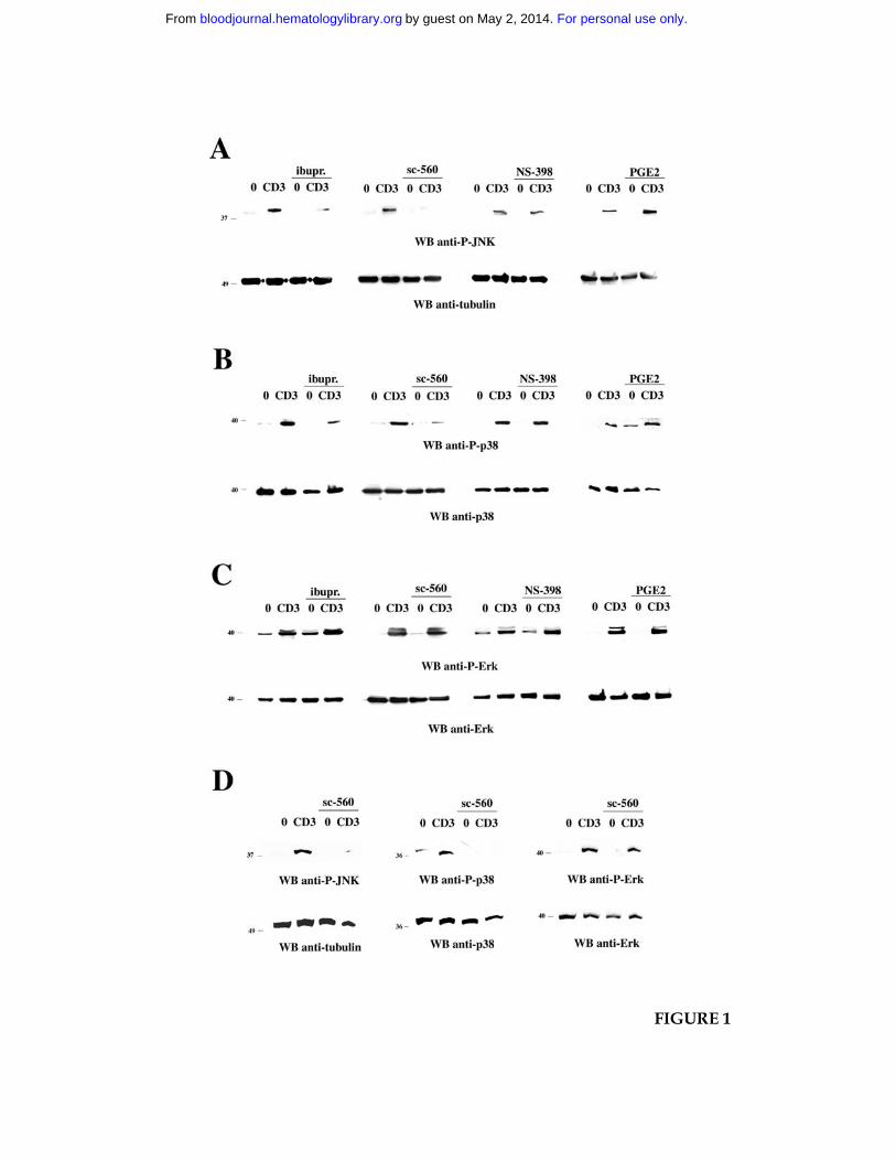
FIGURE 1
For personal use only.on May 2, 2014. by guest bloodjournal.hematologylibrary.orgFrom
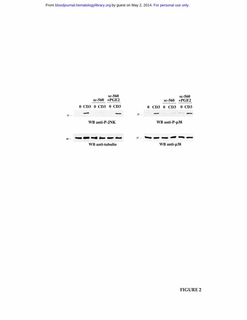
FIGURE 2
For personal use only.on May 2, 2014. by guest bloodjournal.hematologylibrary.orgFrom
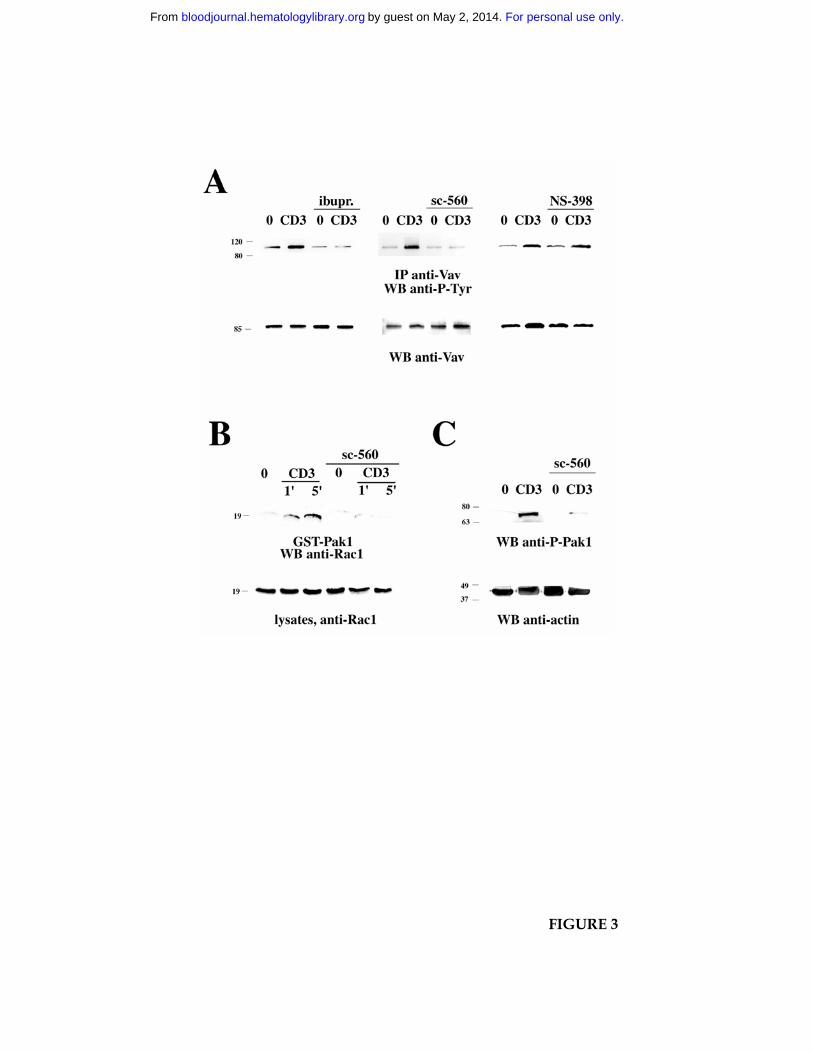
FIGURE 3
For personal use only.on May 2, 2014. by guest bloodjournal.hematologylibrary.orgFrom
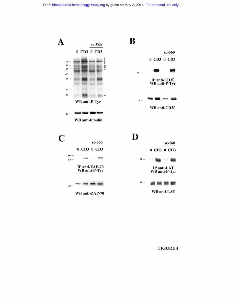
FIGURE 4
For personal use only.on May 2, 2014. by guest bloodjournal.hematologylibrary.orgFrom
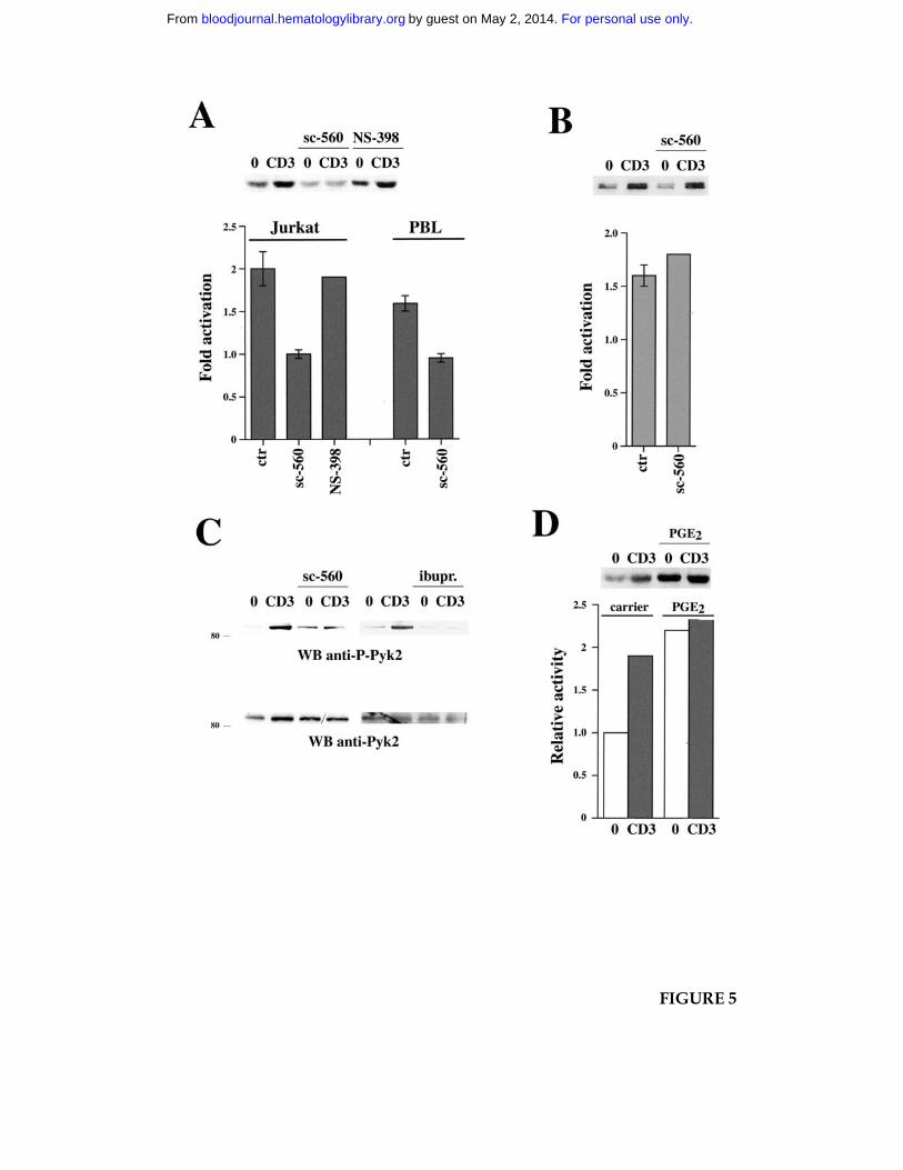
FIGURE 5
For personal use only.on May 2, 2014. by guest bloodjournal.hematologylibrary.orgFrom

FIGURE 6
For personal use only.on May 2, 2014. by guest bloodjournal.hematologylibrary.orgFrom
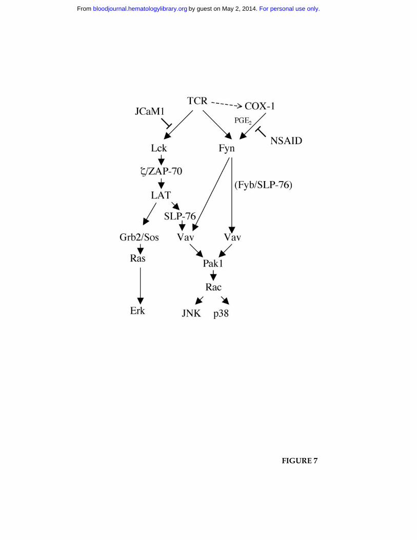
FIGURE 7
For personal use only.on May 2, 2014. by guest bloodjournal.hematologylibrary.orgFrom