Cordaites borassifolius (Sternberg) Unger (Cordaitales) from the Radnice Basin (Bolsovian, Czech...
36
Cordaites borassifolius (Sternberg) Unger (Cordaitales) from the Radnice Basin (Bolsovian, Czech Republic) ZBYNÌK IMÙNEK, STANISLAV OPLUTIL & JANA DRÁBKOVÁ The remains of cordaitalean plants are an important element in Bolsovian plant fossil assemblages from the Radnice Ba- sin (Czech Republic). The study of historic collections and new excavations brings together new data on the Cordaites type species – Cordaites borassifolius. Cordaites borassifolius was probably quite a large tree of monopodial or even sympodial stature. Its trunk diameter was at a minimum 0.5 m. Branches were between 1.1 m and spaced less than 0.7 m appart. The bases of the branches usually attained about 2/3 to 1/2 of the trunk width. The abaxial cuticle has stomata ar- ranged in multiplex stomatal rows that formed a wide stomatiferous band. A transverse crypt above the stoma is an im- portant diagnostic feature. The cordaitalean leaves, twigs, pith casts, fertile organs and seeds found are referable to a sin- gle natural species. The associated fertile organs belong to two types: 1) male fertile organs Florinanthus volkmannii and 2) a more robust, probably female, form similar to Cordaitanthus ovatus. Cuticles from the scales and long bracts of Florinanthus volkmannii have been studied in detail. Most scale cuticles are astomatal, but stomata may occur very rarely on some parts of the abaxial cuticle. Small trichomes grew from the scale margins. The cuticle of the bract has elongate cells and stomata are arranged in single stomatal rows on the abaxial cuticle. Many bilateral monosaccate pol- len grains [Florinites ovalis, Florinites guttatus and Pseudoillinites, with a central body bipolar attachment to the equa- torial saccus were separated from scale surfaces of Florinanthus volkmannii. The pith cast belong to the species Artisia approximata. The seeds are small and of the “Cardiocarpus-type”. Cordaites borassifolius grew in wet, peat-forming habitats and they were most likely trees of medium height. • Key words: Carboniferous, Radnice Basin, Cordaites, cuti- cles, cordaitalean fertile organs. ŠIMŮNEK, Z., OPLUŠTIL, S. & DRÁBKOVÁ, J. 2009. Cordaites borassifolius (Sternberg) Unger (Cordaitales) from the Radnice Basin (Bolsovian, Czech Republic). Bulletin of Geosciences 84(2), 301–336 (33 figures, 3 tables). Czech Geo- logical Survey, Prague. ISSN 1214-1119. Manuscript received March 6, 2009; accepted in revised form May 22, 2009; published online June 18, 2009; issued June 30, 2009. Zbyněk Šimůnek & Jana Drábková, Czech Geological Survey, Klárov 3/131, 118 21 Praha 1, Czech Republic, [email protected], [email protected] • Stanislav Opluštil, Faculty of Sciences, Charles University Praha, Albertov 6, 128 43 Praha 2, Czech Republic, [email protected] Recent excavations of fossil-bearing tuff bed (called bělka) at the base of the Whetstone Horizon at the Ovčín locality in the Radnice Basin (Opluštil et al. 2009) has provided a col- lection of well preserved plant fossils representing a peat- forming vegetation buried in growth position by volcanic ash. Due to this unique burial history, different plant organs derived from the same plant are preserved in a natural asso- ciation with the parent stem. These remains include cordai- talean leaves, pith casts, fertile organs such as cones and seeds, branches and several metre long stems. Cuticles isolated from 11 leaves found at the Ovčín locality are all conspecific with cuticles prepared from the holotype Cordaites borassi- folius (Sternberg) Unger, described by Sternberg (1821) from the Radnice Basin as the remains of a palm – Flabella- ria borassifolia. Since all the cordaitalean leaves found at the Ovčín locality appear to represent Cordaites borassifo- lius it seems that associated stems, branches and fertile or- gans could also belong to this species. Therefore this contri- bution is focused on evaluating the potential of these new findings for the reconstruction of Cordaites borassifolius. Geological setting Samples of Cordaites borassifolius discussed in this paper were recovered from the Radnice Basin, which represents only a small part of an extensive complex of late Palaeo- zoic continental basins in the Czech Republic (Fig. 1A). Sedimentary fill of these basins in Central and Western Bohemia ranges from the middle Westphalian (Bolsovian) through to late Stephanian (Pešek 1994) and it is divided into four formations (Fig. 2) based on the alternation of red beds and grey, coal-bearing strata (Weithofer 1902). How- ever, in the Radnice Basin, only the lower part of the oldest 301 DOI 10.3140/bull.geosci.1130
-
Upload
independent -
Category
Documents
-
view
0 -
download
0
Transcript of Cordaites borassifolius (Sternberg) Unger (Cordaitales) from the Radnice Basin (Bolsovian, Czech...

������������� ���� ���������������� ����������
����������������������������������� �������������
������ ����� ��������� ��������� ���� ��������
The remains of cordaitalean plants are an important element in Bolsovian plant fossil assemblages from the Radnice Ba-sin (Czech Republic). The study of historic collections and new excavations brings together new data on the Cordaitestype species – Cordaites borassifolius. Cordaites borassifolius was probably quite a large tree of monopodial or evensympodial stature. Its trunk diameter was at a minimum 0.5 m. Branches were between 1.1 m and spaced less than 0.7 mappart. The bases of the branches usually attained about 2/3 to 1/2 of the trunk width. The abaxial cuticle has stomata ar-ranged in multiplex stomatal rows that formed a wide stomatiferous band. A transverse crypt above the stoma is an im-portant diagnostic feature. The cordaitalean leaves, twigs, pith casts, fertile organs and seeds found are referable to a sin-gle natural species. The associated fertile organs belong to two types: 1) male fertile organs Florinanthus volkmanniiand 2) a more robust, probably female, form similar to Cordaitanthus ovatus. Cuticles from the scales and long bracts ofFlorinanthus volkmannii have been studied in detail. Most scale cuticles are astomatal, but stomata may occur veryrarely on some parts of the abaxial cuticle. Small trichomes grew from the scale margins. The cuticle of the bract haselongate cells and stomata are arranged in single stomatal rows on the abaxial cuticle. Many bilateral monosaccate pol-len grains [Florinites ovalis, Florinites guttatus and Pseudoillinites, with a central body bipolar attachment to the equa-torial saccus were separated from scale surfaces of Florinanthus volkmannii. The pith cast belong to the species Artisiaapproximata. The seeds are small and of the “Cardiocarpus-type”. Cordaites borassifolius grew in wet, peat-forminghabitats and they were most likely trees of medium height. • Key words: Carboniferous, Radnice Basin, Cordaites, cuti-cles, cordaitalean fertile organs.
ŠIMŮNEK, Z., OPLUŠTIL, S. & DRÁBKOVÁ, J. 2009. Cordaites borassifolius (Sternberg) Unger (Cordaitales) from theRadnice Basin (Bolsovian, Czech Republic). Bulletin of Geosciences 84(2), 301–336 (33 figures, 3 tables). Czech Geo-logical Survey, Prague. ISSN 1214-1119. Manuscript received March 6, 2009; accepted in revised form May 22, 2009;published online June 18, 2009; issued June 30, 2009.
Zbyněk Šimůnek & Jana Drábková, Czech Geological Survey, Klárov 3/131, 118 21 Praha 1, Czech Republic,[email protected], [email protected] • Stanislav Opluštil, Faculty of Sciences, Charles UniversityPraha, Albertov 6, 128 43 Praha 2, Czech Republic, [email protected]
Recent excavations of fossil-bearing tuff bed (called bělka)at the base of the Whetstone Horizon at the Ovčín locality inthe Radnice Basin (Opluštil et al. 2009) has provided a col-lection of well preserved plant fossils representing a peat-forming vegetation buried in growth position by volcanicash. Due to this unique burial history, different plant organsderived from the same plant are preserved in a natural asso-ciation with the parent stem. These remains include cordai-talean leaves, pith casts, fertile organs such as cones and seeds,branches and several metre long stems. Cuticles isolatedfrom 11 leaves found at the Ovčín locality are all conspecificwith cuticles prepared from the holotype Cordaites borassi-folius (Sternberg) Unger, described by Sternberg (1821)from the Radnice Basin as the remains of a palm – Flabella-ria borassifolia. Since all the cordaitalean leaves found atthe Ovčín locality appear to represent Cordaites borassifo-lius it seems that associated stems, branches and fertile or-
gans could also belong to this species. Therefore this contri-bution is focused on evaluating the potential of these newfindings for the reconstruction of Cordaites borassifolius.
����������������
Samples of Cordaites borassifolius discussed in this paperwere recovered from the Radnice Basin, which representsonly a small part of an extensive complex of late Palaeo-zoic continental basins in the Czech Republic (Fig. 1A).Sedimentary fill of these basins in Central and WesternBohemia ranges from the middle Westphalian (Bolsovian)through to late Stephanian (Pešek 1994) and it is dividedinto four formations (Fig. 2) based on the alternation of redbeds and grey, coal-bearing strata (Weithofer 1902). How-ever, in the Radnice Basin, only the lower part of the oldest
����������� �!"#$$�%&'()*�����

unit, the Kladno Formation, is preserved. These sedimentsbelong to the Radnice Member (Bolsovian), which is themost important coal-bearing unit of the basins in Centraland Western Bohemia. In the Radnice Basin, coal seamsare represented by the Lower and Upper Radnice Coals,which together comprise the Radnice group of seams. TheLower Radnice Coal is usually 1–4 m thick. The UpperRadnice Coal is the most important seam of the basin, lo-
cally > 10 m thick. Both coal seams are separated by theWhetstone Horizon, which tends to be a few meters thickbut locally can reach over 10 m in thickness. The Whet-stone Horizon has a sharp contact with the underlying rockabove which is a 0.6 m thick basal unit of pale yellow fossi-liferous tuff (the “bělka”), overlain by up to 10 m of the“whetstone”, poorly- to well-laminated tuffitic claystoneand mudstone (Fig. 2). The bělka or tuff bed contains uprightstanding stems of peat-forming plants. Observation of thedistribution of plant remains and their mode of occurrenceindicates a simple burial history of the former peat-formingforest (Opluštil et al. 2007, 2009). In contrast, the overlyingtuffitic mudstones (whetstone) contain only detrital plant re-mains concentrated on discrete bedding surfaces. The occa-sional larger plant fragments are randomly scattered withinthe unit. In the upper part of the whetstone, vertebrate and in-sect ichnofossils are frequently found (Turek 1989). Plantfossils of the Radnice Basin are mostly preserved in theWhetstone Horizon between the Lower and Upper RadniceCoals, and from the siliciclastic or volcanoclastic partingsand roof shale of the Upper Radnice Coal.
�������
Apart from Sternberg’s holotype of Cordaites borassifo-lius (Sternberg) Unger and Corda’s specimens from Svinná
��+
����������� ������ �������������
!�����"# Location of the study area. A – Late Palaeozoic continentalbasis of the Czech Republic. Circled B shows the position of the RadniceBasin. B – Radnice Basin and adjacent relicts of Carboniferous sediments(1 – Pre-Carboniferous rocks, 2 – Carboniferous sediments, 3 – LowerRadnice Coal in workable thickness and quality, 4 – closedmines/opencast mines, 5 – excavation at the Ovčín locality, 6 – urbanisedarea). Circled C shows the position of the Ovčín locality. C – geologicalmap of the Ovčín locality with the position of the Ovčín opencast mineand excavations. 1, 2 – Proterozoic basement (1 – shales, 2 – basicvolcanites), 3 – Radnice Member, 4 – fault, 5 – site of cordaitalean re-mains collected in the opencast mine, 6 – excavations (OE – Ovčín Exca-vations, SE – Sternberg Excavations), 7 – borehole, 8 – road.
!�����$# Lithostratigraphy of the Radnice Member (A) and architectureof the Whetstone Horizon at the Ovčín locality (B).
�
%% �

in northern part of the Radnice Basin (Figs 3A, 7), thestudy material described herein was collected at the Ovčínlocality near the town of Radnice in the southern part of theRadnice Basin (Fig. 1). At this locality plant remains werecollected mostly in the 1980’s during the operation of theopencast Ovčín Mine (formerly Pokrok Opencast Mine).Several tens of thousands of plant remains mostly from thebělka bed, forming the roof of the Lower Radnice Coal,with fewer specimens being from the associated siliciclas-tic sediments, were collected during the 8 years of theopencast operation (the mine was closed in 1986). All thefossils are now housed in the collections of the NationalMuseum in Prague. This material includes cordaitalean le-aves with excellently preserved cuticles from the bělka.Since the mine closed, additional specimens were collectedduring excavation of the fossiliferous bělka bed lateral tothe extent of the opencast mine. In all, five excavations ofthe basal Whetstone Horizon between 2002 and 2006, haveexposed a total area of about 150 m2. Three excavations na-med Ovčín excavation 1, 2, and 3 (OE1, 2, and 3) are loca-ted just at the edge of the former Ovčín opencast mine(Fig. 1). They exposed a continuous area of about 100 m2.
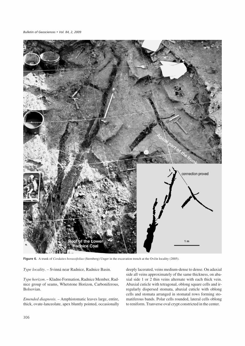
The other two excavations, Sternberg 1 (2005) and 2(2006) (SE1, and 2) are located about 100 m east of theopencast mine margin and 200 m northeast of OE 1, 2, and3 (Fig. 1). SE 1 and 2 exposed an area of about 50 m2.The main purpose of these excavations was the study ofspecies composition and structure of peat-forming forest.Therefore, the exposed surface was cleaned and grided bystring into 1 m2 units for precise location of the fossils. Thetuff was then carefully removed in slabs and all the fossilswere documented on a piece of graph paper unique to eachgrid unit. Such data provided detailed information aboutspatial distribution of species (Fig. 8D). The cordaitaleanremains from the opencast mine and excavations includefragments of stems, branches, pith casts and leaves, someattached to branches, as well as reproductive organs inclu-ding male and female cones and isolated seeds. The largestcordaitalean fragment was about 6 m long, a monopodiallybranched stem found in SE1 (Fig. 6). Although this stemwas leafless, it was associated with shed leaves of Cordai-tes borassifolius. All these findings contributed signifi-cantly to the whole-plant reconstruction of the species.These large specimens are stored in the West Bohemian
���
������ ��������� �� � !"# ��� �"��$�% #����� ��&�� '� �(
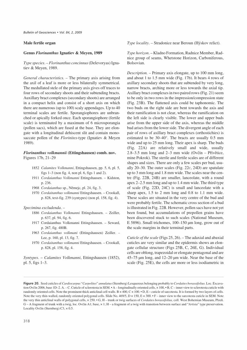
!�����&# Cordaites borassifolius (Sternberg) Unger. • A – holotype, locality Svinná, Radnice Basin, Coll. Sternberg, National Museum Prague, No. E5738, × 0.3. • B – abaxial cuticle with stomatal row isolated from the specimen on Fig 3A. Stomata have typical stomatal crypt. Slide No. 347/2, × 300.• C – adaxial cuticle isolated from the specimen on Fig 3A. Slide No. 347/2, × 300.
%
�

Museum in Plzeň, where they are in the process of restora-tion and preparation. More than 30 fragments of Cordai-tes borassifolius leaves have been used in this study, andcuticular slides are stored in the Czech Geological Surveyin Prague.
Cordaites borassifolius (Sternberg) Unger. – Material:About 100 specimens from the Přívětice – Ovčín locality,Pokrok Mine and new excavations OE1, 2, and 3, RadniceBasin, Radnice group of coals, Whetstone Horizon. Thesamples are stored in the National Museum, Prague (Nos.E 4749, E 4750, E 5738, E 5889–E 5892, E 5895–E 5898,E 6221–E 6223), Czech Geological Survey, Prague(No. ZŠ 301) and West Bohemian Museum, Plzeň (Nos.F 07705, F 07927, F 07931, F 07943, F 07952, F 07958,F 08044, F 08083, F 08084, F 08086, F 08132, F 08192,F 08197, F 08255, F 08333, F 08339, F 08414, F 08443,F 10578, FX 1, FX 3–5, FX 7–13). Cuticular slides:(Ovčín): 108/1–6; 120/1–3; 352/1–5; 353/1–2; 354/1–5;355/1–5; 356/1–6; 357/1–3; 358/1–5; 416/1–23; 417/1–5;468/1–8; (Svinná – holotype): 347/1–2 stored in the CzechGeological Survey in Prague.
Cordaitanthus ovatus (Lesquereux) Ignatiev & Meyen. –Material: West Bohemian Museum, Plzeň (Nos. F 02193,F 08058, FX 14–17).
Cardiocarpus annularis (Sternberg) Lesquereux. – Mate-rial: Sternberg’s (1820) specimens are stored in the Natio-nal Museum, Prague under the numbers E 1207 andE 1208. About 20 seeds from SE2 are stored in the WestBohemian Museum in Plzeň (Nos. F 07986A, B, F08449A, B, C, FX 10–13); cuticular slides: 469/1–5 arestored in the Czech Geological Survey in Prague.
Florinanthus volkmannii (Ettingshausen) comb. nov. –Material: Several cones on one slab from the National Mu-seum, Prague No. E 5896 (Pokrok opencast mine) and fromthe Ovčín excavation 2006 (SE) – West Bohemian Mu-seum, Plzeň Nos. FX 6 and FX 9; cuticular slides:416/1–23 stored in the Czech Geological Survey in Prague.
Artisia approximata Lindley & Hutton. – Material: WestBohemian Museum, Plzeň. Nos. F 08167, F 08168,F 08043, F 08045, F 08046, F 08059, F 08065, F 08074,F 08128, F 08129, F 08131, F 08139, F 08140, F 08142,F 08232, F 08331, F 08335, F 08389, F 08411, F 08426,F 08427, F 08442, F 08445, FX 2.
������
Cuticles were prepared according to the method describedby Kerp (1990). Coalified leafy fragments were separated
from the rock by placing it in concentrated hydrofluoricacid (HF) for several hours. The isolated fragments werethen bleached in Schulze’s reagent (40% HNO3 with crys-tals of KClO3 according to Krings & Kerp 1997 and Kerp& Krings 1999), a procedure which lasted 20 to 73 hoursdepending on preservation. After treatment in Schulze’sreagent, cuticles were washed in 10% potassium hydroxide(KOH) and finally rinsed in distilled water. Some cuticleswere stained with safranin for several hours to accentuateanticlinal walls and stomata. Before embedding in glyce-rine gelatine, the cuticles were dehydrated in pure glyce-rine. The remaining cuticular fragments were affixed tofilm for observation under SEM.
�'����������������
Division Gymnospermophyta (‘seed plants’)Class PinopsidaOrder Cordaitanthales (‘cordaites’) Meyen, 1984
Genus Cordaites Unger, 1850
Type species. – Cordaites borassifolius (Sternberg) Unger
Cordaites borassifolius (Sternberg) UngerFigures 3, 7, 8
1821 Flabellaria borassifolia Sternberg; Sternberg, vol. I,2: tent. 28, 32, pl. 18.
1825 Flabellaria borassifolia Sternberg; Sternberg, vol. I,4: tent. 34, pl. 34, fig. 1.
1825 Cycadites palmatus Sternberg; Sternberg, vol. I, 4,p. 39, tent. 33, pl. 40.
1845 Flabellaria borassifolia Sternberg. – Corda, p. 44,pl. 24, figs 1–3, 8.
1850 Cordaites borassifolia Sternberg sp. – Unger, p. 227.1852 Cordaites borassifolia Ung. – Ettingshausen, pp. 16,
17, pl. 5, fig. 5.1968 Cordaites borassifolius Sternb. – Němejc, p. 219,
pl. 26, fig. 1.2000 Cordaites borassifolius (Sternberg) Unger. – Šimů-
nek, p. 29, figs 3–12.2001 Cordaites borassifolius (Sternberg) Unger (morpho-
type 12 sensu Šimůnek 2000). – Šimůnek, fig. 35,pl. 1, figs 1, 2; pls 24–28.
2003 Cordaites borassifolius (Sternberg) Unger. – Zod-row, Mastalerz & Šimůnek, p. 97, figs 1A, B, D and 2.
2007 Cordaites borassifolius (Sternberg) Unger. – Šimů-nek, pp. 131–133, fig. 29a–j, pl. 1, figs 1, 2,pls 24–28, figs .
Holotype. – Figured by Sternberg (1821), pl. 18, NationalMuseum, Prague, No. Akc. 36675, E 5738.
��
����������� ������ �������������

��,
!�����(# Distribution of remains of Cordaites borassifolius in the excavations Ovčín 1–3. Numerical values given in the format (e.g., 30/150) corre-spond to diameter of branches or stems (e.g., 30 mm) and height above the top of the Lower Radnice Coal at which the remains were found(e.g., 150 mm). In this context, “b” means found at base of bělka tuff bed. If only a single value given, it relates to the distance above the roof of theLower Radnice Coal.
!�����)# Distribution of remains of Cordaites borassifolius in the excavations Sternberg 1 and 2.
������ ��������� �� � !"# ��� �"��$�% #����� ��&�� '� �(
Type locality. – Svinná near Radnice, Radnice Basin.
Type horizon. – Kladno Formation, Radnice Member, Rad-nice group of seams, Whetstone Horizon, Carboniferous,Bolsovian.
Emended diagnosis. – Amphistomatic leaves large, entire,thick, ovate-lanceolate, apex bluntly pointed, occasionally
deeply lacerated, veins medium-dense to dense. On adaxialside all veins approximately of the same thickness, on aba-xial side 1 or 2 thin veins alternate with each thick vein.Abaxial cuticle with tetragonal, oblong square cells and ir-regularly dispersed stomata, abaxial cuticle with oblongcells and stomata arranged in stomatal rows forming sto-matiferous bands. Polar cells rounded, lateral cells oblongto reniform. Tranverse oval crypt constricted in the center.
��-
!�����*# A trunk of Cordaites borassifolius (Sternberg) Unger in the excavation trench at the Ovčín locality (2005).
����������� ������ �������������
1 m

Description of the holotype. – The holotype is preserved asan imprint without coal matter on a 440 mm long and350 mm wide (Fig. 3A) slab which is also without coalmatter. It consists of two large leaf fragments and severalsmall fragments. The largest fragment is in the middleof the leaf rosette. It is 425 mm long and 50 mm wide at itswidest part in the middle. Another leaf is preserved behindthis first one, as can be seen in Sternberg’s (1821) figure.The leaf on the left is 390 mm long and 58 mm wide in itsmiddle part. The leaf margins taper towards the base andtowards the apex, which is not preserved in this slab. Anot-her three narrow leaf fragments occur among the large,wide leaves. These smaller fragments were apparently en-rolled before burial. There are another three smaller leaffragments to the right from the middle of the large frag-ment, the largest of which is about 180 mm long and 40 mmwide. However, this cannot have been its full length, as thebase and apex are not preserved. The base is also not pre-served in these three leaves, and the smallest width repre-sented is 18 mm. Due to the coarse nature of the rock, thevenation is preserved in only a few parts. It is very denseand partly discontinuous. About 50 wide veins occur in1 cm of leaf.
External morphology. – Trunk and branches: The ideasregarding whole-plant morphology are based on new mate-rial from SE1, a section of trunk about 5 m long with seve-ral articulated branches (Fig. 5) as well as several branchesterminated with palm-like arranged leaves. The preservedlength, however, exceeds the size of the excavation so thatonly the middle part of the trunk could be documented. Thelower part of the exposed trunk, about 50 mm above thecoal, is between 450 and 500 mm wide. The width of theopposite end of the stem is about 100 mm. The upper partof the trunk was, however, broken off and buried next tothe main trunk (Figs 5, 6). It is approximately 2.7 m longand its apex is absent. The entire height of the tree was esti-mated based on Niklas (1994) formula [log10H = 1.59 +0.39(log10D) – 0.18(log10D)2, where H = tree height andD = base diameter of stem], which suggested a height be-tween 28.1 and 28.6 m for the tree with a trunk diameter of500 mm. However, such height seems unlikely because themain axis exposed in the excavation exhibits a rapid decre-ase in diameter distally (from 450 to 150 mm over a lengthof just 5.7 m) due to sympodial branching. Therefore, webelieve that the tree was of the order of 15 to 20 m in height(Opluštil et al. 2009). The specimen displays monopodialto sympodial branching with four or five main lateral bran-ches, of which only the basal parts are preserved. Some ofthem display the typical Artisia-type of pith cast(Fig. 20H). Monopodial branching is demonstrated inFig. 20G. The branching apparently began only a few met-res from the base of the tree, resulting in a plant that pro-bably had a laterally and vertically wide crown. The dis-
tance between neighbouring branches decreases upward,from about 1.1 m in the lower part of the trunk to about0.7 m in its upper part. The existence of other branches onthe opposite side of the trunk cannot be proved. The basesof the branches usually attain about 2/3 to 1/2 of the trunkwidth. The angle of branching of the two lowest branches isacute (10° and 30°), whereas that of the upper two branchesis more obtuse (60° and 85°). However, it is possible thatthe preserved branching angles have been modified by vol-canic ash load and post-sedimentary compaction, especi-ally in the lower part of the trunk.
Leaves: No leaves have been found attached to the trunk,but a few C. borassifolius leaves were found in close asso-ciatiation with the trunk or its branches. This observation,in combination with the fact that all of the branches wereleafless, points to the conclusion that this tree had alreadyfallen prior to the eruption. These leaves were probably de-rived from neighbouring cordaites trees standing only fewmetres away.
In other excavations (OE2, 3 and SE2), branches withspirally arranged organically attached leaves were found.Leaves are, entire, ovate-lanceolate, rarely obovate, maxi-mum width 35–90 mm, length 400–700 mm. In the SE 2and OE 1 and 3 excavations (Fig. 4, 8D), the leaf length wasmostly between 450 and 550 mm being 493 mm on average.The terminal leaf rosette of Corda’s specimen from theSvinná locality (Fig. 7, National Museum, No. E 4270)is preserved without leaf terminals, unfortunately. Leaf
��.
!�����+# Cordaites borassifolius (Sternberg) Unger. Leafy apex of atwig. Counterpart of Corda’s (1845) specimen figured on pl. XXIV, fig. 1;loc. Svinná, Radnice Basin, Bolsovian, Coll. National Museum, Prague,No. E 4270. × 0.2.
������ ��������� �� � !"# ��� �"��$�% #����� ��&�� '� �(

fragments are up to 500 mm in length. The leaf bases are13–15 mm wide. Apices are bluntly pointed, but often ap-parently acute (because of the inrolling of the leaf mar-gins). The leaves are seldom lacerated. Selected leaf speci-mens were used for statistical evaluation. The leaf lengthcould only be measured on 13 specimens (Fig. 9), andtherefore the histogram is biased. Most of the leaves from
Ovčín are 450 to 550 mm long. The longest specimen,700 mm, is from Sternberg’s holotype. The mean length is493 mm. The leaves are often fragmentary thus width mea-surements do not usually represent the maximum, Themeasurement of 48 specimens produced highly variable re-sults as the measurements were taken at different positionson the leaves relative to where the widest point would havebeen. The narrowest fragmentary leaves are only15–20 mm wide (Fig. 10). They represent basal or apicalparts of leaves. Most of the leaves are about 35 mm wideand many samples are in the interval from 45 to 60 mm.Several samples are up to 92 mm wide. The mean of thisvaried width is 45 mm. Fig. 11 shows the correlation be-tween leaf length and width based on 13 complete leaves. Ifwe use data from only these 13 specimens for calculationof the mean width, we find that 60 mm is the mean width.The symbols shown in Fig. 11 represent the maximumwidth of leaves.
Veins: The leaf veins are straight, parallel to the marginsand to each other. The venation pattern depends on whetherthe adaxial or abaxial side is viewed, and on preservation.Usually one (or two) narrow veins alternate with one wide(or “true”) vein. The number of veins per 1 cm ranges con-siderably, from 18–62. By means of the cuticles, it is pos-sible to distinguish which parts represent the narrow andwide veins. The vein densities typically fall within a rangefrom 24 to 26 veins per cm and 46 to 50 veins per cm. The
��/
!�����,# Cordaites borassifolius (Sternberg) Unger, locality Ovčínnear Radnice, Pokrok Mine, Radnice Member, Bolsovian. • A – venationfrom a leaf, coll. National Museum, Prague, No.: E 5898; adaxial surfaceis white, abaxial surface is black. × 8. • B – venation with dense veins,probably of abaxial surface, coll. West Bohemian Museum, Plzeň, × 8.• C – venation with sparse veins alternating with very thin veins, probablyof adaxial surface, coll. West Bohemian Museum, Plzeň, × 8. • D – severalleaves from the terminal part of a twig,. West Bohemian Museum, Plzeň,× 0.4.
!�����-# Histogram of relative frequencies of leaf length of Cordaitesborassifolius (Sternberg) Unger plotted for 13 specimens.
����������� ������ �������������
�
.
%

former values probably represent the abaxial side, wherethe costal fields are prominent, whereas the latter valuesprobably represent the adaxial side, where the wide veinsare thinner and sometimes difficult to distinguish from thethin veins. The number of veins per 1 cm is shown onFig. 12 based on 34 specimens. It was taken from the wi-dest part of the studied leaf fragment. The veins terminateat the margin. Even though the mean is 34.3 veins per cm,the histogram has two peaks. The peak with a value of26 veins per cm probably represents the adaxial side(Fig. 8A, C), where differences between the vascular bun-
dles and sclerenchymatous tissue are prominent, whereasthe peak with a value of 44–50 veins per cm probably re-presents the abaxial side (Fig. 8B). The strips of vascularbundles and sclerenchymatous tissue have approximatelythe same thickness, and they are usually considered as ordi-nary veins (Fig. 13). Even under magnification it is notpossible to distinguish which veins belong to vascular bun-dles and which veins belong to sclerenchymatous tissue.The leaves are amphistomatic.
Cuticles. – Cuticle preparations make it possible to distin-guish between narrow and wide veins.
��0
!�����"/# Histogram of relative frequencies of leaf width of Cordaitesborassifolius (Sternberg) Unger plotted for 48 specimens.
!�����""# Graph showing the correlation between the length and widthof Cordaites borassifolius (Sternberg) Unger leaves plotted for 13 speci-mens.
!�����"$# Histogram of relative frequencies of number of veins per1 cm for Cordaites borassifolius (Sternberg) Unger leaves plotted for34 specimens.
!�����"&# Venation of Cordaites borassifolius (Sternberg) Unger.• a – adaxial side, • b – abaxial side with two different vein thicknesses,and fine transverse wrinkles in the place of stomatiferous bands.
������ ��������� �� � !"# ��� �"��$�% #����� ��&�� '� �(
2 mm
��

Adaxial cut ic le (Fig. 14): Some adaxial cuticles havebands of cells that are slightly darker than others. The lig-hter cells are costal bands above vascular strands and areusually 80–160 μm wide. The darker intercostal bands are240–490 μm wide with very rarely stomata. Occasionally,the costal band is only 50 μm wide and probably representsthe epidermis above the sclerenchymatic tissue.
The adaxial cuticle is weakly cutinised, but moreprominently than the abaxial cuticle. Cells are the same inboth costal and intercostals areas. They are tetragonal andmostly oblong, and less often square or pentagonal to hex-agonal in shape. The cells are 30–75 μm long and20–35 μm wide. Anticlinal walls are slightly or stronglybent. The cells have rounded corners and are parallel to
the veins. Stomata are at distances approximately 400 μmapart, but not in regular rows (Fig. 14A). The stomatalcomplex is formed by a pair of sunken elliptical guardcells, two polar subsidiary cells and two lateral subsidiarycells. Pairs of guard cells are 22–26 μm long and13–16 μm wide. The complex is 60–80 μm long and60–70 μm wide. Polar subsidiary cells are prominent,oval, and 20–30 μm in diameter (Fig. 14B). Lateral sub-sidiary cells are prominent, oblong, with bulging anticli-nal walls, 34–50 μm long and 16–25 μm wide. A square toround outer stomatal cavity is developed above thestomatal pore. The stomatal density is very low, rangingfrom 6–9 stomata per 1 mm2. The value of the stomatal in-dex varies from 0.7 to 0.9 (Table 1).
���
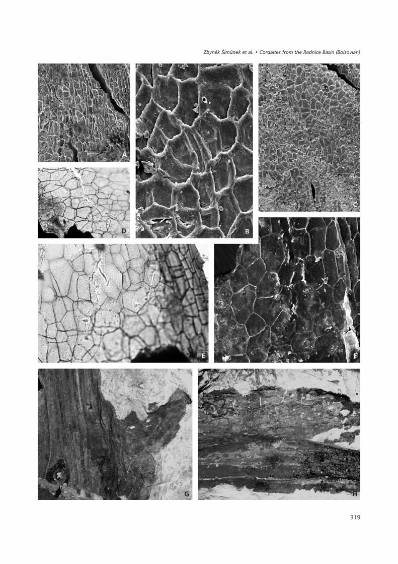
!�����"(# Adaxial epidermis of Cordaites borassifolius (Sternberg) Unger, locality Ovčín near Radnice, Pokrok Mine, Radnice Member, Bolsovian.• A – adaxial epidermis with stomata, v, s – cells of “costal” fields: v – above the vascular bundles, s – above the sclerenchyma strands. Slide No. 353/2,× 100. • B – detail from Fig. 14A with a stoma, × 400. • C – SEM microphotograph No. 0754. View to inner side with prominent anticlinal cell walls,× 200. • D – SEM microphotograph No. 0758. View to outer surface with a small stoma, × 500. • E – SEM microphotograph No. 0759. Detail of a stomafrom Fig. 14D, × 2000.
����������� ������ �������������
�
. 0
%
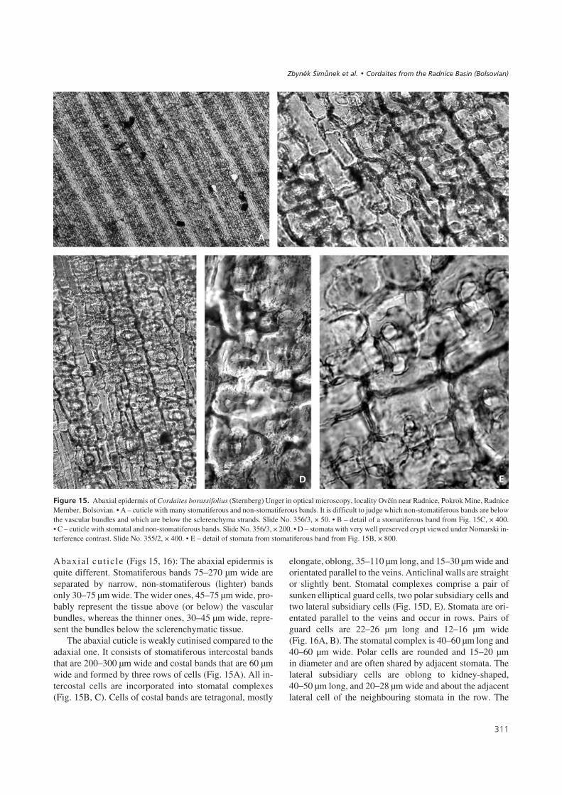
Abaxial cut ic le (Figs 15, 16): The abaxial epidermis isquite different. Stomatiferous bands 75–270 μm wide areseparated by narrow, non-stomatiferous (lighter) bandsonly 30–75 μm wide. The wider ones, 45–75 μm wide, pro-bably represent the tissue above (or below) the vascularbundles, whereas the thinner ones, 30–45 μm wide, repre-sent the bundles below the sclerenchymatic tissue.
The abaxial cuticle is weakly cutinised compared to theadaxial one. It consists of stomatiferous intercostal bandsthat are 200–300 μm wide and costal bands that are 60 μmwide and formed by three rows of cells (Fig. 15A). All in-tercostal cells are incorporated into stomatal complexes(Fig. 15B, C). Cells of costal bands are tetragonal, mostly
elongate, oblong, 35–110 μm long, and 15–30 μm wide andorientated parallel to the veins. Anticlinal walls are straightor slightly bent. Stomatal complexes comprise a pair ofsunken elliptical guard cells, two polar subsidiary cells andtwo lateral subsidiary cells (Fig. 15D, E). Stomata are ori-entated parallel to the veins and occur in rows. Pairs ofguard cells are 22–26 μm long and 12–16 μm wide(Fig. 16A, B). The stomatal complex is 40–60 μm long and40–60 μm wide. Polar cells are rounded and 15–20 μmin diameter and are often shared by adjacent stomata. Thelateral subsidiary cells are oblong to kidney-shaped,40–50 μm long, and 20–28 μm wide and about the adjacentlateral cell of the neighbouring stomata in the row. The
���
!�����")# Abaxial epidermis of Cordaites borassifolius (Sternberg) Unger in optical microscopy, locality Ovčín near Radnice, Pokrok Mine, RadniceMember, Bolsovian. • A – cuticle with many stomatiferous and non-stomatiferous bands. It is difficult to judge which non-stomatiferous bands are belowthe vascular bundles and which are below the sclerenchyma strands. Slide No. 356/3, × 50. • B – detail of a stomatiferous band from Fig. 15C, × 400.• C – cuticle with stomatal and non-stomatiferous bands. Slide No. 356/3, × 200. • D – stomata with very well preserved crypt viewed under Nomarski in-terference contrast. Slide No. 355/2, × 400. • E – detail of stomata from stomatiferous band from Fig. 15B, × 800.
�
. 0
%
������ ��������� �� � !"# ��� �"��$�% #����� ��&�� '� �(
outer stomatal cavity is developed transversely across thelateral subsidiary cells. It is elongate and constricted in thecentral part (Fig. 15D, E, 16C–F). Stomatal complexes arearranged in rows; with three to six rows forming a band.The stomatal density is 350–370 per 1 mm2. The value ofthe stomatal index varies from 16 to 20 (Table 1).
Remarks. – A detailed description of material later assig-ned to C. borassifolius by Unger 1850 was made by Corda(1845), who described large specimens from Svinná in theRadnice Basin (Fig. 7) under the name Flabelaria borassi-folia. He also studied the anatomy of partly carbonisedstems and cuticles of leaves. Corda (1845) described the ty-pical Cordaites borassifolius venation as narrow and widealternating veins. A few years later, Unger (1850) erectedthe name Cordaites, and the species Cordaites borassifo-lius was selected as the type of this genus. Unger’s defini-tion was based on both leaf form and stem anatomy.
Zeiller (1886, p. 628) excluded from Cordaites boras-sifolius leaves figured by Ettingshausen (1852, pl. 5, fig. 5).According to Zeiller (1886), the veins are varied and he
suggested that Ettinghausen’s material instead belonged toCordaites principalis (Germar) Geinitz. This opinion wasalso accepted by Crookall (1970). However, Ettings-hausen’s (1852) specimen comes from Stradonice (Hýskovrelict), which is relatively close to Svinná, the type area ofC. borassifolius. In fact, the horizon is practically the sameas the unit from which the holotype of C. borassifolius wascollected. The venation pattern can vary, as we know fromthe C. borassifolius sample population from the Ovčín lo-cality. Unfortunately, some cuticles prepared from a C.borassifolius specimen from Stradonice (NaturhistorischesMuseum, Wien) are strongly corroded, which does not allowcomparison with the holotype of C. borassifolius. The cellu-lar structures of the adaxial epidermis of C. borassifoliuswere already studied by Corda (1845, pl. 24, figs 2, 3). Heobserved leaves without maceration and correctly inter-preted the alternating thin and thick veins, cellular shape andorientation and also the stomata distribution. However, hewas unable to observe details of the stomatal structure.
Florin (1931, p. 499, fig. 105a) figured abaxial cuticlesfrom a specimen determined as Cordaites sp. 1, which origi-
��+
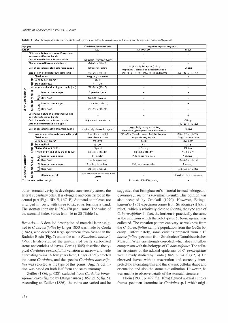
1�����"# Morphological features of cuticles of leaves Cordaites borassifolius and scales and bracts Florinites volkmannii.
����������� ������ �������������
nated from the Radnice Member (Bolsovian) of theRakovník locality, Czech Republic, and from another speci-men he referred to as Cordaites sp. 2 from New Brunswick(Westphalian D), Canada. The stomatal complexes of bothspecimens are structurally similar; two large lateral subsid-iary cells and two small polar subsidiary cells. They onlydiffer in the shape of these cells. Both species have a cryptacross the guard cells as in C. borassifolius. However, theydiffer in the shape of the subsidiary cells and therefore theirmutual identity is questionable. These samples differ fromour material in the shape of the subsidiary cells and the dis-tribution of stomata. Stomata of Florin’s (1931) Cordaitessp. 1 are arranged in simple stomatal rows, whereas stomataof Cordaites borassifolius are in multiple rows in bands.
Barthel (1964) described Cordaites cuticular morpho-type 6 from the Autunian of the Döhlen Basin as having atransverse crypt across the guard cells, which is very simi-lar to the Bohemian species Cordaites borossifolius (Stern-berg) Unger from the Radnice Member. Barthel comparedthis species with Florin’s (1931) Cordaites sp. 1 from theBolsovian of Rakovník.
Fertile organ of indeterminate sex (possibly female). –Two types of strobili were found at the Ovčín localities.We consider the more robust form to be the female fertileorgan even though seeds have not yet been found in con-nection with it [determined as Cordaitanthus ovatus (Les-quereux) Ignatiev & Meyen], and the smaller form as the
���
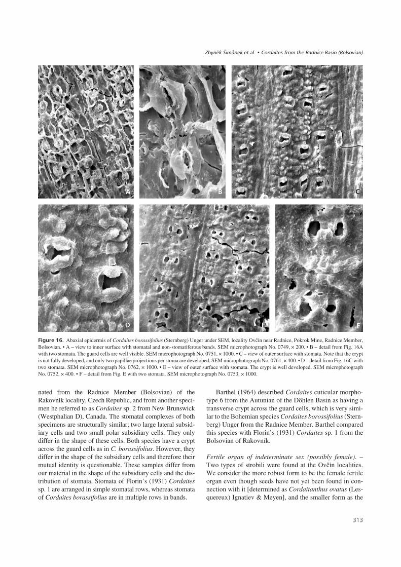
!�����"*# Abaxial epidermis of Cordaites borassifolius (Sternberg) Unger under SEM, locality Ovčín near Radnice, Pokrok Mine, Radnice Member,Bolsovian. • A – view to inner surface with stomatal and non-stomatiferous bands. SEM microphotograph No. 0749, × 200. • B – detail from Fig. 16Awith two stomata. The guard cells are well visible. SEM microphotograph No. 0751, × 1000. • C – view of outer surface with stomata. Note that the cryptis not fully developed, and only two papillae projections per stoma are developed. SEM microphotograph No. 0761, × 400. • D – detail from Fig. 16C withtwo stomata. SEM microphotograph No. 0762, × 1000. • E – view of outer surface with stomata. The crypt is well developed. SEM microphotographNo. 0752, × 400. • F – detail from Fig. E with two stomata. SEM microphotograph No. 0753, × 1000.
�
. 0 !
%
������ ��������� �� � !"# ��� �"��$�% #����� ��&�� '� �(

male fertile organ of the form refereable to as Florinanthusvolkmannii (Ettingshausen) Šimůnek, Opluštil & Dráb-ková comb. nov.
Genus Cordaitanthus Feistmantel, 1876
Type species. – Cordaitanthus communis Feistmantel,1876
Basionym. – Antholithes triticum Andrae, 1864, pp. 174,175, pl. 4, fig. 6.
Proposed lectotype. – Cordaitanthus communis Feistman-tel, 1876, pl. 12, fig. 4.
Remark. – Feistmantel (1876) on pl. 12, figs 1–4, figured 4syntypes of his Cordaitanthus communis. They come fromdifferent localities (Stradonice, Kralupy nad Vltavou, Ný-řany) and evidently they belong to different species. Wepropose to choose Feistmantel’s (1876) specimen figuredin his pl. 12, fig. 4, as a lectotype of the genus Cordaitan-thus. Feistmantel (1876) refigured Andrae’s (1864) Antho-lithes triticum from pl. 4, fig 6. Antholithes triticum is anearlier nomenclatural synonym of Cordaitanthus commu-nis and takes priority over Cordaitanthus communis. The-refore Cordaitanthus triticus becomes the effective typespecies of Cordaitanthus.
Diagnosis of the genus Cordaitanthus. – Original diagno-sis of Ignatiev & Meyen, 1989. Primary axis elongate, upto 300 mm long, and 15 mm wide. Axillary bract comple-xes borne on both sides of the axis, arranged in juxtaposi-tion or alternate, and composed of helically arrangedscaly appendages. Bracts ranging from scaly toneedle-shaped, up to 33 mm long. The bracts frequentlyhave become turned down and the fertile organ looks as ifit lacks bracts.
Cordaitanthus ovatus (Lesquereux) Ignatiev & Meyen,1989Figures 17a, 18A–F
1880 Cordaianthus ovatus Lesquereux, p. 545, pl. 76,fig. 5, 5a.
1989 Cordaitanthus ovatus Lesquereux. – Ignatiev & Meyen,p. 335.
Type locality. – Cannelton, Pennsylvania, U.S.A.
Type horizon. – Westphalian D.
Description. – Based on material from OE1, 2, and 3, WestBohemian Museum, No. F 08054 and F 07986A. Frag-ments of primary axes up to 90 mm long (Fig. 18A) and ty-pically 3 mm wide. The axillary complexes are borne onboth sides of the axis somewhat irregularly and alternately.They are closely arranged in the terminal part and sparsely
��
!�����"+# Fertile organs of Cordaites borassifolius (Sternberg) Unger,• a – probably female cone Cordaitanthus ovatus (Lesquereux) Ignatiev &Meyen, • b – male cone Florinanthus volkmannii (Ettingshausen) comb.nov. Scale bar = 10 mm.
!�����",# A–F – Cordaitanthus cf. ovatus (Lesquereux) Ignatiev & Meyen. Probably the female fertile organ of Cordaites borassifolius. LocalityOvčín near Radnice, Radnice Member, Bolsovian. Coll. West Bohemian Museum, Plzeň, × 1. C – detail from Fig. 18B, × 3. D – detail from Fig. 18A, × 5.E, F – cone fragments, E – locality Ovčín, D1; 100 mm above coal, No. F 08054, × 2; F – locality Ovčín, No. F 02193, × 3. • G–K – seeds Cardiocarpusannulatus (Sternberg) Lesquereux that probably belong to Cordaites borassifolius; coll. West Bohemian Museum, Plzeň. G – locality Ovčín, D3 base,No. F 08045, × 3; H – locality Ovčín, D 2 base, No. F 07986B, × 3; I – locality Ovčín, D 2 base, No. F 07986A, × 3. J, K – imprint and counterpart of Stern-berg’s type specimens of Cardiocarpus “Carpolites” annulatus Sternberg, possible seed of Cordaites borassifolius. Locality Radnice, coll. NationalMuseum, Prague, Nos. E 1207 and 1208, × 3.
����������� ������ �������������
��
10 mm

��,
.
�
�0 !
2 3 4 5
%
������ ��������� �� � !"# ��� �"��$�% #����� ��&�� '� �(

in the basal part of the fertile organ. Axillary buds are for-med by a small secondary axis and helically arranged sca-les. The shape, dimension and number of scales per bud areusually difficult to determine due to poor preservation. Thebuds are 6–9 mm long and 4–6 mm wide. Their apex is co-nical (Fig. 18D). The shape of the scales is probably lance-olate. The bracts are 0.4 to 1 mm wide, 10–15 mm long,and turned upwards (Fig. 18C).
Remarks. – The specimens found at Ovčín OE1, 2, and 3 fitLesquereux’s description of Cordaitanthus ovatus. It ismore robust than the male fertile organ Florinanthus volk-mannii (Fig. 17B), and for this reason we consider it to bethe female fertile organ of Cordaites borassifolius, althoughdirect evidence is lacking. The buds of Cordaitanthus ovatusare 6–9 mm long and 4–6 mm wide, whereas those of Flori-nanthus volkmannii are 2–4 mm long and only 1.5–3 mmwide. The sterile scales of C. ovatus are lanceolate, whereasthose of F. volkmannii are spatulate. Only the small innerscales of F. volkmannii are lanceolate.
Seeds(Figs 18G–K, 19, 20A–F)
Sternberg (1820) figured 12 seeds of the genus “Carpolites”from Radnice, and some of them could belong to cordaita-lean plants. Of these seeds, Carpolites (Cardiocarpus) an-nularis Sternberg, 1820 (Lesquereux, 1884), Carpolites(Cardiocarpus) regularis Sternberg, 1820 (Lesquereux,1880), and perhaps Carpolites (Cardiocarpus) retusumSternberg, 1820 (Newberry, 1873) are most similar to theseeds found at Ovčín. Because Sternberg (1820) did notdescribe them, all of these names are invalid as nominanuda. We believe that the shape and dimensions of materialfrom Ovčín fit best to the name Carpolites (Cardiocarpus)annularis and therefore we add its description and diagno-sis to validate this name.
Cardiocarpus annularis (Sternberg) Lesquereux, 1880Figures 18G–K, 19, 20A–F
1820 Carpolites annularis Sternberg, pl. 7, fig. 15.1825 Carpholites annularis Sternberg, tent. 40.1884 Cardiocarpus annularis (Sternberg). – Lesquereux,
p. 814, pl. 110, figs 28–30.
Holotype. – National Museum, Prague, No. E 1207, Stern-
berg, pl. 7, fig. 15, refigured here on Fig. 18J; counterpart E1208 is figured here on Fig. 18K.
Diagnosis. – Seeds small, round to oval, surrounded bynarrow rim (sarcotesta), surface of sclerotesta smooth(occasionally crumpled). Epidermis composed of polygo-nal cells.
Description of the holotype (Fig. 18J). – The seed isoval, 13 mm long and 11 mm wide, the sarcotesta rim iswidest near the base > 1 mm and tapers near the apex to0.7 mm.
Description. – Seeds are small wingless, platyspermic,round or oval, and in their terminal parts a little tapered.The inner sclerotesta layer and outer sarcotesta layer arepreserved (Fig. 18I–K, 19D, E). The sclerotesta consistsof thick-walled sclerotic cells and the sarcotesta of fleshycells. The seeds are 10–11 mm long and 9–11 mm wide.Most of the seed area is occupied by sclerotesta, while thesarcotesta forms a narrow rim less than 0.7 mm thick.
Cuticles. – Two types of cuticles have been recoveredfrom the SE material. The most common is a thick-walledcuticle with randomly orientated polygonal cells, 40–90μm long and 30–50 μm wide (Fig. 19H, I). The anticlinalwalls are very thick and prominent (Fig. 20B). In someparts, the cells tend to be elongated, tetragonal, parallelorientated (Fig. 19F, G), 40–110 μm long and 20–40 μmwide. Only one stomata-like oblong aperture (Fig. 19J,K), 17 μm long and 12 μm wide, surrounded by 5 verysmall subsidiary cells was observed. Guard cells are notpresent.
The second type consists of two thin-walled layers ofcells (Fig. 20D–F). The cells are polygonal, 35–100 μmlong and 35–50 μm wide. The anticlinal walls are very thin(Fig. 20F).
Remark. – It is not exactly known from which part of theseed a particular cuticular type came from, however,we presume that the thick-walled cuticle covered thesclerotesta and the thin-walled cuticle covered the sarco-testa, because this layer is fleshy and its cuticle is usuallythinner than cuticle of the sclerotesta. Dispersal of thesewingless seeds was limited and resulted in them beingclustered on the ground at the base of the tree as they werefound in Ovčín locality.
��-
!�����"-# Seeds of Cordaites borassifolius comparable to Cardiocarpus “Carpolites” annulatus (Sternberg) Lesquereux. Locality Excavation Ovčín2006, base 1D–2. • A – accumulation of seeds together with Cordaites borassifolius leaves, × 1. • B – detail from Fig. 19A, × 2. • C, D – detail fromFig. 19A. Most of the seed body encompass sclerotesta and only a thin rim sarcotesta. × 3. • E – a seed adpression from excavation “Ovčín”, × 3.• F–K – seed cuticle probably from the sclerotesta layer from seeds on Fig. 19A. F, G –cuticle with longitudinally oriented cells, slide No. 469/2, F × 100,G × 200; J, I – cuticle with randomly oriented cells, slide No. 469/1, H × 100, I × 200; J, K – stomata-like structure, slide No. 469/3. J × 400, K × 200.
����������� ������ �������������

��.
�
. 0 2
3
� 2
%
4 5
!
������ ��������� �� � !"# ��� �"��$�% #����� ��&�� '� �(
Male fertile organ
Genus Florinanthus Ignatiev & Meyen, 1989
Type species. – Florinanthus concinnus (Delevoryas) Igna-tiev & Meyen, 1989.
General characteristics. – The primary axis arising fromthe axil of a leaf is more or less bilaterally symmetrical.The medullated stele of the primary axis gives off traces tofour rows of secondary shoots and their subtending bracts.Auxillary bract complexes (secondary shoots) are arrangedin a compact helix and consist of a short axis on whichthere are numerous (up to 100) scaly appendages. Up to 40terminal scales are fertile. Sporangiophores are unbran-ched or apically forked once. Each sporangiophore (fertilescale) is terminated by a maximum of 6 microsporangia(pollen sacs), which are fused at the base. They are elon-gate with a longitudinal dehiscent slit and contain mono-saccate pollen of the Florinites-type (Ignatiev & Meyen1989).
Florinanthus volkmannii (Ettingshausen) comb. nov.Figures 17b, 21–29
1852 Calamites Volkmanni, Ettingshausen, pp. 5, 6, pl. 5,figs 1–3 (non fig. 4, non pl. 6, figs 1 and 2).
1911 Cordaianthus Volkmanni Ettingshausen. – Kidston,p. 236.
1968 Cordaianthus sp., Němejc, pl. 24, fig. 3.1970 Cordaitanthus volkmanni Ettingshausen. – Crookall,
p. 828, text-fig. 239 (syntypes) (non pl. 158, fig. 4).
Specimina excludenda. –1886 Cordaianthus Volkmanni Ettingshausen. – Zeiller,
p. 637, pl. 94, fig. 6.1917 Cordaianthus Volkmanni Ettingshausen. – Seward,
p. 267, fig. 480B.1963 Cordaianthus volkmani (Ettingshausen) Zeiller. –
Lee, p. 160, pl. 13, fig. 7.1970 Cordaitanthus volkmanni Ettingshausen. – Crookall,
p. 828, pl. 158, fig. 4.
Syntypes. – Calamites Volkmanni, Ettingshausen (1852),pl. 5, figs 1–3.
Type locality. – Stradonice near Beroun (Hýskov relict).
Type horizon. – Kladno Formation, Radnice Member, Rad-nice group of seams, Whetstone Horizon, Carboniferous,Bolsovian.
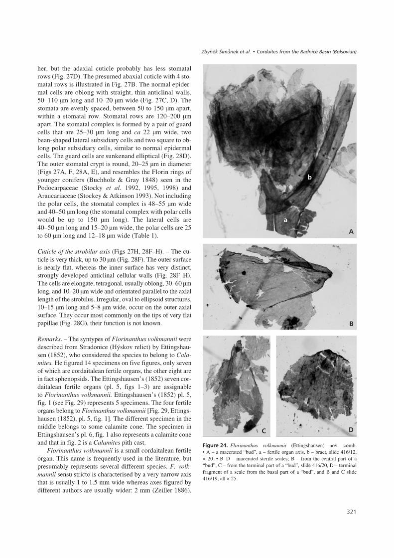
Description. – Primary axis elongate, up to 100 mm long,and about 1 to 1.5 mm wide (Fig. 17b). It bears 4 rows ofaxillary secondary shoots that are subtended by very long,narrow bracts, arching more or less towards the axial tip.Axillary bract complexes in two paired rows (Fig. 21) seemto be only in two rows in the impression/compression state(Fig. 23B). The flattened axis could be taphonomic. Thetwo buds on the right side are bent towards the axis andtheir ramification is not clear, whereas the ramification onthe left side is clearly visible. The lower and upper budsarise from the upper side of the axis, whereas the middlebud arises from the lower side. The divergent angle of eachpair of rows of axillary bract complexes (orthostichies) isestimated to be 30–40°. The bracts are usually 0.5 mmwide and up to 25 mm long. Their apex is sharp. The buds(Fig. 22A) are relatively small and wide, usually2.8–3.5 mm long and 2–3 mm wide (Ovčín – Přívětice,mine Pokrok). The sterile and fertile scales are of differentshapes and sizes. There are only a few scales per bud, usu-ally 20–30. The outer scales (Fig. 22C, 24D) are spatulateup to 3 mm long and 1.8 mm wide. The scales near the cen-tre (Fig. 22B, 24B) are smaller, lanceolate, with a roundapex 2–2.5 mm long and up to 1.4 mm wide. The third typeof scale (Fig. 22D, 24C) is small and lanceolate with asharp apex, 1.5 to 2 mm long and 0.8 to 1.1 mm wide.These scales are situated in the very centre of the bud andwere probably fertile. The schematic cross section of a budis illustrated in Fig. 22B. However, pollen sacs have not yetbeen found, but accumulations of prepollen grains havebeen discovered stuck to such scales (National Museum,E 5896). Small trichomes, 100–150 μm long, grow out ofthe scale margins in their terminal parts.
Cuticle of the scale (Figs 25, 26). – The adaxial and abaxialcuticles are very similar and the epidermis shows an elon-gate cellular structure (Figs 25B, C, 26E, G). Individualcells are oblong, trapezoidal or elongate pentagonal and are45–75 μm long, and 12–20 μm wide. Near the base of thescale (Fig. 25E), the cells are more or less isodiametric in
��/
!�����$/# Seed cuticles of Cardiocarpus “Carpolites” annulatus (Sternberg) Lesquereux belonging probably to Cordaites borassifolius. Loc. Excava-tion Ovčín 2006, base 1D–2. A. – C. Cuticle of sclerotesta in SEM. • A – longitudinally oriented cells, × 100, • B, C – inner view to sclerotesta cuticle withrandomly oriented cells. Note the prominent thick anticlinal cell walls. B × 400, C × 100. • D, E – cuticle of sarcotesta. It is formed by two layers of cells.Note the very thin-walled, randomly oriented polygonal cells. Slide No. 469/5, D × 150, E × 300. • F – inner view to the sarcotesta cuticle in SEM. Notethe very thin anticlinal walls of polygonal cells, × 250. • G, H – trunk or twig surfaces of Cordaites borassifolius, coll. West Bohemian Museum, Plzeň.G – A fragment of trunk with a twig, loc. Ovčín A1; base, × 1; H – a fragment of a twig with transition between surface and “Artisia” type preservation.Locality Ovčín (Sternberg) C3, × 0.5.
����������� ������ �������������
��0
. �
�
!
%
2
0
������ ��������� �� � !"# ��� �"��$�% #����� ��&�� '� �(

shape, randomly orientated and 30–50 μm in diameter. Theanticlinal cell walls are straight or slightly bent. The sto-mata are irregularly dispersed on the cuticle (Fig. 25A, B),but it is not known whether they occur on the adaxial orabaxial cuticle. The stomatal complex is simple, consistingof two guard cells surrounded by 4 to 6 subsidiary cells ofthe same shape as the ordinary epidermal cells. The visiblepart of the guard cells is somewhat oblong, 22–28 μm long,and 10–15 μm wide. The stomatal density varies from noneto 40 stomata per 1 mm2 (measured on 0.1 mm2 area) andthe stomatal index is less than 1. Three rows of trichomesare prominent along the scale margin (Fig. 26A, C, D).They are simple, uniseriate, 100–150 μm long and 12–20 μmwide (Fig. 25F), and are formed by 3 to 4 barrel-shaped ce-lls and a terminal cell that is elongate triangular in shapearising almost perpendicular to the other cells. The anticli-nal cell walls are moderately developed. The periclinal wa-lls are slightly arched and bear papillae and small depressi-ons. Papillae are small with a bluntly pointed tip, their baseis elliptical, 25–35 μm long and 10–15 μm in height(Fig. 26H). Small elliptical depressions of unknown fun-ction are seen on some periclinal walls (Fig. 26H). Thesedepressions are 12–18 μm long and 4–7 μm wide(Fig. 26B, E). They appear to be small stoma and representtrue holes, 7 μm long and 2 μm wide, through the cuticle.They are transversely orientated to the cell direction.
Cuticle of the bract (Figs 27, 28A–E). – The adaxial andabaxial cuticles are difficult to distinguish from one anot-
�+�
!�����$$# Florinanthus volkmannii (Ettingshausen) comb. nov. – re-construction of a bud (A) and its section (B), scale bars = 1 mm.• C–E – sterile scales of the cone with different shape, scale bar =1 mm.
!�����$"# Part of the male cone Florinanthus volkmannii (Ettings-hausen) nov. comb. (reconstruction by J. Svoboda), scale bar = 2 mm.
!�����$&# Florinanthus volkmannii (Ettingshausen) comb. nov., a malefertile organ of Cordaites borassifolius. Loc. Ovčín near Radnice, PokrokMine, Radnice Member, Bolsovian; National Museum in Prague, No.5896. • A – a specimen with Florinanthus cones and Cordaites bo-rassifolius leaves, × 1. • B – isolated part of Florinanthus cone fromFig. 23A with 5 fertile “buds” and bracts, Slide No. 416/22, × 5. • C – a de-tail of two cones from Fig. 23A, × 5.
����������� ������ �������������
�
. 0
%
1m
m
1m
m2
mm
1m
m
%
�
her, but the adaxial cuticle probably has less stomatalrows (Fig. 27D). The presumed abaxial cuticle with 4 sto-matal rows is illustrated in Fig. 27B. The normal epider-mal cells are oblong with straight, thin anticlinal walls,50–110 μm long and 10–20 μm wide (Fig. 27C, D). Thestomata are evenly spaced, between 50 to 150 μm apart,within a stomatal row. Stomatal rows are 120–200 μmapart. The stomatal complex is formed by a pair of guardcells that are 25–30 μm long and ca 22 μm wide, twobean-shaped lateral subsidiary cells and two square to ob-long polar subsidiary cells, similar to normal epidermalcells. The guard cells are sunkenand elliptical (Fig. 28D).The outer stomatal crypt is round, 20–25 μm in diameter(Figs 27A, F, 28A, E), and resembles the Florin rings ofyounger conifers (Buchholz & Gray 1848) seen in thePodocarpaceae (Stocky et al. 1992, 1995, 1998) andAraucariaceae (Stockey & Atkinson 1993). Not includingthe polar cells, the stomatal complex is 48–55 μm wideand 40–50 μm long (the stomatal complex with polar cellswould be up to 150 μm long). The lateral cells are40–50 μm long and 15–20 μm wide, the polar cells are 25to 60 μm long and 12–18 μm wide (Table 1).
Cuticle of the strobilar axis (Figs 27H, 28F–H). – The cu-ticle is very thick, up to 30 μm (Fig. 28F). The outer surfaceis nearly flat, whereas the inner surface has very distinct,strongly developed anticlinal cellular walls (Fig. 28F–H).The cells are elongate, tetragonal, usually oblong, 30–60 μmlong, and 10–20 μm wide and orientated parallel to the axiallength of the strobilus. Irregular, oval to ellipsoid structures,10–15 μm long and 5–8 μm wide, occur on the outer axialsurface. They occur most commonly on the tips of very flatpapillae (Fig. 28G), their function is not known.
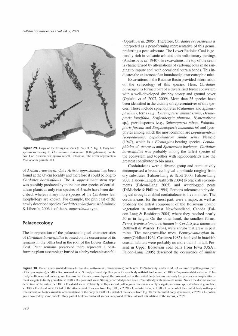
Remarks. – The syntypes of Florinanthus volkmannii weredescribed from Stradonice (Hýskov relict) by Ettingshau-sen (1852), who considered the species to belong to Cala-mites. He figured 14 specimens on five figures, only sevenof which are cordaitalean fertile organs, the other eight arein fact sphenopsids. The Ettingshausen’s (1852) seven cor-daitalean fertile organs (pl. 5, figs 1–3) are assignableto Florinanthus volkmannii. Ettingshausen’s (1852) pl. 5,fig. 1 (see Fig. 29) represents 5 specimens. The four fertileorgans belong to Florinanthus volkmannii [Fig. 29, Ettings-hausen (1852), pl. 5, fig. 1]. The different specimen in themiddle belongs to some calamite cone. The specimen inEttingshausen’s pl. 6, fig. 1 also represents a calamite coneand that in fig. 2 is a Calamites pith cast.
Florinanthus volkmannii is a small cordaitalean fertileorgan. This name is frequently used in the literature, butpresumably represents several different species. F. volk-mannii sensu stricto is characterised by a very narrow axisthat is usually 1 to 1.5 mm wide whereas axes figured bydifferent authors are usually wider: 2 mm (Zeiller 1886),
�+�
!�����$(# Florinanthus volkmannii (Ettingshausen) nov. comb.• A – a macerated “bud”, a – fertile organ axis, b – bract, slide 416/12,× 20. • B–D – macerated sterile scales; B – from the central part of a“bud”, C – from the terminal part of a “bud”, slide 416/20, D – terminalfragment of a scale from the basal part of a “bud”, and B and C slide416/19, all × 25.
�
.
�
�
%
������ ��������� �� � !"# ��� �"��$�% #����� ��&�� '� �(

4 mm (Seward 1917), 4.5 mm (Crookall 1970), 2 mm(Havlena 1971), or narrower, < 1 mm (Lee 1963). Disre-garding axis width, the fertile organs “Cordaitanthusvolkmannii” Ettingshausen of the above mentioned au-thors, differs from Ettingshausen’s (1852) species. Themost similar to Ettingshausen’s species is the specimen fig-ured by Crookall (1970). The shape and dimension ofaxillary shoots are very similar, however, the bracts ofCrookall’s specimen are shorter and bent differently. Lee’s(1963) specimen has obovate axillary shoots and very shortbracts. Zeiller’s (1886) specimen has oval axillary shootswithout scales, very different from Ettingshausen’s (1852)
specimens. Preservation is a problem however, as manyfertile organs are not well enough preserved for detailedcomparison. Fertile organs of this Florinanthus volk-mannii have been described as being female cones(Seward 1917, Lee 1963, Crookall 1970). However, thisstudy shows that F. volkmannii is a male fertile organwhich is proved by accumulations of prepollen clusters onsterile scales and pollen sacs on some Stradonice samples.Moreover, Halle in 1942 wrote on some labels for Ettings-hausen’s collection in the “Geologische Bundesanstalt”in Vienna a remark that Florinanthus volkmannii is a malefertile organ.
�++
!�����$)# Cuticles of sterile scales of Florinanthus volkmannii (Ettingshausen) comb. nov. (from Fig. 25A). • A – a scale fragment (abaxial side) withfew stomata, × 50. • B – abaxial cuticle of a scale with two stomata (a detail from Fig. A), 416/10, × 400. • C – adaxial and abaxial cuticles of a scale, slide416/11, × 100. • D – detail of cells from Fig. 25E, × 200. • E – adaxial cuticle of a scale in the basal part, slide 416/11, × 100. F – margin of a scale with tri-chomes, slide 416/11, × 400.
����������� ������ �������������
�
. 0 !
%

�+�
!�����$*# Cuticles of sterile scales of Florinanthus volkmannii (Ettingshausen) comb. nov. under SEM. • A – outer surface of a scale with small tri-chomes situated on the margin, stump 30, photo E5896-10, × 110. • B – small slit (arrow) on the cuticle of unknown function (from Fig. 26A), × 110.• C, D – margin of a scale with trichomes (from Fig. 26A), × 550. • E – scale surface showing oriented cells and a “slit” (arrow) (from Fig. 26A, B), × 1100.• F – inner surface of scale with well visible anticlinal cell walls, × 110. • G – detail of cells from Fig. 26F, × 440. • H – detail of outer view of the scale sur-face with papillae and elongate oval structures of unknown function, × 440.
0
.
� !
� 2
%
������ ��������� �� � !"# ��� �"��$�% #����� ��&�� '� �(
Pollen grains
“Cordaianthus” and “in situ” prepollen grains
Pollen grains isolated from pollen sacs of the male repro-ductive organ “Cordaianthus” Grand’Eury, 1877 werevery credibly described by Renault (1879) in his compre-hensive study of reproductive cordaitalean parts from sili-cified material of Stephanian age collected from theGrand-Croix locality near Saint-Étienne, France. The ge-neral diagnoses for “in situ” and dispersed pollen grainswas fixed by Renault (1879) and used in subsequent publi-cations dealing with “Cordaianthus” pollen grains (Re-nault 1881, 1896). Florin (1936) described “in situ” pollenof C. saportanus Renault from the typical locality. Schopfet al. (1944, pp. 58, 59) proposed the genus Florinites tocircumscribe all sporae dispersae pollen grains of known
cordaitalean affinity. The genus Florinites was defined(Schopf et al. 1944) as: bilateral pollen grains, broadlyelliptical in outline. Body somewhat more spherical andnearly entirely enclosed by bladder. Bladder and body wallsare joined distally. Trilete imprint (where discernible) isvestigial.
However, the diagnosis of Florinites became broadlyinterpreted and a great number of species with a wide rangeof sizes, morphologies, and haptotypic features were as-cribed to the genus Florinites Schopf, Wilson & Bentall,1944 (see Kosanke 1950, Butterworth & Williams 1954,Balme & Henelly 1955, Potonié & Kremp 1956, Wilson &Hoffmaister 1956, Bhardwaj 1957, Dybová & Jachowicz1957, Alpern 1959, Kalibová 1965, Felix & Burdbridge1967, Peppers 1970).
Millay & Taylor (1974) studied in detail numerous wellpreserved Florinites-type pollen grains isolated from
�+
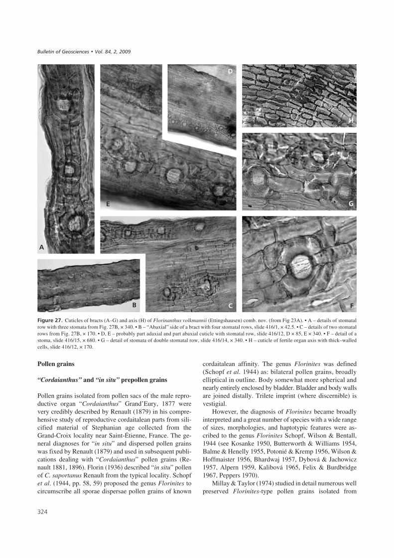
!�����$+# Cuticles of bracts (A–G) and axis (H) of Florinanthus volkmannii (Ettingshausen) comb. nov. (from Fig 23A). • A – details of stomatalrow with three stomata from Fig. 27B, × 340. • B – “Abaxial” side of a bract with four stomatal rows, slide 416/1, × 42.5. • C – details of two stomatalrows from Fig. 27B, × 170. • D, E – probably part adaxial and part abaxial cuticle with stomatal row, slide 416/12, D × 85, E × 340. • F – detail of astoma, slide 416/15, × 680. • G – detail of stomata of double stomatal row, slide 416/14, × 340. • H – cuticle of fertile organ axis with thick–walledcells, slide 416/12, × 170.
����������� ������ �������������
0
2
� !
�
%
.

“Cordaianthus” concinnus Delevoryas, 1953 and “Cor-daitanthus” schueleri (Darrah) Fry, 1956, together withother dispersed pollen from Kansas and Iowa. They de-fined Florinites as bilaterally symmetrical alete pollengrains consisting of a circular to oval central body sur-rounded by a large equatorial saccus. The saccus is at-tached to the corpus on both the proximal and distal sur-faces (see Millay & Taylor 1974, p. 81).
Florinites pellucidus (Wilson & Coe) Wilson, 1958was chosen as the type species. This implies that only aletepollen grains are accommodated in the genus Florinites,whereas other pollen grains with a monolete, triletoid ortrilete scar have been expelled from the genus Florinites.
Millay & Taylor (1974) also described pollen grainsfrom a pollen sac dispersed among cone scales of“Cordaianthus-like” reproductive structures, and estab-
lished two new monosaccate genera. The characteristicfeatures of Sullisaccites and Felixipollenites are given inTable 3. These authors also discussed the ultrastructural or-ganization of saccate pollen and evolution of cordaitaleanpollen. Saccate pollen grains have a two-layered exine,with an outer wall layer (sexine) and an inner wall layer(nexine). Millay & Taylor (1976) also proposed an evolu-tionary sequence for cordaitalean pollen grains.
Description of pollen grains from Florinanthusvolkmannii (Ettingshausen) comb. nov.
Pollen grains of F. volkmannii are monosaccate, bilateral,60 (75) 85 μm × 32(55) 62 μm, with an amb that is broadlyelliptical from a polar view and elongate oval froman equatorial view. The central body is hexagonal, oval to
�+,
!�����$,# Cuticles of bracts of Florinanthus volkmannii (Ettingshausen) comb. nov. under SEM. • A – detail of 4 stomata from Fig. 28B with slightlydeveloped crypt, × 340. • B – outer view of abaxial cuticle of a bract with three stomatal rows, stump 30, photo E5896-22, × 85. • C – inner view on abaxialcuticle with a stomatal row, photo E5896-23, × 170. • D – a detail of a stoma from Fig. 28C, × 510. • E – a detail of two stomata with crypts, photoE5896-22det3, × 340. • F – inner view on well-developed anticlinal cell walls – a detail from Fig. 28H, × 340. • G – a detail from Fig. 28H with oval struc-tures of unknown function, × 340. • H – cuticle of the fertile organ axis. In front, outer view of the cuticle with slightly marked cellular outlines; behind, in-ner view with prominent anticlinal walls; SEM stump 30, photo E5896-28, × 170.
!
�
� 0
2
.
%
������ ��������� �� � !"# ��� �"��$�% #����� ��&�� '� �(

nearly circular from the polar view 30–51 μm long and25–38 μm broad, and is a distinct darker brown colour. Thelonger axes of the spore and those one of the central body aremutually perpendicular. The proximal body surface is wit-hout folds, it has 8–15 μm long monolete scars (Fig. 30D, H)which curve in the centre, sometimes with an indication ofan additional long third ray that suggests a transition to a tri-lete scar. This feature is often indistinct in light microscopy.The body outline is trapezoidal in equatorial view and taperstowards the distal side of the pollen grain (Fig. 31D). Thewall of the body on the distal side has folds or pleats runningalong its long axis. The central body is surrounded by a largeequatorial saccus, which is attached to the central body onboth the proximal and distal surfaces (Fig. 30C, E, I).
The equatorial saccus covers the proximal side of thecentral body. The saccus is attached at a narrow zone nearthe proximal surface of the central body (Fig. 30I). Only partof the central body, with an oval or sickle shape, remains un-covered on the distal side. The slit is usually indiscernible.
Most studied pollen grains have the saccus torn fromthe proximal side and the surface of the central body is wellexposed. The central body is wrinkled. The saccus is
laevigate from the outer side and reticulate from the innerside (Fig. 30J).
Comparison of prepollen grains. – Three species of thegenus Florinites were originally described, which are incertain aspects similar to the pollen grains isolated fromFlorinanthus volkmannii. They are Florinites ovalis Bhar-dwaj, 1957, Florinites guttatus Felix & Burdbridge, 1967and Florinites diversiformis Kosanke, 1950. These bilate-ral monosaccate species possess a central body with a dar-ker colour, in which the longer axis is mostly perpendicularto the longer axis of the saccus. However, after the Taylor& Millay’s (1974) revision of the heterogenous genus Flo-rinites, only monosaccate alete species (inclusive of Flori-nites ovalis) were kept in the genus Florinites, and the re-mainder of species (inclusive of Florinites guttatus andFlorinites diversiformis) were excluded from this genus.
The pollen grains of Florinanthus volkmannii fromPokrok Mine, Ovčín – Přívětice Radnice Basin are verysimilar in morphology to two other species of pollen,Florinites ovalis and Florinites guttatus. However Flori-nanthus volkmannii pollen, which has a diameter
�+-
1�����$# Overview of some in situ Florinites isolated from cordaitalean fertile organs. Abbreviation: * type species of Florinanthus Ignatiev & Meyen,1989, ** D3 – petrified specimens, D2 – compression/impression specimens.
Parent plants ** Diameter Shape and features Name Age and country References
Florinanthus (al.Cordaitanthus) saportanus(Renault, 1879) Ignatiev &Meyen, 1989
D3 78–93 μm Oval, monosaccate,leptomate, corpussubcircular
Florinites sp.(isolated fromanther sacs)
Carboniferous,France
Florin (1936), Renault (1879),Scott (1923),Potonié (1962), Balme (1995)
Cordaianthus gemmiferGrand’Eury, 1877
D3 About 60 μmbroad
A small trilete markon the proximal faceof the corpus
Florinites sp. Carboniferous,France
Brush & Barghoorn (1962);Potonié (1962, 1967)
Florinanthus (al.Cordaianthus) concinnus(Delevoryas, 1953) Ignatiev &Meyen, 1989 *
D3 44–71 μm Oval, monosaccate,corpus circular smoothproximally andgranulate distally
Florinitespellucidus(Wilson & Coe)Wilson
Carboniferous,USA
Millay & Taylor (1974, 1976);Brush & Barghoorn (1962);Meyen (1984, 1987); Taylor(1981, 1988); Taylor & Taylor(1993); Balme (1995)
Renaulticonus (al.Cordaianthus) grandeuryi(Brongniart) Ignatiev &Meyen, 1989
D3 About 100 μm Oval, monosaccate,corpus subcircular
Florinites sp.(recovered fromthe canal of thepollen chamber)
Carboniferous,France
Renault (1879)
Florinanthus (al.Cordaitanthus) schueleri(Darrah) Ignatiev & Meyen,1989
D3 62–89 μm Oval, monosaccate,corpus circular smoothproximally andgranulate distally
Florinites sp.(isolated fromanther sacs)
Carboniferous,USA
Millay & Taylor (1974); Darrah(1952); Fry (1956); Brush &Barghoorn (1962); Potonié(1962, 1967); Balme (1995)
Lesqueranthus (Gothania)cones attached to stems ofMesoxylon priapi (Trivett &Rothwell, 1985)
D3 80 μm Oval, monosacate,trilete, ?flexedmonolete, corpuscircular
Sullisaccites Carboniferous,USA
Millay & Taylor (1974),Trivett & Rothwell (1985),Taylor & Taylor (1993)
Lesqueranthus (al. Gothania)lesliana (Daghlian & Taylor,1979) Ignatiev & Meyen, 1989
D3 180 μm Oval to subcircular,monosacate, trilete,distally leptomate
Felixipollenites Carboniferous,USA
Millay & Taylor (1974),Taylor & Taylor (1993)
Florinanthus volkmannii(Ettingshausen) comb. nov.
D2 58–85 μm Oval, monosaccate,corpus oval smoothproximally andgranulate distally
Florinites cf.ovalisFlorinites cf.guttatus
Carboniferous,Czech Republic
Herein
����������� ������ �������������

58–85 μm, is intermediate in size between Florinitesovalis (42–65 μm) and Florinites guttatus (95–135 μm)with a monolete-triletoid scar.
The central body of some Bohemian specimens fromthe Radnice Basin extends nearly the full width of thesaccus, resulting in the appearance of a bisaccate grain.Such pollen grains could be morphologically similar toPseudoillinites Ravn (1979), a genus erected to accommo-date bilateral, monosaccate, monolete prepollen or pollengrains. Pseudoillinites (al. Florinites) diversiformis (Ko-sanke, 1950) Ravn, 1979 was chosen as its type species.
Pollen grains isolated from Florinanthus volkmanniidiffer from pollen grains of the morphogenera Florinitesand Sulisaccites (Schopf, Wilson & Bental) Millay & Tay-lor, 1974 by the presence of a monolete – triletoid scar onthe proximal side of the central body and by having athicker exine.
These pollen grains are partly comparable with the ge-nus Felixipollenites in the shape of the haptotypic feature(monolete-triletoid) and in the stronger nature of the cen-tral body exine. However, Felixipollenites pollen grainsare radially – bilaterally symmetrical and the proximal sur-face of the grains (Millay & Taylor 1974, pl. 42, figs 5, 6)show a reticulate ornamentation that is clearly limited tothe proximal saccus corpus attachment region. Millay &Taylor (1976, p. 68) consider this feature to be primitive.
Potonieisporites is a monosaccate, monolete spore, butpollen grains isolated from Florinanthus volkmannii showa gradation from monolete to trilete. It seems that theFlorinanthus volkmannii pollen grains studied are notcomparable with any of above-mentioned taxa (Table 3),and they belong, perhaps, to a new genus. However, thestudied pollen grains are preserved as compression/impres-sions and they are not as well-preserved as pollen grainsfrom coal balls described by Millay & Taylor (1974).
Pith casts
Artisia Sternberg, 1838
Type species. – Artisia transversa (Artis) Corda in Stern-berg, 1838
Artisia approximata Brongniart ex Lindley & Hutton,1837Figures 32A–D
1821 Calamites fasciatus Sternberg, p. 27, pl. 17, fig. 3.1825 Calamites fasciatus Sternberg, tent. 24.1828 Sternbergia approximata Brongniart, p. 137 (nomen
nudum).1837 Sternbergia approximata Brongniart ex Lindley &
Hutton, p. 187, pls 224, 225.1838 Artisia approximata (Brongniart ex Lindley & Hut-
ton). – Corda in Sternberg, p. XXII (Skizzen).1838 Tithymalites biformis Presl in Sternberg, p. 205, pl. 53,
figs 1–6.
Description. – Casts more or less cylindrical, 15–30 mm indiameter and 50–200 mm-long fragments, bearing alter-nate approximately horizontal septa and spaces betweenthe septa (Specimen numbers are given in the “Material”section). The spaces are usually rounded, 0.6 to 2.0 mm inheight, rarely anastomosing. Connection between Artisiaand a trunk or twig surface is illustrated in Fig. 20H.
Remarks. – Only two species of Artisia, Artisia tranversaand Artisia approximata, are generally accepted (VanAmerom 1998, Fossilium catalogues, pp. 6–9). The other“species” are considered synonyms of these two. The Arti-sia approximata-type does not have the longitudinal ridges
�+.
1�����&# Comparison of some pollen taxa with “in situ” pollen isolated from Florinanthus volkmannii.
Taxon Symmetry an diameter Haptotypic features Attachement ofequatorial saccus
Notice
Florinites Monosaccate, radially–bilaterally symmetrical
Alete, with a distalaperture
On both proximaland distal surface
Proximal saccus corpus overlap slight toabsent. Saccus levigate, intrareticulate.
Sulisaccites Monosaccate, bilaterallysymmetrical 55–80 μm
Trilete On both proximaland distal surface
Proximal saccus corpus overlap slight.Distal overlap approximately 1/2.
Felixipollenites Monosaccate, radially–bilaterally symmetrical115 (150) 180 μm
Trilete, triletoidmore seldommonolete
On both proximaland distal surface
Proximal saccus corpus overlap slight toabsent.
Florinites guttatus Monosaccate, bilaterallysymmetrical95–140 μm × 120–70 μm
Monolete-triletoid On both proximaland distal surface
The spore body covered by thin granulosemembrane proximally and coarserreticuloid membrane distally.
PseudoillinitesBasionym = Florinitesdiversiformis
Monosaccate, bilaterallysymmetrical
Monolete Proximal surface ofcentral body freefrom saccus
Appearance of a bisaccate grain. Saccuscoarsely intrareticulate proximal surface ofcentral body free from saccus.
Pollen isolated fromFlorinanthus volkmannii
Monosaccate, bilaterallysymetrical 60 (75) 85 μm ×32 (55) 62 μm
Monolete-triletoid On both proximaland distal surface
The spore body covered by thin smooth togranulose membrane proximally andcoarser granulate membrane distally.
������ ��������� �� � !"# ��� �"��$�% #����� ��&�� '� �(
of Artisia transversa. Only Artisia approximata has beenfound at the Ovčín locality and therefore it could belong toCordaites borassifolius. The A. approximata stem typewas possibly produced by more than one species of cordai-talean plants as only two species of Artisia have been des-cribed, whereas many more species of the Cordaites leafmorphology are known. For example, the pith cast of thenewly described species Cordaites schatzlarensis Šimůnek& Libertín, 2006 is of the A. approximata type.
6����������'
The interpretation of the palaeoecological characteristicsof Cordaites borassifolius is based on the occurrence of itsremains in the bělka bed in the roof of the Lower RadniceCoal. Plant remains preserved there represent a peat-forming plant assemblage buried in situ by volcanic ash fall
(Opluštil et al. 2005). Therefore, Cordaites borassifolius isinterpreted as a peat-forming representative of this genus,preferring a peat substrate. The Lower Radnice Coal is ge-nerally rich in volcanic ash and thin sedimentary partings(Andrusov et al. 1940). In excavations, the top of the seamis characterised by alternations of carbonaceous shale ran-ging to impure coal with occasional vitrain bands. This in-dicates the existence of an inundated planar eutrophic mire.
Excavations in the Radnice Basin provided informationon the synecology of this species. Here, Cordaitesborassifolius formed part of a diversified forest ecosystemwith a well-developed shrubby storey and ground cover(Opluštil et al. 2007, 2009). More than 25 species havebeen identified in the vicinity of representatives of this spe-cies. These include sphenophytes (Calamites and Spheno-phyllum), ferns (e.g., Corynepteris angustissima, Desmo-pteris longifolia, Senftenbergia plumosa, Hymenothecasp.), pteridosperms (e.g., Sphenopteris mixta, Palmato-pteris furcata and Eusphenopteris nummularia) and lyco-phytes among which the most common are Lepidodendronlycopodioides, Lepidodendron simile sensu Němejc(1947), which is a Flemingites-bearing species, Lepido-phloios cf. acerosus and Spencerites havlenae. Cordaitesborassifolius was probably among the tallest species ofthe ecosystem and together with lepidodendrids also thegreatest contributor to bio mass.
Cordaitaleans were a diverse group and cumulativelyencompassed a broad ecological amplitude ranging fromdry substrates (Falcon-Lang & Scott 2000, Falcon-Lang2003, Falcon-Lang & Bashforth 2004) to brackish environ-ments (Falcon-Lang 2005) and waterlogged peats(DiMichele & Phillips 1994). Perhaps tolerance to physio-logical drought enabled cordaitaleans to live in mires. Thecordaitaleans, for the most part, were a major, as well asprobably the tallest component of the Bolsovian uplandvegetation in southwest Newfoundland, Canada (Fal-con-Lang & Bashforth 2004) where they reached nearly50 m in height. On the other hand, the smallest forms,Pennsylvanioxylon nauertianum (= Cordaixylon dumusumRothwell & Warner, 1984), were shrubs that grew in peatmires. The mangrove-like trees, Pennsylvanioxylon bi-rame (Cridland 1964, Costanza 1985) that lived in brackishcoastal habitats were probably no more than 5 m tall. Pre-sent in Upper Bolsovian coal balls from Iowa (USA),Falcon-Lang (2005) described the occurrence of similar
�+/
!�����$-# Copy of the Ettingshausen’s (1852) pl. 5, fig. 1. Only fourspecimens belong to Florinanthus volkmannii (Ettingshausen) comb.nov. Loc. Stradonice (Hýskov relict), Bolsovian. The arrow represents aRhacopteris pinnule. × 1.
!�����&/# Pollen grains isolated from Florinanthus volkmannii (Ettingshausen) comb. nov., Ovčín locality, under SEM. • A – clump of pollen grains (partof the sporangium), × 340. • B – proximal view. Strongly corroded pollen grain. Central body with triletoid suture, × 1180. • C – proximal-lateral view. Rela-tively well-preserved pollen grain. It seems that the saccus overlaps all the proximal part of the central body. Saccus unevenly levigate, saccus corpus attach-ment levigate to finely granulate, × 1180. • D – proximal view. Strongly corroded pollen grain. Central body with monolete suture. Notice the distinct medialdeflection of the suture, × 1180. • E – distal view. Relatively well-preserved pollen grain. Saccus unevenly levigate, saccus-corpus attachment granulate,× 1180. • F – distal view. Detail of the attachement of saccus from Fig. 30C, × 2320. • G – distal view, × 1180. • H – detail of the central body with opentriletoid suture. Notice rugulate ornamentation of the body, × 2320. • I – detail of the saccus from Fig. 30C with central body attachment, × 2320. • J – pollengrain covered by some cuticle. Only part of broken equatorial saccus is exposed. Notice internal reticulation of the saccus, × 2320.
����������� ������ �������������

�+0
�
.
!
%
2
�
3 4
0
������ ��������� �� � !"# ��� �"��$�% #����� ��&�� '� �(
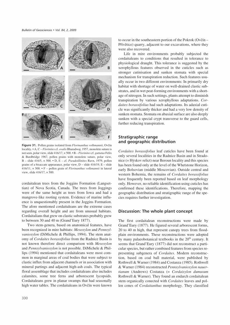
cordaitalean trees from the Joggins Formation (Langset-tian) of Nova Scotia, Canada. The trees from Joggingswere of the same height as trees from Iowa and had amangrove-like rooting system. Evidence of marine influ-ence is unquestionably present in the Joggins Formation.The afore mentioned cordaitaleans are the extreme casesregarding overall height and are from unusual habitats.Cordaitalians that grew on clastic substrates probably grewto between 30 and 40 m (Grand’Eury 1877).
Two stem-genera, based on anatomical features, havebeen recognized in mire habitats: Mesoxylon and Pennsyl-vanioxylon (DiMichele & Phillips, 1994). The stem anat-omy of Cordaites borassifolius from the Radnice Basin isnot known therefore direct comparison with Mesoxylonand Pennsylvanioxylon is not possible. DiMichele & Phil-lips (1994) mentioned that cordaitaleans were most com-mon in marginal areas of coal bodies that were subject toclastic influx from adjacent channels or in association withmineral partings and adjacent high-ash coals. The typicalfloral assemblage that includes cordaitaleans also includescalamites, some tree ferns and arborescent lycopsids.Cordaitaleans grew in planar swamps that had seasonallyhigh water tables. The cordaitaleans in Ovčín were known
to occur in the southeastern portion of the Pokrok (Ovčín –Přívětice) quarry, adjacent to our excavations, where theywere also recovered.
Life in mire environments probably subjected thecordaitaleans to conditions that resulted in tolerance tophysiological draught. This tolerance is suggested by thexerophyllous features observed in the cuticles such asstronger cutinisation and sunken stomata with specialmechanism for transpiration reduction. Such features usu-ally occur in two different environments: In primarily dryhabitat with shortage of water on well-drained clastic sub-strates, and in wet peat-forming environments with a short-age of nitrogen. In such settings, plants attempt to diminishtranspiration by various xerophyllous adaptations. Cor-daites borassifolius had such adaptations. Its adaxial cuti-cle was significantly thicker and had a very low density ofsunken stomata. Stomata on abaxial surface are also deeplysunken with a special crypt transverse to the guard cells,further reducing transpiration.
������������������������������������������
Cordaites borassifolius leaf cuticles have been found atonly several localities in the Radnice Basin and in Strado-nice (= Hýskov relict) near Beroun locality and this specieshas been found only at the level of the Whetstone Horizon,early Bolsovian (middle Moscovian). Outside central andwestern Bohemia, the remains of Cordaites borassifoliushave frequently been reported based on leaf morphologyonly. However, no reliable identification using cuticles hasconfirmed these identifications. Therefore, mapping thegeographic distribution and stratigraphic range of the spe-cies requires further investigation.
.���������7�1���8������������������
The first cordaitalean reconstructions were made byGrand’Eury (1877). He figured several arborescent forms,20 to 40 m high, that represent canopy trees from flood-plain environments. These reconstructions were adoptedby many palaeobotanical textbooks in the 20th century. Itseems that Grand’Eury (1877) did not reconstruct a parti-cular species, but rather combined features from species re-presenting subgenera of Cordaites. Modern reconstruc-tion, based on coal ball material, were published byRothwell & Warner (1984) and Costanza (1985). Rothwell& Warner (1984) reconstructed Pennsylvanioxylon nauer-tianum (Andrews) Costanza (= Cordaixylon dumusumRothwell & Warner). They found an endarch cordaitaleanstem organically conected with Cordaites leaves and pol-len cones of Cordaitanthus morphology. They classified
���
!�����&"# Pollen grains isolated from Florinanthus volkmannii, Ovčínlocality. • A, C – Florinites cf. ovalis Bharadwaj, 1957, monolete suture isnot seen, polar view, slide 416/17, × 500. • B – Florinites cf. guttatus Felix& Burdbridge 1967, pollen grains with monolete suture, polar view,B – slide 416/5, × 500. • D, E – cf. Pseudoillinites Ravn, 1979, pollengrains of a bisaccate appearance, polar view, D – slide 416/19, E – slide416/11, × 500. • F – pollen grain of Florinanthus volkmannii in lateralview, slide 416/17, × 500.
����������� ������ �������������
�
.
0 !
%
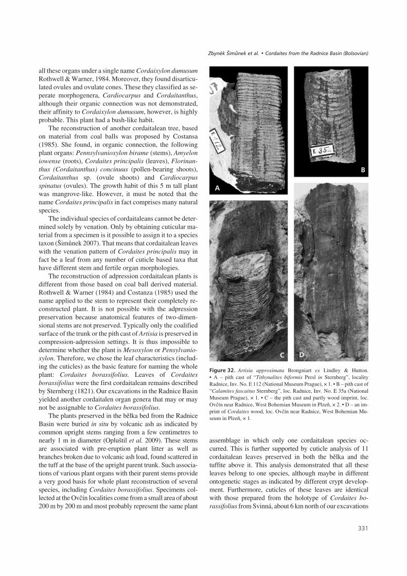
all these organs under a single name Cordaixylon dumusumRothwell & Warner, 1984. Moreover, they found disarticu-lated ovules and ovulate cones. These they classified as se-perate morphogenera, Cardiocarpus and Cordaitanthus,although their organic connection was not demonstrated,their affinity to Cordaixylon dumusum, however, is highlyprobable. This plant had a bush-like habit.
The reconstruction of another cordaitalean tree, basedon material from coal balls was proposed by Costansa(1985). She found, in organic connection, the followingplant organs: Pennsylvanioxylon birame (stems), Amyeloniowense (roots), Cordaites principalis (leaves), Florinan-thus (Cordaitanthus) concinuus (pollen-bearing shoots),Cordaitanthus sp. (ovule shoots) and Cardiocarpusspinatus (ovules). The growth habit of this 5 m tall plantwas mangrove-like. However, it must be noted that thename Cordaites principalis in fact comprises many naturalspecies.
The individual species of cordaitaleans cannot be deter-mined solely by venation. Only by obtaining cuticular ma-terial from a specimen is it possible to assign it to a speciestaxon (Šimůnek 2007). That means that cordaitalean leaveswith the venation pattern of Cordaites principalis may infact be a leaf from any number of cuticle based taxa thathave different stem and fertile organ morphologies.
The reconstruction of adpression cordaitalean plants isdifferent from those based on coal ball derived material.Rothwell & Warner (1984) and Costanza (1985) used thename applied to the stem to represent their completely re-constructed plant. It is not possible with the adpressionpreservation because anatomical features of two-dimen-sional stems are not preserved. Typically only the coalifiedsurface of the trunk or the pith cast of Artisia is preserved incompression-adpression settings. It is thus impossible todetermine whether the plant is Mesoxylon or Pensylvanio-xylon. Therefore, we chose the leaf characteristics (includ-ing the cuticles) as the basic feature for naming the wholeplant: Cordaites borassifolius. Leaves of Cordaitesborassifolius were the first cordaitalean remains describedby Sternberg (1821). Our excavations in the Radnice Basinyielded another cordaitalen organ genera that may or maynot be assignable to Cordaites borassifolius.
The plants preserved in the bělka bed from the RadniceBasin were buried in situ by volcanic ash as indicated bycommon upright stems ranging from a few centimetres tonearly 1 m in diameter (Opluštil et al. 2009). These stemsare associated with pre-eruption plant litter as well asbranches broken due to volcanic ash load, found scattered inthe tuff at the base of the upright parent trunk. Such associa-tions of various plant organs with their parent stems providea very good basis for whole plant reconstruction of severalspecies, including Cordaites borassifolius. Specimens col-lected at the Ovčín localities come from a small area of about200 m by 200 m and most probably represent the same plant
assemblage in which only one cordaitalean species oc-curred. This is further supported by cuticle analysis of 11cordaitalean leaves preserved in both the bělka and thetuffite above it. This analysis demonstrated that all theseleaves belong to one species, although maybe in differentontogenetic stages as indicated by different crypt develop-ment. Furthermore, cuticles of these leaves are identicalwith those prepared from the holotype of Cordaites bo-rassifolius from Svinná, about 6 km north of our excavations
���
!�����&$# Artisia approximata Brongniart ex Lindley & Hutton.• A – pith cast of “Tithynalites biformis Presl in Sternberg”, localityRadnice, Inv. No. E 112 (National Museum Prague), × 1. • B – pith cast of“Calamites fascaitus Sternberg”, loc. Radnice, Inv. No. E 35a (NationalMuseum Prague), × 1. • C – the pith cast and partly wood imprint, loc.Ovčín near Radnice, West Bohemian Museum in Plzeň, × 2. • D – an im-print of Cordaites wood, loc. Ovčín near Radnice, West Bohemian Mu-seum in Plzeň, × 1.
�
.
%
������ ��������� �� � !"# ��� �"��$�% #����� ��&�� '� �(

(Fig. 3B, C). Abaxial cuticle from the holotype shows thesame crypt as samples from Ovčín. Such a crypt does not oc-cur in any other samples from the Czech Republic (similarcrypt development is known from the Döhlen Basin, Ger-many) (Barthel 1964, Šimůnek 2007). Therefore we assignour specimens to the same species as Sternberg’s originalmaterial. Unfortunately, the assignment of other plant organssuch as seeds (Cardiocarpus) and strobili (Florinanthusvolkmannii) to Cordaites borassifolius is not so straightfor-ward because all organs were found isolated, not in organicconnection to leafy twigs. However, as already stated above,these organs are associated with leaves which, based on cuti-cles, are identical to Cordaites borassifolius. Therefore webelieve that Florinanthus volkmannii and Cordaitanthusovatus strobili as well as Cardiocarpus seeds were producedby the same plant species, Cordaites borassifolius.
Although the seeds are platyspermic, they are notwinged. They probably fell near the parent plant. We as-sign these seeds to Cordaites borassifolius.
The several specimens of the second fertile organ iden-tified as Cordaitanthus ovatus were found in isolation. Weconsider these to also belong to Cordaites borassifolius, al-though we do not have organic attachment to prove thishypothesis. None of these fertile organs are well preserved.Thus, cuticular study is not possible. This type of fertile or-gan is more robust than Florinanthus volkmannii and there-fore is considered to be female. No seeds were found in situwith this fertile organ so the gender of the cone remains indoubt.
Florinanthus volkmannii and “in situ” prepollen grains:Pollen sacs were probably overripe and not preserved in thisfertile organ. However, accumulations of Florinitesprepollen were found among the sterile scales (Fig. 30A),though this in itself does not prove beyond doubt that theyare the prepollen of Cordaites borassifolius. However, it ismore likely that this is the case than to suggest that they weretransported by wind from an upland cordaitalean plant kilo-metres away. They have been classified as 3 species ofcordaitalean prepollen, probably based on their maturity.Rare Artisia pith casts were also found in association withCordaites borassifolius leaves. Even though their organicconnection is not preserved, their affinity to Cordaitesborassifolius is probable. It should be noted, however, thatArtisia approximata, a species with few diagnostic features,occurs with many other cordaitalean species.
Summary of the whole plant concept: Based on cuticu-lar studies, it has been proven that, of the possible cordaita-leans, only Cordaites borassifolius leaves are present in thebělka tuff bed in the roof of the Lower Radnice Coal, Rad-nice Basin. Given that the leaves are all of one type, thenthe other cordaitalean organs present, the seeds Cardiocar-pus annularis and the male fertile organs Florinanthusovatus, were found with accumulations of Cordaites bo-rassifolius leaves. Therefore their affinity to Cordaites bo-rassifolius is highly probable. The Artisia approximatapith cast and Cordaitanthus ovatus fertile organ were notfound in organic connection with Cordaites borassifoliusand their affinity to this species is less probable. However,there is only one cordaitalean species in the deposits thusthe possibility that these organs belong to another cordaita-lean species is not likely. The proposed reconstruction ofCordaites borassifolius can be seen in Fig. 33.
The Stradonice locality (Hýskov relict) is important tothe current discussion because it is the type locality ofFlorinanthus volkmannii. At least four types ofcordaitalean fertile organs have been discovered from thislocality, including Cordaitanthus triticus (Andrae) andCardiocarpus orbiculare type seeds (Ettingshausen 1852).The others are not yet described. Adequate cuticular recov-ery from the cordaitaleans from Stradonice has been unsuc-
��+
!�����&&# Whole-plant reconstruction of Cordaites borassifolius(Sternberg) Unger (reconstruction by J. Svoboda).
����������� ������ �������������
1m

cessful. It seems that at least two natural cordaitalean spe-cies occur at the Stradonice locality (Hýskov relict).
The cuticles of the different cordaitalean organs(leaves, cone scales, and seeds) found at the Ovčín locali-ties exhibit different cellular details. These differences,however, are quite common and there are many examplesin different plant groups. In the pteridospermLaveineopteris for example, the cuticle of the pinnulesdifferes from the cuticle of the cyclopterid of the same spe-cies (Cleal & Shute 2003). In the cordaitaleans it has beendemonstrated that the leaf cuticle of Cordaitesschatzlarensis Šimůnek & Libertín differs from the seedcuticle of Samaropsis newberryi (Andrews) Seward. Nev-ertheless, both morphogenera belong to one biological spe-cies (Šimůnek & Libertín 2006). The fact that cuticles ofdifferent organs are not identical in all details does not pre-clude them from belonging to the same biological species.We believe this to be case for the cordaitalean remainsfound in the bělka bed at Ovčín; several cuticular types rep-resenting one biological species.
Concerning the botanical affinity of the large trunkfragment found in SE1, its identification as a cordaitelanremain is mostly indirect since no leaves were found at-tached to the branches. Nevertheless the character ofbranching and surface of the trunk clearly differentiate thisspecimen from associated calamites, medullosans, treeferns and lepidodendrid (even decorticated) stems. More-over, this stem is associated with leaves of Cordaitesborassifolius, which again points to the conclusion that thisis most likely the trunk of the same species. If so, this spe-cies was a fairly large tree.
����������
Cordaites borassifolius represents a distinct cordaitaleanleaf known from several localities in the Radnice Basin.Some cordaitalean leaves from Stradonice (Hýskov relict)probably also belong to this species. The venation of Cor-daites borassifolius exhibits two patterns: the abaxial sur-face shows the typical venation of Cordaites borassifoliuswith alternating wide and narrow veins, whereas the ada-xial side shows veins of approximately equal width.
Morphological features of the cuticle are significant forrecognition of this species. The adaxial cuticle has orien-tated oblong cells and rare stomata more or less regularlydispersed on the leaf surface. Stomatal complexes of theabaxial cuticle, however, are arranged in multiple stomatalrows, forming stomatiferous bands. Each stoma (guardcells) is covered by a transverse outer stomatal cavity(crypt) that is a reliable diagnostic feature for this species.This feature has only been observed in Cordaites sp. type 6by Barthel (1964) from the Permian strata of the DöhlenBasin in Germany.
Fertile organs found associated with the Cordaitesborassifolius leaves are classified as two species. In accor-dance with the system of Ignatiev & Meyen (1989), themale inflorescence found at both Stradonice (Hýskov rel-ict) and the Radnice Basin is named Florinanthus volk-mannii (Ettingshausen) nov. comb. [Representatives of“Cordaianthus” volkmannii described from other areas(United States, Great Britain and China) do not belong tothis species. They are usually described as female fertileorgans, and according to Ignatiev & Meyen (1989), shouldbe classified within the genus Grandeuryconus Ignatiev &Meyen.] F. volkmannii has long bracts and very small fertilebuds. The cuticle of the bracts shows orientated tetragonalcells and stomatal complexes arranged into single stomatalrows. The fertile axillary complexes are composed of 20 to30 spatulate to lanceolate shaped scales. Small uniseriate tri-chomes grew from the scale margins. Pollen grains associ-ated with these scales resemble dispersed Florinites ovalisBharadwaj, Florinites guttatus Felix & Burdbridge, andPseudoillinites sp. Comparison of Bohemian “in situ” pollengrains with other cordaitalean “in situ” species could not beaccomplished, because it was necessary to use SEM andTEM analysis for exact determination.
The second fertile organ is comparable to the NorthAmerican Westphalian D species Cordaitanthus ovatus(Lesquereux) Ignatiev & Meyen. Even though it is pre-sumed to be female, its gender could not be unquestionablydemonstrated. The sex of this inflorescence is indetermi-nate, and therefore it is classified as Cordaitanthus Feist-mantel, which was established to accommodate cordai-talean fertile organs of indeterminate sex.
The seeds from SE, discovered in 2006 are similar toCardiocarpus annularis (Sternberg) Lesquereux. The cuti-cle of seed integuments consists of polygonal cells. Thestrongly cutinised cells with thick walls probably coveredthe sclerotesta whereas the two-layerd thin cuticle coveredthe sarcotesta.
Twigs and pith casts were previously described bySternberg (1821, 1825) as Calamites fasciatus Sternbergand as Tithymalites biformis Presl by Presl in Sternberg(1838). The valid name is Artisia approximata Brongniartex Lindley & Hutton.
Cordaites borassifolius was an important element ofthe peat-forming floras of the Radnice Basin and shared theecosystem with peat-forming lepidodendrids. In life, it wasa tree of medium height, reaching 20 m high.
%�9��8��������
This research was supported by the Grant Agency of the CzechRepublic, grants 205/05/2034 and 205/05/0105 and it is a part ofthe Research project of the CGS No. MZP0002579801 and ofCharles University in Prague, Faculty of Science MSM
���
������ ��������� �� � !"# ��� �"��$�% #����� ��&�� '� �(
0021620855. The authors thank P. Týcová from the laboratory ofthe Czech Geological Survey, Prague and A. Langrová from theAcademy of Science of the Czech Republic for SEM digital im-ages of cordaitalean cuticles, also J. Pšenička for making avail-able cordaitalean remains from the West Bohemian Museum inPlzeň, and M. Libertín for enabling us to study in the NationalMuseum in Prague. The authors are obliged to A. Bashforth(Coppenhagen, Denmark) and to D. Chaney (Washington, USA)for checking this manuscript and to C. Cleal (Cardiff, Great Brit-ain) for his valuable remarks.
����������
ALPERN, B. 1959. Contribution à l’étude palynologique et petro-graphique des charbons français. 314 pp. Ph.D. thesis,Faculté des Sciences, Université de Paris.
ANDRAE, C.J. 1864. Die Versteinerungen der Steinkohlen-formation von Stradonitz in Böhmen. Neues Jahrbuch fürMineralogie, Geologie und Palaeontologie 1864, 160–176.
ANDREWS, H.N. JR. 1970. Index of generic names of fossilplants, 1820–1965. Geological Survey Bulletin 1300, 1–353.
ANDRUSOV, D., ČEPEK, L., HYNIE, O., KETTNER, R., KODYM,O. & URBAN, K. 1940. Vysvětlivky ke geologické mapěprotektorátu Čechy a Morava, List Královice 4051. KnihovnaGeologického ústavu pro Čechy a Moravu 19, 1–142.
BALME, B.E. 1995. Fossil in situ spores and pollen grains an an-notated catalogue. Review of Palaeobotany and Palynology87, 81–323. DOI 10.1016/0034-6667(95)93235-X
BALME, E.B. & HENELLY, J.P.F. 1955. Bisaccate sporomorphsfrom Australian Permian coals. Australian Journal of Botany3, 89–98. DOI 10.1071/BT9550089
BARTHEL, M. 1964. Coniferen- und Cordaiten-Reste aus demRotliegenden des Döhlener Beckens. Geologie (Berlin) 13(1),60–89.
BHARADWAJ, D.C. 1957. The palynological investigations of theSaar coals. Part I – Monography of sporae dispersae. Palaeon-tographica B 101, 73–125.
BRONGNIART, A. 1828. Prodrome d’une histoire des végétauxfossiles. Dictionaire des sciences naturelles 57, 1–223.
BRUSH, G.S. & BARGHOORN, E.S. 1962. Identification and struc-ture of cordaitean pollen. Journal of Paleontology 36,1357–1360.
BUCHHOLZ, J.T. & GRAY, N.E. 1948. A taxonomic revision ofPodocarpus. I. The sections of the genus and their subdivi-sions with special reference to leaf anatomy. Journal of ArnoldArboretum 50, 49–63.
BUTTERWORTH, M.A. & WILLIAMS, R.W. 1954. Description ofnine species of small spores from the British Coal Measures.The Annals and Magazine of Natural History 12(7), 753–764.
CLEAL, C.J. & SHUTE, C.H. 2003. Systematics of the Late Car-boniferous medullosalean pteridosperm Laveineopteris and itsassociated Cyclopteris leaves. Palaeontology 46(2), 353–411.DOI 10.1111/1475-4983.00303
CORDA, A.C.J. 1845. Beiträge zur Flora der Vorwelt. 128 pp.C.J. Calvesche Buchhandlung, Prag.
COSTANZA, S.H. 1985. Pennsylvanioxylon of Middle and UpperPennsylvanian coals from the Illinois Basin and its compari-son with Mesoxylon. Palaeontographica B 197(4–6), 81–121.
CRIDLAND, A.A. 1964. Amyelon in American coal balls. Palae-ontology 7, 186–209.
CROOKALL, R. 1970. Fossil plants of the Carboniferous rocks ofGreat Britain. Memoirs of the Geological Survey of Great Brit-ain. 2nd section. Palaeontology 4(6), 793–840.
DAGHLIAN, C.P.& TAYLOR, N.T. 1979. A new method for isolat-ing pollen and spores from acetate peels for scanning electronmicroscopy. Review of Palaeobotany and Palynology 27(1),85–89. DOI 10.1016/0034-6667(79)90046-0
DARRAH, W.C. 1952. The material and methods of palaeobotany.Palaeobotanist 1, 145–153.
DELEVORYAS, T. 1953. A new male cordaitean fertile organ fromthe Kansas Carboniferous. American Journal of Botany 40,144–150. DOI 10.2307/2438771
DIMICHELE, W.A. & PHILIPS, T.L. 1994. Paleobotanical andPaleoecological constraints on models of peat formation in theLate Carboniferous of Euramerica. Palaeogeography,Palaeoclimatology, Palaeoecology 106, 39–90.DOI 10.1016/0031-0182(94)90004-3
DYBOVÁ, S. & JACHOWITZ, A. 1957. Microspores of the UpperSilesian Coal Measures. Prace Institutu geologicznego 23,1–328.
ETTINGSHAUSEN, C. 1852. Die Steinkohlenflora von Stradonitzin Böhmen. Abhandlungen der Kaiserlich-königlichen geolo-gischen Reichsanstalt in Wien 1(3), 1–18.
FALCON-LANG, H.J. 2003. Late Carboniferous Tropical DrylandVegetation in an Alluvial-plain Setting, Joggins, Nova Scotia,Canada. Palaios 18, 197–211.DOI 10.1669/0883-1351(2003)018<0197:LCTDVI>2.0.CO;2
FALCON-LANG, H.J. 2005. Small cordaitalean trees in a ma-rine-influenced coastal habitat in the Pennsylvanian JogginsFormation, Nova Scotia. Journal of the Geological Society ofLondon 162, 485–500. DOI 10.1144/0016-764904-080
FALCON-LANG, H.J. & BASHFORTH, A.R. 2004. Pennsylvanianuplands were forested by gigant cotdaitalean trees. Geology32(5), 417–420. DOI 10.1130/G20371.1
FALCON-LANG, H.J. & SCOTT, A.C. 2000. Upland ecologyof some Late Carboniferous cordaitalean trees from NovaScotia and England. Palaeogeography, Palaeoclimatology,Palaeoecology 156, 225–242.DOI 10.1016/S0031-0182(99)00142-X
FEISTMANTEL, O. 1876. Versteinerungen der böhmischen Koh-len-ablagerungen, 3. Palaeontographica, Band 23, 223–316.
FELIX, C.J. & BURDBRIDGE, P.P. 1967. Palynology of theSpringer Formation of southern Oklahoma, U.S.A. Palaeon-tology 10, 349–425.
FLORIN, R. 1931. Untersuchungen zur Stammesgeschichte derConiferales und Cordaitales. Kunglige Svenska vetenskaps-akademiens handlingar, Serie III 10(1), 1–588.
FLORIN, R. 1936. On the structure of the pollen-grains in theCordaitales. Svensk Botanisk Tidskrift 30, 624–651.
FRY, W.L. 1956. New cordaitean cones from the Pennsylvanianof Iowa. Journal of Paleontology 30, 35–45.
��
����������� ������ �������������

GRAND’EURY, C. 1877. Flore carbonifère du départment da laLoire et du centre de la France. Mémoires présentés par di-vers savants à l’Académie des Sciences, Première partie –Botanique. 624 pp. Imprimerie Nationale, Paris.
HAVLENA, V. 1971. Dvě rostlinné zkameněliny nové pro namurhornoslezské pánve (Two new plant fossils found in theNamurian of the Upper Silesian Basin). Časopis pro mine-ralogii a geologii 16(4), 449–451.
IGNATIEV, I.A. & MEYEN, S.V. 1989. Suchoviella – gen. nov.from the Permian of Angaraland and a review of the system-atics of Cordaitanthales. Reviev of Palaeobotany andPalynolynology 57, 313–339.DOI 10.1016/0034-6667(89)90028-6
KALIBOVÁ, M. 1965. New species of miospores from the Bohe-mian Stephanian. Sborník geologických věd, Paleontologie 6,39–59.
KERP, H. 1990. The study of fossil gymnosperms by means ofcuticular analysis. Palaios 5, 548–549.DOI 10.2307/3514861
KERP, H. & KRINGS, M. 1999. Light microscopy of fossil cuti-cles, 52–56. In JONES, T.P. & ROWE, N.P. (eds) Fossil plantsand spores. Modern techniques. Geological Society, Lon-don.
KIDSTON, R. 1911. Les végétaux houillers recueillis dans leHainaut Belge. Mémoires du Musée royal d’histoire naturellede Belgique 5(4), 1–282.
KOSANKE, R.M. 1950. Pennsylvanian spores of Illinois and theiruse in correlation. Illinois Geological Survey Bulletin 74,1–128.
KOSANKE, R.M. 1982. Illustrations and comments concerningthe type specimens of Calamospora, Florinites, and Raistric-kia. Journal of Paleontology 56(5), 1171–1176.
KRINGS, M. & KERP, H. 1997. An improved method for obtain-ing large pteridosperm cuticles. Review of Palaebotany andPalynology 96, 453–456.DOI 10.1016/S0034-6667(96)00059-0
KVAČEK, J. & STRAKOVÁ, M. 1997. Catalogue of fossil plantsdescribed in works of Kaspar M. Sternberg. 201 pp., 67 pls.National Museum, Prague.
LEE, HSING-HSÜEH 1963. Fossil plants of the Yuehmenkou Se-ries, North China. Palaeontologia Sinica, New Series A148(6), 100–186.
LESQUEREUX, L. 1880. Description of the coal flora of the Car-boniferous formation in Pennsylvania and throughout theUnited states. Pennsylvania 2nd Geological Survey ReportProgress 1, 1–354; 2, 355–694, pl. 86, Atlas 1879, pls 1–85.
LESQUEREUX, L. 1884. Description of the coal flora of the Car-boniferous formation in Pennsylvania. Pennsylvania 2nd Geo-logical Survey Report Progress 3, 695–977, pls 88–111.
LINDLEY, J. & HUTTON, W. 1837. The fossil flora of Great Brit-ain, or figures and descriptions of the vegetable remains foundin a fossil state in the country. Volume 3, 123–205. JamesRigdway & Sons, London.
MAŠEK, J. 1963. Složení a vznik brouskového horizontu ve stře-dočeské kamenouhelné pánvi (Die Zussammensetzung undEntstehung des Schleifsteinhorizontes im Mittelböhmischen
Steinkohlbecken). Časopis pro mineralogii a geologii 8,175–177.
MEYEN, S.V. 1984. Basic features of gymnosperm systematicsand phylogeny as evidenced by the fossil record. The Botani-cal Review 50(1), 1–112. DOI 10.1007/BF02874305
MEYEN, S.V. 1987. Fundamentals of palaeobotany. 432 pp.Chapman & Hall, London.
MILLAY, M.A. & TAYLOR, T.N. 1974. Morphological studies ofPaleozoic saccate pollen. Palaeontographica, Abteilung B147, 75–99.
MILLAY, M.A. & TAYLOR, T.N. 1976. Evolutionary trends infossil gymnosperm pollen. Review of Palaeobotany and Paly-nology. Patterns in gymnosperm evolution 21(1), 65–91.
NĚMEJC, F. 1968. Paleobotanika III. 478 pp. Academia, Praha.NEWBERRY, J.S. 1873. Description of fossil plants from the Coal
Measures of Ohio. Ohio Geological Survey Report 1(2), Pa-laeontology 3, 359–385.
NIKLAS, K.J. 1994. Predicting the height of fossil plant remains:an allometric approach to an old problem. American Journalof Botany 81, 1235–1243. DOI 10.2307/2445398
OPLUŠTIL, S. 2005. Evolution of the Middle Westphalian rivervalley drainage system in central Bohemia (Czech Republic)and its palaeogeographic implication. Palaeogeography,Palaeoclimatology, Palaeoecology 222, 223–258.DOI 10.1016/j.palaeo.2005.03.016
OPLUŠTIL, S., PŠENIČKA, J., LIBERTÍN, M. & BEK, J. 2005.Peat-forming plant assemblages conserved in volcanic ash ofthe Whetstone Horizon (Bolsovian) in central and western Bo-hemia, Czech Republic. North American Paleontology Con-vention, Dalhousie University, Halifax, Nova Scotia, Canada.PaleoBios 25, Supplement 2, 92–93.
OPLUŠTIL, S., PŠENIČKA, J., LIBERTÍN, M. & ŠIMŮNEK, Z.2007. Vegetation patterns of Westphalian and LowerStephanian mire assemblages preserved in tuff beds of the-continental basins of Czech Republic. Review of Palaeo-botany and Palynology 143, 107–154.DOI 10.1016/j.revpalbo.2006.06.004
OPLUŠTIL, S., PŠENIČKA, J., LIBERTÍN, M., BASHFORTH, A.R.,ŠIMŮNEK, Z., DRÁBKOVÁ, J. & DAŠKOVÁ, J. 2009. A Mid-dle Pennsylvanian (Bolsovian) peat-forming forest pre-served in situ in volcanic ash of the Whetstone Horizon in theRadnice Basin, Czech Republic. Review of Palaeobotanyand Palynology 155, 234–274.DOI 10.1016/j.revpalbo.2009.03.002
PEPPERS, R.A. 1970. Correlation and palynology of coals in theCarbondale and Spoon formations (Pennsylvanian) of thenortheastern part of the Illinois Basin. Illinois State Geologi-cal Survey Bulletin 93, 1–173.
PEŠEK, J. 1994. Carboniferous of Central and Western Bohemia(Czech Republic). 64 pp. Czech Geological Survey, Prague.
POTONIÉ, R. 1962. Regeln, nach denen sich die Sekundaerfaltender Sporen bilden. Palaeontologische Zeitschrift 36(1–2),46–54.
POTONIÉ, R. 1967. New phylogenetic facts on fossil spores. Re-view of Palaeobotany and Palynology 1(1–4), 75–82.DOI 10.1016/0034-6667(67)90111-X
��,
������ ��������� �� � !"# ��� �"��$�% #����� ��&�� '� �(

POTONIÉ, R. & KREMP, G. 1956. Die Sporae dispersae des Ruhr-karbons; ihre Morphographie und Stratigraphie mit Aus-blicken auf Arten anderer Gebiete und Zeitabschnitte, Teil II.Palaeontographica, Abteilung B 99, 85–186.
RAVN, R.L. 1979. An introduction to the stratigraphic palynologyof the Cherokee Group (Pennsylvanian) coals of Iowa. IowaGeological Survey, Technical Paper 6, 1–117.
RENAULT, B. 1879. Structure comparée de quelques tiges de laflore Carbonifère. Nouvelles Archives du Muséum d’histoirenaturelle de Paris 2, 213–348.
RENAULT, B. 1881. Cours de botanique fossile. Volume 1. 184pp. G. Masson, Paris.
RENAULT, B. 1896. Études des gîtes mineraux de la France.Bassin houiller et permien d’Autun et d’Épinac. IV. Florefossile, I. 578 pp. Imprimerie Nationale, Paris.
ROTHWELL, G.W. & WARNER, S. 1984. Cordaixylon dunusum n.sp. (Cordaitales) I. Vegetative structures. Botanical Gazette145(2), 275–291. DOI 10.1086/337456
SCHOPF, J.M., WILSON, L.R. & BENTALL, R. 1944. An annotatedsynopsis of Paleozoic fossil spores and the definition of ge-neric groups. Illinois State Geological Survey, Report of inves-tigations 28(10), 1–73.
SEWARD, A.C. 1917. Fossil plants. Volume III. Pteridospemae,Cycadofilices, Cordaitales, Cycadophyta. 656 pp. HafnerPublishing Company, New York & London (reprint 1963).
ŠIMŮNEK, Z. 2000. Cuticles of Cordaites from the Westphalian,Stephanian and Autunian of the Bohemian Massif (Czech Re-public). A preliminary report. Acta Palaeobotanica 40(1),25–34.
ŠIMŮNEK, Z. 2001. Kordaity svrchního karbonu a permu Českéhomasivu (Cordaites from the Upper Carboniferous and Permianof the Bohemian Massif; A study of epidermal structures). 125pp. Ph.D. thesis, Czech Geological Survey, Prague. [in Czech]
ŠIMŮNEK, Z. 2007. New classification of the genus Cordaitesfrom the Carboniferous and Permian of the Bohemian Massif,based on cuticle micromorphology. Sborník Národního muzeav Praze, Řada B – přírodní vědy 62(3–4), 97–210.
ŠIMŮNEK, Z. & LIBERTÍN, M. 2006. Cordaites schatzlarensis sp.nov. and Samaropsis newberryi (Andrews) Seward from theWestphalian (Carboniferous) of the Žacléř area (Czech Re-public). Review of Palaeobotany and Palynology 138, 43–62.DOI 10.1016/j.revpalbo.2005.10.001
STERNBERG, K. VON 1820. Versuch einer geognostisch-bota-nischer Darstellung der Flora der Vorwelt I, 1. 24 pp. F.Fleischer, Leipzig.
STERNBERG, K. VON 1821. Versuch einer geognostisch-bota-nischer Darstellung der Flora der Vorwelt I, 2. 33 pp. F.Fleischer, Leipzig.
STERNBERG, K. VON 1825. Versuch einer geognostisch-bota-nischer Darstellung der Flora der Vorwelt I, 4. 48 pp. ErnstBrenck’s, Regensburg.
STERNBERG, K. VON (PRESL, C.B & CORDA, A.J. co-authors)1838. Versuch einer geognostisch-botanischer Darstellungder Flora der Vorwelt II, 7/8, 81–220. Gotlieb Hässe Söhne,Prague.
STOCKEY, R.A. & ATKINSON, I.A. 1993. Cuticle micromorpho-logy of Agathis Salisbury. International Journal of Plant Sci-ences 154(1), 187–225. DOI 10.1086/297104
STOCKEY, R.A., FREVEL, B.J. & WOLTZ, P. 1998. Cuticlemicromorphology of Podocarpus, subgenus Podocarpus,section Scytopodium (Podocarpaceae) of Madagascar andSouth Africa. International Journal of Plant Sciences159(6), 923–940.
STOCKEY, R.A., KO, H. & WOLTZ, P. 1992. Cuticle micro-morphology of Falcatifolium De Laubenfels (Podocarpaceae).International Journal of Plant Sciences 153(4), 589–601.DOI 10.1086/297082
STOCKEY, R.A., KO, H. & WOLTZ, P. 1995. Cuticle micro-morphology of parasitaxus De Laubenfels (Podocarpaceae).International Journal of Plant Sciences 156(5), 723–730.DOI 10.1086/297294
TAYLOR, N.T. 1981. Pollen and pollen organ evolution in earlyseed plants,1–25. In NIKLAS, K.J. (ed.) Paleobotany, paleo-ecology and evolution, vol. 1. Praeger, New York.
TAYLOR, N.T. 1988. Pollen and Pollen organs of fossil gymno-sperms: phylogeny and reproductive biology, 117–217. InBECK, C.B. (ed.) Origin and Evolution of Gymnosperms. Co-lumbia University Press, New York.
TRIVETT, M.L. & ROTHWELL, G.W. 1985. Morphology, system-atics, and paleoecology of Paleozoic fossil plants: Mesoxylonpriapi sp. nov. (Cordaitales). Systematic Botany 10(2),205–223. DOI 10.2307/2418347
TUREK, V. 1989. Fish and amphibian trace fossils from West-phalian sediments of Bohemia. Palaeontology 32(3),623–643.
UNGER, F. 1850. Genera et species plantarum fossilium. 627 pp.Wilhelm Braunmüller, Wien.
VAN AMEROM, H.W.J. 1998. Fossilium Catalogus Plantae. Pars99, Cordaitales und einige Vertreter der Monocotylen 1. Sup-plement. 195 pp. Backhuys Publishers, Leiden.
WEITHOFER, K.A. 1902. Geologische Skizze des Kladno-Ra-konitzer Kohlenbeckens. Verhandlungen der Kaiserlich-königlichen geologischen Reichsanstalt, 399–420.
ZEILLER, R. 1886–1888. Études des gîtes minéraux de la France.Flore fossile du bassin houiller de Valencienes. 731 pp. (text1888), pls 1–94 (Atlas 1886). Quantin, Paris.
ZODROW, E.L., MASTALERZ, M. & ŠIMŮNEK, Z. 2003. FTIR-de-rived characteristics of fossil-gymnosperm leaf remains ofCordaites principalis and Cordaites borassifolius (Pennsylva-nian Maritimes Canada and Czech Republic). InternationalJournal of Coal Geology 55, 95–102.DOI 10.1016/S0166-5162(03)00084-3
��-
����������� ������ �������������