Conversion and conservation of light energy in a photosynthetic microbial mat ecosystem
10
ORIGINAL ARTICLE Conversion and conservation of light energy in a photosynthetic microbial mat ecosystem Mohammad AA Al-Najjar 1 , Dirk de Beer 1 , Bo Barker Jørgensen 1 , Michael Ku ¨ hl 2 and Lubos Polerecky 1 1 Max-Planck Institute for Marine Microbiology, Celsiusstrasse, Bremen, Germany and 2 Marine Biological Laboratory, Department of Biology, University of Copenhagen, Strandpromenaden, Helsingør, Denmark Here we present, to the best of our knowledge, the first balanced light energy budget for a benthic microbial mat ecosystem, and show how the budget and the spatial distribution of the local photosynthetic efficiencies within the euphotic zone depend on the absorbed irradiance (J abs ). Our approach uses microscale measurements of the rates of heat dissipation, gross photosynthesis and light absorption in the system, and a model describing light propagation and conversion in a scattering–absorbing medium. The energy budget was dominated by heat dissipation on the expense of photosynthesis: in light-limiting conditions, 95.5% of the absorbed light energy dissipated as heat and 4.5% was channeled into photosynthesis. This energy disproportionation changed in favor of heat dissipation at increasing irradiance, with 499% of the absorbed light energy being dissipated as heat and o1% used by photosynthesis at J abs 4700 lmol photon m 2 s 1 (4150 J m 2 s 1 ). Maximum photosynthetic efficiencies varied with depth in the euphotic zone between 0.0140.047 O 2 per photon. Owing to steep light gradients, photosynthetic efficiencies varied differently with increasing irradiances at different depths in the euphotic zone; for example, at J abs 4700 lmol photon m 2 s 1 , they reached around 10% of the maximum values at depths 00.3 mm and progressively increased toward 100% below 0.3 mm. This study provides the base for addressing, in much more detail, the photobiology of densely populated photosynthetic systems with intense absorption and scattering. Furthermore, our analysis has promising applications in other areas of photosynthesis research, such as plant biology and biotechnology. The ISME Journal (2010) 4, 440–449; doi:10.1038/ismej.2009.121; published online 12 November 2009 Subject Category: microbial ecosystem impacts Keywords: efficiency; light conversion; microbial mats; microsensors; photosynthesis Introduction About 3.8 10 24 J of solar energy is anually absorbed by the Earth 0 s surface and atmosphere. The total energy consumption of humans in the year 2007 was 0.01% of this flux, whereas the primary productivity of global ecosystems is estimated at around 0.1% (Makarieva et al., 2008). Thus, the amount of solar energy that is available but not yet harvested is enormous. Photosynthesis offers a possibility to use this energy source, as it is a mechanism by which solar energy is converted into chemical energy and stored as biomass in phototrophic organisms, such as plants, algae or cyanobacteria. Once stored, the organic material can serve as food for heterotrophic organisms, or can be further converted to other forms of usable energy such as fuel (Ragauskas et al., 2006). Therefore, the study of photosynthetic efficiency is a very active research field, especially in recent years because of a growing demand for biofuels and technologies for sustainable energy production (Ragauskas et al., 2006; Mussgnug et al., 2007; Dismukes et al., 2008; Rosenberg et al., 2008). Research on photosynthetic efficiency has mainly focused on plants (Singsaas et al., 2001; Zhu et al., 2008) and planktonic algae (Dubinsky et al., 1986; Flameling and Kromkamp, 1998; Rosenberg et al., 2008). The measurements are typically conducted in a homogeneous light field allowing a uniform transfer of light energy to the studied photosynthetic system, and thus facilitating detailed studies of underlying physiological mechanisms of light adap- tation and acclimation. Benthic photosynthetic systems, such as biofilms and microbial mats, constitute an important component of shallow water habitats by contributing significantly to primary productivity (Cahoon, 1999; Guerrero et al., 2002), but the photosynthetic efficiency and energy budget for such systems are virtually unexplored. Photosynthetic microbial mats are complex and highly compacted microbial ecosystems, where a high diversity of phototrophic and heterotrophic populations interact within a few millimeters thick Received 11 August 2009; revised 5 October 2009; accepted 8 October 2009; published online 12 November 2009 Correspondence: M Al-Najjar, Microsensor Research Group, Max-Planck institute for marine microbiology, Celsiusstrasse 1, Bremen 28359, Germany. E-mail: [email protected] The ISME Journal (2010) 4, 440–449 & 2010 International Society for Microbial Ecology All rights reserved 1751-7362/10 $32.00 www.nature.com/ismej
-
Upload
independent -
Category
Documents
-
view
0 -
download
0
Transcript of Conversion and conservation of light energy in a photosynthetic microbial mat ecosystem

ORIGINAL ARTICLE
Conversion and conservation of light energyin a photosynthetic microbial mat ecosystem
Mohammad AA Al-Najjar1, Dirk de Beer1, Bo Barker Jørgensen1, Michael Kuhl2
and Lubos Polerecky1
1Max-Planck Institute for Marine Microbiology, Celsiusstrasse, Bremen, Germany and 2Marine BiologicalLaboratory, Department of Biology, University of Copenhagen, Strandpromenaden, Helsingør, Denmark
Here we present, to the best of our knowledge, the first balanced light energy budget for a benthicmicrobial mat ecosystem, and show how the budget and the spatial distribution of the localphotosynthetic efficiencies within the euphotic zone depend on the absorbed irradiance (Jabs).Our approach uses microscale measurements of the rates of heat dissipation, gross photosynthesisand light absorption in the system, and a model describing light propagation and conversion in ascattering–absorbing medium. The energy budget was dominated by heat dissipation on the expenseof photosynthesis: in light-limiting conditions, 95.5% of the absorbed light energy dissipated asheat and 4.5% was channeled into photosynthesis. This energy disproportionation changed in favorof heat dissipation at increasing irradiance, with 499% of the absorbed light energy being dissipatedas heat and o1% used by photosynthesis at Jabs4700lmol photon m�2 s�1 (4150 J m�2 s�1). Maximumphotosynthetic efficiencies varied with depth in the euphotic zone between 0.014�0.047 O2 perphoton. Owing to steep light gradients, photosynthetic efficiencies varied differently with increasingirradiances at different depths in the euphotic zone; for example, at Jabs4700 lmol photon m�2 s�1,they reached around 10% of the maximum values at depths 0�0.3 mm and progressively increasedtoward 100% below 0.3 mm. This study provides the base for addressing, in much more detail, thephotobiology of densely populated photosynthetic systems with intense absorption and scattering.Furthermore, our analysis has promising applications in other areas of photosynthesis research, suchas plant biology and biotechnology.The ISME Journal (2010) 4, 440–449; doi:10.1038/ismej.2009.121; published online 12 November 2009Subject Category: microbial ecosystem impactsKeywords: efficiency; light conversion; microbial mats; microsensors; photosynthesis
Introduction
About 3.8� 1024 J of solar energy is anually absorbedby the Earth0s surface and atmosphere. The totalenergy consumption of humans in the year 2007 was0.01% of this flux, whereas the primary productivityof global ecosystems is estimated at around 0.1%(Makarieva et al., 2008). Thus, the amount of solarenergy that is available but not yet harvested isenormous. Photosynthesis offers a possibility to usethis energy source, as it is a mechanism by whichsolar energy is converted into chemical energy andstored as biomass in phototrophic organisms, suchas plants, algae or cyanobacteria. Once stored, theorganic material can serve as food for heterotrophicorganisms, or can be further converted to otherforms of usable energy such as fuel (Ragauskaset al., 2006). Therefore, the study of photosynthetic
efficiency is a very active research field, especiallyin recent years because of a growing demand forbiofuels and technologies for sustainable energyproduction (Ragauskas et al., 2006; Mussgnug et al.,2007; Dismukes et al., 2008; Rosenberg et al., 2008).
Research on photosynthetic efficiency has mainlyfocused on plants (Singsaas et al., 2001; Zhu et al.,2008) and planktonic algae (Dubinsky et al., 1986;Flameling and Kromkamp, 1998; Rosenberg et al.,2008). The measurements are typically conductedin a homogeneous light field allowing a uniformtransfer of light energy to the studied photosyntheticsystem, and thus facilitating detailed studies ofunderlying physiological mechanisms of light adap-tation and acclimation. Benthic photosyntheticsystems, such as biofilms and microbial mats,constitute an important component of shallow waterhabitats by contributing significantly to primaryproductivity (Cahoon, 1999; Guerrero et al., 2002),but the photosynthetic efficiency and energybudget for such systems are virtually unexplored.Photosynthetic microbial mats are complex andhighly compacted microbial ecosystems, where ahigh diversity of phototrophic and heterotrophicpopulations interact within a few millimeters thick
Received 11 August 2009; revised 5 October 2009; accepted 8October 2009; published online 12 November 2009
Correspondence: M Al-Najjar, Microsensor Research Group,Max-Planck institute for marine microbiology, Celsiusstrasse 1,Bremen 28359, Germany.E-mail: [email protected]
The ISME Journal (2010) 4, 440–449& 2010 International Society for Microbial Ecology All rights reserved 1751-7362/10 $32.00
www.nature.com/ismej
photosynthetically active (euphotic) zone (vanGemerden, 1993; Stal, 2000). Owing to the highconcentration of cells and the presence of abioticcomponents, such as sediment particles, thesebenthic systems are fundamentally different fromthe terrestrial and planktonic systems investigatedso far in that the light field is highly heterogeneouson spatial scales comparable with the size of and thedistance between the individual organisms (Kuhland Jørgensen, 1992, 1994; Kuhl et al., 1994). Towhat extent such pronounced light gradients affectthe photosynthetic efficiency of the individual cellsand the energy budget of these microbial ecosystemsas a whole is still an open question.
In photosynthesis research, the quantum effi-ciency (QE) is a frequently used measure of lightconversion, as the photochemical reactions aredriven by photons that are absorbed by the chlor-ophylls and their antenna in the photosystems, andlead to a charge separation across the thylakoidmembrane. The photosynthetic QE is defined as theamount of CO2 molecules assimilated per number ofphotons absorbed. Assuming a 1:1 stoichiometry ofCO2 fixation and O2 production, which is reasonablewhen NH4
þ is the predominant source of nitrogen,rather than NO3
�, QE can also be determined fromthe amount of O2 molecules produced per photonabsorbed. QE is typically derived either from CO2 orO2 exchange measurements performed in fluxchambers or by fluorimetry using variable chloro-phyll a fluorescence measuring the quantum effi-ciency of photosystem II (Falkowski and Raven,1997). Assuming no losses, the maximal QE ofphotosynthesis is 0.125. This follows from the basicphoton requirements for the amount of electronsthat need to be transferred to oxidize water andreduce CO2, and from the fact that this electrontransfer takes place sequentially over two reactioncenters, each requiring one photon to separate oneelectron (thus, requiring in total eight photons perCO2 molecule fixed or per O2 molecule produced).Under light-limiting conditions, QE in terrestrialplants and planktonic algae can reach up to 0.110–0.111 (see Supplementary Table S1), which isindeed close to the theoretical maximum.
The energy efficiency (EE) is the preferredmeasure when considering photosynthesis in thecontext of energy production. According to defini-tion, EE is the ratio between the energy stored asbiomass and the absorbed light energy, and can bequantified directly from biomass growth experi-ments. Alternatively, EE can be derived from QEby considering the following steps: first, in the light-dependent reactions, in which O2 is formed bysplitting of water, reducing equivalents are used toform nicotinamide adenine dinucleotide phosphate(NADPH), and ATP is formed by the proton motiveforce, the energy gained and stored is EG¼ 482.9 kJ(mol O2)
�1. This follows from the Gibbs freeenergies of reactants in the reactions 2H2Oþ2NADPþ-O2þ 2NADPHþ 2Hþ (DG¼ 439 kJ mol�1)
and ADPþPi- ATP (DG¼ 43.9 kJ mol�1, both at pH 7;Thauer et al., 1977). Second, the molar energycontent of light with wavelength l is El¼NA(hc/l),where NA, h and c are the Avogadro0s number, thePlanck constant and the speed of light, respectively.Thus, assuming no intermediate losses, that is, theenergy stored in the light-dependent reaction is fullyused for CO2 fixation in the dark reaction (equiva-lent to the assumption of O2:CO2 stoichiometrybeing equal to 1), a relationship between EE andQE can be written as
EE ¼EG
ElQE ð1Þ
Considering blue (450 nm) and red (670 nm)photons, which are most efficiently absorbed bychlorophyll a, the factor EG/El ranges between 1.8–2.7 photon per O2, averaging at 2.22 photon per O2
for an ‘average’ photon (550 nm) within the rangeof photosynthetically active radiation (PAR, 400–700 nm). Thus, the theoretical maximum EE ofphotosynthetic light utilization is about 27.7%. Thisrepresents a situation in which all incident photonsare absorbed and used for O2 evolution andequivalent CO2 assimilation. The actual photosyn-thetic EE is lower, primarily because (i) capture ofphotons by photopigments and channeling of theirenergy to the reaction center is inefficient, (ii)photons are absorbed by cell components andaccessory pigments that are not photosyntheticallyactive, (iii) excess excitation energy is dissipated asheat by non-photochemical quenching processes,and (iv) part of the energy stored in the light-dependent reaction is used for processes other thanCO2 fixation (Schneider, 1973; Osmond, 1994;Huner et al., 1998; Zhu et al., 2008). Direct biomassgrowth measurements for terrestrial C3 and C4plants obtained EE values of 4.6 and 6.0%, respec-tively (Supplementary Table S1; Zhu et al., 2008). Sofar the photosynthetic EE in benthic biofilms andmicrobial mats is unexplored.
Detailed studies of microbenthic photosynthesis(PS) require specialized tools and methods toresolve the distribution of light and photosyntheticactivity at relevant spatial scales. Oxygen micro-sensors can resolve the steep concentration gradi-ents of O2 in microbial mats at mm resolution and thelight-dependent O2 dynamics at 0.1�0.2 s resolution(Revsbech et al., 1983; Revsbech and Jørgensen,1986). Using the so-called light–dark shift method(Revsbech and Jørgensen, 1983), O2 microsensorscan measure the volumetric rates of gross photo-synthesis with a spatial resolution of 100�200 mm.When combined with measurements of the locallyavailable light using fiber-optic microprobes (Kuhl,2005), the vertical distribution of the relativephotosynthetic efficiency within a microbial matcan be quantified (Lassen et al., 1992b). Microscalemeasurements of volumetric gross PS are typicallydone throughout the mm-thin euphotic zone under
Light conversion in a microbial mat ecosystemM Al-Najjar et al
441
The ISME Journal
different incident irradiance levels and are thenintegrated over depth to yield the areal rate of grossPS for the entire ecosystem as a function of theincident irradiance. We compiled such data from anumber of sources and, assuming that 90% of theincident irradiance was absorbed and used, ob-tained maximal QE for benthic photosyntheticsystems in the range of 0.01�0.07 (SupplementaryTable S1). In principle, Equation 1 could be used tosimply convert QE to EE, but this approach wouldnot satisfactorily address the fundamental questionof how the energy budget of a microbial matecosystem is balanced between the incident down-welling irradiance, upwelling irradiance, photo-synthesis and heat dissipation, especially as afunction of depth spanning the steep light andchemical gradients across the euphotic zone.
This study provides, to the best of our knowledge,the first complete energy budget assessment in acyanobacterial mat, a model representative of amicrophytobenthic ecosystem. We aimed to quantifyhow much of the incident light energy flux (JIN) isback-scattered and thus lost to the environment (JR),dissipated as heat (JH) or chemically stored byphotosynthesis (JPS), and how this energy flowvaries with depth across the euphotic zone. Thisgeneral aim was divided into three main challenges:(i) to experimentally verify that JIN¼ JRþ JHþ JPS,(ii) to characterize how the budget depends on theincident irradiance, and (iii) to elucidate how theoverall photosynthetic light utilization efficiency ofthe entire mat depends on the partial efficiencies ofthe different layers in the mat. We applied micro-sensors for O2, temperature and scalar irradiance toresolve the vertical variability of the energy conver-sion in the mat ecosystem, and analysed the databased on a new model that describes light propaga-tion and conversion in a medium with intenseabsorption and scattering (see SupplementaryInformation).
Materials and methods
The studied cyanobacterial mat originated froman intertidal flat near Abu Dhabi (United ArabEmirates), and was kept in the laboratory in 0.2 mmfiltered seawater (originating from the North Sea)under a 10-h light–14-h dark illumination regimeat incident irradiance B480 mmol photon m�2 s�1
(spectral composition similar to sunlight) beforemeasurements. The measurements were conductedin a thermostated flow chamber that was verticallyilluminated with a collimated light beam of PARfrom a tungsten halogen lamp. A stable laminar flowof filtered aerated seawater (temperature 23 1C,salinity 35) above the mat surface was maintainedthroughout the measurements.
High spatial resolution measurements of O2 con-centration, temperature and scalar irradiance wereperformed with a fast-responding Clark-type micro-
electrode (tip diameter B30 mm; Revsbech, 1989),a thermocouple microsensor (tip diameter B50mm;T50, Unisense A/S, Aarhus, Denmark) and a fiber-optic scalar irradiance microprobe (integratingsphere diameter B100 mm; Lassen et al., 1992a)connected to a spectrometer (USB4000, OceanOptics, Dunedin, FL, USA), respectively. Thedownwelling spectral quantum irradiance (Il inmmol photon m�2 s�1 nm�1) was measured by a spec-trometer equipped with a cosine collector andintercalibrated against a PAR quantum irradiancesensor (LI-190 Quantum) connected to a light meter(LI-250, both from LI-COR Biosciences, Lincoln,NE, USA). The spectral irradiance reflectance ofthe mat (Rl) was calculated as Rl¼ Il,mat/Il,ref, whereIl,mat and Il,ref are the back-scattered spectralradiances measured above the mat and above awhite reflectance standard (Spectralon; Labsphere,North Sutton, NH, USA), respectively, with a fiber-optic field radiance microprobe (Jørgensen anddes Marais, 1988). This calculation assumed thatthe light back-scattered by the mat was diffused,which is typically the case for highly scatter-ing benthic microbial communities (Kuhl andJørgensen, 1994).
The light energy absorbed in the mat (Jabs inJ m�2 s�1) was calculated as the vector irradianceby subtracting the downwelling and upwellingirradiance:
Jabs ¼Z700
400
IlElð1� RlÞdl ð2Þ
where El is the energy of a photon with wavelengthl (see Introduction).
The incident light energy that was absorbed by themat but not conserved as chemical energy byphotosynthesis resulted in an increase of the mattemperature relative to that of the overlaying water.This allowed calculation of the areal rate of heatdissipation inside the euphotic zone of the mat (JH inJ m�2 s�1) from Fourier’s law of conduction, that is,
JH ¼ kqT
qz; ð3Þ
where k is the thermal conductivity in water(0.6 J m�1 s�1 K�1) and qT/qz is the temperaturegradient measured in the thermal boundary layer(Jimenez et al., 2008).
Volumetric rates of gross photosynthesis (P inmmol O2 m�3 s�1) were measured by the light–darkshift method (Revsbech and Jørgensen, 1983) invertical steps of 100mm throughout the upper layersof the microbial mat until no photosynthesis wasdetected. Areal rates of photosynthesis (Pa in mmolO2 m�2 s�1) were calculated by integrating the volu-metric rates over the euphotic zone. The flux ofenergy conserved by photosynthesis (JPS in J m�2 s�1)was calculated as JPS¼EGPa, where EG¼ 482.9 kJ
Light conversion in a microbial mat ecosystemM Al-Najjar et al
442
The ISME Journal
(mol O2)�1 is the Gibbs energy produced when O2 is
formed by splitting water (see Introduction). Thisexpression quantifies the total flux of photosynthe-tically conserved energy, including all other sub-sequent energy conversions and mineralizationprocesses taking place in the mat, such as excretionof carbohydrates and their subsequent respirationand/or incorporation into biomass by heterotrophicbacteria.
For a given absorbed light energy, Jabs, theefficiencies of photosynthetic energy conservation(ePS) and heat dissipation (eH) for the entire euphoticzone of the mat were calculated from the energyfluxes as
ePS ¼JPSðJabsÞ
Jabsand eH ¼
JHðJabsÞJabs
: ð4Þ
In light-limiting conditions, that is, at Jabs-0, theefficiencies were estimated as follows: First, themeasured JPS as a function of Jabs was fitted with thesaturated exponential model (Webb et al., 1974)
JPSðJabsÞ ¼ JPS;maxð1� e�Jabs=EkÞ ð5Þ
to estimate the maximum photochemically con-served energy flux, JPS,max, and the parameter Ek
characterizing the apparent photochemical lightadaptation. Subsequently, it was assumed thatautofluorescence from the mat is negligible andthus the energy budget in the mat satisfies theEquation Jabs¼ JPSþ JH, that is, ePSþ eH¼ 1. Therespective efficiencies in light-limiting conditionswhere then calculated as
ePS;max ¼JPS;max
Ekand eH;min ¼ 1� ePS;max ð6Þ
The regulation of the photosynthetic efficiencyby light in each layer within the euphotic zonewas assessed as follows. First, the locally measured
scalar irradiance integrated over PAR, Es, wasfitted with an exponential function Es(z)¼Es(0)exp[�a(z�z0)] to estimate the light attenuationcoefficient, a. Then, assuming a totally diffuse lightfield inside the mat (that is, photons at each depthpropagated in all directions with equal probabil-ities), the local density of absorbed light (Eabs(z)in mmol photon m�3 s�1) was calculated as Eabs(z)¼(K/2)Es(z) (see derivation in Supplementary Infor-mation), where the absorption coefficient K isrelated to a and the irradiance reflectance R as
K¼ a1�R
1þR: ð7Þ
Finally, the local photosynthetic efficiency, Z, wascalculated by dividing the locally measured volu-metric rates of gross photosynthesis with Eabs(z),that is,
ZðzÞ ¼ PðzÞ~EabsðzÞ
¼ 2PðzÞKEsðzÞ
ð8Þ
Furthermore, the local photosynthesis rates as afunction of Eabs were fitted with the saturatedexponential model (Equation 5) to yield the max-imum photosynhesis rates Pmax(z) and parametersEk(z), from which the maximum local quantum effi-ciencies were calculated as Zmax(z)¼Pmax(z)/Ek(z).
More detailed descriptions of materials, methodsand derivations are given as Supplementary Infor-mation.
Results
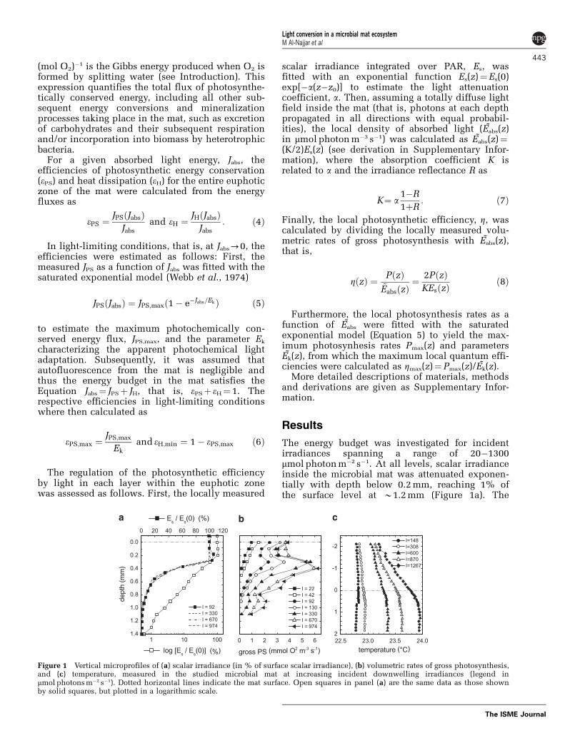
The energy budget was investigated for incidentirradiances spanning a range of 20�1300mmol photon m�2 s�1. At all levels, scalar irradianceinside the microbial mat was attenuated exponen-tially with depth below 0.2 mm, reaching 1% ofthe surface level at B1.2 mm (Figure 1a). The
Figure 1 Vertical microprofiles of (a) scalar irradiance (in % of surface scalar irradiance), (b) volumetric rates of gross photosynthesis,and (c) temperature, measured in the studied microbial mat at increasing incident downwelling irradiances (legend inmmol photons m�2 s�1). Dotted horizontal lines indicate the mat surface. Open squares in panel (a) are the same data as those shownby solid squares, but plotted in a logarithmic scale.
Light conversion in a microbial mat ecosystemM Al-Najjar et al
443
The ISME Journal

attenuation coefficient averaged over PAR wasa¼ 4.4 mm�1. The scalar irradiance directly underthe surface (0.1�0.2 mm) was slightly enhanced incomparison to the scalar irradiance at the surface,due to light scattering and photon trapping in themat (for example, see Kuhl and Jørgensen, 1994;Kuhl et al., 1996). Similar to previous observations(Jørgensen et al., 1987; Jørgensen and des Marais,1988; Lassen et al., 1992b; Kuhl and Fenchel, 2000),spectral light attenuation was strongly enhancedaround wavelengths corresponding to the absorp-tion maxima of predominant photopigments in themat (Supplementary Figure S2a). Owing to thisenhanced absorption, the irradiance reflectance,R, of the mat also exhibited pronounced minimaaround the same wavelengths (SupplementaryFigure S2b).
When integrated over PAR, 17.5% of the incidentirradiance was back-scattered from the microbialmat. The remaining 82.5% of the energy carried bythe incident irradiance was thus absorbed in themat, with 99% of this absorption taking place in thetop 1 mm. Chlorophyll a autofluorescence consti-tuted less than 0.03%, 0.024% and 0.16% of theincident blue, green and amber excitation, respec-tively, and could thus be neglected in the overallenergy budget.
The thickness of the euphotic zone varied withthe incident irradiance, increasing from B0.4 mmat 22 mmol photon m�2 s�1 to B1.1 mm at 974 mmolphoton m�2 s�1 (Figure 1b). Besides a peak in PSfound 0.3�0.4 mm below the mat surface at allirradiances, a second peak in photosynthetic activ-ity emerged at a depth of B0.9 mm at incidentirradiances X330 mmol photon m�2 s�1.
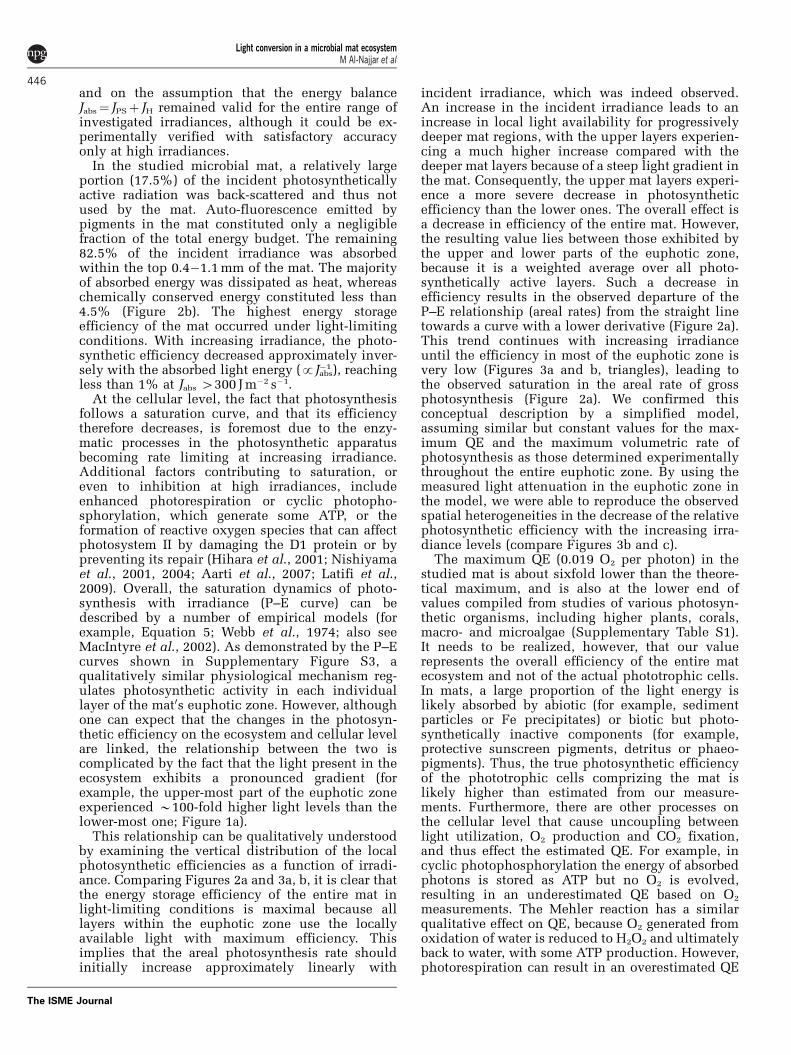
Areal rates of photosynthesis increased withthe absorbed irradiance, Jabs, according to thesaturated exponential model (Equation 5), reachinga maximum value of Pmax¼ 4.0 mmol O2 m�2 s�1 atJabs4800 mmol photon m�2 s�1 (Figure 2a). Taking the
initial slope of this photosynthesis vs absorbedirradiance relationship (P–E curve), the maximumQE of the microbial mat was 0.019±0.001 O2 perphoton. When converted to energy units, weobtained a relationship between the photosyntheti-cally conserved energy and vector irradiance, that is,the net downwelling energy flux used here as ameasure of the absorbed light energy (Figure 2a).The initial slope of this relationship, correspondingto the maximal energy conservation efficiency of themat in light-limiting conditions, was 0.045±0.003.The amount of photosynthetically conserved energyreached an asymptotic value of B1.9 J m�2 s�1 atvector irradiances above B200 J m�2 s�1.
The microbial mat temperature was only a fractionof a degree higher than in the overlying water(Figure 1c). However, the temperature differenceincreased with increasing irradiance, and a distinctthermal boundary layer was detectable at irra-diances4300mmol photon m�2 s�1 (vector irradiances454 J m�2 s�1). The flux of heat dissipated in theeuphotic zone, as derived from the temperaturegradient in thermal boundary layer (Equation 3),increased with the vector irradiance (Figure 2a).When using only data measured above 54 J m�2 s�1,that is, energy levels in which a temperature gradientin the thermal boundary layer was detectable, theslope of the linear fit was 0.969±0.035.
A plot of the summed flux of energy conserved byphotosynthesis and heat dissipation vs vectorirradiance exhibited a linear relationship with aslope of 0.979±0.036, which was close to thetheoretically expected slope of 1 (Figure 2a). Assum-ing that the experimentally determined slope wasexactly 1, we derived efficiencies of energy con-servation and heat dissipation from Equation 4and plotted them against the vector irradiance(Figure 2b). The plot shows that under light-limitingconditions B4.5% of the absorbed light energy wasconserved by photosynthesis, whereas B95.5% was
Figure 2 Energy budget in the studied mat. (a) Relationships between the areal rate of gross photosynthesis and vector irradiance (greensymbols), and between the energy flux dissipated as heat and vector irradiance (red symbols), inside the mat. The vector irradiance, thatis, the net downwelling light energy flux used here as a measure of the absorbed light, is expressed both in terms of quanta and energy.Green curve shows the fit by Equation 5 (r2¼0.996), red line is the linear fit (r2¼0.983) of used energy, that is, the sum ofphotosynthetically conserved energy and energy dissipated as heat, JPSþ JH, vs absorbed light energy, Jabs. For comparison, thetheoretically expected relationship JPSþ JH¼ Jabs is shown by the dotted line. (b) The relative disproportionation of the incident lightenergy among photosynthesis, heat and back-scattering at various levels of absorbed light energy in the mat.
Light conversion in a microbial mat ecosystemM Al-Najjar et al
444
The ISME Journal

dissipated as heat. This energy disproportionationchanged in favor of heat dissipation at increasingirradiance, with 499% of the absorbed light energybeing dissipated as heat and o1% being used byphotosynthesis at vector irradiances 4150 J m�2 s�1.
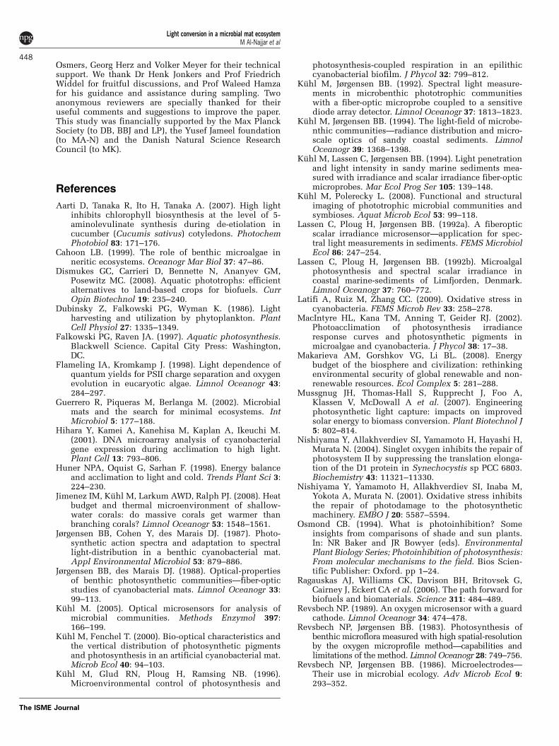
Throughout the euphotic zone, the increase in thevolumetric rates of gross photosynthesis with thelocally absorbed irradiance was generally fitted well(r240.88) by the saturated exponential model inEquation 5 (Supplementary Figure S3), whichallowed calculation of the local QE values, Z(Equation 8). Maximum Z varied between0.014�0.047 O2 per photon and exhibited twopronounced maxima at 0.3�0.4 mm and 1.0 mmbelow the mat surface (Figure 3a), coinciding withthe measured maxima of gross photosynthesis rates(compare with Figure 1b). The local QE valuesgradually diminished with increasing irradiance,with the most pronounced decrease observed in theupper B0.5 mm of the mat. This effect is betterillustrated in Figure 3b; for example, although thelocal QE values at depths 0�0.5 mm decreased to10�50% of the maximum efficiency at incidentquantum irradiances 4300 mmol photon m�2 s�1, thelocal QE values in the deeper parts of the matremained around 70�100% of the maximum values.
Discussion
At the cellular level, optimal light utilization inoxygenic phototrophs follows the well-establishedZ-scheme, whereby energy of the photons absorbedby the light harvesting complex drives a series ofredox reactions after an initial charge separationacross the photosynthetic membrane, resulting inthe generation of a proton gradient (used for ATPsynthesis) and reduction equivalents. By coupling tothe CO2 fixation machinery and nutrient uptake,this process eventually results in the conversion ofsolar energy to chemical energy bound in organic-C
compounds. It also leads to evolution of molecularoxygen as a byproduct. At very low photon fluxes,the rate of photosynthesis is limited by the rate ofexcitation energy supply to the photosystems, theefficiency of primary photochemistry and the stoi-chiometry of the two photoreactions required togenerate the appropriate ATP and NADPH stoichio-metry for O2 evolution and CO2 fixation. At higherphoton fluxes, the redox reactions limit the rate ofbiochemical energy transformation, and only part ofthe absorbed light energy is actually stored, whereasthe rest is dissipated non-photochemically as heatand fluorescence. Our measurements show thatsuch a conceptual model of energy flow can beformulated and experimentally validated for aphotosynthetic microbial mat, which representsa complex microbial ecosystem characterized by ahigh density of phototrophic (and other) cells andsteep light gradients.
We demonstrated that the sum of photosyntheti-cally stored energy, JPS, and energy dissipated asheat, JH, accounted well for the total energy absorbedby the microbial mat, Jabs. At high irradiance, thediscrepancy between JPSþ JH and Jabs was only about2%. This was considered satisfactory to prove ouroriginal hypothesis about the energy flow, especiallywhen taking into account the experimental chal-lenges involved, such as the necessity to conductaccurate light, temperature and photosynthesismeasurements at sub-millimeter spatial resolution.At low irradiance, the discrepancy in the energybalance was larger; for example, although the slopeof JH vs Jabs was expected to decrease at low Jabs (dueto proportionally higher contribution of photosynth-esis to light energy utilization), this was not clearfrom our data, mainly because the temperaturegradients measured at Jabso50 J m�2 s�1 were closeto or below the detection limit of the temperaturemicrosensor. Therefore, the determination of theenergy conversion efficiencies at low irradiancerelied on the measurements of photosynthesis
Figure 3 Vertical profiles of photosynthetic quantum efficiency (QE) in the studied microbial mat. (a) Local QE values, derivedfrom data in Figures 1a,b, with the gray area indicating the maximum QE values in light-limiting conditions. (b) Relative localQE values. (c) Modeled depth profiles of relative local QE values, assuming that the maximum QE values and the maximum volumetricphotosynthesis (PS) rates were independent of depth in the euphotic zone (Zmax¼0.025 O2 per photon, Pmax¼4 mmol O2 m�3 s�1). Legendsindicate incident downwelling irradiance (integrated over photosynthetically active radiation (PAR)) in mmol photons m�2 s�1.
Light conversion in a microbial mat ecosystemM Al-Najjar et al
445
The ISME Journal
and on the assumption that the energy balanceJabs¼ JPSþ JH remained valid for the entire range ofinvestigated irradiances, although it could be ex-perimentally verified with satisfactory accuracyonly at high irradiances.
In the studied microbial mat, a relatively largeportion (17.5%) of the incident photosyntheticallyactive radiation was back-scattered and thus notused by the mat. Auto-fluorescence emitted bypigments in the mat constituted only a negligiblefraction of the total energy budget. The remaining82.5% of the incident irradiance was absorbedwithin the top 0.4�1.1 mm of the mat. The majorityof absorbed energy was dissipated as heat, whereaschemically conserved energy constituted less than4.5% (Figure 2b). The highest energy storageefficiency of the mat occurred under light-limitingconditions. With increasing irradiance, the photo-synthetic efficiency decreased approximately inver-sely with the absorbed light energy (pJ�1
abs), reachingless than 1% at Jabs 4300 J m�2 s�1.
At the cellular level, the fact that photosynthesisfollows a saturation curve, and that its efficiencytherefore decreases, is foremost due to the enzy-matic processes in the photosynthetic apparatusbecoming rate limiting at increasing irradiance.Additional factors contributing to saturation, oreven to inhibition at high irradiances, includeenhanced photorespiration or cyclic photopho-sphorylation, which generate some ATP, or theformation of reactive oxygen species that can affectphotosystem II by damaging the D1 protein or bypreventing its repair (Hihara et al., 2001; Nishiyamaet al., 2001, 2004; Aarti et al., 2007; Latifi et al.,2009). Overall, the saturation dynamics of photo-synthesis with irradiance (P–E curve) can bedescribed by a number of empirical models (forexample, Equation 5; Webb et al., 1974; also seeMacIntyre et al., 2002). As demonstrated by the P–Ecurves shown in Supplementary Figure S3, aqualitatively similar physiological mechanism reg-ulates photosynthetic activity in each individuallayer of the mat0s euphotic zone. However, althoughone can expect that the changes in the photosyn-thetic efficiency on the ecosystem and cellular levelare linked, the relationship between the two iscomplicated by the fact that the light present in theecosystem exhibits a pronounced gradient (forexample, the upper-most part of the euphotic zoneexperienced B100-fold higher light levels than thelower-most one; Figure 1a).
This relationship can be qualitatively understoodby examining the vertical distribution of the localphotosynthetic efficiencies as a function of irradi-ance. Comparing Figures 2a and 3a, b, it is clear thatthe energy storage efficiency of the entire mat inlight-limiting conditions is maximal because alllayers within the euphotic zone use the locallyavailable light with maximum efficiency. Thisimplies that the areal photosynthesis rate shouldinitially increase approximately linearly with
incident irradiance, which was indeed observed.An increase in the incident irradiance leads to anincrease in local light availability for progressivelydeeper mat regions, with the upper layers experien-cing a much higher increase compared with thedeeper mat layers because of a steep light gradient inthe mat. Consequently, the upper mat layers experi-ence a more severe decrease in photosyntheticefficiency than the lower ones. The overall effect isa decrease in efficiency of the entire mat. However,the resulting value lies between those exhibited bythe upper and lower parts of the euphotic zone,because it is a weighted average over all photo-synthetically active layers. Such a decrease inefficiency results in the observed departure of theP–E relationship (areal rates) from the straight linetowards a curve with a lower derivative (Figure 2a).This trend continues with increasing irradianceuntil the efficiency in most of the euphotic zone isvery low (Figures 3a and b, triangles), leading tothe observed saturation in the areal rate of grossphotosynthesis (Figure 2a). We confirmed thisconceptual description by a simplified model,assuming similar but constant values for the max-imum QE and the maximum volumetric rate ofphotosynthesis as those determined experimentallythroughout the entire euphotic zone. By using themeasured light attenuation in the euphotic zone inthe model, we were able to reproduce the observedspatial heterogeneities in the decrease of the relativephotosynthetic efficiency with the increasing irra-diance levels (compare Figures 3b and c).
The maximum QE (0.019 O2 per photon) in thestudied mat is about sixfold lower than the theore-tical maximum, and is also at the lower end ofvalues compiled from studies of various photosyn-thetic organisms, including higher plants, corals,macro- and microalgae (Supplementary Table S1).It needs to be realized, however, that our valuerepresents the overall efficiency of the entire matecosystem and not of the actual phototrophic cells.In mats, a large proportion of the light energy islikely absorbed by abiotic (for example, sedimentparticles or Fe precipitates) or biotic but photo-synthetically inactive components (for example,protective sunscreen pigments, detritus or phaeo-pigments). Thus, the true photosynthetic efficiencyof the phototrophic cells comprizing the mat islikely higher than estimated from our measure-ments. Furthermore, there are other processes onthe cellular level that cause uncoupling betweenlight utilization, O2 production and CO2 fixation,and thus effect the estimated QE. For example, incyclic photophosphorylation the energy of absorbedphotons is stored as ATP but no O2 is evolved,resulting in an underestimated QE based on O2
measurements. The Mehler reaction has a similarqualitative effect on QE, because O2 generated fromoxidation of water is reduced to H2O2 and ultimatelyback to water, with some ATP production. However,photorespiration can result in an overestimated QE
Light conversion in a microbial mat ecosystemM Al-Najjar et al
446
The ISME Journal

values when determined from O2 measurements,because ribulose-1,5-bisphosphate carboxylase/oxy-genase reacts competitively with O2 instead of CO2,which leads to lower CO2 fixation than the mea-sured O2 evolution. Furthermore, part of the elec-trons generated from water oxidation are used forother reductive processes in the cell, such asreduction of nitrogen and sulfur, leading also toQE overestimation. Although the effects of theseadditonal processes are expected not to exceed 10%of the estimated QE at light-limiting conditions(Falkowski and Raven, 1997), they are probablymore significant under higher photon fluxes, espe-cially because of a strong build-up of O2, reactiveoxygen species and pH in the euphotic zone.However, an accurate quantification of such effectswas not possible in our experimental approach.
The maximum QE in the studied mat exhibited apronounced vertical stratification (Figure 3a). Thepeaks at depths of 0.3�0.4 and 1 mm could be eitherdue to the phototrophic cells being proportionallymore abundant in these layers than the photo-synthetically inactive mat components, or becausethe phototrophic cells were indeed physiologicallyadapted or acclimated to capture and use photonsmore efficiently (for example, by having a higherpigment content and/or absoption cross section).The position of these layers was probably the resultof cell growth at optimal light conditions within thegradient light field generated by the incident lightduring mat growth. A detailed clarification of theseinteresting aspects would go beyond the scope ofthis study, but could be experimentally investigated,for example, by conducting microscale variablefluorescence measurements to map photosyntheticbiomass in concert with quantum yields (Schreiberet al., 1996; Kuhl, 2005), or by a range of newimaging methods (Kuhl and Polerecky, 2008).
The overall EE of the studied mat ecosystem wasin the order of 1�2% at irradiances correspondingto typical daytime values (100�1000 mmol photonm�2 s�1). This EE value may seem low whencompared with the theoretical maximum of 27.7%or with the maximum efficiencies of individualorganisms, such as plants or microalgae (Supple-mentary Table S1). However, the studied microbialmat is about 10�20 times more efficient thanthe estimated primary productivity of the globalecosystem, which converts around 0.1% of theavailable light to biomass (Makarieva et al., 2008).
Besides presenting a new experimental approachfor studying photosynthetic and energy efficiencies,this study also presents a theoretical framework forthe description of light energy propagation, conver-sion and conservation in benthic photosyntheticsystems such as microbial mats (see SupplementaryInformation). Owing to a high density of pigmentedcells and other particles, light is intensively scat-tered and absorbed as it propagates through the mat.This results in a rapid switch from incidentpredominantly collimated light to diffuse light that
is highly attenuated with depth in the mat (Kuhl andJørgensen, 1992). Previous study showed that lightabsorption at a specific depth in the mat could bequantified from combined measurements of thedownwelling and upwelling irradiance and thescalar irradiance (Kuhl and Jørgensen, 1994). How-ever, in practice such measurements are laboriousand difficult to use in complex samples.
In this study, we show that the locally absorbedlight can be estimated from the locally availablelight. The proposed calculation (see SupplementaryInformation) was adapted from the work of Yanget al. (2004), who extended the original Kubelka–Munk theory of light propagation in absorbing andscattering media. The essential step was the realiza-tion that (i) the downwelling and upwelling photonfluxes, for which the Kubelka–Munk formalism wasoriginally developed, are proportional to the scalarirradiance if a totally diffused light field is assumed,and that (ii) the absorption coefficient required tocalculate the absorbed light can be derived from thelight attenuation coefficient obtained from thevertical profile of scalar irradiance and the reflec-tance of the mat (see Equation 7 and SupplementaryInformation). As all of these parameters can easilybe measured, this is a major advantage which nowmakes it possible to calculate photosynthetic effi-ciencies inside dense assemblages of cells such asbenthic biofilms or mats (Equation 8). However, oneneeds to be cautious with interpretation within thetop 0.1�0.2 mm of the mat, where the light field isanisotropic (Kuhl and Jørgensen, 1994). However,this region forms only a relatively small part of theeuphotic zone.
In conclusion, this study presents, to the best ofour knowledge, the first balanced assessment of thefate of light energy inside a microphytobenthicecosystem. The energy budget is dominated by heatdissipation and the efficiency of photosyntheticenergy conservation decreases with the increasingirradiance. Owing to steep light gradients, thisdecrease is highly heterogeneous across the eupho-tic zone. Moreover, we derived a mathematicalformula to quantify the locally absorbed light fromeasily measured parameters, that is, the lightattenuation coefficient, the reflectance and thescalar irradiance. On the basis of this study, it isnow possible to address the photobiology of denselypopulated biofilms and microbial mats with intenseabsorption and scattering in much more detail, butour analysis may also find application in other areasof photosynthesis research, such as plant biologyand biotechnology, for example, while optimizingquantum yields and growth efficiency of photo-trophic cell cultures for biofuel production.
Acknowledgements
We thank the technicians of the microsensor group formicrosensor construction and Paul Faerber, Harald
Light conversion in a microbial mat ecosystemM Al-Najjar et al
447
The ISME Journal
Osmers, Georg Herz and Volker Meyer for their technicalsupport. We thank Dr Henk Jonkers and Prof FriedrichWiddel for fruitful discussions, and Prof Waleed Hamzafor his guidance and assistance during sampling. Twoanonymous reviewers are specially thanked for theiruseful comments and suggestions to improve the paper.This study was financially supported by the Max PlanckSociety (to DB, BBJ and LP), the Yusef Jameel foundation(to MA-N) and the Danish Natural Science ResearchCouncil (to MK).
References
Aarti D, Tanaka R, Ito H, Tanaka A. (2007). High lightinhibits chlorophyll biosynthesis at the level of 5-aminolevulinate synthesis during de-etiolation incucumber (Cucumis sativus) cotyledons. PhotochemPhotobiol 83: 171–176.
Cahoon LB. (1999). The role of benthic microalgae inneritic ecosystems. Oceanogr Mar Biol 37: 47–86.
Dismukes GC, Carrieri D, Bennette N, Ananyev GM,Posewitz MC. (2008). Aquatic phototrophs: efficientalternatives to land-based crops for biofuels. CurrOpin Biotechnol 19: 235–240.
Dubinsky Z, Falkowski PG, Wyman K. (1986). Lightharvesting and utilization by phytoplankton. PlantCell Physiol 27: 1335–1349.
Falkowski PG, Raven JA. (1997). Aquatic photosynthesis.Blackwell Science. Capital City Press: Washington,DC.
Flameling IA, Kromkamp J. (1998). Light dependence ofquantum yields for PSII charge separation and oxygenevolution in eucaryotic algae. Limnol Oceanogr 43:284–297.
Guerrero R, Piqueras M, Berlanga M. (2002). Microbialmats and the search for minimal ecosystems. IntMicrobiol 5: 177–188.
Hihara Y, Kamei A, Kanehisa M, Kaplan A, Ikeuchi M.(2001). DNA microarray analysis of cyanobacterialgene expression during acclimation to high light.Plant Cell 13: 793–806.
Huner NPA, Oquist G, Sarhan F. (1998). Energy balanceand acclimation to light and cold. Trends Plant Sci 3:224–230.
Jimenez IM, Kuhl M, Larkum AWD, Ralph PJ. (2008). Heatbudget and thermal microenvironment of shallow-water corals: do massive corals get warmer thanbranching corals? Limnol Oceanogr 53: 1548–1561.
Jørgensen BB, Cohen Y, des Marais DJ. (1987). Photo-synthetic action spectra and adaptation to spectrallight-distribution in a benthic cyanobacterial mat.Appl Environmental Microbiol 53: 879–886.
Jørgensen BB, des Marais DJ. (1988). Optical-propertiesof benthic photosynthetic communities—fiber-opticstudies of cyanobacterial mats. Limnol Oceanogr 33:99–113.
Kuhl M. (2005). Optical microsensors for analysis ofmicrobial communities. Methods Enzymol 397:166–199.
Kuhl M, Fenchel T. (2000). Bio-optical characteristics andthe vertical distribution of photosynthetic pigmentsand photosynthesis in an artificial cyanobacterial mat.Microb Ecol 40: 94–103.
Kuhl M, Glud RN, Ploug H, Ramsing NB. (1996).Microenvironmental control of photosynthesis and
photosynthesis-coupled respiration in an epilithiccyanobacterial biofilm. J Phycol 32: 799–812.
Kuhl M, Jørgensen BB. (1992). Spectral light measure-ments in microbenthic phototrophic communitieswith a fiber-optic microprobe coupled to a sensitivediode array detector. Limnol Oceanogr 37: 1813–1823.
Kuhl M, Jørgensen BB. (1994). The light-field of microbe-nthic communities—radiance distribution and micro-scale optics of sandy coastal sediments. LimnolOceanogr 39: 1368–1398.
Kuhl M, Lassen C, Jørgensen BB. (1994). Light penetrationand light intensity in sandy marine sediments mea-sured with irradiance and scalar irradiance fiber-opticmicroprobes. Mar Ecol Prog Ser 105: 139–148.
Kuhl M, Polerecky L. (2008). Functional and structuralimaging of phototrophic microbial communities andsymbioses. Aquat Microb Ecol 53: 99–118.
Lassen C, Ploug H, Jørgensen BB. (1992a). A fiberopticscalar irradiance microsensor—application for spec-tral light measurements in sediments. FEMS MicrobiolEcol 86: 247–254.
Lassen C, Ploug H, Jørgensen BB. (1992b). Microalgalphotosynthesis and spectral scalar irradiance incoastal marine-sediments of Limfjorden, Denmark.Limnol Oceanogr 37: 760–772.
Latifi A, Ruiz M, Zhang CC. (2009). Oxidative stress incyanobacteria. FEMS Microb Rev 33: 258–278.
MacIntyre HL, Kana TM, Anning T, Geider RJ. (2002).Photoacclimation of photosynthesis irradianceresponse curves and photosynthetic pigments inmicroalgae and cyanobacteria. J Phycol 38: 17–38.
Makarieva AM, Gorshkov VG, Li BL. (2008). Energybudget of the biosphere and civilization: rethinkingenvironmental security of global renewable and non-renewable resources. Ecol Complex 5: 281–288.
Mussgnug JH, Thomas-Hall S, Rupprecht J, Foo A,Klassen V, McDowall A et al. (2007). Engineeringphotosynthetic light capture: impacts on improvedsolar energy to biomass conversion. Plant Biotechnol J5: 802–814.
Nishiyama Y, Allakhverdiev SI, Yamamoto H, Hayashi H,Murata N. (2004). Singlet oxygen inhibits the repair ofphotosystem II by suppressing the translation elonga-tion of the D1 protein in Synechocystis sp PCC 6803.Biochemistry 43: 11321–11330.
Nishiyama Y, Yamamoto H, Allakhverdiev SI, Inaba M,Yokota A, Murata N. (2001). Oxidative stress inhibitsthe repair of photodamage to the photosyntheticmachinery. EMBO J 20: 5587–5594.
Osmond CB. (1994). What is photoinhibition? Someinsights from comparisons of shade and sun plants.In: NR Baker and JR Bowyer (eds). EnvironmentalPlant Biology Series; Photoinhibition of photosynthesis:From molecular mechanisms to the field. Bios Scien-tific Publisher: Oxford. pp 1–24.
Ragauskas AJ, Williams CK, Davison BH, Britovsek G,Cairney J, Eckert CA et al. (2006). The path forward forbiofuels and biomaterials. Science 311: 484–489.
Revsbech NP. (1989). An oxygen microsensor with a guardcathode. Limnol Oceanogr 34: 474–478.
Revsbech NP, Jørgensen BB. (1983). Photosynthesis ofbenthic microflora measured with high spatial-resolutionby the oxygen microprofile method—capabilities andlimitations of the method. Limnol Oceanogr 28: 749–756.
Revsbech NP, Jørgensen BB. (1986). Microelectrodes—Their use in microbial ecology. Adv Microb Ecol 9:293–352.
Light conversion in a microbial mat ecosystemM Al-Najjar et al
448
The ISME Journal

Revsbech NP, Jørgensen BB, Blackburn TH, Cohen Y.(1983). Microelectrode studies of the photosynthesisand O2, H2S, and pH profiles of a microbial mat.Limnol Oceanogr 28: 1062–1074.
Rosenberg JN, Oyler GA, Wilkinson L, Betenbaugh MJ.(2008). A green light for engineered algae: redirectingmetabolism to fuel a biotechnology revolution. CurrOpin Biotechnol 19: 430–436.
Schneider TR. (1973). Efficiency of photosynthesisas a solar energy converter. Energy Conversion 13:77–84.
Schreiber U, Kuhl M, Klimant I, Reising H. (1996).Measurement of chlorophyll fluorescence withinleaves using a modified PAM Fluorometer witha fiber-optic microprobe. Photosynth Res 47:103–109.
Singsaas EL, Ort DR, DeLucia EH. (2001). Variation inmeasured values of photosynthetic quantum yield inecophysiological studies. Oecologia 128: 15–23.
Stal LJ. (2000). Cyanobacterial mats and stromatolites.In: AB Whitton and M Potts (eds). The Ecologyof Cyanobacteria. Kluwer Academic Publishers:Dordrecht. pp 61–120.
Thauer RK, Jungermann K, Decker K. (1977). Energyconservation in chemotrophic anaerobic bacteria.Bacteriol Rev 41: 100–180.
vanGemerden H. (1993). Microbial mats: a joint venture.Mar Geol 113: 3–25.
Webb WL, Newton M, Starr D. (1974). Carbon-dioxideexchange of Alnus-Rubra—mathematical model.Oecologia 17: 281–291.
Yang L, Kruse B, Miklavcic SJ. (2004). Revised Kubelka–Munk theory. II. Unified framework for homogeneousand inhomogeneous optical media. J Opt Soc Am AOpt Image Sci Vis 21: 1942–1952.
Zhu XG, Long SP, Ort DR. (2008). What is the maximumefficiency with which photosynthesis can convert solarenergy into biomass? Curr Opin Biotechnol 19: 153–159.
Supplementary Information accompanies the paper on The ISME Journal website (http://www.nature.com/ismej)
Light conversion in a microbial mat ecosystemM Al-Najjar et al
449
The ISME Journal