Conscious and Unconscious Representations of Observed Actions in the Human Motor System.

Mattiassi, Mele, Ticini, Urgesi - manuscript version accepted for publication in Journal of Cognitive Neuroscience on February 2014
Conscious and unconscious representations of observed actions in the human motor system
Running title: Unseen actions and motor resonance
Alan D.A. Mattiassi1,§
, Sonia Mele1,§
, Luca F. Ticini2, Cosimo Urgesi
1,3,*
1 Dipartimento di Scienze Umane, Università di Udine, I-33100 Udine, Italy.
2 Wellcome Laboratory of Neurobiology, University College London, London, WC1E 6BT, UK.
3Istituto di Ricovero e Cura a Carattere Scientifico “E. Medea”, Polo Friuli Venezia Giulia, I-33178
San Vito al Tagliamento (Pordenone), Italy.
§ These authors contributed equally to the study.
*Corresponding author: Cosimo Urgesi, Department of Human Sciences, University of Udine, via
Margreth, 3, I-33100 Udine, Italy. Tel. +39-0432-249889. E-mail: [email protected]
Number of words (excluding abstract, references, tables and figures sections): 6391

Mattiassi, Mele, Ticini, Urgesi - manuscript version accepted for publication in Journal of Cognitive Neuroscience on February 2014
ABSTRACT
Action observation activates the observer’s motor system. These motor resonance responses
are automatic and triggered even when the action is only implied in static snapshots. However, it is
largely unknown whether an action needs to be consciously perceived to trigger motor resonance. In
this study, we used single-pulse transcranial magnetic stimulation to study the facilitation of
corticospinal excitability (a measure of motor resonance) during supraliminal and subliminal
presentations of implied action images. We used a forward and backward dynamic masking
procedure that successfully prevented the conscious perception of prime stimuli depicting a still
hand or an implied abduction movement of the index or little finger. The prime was followed by the
supraliminal presentation of a still or implied action probe hand. Our results revealed a muscle-
specific increase in corticospinal excitability following observation of the probe hand actions that
were consciously perceived as compared to observation of a still hand. Crucially, unconscious
perception of prime hand actions presented before probe still hands did not increase corticospinal
excitability as compared to observation of a still hand, suggesting that motor resonance responses
require perceptual awareness. However, the presentation of a masked prime depicting an action that
was incongruent with the probe hand action suppressed motor resonance to the probe action such
that comparable corticospinal excitability was recorded during observation of implied actions and
still hands probes. This suppression of motor resonance may reflect the processing of action
conflicts in areas upstream of the motor cortex and may subserve a basic mechanism for dealing
with the multiple and possibly incongruent actions of other individuals.
Keywords: motor resonance, subliminal priming, motor facilitation, action observation, transcranial
magnetic stimulation, unconscious processing

Mattiassi, Mele, Ticini, Urgesi - manuscript version accepted for publication in Journal of Cognitive Neuroscience on February 2014
INTRODUCTION
In everyday life, we simultaneously deal with the actions of numerous agents that we must
flexibly imitate, complement or react to (Sartori, Bucchioni, & Castiello, 2013). Optimal
interactions in such a crowded social world require fast, accurate and dynamic representations of
others’ actions that involve motor resonance responses in the observers’ brains (Rizzolatti &
Craighero, 2004). Single-pulse transcranial magnetic stimulation (TMS) experiments have shown
that action observation triggers a strictly congruent, muscle-specific facilitation of corticospinal
excitability that reflects motor resonance (e.g., Fadiga, Craighero, & Olivier 2005). Although
mirror-like motor facilitation during action observation was initially thought to imply covert
imitation, it has been demonstrated that it takes place independently of explicit instruction to
rehearse the actions (Fadiga, Fogassi, Pavesi, & Rizzolatti, 1995) and to be modulated by subtle
kinematic differences in the movement kinematics even when these differences cannot be
recognized by the observers (Sartori, Bucchioni, & Castiello, 2012). Moreover, it even occurs
during passive viewing of static images that imply body actions (Avenanti., Annella, Candidi,
Urgesi, & Aglioti, 2013; Urgesi, Moro, Candidi, & Aglioti, 2006; Urgesi, Maieron, Avenanti,
Tidoni, Fabbro, & Aglioti, 2010) In a similar vein behavioral studies have shown that viewing
incongruent versus congruent dynamic action sequences (Brass, Bekkering, Wohlschläger, & Prinz,
2000; Brass, Bekkering, & Prinz, 2001; Stürmer, Aschersleben, & Prinz, 2000; Kilner, Paulignan,
& Blakemore, 2003) or single frames that imply actions (Craighero, Fadiga, Umiltà, & Rizzolatti,
1996; Craighero, Fadiga, & Rizzolatti 1999, Craighero, Bello, Fadiga, & Rizzolatti, 2002; Brass et
al., 2000; Vogt, Taylor, & Hopkins, 2003) may affect the correct execution of the observer’s
movements. These visuomotor interference effects have been demonstrated to occur even if the
observer ignores the observed action stimulus while performing a different task-relevant action
(Vainio, Tucker, & Ellis, 2007). Furthermore, they are not suppressed by attentional modulation of

Mattiassi, Mele, Ticini, Urgesi - manuscript version accepted for publication in Journal of Cognitive Neuroscience on February 2014
the predictability of the prime-cue association obtained by varying the proportion of congruent and
incongruent trials (Hogeveen & Obhi, 2013). It is worth noting that there is evidence that both
motor facilitation, at the neurophysiological level, and visuomotor interference effects, at the
behavioral level, are modulated by attention. For example, the electrophysiological indices of motor
activation during action observation (i.e., suppression of beta band recorded from central sites) are
amplified by the increasing task demands of active imitation or counting task with respect to
passive observation, but they are present in all conditions (Muthukumaraswamy & Singh, 2008). In
a similar vein, visuomotor interference effects are attenuated by the activation of self-related
processing by mirror self-observation or judgment of evaluative personal statements, but are still
present in all conditions (Spengler, von Cramon, & Brass, 2010).
Together, these studies suggest that motor resonance is elicited automatically, i.e. in every
instance in which a biological movement is perceived independently from the presence or absence
of executive control, cognitive effort, intentionality, strategies or focus of attention of the observer
(Bargh, Schwader, Hailey, Dyer, & Boothby, 2012). Although these top-down processes are not
necessary for motor resonance to occur, they can modulate its intensity, increasing or attenuating its
manifestations. Currently, however, it is still unclear whether motor resonance requires observers’
awareness of the perception of the movement or whether it also occurs in response to actions that
are not consciously perceived. Varying the visibility of a stimulus has been widely used to
manipulate perceptual awareness, as indexed by the observer’s ability to report the perception when
he/she is prompted to do so (Van den Bussche, Van den Noortgate, & Reynvoet, 2009). It has been
argued that phenomenal experience is not a direct reflection of perceptual processes, and that the
effect of consciously perceived stimuli may be different than that of unconsciously perceived
stimuli (see for example Merikle, 1998 for a review). For instance, masked visual presentations of
prime stimuli that are associated with a lateralized motor response, including hand postures (Vainio
& Mustonen, 2011), may affect the execution of incongruent versus congruent responses and
modulate motor cortex activity (Dehaene, Naccache, Le Clec’H, Koechlin, & Mueller, 1998;

Mattiassi, Mele, Ticini, Urgesi - manuscript version accepted for publication in Journal of Cognitive Neuroscience on February 2014
Théoret, Halligan, Kobayashi, Merabet, & Pascual-Leone, 2004; D’Ostilio & Garraux, 2012).
However, these masked visuomotor prime effects strictly depend on the selection of the motor
response concurrently required by the explicit task (Eimer & Schlaghecken, 1998, 2003) and may
reflect the influence of the masked primes on action execution processes rather than motor
resonance representations.
On the other hand, automatic motor responses during passive observation of emotional face
and body expressions have been reported even when stimuli are presented to the blind hemifield of
hemianopic patients (Tamietto, Castelli, Vighetti, Perozzo, Geminiani, & Weiskrantz, 2009; Van
den Stock, Tamietto, Sorger, Pichon, Grézes, & De Gelder, 2011) or to healthy individuals with
masking procedures (Dimberg, Thunberg, & Elmehed, 2000). However, such automatic motor
responses may be specific for stimuli with emotional valence and may not be involved in non-
emotional action perception (Tamietto & De Gelder, 2010). In fact, mirror-like, muscle-specific
facilitation of corticospinal excitability during observation of grasping actions was attenuated by
presentation of the moving finger in partially shadowed illumination conditions (Sartori &
Castiello, 2012); the grasping act, however, was still visible in the shadowed illumination condition
and mirror-like motor facilitation was attenuated but not extinguished, thus leaving unquestioned
whether motor resonance requires perceptual awareness.
In this study, to investigate the impact of the unconscious perception of nonemotional
actions on motor resonance responses, we tested the modulation of corticospinal activity of
individuals that passively observed masked hand action stimuli.
MATERIALS AND METHODS
Subjects
Twenty-two healthy volunteers (11 females and 11 males) aged 19-34 years (mean = 23,
standard deviation, SD = 4) participated to the experiment. Participants had normal or corrected-to-

Mattiassi, Mele, Ticini, Urgesi - manuscript version accepted for publication in Journal of Cognitive Neuroscience on February 2014
normal visual acuity and were all right-handed according to a standard handedness inventory
(Briggs & Nebes, 1975). All participants were naive to the purpose of the experiment but received
detailed information about the procedures. Participants gave written informed consent and received
course credit for their participation in the study. The procedures were approved by the ethics
committee of the Scientific Institute E. Medea (Bosisio Parini, Como, Italy) and were in accordance
with the ethical standards of the 1964 Declaration of Helsinki. None of the participants had
neurological, psychiatric, or other medical problems or any contraindications to TMS (Wasserman,
1998). There were no reports or observations of any discomfort or adverse effects during TMS.
Stimuli
Stimuli were color pictures taken with a digital camera during the execution of abduction
movements of the right index or little finger. Pictures from eight models (4 females and 4 males,
aged 20-23 years) were used as stimuli to minimize habituation and loss of attention. For each
model, three pictures of the hand were taken: a) a picture in a resting position; b) a picture with the
index finger abducted (a movement that requires contraction of the first dorsal interosseous or FDI
muscle); and c) a picture with the little finger abducted (a movement that requires the contraction of
the abductor digiti minimi or ADM muscle). To preserve the appearance of naturalistic movement,
each picture was taken while the model was moving the finger. Pictures were taken from an
egocentric perspective in light of evidence of higher mirror-like motor facilitation in response to
hand movements viewed from egocentric than allocentric perspective (Alaerts, Heremans, Swinnen,
& Wenderoth, 2009; Maeda, Kleiner-Fisman & Pascual-Leone, 2002). Light conditions were kept
constant across the three images of each model and the remaining luminance differences were
manually corrected using Corel Paint Shop Pro X (Corel Inc., Mountain View, CA, USA). A
rotating mask was prepared by overlapping two identical star-like geometrical figures that were
textured with a scrambled version of the hand pictures using the Bryce 3D software (DAZ

Mattiassi, Mele, Ticini, Urgesi - manuscript version accepted for publication in Journal of Cognitive Neuroscience on February 2014
Productions, Inc, Salt Lake City, UT, USA). To mask the displacement of the fingers to the left or
the right side of the screen, the two star-like figures rotated in opposite directions at 0.63 Hz (i.e., 3°
every 13.33 ms refresh event), with a velocity almost comparable to that of the abduction/adduction
displacement of the finger (2.8°-4.3° every 13.33 ms refresh event). This way, we were able to
mask not only the perception of the form cues (hand shape) but also any motion cues induced by the
apparent displacement of the index or little finger. We prepared two different versions of the mask
that were presented randomly in different trials. In one version, the foreground figure rotated
clockwise, while the background figure rotated counterclockwise and vice versa for the other
version. Hand and mask stimuli were presented on a uniform background and subtended a central
6° × 8.5° region.
Procedure
Stimuli were presented in a subliminal masked priming paradigm (Fig. 1). Each trial started
with a fixation cross presented in the center of the screen for 500 ms and proceeded to the
successive presentation of the three following hand stimuli: a sample hand for 250 ms; a prime hand
for 53 ms; and a probe hand for 250 ms. Prime duration was selected to be just below the threshold
of conscious perception (i.e., subliminal) on the basis of preliminary behavioral data on a different
group of individuals that could not report the presence of hand primes presented in the same
paradigm as in this study. Pictures showing relaxed or abducted fingers were used instead of video
clips since the stimulus duration required by subliminal masked presentations is too short for
allowing even two-frame movement sequence at the typical 25 Hz video frame rate. Nonetheless,
previous studies (Avenanti, Annella et al., 2013; Urgesi, Moro et al., 2006; Urgesi, Maieron et al.,
2010) have provided evidence of muscle-specific, mirror-like facilitation in response to single
frames depicting an implied action, thus supporting that our stimuli are adept to study motor
resonance. The sample hand was always a still hand, while the prime and the probe stimuli could

Mattiassi, Mele, Ticini, Urgesi - manuscript version accepted for publication in Journal of Cognitive Neuroscience on February 2014
depict a still hand or a hand implying an abduction movement of the index or little finger. Prime
presentation was forward and backward masked with the rotating star-like figure. The mask
preceding the prime (forward mask) could rotate for 40, 80, 120 or 160 ms; the duration of the
rotation was randomly selected for each trial and could not be predicted; random duration of the
rotating forward mask was aimed at limiting anticipation of the time of prime presentation, which
could be perceived by participants as a flickering of the mask. On the contrary, the mask following
the prime (backward mask) presented a rotation of 120 ms, thus inducing constant backward
masking effects. Notably, the rotation of the backward mask started from a point congruent to that
expected from a continuous rotation of the star-like figure during the prime presentation time; this
facilitated the perceptual fusion of the forward and backward mask rotation into a single, smooth
movement, thus strengthening the possible masking effect. After a 2,000, 2,500, 3,000 or 3,500 ms
delay following probe presentation (with delay duration randomly varying for each trial), a response
screen was presented with the request to report whether a still or a moving probe (either index or
little finger movement) was pictured in the preceding trial. In different trials, participants were
asked: “Was the hand still or moving?”, “Was the hand moving or still?”, “Was the hand moving?”,
or ”Was the hand still?”. In no trials were the participants asked to report whether the probe hand
displayed movement of the index or little finger. The question was presented for 3,000 ms on the
left of the response screen, while two possible answers (still or moving; yes or no) were
simultaneously presented on the right. Subjects were instructed to report by means of a vocal
response the position of the correct answer (up or down) based on the type of probe. The vertical
position of the correct answer was randomly varied across trials. This procedure was chosen to
prevent spurious priming effects on the size of motor evoked potentials (MEPs) such as sublexical
processes that are known to activate M1 (e.g., McGettigan, Warren, Eisner, Marshall,
Shanmugalingam, & Scott, 2012).
A single TMS pulse was delivered at one of two moments during the trial, either 133 ms
(early delay) or 307 ms (late delay) after the onset of the probe, corresponding to 307 ms after the

Mattiassi, Mele, Ticini, Urgesi - manuscript version accepted for publication in Journal of Cognitive Neuroscience on February 2014
onset of prime in the early delay and 307 ms after the onset of the probe in the late delay. A blank
screen was presented before the next trial to create interpulse intervals ranging from 11713 to 11833
ms (Brasil-Neto, Cohen, Panizza, Nilsson, Roth, & Hallett, 1992).
Participants were tested in a single experimental session lasting ~75 min. They sat in a
dimly lit room 57 cm away from a 21-inch CRT monitor (resolution: 1024 x 768 pixels, refresh
frequency: 75 Hz) with their heads positioned on a chin rest. Participants were instructed to pay
attention to the sequence of hands presented on the screen but were informed only of the presence
of the sample, mask, and probe hands. Thus, they were naïve regarding the presence of the prime
hand.
The different conditions were presented in a randomized order in eight blocks of 48 trials
each. In half of the trials, a still probe was presented, and in the other half, a moving probe was
presented. Half of the moving probe trials displayed an index finger movement, and the other half
presented a movement of the little finger. Thus, the same numbers of moving and non-moving trials
were presented to avoid biasing the participants in the motion detection task. The TMS delay was
randomly varied in each trial. In total, 384 MEPs were recorded from each muscle, with 192 still
probe trials (32 trials for each of the 2 delays × 3 primes), 96 index finger movement probes (16
trials for each of the 2 delays × 3 primes) and 96 little finger movement probes (16 trials for each of
the 2 delays × 3 primes). Two series of eight baseline MEPs were recorded at the beginning and at
the end of the experimental session during which participants were required to look at the screen,
but no stimuli were presented.
After the experimental session, participants were prompted to report any discrepancies
between the instructions given and their understanding of the trial structure during the experiment.
This allowed us to test for spontaneous reports of the prime presence. Following this report, the
experimenter described the actual trial structure, including the presence of the prime. Participants
were then asked for a confirmation of their previous report and were required to perform a control,

Mattiassi, Mele, Ticini, Urgesi - manuscript version accepted for publication in Journal of Cognitive Neuroscience on February 2014
forced-choice task to determine whether they could discriminate the type of prime after being
informed of its presence. In this forced-choice task, participants were presented with the
experimental trials, and after each trial, they were asked to press one of three keys to report whether
the prime showed a still hand, a hand with the index finger abducted or a hand with the little finger
abducted. They were instructed to answer “by feeling” and to guess for every trial in which they did
not notice the presence of the prime. The trial structure used was identical to that used during TMS,
with the exception that the response screen was replaced by a request for a button press in the
middle of the screen. This ensured participants were faced with exactly the same conditions as in
the main experiment. No time limit was given for the participants’ responses. The next trial started
immediately after the response. All 384 trials of the experimental session were presented and
randomized in 2 blocks of 192 trials each. The control task lasted approximately 4 min.
Electromyography recording and transcranial magnetic stimulation
To check for muscle specificity and for any modulation related to the different observational
conditions, both FDI and ADM MEPs were simultaneously recorded. Surface Ag/AgCl disposable
electrodes (1 cm diameter) were placed in a belly-tendon montage for each muscle and connected to
a Biopac MP-36 system (BIOPAC Systems, Inc., Goleta, CA, USA) for amplification, band-pass
filtering (5 Hz–20 kHz), and digitization of the EMG signal (sampling rate: 50 kHz).
A 70 mm figure-eight stimulation coil (Magstim polyurethane-coated coil) connected to a
Magstim 200 Rapid (The Magstim Company, Carmarthenshire, Wales, UK) was used to perform
focal TMS (maximum output: 2 T at coil surface, pulse duration: 250 µs, rise time: 60 µs). The coil
was placed tangentially on the scalp, with the handle pointing backward and approximately 45°
lateral from the midline such that coil was placed perpendicularly to the line of central sulcus (Di
Lazzaro et al., 1998). The coil position was marked on the subjects’ scalp. The coil was held on the
scalp by a coil holder with an articulated arm, and the experimenter continuously checked the

Mattiassi, Mele, Ticini, Urgesi - manuscript version accepted for publication in Journal of Cognitive Neuroscience on February 2014
position of the coil with respect to the marks and compensated for any small movements of the
participant’s head during data collection. The exact position of the coil varied for each participant
on the basis of the optimal scalp position (OSP), which was defined as the position from which
MEPs from both the FDI and ADM muscles with maximal amplitude were recorded. The OSP was
detected by moving the coil around the motor hand area of the left motor cortex projection on the
scalp and delivering single TMS pulses of constant intensity. The resting motor threshold (rMT),
defined as the lowest TMS intensity able to evoke MEPs with amplitudes of at least 50 µV after 5
out of 10 stimulations in the higher threshold muscle (ADM), was determined by holding the
stimulation coil over the OSP. To record stable MEPs from both muscles, stimulation intensity
during the recording session was 120% of the rMT and ranged from 50% to 84% (mean: 67%, SD:
8.46%) of maximum stimulator output. Importantly, the chosen scalp positions and stimulation
intensities allowed us to record clear and stable EMG signals (10 MEPs from 10 TMS pulses) from
both recorded muscles in all participants. During MEP recordings, the background EMG signal was
continuously monitored, and when voluntary contractions of the recorded muscles were detected,
participants were encouraged to fully relax their muscles. The peak-to-peak MEP amplitudes (in
millivolts) were collected and stored on a computer for offline analysis.
Data handling
Five participants were excluded from the analysis because they reported the presence of the
prime spontaneously or after receiving a description of the actual trial structure. This procedure is
weak to false positives (i.e., reports of seeing the prime after the explanation of its presence by
participants that were actually unaware of the prime) but not to false negatives (i.e., participants that
were aware of the prime failing to report its presence). The remaining participants were entirely
unaware of the presence of the prime. The peak-to-peak amplitude of each MEP was calculated, and
trials with background activities greater than 50 µV, amplitudes less than or equal than 50 µV, or

Mattiassi, Mele, Ticini, Urgesi - manuscript version accepted for publication in Journal of Cognitive Neuroscience on February 2014
amplitudes greater or less than ± 2.5 SD from the mean were discarded. After this procedure, mean
values were obtained from an average of 92.52% (SD: 7.31%) of the recorded MEPs per condition.
The number of recorded MEPs did not vary across the different experimental conditions between
the early and late delays (all Fs < 2.6), but there was a significant effect of muscle in the early delay
MEPs; fewer MEPS from the little finger (mean: 91.87%, SD: 1.09%) than from the index finger
(mean: 92.79%, SD: 1.02%) were used.
For each subject and each condition, the mean MEP amplitude for each muscle and for each
TMS pulse delay was expressed as % change from the mean value of the baseline MEPs of that
muscle (baselines were collapsed across the MEPs recorded at the beginning and at the end of the
experimental session). This procedure allowed us to obtain a MEP ratio index of motor facilitation,
hereafter referred to as the MEPratio, which takes into account inter-individual differences in
baseline corticospinal excitability. The levels of the prime and probe for each experimental trials
were coded into the following categories based on the correspondence between the muscle from
which MEPs were recorded and the muscle that drives the observed movement: a) still —still hands
for both muscles; b) related—index finger movements for FDI MEPs and little finger movements
for ADM MEPs; c) unrelated—little finger movements for the FDI MEPs and index finger
movements for ADM MEPs. It is worth noting that behavioral studies of visuomotor priming
effects (eg., Brass, et al., 2000; Stürmer et al., 2000; Kilner et al., 2003) tend to code the conditions
according to the congruency between the prime and the probe actions. However, since the critical
measure in this study is the facilitation of the corticospinal excitability of a specific muscle, which
action is shown and not whether prime and probe pairs are congruent or not is crucial for the
effects. Thus, in keeping with previous single-pulse TMS studies of visuomotor interaction (Catmur
Mars, Rushworth, & Heyes, 2011), we coded the prime and probe conditions in terms of whether
they showed a static hand or an implied action that was related vs. unrelated to the motor role of the
recorded muscle. Furthermore, since the action (i.e., index finger abduction) that is related to the
FDI motor role is unrelated to the ADM motor role, and vice versa, expressing conditions in terms

Mattiassi, Mele, Ticini, Urgesi - manuscript version accepted for publication in Journal of Cognitive Neuroscience on February 2014
of related/unrelated actions, instead of index/little finger abduction, allowed us to directly test
somatotopic motor facilitation (i.e., greater motor facilitation during observation of actions whose
execution requires the recorded muscle versus observation of static hand and of actions that do not
involve the recorded muscle), independently of which specific action and muscle were involved.
The MEPratios for each condition were entered into two separate 2 × 3 × 3 repeated-
measures ANOVAs, one for each TMS delay, with muscle (FDI vs. ADM), prime (still vs. related
vs. unrelated) and probe (still vs. related vs. unrelated) as within subject variables. Separate
analyses for the two TMS delays were performed to avoid the spurious effect of the context in
which the pulse was delivered: in the early condition the pulse was delivered during probe
observation, while in the late condition the pulse was delivered during blank screen observation,
thus confounding any possible comparison. Post-hoc multiple, pairwise comparisons were
performed using the Duncan test. A significance threshold of p < 0.05 was set for all statistical
analyses. Data are reported as the mean ± the standard error of the mean (s.e.m.).
We expected that, if motor facilitation is independent of perceptual awareness, muscle-
specific motor facilitation should be obtained in response to both probe and prime action stimuli
(main effects of prime and probe). Conversely, if the motor representation of observed actions is
dependent on conscious processing, we expected to obtain motor facilitation in response to the
probe but not the prime (main effect of probe). Finally, the masked action prime may not trigger
motor facilitation per se, but it may modulate the response of the motor system to consciously
perceived actions. In this case, we would expect an effect of the prime-probe congruence only for
implied action probes that call for motor representations, i.e., only when the probe shows a
movement related to the recorded muscle (prime × probe interaction). Thus, the presentation of
congruent vs. incongruent prime-probe pairs should be specific for trials in which the probe shows
an action that is related to the motor role of the recorded muscle (i.e., related prime and related
probe pairs vs. unrelated or still prime and related probe pairs) but not when the probe shows an

Mattiassi, Mele, Ticini, Urgesi - manuscript version accepted for publication in Journal of Cognitive Neuroscience on February 2014
action that is not related to the motor role of the recorded muscle (i.e., related prime and unrelated
probe pairs vs. unrelated or still prime and unrelated probe pairs). This would provide evidence that
unconscious perception of the prime affected muscle-specific, mirror-like motor facilitation.
RESULTS
Behavioral data
Analysis of the behavioral responses of participants in the TMS session revealed that they
paid attention to the stimuli and successfully discriminated whether the probe was moving or not
(the mean accuracy in each condition ranged from 89.89% ± 2.66% to 96.69% ± 1.43%). No
differences were obtained between the different experimental conditions (all Fs ≤ 2.22), suggesting
that participants were equally accurate in both early and late delay trials, regardless of the prime or
probe. In a similar vein, a signal detection analysis (Macmillan, & Kaplan, 1985) showed that
participants had high sensitivity levels for detecting the probe motion when it was preceded by both
still hand (d’ = 4.07 ±0.24; one-sample t-test against 0: t(16) = 17.014, p < 0.001) and implied action
primes (d’ = 3.57 ± 0.19; one-sample t-test against 0: t(16) = 18.62, p < 0.001), with the difference
between the two conditions not reaching the significance threshold (t(16) = 1.781, p = 0.094). On the
other hand no response bias was obtained when the probe was preceded by still (ln(ß) = -0.21 ±
0.63; one-sample t-test against 0: t(16) = -0.34, p = 0.616) and implied action primes (ln(ß) = 0.71 ±
0.38; one-sample t-test against 0: t(16) = 1.82, p = 0.087); no difference was obtained between the
two prime conditions (t(16) = -0.909, p = 0.151).
The 17 participants who were entered into the analysis reported that they were unaware of
the presence of the prime in the TMS session. Their overall discrimination accuracy in the post-
TMS session was 39.22% (SD: 10.03%), which was not significantly different from chance (two-
tailed one-sample t-test against 33.333%, t(16) = -1.895, p = 0.076, Cohen’s d = 0.47). Although such
overall comparison showed that participants’ discrimination ability tended to be higher than that
Mattiassi, Mele, Ticini, Urgesi - manuscript version accepted for publication in Journal of Cognitive Neuroscience on February 2014
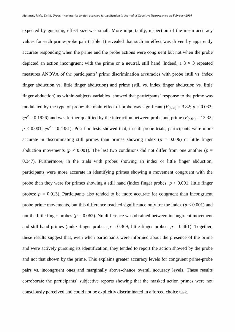
expected by guessing, effect size was small. More importantly, inspection of the mean accuracy
values for each prime-probe pair (Table 1) revealed that such an effect was driven by apparently
accurate responding when the prime and the probe actions were congruent but not when the probe
depicted an action incongruent with the prime or a neutral, still hand. Indeed, a 3 × 3 repeated
measures ANOVA of the participants’ prime discrimination accuracies with probe (still vs. index
finger abduction vs. little finger abduction) and prime (still vs. index finger abduction vs. little
finger abduction) as within-subjects variables showed that participants’ response to the prime was
modulated by the type of probe: the main effect of probe was significant (F(2,32) = 3.82; p = 0.033;
ŋp2 = 0.1926) and was further qualified by the interaction between probe and prime (F(4,64) = 12.32;
p < 0.001; ŋp2 = 0.4351). Post-hoc tests showed that, in still probe trials, participants were more
accurate in discriminating still primes than primes showing index (p = 0.006) or little finger
abduction movements (p < 0.001). The last two conditions did not differ from one another (p =
0.347). Furthermore, in the trials with probes showing an index or little finger abduction,
participants were more accurate in identifying primes showing a movement congruent with the
probe than they were for primes showing a still hand (index finger probes: p < 0.001; little finger
probes: p = 0.013). Participants also tended to be more accurate for congruent than incongruent
probe-prime movements, but this difference reached significance only for the index (p < 0.001) and
not the little finger probes (p = 0.062). No difference was obtained between incongruent movement
and still hand primes (index finger probes: p = 0.369; little finger probes: p = 0.461). Together,
these results suggest that, even when participants were informed about the presence of the prime
and were actively pursuing its identification, they tended to report the action showed by the probe
and not that shown by the prime. This explains greater accuracy levels for congruent prime-probe
pairs vs. incongruent ones and marginally above-chance overall accuracy levels. These results
corroborate the participants’ subjective reports showing that the masked action primes were not
consciously perceived and could not be explicitly discriminated in a forced choice task.

Mattiassi, Mele, Ticini, Urgesi - manuscript version accepted for publication in Journal of Cognitive Neuroscience on February 2014
Corticospinal excitability
The MEPs recorded from each muscle at the beginning and at the end of the experimental session
were entered into a 2 × 2 repeated-measures ANOVA with muscle (FDI vs. ADM) and session
(pre- vs. post-TMS) as within subject variables. A main effect of muscle was found (F(1,16) = 6.12; p
< 0.001; ŋp2 = 0.5705): MEPs recorded from the FDI (1.811 ± 0.473 mV) were greater than MEPs
recorded from the ADM (0.883 ± 0.131 mV, p = 0.025). The main effect of session and the
interaction between muscle and session were not significant (all Fs < 1, ŋp2 < 0.01).
Table 2 shows the raw MEPs amplitudes (in millivolts) in the different experimental conditions.
Inspection of Table 2 reveals that MEPs amplitude at both TMS delays was higher during the
observational task than at baseline, independently of whether participants were fixating at a still
hand or a hand performing a movement that was either related or unrelated to the motor role of the
recorded muscles. This was confirmed by dependent-sample t-tests (one-tailed) comparing the
average of raw MEPs during the 9 observation conditions at each TMS delay and for each muscle.
Indeed, FDI MEPs amplitude was higher during observation than at baseline when TMS was
delivered at either early (2.067 ± 0.511 mV; t(16) = 2.8, p=0.006) or late (2.039 ± 0.513 mV; t(16)
= 2.52, p = 0.011) delay from stimulus onset; the same comparisons with baseline for the ADM
muscle were only marginally significant (early: 1.078 ± 0.195 mV; t(16) = 1.69, p = 0.055; late:
1.066 ± 0.192 mV; t(16) = 1.61, p = 0.064). In a similar vein, dependent-sample t-tests (two-tailed)
showed that no difference was obtained between MEPs amplitudes recorded at early and late TMS
delays for either FDI (t(16) = 0.88, p = 0.39) or ADM (t(16) = 0.9, p = 0.38) muscle during the
observation conditions. In sum, during observation of hand stimuli we found an overall increase of
corticospinal excitability that was not specifically associated to implied actions as it also occurred
for still hand images. This is in keeping with previous studies showing that body part observation
activates the motor cortex more than baseline trials in which participants either keep the eyes closed
or fixate at a blank screen (e.g., Borgomaneri, Gazzola, & Avenanti, 2012; Hodzic, Muckli, Singer,

Mattiassi, Mele, Ticini, Urgesi - manuscript version accepted for publication in Journal of Cognitive Neuroscience on February 2014
& Stirn, 2009; Raos, Kilintari, & Savaki, 2013; Schütz-Bosbach, Mancini, Aglioti, & Haggard,
2006)
Modulation of motor facilitation
Previous analysis of raw MEP amplitudes showed that as compared to viewing a blank screen,
observation of hand images increased corticospinal excitability. Since mirror-like motor facilitation
is indexed by a greater activation during observation of dynamic (or implied action) bodies or body
parts as compared to still bodies or body parts (Fadiga et al., 2005; Urgesi, Candidi, et al., 2006;
Urgesi, Moro et al., 2006), in the successive analysis we tested how corticospinal excitability was
modulated according to the hand image that was presented. We thus expressed MEPs amplitude of
each muscle as % change (MEPratio; Fig. 2) from that muscle baseline, averaging the baseline
MEPs collected at the beginning and at the end of the experimental session. This gave a motor
facilitation index of how much the motor cortex was facilitated during observation of hand images
as compared to baseline. The MEPratio values for the two TMS delays were entered into two
separate 2 × 3 × 3 (muscle × prime × probe) repeated-measures ANOVAs to test whether motor
facilitation was greater during observation of related actions than still hands and unrelated actions
depicted in either the probe or the prime.
The ANOVA for the early TMS delay condition yielded no significant effect (all Fs < 1.1; p >
0.35), suggesting that motor facilitation at this delay from stimulus onset was not specific for the
action depicted in the images. The ANOVA for the late TMS delay condition yielded significant
main effect of probe (F(2,32) = 5.6776; p = 0.008; ŋp2 = 0.2619). The main effect of prime was not
significant (F(2,32) = 1.5; p = 0.239; ŋp2 = 0.086), but a significant interaction between prime and
probe was found (F(4,64) = 3.23; p = 0.017; ŋp2 = 0.168), suggesting that motor facilitation during
observation of muscle-related probe actions was modulated by masked prime actions. The three-
way interaction was not significant (F(4,64) < 1; ŋp2 < 0.05), suggesting comparable patterns of

Mattiassi, Mele, Ticini, Urgesi - manuscript version accepted for publication in Journal of Cognitive Neuroscience on February 2014
results for the two muscles when movements that were related versus unrelated to their motor roles
were observed. Simple effect analysis of mirror-like motor facilitation in response to related as
compared to still and unrelated probes showed significant effects for still (F(1,16) = 9.09, p = 0.008)
and related (F(1,16) = 11.76, p = 0.003) prime trials but not for unrelated prime trials (F(1,16) < 1).
Thus, the observation of a probe stimulus implying a finger movement induced a significant
facilitation of the corticospinal representation of the muscle involved in the execution of that
movement when the probe was preceded by a still (neutral) prime and by a related (congruent)
prime. In contrast, no mirror-like facilitation was obtained when the probe was preceded by
unrelated primes, showing comparable motor facilitation for related than still and unrelated probes..
Pair-wise post-hoc analysis of the interaction between the probe and prime hands revealed no
differences among the three prime types in the unrelated and still probe conditions (all p > 0.102).
Conversely, when the probe was related to the recorded muscle, MEPratios in the unrelated prime
trials (117.34 ± 10.89%) were lower than those in the related prime trials (130.9 ± 13.08%, p =
0.006) and marginally lower than the MEPratios in the still prime trials (126.66 ± 11.77%; p =
0.059); the related and still prime trials did not differ from each other (p = 0.308). Indeed,
MEPratios in the related probe-unrelated prime condition were not significantly different from
MEPratios in the still and unrelated probe conditions regardless of the prime type (all p > 0.319).
Since motor facilitation for still hands and unrelated movements reflects non-specific activation in
response to viewing body parts, no evidence of mirror-like motor facilitation was obtained when
related movement probes were preceded by primes showing a movement that is unrelated to the
motor role of the recorded muscle. Thus, presentation of an unrelated prime suppressed motor
facilitation to a level that is expected in response to observation of body parts independently of
movement information.
DISCUSSION

Mattiassi, Mele, Ticini, Urgesi - manuscript version accepted for publication in Journal of Cognitive Neuroscience on February 2014
In this study, we provide evidence that motor resonance, as indexed by greater motor
facilitation in response to related than static hands and unrelated actions, is not elicited by
unconscious action perception but requires perceptual awareness. Nevertheless, its expression in
response to consciously perceived actions is modulated by unconscious perception of incongruent
actions. We tested corticospinal excitability while participants passively observed static images of
either still or moving hands preceded by masked primes of either the same or different movement.
Our results show the following: i) observation of the action probe, which was consciously
perceived, engendered a somatotopic activation of the cortical representation of the muscles
actually involved in the same action when compared with observation of a still hand or of unrelated
actions; ii) subliminal perception of the action prime did not increase motor facilitation more than
observation of a still hand, suggesting that actions need to be consciously perceived to evoke
mirror-like motor facilitation (motor resonance); and iii) unconscious priming of actions unrelated
to the motor role of the recorded muscle interfered with mirror-like motor facilitation (motor
resonance) in response to consciously perceived probe actions that are related to the motor role of
the recorded muscle.
Conscious perception of implied actions activates the motor system
Several studies have shown that passive observation of dynamic displays (Fadiga et al.,
2005; Urgesi, Candidi, et al., 2006; Avenanti, Bolognini, Maravita, & Aglioti, 2007; Avenanti &
Urgesi, 2011; Tomeo, Cesari, Aglioti, & Urgesi, 2013) and static images of bodies in action
(Urgesi, Moro et al., 2006, 2010; Proverbio, Riva, & Zani, 2009; Avenanti, Annella et al., 2013)
triggers somatotopic activation of the motor system and thus facilitates the corticospinal
representation of those muscles involved in the perceived actions; this effect is thought to reflect
motor resonance. Hence, motor resonance is involved in the extrapolation of dynamic information
from actual as well as implied action stimuli. Although static images were used in the present study,

Mattiassi, Mele, Ticini, Urgesi - manuscript version accepted for publication in Journal of Cognitive Neuroscience on February 2014
these images were presented in a sequence of three snapshots (i.e., sample, prime and probe) rather
than in a single static frame. This sequence may have contributed to the apparent motion perception
if the mask served as a temporal occluding object and the successive hand postures were amodally
completed into a continuous perception of movement (Shiffrar & Freyd, 1993). If true, this
interpretation is in keeping with the notion that the amodal completion of occluded actions may
involve automatic motor resonance processes (Orgs, Bestmann, Schuur, & Haggard, 2011; Urgesi
et al., 2010; Avenanti & Urgesi, 2011).
Importantly, the different delays of TMS stimulation allowed us to test modulation of motor
facilitation during observation of still, related and unrelated actions after a comparable delay (307
ms) from prime (early delay) and probe (late delay) presentation. Thus, we could test muscle-
specific, mirror-like facilitation separately for unconsciously and consciously perceived stimuli.
Magnetoencephalographic (MEG) studies (Nishitani & Hari, 2000; Nishitani, Avikainen, & Hari,
2004) have found that the responses to action observation peak first in the visual occipital areas (at
approximately 118 ms from the onset of the stimulus) before subsequent activation of the superior
temporal sulcus, inferior parietal lobule, inferior frontal areas and, finally, M1 at approximately 300
ms. Similarly, a recent TMS study (Barchiesi & Cattaneo, 2012) found that muscle-specific effects
in M1 were observed only 250 ms after action stimuli onset. Therefore, any direct motor response
to unconscious prime perception (i.e., main effect of prime) was likely to occur in the early TMS
delay, since in that case processing of the prime action, but not of the probe action was likely to
have reached the motor cortex. In contrast we did not find any modulation of the corticospinal
excitability for the unconsciously perceived prime at the early TMS delay independently of the type
of probe presented; nor did we found any effect of prime presentation when the probe depicted a
still hand or an unrelated action, thus ruling out that unconscious action prime action perception
may increase motor facilitation in a muscle-specific fashion more than observation of a still hand.
Conversely, the late TMS delay, when the TMS pulse was delivered after 307 ms from probe onset,
allowed us to show that muscle-specific motor responses are facilitated for the consciously

Mattiassi, Mele, Ticini, Urgesi - manuscript version accepted for publication in Journal of Cognitive Neuroscience on February 2014
perceived probe. In sum, our finding that motor facilitation in response to consciously perceived
probe actions was obtained at the late TMS delay corroborates the temporal dynamics of cortical
activations reported in previous MEG and TMS studies and suggests a late involvement of M1 in
mapping observed (implied) actions.
Motor resonance requires perceptual awareness
The main aim of the present study was to test whether perceptual awareness is a condicio
sine qua non for the emergence of motor resonance responses, measured as a facilitation of
corticospinal excitability during observation of actions that are related to the motor role of the
recorded muscle as compared to observation of a still hand or of an action that is unrelated to the
motor role of the recorded muscle. The forward and backward masking procedure allowed us to
present the prime for a relatively long time (53 ms) but nonetheless prevented conscious perception
by most participants (17/22 individuals). Indeed, even when these participants were explicitly
informed about the presence of the prime and were required to discriminate whether it depicted a
still hand or an index or little finger abduction, their discrimination abilities were at chance. This
suggests that manipulation of the voluntary control of the attentional focus did not affect the
perception of the prime, since prime discrimination was not possible even when subjects were
actively pursuing it. This does not exclude, however, that presentation of the probe stimulus may
have contributed to prevent conscious perception of the prime with automatic attentional shift.
Whether the locus of prime perception disruption was at a perceptual or attentional level, our
paradigm ensured that subjects remained completely unaware of the prime action.
In striking contrast with the muscle-specific response to the probe, no motor facilitation was
measured in response to the presentation of the prime (independently of the probe type and TMS
delay). The absence of motor responses to masked action primes cannot be ascribed to the
interferential effect of probe movement presentation because M1 responses to the prime in the still-
Mattiassi, Mele, Ticini, Urgesi - manuscript version accepted for publication in Journal of Cognitive Neuroscience on February 2014
hand probe conditions were also absent. Nor can this absence reflect that the presentation of either
static or implied action probes completely suppressed the early processing of the masked prime
stimulus in the visual system, thus preventing the result of this processing to be forwarded to the
motor system. Indeed, we have evidence that the prime was processed to a certain extent and the
result of this processing affected the activation of the motor system, since the presentation of
incongruent action primes suppressed the motor resonance to implied action probes (see below).
Furthermore, it is also unlikely that the absence of motor responses to masked action primes was
due to inappropriate timing of the TMS pulses; indeed, in both the early and late TMS delay
conditions, corticospinal excitability was tested after the 250 ms threshold for the establishment of
muscle-specific motor resonance responses to the prime (Barchiesi & Cattaneo, 2012). Given our
experimental settings, however, we cannot exclude the possibility that masked action stimuli may
have modulated motor cortex activity at earlier phases of stimulus processing, for example in the
60-90 ms time window in which a nonspecific activation of the motor cortex in response to action
stimuli has been reported (Lepage, Tremblay, & Théoret, 2010). However, because this early
activation lacks muscle specificity, it is unlikely to reflect motor resonance. Together, our data
suggest that motor resonance requires awareness of the action stimuli.
Action stimuli with an emotional valence are known to activate the motor system in
conditions of reduced visibility or when subjects are not aware of the stimuli. Emotional face and
body expressions induce fast facial mimicry responses (Tamietto et al., 2009) (i.e.,
electromyographic preactivation of the muscles involved in the same expressions) and motor and
premotor cortical activations (Van den Stock et al., 2011) even when presented in the blind
hemifield of hemianopic patients or in masked presentation conditions in healthy individuals
(Dimberg et al., 2000). Such affective facial mimicry responses, however, are not specific for the
observed body parts because they also occur in response to whole-body emotional expressions and
lack the muscle-specific properties of motor resonance responses. Thus, these responses seem to be

Mattiassi, Mele, Ticini, Urgesi - manuscript version accepted for publication in Journal of Cognitive Neuroscience on February 2014
mediated by the activation of subcortical structures in response to emotionally valenced stimuli and
are not involved in nonemotional actions (Tamietto & De Gelder, 2010).
Unconscious presentation of incongruent actions inhibits motor resonance
Although the subliminal presentation of masked prime actions did not trigger motor
facilitation per se, it affected the activation of the motor cortex in response to consciously perceived
actions. In other words, masked prime actions that were incongruent with the probe interfered with
motor facilitation in response to the probe. This evidence of the implicit effects of unconscious
action perception on motor resonance suggests the dissociation between visual awareness and the
perceptual effects of masked action primes (Van den Bussche et al., 2009). Thus, in keeping with
the finding that unconscious prime perception may affect motor execution processes during
visuomotor masked priming tasks (Eimer & Schlaghecken, 1998, 2003), we show evidence that
masked primes can affect motor resonance.
The suppression of motor facilitation during action observation is unlikely to involve a
specific exogenous inhibition of motor activation induced by the abrupt interruption of stimulus
presentation, as reported in visuomotor masked priming tasks (Eimer & Schlaghecken, 1998, 2003).
Indeed, motor facilitation was suppressed for incongruent presentations of prime and probe
movements, whereas visuomotor masked primes tend to inhibit the execution of congruent actions.
Because no facilitatory or inhibitory responses to the prime were obtained with neutral (i.e., still
hand) probes, the interaction between the probe and prime does not seem to be due to the orienting
of spatial attention to the one or to the other side of the space. Accordingly, previous single-pulse
TMS studies have shown that motor facilitation during action observation is based on mapping the
specific muscle involved in the actual execution of the action rather than on the spatial
compatibility between the model’s and observer’s body part (Alaerts, Van Aggelpoel, Swinnen, &
Wenderoth, 2009; Urgesi, Candidi, et al. 2006). Furthermore, behavioral studies have shown that

Mattiassi, Mele, Ticini, Urgesi - manuscript version accepted for publication in Journal of Cognitive Neuroscience on February 2014
visuomotor interference effects occurred when the observed and executed movements involved
different hands (i.e., left vs. right hand; Bertenthal, Longo, & Kosobud, 2006; Brass et al., 2000;
Catmur & Heyes, 2011) or the same hand viewed from different perspectives (egocentric vs.
allocentric perspective; Bortoletto, Mattingley, & Cunnington, 2013) and thus occurred in the
opposite sides of the space. Even when visuomotor interaction was affected by the spatial
compatibility between the executed and observed movement, this resulted into inversion of the
effect (i.e., facilitation for incongruent actions) rather than into its suppression (Vainio & Mustonen,
2011). As such, the suppression of mirror-like (i.e. muscle specific) facilitation obtained in this
study is unlikely to be due to the effects of spatial attention. In a similar vein it seems untenable a
pure attentional account for the suppression of motor resonance to the probe, according to which the
presentation of the incongruent prime may have diverted the focus of attention away from
processing the probe action. Indeed, on the one hand, the prime was not perceived consciously and
could not be discriminated even when the focus of attention of the observer was explicitly focused
on its presentation, thus ruling out a role for voluntary control of attention. On the other hand,
although prime presentation may have influenced probe processing by causing automatic attentional
shift, the results of the online task suggest that participants could easily detect the probe action
independently from whether the action shown by the prime, thus ruling out that the prime disrupted
conscious perception of the probe at an extent capable of explaining the complete suppression of
mirror-like motor facilitation. Accordingly, previous studies showed that attentional modulation of
action perception may modulate, but not suppress motor resonance (Muthukumaraswamy & Singh,
2008). Thus, it is likely the effects of unconscious perception of prime actions on motor resonance
to probe actions are at least partially independent from attentional modulation processes.
The present single-pulse TMS study is not informative regarding the possible neural site of
the interaction between subliminal and supraliminal action processing. We can speculate that such
interaction likely involves an area or set of areas that have direct or indirect connections to the
primary motor cortex and that process both supra- and sub-liminal presentations of actions, thus

Mattiassi, Mele, Ticini, Urgesi - manuscript version accepted for publication in Journal of Cognitive Neuroscience on February 2014
explaining the effects of the masked action prime. Considering the pattern of results, two possible
scenarios can be hypothesized. On the one hand, the facilitation induced by supraliminal probe
actions and its suppression by subliminal incongruent prime actions may be due to separate
mechanisms operating within the motor system. A possible candidate is the ventral premotor cortex,
which is strongly connected to M1 (Prabhu et al., 2009) and seems to be the main source of the
motor-resonance facilitation responses observed in M1 (Avenanti et al., 2007; Avenanti, Annella, et
al., 2013; Avenanti, Candidi, et al., 2013). There is evidence of pyramidal tract neurons in the
macaque ventral premotor cortex (Kraskov, Dancause, Quallo, Sheperd, & Lemon, 2009; but see a
similar finding in M1; Vigneswaran, Philipp, Lemon, & Kraskov, 2013) that may exhibit one of
two main patterns of activity: while one pattern consists in facilitation of the hand motor-neurons
that mirrors the observed action and corresponds to what is generally called motor resonance, the
other consists in a number of neurons being suppressed during action observation. This suppression
is thought to prevent overt movements during action observation, but since the same neurons
showed inhibitory responses in a variety of conditions (e.g., grip with object, grip without object,
concealed grip) it may be that this inhibitory mechanism is less selective with regard to the specific
action observed when compared to classical mirror facilitation. Importantly, facilitation and
suppression neurons seem to be part of two parallel mechanisms in the ventral premotor cortex and
their possible differential sensitivity to perceptual awareness and attentional modulation may
explain the pattern of results obtained in the present study in the terms of facilitation of cortico-
spinal excitability. Indeed, assuming that suppression neurons may respond to observed actions
independently of their perceptual awareness, suppression of motor activity in response to the prime
and/or probe actions may remain unnoticed in a gross measure of cortico-spinal excitability as done
with TMS-evoked motor potentials. Nevertheless, the inhibition of their activity after presentation
of two incongruent actions, which is likely stronger as compared to presentation of a single action,
may contribute to suppress the expression of facilitation neuron activity in response to consciously
perceived probe actions.
Mattiassi, Mele, Ticini, Urgesi - manuscript version accepted for publication in Journal of Cognitive Neuroscience on February 2014
On the other hand, the suppression of motor facilitation for incongruent prime-probe actions
may stem specifically from the processing of the conflict between two incongruent action
representations in areas beyond the motor cortex but directly or indirectly connected to it. The
detection of a conflict in these areas may generate a signal that is sent to the motor system to
suppress motor resonance in response to consciously perceived actions. Unconscious processing of
the conflict between different action-related cues may involve several cortical and subcortical
regions (D’Ostilio & Garraux, 2012). For example, the pre-supplementary motor area is involved in
exerting control over voluntary actions during response conflict (Nachev, Wydell, O’neill, Husain,
& Kennard, 2007) and may also subserve the suppression of incongruent motor resonance
responses. Furthermore, controlling the automatic tendency to imitate other’s actions, as tested in
the visuomotor priming paradigm, involves activation in the ventromedial prefrontal cortex and the
temporo-parietal junction (Brass et al., 2009; Spengler, Brass, Kühn, & Schütz-Bosbach, 2010).
These areas are implicated in higher level mentalizing abilities, such as the sense of agency,
perspective taking and self-referential processing (Brass, Ruby, & Spengler, 2009) and seem to be
also involved in preventing the coding of others’ movements to be translated into self-movements.
Finally, inhibitory afferents to M1 via cortico-striatal pathways play a role in masked visuomotor
priming effects (Bowman, Schlaghecken, & Eimer, 2006; D’Ostilio & Garraux, 2012) and may also
be involved during action observation (Marceglia et al, 2009) and, especially, suppression of
unwanted motor resonance responses.
In conclusion, we show that although motor resonance in response to non-emotional actions
is automatic, it requires the perceptual awareness of actions and operates at a conscious level of
motor representation. While the presentations of subliminal actions do not activate the motor cortex,
they suppress motor resonance responses to incongruent actions that are consciously perceived. We
suggest that this suppression reflects the processing of action conflicts in areas upstream of the
motor cortex and may subserve a basic mechanism for dealing with the multiple and possibly
incongruent actions of other individuals. Motor resonance responses, indeed, seem to endow the

Mattiassi, Mele, Ticini, Urgesi - manuscript version accepted for publication in Journal of Cognitive Neuroscience on February 2014
social brain with the ability of simulating anticipatory representations of unfolding actions ahead of
their realization and in the absence of complete perceptual information from the environment
(Wilson & Knoblich, 2005; Avenanti & Urgesi, 2011; Avenanti, Candidi et al., 2013). Such
representations, however, need to be flexibly updated on the basis of upcoming information, to take
into account not only actual deployment of actions but also their future phases, and to be modulated
by the social context toward imitative or complimentary responses (Sartori et al., 2013). The results
of the present study suggest that modulation of motor resonance according to the social context (in
this case incongruent actions) may also occur at an implicit level and independently of observer’s
perceptual awareness.
Acknowledgments: The research was supported by grants from Istituto Italiano di Tecnologia
SEED 2009 [Prot. no. 21538; to C.U.], from the Ministero Istruzione Università e Ricerca [Progetti
di Ricerca di Interesse Nazionale, PRIN 2009; Prot. no. 2009A8FR3Z; Futuro In Ricerca, FIR 2012,
Prot. N. RBFR12F0BD; to C.U.], and from Istituto di Ricovero e Cura a Carattere Scientifico “E.
Medea” [Ricerca Corrente 2012, Ministero Italiano della Salute; to C.U.]. The authors declare no
competing financial interests.

Mattiassi, Mele, Ticini, Urgesi - manuscript version accepted for publication in Journal of Cognitive Neuroscience on February 2014
REFERENCES
Alaerts, K., Van Aggelpoel, T., Swinnen, S. P., & Wenderoth, N. (2009). Observing shadow
motions: Resonant activity within the observer’ s motor system? Neuroscience Letters, 461,
240–244.
Alaerts, K., Heremans, E., Swinnen, S.P., & Wenderoth, N. (2009). How are observed actions
mapped to the observer's motor system? Influence of posture and perspective.
Neuropsychologia, 47, 415-422
Avenanti, A., Annella, L., Candidi, M., Urgesi, C., & Aglioti, S.M. (2013). Compensatory plasticity
in the action observation network: virtual lesions of STS enhance anticipatory simulation of
seen actions. Cerebral Cortex, 23, 570-580.
Avenanti, A., Candidi, M., & Urgesi, C. (2013). Vicarious motor activation during action
perception: beyond correlational evidence. Frontiers in Human Neuroscience, 7,185.
Avenanti, A., Bolognini, N., Maravita, A., & Aglioti, S.M. (2007). Somatic and motor components
of action simulation. Current Biology, 17, 2129–2135.
Avenanti, A., & Urgesi, C. (2011). Understanding “what” others do: mirror mechanisms play a
crucial role in action perception. Social Cognitive & Affective Neuroscience, 6, 257–259.
Barchiesi, G., & Cattaneo, L. (2012). Early and late motor responses to action observation. Social
Cognitive & Affective Neuroscience, doi: 10.1093/scan/nss049.
Bargh, J.A., Schwader, K.L., Hailey, S.E., Dyer, R.L., & Boothby, E.J. (2012). Automaticity in
social-cognitive processes. Trends in Cognitive Science, 16, 593-605.
Bertenthal, B. I., Longo, M. R., & Kosobud, A. (2006). Imitative response tendencies following
observation of intransitive actions. Journal of experimental psychology. Human perception
and performance, 32(2), 210–225.
Mattiassi, Mele, Ticini, Urgesi - manuscript version accepted for publication in Journal of Cognitive Neuroscience on February 2014
Borgomaneri, S., Gazzola, V., & Avenanti, A. (2012). Motor mapping of implied actions during
perception of emotional body language. Brain Stimulation, 5, 70-76.
Bortoletto, M., Mattingley, J. B., & Cunnington, R. (2013). Effects of Context on Visuomotor
Interference Depends on the Perspective of Observed Actions. PlosOne, 8(1), e53248
Bowman, H., Schlaghecken, F., & Eimer, M. (2006). A neural network model of inhibitory
processes in subliminal priming. Visual Cognition, 13, 401–480.
Brass, M., Bekkering, H., & Prinz, W. (2001). Movement observation affects movement execution
in a simple response task. Acta Psychologica, 106, 3–22.
Brass, M., Bekkering, H., Wohlschläger, A., & Prinz, W. (2000). Compatibility between observed
and executed finger movements: comparing symbolic, spatial, and imitative cues. Brain and
Cognition, 44, 124–143.
Brass, M., Ruby, P., & Spengler, S. (2009). Inhibition of imitative behaviour and social cognition.
Philosophical Transactions of the Royal Society B: Biological Sciences, 364(1528), 2359-
2367.
Brasil-Neto, J.P., Cohen, L.G., Panizza, M., Nilsson, J., Roth, B.J., & Hallett, M. (1992). Optimal
focal transcranial magnetic activation of the human motor cortex: effects of coil orientation,
shape of the induced current pulse, and stimulus intensity. Journal of Clinical
Neurophysiology, 9,132–136.
Briggs, G.G., & Nebes, R.D. (1975). Patterns of hand preference in a student population. Cortex,
11, 230–238.
Catmur, C., & Heyes, C. (2011). Time course analyses confirm independence of imitative and
spatial compatibility. Journal of experimental psychology. Human perception and
performance, 37(2), 409–421.

Mattiassi, Mele, Ticini, Urgesi - manuscript version accepted for publication in Journal of Cognitive Neuroscience on February 2014
Catmur, C., Mars, R.B., Rushworth, M.F., & Heyes, C. (2011). Making mirrors: premotor cortex
stimulation enhances mirror and counter-mirror motor facilitation. Journal of Cognitive
Neuroscience, 23, 2352-2362.
Craighero, L., Bello, A., Fadiga, L., & Rizzolatti, G. (2002). Hand action preparation influences the
responses to hand pictures. Neuropsychologia, 40, 492–502.
Craighero, L., Fadiga, L., & Rizzolatti, G. (1999). Action for perception: a motor-visual attentional
affect. Journal of Experimental Psychology: Human Perception and Performance, 25, 1673–
1692.
Craighero, L., Fadiga, L., Umiltà, C. A., & Rizzolatti, G. (1996). Evidence for visuomotor priming
effect. Neuroreport, 8, 347–349.
Dehaene, S., Naccache, L., Le Clec’H, G., Koechlin, E., & Mueller, M. (1998). Imaging
unconscious semantic priming. Nature, 395, 597–600.
Di Lazzaro, V., Oliviero, A., Profice, P., Saturno, E., Pilato, F., Insola, A., et al. (1998).
Comparison of descending volleys evoked by transcranial magnetic and electric stimulation in
conscious humans. Electroencephalography and Clinical Neurophysiology, 109, 397–401.
Dimberg, U., Thunberg, M., & Elmehed, K. (2000). Unconscious Facial Reactions to Emotional
Facial Expressions. Psychological Science, 11, 86–89.
D’Ostilio, K., & Garraux, G. (2012). Dissociation between unconscious motor response facilitation
and conflict in medial frontal areas. European Journal of Neuroscience, 35, 332–340.
Eimer, M., & Schlaghecken, F. (1998). Effects of masked stimuli on motor activation: behavioral
and electrophysiological evidence. Journal of Experimental Psychology: Human Perception
and Performance, 24, 1737–1747.
Eimer, M., & Schlaghecken, F. (2003). Response facilitation and inhibition in subliminal priming.
Biological Psychology, 64, 7–26.

Mattiassi, Mele, Ticini, Urgesi - manuscript version accepted for publication in Journal of Cognitive Neuroscience on February 2014
Fadiga, L., Craighero, L., & Olivier, E. (2005). Human motor cortex excitability during the
perception of others’ action. Current Opinion in Neurobiology, 15, 213–218.
Fadiga, L., Fogassi, L., Pavesi, G., & Rizzolatti, G. (1995). Motor facilitation during action
observation : a magnetic stimulation study. Journal of Neurophysiology, 73, 2608–2611.
Hodzic, A., Muckli. L., Singer, W., & Stirn, A. (2009). Cortical responses to self and others.
Human Brain Mapping, 30, 951-962.
Hogeveen, J., & Obhi, S. S. (2013). Automatic imitation is automatic, but less so for narcissists.
Experimental Brain Research, 224, 613–621.
Kilner, J. M., Paulignan, Y., & Blakemore, S.J. (2003). An interference effect of observed
biological movement on action. Current Biology, 13, 522–525.
Kraskov, A., Dancause, N., Quallo, M. M., Shepherd, S., & Lemon, R. N. (2009). Corticospinal
neurons in macaque ventral premotor cortex with mirror properties: a potential mechanism for
action suppression? Neuron, 64(6), 922–930.
Lepage, J. F., Tremblay, S., & Théoret, H. (2010). Early non-specific modulation of corticospinal
excitability during action observation. European Journal of Neuroscience, 31, 931–937.
Macmillan, N. A., & Kaplan, H. L. (1985). Detection theory analysis of group data: Estimating
sensitivity from average hit and false-alarm rates. Psychological Bulletin, 98, 185-199.
Maeda, F., Kleiner-Fisman, G., & Pascual-Leone, A. (2002). Motor facilitation while observing
hand actions: specificity of the effect and role of observer's orientation. Journal of
Neurophysiology, 87(3), 1329-1335.
Marceglia, S., Fiorio, M., Foffani, G., Mrakic-Sposta, S., Tiriticco, M., Locatelli, M., et al. (2009).
Modulation of beta oscillations in the subthalamic area during action observation in
Parkinson's disease. Neuroscience, 161, 1027-1036.

Mattiassi, Mele, Ticini, Urgesi - manuscript version accepted for publication in Journal of Cognitive Neuroscience on February 2014
McGettigan, C., Warren, J. E., Eisner, F., Marshall, C. R., Shanmugalingam, P., & Scott, S. K.
(2012). Neural correlates of sublexical processing in phonological working memory. Journal
of Cognitive Neuroscience, 23, 121–143.
Muthukumaraswamy, S. D., & Singh, K. D. (2008). Modulation of the human mirror neuron system
during cognitive activity. Psychophysiology, 45(6), 896–905.
Nachev, P., Wydell, H., O’neill, K., Husain, M., & Kennard, C. (2007). The role of the pre-
supplementary motor area in the control of action. Neuroimage, 36, T155–T163.
Nishitani, N., Avikainen, S., & Hari, R. (2004). Abnormal imitation-related cortical activation
sequences in Asperger’s syndrome. Annals of Neurology, 55, 558–562.
Nishitani, N., & Hari, R. (2000). Temporal dynamics of cortical representation fo action.
Proceedings of the National Academy of Sciences of the United States of America, 97, 913–
918.
Orgs, G., Bestmann, S., Schuur, F., & Haggard, P., (2011). From body form to biological motion:
the apparent velocity of human movement biases subjective time. Psychological Science, 22,
712-717.
Prabhu, G., Shimazu, H., Cerri, G., Brochier, T., Spinks, R. L., Maier, M. A., & Lemon, R. N.
(2009). Modulation of primary motor cortex outputs from ventral premotor cortex during
visually guided grasp in the macaque monkey. The Journal of physiology, 587(Pt 5), 1057–
1069.
Proverbio, A. M., Riva, F., & Zani, A. (2009). Observation of static pictures of dynamic actions
enhances the activity of movement-related brain areas. PLoS One, 4(5), e5389.
Raos, V., Kilintari, M., & Savaki H.E. (2013). Viewing a forelimb induces widespread cortical
activations. Neuroimage, doi: 10.1016/j.neuroimage.2013.12.010.

Mattiassi, Mele, Ticini, Urgesi - manuscript version accepted for publication in Journal of Cognitive Neuroscience on February 2014
Rizzolatti, G., & Craighero, L. (2004). The mirror-neuron system. Annual Review of Neuroscience,
27, 169–192.
Sartori, L., Bucchioni, G., & Castiello, U. (2013). When emulation becomes reciprocity. Social
Cognitive & Affective Neuroscience, 8, 662-669.
Schütz-Bosbach, S., Mancini, B., Aglioti, S.M., & Haggard, P. (2006) Self and other in the human
motor system. Current Biology, 16, 1830-1834.
Shiffrar, M., & Freyd, J. J. (1993). Timing and apparent motion path choice with human body
photographs. Psychological Science, 4, 379–384.
Spengler, S., Brass, M., Kühn, S., & Schütz-Bosbach, S. (2010). Minimizing motor mimicry by
myself: self-focus enhances online action-control mechanisms during motor contagion.
Consciousness & Cognition, 19, 98-106.
Spengler, S., von Cramon, D.Y., & Brass, M. (2010). Resisting motor mimicry: control of imitation
involves processes central to social cognition in patients with frontal and temporo-parietal
lesions. Social Neuroscience, 5(4), 401-416.
Stürmer, B., Aschersleben, G., & Prinz, W. (2000). Correspondence effects with manual gestures
and postures: A study of imitation. Journal of Experimental Psychology: Human Perception
and Performance, 26, 1746–1759.
Tamietto, M., Castelli, L., Vighetti, S., Perozzo, P., Geminiani, G., & Weiskrantz, L. (2009).
Unseen facial and bodily expressions trigger fast. Proceedings of the National Academy of
Sciences of the Uunited States of America, 106, 17661-17666.
Tamietto, M., & De Gelder, B. (2010). Neural bases of the non-conscious perception of emotional
signals. Nature Reviews Neuroscience, 11, 697–709.

Mattiassi, Mele, Ticini, Urgesi - manuscript version accepted for publication in Journal of Cognitive Neuroscience on February 2014
Théoret, H., Halligan, E., Kobayashi, M., Merabet, L., & Pascual-Leone, A. (2004). Unconscious
modulation of motor cortex excitability revealed with transcranial magnetic stimulation.
Experimental Brain Research, 155, 261–264.
Tomeo, E., Cesari, P., Aglioti, S.M., & Urgesi, C. (2013). Fooling the Kickers but not the
Goalkeepers: Behavioral and Neurophysiological Correlates of Fake Action Detection in
Soccer. Cerebral Cortex, 23, 2765-2778..
Urgesi, C., Candidi, M., Fabbro, F., Romani, M., & Aglioti, S. M. (2006). Motor facilitation during
action observation: topographic mapping of the target muscle and influence of the onlooker's
posture. European Journal of Neuroscience, 23, 2522-2530.
Urgesi, C., Moro, V., Candidi, M., & Aglioti, S. M. (2006). Mapping implied body actions in the
human motor system. Journal of Neuroscience, 26, 7942–7949.
Urgesi, C., Maieron, M., Avenanti, A., Tidoni, E., Fabbro, F., & Aglioti, S. M. (2010). Simulating
the future of actions in the human corticospinal system. Cerebral cortex, 20, 2511–2521.
Vainio, L., & Mustonen, T. (2011). Mapping the identity of a viewed hand in the motor system:
evidence from stimulus-response compatibility. Journal of Experimental Psychology: Human
Perception and Performance, 37, 207–221.
Vainio, L., Tucker, M., & Ellis, R. (2007). Precision and power grip priming by observed grasping.
Brain Cognition, 65, 195–207.
Van den Bussche, E., Van den Noortgate, W., & Reynvoet, B. (2009). Mechanisms of masked
priming: a meta-analysis. Psychological Bulletin, 135, 452-477.
Van den Stock, J., Tamietto, M., Sorger, B., Pichon, S., Grézes, J., & De Gelder, B. (2011).
Cortico-subcortical visual, somatosensory, and motor activations for perceiving dynamic
whole-body emotional expressions with and without striate cortex (V1). Proceeding of the
National Academy of Sciences of the United States of America, 108, 16188–16193.

Mattiassi, Mele, Ticini, Urgesi - manuscript version accepted for publication in Journal of Cognitive Neuroscience on February 2014
Vigneswaran, G., Philipp, R., Lemon, R.N., & Kraskov, A. (2013). M1 corticospinal mirror neurons
and their role in movement suppression during action observation. Current Biology, 23, 236-
243.
Vogt, S., Taylor, P., & Hopkins, B. (2003). Visuomotor priming by pictures of hand postures:
perspective matters. Neuropsychologia, 41, 941–951.
Wasserman, E.M. (1998). Risk and safety of repetitive transcranial magnetic stimulation: report and
suggested guidelines from the International Workshop on the Safety of Repetitive Transcranial
Magnetic Stimulation, June 5–7, 1996. Electroencephalography and Clinical
Neurophysiology, 108, 1–16.
Wilson, M., & Knoblich, G. (2005). The case for motor involvement in perceiving conspecifics.
Psychological Bullettin, 131, 460-473.
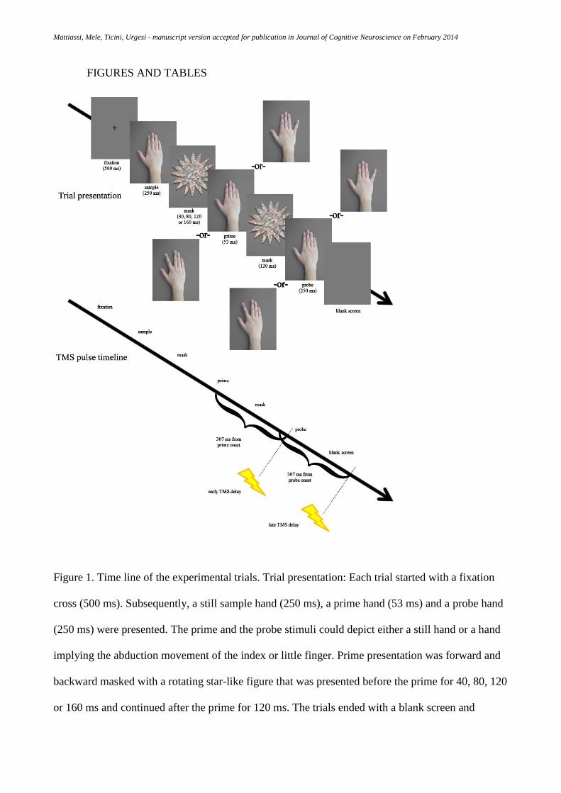
Mattiassi, Mele, Ticini, Urgesi - manuscript version accepted for publication in Journal of Cognitive Neuroscience on February 2014
FIGURES AND TABLES
Figure 1. Time line of the experimental trials. Trial presentation: Each trial started with a fixation
cross (500 ms). Subsequently, a still sample hand (250 ms), a prime hand (53 ms) and a probe hand
(250 ms) were presented. The prime and the probe stimuli could depict either a still hand or a hand
implying the abduction movement of the index or little finger. Prime presentation was forward and
backward masked with a rotating star-like figure that was presented before the prime for 40, 80, 120
or 160 ms and continued after the prime for 120 ms. The trials ended with a blank screen and

Mattiassi, Mele, Ticini, Urgesi - manuscript version accepted for publication in Journal of Cognitive Neuroscience on February 2014
participants were prompted to report the motion of the probe stimulus. TMS pulse timeline: A
single TMS pulse was delivered at one of two moments during the trial, either 307 ms (early TMS
delay) after prime onset (corresponding to 133 ms after probe onset) or 307 ms (late TMSdelay)
after probe onset.

Mattiassi, Mele, Ticini, Urgesi - manuscript version accepted for publication in Journal of Cognitive Neuroscience on February 2014
Figure 2. Effects of conscious and unconscious observation of actions. MEPratio (% of baseline) of
motor evoked potentials (MEPs) recorded from the FDI and ADM muscles in the different prime
and probe combinations. MEPs recorded from FDI and ADM are collapsed because the same
pattern of results was obtained for the two muscles (see text). The prime and probe types were
coded according to whether the hand depicted a still hand (still), a finger abduction movement that
was related to the recorded muscle (related), or a finger abduction movement that was not related to
the recorded muscle (unrelated). Error bars indicate the s.e.m., asterisks indicate significant
comparisons, @ indicates marginally significant comparisons.

Mattiassi, Mele, Ticini, Urgesi - manuscript version accepted for publication in Journal of Cognitive Neuroscience on February 2014
Accuracy in the prime discrimination task (%)
Prime
Still Index finger Little finger
Pro
be
Still 66.91 ± 1.62 36.76 ± 1.54 26.1 ± 1.21
Index finger 26.47 ± 1.56 66.91 ± 1.84 16.91 ± 0.9
Little finger 27.21 ± 1.11 34.56 ± 1.99 54.41 ± 1.8
Table 1. Accuracy (% correct responses) in discriminating the prime during the post-experimental
control task. Mean (s.e.m.) accuracy values for each combination of prime and probe presentation
are shown. “Still” indicates the presentation of a still hand stimulus, while “Index finger” and
“Little finger” indicate the presentation of hand with the index or little finger abducted,
respectively.

Mattiassi, Mele, Ticini, Urgesi - manuscript version accepted for publication in Journal of Cognitive Neuroscience on February 2014
Raw amplitude of MEPs (mV)
Early delay Late delay
Probe Prime FDI ADM FDI ADM
Still Still 2.08 ± 0.51 1.07 ± 0.2 2.12 ± 0.52 1.06 ± 0.19
Still Related 2.03 ± 0.5 1.05 ± 0.18 1.99 ± 0.52 1.01 ± 0.18
Still Unrelated 1.95 ± 0.46 1.1 ± 0.2 1.99 ± 0.5 1.07 ± 0.2
Related Still 2.11 ± 0.52 1.13 ± 0.21 2.11 ± 0.54 1.14 ± 0.2
Related Related 2.11 ± 0.52 1.09 ± 0.2 2.18 ± 0.52 1.1 ± 0.2
Related Unrelated 2.12 ± 0.53 1.05 ± 0.18 2.01 ± 0.49 1.06 ± 0.19
Unrelated Still 2.04 ± 0.54 1.07 ± 0.21 2.01 ± 0.52 1.06 ± 0.2
Unrelated Related 2.05 ± 0.51 1.04± 0.19 2.01 ± 0.51 1.03 ± 0.18
Unrelated Unrelated 2.1 ± 0.53 1.11 ± 0.2 1.93 ± 0.52 1.07 ± 0.2
Table 2. Mean ± s.e.m. raw amplitudes (in mV) of motor evoked potentials (MEPs) recorded from
the FDI and ADM muscles in each experimental condition. “Still” indicates the presentation of a
still hand stimulus; “related” indicates the presentation of a stimulus that depicts an abduction of the
finger controlled by the recorded muscle (shown in column); “unrelated” indicates the presentation
of a stimulus that depicts an abduction of the finger not controlled by the recorded muscle.
Copyright © 2022 FDOKUMEN