Bioconversion of Lactose into Glucose–Galactose Syrup by ...
Upload
independentCategory
view
2download
0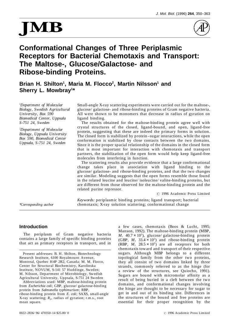
J. Mol. Biol. (1996) 264, 350–363
Conformational Changes of Three PeriplasmicReceptors for Bacterial Chemotaxis and Transport:The Maltose-, Glucose/Galactose- andRibose-binding Proteins.
Brian H. Shilton 1, Maria M. Flocco 2, Martin Nilsson 1 andSherry L. Mowbray 1*
1Department of Molecular Small-angle X-ray scattering experiments were carried out for the maltose-,Biology, Swedish Agricultural glucose/galactose- and ribose-binding proteins of Gram negative bacteria.University, Box 590 All were shown to be monomers that decrease in radius of gyration on
ligand binding.Biomedical Center, UppsalaThe results obtained for the maltose-binding protein agree well withS-751 24, Sweden
crystal structures of the closed, ligand-bound, and open, ligand-free2Department of Molecular protein, suggesting that these are indeed the primary forms in solution.Biology, Uppsala University The closed form is stabilized by protein–sugar interactions, while the openBox 590, Biomedical Center conformation is stabilized by close contacts between the two domains.Uppsala, S-751 24, Sweden Since it is the proper spacial relationship of the domains in the closed form
that is most important for interaction with chemotaxis and transportpartners, the stabilization of the open form would help keep ligand-freemolecules from interfering in function.
The scattering results also provide evidence that a large conformationalchange takes place in association with ligand binding to theglucose/galactose- and ribose-binding proteins, and that the two changesare similar. Modeling suggests that the open forms resemble those foundin the related leucine and leucine/isoleucine/valine-binding proteins, butare different from those observed for the maltose-binding protein and therelated purine repressor.
7 1996 Academic Press Limited
Keywords: periplasmic binding proteins; ligand transport; bacterial*Corresponding author chemotaxis; X-ray solution scattering; conformational change
Introduction
The periplasm of Gram negative bacteriacontains a large family of specific binding proteinsthat act as primary receptors in transport, and in
a few cases, chemotaxis (Boos & Lucht, 1995;Manson, 1992). The maltose-binding protein (MBP,Mr 40.7 × 10³), glucose/galactose-binding protein(GBP, Mr 33.4 × 10³) and ribose-binding protein(RBP, Mr 28.5 × 10³) are all receptors for bothchemotaxis toward and transport of their respectivesugars. Although MBP belongs to a differenttopological family from the other two proteins,they all consist of two domains linked by threestrands, commonly referred to as the hinge (fora review of the structures, see Quiocho, 1991).Sugars are bound with micromolar affinity as aresult of being buried in a cleft between the twodomains, and conformational changes involvingthe hinge are thought to be necessary for sugar toget in and out of its binding site. Differences inthe structures of the bound and free proteins areessential for their proper recognition by the
Present addresses: B. H. Shilton, BiotechnologyResearch Institute, 6100 Royalmount Avenue,Montreal, Quebec H4P 2R2, Canada; M. M. Flocco,Center for Structural Biochemistry, KarolinskaInstitute, NOVUM, S-141 57 Huddinge, Sweden;M. Nilsson, Department of Microbiology, SwedishAgricultural University, Uppsala, S-751 24 Sweden
Abbreviations used: MBP, maltose-binding proteinfrom Escherichia coli; GBP, glucose/galactose-bindingprotein from Salmonella typhimurium; RBP,ribose-binding protein from E. coli; SAXS, small-angleX-ray scattering; Rg , radius of gyration; r.m.s., rootmean square.
0022–2836/96/470350–14 $25.00/0 7 1996 Academic Press Limited

Conformational Changes in Chemotaxis-transport Receptors 351
Figure 1. Guinier plots for samples of MBP (green), GBP (red) and RBP (blue); in each case, the data with sugarare shown as a continuous line, and those without sugar as a broken line. It was determined that the range of s valuesfrom 0.006 to 0.01 gave a satisfactory result for MBP, but that the GBP and RBP samples suffered from interparticleinterference, and the interval of s was altered to minimize the influence of these effects. The linear fit in the range0.006 < s < 0.01 is shown for all three proteins as a thin continuous line, and the fit in the range 0.008 < s < 0.012 isshown for GBP and RBP as a dotted line.
membrane components. In a similar way, confor-mational changes are thought to be critical to thefunction of the lac family of repressors (Schumacheret al., 1994, 1995), and lactoferrin/transferrin family(Baker et al., 1987), both of which show structuralrelationships to the binding proteins.
Small-angle X-ray scattering (SAXS) has foundwide application in the study of conformationalchanges in solution (Glatter & Kratky, 1982; Feigin& Svergun, 1987). In two previous studies,SAXS data obtained for binding proteinsinvolved in transport were compared withknown crystallographic structures for one of theforms, while the second form was a modeledone (Newcomer et al., 1981; Olah et al., 1993). Inthe present study, we describe the results obtainedin a SAXS study of MBP, allowing for the firsttime a comparison with known crystal structuresfor both the open and closed forms of a bindingprotein (Spurlino et al., 1991; Sharff et al., 1992;Shilton et al., 1996). In addition, we use datameasured for GBP and RBP, where the X-raystructures of only the closed forms are known(Vyas et al., 1987; Zou et al., 1993; Mowbray &Cole, 1992), together with other structural infor-mation, to investigate the nature of their openforms.
Results and Discussion
Dynamic light scattering
Dynamic light scattering studies of solutions ofMBP, GBP and RBP indicated that all weremonodisperse; the average measured hydrody-namic radii were 28, 28 and 26.5 A, respectively.Addition of ligand caused no change in theapparent solvated size of the proteins, but changesof less than 2 A would be within the limits of errorof the method.
Radii of gyration
Representative Guinier plots for each proteinwith and without added sugar are shown in Fig-ure 1, and a summary of the Rg values obtainedfrom the slopes of the curves is given in Table 1.
The addition of either of two ligands that areknown to bind well to MBP (Kd maltose = 1 mM, Kd
maltotriose = 0.16 mM; Szmelcman et al., 1976) givesrise to substantial decreases in the Rg , but additionof a ligand that binds poorly does not (Kd
glucose > 1 mM). The radii of gyration obtained forMBP agree well with those calculated from X-rayco-ordinates (see Table 1). Addition of water

Tab
le1.
Exp
erim
enta
lan
dca
lcul
ated
rad
iiof
gyra
tion
Exp
erim
enta
lR
gva
lues
Mod
elR
g
Gui
nier
Rg
Wit
hX
-ray
wat
erW
ith
mod
elw
ater
Dif
fere
nce
inR
g
Sam
ple
0.00
6<
s<
0.01
0.00
8<
s<
0.01
2G
NO
MR
gW
ith
now
ater
(num
ber)
(num
ber)
mod
el
MB
P21
.98
20.
1522
.32
0.2
21.7
221
.79
22.3
41.
152
0.16
(203
wat
er)
(265
wat
er)
(0.9
32
0.03
)M
BP-
mal
20.9
52
0.11
21.1
20.
220
.79
20.8
321
.44
(164
wat
er)
(289
wat
er)
MB
P-m
alto
trio
se20
.66
20.
0720
.72
0.2
MB
P-gl
u21
.93
20.
0222
.22
0.2
GB
P21
.81
20.
1122
.16
20.
1922
.72
0.2
(21.
91)
—(2
2.37
)1.
402
0.18
(259
wat
er)
GB
P-gl
u20
.30
20.
0820
.97
20.
0621
.22
0.2
20.5
320
.66
21.3
0(1
53w
ater
)(2
53w
ater
)G
BP-
rib
21.7
62
0.12
RB
P19
.80
20.
0920
.70
20.
1820
.52
0.2
(21.
09)
—(2
1.61
)1.
422
0.21
(252
wat
er)
RB
P-ri
b18
.61
20.
0419
.28
20.
0818
.92
0.2
19.2
019
.45
19.9
5(2
16w
ater
)(2
17w
ater
)R
BP-
glu
19.7
52
0.04
Stan
dar
dd
evia
tion
sar
egi
ven
whe
reap
prop
riat
e.W
here
only
am
odel
valu
eis
avai
labl
e,it
isgi
ven
inpa
rent
hese
s.

Conformational Changes in Chemotaxis-transport Receptors 353
increases the calculated Rg , but the results areslightly different for X-ray and computer-generatedwater models with the same number of solventatoms, suggesting that such models should not beviewed as accurate in detail.
Similarly, GBP and RBP showed significantdecreases in Rg with the addition of sugars theyrecognise, but not with ones they do not; the Kd ofGBP for glucose is approximately 0.1 mM (seereferences cited by Aqvist & Mowbray, 1995), andthat of RBP for ribose is 0.13 mM (Binnie et al.,1992). The scattering in the lowest-angle regionsfor RBP, and to some extent, GBP, seemed to beattenuated by interparticle interference, giving riseto a decrease in the apparent Rg . In an attempt toreduce the influence of these concentration-relatedeffects, the Rg calculations were repeated using aslightly different range of s values (Figure 1). TheRg values obtained in this second calculation wereindeed larger, and those for the ligand-boundproteins compare better with values calculatedfrom the co-ordinates (Table 1). Both ligand-boundRg values, however, are smaller than suggested bythe X-ray structures, and the corresponding I(0)values used in scaling RBP and GBP data arelikely to be slight underestimates. The differencein Rg between the sugar-bound and free forms ofall of the proteins, however, appears to be quiteinsensitive to the choice of different ranges of s.Since the goal is not to determine Rg , per se, butto compare the scattering behaviour of the boundand free forms, these differences provide thedesired information. Indeed, it was consideredimportant to make the measurements in thephysiologically relevant concentration range rep-resented here (0.2 to 1.0 mM), regardless of anysmall problems in scaling due to concentrationeffects.
Experimental scattering curves
The measured intensities as functions of scatter-ing angle for the three proteins with and withoutsugar are shown as I(s) versus s plots in Fig-ure 2(a). As expected from the Rg results, thescattering behaviour of all three proteins ischanged by the addition of ligand; these differ-ences persist to the highest angles measured. Thecalculated scattering curves for the availablestructures with a solvent model are shown forcomparison in Figure 2(b). Inclusion of hydrogenatoms in the model changed the curves veryslightly, while inclusion of solvent decreased therelative intensity in the higher-angle regions. Sinceuse of the X-ray water molecules in the modelcalculation gave a slightly different profile from asimilar number of water molecules introducedcomputationally, it seems likely that inadequaciesin water modeling explain any minor differencesin fit to the experimental data. However, themodels reproduce very well the relative behaviourof bound versus free protein in each case, as wellas differences between the three proteins.
Distance distribution functions
A more intuitively accessible view of thescattering data is given by the distance distribution(p(r)) plots in Figure 3(a). Each of the proteinsshows a characteristic elongated shape, as deducedfrom the preponderance of interatomic vectorslonger than the mode. All show a decrease in theaverage distance with the binding of sugar,although the position of the maximum in each caseis altered very little. The curves compare well withthose calculated from the available co-ordinatesincluding a water model (Figure 3(b)). Addition ofwater results in some smoothing of the calculatedcurves, and a shift to slightly higher r values. Bothconcentration effects and changes in the watermodel lead to small alterations in the curve shapeat longer r values, but these are smaller than theobserved experimental differences, and affect bothforms of a single protein in a similar way. Theestimates of Rg obtained from this ‘‘real space’’analysis are summarized in Table 1. Concentrationeffects are again most evident for RBP, where theestimated Rg is smaller than the ligand-boundstructure suggests. For the other two proteins, theRg estimates agree with the model within theexpected error of the method.
Conformational stabilization byclose-packed interfaces
The simplest interpretation of the MBP scatteringdata presented here is that a primary open formexists in the absence of bound sugar, and a primaryclosed form is found in the presence of boundligand. Since it is known that at least some bindingproteins are highly dynamic in solution, particu-larly in the ligand-free state (see, for example, Wolfet al., 1994; Flocco & Mowbray, 1994; Careaga et al.,1995; Zhang et al., 1996), other explanations mustalso be considered. Multiple forms with similarshape characteristics could give rise to similarscattering curves, and mixed populations mightalso fortuitously show the same behaviour. Thesimple explanation, however, is supported by thefact that almost identical forms are found for bothopen and closed MBPs in a number of differentcrystal environments (see discussion by Shiltonet al., 1996). Thus, although the structure of MBPmay be highly dynamic, it clearly has preferredstates.
The large number of interactions formed by bothstructural domains of the protein with sugar easilyexplain why the closed form should be overwhelm-ingly favoured when ligand is present. However, ifthe proteins are flexible, one has to ask why anyparticular open form is favoured when ligand is notbound.
The observation of Gerstein et al. (1993) that theopen and closed forms of lactoferrin are stabilizedby two distinct sets of interdomain contact surfacesprovides an important clue. As shown in Figure 4,similar surfaces can be seen in MBP using

Conformational Changes in Chemotaxis-transport Receptors354
(a)
(b)
Figure 2. Scattering behaviour of MBP (green) GBP (red) and RBP (blue). The experimental (a) and model (b)intensities are shown on a logarithmic scale. In each case the intensity for the sugar-bound form is shown as acontinuous line and that for the ligand-free form is shown as a broken line. All of the protein models included anenergy-minimized water model described in Materials and Methods; models for possible open forms of GBP and RBPare also described in the text.

Conformational Changes in Chemotaxis-transport Receptors 355
(a)
(b)
Figure 3. Distance distribution plots for MBP (green), GBP (red) and RBP (blue); the experimental (a) and model(b) curves are shown on the same scale. In each case, the curve for the sugar-bound form is shown as a continuousline, and that for the ligand-free form is shown as a broken line. All of the protein models included anenergy-minimized water model described in Materials and Methods; models for possible open forms of GBP and RBPare also described in the text.

Conformational Changes in Chemotaxis-transport Receptors356
Figure 4. Differences in surface accessibility between the open and closed forms of MBP, illustrated using the twoX-ray structures. Surface accessibility was calculated as the sum for all atoms in each residue. Side-chains are shownas grey if the residues are more buried in the open form (difference 20 to 88 A2), and black if they are more buriedin the closed form (difference 20 to 61 A2); side-chains for which the difference in surface accessibility is less than 20 A2
are not shown. A few isolated residues show changes because of side-chain mobility, or because of differences inconformation due to crystal packing. The maltose in the bound form is shown in a space-filling representation.
differences in surface accessibility between the twoMBP structures. Although relatively few inter-domain interactions exist in the closed form, anumber of residues in the binding cleft are buriedas a result of protein–sugar interactions. In contrast,
many contacts between the two domains arefound only in the open form, on the surface ofthe protein near the hinge; here, the surface thatis buried in the open form, but exposed in theclosed form, is approximately 1400 A2 in area. The

Conformational Changes in Chemotaxis-transport Receptors 357
Figure 5. A proposed confor-mational change for RBP. (a) Theclosed form of RBP seen in theribose-bound X-ray structure isshown, with the sugar as a ball-and-stick representation. The endstrands of the two domains areshaded to highlight their proximityin the closed protein. Rotation ofone domain of RBP around animaginary axis between the a-car-bons of residues 103 and 236 wouldgenerate a family of open forms thatmaintain the correct alignment ofthe two sheets. (b) An open modelof RBP, created by a 60° rotationaround the 103 to 236 vector, asdescribed in the text. (c) The openform of the related leucine-bindingprotein (Sack et al., 1989), afteralignment of the C-terminal domain
(a) with that of RBP.
same phenomenon emerges from a similar analysisof the lysine/arginine/ornithine-binding protein.Although the latter protein belongs to the sametopological family as MBP, its relationship is distant(15% sequence identity in related portions), and itsconformational change is distinct. These obser-vations suggest that the stabilization of open,ligand-free forms by new interdomain contacts isgeneral, and not a unique feature of a particularprotein. It is envisaged that, on opening, a bindingprotein will search a number of possible openconformations, but spend the largest fraction oftime in those that are stabilized through newpacking interfaces between the two domains.
Modeling of possible open forms of GBPand RBP
While both GBP and RBP are structurallyunrelated to MBP, they are related to each other.
They belong to the same topological class (sequenceidentity 24%), and the X-ray structures of the closedforms are similar (r.m.s difference of 1.65 A for 254equivalent atoms; Mowbray, 1992). The almostidentical changes in Rg on binding further suggestthat their open forms will resemble each other.Therefore, the open forms of these proteins wereconsidered as pairs of possibilities.
Conformational changes in MBP and relatedproteins have been shown to be due primarily torigid body rotations of one domain with respect tothe other (Sharff et al., 1992; Oh et al., 1993; Gersteinet al., 1993). The hinges of GBP and RBP haverelatively simple construction; few bonds are notrestrained by neighbouring atoms, and so rotationsoriginating there should give a reasonable firstapproximation for modeling of open forms. Threeorthogonal rotation axes must be considered (onefor ‘‘opening’’, and two for different kinds of twist),as well as small translational components relating

Conformational Changes in Chemotaxis-transport Receptors358
(b) (c)
Figure 5(b–c) legend on page 357
the two domains. (The contributions of the differentcomponents will vary according to the definitionschosen for the axial system.) It should be noted thatthese are static descriptions of the structures, andmay or may not coincide with the actual path ofmotion that connects them. Generation of a largenumber of conformations based on combinations ofrotations at the hinges of GBP and RBP showed thatmany different structural possibilities will providethe right Rg values and scattering curves for openforms of both proteins. Therefore, other infor-mation must be considered in the interpretation ofthe SAXS results.
An analysis of the known binding proteinstructures provides some useful facts. In all closed,bound structures, ligand is found between theC-terminal ends of the b-sheets of two domains.The strands furthest from the hinge in each case arein close proximity (e.g. RBP in Figure 5(a)). In allknown open forms (belonging to the two topologi-cal families investigated here), these end strandsare already pointing toward each other. This fact
suggests a fairly simple closing motion in each case(perhaps directed by ligand binding), and furthersupports the idea that certain open forms will befavoured. However, regardless of the individualbond rotations in the hinge that actually generatethe conformational change, the open forms of GBPand RBP will be expected to maintain thecharacteristic arrangement of the sheets. Rotation ofone domain of the closed form of RBP around asingle imaginary axis drawn between the Ca atomsof hinge residues 103 and 236 (see Figure 5(a))generates a family of open forms that fulfil thatcriterion. A similar rotation can be defined using avector between the equivalent Ca atoms of residues110 and 257 in GBP. The effects of such rotations onthe expected Rg values of GBP and RBP werecalculated, and are reported in Table 2. Rotationsthat involve a 40 to 60° opening of the proteins bestfit the observed difference in Rg between the openand closed forms for both proteins, and have the‘‘correct’’ sheet relationship. Small amounts of localunwinding or twisting in the hinge would shift the
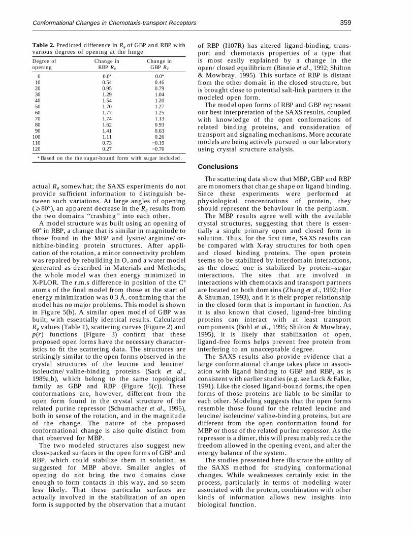
Conformational Changes in Chemotaxis-transport Receptors 359
Table 2. Predicted difference in Rg of GBP and RBP withvarious degrees of opening at the hingeDegree of Change in Change inopening RBP Rg GBP Rg
0 0.0a 0.0a
10 0.54 0.4620 0.95 0.7930 1.29 1.0440 1.54 1.2050 1.70 1.2760 1.77 1.2570 1.74 1.1380 1.62 0.9390 1.41 0.63
100 1.11 0.26110 0.73 −0.19120 0.27 −0.70
a Based on the the sugar-bound form with sugar included.
of RBP (I107R) has altered ligand-binding, trans-port and chemotaxis properties of a type thatis most easily explained by a change in theopen/closed equilibrium (Binnie et al., 1992; Shilton& Mowbray, 1995). This surface of RBP is distantfrom the other domain in the closed structure, butis brought close to potential salt-link partners in themodeled open form.
The model open forms of RBP and GBP representour best interpretation of the SAXS results, coupledwith knowledge of the open conformations ofrelated binding proteins, and consideration oftransport and signaling mechanisms. More accuratemodels are being actively pursued in our laboratoryusing crystal structure analysis.
Conclusions
The scattering data show that MBP, GBP and RBPare monomers that change shape on ligand binding.Since these experiments were performed atphysiological concentrations of protein, theyshould represent the behaviour in the periplasm.
The MBP results agree well with the availablecrystal structures, suggesting that there is essen-tially a single primary open and closed form insolution. Thus, for the first time, SAXS results canbe compared with X-ray structures for both openand closed binding proteins. The open proteinseems to be stabilized by interdomain interactions,as the closed one is stabilized by protein–sugarinteractions. The sites that are involved ininteractions with chemotaxis and transport partnersare located on both domains (Zhang et al., 1992; Hor& Shuman, 1993), and it is their proper relationshipin the closed form that is important in function. Asit is also known that closed, ligand-free bindingproteins can interact with at least transportcomponents (Bohl et al., 1995; Shilton & Mowbray,1995), it is likely that stabilization of open,ligand-free forms helps prevent free protein frominterfering to an unacceptable degree.
The SAXS results also provide evidence that alarge conformational change takes place in associ-ation with ligand binding to GBP and RBP, as isconsistent with earlier studies (e.g. see Luck & Falke,1991). Like the closed ligand-bound forms, the openforms of those proteins are liable to be similar toeach other. Modeling suggests that the open formsresemble those found for the related leucine andleucine/isoleucine/valine-binding proteins, but aredifferent from the open conformation found forMBP or those of the related purine repressor. As therepressor is a dimer, this will presumably reduce thefreedom allowed in the opening event, and alter theenergy balance of the system.
The studies presented here illustrate the utility ofthe SAXS method for studying conformationalchanges. While weaknesses certainly exist in theprocess, particularly in terms of modeling waterassociated with the protein, combination with otherkinds of information allows new insights intobiological function.
actual Rg somewhat; the SAXS experiments do notprovide sufficient information to distinguish be-tween such variations. At large angles of opening(e80°), an apparent decrease in the Rg results fromthe two domains ‘‘crashing’’ into each other.
A model structure was built using an opening of60° in RBP, a change that is similar in magnitude tothose found in the MBP and lysine/arginine/or-nithine-binding protein structures. After appli-cation of the rotation, a minor connectivity problemwas repaired by rebuilding in O, and a water modelgenerated as described in Materials and Methods;the whole model was then energy minimized inX-PLOR. The r.m.s difference in position of the Ca
atoms of the final model from those at the start ofenergy minimization was 0.3 A, confirming that themodel has no major problems. This model is shownin Figure 5(b). A similar open model of GBP wasbuilt, with essentially identical results. CalculatedRg values (Table 1), scattering curves (Figure 2) andp(r) functions (Figure 3) confirm that theseproposed open forms have the necessary character-istics to fit the scattering data. The structures arestrikingly similar to the open forms observed in thecrystal structures of the leucine and leucine/isoleucine/valine-binding proteins (Sack et al.,1989a,b), which belong to the same topologicalfamily as GBP and RBP (Figure 5(c)). Theseconformations are, however, different from theopen form found in the crystal structure of therelated purine repressor (Schumacher et al., 1995),both in sense of the rotation, and in the magnitudeof the change. The nature of the proposedconformational change is also quite distinct fromthat observed for MBP.
The two modeled structures also suggest newclose-packed surfaces in the open forms of GBP andRBP, which could stabilize them in solution, assuggested for MBP above. Smaller angles ofopening do not bring the two domains closeenough to form contacts in this way, and so seemless likely. That these particular surfaces areactually involved in the stabilization of an openform is supported by the observation that a mutant

Conformational Changes in Chemotaxis-transport Receptors360
Materials and Methods
Sample preparation
Escherichia coli MBP was prepared as described inthe accompanying paper (Shilton et al., 1996); the finalbuffer was 10 mM Tris-HCl (pH 8). This type ofpurification scheme has been reported to remove anyendogenous sugar bound to MBP (Miller et al., 1983).Purification of Salmonella typhimurium GBP and E. coliRBP was carried out using the methods describedby Mowbray et al. (1990) and Mowbray & Cole(1992). Removal of sugar from these proteins requireddialysis against guanidine hydrochloride, performedas described by Flocco & Mowbray (1994). Thefinal buffer was 10 mM Tris-HCl (pH 7.4); thatfor GBP also contained 0.5 mM CaCl2. Isoelectricfocusing gels were used as a final check of purity ofthe protein preparations and of removal of sugar.Protein concentrations were determined for purifiedMBP, GBP and RBP using e280 values of 1.94, 0.95 and0.13 ml/mg-cm, respectively (Miller et al., 1983; andour unpublished data).
Dynamic light-scattering measurements
Measurements of the hydrodynamic radius werecarried out at room temperature, with a model DP-801instrument from Protein Solutions (Charlottesville, VA).Protein concentrations were 7.4, 2.8 and 10 mg/ml forMBP, GBP and RBP, respectively.
SAXS measurements
X-ray scattering experiments were carried out at theDaresbury Synchrotron Radiation Source, station 2.1,using a wavelength, l, of 1.5 A (1 A = 0.1 nm). Samples(100 m1) were loaded in a brass holder with micawindows 25 mm thick and 15 mm in diameter, and apathlength of 1.5 mm. An ion chamber placed after thesample cell was used to normalize the signal forfluctuations in beam intensity and sample absorption;a second chamber placed in front of the sample wasused as an additional check of beam intensity. Datawere collected on a segment-shaped, one-dimensional,position-sensitive proportional counter (Lewis et al.,1988); the sample-to-detector distance was 3 m. Thediffraction pattern of wet rat tail collagen was used toadjust the position of the detector relative to the beam,and to correlate detector channels with scatteringangle. Detector response curves were measured beforeand midway through the experiments by irradiatingthe detector uniformly with a radioactive source (55Fe).The two curves, collected for one and nine hours,respectively, differed only in scale; the second wasused to correct for spacial differences in detectorsensitivity. Protein concentrations in the scatteringexperiments were 7.2 (0.18 mM), 5.6 (0.17 mM) and21 mg/ml (0.75 mM) for MBP, GBP and RBP, respect-ively; the specified sugars (obtained from Sigma) wereadded at a concentration of 1 mM (volume changes 1to 2%). Fifteen 20-second frames were measured for allprotein and buffer samples, for a total sampleexposure time of five minutes at 10°C. Four indepen-dent samples of each type were measured, except forthe RBP-glucose, GBP-ribose, MBP-maltotriose andMBP-glucose combinations, where only two sampleswere analysed.
Processing and analysis of SAXS data
Scattering data are expressed in terms of the scatteringvector s = 2 sin u/l ( = 1/d), where 2u is the scatteringangle and d is the resolution in A. The dependable datacover the range 0.006 < s < 0.035. Initial data processing(normalization according to beam intensity, correctionfor detector response, averaging of usable frames) wascarried out using the program OTOKO (Boulin et al.,1986). Individual frames were checked for possibledeterioration with a preliminary Guinier plot (see below)prior to averaging. Later processing steps (buffer scalingand subtraction, and calculation of Guinier curves for thepurposes of final normalization), as well as presentationof data, made use of Macintosh programs Mathematica(Wolfram Research), Ultrafit (Biosoft), and Cricket Graph(Computer Associates International).
Background subtraction was performed using the factsthat at large angles a dependence of scattering on s−4 canbe assumed, and that as s becomes large, the intensity ofscattering for the protein (Ib+p) and buffer (Ib) solutionsshould coincide (Grossmann et al., 1992). The ratio Ib+p/Ib
was calculated in regions of s > 0.03, and the linear plotsof this ratio versus s−4 were fit to the function a + m s−4; Ib
was then multiplied by a prior to its subtraction from Ib+p
to obtain I(s) (the scattering due to the protein alone, asa function of s). The values of a were approximately 1.0,0.98 and 0.94 for samples of MBP, GBP and RBP,respectively.
The radius of gyration, Rg , represents the r.m.s.distance of all atoms from the centre of mass. Guinierplots (Guinier & Fournet, 1955) were used for thecalculation of Rg and for normalization of the data. Atsmall angles, the scattering behaviour of a protein can beapproximated by:
I(s) = I(0)exp( − 4p2s2R2g /3) (1)
where I(0), the scattering at s = 0, is proportional to thenumber of electrons squared. Plots of ln I(s) versus s2
should thus have a slope of −4p2R2g /3, and an intercept
of ln I(0). The linear ‘‘Guinier region’’ extends to higherangles for smaller particles, as well as being somewhatdependent on molecular shape. The values of I(0)obtained for each sample were used to normalize the datato an I(0) of 1.0. After checking that the scattering curvesfor similar samples agreed within experimental error, anaverage curve was calculated for each sample type.
The distance distribution function, p(r), represents theprobability of finding interatomic vectors of length r. Theprogram GNOM was used to calculate p(r) distributionsand real space Rg values from the normalized intensitydata, assuming homogeneous particles (Semenyuk &Svergun, 1991; Svergun et al., 1988).
Calculation of Rg , scattering profiles and p(r )distributions from atomic co-ordinates
The co-ordinate sets used are available from theBrookhaven Protein Data Bank (Bernstein et al., 1977).MBP co-ordinates were obtained from X-ray structures ofa mutant MBP (W230R) in open, (ligand-free) and closed(ligand-bound) forms solved and refined in thislaboratory (Shilton et al., 1996; identity codes 1MPC and1MPD, respectively). Co-ordinate sets for ligand-boundGBP (Zou et al., 1993) and RBP (Mowbray & Cole, 1992)are described by identity codes 1GCA and 2DRI,respectively; the structures of the open forms are notknown.

Conformational Changes in Chemotaxis-transport Receptors 361
Water models were generated, starting from a 7.0 Ashell added with the ‘‘soak’’ feature of the programInsight (Biosym Technologies). The combined proteinand water models were then energy minimized with theprogram package X-PLOR (Brunger, 1992). Harmonicrestraints were used initially for those water moleculesmore than 5 A away from the protein, to enable the innershell of water to stabilize; final energy minimizationallowed all waters to move freely. Water molecules werethen removed if their oxygen atom was more than 3.15 A(that is, beyond hydrogen-bonding distance) from anynon-hydrogen protein atom (Grossman et al., 1993).Results obtained with these models were compared withthose using different distance cutoffs for water exclusion,as well as with water models from X-ray studies, whereavailable.
Theoretical scattering curves and Rg values wereobtained from atomic co-ordinates using the programDALAI (Pantos & Bordas, 1994; Pantos et al., 1996), whichperforms calculations based on the Debye formula(Debye, 1915). The values of Rg were obtained using theformula:
Rg = ((SiziR2i )/Sizi )1/2 (2)
where zi is the atomic number of atom i, and Ri is itsdistance from the centre of mass. All non-hydrogenprotein, water and sugar atoms were used unlessotherwise stated. Uniform atomic radii of 1.7 A wereassumed, and scattering factor values were takenfrom the International Tables of Crystallography (Hahn,1987).
Distance distribution functions for models werecalculated from all non-hydrogen atoms using a programwritten by Jinyu Zou (Uppsala University). Distribu-tions were also calculated using the model intensitydata obtained from DALAI (after scaling so thatI(0) = 1.0) with the program GNOM; the results werevery similar.
Structural work and modeling
The molecular graphics program O (Jones &Kjeldgaard, 1995) was used for inspection and compari-son of protein structures. Calculation of static surfaceaccessibility of protein models was performed accordingto the algorithm of Lee & Richards (1971), with a probeof radius 1.4 A. Various open models of GBP and RBPwere generated and energy minimized using theprogram X-PLOR, with water models generated asdescribed above. Figures 4 and 5 were prepared using theprogram Molscript (Kraulis, 1991).
AcknowledgementsThis work was supported by grants from the
Swedish Natural Science Research Council to S.L.M. andB.H.S., by a post-doctoral fellowship to B.H.S. fromthe Canadian Medical Research Council, and by supportto M.M.F. from the Goran Gustafsson Stiftelse througha grant to T.A. Jones. Manolis Pantos and GunterGrossman contributed many helpful discussions; andElizabeth Townes-Andrews, John Harries and PeterLindley of the Daresbury Laboratory gave otherassistance. Dick Brennan and Maria Schumacher pro-vided co-ordinates and information on the purinerepressor prior to publication, M. Manson provided other
data, and Joakim Bjorkman assisted in the modelingwork.
ReferencesAqvist, J. & Mowbray, S. L. (1995). Sugar recognition by
a glucose/galactose receptor: evaluation of bindingenergetics from molecular dynamics simulations.J. Biol. Chem. 270, 9978–9981.
Baker, E. N., Rumball, S. V. & Anderson, B. F. (1987).Transferrins: insights into structure and functionfrom studies on lactoferrin. Trends Biochem. Sci. 12,350–353.
Bernstein, F. C., Koetzle, T. F., Williams, G. J. B.,Meyer, E. T., Jr, Brice, M. D., Rodgers, J. R.,Kennard, O., Shimanouchi, T. & Tasumi, M. (1977).The Protein Data Bank: a computer-based archivalfile for macromolecular structures. J. Mol. Biol. 112,535–542.
Binnie, R. A., Zhang, H., Mowbray, S. L. & Hermodson,M. A. (1992). Functional mapping of the surface ofEscherichia coli ribose-binding protein: mutations thataffect chemotaxis and transport. Protein Sci. 1,1642–1651.
Bohl, E., Shuman, H. A. & Boos, W. (1995). Mathematicaltreatment of the kinetics of binding protein-depen-dent transport systems reveals that both thesubstrate loaded and unloaded binding proteinsinteract with the membrane components. J. Theor.Biol. 172, 83–94.
Boos, W. & Lucht, J. M. (1995). Periplasmic binding-pro-tein-dependent ABC transporters. In Escherichia coliand Salmonella typhimurium: Cellular and MolecularBiology (Lin, E., ed.), pp. 1175–1209, AmericanSociety for Microbiology, Washington, DC.
Boulin, C., Kempf, R., Koch, M. H. J. & McLaughlin, S.(1986). Data appraisal, evaluations and display forsynchrotron radiation experiments: hardware andsoftware. Nucl. Instr. Methods, A249, 399–407.
Brunger, A. T. (1992). X-PLOR: A System for X-rayCrystallography and NMR, Yale University Press,New Haven.
Careaga, C. L., Sutherland, J., Sabeti, J. & Falke, J. J.(1995). Large amplitude twisting motions of aninterdomain hinge: a disulfide trapping study of theglucose-galactose binding protein. Biochemistry, 34,3048–3055.
Debye, P. (1915). Zerstreuung von Rontgenstrahlen. Ann.Phys. (Leipsig), 46, 809–823.
Feigin, L. A. & Svergun, D. I. (1987). Structure Analysis bySmall-Angle X-Ray and Neutron Scattering, PlenumPress, New York.
Flocco, M. M. & Mowbray, S. L. (1994). The 1.9 AX-ray structure of a closed unliganded form ofthe periplasmic glucose/galactose receptor fromSalmonella typhimurium. J. Biol. Chem. 269, 8931–8936.
Gerstein, M., Anderson, B. F., Norris, G. E., Baker, E. N.,Lesk, A. M. & Chothia, C. (1993). Domain closure inlactoferrin: two hinges produce a see-saw motionbetween alternative close-packed interfaces. J. Mol.Biol. 234, 357–372.
Glatter, O. & Kratky, O. (1982). Small Angle X-rayScattering, Academic Press, London.
Grossman, J. G., Neu, M., Pantos, E., Schwab, F. J., Evans,R. W., Townes-Andrews, E., Lindley, P. F., Appel,H., Theis, W.-G. & Samar Hasnain, S. (1992). X-raysolution scattering reveals conformational

Conformational Changes in Chemotaxis-transport Receptors362
changes upon iron uptake in lactoferrin, serum andovo-tranferrins. J. Mol. Biol. 225, 811–819.
Grossman, J. G., Abraham, Z. H. L., Adman, E. T.,Neu, M., Eady, R. R., Smith, B. E. & Hasnain, S. S.(1993). X-ray scattering using synchrotron radiationshows nitrite reductase from Achromobacter xylosoxi-dans to be a trimer in solution. Biochemistry, 32,7360–7366.
Guinier, A. & Fournet, G. (1955). Small Angle Scattering ofX-rays, John Wiley & Sons, Inc., New York.
Hahn, T. (1987). International Tables for Crystallography,vol. 4, D. Reidel Publishing Company, Boston.
Hor, L.-I. & Shuman, H. A. (1993). Genetic analysisof periplasmic binding protein dependent transportin E. coli: each lobe of maltose-binding proteininteracts with a different subunit of the MalFGK2
membrane transport complex. J. Mol. Biol. 233,659–670.
Jones, T. A. & Kjeldgaard, M. O. (1995). O–The Manual,Uppsala, Sweden.
Kraulis, P. (1991). Molscript: a program to produce bothdetailed and schematic plots of protein structures.J. Appl. Crystallog. 24, 946–950.
Lee, B. & Richards, F. M. (1971). The interpretation ofprotein structures: estimation of static accessibility.J. Mol. Biol. 55, 379–400.
Lewis, R., Sumner, I., Berry, A., Bordas, J., Gabriel, S.,Mant, G., Parker, B., Roberts, K. & Wogan, J.(1988). Multiwire X-ray detector systems at theDaresbury SRS. Nucl. Instr. Methods, A273,773–777.
Luck, L. A. & Falke, J. J. (1991). Open conformation of asubstrate binding cleft: 19F NMR studies of cleftangle in the D-galactose chemosensory receptor.Biochemistry, 30, 6484–6490.
Manson, M. D. (1992). Bacterial motility and chemotaxis.Advan. Microbiol. Physiol. 33, 277–345.
Miller, D. M. III, Olson, J. S., Pflugrath, J. W. & Quiocho,F. A. (1983). Rates of ligand binding to periplasmicproteins involved in bacterial transport and chemo-taxis. J. Biol. Chem. 258, 13665–13672.
Mowbray, S. L. (1992). The ribose and glucose/galactosereceptors: competitors in bacterial chemotaxis. J. Mol.Biol. 227, 418–440.
Mowbray, S. L. & Cole, L. B. (1992). The 1.7 A X-raystructure of the periplasmic ribose receptor ofEscherichia coli. J. Mol. Biol. 225, 155–175.
Mowbray, S. L., Smith, R. D. & Cole, L. B. (1990).Structure of the periplasmic glucose/galactosereceptor of Salmonella typhimurium. Receptor, 1,41–54.
Newcomer, M. E., Lewis, B. A. & Quiocho F. A. (1981).The radius of gyration of L-arabinose-bindingprotein decreases upon binding of ligand. J. Biol.Chem. 256, 13218–13222.
Oh, B.-H., Pandit, J., Kang, C.-H. Nikaido, K. Gokcen, S.,Ames G. F.-L. & Kim, S.-H. (1993). Three-dimen-sional structures of the periplasmic lysine-, arginine-,ornithine-binding protein with and without a ligand.J. Biol. Chem. 268, 11348–11355.
Olah, G. A., Trakhanov, S., Trewhella, J. & Quiocho, F. A.(1993). Leucine/isoleucine/valine binding proteincontracts upon binding of ligand. J. Biol. Chem. 269,16241–16247.
Pantos, E. & Bordas, J. (1994). DALAI. Pure Appl. Chem.66, 77–82.
Pantos, E., van Garderen H. F., Hilbers, P. A. J., Beelen,T. P. M. & van Santen, R. A. (1996). Simulation ofsmall angle scattering from large assemblies of
multi-type scatterer particles. J. Mol. Struct. In thepress.
Quiocho F. A. (1991). Atomic structures and function ofperiplasmic receptors in active transport andchemotaxis. Curr. Opin. Struct. Biol. 1, 922–933.
Sack J. S., Saper, M. A. & Quiocho F. A. (1989a).Periplasmic binding protein structure and function:refined X-ray structures of the leucine/isoleucine/valine-binding protein and its complex with leucine.J. Mol. Biol. 206, 171–191.
Sack J. S. Trakhanov, S. D. Tsigannik, I. H. & QuiochoF. A. (1989b). Structure of the L-leucine-bindingprotein refined at 2.4 A resolution and comparisonwith the Leu/Ile/Val-binding protein structure.J. Mol. Biol. 206, 193–207.
Schumacher, M. A., Choi, K. Y., Zalkin, H. & Brennan,R. G. (1994). Crystal structure of LacI member, PurR,bound to DNA: minor groove binding by a helices.Science, 266, 763–770.
Schumacher, M. A., Choi, K. Y., Lu, F., Zalkin, H. &Brennan R. G. (1995). Mechanism of corepressor-me-diated specific DNA binding by the purine repressor.Cell, 83, 147–155.
Semenyuk, A. V. & Svergun D. I. (1991). Small-angle-scat-tering data treatment by the regularization method.J. Appl. Crystallog. 24, 537–540.
Sharff, A. J., Rodseth, L. E., Spurlino, J. C. & Quiocho,F. A. (1992). Crystallographic evidence of a largeligand-induced hinge-twist motion between the twodomains of the maltodextrin binding proteininvolved in active transport and chemotaxis.Biochemistry, 31, 10657–10663.
Shilton, B. H. & Mowbray, S. L. (1995). Simple models forthe analysis of binding protein-dependent transportsystems. Protein Sci. 4, 1346–1355.
Shilton, B. H., Shuman, H. & Mowbray, S. L. (1996).Crystal structures and solution conformations ofa dominant-negative mutant of Escherichia colimaltose-binding protein. J. Mol. Biol. 264, 364–376.
Spurlino, J. C., Lu, G.-Y. & Quiocho, F. A. (1991). The2.3 A resolution structure of the maltose- ormaltodextrin-binding protein, a primary receptor ofbacterial active transport and chemotaxis. J. Biol.Chem. 266, 5202–5219.
Svergun, D. I., Semenyuk, A. V. & Feigin, F. A. (1988).GNOM: a program package for small-angle scatter-ing data processing. Acta Crystallog. sect. A, 44,244–250.
Szmelcman, S., Schwartz, M., Silhavy, T. J. & Boos, W.(1976). Maltose transport in Escherichia coli K12: acomparison of transport kinetics in wild-type andl-resistant mutants with the dissociation constants ofthe maltose-binding protein as measured by fluor-escence quenching. Eur. J. Biochem. 65, 13–19.
Vyas, N. K., Vyas, M. N. & Quiocho, F. A. (1987). A novelcalcium binding site in the galactose-binding proteinof bacterial transport and chemotaxis. Nature, 327,635–638.
Wolf, A., Shaw, S. W., Nikaido, K. & Ames, G. F.-L.(1994). The histidine-binding protein undergoesconformational changes in the absence of ligand asanalyzed with conformation-specific monoclonalantibodies. J. Biol. Chem. 269, 23051–23058.
Zhang, Y., Conway, C., Rosato, M., Suh, Y. & Manson, M.(1992). Maltose chemotaxis involves residues on thesame face of the N-terminal and C-terminal domainsof maltose-binding protein. J. Biol. Chem. 267,22813–22820.
Zhang, Y., Mannering, D. E., Davidson, A. L. & Manson,

Conformational Changes in Chemotaxis-transport Receptors 363
M. D. (1996). Maltose-binding protein containing aninterdomain disulfide bridge confers a dominantnegative phenotype for transport and chemotaxis.J. Biol. Chem. 271, 17881–17889.
Zou, J., Flocco, M. M. & Mowbray, S. L. (1993). The 1.7 Arefined X-ray structure of the periplasmic glucose/galactose receptor of Salmonella typhimurium. J. Mol.Biol. 233, 739–752.
Edited by R. Huber
(Received 14 February 1996; received in revised form 3 September 1996; accepted 12 September 1996)
Copyright © 2022 FDOKUMEN