Completion Pneumonectomy for Non-Small-Cell Lung Cancer
11
Cancers 2022, 14, 3408. https://doi.org/10.3390/cancers14143408 www.mdpi.com/journal/cancers Article Completion Pneumonectomy for Non-Small-Cell Lung Cancer: Does Induction Treatment Influence Postoperative Outcomes? Domenico Galetta 1,2, * and Lorenzo Spaggiari 1,2 1 Division of Thoracic Surgery, European Institute of Oncology IRCCS, 20141 Milan, Italy; [email protected] 2 Department of Oncology and Hematology-Oncology-DIPO, University of Milan, 20122 Milan, Italy * Correspondence: [email protected]; Tel.: +39-0257489801 Simple Summary: In recent years there have been important improvements in surgical and adju- vant therapy for lung cancer which have led to an increasing number of patients with non-small- cell lung cancer (NSCLC) which had been previously cured by surgery being identified as having a second primary NSCLC or a recurrence of the previous tumor. In these cases, a completion pneu- monectomy (CP), defined as the complete removal of the remaining lung after an ipsilateral pulmo- nary resection, may be performed. Although this procedure has a higher morbidity and mortality than standard pneumonectomy due to the high degree of surgical difficulty strongly associated with the previous surgery, the number of patients undergoing CP is increasing with improvement in morbidity and mortality. To the best of our knowledge, there is no study evaluating the role of induction therapy (IT) on the outcomes of patients who have undergone CP. We reviewed our sin- gle-center experience in patients receiving CP for recurrent/second NSCLC after IT and analyzed perioperative results and long-term outcomes. Our results revealed that postoperative complica- tions were not influenced by IT, and long-term survival was adversely influenced by the absence of IT, the presence of squamous cell carcinoma, and cancers at advanced stages. Correct patient selec- tion is crucial to evaluating possible contraindications and adopting technical details to reduce the complication rate. Abstract: Background: Completion pneumonectomy (CP) is associated with high morbidity and mortality. We reviewed our experience to evaluate whether induction treatment (IT) may affect postoperative outcomes and analyzed factors influencing long-term results. Methods: Between 1998 and 2020, 69 patients with lung cancer underwent CP (50 males, median age 63 years, right CP in 47 patients). A total of 23 patients (33.3%) received IT (chemotherapy in 15, chemoradiotherapy in 7, and radiation in 1). Surgery included 25 (36.2%) extended resections and five (7.2%) tracheal sleeve CP. Results: The 30-day mortality rate was 7.2% (5/69), and overall morbidity was 37.6%. Major complications occurred in five patients (7.2%): one cardiac dislocation, one diaphragmatic hernia, one transient ischemic attack (TIA), and two bronchopleural fistulas. Minor complications occurred in 21 cases (30.4%): pulmonary in 12, cardiac in 7, and neurological in 2. The median hos- pital stay was 8 days (range, 5–56 days). IT did not influence postoperative morbidity and mortality. Pathological staging included 19 (27.5%) stage I, 36 (52.2%) stage II, and 14 (20.3%) stage III. Overall 5-year survival was 51.7%. Factors influencing survival were IT (p = 0.01), extension of resection (p = 0.04), histology (p = 0.01), pathological stage (p = 0.03), and T and N factors (p = 0.2, respectively). Factors affecting survival in multivariate analysis included IT (p = 0.02) and histology (p = 0.03). Conclusions: In our experience, CP had a low mortality, acceptable morbidity, and good long-term survival, which justifies this surgical procedure. Postoperative complications were not influenced by IT. Long-term survival was adversely influenced by the absence of IT, the presence of extended resection, the presence of squamous cell carcinoma, and cancers at advanced stages. Keywords: lung cancer surgery; pneumonectomy; chemotherapy Citation: Galetta, D.; Spaggiari, L. Completion Pneumonectomy for Non-Small-Cell Lung Cancer: Does Induction Treatment Influence Postoperative Outcomes? Cancers 2022, 14, 3408. https://doi.org/ 10.3390/cancers14143408 Academic Editor: Dirk De Ruysscher Received: 9 May 2022 Accepted: 11 July 2022 Published: 13 July 2022 Publisher’s Note: MDPI stays neutral with regard to jurisdictional claims in published maps and institutional affiliations. Copyright: © 2022 by the authors. Li- censee MDPI, Basel, Switzerland. This article is an open access article distributed under the terms and con- ditions of the Creative Commons At- tribution (CC BY) license (https://cre- ativecommons.org/licenses/by/4.0/).
-
Upload
khangminh22 -
Category
Documents
-
view
4 -
download
0
Transcript of Completion Pneumonectomy for Non-Small-Cell Lung Cancer

Cancers 2022, 14, 3408. https://doi.org/10.3390/cancers14143408 www.mdpi.com/journal/cancers
Article
Completion Pneumonectomy for Non-Small-Cell Lung Cancer:
Does Induction Treatment Influence Postoperative Outcomes?
Domenico Galetta 1,2,* and Lorenzo Spaggiari 1,2
1 Division of Thoracic Surgery, European Institute of Oncology IRCCS, 20141 Milan, Italy;
[email protected] 2 Department of Oncology and Hematology-Oncology-DIPO, University of Milan, 20122 Milan, Italy
* Correspondence: [email protected]; Tel.: +39-0257489801
Simple Summary: In recent years there have been important improvements in surgical and adju-
vant therapy for lung cancer which have led to an increasing number of patients with non-small-
cell lung cancer (NSCLC) which had been previously cured by surgery being identified as having a
second primary NSCLC or a recurrence of the previous tumor. In these cases, a completion pneu-
monectomy (CP), defined as the complete removal of the remaining lung after an ipsilateral pulmo-
nary resection, may be performed. Although this procedure has a higher morbidity and mortality
than standard pneumonectomy due to the high degree of surgical difficulty strongly associated with
the previous surgery, the number of patients undergoing CP is increasing with improvement in
morbidity and mortality. To the best of our knowledge, there is no study evaluating the role of
induction therapy (IT) on the outcomes of patients who have undergone CP. We reviewed our sin-
gle-center experience in patients receiving CP for recurrent/second NSCLC after IT and analyzed
perioperative results and long-term outcomes. Our results revealed that postoperative complica-
tions were not influenced by IT, and long-term survival was adversely influenced by the absence of
IT, the presence of squamous cell carcinoma, and cancers at advanced stages. Correct patient selec-
tion is crucial to evaluating possible contraindications and adopting technical details to reduce the
complication rate.
Abstract: Background: Completion pneumonectomy (CP) is associated with high morbidity and
mortality. We reviewed our experience to evaluate whether induction treatment (IT) may affect
postoperative outcomes and analyzed factors influencing long-term results. Methods: Between 1998
and 2020, 69 patients with lung cancer underwent CP (50 males, median age 63 years, right CP in
47 patients). A total of 23 patients (33.3%) received IT (chemotherapy in 15, chemoradiotherapy in
7, and radiation in 1). Surgery included 25 (36.2%) extended resections and five (7.2%) tracheal
sleeve CP. Results: The 30-day mortality rate was 7.2% (5/69), and overall morbidity was 37.6%.
Major complications occurred in five patients (7.2%): one cardiac dislocation, one diaphragmatic
hernia, one transient ischemic attack (TIA), and two bronchopleural fistulas. Minor complications
occurred in 21 cases (30.4%): pulmonary in 12, cardiac in 7, and neurological in 2. The median hos-
pital stay was 8 days (range, 5–56 days). IT did not influence postoperative morbidity and mortality.
Pathological staging included 19 (27.5%) stage I, 36 (52.2%) stage II, and 14 (20.3%) stage III. Overall
5-year survival was 51.7%. Factors influencing survival were IT (p = 0.01), extension of resection (p
= 0.04), histology (p = 0.01), pathological stage (p = 0.03), and T and N factors (p = 0.2, respectively).
Factors affecting survival in multivariate analysis included IT (p = 0.02) and histology (p = 0.03).
Conclusions: In our experience, CP had a low mortality, acceptable morbidity, and good long-term
survival, which justifies this surgical procedure. Postoperative complications were not influenced
by IT. Long-term survival was adversely influenced by the absence of IT, the presence of extended
resection, the presence of squamous cell carcinoma, and cancers at advanced stages.
Keywords: lung cancer surgery; pneumonectomy; chemotherapy
Citation: Galetta, D.; Spaggiari, L.
Completion Pneumonectomy for
Non-Small-Cell Lung Cancer: Does
Induction Treatment Influence
Postoperative Outcomes? Cancers
2022, 14, 3408. https://doi.org/
10.3390/cancers14143408
Academic Editor: Dirk De Ruysscher
Received: 9 May 2022
Accepted: 11 July 2022
Published: 13 July 2022
Publisher’s Note: MDPI stays
neutral with regard to jurisdictional
claims in published maps and
institutional affiliations.
Copyright: © 2022 by the authors. Li-
censee MDPI, Basel, Switzerland.
This article is an open access article
distributed under the terms and con-
ditions of the Creative Commons At-
tribution (CC BY) license (https://cre-
ativecommons.org/licenses/by/4.0/).
Cancers 2022, 14, 3408 2 of 11
1. Introduction
Tumor recurrence or a second tumor after a previous resection for non-small-cell
lung cancer (NSCLC) may occur in 30–70% of patients [1]. It represents one of the most
important factors affecting survival after NSCLC resection, which still remains less than
50% [1]. In these cases, a completion pneumonectomy (CP), defined as the complete re-
moval of the remaining lung after an ipsilateral pulmonary resection (usually after lobec-
tomy or bilobectomy) [2,3], may be performed [1,3–5]. This procedure has a higher mor-
bidity and mortality than standard pneumonectomy [3] due to the high degree of surgical
difficulty strongly associated with the previous surgery (i.e., presence of dense adhesions
or fibrosis in the hilum structures). Published studies demonstrate an overall complication
rate for CP performed for NSCLC ranging from 3.8% to 46.9% and operative mortality
ranging from 0% to 17.6% [6]. Nevertheless, due to the rising incidence of lung cancer and
thanks to the extended survival time after resection, the number of patients undergoing
CP is increasing; moreover, the improvement in recent years of preoperative workup tests,
anesthesia techniques, and postoperative management have led to a reduction in CP-re-
lated morbidity and mortality rates.
In the English-language literature there are few publications dealing with survival
and the risk factors linked to morbidity and mortality following CP, which remains a high-
risk surgical procedure. For this reason, CP is recommended only for selected patients and
performed by experienced surgeons at high-volume and experienced centers [7,8].
To the best of our knowledge, there is no study evaluating the role of induction ther-
apy (IT) on the outcomes of patients who have undergone CP. We sought to review our
single-center experience in patients receiving CP for recurrent/second NSCLC after IT,
analyzing perioperative results and long-term outcomes.
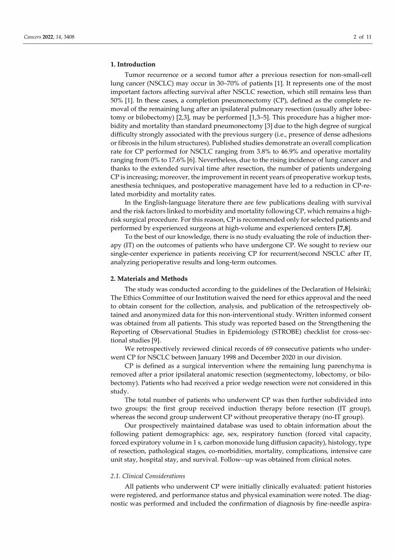
2. Materials and Methods
The study was conducted according to the guidelines of the Declaration of Helsinki;
The Ethics Committee of our Institution waived the need for ethics approval and the need
to obtain consent for the collection, analysis, and publication of the retrospectively ob-
tained and anonymized data for this non-interventional study. Written informed consent
was obtained from all patients. This study was reported based on the Strengthening the
Reporting of Observational Studies in Epidemiology (STROBE) checklist for cross-sec-
tional studies [9].
We retrospectively reviewed clinical records of 69 consecutive patients who under-
went CP for NSCLC between January 1998 and December 2020 in our division.
CP is defined as a surgical intervention where the remaining lung parenchyma is
removed after a prior ipsilateral anatomic resection (segmentectomy, lobectomy, or bilo-
bectomy). Patients who had received a prior wedge resection were not considered in this
study.
The total number of patients who underwent CP was then further subdivided into
two groups: the first group received induction therapy before resection (IT group),
whereas the second group underwent CP without preoperative therapy (no-IT group).
Our prospectively maintained database was used to obtain information about the
following patient demographics: age, sex, respiratory function (forced vital capacity,
forced expiratory volume in 1 s, carbon monoxide lung diffusion capacity), histology, type
of resection, pathological stages, co-morbidities, mortality, complications, intensive care
unit stay, hospital stay, and survival. Follow--up was obtained from clinical notes.
2.1. Clinical Considerations
All patients who underwent CP were initially clinically evaluated: patient histories
were registered, and performance status and physical examination were noted. The diag-
nostic was performed and included the confirmation of diagnosis by fine-needle aspira-

Cancers 2022, 14, 3408 3 of 11
tion biopsy, endobronchial biopsy, or bronchoscopic brushing. The staging workup in-
cluded computed tomography (CT) of the chest, abdomen, and brain as well as positron
emission tomography (PET) scan. Mediastinal nodal involvement was assessed in cases
of suspected of N2 disease on CT scan or PET scan by cervical mediastinoscopy or endo-
bronchial ultra sound (EBUS).
Distinction between a second primary lung cancer and local recurrence was made
according to the Martini and Melamed criteria [10]: a local recurrence was defined as a sec-
ond malignant tumor with the same cell type occurring in the same anatomic site within
2 years of the first operation, and a second primary lung cancer was defined as a second
malignant tumor when the cell type was different or when a tumor with same cell type
occurred in a different anatomic site more than 2 years after the first cancer in the absence
of residual tumor after the first operation.
IT was administered to all patients who were candidates to receive CP with a poten-
tially resectable T4 disease (tumor invading the carina or the tracheo-bronchial angle or
left atrium or aorta) with or without N2 disease. In our practice, patients with nodal in-
volvement of mediastinal lymph node station #2R or #2L or with mediastinal lymph node
station #4R bulky disease are considered unsuitable for surgery, while patients with me-
diastinal lymph node station #4R (no bulky) adenopathy are considered suitable for sur-
gical resection after IT
The no-IT group included patients without clinical mediastinal nodal involvement,
those with clinical T3 (chest wall), and staged IIB or IIIA (T3 N0 or N1, respectively) (8th
TNM staging system) [11].
The IT group received a platinum-based chemotherapy (cisplatin/gemcitabine or vi-
norelbine or taxol) or chemoradiotherapy. One patient received preoperative radiother-
apy. Radiotherapy was delivered to a dose of 1.8 Gy per day over a 4 or 5-week period
(total radiation dose of 50.4 Gy). All patients receiving IT were re-staged to assess clinical
response and to rule out interval progression of disease before preoperative intervention.
Patients repeated cardiologic evaluation, function tests, whole body CT scan, and PET
scan.
The group of patients receiving IT and operated on underwent thoracotomy 1 to 2
weeks after re-staging.
2.2. Surgical Procedure
Single-lung ventilation was obtained by using a double-lumen tube. In cases of cari-
nal or trachea-bronchial angle involvement, some technical intraoperative maneuvers
were used: during the tracheo-bronchial resection and reconstruction, the tube was pulled
back into the trachea, and a sterile endotracheal tube of small diameter was positioned
under visual control by the surgeon in the contralateral main bronchus. Then the endotra-
cheal tube was pulled down in the left main bronchus beyond the anastomosis after the
reimplantation of the medial portion of the left main bronchus. and the tracheo-bronchial
suture was thereby completed.
A lateral right “muscle-sparing” thoracotomy was preferred. In cases of chest wall
involvement requiring resection and reconstruction, the type of thoracotomy was decided
accordingly. The chest wall reconstruction was performed with polypropylene
mesh/methylmethacrylate prosthesis.
When entering the pleural cavity after a previous lung resection, it was unavoidable
to find pleural adhesions, which were released with great care in order to avoid visceral
pleural tears and the contamination of the resulting pleural space. Sometimes, an ex-
trapleural dissection was needed. Following this step, hilar vessels were immediately cen-
trally controlled, usually intrapericardially. Attention was paid to keep as short as possi-
ble the bronchial stump and to reinforce it by vital pedicled mediastinal tissue (usually,
pericardial fat pad). The chest wall was closed in airtight fashion, and the pleural space
drained with a balanced system. Patients were managed in an intensive care unit over-
night and moved to a stepdown unit when clinically appropriate.

Cancers 2022, 14, 3408 4 of 11
All hospital survivors with pathological N2 disease received postoperative radiation
therapy. Radiation therapy was given 4 to 6 weeks after the operation. The prescription
dose was 50.4 Gy, administered in single, daily 1.8 Gy fractions on weekdays.
Survival and tumor recurrence were evaluated by patient follow-up. In particular,
survival was defined as the interval between the day of CP and the time of the evaluation
of this report, death, or loss to follow-up. Disease-=free survival was calculated from the
first day of surgical intervention until any event such as recurrence or metastases.
2.3. Statistical Methods
The two groups were compared with respect to the demographics and clinical out-
comes using Student’s t tests or Fisher’s exact tests as appropriate. The survival probabil-
ities were calculated via the Kaplan–Meier method and compared by using the long-rank
test. Differences were considered significant when p < 0.05.
3. Results
During the study period, a total of 116 patients who had a prior anatomic lung resec-
tion (segmentectomy, lobectomy, or bilobectomy) for NSCLC and presenting an ipsilat-
eral recurrence or a second primary NSCLC were evaluated for possible CP. The flow
diagram of selected population is reported in Figure 1.
Figure 1. Flow diagram of population with NSCLC selected for this study. IT = induction therapy.
Of these, 28 (24.1%) were turned away because of the presence of metastatic disease
(n = 13) or bulky mediastinal nodal disease at the initial staging (n = 15). Of the remaining
88 patients, 46 were operated on without IT, and 42 (36.2%) received IT. Of this latter
group, only 23 patients (19.8%) were operated on because 19 patients had progression of
disease after IT (6 local and 13 distant). Thus, of the 116 observed patients, a total of 69
patients (59.5%) underwent CP (7.5% of all the performed pneumonectomies at our divi-
sion): 23 patients (19.8%) underwent CP after IT (IT group), and this group was compared
with the no-IT group (n = 46) which was comprised of patients who underwent CP with-
out IT. Of the 69 patients, 22 (31.9%) had an ipsilateral recurrent tumor, and 47 (68.1%)
had an ipsilateral second primary tumor.
In the IT group, 7 patients received chemoradiotherapy, 15 received chemotherapy,
and 1 patient received radiation therapy. The mean number of cycles of chemotherapy
received in the IT group was 4 (range, 3–9). The preoperative delay following IT before
surgery was 4 weeks (range, 4–6 weeks). All patients receiving IT (n = 42), except 3 (due

Cancers 2022, 14, 3408 5 of 11
to severe hematological toxicity), had the planned induction protocol; no patient died dur-
ing the study.
The baseline characteristics of the two groups are shown in Table 1.
Table 1. Baseline characteristics of the study groups (n = 69).
Variable IT
(n = 23)
No-IT
(n = 46)
p
Value
Age (y SD) 62.70 6.22 61.62 5.86 0.75
Sex (male/female) 17M/6F 33M/13F 0.45
Side (right/left) 15/8 32/14 0.52
FEV1 (mean % predicted SD)
FVC (mean % predicted SD)
81.72 12.16
79.73 16.88
80.48 13.85
80.62 14.67
0.65
0.73
DLCO (mean % SD) 76.30 15.23 78.45 16.08 0.69
Histology (%)
Squamous 8 (34.8) 15 (32.6)
Adenocarcinoma 13 (56.5) 25 (54.3)
Others 2 (8.7) 6 (13.1) 0.34
Pathological stage (%)
I 7 (30.4) 12 (26.1)
II 11 (47.8) 25 (54.3)
III 5 (21.7) 9 (19.5) 0.38
Extended resections
Chest wall 3 (13.0) 7 (15.2)
Pulmonary artery 3 (13.0) 5 (10.8)
Left atrium 2 (8.7) 1 (2.2)
Diaphragm 1 (4.3) 2 (4.3)
Aorta
Tracheal sleeve pneumonectomy
1 (4.3)
2 (8.7)
0
3 (6.5)
0.12
0.23
IT = induction therapy; SD = standard deviation; FEV1 = forced expiratory volume in 1 min; FVC =
expiratory volume; DLCO = diffusing capacity of the lungs for carbon monoxide.
The IT and no-IT groups had similar ages (p = 0.75) and gender distributions (p = 0.45)
and similar mean preoperative forced expiratory volume in 1 min (p = 0.65) as well as
diffusing capacity of the lungs for carbon monoxide (p = 0.69). Both histology and patho-
logical stage were similar in both the groups (p = 0.34 and p = 0.38, respectively). The me-
dian time from the first operation to the CP was 25.5 months (range, 5–184 months). The
first operation included 21 right upper lobectomies (3 sleeve lobectomies), 8 middle lobec-
tomies, 13 right lower lobectomies, 5 bilobectomies, 12 left upper lobectomies, 5 left lower
lobectomies, and 5 segmentectomies.
An associated resection of adjacent organs/structures was performed in 25 patients
(36.2%) (Table 1). A chest wall resection and reconstruction were performed in 10 cases
(14.5%), while a resection and reconstruction of the pulmonary artery were performed in
8 cases (11.6%), and a left atrium resection and a diaphragm resection were performed in
3 cases (4.3%). No differences were noted between the two groups (p = 0.12). A tracheal
sleeve CP was performed in five cases (7.2%), two of which after IT (p = 0.23).
During surgical resection, some difficulties related to chemotherapy-induced fibrosis
were encountered in the majority of IT-group patients; fortunately, these difficulties did
not prevent the performance of the CP, and in fact, a complete resection was achieved in
100% of the entire population.
Intraoperative mortality was nil. Table 2 reports the incidence of postoperative mor-
bidity and mortality.

Cancers 2022, 14, 3408 6 of 11
Table 2. Postoperative results.
Variable IT
(n = 23)
No-IT
(n = 46)
p
Value
Intraoperative mortality (%)
30-day mortality (%)
0
2 (8.7)
0
3 (6.5)
0.12
Morbidity (%) 9 (39.1) 17 (36.9) 0.54
Major 2 (8.7) 3 (6.5) 0.12
Diaphragmatic hernia 1 (4.3) 0
Cardiac hernia 0 1 (2.2)
Bronchopleural fistula
TIA
1 (4.3)
0
1 (2.2)
1 (2.2)
Minor 7 (30.4) 14 (30.4) 0.43
Pulmonary 4 (17.4) 8 (17.4)
Cardiac 2 (8.7) 5 (10.8)
Neurological
ICU stay (days, median)
Hospital stay (days, median)
1 (4.3)
1
8
1 (2.2)
1
7
1.00
0.83
IT = induction therapy; TIA = transient ischemic attack; ICU = intensive care unit.
Overall 30-day mortality was 7.2% (5 of 69). There was 8.7% (2 of 23) 30-day mortality
in the IT group (one case of pneumonia after bronchopleural fistula and one case of acute
respiratory distress syndrome -ARDS) and 6.5% (3/46) in no-IT group (one case ARDS
after bronchopleural fistula; one case of cardiac arrest, and one case of acute respiratory
failure with pneumonia) (p = 0.12).
No difference in terms of morbidity was registered between the two groups (p = 0.54),
both for major (p = 0.12) or minor complications (p = 0.43). Overall morbidity was 37.7%
(26 of 69), with 39.1% (9 of 23) in the IT group and 36.9% (17 of 46) in the no-IT group (p =
0.54); details are reported in Table 2.
In the IT group, bronchopleural fistulas occurred on the 29th postoperative day after
a tracheal sleeve CP and were due to a dehiscence of the lateral portion of the anastomosis.
A re-anastomosis was performed and reinforced externally by a pedunculated omental
flap. The patient died on the 24th subsequent postoperative day from contralateral pneu-
monia. Additionally, a left diaphragmatic hernia occurred on the second postoperative
day after a CP associated with a large diaphragmatic resection and reconstruction with a
bovine pericardial patch. It was due to the disruption of two stitches. The hernia was re-
paired by using a new patch and assuring the absence of high tension in the diaphragmatic
prosthesis.
In the no-IT group, one patient developed a bronchopleural fistula on the 15th post-
operative day from a right CP. He was re-operated on and the small fistula was repaired
by stiches and a pedunculated omental flap. The patient developed contralateral ARDS
and died on the 32nd subsequent postoperative day. Additionally, a cardiac herniation
occurred 24 h after a right tracheal sleeve CP and was due to the rupture of the pericardial
prosthesis used to close a large pericardial defect. The prosthesis was fixed to the pericar-
dium again by multiple separated stiches. The patient was discharged on the fifth postop-
erative day without complications. Finally, one patient had a transient ischemic attack on
the sixth postoperative day which resolved itself after appropriate therapy.
In the IT group, 14 of 23 patients (52.1%) with clinical N2 disease were downstaged,
with 2 patients achieving a complete response, and 10 patients who were downstaged
from N2 to N1 disease. In the no-IT group, 19 of 46 patients (41.3%) were upstaged: 7
patients with N1 and 2 patients with N0 preoperative disease went to pathological N2,
and 10 clinical N0 passed to pN1. Fifteen patients remained pN1, and two remained pN0.
All 14 patients with pathological N2 disease (5 in the IT group and 9 in the no-IT group)
received adjuvant radiation therapy.

Cancers 2022, 14, 3408 7 of 11
Follow-up was completed for all patients. The mean overall survival was 35 months
(range, 1–96 months; 95% confidence interval, 31.5–72.4). The overall 5-year survival for
the entire population was 51.7% (Figure 2), while the overall 5-year disease-free survival
was 36.2% (mean, 23 months; range, 3–94 months; 95% confidence interval, 18.2–49.7).
Figure 2. Overall survival of the entire population who underwent completion pneumonectomy.
The overall 5-year survival for the IT group was 69.8% compared with 39.2% for the
no-IT group (p = 0.01) (Figure 3).
Figure 3. Kaplan–Meier survival of patients undergoing completion pneumonectomy, stratified by
induction treatment.
Factors influencing survival were IT (p = 0.01), extension of resection (p = 0.04), his-
tology (p = 0.01), pathological stage (p = 0.03), and T and N factors (p = 0.2, both). Factors
affecting survival at multivariate analysis included IT (p = 0.02) and histology (p = 0.03).

Cancers 2022, 14, 3408 8 of 11
4. Discussion
CP remains a highly morbid operation with a significant risk of perioperative mor-
tality. In our cohort of patients, 26 of 69 (37.7%) had postoperative complications and 5
(7.7%) died perioperatively, and these results are rather similar to prior published series
[1,3,4,6,8,12–15]. In our series, major complications occurred in 5 patients (7.2%), while
minor complications occurred in 21 patients (30.4%), and there was no difference between
the two groups of patients (IT versus no-IT), which was also reported by Cardillo in a
multicenter international study [3].
BPF occurred in two (2.9%) patients, both on the right side, one of which occurred
with preoperative radiation. This rate is slightly lower than the majority of previous re-
ports (2.7%–13.3%) [3,5,7,12,16,17]. The following factors are considered the key factors
impacting the outcomes of patients undergoing CP: surgical technique, the development
of BPF, and the resulting morbidity and mortality [5,15]. Unfortunately, despite this
knowledge, bronchial stump reinforcement with the coverage of a pedicled flap is not
universally employed [5,15,18–20]. For our series, we employed a relatively uniform tech-
nique: for covering all bronchial stumps, which were kept as short as possible, we utilized
regional mediastinal pedicled tissue (thymus or pericardial fat pad). Nevertheless, two
patients developed BPF (one in each group), but there was no statistical difference be-
tween them.
Despite notable improvements in the management of the patients operated upon (an-
esthetic techniques, perioperative critical care, and attention to respiratory therapy) over
the last decades, our results likely reflect the issue of a major operation in a frail patient
population. For these reasons, a careful diagnosis and the preparation and patient selec-
tion are all crucial in order to reduce the morbidity rate. Preoperative staging, including
whole body CT scan and PET scan, is high recommended as well as preoperative patho-
logical diagnosis of the pulmonary lesion. Invasive mediastinal staging of suspicious
nodes on CT/PET scan is also required to verify the correct mediastinal nodal staging and
to identify those patients who must be definitively excluded from a surgical approach or
who must receive IT. We suggest performing the mediastinal staging by EBUS and not by
mediastinoscopy in order to avoid dense adhesions from primary surgery. The cardio-
pulmonary functional evaluation is mandatory before CP: it has been demonstrated that
a primary resection ensures better tolerance by the cardiopulmonary system of the second
procedure compared with a one-stage pneumonectomy [7]. Moreover, a multidisciplinary
tumor board discussion is paramount in order to ensure that surgical resection is the most
appropriate treatment for every patient considered for CP for cancer. Preoperative opti-
mization of patients undergoing CP is very important and includes smoking cessation,
ensuring optimal pulmonary rehabilitation, and appropriate preoperative anti-infective
therapy for patients with suspicion of associated pulmonary infections. This selective and
planned approach demonstrated a slight improvement in outcomes compare with those
in previous series [1,3,4,6,8,12–15].
Technical tips may be very important to reduce the risk of intra- and postoperative
complications. Maneuvers such as intrapericardial blood vessel ligation, division of the
bronchus first, bronchial reinforcement, and local application of glues and hemostatic
agents may be very useful to avoid perioperative troubles.
Little has been published regarding the impact of preoperative treatments on CP,
regardless of histology or indication, and also its influence on early and long-term out-
comes. In fact, some authors have considered the administration of preoperative chemo-
therapy detrimental for postoperative outcomes [21,22], even in patients over 70 years old
[23], but some large trials have shown no detrimental effect on morbidity and mortality
for neoadjuvant chemotherapy [24,25].
Moreover, treatment of lung cancer can cause reduced quality of life, especially due
to related respiratory or mediastinal complications such as swallowing or voice disorders.
These complications are due to surgical injury or to exposition of cancer invasion of the
recurrent laryngeal nerve and left vagus nerve. The impaired laryngeal mobility resulting

Cancers 2022, 14, 3408 9 of 11
from these nerve injuries may be dramatic after lung resection and nodal dissection and
may be responsible for an important swallowing disorder, an ineffective cough, and the
risk of pulmonary infections [26,27]. Glottic competence, and thus the resolution of these
postoperative complications, may be quickly restored by some laryngoplastic techniques
[28,29] with good outcomes in terms of quality of life for these patients.
It is well known that IT may increase the density of fibrosis and adhesions and this
is particularly evident after a previous pulmonary resection. In our experience, we en-
countered increased difficulties in the surgical procedure in the IT group, but there was
no difference between the two groups (IT versus no-IT). In terms of long-term results, in
the study by White, 50% of patients received chemotherapy or chemoradiotherapy prior
to CP [16]. With the constraints of small sample size, this does not appear to confer a sur-
vival advantage. On the contrary, in our experience, only 33.3% of patients undergoing
CP received IT (15 cases of chemotherapy, 7 chemoradiation, and 1 radiation therapy).
Although IT did not influence morbidity and mortality, it significantly influenced long-
term outcomes. With an overall survival rate of 51.7%, patients who received IT had the
best prognosis (69.8%) compared with those who did not receive IT (39.2%) (p = 0.01). The
high survival rate, compared with the rates of 23% to 44.5% reported previously, was
probably due to the inclusion of patients with early-stage lung cancer. Nevertheless, these
favorable long-term results justify the procedure for the treatment of disease with severe
prognosis.
The role of surgery after concurrent chemoradiation (CRT) versus RT alone for the
treatment of locally advanced NSCLC has been evaluated [30–33] with largely uncertain
results. In a retrospective study of the Dana–Farber/Brigham and Women’s Cancer Cen-
ter, the authors showed that patients who were able to undergo surgical resection after
CRT had an excellent outcome, and surgery remains a therapeutic option for properly
selected patients [34].
Contrary to what is reported by other authors [3,6,8], in our series, patients with
squamous cell carcinoma had worse survival (p = 0.03), but it was not influenced by IT.
Although some authors find differences in terms of survival between patients who had
recurrence versus those who had a second primary lung cancer [10,13,35], in our study,
like in those reported by other authors, there was no difference in survival based on the
interval between operations (p = 0.283) and also no difference between the two groups
according to IT.
This study has several limitations: (a) although data were obtained from our single-
institution, prospectively maintained database, the study remains retrospective in nature,
and therefore, subject to bias in regard to data collection; (b) a selection bias could be con-
sidered our inability to capture patients with recurrent disease who were not offered or
refused surgery for recurrent/second primary disease; (c) although this study was partic-
ularly focused on surgical outcomes (morbidity, mortality, and long-term outcomes) for
ipsilateral recurrence/second primary NSCLC, an adequate adjustment to control the de-
gree of bias, ideally using an SBRT control group, may be preferable. However, due to the
rare entity of the event of NSCLC, a proper comparison may be difficult in a single-center
experience; (d) moreover, we have employed a conservative and selective approach to the
preoperative treatment groups (with and without induction treatment), which limited our
sample size. Although propensity score matching might be a potential remedy to control
the potential bias between the two treatment groups, it may risk overestimation due to
the limited number of the study population [36].

Cancers 2022, 14, 3408 10 of 11
5. Conclusions
Despite a selective approach, CP remains a demanding surgical procedure, associ-
ated with an acceptable morbidity and mortality, which should be performed in highly
selected patients and in experienced thoracic surgery centers. In our experience, IT did
not influence postoperative complications, while the absence of IT, advanced stages, and
squamous cell carcinoma adversely influenced long-term outcomes. We think that correct
patient selection is crucial before proposing CP as it is necessary to evaluate possible con-
traindications and to adopt technical details to reduce the complication rate.
Author Contributions: D.G. conceived the study; D.G. and L.S. contributed to the design and im-
plementation of the research, to the analysis of the results, and to the writing of the manuscript. All
authors have read and agreed to the published version of the manuscript.
Funding: This work was partially supported by the Italian Ministry of Health with “Ricerca Cor-
rente”, “5 × 1000” funds.
Institutional Review Board Statement: The study was conducted according to the guidelines of the
Declaration of Helsinki; the Ethics Committee of the European Institute of Oncology waived the
need for ethics approval and the need to obtain consent for the collection, analysis, and publication
of the retrospectively obtained and anonymized data for this non-interventional study.
Informed Consent Statement: Written informed consent was obtained from all subjects involved in
the study.
Data Availability Statement: Available upon request.
Conflicts of Interest: The authors declare no conflicts of interest.
References
1. Subotich, D.; Van Schil, P.; Grigoriu, B. Optimizing treatment for postoperative lung cancer recurrence. Eur. Resp. J. 2016, 47,
374–378.
2. Tronc, F.; Grégoire, J.; Rouleau, J.; Deslauriers, J. Techniques of pneumonectomy. Completion pneumonectomy. Chest Surg. Clin.
N. Am. 1999, 9, 393–405.
3. Cardillo, G.; Galetta, D.; van Schil, P.; Zuin, A.; Filosso, P.; Cerfolio, R.J.; Forcione, A.R.; Carleo, F. Completion pneumonectomy:
A multicenter international study on 165 patients. Eur. J. Cardiothorac. Surg. 2012, 42, 405–409.
4. Subotic, D.; Molins, L.; Soldatovic, I.; Moskovljevic, D.; Collado, L.; Hernandez, J. Completion pneumonectomy: A valuable
option for lung cancer recurrence or new primaries. World J. Surg. Oncol. 2018, 16, 98.
5. Miller, D.L.; Deschamps, C.; Jenkins, G.D.; Bernard, A.; Allen. M.S.; Pairolero. P.C. Completion pneumonectomy: Factors affect-
ing operative mortality and cardiopulmonary morbidity. Ann. Thorac. Surg. 2002, 74, 876–3.
6. Sezen, C.B.; Kocaturk, C.I.; Bilen, S.; Kalafat, C.E.; Aker, C.; Karapinar, K. Long-term outcoes of completion pneumonectomy
for non-small cell lung cancer. Acta Chir. Bel. 2019, 119, 303–308.
7. Chataigner, O.; Fadel, E.; Yildizeli, B.; Achir, A.; Mussot, S.; Fabre, D.; Mercier, O.; Dartevelle, P.G. Eur. J. Cardiothorac. Surg.
2008, 33, 837–843.
8. Terzi, A.; Lonardoni, A.; Falezza, G.; Scanagatta, P.; Santo, A.; Furlan, G.; Calabrò, F. Completion pneumonectomy for non-
small cell lung cancer: Experience with 59 cases. Eur. J. Cardiothorac. Surg. 2002, 22, 30–34.
9. von Elm, E.; Altman, D.G.; Egger, M.; Pocock, S.J.; Gøtzsche, P.C.; Vandenbroucke, J.P. The Strengthening the Reporting of
Observational Studies in Epidemiology (STROBE) statement: Guidelines for reporting observational studies. Lancet 2007, 370,
1453–1457.
10. Martini, N.; Melamed, M.R. Multiple primary lung cancers. J. Thorac. Cardiovasc. Surg. 1975, 70, 606–612.
11. Goldstraw, P.; Chansky, K.; Crowley, J.; Rami-Porta, R.; Asamura, H.; Eberhardt, W.E.; Nicholson, A.G.; Groome, P.; Mitchell,
A.; Bolejack, V. International Association for the Study of Lung Cancer Staging and Prognostic Factors Committee, Advisory
Boards, and Participating Institutions. International Association for the Study of Lung Cancer Staging and Prognostic Factors
Committee Advisory Boards and Participating Institutions. The IASLC Lung Cancer Staging Project: Proposals for Revision of
the TNM Stage Groupings in the Forthcoming (Eighth) Edition of the TNM Classification for Lung Cancer. J. Thorac. Oncol.
2016, 11, 39–51.
12. McGovern, E.M.; Trastek, V.F.; Pairolero, P.C.; Payne, W.S. Completion pneumonectomy: Indications, complications, and re-
sults. Ann. Thorac. Surg. 1988, 46, 141–146.
13. Guggino, G.; Doddoli, C.; Barlesi, F.; Acri, P.; Chetaille, B.; Thomas, P.; Giudicelli, R.; Fuentes, P. Completion pneumonectomy
in cancer patients: Experience with 55 cases. Eur. J. Cardiothorac. Surg. 2004, 25, 449–455.
14. Regnard, J.F.; Icard, P.; Magdeleinat, P.; Jauffret, B.; Farés, E.; Levaseur, P. Completion pneumonectomy: Experience in eighty
patients. J. Thorac. Cardiovasc. Surg. 1999, 117, 1095–1101.

Cancers 2022, 14, 3408 11 of 11
15. Zhang, P.; Jiang, C.; He, W.; Song, N.; Zhou, X.; Jiang, G. Completion pneumonectomy for lung cancer treatment: Early and
long term outcomes. J. Cardiothorac. Surg. 2012, 7, 107.
16. White, A.; Kukukak, S.; Lee, D.N.; Bueno, R.; Jaklitsch, M.; Mentzer, S.; Sugarbaker, D.; Wee, J.; Swanson, S.J. Completion pneu-
monectomy is safe and effective in selected patients with recurrent non-small cell lung cancer. J. Thorac. Dis. 2020, 12, 217–222.
17. Grégoire, J.; Deslauriers, J.; Guojin, L.; Rouleau, J. Indications, risks, and results of completion pneumonectomy. J. Torac. Cardi-
ovasc. Surg. 1993, 105, 918–924.
18. Fujiomoto, T.; Zaboura, G.; Fechner, S.; Hillejan, L.; Schroder, T.; Marra, A.; Krbek, T.; Hinterthaner, M.; Greschuchna, D.;
Stamatis, G. Completion pneumonectomy: Current indications, complications, and results. J. Thorac. Cardiovasc. Surg. 2001, 121,
484–490.
19. Tabutin, M.; Couraud, S.; Guibert, B.; Mulsant, P.; Souquet, P.J.; Tronc, F. Completion pneumonectomy in patients with cancer:
Postoperative survival and mortality factors. J. Thorac. Oncol. 2012, 7, 1556–1562.
20. Haraguchi, S.; Koizumi, K.; Hirata, T.; Hirai, K.; Mikami, I.; Kubokura, H.; Shimizu, K. Surgical results of completion pneumo-
nectomy. Ann. Thorac. Surg. 2011, 17, 24–28.
21. Spiro, S.G.; Douse, J.; Read, C.; Janes, S. Complications of lung cancer treatment. Semin. Respir. Crit. Care Med. 2008, 29, 302–317.
22. Bernard, A.; Deschamps, C.; Allen, M.S.; Miller, D.L.; Trastek, V.F.; Jenkins, G.D.; Pairolero, P.C. Pneumonectomy for malignant
disease: Factors affecting early morbidity and mortality. J. Thorac. Cardiovasc. Surg. 2001, 121, 1076–1082.
23. Leo, F.; Solli, P.; Veronesi, G.; Radice, D.; Floridi, A.; Gasparri, R.; Petrella, F.; Borri, A.; Galetta, D.; Spaggiari, L. Does chemo-
therapy increase the risk of respiratory complications after pneumonectomy? J. Thorac. Cardiovasc. Surg. 2006, 132, 519–523.
24. Van Schil, P.; Van Meerbeeck, J.; Kramer, G.; Splinter, T.; Legrand, C.; Giaccone, G.; Manegold, C.; van Zandwijk, N. Morbidity
and mortality in the surgery arm of EORTC 08941 trial. Eur. Respir. J. 2005, 26, 192–197.
25. Gilligan, D.; Nicolson, M.; Smith, I.; Groen, H.; Dalesio, O.; Goldstraw, P.; Hatton, M.; Hopwood, P.; Manegold, C.; Schramel,
F.; et al. Preoperative chemotherapy in patients with resectable non-small cell lung cancer: Results of the MRC LU22/NVALT
2/EORTC 08012 multicentre randomised trial and update of systematic review. Lancet 2007, 369, 1929–1937.
26. Massard, G.; Wihlm, J.M.; Ameur, S.; Jung, G.M.; Rougé, C.; Dumont, P.; Roeslin, N.; Morand, G. Association of bronchial and
pharyngo-laryngeal malignancies. A reappraisal. Eur. J. Cardiothorac. Surg. 1996, 10, 397–402.
27. Périé, S.; Laccourreye, O.; Bou-Malhab, F.; Brasnu, D. Aspiration in unilateral recurrent laryngeal nerve paralysis after surgery.
Am. J. Otolaryngol. 1998, 19, 18–23.
28. Cocuzza, S.; Di Luca, M.; Maniaci, A.; Russo, M.; Di Mauro, P.; Migliore, M.; Serra, A.; Spinato, G. Precision treatment of post
pneumonectomy unilateral laryngeal paralysis due to cancer. Future Oncol. 2020, 16, 45–53.
29. Mom, T.; Filaire, M.; Advenier, D.; Guichard, C.; Naamee, A.; Escande, G.; Llompart, X.; Vallet, L.; Gabrillargues, J.; Courtalhiac,
C.; et al. Concomitant type I thyroplasty and thoracic operations for lung cancer: Preventing respiratory complications associ-
ated with vagus or recurrent laryngeal nerve injury. J. Thorac. Cardiovasc. Surg. 2001, 121, 642–648.
30. Fournel, P.; Robinet, G.; Thomas, P.; Souquet, P.J.; Léna, H.; Vergnenégre, A.; Delhoume, J.Y.; Le Treut, J.; Silvani, J.A.; Dansin,
E.; et al. Randomized phase III trial of sequential chemoradiotherapy compared with concurrent chemoradiotherapy in locally
advanced non-small-cell lung cancer: Groupe Lyon-Saint-Etienne d’Oncologie Thoracique-Groupe Français de Pneumo-Can-
cérologie NPC 95-01 Study. J. Clin. Oncol. 2005, 23, 5910–5917.
31. Albain, K.S.; Rusch, V.W.; Crowley, J.J.; Rice, T.W.; Turrisi, A.T. 3rd; Weick, J.K.; Lonchyna, V.A.; Presant, C.A.; McKenna, R.J.;
Gandara, D.R. Concurrent cisplatin/etoposide plus chest radiotherapy followed by surgery for stages IIIA (N2) and IIIB non-
small-cell lung cancer: Mature results of Southwest Oncology Group phase II study 8805. J. Clin. Oncol. 1995, 13, 1880–1892.
32. Bueno, R.; Richards, W.G.; Swanson, S.J.; Jaklitsch, M.T.; Lukanich, J.M.; Mentzer, S.J.; Sugarbaker, D.J. Nodal stage after induc-
tion therapy for stage IIIA lung cancer determines patient survival. Ann. Thorac. Surg. 2000, 70, 1826–1831.
33. Allen, A.M.; Mentzer, S.J.; Yeap, B.Y.; Soto, R.; Baldini, .E.; Rabin, M.S.; Sugarbaker, D.J.; Bueno, R. Pneumonectomy after
chemoradiation: The Dana-Farber Cancer Institute/Brigham and Women’s Hospital experience. Cancer 2008, 112, 1106–1113.
34. Caglar, H.B.; Baldini, E.H.; Othus, M.; Rabin, M.S.; Bueno, R.; Sugarbaker, D.J.; Mentzer, S.J.; Jänne, P.A.; Johnson, B.E.; Allen,
A.M. Outcomes of patients with stage III nonsmall cell lung cancer treated with chemotherapy and radiation with and without
surgery. Cancer 2009, 115, 4156–4166.
35. Pan, X.; Fu, S.; Shi, J.; Yang, J.; Zhao, H. The early and long-term outcomes of completion pneumonectomy: Report of 56 cases.
Interact Cardiovasc. Thorac. Surg. 2014, 19, 436–440.
36. Austin, P.C. An Introduction to Propensity Score Methods for Reducing the Effects of Confounding in Observational Studies.
Multivar. Behav. Res. 2011, 46, 399–424.