Complete Transfer of Perceptual Learning across Retinal Locations Enabled by Double Training
10
Complete Transfer of Perceptual Learning across Retinal Locations Enabled by Double Training Lu-Qi Xiao 1,* , Jun-Yun Zhang 1,* , Rui Wang 1 , Stanley A. Klein 2 , Dennis M. Levi 2 , and Cong Yu 1 1 State Key Laboratory of Cognitive Neuroscience and Learning, Beijing Normal University, Beijing, China 2 School of Optometry and Helen Wills Neuroscience Institute, University of California, Berkeley, CA, USA Summary Practice improves discrimination of many basic visual features, such as contrast, orientation, positional offset, etc. [1–7]. Perceptual learning of many of these tasks is found to be retinal location specific, in that learning transfers little to an untrained retinal location [1,6–8]. In most perceptual learning models, this location specificity is interpreted as a pointer to a retinotopic early visual cortical locus of learning [1,6–11]. Alternatively, an untested hypothesis is that learning could occur in a central site, but it consists of two separate aspects: learning to discriminate a specific stimulus feature (“feature learning”), and learning to deal with stimulus non-specific factors like local noise at the stimulus location (“location learning”) [12]. Therefore, learning is not transferable to a new location that has never been location-trained. To test this hypothesis we developed a novel double-training paradigm that employed conventional feature training (e.g., contrast) at one location, and additional training with an irrelevant feature/task (e.g. orientation) at a second location, either simultaneously or at a different time. Our results showed that this additional location training enabled a complete transfer of feature learning (e.g., contrast) to the second location. This finding challenges location specificity and its inferred cortical retinotopy as central concepts to many perceptual learning models, and suggests perceptual learning involves higher non-retinotopic brain areas that enable location transfer. Results We first replicated the common finding of location specificity in a conventional perceptual learning paradigm. Observers practiced contrast discrimination (i.e. “which interval contained a higher contrast stimulus in a two-interval trial?”) for a vertical (V)-Gabor patch (Fig. 1a) located 5°from fixation in the lower- or upper-left quadrant of the visual field (denoted as “ctrst-loc1”: contrast discrimination at location 1, Fig. 1b). Significant learning was evident at loc1 after five to six two-hour sessions of practice with one session per day in eight observers (Fig. 1b,d; Mean % improvement (MPI) = 24.6±2.7, p<0.001, paired t-test). However, contrast discrimination did not improve significantly for the same stimulus at an Correspondence should be addressed to Cong Yu ([email protected]). * These authors contributed equally to this work Publisher's Disclaimer: This is a PDF file of an unedited manuscript that has been accepted for publication. As a service to our customers we are providing this early version of the manuscript. The manuscript will undergo copyediting, typesetting, and review of the resulting proof before it is published in its final citable form. Please note that during the production process errors may be discovered which could affect the content, and all legal disclaimers that apply to the journal pertain. NIH Public Access Author Manuscript Curr Biol. Author manuscript; available in PMC 2011 February 25. Published in final edited form as: Curr Biol. 2008 December 23; 18(24): 1922–1926. doi:10.1016/j.cub.2008.10.030. NIH-PA Author Manuscript NIH-PA Author Manuscript NIH-PA Author Manuscript
Transcript of Complete Transfer of Perceptual Learning across Retinal Locations Enabled by Double Training

Complete Transfer of Perceptual Learning across RetinalLocations Enabled by Double Training
Lu-Qi Xiao1,*, Jun-Yun Zhang1,*, Rui Wang1, Stanley A. Klein2, Dennis M. Levi2, and CongYu11 State Key Laboratory of Cognitive Neuroscience and Learning, Beijing Normal University,Beijing, China2 School of Optometry and Helen Wills Neuroscience Institute, University of California, Berkeley,CA, USA
SummaryPractice improves discrimination of many basic visual features, such as contrast, orientation,positional offset, etc. [1–7]. Perceptual learning of many of these tasks is found to be retinallocation specific, in that learning transfers little to an untrained retinal location [1,6–8]. In mostperceptual learning models, this location specificity is interpreted as a pointer to a retinotopic earlyvisual cortical locus of learning [1,6–11]. Alternatively, an untested hypothesis is that learningcould occur in a central site, but it consists of two separate aspects: learning to discriminate aspecific stimulus feature (“feature learning”), and learning to deal with stimulus non-specificfactors like local noise at the stimulus location (“location learning”) [12]. Therefore, learning isnot transferable to a new location that has never been location-trained. To test this hypothesis wedeveloped a novel double-training paradigm that employed conventional feature training (e.g.,contrast) at one location, and additional training with an irrelevant feature/task (e.g. orientation) ata second location, either simultaneously or at a different time. Our results showed that thisadditional location training enabled a complete transfer of feature learning (e.g., contrast) to thesecond location. This finding challenges location specificity and its inferred cortical retinotopy ascentral concepts to many perceptual learning models, and suggests perceptual learning involveshigher non-retinotopic brain areas that enable location transfer.
ResultsWe first replicated the common finding of location specificity in a conventional perceptuallearning paradigm. Observers practiced contrast discrimination (i.e. “which intervalcontained a higher contrast stimulus in a two-interval trial?”) for a vertical (V)-Gabor patch(Fig. 1a) located 5°from fixation in the lower- or upper-left quadrant of the visual field(denoted as “ctrst-loc1”: contrast discrimination at location 1, Fig. 1b). Significant learningwas evident at loc1 after five to six two-hour sessions of practice with one session per day ineight observers (Fig. 1b,d; Mean % improvement (MPI) = 24.6±2.7, p<0.001, paired t-test).However, contrast discrimination did not improve significantly for the same stimulus at an
Correspondence should be addressed to Cong Yu ([email protected]).*These authors contributed equally to this workPublisher's Disclaimer: This is a PDF file of an unedited manuscript that has been accepted for publication. As a service to ourcustomers we are providing this early version of the manuscript. The manuscript will undergo copyediting, typesetting, and review ofthe resulting proof before it is published in its final citable form. Please note that during the production process errors may bediscovered which could affect the content, and all legal disclaimers that apply to the journal pertain.
NIH Public AccessAuthor ManuscriptCurr Biol. Author manuscript; available in PMC 2011 February 25.
Published in final edited form as:Curr Biol. 2008 December 23; 18(24): 1922–1926. doi:10.1016/j.cub.2008.10.030.
NIH
-PA Author Manuscript
NIH
-PA Author Manuscript
NIH
-PA Author Manuscript
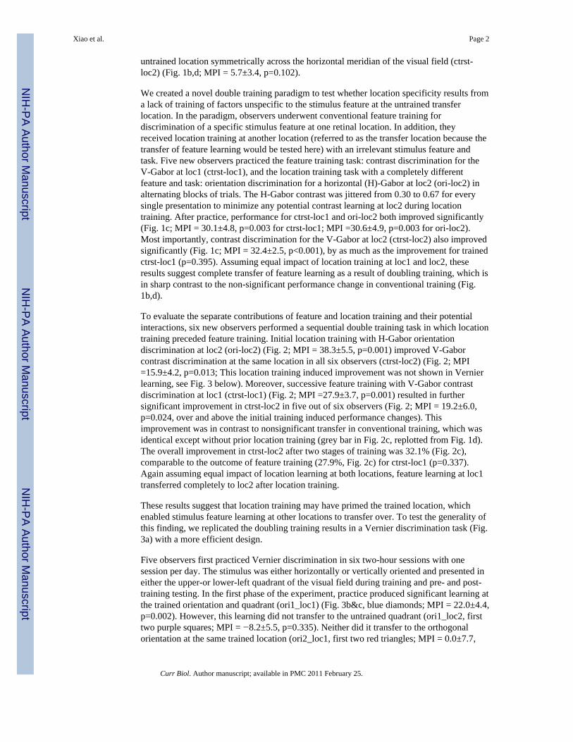
untrained location symmetrically across the horizontal meridian of the visual field (ctrst-loc2) (Fig. 1b,d; MPI = 5.7±3.4, p=0.102).
We created a novel double training paradigm to test whether location specificity results froma lack of training of factors unspecific to the stimulus feature at the untrained transferlocation. In the paradigm, observers underwent conventional feature training fordiscrimination of a specific stimulus feature at one retinal location. In addition, theyreceived location training at another location (referred to as the transfer location because thetransfer of feature learning would be tested here) with an irrelevant stimulus feature andtask. Five new observers practiced the feature training task: contrast discrimination for theV-Gabor at loc1 (ctrst-loc1), and the location training task with a completely differentfeature and task: orientation discrimination for a horizontal (H)-Gabor at loc2 (ori-loc2) inalternating blocks of trials. The H-Gabor contrast was jittered from 0.30 to 0.67 for everysingle presentation to minimize any potential contrast learning at loc2 during locationtraining. After practice, performance for ctrst-loc1 and ori-loc2 both improved significantly(Fig. 1c; MPI = 30.1±4.8, p=0.003 for ctrst-loc1; MPI =30.6±4.9, p=0.003 for ori-loc2).Most importantly, contrast discrimination for the V-Gabor at loc2 (ctrst-loc2) also improvedsignificantly (Fig. 1c; MPI = 32.4±2.5, p<0.001), by as much as the improvement for trainedctrst-loc1 (p=0.395). Assuming equal impact of location training at loc1 and loc2, theseresults suggest complete transfer of feature learning as a result of doubling training, which isin sharp contrast to the non-significant performance change in conventional training (Fig.1b,d).
To evaluate the separate contributions of feature and location training and their potentialinteractions, six new observers performed a sequential double training task in which locationtraining preceded feature training. Initial location training with H-Gabor orientationdiscrimination at loc2 (ori-loc2) (Fig. 2; MPI = 38.3±5.5, p=0.001) improved V-Gaborcontrast discrimination at the same location in all six observers (ctrst-loc2) (Fig. 2; MPI=15.9±4.2, p=0.013; This location training induced improvement was not shown in Vernierlearning, see Fig. 3 below). Moreover, successive feature training with V-Gabor contrastdiscrimination at loc1 (ctrst-loc1) (Fig. 2; MPI =27.9±3.7, p=0.001) resulted in furthersignificant improvement in ctrst-loc2 in five out of six observers (Fig. 2; MPI = 19.2±6.0,p=0.024, over and above the initial training induced performance changes). Thisimprovement was in contrast to nonsignificant transfer in conventional training, which wasidentical except without prior location training (grey bar in Fig. 2c, replotted from Fig. 1d).The overall improvement in ctrst-loc2 after two stages of training was 32.1% (Fig. 2c),comparable to the outcome of feature training (27.9%, Fig. 2c) for ctrst-loc1 (p=0.337).Again assuming equal impact of location learning at both locations, feature learning at loc1transferred completely to loc2 after location training.
These results suggest that location training may have primed the trained location, whichenabled stimulus feature learning at other locations to transfer over. To test the generality ofthis finding, we replicated the doubling training results in a Vernier discrimination task (Fig.3a) with a more efficient design.
Five observers first practiced Vernier discrimination in six two-hour sessions with onesession per day. The stimulus was either horizontally or vertically oriented and presented ineither the upper-or lower-left quadrant of the visual field during training and pre- and post-training testing. In the first phase of the experiment, practice produced significant learning atthe trained orientation and quadrant (ori1_loc1) (Fig. 3b&c, blue diamonds; MPI = 22.0±4.4,p=0.002). However, this learning did not transfer to the untrained quadrant (ori1_loc2, firsttwo purple squares; MPI = −8.2±5.5, p=0.335). Neither did it transfer to the orthogonalorientation at the same trained location (ori2_loc1, first two red triangles; MPI = 0.0±7.7,
Xiao et al. Page 2
Curr Biol. Author manuscript; available in PMC 2011 February 25.
NIH
-PA Author Manuscript
NIH
-PA Author Manuscript
NIH
-PA Author Manuscript

p=0.997). These results confirmed the well known location and orientation specificities ofVernier and hyper-acuity learning in the conventional learning paradigm [5].
In the context of double training, here the observers were performing two parallel sets ofsuccessive double training. Practice at ori1_loc1 in the first set of double training could beregarded as feature training for target stimulus ori1_loc2 (same orientation, secondarylocation), and in the second set could be regarded as location training for another targetstimulus ori2_loc1 (orthogonal orientation, same location). In the next phase of doubletraining, the observers practiced Vernier discrimination for an orthogonal orientation at asecondary location (ori2_loc2). This new training served as successive location training fortarget stimulus ori1_loc2 in the first set of double training, and as successive feature trainingfor another target stimulus ori2_loc1 in the second set of double training.
Our results showed that, after successive location training (ori2_loc2) (Fig. 3b&c, greencircles; MPI = 27.2±4.8, p=0.003), performance for the target stimulus ori1_loc2 was nowimproved significantly (the 2nd and 3rd purple squares; MPI = 24.7±4.0, p=0.004, over andabove the initial training induced performance changes). This result confirmed that locationtraining, even after feature learning, triggered the location transfer of earlier feature learning(ori1_loc1). The MPIs for feature-trained ori1_loc1 and feature-untrained but location-trained target stimulus ori1_loc2 were 22.0% and 24.7%, respectively (Fig. 3d, the blue barand the right purple bar; p=0.552), showing complete transfer of feature learning acrossretinal locations after double training.
Moreover, the same secondary training at ori2_loc2 as successive feature learning alsotransferred significantly to a secondary location (ori2_loc1) (the 2nd and 3rd red triangles;MPI = 20.7±4.0, p=0.001, over and above the initial training induced performance changes)after earlier location training at ori1_loc1. The MPIs for feature-trained ori2_loc2 andfeature-untrained but location-trained target stimulus ori2_loc1 were 27.3% and 20.7%,respectively (Fig. 3d, the green bar and the right red bar; p=0.287), so the transfer of featurelearning from loc2 to loc1 was again nearly complete after double training. In both sets ofdouble training, the degrees of learning and transfer were highly correlated (r = 0.96).Observers who improved most at trained ori1_loc1 and ori2_loc2 showed the largestimprovement at untrained target stimuli ori2_loc1 and ori1_loc2.
In general, results from these two sets of double training are in line with earlier doubletraining results in contrast learning tasks. Therefore, complete transfer of feature learningenabled by location training appears to be a general effect.
DiscussionSingle-unit and fMRI studies have shown that not only the retinotopic early visual cortex,but also the non-retinotopic higher brain areas that are more related to attention and decisionmaking, are involved in visual discrimination [13,14]. However, most studies on the neuralmechanisms of perceptual learning have focused on response changes in the retinotopicearly visual cortex [9,15–18], as motivated by the often observed strict location specificityand the inferred retinotopy. The complete transfer of perceptual learning to new retinallocations revealed by double training calls into question both location specificity as a keyproperty of visual perceptual learning, and the well-received belief by many researchers thatthe retinotopic early visual cortex is the neuronal basis of perceptual learning. Rather itpoints to a crucial role for non-retinotopic higher brain areas that engage attention anddecision making for perceptual learning.
The complete transfer of learning across retinal locations challenges many existingperceptual learning models that are more or less constrained by location specificity. For
Xiao et al. Page 3
Curr Biol. Author manuscript; available in PMC 2011 February 25.
NIH
-PA Author Manuscript
NIH
-PA Author Manuscript
NIH
-PA Author Manuscript

example, Adini, Tsodyks, and Sagi [19] modeled perceptual learning as training inducedmodification of recurrent connections in V1. This model would not predict transfer oflearning to other retinal locations without incorporating learning in higher brain areas. At thepost-V1 level, the Lu and Dosher model [20,21] suggests training induced re-weighting ofV1 neuronal responses. Such response re-weighting, as our data suggest, would have to atleast partially occur in non-retinotopic areas, so that the model would allow location transferof learning. Apparently our results are more consistent with Mollon and Danilova’shypothesis [12] that feature learning could take place in a more central site, but is nottransferable to a new location because of the local noise at the new location. Anotherpossibility is that learning could occur in both central and peripheral sites, which would beconsistent with all current models and not necessarily contradictory to our data. However,the latter hypothesis is less favored by recent evidence that motion direction learning iscorrelated with activity changes in a decision area LIP, but not the sensory area MT [22],and that orientation learning is more correlated to changes in V4, rather than V1, neurons[23,24]. For orientation learning, even orientation tuning changes in V4 neurons are still toosmall to fully account for behavioral orientation learning [23]. Therefore, brain areas higherthan V4 must have involved. This is consistent recent fMRI evidence [13] that high brainareas, including the intraparietal sulcus, frontal eye field, and supplementary eye field,participate in visual perceptual learning.
As an extension of the Mollon and Danilova hypothesis, our results further demonstrate thatcentral learning can guide visual discrimination at a new location, provided that the newlocation has been primed by location training. Fig. 2 shows that contrast learning transfersfrom loc1 to loc2 after orientation training at loc2, suggesting that location learning isstimulus feature/task non-specific. We hypothesize that location learning improves spatialattention, which is stimulus nonspecific, to a peripheral location. Such improvement may benecessary since our everyday experience is probably insufficient as location training in thevisual periphery for demanding visual discrimination tasks near their thresholds. There isevidence that spatial attention excludes unwanted noise without affecting the stimulustemplate [25]. If the stimulus template learned through feature training is stored in a centralsite, it would now be able to respond to the same stimulus at a new retinal location wherenoise has been excluded through location training.
Many perceptual learning studies including ours involve discrimination of basic simplestimulus features like orientation, contrast, etc. Retinal location specificity is central to manymodels proposing that perceptual learning of this kind relies on unique mechanisms largelybased in sensory cortex, and does not involving general mechanisms such as those involvedin associative learning [26]. In addition, there are studies that use more complex stimuli toinvestigate the roles of more general processes in perceptual learning [27,28]. Complexstimuli are typically not presented in a single retinal location, so their learning is presumablynonspecific to retinal locations and occurs in higher brain areas. By demonstrating completelocation transfer of perceptual learning of basic visual features, we argue that higher brainareas are also critically involved in basic visual feature learning. Therefore, it is likely that atleast partially overlapping mechanisms in higher brain areas could contribute to perceptuallearning of both basic and more complex stimuli.
Experimental ProceduresObservers and apparatus
Twenty two observers with normal or corrected-to-normal vision participated in differentexperiments of this study. All were new to psychophysical experiments and unaware of thepurposes of the study.
Xiao et al. Page 4
Curr Biol. Author manuscript; available in PMC 2011 February 25.
NIH
-PA Author Manuscript
NIH
-PA Author Manuscript
NIH
-PA Author Manuscript

The stimuli were generated by a PC-based WinVis program (Neurometrics Institute,Oakland, CA). Experiments with Gabor stimuli were run in one system using a 21-inchSony G520 color monitor (1024 pixel × 768 pixel, 0.37 mm (H) × 0.37 mm (V) per pixel,120 Hz frame rate, 50 cd/m2 mean luminance). Experiments with illusory line stimuli wererun in another system using a 21-inch NEC MultiSync FE2111 color monitor (1600 pixel ×1200 pixel, 0.24 mm (H) × 0.24 mm (V) per pixel, 85 Hz frame rate, 41 cd/m2 meanluminance). Luminance of the monitors was linearized by an 8-bit look-up table. Viewingwas monocular with one eye covered with a translucent plastic pad. A chin-and-head resthelped stabilize the head of the observer. Experiments were run in a dimly lit room.
StimuliFor contrast discrimination tasks, the test stimulus was a Gaussian windowed sinusoidalgrating (Gabor) on a mean luminance screen background and presented in the upper- orlower-left visual quadrant at 5° retinal eccentricity (Fig. 1a). The stimulus contrast weredefined as (Lmax− Lmin)/(Lmax+Lmin), where Lmax was the maximal luminance and Lminwas the minimal luminance of the stimuli (i.e. Michelson contrast). The spatial frequency ofthe Gabor stimuli was 1.5 cycles per degree (cpd), the standard deviation of the Gaussianenvelope was equal to the wavelength (λ) of the sinusoidal carrier, and the base contrast was0.45. The viewing distance was 1 meter. The same Gabor stimuli were also used fororientation discrimination training (Figs. 1c & 2) with contrast jittered from 0.30 to 0.67.When orientation discrimination was performed, stimuli were viewed through a circularopening (diameter = 170°) of a black cardboard that covered the entire monitor screen. Thiscontrol prevented observers from using external references to determine the orientations ofthe stimuli.
For Vernier discrimination tasks, the test stimulus was formed by a pair of identical Gaborson a mean luminance screen background and presented in the upper- or lower-left visualquadrant at 5° retinal eccentricity (Fig. 3a). The two Gabors had the same spatial frequency(3cpd), standard deviation (2λ), contrast (0.45), and orientation (vertical or horizontal), andhad a center-to-center distance of 4λ The position of each Gabor shifted half the Vernieroffset in opposite directions perpendicular to the Gabor orientation. The viewing distancewas 1 meter.
ProcedureContrast and orientation discrimination thresholds were measured with a temporal 2AFCstaircase procedure. In each trial, the test and reference stimuli were separately presented intwo 92-ms stimulus intervals in a random order separated by a 600-ms inter-stimulusinterval. The observer’s task was to judge which stimulus interval contained the stimulus ata higher contrast (contrast discrimination) or at a more clockwise orientation (orientationdiscrimination). A small fixation cross preceded each trial by 400 ms and stayed through thetrial.
Vernier discrimination thresholds were measured with a single-trial 2AFC staircaseprocedure. In each trial, the Vernier stimulus was presented for 92 ms. The observer’s taskwas to judge whether the right Gabor was higher or lower than the left Gabor for ahorizontal Vernier stimulus, or the lower Gabor was to the left or right of the upper Gaborfor a vertical Vernier stimulus. A small fixation cross preceded each trial by 400 ms andstayed through the trial.
Auditory feedback was given on incorrect responses. The step size of the staircase was 0.05log units. A classical 3-down-1-up staircase rule was used, which resulted in a 79.4%convergence rate. Each staircase consisted of four preliminary reversals and six
Xiao et al. Page 5
Curr Biol. Author manuscript; available in PMC 2011 February 25.
NIH
-PA Author Manuscript
NIH
-PA Author Manuscript
NIH
-PA Author Manuscript

experimental reversals. The geometric mean of the experimental reversals was taken as thethreshold for each staircase run.
Eye movementWe used an Eyelink II eye tracker to assess the impact of eye movement. Five newobservers performed an orientation discrimination task identical to the one in Fig. 1c for 3–5sessions. During the first interval, the eye positions on the average were within 0.5° and 1°from the fixation in 90.3% and 98.9% of the trials, respectively, similar to those in afixation-only control condition (p=0.29 & 0.37, respectively). Therefore Fig. 3 data forsingle-interval vernier learning were unaffected by eye movement. Moreover, the eyepositions in the second interval were within 0.5° and 1° from the fixation in 78.3% and96.1% of the trials, respectively, suggesting some mainly within −0.5° involuntary eyedrifts. However, practice did not reduce the second-interval eye drifts to improve visualdiscrimination. The last-/first-day ratios of these off-fixation percentages in the secondinterval were 0.954 (<0.5 °) and 0.995 (<1 °), suggesting no reduction of eye drifts aftertraining. Therefore, perceptual learning in these two-interval tasks (Figs. 1 & 2) was littleaffected by eye movement either.
AcknowledgmentsThis research is supported by the Natural Science Foundation of China (30725018) and the United States NationalInstitute of Health (R01-04776 & R01-01728). We thank Drs. Merav Ahissar, Zoe Kourtzi, Roger Li, Wu Li, andLi Zhaoping for their helpful comments. The last paragraph in Discussion was adapted from Dr. Dominic Dwyer’sreview on the submitted manuscript.
References1. Shiu LP, Pashler H. Improvement in line orientation discrimination is retinally local but dependent
on cognitive set. Perception & psychophysics 1992;52:582–588. [PubMed: 1437491]2. Schoups AA, Vogels R, Orban GA. Human perceptual learning in identifying the oblique
orientation: retinotopy, orientation specificity and monocularity. The Journal of physiology1995;483(Pt 3):797–810. [PubMed: 7776259]
3. Fahle M. Specificity of learning curvature, orientation, and vernier discriminations. Vision Research1997;37:1885–1895. [PubMed: 9274774]
4. Saarinen J, Levi DM. Perceptual Learning in Vernier Acuity: What is Learned? Vision Research1995;35:519–527. [PubMed: 7900292]
5. Crist RE, Kapadia MK, Westheimer G, Gilbert CD. Perceptual learning of spatial localization:specificity for orientation, position, and context. Journal of neurophysiology 1997;78:2889–2894.[PubMed: 9405509]
6. Yu C, Klein SA, Levi DM. Perceptual learning in contrast discrimination and the (minimal) role ofcontext. J Vis 2004;4:169–182. [PubMed: 15086307]
7. Karni A, Sagi D. Where practice makes perfect in texture discrimination: evidence for primaryvisual cortex plasticity. Proceedings of the National Academy of Sciences of the United States ofAmerica 1991;88:4966–4970. [PubMed: 2052578]
8. Fahle M. Human pattern recognition: parallel processing and perceptual learning. Perception1994;23:411–427. [PubMed: 7991342]
9. Ghose GM, Yang T, Maunsell JH. Physiological correlates of perceptual learning in monkey V1 andV2. Journal of neurophysiology 2002;87:1867–1888. [PubMed: 11929908]
10. Li RW, Levi DM, Klein SA. Perceptual learning improves efficiency by re-tuning the decision‘template’ for position discrimination. Nature neuroscience 2004;7:178–183.
11. Dosher BA, Lu ZL. Perceptual learning reflects external noise filtering and internal noise reductionthrough channel reweighting. Proceedings of the National Academy of Sciences of the UnitedStates of America 1998;95:13988–13993. [PubMed: 9811913]
Xiao et al. Page 6
Curr Biol. Author manuscript; available in PMC 2011 February 25.
NIH
-PA Author Manuscript
NIH
-PA Author Manuscript
NIH
-PA Author Manuscript

12. Mollon JD, Danilova MV. Three remarks on perceptual learning. Spat Vis 1996;10:51–58.[PubMed: 8817771]
13. Mukai I, Kim D, Fukunaga M, Japee S, Marrett S, Ungerleider LG. Activations in visual andattention-related areas predict and correlate with the degree of perceptual learning. J Neurosci2007;27:11401–11411. [PubMed: 17942734]
14. Gold JI, Shadlen MN. The neural basis of decision making. Annual review of neuroscience2007;30:535–574.
15. Crist RE, Li W, Gilbert CD. Learning to see: experience and attention in primary visual cortex.Nature neuroscience 2001;4:519–525.
16. Schoups A, Vogels R, Qian N, Orban G. Practising orientation identification improves orientationcoding in V1 neurons. Nature 2001;412:549–553. [PubMed: 11484056]
17. Schwartz S, Maquet P, Frith C. Neural correlates of perceptual learning: a functional MRI study ofvisual texture discrimination. Proceedings of the National Academy of Sciences of the UnitedStates of America 2002;99:17137–17142. [PubMed: 12446842]
18. Furmanski CS, Schluppeck D, Engel SA. Learning strengthens the response of primary visualcortex to simple patterns. Curr Biol 2004;14:573–578. [PubMed: 15062097]
19. Adini Y, Sagi D, Tsodyks M. Context-enabled learning in the human visual system. Nature2002;415:790–793. [PubMed: 11845209]
20. Dosher BA, Lu ZL. Mechanisms of perceptual learning. Vision Research 1999;39:3197–3221.[PubMed: 10615491]
21. Lu ZL, Dosher BA. Perceptual learning retunes the perceptual template in foveal orientationidentification. J Vis 2004;4:44–56. [PubMed: 14995898]
22. Law CT, Gold JI. Neural correlates of perceptual learning in a sensory-motor, but not a sensory,cortical area. Nature neuroscience 2008;11:505–513.
23. Raiguel S, Vogels R, Mysore SG, Orban GA. Learning to see the difference specifically alters themost informative V4 neurons. J Neurosci 2006;26:6589–6602. [PubMed: 16775147]
24. Yang T, Maunsell JH. The effect of perceptual learning on neuronal responses in monkey visualarea V4. J Neurosci 2004;24:1617–1626. [PubMed: 14973244]
25. Lu ZL, Dosher BA. Spatial attention excludes external noise without changing the spatialfrequency tuning of the perceptual template. J Vis 2004;4:955–966. [PubMed: 15595898]
26. Fahle, M. Introduction. In: Fahle, M.; Poggio, T., editors. Perceptual Learning. Cambridge, MA,USA: MIT Press; 2002. p. ix-xx.
27. Goldstone RL. Perceptual learning. Annu Rev Psychol 1998;49:585–612. [PubMed: 9496632]28. McLaren IPL, Mackintosh NJ. An elemental model of associative learning: I. Latent inhibition and
perceptual learning. Anim Learn Behav 2000;28:211–246.
Xiao et al. Page 7
Curr Biol. Author manuscript; available in PMC 2011 February 25.
NIH
-PA Author Manuscript
NIH
-PA Author Manuscript
NIH
-PA Author Manuscript

Fig. 1.Retinal location specificity studied with the conventional and double-training paradigms. a.The stimulus configuration in a 2AFC trial in Figs. 1 & 2 experiments. One interval containsa higher-contrast Gabor stimulus (Gaussian windowed sinusoidal grating). b. Conventionaltraining: Contrast discrimination for a V-Gabor was practiced at loc1 (ctrst-loc1, greencircles) and its transfer was tested at loc2 (ctrst-loc2, red triangles). Data are pooled from allparticipating observers, and error bars represent one standard error of the mean. c. Doubletraining: Contrast discrimination was practiced at loc1 as feature training (ctrst-loc1, greencircles; contrast thresholds indicated by the left ordinate), and orientation discrimination fora H-Gabor was practiced at loc2 as additional location training (ori-loc2, blue diamonds;orientation thresholds indicated by the right ordinate), in alternating blocks of trials(staircases). The transfer was tested for V-Gabor contrast discrimination at loc2 (ctrst-loc2;red triangles). The left and right ordinates have the identical scale factor in log units. d. Asummary of training results as well as the resultant transfers as % improvement withconventional and double training.
Xiao et al. Page 8
Curr Biol. Author manuscript; available in PMC 2011 February 25.
NIH
-PA Author Manuscript
NIH
-PA Author Manuscript
NIH
-PA Author Manuscript

Fig. 2.Sequential double training. a&b. Individual and mean plots. H-Gabor orientationdiscrimination was first practiced at loc2 as location learning (ori-loc2, blue diamonds;orientation thresholds indicated by the right ordinate), and its transfer to V-Gabor contrastdiscrimination at the same loc2 was tested (ctrst-loc2, first two red triangles; contrastthresholds indicated by the left ordinate). Then V-Gabor contrast discrimination at loc1 waspracticed as feature training (ctrst-loc1, green circles), and its transfer to ctrst-loc2 wastested (the 2nd and 3rd red triangles). This part of training was identical to conventionaltraining in Fig. 1b. The right and left ordinates have the identical scale factor in log units. c.A summary of initial location learning and its transfer (left blue and red bars), successivefeature learning and its transfer (middle green and red bars; the grey bar replotted from Fig.1d indicates conventional learning transfer), and the overall transfer (right red bar) as %improvement.
Xiao et al. Page 9
Curr Biol. Author manuscript; available in PMC 2011 February 25.
NIH
-PA Author Manuscript
NIH
-PA Author Manuscript
NIH
-PA Author Manuscript

Fig. 3.Sequential double training for Vernier discrimination. a. The stimulus configuration. TheVernier stimulus was presented at either the horizontal or vertical orientation, and in eitherthe lower- or upper-left visual quadrant. b&c. Individual and mean plots. Vernierdiscrimination was practiced at ori1_loc1 (blue open diamonds) and its transfer to the adifferent quadrant (ori1_loc2, the first two purple squares) and to the orthogonal orientationat the same quadrant (ori2_loc1, the first two red triangles) were measured to showconventional location and orientation specificity in Vernier learning. In a double trainingcontext, for the target stimulus ori2_loc1 (red triangles), initial practice at ori1_loc1 (bluediamonds) served as location training, and the later practice at ori2_loc2 (green circles)served as feature training. This double training resulted in nearly complete transfer oflearning to the target stimulus ori2_loc1 (last two red triangles). In parallel, for anothertarget stimulus ori1_loc2, initial practice at ori1_loc1 also served as feature training, and thelater practice at ori2_loc2 (green circles) served as location training. Again this doubletraining resulted in complete transfer of feature learning to target stimulus ori1_loc2 (lasttwo purple squares). d. A summary of initial (feature or location) learning at ori1_loc1 andits transfers to target stimuli ori1_loc2 and ori2_loc1 (the left three bars), and later (locationor feature) training at ori2_loc2 and its additional transfers to target stimuli ori1_loc2 andori2_loc1 (right three bars).
Xiao et al. Page 10
Curr Biol. Author manuscript; available in PMC 2011 February 25.
NIH
-PA Author Manuscript
NIH
-PA Author Manuscript
NIH
-PA Author Manuscript