
Complete Guide to 4th Year - WordPress.com
709
Tom’s Guide 2011-2012 Complete Guide to 4 th Year
-
Upload
khangminh22 -
Category
Documents
-
view
0 -
download
0
Transcript of Complete Guide to 4th Year - WordPress.com

Tom’s Guide
2011-2012
Complete Guide to 4th Year

Page | 2
Contents
Psychiatry Page 3
Paediatrics
Page 80
Otorhinolaryngology
Page 414
Ophthalmology
Page 460
Obstetrics and gynaecology
Page 500
*NOTES Dermatology – just use the online lectures and memorise the pictures HCE – just use the online lectures and learn the stats Obstetrics and gynaecology guide is not objective based (read the objectives and you will understand why)

Page | 3
Psychiatry Topics and Topic Outcomes
Introductory Sessions 1. Understand the expectations of students during the placement 2. Interview a patient in a professional manner and instil therapeutic optimism 3. Obtain a relevant history from a patient and/or carer and present it verbally to a colleague using appropriate terminology There is no set way of taking a psychiatric history and in most respects it is very similar to taking a normal history. The following are a list of recommended headings and some information that should be included. Obviously different conditions will require a different approach and more specific questions. Identifying Information – Name, age, marital status, children, occupation, legal status (i.e. sectioned or voluntary) Presenting Complaint – Try to record this in the patient’s own words. For example a patient does not usually present with depression, instead they may present with self harm. Try to include common symptoms of conditions as well i.e. low mood, difficulty sleeping and poor self esteem with depression. History of Presenting Complaint – The following headings may be useful in structuring this question: duration, development, mode of onset, course, severity, associated symptoms, and precipitating factors Past Psychiatric History – This should include dates and durations of previous mental illness, details of previous treatment (medication, psychotherapy, ECT or even hospitalisation), details of previous contact with psychiatric services and details of previous assessment or treatment under mental health legislation. Family History – Enquire about the presence of psychiatric illness (including suicide and substance abuse) in family members. A family tree may also be useful since genetics are implicated in the aetiology of most psychiatric conditions. Enquire whether parents are still alive and causes of death if not. Finally enquire about the patient’s relationship with close family members.

Page | 4
Past Medical History – As you would for a normal history. In particular check for head injury/surgery, neurological conditions (i.e. epilepsy) and endocrine abnormalities. Drug History – Note all medications including psychiatric, non-psychiatric and OTC drugs. Check for non-compliance as well as adverse reactions and allergies. Substance Misuse – Never avoid this due to embarrassment or fear of upsetting the patient. Alcohol and substance related psychiatric conditions are not uncommon! When assessing alcohol consumption as them about a typical week. Try overestimating the amount if they won’t give an answer and see their response. Check the type of alcohol and when they have their first drink and where. If illicit drugs then record name, route of admission, years of use and frequency. Also try to illicit signs or symptoms of dependence (see later). Personal History – This is basically taking a life history. There are four main areas that should be focused on:
• Infancy and early childhood (until age 5) – pregnancy and birth complications, developmental milestones, childhood illness and unusually aggressive behaviour or impaired social interaction.
• Later childhood and adolescence (until end of higher education) – school record, relationships (parents, teachers, peers), history of abuse (physical, sexual or emotional), behavioural problems, drug use, truancy, higher education, training
• Occupation record – type of duration of jobs, reasons for unemployment and/or dismissal
• Relationship, marital and sexual history – puberty, significant relationships, reasons for breakup, marriage/divorce, and ability to engage in satisfactory sexual relationships.
Premorbid Personality – An indication of the patient’s personality and character before the onset of mental illness. A collateral history may be useful here but the patient may sometimes be able to give details. Forensic History – Details and dates of previous offences and antisocial behaviour, prosecutions, convictions and prison sentences. Ask about violent crime in particular and the age of the patient’s first offence. Social Circumstances – Accommodation, social support, relationship, employment, financial circumstances, hobbies and leisure activities. 4. Recognise the psychopathology of common psychiatric disorders

Page | 5
5. Perform a mental state examination and present it verbally to a colleague using appropriate terminology Mental state examination (MSE) describes the interviewers impression of many aspects of a patient’s mental functioning at a certain period of time. This should not be seen as a separate section as it begins the second the patient enters the room. The key components are as follows: Appearance and Behaviour – Appearance includes the patients physical state, clothing and accessories, do clothes match, are they appropriate for the time of year and day, are they carrying strange objects, are the clothes clean? Self-care and hygiene, self-neglect, injuries or self harm. Behaviour focuses on motor behaviour such as tics, tremors, abnormal movements, twitches as well as fear, aggression, suspiciousness and catatonic features. Also assess psychomotor abnormalities such as retardation and agitation. Rapport – Note whether you are establishing a good rapport and what is the patient’s attitude towards you. Some ways to describe this include cooperative, cordial, uninterested, aggressive, defensive, guarded, suspicious, fearful, perplexed, preoccupied and disinhibited etc. Speech – The three important aspects of speech are rate, rhythm and volume. Difficulties in quality and flow of speech should also be assessed along with incoherent, bizarre or disorganised speech. Mood and Affect – Mood should be assessed subjectively (by asking the patient how they are feeling) over a period of time and objectively (by yourself). Affect (external expression of emotion as perceived by others) is measured by observing the patient’s posture, facial expression, emotional reactivity and speech. When reporting affect there are two components to consider: 1) Congruity – does the patients subjectively reported mood match their observed mood (i.e. a smiling woman saying she is sad and suicidal is incongruous affect) 2) The range of affect – within normal range (reactive), blunted (reduced intensity of expression) or flat (very little expression of emotion). Labile mood refers to a fluctuating mood state that alternates between extremes. Thought Form – Is it logical or is it disorganised and erratic such as tangential thinking, loosening of association, flight of ideas etc. Thought Content – This includes delusions, abnormal beliefs and overvalued ideas. The delusions may be primary or secondary, mood congruent or incongruent and bizarre or non-bizarre.

Page | 6
Perception – This includes any form of hallucination, what modality is it in, are they Hypnagogic or hypnopompic or neither etc. Cognition – Usually an MMSE should be done to assess orientation, registration, attention, calculation, language, executive functioning and visuospatial skills. Insight – The patient’s understanding of their illness as well as the recognition of the need of treatment. This may range from complete denial to a genuine understanding and acceptance. Important questions should include – does the patient think they have a mental illness?; does the patient feel ill?; and would the patient accept treatment? 6. An awareness of the risk factors and principles of acute management for suicide, self-harm, neglect and harm to others 7. Carry out a clinical risk assessment on a patient with a common psychiatric disorder A risk assessment is very important to not only check for a risk of self harm but also for harm to others or property. Currently psychiatry is bad at getting this right so results should be used in combination with a comprehensive assessment. The following areas should be considered:
• Self-harm and Suicide – risk factors include being male, aged 19-34, divorced>widowed>single, unemployed/retired, social class 5, living alone, social isolation, psychiatric illness, previous self harm, alcohol abuse, physical chronic illness (often painful), family history. Intent can also be assessed and this includes if the attempt was planned, were precautions taken to avoid discovers, was a dangerous method used, was help sought afterwards etc.
• Self Neglect – Partly the risk factors above but also depression and psychiatric illness that affects cognition/memory
• Exploitation – are they are risk from exploitation (either from random people in mania or from family and friends i.e. financially).
• Deterioration – Are they are risk of getting worse if no treatment is given
• Violence and Aggression – either are they at risk to others or are they at risk from others.
• Children – what age are the children, has the patient ever had any command hallucinations or thought of harming them

Page | 7
• Property and Driving – risk from arson, accidental damage, driving
whilst on medication, trying to kill themselves using a car (endangering others)
8. Reflect on the impact of a patient’s, carer’s or colleague’s actions on one’s own emotional and behavioural responses 9. A basic understanding of the structure and delivery of psychiatric services in the UK 10. Carry out a clinical risk assessment on a patient with a common psychiatric disorder (duplicate of 7) 11. Recognise the limits of their own professional competence and seek appropriate supervision Psychosis 1. Knowledge of the epidemiology, aetiology and prognosis of psychosis Epidemiology –
• incidence = 5-50/100,000 individuals per year and prevalence is around 1%. Lifetime risk is around 1%.
• Age of onset is between late teens and mid 30s. Women have a later onset than men. Men: 18-25, Women: 25-35
• Men and women have an equal incidence but the difference is hard to compare due to differences in age of onset
• There is an increased prevalence in lower socioeconomic classes (class IV and V). A theory to explain this may be that psychosis leads to a ‘drift’ down the social economic scale. There is a higher prevalence in urban areas compared to rural
• Incidence is higher for immigrants, especially African-caribbeans in the UK
Aetiology
• Genetic – Schizophrenia runs in families with a 50% concordance rate for monozygotic twins compared with dizygotic twins (10%). This is also supported by adoption studies.
• Developmental factors – May be linked to complications during pregnancy and birth. An observation that more schizophrenics are bored in late winter or spring suggests that second trimester influenza infection may play a role

Page | 8
• Brain abnormalities – Imaging has shown structural and functional changes in the brain. These finding may be secondary to the condition or in fact its treatment. Changes included ventricular enlargement (associated with negative symptoms) and reduced brain size (frontal and temporal lobes, hippocampus, amygdala, parahippocampal gyrus).
• Neurotransmitter abnormalities – Schizophrenia is thought to be due to over activity of the mesolimbic dopamine pathway. Drugs that potentiate this pathway (amphetamines and antiparkinsonian drugs) are known to produce psychotic symptoms.
• Life events – Stressful life events often occur before the first psychotic episode or recurrence and therefore may precipitate the illness. It may however be the early stages of illness that cause these stressful events
• Expressed emotion – When carers/family become over involved, over critical or hostile towards psychotic patients then they are more likely to relapse.
Prognosis • Generally the disease is chronic and shows a remitting and
relapsing pattern. About 20% have a single lifetime episode without relapse.
• More than 50% of patients have a poor outcome characterised by repeated psychotic episodes with hospitalizations, depression and suicide attempts.
• About 10% of schizophrenic patients will successfully commit suicide. The most at risk are young, well educated men who have good insight into their disease. The period after discharge is the most vulnerable.
• Schizophrenic patients, on average, live 10 years less than the general population
• The prognosis is generally better in developing countries where there may be better family support.
• Factors associated with a good prognosis are: female sex, married, older age of onset, abrupt onset, onset precipitated by life stress, short duration of illness prior to treatment, good response to medication, paranoid subtype, absence of negative symptoms, prominent mood symptoms or a family history of mood disorder, good premorbid functioning.
2. Knowledge of clinical presentation, including an understanding of the ICD-10 diagnostic criteria, of psychosis Psychosis can present with many symptoms including delusions, hallucinations, psychomotor abnormalities, mood/affect disturbance, cognitive deficits and disorganised thoughts and behaviour.

Page | 9
Perceptions – hallucinations occur in the absence of physical stimuli and appear as if real to a patient. Therefore patients often have little insight into these. They occur in one of the five senses and are not ideas, thoughts or images arising in the patient’s own mind. Illusions are a distortion of a real external stimulus and occur in healthy adults. Pseudohallucinations are hallucinations that arise from the patients ‘inner eye’ and not through a sensory organ. Auditory hallucinations – Simple unstructured sounds or single words are more indicative or an acute organic state. Second person voices are often associated with mood disorders with psychotic features so are often critical or persecutory, i.e. mood congruent. Other forms include third person hallucinations and audible thoughts. Visual hallucinations – These are rarer than auditory and occur more often in organic disorders or with psychoactive substances. Somatic hallucinations – Sensations of bodily sensation and include: tactile which is the skin being touched. This includes formication which is the sensation of insects crawling on the skin, often associated with long term cocaine use or alcohol withdrawal. Other types include thermal, hygric (fluid), visceral and kinaesthetic. Others – Hypnagogic hallucinations occur as a person is falling to sleep where as hypnopompic hallucinations occur as a person is waking up. Extracampine hallucinations occur outside of the patients body. Thought disorder Abnormal beliefs and delusions – A delusion is an unshakeable false belief that is not accepted by other members of the patients culture. The belief is as real as a true belief to the patient and is false due to faulty reasoning. This also out of keeping with the patients social and cultural background. Delusions are usually classified as: primary/secondary, mood congruent/incongruent, bizarre/non-bizarre and according to content.
• Primary delusions are delusions that do not occur in response to any other psychopathology. They typically occur in schizophrenia and other primary psychotic disorders.
• Secondary delusions are the consequences of a pre-existing psychopathological state; usually mood disorders.
• Mood congruent/incongruent and bizarre/non-bizarre are self explanatory.
• Delusion by content – there are many forms of delusional content which included:

Page | 10
1) Persecutory, 2) Grandiose – thinking you are extremely powerful, 3) delusions of reference – thinking objects , people or events have intense personal meaning (e.g. the news reader is referring to you), 4) Religious, 5) delusions of love (erotomania), 6) delusions of infidelity, 7) delusions of misidentification, 8) nilhilisitic delusions are false beliefs that the world is about to end or they do not exist, 9) somatic e.g your bowels are rotting away, 10) delusions of infestation, 11) delusions of control – includes thought insertion, withdrawal and broadcasting Overvalued ideas – A plausible belief that the patient is preoccupied with to an unreasonable extent. They must have a significant impact on the patient’s life and are distinguished from obsessions as they are not recurrent intrusions. Typical disorder that features these include anorexia nervosa. Disorganised thinking – the following are important signs of disorganised thinking:
• Circumstantiality and tangentiality – speech that is delayed in reaching its final goal because of the over inclusion of detail but does eventually get to the point. Normal people may exhibit circumstantiality but tangentiality is pathological as the speaker never returns to the point.
• Flight of ideas – A stream of connected concepts through words, puns or clang association.
• Loosening of association (derailment or knight’s move thinking) – when a patients speech moves from one loosely or unrelated topic to the next. Can be characteristic of schizophrenia.
• Thought blocking – stopping mid-sentence and then having no recall of what they were saying and talk about a new topic.
• Perseveration – the unnecessarily repeating of words or syllables. Palilalia describes the repeating of the last word of the sentence. This is highly indicative of an organic brain disorder.
• Echolalia – senselessly repeating words or phrases spoken to them
Negative symptoms – These are a clinical deficit and include marked apathy, poverty of thought and speech, blunting of affect, social isolation, poor self care and cognitive deficits. Psychomotor function – Patients may present with some affect on motor function, usually due to side effects of medication. However these can occasionally occur which are not caused by medication or organic brain disorder. Conditions include rigidity, stupor and tics. ICD-10 Schizophrenia: One or more of the following:
• Thought echo, insertion, withdrawal or broadcast • Delusions of control or passivity

Page | 11
• Hallucinatory voices giving a running commentary discussing the patient amongst themselves
• Bizarre delusions OR Two or more of the following:
• Hallucinations that either occur every day for weeks or that are associated with fleeting delusions or sustained overvalued ideas
• Thought disorganisation (loosening of association, incoherence or neologism)
• Catatonic symptoms • Negative symptoms • Change in personal behaviour (loss of interest, aimlessness or social
withdrawal) All symptoms should be present for most of the time during at least 1 MONTH. Schizophrenia should not be diagnosed in the presence of organic brain disease or drug intoxication/withdrawal. First rank symptoms
• Thought insertion, broadcast or withdrawal • Hallucinations of 3rd person, running commentary or thought echo • Somatic hallucinations, delusions of control/passivity, delusions of
perception Schizophrenia Subtypes
• Paranoid – dominated by delusions and hallucinations. Negative symptoms and catatonic symptoms as well as thought disorder are not prominent. Better prognosis and later onset
• Hebephrenic – thought disorder, disturbed behaviour and inappropriate/flat affect. Delusions and hallucinations are fleeting and onset of illness is earlier with a poorer prognosis
• Catatonic – characterised by one or more catatonic symptoms • Residual – 1 year of predominantly chronic negative symptoms
preceded by one clear cut psychotic episode • Post-schizophrenia depression – follows a schizophrenic episode but
has few characteristics of schizophrenia. • Simple – insidious onset with signs of residual depression without a
psychotic episode to precede it. Mostly negative symptoms Schizophrenia-like psychotic disorders
• Schizoaffective – schizophrenia plus mood symptoms (depression or mania) that present in the same episode of illness (within a few days). The mood symptoms should meet the criteria for depression/mania and the patient should have one or preferably two core symptoms of schizophrenia.

Page | 12
• Delusional disorder – a set or single delusion for at least 3 months which is the predominant symptom. Typical schizophrenia delusions such as control or passivity will exclude this diagnosis. Hallucinations may be fleeting and there may be brief depressive episodes. On the whole the patient will function well both socially and personally.
3. A basic understanding of the importance of therapeutic engagement, including factors which may affect engagement 4. Knowledge of the physical, psychiatric, and social consequences of psychosis, including stigma 5. Knowledge of the indications for antipsychotics Before starting antipsychotics it is important to ask about diabetes, hypertension and cardiovascular disease (and any family history of these). Advice on diet, weight control and exercise should also be given. Baseline tests include weight, fasting blood glucose, lipid profile, FBC, and ECG. 6 monthly monitoring is also essential of LFT’s, U&E’s, prolactin, weight and Hba1c. More regular monitoring is required with Clozapine. NICE recommends the use of atypical antipsychotics for all new patients or those who are suffering from side effects of typical antipsychotics. Typical antipsychotics are effective at treating positive symptoms but can fail to treat or even worsen negative symptoms. They are also associated with extra-pyramidal side effects (EPSE), neuroleptic malignant syndrome and hyperprolactinaemia. Atypical antipsychotics are at least effective as typical antipsychotics in treating positive symptoms and may improve negative symptoms, mood symptoms and perhaps even cognition. They are less likely to cause EPSE and tardive diskinesia and so improve compliance. Clozapine, quetiapine and, to a less extent, Olanzapine are prolactin sparing. However antipsychotics do still cause many side effects. These include:
• Agranulocytosis – Clozapine • Diabetes, weight gain, lipid abnormalities – Clozapine, Olanzapine
and quetiapine • Increased prolactin levels – risperidone and amisulpride
No atypical antipsychotics have shown to be more effective than another except Clozapine which is used for treatment resistant schizophrenia (a lack of satisfactory clinical improvement despite the sequential use of at least two antipsychotics for 6-8 weeks, one of which should be an

Page | 13
atypical). Clozapine is not used first line due to the risk of potentially fatal agranulocytosis. Compliance is often poor (up to 80% of patients failing to use medication) and so depot injections may provide a solution in some cases. The length of treatment varies and patients may be able to withdraw gradually after 6-8 months of treatment. However medication remains a lifelong treatment for most patients. Other side effects include an increased risk of cardiovascular events when used to treat the behavioural problems in dementia patients (Olanzapine and risperidone). A fatal prolongation of the QT interval can also occur with multiple antipsychotics. Olanzapine and Clozapine can lead to a drastic gain in weight which needs to be monitored. A survey of consultants showed that most would like to be treated with risperidone if they personally were diagnosed with psychosis. Other Medications Benzodiazepines can provide short term relief of behavioural disturbances, insomnia, aggression and agitation but do not have any specific antipsychotic effects. Antidepressants such as lithium can be used to augment antipsychotics in treatment resistant cases, especially when there are significant affective symptoms. ECT is now rarely used. 6. Knowledge of the indications for psychological interventions in psychosis There are many psychological treatments that can be used in combination with drug therapy. Until recently psychotic disorders were thought to be unresponsive to psychological interventions.
• Cognitive behavioural therapy – effective at reducing symptoms and helping patients with poor insight to come to terms with their illness. This helps increase compliance with medication
• Family psychological interventions – focus on alliance building, reduction of expressions of hostility and criticism, setting of appropriate expectations and limits, and effecting change in relatives’ behaviour and belief systems. This has been shown to reduce relapse and admission rates
• Support, advice, reassurance and education to both patients and carers cannot be over emphasised
• Social-skills training can help increase competence and help with adaptive functioning in the community
Page | 14
• Psychodynamic psychotherapy is not generally used with schizophrenia
7. Knowledge of the indications and practical implementation of social interventions in psychosis Other issues, besides drugs and psychological treatments, need to be addressed to enable rehabilitation into the community. This includes finance, occupation, accommodation, daytime activities, social support and support for carers. All patients with schizophrenia should be assessed for the care-programme approach (CPA) to maximise the coordination in delivery of services. Community psychiatric nurses (CPNs), psychiatrists, OTs, psychologists or social workers can be appointed as care coordinators and their primary role is to coordinate the multifaceted aspects of patients’ care and to monitor mental state and compliance with medication. 8. Develop a structured targeted management plan for an individual patient with psychosis 9. Carry out a clinical risk assessment on a patient with psychosis 10. Recognise the psychopathology of psychosis 11. Employ a professional approach to distressed patients and carers 12. Act respectfully towards patients, carers and colleagues at all times 13. Show a non-judgemental approach to psychiatric disorders 14. Employ a professional approach to distressed patients and carers (duplicated of 11) 15. Act respectfully towards patients, carers and colleagues at all times (duplicated of 12) 16. Show a non-judgemental approach to psychiatric disorders (duplicated of 13) 17. Employ a professional approach to distressed patients and carers (duplicated of 11) 18. Work therapeutically with patients, taking into account a person’s unique socio-cultural background

Page | 15
19. Interview a patient in a professional manner and instil therapeutic optimism (duplicate of 2 – previous section) 20. An awareness of ethical and legal principles in clinical practice, including the mental health act and mental capacity act 21. Explain the aetiology, bio-psycho-social management plan and prognosis for psychosis to a patient, carer or colleague 22. Recognise the psychopathology of common psychiatric disorders (duplicate of 4 – previous section) Antipsychotics 1. Knowledge of the indications. Mechanism of action and side effects of antipsychotics The typical antipsychotics largely target the D2 dopamine receptor in the brain and cause its blockade. However these drugs often lead to serious side effects and extra-pyramidal symptoms. These are related to dopamine blockade in other areas of the brain. The atypical antipsychotics have relatively little action at the D2 receptors and have few EPSEs. These newer antipsychotics also can have the ability to block serotonin 2A (5-HT2a) receptors. Typicals
• Phenothiazines – e.g. chlorpromazine • Butyrophenones – e.g. haloperidol • Thiozanthenes – e.g. flupentixol
Atypicals
• Dibenzodiazepines – e.g. Clozapine • Thienobenzodiazepines – e.g. Olanzapine • Benzisoxazoles – e.g. Risperidone • Dibenzothiazepines – e.g. Quetiapine • Substituted benzamides – e.g. amisulpride
Indications Psychiatric
• Schizophrenia, schizoaffective disorder, delusional disorder • Depression or mania with psychotic features • Psychotic episodes secondary to medical conditions or psychoactive
substances

Page | 16
• Delirium (caution in alcohol withdrawal as lowers seizure threshold) • Behavioural disturbances in dementia • Severe agitation, anxiety and violent or impulsive behaviour
Non-psychiatric
• Motor tics • Nausea and vomiting e.g. prochlorperzaine • Intractable hiccups and pruritus
Mechanism of action Conventional antipsychotics (typical) – these are thought to block D2 receptors in the mesolimbic pathway. Unfortunately they also affect D2 receptors in the rest of the brain resulting in their characteristic side effects. Other side effects are caused by the blockade of muscarinic, histaminergic and alpha-adrenergic receptors. Newer antipsychotics (atypical) – although their actions are diverse, they mostly block both serotonin 2A receptors and dopamine D2 receptors. They also have differing affinities to other receptors, such as the muscarinics, which accounts for their varied side effect profiles. Side Effects Typicals:
• Mesolimbic pathway – treatment of psychotic symptoms • Mesocortical pathway – worsening of negative and cognitive
symptoms • Nigrostriatal pathway – EPSEs • Tuberoinfindibular pathway – Hyperprolactinaemia • Chemoreceptor trigger zone – Anti-emetic effect • Anticholinergic – dry mouth, constipation, urinary retention, blurred
vision • Alpha-adrenergic receptors – postural hypotension • Histaminergic – sedation, weight gain • Cardiac – Prolongation of QT interval, arrhythmias, myocarditis,
sudden death • Dermatological – photosensitivity, skin rashes • Others – lowering of seizure threshold, hepatotoxicity, cholestatic
jaundice, pancytopenia, agranulocytosis • Neuroleptic malignant syndrome
Atypicals:
• Clozapine – agranulocytosis (1%) so regular FBC and white cell count (first 18 weeks then 2 weekly until 1 year then monthly) are essential, risk of seizures in high doses, hypersalivation, weight gain, moderate risk of diabetes

Page | 17
• Risperidone – can cause EPSE in higher doses, weight gain and increased prolactin
• Olanzapine – weight gain, sedation, high risk of diabetes • Amisupiride – increased prolactin
2. Develop a structured targeted management plan for an individual patient with psychosis Mood disorders, suicide and deliberate self harm 1. Knowledge of the epidemiology, aetiology and prognosis of mood disorders, self harm and suicide Epidemiology – Depression and bipolar
• Recurrent depressive disorder occurs in 10-25% of women and 5-12% of men. The usual age of onset is a patients late 20s and is twice as common in women (2:1)
• Bipolar affective disorder occurs in around 1% of the population and occurs, on average, at the age of 20. There is no variation between the incidence in females and males
• Cyclothymia occurs in 0.5-1% of the population and has an average onset age of adolescence or early adulthood. Again there is no difference in incidence between males and females
• Dysthymia occurs in up to 3-6% of the population and has an average age of onset in childhood, adolescence or early adulthood. This condition is 2-3 times more common in females than males (2-3:1)
Aetiology – Depression The monoamine theory suggests that depression is due to a shortage of noradrenaline, serotonin and possibly dopamine. This offers an explanation as to why antidepressants can be an effective treatment. It is likely that GABA and various other peptides are involved, although this has not yet been proven. There is also a suggestion that depression may be linked with abnormalities of corticosteroid regulation by the hypothalamic-pituitary-adrenal axis, or to disturbances in the lipid constituents of neuronal membranes. Twin studies have demonstrated a genetic element to depression, with a history of depression in first-degree relatives being a significant risk factor. Psychological and social factors are also likely to play a significant role in depression. Families that show high expressed emotion and criticism have been shown to increase the relapse of depression. The risk of

Page | 18
depression is also increased in patients with certain personality disorders. Another risk is adverse life events such as divorce, job loss, bereavement. Vulnerability factors in women include having three or more children at home under the age of 14, not working outside the home, lack of a confiding relationship and loss of mother before the age of 11.
Prognosis – Depression Depression is usually self limiting, and without treatment a first depressive episode will generally remit within 6-12 months. However depression is chronic and relapsing and at least 60% of patients have a further depressive episode with the risk of future episodes increasing with each relapse. Depression is one of the most important risk factors for suicide. Rate of suicide are over 20 times greater in patients with depression compared to the general population. Aetiology – Bipolar The monoamine theory for depression is also applicable to elevated mood, with manic episodes thought to be due to an increased central noradrenaline or serotonin level. Twin studies have shown strong evidence for a genetic component of bipolar affective disorder. Concordance rates in monozygotic twins were 65-75% compared with 14% in dizygotic twins. Significant life events and stress can also provoke the first manic or hypomanic episode. However, unlike depression, there are no personality traits strongly associated with the development of bipolar affective disorder. Prognosis – Bipolar The prognosis is generally poor as more than 90% of patients, who suffer from a single manic episode, go on to have further episodes. The frequency of these episodes varies greatly but the average is four episodes in 10 years. 5-15% of patients have four of more episodes in 1 year which is termed rapid cycling and is associated with poor prognosis. 10-15% of patients complete suicide successfully. Other mood disorders Dysthymia and Cyclothymia The extent to which these two conditions resemble depression and bipolar is unclear. There are biological and genetic similarities between these conditions.

Page | 19
Both conditions have an insidious onset and chronic course, often beginning in childhood or adolescence. A significant number of patients with Cyclothymia will go on to suffer more severe affective disorders, most likely bipolar affective disorder. Dysthymia can coexist with depression (double depression), anxiety disorders and borderline personality disorders. For suicide see objective 6 2. Knowledge of clinical presentation, including an understanding of the ICD-10 diagnostic criteria, of mood disorders Feelings are a short-lived emotional state whereas mood describes a patients sustained, subjectively experienced state of emotion over a period of time. A depressed mood is when patients report feeling sad, down in the dumps, or low in spirits and are unable to lift themselves out of this mood. Its severity is often out of proportion to the stressors in their surrounding social environment. Two symptoms that are particularly important depressive features commonly associated with depressed mood are:
• A markedly reduced interest in almost all activities and a lack of ability to derive pleasure from these activities that were formerly enjoyed (anhedonia)
• Lack of energy or increased fatigability on minimal exertion leading to diminished activity (anergia).
It can be helpful to divide symptoms into cognitive, biological and psychotic and severe motor symptoms. Cognitive symptoms These are thoughts that the patient has about themselves and the world as well as global brain functioning. This includes:
• Reduced concentration and attention • Poor self-esteem • Guilt – about past minor failings and is disproportionate to the
original ‘offence’. May feel guilty about being depressed • Hopelessness
Biological symptoms Some believe that a ‘biological’ depression exists which is depression in the absence of any external environmental cause. If a patient has 4 or more of the following symptoms then it can be coded for in the ICD-10:
• Loss of interest or pleasure in activities that are normally enjoyable • Reduced emotional reactivity • Early morning wakening

Page | 20
• Depression worse in the morning • Psychomotor retardation or agitation • Marked loss of appetite • Weight loss of 5% in past month • Loss of libido
Psychotic and severe motor symptoms In severe depressive episodes patients may suffer from delusion, hallucinations or a depressive stupor; termed psychotic symptoms. These delusions and hallucinations can be mood congruent or mood incongruent. These are most often mood congruent so take the form of criticizing voices or the smell of rotting flesh. Stupor may occur which is an extreme unresponsiveness and lack of voluntary movement that can border on mutism. Severe motor symptoms are more common in schizophrenia and bipolar affective disorder but can occur in unipolar depression. ICD-10 for depressive episodes The following symptoms should be present for at least two weeks At least two of the following core symptoms:
• Depressed mood • Loss of interest and enjoyment • Reduced energy or increased fatigability
AND At least two of the following:
• Reduced concentration and attention • Reduced self-esteem and self-confidence • Ideas of guilt and unworthiness • Bleak and pessimistic views of the future • Ideas or acts of self-harm or suicide • Disturbed sleep • Diminished appetite
Severity Mild: 4 or more symptoms Moderate: 5/6 symptoms Severe: 7 or more symptoms including all 3 core symptoms Severe with psychotic symptoms With somatic syndrome: 4 or more of the 8 biological symptoms Mania Mania is an elevated or irritable mood where the patient may often feel ‘high’. Some patients become extremely irritable or suspicious and do not enjoy the experience at all. They have a low frustration tolerance and any thwarting of their plans can lead to a rapid escalation in anger or even

Page | 21
delusions of persecution. A mixture of manic and depressive symptoms is diagnosed as mixed affective disorder. Biological symptoms
• Decreased need for sleep – early warning sign and no associated with fatigue
• Increased energy – goal directed energy and impaired judgement can have disastrous consequences. The behaviour can become repetitive, stereotyped and purposely even progressing to a manic stupor. On examination the patient is unable to sit still, pacing around, gesticulating expansively.
Cognitive symptoms
• Elevated self-esteem or grandiosity – extreme is delusions of grandeur
• Poor concentration – highly distractible • Accelerated thinking – may present with flight of ideas and
pressure of speech (need to express ideas and hard to interrupt) • Impaired judgement and insight –
Psychotic symptoms
• Disorders of thought form – circumstantiality, tangentiality, flight of ideas and secondary delusional thinking
• Perceptual disturbances – altered intensity of perceptions ICD-10 does not specify a set number of symptoms but focuses on the degree of psychosocial impairment. However the patient will usually have an elevated or irritable mood with an increase in quantity and speed of mental and physical activity. Must be present for 1 week to diagnose mania 3. Knowledge of physical conditions that present with psychiatric symptoms, including appropriate investigations for mood disorders There are many conditions that cause low mood and these can be divided into neurological, endocrine, infections, others and drugs. Neurological – MS, Parkinson’s disease, Huntington’s disease, spinal cord injury, stroke, head injury or cerebral tumours Endocrine – Cushing’s disease, Addison’s disease, Thyroid disorders Parathyroid disorders or menstrual cycle-related Infections – Hepatitis, infectious mononucleosis, Herpes simplex, brucellosis, typhoid, HIV/AIDS or syphilis

Page | 22
Others – Malignancies, SLE, rheumatoid arthritis, renal failure, porphyria, vitamin deficiencies or chronic pain Drugs –
• Antihypertensives: Beta-blockers, methyldopa, reserpine • Steroids: corticosteroids, oral contraceptives • Neurological drugs: L-dopa, carbamazepine, phenytoin,
benzodiazepines • Analgesics: Opiates, ibuprofen, indometacin • Psychiatric: antipsychotics
However it should be noted that many of these medical conditions can cause a lot of suffering and a significant degree of this may cause secondary depression for psychological reasons. Assessment Initially a clinical assessment should be conducted. The following questions may be useful in eliciting the key symptoms of depression: 1) Have you been cheerful or quite low in mood or spirits lately? 2) Do you find that you no longer enjoy things the way you used to? 3) Do you find yourself often feeling very tired or worn out? 4) How do you see things turning out in the future? 5) Sometimes when people are depressed they have a poor sex drive. Has this happened to you? After this specialist investigations are needed. These included social, psychological and physical aspects. Social –
• Collateral information from the GP, CPN and family • Consider a home visit • Consider interviewing immediate family
Psychological –
• Ask patient to keep a mood diary • Use self report inventories for quantitative ratings of mood
Physical –
• Conduct a thorough neurological and endocrine system examination to exclude all organic causes
• Examinations also can help assess baseline values for medication, assess renal and liver function and check for signs of neglect
• Tests should include FBC, U&E’s, LFTs, TFTs, calcium and ESR • Also if indicated: B12, folate, drug screen, ECG, EEG and CT head

Page | 23
4. A basic understanding of the importance of therapeutic engagement, including factors which may affect engagement 5. Knowledge of the physical, psychiatric and social consequences of mood disorders 6. An awareness of the risk factors and principles of acute management for suicide, self harm, neglect and harm to others Self harm The motives for deliberate self harm are huge, with the majority of people not intending to kill themselves. Such motives include emotional relief, self punishment, attention seeking and even a way of self help by channelling intolerable emotional experiences into a discrete physical sensation. Epidemiological risk factors:
• Men are more likely to complete suicide but women engage in more parasuicidal activity
• Rates highest in men 19-34 years old • Divorced>widowed>single • Social class V • Living alone
Clinical risk factors:
• Psychiatric illness • Previous deliberate self harm • Alcohol dependence • Physical illness – especially terminal illness and debilitating or
chronically painful conditions • Family history of depression, alcohol dependence or suicide • Recent adverse life-events (especially bereavement)
Psychiatric illness About 90% of patients who commit suicide have a diagnosable psychiatric disorder but only 25% of these are in contact with services. Patients who have recently been discharged from hospital are a very high risk group.
• Depression – most common (50-80% of completed suicides) with 15% lifetime risk
• Schizophrenia – 10% lifetime risk (highest in young, intelligent, unemployed males with good insight)
• Alcohol dependence – 3-4% lifetime risk • Personality disorders – high risk

Page | 24
• Organic brain disease – 5% of all completed suicides • Anxiety and eating disorders – Increased risk
Suicidal Intent There are certain aspects of a suicide attempt that suggest the person has a serious wish to end their life rather than a cry for help. 1) The attempt was planned in advance 2) Precautions were taken to avoid discovery or rescue 3) A dangerous method was used e.g. firearms 4) No help was sought after the act Management Initially a mental state examination should be done in a calm, quiet and confidential setting. It is also important that the patient has had a chance to rest and is not under the influence of any drugs or alcohol. It is also vital to carry out an extensive risk assessment. The following should then be assessed:
• Current mood state – is there any regret or ongoing suicidal ideation. Features of hopelessness or worthlessness are associated with higher risk of suicide
• Ascertain protective factors – anything to stop the patient doing it again i.e. not wanting to leave their kids alone
• Check for an undiagnosed mental illness – especially depression, schizophrenia, alcohol dependence and personality disorders.
• Social support – what do they have available to them if discharged, do they have the ability to cope?
After the hospital has initially managed and stabilised the patient it must be considered whether an inpatient stay is needed or if they can be released back into the community and followed up by a CPN. Prevention There are many things that can be done long term to help reduce the risk of suicide. Such things include:
• Eat healthily and exercise regularly • Avoid alcohol or stick to your recommended units • Don't use drugs to cope (unless prescribed) • Avoid isolation • Stay positive
7. Knowledge of the treatment strategies for, physical, psychiatric and social consequences of harmful use of and dependence on alcohol and other drugs

Page | 25
Substance dependence describes a syndrome that incorporates physiological, psychological and behavioural elements. A patient exhibiting either tolerance or withdrawal is said to be dependent. Dependence syndrome is diagnosed if three or more of the following have been present during the previous year. 1. A strong desire or compulsion to take the substance 2. Difficulties in controlling substance taking behaviour 3. Physiological withdrawal state when reducing dose, or continuing to use substance to avoid this state 4. Signs of tolerance: increased quantities needed to provide the same effect originally produced by a lower dose 5. Neglect of other interests and activities due to time spent acquiring and taking substance or recovering from its effects. 6. Persistence with substance use despite clear awareness of harmful consequences. Treating alcohol withdrawal Firstly note that not all dependent patients will experience significant withdrawal symptoms. The treatment of withdrawal is commonly termed detox. The following should be regarded:
• It should be possible to safely and effectively detoxify most patients in the community as an out-patient over the course of 1 week.
• Contraindications to this include severe dependence, a history of withdrawal seizures or delirium tremens, an unsupportive home environment and a previously failed community detoxification. In this case an in-patient stay is advised.
• To ameliorate severe symptoms and reduce the risk of seizures/delirium, a drug with cross tolerance to alcohol is used (usually diazepam or lorazepam). High doses are initially given and then tapered down over 5-7 days.
• In order to avert a Wernicke’s encephalopathy it is wise to give thiamine (vitamin B1) supplements daily (100mg).
If delirium tremens develops then emergency hospitalisation is essential. Firstly search for medical complications including infection, head injury, liver failure, GI haemorrhage or Wernicke’s encephalopathy. Then give large doses of drugs with alcohol cross tolerance, large doses of parenteral thiamine and consider antipsychotics for only severe psychotic symptoms (lowers seizure threshold). There is also a risk of hyperthermia, dehydration, hypoglycaemia, hypokalaemia and hypomagnesaemia so these should all be monitored. Wernicke’s encephalopathy can also lead to Korsakoff’s syndrome (amnesia – 80%). Maintenance

Page | 26
Detox does not remove any addiction but simply helps the patient manage with withdrawal symptoms. Therefore it may be necessary to provide further support to prevent re-drinking. Disulfiram (Antabuse) block alcohol oxidation and leads to an accumulation of acetaldehyde. This leads to anxiety, flushing, palpitations, headaches and a choking sensation within 20 minutes. Psychosocial interventions
• Motivational interviewing and the application of Prochaska and DiClemente’s stages of change model, which moves patients through a cycle of change from ‘precontemplation’ to ‘contemplation’ to ‘determination’ to ‘action’ to ‘maintenance’.
• CBT • Group therapy • Alcoholics anonymous is a 12 step programme that may be used • Social support: social workers, probation officers and citizens advice
agencies can all help • Primary prevention: increasing the cost of alcohol through taxation
appears to be the most effective strategy in reducing overall consumption. Limiting availability, curtailing advertising and health education seem less effective measures.
Course and Prognosis Alcohol dependence has a variable course and is characterised by many relapses. However highly functioning individuals show a higher than 65% 1-year abstinence rate following treatment. Good prognostic indicators include a stable relationship, employment, stable living conditions, good insight, good motivation and good social support. Alcohol-dependent individuals have a 3.6 fold excess mortality compared with age-matched controls. The lifetime suicide risk is 3-4% which is 60-120 times that of the general population. Other Psychoactive Substances Opiates
• Patients should be educated about harm minimisation including the risk of using contaminated injecting equipment (HIV, hepatitis B and C, infective endocarditis etc) and unsafe sexual behaviour.
• Clean needles and injecting equipment, hepatitis B vaccination and condoms should be offered.
• Withdrawal can be distressing but is not life-threatening and may be attempted in mild to moderate dependence. The symptoms may be ameliorated by lofexidine, a centrally acting alpha-adrenoceptor agonist that reduces sympathetic outflow.

Page | 27
• Patients can be offered maintenance opiates if severely dependent. This is done by converting to a longer acting oral opiate methadone. This helps stabilise a user’s life and withdraws the complication of injecting. A urine drug screen is needed initially to determine the level of dependence.
• Methadone can be used indefinitely but the dose should be aimed to be reduced over time.
• Sublingual buprenorphine (subutex) is a partial opiate agonist and is also used as a substitute therapy for patients with moderate dependence. Because it is a partial agonist it may cause withdrawal in patient who are highly dependent (>30mg daily).
• Once detoxified a drug called naltrexone (an opiate antagonist) can be used to block the euphoric effects of continued opiate use.
• Psychological interventions are also vital to good therapeutic outcomes and include motivational interviewing, CBT and social support.
Benzodiazepines
• As with alcohol, withdrawing from benzodiazepines can be fatal and lead to hallucinations, convulsions and delirium. Initially patients should be converted from short acting compounds (e.g. lorazepam) to longer acting compounds (usually diazepam). Doses are then reduced very slowly by a small amount each week.
Cocaine and amphetamine
• Both cocaine and amphetamine can be stopped abruptly. Antidepressants may help the ensuing depressed mood that follows withdrawal.
• Psychotic disorders induced by these drugs benefit from symptomatic treatment with short courses of benzodiazepines and antipsychotics.
8. Knowledge of the indications, mechanism of action and side effects of antidepressants, mood stabilisers and ECT Antidepressants will be talked about in psychopharmacology 2: antidepressants. Mood stabilisers This includes lithium and the anticonvulsants valporate and carbamazepine. Other anticonvulsants such as lamotrigine, gabapentin, vigabatrin and topiramate are currently being investigated for mood stabilising properties.

Page | 28
Mechanism of action It is currently not known how mood stabilisers actually work. Lithium is thought to modulate the neurotransmitter induced activation of second messenger systems. Valporate and carbamazepine may exert their effect via the GABA system: carbamazepine is a GABA agonist and valporate inhibits GABA transaminase. Indications
• Lithium is used in the treatment of: acute mania, prophylaxis of bipolar affective disorder, treatment resistant depression (lithium augmentation), others – adjunct to antipsychotics in schizoaffective disorder and schizophrenia, and aggression/impulsivity.
• Valporate is used in the treatment of: epilepsy, acute mania, prophylaxis of bipolar affective disorder.
• Carbamazepine is used in the treatment of: epilepsy, prophylaxis of
bipolar affective disorder (if unresponsive to lithium), rapid cycling bipolar disorder, others: treatment resistant mania, depression or schizophrenia, trigeminal neuralgia, impulse control disorders.
Side effects and contraindications All these medications have multiple drug interactions so the BNF should be consulted before new medications are introduced. Lithium: lithium has a narrow therapeutic range (0.5-1 is therapeutic, >1.5 is toxic and >2 is dangerous). Lithium is also excreted almost entirely by the kidneys so clearance is decreased with renal insufficiency and sodium depletion. Drugs such as diuretics, NSAIDS and ACEIs can also increase lithium levels. Antipsychotics can also increase lithium neurotoxicity and so should be combined cautiously. Prior to therapy the following investigations are needed: FBC, GFR, U&Es, TFT, ECG and pregnancy test. Blood levels are monitored weekly until a therapeutic level has been stable for 4 weeks. Then bloods should be checked every 3 months, renal function every 6 months and thyroid function every year. Contraindications include pregnancy, renal insufficiency, thyroid disease, cardiac conditions, neurological conditions (e.g. Parkinson’s or Huntington’s). Side effects: thirst, polydipsia, polyuria, weight gain, oedema, fine tremor, worsening of skin problems, concentration problems, hypothyroidism, impaired renal function, t-wave flattening or inversion, leucocytosis, teratogenicity.

Page | 29
Toxicity signs include nausea, vomiting, apathy, ataxia, muscle weakness, impaired consciousness, dysarthria, oliguria, hypotension, convulsions and coma. Carbamazepine and Sodium Valporate Liver function tests and haematological functions should be checked prior to and soon after starting these drugs due to the risk of serious blood and hepatic disorders. The side effects are:
• Valporate: increased appetite, weight gain, ankle swelling, hair loss, nausea and vomiting, tremor, haematological abnormalities, raised liver enzymes
• Carbamazepine: nausea, vomiting, skin rashes, blurred or double vision, ataxia, drowsiness, fatigue, hyponatraemia, fluid retention, haematological abnormalities, raised liver enzymes
ECT Indications ECT is mostly used for depression although antidepressants are usually first line treatment. ECT is considered for the following forms of depression:
• With life-threatening poor fluid intake • With strong suicidal intent • With psychotic features or stupor • When antidepressants are ineffective or not tolerated
ECT can precipitate a manic episode for bipolar patients but is also an effective treatment in established mania. ECT can also be used for certain types of schizophrenia, specifically catatonic states, positive psychotic symptoms and schizoaffective disorder. ECT is also used in puerperal psychosis (new mothers) to rapidly reunite her with her baby. Mechanism of action ECT is administered 2-3 times per week and most patients need 4-12 treatment. An anaesthetist gives a short acting inducing agent and muscle relaxant that ensures about 5 minutes of general anaesthesia. Electrodes are then placed bilaterally or unilaterally on the patients head and an electric current of sufficient charge is delivered to affect a generalized seizure lasting for 15 seconds or greater in duration. It is not clear how ECT works. It causes a release of neurotransmitters in the brain as well as hypothalamic and pituitary hormones whilst also affective neurotransmitter receptors and second-messenger systems thereby resulting in a transient increase in blood-brain barrier permeability.

Page | 30
Side effects The mortality is the same as any surgery under general anaesthetic. Loss of memory is a common complaint, particularly for events surrounding the ECT process. Some patients may have an impairment of autobiographical memory. Memory impairment can be reduced by unilateral electrode placement. Minor complains such as nausea, confusion, headache and muscle pains are experiences by 80% of patients. In patients on antidepressants and antipsychotics a prolonged seizure may occur due to a lowered seizure threshold. Contraindications There are no absolute contraindications to ECT. However some relative contraindications include:
• Heart disease • Raised intracranial pressure • Risk of cerebral bleeding • Poor anaesthetic risk
9. Knowledge of the indications, theories and practical implementation of psychological interventions There are three main types of psychological interventions that can be used. Counselling and supportive psychotherapy This represents the least complex of the therapies available. It is usually brief in duration and is recommended for clients with minor mental health or interpersonal difficulties. It can also be used for grief or bereavement. The emphasis relies on the client using their own strength with the therapist being reflective and empathetic. This title also includes given advice and information which is done by all healthcare professionals. Counselling may be person centred where the therapist takes an empathetic and reflective role and allows the client to discover their own insights as they ultimately know best. Problem solving counselling is more directive and focused as clients are actively assisted in finding solutions to their problems. There is evidence that this may help in anxiety and depression in primary care but is not useful for more severe forms of these or other mental disorders.

Page | 31
Psychodynamic/Psychoanalytic Psychotherapy In this form of therapy it is assumed that mainly unconscious thoughts, feelings and fantasies give rise to the distressing symptoms. These are thought to arise in childhood if an individual does not progress through the various stages of psychological development. The aim of this therapy is to make the symptom causing unconscious processes conscious. It is the therapists job to interpret these processes and help the patient understanding in the context of a safe and caring environment. The main principles of psychoanalytic psychotherapy are as follows:
• Transference: where the patient inappropriately transfers feelings or attitudes experienced in an earlier significant relationship onto the therapist e.g. a patient becomes cold and angry to his therapist whom he sees as uncaring, unconsciously reminding him of his mother.
• Counter-transference: This is the reverse of transference and is when the therapist inappropriately transfers feelings or attitudes experienced in an earlier significant relationship onto the patient.
• Defence mechanisms: ways of defending ourselves from mental pain or upset. These are used by everyone but can be maladaptive if used in the wrong situation. One example is projection.
This form of therapy can be done with individuals, a couple or a group. The sessions are usually once a week for 50 minutes. They can be open ended or time limited depending on the severity and type of problem. If the problem is thought to have a focus then it may be possible to work within a shorter time frame, e.g. 6 months. Cognitive Behavioural Therapy This is a process based on the concept that the way individuals think about/interpret things subsequently determines how they feel and behave. Cognitive technique includes eliciting automatic thoughts and dysfunctional assumptions and then testing their validity. Automatic thoughts are the many thoughts that involuntarily enter an individual’s mind in response to a specific situation e.g. ‘He doesn’t like me’; ‘I’m such an idiot’. Dysfunctional assumptions are the faulty rules that individuals live by which, when broken, lead to psychological distress e.g. ‘If I don’t come first then I am completely useless’. These can be challenged by behavioural experiments. CBT varies from psychodynamic/psychoanalytic therapy in the following ways:
• CBT tends to be time limited (12-25 sessions)

Page | 32
• CBT is goal orientated and focuses on the presenting problem, and hence is less concerned with how the problem developed
• The client and CBT therapist are strongly collaborative in deciding on the sessions agenda and case formulation
• CBT involves the patient does ‘homework’ assignments Other Therapy There a numerous other types of therapy which include:
• Interpersonal therapy (IPT) – evaluating social interactions and skills
• Group therapy – provides a supportive and effective environment. The patients see that they are not on their own
• Family therapy – focuses on the family as a whole to improve communication and reduce conflict
• Therapeutic communities – cohesive residential units that consist of about 30 patients for 9-18 months. The residents are encourage to take responsibility for themselves during this time and are particularly useful for personality disorders.
Indications of psychological treatment
Stressful life event Counselling Depression CBT, IPT, group therapy,
psychodynamic therapy Anxiety disorder and OCD CBT
PTSD Systematic desensitization, hypnotherapy, psychodynamic
therapy Schizophrenia CBT, family therapy Eating disorder CBT, IPT, family therapy
Borderline personality disorder Psychodynamic therapy, therapeutic communities, cognitive analytic
therapy Alcohol dependence CBT, group therapy
10. Knowledge of the indications and practical implementation of social interventions in common psychiatric disorders 11. Develop a structured targeted management plan for an individual patient with a mood disorder or self harm 12. Recognise the psychopathology of mood disorders

Page | 33
13. Explain the aetiology, bio-psycho-social management plan and prognosis for mood disorders to a patient, carer or colleague 14. Carry out a clinical risk assessment on a patient with a common psychiatric disorder Psychopharmacology 2: Antidepressants 1. Knowledge of the indications, mechanism of action and side effects of antidepressants Indications Firstly it should be noted that, although named antidepressants, these drugs are used to treat a wide variety of disorders including depression, anxiety and even eating disorders. The large number of antidepressants out there gives physicians a lot of options for treatment. Secondly, few antidepressants have been shown to be more effective than another (apart from Venlafaxine which has been shown to be slightly more effective). Therefore it is common to choose an antidepressant based on its side effect profile rather than its efficacy. An adequate dose for 4-6 weeks is usually enough to produce a response in 60-70% of patients. When remission is brought about these drugs should be continued for a further 6 months before being tapered off. TCAs: Depression, anxiety disorders, OCD, chronic pain, nocturnal enuresis, narcolepsy, eating disorders SSRIs: Depression, anxiety disorder, OCD, bulimia nervosa MAOIs: Depression (especially atypical – hypersomnia, overeating, anxiety), anxiety disorders, eating disorders, chronic pain Mechanism of action Each antidepressant class varies slightly in its mechanism of action. However, what is common is that they all act to increase neurotransmitters in the brain. This can be through the dopamine, serotonin or noradrenaline pathway. TCAs (e.g. amitriptyline): Presynaptically blockade both noradrenaline and serotonin reuptake pumps (and dopamine to a lesser extent). However they also blockade muscarinic, alpha-adrenergic and

Page | 34
histaminergic receptors which is the reason for their significant side effect profile. SSRIs (e.g. fluoxetine, citalopram): selective presynaptic blockade of serotonin reuptake pumps SNRIs (serotonin-noradrenaline reuptake inhibitors e.g. venlafaxine): presynaptic blockade of both the noradrenaline and serotonin reuptake pumps (also dopamine if using high doses). Negligible effects are had on the muscarinic, histaminergic and alpha-adrenergic receptors. MAOIs (monoamine oxidase inhibitors e.g. isocarboxazid): non-selective and irreversible inhibition of monoamine oxidase A and B RIMA (reverse inhibitor of monoamine oxidase A e.g. moclobemide): selective and reversible inhibition of monoamine oxidase A NaSSA (noradrenergic and specific serotonergic antidepressant e.g. mirtazapine): presynaptic alpha 2 receptor blockade (results in increased noradrenaline and serotonin from presynaptic neurons) NRIs (noradrenaline reuptake inhibitor e.g. reboxetine): selective presynaptic blockade of noradrenaline reuptake pumps. Side effects and contraindications TCAs Most of the side effects of this class are related to their multi-receptor blocking effect. Their sedative properties can also be of benefit to some patients. Due to their cardio toxic effects they are particularly dangerous in overdose, especially amitriptyline. Contraindications include a recent MI, arrhythmias, severe liver disease and mania. Side effects include:
• Muscarinic: dry mouth, constipation, urinary retention and blurred vision
• Alpha-adrenergic: postural hypotension • Histaminergic: weight gain, sedation • Cardio toxic effects: QT interval prolongation, ST segment
elevation, heart block, arrhythmias SSRIs This class have fewer anticholinergic side effects and are less sedating than TCAs. They are the class of choice in patients with heart disease or those at risk of overdose, due to their absence of cardio toxic effects. Contraindications include mania Side effects include:
• GI disturbance (early only): nausea, vomiting, diarrhoea, pain

Page | 35
• Anxiety and agitation (early only) • Loss of appetite and weight loss (occasionally weight gain) • Insomnia • Sweating • Sexual dysfunction (anorgasmia, delayed ejaculation)
MAOIs/RIMA This class is largely second line due to the extensive and serious risk of interactions with foods and other drugs. This reaction results in the accumulation of amine neurotransmitters and impairs the metabolism of some amines found in drugs and foods (cheese reaction). This can lead to a life threatening hypertensive crisis. Note, the early warning sign is a throbbing headache. RIMAs are less likely to cause this effect as they reversibly inhibit monoamine oxidase A. Therefore no dietary restrictions are generally required with this class. When administering this drug with other antidepressants with strong serotonergic effects then there is a risk of developing a potentially lethal serotonin syndrome. Therefore this class of drug should not be used for 2-3 weeks after stopping a previous antidepressant or before starting a new one. Opiates should also be avoided with this class for the same reason. Contraindications include phaeochromocytoma, cerebrovascular disease, hepatic impairment and mania. Side effects include: Similar to the TCAs and include postural hypotension and anticholinergic effects. Withdrawal When withdrawing from antidepressants it is important that they are not abruptly stopped. This may cause a discontinuation syndrome with GI disturbances, agitation, dizziness, headache, tremor and insomnia. SSRIs with short half lives and venlafaxine (SNRI) are particular culprits. Therefore all antidepressants should be gradually tapered down. However, note that this is not a dependence syndrome or addiction. Personality Disorders 1. A basic understanding of the structure and delivery of psychiatric services in the UK 2. Knowledge of the normal psychological and behavioural responses to life events 3. Knowledge of clinical presentation, including an understanding of the ICD-10 diagnostic criteria, of personality and somatisation disorders

Page | 36
Epidemiology The prevalence of personality disorders is hard to measure as the case definitions are often poor and a consensus is yet to be agreed. However it is thought that the community prevalence is 4-13% (50-80% in prisons). There are a wide range of personality disorders, each with a prevalence in the general population of 1-3%. Aetiology The aetiology of personality disorders is not known but it is thought that both genetic and environmental factors are important. There is strong genetic evidence from twin studies and the fact that similar disorders (i.e. schizotypal personality disorder and schizophrenia) run in families. Others suggest that these problems may be neurodevelopmental disorders, possibly within the autistic spectrum. There is evidence that minimal brain damage can increase the risk of a personality disorder which may be associated with EEG changes. Low levels of serotonin, and the fact that some patients improve when on SSRIs, indicates that neurotransmitters might have a significant influence on personality. A common theory is that of early adverse social circumstances being associated with the development of a borderline personality disorder. This may be physical, sexual or mental abuse. This may prevent the child from progressing through the stages of psychosexual development with the subsequent development of characteristic defence mechanisms. Presentation Personality as a term is hard to describe and there are over 100 different definitions. The DSM-IV defines personality traits as enduring patterns of perceiving, thinking about and relating to the environment and oneself that are exhibited in a wide range of social and personal contexts. It is only when these traits are persistently inflexible and maladaptive and which cause personal distress, is a personality disorder said to exist. Patients with personality disorders do not regard their behaviour and coping style as abnormal and therefore will not present with that as their primary complaint. Instead they usually present with a wide range of problems e.g. self harm, depression, anxiety, violence, PTSD, disorderly conduct etc. Such patients are also likely to have other mental illnesses (30-60% of patients with a psychotic disorder also have a personality disorder). Classifications

Page | 37
Personality disorders are classified into two groups according to their aetiology. The first is a group of ‘acquired’ personality disorders where the disorder clearly develops after, and is directly related to, a recognizable insult. Organic personality disorder results when the insult is some form of brain damage or disease. It is characterised by sexual inhibition and abnormalities of emotional expression. The lesion is typically in the frontal lobe. This category also contains people who have an enduring personality change after experiencing a catastrophic event (i.e. hostage situation, concentration camp). The second group includes specific personality disorders where a casual relationship with one specific thing is difficult to find (although genetic and environmental factors may be implicated). These usually have their onset in adolescence or early adulthood and have a fairly steady course. The ICD-10 and DSM-IV use a categorical approach to classify these disorders also patients are on a continuum so rarely fit into one specific area. These categories are simplified further into three discrete clusters based on general similarities. Cluster A: ‘odd or eccentric’
• Paranoid – suspecting others of hurting/deceiving them, doubt spouses fidelity
• Schizoid – emotional coldness, does not desire or enjoy relationships, takes pleasure in few activities, indifferent to praise or criticism
• Schizotypal – eccentric behaviour, odd beliefs or magical thinking, unusual perceptual experiences, social withdrawal, ideas of reference, circumstantial thinking
Cluster B: ‘dramatic, emotional, erratic’
• Borderline – unstable, intense relationships fluctuating between extremes of idealization and devaluation. Unstable self image, impulsive (sex, binge eating, substance abuse, spending money), repetitive suicidal or self-harming behaviour, efforts to avoid abandonment
• Antisocial – repeated unlawful or aggressive behaviour, deceitfulness, lying, reckless, irresponsible, lack of remorse or incapacity to experience guilt
• Histrionic – dramatic, exaggerated expressions of emotion, attention seeking, seductive behaviour, labile shallow emotions
• Narcissistic – grandiose sense of self-importance, need for admiration
Cluster C: ‘anxious or fearful’
• Dependent – excessive need to be cared for, submissive, need others to assume responsibility for major life areas, fear of separation

Page | 38
• Avoidant – hypersensitivity to critical remarks or rejection, fears of inadequacy, inhibited in social situations
• Obsessive compulsive – preoccupation with orderliness, perfectionism and control. Devoted to work at expense of leisure, pedantic, rigid and stubborn. Overly cautious
Management There is still considerable debate about how personality disorders should be managed and by whom. A multidisciplinary approach is often essential as psychological, social and biological treatment modalities all have an important role. Psychosocial interventions often include:
• Assistance with social problems such as housing, finance and employment
• Supportive psychotherapy, providing an authority figure for patients in a time of crisis
• CBT can target specific symptoms or behaviours e.g. depression, anxiety, anger or deliberate self harm
• DBT was developed to treat borderline personality disorders and has been shown to reduce parasuicidal behaviour and an improvement in social and global functioning
• Assertive community outreach programmes are often used for chronic schizophrenia and bipolar patients
• Group or individual psychodynamic therapy (discussed earlier) • Therapeutic communities may benefit highly motivated patients
Pharmacological treatments: • Mood stabilisers – help with aggression, impulsivity and mood
instability • Antipsychotics – help with psychotic symptoms, impulsivity and
aggression • Antidepressants – help with OCD and depression • Benzodiazepines – use with caution as may lead to dependence and
abuse. Useful to alleviate anxiety or to sedate an acutely agitated or aggressive patient
Course and Prognosis Patients with personality disorders often have other psychiatric conditions. These tend to have a more severe and worse prognosis than if the personality disorder were not present. Patients with personality disorders, particularly cluster B, also have a high risk of suicide or accidental death than the general population. Half of all borderline personality patients will show clinical recovery at 10-25 years follow up. Antisocial personality disorders may improve with time, particularly if they have formed a relationship with a therapist. Schizotypal and obsessive compulsive personality disorders tend to be

Page | 39
stable over time, although schizotypal patients may go on to develop schizophrenia. Somatoform Disorders These are a collection of mental disorders with features that are suggestive of physical illness. However there are no detectable organic or neurophysiological abnormalities to explain these symptoms. Note that these symptoms are not under voluntary control and occur unintentionally (unlike factitious and malingering disorders). Somatisation disorder and hypochondrial disorder are the most common of these disorders. Somatisation disorder The main features are multiple, recurrent, frequently changing physical symptoms. These include but are not limited to:
• GI disturbances – nausea, vomiting etc • Skin problems – itching, burning, numbness etc • Sexual or reproductive problems – loss of libido, erectile dysfunction
etc • Urinary problems – dysuria, frequency, retention, incontinence • Neurological problems – paralysis, visual loss, sensory loss etc
Patients should have numerous symptoms from almost all of these systemic groups, not just a few isolated symptoms. ICD-10 suggests the following should be present:
• At least 2 years of symptoms with no adequate physical explanation found
• Persistent refusal by the patient to accept reassurance from several doctors that there is no physical cause for the symptoms
• Some degree of functional impairment due to the symptoms and resulting behaviour
Most of these patients will have had a long history of contact with medical services and multiple investigations. This can sometimes lead to iatrogenic disease with explainable symptoms (e.g. adhesions from exploratory surgery). These patients are also often dependent on many medications, mainly painkillers and sedatives. Hypochondrial disorder In somatisation disorder patients express concern about numerous physical symptoms, whereas in hypochondrial disorder patients misinterpret normal bodily sensations and believe that they have a serious and progressive physical disease. Patients with this disorder may ask for investigations to definitely diagnose a specific disease whereas patients with somatisation disorder

Page | 40
will ask for treatment to remove their symptoms. Hypochondrial patients too refuse to accept reassurance from numerous doctors that they do not suffer from a serious physical illness. Body dysmorphic disorder is a variant of hypochondrial disorder where patients excessively imagine or accentuate a slight defect in their appearance. Factitious disorder and malingering In both these disorders physical or psychological symptoms are produced intentionally. This can be extensive and even result in the patient taking medication to mimic symptoms. In factitious disorder (Munchausen’s syndrome) patients are focused on the gain of the ‘sick role’. This care seeking behaviour is often a sign of psychological distress. Malingering patients on the other hand are focused on the secondary external gain of being diagnosed with a disease. This includes medication, benefits, avoiding prison or military service etc. Munchausen’s syndrome can also be by proxy, i.e. a parent poisons their child so they can take care of them. Epidemiology and aetiology Somatisation disorder: 0.2-2% lifetime prevalence with usual onset before 25, often in adolescence. Far more common in women (10:1) Hypochondrial disorder: 1-5% lifetime prevalence with age of onset usually in early adulthood. This occurs equally in men and women. The aetiology is poorly understood although episodes commonly follow the appearance of stressors. Somatisation disorder may, in part, be due to genetics as up to 1/5 of sufferers’ 1st degree female relative also have the condition. Course and Prognosis Both disorders tend to have a chronic episodic course with symptoms often exacerbated by stress. Treatment Medication will only help treat symptoms to which the patient has an underlying condition, such as anxiety disorder. The most effective strategy to help patients is to make appointments at fixed intervals rather than seeing patients when requested. Limit special investigations and medications unless really necessary. Help patients to think in terms of

Page | 41
coping with their problems rather than curing them. Finally try to involve other family members in their care. 4. An awareness of the risk factors and principles of acute management for suicide, self harm, neglect and harm to others 5. An awareness of the interaction between crime, the criminal justice system, psychiatric disorders and psychiatric services 6. Recognise and explain the interaction between physical conditions and psychiatric symptoms Liaison Psychiatry 1. Recognise the psychopathology of common psychiatric disorders 2. Produce a differential diagnosis for a psychiatric presentation 3. Justify the selection of appropriate investigations for common psychiatric scenarios 4. Knowledge of the physical, psychiatric and social consequences of common psychiatric disorders, including stigma 5. A basic understanding of the importance of therapeutic engagement, including factors which may affect engagement Eating Disorders 1. A basic understanding of the structure and delivery of psychiatric services in the UK 2. Knowledge of clinical presentation, including an understanding of the ICD-10 diagnostic criteria, of common psychiatric disorders Weight loss can be deliberately intended or occur secondary to a consequence of a medical conditions, psychiatric illness or use of a substance. Two syndromes characterised by conscious and deliberate attempts to reduce body weight are anorexia nervosa and bulimia nervosa. Anorexia nervosa and bulimia nervosa

Page | 42
The psychopathology in these two conditions takes the form of an overvalued idea and is characterised by a dread of fatness resulting in patients imposing a low target weight. This can be achieved through poor calorific intake, self-induced vomiting, excessive exercise or the use of drugs. Anorexia: body weight maintained at least 15% below normal or a BMI below 17.5kg/m2. There is also evidence of generalised endocrine disturbances i.e. amenorrhoea in post-menarchal women, loss of sexual interest, raised GH and cortisol, reduced T3. Pubertal events may be delayed or arrested in certain age groups. Bulimia: patients usually have a normal body weight. The characteristic feature is a preoccupation with eating and an irresistible craving for food that results in binge eating. This is associated with a feeling of lack of control and inevitably feelings of shame and disgust. To counteract this patients engage in purging (vomiting, laxatives and diuretic use), fasting or excessive exercise. Russell’s sign (calluses on the back of hands when the hand has been used to induce vomiting). ICD-10 criteria for anorexia and bulimia nervosa Anorexia: all of the following
• Low body weight (BMI<17.5) • Self-induced weight loss • Overvalued idea • Endocrine disturbances (failure to make expected development if
prepubertal) Bulimia: all of the following
• Binge eating • Methods to counteract weight gain • Overvalued idea
Other symptoms and complications Patients often complain of a low mood and anxiety. This may be surrounding eating but can often be generalised. If these symptoms are severe enough then they can be said to be co-morbid to the eating disorder. Complications: There are many, many complications related to starvation and vomiting. Perhaps the one worth remembering is hypokalaemia with repeated vomiting which can be life threatening. This should be treated gradually and the patient should be encouraged to eat potassium rich foods i.e. bananas.

Page | 43
Differentials Almost any medical condition may lead to weight loss so this should be considered before considering anorexia or bulimia. Other important conditions are depression, OCD, psychotic disorders, dementia and alcohol or substance abuse. Epidemiology and aetiology Both conditions have a female to male ratio of 10:1. Bulimia (3-5%) tends to be more common than anorexia (1%) with an incidence of 12.2 per 100,000 compared with 4.2 per 100,000. Anorexia tends to onset in mid to late adolescence and bulimia onsets slightly later at late adolescence to early adulthood. Social economic class is no longer thought to play a large role. No cause for either anorexia or bulimia has been identified also both biological and psychosocial factors have been implicated. Genetics: Twin studies have shown higher incidences in monozygotic twins. First degree relatives also have a higher incidence of eating disorders as well as mood disorders. Neurotransmitter levels are also thought to play a part as serotonin is thought to suppress food consumption and some anorectics have been shown to have increased concentrations. Environment: Western culture undoubtedly plays a big part in both conditions. With anorexia it is also thought that families, who are overprotective, over involved, avoid conflict and are resistant to change, may be at risk. Other theories suggest that sexual maturity presents a conflict to anorectics so they avoid menstruation to stop a change in body shape. With bulimia it is clear that a past history of dieting increasing the risk of developing bulimia 8 fold. Perfectionism, low self-esteem, alcohol abuse, substance abuse, personality disorders and depression are all associated conditions. Management Anorexia: These patients are the hardest to treat due to their ambivalence towards treatment coupled with the consequences of starvation (poor concentration, lethargy, depression).
• The first option is to educate patients about nutrition and monitoring of weight (most useful in those who only diet excessively). Their weight can be regularly monitored and self help groups may be useful.
• The preferred treatment is some form of brief outpatient psychotherapy with the encouragement of family involvement.

Page | 44
Some of these options are: psychoeducation about nutrition and weight (advice, education motivation), nutritional management and weight restoration (negotiate target weight, eating plans, teaching shopping and cooking skills), CBT (20-24 session exploring issues of self control, low self-esteem and perfectionism, IPT (improving social functioning and interpersonal skills), family therapy (affective if living with family and onset before 18) and psychodynamic psychotherapy (reserved for specialists in eating disorders).
• There should be a low threshold for referral to a specialised eating disorder unit, especially with patients who are resistant to out-patient treatment and who have severe anorexia or poor prognostic factors (long duration of illness, late age of onset, very low weight, associated bulimic symptoms, personality difficulties, poor family relationship, poor social adjustment).
• Hospitalisation may be considered with very low weights (BMI<13.5), rapid weight loss, electrolyte abnormalities (particularly sodium and potassium), and syncope. Occasionally it may be necessary to treat patients against their will and includes nasogastric or IV feeding.
• The use of medication is limited and special care should be taken in patients with a very low weight. SSRIs may be useful for treating co-morbid depression and OCD. Fluoxetine may be helpful in maintaining weight gain and preventing relapse.
Bulimia: patients with bulimia tend to be more motivated to improve and are usually a healthy weight. Treatment is mostly psychological and ranges from psychoeducation, self help groups and manuals in mild cases, to CBT and IPL in more serious cases. TCAs and SSRIs (fluoxetine 60mg) have been shown to reduce bingeing and purging behaviours but psychotherapy remains the treatment of choice. Co-morbid substance abuse and depression are common so should be managed. Prognosis Anorexia: up to 50% of patients recover and return to a normal weight, eating and menstruating. 25% of patients go on to develop normal weight bulimia. A third of all patients fail to recover and the mortality is over 10% (the highest of all psychiatric disorders). Half of these deaths are due to complications of starvation and a third are due to suicide. Bulimia: the course is also variable but generally better than anorexia with 50-70% if patients making a recovery after 2-5 years. There is no increase in mortality. Poor prognostic factors include severe bingeing and purging behaviours, low weight and co-morbid depression.

Page | 45
3. Knowledge of the physical, psychiatric and social consequences of common psychiatric disorders, including stigma 4. Knowledge of the treatment strategies for, physical, psychiatric and social consequences of harmful use of and dependence on alcohol and other drugs 5. Knowledge of the indications, theories and practical implementation of psychological interventions Psychotherapy 1. Knowledge of the indications, theories and practical implication of psychological interventions 2. Develop a structured management plan for an individual patient with a common psychiatric disorder Anxiety Disorders (including OCD) 1. Knowledge of the normal psychological and behavioural responses to life events 2. Knowledge of the epidemiology, aetiology and prognosis of anxiety disorders Epidemiology The anxiety disorders are the most prevalent of the psychiatric disorders with a combined 1 year prevalence rate of 12-17%. However anxiety disorders are generally under diagnosed in primary care or only recognised years after onset. Anxiety Disorder One Year
Prevalence Age of Onset Sex Ratio
Generalised anxiety disorder 2.8% Childhood to late adulthood
2-3:1
Panic disorder (+/- agoraphobia) 3.9% Late adolescence to mid 30s
2-3:1
Social phobia 3.7% Mid-teens Equal Specific phobia 4.4% Childhood to
adolescence 2:1
PTSD 3.6% Any age – after trauma 2:1 OCD 2.1% Adolescence to early Equal

Page | 46
adulthood Aetiology Genetic factors: these are thought to play some role in most anxiety disorders. Panic disorder and OCD seem to be the most heritable anxiety disorders with more than 1/3 of those affected having a 1st degree relative who has had the same diagnosis. There is an association between generalised anxiety disorder and relatives who abuse alcohol. Biological factors: defects in neurotransmitter systems such as abnormal receptors may contribute to the development of specific disorders (e.g. panic disorder and serotonin levels). OCD is associated with hypersensitivity of some serotonin receptors. Social and psychological factors: anxiety disorders have been linked to stressful life events. In PTSD a significant traumatic event is essential to diagnosis. Psychosocial stressors may also precede the onset of symptoms in other anxiety disorders. Some psychiatrists believe that anxiety disorders are predominantly psychological in origin and are a consequence of inappropriate thought processes and overestimations of danger (hence why CBT is an effective treatment). Above is the proposed vicious cycle in which patients get perpetually stuck. Course and prognosis Prognosis of anxiety disorders varies greatly between individuals.
• Generalised anxiety is likely to be chronic, but fluctuating, and often worse in times of stress.

Page | 47
• Up to ½ of panic disorder patients may be symptoms free in 3 years but 1/3 will have chronic symptoms that are distressing enough to reduce their quality of life. Panic attacks are central to the development of agoraphobia which usually develops within 1 year after onset of recurrent panic attacks.
• Social phobia is usually chronic although adults may have long periods of remission. Life stressors may exacerbate symptoms
• Specific phobias as less likely to remit if developed in childhood. However less is known about this
• 50% of patients with PTSD will recover fully in 3 months but 1/3 of patients will have severe-moderate symptoms in the long term. The severity, duration and proximity of a patient’s exposure to the trauma are the most important prognostic indicators.
• Patients with OCD often have a chronic fluctuating course with worsening symptoms during times of stress. About 15% of patients show a progressive deterioration in functioning.
3. Knowledge of the clinical presentation, including an understanding of the ICD-10 diagnostic criteria, of anxiety disorder The experience of anxiety consists of two interrelated components: thoughts of being apprehensive, nervous or frightened, and the awareness of a physical reaction to anxiety. The experience of anxiety may then lead to a change in behaviour and particularly the avoidance of the real or imagined threat. There are two specific patterns of pathological anxiety: 1) Generalised anxiety: does not occur in discrete episodes but rather lasts for hours to days or longer and is mild-moderate in severity. There is no association with a specific external threat or situation but rather an excessive worry/apprehension about normal life events, 2) Paroxysmal anxiety: abrupt onset, occurs in discrete episodes and is pretty severe. In the severest of forms it presents as panic attacks. These episodes are usually less than an hour in duration, intense and a short time between onset and peak. These are accompanied by autonomic symptoms (tachycardia, palpitations etc) which may lead the patient to think they are dying and so the vicious cycle repeats. This classification can be further divided into those episodes without a stimulus (panic disorder) and episodes with an external threat (phobic disorders). Anxiety disorders Phobic disorders: associated with a prominent avoidance of a feared situation which can take the form of a panic attack:
• Agoraphobia: the fear of public places or crowded spaces where
escape is not easy, especially if concerned about having a panic

Page | 48
attack. There is a close relationship between this and panic disorder and 95% of patients with agoraphobia may have/had panic disorder.
• Social phobia: the fear of situation where the patient may come under scrutiny by others leading to their humiliation or embarrassment. This can also take the form of an isolated fear such as public speaking.
• Specific phobias: fear of certain situations. The following are the most common in order of decreasing prevalence – situational, natural environment, blood/medical, animals, others (choking, illness, AIDS etc)
Non-situational anxiety disorders: anxiety symptoms that are not restricted to any specific situation or circumstance:
• Generalised anxiety disorder: excess worry about minor matters on most days for about 6 months. The ICD-10 criteria suggests three keys elements – apprehension, motor tension and autonomic over activity.
• Panic disorder: panic attacks that occur at random and are not restricted to any particular situation. These are so distressing that patients often develop a fear of having these attacks. Between attacks patients are relatively free of anxiety.
• Other psychiatric conditions: anxiety can occur secondarily to many psychiatric conditions including anorexia (fear of weight gain), somatisation, hypochondria (fear of serious illness), delusional beliefs, depression, OCD. It is also worth noting that depression and anxiety are linked and either one can lead to the other. It has also been suggested that these are aetiologically related. Therefore it is vital to consider which of these conditions came first and treat appropriately.
There are a huge number of medical conditions and substances that can lead to anxiety. These should be ruled out before deciding on a psychological diagnosis. OCD Obsessions: involuntary thoughts, images or impulses which are recurrent, intrusive, unpleasant and distressing. They enter the mind against consciousness resistance and are recognised as being a produced of the patients own mind.

Page | 49
Compulsions: repetitive mental operations or physical acts that the patient feels propelled to perform to reduce anxiety through the belief that something terrible will happen if they do not. These are not realistically connected to the event or are ridiculously excessive. They are experiences as unpleasant and serve no purpose except to decrease anxiety. ICD-10 criteria are as follows:
• Obsessions or compulsions must be present for at least 2 successive weeks and are a source of distress or interfere with the patient’s functioning.
• They are acknowledged as coming from the patient’s own mind • The obsessions are unpleasantly repetitive • At least one thought or act is resisted unsuccessfully (chronically
the patient may no longer resist) • A compulsive act is not in itself pleasurable (excluding relief of
anxiety) Obsessions and compulsions can be features of several other psychiatric disorders so these should be ruled out before giving a diagnosis of OCD. These included:
• Depression • Other anxiety disorders • Eating disorders • Schizophrenia • Habit and impulse control disorders • Anankastic personality disorder • Hypochondria
Depression should always be considered with obsessions or compulsions as over 20% of depression patient have these symptoms and which resolve with treatment. Over 2/3 of patients experience a depressive episode in their lifetime. Management and Treatment Medication SSRIs are considered first line treatments for most anxiety disorders due to their proven efficacy and tolerable side effect profile. Venlafaxine also has proven efficacy in generalised anxiety disorder. TCAs are considered second line due to their less tolerable side effect profile. Clomipramine (TCA) has proven efficacy in OCD. Restlessness and an initial increase in anxiety may occur in the first few days of using an SSRI or TCA which can hamper compliance. The anxiolytic effects of benzodiazepines may be useful in the first few weeks

Page | 50
of treatment as their effects are immediate. However these should only be used acutely due to the risk of addiction. MAOIs are effective but are not considered first line due to their side effects and interactions with other drugs and food. Psychological CBT is proven in most anxiety disorders and often has a synergistic effect with medication. It is first line for specific phobias which may involve systematic desensitisation, flooding or modelling. CBT may also be useful in panic disorder to help break the cycle that is demonstrated in the diagram above. Other options include supportive, psychodynamic and family therapies. Counselling may be useful in those experiencing stressful life events.
Anxiety disorder Pharmacotherapy Psychotherapy Generalised 1st line: SSRI/SNRI
2nd line: TCA Treatment resistant: benzodiazepines
CBT Psychodynamic
Relaxation Panic/agoraphobia 1st line: SSRI
2nd line: TCA Treatment resistant: MAOIs,
benzodiazepines, SNRI
CBT
Social 1st line: SSRI 2nd line: MAOI
Treatment resistant: benzodiazepines, SNRI, MAOIs
CBT
Specific Not standard treatment CBT OCD 1st line: SSRI
2nd line: Clomipramine (TCA) Treatment resistant: antipsychotics
CBT Family therapy
PTSD 1st line: SSRI 2nd line: TCA
Treatment resistant: MAOI
Desensitisation CBT
Psychodynamic 4. Knowledge of the physical, psychiatric and social consequences of anxiety disorders 5. Knowledge of the treatment strategies for, physical, psychiatric and social consequences of harmful use of dependence on alcohol and other drugs 6. Knowledge of the indications, mechanism of action and side effects of antidepressants and benzodiazepines

Page | 51
The following section will focus on anxiolytics and hypnotics as antidepressants have already been covered. Hypnotic drugs induce sleep whereas anxiolytic drugs help reduce anxiety. However this differentiation is not particularly useful as hypnotic drugs at low doses help reduce anxiety and anxiolytic drugs at high doses help induce sleep. Most of these drugs can result in tolerance, dependent and withdrawal symptoms so care is needed. Alcohol use in combination with these medications will have an additive affect. The most important medication in this group is the benzodiazepine class. Benzodiazepines are classified by their length are action, strength and route of administration:
Drug Dose equivalent to 5mg diazepam
Length of action Half-life Route of administration
Temazepam 10mg Short 11h Oral Oxazepam 15mg Short 8h Oral Lorazepam 0.5mg Short 15h Oral, IV, IM
Chlordiazepoxide 15mg Long 100h Oral Diazepam 5mg Long 100h Oral, PR, IV
Mechanism of action Benzodiazepines potentiate the action of GABA, the main inhibitory neurotransmitter in the brain. They bind to specific receptors on GABAA which results in an increased affinity of the complex for GABA. This results in an increase of chloride ions flowing into the neuron, thereby hyperpolarising the post-synaptic membrane. This makes them effective hypnotics, anxiolytics, anticonvulsants and muscle relaxants. Indications The indications include insomnia, anxiety, alcohol withdrawal (especially Chlordiazepoxide), akathisia (inner restlessness), acute mania/psychosis (sedation), epilepsy, seizures etc. Side Effects
• Avoid driving or operating machinery due to drowsiness, ataxia and reduced coordination
• They depress respiration so use with caution in COPD • Risk of dependence, especially with prolonged use and shorter
acting formulations Other options

Page | 52
• Short acting hypnotics: zopiclone, zolpidem and zaleplon also act on the benzodiazepine receptor, have a short half life and do not cause a hangover
• Buspirone: a 5-HT1A receptor agonist that is useful in treating generalised anxiety disorder. Response may take up to two weeks
• Sedating antihistamines: diphenhydramine (nytol) is available OTC for insomnia but their long duration can cause drowsiness the next day
• Clomethiazole, meprobamate and chloral hydrate are no longer first line sedatives/hypnotics due to their adverse effects
7. Knowledge of the indications, theories and practical implementation of psychological interventions 8. Recognise the psychopathology of common psychiatric disorders Child and Adolescent Psychiatry 1. An awareness of the assessment and treatment strategies for common psychiatric disorders in children and adolescence, including how these strategies may vary from those employed in working age populations I will include learning disabilities and adolescent psychiatry in this section as the objectives are basically the same. Assessment This assessment section is generalised to most conditions that affect children and adolescents. The following should be considered when assessing someone from this age group:
• It is often useful to first interview the parents with/without the child present to obtain a list of current concerns as well as a complete psychiatric, neurodevelopmental, educational and medical history. This also allows for an indirect evaluation of the parents personalities, marital relationship and style of parenting which often gives a different perspective to understanding the context of the presenting complaint.
• An interview with the child will usually follow although the information gathered here will usually depend on the age of the child. Although younger children may not be able to articulate themselves, it is often useful to observe them in a play situation.
• Obtaining collateral information is extremely important to understanding the development of the presenting complaint as well as the child’s premorbid functioning. This will include academic,

Page | 53
educational or psychological reports as well as discussions with teachers and other agencies involved.
• Finally further information can be gathered by structure and semi-structured interviews as well as parent/teacher rating scales.
Classification Disorders of children and adolescence are divided into four broad categories which are: 1) Mental retardation (learning disability) 2) Developmental disorders (specific and pervasive) 3) Acquired disorders with onset usually in childhood or adolescence 4) Acquired ‘adult’ disorders with onset in childhood or adolescence Learning Disability This condition describes the failure to develop a normal level of cognition rather than the loss of it, such as in dementia. Cognition is measured by IQ (intelligence quotient) and a value <70 (2 standard deviations below the mean) is said to be sub average. The problem with this scale is that it does not take into account factors such as sociocultural background, native language as well as sensory, motor or communication handicaps which may lead to a falsely low score. Therefore a learning disability should not be diagnosed if there is no evidence of significant impairment in adaptive functioning (communication, self care, social skills, academia etc). Classification and clinical features ICD-10 specifies learning disabilities as mild, moderate, severe and profound according to intellect and adaptive impairment. There may also be clinical features of the causative process, e.g. cardiac septal defects in Down’s syndrome. Often other features include aggression, self-injurious behaviour, repetitive stereotypical motor movements and poor impulse control.
• Mild (85%) – IQ 50-69, usually capable of unskilled or semi-skilled manual labour and may be able to live independently
• Moderate (10%) – IQ 35-49, language, self care and comprehension are limited and usually needs supervision. May be able to do some practical work with supervision
• Severe (3-4%) – IQ 20-34, needs substantial care, limited motor skills and speech. Capable of only very basic self care
• Profound (1-2%) – IQ<20, severely limited in ability to understand or comply with requests or instructions. Little or no self care and often need residential homes.
Epidemiology and aetiology

Page | 54
The prevalence of mental retardation in the general population is about 1-2% with a male to female ration of 1.5:1. 85% of these cases are classified as mild. In 30-40% of cases no clear aetiology can be determined and these patients may simply represent the lower end of the normal distribution curve for intellectual functioning. However, in patients with severe or profound learning disabilities it is likely that there is a specific cause. There are a huge number of causes of mental retardation, too many to list. However some of the major categories and culprits are as follows:
• Genetic – down’s, fragile x, prader-willi • Prenatal – CMV, rubella, toxoplasmosis, syphilis, VZV, HSV, AIDS,
substance abuse, pre-eclampsia • Perinatal – Birth trauma, hypoxia, kernicterus • Environmental – poor, socioculturally deprived, neglect,
malnutrition, abuse • Psychiatric – pervasive developmental disorders • Medical conditions – meningitis, encephalitis, head injury, toxins
Management and prognosis Since learning disabilities cannot be cured in most cases, it is important to focus on prevention. Such steps include improved perinatal and child healthcare, early detection of metabolic abnormalities, genetic counselling and the option of therapeutic abortion. Although there is no cure, adaptive functioning can be improved with comprehensive educational and vocational programmes, family education and support, behavioural therapy, medication in some cases (for aggression, destructive behaviour – antipsychotics, benzodiazepines, lithium and carbamazepine), appropriate residential placement and treatment of co-morbid psychiatric and medical conditions. Developmental Disorders There are two main types: specific and pervasive developmental disorders. Specific developmental disorders refer to a disturbed acquisition of a specific cognitive or motor function during development e.g. language, reading, spelling, arithmetic etc. Other areas of cognitive functioning are average or even above average. These disorders are not simple the consequence of a lack of opportunity to learn, sensory impairment or neurological disease but are thought to arise from specific biological abnormalities in cognitive processing. Compared with mental retardation these patients have less difficulty with overall social and personal functioning although teasing and school problems may cause some emotional or behavioural problems.

Page | 55
Pervasive developmental disorders are characterised by a severe impairment in social interactions and communication skills, and restricted, stereotyped interests and behaviours. These behaviours usually affect all areas of a patients’ functioning and are evident within the first few years of life. They are often associated with mental retardation but this is not necessary for a diagnosis. The emphasis is on deviant behaviour irrespective of intellectual functioning. These disorders include autism, asperges and rett’s syndrome as well as childhood disintegrative disorder. Autism Characteristic features that manifest within the first 3 years of life include: 1) Impairment of social interaction – poor eye contact, facial expressions, failure to share and enjoy peer relationships 2) Impairment in communication – poor spoken language, extreme difficulty initiating and sustaining conversation, lack of imaginative play 3) Restricted, stereotyped interests and behaviours – intense preoccupation with interests such as dates, phone numbers, timetables etc, inflexible adherence to routines and rituals, repetitive motor movements such as clapping and an unusual interest in parts of hard or moving objects. Patients may also exhibit behavioural problems such as aggression, impulsivity and self injurious behaviour. Although autistic children can be of normal intelligence, 75% have significant mental retardation. Epilepsy may develop in about 25-30% of cases. Epidemiology and aetiology Prevalence of autism is about 0.05% in the general population with a male to female ratio of 3-5:1 but females are more seriously affected. The exact cause has not been clarified but it is clear that genetic, prenatal, perinatal and immunological factors are all implicated. Phenylketonuria, tuberous sclerosis and congenital rubella are associated conditions. There is no good evidence that the MMR vaccine results in autism. Management and prognosis Prognosis is generally poor with only 1-2% achieving full independence and 20-30% of patients achieving partial independence. The best prognostic factors are an IQ above 70 and good language development by the age of 5-7. Prognosis is also improved if the home environment is supportive with good family education and support. The treatment approach is similar to mental retardation.

Page | 56
Asperger’s Syndrome This is similar to autism in that there is an impairment of social interaction coupled with restricted, stereotyped interests and behaviours. However the difference is that there is no abnormalities in language acquisition and ability or in cognition development and intelligence. This syndrome is more common in boys and schizoid and Anankastic personality traits are common. This disorder is on the ‘autistic spectrum’. Rett’s syndrome Almost only ever seen in girls and is characterised by normal development at birth up to around 5 months. This is followed by a progressive and destructive encephalopathy from 6 months to 2 years of age. This leads to a loss or lack of language development and loss of fine motor skills. After a decade most girls are wheelchair bound, have incontinence and profound muscle wasting plus rigidity. They have almost no language ability. Child disintegrative disorder (Heller’s syndrome) This disorder is more common in boys and is characterised by around two years of normal development followed by a loss of previously acquired skills (language, social, adaptive, bowel and bladder control, and motor skills) before the age of 10. It is also associated with autism like impairment of social interaction and repetitive stereotyped interests and mannerisms. Acquired Disorders These are disorders that are effective superimposed on a relatively normal developing child, hence if the illness was removed than a relatively normally developed child would remain. They tend to follow a fluctuating course and are often treatable. These disorders can be further divided into those specifically developing in childhood and those that are ‘adult’ psychiatric disorders that have a childhood onset. Acquired disorders with onset usually in childhood or adolescence ADHD or hyperkinetic disorder Diagnosis and clinical features: usually has its onset before the age of 6-7 and is characterised by impaired attention or hyperactivity or impulsivity.
• Impaired attention includes difficulty sustaining attention in work or play, not listening when being spoken to and being highly distractible.

Page | 57
• Hyperactivity includes restlessness, fidgeting, running and jumping around in inappropriate situations, excess noisiness and difficulty engaging in quiet activities. They also often interrupt others, cannot wait their turn and may prematurely blurt out answers to questions.
It is important that these symptoms are evident in more than one situation i.e. at school and at home, and should be present for at least 6 months. It is also important to distinguish ADHD from age appropriate behaviour in young active children. Other aspects should be considered to explain the behaviour such as is the child in a class above or below their level of intellect and are there concurrent mental illnesses such as depression. Epidemiology and aetiology The prevalence in the USA is 3-7% in school aged children compared to only 1% in the UK, perhaps due to narrower diagnostic criteria. The male to female ration is 3-9:1 and causes are not yet known, although genetic, dietary and psychosocial factors as well as brain damage have all been implicated. Management and prognosis
• Pharmacological: CNS stimulants such as methylphenidate (Ritalin) and dexamphetamine have been shown to be highly effective in ¾ of children, producing increased concentration and academic efficiency. Antidepressants and some antipsychotics are second line.
• Psychotherapy: behavioural modification and family education are important. Permissive parents are not helpful in this situation.
Improvements usually occur with development and remission of symptoms usually occurs between the ages of 12 and 20, although 15% of patients have symptoms persisting into adulthood. Unstable family dynamics and coexisting conduct disorder are associated with a worse prognosis. Conduct disorder This occurs usually before the age of 18. It mostly affects boys by the age of 10-12 and girls by 14-16. This disorder is characterised by the repetitive and persistent pattern of aggression to people and animals, destruction of property, deceitfulness or theft and major violations of age appropriate societal expectation or rules e.g. truancy. Aetiological factors include genetics, parental psychopathology, abuse, neglect, education and socioeconomic status. It affects 5-15% of adolescent boys and 2-10% of girls with a female to male ratio of 3-12:1. Many improve whilst some go on to develop an antisocial personality disorder and substance related problems.

Page | 58
Management strategies include behavioural, cognitive, family and group therapies. Opposition defiant disorder describes a pattern of defiant and hostile behaviour that does not violate the law or basic rights of others. Emotional disorders These disorders revolve around anxiety and depression and seldom persist into adult life and tend to have a good prognosis. The treatment of these disorders is focused on behavioural and family therapy. Such disorders include:
• Separation anxiety – normal in 6 months to 2 years • Phobic anxiety • Social anxiety – normal between 8 months and 1 year • Sibling rivalry – normal immediately after birth
These disorders are diagnosed when the response seems exaggerated. Others There are many other disorders including elective mutism, tic disorder, non-organic enuresis and non-organic encopresis which will not be covered here. Adult disorders with onset in childhood This is the final classification of disorders and includes all psychiatric disorders previously discussed. The diagnostic criteria are essentially the same as they are for adults. Child abuse This is vital to look out for and may take the form of sexual, physical and emotional abuse, neglect or deprivation. These children may present with failure to thrive, depression, anxiety, aggression, precocious sexual behaviour, PTSD and suicidal behaviour. They are also at an increased risk of developing most of the psychiatric disorders covered. A low threshold for referral to social services should be had if suspicions are raised. Risk factors include: Parental/environmental factors Child factors
Parents who were abused Parental substance abuse
Parental mental illness Step-parent
Young, immature parents Parental criminality
Poor socioeconomic status Overcrowding
Low birth weight Early maternal separation
Unwanted child Mental retardation or disability
Challenging behaviour Hyperactivity
Excessive crying

Page | 59
2. Produce a differential diagnosis for a psychiatric presentation Learning Disabilities 1. Knowledge of the definition, aetiology and assessment strategies for people with learning disabilities 2. Produce a differential diagnosis for a psychiatric presentation Adolescent Psychiatry 1. An awareness of the assessment and treatment strategies for common psychiatric disorders in children and adolescence, including how these strategies may vary from those employed in working age populations 2. Produce a differential diagnosis for a psychiatric presentation Exam Preparation 1. Overview of the knowledge and skills required to practice psychiatry as an F1 doctor Substance Misuse 1. A basic understanding of the structure and delivery of psychiatric services in the UK 2. Knowledge of the clinical presentation, including an understanding of the ICD-10 diagnostic criteria, of common psychiatric disorders 3. Knowledge of the physical conditions that present with psychiatric symptoms, including appropriate investigations for these conditions 4. Knowledge of the treatment strategies for, physical, psychiatric and social consequences of harmful use of and dependence on alcohol and other drugs

Page | 60
5. Justify the selection of appropriate investigations for common psychiatric scenarios 6. Reflect on the impact of a patient’s, carer’s or colleague’s actions on one’s own emotional and behavioural response Mental Health Act 1. An awareness of the ethical and legal principles in clinical practice, including the mental health act The England and Wales mental health act of 1983 provides a legal framework for the detention and care of mentally disordered patients in hospital. It is primarily composed of four parts: 1) The application and extent of the act plus a definition of mental disorder 2) Compulsory admission to hospital and guardianship 3) Patients concerned with criminal proceedings 4) Consent to treatment Note dependence on drugs and alcohol, promiscuity, sexual deviancy or immoral conduct alone is not evidence of a mental disorder Part 1 Here are the four types of mental disorder under which individuals may be detained:
• Mental illness: not defined by the MHA but left as a matter of clinical judgement. It should be of a degree as to warrant detention in the interests of health, safety or protection of others
• Mental impairment: a state of arrested or incomplete development of the mind which includes significant impairment of intelligence and social functioning and is associated with abnormally aggressive or seriously irresponsible conduct
• Severe mental impairment: as for above but with severe impairment of social functioning and intelligence
• Psychopathic disorder: a persistent disorder or disability of mind (may or may not include a significant impairment of intelligence) which results in abnormally aggressive or seriously irresponsible conduct
Part 2 This second relates to compulsory admission to hospital. The usual sequence of events for section 2 or 3 is as follows:

Page | 61
• An approved social worker (ASW) or nearest relative will make an application for assessment after having seen the patient within the last 14 days
• The application must then be supported following an assessment and recommendation of two doctors, one of whom should be section 12 approved. It is also recommended that one doctor has had previous acquaintance with the patient (usually GP)
• Patients are informed of the section and their rights, and may apply to have their case reviewed by mental health review tribunal or hospital managers
• Patients can be discharged from their section by the: responsible medical officer, hospital manager, mental health tribunal or nearest relative (the last one may be blocked by psychiatrist and 72 hours notice is needed).
• Patients can be granted section 17 leave from the hospital for a specific amount of time with certain attached conditions.
Informal admission is applied to patients who are not detained on section but have agreed to voluntary admission (usually about 90% of patients). The following is a summary of the important sections that need learning: Section 2: 28 days – compulsory detention for assessment and may be converted to a section 3. Application is by an approved social worker or nearest relative and but be approved by two doctors, one who is section 12 approved. Section 3: 6 months – compulsory detention for treatment, usually when the diagnosis and treatment response is established. This can be extended later. An ASW or nearest relative must apply and two doctors should approve it (one with section 12 approval) Section 4: 72 hours – emergency admission to hospital for assessment when there is not time to wait for a section 2. Applicants are an ASW or nearest relative and only one doctor is needed to approve it. Section 5(2): 72 hours – detention of a hospital in-patient who is receiving any form of treatment in order to give time to convert to a section 2/3. The doctor responsible for their care usually does this. Section 5(4): 6 hours – urgent detention of an in-patient receiving treatment for a mental disorder when a doctor is unable to attend. A registered nurse trained in mental health may do this. Part 3

Page | 62
This related to mentally ill patients involved in criminal proceedings or under sentence. The sections relating to this are 35, 36, 37 and 41. Part 4 This section covers consent to treatment and the details of what can be imposed on a patient. Patients on long term treatment orders (section 3) may be treated with psychiatric medication for 3 months with or without their consent. However, after 3 months and in other special cases, an extra section is required to continue treatment. These circumstances include section 57 and 58 that require the patients consent and a second opinion (use of ECT, psychosurgery, surgical implants and a further 3 months of treatment). Note that in circumstances where urgent treatment is required to save a patient’s life then the second opinion can be waived. This, for example, may be done when emergency ECT is use on a patient who is not eating or drinking. Other useful sections to remember include:
• Section 135 – an ASW can apply to a magistrate for a warrant to allow police to enter and remove someone with a mental disorder from their home and transfer them to a place of safety.
• Section 136 – police who find a person in a public place appearing to suffer from a mental disorder may remove him/her to a place of safety.
These persons may not be detained for greater than 72 hours until they have been assessed for section 2/3. Common law Common law is law that is based on court decisions (case law) or customs, rather than laws made in parliament (this is statute law). The mental health act is an example of statute law whereas instituting life-saving treatment on an unconscious patient, who is unable to consent, is justified under common law. Mental Capacity Act 2005 1. An awareness of the ethical and legal principles in clinical practice, including the mental capacity act Consent and Capacity Adults have the right to refuse treatment, even if this refusal results in death or serious disability. When patients refuse essential treatment,

Page | 63
clinicians should ascertain whether they have the capacity to consent to treatment and have made a free decision without coercion. Capacity is based on whether a person is able to: 1) Understand the information relevant to the decision 2) Retain the information 3) Use or weight the information as part of the process of making a decision 4) To communicate their decision (any means) If any one of these is lacking then it can be said that a patient is lacking capacity. However it should be noted that a patient only has to retain the information long enough to make a decision and can forget it after the decision has been made. Best Interests If a patient is judged to lack capacity then they should be treated using the principles of best interests. This section of the mental capacity act states what she be taken into consideration when a decision on someone’s behalf is made. This includes:
• The persons past and present wishes and feelings (also any relevant written statements from when they had capacity)
• Their beliefs and values that would influence the situation if they had capacity
• The wishes of any family and friends or those appointed with power of attorney.
• Whether the patient will regain capacity in the future Deprivation of liberty safeguards Within the mental capacity act is a section regarding the deprivation of liberty. This covers when a patient may or may not have their liberties removed to protect themselves or others. This principle can be applied to adults who are lacking capacity and the deprivation of their liberties is in their best interests. Testamentary capacity This is the individual’s competence to make a valid will. It should be determined whether the testator is of sound disposing mind when drawing up and executing a will. The four criteria that must be fulfilled here are: 1) The testator understands that he is giving his property, after his death 2) He understands the nature of, and is able to recollect the extent of, his property 3) He is aware of who might have a reasonable claim to his estate

Page | 64
4) He is able to weight up each relatives claims and make rational judgements about allocation. Therefore a patient with dementia or psychosis may still have testamentary capacity provided that they meet the above criteria. Fitness to drive Both mental illness and psychiatric medication can impair fitness to drive. Therefore the following are important:
• It is the responsibility of the driver and DVLA to make the decision as to whether an individual is fit to keep driving.
• It is the drivers responsibility to inform the DVLA of any condition that may impair their ability
• It is the doctors responsibility to advise patients to inform the DVLA of any condition that may interfere with their driving.
• Doctors may have to breach confidentiality to contact the DVLA medical advisor themselves if the patient fails to do so. The same applies to patients who are unlikely/unable to contact the DVLA due to their illness (e.g. dementia or psychosis).
Forensic Psychiatry 1. An awareness of the ethical and legal principles in clinical practice, including the mental health act 2. An awareness of the interaction between crime, the criminal justice system, psychiatric disorders and psychiatric services Organic Psychiatry
1. Knowledge of the physical conditions that present with psychiatric symptoms including appropriate investigations for these conditions (I’m taking this to mean dementia, delirium and other memory conditions) A Brief note on memory It is vital to know how memory is classified to be able to understand the various psychiatric conditions. It should also be noted that different doctors may use certain terminology in different ways so this should always be clarified. Immediate memory (or sensory memory) – This memory is held for less than a second, is unprocessed and is in the form in which it was perceived by the sensory organ. This allows the brain time to process the vast

Page | 65
amount of visual (iconic), auditory (echoic) and touch (haptic) input that it receives each second. Short-term memory (primary/working memory) – Once immediate memory is received it may be transferred to a temporary memory store which has a limited capacity of around 7+/-2 items at a time. This will be forgotten in 15-30 seconds unless it is rehearsed or converted to long term memory. A digit span test can be used clinically to test this. Long-term memory (secondary memory) – This store is thought to probably be limitless in terms of capacity and features a duration of storage from minutes to decades. Other forms of memory –
• Recent • Remote – stored many years ago • Explicit/declarative – stored material the person is consciously
aware of • Implicit/procedural – stored without individuals conscious
awareness e.g. ability to speak Amnesia – This is the loss of the ability to store new memories or recall old memories. Anterograde amnesia refers to a condition where patients cannot store new memories from the causing event and onwards. The ability to retrieve memories before the event remains intact. Retrograde amnesia refers to a condition where patients cannot recall memories before the causing event. Storage of new memories is not impaired in these patients. Dementia Epidemiology The prevalence in the total UK population is around 0.3% but rises sharply with age. The prevalence over the age of 65 is around 5% and 20% over the age of 80. Senile or late onset dementia is used for patients over 65 and presenile or early dementia is used for patients at or under 65. Relative prevalence’s as follows:
• Alzheimer’s Disease – 30-60% • Vascular Dementia – 10-30% • Combined alzheimer’s and vascular dementia – 10-30% • Dementia with lewy bodies (DLB) – 15% • Frontotemperal dementia (including picks disease) – the most
common form of degenerative dementia, after Alzheimer’s, that affects middle age, and account for up to 20% of presenile cases.
Aetiology

Page | 66
Alzheimer’s disease: can be divided into early and late onset as well as familial (family affected) and sporadic (family not affected) types. Late onset is the most common and accounts for around 90%. However it is important to note that familial does not mean genetic. It could be that multiple family members have been exposed to something or similar environmental conditions. The opposite applies for sporadic, i.e. genetics can play a significant role in development. At present the cause of most cases is not known but it is thought to be a combination of multifactorial genetic risk facts and uncertain environmental factors. Increased production and deposition of B-amyloid appears to be the central pathological process. Important factors Genetics: It is not clear how much genetics contributed to late onset AD but it is clear that they are the largest single risk factor. Family studies have shown a threefold increase in risk in first degree relatives of sufferers. The most important gene is that one that codes for apolipoprotein E (ApoE) which occurs at three different alleles (so inheriting more, further increases risk). The gene tends to lead to AD that develops at an earlier age (still over 65). With early onset AD the main rare genes are amyloid precursor protein, presenilin-1 and presenilin-2. These account for 30-50% of cases and are autosomal dominant, usually presenting between 30 and 60 years of age. As amyloid precursor protein is on chromosome 21, Down’s syndrome adults invariably develop neuropathological changes similar to AD. Neurotransmitter abnormalities: Symptoms are thought to be due to a reducing in brain acetylcholine activity, secondary to degeneration of cholinergic neurons. Powerful inhibitors of acetylcholinesterase can help improve symptoms in mild to moderate AD. Environment: There is no consistent evidence here although aluminium is related to AD in dialysis patients. Poor education appears to be a risk but this may be confounded by socioeconomic factors and nutrition. HRT also appears to be a protective factor. Neuropathology: Characterised by generalised atrophy of the brain with widened sulci and enlarged ventricles. Microscopically there may be intracellular neurofibrillary tangles and extracellular senile plaques (consisting of B-amyloid cores). Types of dementia Vascular: thought to be due to multiple cortical infarcts or many small infarctions in the white matter due to widespread cerebrovascular disease. Occasionally vascular dementia can arise from a single infarct. It
Page | 67
is associated with increasing age and the risk factors are the same as for CVD (male, smoking, stroke, hypertension, diabetes, MI, carotid stenosis, valvular disease, hypercholesterolaemia or hypercoagulation disorders). Dementia with Lewy Bodies (DLB): Little is known about the cause but an allelic variation on the apolipoprotein E gene may be linked. Lewy bodies are abnormal proteins aggregated with ubiquitin and alpha-synuclein. These are the same as found in Parkinson’s but in a different location. Frontotemporal Dementia: The cause here is also unknown but it is associated with a bilateral atrophy of the frontal and anterior temporal lobes. Symptoms and Signs Dementia is an acquired syndrome characterised by a global impairment of cognitive function and personality without an impairment of consciousness. Most cases are chronic and irreversible with a progressive deterioration in social and occupational functioning. Symptoms should be present for 6 months before a diagnosis can be confirmed. The following are general categories of impairment.
• Memory impairment – This is a common feature with most recent memories being affected first. As the disease progresses all aspect of memory are affected, although highly personal information is retained until very late in the disease. Memory is also essential for orientation to person, time and place.
• Loss of language ability (aphasia) – Both receptive and expressive dysphasias may occur and manifest as difficulty understanding commands or by vague circumstantial speech. Eventually patients may exhibit echolalia (repeating what is heard) or palilalia (repeating their own words), or even becoming mute.
• Apraxia – lose of ability to carry out skilled motor movements despite intact motor and sensory function.
• Agnosia – lose of ability to recognise or identify previously familiar objects or people despite intact sensory function
• Impaired executive functioning – Difficulty in planning and sequencing complex activities.
• Personality and behavioural changes – May become introverted and socially withdrawn (family usually notice this first), or hostile, irritable and socially disinhibited.
• Psychiatric symptoms – Hallucinations in all sensory modalities (particularly visual) can occur in 30% of patients. Delusions, especially persecutory, may occur in up to 40% of patients. Depression and anxiety may occur in up to 50% of patients and finally dementia patients are particularly at risk of delirium.
• Neurological symptoms – 10-20% of patients will experience seizures in addition to agnosia, apraxia, and aphasia.

Page | 68
Distinguishing clinical features of the common types Alzheimer’s disease: Gradual onset with progressive cognitive decline - Diagnosis of exclusion. Vascular Dementia: Focal neurological signs and symptoms, evidence of cerebrovascular disease/stroke and uneven/stepwise deterioration in cognitive function Dementia with Lewy Bodies (DLB): Day to day fluctuations in cognitive performance, recurrent visual hallucinations, spontaneous motor signs of parkinsonism, recurrent falls/syncope, transient disturbance of consciousness and extremely sensitive to antipsychotics. Frontotemporal Dementia: Early decline in social and personal conduct, early emotional blunting, attenuated speech output, early loss of insight but relatively sparing of other cognitive functions. Potential Causes There is a huge list of potential causes but the major classes are:
• Neurodegenerative – AD, DLB, Parkinson’s etc • Vascular dementia • Space occupying lesion • Trauma • Infection – CJD, HIV, neurosyphilis etc • Metabolic and endocrine – thyroid (high/low), parathyroid
(high/low), liver failure etc • Nutritional – thiamine, vitamin B12, folic acid or niacin deficiency • Drugs and toxins – alcohol, benzodiazepines, barbiturates, solvents • Anoxia • Inflammatory disorders – MS, SLE etc • Normal pressure hydrocephalus
Management There is no cure for any of the neurodegenerative forms of dementia. Although the prognosis is invariably poor, considerable improvements in the patient’s quality of life are possible through various psychosocial and pharmaceutical approaches. The major management principles are: 1) Treat the underlying cause 2) Treated associated disorders or complications 3) Address functional problems 4) Provide advice and support for carers 5) Symptomatic treatment with cholinesterase inhibitors when indicated

Page | 69
Management strategies:
• Treat the patient in primary care or an outpatient setting until the final stages of dementia (if possible)
• Treat disturbed behaviour with antipsychotics or benzodiazepines. Psychotic and depressive symptoms may be treated with antipsychotics and antidepressants. Low doses are essential to avoid Parkinson like effects in the elderly. These can also worsen cognitive function. Do not use antipsychotics with lewy body dementia unless necessary
• Cholinesterase inhibitors are licences for patients with an MMSE above 12. Up to 50% of these patients will show a slower rate of cognitive decline and even possible improvement.
• Memantine is a new drug that has been shown to be effective (NDMA receptor antagonist) in moderate to severe disease.
• Assess ADL scores to help patients retain skills and resources Course and Prognosis The course is invariably progressive and fatal. Successful treatment of medical causes (i.e. hypothyroidism) usually arrests progression rather than resolving cognitive decline. The duration of survival from time of diagnosis varies depending on which form of dementia it is:
• AD – 7 to 9 years • Vascular dementia – Usually less than AD • DLB – 1 to 2 years • Frontotemporal – 8 to 11 years • Huntington’s - 12 to 16 years • CJD – 6 to 8 months (new variant is 18 months)
Delirium Epidemiology The elderly, infants and young children are particularly at risk of delirium. The prevalence in hospitalised, medically ill patients ranges from 10 to 30%. Between 10-15% of patients over 65 are delirious on admission and 10-40% develop a delirium during hospitalisation. Dementia patients are particularly at risk with up to 2/3 of cases of delirium occurring in dementia patients. Aetiology An underlying drug or medical cause is usually identified. The exact pathophysiological mechanisms for delirium remain unclear but postulated mechanisms include an interruption in the blood brain barrier or an alteration in cholinergic and noradrenergic neurotransmitter systems. Causes may include:
Page | 70
• Intracranial – neurodegenerative, space occupying, head injury, infection, epilepsy, cerebrovascular disorders
• Systemic causes – Toxic, drugs, poisons, metabolic/endocrine, nutritional, infections, sepsis and anoxia
Clinical Features The essential feature is an impairment of consciousness with a reduced ability to focus or maintain attention. This state tends to develop over a short period of time and is transient. The following symptoms are the most prominent:
• Impaired consciousness – reduced awareness from clouding of consciousness to coma. Their ability to sustain attention is reduced and they are easily distractible.
• Impaired cognitive function – Short term memory and recent memory are impaired but with relative preservation of remote memory. Delirious patients are almost always disorientated to time and often to place but orientation to self is rarely lost. Language abnormalities such as rambling, incoherent speech and an impaired ability to understand are common.
• Perceptual and thought disturbance – perceptual disturbances ranging from misinterpretations to illusions and hallucinations (especially visual). Transient persecutory delusions and delusions of misidentification can occur.
• Psychomotor abnormalities – patients may be hyper or hypoactive or fluctuate from one to another. They may also have an enhanced startle reaction.
• Sleep-wake cycle disturbance – sleep is characteristically disturbed and can range from daytime drowsiness and night-time hyperactivity to a complete reversal of the normal cycle. Nightmares of delirious patients may continue as hallucinations after awakening.
• Mood disturbances – Depression, euphoria, anxiety, anger, fear and apathy are all common.
Distinguishing Between Delirium and Dementia Both syndromes have symptoms of memory and cognitive impairment. However they differ dramatically in cause, management and prognosis. Therefore it is imperative that the difference between them is distinguished. The following table summarises some of the main features of each:
Feature Delirium Dementia Onset Acute Gradual
Duration Hours to weeks Months to years Course Fluctuating Progressive
deterioration Consciousness Impaired Normal
Perceptual disturbance Common Occurs in late stages

Page | 71
Sleep-wake cycle Disrupted Usually normal Management Hospitalisation is essential in delirious patients so vigorous investigation and treatment of any underlying condition may be done. Using the same members of staff can help reduce confusion/foster trust and this reassuring presence can often calm distress patients. Visual acuity and hearing ability should be maximised to avoid misinterpretation of stimuli and the use of clocks, calendars and familiar objects may be helpful with orientation. The presence of close family and friends may also help comfort and orientate them. Antipsychotics (especially low dose haloperidol) are effective at treating delirious symptoms, in part due to their sedating qualities and secondly due to their effects on the dopamine-acetylcholine balance. Sedatives should be avoided if possible (I know this contradicts the last sentence but it’s what the book says! Plus I assume they mean drugs that are principle sedatives rather than it being a side effect), especially drugs with powerful anticholinergic properties. Benzodiazepines should be used with caution as they are less effective than antipsychotics (except in alcohol/substance induced delirium where they are highly effective). Course and Prognosis The average duration is 7 days. In-patients with a delirium have an increased mortality with elderly patients having up to a 75% chance of dying during the admission. This in unsurprising due to the serious nature of the underlying medical conditions. 2. Produce a differential diagnosis for a psychiatric presentation 3. Justify the selection of appropriate investigations for common psychiatric scenarios Perinatal Psychiatry 1. A basic understanding of the structure and delivery of psychiatric services in the UK 2. Knowledge of the epidemiology, aetiology and prognosis of common psychiatric disorders Psychiatric considerations in pregnancy Pregnancy is generally a time of mental well-being and the development of mental disorders is unusual. Even patients with a pre-existing

Page | 72
psychiatric history have no increase in episodes in pregnancy. However these facts are not true for the puerperium period (after birth). There is evidence of an increased incidence of adverse life events in the weeks and months prior to spontaneous abortion (miscarriage) and at a month following miscarriage up to 50% of women have a diagnosable depressive disorder (four times normal) with typical features of bereavement. However there appears to be no significant increase in the rates of mental illness following a termination of pregnancy, perhaps since society is much more accepting now. When medication is indicated a judgement is needed to regard the risk of relapse against risk of teratogenic/adverse effects. In pregnancy lithium and benzodiazepines are probably teratogenic whilst TCAs appear to be safer. SSRIs have not been shown to have adverse effects and neither do antipsychotics, although extra-pyramidal side effects may occur in neonates. Puerperal Disorders This is a relatively high risk period for the relapse of pre-existing mental illness as well as the development of a new mental illness. There are three main conditions that should be considered when evaluating a woman’s psychological symptoms:
• Postnatal blues • Postnatal depression • Puerperal psychosis
Postnatal Blues This is very common condition that occurs in 50% (1 in 2!) postpartum women. It occurs within the first 10 days post delivery and is characterised by episodes of weepiness associated with mild depression or emotional lability, anxiety and irritability that peak between the 3rd and 5th day. The lack of a link between postnatal blues and life events, demographic factors or obstetric events suggests an underlying biological cause (e.g. a fall in progesterone post-delivery). This is a self limiting condition that usually only requires reassurance. However an apparently bad case may actually be the onset of postnatal depression. Postnatal depression Aetiology: psychosocial factors are strongly linked to PND with recent stressful life events, lack of a close confiding relationship, a young maternal age and marital status all implicated. A previous history of depression, particularly PND or postnatal blues is particularly important. An obstetric complication during delivery is also a risk factors to those with a history of depression. Biological factors are less important than in the other two main conditions.

Page | 73
Epidemiology: The prevalence is around 10% (roughly 1 in 8 women). This is similar to depression in the general population of women. No socioeconomic class or parity factors are implicated. Clinical features: Usually develops within 3 months after delivery and typically lasts between 2 and 6 months. The symptoms are similar to normal depressive episodes and include low mood, loss of interest, fatigability and suicidal ideation. However it should be noted that sleeping difficulties, weight loss and decreased libido can be normal in the first few months after delivery. Additional features may include:
• Anxious preoccupation with the babies health • Reduced affection for the baby with impaired bonding • Obsessional phenomena which may involves recurrent intrusive
thoughts of harming the baby (ascertain if these are ego-dystonic or not – repulsive thoughts)
• Infanticide thoughts that are not experienced as repugnant (ego-syntonic) and may be seriously entertained. Worryingly they can also involve a degree of planning.
Management: The treatment of choice is counselling in most cases. In more severe cases antidepressant medication is necessary. These may be transferred in breast milk in small quantities but this does not mean breast feeding must be stopped. Severe PND may require admission to a mother and baby unit. ECT is highly effective when indicated and usually results in rapid improvement. An assessment of the infant’s wellbeing is vital as part of a comprehensive psychosocial and risk assessment. Prognosis: Most respond to treatment but should be followed up closely and may need long term therapy. Women who develop PND without pre-existing history of depression are at risk of future episodes of PND but not of non-puerperal depression. Puerperal Psychosis These episodes typically have a rapid onset, usually between day 4 to 3 weeks post delivery and almost always within 8 weeks. Initial symptoms are often insomnia, restlessness and perplexity, later progressing to suspiciousness and marked confusion with psychotic symptoms. These can fluctuate dramatically in their nature and intensity over a relatively short period of time. It is still unclear whether the puerperal psychoses represent a separate disease entity, a mood disorder with psychotic features, a schizophrenia episode, an organic psychosis or a combination of these. However, in 80% of cases the presentation resembles a mood disorder with delusions and hallucinations. Epidemiology: Occurs in around 1 in 500 births.

Page | 74
Aetiology: Evidence suggests this disorder is most closely related to bipolar affective disorder (and probably depression). The relatives of these patients have a similar incidence of mood disorders as the relatives of patients with mood disorders. Patients with puerperal psychosis are also more likely to have a past psychiatric history of a mood disorder or a family history of mental illness. Psychosocial factors seem to place less of a role here compared with PND. Occasionally medication and obstetric complications can be a cause so should be investigated. Other risk factors include a primiparous mother (having first baby), a previous puerperal psychosis or a delivery associated with caesarean section or perinatal death. Management: Here the assessment of risk of infanticide and suicide on a mental state examination is crucial. Major concerning symptoms include thoughts or self-harm or harm to the baby, severe depressive delusions and command hallucinations. Hospitalisation is invariably necessary and admission to a mother and baby unit is common. Detention under mental health legislation may be necessary. Pharmacological treatment is the same as for other psychotic episodes and may include antipsychotics, antidepressants and lithium. Benzodiazepines can also be used with severe behavioural disturbances. Note that, if breast feeding, caution should be used with medications. ECT can be particularly useful in severe or treatment resistant cases, irrespective of clinical presentation. Prognosis: Most cases recover within 3 months (75% within 6 weeks). There is around a 30% risk of recurrence with future pregnancies. Women who have both puerperal and non-puerperal depressive or manic episodes (an established mood disorder) have an 50-85% chance of future puerperal episodes. Medication use in breast feeding
• TCAs – The amount transmitted in breast milk is too small to be harmful. Low dose amitriptyline appears to be safe
• SSRIs – Limited information available but the manufacturers advise caution. Fluoxetine is excreted in very small amounts but has a long half-life so may accumulate.
• Lithium – Risk of neonatal lithium toxicity as breast milk contains 40% of maternal lithium concentration,
• Antipsychotics – Only small amounts are excreted but there is a possible effect on developing nervous systems. Avoid high doses due to risk of lethargy in infants. Only use when benefits outweigh risks.
• Benzodiazepines and other hypnotics – Avoid. May cause lethargy.

Page | 75
3. An awareness of the risk factors and principles of acute management for suicide, self-harm, neglect and harm to others 4. Explain the aetiology, bio-psycho-social management plan and prognosis for common psychiatric disorders to a patient, carer or colleague Bereavement and Guided Mourning 1. Knowledge of the normal, psychological and behavioural responses to life events Stress A psychosocial stressor is the term used for any life event, condition or circumstance that places a strain on a person’s current coping skills. What constitutes stress is entirely dependent on the specific person’s ability to adapt, or respond to a specific life challenge. A person’s coping skills will also vary throughout their developmental life; the death of a distant relative may affect a middle aged man contemplating his own mortality more than an invincible adolescent. Whenever a patient presents with low mood or anxiety you should always check for possible psychosocial stressors and establish how they temporally relate to the onset of psychological symptoms. Some seemingly insignificant stressors may be a significant factor to vulnerable patients e.g. a change of accommodation for an elderly widow. 2. Knowledge of the clinical presentation, including an understanding of the ICD-10 diagnostic criteria, of common psychiatric disorders It is not unusual to have some psychological symptoms after a stressful or traumatic event/bereavement but in some cases these symptoms may be in excess of those usually expected and subsequently impair a patients functioning. When assessing a patient for a pathological response to a stressful event it is important to consider two variables: 1) The nature and severity of the event 2) The nature and severity of the patient’s reaction Traumatic Stress This is a stressor that occurs outside of the normal range of human experience and is of such a magnitude that almost any normal person would experience it as traumatic. This type of stress occurs in situations

Page | 76
where a person feels that their own, or a loved one’s physical integrity is under serious threat. This includes natural disasters, violent physical or sexual assault, fatal or near fatal car accident, terrorist attacks, torture or military combat. Bereavement is a more special case of traumatic stress that is discussed later. Depending on the nature of the traumatic event and the vulnerability of the patient, the following conditions may develop: 1) An adjustment reaction 2) An acute stress reaction or PTSD 3) A dissociative disorder 4) Another major mental illness e.g. depression, anxiety or psychosis Adjustment reaction This reaction occurs in response to a wide, non-specific range of emotional and behavioural symptoms in response to a psychosocial stressor to which a patient has to adapt/adjust to. This can be to simple things like moving house or to more severe things such as a traumatic stressor in an individual who is resilient enough to not develop PTSD. Symptoms include mild depression and/or anxiety and the feeling of being unable to cope. Rarely there may also be disturbances of conduct e.g. increased aggression. This reaction would not occur without the stressor but a person’s personality and vulnerability play a large role in their response. A diagnosis is made when an adjustment reaction occurs within 1 month of a stressful life event and the duration of symptoms do not usually exceed 6 months. This is a diagnosis of exclusion and is made when symptoms do not meet the criteria for any other more specific disorder. Stress reaction The symptoms of an acute stress reaction occur immediately after or within a few minutes of a stressor. These usually resolve in a few hours or within 3 days if the stress is continued/cannot be reversed. Patients will typically experience an initially dazed state, narrowing of attention, disorientation and an inability to process external stimuli. This may be followed by a period of diminished responsiveness or over activity. Patients may also have amnesia for the episode. PTSD The symptoms of PTSD usually develop within 6 months of a traumatic stressor and include all of the following:
• Repetitive re-experiencing of the traumatic event in the form of flashbacks, hallucinations and illusions and distress caused by internal and external cues that resemble the stressor

Page | 77
• Avoidance of stimuli associated with the stressor, amnesia for aspects of the trauma as well as emotional numbness and social withdrawal
• Increased arousal – insomnia, anger outbursts, poor concentration etc
• Affects sleep – nightmares • Perceived life-threatening event
Dissociation These are disorders that describe a disruption in the integration between consciousness, memory, identity, perception and movement and is where a person’s behaviour and personality become separated. ICD-10 requires there to be some evidence of psychological causation in association with the onset of dissociative symptoms. Examples include:
• Dissociative amnesia – memory loss of recent events • Dissociative fugue – purposeful sudden travel beyond a person’s
normal range where self care and normal social interactions are maintained
• Dissociative stupor – psychomotor retardation, unresponsiveness, mutism, lack of movement
• Dissociative convulsions – pseudo seizures i.e. not real seizures Bereavement This is experienced by almost everyone at some part in their life and is a normal part of human experience. This is usually after the loss of a loved one but can also occur in response to other things such as the loss of health (mental or physical), status, national figures or a pet. There are five main stages of bereavement that a person can move between before reaching the final stage (mostly moving between pining and depression). The stages of bereavement are as follows: • Alarm - A highly stressed emotional state marked by physiological
arousal (increased heart rate and BP) • Numbness - A state of being emotionally disconnected - a form of
self-protection against the acute pain of loss • Pining - A state where the bereaved are constantly reminded of and
preoccupied with the deceased. Marked by pangs of anxiety and grief. Hypnagogic and hypnopompic Pseudohallucinations and illusions of the deceased may occur but are transient and always involve the dead person
• Depression and despair - A state where the bereaved have a depress and irritable mood, thoughts of being 'better off dead' or that they should have died with the deceased, anhedonia, loss of appetite and weight, insomnia, impaired concentration and poor short term memory

Page | 78
• Recover and reorganisation – Acceptance of loss, return of food, social and sexual appetite, weight is regained; grief diminishes but may return for a time at anniversaries of the deceased
Patients who suffer bereavement are at subsequent risk of depression. The following suggest the development of a major depressive episode:
• Guilt about things other than actions taken/not taken by the patient at the time of a loved one’s death
• Thoughts of death other than the patient would have been better off dead or should have died too
• Morbid preoccupation with worthlessness • Marked psychomotor retardation • Prolonged and marked functional impairment • Hallucinatory experiences other than the patient thinking that they
are transiently seeing or hearing the deceased If a bereavement reaction, which is complicated by either a prolonged duration or an abnormal quality of symptoms, does not meet the criteria for a depressive episode then a diagnosis of adjustment disorder is made. Friday Tutorials 2011 1. Carry out a clinical risk assessment on a patient with a common psychiatric disorder 2. Justify the selection of appropriate investigations for common psychiatric scenarios 3. Produce a differential diagnosis for a psychiatric presentation 4. Develop a structured targeted management plan for an individual patient with a common psychiatric disorder 5. Recognise the limits of their own professional competence and seek appropriate supervision

Page | 79

Page | 80
Child Health Topics and Topic Outcomes
Respiratory Asthma 1. Understand the natural history of asthma during childhood Asthma is common in childhood and affects 15-20% of children. Diagnosis can be difficult since approximately half of all children wheeze at some time during the first 3 years of life. In general there are two patterns of wheezing: transient early wheezing and persistent and recurrent wheezing. Asthma often begins as wheezing in infants with respiratory infections. If these episodes remain mild and infrequent then asthma does not usually persist into the school years. This is classed as transient early wheezing and is thought to result from small airways being more likely to narrow and obstruct due to inflammation and aberrant immune responses to viral infection. Here a family history of asthma or allergy is not a risk factor but maternal smoking is. Recurrent and persistent wheezing on the other hand can affect both pre-school and school-aged children and may be triggered by many stimuli. The presence of IgE to common inhalant allergens is associated with persistence of wheezing beyond the preschool years. This coupled with evidence of allergy to one of more of these allergens is termed atopic asthma. The patients have persistent symptoms and decreased lung function and there is a strong association with other atopic diseases. After infancy incidence falls to around 100/100,000 where it remains for life. 2. Be familiar with the key features of history and examination that support a diagnosis of asthma Asthma should be suspected in children with wheezing on more than one occasion. The wheeze should be polyphonic and originate from the airways. This can be described as a whistling to parents as the child breathes out and its origin can be confirmed by auscultation. Other features associated with a high probability of asthma include:
• Symptoms worse at night and in the early morning • Symptoms that have a trigger i.e. pets • Interval symptoms between acute exacerbation • Personal/family history • Positive response to asthma therapy
On examination the chest is usually normal between attacks although in long-standing asthma there may be hyperinflation of the chest, generalised expiratory wheeze and a prolonged expiratory phase. Onset of the disease in early childhood may result in Harrison sulci (depressions

Page | 81
at the base of the thorax). Signs of other allergic disorders should be sought including the skin (eczema) and nasal mucosa (allergic rhinitis). Growth should be plotted although is rarely abnormal. A detailed examination may reveal clubbing, a wet productive cough or poor growth and this could indicate a chronic infection such as CF or bronchiectasis. In practice a diagnosis is made on a history of recurrent wheeze, with exacerbations usually precipitated by respiratory infections. Investigations are not usually necessary but skin prick tests, a chest x-ray and PEFR can all help to exclude other conditions. PEFR can only be realistically used in children over 5 and will show a diurnal variation (lower in the morning) as well as a daily variation. A positive response to treatment will show a 10-15% improvement in PEFR. 3. Be familiar with the other common clinical conditions that can mimic asthma (gastroesophageal reflux, cystic fibrosis, viral induced wheezing, bronchiolitis and croup) Most of these will be mentioned later. Viral induced wheeze is very common and is thought to affect around half of children up to the age of 3. It is most common in boys and usually resolves around the age of 5, probably due to an increase in airway size. 4. Be able to manage an acute exacerbation of asthma Assessing an acute exacerbation is important and the clinical features to look for are:
• Wheeze and tachypnoea (RR>50 in children 2-5 and >30 in children ≥5) – poor guide to severity
• Increased tachycardia (>130bpm in children 2-5 and >120bpm in children ≥5) – better guide to severity
• Use of accessory muscles and chest recessions • Marked pulsus paradoxus (difference between SBP on inspiration
and expiration) – increase indicates moderate to severe asthma • If breathlessness interferes with talking then severe • Cyanosis, fatigue and drowsiness are all late signs. A silent chest is
an emergency and is a sign that the child is about to arrest. • PEFR and oxygen saturations should also be assessed (<92% in air
is severe) Management of an acute exacerbation varies depending on the severity of the attack. Moderate – oxygen saturation >92% and PEFR >50% with no severe clinical features. Here give a short acting B2 agonist via spacer, 2-4 puffs

Page | 82
and increasing by 2 puffs every 2 minutes to 10 puffs if required. Consider oral prednisolone and reassess within 1 hour. Severe – too breathless to talk or feed, use of accessory muscles, oxygen saturations <92%, RR >50/min or 30/min depending on age, pulse >130 or 120 and PEFR <50%. Oxygen + Give short acting B2 agonist 10 puffs via spacer or nebuliser. Oral prednisolone or IV hydrocortisone should be given and nebulised ipratropium bromide if a poor response. Repeat bronchodilators every 20-30 minutes. Life threatening – silent chest, poor respiratory effort, altered consciousness, cyanosis, oxygen saturations <92% and PEFR <33%. Oxygen + nebulised B2 agonist plus ipratropium bromide. IV hydrocortisone should be given and the case discussed with a senior clinician and the PICU team. Repeat bronchodilators every 20-30 minutes. If responding continue bronchodilators 1-4 hours prn and discharge when stable on 4 hourly treatment. Continue oral prednisolone for up to 3 days. 5. Know the details of the drugs used to treat acute and chronic asthma and understand their mechanism of action B2 agonists – e.g. Salbutamol and Terbutaline – these act on beta receptors to directly cause bronchodilation. They can be less effective in very young children as they have fewer active beta receptors. Anticholinergic bronchodilators – e.g. Ipratropium bromide – these have a similar effect to beta agonist but act via a different receptors (the sympathetic system) to achieve their affect. Inhaled steroids – e.g. Budesonide, Beclometasone, Fluticasone and Mometasone – these are a preventative treatment that act to prevent to creation of inflammatory proteins and hence reduce any response caused by the release of IgE or other chemical. Long acting B2 bronchodilators – e.g. Salmeterol and Formoterol – act on the B2 receptors for longer than Salbutamol Methylxanthines – e.g. Theophylline – a complicated pathway that leads to the relaxation of bronchiole smooth muscle Leukotriene inhibitors – e.g. Montelukast – taken orally in children under 5 instead of a LABA. This drug is an antagonist that blocks the action of leukotriene and hence reduces the bronchoconstriction otherwise caused by it.

Page | 83
Oral steroids – e.g. Prednisolone – same action as inhaled steroids but more potent and greater systemic effects (and also side effects). Anti-IgE injections – e.g. Omalizumab – A monoclonal antibody designed to target IgE and prevent an atopic reaction 6. Know the 5 steps of the SIGN/NTS guidelines for the management of asthma Children 5-12
• Step 1 – Inhaled short acting B2 agonist as required • Step 2 – Add inhaled steroid 200-400mcg/day • Step 3 – Add LABA and assess control. If good then maintain, if
average then continue and increase steroid to 400mcg/day, if no response then stop LABA and increase steroid to 400mcg/day. If this is still inadequate then trial leukotriene receptor antagonist or theophylline.
• Step 4 – Increased inhaled steroid to 800mcg/day • Step 5 – Use daily steroid tablet in lowest dose to give control whilst
maintaining inhaled steroid at 800mcg/day. Also refer to respiratory paediatrician.
Children under 5
• Step 1 – Inhaled short acting B2 agonist as required • Step 2 – Add inhaled steroid 200-400mcg/day or leukotriene
receptor antagonist if steroid cannot be used. • Step 3 – Consider adding leukotriene receptor antagonist to inhaled
steroid or visa versa. In children under 2 consider moving to step 4 • Step 4 – Refer to respiratory paediatrician.
7. Be able to assess asthma control during childhood Look for common signs of poor asthma control mentioned and also regularly monitor PEFR. Assess compliance, correct usage and if the correct regime is being used. Patient education is very important as well as correct inhaler technique. 8. Be able to advice parents about how to care for a child with asthma Provide an asthma management plan. Educate on when to use drugs, how to use them, what they are for, how often and how much and what to do if the asthma gets worse.

Page | 84
The child and parent need to know that increasing cough, wheezing, breathlessness and difficulty in walking, talking, sleeping or decreasing relief from bronchodilators all indicate poorly controlled asthma. A supply of oral steroids can also be provided if necessary. Bronchiolitis 1. Understand the aetiology and natural history of bronchiolitis Bronchiolitis is the commonest serious respiratory infection of infancy. 2-3% of all infants are admitted each year during the annual winter epidemic and 90% are aged 1-9 months (bronchiolitis is rare after 1 year). Respiratory syncytial virus (RSV) is the pathogen responsible for 80% of cases and the remainder are accounted for by human metapneumovirus, parainfluenza virus, rhinovirus, adenovirus, influenza virus and mycoplasma pneumoniae. 2. Recognise and be able to describe the clinical features of bronchiolitis and be able to relate these to normal physiology Coryzal (common cold) symptoms precede a dry cough and increasing breathlessness. Feeding difficulty associated with dyspnoea is often the reason for admission. Recurrent apnoea is a serious complication, especially in young infants. Those born prematurely, with CLD, CF or congenital heart disease are most at risk of severe bronchiolitis. Characteristic clinical findings include:
• Sharp dry cough • Tachpnoea • Subcostal and intercostals recessions • Hyperinflated chest (prominent sternum and liver displaced
downwards) • Find end-inspiratory crackles • High-pitched wheezes (expiratory>Inspiratory) • Tachycardia • Cyanosis or pallor • Prolonged expiration
3. Know how to treat acute bronchiolitis Investigations are rarely necessary but PCR of respiratory secretions may be done to show RSV. A chest x-ray is rarely useful but will show hyperinflation of the lungs due to small airways obstruction, air trapping and often focal atelectasis (collapsing of part of the lung). Continuous

Page | 85
pulse oximetry will be done but blood gas analysis is rarely used unless artificial ventilation is being considered. Management is almost entirely supportive with humidified oxygen via nasal cannulae at a concentration determined by pulse oximetry. The infant is also monitored for apnoeas. Mist, antibiotics, steroids and nebulised bronchodilators have not been shown to reduce the severity or duration of illness. Fluids can be given IV or NG and assisted ventilation via nasal or mask CPAP is used in a small percentage of infants. RSV is very infectious so infection control measures are very important, mainly good hand hygiene. The prognosis is good and most infants recover within 2 weeks. However as many as half will have recurrent episodes of cough and wheeze and rarely the illness will result in permanent pulmonary damage (following adenovirus infection). A monoclonal antibody to RSV reduces the number of hospital admissions in high-risk preterm infants. Its use is limited by cost and need for monthly IM injections. 4. Be able to advise parents about how to care for a child with a bronchiolitis
• Supportive • Warning signs for admission • Infection risk • What to expect
Cystic Fibrosis 1. Know and understand the aetiology and natural history of cystic fibrosis Cystic fibrosis is the commonest life-limiting autosomal recessive condition in Caucasians with an incidence of 1 in 2500 live births and a 1 in 25 carriage rate. Average life expectancy has risen from a few years to the mid 30’s to early 40’s. The fundamental problem is a defective CFTR protein which is basically a chloride dependent channel on the membrane of cells. The gene is on chromosome 7 and over 1000 mutations have been discovered to cause a defect, the most common of which is F508. Additional factors such as environment can play a big role. CF is a multisystem disorder which results mostly from the abnormal ion transport across epithelial cells. In the airways this leads to a reduction in the airway surface mucous layer (it thickens) and consequently an impaired ciliary function and retention of mucopurulent secretions. Chronic infection with pseudomonas aeruginoas ensues. Defective CFTR also causes a dysregulation of inflammation and defence against infection,

Page | 86
a thick viscid meconium in the intestine (meconium ileus in 10-20% of infants), blocked pancreatic secretions and abnormal function of male reproductive ability and sweat glands. In the UK the heel-prick test for immunoreactive trypsinogen is used to screen for CF. Diagnosis is then confirmed by looking for the exact CF mutations and performing a sweat test (CL 60-125 mmol/L – normally 10-40mmol/L). 2. Recognise and be able to describe the clinical features of cystic fibrosis and be able to relate these to normal physiology The majority of children with CF are identified from screening but the remainder may present with recurrent chest infections poor growth and malabsorption. Chronic infection with specific bacteria leads to damage of the bronchial walls, bronchiectasis and abscess formation. The child has a persistent loose cough, productive of purulent sputum. On examination there is hyperinflation of the chest due to air trapping, coarse Inspiratory crepitations and/or expiratory wheeze. With established disease there is finger clubbing and eventually 95% die of respiratory failure. 90% of children with CF have pancreatic exocrine insufficiency resulting in maldigestion and malabsorption. Untreated, this leads to failure to thrive and the passing of frequent large, pale, offensive and greasy stools. Moving into adulthood an increasing complication is diabetes due to pancreatic insufficiency. Up to 1/3 of patients will also show evidence of liver disease with hepatomegaly on liver palpation, abnormal LFTs or an abnormal ultrasound. Rarely this may progress to cirrhosis, portal hypertension and liver failure. Liver transplants are very successful in CF patients. Intestinal obstructions are also common and can be cleared by oral Gastrografin. There may be increased chest infections as well as other late respiratory complications including pneumothorax and life-threatening haemoptysis. Females maintain normal fertility but males are almost always infertile due to blockage of the vas deferens. Finally psychological implications need to be considered in all patients. 3. Be aware of the treatments available to children with cystic fibrosis including medications, physiotherapy and nutrition Recurrent and persistent bacterial chest infections are the major problem in CF. In younger children their respiratory status can be monitored by

Page | 87
symptoms but in older children a FEV1 should be done as a clinical indicator of severity and decline with disease progression. With regular treatment children should have no respiratory symptoms and often no abnormal signs. Children should have physiotherapy at least twice a day, aimed at clearing the airways of secretions. In younger children parents are taught to do airway clearance at home with chest percussion and postural drainage. Older patients can perform controlled deep breathing exercises and use a variety of physiotherapy devices for airway clearance. Physical exercise is beneficial and should be encouraged. Many experts recommend continuous prophylactic oral antibiotics (usually flucloxacillin), with additional rescue antibiotics for any increase in symptoms or decline in lung function. Persistent symptoms need to be treated with 14 days of intensive IV antibiotic therapy that can now be administered through a peripheral venous long line. Chronic pseudomonas can also be a problem so daily anti-pseudomonal antibiotics are given. Nebulised DNAse or hypertonic saline may be helpful in decreasing the viscosity of the sputum and hence increase clearance. Bilateral sequential lung transplantation is the only therapeutic option for end-stage CF lung disease. Fortunately this is rarely required but carries a 10 year survival of over 50%. Nutritional management is also important and should be assessed regularly. Pancreatic insufficiency is treated with oral enteric coated pancreatic replacement therapy taken with all meals and snacks. Dosage is adjusted according to clinical response. A high-calorie diet is essential and dietary intake is recommended at 150% of normal. To achieve this, overnight feeding via a gastrostomy is increasingly used. Most patients need fat-soluble vitamin supplements. Epiglottitis 1. Be able to recognise the clinical features of Epiglottitis Epiglottitis is a life-threatening emergency due to the high risk of respiratory obstruction. It is caused by H.influenzae type B. In the UK and many other countries the introduction of a universal Hib immunisation in infancy has led to a >99% reduction in the incidence of Epiglottitis and other invasive Hib infections. Epiglottitis is characterised by intense swelling of the epiglottis and surrounding tissue associated with septicaemia. This is most common in

Page | 88
children aged 1-6 years but affects all age groups. It is important to distinguish between this and croup as they require different treatments. The onset of Epiglottitis is very acute with:
• High fever in an ill, toxic-looking child • An intensely painful throat that prevents the child from speaking or
swallowing; saliva drools down the chin • Soft Inspiratory stridor and rapidly increasing respiratory difficulty
over hours • The child sitting immobile, upright, with an open mouth to optimise
the airway In contrast to viral croup a cough is minimal or absent. Attempts to lie the child down or examine the mouth with a spatula or perform a lateral neck x-ray must not be undertaken as they can precipitate total airway obstruction and death. Urgent hospital admission and treatment are required. A senior anaesthetist, paediatrician and ENT surgeon should be summoned and treatment initiated without delay. The child should be intubated and only after the airway is secure should antibiotics and other treatment be started. The tube can usually be removed after 24 hours of antibiotics and the remaining course given over 3-5 days. Prophylaxis with rifampicin is offered to close household contacts. 2. Be able to distinguish Epiglottitis from other causes of upper airway obstruction Viral croup, foreign bodies, smoke inhalation and bacterial tracheitis are all discussed later Otitis Media 1. Recognise the importance of otitis media, be aware of causative organisms and the treatment options available Otitis media can be sub divided into two forms: acute and chronic. Most children will have at least one episode of acute otitis media in their lifetime and this is most common at 6-12 months. Up to 20% will have three or more episodes. Young children are particularly vulnerable due to a short and more horizontal eustachian tube. Characteristic symptoms are fever and pain in the ear. The tympanic membrane will be visibly red and bulging with a loss of the normal light reflex. Occasionally there may also be a perforation.
Page | 89
Pathogens include viruses (RSV in particular and rhinovirus) and bacteria (pneumococcus, H.influenzae and Moraxella catarrhalis). Serious complications include mastoiditis and meningitis but these are uncommon. Pain should be treated with an analgesic such as paracetamol or ibuprofen with regular pain relief being better than intermittent as required relief. This may be needed for up to a week until the acute inflammation has settled down. Most cases of acute otitis media will resolve by themselves and don’t require any treatment. Antibiotics can marginally shorten the duration of pain but have not been shown to reduce the risk of hearing loss. It is often useful to give parents a prescription but ask them to only use it if the child remains unwell after 2-3 days. Amoxicillin is widely used and neither decongestants nor antihistamines are beneficial. Recurrent otitis media (OME or glue ear) is a chronic asymptomatic (apart from reduce hearing) condition in children. The ear drum is seen to be dull and retracted, often with a visible fluid level. Tympanometry will produce a flat trace and pure tone audiometry will show a conductive loss (only useful if child is >4 years, otherwise try a distraction test). This condition is very common between the ages of 2 and 7 with a peak incidence between 2.5 and 5. Again this condition will resolve spontaneously and there is no benefit to antibiotics, steroids or decongestants. However the conductive hearing loss can cause a developmental delay in speech development and hence learning difficulties at school. Here grommets (ventilation tubes) can be inserted or an adenoidectomy can be performed. 2. Be able to advise parents about how to care for a child with acute otitis media
• Analgesia • Support • Reassurance • Antibiotics if not clear in 2-3 days (acute)
Pneumonia 1. Know and understand the aetiology and natural history of pneumonia including knowledge of the common causative organisms Pneumonia is most common in infancy and old age but the incidence is relatively high in childhood. It is caused by a variety of bacteria and

Page | 90
viruses although, in over 50% of cases, no causative pathogen is found. Viruses are the most common cause in young children whilst bacteria are the most common cause in older children. Clinically it is difficult to distinguish between these two types. The pathogens generally found are:
• Newborns – organisms from the mothers genital tract i.e. group B strep and gram negative enterococci
• Infants and young children – RSV is most common but also strep pneumoniae and H.influenzae. Bordetella pertussis and chlamydia trachomatis can also be a cause. Infrequently staph aureus can be a cause and is very serious
• Children over 5 - mycoplasma pneumoniae, strep pneumoniae and chlamydia pneumoniae
• At all ages mycobacterium tuberculosis should be considered All children should be immunised against Hib and 13 common serotypes of strep pneumoniae 2. Recognise and be able to describe the clinical features of pneumonia and be able to relate these to normal physiology Fever and difficulty breathing are the commonest presenting symptoms, usually preceded by an upper respiratory tract infection. Other symptoms include cough, lethargy, poor feeding and an ‘unwell’ child’. Localised chest, abdominal or neck pain is a feature of pleural irritation and suggests bacterial infection. Examination may reveal tachypnoea, nasal flaring and chest indrawing. The best clinical sign in children is an increased respiratory rate. There may be end-inspiratory coarse crackles over the affected area. The classic signs of consolidation with dullness on percussion, decreased breath sounds and bronchial breathing over the affected area are often absent in young children. Decreased oxygen saturations are an indication for hospital admission. A chest x-ray may confirm the diagnosis but, with the exception of a classic lobar pneumonia (characteristic of strep pneumoniae), a chest x-ray cannot differentiate between bacterial and viral pneumonia. A nasal pharyngeal aspirate can help with the diagnosis but other blood tests are less useful. A small proportion of pneumonias may develop a pleural effusion with blunting of the costophrenic angles on x-ray. These effusions may develop into empyemas which make drainage difficult. This can be confirmed with an ultrasound of the chest. 3. Have knowledge of the treatment available to children with pneumonia including antibiotics, oxygen ad physiotherapy
Page | 91
Most cases can be managed at home but indications for hospital admission include oxygen saturations less than 93%, severe tachypnoea and difficulty breathing, grunting, apnoea, not feeding or a family unable to provide appropriate care. General supportive care should include oxygen for hypoxia, analgesia for pain and IV fluids to maintain adequate hydration and salt balance. Physiotherapy has no role here. The choice of antibiotics is determined by age, severity of illness and the appearance on chest x-ray. Newborns should have broad spectrum IV antibiotics whilst most older infants can manage with oral amoxicillin. Broad spectrum antibiotics are reserved for complicated or unresponsive children here (i.e. co-amoxiclav). For children over 5 either oral amoxicillin or erythromycin are the treatments of choice. Effusions usually resolve with antibiotics but the small proportion that develop an empyemas require drainage and collection. The may be done via the placement of a chest drain with or without the installation of a fibrinolytic agent into the pleural space to break down any septations, or by surgical decortications. Prognosis is good although those with evidence of lobar collapse, atelectasis or empyemas should have a follow up x-ray at 4-6 weeks. Virtually all children make a full recovery. 4. Be able to advise parents about how to care for a child with a chest infection
• Supportive care • Analgesia • Antibiotics • Signs and symptoms of concern • Reassurance
Tonsillitis 1. Know and understand the aetiology and natural history of tonsillitis including knowledge of the common causative organisms Tonsillitis is a form of pharyngitis where there is intense inflammation of the tonsils, often with a purulent exudate. Common pathogens include group A B-haemolytic streptococci and EBV. However the streptococci may be cultured on many children’s tonsils who are not ill so it is unclear why it sometimes causes disease. It is not really possible to clinically

Page | 92
distinguish between viral and bacterial causes although the two entities are said to look slightly different. Marked constitutional disturbances, such as headache, apathy and abdominal pain, white tonsillar exudate and cervical lymphadenopathy are more common with bacterial infection. Antibiotics (penicillin or erythromycin if allergy) are often prescribed for severe tonsillitis even though only one third are caused by bacteria. They may however hasten the recovery from streptococcal infection. In severe cases children may require hospitalisation for IV fluid administration and analgesia if they are unable to swallow. Amoxicillin is best avoided as it can cause a papular rash if the tonsillitis is due to infectious mononucleosis. 2. Be able to advise parents about how to care for a child with tonsillitis
• Supportive measures • Analgesia • Reassurance • Surgical options if repeat infections (5+ in one year?) – usually
indications include recurrent otitis media with effusion, recurrent severe tonsillitis, a peritonsillar abscess and obstructive sleep apnoea
Upper Respiratory Tract Infection 1. Know and understand the aetiology and natural history of URTI including knowledge of the common causative organisms URTI include a wide range of conditions which are the common cold (coryza), sore throat (pharyngitis), tonsillitis, otitis media and sinusitis. Approximately 80% of all respiratory tract infections involve only the nose, throat, ears or sinuses. The commonest presentation is a child with a combination of nasal discharge and blockage, fever, painful throat and earache. There may be a troublesome cough as well as difficulty feeding in infants, febrile convulsions and acute exacerbations of asthma. Coryza (the common cold) – classically includes a clear or mucopurulent nasal discharge and nasal blockage. The commonest pathogens are rhinoviruses, coronaviruses and RSV. Parents should be advised that these are self limiting and no specific curative treatment is needed. Antibiotics are not necessary and a secondary bacterial infection is very uncommon.

Page | 93
Pharyngitis (sore throat) – this is where the pharynx and soft palate are inflamed and local lymph nodes are enlarged and tender. Sore throats are usually due to a viral infection with respiratory viruses (adenoviruses, enteroviruses and rhinoviruses). In older children group A B-haemolytic streptococcus is a common pathogen. Sinusitis – an infection of the paranasal sinuses may occur with viral URTIs. Occasionally there may be secondary bacterial infection with pain, swelling and tenderness over the cheek from infection of the maxillary sinuses. The frontal sinuses do not develop until later childhood so frontal sinusitis is uncommon in the first decade of life. Antibiotics and analgesics are used for acute sinusitis in addition to topical decongestants. The concurrent use of intranasal steroids or antihistamines with antibiotics may hasten recovery. 2. Know the physiological consequences of fever and the therapeutic options and indications for the treatment of fever during childhood Most febrile children have a self limiting viral infection. Mild localised infections such as otitis media or tonsillitis may be diagnosed clinically. It is important to consider: 1) How was the fever identified - <4 weeks by an electronic thermometer in the axilla and 4 weeks to 5 years by an electronic or chemical dot thermometer in the axilla or an infrared tympanic thermometer. 2) How old is the child – infants under 3 months with non-specific features often have a bacterial infection. It is uncommon for them to have the common viral infections of older children as they should still have passive immunity from their mother. 3) Risk factors – illness of other family members, unimmunised, travel abroad, immunodeficiency and contact with animals. 4) How ill is the child – Serious if fever >38 if <3 months and >39 if 3-6 months. Pale or blue, reduced consciousness, respiratory distress, dehydrated or shocked. 5) Is there a rash? 6) Is there a focus for infection? Management – the source of infection may need treating depending on its severity. Most children simply have a URTI and can be managed at home if the parents are given clear instructions. The child should not be undressed and the use of anti-pyretic agents should be considered in children with a fever who appear distressed or unwell. Either paracetamol or ibuprofen can be used and they may be used alternatively if the child is irresponsive to either agent alone. Febrile seizures may occur and are mentioned in later objectives.

Page | 94
3. Be able to advise parents about how to care for a child with an URTI
• Advice on management • Reassurance • Signs to look for
Viral Croup 1. Know and understand the aetiology and natural history of viral croup including knowledge of the common causative organisms Croup is a condition characterised by mucosal inflammation and increased secretions affecting the airway. It is the oedema of the subglottic area that is potentially dangerous in young children because it may result in critical narrowing of the trachea. Viral croup accounts for 95% of laryngotracheal infections. Parainfluenza viruses are the commonest cause, but other viruses, such as human metapneumovirus, RSV and influenza can produce a similar clinical picture. Croup occurs from 6 months to 6 years of age but the peak incidence is in the second year of life. It is commonest in the autumn and typical features include a barking cough, harsh stridor and hoarseness, usually preceded by fever and coryza. The symptoms often start and are worse at night. 2. Know the management options available, including drugs, oxygen and supportive therapy When the airway obstruction is mild the stridor and chest recessions disappear at rest. The child can usually be managed at home but the parents need to observe the child closely. The decision of where to manage the child is based on severity, age, time of day, parental confidence and access to services. Inhalation of warm moist air is widely used but is of no proven benefit. Oral dexamethasone, oral prednisolone and nebulised steroids reduce the severity and duration of croup, and the need for hospitalisation. In severe upper airway obstruction, nebulised epinephrine (adrenaline) with oxygen by facemask provides transient improvement. Close monitoring along with advice from an anaesthetist or intensivist is imperative due to the risk of rebound symptoms once the adrenaline has worn off after around 2 hours. Few children require tracheal intubation due to the introduction of steroid therapy.

Page | 95
3. Be able to advise parents about how to care for a child with viral croup
• Close monitoring • Support • Reassurance
4. Be able to make a confident differential diagnosis for the various causes of upper airway obstruction Croup Epiglottitis Onset Days Hours Preceding coryza Yes No Cough Severe, barking Absent, slight Able to drink Yes No Drooling No Yes Appearance Unwell Toxic, very ill Fever <38.5 >38.5 Stridor Harsh, rasping Soft, whispering Voice, cry Hoarse Muffled, reluctant to
speak Other causes include an inhaled foreign body (very short history), trauma, smoke inhalation, bacterial tracheitis or congenital. Most of these have quite clear cut histories which differentiate them from each other. Inhaled Foreign Body 1. Be able to provide immediate care for a choking child Assess the severity then call for help. If there is an effective cough then encourage coughing and continue to check for deterioration. If the cough is ineffective then give 5 back blows followed by 5 thrusts (chest for infant and abdominal for >1 year) if conscious. If unconscious then open airway, give 5 breaths and start CPR. Pertussis 1. Know the aetiology and natural history of Pertussis Also know as whooping cough, this is a highly contagious respiratory infection caused by bordetella pertussis. It is endemic, with epidemics every 3-4 years. After a week of coryza (catarrhal phase) the child develops a characteristic paroxysmal or spasmodic cough followed by a characteristic Inspiratory whoop (paroxysmal phase). The spasms of

Page | 96
cough are often worse at night and may culminate in vomiting. During a paroxysm the child goes red or blue in the face and mucus flows from the mouth and nose. The whoop is often absent in infants but apnoea is common instead. Epistaxis and subconjunctival haemorrhages can occur with vigorous coughing and the paroxysmal phase can last 3-6 weeks. The symptoms then gradually decrease (convalescent phase) but may still persist for months. Complications including pneumonia, convulsions and bronchiectasis are uncommon but there is still significant mortality. Infants who have not completed their primary vaccination at 4 months are particularly at risk. 2. Understand the effect of immunisation on presentation of clinical features Pertussis organism can be eradicated with erythromycin although this only reduces symptoms if started in the catarrhal phase. Close contacts should receive erythromycin prophylaxis and unvaccinated contacts should be vaccinated. Immunisation reduces the risk of developing pertussis and the severity of the disease in those affected, but does not guarantee protection. The level of protection steadily declines during childhood. 3. Be able to advise parents of a child with a suspected case of pertussis
• Symptoms will eventually resolve • Get prophylaxis or immunisation for close contacts • Advise about common symptoms • Explain symptoms that are worrying
Tuberculosis 1. Be able to recognise the clinical features of tuberculosis in children TB infection can be both symptomatic and asymptomatic. Nearly half of infants and 90% of older children will show minimal signs and symptoms of infection. A local inflammatory reaction limits the progression of infection. However the disease remains latent and may therefore develop into active disease at a later time. A mantoux test may become positive and is sufficient evidence to initiate treatment (+ve if >10mm or >15mm if they have had the BCG immunisation). If the local host response fails to contain the inhaled tubercle bacilli then it may spread via the lymphatic system to regional lymph nodes. The lung

Page | 97
lesion plus lymph nodes constitutes the Ghon or primary complex. The host’s cellular immune response will respond 3-6 weeks later and mycobacterial replication will diminish but systemic symptoms will develop: fever, anorexia/weight loss, cough and chest x-ray changes. The primary complex usually heals and may even calcify. The inflammatory reaction may lead to local enlargement of peribronchial lymph nodes which may cause bronchial obstruction, with collapse and consolidation of the affected lung. Pleural effusions may also be present. TB may also involve other organs including the gut, skin and superficial lymph nodes. TB can become dormant but subsequently reactive and cause post primary TB. This may be local or widely disseminated military TB to sites such as the bones, joints, kidneys, pericardium and CNS. In infants and young children seeding to the CNS is particular likely and leads to tuberculous meningitis. 2. Have a knowledge of treatment options required and the difficulties in ensuring adherence in children Treatment is usually with quadruple therapy (rifampicin, isoniazid, pyrazinamide and ethambutol). This is decreased to two drugs (rifampicin and isoniazid) after 2 months, by which time antibiotic sensitivities should be known. Treatment for uncomplicated TB is usually for 6 months but longer courses are required for meningitis or if it is disseminated. Pyridoxine is given weekly, if the person is post puberty, to prevent a peripheral neuropathy from isoniazid. Asymptomatic children who are mantoux positive, and therefore latently infected, should be treated with two drugs for 3 months to decrease the risk of reactivation of infection in later life. Adherence to drug therapy can be problematic but is essential for successful treatment. Bacterial Tracheitis 1. Be able to recognise the clinical features of bacterial Tracheitis This condition is also called pseudomembranous croup and is a rare but dangerous condition very similar to croup with the exception that the child has a high fever, appears toxic and has rapidly progressive airway obstruction with copious thick airway secretions. It is caused by an infection with staph aureus and treatment is with IV antibiotics and intubation and ventilation if required.

Page | 98
Smoke Inhalation 1. Understand the immediate danger posed by burns and smoke inhalation in relation to the anatomy of the airway and be familiar with emergency protocols for their management When a person inhales a large amount of smoke the bodies normal defence mechanisms for removing particulate matter are overcome. This means that coughing and sneezing become practically useless. The alveoli, and other parts of the respiratory tract, become coated in particulates which impair gas exchange and reduce the amount of oxygen getting into the body. Smoke can also be incredibly hot and inhalation burns are common to the very upper respiratory tract. These burns around the airway can lead to swelling and occlusion of the airway further down the line. Finally smoke can contain many nasty chemicals such as carbon monoxide or toxic fumes which can cause a desaturation of haemoglobin and loss of consciousness. Symptoms of smoke inhalation include a cough, SOB, sore throat, headache and confusions. There can also be a lot of mucosal oedema caused by the burning of the mouth and throat. The patient may be blue or cyanosed as asphyxia sets in due to the deposition of smoke in the lower lungs. Respiratory rate will also increase accordingly. To manage the patient should be taken to safety and placed in fresh air before being given high flow and humidified oxygen to breathe. 100% oxygen helps to remove CO from the blood quickly and reduces any poisoning affect that it may have had. CO is the leading cause of cardiac arrest and death before patients reach hospital. About 50% of patients will need intubation and PEEP to maintain the airway. Cardiovascular Cardiac Failure 1. Be able to describe the presenting features Symptoms:
• Breathlessness (particularly on feeding or exertion) • Sweating • Poor feeding • Recurrent chest infections
Signs:
• Poor weight gain or ‘faltering growth’

Page | 99
• Tachypnoea • Tachycardia • Heart murmur, gallop rhythm • Enlarged heart • Hepatomegaly • Cool peripheries
Signs of right sided heart failure (ankle oedema, sacral oedema and ascities) are rare in developed countries but can be seen with long-standing rheumatic fever or pulmonary hypertension, with tricuspid regurgitation and right atrial dilatation. 2. Take a history and examination from a child with cardiac symptoms
• Cyanosis • Clubbing of fingers or toes • Pulse – rate rhythm, volume • Inspection – distress, precordial bulge, scars, ventricular impulse • Palpation – thrill (palpable murmur ), apex (4th-5th intercostals
space, mid-clavicular line), right ventricular heave (lower left sterna edge) shows right ventricular hypertrophy
• Auscultation – heart sounds in four areas (apex, LLSE, ULSE, URSE) and the back. Check for murmurs, loud heart sounds, splitting of heart sounds
• Hepatomegaly • Lung bases • Femoral pulses • Blood pressure
Normal pulse rate
Age Beats/min <1 Year 110-160
2-5 Years 95-140 5-12 Years 80-120 >12 Years 60-100
3. Be able to consider the differential diagnosis In the first week of life heart failure usually results from left heart obstruction (e.g. Coarctation of the aorta). If the obstructive lesion is very severe then arterial perfusion may be predominantly by right to left flow of blood via the arterial duct, so called duct-dependent systemic

Page | 100
circulation. Closure of the duct under these circumstances rapidly leads to severe acidosis, collapse and death unless ductal patency is restored (usually by prostaglandin infusion). After the first week of life, progressive heart failure is most likely due to a left to right shunt. During the subsequent weeks, as the pulmonary vascular resistance falls, there is a progressive increase in left to right shunt and increasing pulmonary blood flow. This causes pulmonary oedema and breathlessness. These symptoms will increase up to the age of about 3 months but may subsequently improve as the pulmonary vascular resistance rises in response to the left to right shunt. If left untreated these children will develop Eisenmenger syndrome which is an irreversibly raised pulmonary vascular resistance. This means the new shunt is from right to left and the teenager is blue. If this occurs then the only surgical option is a heart-lung transplant. Causes of heart failure:
• Neonates (duct-dependent) – aortic valve stenosis, Coarctation of the aorta, interruption of the aortic arch and hypoplastic left heart syndrome
• Infants (high pulmonary blood flow) – VSD, AVSD or large persistent ductus arteriosus
• Older children and adolescents (right to left heart failure) – Eisenmenger syndrome, rheumatic heart disease, cardiomyopathy
4. Outline the initial management of a child with cardiac failure This depends on the time of presentation, symptoms and what the cause is thought to be. Investigations are vital and should include a general examination, blood pressures, peripheral pulses, bloods, ECG, chest x-ray, pulse oximetry and echocardiogram. With duct dependent circulations then a prostaglandin infusion should be given to maintain the duct patency until the defect can be fixed. With a right to left shunt and high pulmonary flow the patient should be put on diuretics and captopril (ACE inhibitor). Depending on the problem it should either resolve or will need surgical intervention. Beta blockers and digoxin also have their role to play. Murmurs (including innocent) 1. List the key features that distinguish innocent from pathologic murmurs The vast majority of children with murmurs have a normal heart. Innocent murmurs can be heard at some time in almost 30% of children

Page | 101
(most commonly at the age of 3-4). The signs of an innocent ejection murmur are:
• Asymptomatic patient • Soft blowing murmur • Systolic murmur only, not diastolic • Left sternal edge
Also • Normal heart sounds with no added sounds • No parasternal thrill • No radiation
During a febrile illness or anaemia, innocent or flow murmurs are often heard because of increased cardiac output. Therefore it is important to examine a child when such other illnesses have been corrected. Many newborn infants with potential shunts have neither symptoms nor a murmur at birth, as the pulmonary vascular resistance is still high. Therefore, conditions such as VSD or ductus arteriosus may only become apparent at several weeks of age when the pulmonary vascular resistance falls. 2. Describe the features of a venous hum Venous hum is a common and harmless condition found in children. The murmur can be heard above the right clavicle and over the right jugular vein. The flow of blood here causes the vein walls to vibrate and this creates a humming noise. The hum is heard throughout the cardiac cycle and placing a finger on the jugular vein will abolish the sound. The murmur may also disappear if the patient is supine or if the patient turns their heard to one side. 3. Following CVS examination, be able to diagnose common murmurs Murmurs and their characteristics are included in the individual sections for each congenital heart condition. Acyanotic Congenital Heart Disease 1. List the epidemiology, features and management of common types of acyanotic heart disease e.g. VSD/ASD/AS/PS General epidemiology – 8 per 1000 live born infants have significant cardiac malformations and some abnormality of the cardiovascular system (e.g. a bicuspid aortic valve), is present in 1-2% of live births.

Page | 102
ASD This is a form of left to right shunt and makes up around 7% of cardiac malformations. There are two types of ASD: a secundum ASD (80%) and a partial AVSD. Both present with similar symptoms and signs but their anatomy is quite different. The secundum ASD is a defect in the centre of the atrial septum involving the foramen ovale. A partial AVSD is a defect of the atrioventricular septum and is characterised by an inter-atrial communication between the bottom end of the atrial septum and the atrioventricular valves. Clinical features Symptoms: commonly none, recurrent chest infections/wheeze, arrhythmias (4th decade onwards) Physical signs: an ejection systolic murmur best heard at the ULSE (due to increased flow across the pulmonary valve because of the left to right shunt). A fixed and widely split second heart sound (hard to hear). With a partial AVSD an apical pansystolic murmur may be heard Investigations Chest radiograph: cardiomegaly, enlarged pulmonary arteries and increased pulmonary vascular markings. ECG: Secundum ASD will show a partial RBBB (can also occur in normal children) and right axis deviation due to right ventricular enlargement. A partial AVSD will give a ‘superior’ QRS axis (largely negative in AVF) due to the defect being near the AV node. Echocardiogram: Will delineate the anatomy and is the mainstay of diagnostic investigations Management Children with a large enough defect to cause right ventricular dilation will require treatment. For a secundum ASD this is by cardiac catheterisation with the insertion of an occlusion device. For partial AVSD surgical correction is required. Treatment is usually undertaken at about 3-5 years of age in order to prevent right heart failure and arrhythmias in later life. VSD

Page | 103
This is another form of left to right shunt and accounts for roughly 30% of all cases of congenital heart disease (most common by far!). There is a defect anywhere in the ventricular septum, perimembranous (adjacent to the tricuspid) or muscular. They can be considered according to their size. Small VSD (smaller than the aortic valve <3mm) Clinical features Symptoms: Asymptomatic Physical signs: Loud pansystolic murmur at LLSE (loud = small) and a quiet pulmonary second sound Investigations Chest radiograph: normal ECG: normal Echocardiogram: shows the defect with no pulmonary hypertension Management These lesions will close spontaneously and this is ascertained by the disappearance of the murmur with a normal ECG on follow-up by a paediatrician or paediatric cardiologist, and by a normal echocardiogram. Whilst the VSD is present, prevention of bacterial endocarditis is by maintaining good dental hygiene. Large VSD (bigger than the aortic valve >3mm) Clinical features Symptoms: heart failure with breathlessness and failure to thrive after 1 week old. Patient will also have recurrent chest infections. Physical signs: tachypnoea, tachycardia and enlarged liver all from heart failure. Active precordium should be felt (volume overload) and a soft (large defect) pansystolic murmur will be heard at the LLSE. An apical mid-diastolic murmur will be present (due to increased flow across the mitral value from blood leaving the lungs) as will a loud pulmonary second sound due to raised pulmonary arterial pressure. Investigations

Page | 104
Chest radiograph: cardiomegaly, enlarged pulmonary arteries, increased pulmonary vascular markings and pulmonary oedema. ECG: biventricular hypertrophy by 2 months of age (upright T wave – pulmonary hypertension) Echocardiography: shows defect Management Drug therapy for heart failure is with diuretics, often combined with captopril. Additional calorie input is also required. There is always pulmonary hypertension with a large VSD and left to right shunt and this will ultimately lead to irreversible damage and Eisenmenger syndrome. To prevent this surgery is usually performed at 3-6 months of age. AS Aortic stenosis makes up 5% of congenital heart malformations and is where the aortic valve leaflets are partly fused together, giving a restrictive exit from the left ventricle. There may be one to three leaflets. Aortic stenosis may not be an isolated lesion and is often associated with mitral valve stenosis and Coarctation of the aorta. Clinical features Most present with an asymptomatic murmur but those with severe stenosis may present with reduced exercise tolerance, chest pain on exertion or syncope. In the neonatal period those with critical aortic stenosis and a duct-dependent systemic circulation may present with severe heart failure leading to shock Physical signs: small volume and slow rising pulse, carotid thrill (always), ejection systolic murmur at the URSE radiating to the neck, delayed and soft aortic second sound and an apical ejection click. Investigations Chest radiograph: normal or prominent left ventricle with post-stenotic dilatation of the ascending aorta. ECG: may show left ventricular hypertrophy (deep s wave in V2 and tall r wave in V6) (down going t wave suggests left ventricular strain and severe aortic stenosis). Management

Page | 105
In children a regular clinical and echocardiographic assessment is required in order to assess when to intervene. Children with symptoms on exercise or who have a high resting pressure gradient (>64mmHg) across the aortic valve will undergo balloon valvotomy. Balloon dilatation in older children is generally safe but in neonates it is much more difficult and dangerous. Most children with significant stenosis requiring treatment in the first few years of life will eventually require aortic valve replacement. PS Pulmonary stenosis makes up 7% of congenital heart malformations and is where the pulmonary valve leaflets are partly fused together giving a restricted exit from the right ventricle. Clinical features Most are asymptomatic and are diagnosed clinically. A small number of neonates with critical pulmonary stenosis have a duct dependent pulmonary circulation and present in the first few days of life with cyanosis. Physical signs: an ejection systolic murmur best heard at the ULSE and a thrill may be present. An ejection click may be heard at the ULSE and, when severe, there is a prominent right ventricular impulse (heave). Investigations Chest radiograph: normal or post-stenotic dilatation of the pulmonary artery. ECG: evidence of right ventricular hypertrophy (upright t wave in V1) Management Most children are asymptomatic and when the pressure gradient across the pulmonary valve becomes markedly increased (>64mmHg) intervention will be required. Trans-catheter balloon dilatation is the treatment of choice in most children. AVSD 1. Describe the anatomy and murmur characteristics Atrioventricular septal defects can be complete or partial (described above in ASD). They are most commonly seen in children with down’s syndrome and affects nearly 25% if these children. The anatomy of this defect is

Page | 106
basically that there is a hole in the centre of the heart. This creates a passage between both atria and a passage between both ventricles. This also leads to the obliteration of valves so that there is only one valve on each side of the heart (between the atrium and ventricle) but this is often very leaky. Since the left side of the heart is at a high pressure this causes blood to move from the left atrium and ventricle into the right atrium and ventricle and causes pulmonary hypertension. There is generally an increase in right ventricular compliance so the right atrium enlarges and the left atrium may too. In partial AVSD there is only an atrial component and there are two valves. However the mitral valve is often poorly formed and leaky. The murmur characteristics may vary significantly but could include the following:
• Systolic ejection murmur (increase flow through pulmonary valve and splitting of S2)
• Mid-diastolic murmur (LLSE – increased flow through mitral valve) • Apical holosystolic murmur (radiating to left axilla due to mitral
insufficiency) The patient may present on antenatal ultrasound screening or with cyanosis at birth/heart failure at 2-3 weeks of life. Management is to treat heart failure medically and surgical repair of the defect at 3-6 months. 2. Be aware of the association with trisomy 21 Around 15-20% of children with down’s syndrome with have AVSD and around 45% of children with down’s syndrome will have some form of congenital heart disease. 3. Outline the presenting features clinically, on ECG and CXR The ECG will be complex but should show left axis deviation, right atrial enlargement, bi-ventricular hypertrophy, and an incomplete RBBB. A prolonged PR interval (1st degree heart block) is likely due to an abnormal AV node. CXR will show cardiomegaly and increased pulmonary vasculature. Coarctation of the Aorta
Page | 107
1. Presenting features and diagnosis This is due to arterial duct tissue encircling the aorta just at the point of insertion of the duct. When the duct closes, the aorta also constricts, causing severe obstruction to the left ventricular outflow. This is the commonest cause of collapse due to left outflow obstruction. Clinical features Examination on the first day of life is usually normal. The neonates usually presents with acute circulatory collapse at 2 days of age when the duct closes. Physical signs: a sick baby with severe heart failure, absent femoral pulses and severe metabolic acidosis. A murmur may also be heard at the ULSE. Investigations Chest radiograph: cardiomegaly from heart failure and shock ECG: normal It is often associated with other problems such as a VSD, Turners syndrome and bicuspid aortic valve. 2. Be aware of the association with bicuspid AV and common syndromes A bicuspid aortic valve is a congenital condition where two aortic valvelets have fused during development to give a bicuspid valve. These are the most common congenital valve deformity and occur in 1-2% of the population. Often these cause no problems but they can become calcified in later life which will lead to varying degrees of stenosis and a murmur. This can eventually lead to aortic regurgitation and the need for surgery. These people will lack stamina and find themselves getting tired easily. VSD is another association with Coarctation of the aorta Turner’s syndrome is also associated and is only found in females. It is where there is only one complete X chromosome in each cell and this can lead to many problems of almost every body system. 3. Be aware of the surgical management

Page | 108
As for all children with an obstructed left outflow tract, surgical repair is performed soon after diagnosis. Firstly prostaglandins and drugs to manage the heart failure are given. Angioplasty with or without stenting may be used to correct the area. However sometimes open surgery is needed and techniques include resection and anastomoses, a bypass graft or a more tailored reconstructive approach. Balloon angioplasty appears to only buy time. Duct Dependent Lesions 1. List the common types e.g. Coarctation/TGA/HLHS/PA/TA Coarctation – Coarctation of the aorta TGA – Transposition of the great arteries HLHS – Hypoplastic left heart syndrome PA – Pulmonary atresia TA – Tricuspid atresia Aortic stenosis – if severe Pulmonary stenosis – if severe Almost all of these conditions have their own sections dedicated to them. However it is worth mentioning about pulmonary and tricuspid atresia. Tricuspid atresia is where this valve is absent or abnormally developed and hence blood flow is blocked from passing from the right atrium to right ventricle. A child with this condition must have an ASD and VSD to survive. A patent ductus arteriosus usually also persists to allow greater pulmonary flow. Pulmonary atresia is where the pulmonary valve fails to develop and completely blocks the outflow of blood from the heart to the lungs. In utero this does not cause a problem but when born the only thing providing oxygen to the lungs is the ductus arteriosus. The baby will usually turn blue rapidly and this should lead to a quick diagnosis. 2. Outline the immediate management when the duct is closing The immediate management is to restore the duct via a prostaglandin infusion. This is a short term solution and formaldehyde should be infiltrated into the structure for the longer term. Patent Ductus Arteriosus

Page | 109
1. Outline from fetal circulation to later presenting features and its management In a fetus the ductus arteriosus provides a connection from the pulmonary artery to the descending aorta. When maternal oxygenated blood enters the right atrium it is pumped across the foramen ovale into the left atrium as well as some into the right ventricle. As no blood is really needed in the lungs, the blood enters the pulmonary artery and crosses the ductus arteriosus into the aorta. When born the pulmonary resistance increases which means the left side of the heart increase in pressure by over 6 times and this closes the foramen ovale. The ductus arteriosus now has blood flowing in the opposite direction and should close in the next few hours or days. If the duct has failed to close 1 month after the expected date of delivery then it is classified as persistent. This is usually because the mechanism that constricts the duct has failed. The blood flow is now from the aorta and into the pulmonary artery i.e. left to right which will make the patient breathless. In preterm infants this will likely be due to prematurity and should close by itself eventually. Clinical features Most children present with a continuous murmur beneath the left clavicle. This murmur continues into diastole because the pressure in the pulmonary artery is always lower than the aorta. The pulse pressure is increased and this causes a collapsing or bounding pulse. Symptoms can be unusual but when the duct is large there will be increased pulmonary blood flow with heart failure and pulmonary hypertension. Investigations The chest radiography and ECG will usually be normal but if large enough then the features seen will be indistinguishable from those seen with a large VSD. The duct can be easily identified on echocardiography. Management Closure is recommended to abolish the lifelong risk of bacterial endocarditis and of pulmonary vascular disease. Closure is with a coil or occlusion device introduced via a cardiac catheter at about 1 year of age. Occasionally surgical ligation is required. SVT

Page | 110
1. Describe the presenting features both clinically and on ECG This is the most common childhood arrhythmia and presents with a rapid heart rate between 250 and 300 beats/min. There can cause poor cardiac output and pulmonary oedema. It typically presents with symptoms of heart failure in the neonate or young infant and it is a cause of hydrops fetalis (accumulation of fluid in several separate compartments) and intrauterine death. The term re-entry tachycardia is used because a circuit of conduction is set up, with premature activation of the atrium via an accessory pathway. There is rarely a structural heart problem but an echocardiogram should be performed to be certain. The ECG will show a narrow complex tachycardia of 250-300 beats/min. It may also be possible to discern P waves after the QRS complex due to retrograde activation of the atrium via the accessory pathway. T wave inversion may occur with heart failure. When in sinus rhythm a short P-R interval may be discernable. In Wolff-Parkinson-White syndrome there will be a short P-R interval and a delta wave. 2. Describe management options both pharmacologically and non-pharmacologically In an ill child the prompt restoration of sinus rhythm is key to improvement. This can be achieved in several ways:
• Circulatory and respiratory support – correct tissue acidosis and give positive pressure ventilation if required
• Vagal stimulation manoeuvres e.g. carotid sinus massage or ice cold pack to face, successful in around 80%
• IV adenosine is the treatment of choice. This is safe and effective and induces atrioventricular block after a rapid bolus injection. It terminates the tachycardia and hence breaks the re-entry circuit that has been set up between the AV node and accessory pathway. It is given incrementally in increasing doses.
• Electrical cardioversion with a synchronised DC shock (0.5-2 J/kg body weight) if adenosine fails
Once sinus rhythm is restored maintenance therapy is needed e.g. with flecainide or sotalol. Digoxin can be used on its own when there is no overt pre-excitation wave on the resting ECG but propranolol can be added in the presence of pre-excitation. Even with an abnormal resting ECG, 90% of children will have no further attacks in infancy. Therefore treatment is stopped at one year. Those with WPW need to be assessed and given potential atrial pacing or cryoablation (or radiofrequency ablation) of the accessory pathway.

Page | 111
3. Be aware of the aetiology See first section Tetralogy of Fallot 1. List the key features that make TOF This is the most common cause of cyanotic congenital heart disease and is composed of four cardinal anatomical features:
• A large VSD • Overriding of the aorta with respect to the ventricular septum
(basically this means it receives into from both the left and right ventricles)
• Subpulmonary stenosis causing right ventricular outflow tract obstruction
• Right ventricular hypertrophy as a result Symptoms Most are diagnosed antenatally or following the identification of a murmur in the first two months of life. Cyanosis at this stage may not be obvious although a few present with severe cyanosis in the first few days of life. Hypercyanotic spells are now rare in developed countries but are basically a rapid increase in cyanosis usually associated with irritability and inconsolable crying because of the severe hypoxia. There is also breathlessness and pallor because of tissue acidosis. Signs Clubbing of the fingers and toes will develop in older children. There will also be a loud, harsh ejection systolic murmur at the left sterna edge from day 1 of life. With increasing right ventricular outflow obstruction the murmur will shorten and cyanosis will increase. Investigations Chest radiograph: a radiograph will show a relatively small heart, possibly with an uptilted apex due to right ventricular hypertrophy (this will be more prominent in an older child). There may be a right sided aortic arch but more characteristically there is a pulmonary artery ‘bay’ which is a concavity on the left heart border where the convex-shaped main pulmonary artery and right ventricular outflow tract would normally be

Page | 112
seen. There may also be decreased pulmonary vascular markings reflecting the reduction in pulmonary blood flow. ECG: Normal at birth and showing right ventricular hypertrophy when older 2. Early and late management (BT shunt vs. complete correction) Initial management should be medical with definitive surgery at around 6 months of age. This involves closing the VSD and relieving the right ventricular outflow obstruction, sometimes with an artificial patch which extends across the pulmonary valve (to repair the VSD). Infants who are very cyanosed in the neonatal period may require a BT shunt (not often done) to increase pulmonary blood flow. This is usually done surgically by placing an artificial tube between the subclavian artery and the pulmonary artery. This can sometimes be achieved by balloon dilatation of the right ventricular outflow tract. Hypercyanotic spells are usually self limiting but if beyond 15 minutes then they require prompt treatment. This involves sedation, IV propranolol, IV fluids, bicarbonate for the acidosis and paralysis plus ventilation. Transposition of the Great Vessels 1. Understand the parallel circulation and duct dependence With this condition the aorta is connected to the right ventricle and the pulmonary artery is connected to the left ventricle. Therefore the blue blood is returned to the body and the pink blood is returned to the lungs. This creates two parallel and completely isolated circuits and, unless there is mixing of blood between them, then this condition is incompatible with life. Fortunately naturally occurring defects such as VSD and ASD are common. A PDA as well as therapeutic interventions can achieve this mixing for a short amount of time. Clinical features Symptoms: Cyanosis is the predominant symptom and it can be profound or even life threatening. Presentation is usually on day two of life when the ductal closure leads to a marked reduction in mixing of the desaturation and saturated blood. Cyanosis will be less severe and the presentation delayed if there is more mixing of the blood for associated abnormalities.

Page | 113
Physical signs: Cyanosis is always present and the second heart sound is often loud and single. There is usually no murmur but there can be a systolic murmur from the increased flow or stenosis within the left ventricular (pulmonary) outflow tract. Investigations Chest radiograph: This may reveal the classical findings of a narrow upper mediastinum with an ‘egg on side’ appearance of the cardiac shadow (due to the anteroposterior relationship of the great vessels, narrow vascular pedicle and hypertrophied right ventricle respectively). There will be increased pulmonary vascular markings due to increased pulmonary blood flow. ECG: usually normal Echocardiogram: will clearly demonstrate the abnormal connections. 2. Recognise the urgent need for atrial septostomy and later arterial switch In the sick neonate the key is to improve mixing. Maintain this patency of the ductus arteriosus with a prostaglandin infusion is mandatory. A balloon atrial septostomy may be a life-saving procedure which may need to be performed in 20% of those with transposition of the great arteries. A catheter, with an inflatable balloon tip, is passed through the umbilical or femoral vein and then on through the right atrium and foramen ovale. The balloon is inflated within the left atrium and then pulled back through the atrial septum. This tears the atrial septum, renders the flap valve of the foramen ovale incompetent, and so allows mixing of the systemic and pulmonary venous blood within the atrium. All patients with transposition of the great arteries will require surgery, which is usually the arterial switch procedure in the neonatal period. In this operation, performed in the first few days of life, the pulmonary artery and aorta are transacted above the arterial valves and switched over. In addition the coronary arteries have to be transferred across to the new aorta. VSD/ASD 1. List the key presenting features clinically, on ECG, CXR and echo Discussed in acyanotic congenital heart disease section

Page | 114
2. Outline the basic medical and surgical management Discussed in acyanotic congenital heart disease section Cardiac Disease and Association with other Syndrome 1. List the syndrome associations and cardiac disease e.g. Turner’s, Noonan’s and Marfan’s syndrome Turner’s syndrome is discussed in the Coarctation of the aorta section. Noonan’s syndrome is a genetic syndrome with a wide range of potential symptoms. The most common three are short stature, distinctive and unusual facial features and congenital heart disease. The severity of these can range from mild to life threatening. Unusual features include a broad forehead, wider distance between eyes, drooped eyelids, low set ears rotated backwards, a small jaw, short next with excess skin folds and a lower than normal hairline at the back of the head. With regards to congenital heart disease there may be one of the following: Pulmonary stenosis: the most common and affects around half of people with Noonan’s. In many cases there will be no symptoms and hence no treatment is required. Hypertrophic cardiomyopathy: the second most common type and affects 10-20% of children. Here the heart muscles are much larger than they should be and this puts strain on the heart and cause breathlessness. This can cause heart failure in infancy but generally improves with age. Septal defects: can be an ASD or VSD Marfan’s syndrome is a hereditary condition that mostly affects connective tissue. The symptoms vary from person to person and depend on which area of the body is affected. It can affect blood vessels (causing damage to the heart), the skeleton (causing long, thin limbs) and eyes (causing lens dislocation). Physical signs: Skeleton: tall, slim, long thin arms and legs, loss and flexible joints, small bottom jaw, high arched palate, deep-set eyes, flat feet, sternum protrudes and crowded teeth.

Page | 115
Scoliosis, spondylolisthesis and dural ectasia can all occur. Eyes: short sighted, glaucoma, cataracts and detached retina Cardiovascular System: Aorta: The walls of the aorta are weakening in Marfan’s syndrome which can cause the aorta to enlarge and bulge outwards as an aneurysm or it can dissect and burst. This can cause fatal internal bleeding if not immediately fixed. Valves: The mitral and tricuspid valves can prolapse and not close properly which leads to regurgitation. Hypoplastic Left Heart 1. Basic understanding of how single ventricular CHD presents and is managed Hypoplastic left heart syndrome is a condition where there is complete underdevelopment of the entire left side of the heart. The mitral valve is small or atretic, the left ventricle is diminutive and there is usually aortic atresia. There is almost invariably Coarctation of the aorta as well. For the child to survive there must be an ASD to allow the returning blood to leave the left atrium and re-circulate. Clinical features These children may be detected antenatally at ultrasound screening and this allows for effective counselling which helps prevent the child becoming sick after birth. If they do present after birth they are the sickest of all neonates presenting with a duct dependent systemic circulation. There is no flow through the left side of the heart so ductal constriction leads to profound acidosis and rapid cardiovascular collapse. There is weakness or absence of all peripheral pulses in contrast to weak femoral pulses in Coarctation of the aorta. Management This consists of a difficult neonatal operation called the Norwood procedure. This is followed by further operations at 6 months and again at 3 years. Myocarditis

Page | 116
1. Be aware of the causes and presenting features Myocarditis is an uncommon disorder which is usually caused by a viral infection that reaches the heart, when found in children. Such viruses include influenza, coxsackie virus and adenovirus. However it can occur after or during other viral or bacterial infections such as polio, rubella, Lyme disease and others. When infected the body tries to fight of the disease. If the infection enters the heart then so will the immune cells. The resulting chemicals released from both the immune cells and the disease can cause damage to the heart. This causes the heart muscle to become swollen and thick and can lead to heart failure. Other causes of paediatric Myocarditis include allergies to certain medication, exposure to certain chemicals, fungus, parasites, radiation, and some drugs. Paediatric Myocarditis tends to be more severe in infants and newborns than in children over 2 years old. Symptoms These can be mild at first and difficult to detect or can sometimes appear suddenly. They include failure to thrive, anxiousness, feeding difficulties, fever, heart failure, listlessness, low urine output and pale peripheries. Symptoms in older children (>2) can include nausea, belly ache, chest pain, cough, fatigue and swelling (legs, feet and face). Investigations This can be difficult to diagnose as symptoms tend to mimic other heart and lung disorders. A rapid heartbeat might be heard or abnormal heart sounds. It may also be possible to hear fluid at the lung bases due to pulmonary oedema. Signs of infection may be present. Chest radiograph: enlargement of the heard borders Further tests: blood cultures, LFTs, U&E’s, antibody screen, heart biopsy and FBC. The diagnosis is generally readily made of echocardiography. Management There is cure for Myocarditis so the aim of treatment is to minimise the damage and treat symptoms until the condition resolves. Treating symptoms with diuretics and ACE inhibitors and carvedilol (a B-

Page | 117
adrenoceptor blocking agent) is important. Antibiotics, steroids, anti-inflammatory drugs and IV immunoglobulin all have their role to play in protecting the heart. Rest is important to prevent the heart from being over strained. SBE 1. Be aware of the risk factors, causes and management Sub-acute bacterial endocarditis can occur in all children of any age with congenital heart disease (except secundum ASD), including neonates. The risk is highest when there is a turbulent jet of blood, as with a VSD, Coarctation of the aorta and persistent ductus arteriosus or if prosthetic material have been inserted during surgery. This can be difficult to diagnose but should be suspected in any child with a sustained fever, malaise, raised ESR and unexplained anaemia or haematuria. The presence of the classical peripheral signs cannot be relied upon. Clinical features Fever, anaemia, pallor, splinter haemorrhages, clubbing (late), necrotic skin lesions, changing cardiac signs, splenomegaly, neurological signs of infarcts, retinal infarcts, arthritis/arthralgia and haematuria (microscopic). Diagnosis Multiple blood cultures should be taken before antibiotics are started. Detailed cross-sectional echocardiography may confirm the diagnosis by identification of vegetations but can never but used to exclude the diagnosis. Acute phase reactants are raised and can be used to monitor response to treatment. The most common causative organism is a-haemolytic streptococcus (streptococcus viridians). Management SBE is usually treated with high dose penicillin in combination with an aminoglycoside for 6 weeks using IV therapy. If there is infected prosthetic material then there is less chance of complete eradication and surgical removal may be required. Prophylaxis is the most important factor against endocarditis and involves good dental hygiene. Antibiotic prophylaxis is no longer recommended in the UK.

Page | 118
Gastroenterology Breast and Formula Feeding 1. To be aware of the current NICE guidelines on infant feeding The DOH and WHO recommend that breast feeding should be done exclusively for the first 6 months of life. NICE guidelines recommend that the first feed should ideally be given within the first hour after birth along with baby to mother skin to skin contact. NICE guidelines also recommend that skilled professional should be available to support breast feeding and give appropriate counselling. Currently only 78% of mother breast feed their child from birth but infants are often weaned onto solids before 6 months. There are many advantages to breast feeding including the ideal nutrition, life saving in developing countries, reduces GI infection and necrotising enterocolitis, enhances relationship and reduces the risk of insulin dependent diabetes, hypertension and obesity later in life. There is also a reduction in breast cancer risk in the mother. The potential complications however are that an unknown quantity is taken each time, there can be transmission of some diseases, transmission of drugs and environmental contaminants, less flexible than formula feeding, nutrient inadequacies and the risk of breast-milk jaundice. 2. To be able to counsel parents on where to obtain advice/support with relation to breast feeding Within the first 24 hours a women should be given an information pack about breast feedings, what to do and where to get help. There should also be skilled support offered from the first feed, whether it be a healthcare professional, mother-mother, or peer support. A woman’s experience of breast feeding should be discussed at each contact to establish if everything is going well and what concerns there may be. Help will generally be available in hospital from the maternity nurses and midwives followed by the health visitor whilst in the community. There are also community nurses available for assistance, especially in those first few days where correct breast feeding is so important to maintain the supply of milk. If weaning takes place before 6 months of age then wheat, eggs and fish should be avoided, as should food high in salt, sugar, or with honey (risk of botulism).

Page | 119
3. To have a knowledge of specialist formulas and indications for their use i.e. whole protein vs. semihydrolysed vs. hydrolysed feed Formula feeding is based on cow’s milk. Unmodified cow’s milk is unsuitable as it contains too much protein, electrolytes and inadequate iron and vitamins. Breast-feeding or formula feeding is recommended for 12 months (with weaning after 6 months) and pasteurised cow’s milk may be given from 1 year of age but is deficient in vitamins A, C and D and in iron. Hence supplementation will be needed unless the infant is having a good diet of mixed solids. Alternatively follow on formula can be used. Children should receive full fat milk up to the age of 5. A specialised formula may be used for cow’s milk protein allergy/intolerance, lactose intolerance, CF, neonatal cholestatic liver disease or after intestinal resection. In normal cow’s milk based formula the protein is from cow’s milk, the carbohydrate is lactose and the fat is long-chain triglycerides. In a specialised formula the protein is hydrolysed cow’s milk protein, amino acids or from soya, the carbohydrate is glucose polymer and the fat is a combination of medium and long chain triglycerides (medium chain are directly absorbed without the needed for bile or pancreatic enzymes). A soya formula should not be used below 6 months due to its high aluminium contents and phytoestrogens. Hydrolysed formulae – what are they, who uses them, and what are their roles? Hydrolysed formulae contain cow’s milk in which the milk proteins and lactose have been broken down and are easier to digest. Formulae can be either ‘partially’ or ‘extensively’ hydrolysed. Partial hydrolysates Partial hydrolysates are characterised by a larger proportion of long chains (peptides). They are considered more palatable than extensively hydrolysed formulas. However, they are intended for prophylactic use - to reduce the risk of cow’s milk allergy in formula fed babies where there is a family history of allergy. Partially hydrolysed formulas are not suitable for treatment of cow’s milk allergy/intolerance as there have been many reports of adverse reactions to these products. Constipation

Page | 120
1. To be able to take a history to differentiate simple constipation from motility disorders such as Hirschprung’s disease Constipation is common in children and can be due to a variety of different reasons. It is generally defined as the infrequent passing of dry, hardened faeces often accompanied by straining or pain. The normal frequency in an infant is four times a day, two times a day at the age of one and by four the child will have an adult pattern. The main concerns that need to be considered in babies are Hirschprung’s, hypothyroidism, hypercalcaemia and anorectal abnormalities. Constipation may also be simply due to dehydration or reduced fluid intake or an anal fissure causing pain. In older children it may relate to problems with toilet training, unpleasant toilets or stress. Examination will often reveal a palpable abdominal mass in an otherwise well child. DRE should only be done by a paediatric specialist and if there is a justified reason to do so. Constipation arising acutely in young children, e.g. after a febrile illness, usually resolves spontaneously or with mild laxatives and extra fluid. In longer standing constipation the rectum can become over distended and there is a loss of the feeling to defecate which can lead to involuntary soiling. Red flag symptoms are as following:
• Failure to pass meconium within first 24 hours of life – Hirschprung’s
• Failure to thrive/grown – Hypothyroidism, celiac disease, other • Gross abdominal distension – Hirschprung’s or other GI dysmotility • Abnormal lower limb neurology – lumbosacral pathology • Sacral dimple – spinal bifida occulta • Bruising and fissures – sexual abuse
Simple constipation can be differentiated from motility disorders by its period of onset, age, how often it is occurring, is there a reasonable explanation etc. Motility disorders are generally chronic and some will be seen from birth. 2. To understand the management of simple constipation/stool withholding Firstly there should be encouragement and close supervision of the child. Psychological support can be given if indicated. If faeces are not palpable then give a balanced diet and sufficient fluids. In this case maintenance mild laxatives can be used.

Page | 121
If faeces are palpable then start the child on mild laxatives such as polyethylene glycol (movicol). If the stools pass spontaneously then maintain a balanced diet and fluids. If this does not resolve then move the child on to stimulate laxatives (e.g. senna or picosulphate) +/- osmotic laxatives (e.g. lactulose). Finally if this is still not successful then consider an enema (+/- sedation) or manual evacuation under general anaesthetic. Faltering Growth 1. To be able to take a history to determine diagnosis in case of failure to thrive Failure to thrive basically means suboptimal weight gain in infants and toddlers. Mild failure to thrive is a fall across two centiles and severe across three centiles. Between week 6 and 1 year of age only 5% of children will cross two lines and only 1% will cross three. Most children with failure to thrive have weight below the 2nd centile. Many measurements should be taken over a period of time to assess failure to thrive as a one of measurement gives little information. Any child under the 0.4th centile should be reviewed as a matter of urgency. It can often be difficult to determine the difference between a child failing to thrive and a child who is normally thin and small. However normally thin infants should have no symptoms, be responsive, happy and have a normal development. Another phenomenon is catch down weight where a baby is born at a normal weight and then drops down to their genetically determined weight which is completely normal for them. More than a 10% weight drop needs hospital assessment. There are many different causes of failure to thrive and these can be classified into organic and non-organic. Less than 5% of children with failure to thrive are found to have an organic cause. 5-10% of children with failure to thrive will be on the child protection register or be subject to abuse or neglect. Traditionally non-organic failure to thrive is associated with a broad spectrum of psychosocial and environmental deprivation. Causes Non-organic: feeding problems, lack of food offered, irregular feeding times, conflict over feeding, problems obtaining food, low socioeconomic status, poor bond with child, depression of mother, poor maternal education and finally abuse
Page | 122
Organic: impaired suck/swallow (cleft palate or other dysfunction) and chronic illness (Crohn’s disease, chronic renal failure, CF, liver disease etc). Inadequate retention: vomiting, severe gastro-oesophageal reflux Malabsorption: Coeliac disease, CF, cow’s proteins intolerance, cholestatic liver disease, short gut syndrome, NEC Failure to utilise nutrients: Down’s syndrome, IUGR, prematurity, infection, metabolic disorders etc Increase requirements: thyrotoxicosis, CF, malignancy, chronic infection, congenital heart disease, chronic renal failure Clinical features and investigations Detailed history and food diary, symptoms (vomiting, diarrhoea), prematurity, problems at home, illness etc Examination should focus at looking for the signs of organic disease but investigations that might be useful include FBC, U&E’s, LFT’s, TFT’s, CRP, urine and stool cultures and chest x-ray plus a sweat test. Serum ferritin is also worth checking as, if this is low, there may be loss of appetite. Management Usually carried out in the community and consists of assessing eating behaviours and providing support. Direct practical advice following observation may be useful. Hospital admission is usually only necessary in children under 6 months with severe failure to thrive and who require active refeeding. This will also show if the child can gain weight when fed appropriately. Outcome Children with non-organic failure to thrive continue to under eat and this can cause a lasting deficit with these children remaining under weight. In contrast the impairment in development is only short term. 2. To understand the management of simple constipation/stool withholding See above – duplication of objective

Page | 123
Gastroenteritis 1. To be aware of current NICE guidelines on management of gastroenteritis A brief background: In the UK around 10% of under 5 year olds annually present with gastroenteritis to health services. The most common cause in developed countries is rotavirus accounting for up to 60% of cases in children <2. Diagnosis Suspect gastroenteritis if there is a sudden change to loose water stools or onset of vomiting. If gastroenteritis is suspected then consider recent contact with someone with acute diarrhoea and vomiting and exposure to a known source of enteric infection and recent travel abroad. Any one of the following may indicate a diagnosis other than gastroenteritis: temperature >38 (if under 3 months) or >39 (over 3 months), SOB, tachypnoea, altered state of consciousness, neck stiffness, bulging fontanelle, non-blanching rash, blood and/or mucus in stools, bilious vomit, severe abdominal pain and abdominal distension/rebound tenderness. A stool sample should be taken if you suspect septicaemia, there is blood/mucus in the stool or the child is immunocompromised. Consider microbiology if there has been recent travel abroad, the diarrhoea has not improved in 7 days and if the diagnosis is uncertain. Assessing dehydration The following are at risk of dehydration:
• Children <1 but especially under 6 months • Infants who were of low birth weight • Children who have passed 6 or more diarrhoea stools in 24 h • Children who have vomited 3 or more times in 24 h • Children who have not be able to have fluids before presentation • Infants who have stopped breast feeding during illness • Children with signs of malnutrition
Suspect hypernatraemia if: jittery movements, increased muscle tone, hyperreflexia, convulsions, drowsiness or coma. Clinical assessment of dehydration

Page | 124
Fluid management Assess dehydration state then do one of the following:
• No clinical dehydration – continue breast feeding, encourage fluid intake, discourage fruit juices and carbonates drinks, use oral rehydration solution
• Clinical dehydration – give fluid deficit replacement (50ml/kg) over 4 hours as well as maintenance fluids. Give ORS often and in small amounts. Continue breast feeding and consider supplementing ORS with other fluids if a poor intake. If poor intake and vomiting then consider NG tube
• Shock – give rapid infusion of 0.9% saline and repeat if necessary. If remaining shocked consider intensive care.
If the shock improves or clinical dehydration deteriorates then IV therapy for rehydration can be done.
• Replace fluid deficit and give maintenance fluids. Fluid deficit is 100ml/kg if initially shocked or 50ml/kg if not shocked. Also give

Page | 125
maintenance fluids (100ml/kg for first 10kg, then 50ml/kg for next 10 then 20ml/kg for remainder up to 2.5L)
• Give 0.9% saline +/- 5% glucose and monitor electrolytes, urea, creatinine and glucose.
• Continue breast feeding if possible After rehydration give full strength milk and reintroduce usual solid foods. Avoid fruit juices and carbonated drinks and advise parents on good hygiene for next few days plus not to return child to school until 48 hours after last episode. Antibiotics and antidiarrhoeals
• Do not give antidiarrhoeals • Do not routinely give antibiotics • Give antibiotics when – suspect septicaemia, bacterial infection,
salmonella, C.diff or if immunocompromised. • Seek advice about antibiotics if there has been recent travel abroad.
Infrequently, following an episode of gastroenteritis, the introduction of a normal diet results in a return of watery diarrhoea. Temporary lactose intolerance may have developed, which can be confirmed by the presence of non-absorbed sugar in the stools giving a positive ‘Clinitest’ result. In such circumstances, a return to an oral rehydration solution for 24 hours, followed by a further reintroduction of a normal diet, is usually successful. Gastro-oesophageal Reflux 1. Understand the concept of gastro-oesophageal reflux and the pathophysiology Gastro-oesophageal reflux is the involuntary passage of gastric contents into the oesophagus. It is extremely common in infancy and is caused by the inappropriate relaxation of the lower oesophageal sphincter as a result of functional immaturity. A predominantly fluid diet, a mainly horizontal posture and a short intra-abdominal length of oesophagus all contribute. This is very common in the first year of life but, by 12 months, nearly all the symptoms will have spontaneously resolved. This is probably due to a combination of maturation of the lower oesophageal sphincter, assumption of an upright posture and more solids in the diet. Most infants with gastro-oesophageal reflux have recurrent regurgitation or vomiting but are putting on weight normally and are otherwise well. Investigations

Page | 126
Gastro-oesophageal reflux is usually diagnosed clinically and no investigations are required. However investigations may be implicated if the history is atypical, if complications are present or if there is failure to respond to treatment. Investigations include:
• 24h oesophageal pH monitoring • 24h impedance monitoring • Endoscopy with biopsies • Contrast studies – help support a diagnosis but are neither sensitive
or specific
2. Outline non-medical, medical and surgical management strategies Uncomplicated gastro-oesophageal reflux has an excellent prognosis and can be managed by parental reassurance, adding inert thickening agents to feeds and positioning in a 30 degree head-up prone position after feeds. More significant disease is managed with acid suppression with either a H2 receptor antagonist (e.g. ranitidine) or proton pump inhibitor (e.g. omeprazole). These drugs reduce the volume of gastric contents and treat acid-related oesophagitis. There is little evidence for drugs that enhance gastric emptying (e.g. domperidone) but these may still be tried. If the child fails to respond to any of these then another diagnosis, such as cow’s milk protein allergy, should be sought and further investigation performed. Surgical management is reserved for children with complications unresponsive to intensive medical treatment or oesophageal strictures. A Nissen fundoplication is where there fundus of the stomach is wrapped around the intra-abdominal oesophagus and can be performed as an abdominal or laparoscopic procedure. 3. Understand rare complications (SIDS, aspiration, Barrett’s oesophagus) General complications:
• Failure to thrive from severe vomiting • Oesophagitis – haematemesis, discomfort on feeding, heartburn
and iron deficiency anaemia • Recurrent pulmonary aspiration – pneumonia, cough, wheeze,
apnoea in preterm infants • Dystonic neck posturing (Sandifer syndrome) • Apparent life-threatening events (ALTE)
SIDS is a large topic and is covered in its own objective. It is associated with gastro-oesophageal reflux. Aspiration is a complication as mentioned above

Page | 127
Barrett’s oesophagus is a condition that is very rare in children and mostly seen in adults. It is where the lower oesophagus is repeatedly damaged by acid and this leads to the cells changing from squamous to columnar and can predispose to cancer. This condition is managed well with acid suppressing drugs. Jaundice 1. Understand the aetiology and pathogenesis of conjugated and unconjugated jaundice Unconjugated:
• Breast milk jaundice • Infection • Haemolytic anaemia • Hypothyroidism • High gastrointestinal obstruction • Criger-Najjar syndrome
Conjugated:
• Bile duct obstruction – biliary atresia or choledochal cyst • Neonatal hepatitis syndrome – CF, alpha 1 antitrypsin deficiency,
congenital infection, inborn error etc • Intrahepatic biliary hypoplasia
Overview of conjugation The breakdown of haemoglobin and other haem proteins turns into unconjugated bilirubin bound to albumin. If there is an excess of unconjugated bilirubin that saturates the albumin then it is free to cross the BBB and deposit itself in the basal ganglia, causing kernicterus. The unconjugated bilirubin normally enters the liver and is conjugated before being excreted in the bile. It is the conjugated bilirubin (water soluble) that gives the urine and stools their dark colour. Reabsorption of bilirubin from the gut is increased when milk intake is low. Jaundice Over 50% of all newborn infants become visibly jaundice and this is because:
• There is a marked physiological release of haemoglobin from the breakdown of red cells because of the high Hb concentration at birth
• The red cell life span of newborn infants (70 days) is shorter than in adults (120 days)
• Hepatic bilirubin metabolism is less efficient in the first few days of life

Page | 128
Kernicterus
This is the encephalopathy resulting from the deposition of unconjugated bilirubin in the basal ganglia and brainstem nuclei. It usually occurs when the level of bilirubin exceeds the albumin binding capacity in the blood. It has a neurotoxic effect and its effects can vary in severity from a transient disturbance to severe damage and death. Manifestations include poor feeding and lethargy, irritability, increased muscle tone and an arched back. Infants who survive may develop choreoathetoid cerebral palsy, learning difficulties and sensorineural deafness.
Classified by age of onset
Jaundice <24 hours of age
• Haemolytic disorders: rhesus haemolytic disease, ABO incompatibility, spherocytosis and G6PD deficiency can all cause this
• Congenital infection: should include other abnormal signs such as growth restriction, hepatosplenomegaly and thrombocytopenic purpura
Jaundice at 2 days to 2 weeks of age
• Physiological jaundice: normal for most infants • Breast milk jaundice: multifactorial causes but may involve an
increase in enterohepatic circulation (unconjugated). The most common cause affecting up to 15% of healthy breast fed infants and gradually disappears by 4-5 weeks
• Dehydration: exacerbated if milk intake is poor from a delay in establishing breast feeding. Sometimes IV fluids will be needed to correct dehydration
• Infection: an unconjugated hyperbilirubinaemia from poor fluid intake, haemolysis and reduced hepatic function
Jaundice at >2 weeks of age Jaundice in babies over 2 weeks (or 3 weeks in preterm) is called persistent neonatal jaundice. It may be caused by biliary atresia (conjugated) which is important to diagnose promptly as surgical treatment is adversely affected by delay. However most persistent neonatal jaundice is unconjugated and is due to breast milk jaundice, infection or congenital hypothyroidism. Conjugated hyperbilirubinaemia is suggested by the baby passing dark urine and unpigmented pale stools. Hepatomegaly and poor weight gain are other clinical signs that may be present.

Page | 129
2. Be aware of RCPCH/NICE guidelines for investigation and management of jaundice Investigations Inspection for jaundice: check the baby in a bright and ideally natural light and examine the sclera, gums, and blanched skin. Do not rely on visual appearance alone to estimate bilirubin levels. If the baby is thought to have jaundice then they should be regularly monitored and visually inspected. If there is visible jaundice then record the serum bilirubin within 2 hours (if within the first 24 hours, otherwise within 6 hours). If within the first 24 hours then continue to measure every 6 hours whilst receiving treatment until the level is below the threshold for treatment and is stable and falling. To manage the hyperbilirubinaemia the infant can have phototherapy, be treated with exchange transfusion or simply be monitored depending on the situation and bilirubin levels. Initially the bilirubin level should be measured using a transcutaneous bilirubinometer and if this shows a level greater than 250 micromol/litre then confirm this with a serum sample. If the baby is less than 24 hours old or under 35 weeks gestation then just use serum. Phototherapy This is when a wavelength of light (450nm) is used to convert the unconjugated bilirubin into a harmless water soluble pigment. It is delivered by an overhead source and the child should be position supine with maximal skin exposed. Their eyes should be protected from the bright light. The level of bilirubin dictates what form of treatment they get. If the level of bilirubin is not extremely high then start on a single phototherapy dose encouraging breaks, normal feeding and hydration. If the level drops and is below the treatment threshold then stop, if it is not falling then move onto continuous phototherapy. Here they should not be interrupted for feeding but be given it IV/enteral and their hydration should be monitored. If the level is falling then it can be stepped down to single phototherapy but if not then continue. If there is no change then consider other treatment. Exchange transfusion

Page | 130
This is used when the bilirubin levels are dangerously high. It involves transfusing the child with donor blood up to twice its circulating volume (so effectively exchanged twice). This aims to remove the bilirubin but does carry some morbidity and mortality risk. Rarely done. During this the continuous phototherapy should be continued. The child should then have its bilirubin checked again and if too high then start the exchange. The levels should then be checked within two hours of the exchange and treated accordingly. 3. Understand the importance of stool colour in assessing a child with jaundice Stool colour is obtained from conjugated bilirubin. If the stools are very pale then this indicates a lack of conjugated bilirubin. This is most likely due to a biliary tree or post hepatic obstruction and hence helps to locate the problem. 4. Be aware of biliary atresia and know the features within history examination and investigation findings that would point you towards this diagnosis Biliary atresia occurs in 1 in 14,000 live births. It is a progressive disease in which there is a destruction or absence of the extra hepatic biliary tree and intrahepatic biliary ducts. This leads to chronic liver failure and death unless surgical intervention is performed. Babies with biliary atresia have a normal birth weight but fail to thrive as the disease progresses. They are usually mildly jaundices and following the meconium they pass pale stool and dark urine. Hepatomegaly is often present and splenomegaly will soon develop secondary to portal hypertension. Other signs will include bruising and failure to thrive. Standard LFT’s are of little value in the differential diagnosis. A fasting abdominal ultrasound may demonstrate a contracted or absent gallbladder, though it may be normal. A radioisotope scan with TIBIDA shows good uptake by the liver, but no excretion into the bowel. Liver biopsy will demonstrate features of extrahepatic biliary obstruction, although features may overlap with those of neonatal hepatitis, especially in the early stages of disease. The diagnosis is confirmed at laparotomy by operative cholangiography which fails to outline a normal biliary tree. Treatment is surgical and involves joining a loop of jejunum to the cut surface of the porta hepatis. The longer that is left, the less chance of achieving normal bile drainage.

Page | 131
Coeliac Disease 1. Be aware of NICE guidelines for diagnosis and management of coeliac disease Coeliac disease is an Enteropathy in which a component of gluten provokes a damaging immunological response in the proximal small intestinal mucosa. The incidence of classical coeliac disease, diagnosed in children on the basis of clinical symptoms, is around 1 in 3000 in Europe. The classical presentation is of malabsorptive symptoms at 8-24 months of age after the introduction of wheat containing products. There is failure to thrive, abdominal distension, buttock wasting, abnormal stools and general irritability. However children are now more likely to present in later life with anaemia and non-specific gastrointestinal symptoms. Signs and symptoms: chronic or intermittent diarrhoea, failure to thrive, persistent or unexplained gastrointestinal symptoms including diarrhoea and vomiting, prolonged fatigue, recurrent abdominal pains, sudden or unexpected weight loss and unexplained iron deficient anaemia. Conditions which increase the likelihood of coeliac disease include: autoimmune thyroid disease, IBS, dermatitis herpetiformis, type 1 DM, first degree relatives with coeliac disease, Down’s syndrome and about 20 others NICE guidelines are a bit complicated and involve a huge flowchart. The general idea is: If the person is on a gluten diet with the signs and symptoms then offer serological testing. Also consider offering the test if the person has any of the conditions above. The person should be advised that they need to eat gluten in more than one meal every day for at least 6 weeks before serological testing. They should not start a gluten free diet until diagnosis is confirmed by intestinal biopsy. If the IgA test is negative then check for IgA deficiency and maybe refer for biopsy or exclude diagnosis. If positive then refer for a biopsy. NICE hasn’t mentioned management much but it is basically avoiding gluten for life. If the disease presents before the age of two then a gluten challenge can be given later in life to check if sensitivity remains. At the start the serological markers will be negative and will become positive at the end of the test period. Colic

Page | 132
1. Understand the concept of colic and sources of parental advice and support Colic is a common symptom complex which occurs during the first few months of life. Paroxysmal, inconsolable crying or screaming often accompanied by drawing up the knees and passage of excessive flatus takes place several times a day, particularly in the evening. There is no firm evidence that the cause is gastrointestinal, but this is often suspected. The condition occurs in up to 40% of babies. It typically occurs in the first few weeks of life and resolves by 4 months of age. The condition is benign but it is very frustrating and worrying for parents and may precipitate non-accidental injury in infants already at risk. Support and reassurance should be given. Gripe water is often recommended but is of unproven benefit. If severe and persistent, it may be due to a cow’s milk protein allergy or gastro-oesophageal reflux and an empirical 2-week trial of a whey hydrolysates formula followed by a trial of anti-reflux treatment may be considered. The NHS recommends going to the GP if you are worried so advice should be given there. There is also the health visitor, family and friends. There is also a group called CRY-SIS which can give support to mothers who find their babies always crying. Support is also available online on the birth to five section of the NHS website. Cow’s Milk Protein Enteropathy 1. Understand the suggestive features in history and recommended management of infant with cow’s milk protein intolerance Cow’s milk protein allergy is an immune response that can be reproduced and is most likely IgE mediated. Cow’s milk protein intolerance is not immune mediated and is said to be undefined because an immune component is not clearly identifiable. Sensitivity and allergy are said to be the most common allergy seen in infancy with incidence around 1.8-7.5%. These children can present with a variety of clinical features, most commonly including cutaneous reactions (urticaria, atopic dermatitis, angioedema and contact rashes), GI reactions (nausea, vomiting, colic, diarrhoea, colitis, constipation and transient enteropathies) and respiratory reactions (asthma, wheezing, rhinoconjunctivitis, laryngeal oedema, otitis media and anaphylaxis). A history of atopic disease in the family is more common with children affected by this allergy.

Page | 133
There is also a latency associated with ingestion and symptoms. Those who have an immediate reaction are more likely to develop rashes and respiratory problems, GI symptoms are more likely after a few hours and a cough or wheeze after 24 hours. Management If breast feeding then eliminate cow’s milk protein in the diet and take calcium supplements instead (avoid eggs). If there is no improvement then consider other options, if there is an improvement then try reintroduction and go from there. If there is a suspicion of a severe allergy then refer to special services. If formula fed then the process is similar and involves changing the formula to an amino acid formula. Testing clinically will involve a skin prick test with CMP and a blood test for total IgE and specific IgE for CMP. Crohn’s Disease/Ulcerative Colitis 1. Be aware of presenting features Crohn’s disease generally presents with lethargy and ill health, not necessarily with GI symptoms (particularly in older children). Symptoms may be mistake for psychological problems or anorexia nervosa. Other presenting features may include growth failure, puberty delay, abdominal pain, diarrhoea, weight loss and fever. Ulcerative colitis usually presents with rectal bleeding, diarrhoea and colicky pain. Weight loss and growth failure may occur although this is less frequent than in Crohn’s disease. 2. Understand the difference in pathology and clinical signs/symptoms of Crohn’s and UC Crohn’s disease is a transmural, focal and subacute or chronic inflammatory disease most commonly affecting the distal ileum and proximal colon (but can affect anywhere from mouth to anus). There is initially inflamed and thickened bowel which subsequently forms strictures as well as fistulae between adjacent bowel loops. Symptoms are listed above but addition signs include oral lesions, perianal skin tags, uveitis, arthralgia, erythema nodosum and iron deficient anaemia. Diagnosis is based on endoscopic and histological findings on biopsy. The histological hallmark is the presence of non-caseating epithelioid cell granulomata. Small bowel imaging may reveal narrowing, fissuring, mucosal irregularities and bowel wall thickening.

Page | 134
Ulcerative colitis is a recurrent, inflammatory and ulcerating disease involving the mucosa of the colon. The symptoms are listed above but other signs include erythema nodosum and arthritis. The diagnosis is made on endoscopy and on the histological features after exclusion of infective causes of colitis. There is confluent colitis extending from the rectum proximally for a variable length. In contrast to adults, who have colitis usually confined to the distal colon, 90% of children have pancolitis. Histology shows mucosal inflammation, crypt damage and ulceration. 3. Be aware of extra systemic manifestations Crohn’s disease: oral lesions, perianal skin tags, uveitis, arthralgia, erythema nodosum, growth failure, clubbing and osteoporosis UC: aphthous ulcers, uveitis, episcleritis, seronegative arthritis, ankylosing spondylitis, erythema nodosum, DVT and clubbing 4. Be aware of main treatment options Crohn’s disease is primarily treated with nutritional therapy (where the normal diet is replaced by whole protein modular feeds for 6-8 weeks). This is effective in 75% of cases. Systemic steroids may be required if this is ineffective. Relapse is common and immunosuppressant medication (azathioprine, mercaptopurine and methotrexate) may be required to maintain remission. Anti-tumour necrosis factor agents (infliximab or adalimumab) may be needed when conventional treatments have failed. Long term supplemental enteral nutrition (often by overnight NG/gastrostomy feeding) may be helpful in correcting growth failure. Surgery may be needed to treat the complications of Crohn’s disease such as obstruction, fistulae, abscess formation or severe localised disease unresponsive to medication. In general the long term prognosis for Crohn’s disease beginning in childhood is good and most patients’ lead normal lives, despite occasional relapse. Ulcerative colitis can be treated with aminosalicylates in mild disease (balsalazide and mesalazine) to induce remission and for maintenance. Disease confined to the rectum and sigmoid colon may be managed with topical steroids but more extensive disease will require systemic steroids for acute exacerbations and immunomodulatory therapy (azathioprine) to maintain remission (sometimes in combination with a low dose systemic steroid). Severe fulminating disease is a medical emergency and requires treatment with IV fluids and steroids. If this fails to induce remission the ciclosporin may be used. A colectomy with an ileostomy or ileorectal

Page | 135
pouch is undertaken for severe fulminating disease which may be complicated by a toxic megacolon, or for chronic poorly controlled disease. There is also an increased incidence of adenocarcinomas of the colon in adults so regular screening is started 10 years after diagnosis. Encopresis and Soiling 1. Be aware of features of history that differentiate soiling due to constipation and overflow and functional encopresis Encopresis can be classified by DSM as with constipation and overflow or without. There definitions are unclear but I will take functional encopresis to be a more psychological condition rather than physiological although most sources of information considerably overlap these. The cause of encopresis is not found in about 90% of cases and is defined as a child over 4, who was previously toilet trained, passing stools into their underwear. These cases are due to functional constipation that has no known cause. The constipation can result from anything but starts a vicious cycle when the stools are retained by the child to prevent pain, but in the colon they lose more water so will be even more painful. This cycle leads to the distension of the colon and loss of sensation to defecate but also distends the rectal sphincter and stools can be forced out if there is overloading. Around the faeces may also be soft stools due to overflow encopresis (where the colon is completely full so stools force their way out). Functional encopresis on the other hand (this may be completely wrong) is a rare form and is thought to be more psychological in nature. This includes things such as never being toilet trained, a toilet phobia, manipulative soiling or IBS. 2. Be aware of sources of support for children and families with soiling and encopresis Depending on the cause the GP will usually be the first person to see. A large proportion of these children will end up seeing paediatric gastroenterologists. There is also psychological and parental help in training the child and parent to reward good behaviour. There is also a wide variety of online information and even encopresis support groups for mothers. Food intolerance

Page | 136
1. Understand how to take a history to elicit symptoms of and risk factors for food intolerance A food allergy occurs when a pathological immune response is mounted against a specific food protein. It is usually IgE mediated but may not be. A non-immunological hypersensitivity reaction to a specific food is called food intolerance. Food allergy is usually primary which means a tolerance has never been developed. Presentation will vary with age but in infants the most common foods to cause a problem are milk, eggs and peanuts and in older children peanuts, tree nuts, fish and shellfish. Food allergy can also be secondary where children initially are tolerant to a food and later become allergic to it. A secondary food allergy can be due to cross reactivity between proteins (particularly in fruit and nuts). IgE mediated food allergies have a history of allergic symptoms varying from urticaria to facial swelling to anaphylaxis, usually occurring within 10-15 minutes after ingestion of food. It is often the first occasion the food is knowingly ingested. Non-IgE mediated food allergy usually presents with GI symptoms after many hours and these include diarrhoea, vomiting, abdominal pain and sometimes failure to thrive. Colic or eczema may also be present and it sometimes presents with blood in the stools in the first few weeks of life. Food intolerance is incredibly common and its diagnosis is usually based on the period of onset between ingestion and symptoms as well as detailed tests. However these allergies can often be very difficult to pinpoint. Food allergy on the other hand affects around 6% of children and it’s time to onset is much quicker. These allergies generally have a family history and are easier to diagnose. Diagnosis usually involves a skin-prick test and measurement of specific IgE antibodies in the blood. If indicated an intestinal biopsy may be obtained to support a diagnosis. The gold standard test is a double blinded placebo controlled food challenge. 2. Understand difference between type 1 and type 2 reactions and their management A type 1 reaction is usually instant and is where the body suffers notable itching, swelling, hives and breathing difficulties. Whilst most of these reactions are mild, some can be more serious and result in an anaphylactic shock. A type 2 reaction is not immediate which makes them difficult to detect. Different foods breakdown at different rates so a reaction can be anywhere from 24-72 hours after ingestion.

Page | 137
The management of type 1 and type 2 reactions is to avoid the food in question. This can be difficult but all EU packaging legally should be labelled if foods contain common allergens. The advice of a paediatric dietitian is essential to aid parents’ avoidance of foods whilst avoiding nutritional deficits. In addition the child and family need the ability to manage an allergic attack so a written self management plan and training are essential. Drug management for mild attacks (no cardiorespiratory symptoms) is with oral antihistamines whilst a severe reaction may require epinephrine (adrenaline) given IM with an auto-injector (e.g. Epipen or Anapen). Food allergy to cow’s milk and eggs often resolves in early childhood whilst allergies to nuts and seafood usually persist through to adulthood. Functional Abdominal Pain 1. Understand history, examination and investigation findings that would point to diagnosis of functional abdominal pain Recurrent abdominal pain (RAP) is a common childhood problem and it is often defined as pain sufficient to interrupt normal activities and lasts for at least 3 months. It occurs in about 10% of school aged children and a cause is only identified in <10%. The pain is characteristically periumbilical and the children are otherwise well. This may be a manifestation of stress, or it may become part of a vicious cycle of anxiety with escalating pain leading to family distress and demands for increasingly invasive investigations. There is also evidence that anxiety may lead to altered bowel motility, which may be perceived by the child as pain. It is recognised that many of these children will have one of three distinct symptom constellations resulting from functional abnormalities of gut motility or enteral neurons: IBS (most common), abdominal migraine or functional dyspepsia. Management The aim here is to find any serious cause without subjecting the child to unnecessary investigations whilst reassuring the parents and child. A full examination should include inspecting the perineum for anal fissures and assessing a child’s growth. A urine microscopy and culture is mandatory as urinary tract infections may cause pain in the absence of other symptoms or signs. An abdominal ultrasound is particularly useful is excluding gall stones and pelviureteric junction obstruction. Most organic causes are rare so further investigations should be performed only if clinically indicated.

Page | 138
It is important to make the distinction between serious and dangerous to the parents. These disorders can be serious if, for example, they lead to substantial loss of schooling, but they are not dangerous. The long term prognosis is that about ½ rapidly recover, ¼ take some months to recover and ¼ continue or return in adulthood as a migraine, IBS or functional dyspepsia. Abdominal migraine This is abdominal pain associated with headaches and in some children the abdominal pain predominates. The abdominal pain is midline and associated with vomiting and facial pallor. There is usually a personal or family history of migraine. Irritable bowel syndrome This is a disorder, also common in adults, that is associated with altered gastrointestinal motility and an abnormal sensation of intra-abdominal events. Symptoms may occur following a gastrointestinal infection and pressure studies have shown abnormally large bowel wall contractions. Studies have also shown that the bowel wall has increased sensitivity to pain during these attacks. Therefore it is thought that these two factors interplay with psychosocial factors such as stress and anxiety. There is often a family history and symptoms include:
• Abdominal pain, often worse before or relieved by defecation • Explosive, loose or mucousy stools • Bloating • Feeling of incomplete defecation • Constipation (often alternating with normal or loose stools)
Functional dyspepsia As well as having symptoms of peptic ulceration (but normal mucosa on GI endoscopy), children with functional dyspepsia have rather more non-specific symptoms, including early satiety, bloating and postprandial vomiting and may have delayed gastric emptying as a result of gastric dysmotility. Treatment is difficult but some children respond to a hypoallergenic diet. 2. Understand the role of multidisciplinary team in its management Doctor: diagnose, investigate treat etc Nurses: provide reassurance, advice, monitor Dietician: help with weight gain, nutrition Psychiatrist: depends on the problem but may have a role

Page | 139
Gastritis 1. Understand the symptoms, signs and aetiologies Usually H.pylori doesn’t cause a problem in children however, if left untreated, it can cause gastritis, a peptic ulcer and stomach cancer in later life. Most H.pylori infections are silent and produce no symptoms. However when they do cause symptoms they are of gastritis or peptic ulcer disease. Symptoms of gastritis in children may include nausea, vomiting and frequent complaints about abdominal pain. However these complaints can be seen in many other childhood illnesses. Peptic ulcers can occur in older children and signs include a gnawing/burning feeling in the abdomen, usually in the area below the ribs and above the navel. This pain is often reduced by eating food, drinking milk or taking antacid medication. The ulcers can also bleed causing haematemesis or melena. Other general symptoms include indigestion, bloating, hiccups and loss of appetite. The prevalence in developing countries is high at around 3-10% of the population each year compared with about 0.5% in developed countries. There are also other causes of gastritis which include pernicious anaemia, infections and bile reflux. 2. Be aware of role of helicobacter pylori H.pylori is thought to be the cause of most cases of gastritis as well as duodenal and peptic ulcers. It can be readily identified on a urease breath test (13C). Stool antigens may be positive in infected children too but serological testing is unreliable in children. Children with suspected H.pylori should have PPIs given and, if proven, then a course of amoxicillin and metronidazole or clarithromycin. 3. Be aware of lifestyle factors (e.g. alcohol/smoking) I assume these are aimed at older children. They include:
• Eat smaller and more frequent meals • Avoid irritant foods (acidic, spicy, fried) • Drink alcohol in moderation • Avoid NSAIDs • Manage stress • Reduce smoking

Page | 140
Hirschprung’s Disease 1. Understand aetiology, presenting features and management options Hirschprung’s disease affects around 1 in 5000 births and affects boys 4 times as often as girls. About 30% have a genetic history of the disease and it is thought that Hirschprung’s may be associated with other conditions include Down’s syndrome, multiple endocrine neoplasia, malrotation and gastric diverticulum. Hirschprung’s disease is the absence of ganglion cells from the myenteric and submucosal plexuses of part of the large bowel which results in a narrow and contracted segment. The abnormal bowel extends from the rectum for a variable distance proximally, ending in a normally innervated dilated colon. In 75% of cases the lesion is confined to the rectosigmoid but in 10% then entire colon is involved. Presentation is generally in the neonatal period with intestinal obstruction heralded by failure to pass meconium within the first 24 hours of life. Abdominal distension and later bile-stained vomit develop. On rectal examination there may be a narrowed segment and withdrawal of the examining finger often releases a gush of liquid stool and flatus. In older infants and children this presents as chronic constipation that is resistant to conventional treatments. They may rarely have overflow or constipation incontinence and often suffer from early satiety and abdominal discomfort leading to poor nutrition and poor weight gain. Management Diagnosis can be confirmed by demonstrating the absence of ganglionic cells, in that area of colon, together with the presence of large acetylcholinesterase-positive nerve trunks on a suction rectal biopsy. Anorectal manometry or barium studies may be useful in giving the surgeon an idea of the length of aganglionic segment but are unreliable for diagnostic purposes. Treatment is surgical and usually involves an initial colostomy followed by anastomosing normally innervated bowel to the anus. 2. Be aware of serious/life threatening complications (enterocolitis) and presenting features Occasionally infants present with life-threatening Hirschsprung enterocolitis (inflammation of the colon and small intestines) during the first few weeks of life, sometimes due to C.diff infection. The mortality here is 25-30% which is almost all the mortality from Hirschsprung disease. It presents with abdominal pain, fever, foul smelling and/or

Page | 141
bloody diarrhoea as well as vomiting and can lead to sepsis, transmural intestinal necrosis and perforation. Broad spectrum IV antibiotics and fluids should be administered immediately and the patient monitored closely. Malabsorption 1. Understand what is meant by the term malabsorption Malabsorption is fairly self explanatory but is a disorder that affects the digestion or absorption of nutrients and manifests as abnormal stools, failure to thrive (or poor growth) and specific nutrient deficiencies either singly or in combination. In general parents know when the child’s stools have become abnormal, with the true malabsorption stool being difficult to flush and having a very strong odour. In general colour is a poor guide to abnormality. A dietician assessment is vital as the child may simply have poor calorific intake rather than a malabsorption syndrome and hence investigations would be inappropriate here. Some disorders affecting the small intestinal mucosa or pancreas may lead to malabsorption of many nutrients (pan-malabsorption), whereas others are highly specific e.g. Zinc malabsorption in acrodermatitis enteropathica. 2. Be able to take a history/undertake examination to determine differential diagnosis in case of suspected malabsorption Probably the commonest malabsorption syndrome is coeliac disease which has been discussed in detail previously. Food allergy or intolerance can also be a cause as previously described. Other causes include:
• Cholestatic liver disease or biliary atresia – bile salts no longer enter the duodenum in the bile. This leads to defective solubilisation of the products of triglyceride hydrolysis. Fat and fat-soluble malabsorption result.
• Lymphatic leakage or obstruction – chylomicrons (containing absorbed lipids) are unable to reach the thoracic duct and the systemic circulation, e.g. intestinal lymphangiectasia (abnormal lymphatics).
• Short bowel syndrome – small-intestinal resection, due to congenital abnormalities or necrotising enterocolitis, leads to nutrient, water and electrolyte malabsorption

Page | 142
• Loss of terminal ileal function – e.g. resection or Crohn’s disease. Absent bile acid and vitamin B12 absorption
• Exocrine pancreatic dysfunction e.g. CF – Absent lipase, protease and amylase lead to defective digestion of triglyceride, protein and starch (pan-nutrient malabsorption).
• Small-intestinal mucosal disease – Loss of absorptive surface area e.g. coeliac disease. Specific enzyme defects e.g. lactase deficiency following gastroenteritis is common in Black and Oriental races. Specific transporter defects e.g. glucose-galactose malabsorption and acrodermatitis enteropathica.
3. Be aware of first line investigations Review by a dietician is mentioned Stool samples – check for pH (carbohydrate malabsorption), bile acids, large proteins, parasites and other infections Urinalysis – the kidney shares many of the same transporters as the gut so may show high concentrations of certain substances here. There may also be evidence of infection Depending on what condition is suspected there are a whole host of investigations. The common ones are blood tests and endoscopy or biopsy of the intestine. See the specific conditions for more detail on their management. Malnutrition 1. Understand what is meant by this term Malnutrition is common worldwide and is directly or indirectly responsible for a third of all deaths in children under 5. Specific nutritional deficiencies, particularly iron, remain common in developed countries. Malnutrition occurs in 20-40% of children in specialist hospitals and particularly at risk are those with chronic illness e.g. preterm, congenital heart disease, malignant disease, IBD, chronic renal failure, cerebral palsy or chronic GI conditions. Malnutrition in older children may result from eating disorders. Assessment is divided into past and present dietary intake, anthropometry and laboratory assessments. Dietary assessment is important and parents are asked to record the food the child eats during several days. This gives a good guide to food intake.

Page | 143
In addition to weight and height measurements, the skin fold thickness of the triceps reflects subcutaneous fat stores. This can however be difficult to measure in young children so mid-upper arm circumference (which is related to skeletal muscle mass) can be measured easily and repeatedly. Mid-upper arm circumference is also independent of age in children 6 months to 6 years. It is especially useful for screening children in the community. Laboratory investigations are useful to detect early physiological adaptations to malnutrition, but clinical history, examination and anthropometry are of greater value than any single biochemical or immunological measurement. The consequences of malnutrition can be severe as it is a multisystem disorder. When severe the immunity is impaired, wound healing is delayed and operative morbidity and mortality increased. Malnutrition worsens the outcome of illness and malnourished children are less active, less exploratory and more apathetic. Prolonged and profound malnutrition can cause permanent delay in intellectual development. Nutritional support Enteral nutrition – used when the digestive tract is functioning, as it maintains gut function and is safe. Feeds are given nasogastrically, by gastrostomy or occasionally via a feeding tube in the jejunum. Feeds are often given continuously overnight, allowing the child to feed normally in the day. If long term supplemental nutrition is needed then a gastrostomy is preferred to avoid the discomfort of repeatedly changing NG tubes. Parenteral nutrition – can be used exclusively or as an adjunct to enteral feeds to maintain and/or enhance nutrition. The aim here is to provide a nutritionally complete feed in an appropriate volume of IV fluid. However this is a complex and expensive therapy that requires a lot of professional input. Short term it can be given by a peripherally situated cannulae but long term a central venous catheter is needed. Complications include those of vascular access, sepsis and liver disease from the parenteral nutrition. 2. Understand importance of nutrition scoring (MUST tool and paediatric equivalents) The MUST tool (Malnutrition universal screening tool) is a five step tool to identify ADULTS who are malnourished, at risk of malnourishment or are obese. It also includes management guidelines that can be used to develop a care plan. There are five steps to the MUST tool which are:
• Step 1: Measure height and weight to get a BMI score

Page | 144
• Step 2: Note percentage unplanned weight loss and score using table provided
• Step 3: establish acute disease effect and score • Step 4: add scores from steps 1, 2 and 3 together to obtain an
overall risk of malnutrition. • Step 5: use management guidelines and/or local policy to develop a
care plan Using a score, such as this, to assess nutrition is vital in all patients to help indicate those that are risk. It has also been shown to be a good indicator of mortality so can be used to indicate where interventions are needed. A similar score called the Paediatric Yorkhill Malnutrition Score is available for use on children. It is based on BMI, weight loss, reduced dietary intake and the risk of the child’s nutrition being affected by admission. 3. Understand the concept and presenting features of protein/energy malnutrition (Kwashiorkor) Globally over 1/3 of childhood deaths are attributable to under nutrition, which leaves the child susceptible and unable to survive common infections. The WHO recommends that nutritional status is expressed as:
• Weight for height – a measure of wasting and an index of acute malnutrition. Severe malnutrition is weight for height 3 standard deviations below the median plotted on the WHO standard growth chart. This corresponds to a weight for height <70% below the median.
• Mid upper-arm circumference (MUAC) - <115mm is severe malnutrition
• Height for age – a measure of stunting and an index of chronic malnutrition.
Severe protein-energy malnutrition in children usually leads to one of two conditions:
• Marasmus – children have a weight for height <70% of normal and have a wasted, wizened appearance. Oedema is not present. Skin fold thickness and mid-arm circumference are markedly reduced and affected children are withdrawn and apathetic.
• Kwashiorkor – another manifestation of severe protein malnutrition and is where there is generalised oedema as well as severe wasting. Because of the oedema, the weight may not be as severely reduced. In addition there may be a flaky-paint skin rash with hyperkeratosis and desquamation, a distended abdomen and enlarged liver, angular stomatitis, hair which is sparse and

Page | 145
depigmented, diarrhoea, hypothermia, bradycardia and hypotension and a low plasma albumin, potassium, glucose and magnesium.
Kwashiorkor is more common in societies where children are not weaned off breast milk until 12 months and then move on to a diet relatively high in starch. It often develops after an acute inter-current infection such as measles or gastroenteritis. Mesenteric Adenitis 1. Understand the concept and differential diagnosis Mesenteric adenitis means inflamed lymph glands in the abdomen which cause abdominal pain. It is not usually serious and gets better without treatment. Mesenteric adenitis is a fairly common cause of abdominal pain in children aged less than 16 years but is less common in adults. It is probably caused by an infection which is much more likely to be viral, although a bacterial infection can also be a cause. The following are typical symptoms:
• Pain in the abdomen usually centrally or in the right iliac fossa. • Fever and generally unwell • Nausea and/or diarrhoea • Sore throat or symptoms of a cold before the pain started
Diagnosis is usually clinically and by ruling out anything else that might be causing the pain. It is difficult to prove as the glands are deep in the abdomen and cannot be palpated. If the doctor is unsure then a period of observation (a few hours) may be suggested. The main two differentials are ectopic pregnancy (obviously only in older girls) and appendicitis. To rules these out a series of blood tests, a pregnancy test and a PR may be required. Occasionally surgery may be required to investigate (a laparotomy usually). No treatment is required for mesenteric adenitis and it should go by itself. It is also much less common with age so attacks will become less frequent. Overfeeding 1. Know recommended intake for infants 0-3 months, 3-6 months and 6-12 months The nourishment a child requires per unit body size is greatest in infancy because of their rapid growth during this period. At 4 months of age 30%

Page | 146
of an infant’s energy intake is used for growth but by 1 year of age this has fallen to 5%. The risk of growth failure from restricted energy intake is therefore greater in the first 6 months of life than in later childhood.
Age Energy (kcal/kg/24h) Protein (g/kg/24h) 0-6 months 115 2.2 6-12 months 95 2.0
2. Recognise symptoms and signs of overfeeding Couldn’t find a huge amount of information here but the following are a few ideas:
• GI reflux is increased in overfeeding • Obesity • Lactose overload – cramps, gas, crying, watery bowel motions
Toddler Diarrhoea 1. Understand the age range and clinical features This condition is also called chronic non-specific diarrhoea and is the commonest cause of persistent loose stools in preschool children. Characteristically the stools are varying in consistency, sometimes well formed and sometimes explosive and loose. The presence of undigested vegetables in the stools is common and they are often more pale and smelly than usual. Affected children are well and thriving and there are no precipitating dietary factors. Toddler diarrhoea probably results from an underlying maturational delay in intestinal motility which leads to intestinal hurry. The loose stools are not due to malabsorption. Most children have grown out of their symptoms by 5 years of age but achieving faecal continence may be significantly delayed. Some relief of symptoms can be achieved by ensuring that the child’s diet contains adequate fat (which slows gut transit) and fibre. Excessive consumption of fresh fruit juice, particularly those high in non-absorbable sorbitol, can exacerbate symptoms. It is also important to maintain the child’s hydration during these episodes. 2. Offer reassurance from more serious forms of diarrhoea

Page | 147
Other more serious forms of diarrhoea include coeliac disease, gastroenteritis, lactose intolerance, following bowel surgery and with malabsorption. All these conditions have symptoms associated with them and the child is usually noticeably unwell or failing to thrive. With toddlers diarrhoea there is intermittent bouts that seem to not be associated with anything in particular (unlike the introduction of wheat in Coeliac’s). Investigations are not usually needed as this is extremely common. Parasites 1. Be aware of these as a differential diagnosis especially with travels abroad Children are particularly vulnerable to picking up parasites due to poor hygiene and frequently being on the floor. Worms are probably the most common in children (threadworm – enterobius vermicularis). These are white and look like white thread. These worms do not usually cause symptoms but can cause itching around the anus and vagina (in females). This itching is particularly noticeable at night and can disturb sleep. It is estimated that up to 40% of children under 10 have been infected by this worm which lives in the intestine. To clear this infection it is necessary to treat the whole household with good hygiene and medication (mebendazole or piperazine). Giardia lamblia is an important cause of persistent diarrhoea and malabsorption. It occurs worldwide but is more common with poor sanitation, reduced immunity and in endemic areas. The transmission is faecal-orally and the main source of ingestion is contaminated water. Symptoms include traveller’s diarrhoea lasting >10days and with associated weight loss. There may be failure to thrive, nausea, abdominal pain and anorexia. There are generally few signs besides malnutrition and dehydration. The main investigation is stool microscopy. Crytosporidium is a parasite that usually affects children and is particularly common in children with AIDS. This causes diarrhoea through the host’s response to the organism but primarily through malabsorption. Contamination is from animals but particularly from live stock and petting zoos. It can also be transferred from infected individuals such as at nursery, or through food or water. It presents with a low grade fever, general malaise and sudden onset watery diarrhoea. Symptoms on average last two weeks but can go on for months. A relapse of symptoms indicates a persistent infection.

Page | 148
Ulcers 1. Be aware of the causes and treatment Duodenal ulcers can also occur in children as well as adults are commonly caused by H.pylori infection. Certain medications can also contribute to ulcers such as NSAIDS. Most of this detail has been previously mentioned when discussing gastritis. Viral Hepatitis 1. Be aware of the aetiology and presenting features The clinical features of viral hepatitis include nausea, vomiting, abdominal pain, lethargy and jaundice; however 30-50% of children do not develop jaundice. A large tender liver is common and 30% will have splenomegaly. The liver transaminases are usually markedly elevated. Coagulation is usually normal. Hepatitis A This is an RNA virus that is spread by faecal-oral transmission. The incidence in childhood has fallen as socioeconomic conditions have improved. Many adults are not immune so vaccination may be needed for travellers going to endemic areas. The disease can be asymptomatic but the majority of children have a mild illness and recover both clinically and biochemically within 2-4 weeks. Some may develop prolonged cholestatic hepatitis or fulminant hepatitis. Chronic liver disease does not occur. The diagnosis can be confirmed be detecting IgM antibodies to the virus. There is no treatment and no evidence that bed rest or a change of diet is effective. Close contacts need prophylaxis with human normal immunoglobulin (HNIG) or vaccination within 2 weeks of the onset of illness. Hepatitis B This is a DNA virus which is an important cause of acute and chronic liver disease worldwide, with high prevalence and carrier rates in the Far East, Sub-Saharan Africa and parts of North and South America. HBV is transmitted by perinatal transmission, transfusion of infected blood, needlestick injury, renal dialysis and horizontal family spread. It can also be transmitted sexually in adults. Infants who contract HBV prenatally are asymptomatic but at least 90% become chronic carriers. Older children who contract HBV may be asymptomatic or have classical features of acute hepatitis. The majority

Page | 149
will resolve spontaneously but 1-2% develop fulminant hepatic failure whilst 5-10% become chronic carriers. The diagnosis is made by detecting HBV antigens and antibodies. IgM antibodies to the core antigen (anti-HBc) are positive in acute infection. Positivity to hepatitis B surface antigen (HBsAg) denotes ongoing infectivity. There is no treatment. Chronic hepatitis B – of the infants infected by vertical transmission approximately 30-50% will develop chronic HBV liver disease which may progress to cirrhosis in 10%. There is also a long term risk of hepatocellular carcinoma. Current treatment regimens have poor efficacy. Prevention is most important and involves screening all pregnant women antenatally for HBsAg. All babies of positive mothers should have a hepatitis B vaccination with hepatitis B immunoglobulin also being given if the mother is hepatitis B e antigen positive. Antibody response to the vaccination should be checked in high risk infants and 5% will require a further dose. Other members of the family should also be vaccinated. Hepatitis C This is an RNA virus that was responsible for 90% of post transfusion hepatitis until the screening of donor blood in 1991. The prevalence is high among IV drug users. Vertical transmission occurs in 6% of infected mothers but is twice as common if there is co-infection with HIV and is now the commonest cause of HCV transmission. It seldom causes an acute infection but the majority become chronic carriers, with a 20-25% lifetime risk of progression to cirrhosis or hepatocellular carcinoma. Hepatitis D Is a defective RNA virus that depends on hepatitis B virus for replication. It occurs as a co-infection or as a superinfection causing an acute exacerbation of chronic hepatitis. Cirrhosis develops in 50-70% of those who develop it. Hepatitis E This is an RNA virus that is enterally transmitted, usually by contaminated water. Epidemics occur in some developing countries. Non-A to G Hepatitis Clinical presentation is similar to hepatitis A. When a viral aetiology of hepatitis is suspected but not identified it is known as non-A to G hepatitis EBV

Page | 150
Children with EBV are usually asymptomatic. Some 40% have hepatitis that may become fulminant. Less than 5% are jaundiced. Acute Liver Failure Symptoms: Jaundice, encephalopathy, coagulopathy, hypoglycaemia and electrolyte disturbances, irritability, confusion, aggression. Community Paediatrics and Psychiatry Autism and Asperges 1. Outline the diagnostic criteria and assessment with awareness of key professionals involved in management Autism Characteristic features that manifest within the first 3 years of life include: 1) Impairment of social interaction – poor eye contact, facial expressions, failure to share and enjoy peer relationships 2) Impairment in communication – poor spoken language, extreme difficulty initiating and sustaining conversation, lack of imaginative play 3) Restricted, stereotyped interests and behaviours – intense preoccupation with interests such as dates, phone numbers, timetables etc, inflexible adherence to routines and rituals, repetitive motor movements such as clapping and an unusual interest in parts of hard or moving objects. Patients may also exhibit behavioural problems such as aggression, impulsivity and self injurious behaviour. Although autistic children can be of normal intelligence, 75% have significant mental retardation. Epilepsy may develop in about 25-30% of cases. Epidemiology and aetiology Prevalence of autism is about 0.05% in the general population with a male to female ratio of 3-5:1 but females are more seriously affected. The exact cause has not been clarified but it is clear that genetic, prenatal, perinatal and immunological factors are all implicated. Phenylketonuria, tuberous sclerosis and congenital rubella are associated conditions. There is no good evidence that the MMR vaccine results in autism. Management and prognosis

Page | 151
Prognosis is generally poor with only 1-2% achieving full independence and 20-30% of patients achieving partial independence. The best prognostic factors are an IQ above 70 and good language development by the age of 5-7. Prognosis is also improved if the home environment is supportive with good family education and support. The treatment approach is similar to mental retardation. Asperger’s Syndrome This is similar to autism in that there is an impairment of social interaction coupled with restricted, stereotyped interests and behaviours. However the difference is that there are no abnormalities in language acquisition and ability or in cognition development and intelligence. This syndrome is more common in boys and schizoid and anakastic personality traits are common. This disorder is on the ‘autistic spectrum’. Delay in Motor Skills 1. Students will be able to list, describe and examine the gross and fine motor developmental milestones of children and describe how vision impairment may affect these domains Development is divided into four main sections:
• Gross motor • Fine motor and vision • Hearing, speech and language • Social, emotional and behavioural
The acquisition of developmental abilities for each skill field is remarkable constant between children but may vary in rate. The normal pattern of acquisition is sequential, longitudinal (one step leads to the next) and varies in rate. Developmental milestones can be divided into the median age of achieving it and the limit age after which it is abnormal. Premature children will obviously need their age adjusting (up to 2 years of age) before classing development as abnormal. Gross motor development
Age Skill Newborn Limbs flexed, symmetrical posture Newborn Marked head lag on pulling up 6-8 weeks Raised head to 45 degrees in prone 6-8 months Sits without support 8-9 months Crawling 10 months Cruises around furniture 12 months Walks unsteadily, broad gait, hands apart 15 months Walks steadily

Page | 152
Limit ages for gross motor are:
• Head control – 4 months • Sit unsupported – 9 months • Standing independently – 12 months • Walks independently – 18 months
Fine motor and vision
Age Skill 6 weeks Follows moving object or face by turning head 4 months Reaches out for toy 4-6 months Palmar grasp 7 months Transfers toy from one hand to another 10 months Mature pincer grip
16-18 months Makes marks with a crayon 14 months – 4
years Building of shapes (various patterns correspond to
different ages) 2-5 years Coping shapes without seeing how it is done (line
2 years to triangle at 5 years) Limit ages for fine motor and vision are:
• Fixes and follows visually – 3 months • Reaches for objects – 6 months • Transfers – 9 months • Pincer grip – 12 months
In addition to all of these it is vital that the primitive reflexes disappear as postural reflexes develop. This is essential for independent sitting and walking. Visual impairment will have an obvious affect on the development of motor skills. Although gross motor may only be mildly affected, the obvious deficit will be in the fine motor skills where hand-eye coordination is required. Abnormal motor development This can be a delayed acquisition of motor skills or as a problem with balance, an abnormal gait, asymmetry of hand use, involuntary movements or rarely a loss of motor skill. Concerns about motor development usually present between 3 months and 2 years of age when acquisition of motor skills is occurring most rapidly. Causes of abnormal motor development include a central motor deficit such as cerebral palsy (discussed in its own section), a congenital myopathy or primary muscle disease, spinal cord lesions and global developmental delay.

Page | 153
2. Outline the neurological examination that will then guide a differential diagnosis list pGALS is the examination that would be used to assess motor skills in children. It stands for paediatric gait, arms, legs and spine. It starts by asking three screening questions:
• Do you have an pain in your joints • Do you have an pain or trouble walking • Do you have an pain or problems dressing yourself
Once the screening questions have been asked the examination can begin. Start by looking at the child (only wearing shorts for boys and similar exposure for girls whilst preserving modesty) from the front, side and back for any obvious wasting, deformity or other problems. GAIT - Get the child to walk normally, on their heels and on their toes to assess this. ARMS – Get the child to put their arms out and look at their hands (both sides) for wasting or other deformities. Assess their pronation and supination. Get them to screen their hands into a fist. Get them to touch each finger to their thumb. Squeeze gently between their 2nd and 5th finger to check for pain. Get them to put their hands together and elbows straight out. Do the same but with the back of the hands now touching. Get the child to stretch their hands into the air and then put their head back. Get the child to put their hands behind their head with elbows pointing out. Get the child to put their head to either shoulder. LEGS – Get the child to lay down before assessing legs. Get the child to bring each ankle, in turn, up to their bottom. Then pick up each leg and check it form mobility. Check the knee for oedema and excess fluid using two techniques (1 – push down from above the knee to move fluid into the knee, 2 – push around the knee in a circle to check for fluid). SPINE – Get the child to stand and then reach for their toes. Delay in Speech 1. Students will be able to list and describe communication problems First it is best to describe what is normal. Speech is categorised into the section with hearing and language.
Age Skill Newborn Starts to loud noise
3-4 months Vocalises alone or when spoken to, coos and laughs

Page | 154
7 months Turns to soft sounds out of sight 7-10 months At 7 months uses sounds indiscriminately and
discriminately at 10 months 12 months Two or three words other than mama or dada 18 months 6-10 words, shows two parts of body
20-24 months Uses two or more words to make simple phrases 2 ½ -3 years Talks constantly in 3-4 word sentences
Limit ages for speech are:
• Polysyllabic babble – 7 months • Consonant babble – 10 months • Saying 6 words with meaning – 18 months • Joins words – 2 years • 3-word sentences – 2 ½ years
A child may have a deficit in either receptive or expressive speech and language, or both. The deficit may be a delay or a disorder. Speech and language delay may be due to:
• Hearing loss • Global developmental delay • Difficulty in speech production from an anatomical deficit (e.g. cleft
palate) • Environmental deprivation • Normal variant/familial pattern
Speech and language disorder may be due to:
• Language comprehension • Language expression (inability to produce speech whilst knowing
what is needing to be said) • Phonation and speech production such as stammering, dysarthria or
verbal dyspraxia • Pragmatics (difference between sentence meaning and speakers
meaning), construction of sentences, semantics, grammar • Social/communication skills (autistic spectrum disorder)
Speech and language problems are usually first suspected by parents or primary healthcare professionals. A hearing test and assessment by a speech and language therapist are the initial steps. In early years there is a considerable overlap between language and cognitive development. Global Developmental Delay/Not Reaching Milestones 1. Be able to identify key delays in this domain when performing a developmental assessment

Page | 155
Global developmental delay implies a delay in acquisition of all skill fields (gross motor, vision and fine motor, hearing and speech, and language and cognition, social/emotional and behavioural). It usually becomes apparent in the first 2 years of life. Global developmental delay is likely to be associated with cognitive difficulties although these may only become apparent several years later. When children become older and the clinical picture is clearer, it is more appropriate to describe the individual difficulties such as learning disability, motor disorder and communication disorder. For information of significant delays see above. Below is the remaining section (social, emotional and behavioural development) that has not been covered above.
Age Skill 6 weeks Smiles responsively
6-8 months Puts food in mouth 10-12 months Waves bye-bye, plays peek-a-boo 12 months Drinks from a cut with two hands 18 months Holds spoon and gets food to mouth
18-24 months Symbolic play 2 years Dry by day, pulls off some clothing
2 ½ to 3 years Parallel play, takes turn, interactive play evolving Limit ages for above area:
• Smiles – 8 weeks • Fear of strangers – 10 months • Feeds self/spoon – 18 months • Symbolic play – 2 – 2 ½ years • Interactive play – 3 – 3 ½ years
2. Create a differential diagnosis based on development history and assessment 3. Plan the initial investigations of a child with developmental delay NAI/Physical Abuse 1. Outline the history and examination involved in a child protection assessment and list the categories of abuse

Page | 156
Abuse and neglect can be categorised into physical abuse, emotional abuse, sexual abuse, neglect and fabricated or induced illness (FII). Physical abuse Any form of intentional physical harm to the child. This will present as fractures, bruises, burns and bites. This can often be difficult to differentiate from a genuine condition or injury. Factors that indicate the injury may have been intentional include the history given, the plausibility of the explanation, any background of previous abuse, delay in reporting the injury, inconsistent stories and an inappropriate reaction of the parent(s) who is vague, evasive, unconcerned or excessively distressed or aggressive. Neglect This is the persistent failure to meet a child’s basic physical and/or psychological needs, likely to result in the serious impairment of the child’s health or development. It may involve the failure to provide food, clothing, shelter, protection, supervision or access to medical care. Think about neglect when the child consistently misses medical appointments, lacks glasses or immunisations, seems ravenously hungry, is dirty, is wearing inadequate clothing, is abusing drugs/alcohol, says there is no-one at home. Also consider neglect if the parent appears to be indifferent to the child, seems apathetic or depressed, behaves irrationally or in a bizarre manner and is abusing alcohol or drugs. Emotional abuse This is the persistent emotional mistreatment of a child resulting in severe and persistent adverse effects on the child’s emotional development. It can involve conveying that the child is worthless or unloved. It can features a predominantly developmentally unrealistic expectations being imposed on the child. It may involve seeing or hearing the ill treatment of another and may also involve serious bullying that causes the child to feel frightened or in danger. This is the hardest type of abuse to identify. It is important to consider is the child: the ‘wrong’ gender, born at a time of parental separation or violence, or seen as unduly difficult. There may also be clues from the behaviour of the child depending on their age:
• Babies – apathetic, non-demanding, delayed development, described by the mother as spoiled, attention seeking, in control or not loving her.
• Toddlers and preschool children – violent, apathetic, fearful • School children – wetting, soiling, relationship difficulties, non-
attendance, antisocial behaviour • Adolescents – self-harm, depression, oppositional, aggressive and
delinquent behaviour,

Page | 157
Being bullied is increasing recognised as an important form of emotional abuse and every school should have a policy in place to deal with it. Sexual abuse This involves forcing the child to take part in sexual acts, including prostitution. The activity may involve physical contact, including penetrative acts such as rape, buggery or oral sex, and/or non-contact activities involving children looking at or producing pornographic material or watching sexual activities or encouraging children to behave in a sexual inappropriate way. The child or young person may tell someone about the abuse, be identified in porn, be pregnancy (under 13 years is always sexual abuse), have an STI, have vaginal bleeding, itching, or discharge, have rectal bleeding. They may also have behavioural symptoms including soiling, secondary enuresis, self harm, aggressive or sexualised behaviours, regression and poor school performance. Signs are hard to detect and the genital regions heal quickly, destroying forensic evidence. Examination should be by a specially trained doctor. Fabricated or induced illness Discussed later but a quick summary is that it is the mother in >80% of cases. It can consist of verbal fabrication or the induction of illness (suffocation, poisoning, excessive substances i.e. salt, excess medication and the use of medically provided ports of entry i.e. stoma). Organic illness can coexist with fabrication which makes things even more difficult. Clues include frequent presentations that only occur in the carer’s presence and are not substantiated by clinical findings. 2. Awareness of key professionals involved in its management Police, doctors, specialist paediatric doctor in each hospital that has responsibility for this, social workers, nurses and teachers are all responsible. ADHD 1. Outline the diagnostic criteria for ADHD and its management Diagnosis and clinical features: usually has its onset before the age of 6-7 and is characterised by impaired attention or hyperactivity or impulsivity.
• Impaired attention includes difficulty sustaining attention in work or play, not listening when being spoken to and being highly distractible.

Page | 158
• Hyperactivity includes restlessness, fidgeting, running and jumping around in inappropriate situations, excess noisiness and difficulty engaging in quiet activities. They also often interrupt others, cannot wait their turn and may prematurely blurt out answers to questions.
It is important that these symptoms are evident in more than one situation i.e. at school and at home, and should be present for at least 6 months. It is also important to distinguish ADHD from age appropriate behaviour in young active children. Other aspects should be considered to explain the behaviour such as is the child in a class above or below their level of intellect and are there concurrent mental illnesses such as depression. Epidemiology and aetiology The prevalence in the USA is 3-7% in school aged children compared to only 1% in the UK, perhaps due to narrower diagnostic criteria. The male to female ration is 3-9:1 and causes are not yet known, although genetic, dietary and psychosocial factors as well as brain damage have all been implicated. Management and prognosis
• Pharmacological: CNS stimulants such as methylphenidate (Ritalin) and dexamphetamine have been shown to be highly effective in ¾ of children, producing increased concentration and academic efficiency. Antidepressants and some antipsychotics are second line.
• Psychotherapy: behavioural modification and family education are important. Permissive parents are not helpful in this situation.
Improvements usually occur with development and remission of symptoms usually occuring between the ages of 12 and 20, although 15% of patients have symptoms persisting into adulthood. Unstable family dynamics and coexisting conduct disorder are associated with a worse prognosis. 2. Awareness of key professionals involved and the management Psychiatrist, teachers, parents, doctors, nurses and pharmacists. Anorexia and Eating Disorders 1. Outline the diagnostic criteria for anorexia and eating disorders and its management

Page | 159
Anorexia nervosa and bulimia nervosa The psychopathology in these two conditions takes the form of an overvalued idea and is characterised by a dread of fatness resulting in patients imposing a low target weight. This can be achieved through poor calorific intake, self-induced vomiting, excessive exercise or the use of drugs. Anorexia: body weight maintained at least 15% below normal or a BMI below 17.5kg/m2. There is also evidence of generalised endocrine disturbances i.e. amenorrhoea in post-menarchal women, loss of sexual interest, raised GH and cortisol, reduced T3. Pubertal events may be delayed or arrested in certain age groups. Bulimia: patients usually have a normal body weight. The characteristic feature is a preoccupation with eating and an irresistible craving for food that results in binge eating. This is associated with a feeling of lack of control and inevitably feelings of shame and disgust. To counteract this patients engage in purging (vomiting, laxatives and diuretic use), fasting or excessive exercise. Russell’s sign (calluses on the back of hands when the hand has been used to induce vomiting). ICD-10 criteria for anorexia and bulimia nervosa Anorexia: all of the following
• Low body weight (BMI<17.5) • Self-induced weight loss • Overvalued idea • Endocrine disturbances (failure to make expected development if
prepubertal) Bulimia: all of the following
• Binge eating • Methods to counteract weight gain • Overvalued idea
Other symptoms and complications Patients often complain of a low mood and anxiety. This may be surrounding eating but can often be generalised. If these symptoms are severe enough then they can be said to be co-morbid to the eating disorder. Complications: There are many, many complications related to starvation and vomiting. Perhaps the one worth remembering is hypokalaemia with repeated vomiting which can be life threatening. This should be treated gradually and the patient should be encouraged to eat potassium rich foods i.e. bananas.

Page | 160
Differentials Almost any medical condition may lead to weight loss so this should be considered before considering anorexia or bulimia. Other important conditions are depression, OCD, psychotic disorders, dementia and alcohol or substance abuse. Epidemiology and aetiology Both conditions have a female to male ratio of 10:1. Bulimia (3-5%) tends to be more common than anorexia (1%) with an incidence of 12.2 per 100,000 compared with 4.2 per 100,000. Anorexia tends to onset in mid to late adolescence and bulimia onsets slightly later at late adolescence to early adulthood. Social economic class is no longer thought to play a large role. No cause for either anorexia or bulimia has been identified also both biological and psychosocial factors have been implicated. Genetics: Twin studies have shown higher incidences in monozygotic twins. First degree relatives also have a higher incidence of eating disorders as well as mood disorders. Neurotransmitter levels are also thought to play a part as serotonin is thought to suppress food consumption and some anorectics have been shown to have increased concentrations. Environment: Western culture undoubtedly plays a big part in both conditions. With anorexia it is also thought that families, who are overprotective, over involved, avoid conflict and are resistant to change, may be at risk. Other theories suggest that sexual maturity presents a conflict to anorectics so they avoid menstruation to stop a change in body shape. With bulimia it is clear that a past history of dieting increasing the risk of developing bulimia eight fold. Perfectionism, low self-esteem, alcohol abuse, substance abuse, personality disorders and depression are all associated conditions. Management Anorexia: These patients are the hardest to treat due to their ambivalence towards treatment coupled with the consequences of starvation (poor concentration, lethargy, depression).
• The first option is to educate patients about nutrition and monitoring of weight (most useful in those who only diet excessively). Their weight can be regularly monitored and self help groups may be useful.

Page | 161
• The preferred treatment is some form of brief outpatient psychotherapy with the encouragement of family involvement. Some of these options are: psychoeducation about nutrition and weight (advice, education motivation), nutritional management and weight restoration (negotiate target weight, eating plans, teaching shopping and cooking skills), CBT (20-24 session exploring issues of self control, low self-esteem and perfectionism, IPT (improving social functioning and interpersonal skills), family therapy (affective if living with family and onset before 18) and psychodynamic psychotherapy (reserved for specialists in eating disorders).
• There should be a low threshold for referral to a specialised eating disorder unit, especially with patients who are resistant to out-patient treatment and who have severe anorexia or poor prognostic factors (long duration of illness, late age of onset, very low weight, associated bulimic symptoms, personality difficulties, poor family relationship, poor social adjustment).
• Hospitalisation may be considered with very low weights (BMI<13.5), rapid weight loss, electrolyte abnormalities (particularly sodium and potassium), and syncope. Occasionally it may be necessary to treat patients against their will and includes nasogastric or IV feeding.
• The use of medication is limited and special care should be taken in patients with a very low weight. SSRIs may be useful for treating co-morbid depression and OCD. Fluoxetine may be helpful in maintaining weight gain and preventing relapse.
Bulimia: patients with bulimia tend to be more motivated to improve and are usually a healthy weight. Treatment is mostly psychological and ranges from psychoeducation, self help groups and manuals in mild cases, to CBT and IPL in more serious cases. TCAs and SSRIs (fluoxetine 60mg) have been shown to reduce bingeing and purging behaviours but psychotherapy remains the treatment of choice. Co-morbid substance abuse and depression are common so should be managed. Prognosis Anorexia: up to 50% of patients recover and return to a normal weight, eating and menstruating. 25% of patients go on to develop normal weight bulimia. A third of all patients fail to recover and the mortality is over 10% (the highest of all psychiatric disorders). Half of these deaths are due to complications of starvation and a third are due to suicide. Bulimia: the course is also variable but generally better than anorexia with 50-70% if patients making a recovery after 2-5 years. There is no increase in mortality. Poor prognostic factors include severe bingeing and purging behaviours, low weight and co-morbid depression.

Page | 162
Enuresis 1. Describe the history, examination and management of enuresis Enuresis can be either daytime or nocturnal. Daytime enuresis This is due to a lack of bladder control in the day in a child old enough to be continent (over the age of 3-5). It may be caused by a lack of attention to bladder sensation, with detrusor instability, bladder neck weakness, a neuropathic bladder (associated with spinal bifida and is where bladder is enlarged and fails to properly empty), a UTI, constipation or an ectopic ureter (constant dribbling). Examination may reveal a neuropathic bladder (distension) and there may be abnormal perineal sensation and anal tone or abnormal leg reflexes and gait. Sensory loss in the distribution S2, 3 and 4 should also be sought. Urine should be examined by microscopy and cultured. An ultrasound may be useful in showing incomplete emptying whilst urodynamic studies may help demonstrate a thickened bladder neck (primarily with ultrasound). Affected children in whom a neurological cause has been excluded may benefit from star charts, bladder training and pelvic floor exercises. Constipation should be treated and anticholinergic drugs may be helpful. Secondary enuresis This is the loss of previously achieved urinary continence and may be due to emotional upset, a UTI and polyuria. Investigations should include testing the urine for infection, glucose and protein, assessment of urinary concentrating ability and ultrasound of the renal tract. Nocturnal enuresis I assume this is what the objective actually means. This is quite a common problem with about 6% of 5 year olds and 3% of 10 year olds not being dry at night. Boys outnumber girls 2 to 1. There is a genetically determined delay in acquiring sphincter competence with two thirds of children with enuresis having an affected first degree relative. There may also be interference in learning to become dry at night. Young children need reasonable freedom from stress and a measure of parental approval in order to learn night time continence. Emotional stress can interfere with this process and cause secondary enuresis. Organic causes here are uncommon but include UTI, faecal retention enough to cause bladder neck dysfunction, and polyuria due to diabetes or renal concentrating disorders. A urine sample should always be tested for glucose, protein and infection.

Page | 163
The management for nocturnal enuresis is straight forward but painstaking to succeed. After the age of 4, enuresis resolves spontaneously in only 5% of children. In practice treatment is rarely undertaken before 6 years of age. The first step is to explain to both the child and parent that the problem is common and beyond conscious control. The parents should stop punishing the child. Star charts can help by encouraging the child for not wetting the bed. Alternatively an enuresis alarm may be used to wake the child and prompt them to empty their bladder. Desmopressin can provide short-term relief for holidays or sleepovers as it is an analogue of antidiuretic hormone. Self help groups are also available to provide support. Learning Difficulties 1. Awareness of key professionals involved in its management Most healthcare professions, parents and teachers are involved. 2. List the common causes of learning difficulties either in isolation or as part of global delay Learning difficulties can be classified as mild (IQ 70-80), moderate (IQ 50-70), severe (IQ 35-50) and profound (IQ <35). Severe and profound learning difficulties are usually apparent from infancy as marked developmental delay whereas moderate learning difficulties emerge only as a delay in speech and language. Mild learning difficulties may only become apparent when the child starts school or even later. Most children have an organic cause and these include: Prenatal
• Genetic – Downs, fragile X, microcephaly, hydrocephalus • Vascular – occlusions, haemorrhage • Metabolic – hypothyroidism, Phenylketonuria • Teratogenic – alcohol and drug abuse • Congenital infection – rubella, cytomegalovirus, HIV, toxoplasmosis • Neurocutaneous syndromes – tuberous sclerosis, neurofibromatosis
Perinatal
• Extreme prematurity • Birth asphyxia • Metabolic – symptomatic hypoglycaemia, hyperbilirubinaemia
Postnatal

Page | 164
• Infection – meningitis, encephalitis • Anoxia – near drowning, suffocation, seizures • Trauma – head injury • Metabolic – hypoglycaemia, inborn errors or metabolism • Vascular – stroke
Neglect and Psychosocial Deprivation 1. Be aware of these as forms of child abuse and the impact they can have on the child Discussed above Psychosocial deprivation can lead to a shortened height, underweight and a delay in puberty. Children can catch up if placed in a nurturing environment. 2. Awareness of key professionals involved in the care of such children Discussed above Psychological Problems of Chronic Illness 1. Be aware of the impact of chronic disease on growth, development and psychological well being Chronic illness is a relatively common cause of abnormal growth. These children are usually short and underweight. Inadequate nutrition may be due to insufficient food, restricted diet or poor appetite associated with chronic illness, or from increased nutritional requirement for a raised metabolic rate. Chronic illnesses which may present with short stature include Coeliac disease, Crohn’s disease and chronic renal failure. Psychologically the cognitive response can lie anywhere along the spectrum of over-acceptance to denial, with fluctuation over time. In over acceptance the child may allow the illness to overtake their life resulting in more impairment than is expected for level of symptoms, and high levels of anxiety about the slightest symptoms. With denial symptoms and warning signs may be ignored and treatment poorly adhered to. The emotional response to diagnosis and at times of relapse, may have similarities to a bereavement reaction or reaction to loss, with shock, denial, anger followed by acceptance and adjustment. The behavioural response in young children tends to be a regression when stressed and behaviour younger than they actually are. A toddler may become

Page | 165
overactive or clingy and display sleep and feeding difficulties. Finally the somatic response can include expression of worry and distress through bodily symptoms such as recurrent abdominal pain. Children suffering from chronic illness are more susceptible to mental health problems but this is related to the nature of the illness, the stage, the age of the child, the temperament, intellectual capacity and family factors. 2. Have an awareness of the impact in adolescence on compliance Adherence in adolescents with a chronic disease is generally poor and leads to greater complications and admissions. School Refusal 1. Outline the characteristics for school refusal and its management School refusal is an inability to attend school on account of overwhelming anxiety. Such children may not complain of anxiety but of its physical concomitants or the consequences of hyperventilation. Anxiety may present as complaints of nausea, headache or otherwise not being well, which are confined to weekday, term-time mornings, clearing up by midday. It may be rational, as when the child is being bullied or there is educational underachievement. If it is disproportionate to stresses at school it is termed school refusal, an anxiety problem with two common causes. The first is separation anxiety from parents persisting beyond the toddler years and the second is anxiety provoked by some aspect of school. These two can coexist. School refusal based on separation anxiety is typical of children under the age of 11 and can be provoked by an adverse life event such as illness, family death or moving house. Treatment is aimed at gently promoting increasing separations from the parents whilst arranging an early school return. Some adolescents with school refusal have a depressive disorder but more usually there is an interaction between an anxiety disorder and long-standing personality issues such as intolerance of uncertainty. Management is the following:
• Advice and support parents and school about the condition • Treat any underlying emotional disorder • Plan and facilitate an early and graded return to school • Help the parents make it more rewarding for the child to go to
school than stay at home • Address bullying or educational difficulties if present

Page | 166
2. Awareness of key professionals involved in its management Doctor, psychologist, teachers, parents etc Tantrums, Behaviour Difficulties and Conduct Disorder 1. Outline the key aspects of history and management of tantrums, behavioural difficulties and conduct disorder Tantrums Normal toddlers often go through a phase of refusing to comply with parent’s demands, sometimes angrily. All this can demoralise and exhaust parents. These are an ordinary response to frustration, especially not being allowed to have or do something. They are common and normal in young preschool children. To analyse a tantrum use the ABC approach (Antecedents – what happens in the minutes before, Behaviour – what did the episode consist of, and Consequences – what happened as a result). Next check for potential medical causes such as a global language delay, hearing impairment and medication such as bronchodilators and anticonvulsants. The easiest course of action is to distract the child but if this cannot be done then let the tantrum burn itself out by leaving the room and returning a few minutes later. This should be done calmly without the threat of abandonment. The parent should not give in. An alternative method is using a time out by placing the child in an area where no-one will speak to them (1 minute per year of life). Disobedience can be dealt with by using a star chart and acknowledgement of good behaviour. General steps:
• Ensure your demands are reasonable • Tell the child what you want them to do rather than what you don’t • Praise for compliance • Use positive instructions (if you do this then this will happen) • Avoid threats • Ignore minor episodes • Give affection and attention before the tantrum • Distraction • Avoid antecedents • Ignoring • Time out • Hold child firmly if they are being dangerous • Star charts

Page | 167
Difficult behaviour This covers a range of problems including aggressive behaviour and antisocial behaviour. Generally the same rules that apply to tantrums should apply here too. Conduct Disorder This occurs usually before the age of 18. It mostly affects boys by the age of 10-12 and girls by 14-16. This disorder is characterised by the repetitive and persistent pattern of aggression to people and animals, destruction of property, deceitfulness or theft and major violations of age appropriate societal expectation or rules e.g. truancy. Aetiological factors include genetics, parental psychopathology, abuse, neglect, education and socioeconomic status. It affects 5-15% of adolescent boys and 2-10% of girls with a female to male ratio of 3-12:1 (I know the numbers don’t make sense but that’s what the psychiatry book says). Many improve whilst some go on to develop an antisocial personality disorder and substance related problems. Management strategies include behavioural, cognitive, family and group therapies. Opposition defiant disorder describes a pattern of defiant and hostile behaviour that does not violate the law or basic rights of others. 2. Awareness of key professionals involved in its management Psychiatrist, teachers, police, parents etc Chronic Fatigue Syndrome 1. Describe the diagnostic criteria and differential diagnosis for CFS/ME This condition refers to persisting high levels of subjective fatigue, leading to rapid exhaustion on minimal physical or mental exertion. The term is broader and more neutral than the specific pathology or aetiology implied by myalgic encephalopathy or post-viral fatigue syndrome, which follows an apparently viral febrile illness. There is sometimes serological evidence of recent infection with coxsackie B virus or EBV or a hepatitis virus. Some cases have no history of evidence of a precipitating infection and there are no specific diagnostic tests. The clinical picture is somewhat diffuse and there are no pathognomonic symptoms. Myalgia, migratory arthralgia, headache difficulty getting off to sleep, poor concentration and irritability are virtually universal. Stomach pains, scalp tenderness, eye pain and photophobia and tender cervical lymphadenopathy are frequently encountered. Depressive symptoms are just as much of the

Page | 168
picture and there is continuing debate as to how much of the picture is physical and how much is psychological. The majority of cases will remit with time but this can take months or even years. 2. Outline the management strategies Recommended treatment involved graded exercise therapy and/or cognitive behavioural therapy. Graded exercise therapy is usually provided by physiotherapists and aims to achieve gradual increase in exercise tolerance. However if too much pressure is put on the child then this can lead to tantrums or mute withdrawal. It is important to maintain as much of a normal life as possible including school attendance. The mood of depressed children can respond to antidepressant medication but this is unlikely to alleviate the fatigability. Dyspraxia 1. Describe the diagnostic criteria This is also called developmental coordination disorder and is a disorder of motor planning and/or execution with no significant findings on standard neurological examination. It is a disorder of the higher cortical processes and there may be associated problems of perception (how the child interprets what he sees and hears), use of language and putting thoughts together. These difficulties can impact on educational progress and self-esteem and suggest the child has greater academic difficulties than may actually be the case. Features include problems with:
• Handwriting – awkward, messy, slow, irregular and poorly spaced • Dressing (buttons, laces, clothes) • Cutting up food • Poorly established laterality • Copying and drawing • Messy eating – difficulty in coordinating biting, chewing and
swallowing. Assessment and advice is primarily from an occupational therapist or, when necessary, a speech and language therapist. Dyspraxia in its mildest form often goes undetected during the first years of life as the child achieves gross motor milestones at the normal times. With therapy and maturity the condition should improve. Depression

Page | 169
1. Recognise the features of depression and the at risk groups in children Depression is heavily covered in the psychiatry guide but it is a clinical condition that is more than just sadness. It extends to motivation, judgement, the ability to experience pleasure and provokes emotions of guilt and despair. It may disturb sleep, appetite and weight. It leads to social withdrawal an important sign. It can occasionally affect prepubertal children. It is generally comparable to depression in adults but there are differences:
• More common than adults – apathy, boredom, separation anxiety, decline in school performance, social withdrawal, hypochondriacal ideas, irritable mood, antisocial behaviour
• Less common than adults – loss of appetite and weight, loss of sleep, libido, slowing of thought and movement, delusional ideas
A diagnosis of depression depends critically on interviewing the child on their own. Treatment depends upon severity. With mild depression it can be managed in primary care and many will recover spontaneously, hence a period of watchful waiting for 4 weeks may be appropriate. Alternatively non-directive support therapy can be offered or guided self-help. If mild depression does not respond to these in 2-3 months then the child should be referred to a specialist. All children with moderate or severe depression should be referred. In a specialist setting they may offer CBT, IPT, and family therapy. If psychotherapy is insufficient after 6 weeks then an SSRI (fluoxetine) should be considered. If suicidal then admission may be needed. Fabricated Illness 1. Have an awareness of this in paediatric medicine and the differential diagnosis Discussed with the types of neglect above Psychosis 1. Define psychosis and key questions that help discriminate it when obtaining a history Again psychosis is covered in much greater detail in the psychiatry guide. Psychosis is a breakdown in the perception and understanding of reality and a lack of awareness that the person is unwell. This can affect ideas and beliefs, resulting in a delusional thinking where abnormal beliefs are help with an unshakable quality and lead to odd behaviour. The connectedness and coherence of thoughts may break down so that

Page | 170
speech is hard to follow, leading to thought disorder. Perceptual abnormalities lead to hallucinations, where a perception is experienced in the absence of a stimulus. The psychotic disorders include:
• Schizophrenia – no specific medical cause is found and there is generally no major disturbance of mood other than flattening of affect
• Bipolar affective disorder – where psychosis is associated with lowered mood as in depression or elevated as in mania
• Organic psychosis – occurs in delirium, substance induced disorders and dementia.
Both schizophrenia and bipolar are rare before puberty but increase in frequency during adolescence. Investigations should include a urine dip drug screen, exclusion of medications-induced psychosis, exclusion of medical causes and dementia. When psychosis is suspected there should be urgent referral to the psychiatrist for a comprehensive assessment and treatment with antipsychotics, psycho-education, family therapy and individual therapy. If an organic cause then this needs treating promptly. Self Harm and Drug Abuse 1. Describe the methods of self harm in adolescence and outline strategies in eliciting an accurate history which incorporates their emotional health Covered in the psychiatry guide but the paediatric guide recommends using the PATHOS tool. P – Have you had problems for longer than a month? A – Were you alone in the house at the time? T – Did you plan the overdose for longer than three hours? HO – Are you feeling hopeless about the future? S – Were you feeling sad for most of the time before the overdose? Score 1 for yes and 0 for no. Child is a high risk of >2. 2. Awareness of the categories of recreational illicit drugs Class A – ecstasy, LSD, heroin, morphine, cocaine and methadone Class B – amphetamine, cannabis and dihydrocodeine Class C – GHB, temazepam, valium, temgesic, ketamine Sexual Abuse 1. Be aware of the presenting features and risk factors

Page | 171
Discussed in the neglect section above Sleep Disorders 1. Outline the different types of psychological sleep disorder in childhood and briefly describe their management Difficulty in settling to sleep at bedtime This is a common problem in the toddler years. The child will not go to sleep unless the parent is present. Most instances are normal expressions of separation anxiety, but there may be other obvious reasons for it which can be explored in taking a history. These include:
• Too much sleep in the afternoon • Displaced sleep wake cycle (sleeping in late due to disturbed sleep) • Separation anxiety • Overstimulation in the evening • Kept awake by sibling, noisy neighbours of TV • Erratic parental practices (no bedtime routine, sudden removal from
play to bed) • Use of bedroom as punishment • Dislike of darkness and silence • Some chronic physical conditions
Many cases will respond to simple advice including creating a bedtime routine which cues the child to what is required and telling the child to lie quietly in bed until he/she falls asleep. If this does not work then a more active intervention is needed. This involves the parents imposing a graded pattern of lengthening periods between tucking their child up in bed and coming back after a few minutes to visit, but leaving the room before the child falls asleep, even if they are protesting. The object is to provide the opportunity for the child to learn how to fall asleep alone, a skill not yet developed. Waking at night This is normal but some children cry because they cannot settle themselves bad to sleep without their parent’s presence. This is often associated with difficulty settling in the evening which should be addressed first. The graded approach described above can also be used in the middle of the night but parents may find it helpful to take alternative nights so to share the burden. Nightmares

Page | 172
These are bad dreams which can be recalled by the child. They rarely require medical attention unless frequent or stereotyped in content indicating a morbid preoccupation or symptoms of a psychiatric disorder such as PTSD. Night (sleep) terrors These are different from nightmares and occur about 1.5 hours after settling. The parent finds the child sitting up in bed with eyes open, seemingly awake but obviously disorientated, confused and distressed and unresponsive to their questions and reassurances. The child then settles to sleep after a few minutes and has no recollection the next morning. Sleepwalking has similar origins to this condition and parents need simple reassurance. The most important intervention is to make the environment safe if they are sleep-walking. A common cause is an erratic sleep schedule so a sleep routine may be helpful in preventing recurrence Stammer and Speech Impediment 1. Discriminate between a stammer and speech impediment Stammer/stutter A stammer or stutter is a speech disorder in which the flow of speech is disrupted by involuntary repetitions and prolongations of sounds (mostly vowels), syllables, words or phrases and involuntary silent pauses or blocks in which the stutterer is unable to produce sound. For many stutterers repetition is the primary problem and blocks and prolongations are learned mechanisms to mask repetition as the fear of repetitive speaking in public is often the main cause of psychological unease. Many young children go through this stage between the ages of 2 and 5. In many cases the stutter will go by the age of 5 but it can last longer. There are a variety of factors that are thought to contribute towards this condition:
• Genetics: about 60% of those who stutter have a close family member who stutters
• Other speech and language problems or developmental delays • Differences in the brain’s processing of language • Interruption and competition with siblings
Speech impediment A speech impediment is a type of communication disorder where normal speech is disrupted. This can mean stuttering, lisps or being completely unable to speak. These conditions can be classified as:

Page | 173
• Stuttering • Lisping – protruding the tongue between the front teeth while
producing ‘s’ and ‘z’ sounds. • Muteness • Articulation disorders • Voice disorders • Dysarthria
This can be caused for a number of reasons including congenital health conditions such as poor hearing and a cleft palate. Other causes are birth defects that affect the muscles and bones of the face, as well as the digestive system and larynx. Many of these can be remedied by appropriate speech and language therapy. Surgery Acute Abdomen 1. Know the common causes in different age groups The differential diagnosis of acute abdominal pain in children is extremely wide, encompassing non-specific abdominal pain, surgical causes, and medical conditions. In nearly half of the children admitted the pain will resolve undiagnosed. In young children it is essential not to delay the diagnosis and treatment of acute appendicitis, as progression to perforation can be rapid. It is easy to belittle the clinical signs of abdominal tenderness in young children. Of the surgical causes, appendicitis is by far the most common. Causes of acute abdominal pain can be intra-abdominal (surgical and medical) and extra-abdominal. Surgical causes include:
• Acute appendicitis – second decade of life • Intestinal obstruction + intussusception – 3 months to 2 years • Inguinal hernia • Peritonitis • Inflamed Meckels diverticulum – 2-8 years • Trauma • Pancreatitis
Medical causes include: • Gastroenteritis – usually under 5 years • UTI, pyelonephritis, renal calculus • HSP • DKA • Hepatitis • IBD • Constipation – any age

Page | 174
• Gynaecological problems Extra-abdominal causes:
• URTI • Lower lobe pneumonia • Torsion of the testis • Hip and spine
2. Perform a clinical examination and plan initial investigations The abdomen must be palpated and the testes, hernia orifices and hip joints must always be checked. It is noteworthy that lower lobe pneumonia may cause pain referred to the abdomen, primary peritonitis is seen in patients with ascities from nephrotic symptoms or liver disease, DKA may cause severe abdominal pain and a UTI (including pyelonephritis) is a relatively uncommon cause of acute abdominal pain. Inspection: anaemia, jaundice, distension, bruising around umbilicus or flanks, dehydration Auscultation: bowel sounds, bruits Percussion: ascites, bowel gas, tenderness, shifting dullness, fluid thrill, size of mass Palpation: masses, tenderness, guarding, organomegaly, rebound tenderness, hernia, scrotum, lymph nodes Others: PR, pelvis, lower pulses, other systems Investigations will vary depending on what is suspected. However the initial investigations will include:
• Urine dip • Pregnancy test (maybe) • Bloods – U&Es, FBC, LFTs, glucose, calcium • Group and save • Urinalysis • ECG • O2 saturation • Radiology
3. Outline the principles of resuscitation in patients with shock Regardless of the cause the primary assessment should be ABC. The patient’s airway should be open or secured and high flow oxygen at 100% should be used. If in respiratory distress consider intubation and

Page | 175
mechanical ventilation. The patient will need appropriate non-invasive monitoring and blood glucose should be taken at the bedside. Consider the volumes lost from the vascular compartment and cardiovascular signs after a 25% loss (2% of body weight). If in shock then give 20ml/kg isotonic fluid over 5 minutes and reassess. The fluid may be pushed through if needed. If 2-3 volumes are given then the child is at risk of bleeding and packed cells should be given. Dextrose administration is often necessary since children have relatively low glycogen stores which become rapidly depleted during shock. If the bedside glucose check is low then give 0.5-1g/kg IV dextrose, ideally as a continuous infusion. Drugs such as vasopressors and cardiac inotropic agents may also be needed. If sepsis is a concern then give broad spectrum antibiotic cover. Appendicitis 1. Describe the clinical features and differential diagnosis Acute appendicitis is the commonest cause of abdominal pain in childhood requiring surgical intervention. Although it may occur at any age, it is very uncommon in children under 3 years old and is most common in the second decade of life. The clinical features are: Symptoms
• Anorexia • Vomiting (usually only a few times) or diarrhoea (not significant
amounts) • Abdominal pain – initially central and colicky but then localising to
the RIF Signs
• Flushed face with oral fetor • Low-grade fever 37.2-38 degrees • Abdominal pain aggravated by movement e.g. on walking,
coughing, jumping or bumps on the road in a car journey • Persistent tenderness with guarding in the RIF (McBurney’s point) • Rebound tenderness • Obturator sign – internal rotation of a flexed right thigh will give
pain if there is an inflammatory mass overlying the obturator space (pelvic appendicitis)
In preschool children the diagnosis is particularly difficult. Faecoliths are more common and can be seen on an abdominal x-ray. Perforation can be rapid in this age group as the omentum is less well developed and fails to

Page | 176
surround the appendix. Finally the signs are easy to underestimate at this age. Acute onset of pain is not typical of acute appendicitis but is seen with acute ischaemic conditions such as volvulus or intussusception. An ill appearing quiet child who is lying very still in bed, perhaps with the legs flexed, is much more of a concern than an active, happy child. The list of differentials is huge and includes ovarian cysts, PID, pregnancy, mesenteric adenitis and Volvulus. However the key conditions that need to be excluded are:
• Gastroenteritis – discussed below but there should not be confusion here
• Constipation • PID • Volvulus • Hirschprung’s disease • Intussusception
2. Be aware of the common pitfalls: atypical presentation “diarrhoea, tender RIF”, “tender RIF, abnormal urine dipstick” – possible incorrect diagnosis of UTI The common pitfalls described here are gastroenteritis and kidney pathology. Gastroenteritis should not be confused with appendicitis. With gastroenteritis the patient should have nausea, vomiting and diarrhoea. Also the vomiting will most likely precede the pain in gastroenteritis but not in appendicitis. The second pitfall is diagnosing a UTI. A neutrophilia is not always present on a full blood count but white blood cells or organisms in the urine are not uncommon. This is because the inflamed appendix may be adjacent to the ureter or bladder. Here an ultrasound will help differentiate the two problems. Also worth mentioning is a retrocaecal appendix (15%) in which localised guarding may be absent and instead will localise to the psoas muscle. In other patients the tip of the appendix is deep to the pelvis and the signs and symptoms localise to the rectum or bladder (suprapubic). 3. Understand some of the late presentation cases, appendicular mass and their management The two main late presenting cases are an appendicular mass and an appendicular abscess.

Page | 177
An appendicular mass is a complication of appendicitis and is where the omentum and small bowel adhere to the appendix. This usually presents with a fever and a palpable mass. Initial treatment is usually conservative with fluids, analgesia and antibiotics but urgent surgical intervention may be required if the mass enlarges or the patient’s condition deteriorates. Recovery following conservative treatment is usually by appendectomy. An appendicular abscess can be shown by ultrasound or CT scan and the initial treatment is by percutaneous or open drainage but open drainage also enables appendectomy. A worsening CRP with a good history is a sure signal of rupture and abscess formation. Inguinal Hernia 1. Describe the clinical features and differences from hydrocele Inguinal hernias are almost exclusively seen in boys. Normally the inguinoscrotal descent of the testis is preceded by some peritoneum. This peritoneal extension (processus vaginalis) normally obliterates after birth but the failure of this process may lead to the development of an inguinal hernia or hydrocele. The inguinal hernia in children is almost always indirect and is due to this patent processus vaginalis. There are much more frequent in boys and are particularly common in premature infants. Hernias are more common on the right side and at least 1 in 50 boys will develop one. They usually present as an intermittent swelling in the groin or scrotum on crying or straining. Unless observed the diagnosis relies on a history and the palpation of a thickened spermatic cord (or round ligament in girls). The groin swelling may become visible on raising the child’s intra-abdominal pressure. The hernia may also present as an irreducible lump in the groin or scrotum. The lump will be firm and tender and the infant may be unwell with irritability and vomiting. Most ‘irreducible’ hernias can be reduced after opioid analgesia and sustained gentle compression. If reduction is impossible then there becomes the risk of strangulation of bowel and damage to the testis. A hernia associated with an undescended testis should be operated on early to minimise risks to the testis. A hydrocele will be described later but is the same principle as a hernia but the tract is much smaller and only allows peritoneal fluid to accumulate.

Page | 178
Inguinal hernias can also present in girls with the ovaries being incarcerated in the hernia sac. Rarely androgen insensitivity syndrome can present as a hernia in a phenotypic female who actually have a male genotype. 2. Be aware of the risk of incarceration and consequences i.e. bowel and testicular compromise, especially in infant The answer is really explained in the objective title. If the hernia becomes incarcerated then this can compromise the blood supply to the bowel that is enveloped as well as the blood supply to the structures below (the testis). Infants are especially at risk of this. The operation is carried out via an inguinal skin crease incision and involves ligation and division of the hernia sac (processus vaginalis). Intestinal Obstruction 1. Describe the clinical features and abdominal signs Thus may be recognised antenatally on ultrasound scanning. Otherwise, small bowel obstruction presents with persistent vomiting, which is bile-stained unless the obstruction is above the ampulla of Vater. Meconium may initially be passed but subsequently stool passage is usually delayed or absent. Abdominal distension becomes increasingly prominent the more distal the bowel obstruction is. High lesions will present soon after birth whilst low lesions may not present for several days. Abdominal pain in the patient is common and is usually colicky in nature before becoming more constant. It may be noticeable that the child is unable to keep still. Abdominal tenderness may be minimal and diffuse or localised and severe. The abdomen may also be tympanic to percussion. This can go on to become peritonitis. A rectal examination should also be performed. Bowel sounds can be categorised as follows:
• Mechanical obstruction – produces active, high pitched, hyperactive bowel sounds
• Peristalsis may be increased in the upper abdomen and decreased in the lower
• With time peristaltic waves and bowel sounds disappear The clinical examination will vary depending on the cause and these will be described in detail further down. Small bowel obstruction may be caused by:

Page | 179
• Atresia or stenosis of the duodenum • Atresia or stenosis of the jejunum or ileum • Malrotation or Volvulus • Meconium ileus • Meconium plug
Large bowel obstruction may be caused by: • Hirschsprung disease • Rectal atresia
2. Be aware of the diagnostic workup of potentially dangerous conditions like malrotation, intussusception; fluid management and treatment Diagnosis is made on clinical features and abdominal x-ray showing intestinal obstruction. Other tests that may be useful include U&E’s, creatinine, glucose, FBC, urinalysis, ABG and stool for occult blood. Imaging should include an abdominal x-ray and potentially a chest x-ray to assess for perforation. Treatment will be included in the appropriate section but is often surgical. Fluid management for these patients is important. Firstly the patient needs to be assessed for dehydration. If there is no dehydration then give maintenance fluids at 100ml/kg for first 1kg, 50ml/kg for second 10kg then 20ml/kg for the remainder up to 2500ml/kg. If dehydrated then give the deficit (fluid deficit = % dehydration x weight in kg x 10). Intussusception 1. Outline the age at presentation and clinical features Intussusception describes the invagination of proximal bowel into a distal segment. It most commonly involves ileum passing into the caecum through the ileocaecal valve. Intussusception is the commonest cause of intestinal obstruction in infants after the neonatal period. Although it may occur at any age the peak age of presentation is between 3 months and 2 years. The most serious complication is stretching and constriction of the mesentery resulting in venous obstruction, causing engorgement and bleeding from the bowel mucosa, fluid loss and bowel perforation, peritonitis and gut necrosis. Presentation is typically with:
• Paroxysmal, severe colicky pain and pallor – during episodes of pain the child becomes blue around the mouth and draws up his legs. The child will initially recover between painful episodes but subsequently becomes increasingly lethargic

Page | 180
• May refuse feeds, may vomit which may become bile stained depending on the site affected
• A sausage-shaped mass is often palpable • Passage of a characteristic redcurrant jelly stool comprising blood-
stained mucus is a characteristic sign but tends to occur later • Abdominal distension and shock
Usually no identifiable cause is found but a lead point such as Meckel diverticulum or polyp is more likely to be present in children over 2 years old. 2. Understand the importance of prompt diagnosis and subsequent treatment including air enema reduction Prompt diagnosis is important along with immediate fluid resuscitation and urgent reduction of the intussusception to avoid complications. This is because there is often pooling of fluid in the gut which may lead to hypovolaemic shock. An x-ray of the abdomen may show a small bowel and absence of gas in the distal colon or rectum. Sometimes the outline of the intussusception can itself be visualised. Abdominal ultrasound is helpful to both confirm the diagnosis and to check the response to treatment. Unless there are signs of peritonitis then the intussusception can be reduced by rectal air insufflations by a radiologist. This procedure should be done once the child has been resuscitated. The rate of success is around 75% and the remaining 25% require surgery. Recurrence is less than 5%. Pyloric Stenosis 1. Describe the clinical features and epidemiology Pyloric stenosis is a hypertrophy of the pyloric muscle causing gastric outlet obstruction. It presents at between 2 and 7 weeks of age, irrespective of gestational age. It is more common in boys (4:1) and particularly in first-borns, potentially due to a family history on the maternal side. Clinical features are:
• Vomiting, which increases in frequency and forcefulness over time, ultimately becoming projectile
• Hunger after vomiting until dehydration leads to loss of interest in feeding
• Weight loss if presentation is delayed A hypochloraemic metabolic alkalosis with a low plasma sodium and potassium occur as a result of vomiting stomach contents.

Page | 181
The epidemiology is around 2-4/1000 live births and is most common in the white population. 2. Understand the diagnostic investigations and treatment Unless immediate fluid resuscitation is required then a test feed is performed. The baby is given a milk feed, which will calm the hungry infant, allowing examination. Gastric peristalsis may be seen as a wave moving from left to right across the abdomen. The pyloric mass, which feels like an olive, is usually palpable in the RUQ. If the stomach is over distended with air, it will need to be emptied by a nasogastric tube to allow palpation. Ultrasound examination is helpful if the diagnosis is in doubt. The initial priority is to correct fluid (see below for details) and electrolyte imbalances with IV fluids. Once hydration and acid-base and electrolytes are normal, definitive treatment by pyloromyotomy can be performed. This involves division of the hypertrophied muscle down to, but not including, the mucosa. The operation can be performed either as an open procedure via a periumbilical incision or laparoscopically. Postoperatively the child can usually be fed within 6 hours and discharged within 2 days of surgery. 3. Describe the fluid and electrolyte imbalance, why this occurs and fluid resuscitation Hypochloraemic, hypokalaemic metabolic alkalosis is the classic acid-base and electrolyte imbalance seen in pyloric stenosis. Persistent vomiting causes the progressive loss of fluids rich in hydrochloric acid, which causes the kidneys to retain hydrogen ions in favour of potassium. Electrolyte abnormalities will depend on the duration of symptoms. The dehydration may result in hyper or hyponatraemia and may result in prerenal renal failure. Treatment is with 0.45% saline and 5% dextrose with potassium supplements. An initial 20ml/kg bolus should be given followed by 2-3 times their normal maintenance volumes. Regular reassessment is needed. Testicular Torsion 1. Describe the aetiology and causes of acute scrotum including torsion and epididymo-orchitis

Page | 182
Torsion Testicular torsion is common in adolescents but may occur at any age, including the perinatal period. The pain is not always centred on the scrotum but may be in the groin or lower abdomen. Atypical presentation is not unusual and the testes must always be examined whenever a boy or young man presents with inguinal or lower abdominal pain of sudden onset. Torsion of the testes is more correctly torsion of the spermatic cord. It is a surgical emergency and can cause strangulation of the gonadal blood supply with subsequent testicular necrosis and atrophy. Patients often complain of an acute-onset discomfort which may occur at rest or may relate to sports or physical activities. They may describe similar episodes which may suggest intermittent torsion. Patients deny voiding problems or painful urination but may describe nausea and vomiting. The spermatic cord typically twists in the inguinal canal or just below. An extravaginal torsion (5%) usually manifests in the neonatal period and most commonly develops prenatally in the spermatic cord, proximal to the attachment of the tunica vaginalis. An intravaginal torsion (16%) occurs within the tunica vaginalis and usually in older children (13 years typically). The intravaginal torsion is related to anomalous testicular suspension that has been referred to as the bell-clapper anomaly (a lack of fixation). In many instances the anomaly may be bilateral but usually the left is affected. Testicular salvage needs to occur within 6-8 hours. Presentation is typically a firm, hard scrotal mass which does not transilluminate in an otherwise asymptomatic newborn male. The scrotal skin characteristically fixes to the necrotic gonad. In older boys the presentation is sudden onset of severe testicular pain followed by inguinal or scrotal swelling. Pain may lessen as necrosis becomes more complete. Approximately one third of patients also have nausea and vomiting. In some patients scrotal trauma or scrotal disease may precede the presentation. A physical examination will reveal a swollen and tender, high riding testis. There will be an absent cremasteric reflex. Epididymo-orchitis Epididymitis means inflammation of the epididymis whilst orchitis means inflammation of the testis. As these two structures lie next to each other it is often difficult to tell what is and isn’t inflamed. Therefore the above term is used. Most causes are due to infection:
• Urine infection – bacterial infections, such as E.coli, can tract down the vas deferens to cause an acute epididymo-orchitis. This can

Page | 183
happen at any age and is the most common cause over 35. This is because partial blockage of urine becomes more common with age.
• STI – a common cause in young men with chlamydia and gonorrhoeal infection being most common.
• Mumps – can occur in 1 in 5 cases but is now uncommon due to the MMR vaccination
• Operation – any operation in this area can cause this
• Medication – particularly amiodarone Symptoms usually develop quickly over a day or so. The affected testis swells rapidly and the scrotum becomes enlarged, tender, red and very painful. There may also be other symptoms as a complication of the cause i.e. pain when passing urine due to infection, fever, or discharge. 2. Compile a differential diagnosis of the acute scrotum and understand the need for early exploration if in doubt Torsion Differential diagnosis includes:
• Epididymitis, orchitis and epididymo-orchitis – discussed in more detail above and below. Usually occur due to an STI or urinary reflux. Patients can develop these after excessive straining or lifting.
• Testis tumour – rarely acute and rarely painful • Hydrocele – described later • Idiopathic scrotal oedema – thickened and inflamed scrotal skin, the
testis is not inflamed and is in its normal size and position Manual detorsion of the testis is needed within 6-8 hours and after 24 hours the testis will be completely dead. It is often difficult because of acute pain during manipulation. This method is not a substitute for surgical exploration. If successful then perform definitive surgical fixing as an urgent rather than emergency procedure. Epididymo-orchitis Differentials include similar conditions to above: torsion, trauma, abscess formation, tumour or hydrocele. Here appropriate rest and analgesia are needed and there is less urgency (unless there is suspected torsion). NSAIDs may be helpful and the patient should abstain from sex until it has cleared up. An STI check should also be performed.

Page | 184
Volvulus 1. Describe the clinical features in relation to the anatomical abnormality Firstly a brief word on malrotation and Volvulus. Malrotation is an abnormality of the bowel which happens whilst the baby is developing. Volvulus is a complication of malrotation and occurs when the bowel twists so the blood supply to that part of the bowel is cut off. It is worth noting that Volvulus can occur without malrotation but they are often linked. The key symptoms of Volvulus are bloody or dark red stools, constipation, distended abdomen, pain or tenderness of the abdomen, nausea or vomiting which is often bilious, failure to thrive and shock. A typical presentation is bouts of crying and pulling the legs in towards the body which then stops suddenly. This is caused by cramps as the bowel cannot push food and liquid past the obstruction. If ischemia develops then signs of an acute abdomen and peritonitis may be prominent. Bilious vomiting is the key presenting symptoms and the child should be presumed to have volvulus unless proven otherwise. Infants presenting in the first 24 hours after birth through to the first week of life tend to have more severe obstruction. 2. Understand that bilious vomiting in a child is a worrying feature and always requires investigation It is! I’m not sure what to add here? 3. Describe the initial management of a child with a Volvulus Investigations The diagnosis is generally made clinically and management should not be delayed in order to obtain results from tests. FBC will help show the severity, white cells will show sepsis and a low Hb may suggest venous oozing. Regular U&E’s are essential to assess hydration status as well as sepsis and acidosis. Since such a large volume of fluid can migrate from the bowel the patient may present without diarrhoea and vomiting. Hyponatraemia, hyperkalaemia, metabolic acidosis, increased urea and creatinine, hypochloraemia and lactic acidosis can occur in such cases.

Page | 185
In simple rotation a plain radiograph can often be normal so upright, supine and lateral radiographs can be more useful when combined. Contrast studies are the best for diagnosing volvulus and obviously take time. Ultrasound and CT can also be done but are less necessary. Management Treatment is generally surgery and the volvulus is corrected by rotating the small intestine in an anti-clockwise direction, with the caecum being placed on the left side and the duodenum directed down to the right. Initial management should include fluid resuscitation as with intussusception. Balanitis 1. Outline the natural history of foreskin pathology, conservative management with antibiotics; exclude conditions like BXO (white scarring) which needs circumcision Balanitis is an inflammation of the end of the penis (the glans). Often the foreskin is also inflamed at the same time as the glans. This is a common condition that can occur at any age but more commonly it affects boys under 4 years and men who are not circumcised. It is very uncommon in circumcised men. The most common symptoms are redness, irritation and soreness of the end of the penis. It can range from a small patch to the whole glans becoming red, painful and swollen. Sometimes there is a thick clumpy discharge that comes from under the foreskin. There may also be pain or discomfort when passing urine. The main causes are:
• Poor hygiene – combined with a tight foreskin this can lead to irritation by smegma (a cheesy-like substance which forms under the foreskin if the glans is not cleaned). This is the most common cause
• Infection (not STI) – candida is a common infection and is more likely if there is already inflammation, the patient has diabetes or there is phimosis
• STI – less likely in children but should be considered • Allergy or irritants • Skin condition
Pathological phimosis is seen as a whitish scarring of the foreskin and is rare before the age of 5. The condition is due to localised skin disease known as Balanitis xerotica obliterans (BXO), which also involves the glans penis and can cause urethral meatal stenosis. Symptoms here

Page | 186
include burning, pruritus, hypoesthesia, dysuria, painful erection and these occur over months to years. Management The diagnosis is usually clinical but if the doctor is unsure then a swab may be taken for bacterial culture, there may be a check for diabetes or there may be referral to a GUM clinic. A biopsy can be taken if inflammation persists. The following is recommended regardless of cause: avoid soaps when inflammation is present and use luke warm water to wash penis and gently dry. The treatment will depend on the cause of Balanitis but may include:
• Anti-yeast cream of anti yeast tablets • Antibiotics • A mild steroid cream
2. Understand that most non-retractile foreskins are physiologically normal Phimosis (unretractable foreskin) is common in young boys. After the age of five years the foreskin will usually retract easily so the glans can be gently cleaned. You are more likely to get Balanitis if you have phimosis as sweat, debris and urine may collect under the foreskin. A non-retractable foreskin will be present in 50% at one year, 10% at 4 years and 1% at 16 years. Cervical Lymphadenopathy 1. List the differential diagnosis and initial investigations including biopsy for large nodes Cervical lymphadenopathy is a common problem in children. The condition most commonly represents a transient response to a benign local or generalised infection but occasionally it might herald the presence of a more serious disorder. Acute bilateral cervical lymphadenopathy is usually caused by a viral URTI or streptococcal pharyngitis. Acute unilateral cervical lymphadenitis is caused by streptococcal or staphylococcal infection in 40-80% of cases. The most common causes of sub-acute or chronic lymphadenitis are cat scratch disease, mycobacterial infection and toxoplasmosis. Supraclavicular or posterior cervical lymphadenopathy carries a much higher risk of malignancies than does anterior cervical lymphadenopathy.

Page | 187
25% of all malignancies in children occur in the head and neck. During the first 6 years of life, neuroblastoma and leukaemia are the most common tumours associated with cervical lymphadenopathy, followed by rhabdomyosarcoma and non-Hodgkin’s lymphoma. After 6 years the Hodgkin’s lymphoma is the most common tumour. Differential diagnosis includes:
• Mumps – the swelling crosses the angle of the jaw • Thyroglossal cyst – moves up on swallowing or with tongue
protrusion • Brachial cleft cyst – a smooth and fluctuant mass located along the
lower anterior border of the sternomastoid muscle • Sternomastoid tumour – hard, spindle shaped mass in the muscle
resulting from perinatal haemorrhage of the muscle with subsequent healing by fibrosis. Can be moved horizontally but not vertically
• Cervical ribs – orthopaedic anomalies that are usually bilateral, hard and immovable
• Cystic hygroma – a multiloculated, endothelial lined cyst that is diffuse, soft and compressible, contains lymphatic fluid and typically transilluminates
• Haemangioma – a congenital vascular anomaly that often is present at birth or appears shortly thereafter. The mass is usually red or bluish
• Laryngocele – a soft, cystic, compressible mass that extends out of the larynx and through the thyrohyoid membrane and becomes larger with the valsalva manoeuvre. There may be associated stridor or hoarseness.
• Dermoid cyst – a midline cyst that contains solid and cystic components
Investigations Tests are rarely necessary but include a FBC to screen for infection or leukaemia, ESR to check for infection, ASA titre, throat culture, Mantoux test, chest radiology and serological tests (for EBV, CMV and toxoplasmosis). An electrocardiogram and echocardiogram are indicated if Kawasaki disease (autoimmune disease of blood vessels) is suspected. Ultrasound and CT might help to differentiate a solid from cystic mass and to establish the presence and extent of suppuration or infiltration. FNA and culture is a safe procedure to determine the causative organism and appropriate antibiotic. Excisional biopsy and microscopic examination may be necessary if there are signs or symptoms of malignancy.

Page | 188
Cleft Lip and Palate 1. Recognise the clinical features, and know the common associations with other conditions About 1 in 700 children have cleft lip or palate and 0.8 per 1000 have both. Cleft lip may be unilateral or bilateral and it is a result of the failure of fusion of the frontonasal and maxillary processes. It can be incomplete or complete (connection to the nostril or not). Cleft palate results from failure of fusion of the palatine processes and the nasal septum. The cleft often includes the soft palate and here cleft lip is often present too. Again it can be complete (soft and hard palate) or incomplete (soft palate). This hole connects the mouth directly to the nasal cavity. Generally approximately half of all affected babies have cleft palate, a quarter have cleft lip and a quarter have both. The combination is more common in boys and cleft palate is more common in girls. Common problems and presenting clinical features are:
• Feeding problems – inadequate suck • Ear infections and hearing impairment – • Speech and language problems (repaired before speech starts to
develop) • Dental health – change in structure • Psychological issues
Associated conditions/drugs include anticonvulsant therapy, isotretinoin, Patau syndrome and a whole host of other chromosomal disorders. Smoking, alcohol, obesity, lack of folate and hypertension in the mother, have all been linked to this defect. Pierre Robin syndrome is linked and is a rare condition where the baby is born with an abnormally small lower jaw that causes their tongue to fall backwards in their throat, causing breathing difficulties. 2. Be aware of the long term problems and feeding issues The long term problems are similar to those above and include dental, speech and language, ear infections, hearing, and psychological problems. Clefts generally make feeding more difficult but some affected infants can still be breast fed affectively. In bottle-fed infants this can be more difficult and milk may be observed entering the babies’ nose and causing it to cough and choke. Special teats and feeding devices may be helpful here. Orthodontic advice and a dental prosthesis may help with feeding.

Page | 189
Secretory otitis media is relatively common and should be sought on follow-up. Infants are also prone to acute otitis media. Adenoidectomy is best avoided as the resulting gap will exacerbate feeding problems and the nasal quality of speech. These defects can be surgically corrected. Diaphragmatic Hernia 1. Be aware of this congenital condition and the basic embryology A congenital diaphragmatic hernia is one of the more common malformations in the newborn (1 per 2000) and is most frequently caused by failure of one or both of the pleuroperitoneal membranes to close the pericardioperitoneal canals. In that case, the peritoneal and pleural cavities are continuous with one another along the posterior body wall. This hernia allows abdominal viscera to enter the pleural cavity. In 85 to 90% of cases the hernia is on the left side, and intestinal loops, stomach, spleen and part of the liver enter the thoracic cavity. The abdominal viscera in the chest push the heart anteriorly and compress the lungs, which are commonly hypoplastic. A large defect is associated with a high rate of mortality (75%) from pulmonary hypoplasia and dysfunction. Occasionally a small part of the muscular fibres of the diaphragm fails to develop and a hernia may remain undiscovered until the child is several years old. Such a defect, frequently seen in the anterior portion of the diaphragm, is a parasternal hernia. Another type of diaphragmatic hernia is an oesophageal hernia, thought to be due to oesophageal shortness. Most cases are now diagnosed prenatally on ultrasound following the discovery of polyhydramnios in the mother. Depending on the severity the signs will often be:
• Cyanosis shortly after birth • Tachypnoea • Tachycardia • Asymmetry of the chest wall • Absent breath sounds on one side of the chest (usually the left) • Bowel sounds audible over the chest wall • The abdomen feels ‘less full’ on palpation • A shift of cardiac sounds • Signs of pneumothorax
2. Understand the basic management with ventilation and drugs to stabilise patient following surgical repair

Page | 190
Severely affected infants have chronic lung disease and these children may require prolonged therapy of supplemental oxygen and diuretics. These children may also require ventilation whilst their lungs recover. Fluids are restricted to 40ml/kg for the first 24 hours, with an extra 10ml/kg being added until the 7th day. NG or IV feeding should also be started. Intermediate mandatory ventilation is used to wean the child off ventilation and can take up to 6 weeks. Head Injury 1. Outline the evaluation of a child with minor head injury including GCS Initially the patient will need assessing for the mode of injury and if there is any need for resuscitation. A primary survey of ABCDE should be conducted along with GCS (max = motor 6, verbal 5, eyes 4). If the GCS in normal check they are haemodynamically stable, perform a neurological assessment and check for any other injuries. If there are no focal neurological signs then send home with written advice. If there is a potentially serious head injury after assessing ABC then the following should be true:
• Witnessed loss of consciousness >5 minutes • Amnesia > 5 minutes • 3 or more episodes of vomiting • Clinical suspicion of NAI • Post-traumatic seizure without history of epilepsy • GCS <15 (<14 if under 1) • Suspicion of open/depressed skull injury • Sign of skull base fracture (CSF leak, purple around eyes or ear
drum) • Dangerous mechanism (high speed traffic, fall>3m etc)
If the above is the case then an immediate CT head scan is needed and the cervical spine should also be imaged. Along with the results there should also be consideration of a persisting coma, decrease in GCS, seizures without full recovery and focal neurological signs. If there is evidence of secondary damage, a penetrating injury or CSF leak then immediate neurosurgical referral is needed. 2. List the indications for admission and imaging Imaging is required if there is a potentially serious head injury. This is determined on clinical signs as well as the above bullet points. A child may be admitted for observation if it is felt the parent cannot give the necessary support but generally mild injuries will be discharged. A child

Page | 191
will need admitting if they are shown, or it is suspected that they have a serious head injury. 3. Be aware of other injuries that can occur together Primary damage: cerebral contusions or lacerations, dural tears and diffuse axonal damage Secondary damage: hypoxia from airway obstruction or inadequate ventilation, hypoglycaemia and hyperglycaemia, reduced cerebral perfusion due to hypotension or raised ICP, haematoma (extradural, subdural or intracranial) and infection from an open would or CSF leak. Cervical spine injuries also need to be assessed as a matter of urgency as moving a fractured spine can cause paralysis. Hydrocele 1. Describe the pathology and difference from hernia A hydrocele is cause by a patent processus vaginalis, which is sufficiently narrow to prevent the formation of an inguinal hernia but still wide enough to allow peritoneal fluid to track down around the testis to form a hydrocele. Hydroceles are asymptomatic scrotal swellings, often bilateral, and sometimes with bluish discolouration. They may be tense or lax but are non-tender and transilluminate. Some hydroceles are not evident at birth but present in early childhood after a viral or GI illness. The majority resolve spontaneously as the processus continues to obliterate, but surgery is considered if it persists beyond 18-24 months of age. A hydrocele of the cord forms a non-tender mobile swelling in the spermatic cord. 2. Outline the management of a hernia Generally surgical and outlined above Malrotation 1. Have a basic understanding of the embryology of intestinal rotation, clinical features and the risk of Volvulus At the fourth week of gestation the gastrointestinal system is a straight tube from the stomach to the rectum. The bowel then moves into the umbilical cord temporarily whilst it develops into the small and large

Page | 192
bowel. Around the tenth week of pregnancy the bowel moves back into the abdomen and coils up to fit into the limited space there. If the bowel does not coil up in the correct position then it is malrotation. This is because it is not fixed at the duodenojejunal flexure or in the ileocaecal region due to incorrect rotation around the superior mesenteric artery. Malrotation may have no symptoms and many people are never diagnosed with it because it causes them no problems. However bands of tissue that block the small bowel (duodenum) can develop. This means food cannot easily pass through to the rest of the bowel. Generally 1 in 500 live births are affected and 60% present by the age of one month. This malrotation can lead to complete or partial intestinal obstruction. Either way the main sign is green bilious vomiting. Physical signs may include distension and pain, vascular compromise, intraluminal bleeding, guarding, shock and peritonitis. Necrotising Enterocolitis 1. Describe the epidemiology and clinical and radiological features Necrotising enterocolitis is a serious illness mainly affecting preterm infants in the first few weeks of life (pseudomonas aeruginosa is thought to be the cause). It is associated with bacterial invasion of ischaemic bowel wall. This is the most common GI emergency occurring in neonates and is an acute inflammatory disease with a multifactorial and controversial aetiology. The condition is characterised by variable damage to the intestinal tract from mucosal injury to full thickness necrosis and perforation. This condition represents a significant clinical problem and affects close to 10% of infants who weigh less than 1500g with mortality rates of 50% of more depending on severity. It can also be observed in term and near-term babies but is less common. Males and females seem to be equally affected and there is no difference across races. There are 1-2 cases per 1000 live births in the USA. Those with a PDA are at increased risk. NEC mostly affects the terminal ileum and proximal ascending colon but can affect any part of the bowel. Infants fed cow’s milk formula are more likely to develop this condition than if they are breast fed. Initial symptoms can be subtle and include:
• Feeding intolerance • Delayed gastric emptying • Abdominal distension and tenderness • Ileus • Erythema
Or systemic signs that are non-specific:

Page | 193
• Apnoea • Lethargy • Decreased peripheral perfusion • Shock • Cardiovascular collapse • Hypoglycaemia
Specific symptoms that might be part of the history include bilious vomiting, abdominal distension, blood per rectum, free abdominal air and systemic shock. The characteristic x-ray features are distended loops of bowel and thickening of the bowel wall with intramural gas. The disease may progress to perforation and x-ray will show gas under the diaphragm, transillumination of the abdomen and intraperitoneal fluid. 2. Outline the medical management and indications for surgery Investigations should include FBC, blood cultures, U&E’s, ABG and imaging (x-ray +/- ultrasound). Treatment is to stop oral feeding and give broad spectrum antibiotics to cover both aerobic and anaerobic organisms. Parenteral nutrition is always needed and artificial ventilation and circulatory support are often needed. The disease has significant morbidity and mortality and the long-term sequelae include development of strictures and malabsorption if extensive bowel resection has been necessary. Solid Tumours 1. Formulate a differential diagnosis The word tumour does not directly imply cancer. Some tumours are benign but in discussing malignant tumours the word ‘solid’ is used to distinguish between a localised mass of tissue and leukaemia. Different types of tumour are named for the type of cells of which they are composed:
• Sarcomas – cancers arising from the connective tissue such as bone or muscle
• Carcinomas – arising from the body’s glandular cells and epithelial cells which line body tissues
• Lymphomas – cancers of the lymph organs such as the lymph nodes, spleen and thymus.
Now I will talk about the various types of solid tumour that may be found in a young person.

Page | 194
Lymphomas These can broadly be divided into Hodgkin’s disease and non-Hodgkin’s lymphomas. Hodgkin’s disease tends to involve peripheral lymph nodes, where the first sign of disease may be a painless swelling in the neck, armpit or groin. Hodgkin’s disease occurs most commonly in patients in their twenties and thirties and occasionally in adolescents. It is rare in younger children. In children non-Hodgkin’s lymphoma most commonly occurs in the bowel, particularly in the region adjacent to the appendix and in the upper-mid section of the chest. An initial sign here may be abdominal pain or swelling, or swelling of the face and neck. Non-Hodgkin’s lymphoma can also occur in other organs including the liver, spleen, bone marrow, lymph nodes, CNS and bones. Lymphomas can only be diagnosed definitively through a biopsy. Once this is done the tumour will need to be staged and located through a combination of radiological scans and blood tests. Hodgkin’s disease is highly susceptible to radiotherapy for localised disease. However these cancers are often spread throughout the body so chemotherapy is the main treatment of choice. Brain tumours Without being too specific brain tumours are classified and named for the type of tissue in which they develop. As a group, brain tumours are the second most common cancer in children. They can occur at any age, including infancy and in adolescence, but are most often seen in children 5 to 10 years old. Symptoms include seizures, morning headaches, vomiting, irritability, behavioural problems, a change in eating and sleeping, lethargy or clumsiness. Diagnosis is often difficult as these symptoms can and frequently do indicate any number of problems, either physical or emotional. Diagnostic tests usually include an X-ray, MRI or CT scan. Treatment for the most part is with surgery and radiation. Neuroblastoma Will be described below but is a tumour of nerve cells Wilms’ tumour Will be described below but is a tumour of the cells in the kidney

Page | 195
Retinoblastoma This is a rare cancer of the eye and can be hereditary with one third of cases affecting both eyes. This can usually be identified by looking at the eye but direct vision under general anaesthesia may be needed. The disease tends to remain localised for long periods but may spread in advanced disease. If diagnosis is early it is possible to destroy the tumour with radiotherapy and preserve normal vision. If extensive then the eye may need to be removed. Rhabdomyosarcoma This is a soft tissue sarcoma arising in muscles. It occurs slightly more frequently in males and usually affects children between the age of 2 and 6. Although it can occur in any muscle tissue, it is generally found in the head, neck, pelvis and the extremities. It can grow and spread quite rapidly but fortunately its symptoms are more obvious than other cancers. A noticeable lump or swelling is seen in almost all cases. Other symptoms will depend on what structures it grows near to. Definitive diagnosis relies on biopsy. Osteogenic Sarcoma This is the most common type of bone cancer in children and arising as the end of the bones. The bones most frequently involved are the large bones of the upper arm (humerus) and leg (femur and tibia). These usually occur between the age of 10 and 25 and are more common in males. Young people with this type of cancer generally complain of pain and swelling which they may blame on injury. Diagnosis can be difficult but relies on the x-ray picture and a biopsy. Treatment is surgical (amputation or limb sparing) followed by chemotherapy. Ewing’s Sarcoma This differs from an osteosarcoma in that it affects the bone shaft and tends to be found in bones other than the long bones of the arm and leg, such as the ribs. It usually occurs between 10 and 25 and is seen more often in males, frequently spreading to other bones and the lung. Young people with this cancer more often have general signs such as fever, chills and weakness. Biopsy and x-ray are needed for diagnosis and treatment is with intensive radiotherapy and chemotherapy. 2. Have a basic understanding of Wilm’s tumour, neuroblastoma and sacrococcygeal teratoma Wilm’s tumour – affects the kidneys

Page | 196
This is the commonest intra-abdominal cancer of childhood (20%). It is an undifferentiated mesodermal tumour of intermediate cell mass and may be sporadic or familial. It usually develops in otherwise healthy children but approximately 10% occur in children with recognised malformations. It is usually unilateral (95%) and tends to be encapsulated and vascularised, not crossing the midline. The tumour may comprise of muscle, cartilage, bone and fibrous tissue and compresses the normal renal structures. Presentation is at around 3 years with an abnormally large abdomen, abdominal pain in the flank, fever, nausea and vomiting, blood in the urine (20%) and high BP in some cases. It can usually be easily palpated by a doctor but will need a biopsy for definitive diagnosis. Ultrasound and MRI will be done first to locate the mass and guide the biopsy. Usual treatment is a nephrectomy followed by chemotherapy and also radiotherapy of the flank is advised. Survival is 80-90% at 4 years but prognosis after recurrence is poor (30-40%). Neuroblastoma These tumours arise from young nerve cells for an unknown reason. It is quite common and 50% of cases will present in children under 2. It is a neuroendocrine tumour arising from any neural crest element of the SNS. It most frequently originates from the adrenal glands but can develop elsewhere. Symptoms include a mass, listlessness, persistent diarrhoea, breathing problems, (if near lungs), weakness (if near spine) and pain. The tumour often spreads and 50-60% will have metastasised before presentation. Diagnosis includes blood tests and an ultrasound. An IV pyelogram and urine tests may also be helpful. Treatment is surgical with adjunct chemotherapy and radiotherapy. Sacrococcygeal teratoma This is a teratoma located at the base of the coccyx that is thought to be derived from the primitive streak and is benign. It is seen in 1 in 35,000 live births and occurs more commonly in girls (3:1). It is the most common tumour in newborns and may present on antenatal scanning or as a palpable lump and may be mistaken for spinal bifida. The treatment is complete surgical removal. Suppurative Adenitis or Lymphadenitis

Page | 197
1. List the common causes, investigations and diagnosis (including biopsy) Lymphadenitis is the inflammation and/or enlargement of lymph nodes and is common in children (see cervical lymphadenopathy). Most cases present in response to benign, local or generalised infections and may be a single node or a cluster. The onset can be acute, sub-acute or chronic and there are a wide range of causes. Most children with this will exhibit small palpable cervical, axillary and inguinal nodes. The history is generally the following:
• URTI with sore throat, earache, coryza conjunctivitis or impetigo • Fever, irritability and anorexia • Contact with animals, especially kittens
Dental care is important and dental abscesses can cause lymphadenitis. Acute bilateral cervical adenitis can be due to viral pharyngitis or mononucleosis. A history of travel is also important for obvious reasons. Causes are generally infection but can include autoimmune disorders. The following are potential causes:
• Staph, strep and viruses • TB • Cellulitis • Salmonella • HBV • CMV • Toxoplasmosis • Juvenile rheumatoid arthritis • Serum sickness • Leukaemia • Hodgkin lymphoma (or non-Hodgkin’s) • HIV
Investigations Since there are many different causes it is important to tailor the investigations to the clinical findings in addition to the lymphadenitis. Investigations may include a gram stain, culture of aspirate, serology, WBC count, ESR, LFT’s and skin tests. Ultrasound and a chest radiograph may also be useful. Biopsy, either FNA, excisional or partial, may be done. Treatment Completely dependent on the cause but may be antimicrobial, chemotherapy or radiotherapy.

Page | 198
Tracheo-oesophageal Fistula +/- Atresia 1. Describe presentation, the different types and basic management Oesophageal atresia should first be considered as a cause of maternal polyhydramnios (excess fluid in the amniotic sac). There may also be absent gas on prenatal ultrasound. Neonates with oesophageal atresia usually develop copious, fine white frothy bubbles of mucus in the mouth and nose. Secretions will recur despite suctioning. Infants may develop a rattling respiration and episodes of coughing and choking in association with cyanosis. Symptoms will worsen during feeding in the presence of a TEF (tracheo-oesophageal fistula). The symptoms induced by a malignant TEF are cough, aspiration and fever. The average duration from onset of symptoms to diagnosis is around 12 days. A physical examination should be done to exclude other abnormalities. In the presence of a TEF abdominal distension may occur, secondary to collection of air in the stomach. No causes have been founded but this condition is linked to trisomy 18, 21 and 13. Acquired TEFs may arise from trauma or mechanical ventilation. Subtypes There are five basic types of oesophageal abnormality:
• Type A (90%): a blind ending proximal part of the oesophagus with the distal oesophagus joined to the trachea in a TEF
• Type B (4%): a blind ending proximal and distal oesophagus • Type C (4%): the oesophagus communicates with the trachea but is
intact • Type D (1%): a proximal TEF and blind ending distal oesophagus • Type E (1%): a proximal and distal TEF with no connection between
the two Management Investigations will include an ultrasound prenatally which may show polyhydramnios, absence of fluid in the stomach, a small abdomen, lower than expected fetal weight and a distended oesophageal pouch. Postnatal diagnosis should include:
• Chest x-ray – deviation, absence of gastric bubble, aspiration pneumonia of upper lobes

Page | 199
• Insertion of an NG tube – may show coiling in the mediastinum • Contrast studies – seldom requires and increase the risk of
aspiration pneumonia and pulmonary injury. • CT scan will give a clear picture of the type of defect and its location • Flexible oesophagoscopy and bronchoscopy – visual identification
In healthy infants without pulmonary complications a repair is carried out within the first few days of life. If there are complications then initial treatment is with parenteral nutrition, gastrostomy and upper pouch suction until they are considered low risk. Pre-operatively a cuffed tube is placed distally to the fistula to prevent reflux. If palliative then treatment should be relief of obstruction and diversion of contamination away from the respiratory tract. Supportive measures include NG drainage, tracheostomy, gastrostomy and IV hydration and antibiotics. The repair is done via a right thoracotomy.
Undescended Testicle 1. Identify a palpable vs. impalpable testicle and the management An undescended testis has been arrested along its normal pathway of descent. At birth about 4% of full-term male infants will have a unilateral or bilateral undescended testis (cryptorchidism). It is more common in preterm infants because the testicular descent through the inguinal canal occurs in the third trimester. Testicular descent may continue during early infancy and by 3 months of age the overall rate of cryptorchidism in boys is 1.5%, with little change thereafter. Contrary to previous teaching, it is now recognised that a descended testis can ascend to an inguinal position during childhood, accounting for some late-presenting ‘undescended’ testis. This may be due to a shortening of the cord structures during the growth of the child. Examination should be carried out in a warm room with warm hands and a relaxed child. The testes can then be brought down into a palpable position be gently massaging the contents of the inguinal canal towards to scrotum. Classification of the position of the testes is:
• Retractile: can be manipulated into the bottom of the scrotum without tension but subsequently retract back into the inguinal region, pulled up by the cremasteric muscle. With age the testis will usually reside permanently in the scrotum.
• Palpable: can be palpated in the groin but cannot be manipulated into the scrotum. Occasionally a testis is ectopic when it lies outside

Page | 200
of the normal line of descent and may be found in the perineum or femoral triangle.
• Impalpable: no testis can be felt on detailed examination and the testis may be in the inguinal canal, intra-abdominal or absent.
Investigations should include:
• Ultrasound: identifying the testis in the inguinal region but cannot reliably distinguish between an intra-abdominal or absent testis. It is usually done in bilateral impalpable testes to verify their presence.
• Hormonal: for bilateral impalpable testes the presence of testicular tissue can be confirmed by recording a serum rise of testosterone in response to IM injection of human chorionic gonadotrophin (HCG).
• Laparoscopy: investigation of choice Management Surgical placement of the testis in the scrotum (orchidopexy) is undertaken for several reasons:
• Fertility: to optimise spermatogenesis the testis needed to be in the scrotum below body temperature. This is usually done in the second year of life but referral should occur after 6 months. Fertility after one undescended testis is near 100% but this drops to 50% with bilateral undescended testes post surgery. Men with bilateral impalpable testes are usually sterile.
• Malignancy: there is an increased risk of malignancy with the risk highest in bilateral undescended testes. The risk may be lower as the testes can be palpated if in the scrotum allowing for early detection
• Cosmetic and psychological: important to consider and prosthesis can be used followed by an increase in size for adults.
2. Be aware of the risks of operating vs. not operating and the later risks Combined with the management section above Abdominal Wall Defects 1. List the clinical features of gastroschisis and exomphalos (associated anomalities with latter) Exomphalos literally translates to mean ‘outside the naval’. It is a congenital abnormality in which the contents of the abdomen herniate into the umbilical cord through the umbilical ring. The viscera, which often

Page | 201
includes the liver, is covered by a thin membrane consisting of peritoneum and amnion. Gastroschisis means stomach cleft and is a congenital defect of the abdominal wall, usually to the right of the umbilical cord insertion. Abdominal contents herniate into the amniotic sac, usually just involving the small intestine but sometimes also the stomach, colon and ovaries. Unlike exomphalos there is no covering membrane. These both are the most common congenital abnormalities encountered by paediatric surgeons. The incidence of exomphalos is 2 per 5000 births and is linked with increased maternal age and genetics. The incidence of gastroschisis is 4-5 per 10,000 births. Both abnormalities have been linked to conditions affecting placental insufficiency including illness, infection, drugs and smoking. Presentation is pretty obvious at birth but can be diagnosed antenatally with ultrasound or a raise alpha-fetoprotein in the second trimester. Gastroschisis in particular is associated with other congenital defects. 2. Be aware of the common umbilical abnormalities – patent vitello-intestinal duct/urachus, umbilical granuloma, bladder exstrophy Urachus This is where a fibrous remnant of the allantois is persistent. This is normally a canal that joins the urinary bladder of the fetus with the umbilical cord. This will lead to leakage of urine from the umbilicus and needs surgical removal. There are four anatomical cases:
• Urachal cyst – no connection between umbilicus and bladder • Urachal fistula – a free connection between them • Urachal diverticulum – bladder out pouching • Urachal sinus – pouch opens towards umbilicus
Patent vitello intestinal duct This is similar to a urachus but is where the vitello intestinal duct does not close and leads to a discharge of enteric contents from the umbilicus in the first few days of life. Umbilical granuloma This is where the inflammatory process at the umbilicus becomes florid with excess granulation tissue preventing the raw area from developing new epithelial cells. The interruption of this normal process is usually due to infection and will usually respond to silver nitrate cauterisation.

Page | 202
Bladder exstrophy This is a congenital abnormality in which part of the urinary bladder is present outside the body. It is rare (10,000 to 50,000 live births) with a male to female ratio of 2:1. It is due to a failure of the abdominal wall to close during development and leads to the anterior bladder protruding. Treatment here is surgical correction. Anorectal Malformations 1. Be aware of the classification: high and low anomalities and the associations Anorectal malformations are birth defects where the anus and rectum do not develop properly. With an anorectal malformation several abnormalities can occur including the following:
• A membrane may be present over the anal opening • The rectum may not be connected to the anus (imperforate anus) • The rectum may be connected to part of the urinary tract or the
reproductive system through a fistula. • Anal stenosis • The rectum may be connected to another part of the skin
Malformations like this occur in 1 per 4000-5000 births and are slightly more common in boys. The defects can be classified into low defects (close to the skin) or high defects (far away from skin). There are many associations with these conditions. Cardiovascular malformations occur in 12-22%, the most common being TOF and VSD. Many GI malformations are also associated including tracheo-oesophageal anomalies, duodenal atresia, malrotation and Hirschsprung disease. Sacral/spinal problems are associated, especially with high anomalities. Vaginal and uterine problems are common. 2. Outline the surgical management If feeds have been started then they should be stopped and then an NG tube passed to empty the stomach. Fluids will be given via a peripheral cannula and an x-ray will be taken to identify where and what the anomaly is. All babies will be given a small dose of antibiotics initially and these will need to be continued for several months.

Page | 203
Low anomalities are treated with an anoplasty. This is where the anus is exposed under the skin. This will need serial dilators passing through the new hole regularly to prevent initial stenosis. High anomalities will require a temporary colostomy initially. Several weeks after this operation, when the child is feeding again, the bowel will be imaged to assess its anatomy. Then an appropriate operation will occur to form a new anus. Atresias of the Bowel 1. Be aware of these in the newborn period and their clinical/radiological features Jejunoileal atresia and stenosis are major causes of neonatal intestinal obstruction. Atresia refers to a congenital obstruction that is complete. Most newborns will present with a bilious vomit. The prevalence is pretty low at 2 per 10,000 live births yet intestinal atresia counts for 1/3 of all neonatal intestinal obstruction. The atresia can be in the duodenum (heavily associated with Down syndrome), jejunum or ileum. They present, as mentioned, with bilious vomiting, prematurity, polyhydramnios and low birth weight. Additional early signs are jaundice, abdominal distension and a failure to pass meconium. There will also be signs of continuous fluid loss such as dehydration, poor urine output, tachycardia and neurological involvement. Plain abdominal radiograph will show a dilated gas bubble and massively dilated proximal bowel with a gasless abdomen distal to the obstruction. Contrast studies will clearly show the anomaly. Circumcision 1. Outline the medical indications and complications At birth the foreskin is adherent to the surface of the glans penis. These adhesions separate spontaneously with time, allowing the foreskin to become more mobile and eventually retractile. At 1 year of age approximately 50% of boys have non-retractile foreskins but at 4 years this is 10% and 1% at 16 years. A non-retractable foreskin leads to ballooning on micturation, which is physiological. Gentle retraction of the foreskin at bath times helps to maintain hygiene, but forcible retraction should be avoided.

Page | 204
Two conditions that require reassurance are preputial adhesions (where the foreskin remains partly adherent to the gland) and the presence of white ‘pearls’ under the foreskin due to trapped epithelial squames. Both conditions are usually asymptomatic and resolve spontaneously. Circumcision is no longer routinely justified on medical grounds and it is not without its risks and morbidity. The few medical indications are:
• Phimosis: inability to retract the foreskin which is physiological at birth. Pathological phimosis is characterised by white scaring (BXO).
• Recurrent balanoposthitis: single attack of redness and inflammation of the foreskin, sometimes with a purulent discharge, is common and usually responds rapidly to warm baths and a broad-spectrum antibiotic. Recurrent attacks of this are uncommon and circumcision is occasionally indicated
• Recurrent UTIs: although more common in uncircumcised boys the overall incidence is low and routine circumcision is not justified.
• There is some data so show circumcised men are less likely to contract HIV
Surgery is performed under general anaesthetic and healing can take up to 10 days with discomfort for several days. Bleeding and infection are well recognised complications but more serious hazards such as damage to the glands may occur. Topical corticosteroids to the prepuce have been shown to facilitate retraction of a non-retractile prepuce, with success rates of up to 80%. Here it is applied twice daily for 2-3 months. Congenital Neck Cyst 1. Compile a differential diagnosis based on anatomical location Most of these differentials are included in the cervical lymphadenopathy section. They include:
• Thyroglossal duct cyst – a remnant of the developing thyroid gland and tongue. If the duct remains then there will be a midline mass that moves up on swallowing or on protrusion of the tongue. The entire tract needs to be completely removed to stop occurance
• Branchial cleft cyst – congenital lesions that arise from remnants of a slight cleft or defect during gestation. They are usually found on the side of the necks of children aged 2-10 and can change in size and shape. They are often noted after URTI. They may have external openings from which mucus drains out.
• Dermoid cyst – slow growing, benign tumours which may occur in the midline of the neck. They are usually firm lumps attached to the overlying skin.
• Enlarged lymph nodes – most commonly found lumps or swellings in children. They can be caused by bacterial or viral infections,

Page | 205
malignancies or other rarer causes. Their management is described above.
Other enlargements include the salivary glands, sebaceous cysts and thyroid gland swellings. Labial Adhesions 1. Be aware of the clinical features and basic management Labial adhesions are a common disorder in prepubertal females. The disorder is usually asymptomatic and is often first noticed by parents or during a routine physical examination. A host of other pediatric vaginal or urethral disorders, including an imperforate hymen or a septate vagina, must be excluded. Treatment of labial adhesions is typically conservative. They occur most often in infants and girls aged 3 months to 6 years with a peak incidence between 1-2 years. A typical history is of an asymptomatic disorder noticed by parents. However parents may notice urine pooling in the vagina with voiding and the subsequent urine leakage when the child stands post voiding. Children may have some discomfort when voiding (rare) or have increased UTIs (rare). Labial adhesions are generally readily apparent as thin, pale, semi-translucent membranes covering the vaginal os between the labia minora. In severe cases these adhesions entirely close the vignal os. Typically they begin posteriorly and progress a variable distance anteriorly towards to clitoris. A full examination should check for interlabial masses, genital anomalies, fusion of the labia majora and signs of sexual abuse. The cause is thought to be due to low estrogen levels but the protective effect of maternal estrogen makes labial adhesions uncommon in the newborn period. They may also be caused by vaginal inflammation, local irritation of tissue trauma. Therefore child sex abuse needs to be considered and is associated with lacerations and hematomas. Management No investigations are indicated Treatment is generally observation and spontaneous resolution has been reported in as many as 80% within 1 year. If treatment is necessary based on symptoms or parental request then estrogen cream is indicated. It is directly applied to the labia minora and can be used twice daily for 2-4 weeks. The success rate here is around 90%. Parents may also use the

Page | 206
pull down manoeuvre (the labia majora are grasped and gentle retracted caudally and laterally) to facilitate gentle takedown of the adhesions. Once the adhesions are separated apply an emollient or antibiotic ointment 3-5 times a day for several months to allow complete healing and prevent recurrence. Meckels Diverticulum 1. Outline the clinical features and complications namely abdominal pain mimicking appendicitis, lower GI bleed, obstruction This is the vestigial remnant of the vitellointestinal duct due to its incomplete obliteration. It is the most frequent malformation of the GI tract. If present it is located in the distal ileum, usually within 100cm of the ileocaecal valve. It is present in around 2% of the population but only causes problems in a small proportion (4%). For asymptomatic diverticular there is no gender predominance but if symptomatic then it may be more common in boys. There are two types of complication that can require clinical attention. The first is ectopic mucosal tissue which can often lead to GI bleeding in younger children. The second is to do with other intra-abdominal structures i.e. obstruction. In children under 2 the classic sign is painless rectal bleeding. However over half the cases present in children over 2, generally at 2-8 years. Most patients are asymptomatic and Meckels diverticulum is found incidentally on investigation during endoscopy or a barium study. If symptomatic then complications can be divided into several sections:
• Bowel obstruction (35%) – abdominal pain, vomiting and constipation are common. The pain may be anywhere in the abdomen but, when localised to the RIF, it can mimic appendicitis (Meckels Diverticulitis). Various mechanisms cause this obstruction including fibrous bands that can lead to volvulus or intussusception.
• Haemorrhage (32%) – more common in younger children and presents with bright red blood in the stools. The blood can vary in quantity and also in colour depending on the guts transit time
• Diverticulitis (22%) – similar symptoms to appendicitis and is acute inflammation of the diverticulum
• Umbilical abnormalities (10%) – this can include fistulas, cysts, sinuses and fibrous bands from the diverticulum to the umbilicus.
Varicocele

Page | 207
1. List the clinical features and risks A varicocele is an abnormal dilatation of the testicular veins in the pampiniform venous plexus, caused by venous reflux. They are important because they are well recognised to cause a reduction of testicular function and are associated with male infertility. They are most common on the left for anatomical reasons:
• The angle at which the left testicular vein enters the left renal vein • Lack of effective valves between the testicular and renal veins • Increased reflux from compression of the renal veins
It is unusual in boys under the age of 10years and increases in incidence after puberty. The incidence of 15-20% in adolescents is similar to that in adults. They are found in 40% of infertile men. They are usually asymptomatic (between 2-10% have symptoms) and only rarely cause pain. The scrotum is often described as feeling like a bag of worms. Patients may report scrotal heaviness or it may be an incidental finding on examination. They are often found on investigation for infertility. Careful examination of a man stood to attention is the most important method of detection. The scrotum of the side of the varicocele hangs lower than on the normal side. Dilation and tortuosity of the veins increase with standing and usually decrease on lying down. The varicocele can often not be palpated when the patient is lying down. Performing the valsalva manoeuvre whilst standing increases the dilation and there may be a cough impulse. More are in the left testicle (80-90%), some bilateral and very few on the right side. Large and easily identified are grade 3, moderate and identified on palpation are grade 2 and small, only identified on valsalva, are grade 1. However examination is not the most reliable investigation so others such as sperm count, Doppler studies and ultrasound examination are needed. The risk, as mentioned above, is infertility. Surgery can help correct this problem and improves fertility in around half of patients, doubling their fertility rate. Genetics and Syndromes Down Syndrome 1. Understand the cytogenetics and obstetric risk factors

Page | 208
The extra chromosome 21 may result from meiotic non-disjunction, translocation or mosaicism. Meiotic non-disjunction (94%): This is an error in meiosis where a pair of chromosome 21 fail to separate so one gamete has two chromosome 21s. Fertilisation of this gamete adds another 21 and hence there are three. This incidence is related to maternal age but only drastically increases over 30. Furthermore the extra 21 can occur in spermatogenesis so can be of paternal origin. All mothers are now offered screening for this during pregnancy. The risk of recurrent Down syndrome is 1 in 200 for mothers under 35 but remains similar to the age-related population risk for those over the age of 35. Translocation (5%) This is when the extra chromosome 21 is joined onto another chromosome (usually 14 but can be 15, 22 or 21) and this is known as Robertsonian translocation. This may be present in a phenotypically normal carrier with 45 chromosomes (two being joined together) or in someone with Down syndrome with a set of 46 chromosomes but three copies of 21. In this situation parental chromosome analysis is recommended. The risk is 10-15% if the mother is the carrier and 2.5% if the dad is. If a parent carries a 21:21 translocation then all children will be affected. Mosaicism (1%) Some cells are normal and some have trisomy 21. This usually arises after the formation of the chromosomally normal zygote by non-disjunction at mitosis but can arise by later mitotic non-disjunction in a trisomy 21 conception. The phenotype is sometimes milder in Down syndrome mosaicism. 2. List the clinical features and associated problems Down syndrome is the most common autosomal trisomy and the most common genetic cause of severe learning difficulties. The incidence in live-born infants is about 1 in 650. Characteristic clinical manifestations can be divided into two sections: Typical craniofacial appearance:
• Round face and flat nasal bridge • Upslanted palpebral fissures

Page | 209
• Epicanthic folds (a fold of skin running across the inner edge of the palpebral fissure)
• Brushfield spots in the iris (pigmented spots) • Small mouth and protruding tongue • Small ears • Flat occiput and third fontanelle
Other abnormalities:
• Short neck • Single palmar creases, incurved fifth finger and wide ‘sandal’ gap
between toes • Hypotonia • Congenital heart defects (40%) • Duodenal atresia • Hirschsprung disease
It is difficult to give a long term prognosis in the neonatal period as there is individual variation in the degree of learning disability and development of complications. Over 85% survive to 1 year of age. Congenital heart disease is present in 30%. At least 50% of affected individuals live longer than 50 years. 3. Outline the diagnostic investigations The diagnosis can be difficult to make when relying on clinical signs alone and should be confirmed by a senior paediatric clinician. Before blood is sent for analysis the parents should be informed that a test for Down syndrome is being performed. The results may take 1-2 days using rapid FISH (fluorescent in situ hybridisation) techniques. There are also investigations that can be performed in utero including biochemical markers, nuchal thickness on ultrasound and amniocentesis. 4. Outline the long term problems and multi-disciplinary nature of care Later medical problems include:
• Delayed motor milestones • Moderate to severe learning difficulties • Small stature • Increased susceptibility to infections • Hearing impairment from secretory otitis media • Visual impairment for cataracts, squints and myopia • Increased risk of leukaemia and solid tumours • Risk of atlanto-axial instability • Increased risk of hypothyroidism and coeliac disease • Epilepsy

Page | 210
• Alzheimer’s disease Turner Syndrome 1. Understand the cytogenetics Usually >95% result in early miscarriage (incompatible with life for males) and is increasingly detected by ultrasound antenatally when fetal oedema of the neck, hands, feet or a cystic hygroma may be identified. The incidence is 1 in 2500 live births. In about 50% of girls with Turner syndrome there are 45 chromosomes, with only one X chromosome. The other cases have a deletion of the short arm of one X chromosome, an isochromosome that has two long arms but no short arms or a variety of other structural defects of one of the X chromosomes. The presence of a Y chromosome sequence may increase the risk of gonadoblastoma. There can also be a mosaic form of Turner syndrome where the X chromosome is only missing from some cells. 2. List the clinical features and associated problems Girls and women who have Turner syndrome often have a wide range of medical symptoms and characteristics however there are two characteristics that occur in almost all cases. They are:
• Being shorter than average height • A lack of development of the ovaries leading to infertility
The clinical features are:
• Lymphoedema of hands and feet in neonates which may persist • Spoon-shaped nails • Short stature – a cardinal feature • Neck webbing or thick neck • Wide carrying angle • Widely spaced nipples • Congenital heart defects (Coarctation of the aorta) • Delayed puberty • Ovarian dysgenesis resulting in infertility • Hypothyroidism • Renal anomalies • Pigmented moles • Recurrent otitis media • Normal intellectual function in most
3. Outline the diagnostic investigations

Page | 211
An ultrasound investigation will often reveal fetal oedema of the neck, hands and feed or a cystic hygroma. Further tests include chorionic villus sampling and amniocentesis for genetic screening. 4. Outline the long term problems and management options Long term problems include:
• Heart murmur • Underactive thyroid • High BP • Osteoporosis • Scoliosis • Diabetes • Lymphoedema • GI bleeding • Kidney and urinary tract problems
Aside from medical problems there are also psychological problems which include
• Learning difficulties and lack of social intelligence (rare) • Spatial awareness and numeracy problems • Attention and hyperactivity problems • Infertility
Management Regular health checks are indicated for girls and women with Turner syndrome to provide early preventative care. This includes hearing and ears, blood pressure, thyroid gland, glucose levels and bone mineral density. Growth hormone therapy can also be provided to girls who are obviously not growing normally as it will help prevent short height in adulthood. Growth hormone is usually started at the age of 5 or 6 but it can be started later and usually continues until 15 or 16. However the GH can give many side effects including headaches, visual problems, nausea, vomiting, joint pain, insulin resistance and underactive thyroid to name but a few. Oestrogen and progesterone replacement therapy is given to aid sexual development and maintain this until around 50 years old. Treatment should be started around 12 to 15 to minimise its affect on growth. Fertility cannot be restored to these women but IVF if a viable alternative. Additional support is available for learning difficulties and other psychosocial problems.

Page | 212
Duchenne Muscular Dystrophy 1. Understand the mode of inheritance Duchenne muscular dystrophy is the most common phenotype affecting 1 in 4000 male infants. It is inherited as an x-linked recessive disorder, although about a third have new mutations. It results from a deletion on the short arm of the X chromosome (at the Xp21 site). This codes for a protein called dystrophin which connects the cytoskeleton of a muscle fibre to the surrounding extracellular matrix through the cell membrane. Where it is deficient there are several aberrant intracellular signalling pathway associations including an influx of calcium ions, a breakdown of the calcium calmodulin complex and an excess of free radicals, ultimately leading to myofibre necrosis. 2. Describe the clinical features Children present with a waddling gait and/or language delay; they have to mount stairs one by one and run slowly compared to their peers. Although the average age of diagnosis remains 5.5 years, children often become symptomatic much earlier (around 3 years old). They will show Gowers sign (a need to turn prone to rise). There is pseudohypertrophy of the calves because of replacement of muscle fibres by fat and fibrous tissue. In the early school years affected boys tend to be slower and clumsier than their peers. The progression of muscular atrophy and weakness means that they are no longer ambulant by the age of about 10-14. Life expectancy is reduced to the late twenties from respiratory failure or the associated cardiomyopathy. About one third have learning difficulties and scoliosis is a common complication. 3. Outline the early diagnostic signs/symptoms and initial screening tests Early symptoms/signs of DMD include difficulty climbing the stairs, unable to play sports as they used to and finding it hard to lift objects. The child may have difficulty walking and may fall down more often than expected. The child may also use Gowers manoeuvre to stand. Investigations will include a family history (important), blood tests (creatine kinase to check for muscle breakdown) and muscle biopsy (conclusive and also helps determine type of MD). Other tests include MRI, CT, X-ray (for the heart), EMG of the muscle and an echocardiogram. 4. Be aware of the management of cases from time of diagnosis

Page | 213
Appropriate exercise helps to maintain muscle power and mobility and delays the onset of scoliosis. Contractures, particularly at the ankles, should be prevented by passive stretching and the provision of night splints. Walking can be prolonged with the provision of orthoses, in particular those which allow ambulation by leaning from side to side. Lengthening of the Achilles tendon may be required to facilitate ambulation (walking/moving). Attention to maintaining a good sitting posture helps to minimise the risk of scoliosis. Scoliosis is managed with a truncal brace, a moulded seat and ultimately surgical insertion of a metal spinal rod. Later in the condition, episodes of nocturnal hypoxia secondary to weakness of the intercostals muscles may present with lassitude or irritability. Respiratory aids, particularly overnight CPAP or NIPPV may be provided to improve quality of life. Ambulant children with DMD are increasingly treated with corticosteroids to preserve mobility and prevent scoliosis. Heart conditions can also occur and these need appropriate monitoring and treatment if required. Dysmorphism 1. Understand the importance of genetic counselling in paediatrics The main aims of genetic counselling are supportive and educational. Genetic counselling aims to support and provide information for individuals, couples and families:
• To understand their situation • To make their own decisions about managing the disease or risk of
disease, including decisions about genetic testing and reproduction • To adjust to their situation of being affected by or at risk of the
genetic condition A primary goal of genetic counselling is to provide information to allow greater autonomy and choice in reproductive decisions and other areas of personal life. Avoiding additional cases of genetic disease in a family may be a consequence of genetic counselling but is not the primary aim. The elements of genetic counselling include:
• Listening to questions and concerns of the patient, client or family • Establishing the correct diagnosis: This involves detailed history,
examination and appropriate investigations that may include chromosome or DNA or other molecular genetic analysis, biochemical tests, X-rays and clinical photographs. Despite extensive investigation the diagnosis may remain unknown, e.g. a child with learning disability and mild or non-specific dysmorphic features.

Page | 214
• Risk estimation: this requires both diagnostic and pedigree information. Drawing a pedigree of three generations is essential as part of a clinical genetic assessment. The mode of inheritance may be apparent from this. However in some cases it may not be possible to define a precise recurrence risk and uncertainty may remain, e.g. conditions that only affect one member of a family and can be both autosomal recessive or dominant
• Communication: information must be presented in an understandable and unbiased way. Families often prefer written information as they can refer back to it. Diagrams can be particularly useful to explain patterns of inheritance.
• Discussing options for management and prevention: if there appears to be a risk to offspring then all reproductive options should be discussed. These include not having any more children, reducing intended family size, taking the risk and proceeding, having antenatal diagnosis and selective termination, having donor insemination or using IVF with preimplantation diagnosis.
2. Describe some common dysmorphic features associated with syndromes Dysmorphic features can occur from a number of pathogenic mechanisms which include malformation (a structural defect occurring during development), deformation (an intrauterine mechanical force that distorts a normally formed structure), disruption (destruction of parts that originally were normal) and dysplasia (abnormal cellular structure or function of specific tissues. These defects can be a single system defect, a sequence of defects (one leads to another), an association (group of defects but in different patterns each time) or a syndrome (a set of anomalies occurring repeatedly in a specific pattern). Syndromes may be due to chromosomal defects, a single gene defect, exposure to teratogens or due to an unknown cause. Common dysmorphic features include:
• Noonan syndrome – short stature, broad forehead, wide distance between eyes, drooping eyelids, low-set ears, small jaw, short neck and a lower than normal hairline
• Williams syndrome – upturned nose, widely spaced eyes, small chin, slightly puffy cheeks and irregular widespread teeth
• Prader-willi syndrome – almond shaped eyes, narrowing of the forehead at the temple, narrow bridge of nose, thin upper lip and upturned mouth

Page | 215
• Down’s Syndrome – eyes that slant up, small ears, flat back of
head, small mouth, protruding tongue, flattened nasal bridge, white spots on the iris, short fingers, broad hands with single crease, loose skin on back of neck, poor muscle tone, loos joints, low birth weight and vertical folds (epicanthic folds) between the upper eyelids and inner corner of the eye
Neurofibromatosis and Tuberous Sclerosis 1. Outline the diagnostic criteria and types Quick summary – TS is a genetic condition that causes non-malignant tumours to grow in the brain and other vital organs. NF on the other hand is a genetic condition that causes the nerve tissue to grow benign tumours. Neurofibromaosis Type 1 neurofibromatosis affects 1 in 3000 live births and is an autosomal dominant, highly penetrant condition. One third of patients have a new mutation. Type 2 neurofibromatosis affects 1 in 25,000 live births and is a central form with CNS lesions rather than peripheral nerves. Again this is an autosomal dominant and highly penetrant condition. Type 1 diagnostic criteria (2 or more of)
• Six or more café au lait spots >5mm in size before puberty, >15mm after puberty
• More than one neurofibroma, an unsightly firm nodular overgrowth of any nerve
• Axillary freckles • Optic gliomas which may cause visual impairment • One Lisch nodule, a hamartoma of the iris seen on slit-lamp
examination • Bony lesions from sphenoid dysplasia, which can cause eye
protrusion • A first degree relative with NF1
Type 2 diagnostic criteria
• Bilateral 8th nerve masses on MRI scan • A first degree relative with NF2 for a unilateral 8th nerve mass • A first degree relative with NF2 for an individual with at least two of
the following: meningioma, gliomas, schwannoma or juvenile cataracts

Page | 216
Tuberous Sclerosis This disorder is a dominantly inherited disorder but up to 70% are new mutations. Prevalence is 1 in 9000 births. The diagnostic criteria are as follows: (2 major or 1 major and 2 minor) Major:
• Facial angiofibromas • At least 3 hypomelanotic macules (ash leaf spots) • Shagreen patch (connective tissue naevus) • Cortical tuber • Cardiac rhabdomyoma • Retinal hamartoma • Renal angiomyolipoma
Minor:
• Pits in dental enamel • Hamartomatous rectal polyps • Bone cysts • Gingival fibromas • Non-renal hamartoma • Multiple renal cysts • Skin tags • Positive history in first degree relative
It typically presents in childhood with skin changes and epilepsy before the age of 5 years. 2. Be aware of the clinical features and presentation Neurofibromatosis Type 1 - The clinical features and presentation are listed in the diagnostic criteria. However to recap these are coffee-coloured patches, freckles and bumps on or under the skin. Type 1 NF usually presents with the unsightly neurofibroma. There are also several systems that seem to be commonly affected:
• Learning and behaviour – around 60% having learning difficulties, although only mild. ADHD is also relatively common in these children
• The eyes – about 14% develop a non-cancerous tumour of the optic nerve.
• High blood pressure

Page | 217
• Physical development – scoliosis, larger head than normal, smaller and underweight compared to normal
• Brain and CNS – migraines, epilepsy, personality change, weakness of one sign of body
Type 2 – Again the clinical features are listed above and symptoms usually don’t develop until around 20 years of age. The systems that are affected include:
• Ears – gradual hearing loss, tinnitus and balance problems, nausea vomiting, vertigo
• Facial nerve – numbness or weakness of face, facial pain • Brain – headaches, vomiting, seizures (fits), disturbed vision,
additionally a tumour (meningioma) can affect any area of the brain and cause a whole host of problems.
• Spine – back pain, muscle weakness, unpleasant sensations • Skin – tumours • PNS - neuropathy • Eyes - cataracts
Type 2 NF usually presents with bilateral acoustic neuromata, deafness and sometimes CPA syndrome with facial nerve paresis and cerebellar ataxia Tuberous Sclerosis The clinical features consist of cutaneous features:
• Depigmented ‘ash leaf’ shaped patch which fluoresce under UV light (Wood’s light)
• Roughened patches of skin (shagreen patches) usually over the lumbar spine
• Adenoma sebaceum (angiofibromata) has a butterfly distribution over the bridge of the nose and cheeks, which are unusual before the age of 3 years.
Neurological features:
• Infantile spasms (many seizures each lasting a few seconds) and developmental delay
• Epilepsy – often focal • Intellectual impairment
Other features:
• Fibromata beneath the nails • Dense white areas on the retina from local degeneration • Rhabdomyomata of the heart which usually resolve • Polycystic kidneys • Brain lesions • Heart tumours

Page | 218
• Lung tumours 3. Be aware of the long term health problems These are listed above in the clinical features but generally include epilepsy, learning difficulties, tumours and physical deformities of the spine. Achondroplasia 1. Be able to outline the clinical features and long term problems Achondroplasia is an inherited autosomal dominant condition but about 50% are new mutations. Clinical features include short stature from marked shortening of the limbs, a large head, frontal bossing and depression of the nasal bridge. The hands are short and broad, there is marked lumbar lordosis and hydrocephalus sometimes occurs. Achondroplasia is the most common cause of short limb dwarfism and incidence generally increases with paternal age. It is thought to affect 1 in 10,000 live births but the homozygous form is lethal so only heterozygotes are seen. The condition becomes more obvious in the first year of life. Fingertips may only come down to the iliac crest and the shortness is most evident in the proximal limb segments. Limbs appear very broad with deep creases and trident like hands and skulls show a bulging vault. Long term complications are:
• Difficulty in arm functioning and locomotion • Neurological problems (ataxia, incontinence, pain, quadriparesis)
due to spinal canal stenosis • Early osteoarthritis • Kyphosis • Obesity • ENT abnormalities include increased otitis media, speech delay,
deafness, jaw malocclusion and URTI • Respiratory complications such as apnoea
Generally life expectancy is normal Foetal Alcohol Syndrome 1. Understand the aetiology, key features and neurodevelopmental problems

Page | 219
Fetal alcohol syndrome (FAS) is not a uniform picture but a spectrum of disorders varying in severity. The three main components of FAS are facial abnormalities (especially in the mid-facial area), intrauterine growth retardation and failure to catch up, and mental problems of cognitive impairment, learning disabilities and impulsiveness. Alcohol is rated the most common cause of mental and behavioural problems in children, surpassing Down’s syndrome and neural tube defects. It is estimated to affect 2 per 1000 live births. Alcohol can cross the placental barrier and cause stunted fetal growth and weight, damaging neurons and causing brain damage. The exact reason why alcohol is so damaging is unknown but it is thought the metabolites can disrupt protein synthesis and alter metabolism of energy substrates. It has also been suggested that alcohol in excess can lead to fetal hypoxia. FAS can affect almost every system so it is important to focus on the major affects. These are:
• Failure of growth – weight, length and head circumference are all reduced and cannot be restored with ‘catch up’
• Craniofacial abnormalities – hypoplasia of the mid-face, flat philtrum, micrognathia (small jaw), thin upper lip, cleft palate, posterior rotation of ears, microcephaly and microphthalmia (small eyes)
• MSK and urogenital deformities – ranging from contracture of the finger joints to congenital dislocation of the hip
• Cardiac – congenital heart disease and VSD • Neurological – delirium tremens (alcohol withdrawal), epilepsy and
cerebellar ataxia • Developmental delay and learning difficulties – the average IQ of a
child with FAS is 65 compared to the national average of 100. These children also have hyperactivity and significant problems when they start school
Fragile X 1. Be aware of syndrome and the risk of learning difficulties Fragile X syndrome is a genetic syndrome that is the most common inherited cause of mental retardation and is the second most common cause of genetically associated mental deficiencies after trisomy 21. The syndrome is associated with a gene sequence on the x-chromosome which causes a fault in normal neural development. Diagnosis is often made in nursery or primary school and problems include autistic like

Page | 220
behaviour, ADHD and mental retardation. It affects 1 in 4000 males and 1 in 8000 females. The history During infancy developmental milestones are normal but at around the first year of life there are delays in speech and language development, as well as fine motor skills. As the child ages there becomes increasing echolalia and a lack of short-term memory or problem solving abilities. IQ can range anywhere from 20-80 but is more severely affected in males. Patients develop many neuropsychological features including depression, general and separation anxiety and oppositional defiant disorder. Autistic like behaviour is present in around 30% but conversational ability can be preserved. Behavioural features seen are very common to those of ADHD. Around 10% of patients will have seizures and OCD is another common feature. Physically Growth is marked by an early growth spurt although adult height is often average or sub-average. Craniofacial features include a long face thin face with prominent ears facial asymmetry a large head circumference and a prominent forehead and jaw. There is dental overcrowding and a high-arched palate, the ears are typically large and may protrude, and nystagmus or astigmatism of often present. The extremities show hyperextensible finger joints, hand calluses, double-jointed thumbs, a single palmar crease and flat feet. Pectus excavatum and scoliosis are other common findings. A heart murmur consistent with mitral prolapse is common, as is macroorchidism. Marfan’s Syndrome 1. Know the clinical features and later risks Marfan’s syndrome is an inherited condition that affects the body’s connective tissue. This leads to characteristic skeletal, dermatological, cardiac, aortic, ocular and dural malformations. The pattern of inheritance is autosomal dominant with complete penetrance. The incidence is 2-3 per 10,000 of the population; it affects both sexes equally and is equally prevalent worldwide in all races. Symptoms: the condition can be asymptomatic. Patients are disproportionately tall and thin with unusually long arms and legs

Page | 221
compared to their trunk and a ‘cadaverous’ physique. They often have long spidery fingers and toes (arachnodactyly). Signs:
• Skin – striae • Heart and blood vessels – thoracic duct
dilatation/rupture/dissection, aortic regurgitation, mitral valve prolapse, mitral regurgitation, abdominal aortic aneurysm, cardiac dysrhythmia
• Eyes – lens dislocation, closed angle glaucoma, high myopia • Joints – hypermobility, arthralgia, instability • Skeleton – pectus excavatum, kyphoscoliosis • Arachnodactyly – shown by walker’s wrist sign (little finger and
thumb overlap when around wrist) and Steinberg’s sign (thumb in clenched fist overlaps with ulnar border).
• Facial characteristics – maxillary/mandibular retrognathia, long face and high arched palate are all important
Prognosis and complications The average age of death across England and Wales is 70 years with medical intervention and cardiac stabilising drugs/surgery. The main cause of death is cardiovascular disease or other vascular complications PKU 1. Briefly outline the diagnosis and management This is a rare congenital disorder present from birth and is where the body is unable to breakdown phenylalanine which then builds up in the blood and brain. Left untreated high levels of phenylalanine disrupt the normal development of a child’s brain and cause severe learning difficulties. Those who are most severely affected will never surpass the capability of a one or two year old. Treated PKU Most babies with PKU will appear healthy at birth and if dietary treatment is started in the first three weeks of life then there should be no problems. Children and adults affected will suffer problems with certain high level functioning such as attention, planning and problem solving. These are usually due to high phenylalanine and will improve with treatment. Depression, anxiety disorder and phobias are also more common. Untreated PKU

Page | 222
The most common symptom is learning disability and becomes more severe the longer treatment is delayed. Other symptoms include behavioural difficulties, very fair hair and skin, eczema and epilepsy Diagnosis All newborns are screened for PKU by blood test. Regular blood tests will then be done to monitor the condition Management A low protein diet is key and hence avoiding meats, eggs, fish, cheese, beer and flour is vital. Daily dietary supplements will be given to help replace the nutrients normally gained from these foods. Aspartame the artificial sugar found in diet drinks and foods should be avoided. Management is with these measures and regular blood tests to assess blood phenylalanine levels. A drug called sapropterin has been used in some children and is an enzyme that encourages PAH (the faulty enzyme) to work. It is however very expensive at over £100,000 per year. Rett’s Syndrome 1. Appreciate the presenting features and development regression Rett’s syndrome is a pervasive developmental disorder and causes a child’s development to halt and regress. It is the most common of the pervasive developmental disorders but still only affects around 1 in 20,000 people. The condition often causes sufferers to display autism like behaviours but differs from classical autism in that there is a period of normal development. This syndrome almost always affects girls and presents as follows: Stage 1 – development arrest
• Around 6 – 18 months there is a gross motor developmental delay, loss of eye contact and waning interest in play. Hypotonia may occur and hand wringing is common
Stage 2 – rapid developmental regression
• Aged 1 – 4 years there is a deterioration of head circumference, loss of verbal communication, autism like behaviours, loss of social interactions, vacant spells, breath holding, irritability and disturbed sleep
Stage 3 – stationary phase

Page | 223
• Aged 2 – 10 years persisting motor and intellectual impairment but may be improvement in social interactions and communication
• Poor weight gain and scoliosis Stage 4 – late motor deterioration
• At the age of 10 there is dystonia, hypertonia and Parkinsonism. Walking may cease and fitting frequency decreases.
• Quadriparesis, muscle wasting, growth retardation and breathing abnormalities.
Neonatology Congenital Heart Disease 1. Know the incidence and most common congenital heart lesions presenting in the neonatal period This is all discussed in the cardiac section. I assume that the lesions that present in neonates will be the most serious which include duct dependent lesions, transposition of the great arteries, aortic valve stenosis and hypoplastic left heart syndrome. Severe acyanotic lesions can also present here. 2. Know the conditions that present with cyanosis and those that are acyanotic Basically cyanotic lesions are those that involve right to left shunts (Tetralogy of Fallot, transposition of great arteries) and common mixing (AVSD complete). Acyanotic lesions will generally be those with a left to right shunt (VSD, ASD and persistent arterial duct) and outflow obstruction if not severe (PS and AS). Severe outflow obstruction will present with collapse and shock. 3. Identify the clinical and radiological features of common congenital heart disease lesions, i.e. VSD, PDA, pulmonary stenosis, ASD, Tetralogy of Fallot, Coarctation of the aorta and transposition of the great vessels, AVSD Covered in detail in the cardiac section 4. Know the common congenital heart lesions associated with Down Syndrome and Turner Syndrome

Page | 224
Down syndrome is associated with AVSD and VSD with 30% incidence. Turner syndrome is associated with aortic valve stenosis and Coarctation of the aorta in 15%. Group B Streptococcus (GBS) Infection 1. Know the risk factors for neonatal invasive GBS Around 10-30% of pregnant women have faecal or vaginal carriage of group B streptococci. The organism can cause both early and late onset sepsis. In early onset sepsis (first 48 hours) the newborn baby has respiratory distress and pneumonia. In the UK approximately 0.5-1 in 1000 babies have early-onset infection; most have pneumonia only, but it may cause septicaemia and meningitis. The severity of the neonatal presentation depends on the duration of the infection in utero. Mortality in babies with a positive blood or CSF culture is up to 10%. Up to half of infants born to mothers who carry group B streptococcus are colonised on their mucous membranes or skin. Some of these babies develop late onset disease (>48 hours after birth), up to 3 months of age. It usually presents with meningitis, or occasionally with focal infection e.g. osteomyelitis or septic arthritis. In colonised mothers risk factors for infection are:
• Preterm babies • Prolonged rupture of membranes • Maternal fever during labour (>38oC) • Maternal chorioamnionitis (inflammation of fetal membranes –
chorion and amnion) • Previously infected infant
2. Know the antenatal and postnatal management of known GBS positive mums and their babies Prophylactic intrapartum antibiotics given intravenously to the mother can prevent group B streptococcus infection in the newborn baby. There are two approaches to the use of intrapartum antibiotics – either universal screening at 35-38 weeks to identify mothers who are carriers or a risk based approach in which mothers who are high risk are given antibiotics. The infant will usually present with respiratory distress, apnoea and temperature instability. A chest x-ray should be performed together with a septic screen. A full blood count is performed to detect a neutropenia as well as blood cultures. An acute-phase reactant is helpful (CRP) but takes 12-24 hours to rise. Antibiotics are started immediately without waiting for cultures and are usually broad spectrum amoxicillin or benzylpenicillin.

Page | 225
If cultures are negative and clinical signs return to normal then stop after 48 hours. If positive then continue, check for neurological signs and examine + culture the CSF. Neonatal Infection 1. List the common viral and bacterial pathogens causing disease in the newborn 2. List the key features of the following common viral illnesses affecting the fetus/newborn: CMV, Rubella, Toxoplasmosis Cytomegalovirus This is usually transmitted via saliva, genital secretions or breast milk and more rarely via blood products, organ transplants and transplacentally. The virus causes mild or subclinical infection in normal hosts. In developed countries, about half of the adult population show serological evidence of past infection. In developing countries, most children have been infected by 2 years of age, often via breast milk. In the immunocompromised and the fetus, CMV is an important pathogen. CMV is the most common congenital infection, affecting 3-4 per 1000 live births in the UK. In Europe 50% of pregnant women are susceptible to CMV. About 1% of susceptible women will have a primary infection during pregnancy, and in about 40% of them the infant becomes infected. The infant may also become infected following an episode of recurrent infection in the mother, but this is much less likely to damage the fetus. When an infant is infected:
• 90% are normal at birth and have normal development • 5% have clinical features at birth such as hepatosplenomegaly and
petechiae, most of whom will have neurodevelopmental disabilities such as sensorineural hearing loss, cerebral palsy, epilepsy and cognitive impairment
• 5% develop problems later in life, mainly sensorineural hearing loss Infection in the pregnancy woman is usually asymptomatic or causes a mild non-specific illness. There is no CMV vaccine and pregnant women are not screening for CMV. Antiviral therapy for infected infants with ganciclovir is a potential treatment of the future. Rubella

Page | 226
The diagnosis of maternal infection must be confirmed serologically as clinical diagnosis is unreliable. The risk and extent of fetal damage are mainly determined by the gestational age at the onset of maternal infection. Infection before 8 weeks’ gestation causes deafness, congenital heart disease and cataracts in over 80%. About 30% of foetuses of mothers infected at 13-16 weeks’ gestation have impaired hearing; beyond 18 weeks’ gestation the risk to the fetus is minimal. This is preventable with the MMR vaccine. Toxoplasmosis Acute infection with this protozoan parasite may result from the consumption of raw or undercooked meat and from contact with the faeces of recently infected cats. In the UK fewer than 20% of pregnant women have had past infection. Transplacental infection may occur during the parasitaemia of a primary infection, and about 40% of foetuses become infected. In the UK the incidence of congenital infection is only about 0.1 per 1000 live births and most infected infants are asymptomatic. About 10% have clinical manifestations of which the most common are:
• Retinopathy, an acute fundal chorioretinitis which sometimes interferes with vision
• Cerebral calcification • Hydrocephalus
These infants have long term neurological disabilities. Infected newborns are usually treated for 1 year with pyrimethamine and sulfadiazine. 3. Outline the key management steps in the care of the HIV positive mother and her baby Diagnosis in children over 18 months is by detecting HIV antibodies. In children below 18 months they may have these antibodies from their mother and does not necessarily prove infection. Here HIV PCR is needed within the first 3 months of life. Clinical features in an untreated child are lymphadenopathy, hepatosplenomegaly, recurrent fever, parotid swelling, thrombocytopenia, or serious recurrent infections eventually all leading to AIDS. Treatment for the child should be based on clinical status, viral load and CD4 count but infants should all start ART shortly after diagnosis because they are at a high risk of disease progression. As in adults a combination of three or four drugs are used. PCP prophylaxis is given in the form of co-trimoxazole. Other aspects of management include:
• Immunisations including hepatitis A, B, VZV and influenza should be given. BCG should not be given as it is a live vaccine and may lead to disseminated disease

Page | 227
• Multidisciplinary management in a family clinic • Regular follow up with attention paid to weight, neurodevelopment
and clinical signs of disease. In adolescents the importance of safe sex, fertility and pregnancy need discussing.
Vertical transmission is more likely with a high viral load and advanced disease. Breast feeding increases the risk to 25-40% alone. However the risk can be reduced to <1% by:
• Use of maternal antenatal, perinatal and postnatal antiretroviral drugs to achieve an undetectable maternal viral load at the time of delivery
• Avoidance of breast feeding • Active management of labour and delivery to avoid prolonged
rupture of membranes or unnecessary instrumentation • Pre-labour caesarean section if the mother’s viral load is detectable
close to the time of delivery. In some parts of the world avoiding breast feeding is not safe for the baby so the mother needs antiretroviral therapy, as does the child. 4. List the main risk factors for neonatal infection Infection is common in preterm infants and they are at increased risk of infection because no IgG has been transferred across the placenta until the last trimester. Another cause of increased infection is that there is often infection around the cervix being the cause of preterm labour. Many other infections occur days after birth and are hospital derived. There is an increased risk of infection if there is prolonged rupture of membranes (>18 hours). Later infections are most likely to be environmental and come from indwelling lines or catheters. 5. Know and recognise the presenting symptoms and signs of neonatal infection including common sexually transmitted infections Syphilis This is very rare in the UK but if caught when pregnant, leads to a very high infant mortality shortly after birth. Symptoms of a newborn infection include failure to thrive, fever, irritability, no bridge to nose, early rash (small blisters), larger rash, rash of the mouth, anus and genitalia, watery discharge from the nose, splenomegaly, hepatomegaly, bone inflammation. Complications include blindness, deafness, deformities of the face and neurological problems. Treated with penicillin Chlamydia

Page | 228
Usually affects the eyes causing conjunctivitis along with swelling of the eyelids at 1-2 weeks of age but may present shortly after birth. A pneumonia may also develop at 4-6 weeks of age. Treated with oral erythromycin. Gonorrhoea Associated with chorioamnionitis and increased risk of premature labour. 40% of untreated maternal cases develop ophthalmia neonatorum presenting with purulent discharge, lid swelling and corneal haze within 4 days of birth. This needs treating urgently to prevent blindness. Treated with penicillin or a third generation cephalosporin Hepatitis B/C There is a higher risk of chronic hepatitis and all the associated problems. Treatment is passive immunisation within 24 hours of birth. Herpes Occurs in between 1 in 3000 and 20,000 live births and is usually transmitted via an infected birth canal. Infection is more common in preterm infants and presentation is anywhere up to 4 weeks of age with localised herpetic lesions on the skin or eye, or with encephalitis or disseminated disease. Mortality due to local disease is low but even with treatment disseminated disease has a high mortality with considerable morbidity if not fatal. Treatment is ideally caesarean and antiviral treatment. 6. Understand that bilious vomiting in the newborn is pathological and list some common causes This is mostly discussed in the GI section and surgery section. Causes include:
• Intussusception • Obstruction • Volvulus • Malrotation • Tumours • Hirschprung’s disease • Constipation/meconium ileus
Basically any disease that causes obstruction below the duodenum will cause bilious vomiting.
Page | 229
7. Have a basic understanding of the treatment of neonatal bacterial infections This is a bit vague so I will include the treatments in each section on neonatal infection 8. Recognise that neonatal infection results in significant morbidity and mortality especially in preterm infants It does. 9. Understand what is meant by an infection screen in newborn infants I assume the objective here is referring to a sepsis screen in infants where sepsis is being considered. If so then this should at least include:
• FBC • U&E’s with glucose • Blood culture • Chest radiograph • Lumbar puncture • Urine culture and dip • CRP • CT or MRI (if suspected meningitis)
The symptoms of sepsis are often non-specific so it is important to look out for fever, poor feeding, vomiting, apnoea, bradycardia, respiratory distress, abdominal distension, jaundice, neutropenia, hypo/hyperglycaemia, shock, irritability, seizures, lethargy, drowsiness etc 10. Interpret common investigations used for newborn infection, i.e. chest x-rays, lumbar puncture, CRP Self explanatory. CRP should be less than 5 IUGR 1. Define intrauterine growth retardation (IUGR) and how this differs from small for gestational age (SGA) Small for gestation age means a child that is smaller in size than normal for the baby’s sex and gestational age. This is most commonly defined as being below the 10th centile. The majority of these infants are normal but

Page | 230
small. The incidence of congenital abnormalities and neonatal problems is higher in those whose birth weight falls below the second centile. These children are generally genetically programmed to be this small but this category does also include children who have failed to meet their genetic size, i.e. children with IUGR. In comparison to above, IUGR is where infants have been asymmetrically growth restricted so are less than their genetically predetermined size. The weight and abdominal circumference will lie on a lower centile than that of the head due to the brains development taking priority over other structures (at the expense of liver glycogen and skin fat stores). This form of growth restriction is associated with utero-placental dysfunction secondary to maternal pre-eclampsia, multiple pregnancy, maternal smoking or may be idiopathic. 2. List the common causes of IUGR To elaborate on the causes above, other identified factors can be broken down into maternal factors:
• Increased maternal age • Hypertension or heart disease • Diabetes • Alcohol abuse • Use of drugs including cannabis • Maternal smoking (30-40% of cases) • Renal disease • Coeliac disease (untreated) • Thrombophilia • Drugs include warfarin, steroids and phenytoin
Placental causes:
• A small placenta that cannot supply the needed nutrients • Cell death of the placenta • Pre-eclampsia
Fetal causes:
• Multiple pregnancy (15-20% of twins) • Chromosomal abnormalities i.e. Down syndrome, Edwards’
syndrome, Turner syndrome or Patau’s syndrome • Congenital defects – associated with SGA • Intrauterine infection i.e. CMV, toxoplasmosis, rubella or syphilis
3. Understand the short and long term complications of IUGR

Page | 231
When a small fetus is diagnosed the risk of a chromosomal defect needs to be assessed and can be as high as 19%. The risk is higher when restricted growth is associated with structural abnormalities. To manage this there should be a detailed ultrasound examination, assessment of risk of delivery vs. pregnancy and assessment of placental insufficiency with Doppler. There is a question of when to deliver but there is no evidence that early delivery to pre-empt severe hypoxia and acidosis reduces any adverse outcome. When end diastolic blood flow is present in the umbilical artery then delay delivery until 37 weeks. If flow is absent or reversed close observation along with steroids are needed. Babies who have been starved in utero tend to be hungry and feed enthusiastically to gain weight and as an adult then can be expect to grow to a normal or slightly smaller than average stature. If there was slow head growth before 26 weeks then they may show significant developmental delays at 4 years. An extremely low birth weight confers a high risk of perinatal mortality and neonatal morbidity. Studies have shown an infant less than 2.5kg birth weight have a three times increased risk of coronary artery disease later in life. There is also an increased risk of hypertension, type 2 DM and autoimmune thyroid disease. Neonatal Respiratory Distress 1. Know the presenting features, risk factors and outline treatment for common causes of neonatal respiratory distress, i.e. transient tachypnoea of the newborn, respiratory distress syndrome, congenital pneumonia, congenital abnormalities (e.g. heart disease, diaphragmatic hernia), septicaemia and meconium aspiration Signs of respiratory distress include:
• Tachypnoea >60 breaths/min • Laboured breathing, with chest wall recession and nasal flaring • Expiratory grunting • Cyanosis if severe
Transient tachypnoea of the newborn This is by far the commonest cause of respiratory distress in term infants. It is caused by delay in the reabsorption of lung fluid and is more common after birth by caesarean section. The chest x-ray may show fluid in the horizontal fissure. Additional ambient oxygen may be required. The condition usually settles within the first day of life but can take several days to resolve completely. This is a diagnosis made after consideration and exclusion of other causes.

Page | 232
Respiratory distress syndrome This is much more common in immaturity (<28 weeks gestation) and is caused by a deficiency in surfactant and an immature respiratory centre in the brain. Surfactant is produced by type 2 alveolar cells and lowers the surface tension of the alveolar air sacs. In term infants it may have a genetic cause or occur in infant born to diabetic mothers. It is also seen in meconium aspiration. Treatment is with antenatal steroids in 2 doses within 48 hours before delivery when the labour is under 34 weeks gestation. This leads to lung maturation and surfactant production. The second therapy is artificial surfactant that can reduce deaths by over 40%. Infants with RDS develop signs of respiratory distress within 4 hours postpartum. It is characterised by grunting which is breathing out against a closed epiglottis in order to maintain positive pressure in the airways. Congenital pneumonia Prolonged rupture of the membranes, chorioamnionitis and low birth weight predispose to pneumonia. Infants with respiratory distress will usually require investigation to identify infection. Broad spectrum antibiotics are started early until the infection screen comes back. Heart disease There are many lesions of the heart that can lead to respiratory distress and these are mentioned in the cardiac section. RD is usually caused by the more severe lesions such as HPLH syndrome but can be due to any cause. Diaphragmatic hernia Covered in its own objective – a herniation of abdominal contents usually through the left side leading to hypoplasia of one or both lungs. Septicaemia Any severe infection will lead to a child have respiratory distress. This is mostly caused by group B strep and is mentioned above. Meconium aspiration Meconium is passed before birth by 8-20% of babies. It is rarely passed by preterm infants and occurs increasingly with a greater gestational age,

Page | 233
affecting 20-25% of deliveries by 42 weeks. It may be passed in response to fetal hypoxia and at birth these infants may inhale thick meconium. Asphyxiated infants may start gasping and aspirate meconium before delivery. Meconium is a lung irritant and result in both mechanical obstruction and chemical pneumonitis (destroys surfactant), as well as predisposing to infection. The lungs become over inflated, accompanied by patches of collapse and consolidation and there is a high incidence of air leak leading to pneumothorax and pneumomediastinum. Artificial ventilation is often required but infants may develop persistent pulmonary hypertension of the newborn which makes it difficult to achieve adequate oxygenation despite high pressure ventilation. Severe meconium aspiration is associated with significant morbidity and mortality. Prematurity 1. List the common problems associated with prematurity and have a general understanding of their presentation and management, include: respiratory distress syndrome, necrotising enterocolitis, infection, hypoglycaemia, temperature control, apnoea of prematurity, retinopathy of prematurity and intraventricular haemorrhage (IVH). Respiratory distress syndrome Mentioned above Necrotising enterocolitis This was mentioned in the gastro section. It is a serious illness mainly affecting preterm infants in the first few weeks of life. It is associated with bacterial invasion of the ischaemic bowel wall. Preterm infants fed cow’s milk formula are more likely to develop this condition than if they are fed only breast milk. The infant stops tolerating feeds, milk is aspirated from the stomach and there may be vomiting which may be bile stained. The abdomen becomes distended and the stool sometimes contains fresh blood. The infant may rapidly become shocked and require artificial ventilation because of distension and pain. The disease can be seen on x-ray and may rapidly progress to bowel perforation. Treatment is to stop oral feeding and give broad spectrum antibiotics. Parenteral nutrition is always needed and artificial ventilation and circulatory support are often needed. Surgery is performed for bowel perforations. There is a 20% mortality rate. Infection Mentioned previously but is due to a lack of IgG as it is not transferred until the last trimester.

Page | 234
Hypoglycaemia This is particularly likely in the first 24 hours of life in babies with IUGR, who are preterm, born to mothers with diabetes, are large for date, hypothermic, polycythaemic or ill for any reason. Growth restricted and pre-term infants have poor glycogen stores whereas the infants of a diabetic mother have sufficient glycogen stores but hyperplasia of the islet cells causing high insulin levels. Symptoms include jitteriness, irritability, apnoea, lethargy, drowsiness and seizures. Many babies can tolerate low glucose levels due to the use of lactate and ketones but a level above 2.6mmol/L is desirable for good neurodevelopment, although many babies have levels transiently below this in the first 24 hours. Hypoglycaemia can be prevented by early and frequent feeding with breast milk and regular monitoring if at risk. If an asymptomatic infant has two levels below 2.6 or one below 1.6 then IV infusion is given. Abnormal results should be confirmed in the laboratory and high IV concentrations should be given centrally to avoid peripheral skin necrosis. Glucagon and hydrocortisone may also be given. Temperature control Hypothermia causes increased energy consumption and may result in hypoxia and hypoglycaemia, failure to gain weight and increased mortality. Preterm infants are particularly vulnerable as:
• They have large surface area relative to volume • Their skin in thin and more heat permeable • They have little subcutaneous fat • They are often nursed naked and cannot conserve heat by curling
up or shivering There is a neutral temperature range in which an infant’s energy consumption is at a minimal level and this is high in premature babies. This temperature can be maintained by using an incubator. Apnoea of prematurity Episodes of apnoea and bradycardia and desaturation are common in very low birth weight infants until they reach about 32 weeks gestational age. Bradycardia may occur either when an infant stops breathing for over 20-30 seconds or when breathing continues but against a closed glottis. An underlying cause (hypoxia, infection, anaemia, electrolyte disturbance, hypoglycaemia, seizures, heart failure or aspiration) needs to be excluded but in many causes it is due to central respiratory control. Breathing will usually start again after gentle physical stimulation and treatment is with caffeine and potentially CPAP.

Page | 235
Retinopathy of prematurity This affects developing blood vessels at the junction of the vascular and non-vascularising retina. There is vascular proliferation which may progress to retinal detachment, fibrosis and blindness. It was initially recognised that the risk is increased by uncontrolled use of high concentration oxygen. This occurs in around 35% of very low birth weight infants and laser therapy is used to reduce visual impairment. An ophthalmologist will screen the child’s eyes every week. Severe bilateral visual impairment occurs in about 1% of very low birth weight infants. Intraventricular haemorrhage (IVH) This is common in very underweight children (60-70% if 500-750g) and presents in the first few days of life with apnoea, lethargy, poor muscle tone and sleepiness. This may progress to a coma and there may also be increased ICP with a bulging fontanelle. Management is supportive with correction of acidosis, anaemia and hypotension. Fluid treatment may be needed along with medicine to decrease ICP. The definitive treatment is a ventriculoperitoneal shunt. 2. Understand the principles and methods of delivering good nutrition to the premature newborn Preterm infants have a high nutritional requirement because of their rapid growth. Preterm infants at 28 weeks gestation double their weight in 6 weeks and treble it in 12 weeks, whereas term babies only double it in 4.5 months and treble it in 1 year. Infants of 35-36 weeks are mature enough to such and swallow milk but less mature infants need NG tube feeding. Even in very preterm infants enteral feeds are introduced quickly and should be supplemented with phosphate, protein and calories. In very immature or sick infants parenteral nutrition is often required and this is usually given through a central venous catheter inserted peripherally. However these lines carry a significant risk of infection so should be used cautiously. Poor bone mineralisation was previously common but is prevented by provision of adequate phosphate, calcium and vitamin D, Because iron is mostly transferred in the last trimester, preterm babies have low iron stores so require supplementation at several weeks of age and after discharge. Also remember that breast milk is best if available as it provide antibodies to the child that they may have not received if preterm. 3. Understand the importance of breast milk to the premature infant

Page | 236
Advantages to the infant • Provides ideal nutrition for infants during the first 4-6 months of life • Is life saving in developing countries • Reduces the risk of GI infection and, in preterm infants, of
necrotising enterocolitis • Enhances the mother-child relationship • Reduces the risk of diabetes, hypertension and obesity in later life • More easily digested than other sources
4. Briefly discuss the impact of prematurity on lung development and the risk of chronic lung disease and other respiratory morbidity Respiratory distress syndrome is mentioned above and is the major problem associated with the respiratory system and prematurity. This condition leads to the need for ventilation and oxygen support in many cases. If on these therapies long term then Bronchopulmonary dysplasia (chronic lung disease) will develop. Bronchopulmonary Dysplasia This describes infants who have an oxygen requirement at post menstrual age of 36 weeks. The lung damage comes from pressure and volume trauma from artificial ventilation, oxygen toxicity and infection. The chest x-ray is characteristic and shows widespread areas of opacification, sometimes with cystic changes. Some infants need prolonged ventilation but most are weaned onto CPAP followed by additional ambient oxygen, sometimes over months. Corticosteroid therapy may aid early weaning. Pneumothorax In RDS air from the over distended alveoli may track into the interstitum resulting in pulmonary interstitial emphysema. In up to 10% of infants ventilated for RDA, air leaks into the pleural cavity and causes a pneumothorax. When this occurs the infant’s oxygen requirements usually increase and the breath sounds and chest movement on the affected side will reduce. To avoid this infants are ventilated at the lowest pressure to achieve good oxygenation. Treatment of a pneumothorax involves chest x-ray and insertion of a chest drain. 5. Outline the neurodevelopmental complications of prematurity Along with the complications of blindness and deafness mentioned above, there are also several other problems associated with prematurity. The first is cerebral palsy due to a preterm brain injury, intraventricular haemorrhage and raised ICP. At school age up to 50% of children born at

Page | 237
under 28 weeks need additional educational support. Learning difficulties are often associated with the deafness or blindness but can be a separate condition. The Normal Newborn 1. Understand the respiratory and cardiovascular changes that occur during the transition from fetus to newborn Circulatory changes at birth In the fetus the left atrial pressure is low as relatively little blood returns from the lungs. The pressure in the right atrium is higher than in the left as it receives all the systemic venous return including blood from the placenta. The flap valve of the foramen ovale is held open and blood flows across the atrial septum into the left atrium and then into the left ventricle which in turn pumps it to the upper body. With the first breaths the resistance to pulmonary blood flow falls and the volume of blood flowing through the lungs increases six-fold. This results in a rise in the left atrial pressure. Meanwhile the volume of blood returning to the right atrium falls as the placenta is excluded from the circulation. The change in the pressure difference causes the flap valve of the foramen ovale to close. The ductus arteriosus, which connects the pulmonary artery to the aorta in fetal life, will normally close within the first few hours or days. Some babies with congenital heart lesions rely on this circulation and may become very ill when it begins to close. Respiratory changes Lung liquid is reabsorbed – chest compression during birth squeezes out a third and the release of adrenaline promotes reabsorption of the rest. Surfactant is released, triggered by adrenaline and steroids, and synthesis is also begun. A fall in the capillary pressure of the lungs occurs with expansion of the alveoli and the vasodilatory effect of oxygen. Respiratory movements of the chest commence. 2. Know the important time frames for the newborn to: pass urine, open bowels and regain birth weight Bowels – usually within 6 hours or before birth but up to 24 hours Bladder – up to 24 hours Weight – newborns lose around 7-10% of their weight but should regain it in about 2 weeks

Page | 238
3. Understand the importance of maternal bonding and breast feeding This is very important in terms of attachment and psychological development. It also helps the mother deal with post natal blues. The benefits of breast feeding have already been mentioned. Often babies are now nursed by kangaroo care. Immediately after birth the baby is cleaned and then put on the mother’s abdomen and covered in a towel. The baby, over 60 minutes, will move up the abdomen and begin to suckle effectively. This promotes strong bonding. 4. Understand the importance of vitamin K prophylaxis Vitamin K deficiency may result in haemorrhagic disease of the newborn. This disorder can occur early, during the first week of life, or late, from 1 to 8 weeks of age. In most affected infants the haemorrhage is mild, such as bruising, haematemesis and melaena, or prolonged bleeding of the umbilical stump or after a circumcision. However, some suffer from intracranial haemorrhage, half of whom are permanently disabled or die. Breast milk is a poor source of vitamin K whereas infant formula milk has a much higher vitamin K content. Haemorrhagic disease of the newborn may occur in infants who are wholly breast fed but not if fed with an instant formula. Infants of mothers taking anticonvulsants, which impair the synthesis of vitamin K-dependent clotting factors, are at increased risk of haemorrhagic disease, both during delivery and soon after birth. Infants with liver disease are also at increased risk. The disease can be prevented if vitamin K is given IM and in the UK it was widely given to all newborns immediately after birth. After a scare about the link of the IM injection to childhood cancer (no proof was found) mother may request an oral vitamin K alternative. However three doses of this are needed over 4 weeks as this route is less reliable. Mothers on anticonvulsants need prophylaxis from 36 weeks and the baby needs the IM vitamin K injection. 5. Outline the important screening methods used during infancy, namely newborn examination, hearing screening, the Guthrie card and antenatal screening for newborn disorders Routine examination of the newborn infant The purpose of this examination is to detect any congenital abnormalities not already identified at birth, check for potential problems arising from maternal disease or familial disorders, and to provide an opportunity for the parents to discuss any questions about their baby.

Page | 239
Routine examination should include:
• Birth weight, gestational age and centile • General observation of posture and movement • The head circumference and centile • The fontanelle – size is variable, the sagital suture is often
separated and the coronal sutures may be overriding. A tense fontanelle when there is no crying may indicate increased ICP.
• The face – to look for any syndromes • If plethoric or pale – check haematocrit for anaemia or
polycythaemia • Jaundice – within 24 hours of birth requires further investigation • The eyes – check for red reflex and if absent there may be
cataracts, a retinoblastoma or a corneal opacity. The reflex is not present in infants with pigmented skin but vessels can be visualised
• The palate – visually and physically inspected for cleft palate • Breathing and chest wall movement • Auscultation of heart – normal rate is 110-160bpm but can drop to
85bpm during sleep • Palpating the abdomen – liver normally extends 1-2cm below costal
margin, the spleen tip may be palpable, as may the kidney on the left side
• Femoral pulses – reduced is Coarctation of the aorta, increased is patent ductus arteriosus
• Genital and anus – check for patency then check for presence of testis in the scrotum in boys
• Muscle tone • Back and spine • Hips – check for developmental dysplasia of the hips (DHH). Leave
until last as is uncomfortable. This is explained later. Biochemical screening (Guthrie test) Biochemical screening is performed on every baby. A blood sample, usually by heel prick, is taken when feeding has been established on day 5-9 of life. In the UK all infants are screened for:
• Phenylketonuria • CF • Hypothyroidism • Haemoglobinopathies (sickle cell and thalassaemia) • MCAD (medium-chain acyl-CoA dehydrogenase) deficiency. A rare
inborn error of mitochondrial fatty acid metabolism causing acute illness and hypoglycaemia following fasting, which may also present as an ALTE.

Page | 240
Screening for CF is performed by measuring the serum immunoreactive trypsin which is raised if there is pancreatic duct obstruction. If raised then DNA analysis is also performed to reduce the false-positive rate. Newborn hearing screening Universal screening has been introduced in the UK to detect severe hearing impairment in newborn infants. Early detection and intervention improves speech and language. This is done by evoked otoacoustic emission (an earphone produces a sound which evokes an echo or emission from the ear if cochlear function is normal) or automated auditory brainstem response and analysis of the EEG (a computer analysis of waveforms in response to auditory stimulation). The disadvantages of EOAE are that there can be many false positives in the first 24 hours due to amniotic fluid in the ear canal, and they test cochlear function rather than hearing. Distraction testing was the mainstay of hearing testing but has mostly been replaced by universal screening. It is now used as a screening tool for children who have not been fully assessed and can be performed at 7-9 months. The test relies on a baby locating and turning appropriately towards sound. Visual reinforced audiometry is useful in the age range of 10 to 18 months, although it can be used between the ages of 6 months and 3 years. Hearing thresholds are established using visual rewards to reinforce the child’s head turn to stimuli of different frequencies. Audiometry can be done in children who are able to understand and cooperate with instructions, generally after 4 years. Vision A newborns vision is limited to about 6/200. The peripheral retina is well developed but the fovea is immature and the optic nerve unmyelinated. Well focused images on the retina are vital for the development of visual acuity and any obstruction to form this, e.g. a cataract, will interfere with optic pathway development. Many newborns can fix and follow horizontally. By the age of 6 weeks both eyes should move together when following a light source. By 12 weeks no squint should be present. Adult levels of vision are reached by 3-4 years. Antenatal screening During pregnancy there will be a number of blood tests to check for problems along with an ultrasound scan. Infection will be checked for along with rhesus disease and pre-eclampsia. Ultrasound is generally

Page | 241
used to show the babies measurements, the number of babies, an abnormalities (particularly the head and spine), to show the position of the baby and to check for normal development. However ultrasound can be used to find a whole host of conditions including cleft lip, cardiac problems, spinal bifida, bowel problems and Down syndrome. Amniocentesis and chorionic villus sampling are available but are not carried out routinely. 6. Understand the physiology, risk factors and treatment of jaundice including prolonged jaundice especially in respect to early recognition of biliary atresia See the jaundice section for details on all of this. 7. Appreciate that babies are discharged early and severe jaundice may present in the community setting Again see the jaundice section. Jaundice can present immediately (more serious) or after several days to weeks. This can be normal in most babies but needs monitoring. Common Newborn Problems 1. Be able to give an outline of common newborn conditions including: dermatological conditions (erythema toxicum, Mongolian blue spots, capillary haemangiomas), physiological jaundice, feeding difficulties, small for gestational age, birth trauma (including cephalohaematoma and brachial plexus injury) and the sticky eye Erythema toxicum Also called neonatal urticaria, this is a common rash appearing at 2-3 days of age and consisting of white pinpoint papules at the centre of an erythematous base. This fluid contains eosinophils and the lesions are concentrated on the trunk; they come and go at different sites. Mongolian blue spots These are blue or black macular discoloration at the base of the spine and on the buttocks; occasionally they occur on the legs and other parts of the body. Usually but not invariably in Afro-Caribbean or Asian infants. They fade slowly over the first few years and are of no significance unless misdiagnosed as bruising.

Page | 242
Capillary haemangiomas Pink macules on the upper eyelids, mid-forehead and nape of the neck are common and arise from distension of the dermal capillaries. Those on the eyelids gradually fade over the first year; those on the neck become covered in hair. Physiological jaundice Mentioned in the jaundice section and is normal due to several factors explained in the jaundice section. Feeding difficulties This could be due to a variety of reasons which include cleft palate or lip, being premature and unable to suck/swallow, poor attachment to the nipple due to poor technique, gastro-oesophageal reflux (described above) etc. Small for gestational age This is discussed previously but can be normal due to genetics or from IUGR. Birth trauma (cephalohaematoma and brachial plexus injury) A cephalohaematoma is a haematoma from bleeding below the periosteum, confined within the margins of the skull sutures. It usually involves the parietal bone. The centre of the haematoma feels soft and it resolves over several weeks. A brachial plexus injury usually results from traction of the brachial nerve roots. This may occur in breech deliveries or with shoulder dystocia (delivery shoulders first). Upper nerve roots C5 and C6 injury results in Erb palsy (affected arm lies straight, limp and with the hand pronated and the fingers flexed. Most resolve completely but should be referred if not resolved by 2-3 months. Most recover by 2 years. Sticky eye A common condition affected neonates in the first 48 hours after birth. There is yellow discharge from the corner of the eye and formation of a crust. This is sometimes when the very small tear ducts become blocked by fluid and debris during birth. Newborns struggle to produce tears in the first few months so clearage of this blockage is hard for them. The eye should be bathed frequently with sterile water to help clear it.

Page | 243
ABO/Rhesus Incompatibility 1. Outline the basis of Rhesus and ABO incompatibility for the newborn, the risks to the newborn and have an understanding of the management ABO This is now more common than rhesus haemolytic disease (probably due to anti-D Ig). Most ABO antibodies are IgM and do not cross the placenta but some group O women have an IgG anti-A-haemolysin in the blood which can cross the placenta and haemolyse the red cells of a group A infant. Occasionally a group B infant is affected by anti-B haemolysins. Haemolysis can cause severe jaundice but is usually less severe than in rhesus disease, hepatosplenomegaly is absent. The direct antibody test, which demonstrates antibody on the surface of red cells, is positive. The jaundice usually peaks in the first 12-72 hours. Rhesus Affected infants are usually indentified antenatally and monitored and treated if necessary. The birth of a severely affected infant with anaemia, hydrops and hepatosplenomegaly with rapidly developing severe jaundice has now become rare. This condition occurs when a mother is rhesus-negative and gives birth to a rhesus-positive child. This sensitises her to rhesus antigens and the mother produces antibodies. If she gets pregnant with a rhesus positive child again then the IgG antibodies will attack the child. This can be avoided by anti-D Ig in pregnancy. Management for both Diagnosis is by blood tests, biochemistry for jaundice and an antibody screen. Treatment is similar to that of rhesus incompatibility. Before birth options include intrauterine transfusion or early induction of labour when pulmonary maturity has been obtained. Mothers themselves may also undergo plasma exchange to lower their circulating antibodies by 75%. After birth treatment depends on the severity of the condition and may simply involve treating the jaundice with phototherapy. However there may be cause for transfusion with red cells and also bicarbonate to correct an acidosis. Complications are to do with high bilirubin levels. 2. Understand the importance of severe jaundice in the immediate newborn period, kernicterus and later neurodevelopmental problems This is all covered in the jaundice section

Page | 244
Congenital Abnormalities 1. List important risk factors for congenital abnormalities and the importance of maternal health impacting on these A large number of congenital abnormalities are due to genetic conditions which cannot be controlled by health or by reducing other risk factors. There are a huge number of risk factors for abnormalities so it is impossible to list them all here. A few that are important include:
• Maternal and paternal age • Infections (TORCH – toxoplasmosis, others, rubella, CMV and HSV) • Toxins e.g. alcohol, smoking, mercury or prescription drugs • Dietary deficiencies e.g. folic acid
2. Be able to describe the common features of the more common congenital conditions presenting in the newborn period including: Down Syndrome (trisomy 21), CHARGE, VACTERAL Down syndrome This has been discussed in detail in its own section above. CHARGE This is a genetic syndrome that is an acronym to describe a set of unusual congenital features seen in many newborn children. The letters stand for Coloboma of the eye (a hole in one of the eyes structures e.g. the iris, retina, choroid or optic disc), Heart defects, Atresia of the nasal choanae, Retardation of growth and/or development, Genital and/or urinary abnormalities and Ear abnormalities and deafness. This syndrome is the leading cause of congenital deafblindness. It is also worth noting that very few people will have 100% of these features and the prevalence is around 1 in 10,000. VACTERL This is a syndrome (or an association) of birth defects. It is thought to be genetic and is associated with trisomy 18 or more frequently with diabetic mothers. Again it is an acronym which stands for Vertebral defects (hypoplastic vertebrae and scoliosis), Anal atresia, Cardiovascular abnormalities (ASD, VSD and Tetralogy of fallot), Tracheoesophageal fistula, Esophageal atresia, Renal anomalies (usually one umbilical vein instead of two which causes problems – outflow obstruction, reflux and kidney failure) and Limb defects (hypoplastic thumbs, extra digits, fusion

Page | 245
of digits etc). Most of these babies will have normal development and intelligence but can be quite small. It has an incidence of 16 per 100,000 live births. 3. Be aware of other conditions including Patau syndrome (trisomy 13), Edward Syndrome (trisomy 18), fetal alcohol syndrome, cleft lip and palate, neural tube defects Patau syndrome This is a trisomy 13 chromosomal abnormality due to non-disjunction during meiosis. This is the least common and most severe of the trisomies and is much more likely to affect females. The incidence is around 1 in 20,000 and risk increases with affected close family or maternal age (not as significant as with Down syndrome and Edward’s syndrome). Many foetuses never survive but those that do exhibit:
• Low birth weight • IUGR • Congenital heart defects (ASD, VSD, PDA and dextrocardia) • Holoprosencephaly – a structural defect in the brain which means it
does not separate into two halves an cause cleft lip/palate amongst other things
• Neural tube defects • Severe learning disability • Small eyes (microphthalmia) • Scalp defects • Gastrointestinal and urogenital malformations • Polydactyly
Prognosis is poor and median survival is 2.5 days with only 1 in 20 surviving longer than 6 months. Congenital heart disease and pneumonia are the commonest causes of death Edward syndrome This is a trisomy 18 chromosomal abnormality and is a severe disorder that affects all organs of the body. It is the second most common trisomy after Down syndrome and has an incidence of 1 in 6000 births. Again it is more common in females, mostly because males or more severely affected and tend to miscarriage. Again risk factors include a family history and increasing maternal age. 95% of foetuses with this condition will die. Prenatally there may be:
• Polyhydramnios • Oligohydramnios (deficiency of amniotic fluid, opposite to above) • Small placenta • Single umbilical artery

Page | 246
• Fetal distress • IUGR • Weak fetal activity
After birth the following may be noticed:
• Low birth weight • Craniofacial abnormalities – low set and malformed ears,
micrognathia, prominent occiput, small facial features, coloboma of the iris and cleft lip and/or palate
• Skeleton abnormalities – flexed overlapping fingers, prominent calcaneus, hypoplastic nails
• Congenital heart defects – ASD, VSD, PDA and Coarctation of the aorta
• GI abnormalities – Tracheoesophageal fistula, pyloric stenosis, imperforate anus, inguinal hernia
• Urogenital abnormalities – hydronephrosis, cystic kidneys, cryptorchidism
• Neurological problems – hydrocephaly, severe learning disabilities • Pulmonary hypoplasia
Prognosis is again poor with a mean life expectancy of just 4 days with 5-10% surviving beyond one year. Fetal alcohol syndrome Previously discussed in the genetics section Cleft lip and palate Previous discussed in the surgery section Neural tube defects Neural tube defects result from the failure of normal fusion of the neural plate to form the neural tube during the first 28 days following conception. Their birth prevalence has fallen dramatically to 0.1 per 1000 live births and is a combination of natural decline as well as antenatal screening. The reason for natural decline is thought to be due to better nutrition along with folic acid supplementation during pregnancy. Neural tube defects can be divided into several groupings:
• Anencephaly – Failure of development of most of the cranium and brain. Affected infants are stillborn or die shortly after birth. It is detected on antenatal ultrasound screening and termination of pregnancy is usually performed
• Encephalocele – there is extrusion of the brain and meninges through a midline skull defect, which can be corrected surgically.

Page | 247
However, there are often underlying associated cerebral malformations.
• Spinal bifida occulta – this failure of fusion of the vertebral arch is often an incidental finding on x-ray but there may be an associated overlying skin lesion such as a tuft of hair, lipoma, birth mark or small dermal sinus, usually in the lumbar region. There may be underlying tethering of the cord which, with growth, may cause neurological deficits of bladder function and lower limbs. The extent of the underlying disease can be delineated using ultrasound and/or MRI scans. Neurosurgical relief of tethering is usually indicated.
• Meningocele – the skin and meninges form an out-pouching, present with spinal bifida, although the cord fortunately remains in the meninges and vertebral canal so there are fewer problems
• Myelomeningocele – there is a communication between the surface and the meninges, along with some neural tissue (a neural plaque). It is associated with many complications such as paralysis, sensory loss, muscle imbalance, neuropathic bladder and bowel, scoliosis and hydrocephalus.
The most severely affected children have lesions above L3, are unable to walk, have scoliosis, neuropathic bladder and bowel, hydronephrosis and frequently develop hydrocephalus. 4. Describe the common features of surgical congenital anomalies including gastroschisis, exomphalos and bowel atresia These have all been mentioned in the surgery section Haemolytic Disease of the Newborn 1. Outline the common cause of haemolytic disease of the newborn This is mentioned in the rhesus/ABO section. Hepatitis B Infection 1. Outline the risk to the newborn of maternal hepatitis B infection and the preventative measures to avoid newborn transmission This is covered in the viral hepatitis section Hypoxic Ischaemic Encephalopathy (HIE)

Page | 248
1. Understand the risk factors for HIE, the key clinical features and management in terms of therapeutic hypothermia In perinatal asphyxia, gas exchange, either placental or pulmonary, is compromised or ceases altogether resulting in cardiorespiratory depression. Hypoxia, hypercarbia and metabolic acidosis follow. Compromised cardiac output diminishes tissue perfusion causing hypoxic-ischaemic injury to the brain and other organs. In developed countries HIE has a prevalence of about 0.5-1 per 1000 live births. Most incidents follow a significant hypoxic event immediately before or during labour or delivery. These include:
• Failure of gas exchange across the placenta • Interruption of umbilical blood flow – cord compression or shoulder
dystocia • Inadequate maternal placental perfusion, maternal hypotension or
hypertension • Compromised fetus – IUGR or anaemia • Failure of cardiorespiratory adaptation at birth – failure to breathe
The clinical manifestations start immediately or within 48 hours of asphyxia. They can be graded as:
• Mild – irritable, responds excessively to stimulation, may have staring eyes and hyperventilation and has impaired feeding
• Moderate – the infant shows marked abnormalities of tone and movement, cannot feed and may have seizures
• Severe – there are no normal spontaneous movements or response to pain; tone in the limbs may fluctuate between Hypotonia and hypertonia; seizures are prolonged and often refractory to treatment; multi organ failure is present.
The neuronal damage may be immediate or delayed due to reperfusion and hence hypothermia may be neuroprotective here. Management includes respiratory support, an EEG, seizure treatment, fluid restriction, treatment of hypotension and monitoring of hypoglycaemia, electrolyte imbalance and effectiveness of treatment. Trials have show cooling the child down to 33/34 degrees reduces brain damage if done within 6 hours. 2. Appreciate the long term neurodevelopment risks of HIE Prognosis when mild is excellent and a complete recovery can be expected. Infants with moderate HIE who have recovered fully on clinical neurological examination and are feeding normally by 2 weeks of age have an excellent long-term prognosis but if clinical abnormalities persist beyond this time then recovery is unlikely. Severe HIE has a mortality of

Page | 249
30-40% and 80% of survivors have neurodevelopmental disabilities, mainly cerebral palsy. Ambiguous Genitalia 1. Have an awareness of the features of ambiguous genitalia and the management With a male appearance and abnormalities of genitalia there may be severe hypospadias with bifid scrotum, undescended testis/testes with hypospadias or bilateral non-palpable testes in a full term apparently male infant. In an apparently female infant there may be clitoral hypertrophy of any degree, non-palpable gonads and a vulva with a single opening. A baby with an undetermined sex will simply have ambiguous genitalia. Most of this topic will be discussed in the next objective, including the management. 2. Be aware of endocrine and metabolic disorders that can present in the newborn period including congenital adrenal hyperplasia and inborn errors of metabolism The fetal gonad is initially bipotential. In the male a testis determining gene on the Y chromosome (SRY) is responsible for the differentiation of the gonad into a testis. The production of testosterone and its metabolite dihydrotestosterone results in the development of male genitalia. In the absence of SRY the gonads becomes ovaries and he genitalia female. Rarely newborn infants may be born with a disorder of sexual differentiation and there may be uncertainty about the infant’s sex. A disorder of sexual differentiation may be secondary to:
• Excessive androgens producing virilisation in a female – the commonest cause of which is congenital adrenal hyperplasia
• Inadequate androgen actions producing under virilisation in males. This can result from an inability to respond to androgens or to convert testosterone to dihydrotestosterone or abnormalities of the synthesis of androgens from cholesterol
• Gonadotrophin insufficiency, also seen in several syndromes such as Prader-Willi syndrome and congenital hypopituitarism which results in a small penis and cryptorchidism
• Ovotesticular disorder of sex development (DSD) is caused by both XX and Y containing cells being present in the fetus leading to both testicular and ovarian tissue being present and a complex external phenotype; this is rare

Page | 250
Management is very difficult as the parents and friends will want to know the sex of the baby but it is vital to not assign a gender to the baby until tests are done. A discussion is needed with the parents and birth registration must be delayed. It can be very difficult to decide on what sex to rear the child as but female is often chosen as it is easier to surgically construct the genitalia. There has been a move towards delaying definitive surgery to let the child decide but this obviously has its psychological difficulties. Congenital adrenal hyperplasia A number of autosomal recessive disorders of adrenal steroid biosynthesis result in congenital adrenal hyperplasia. Its incidence is about 1 in 5000 births and it is commoner in the offspring of consanguineous marriages. Over 90% have a deficiency of the enzyme 21—hydroxylase, which is needed for cortisol biosynthesis. About 80% are unable to produce aldosterone, leading to salt loss (low sodium, high potassium). In the fetus the resulting cortisol deficiency stimulates the pituitary to produce adrenocorticotrophic hormones (ACTH) which drives overproduction of adrenal androgens. This presents as:
• Virilisation of the external genitalia in female infants, with clitoral hypertrophy and variable fusion of the labia
• In the infant male the penis may be enlarged and the scrotum pigmented but these changes are seldom identified
• A salt-losing adrenal crisis in the 80% of males who are salt losers will occur at 1-3 weeks of age and present with vomiting, weight loss, floppiness and circulatory collapse
• Tall stature in the 20% of male non-salt losers; both male and female non-salt losers also develop a muscular build, adult body odour, pubic hair and acne from excess androgen production, leading to precocious pubarche (early puberty)
There may be a family history of neonatal death if a salt losing crisis has not been recognised and treated. Diagnosis is made by finding markedly raised levels of the metabolic precursor 17 alpha-hydroxyprogesterone in the blood. In salt losers the abnormalities are low sodium, high potassium, metabolic acidosis and hypoglycaemia. Management includes a number of strategies. Firstly there may be the need for corrective surgery in females but they have the structures to be able to have children. In a salt losing crisis, saline, dextrose and hydrocortisone are needed IV. The long term management of both sexes is:
• Lifelong glucocorticoids to suppress ACTH levels and to allow normal growth and maturation

Page | 251
• Mineralocorticoids if there is salt loss • Monitoring growth, skeletal maturity and plasma androgens.
Insufficient hormone replacement will lead to increased ACTH, rapid initial growth and stunted end height.
• Hormones are needed for illness or surgery as the patient cannot mount a cortisol response.
Potters Syndrome 1. Briefly outline the impact of renal abnormalities on the developing fetus and newborn Potter’s syndrome describes the typical physical appearance caused by pressure in utero due to oligohydramnios, classically due to bilateral renal agenesis, but can be due to other conditions such as polycystic kidney disease, renal hypoplasia and obstructive uropathy. Kidneys develop between weeks 5 and 7 with ongoing urine production from about week 14. Amniotic fluid is a dynamic product and fetal urine is a major contributor to its production from the second trimester. Fetal swallowing recycles amniotic fluid. Any disease that impairs urine production causes oligohydramnios whilst disease that impairs fetal swallowing, such as oesophageal atresia and anencephaly, cause polyhydramnios. Amniotic fluid is critical to pulmonary development and without it the consequences are pulmonary hypoplasia and respiratory distress at birth. Infants with Potters syndrome have characteristic faces:
• Flattened ‘parrot-beaked’ nose • Recessed chin • Prominent epicanthal folds • Low-set, cartilage-deficient ears
There are also many cardiac, ophthalmic, neurological and MSK deformities associated with this that I won’t go in to. 2. Understand the impact of the fetal environment its development in respect of hip and food abnormalities I don’t really understand what is being asked here? Nephrology and Genitourinary Henoch Schonlein Purpura 1. Outline the aetiology, clinical features and management

Page | 252
HSP is the combinations of a characteristic skin rash, arthralgia, periarticular oedema, abdominal pain and glomerulonephritis. It usually occurs between the ages of 3 and 10 and is twice as common in boys. It peaks during the winter months and is often preceded by an upper respiratory infection. The cause is still unknown but it is thought to be due to antigen exposure causing increased circulating IgA and disrupting IgG synthesis. The IgA and IgG then react to form complexes, active complement and become deposited in affected organs, precipitating an inflammatory response with vasculitis. At presentation affected children often have a fever. The rash is the most obvious feature and is symmetrical, distributed over the buttocks, the extensor surfaces of the arms and legs, and the ankles. The trunk is spared unless lesions are induced by trauma. The rash may initially be urticarial, rapidly becoming maculopapular and purpuric, is characteristically palpable and may recur over several weeks. The rash is the first clinical feature in about 50% and is the cornerstone of diagnosis. Joint pain occurs in two thirds of patients, particularly the knees and ankles. There is periarticular oedema but long term joint damage does not occur and symptoms will resolve before the rash goes. Colicky abdominal pain occurs in many children and, if severe, can be treated with steroids. GI petechiae can cause haematemesis and melaena. Intussusception can occur and can be particularly difficult to diagnose under these circumstances. Ileus, protein losing enteropathy, orchitis and occasionally CNS involvement are rare complications. Renal involvement is common but rarely the first symptom. Over 80% have microscopic or macroscopic haematuria or mild proteinuria. These children usually make a complete recovery. Management Investigations include urinalysis, FBC, ESR, creatinine, serum IgA, autoantibody screen, abdominal ultrasound (for obstruction), barium enema (confirm obstruction), testicular ultrasound (check for torsion) and renal biopsy (if persistent nephrotic syndrome). HSP is usually self limiting and no form of therapy has been show to appreciably shorten the duration of disease or prevent complications. Most patients receive primarily supportive treatment. NSAIDS may help joint pain but should be used with caution in renal insufficiency. With nephropathy a variety of drugs can be used which includes steroids. 2. Be aware of the long term complications of HSP

Page | 253
If proteinuria is more severe then nephrotic syndrome may result. Risk factors for progressive renal disease are heavy proteinuria, oedema, hypertension and deteriorating renal function, when a renal biopsy will determine if treatment is necessary. All children with renal involvement will be followed for a year to detect those with persisting urinary abnormalities (5-10%), who require long term follow up. This is necessary as hypertension and declining renal function may develop after an interval of several years. 3. Outline the management plan of patients the HSP including follow-up for detection of HSP nephritis The management has been described above along with long term follow up for severe renal involvement. Less than 1% of patients with HSP progress to end stage renal failure but prognosis is worse in older children. If urinalysis is normal then follow up for six months. Nephrotic Syndrome 1. Know the aetiology, incidence and presenting features of childhood nephrotic syndrome Nephrotic syndrome features heavy proteinuria resulting in low plasma albumin and oedema. The cause is unknown but a few cases are secondary to systemic diseases such as HSP and other vasculitides e.g. SLE, malaria or allergens (bee stings). The clinical signs of nephrotic syndrome are:
• Periorbital oedema (particularly on waking) which is the earliest sign
• Scrotal or vulval, leg and ankle oedema • Ascites • Breathlessness due to pleural effusions and abdominal distension.
This condition affects about 16 per 100,000 per year. There are three main categories that this condition can be broken up into. The first is steroid-sensitive nephrotic syndrome (also called minimal change disease) and is the most common. Steroid-sensitive nephrotic syndrome In 85-90% of children with nephrotic syndrome, the proteinuria resolves with corticosteroid therapy. These children do not progress to renal failure. It is commoner in boys than girls and in Asian children than Caucasians. It is often precipitated by an URTI and features suggestive of this are:

Page | 254
• Aged between 1 and 10 • No macroscopic haematuria • Normal blood pressure • Normal complement levels • Normal renal function
Investigations should include urine protein dipstick, FBC, ESR, U&E’s, creatinine, albumin, complement, ASO titre/anti-DNAse B titre, throat swab, urine culture, urinary sodium concentration, hepatitis B/C screen and malaria screen if travel abroad. Management here is to give oral corticosteroids unless there are atypical features. After 4 weeks the dose is reduced from 60mg/m2 per day to 40mg/m2 on alternate days. The median time for urine to become free of protein is 11 days. Children who do not respond to a 4-8 week course of steroid therapy, or have atypical features, may have a more complex diagnosis and require a renal biopsy. The child with nephrotic syndrome is susceptible to several serious complications at presentation or relapse:
• Hypovolaemia – during the initial phase of oedema the intravascular compartment may become depleted and the child will complain of abdominal pain and feel faint. There is peripheral vasoconstriction and urinary sodium retention. A low urinary sodium and high volume of packed red cells indicates the need for urgent treatment with IV albumin.
• Thrombosis – a hypercoagulable state occurs due to urinary loss of antithrombin, thrombocytosis (which may be exacerbated by steroid therapy), increased synthesis of clotting factors and a raised blood viscosity due to higher haematocrit. This can affect the brain, limbs and splanchnic circulation.
• Infection – a child in relapse is at risk of infection from pneumococcus and spontaneous peritonitis may occur.
• Hypercholesterolemia – correlates inversely with serum albumin but the cause is not understood.
Prognosis – 1/3 resolves, 1/3 infrequently relapses and 1/3 frequently relapses. Steroid-resistant nephrotic syndrome These children should be referred to a paediatric nephrologist. Management of the oedema is by diuretic therapy, salt restriction, ACE inhibitors and sometimes NSAIDs which may reduce proteinuria. Congenital nephrotic syndrome This presents in the first 3 months of life but is rare. The commonest kind is recessively inherited and is particularly common in Finns. In the UK the

Page | 255
commonest cause is consanguineous families. It is associated with a high mortality, usually due to complications of hypoalbuminaemia rather than renal failure. This can be so severe that unilateral nephrectomy may be necessary for control, followed by dialysis for renal failure which is done until a renal transplant is viable. 2. Name the most common types in childhood (i.e. minimal change disease) See objective 1. 3. Outline the initial management of children who present with nephrotic syndrome See objective 1. 4. Be aware of the atypical features which would prompt consideration of second line treatment and/or a renal biopsy Atypical features include:
• <1 year old or >10 years old • Hypertensive • Elevated creatinine • Macroscopic haematuria • Failed to respond to steroids after 4-8 weeks
Urinary Tract Infection 1. Know the incidence and common organisms which cause childhood UTI About 3-7% of girls and 1-2% of boys have at least one symptomatic urinary tract infection before the age of 6 years, and 12-30% of them have recurrence within a year. A UTI may involve the kidneys (pyelonephritis) when it is usually associated with fever and systemic involvement, or may be due to cystitis when there may be no fever. UTIs are important in childhood as half the patients will have a structural abnormality of their urinary tract and pyelonephritis can seriously damage a developing kidney. UTIs usually result from the bowel flora entering the urinary tract via the urethra, except in the newborn when it is more likely to be haematogenous. The commonest organism is E.coli followed by Klebsiella, Proteus and Pseudomonas and Strep. faecalis. Proteus infection is more

Page | 256
commonly diagnosed in boys due to it being found under the prepuce. Pseudomonas infection may indicate the presence of some structural abnormalities in the urinary tract affecting drainage. 2. Reach a differential diagnosis for children presenting with haematuria Urine that is red in colour or tests positive for haemoglobin on urine sticks should be examined under the microscope to confirm haematuria. Urinary tract infection is the most common cause of haematuria although seldom as the only symptom. The history and examination may suggest the diagnosis e.g. nephritis or stone formation. Causes of haematuria are extensive and include those that are non-glomerular:
• infection • trauma • stones • tumours • sickle cell disease • bleeding disorders • renal vein thrombosis • hypercalciuria
And those that are glomerular: acute/chronic glomerulonephritis, IgA nephropathy, familial nephritis and thin basement membrane disease. Some of these will now be discussed. Acute nephritis is a differential and may be due to HSP/SLE or similar vasculitis’, post infection with streptococcus, IgA nephropathy or with anti-glomerular basement membrane disease (Goodpasture syndrome). It is where increased glomerular cellularity restricts glomerular flow and therefore filtration is decreased. This leads to decreased urine output and volume overload, hypertension, oedema (periorbital), haematuria and proteinuria. Post-streptococcal and post-infectious nephritis usually follows a streptococcal sore throat or skin infection and is diagnosed by evidence of recent infection by raised ASO titre or anti-DNAse B titres and low compliment C3 levels that return to normal after 3-4 weeks. This is a common condition. HSP has been described previously and can cause haematuria. IgA nephropathy is associated with URTIs and presents with macroscopic haematuria.

Page | 257
Vasculitis can be a cause if it involves the kidney and the commonest form of this is HSP. However renal involvement can occur with rarer forms e.g. Wegener granulomatosis. Symptoms include malaise, fever, weight loss, skin rash and arthropathy. SLE presents mainly in adolescent girls and young women. It is much more common in Asian and Afro-Caribbean’s than Caucasians. It is characterised by multiple autoantibodies which cause, amongst other things, haematuria and proteinuria. 3. List the presenting features of UTI in infants, preverbal children and verbal children Presentation of UTI varies with age. In infants the symptoms are non-specific; fever is usually but not always present, and septicaemia may develop rapidly. The classical symptoms of dysuria, frequency and loin pain become more common with increasing age. Dysuria alone is usually due to cystitis, or vulvitis in girls or Balanitis in uncircumcised boys. A UTI may also occur following sexual abuse. Presentation of an infant:
• Fever • Vomiting • Lethargy and irritability • Poor feeding/failure to thrive • Jaundice • Septicaemia • Offensive urine • Febrile convulsion (>6 months)
Presentation of a child:
• Dysuria and frequency • Abdominal pain or loin tenderness • Fever with or without rigors (exaggerated shivering) • Lethargy and anorexia • Vomiting and diarrhoea • Haematuria • Offensive/cloudy urine • Febrile convulsion • Recurrence of enuresis

Page | 258
4. Know the methods of collecting urine i.e. clean catch urine, bag urine, catheter specimen and suprapubic aspirate and be aware of some of the advantages and disadvantages for each method Clean catch urine A sample is given directly into a clean pot when the nappy is removed. This is the recommended method Bag urine An adhesive plastic bag is applied to the perineum after careful washing, although there may be contamination of the skin Urethral catheter If there is urgency in obtaining a sample and no urine has been passed Suprapubic aspiration When a fine needle attached to a syringe is inserted directly into the bladder just above the symphysis pubis under ultrasound guidance. It may be used in severely ill infants requiring diagnosis and treatment but it is an invasive procedure and is increasingly being replaced by urethral catheter sampling. 5. List the criteria for diagnosis of UTI based on urine dipstick and urine culture

Page | 259
Ideally urine should be microscoped to identify organisms and cultured straight away. A dipstick test can be used to screen for infection but a urine culture should still be performed unless both leukocyte esterase and nitrate are negative, or if the clinical symptoms and dipstick do not correlate. A bacterial culture of >105 organisms per millilitre gives a 90% probability of infection and repeating this increases the odds to 95%. A growth of mixed organisms indicates contamination and any organisms seen on catheter sampling or SPA indicates infection. Dipstick testing
• Nitrites (N) produced by bacteria – positive result is useful as very likely to indicate a true UTI but can be infected with a negative result
• Leukocyte esterase (LE) testing (for WBCs) – may be present in a child with a UTI but may be negative. Can occur in febrile illnesses.
Dipstick interpretation
• If N and LE are positive then regarded as UTI • If LE negative and N positive then start antibiotics and wait on
cultures • If LE positive and N negative then only start antibiotics if clinical
evidence and wait for culture • If LE and N negative then UTI is unlikely • Blood, protein and glucose can help indicate other conditions in an
unwell child but does not discriminate between a child with or without a UTI
6. Know the definition of atypical UTI and recurrent UTI as stated in the NICE guidelines CG54 (childhood UTI) and outline the investigation schedule based on these definitions Atypical UTI
• Seriously ill • Poor urine flow • Abdominal or bladder mass • Raised creatinine • Septicaemia • Failure to respond to suitable antibiotics within 48 hours • Infected with non-E.coli organisms
Recurrent UTI
• Two or more episodes of UTI with acute pyelonephritis/upper urinary tract infection

Page | 260
• One episode of UTI with acute pyelonephritis/upper urinary tract infection plus one or more episodes of UTI with cystitis/lower urinary tract infection
• Three or more episodes of UTI with cystitis/lower urinary tract infection
NICE recommends guidelines for investigations for both atypical and recurrent UTIs but they are divided into the age ranges of <6 months, 6 months to 3 years and >3 years <6 months
• Atypical – ultrasound during acute infection, DMSA (a radionucleotide scan to assess renal function) 4-6 months following acute infection and MCUG (micturating cystourethrogram)
• Recurrent – same as above • Responding to treatment – ultrasound within 6 weeks
6 months – 3 years
• Atypical – ultrasound during acute infection and DMSA 4-6 months following acute infection
• Recurrent – ultrasound within 6 weeks and DMSA • Responding – none
>3 years
• Atypical – ultrasound during acute infection • Recurrent – ultrasound within 6 weeks and DMSA • Responding – none
Vesicoureteric Reflux (VUR) 1. Know the incidence of VUR in the general population and in children who present with a UTI Vesicoureteric reflux is a developmental anomaly of the Vesicoureteric junction. The ureters are displaced laterally and enter directly into the bladder rather than at an angle, with a shortened or absent intramural course. Severe cases can be associated with renal dysplasia. It is familial with a 30-50% chance of occurring in first degree relatives. It may occur with other bladder pathology or temporarily after a UTI. Its severity can vary from reflux into the lower end of an undilated ureter during micturation to reflux during bladder filling and voiding with distended ureters, renal pelvis and clubbed calyces. Mild reflux is unlikely to be significant but severe VUR can be associated with intrarenal reflux and renal scarring. Reflux tends to resolve with age, especially with the milder grades. Reflux with associated ureter dilatation is important as:
• Urine returning to the bladder encourages infection

Page | 261
• The kidneys may become infected • Bladder voiding pressure is transmitted to the renal papillae
VUR in healthy neonates is reported at less than 1% but this may be a gross underestimation because no large population studies have been done. VUR is ten times as common in white children compared to black children and children with red hair have an increased risk. It is also 5-6 times more common in females. The incidence is much higher in infants with febrile UTIs (30-70%). 2. Outline the diagnostic tests for VUR Laboratory studies would first be done to rule out a UTI. Serum creatinine and electrolytes will also be checked to assess renal function and antenatal hydronephrosis. The main tests are radiological. The main suggested tests are a VCUG (voiding cystourethrogram – main test), a renal bladder ultrasonography and occasionally a DMSA. Acute Kidney Injury 1. Know the presenting features of acute kidney injury (AKI) in childhood Acute kidney injury is where there is a sudden and potentially reversible reduction in renal function. Oliguria (<0.5ml/kg/hour) is usually present. The symptoms are usually sudden in onset and vary depending on the cause. However the common ones are:
• Haemorrhage • Fever • Rash • Bloody diarrhoea • Vomiting • Abdominal pain • Pale skin • Oedema • Periorbital swelling • Abdominal masses
The causes can be classified into prerenal, renal and post renal. Prerenal is the commonest cause in children and includes hypovolaemia (burns, sepsis, gastroenteritis, haemorrhage, nephrotic syndrome), or circulatory failure. Renal causes mean there will be salt and water retention and protein will be found in the urine. Renal causes include vascular (HUS, vasculitis, embolus and renal vein thrombosis), tubular (acute tubular

Page | 262
necrosis, ischaemic, toxic, obstructive), glomerular (glomerulonephritis) and interstitial (interstitial nephritis and pyelonephritis). Finally post renal causes are to do with obstruction and include congenital or acquired blockage. Management Children with AKI should have their fluid balance and circulation checked whilst an ultrasound will identify any masses or renal obstruction. Prerenal – this is suggested by hypovolaemia and the fractional excretion of sodium is very low as the body tries to retain fluid. The hypovolaemia needs urgent treatment with fluid replacement and circulatory support if acute tubular necrosis is to be avoided. Renal – if there is circulatory overload then restrict fluid intake and challenge with a diuretic may increase urine output sufficiently to allow gradual correction of the sodium and water balance. A high-calorie, normal protein feed will decrease catabolism, uraemia and hyperkalaemia. If the cause is not obvious then a renal biopsy is indicated to exclude rapidly progressing glomerulonephritis. The two most common causes are HUS and ATN. Postrenal – this requires assessment of the site of obstruction and relief by nephrostomy or bladder catheterisation. Surgery can be performed when electrolyte and fluid abnormalities have been corrected. Dialysis in acute renal failure is indicated when there is:
• Failure of conservative management • Hyperkalaemia • Severe hypo/hypernatraemia • Pulmonary oedema or hypertension • Severe acidosis • Multisystem failure
There is generally a good prognosis. 2. Understand the need for a multi-disciplinary and multi-professional team in the management of children with AKI Understood Chronic Kidney Disease 1. List the 5 stages of chronic kidney disease (CKD)

Page | 263
Stage 1: normal GFR>90 mL/min per 1.73m2 and persistent albuminuria Stage 2: GFR 60-89 mL/min per 1.73m2 and persistent albuminuria Stage 3: GFR 30-59 mL/min per 1.73m2
Stage 4: GFR 15-30 mL/min per 1.73m2
Stage 5: GFR <15 mL/min per 1.73m2 or end stage renal disease Clinical features will vary with stage but can include:
• Anorexia or lethargy • Polydipsia and polyuria • Failure to thrive/grow • Bone deformities • Hypertension • Acute-on-chronic renal failure • Proteinuria • Normochromic, normocytic anaemia
2. Understand the need for a multidisciplinary and multi-professional team in the management of children with CKD Understood. I may as well use this objective to talk about the management. Usually the aim of management is to allow normal growth and development whilst preserving residual renal function. Diet should be controlled to prevent excess protein, water and salt balance need controlling, anaemia should be managed, hormone replacement may be necessary and vitamin D analogues may be needed. Glomerulonephritis 1. Name the most common cause of acute glomerulonephritis in childhood Acute nephritis usually occurs followed a streptococcal sore throat or skin infection. Other causes include vasculitis (SLE, Wegener’s, HSP), IgA nephropathy and anti-glomerular basement membrane disease. This condition is when increased glomerular cellularity restricts glomerular blood flow and therefore filtration is decreased. This leads to:
• Decreased urine output and volume overload • Hypertension (may cause seizures) • Oedema (characteristically periorbital) • Haematuria and proteinuria

Page | 264
2. List the initial investigations in patients presenting with acute glomerulonephritis Management is by attention to water and electrolyte balance and the use of diuretics when needed. Rarely there may be a sudden deterioration in renal function but this is rare, particularly if streptococcus is a cause. If left untreated then irreversible renal failure may occur over weeks or months so renal biopsy and subsequent treatment with immunosuppression and plasma exchange may be necessary. Initial investigations should include:
• Electrolytes and creatinine – to assess renal function • FBC – infection, anaemia • Urinalysis – infection, protein, blood • Urine culture – infection • Complement levels • ASO titre • Anti-DNAase B • Serum IgA measurement
If the child has a history consistent with acute post-streptococcal glomerulonephritis, low C3 and positive ASO and Anti-DNAase B is enough to provide a provisional diagnosis. If this seems unlikely then a renal biopsy is the single most effective mechanism to get a diagnosis. Renal ultrasonography is usually performed to exclude other causes of hypertension and haematuria. Haemolytic Uraemic Syndrome (HUS) 1. Name the most common causative organism of childhood, diarrhoea associated HUS Typical HUS is secondary to gastrointestinal infection with verocytotoxin producing E.coli 0157, acquired through contact with farm animals or eating uncooked beef or less commonly due to shigella. It follows a prodrome of bloody diarrhoea. The toxin from these organisms enters the GI mucosa and preferentially localised to the endothelial cells of the kidney where it causes intravascular thrombogenesis (formation of clots). This clotting results in the microangiopathic haemolytic anaemia. With early supportive therapy, including dialysis, the prognosis is good. 2. List the triad of abnormalities which define HUS

Page | 265
• Acute renal failure • Microangiopathic haemolytic anaemia (destruction of red blood cells
due to vessel anomalies) • Thrombocytopenia (low platelets)
3. Appreciate the difference between diarrhoea associated and non-diarrhoea associated HUS and the implications for diagnosis Atypical HUS has no diarrhoeal prodrome, may be familial and frequently relapses. It has a high risk of hypertension and chronic renal failure and has a high mortality. Children with intracerebral involvement or with atypical HUS may be treated with plasma exchange or plasma infusions, but their efficacy is unproven. Hypertension 1. Understand the importance of blood pressure centiles in children These are very useful as the blood pressure will gradually increase with age. Hence knowing the exact normal range can help demonstrate abnormal values whilst taking into account height. When measuring blood pressure the cuff should be at least 2/3 of the upper arm length to avoid a false reading. In children aged 1-5 the blood pressure should be under 110mmHg and in children 6-10 it should be under 120mmHg. 2. Be aware of the common causes of hypertension in children Hypertension is defined as blood pressure above the 95th centile for height, age and sex. Symptomatic hypertension is usually due to cardiac, renal or endocrine disorders. Presentation includes vomiting, headaches, facial palsy, hypertensive retinopathy, convulsions or proteinuria. Failure to thrive and cardiac failure are the commonest signs in infants. Renal causes
• Renal parenchymal disease • Renovascular (renal artery stenosis) • Polycystic kidneys • Renal tumour
Coarctation of the aorta Catecholamine excess

Page | 266
• Phaeochromocytoma – rare tumour of the adrenals causing excess adrenaline to be released
• Neuroblastoma Endocrine
• Congenital adrenal hyperplasia • Cushing’s syndrome or corticosteroid therapy • Hyperthyroidism
Essential hypertension – a diagnosis of exclusion 3. Understand the importance of investigation for an underlying cause in children who present with hypertension As hypertension can lead to bleeds and serious damage to organs, it is vital that a cause is found so treatment can commence. Many of these causes also have a wider systemic impact that must be managed. Early detection is hence important and children with hypertension should have their blood pressure checked annually throughout life. Children with essential hypertension should be encouraged to restrict their salt intake, avoid obesity and have their blood pressure regularly checked. Pyelonephritis 1. Define pyelonephritis and cystitis as stated in the NICE guidelines CG54 (childhood UTI) Pyelonephritis – a bacterial infection of the upper urinary tract causing inflammation of the kidney(s) Cystitis – inflammation of the bladder 2. Be aware of the treatment For infants and children 3 months or older with acute pyelonephritis/upper urinary tract infection:
• Consider referral to a paediatric specialist • Treat with oral antibiotics for 7-10 days. The use of antibiotics with
low resistance patterns is recommended i.e. cephalosporin and co-amoxiclav
• If oral antibiotics cannot be used, treat with IV antibiotics such as cefotaxime or ceftriaxone for 2-4 days followed by oral antibiotics for a total duration of 10 days

Page | 267
For infants and children 3 months or older with cystitis/lower urinary tract infection:
• Treat with oral antibiotics for 3 days i.e. trimethoprim, nitrofurantoin, cephalosporin or amoxicillin
• The parents or carers should be advised to bring the infant or child for reassessment if the infant or child is still unwell after 24-48 hours. If an alternative diagnosis is not made, a urine sample should be sent for culture to identify the presence of bacteria and determine antibiotic sensitivity if urine culture has not already been carried out.
Antibiotic prophylaxis should not be routinely recommended in infants and children following first-time UTI. Urinary Tract Abnormalities 1. Know the presenting features of urinary tract abnormalities e.g. antenatal diagnosis, UTI Before antenatal ultrasound scanning became routine there were few congenital conditions diagnosed until they caused symptoms in later childhood. Now the majority are identified in utero and can be managed prospectively. Abnormalities are identified in 1 in 200-400 births. Abnormalities include:
• Absence of both kidneys (renal agenesis) – severe oligohydramnios resulting in Potter syndrome
• Multicystic dysplastic kidney – a non-functioning structure with large fluid filled cysts and no renal tissue or connection to the bladder.
• Autosomal dominant or recessive polycystic kidney disease – in comparison to the condition above some renal function is maintained but both kidneys are always affected
• Pelvic or horseshoe shaped kidneys – predisposed to infection or obstructs drainage
• Duplex system – varies from a bifid pelvis to complete division and two ureters. These can cause a variety of problems including reflux and obstruction
• Posterior urethral valves – a valve in the urethra which causes obstruction and reflux
• Hydronephrosis – a dilation and swelling of the kidney due to increased back pressure.
2. Understand the investigations used in the diagnosis of antenatal urinary tract abnormalities

Page | 268
The GFR is low in a newborn infant and is especially low in premature infants. At 28 weeks gestation the GFR is only 10% of the term infant. In a term infant GFR is 15-20 ml/min per 1.73m2 but rapidly rises to a normal adult rate by the age of 2. There are many investigations available to monitor the kidney and renal systems. Most of these are mentioned above or below but the following is a brief overview:
• Ultrasound – provides anatomical assessment but not function • DMSA scan – detects functional defects • MCUG/VCUG – visualise bladder and urethral anatomy and can
detect both reflux and obstruction • MAG3 isotope scan – isotopes excreted from the blood into the
urine can be measured • Plain abdominal X-ray – spinal abnormalities and potentially renal
stones Hypospadias 1. Outline the embryology and be aware if the treatment options In the male foetus the formation of the urethra occurs in a proximal to distal direction under the influence of testosterone. Failure to complete this results in a urethral opening proximal to the normal position on the glans, termed hypospadias. It is a common congenital abnormality occurring in about 1 in 200 boys. Signs and symptoms include ventral urethral meatus normally on the glans penis but can be on the corona, shaft or perineum. There is a hood dorsal foreskin that has failed to fuse ventrally and a chordee-ventral curvature (of the penis head) seen in severe hypospadias. Complications are mostly cosmetic but more severe abnormalities cause problems urinating and with erections. In the most severe disease other genito-urinary abnormalities should be excluded along with intersex disorder. IgA nephropathy 1. Be aware that IgA nephropathy shares the histopathological features of HSP nephritis This may present with episodes of macroscopic haematuria, commonly in association with upper respiratory tract infections. Histological findings and management are as for HSP, which may be a variant of the same

Page | 269
pathological process but not restricted to the kidney. The prognosis in children is better than that in adults. Neuropathic bladder 1. Have an awareness of the presenting features of children with a neuropathic bladder Neurogenic bladder is a condition where the bladder does not empty properly due to a neurological condition or spinal cord injury (or spinal bifida). Symptoms may include:
• Urinary incontinence – the need to urinate frequently and with urgency as well as experiencing small during volume during urination, dribbling urine and loss of sensation of bladder fullness.
• Urinary tract infection – an infection may result from urine being held in the bladder too long
• Kidney injury – these occur as a result of the high pressure caused by urine back log
• Kidney stones – can be difficult to detect if the child cannot feel pain due to spinal injury. Symptoms include pain, blood in urine and fever/chills.
• Erectile dysfunction may present in later life Vulvo-Vaginitis 1. Understand that this is common in young girls and the initial steps in management Vulvovaginitis and vaginal discharge are common in young girls. They may result from infection, poor hygiene, or sexual abuse, although none of these factors is present in most cases. Vulvovaginitis may rarely be associated with thread worm infestation. Parents should be advised about hygiene, the avoidance of bubble bath and scented soaps and the use of loose-fitting cotton underwear. Swabs should be taken to identify any pathogens which can then be specifically treated. Salt baths may be helpful. Oestrogen cream applied sparingly to the vulva may relieve the problem in resistance cases by increasing vaginal resistance to infection as prepubertal tissues tend to be atrophic. If there are any concerns about sexual abuse then the child must be seen by a paediatrician. Rarely, if the vaginal discharge is persistent or purulent, examination under anaesthesia may be needed to exclude a vaginal foreign body or unusual infection. Neurology

Page | 270
Cerebral Palsy 1. To understand the risk factors for development of CP and be able to distinguish between different types of CP Cerebral palsy may be defined as an abnormality of movement and posture, causing activity limitation attributed to non-progressing disturbances that occurred during development of the fetal brain. The motor disorders of CP are often accompanied by:
• Learning difficulties – 60% • Epilepsy – 40% • Squints – 30% • Vision problems – 20% • Hearing problems – 20% • Speech and language disorders • Behavioural disorders • Feeding problems • Joint contractures, subluxations and scoliosis
The lesion is non progressive but the clinical manifestations arise over time. CP is the commonest cause for motor impairment in children and affects 2 in 1000 live births. Causes About 80% of CP is antenatal in origin due to vascular occlusion, cortical migration disorders or structural maldevelopment of the brain during gestation. Other antenatal causes are genetic syndromes and congenital infection. Only about 10% of cases are thought to be due to hypoxic-ischaemic injury during delivery and this proportion has remained relatively constant. About 10% are postnatal in origin and preterm infants are particularly vulnerable to periventricular leukomalacia secondary to ischaemia and/or severe haemorrhage. Other postnatal causes include meningitis/encephalitis/encephalopathy, head trauma, symptomatic hypoglycaemia, hydrocephalus and hyperbilirubinaemia. Presentation and subtypes Early features of CP include:
• Abnormal limb and/or trunk posture and tone in infancy with delayed motor milestones
• Feeding difficulties, with oromotor incoordination, slow feeding, gagging and vomiting
• Abnormal gait once walking is achieved • Asymmetric hand function before 12 months of age

Page | 271
Secondary to this; in CP the primitive reflexes which facilitate the emergence of normal patterns of movement and which need to disappear for motor development to progress, may persist and become obligatory. Spastic cerebral palsy (90%) – due to damage of the UMN pathway. Limb tone is persistently increased with associated brisk deep tendon reflexes and extensor plantar responses. The tone in spasticity is velocity dependent, so the faster the muscle is stretched the greater the resistance it will have. This elicits a dynamic catch which is the hallmark of spasticity. The increased limb tone may also yield under pressure ‘clasp knife’. Limb involvement is unilateral or bilateral. Spastic CP can itself be divided into three subtypes:
• Hemiplegia – unilateral involvement of the arm and leg. The arm is usually affected more than the leg with the face spared. Children present at 4-12 months with fisting of the affected hand, a flexed arm, a pronated forearm, asymmetric reaching or hand function. A tiptoe walking on the affected side may become evidence. Initially the limbs may be hypotonic before increasing in tone
• Quadriplegia – all four limbs are affected, often severely. The trunk is involved with a tendency to opisothonus (extensor protruding – severe arching of the back), poor head control and low central tone. This more severe form is associated with seizures, microcephaly and moderate to severe intellectual impairment.
• Diplegia – all four limbs can be affected but the legs are affected much more than the arms so that hand function may appear normal. Motor abnormalities in the arms are most apparent with functional use of both hands. Walking is abnormal. This is one of the patterns associated with preterm birth due to periventricular brain damage. Intellectual functioning is usually normal.
Dyskinetic cerebral palsy (6%) – refers to movements which are involuntary, uncontrolled and often stereotyped. They are much more evidence with active movement or stress. Muscle tone is variable and primitive motor reflexes predominate. The pattern may be described as chorea (irregular sudden movements), athetosis (slow writhing movements) or dystonia (simultaneous contraction of agonist and antagonist). Intellect may be relatively unimpaired and affected children are often floppy with poor trunk control and delayed motor development. Abnormal movements may only appear towards the end of the first year of life. This is due to damage of the basal ganglia, typically due to kernicterus or HIE. Ataxic (hypotonic) cerebral palsy – more genetically determined. When due to a brain injury (of the cerebellum) the signs occur on the same side as the lesion but are usually relatively symmetrical. There is early trunk and limb Hypotonia, poor balance and delayed motor development.
Page | 272
Incoordinate movements, intention tremor and an ataxic gait may be evident later. 2. To know how to treat these children with therapy, antispasmodic drugs, orthopaedic surgery and baclofen pumps Physiotherapy Started as soon as the diagnosis is given and has two main goals: to prevent weakness of muscles not normally used and to prevent muscles getting stuck in a rigid position. Drugs Diazepam tends to be prescribed as a muscle relaxant but side effects include drowsiness, slurred speech, constipation, nausea and incontinence. Alternative botox injections can be used to relieve some stiffness of muscle groups. Finally baclofen intrathecal therapy can be used which involves connecting a pump outside the body which is linked to the spine. This infusion then blocks some nerve signals. Orthopaedic surgery Can be used to lengthen any muscles which are causing problems. Sometimes multiple surgeries are needed due to a child growing. 3. Understand a multidisciplinary approach Children with CP are likely to have a wide range of associated medical, psychological and social problems making it essential to adopt a multidisciplinary approach to assessment and management. Epilepsy 1. To be able to distinguish between common types, know what an EEG and MRI scan can and cannot show Epilepsy has a prevalence of 0.5%. It is a chronic neurological disorder characterised by recurrent unprovoked seizures consisting of transient signs and/or symptoms associated with abnormal, excessive or synchronous neuronal activity in the brain. Most causes are idiopathic but can result from tumours or damage. Epilepsy can be broadly classified as seizures that are either generalised or focal.

Page | 273
Generalised seizures These occur when discharges arise from both hemispheres and include absence, myoclonic, tonic, tonic-clonic and atonic seizures.
• Absence – transient loss of consciousness with an abrupt onset and termination, unaccompanied by motor phenomena except for some flickering of the eyelids. Absences can often be precipitated by hyperventilation.
• Myoclonic – brief but often repetitive jerking movements of the limbs, neck or trunk. Non-epileptic myoclonic movements can be seen in stage 2 sleep or with hiccoughs.
• Tonic – generalised increase in tone • Tonic-clonic – a rhythmical contraction of muscle groups following
the tonic phase. Children often fall to the ground and stop breathing temporarily, becoming cyanosed. Breathing is irregular and there is a build up of saliva in the mouth. There may also be tongue biting and incontinence. The seizures usually last for a few seconds to minutes and are followed by unconsciousness or deep sleep for up to several hours.
• Atonic – often combined with a myoclonic jerk, followed by a transient loss of muscle tone causing a sudden fall to the floor or drop of the head
Focal seizures
• Frontal – involves the motor or premotor cortex and leads to clonic movement. Asymmetrical tonic seizures can be seen.
• Temporal – the most common of all epilepsies and result in strange warning feelings with smell and taste abnormalities and distortions of sound and shape. Lip-smacking, plucking at clothes and purposeless walking are seen along with déjà-vu.
• Occipital – causes visual distortion • Parietal – causes contralateral dysaesthesias (altered sensation) or
distorted body image Syndromes There are several syndromes associated with epilepsy which are important to know.
• West syndrome (4-6 months) – EEG shows hypsarrhythmia (chaotic background of slow wave activity with sharp multi-focal components). Pattern of seizure is violent flexor spasms of the head, trunk and limbs followed by extension of the arms. Spasms occur for 1-2 seconds and repeat 20-30 times.
• Lennox-Gastaut syndrome (1-3 years) – mostly drop attacks, tonic seizures and atypical absences

Page | 274
• Childhood absence epilepsy (4-12 years) – EEG shows 3/second spike and wave discharge which is bilaterally synchronous.
• Benign epilepsy – tonic-clonic seizures in sleep or simple focal seizures with awareness of abnormal feelings. EEG shows focal sharp waves from the Rolandic area.
• Early onset benign childhood occipital epilepsy – periods of unresponsiveness in young children and hallucinations/visual disturbance in older children. EEG shows occipital discharges
• Juvenile myoclonic epilepsy – myoclonic seizures but generalised tonic-clonic or absences may also occur, mostly shortly after waking. There is a characteristic EEG.
2. Have some knowledge of initial investigations and treatment options Investigations An EEG is indicated whenever epilepsy is suspected. Many children with epilepsy have a normal initial EEG and many children who will never have epilepsy have EEG abnormalities. Unless a seizure is captured, an EEG does no more than add supportive evidence for the diagnosis. If the EEG is normal a sleep or sleep-deprived study can be helpful. Additionally a 24 hour ambulatory EEG or video-telemetry study can be done. To assess structure an MRI or CT brain scan may be used. They are usually indicated if there are neurological signs between seizures or if the seizures are focal, in order as to identify a tumour, vascular lesion or area of sclerosis that can be treated. Functional scans may be done to detect abnormal areas of metabolism suggestive of seizure foci. These include PET and SPECT scans. Other investigations include metabolic and genetic studies. Treatment Anti-epileptic drugs can be used for treatment. The principles that govern their use are:
• Not all seizures require AED therapy and treatment should be based on seizure type, frequency and the social and education circumstances.
• Choose the appropriate drug for the seizure • Monotherapy at the minimum dose is desired • All AEDs have unwanted side effects that need discussing • Drug levels are not measured routinely • Children with prolonged seizures are given rescue therapy (usually
rectal or buccal diazepam)
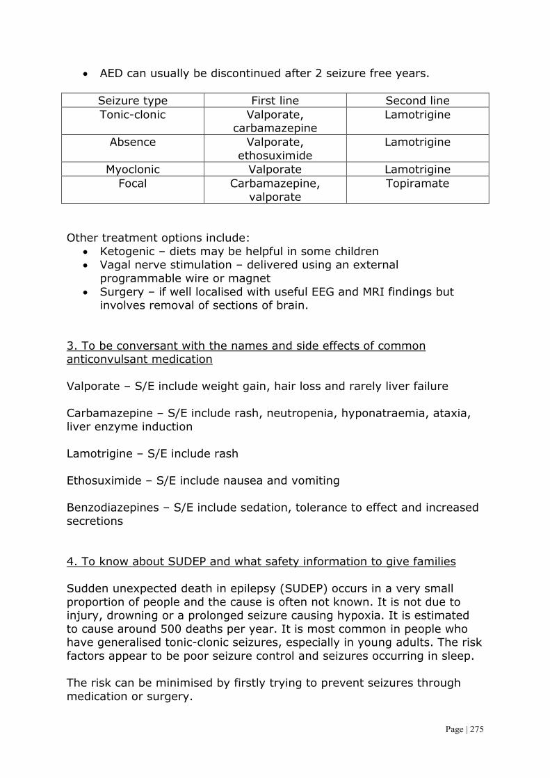
Page | 275
• AED can usually be discontinued after 2 seizure free years.
Seizure type First line Second line Tonic-clonic Valporate,
carbamazepine Lamotrigine
Absence Valporate, ethosuximide
Lamotrigine
Myoclonic Valporate Lamotrigine Focal Carbamazepine,
valporate Topiramate
Other treatment options include:
• Ketogenic – diets may be helpful in some children • Vagal nerve stimulation – delivered using an external
programmable wire or magnet • Surgery – if well localised with useful EEG and MRI findings but
involves removal of sections of brain. 3. To be conversant with the names and side effects of common anticonvulsant medication Valporate – S/E include weight gain, hair loss and rarely liver failure Carbamazepine – S/E include rash, neutropenia, hyponatraemia, ataxia, liver enzyme induction Lamotrigine – S/E include rash Ethosuximide – S/E include nausea and vomiting Benzodiazepines – S/E include sedation, tolerance to effect and increased secretions 4. To know about SUDEP and what safety information to give families Sudden unexpected death in epilepsy (SUDEP) occurs in a very small proportion of people and the cause is often not known. It is not due to injury, drowning or a prolonged seizure causing hypoxia. It is estimated to cause around 500 deaths per year. It is most common in people who have generalised tonic-clonic seizures, especially in young adults. The risk factors appear to be poor seizure control and seizures occurring in sleep. The risk can be minimised by firstly trying to prevent seizures through medication or surgery.

Page | 276
Febrile Convulsion 1. To be able to explain to parents how often these occur and basic first aid advice A febrile seizure is a seizure accompanied by a fever in the absence of intracranial infection due to bacterial meningitis or viral encephalitis. These occur in 3% of children, between the ages of 6 months and 5 years. There is a genetic predisposition, with a 10% risk if the child has a first-degree relative with febrile seizures. The seizure usually occurs early in a viral infection when the temperature is rapidly rising. The seizures are usually brief and are generalised tonic-clonic seizures. About 30-40% will have further febrile seizures. This is more likely the younger the child, the shorter the duration of illness before seizure (the first time), the lower the temperature at the time of seizure and if this is a positive family history. Simple febrile seizures do not cause brain damage and the child’s subsequent intellectual performance is the same as in children who do not experience a febrile seizure. There is a 1-2% chance of developing epilepsy, similar to the risk of all children. However complex febrile seizures, i.e. those which are focal, prolonged or repeated in the same illness, have an increased risk of 4-12% of subsequent epilepsy. Management is to ensure the cause isn’t something more serious and involves ruling out meningitis. This process also involves informing the parents and providing reassurance. First aid basics for seizure management should be taught and are detailed below. Antipyretics and cold sponges are not recommended as they don’t seem to work. Antiepileptic drugs are not used and an EEG is not indicated. First aid – place them in the recovery position on a soft surface to prevent them aspirating vomit once the seizure is over. Whilst the seizure is occurring the child should be placed in a safe location, away from hard objects that can cause injury. Stay with the child and call for help if the seizure lasts longer than 5 minutes. 2. To know about their relationship with epilepsy See objective 1. Fits/Faints/Funny Turns

Page | 277
1. To be able to distinguish between epileptic attacks, syncopal episodes and other movement disorders These can also be labelled as paroxysmal disorders. The major question is whether a diagnosis of epilepsy is correct or if there is a different cause mimicking it. A clinical diagnosis based on history, examination and EEG should be enough to help differentiate. Causes of funny turns include:
• Breath holding attacks – toddler holds breath whilst crying and can lose consciousness but rapidly recovers
• Reflex anoxic seizures – occurs in infants or toddlers and triggers include pain or discomfort. After the trigger the child becomes pale and falls to the floor, the hypoxia may stimulate a tonic-clonic seizure. These are brief and recovery is complete
• Syncope – children may faint from hot environments or prolonged standing
• Migraine – headaches with unsteadiness or light-headedness • Benign paroxysmal vertigo – vertigo lasting several minutes and
associated with nystagmus and possible falling. Usually due to viral Labyrinthitis
• Cardiac causes – cardiomyopathy or prolonged QT syndrome • Other causes – psuedoseizures, Munchausen’s by proxy, NAI, atonic
epileptic seizures Ataxia 1. To be able to list the common causes of ataxia and know how to investigate them Ataxia describes the incoordination of movement, speech and posture due to either cerebellar (more common in children) or posterior pathway problems. In cerebellar ataxis there is an unsteady wide-based gait (truncal ataxis), dysdiadochokinesis, overshooting of target directed movement (dysmetria) and an intention tremor. There may also be a scanning dysarthria (speech problem), positive Romberg’s test and nystagmus. Causes of cerebellar ataxis include:
• Acute causes – medications, drugs, alcohol, solvents, trauma • Post viral – seen with varicella infection • Posterior fossa lesions – CPA syndrome • Genetic and degenerative disorders (see below)
The genetic and degenerative disorders are:
• Ataxic cerebral palsy • Friedreich’s ataxia – this is an autosomal recessive condition. It
presents with worsening ataxia, distal wasting in the legs, absent lower limb reflexes but extensor plantar responses because of pyramidal involvement, pes cavus (high arch) and dysarthria. This

Page | 278
is similar to hereditary motor sensory neuropathies but with FA there is impairment of joint position and vibration sense, extensor plantars and there is often optic atrophy. The cerebellar component will become more apparent with age. Evolving kyphoscoliosis and cardiomyopathy can cause cardiorespiratory compromise and death at 40-50 years.
• Ataxia telangiectasia – this disorder is of DNA repair and is an autosomal recessive condition. There may be a mild delay in motor development in infancy and oculomotor problems with incoordination and delay in ocular pursuit of objects, with difficulty with balance and coordination becoming evident at school age. There is subsequent deterioration with a mixture of dystonia and cerebellar signs. Many children require a wheel chair for mobility. Telangiectasia develops in the conjunctiva, neck and shoulders from about 4 years. These children are more susceptible to infection (IgA defect), develop malignant disorders, have raised alpha-fetoprotein and have increased white cell sensitivity to radiation.
Investigations include genetic testing, lumbar puncture and brain scans to check for tumours or damage. Brain Tumours 1. To be aware of the presenting features of brain tumours Brain tumours are almost always primary tumours in children and 60% are infratentorial (cerebellar and brainstem). They are the most common solid tumour in children and are the leading cause of childhood cancer deaths in the UK. The types of brain tumour and their presentation are:
• Astrocytoma (40%) – varies from benign to highly malignant and often occurs in the cerebral hemispheres. Symptoms include seizures, headaches and focal neurological signs. Outlook is generally poor.
• Medulloblastoma (20%) – arises in the midline of the posterior fossa and may seed through the CNS via the CSF. Up to 20% will have spinal metastases at diagnosis. Symptoms include truncal ataxia, coordination difficulties, abnormal eye movements and morning vomiting. Treatment is surgical followed by total body irradiation. Survival at 5 years is 50%
• Ependymoma (8%) – mostly in the posterior fossa where it behaves like medulloblastoma.
• Brain stem gliomas (6%) – commoner in early childhood and presents with signs of cranial nerve defects, pyramidal tract signs, ataxia and often no increased ICP. Prognosis is poor with only a 20% survival

Page | 279
• Craniopharyngioma (4%) – a developmental tumour arising from the squamous remnant of Rathke pouch. It is not truly malignant but is locally invasive and grows slowly in the suprasellar region. Signs include visual field loss and bitemporal hemianopia, pituitary failure (growth failure, weight gain and diabetes insipidus).
Signs of raised ICP in children and adolescents include:
• Headache – worse in the morning • Vomiting – especially on waking • Behaviour/personality change • Visual disturbance • Papilloedema
And in young infants: • Vomiting • Separation of sutures/tense fontanelle • Increased head circumference • Head tilt/posturing • Developmental delay/regression
Breath holding 1. To be able to recognise the history of a child with this condition Breath holding spells are brief periods when a young child stops breathing for up to a minute. These spells often cause the child to pass out (lose consciousness). Breath holding usually occurs when a child is angry, frustrated, in pain or afraid. But the spell is a reflex and is usually not a deliberate act. Breath holding can be classified as:
• Cyanotic – the most common type and occurs in response to anger or frustration. A child’s skin typically turns red or blue-purple
• Pallid – a pale appearance to the child’s skin in response to fear, pain or injury, especially after head trauma.
Breath holding occur in children between 6 months and 6 years but is most common between 1 and 3 years. Some children may have one spell a year whilst others have several a day. These are not serious and should not cause any serious damage. They should eventually resolve. Symptoms of a cyanotic spell are:
• A short burst of rigorous crying lasting less than 30 seconds • Hyperventilation • A pause in breathing after exhaling • Red or blue skin and lips • Seizures may occur
Developmental Regression

Page | 280
1. To know the common causes of regression e.g. Battens disease, Retts, Leukodystrophies, Wilson’s and SSPE Developmental regression is different to developmental delay as a child loses skills that they have previously acquired rather than never acquiring them. Battens disease is a rare, fatal autosomal recessive neurodegenerative disorder that begins in childhood. Symptoms occur around 4-10 years with a gradual onset of visual problems and seizures. This progresses to a change in behaviour, speech and a regression in learning. There may be a slow in growth and breath holding attacks. Eventually function will deteriorate to dementia and death. Rett’s syndrome is a pervasive developmental disorder mentioned above in the genetic disorders section Leukodystrophies refers to a group of conditions characterised by dysfunction of the white matter of the brain. The cause is incorrect growth of the myelin sheath. Symptoms include a gradual decline in an infant/child who was previously doing well, progressive loss of movement, speech, vision, hearing and behaviour. Wilson’s disease is an autosomal recessive disorder with an incidence of 1 in 200,000. The general result of the condition is a reduced synthesis of copper binding protein as well as defective excretion of copper in the bile which leads to an accumulation of copper in the liver, brain, kidney and cornea. Wilson’s disease rarely presents in children under 3 and can present with almost any form of liver disease including hepatitis, (fulminant or acute), cirrhosis and portal hypertension. Neuropsychiatric features are more common after the second decade and include deterioration in school performance, mood, behaviour and coordination. SSPE (subacute sclerosis panencephalitis) is a rare, chronic, progressive encephalitis caused by a persistent infection of immune resistant measles virus. The history is a primary infection before the age of 2 and then 6-15 asymptomatic years before gradual psychoneurological deterioration. Hydrocephalus 1. To understand the difference between obstructive, communicating and external hydrocephalus In hydrocephalus there is an obstruction to the flow of CSF leading to dilatation of the ventricular system proximal to the site of obstruction.

Page | 281
• Obstructive – the obstruction may be within the ventricular system or aqueduct
• Communicating – the obstruction may be at the arachnoid villi, the site of absorption for CSF (but can also be due to CSF overproduction or venous drainage insufficiency)
• External – this is a benign condition with a self-limiting absorption deficiency of infancy and early childhood that leads to increased ICP. It is thought to be due to an immaturity of arachnoid villi not absorbing fast enough.
Clinical features as a disproportionately large head circumference or excessive rate of head growth (due to failure of suture formation). When the skull sutures separate the anterior fontanelle bulges and the scalp veins become distended. An advanced sign is a fixed downwards gaze or sun setting of the eyes. Older children with develop signs of increased ICP (see brain tumour section). Macrocephaly 1. To know the common causes Macrocephaly is a head circumference above the 98th centile. Most are normal children and often have parents with big heads. A rapidly increasing head circumference, even if below the 98th centile, suggests rapidly increasing ICP and may be due to hydrocephalus, subdural haematoma or brain tumour. It must be investigated promptly by intracranial ultrasound if the anterior fontanelle is still open, otherwise by CT or MRI scan. Common causes of a large head area:
• Tall stature • Familial macrocephaly • Raised intracranial pressure • Hydrocephalus (progressive or arrested) • Chronic subdural haematoma • Cerebral tumour • Neurofibromatosis • Cerebral gigantism (Sotos syndrome) • CNS storage disorders e.g. mucopolysaccharidosis (hurler
syndrome) Microcephaly 1. To know the common causes Microcephaly is a head circumference below the 2nd centile and may be:

Page | 282
• Familial – when it is present from birth and development is often normal
• An autosomal recessive condition – when it is associated with developmental delay
• Caused by congenital infection • Acquired after an insult to the developing brain, e.g. perinatal
hypoxia, hypoglycaemia or meningitis, when it is often accompanied by cerebral palsy and seizures.
Migraine/Headache 1. To be able to distinguish between migraine and tension headaches Headache is a frequent reason for consultation. Headaches can be classified as:
• Primary – migraines, tension-type headaches, cluster headaches and other primary headaches. These are thought to be due to primary malfunction of neurones
• Secondary – symptomatic of some underlying pathology • Trigeminal and other cranial neuralgias – including nerve root pain
Tension-type Headache A symmetrical headache with gradual onset often described as a bilateral tight band-like pain. There are usually no other symptoms but may be accompanied by abdominal pain and behavioural problems and occur daily. Migraine Without aura – this accounts for 90% of migraines and in children the episodes may last 1-72 hours. The headache is commonly bilateral but may be unilateral. Characteristically pulsatile, over temporal or frontal areas and is accompanied by unpleasant GI disturbances such as nausea, vomiting and abdominal pain and photophobia or phonophobia. It can be aggravated by physical activity. With aura – accounts for 10% of migraines. This headache is preceded by an aura (visual, sensory or motor), although the aura may occur without the headache. Features are the absence of problems between attacks and the frequent presence of premonitory symptoms (tiredness, difficulty concentrating, autonomic features etc). The most common auras involve visual disturbances and may include:
• Negative phenomena, such as hemianopia or scotoma • Positive phenomena such as fortification spectra (zigzag lines)

Page | 283
Episodes usually last for a few hours, during which the children often prefer to lie down in a quiet, dark room and sleep (relieves the bout). Symptoms of tension-type headaches and migraines often overlap and are probably a continuum of the same disorder. There is a genetic predisposition to these in some people. Raised Intracranial Pressure 1. To know what features are suggestive of raised ICP and be aware of treatment options The features of raised ICP for both adults/children and infants are listed in the brain tumours section. Treatment really depends upon the cause.
• Tumour – consider prompt removal surgically or radio/chemotherapy
• Subdural haematoma – surgical drainage • Hydrocephalus – ventriculoperitoneal shunt • If idiopathic then specific diuretics can be prescribed by the
neurologist. Hypoventilation of the patient can temporarily decreased ICP by decreased CO2 and causing vasoconstriction
• Craniotomy may be performed if other procedures have not worked Reflex Anoxic Seizures 1. To know how this presents Typically occur in infants or toddlers. They may have a first degree relative with a history of faints. The commonest triggers are pain or discomfort, particularly minor head trauma, cold food (e.g. ice cream or cold drinks), fright or fever. Some children with febrile seizures may have experienced this phenomenon. After the triggering event the child becomes very pale and falls to the floor. The hypoxia may induce a generalised tonic-clonic seizure. The episodes are due to cardiac asystole from vagal inhibition. The seizure is brief and the child rapidly recovers. Ocular compression under controlled conditions often leads to asystole and paroxysmal slow wave discharge on the EEG. Subdural Haematoma 1. To be aware of the causes of this and to be able to recognise the symptoms This results from tearing of the veins as they cross the subdural space. It is a characteristic lesion in non-accidental injury caused by shaking or

Page | 284
direct trauma in infants or toddlers. Retinal haemorrhages are usually present. Subdural haematomas are occasionally seen following a fall from a considerable height. Symptoms include an altered mental state, seizures, apnoea, breathing difficulties, headaches, lethargy or sudden cardiac arrest. Craniosynostosis 1. To understand how these conditions can occur and the way they may present The sutures of the skull bones start to fuse during infancy but do not finally fuse until late childhood. Premature fusion of one or more sutures (Craniosynostosis) may lead to distortion of the head shape. Craniosynostosis is usually localised (sagital suture – long narrow skull, coronal suture – asymmetrical skull, and lambdoid suture – flattening of skull). It most often affects the sagital suture when it results in a low, narrow skull. Rarely it affects the lambdoid suture to result in skull asymmetry which needs to be differentiated from plagiocephaly, where there is asymmetrical flattening of one side of the skull from positional moulding. Craniosynostosis may be generalised when it may be a feature of a syndrome (Crouzon syndrome). The fused sutures may be felt or seen as a palpable ridge and confirmed on skull x-ray or cranial CT. If necessary the condition can be treated surgically because of raised ICP or for cosmetic reasons. Myotonic Dystrophy 1. Be aware of the features of this condition Myotonia is delayed relaxation after sustained muscle contraction. It can be identified clinically and on electromyography. Myotonic dystrophy is a relatively common autosomal dominant condition of triplet repeat extensions. There is a correlation between the number of repeats and with severity and onset. This is a progressive condition with onset between 20 and 50 years of age generally. Signs and symptoms include a child with poor feeding, failure to meet milestones and Hypotonia. There is progressive distal muscular weakness, ptosis, weakness and thinning of the face and sternocleidomastoids along with the ‘carp mouth’. Other features of this syndrome include cataracts, frontal balding, mild cognitive impairment, oesophageal dysfunction, cardiomyopathy (main cause of death) and conductive defects, small

Page | 285
pituitary fossa and hypogonadism, glucose intolerance and low serum IgG. Look for myotonia (a slow relaxation of muscles, classically seen with difficulty releasing one’s hand on shaking it) in the mother. Myopathy 1. To be aware of the features of Duchenne Muscular Dystrophy and Congenital Muscular Dystrophies The DMD section covers everything important with regards to this. Congenital muscular dystrophies are a heterogeneous group of disorders, most with recessive inheritance, which present at birth or early infancy with weakness, Hypotonia or contractures. Typically the proximal weakness is slowly progressive with a tendency to contracture when the ability to walk is lost. Some may run a more static course. Biopsy shows dystrophic features with a reduction of one of the extracellular matrix proteins such as laminin (most common); or one of several glycosyltransferases. These dystrophies may be linked with central nervous abnormalities, which may result in learning difficulties. The main difference that I can see between these and DMD is the fact that these present at birth and tend to have a more variable and longer life expectancy. 2. To know about the treatment of DMD i.e. steroids, cardiac drugs and nocturnal ventilation See the DMD objective. Neuropathy 1. To know about acute conditions e.g. Guillain-Barre, and also chronic neuropathies e.g. Charcot Marie Tooth, CIDP Guillain-Barre syndrome Presentation is typically 2-3 weeks after a URTI or campylobacter gastroenteritis. There may be fleeting abnormal sensory symptoms in the legs, but the prominent features is an ascending symmetrical weakness with loss of reflexes and autonomic involvement. Sensory symptoms, usually in the distal limbs, are less striking than the paresis but can be unpleasant. Involvement of bulbar muscles leads to difficulty chewing and swallowing and the risk of aspiration. Respiratory depression may require artificial ventilation. The maximum muscle weakness may occur only 204

Page | 286
weeks after the onset of illness. Although full recovery may be expected in 95% of cases, this may take up to 2 years. CSF protein is raised after 2 weeks but white cells are negative, there is also a reduction in nerve conduction velocities. Management is supportive, particularly of respiration. This disorder is probably due to the formation of antibody attaching itself to protein components of myelin. Corticosteroids have no beneficial effect and may even delay recovery. Ventilator supported periods can be significantly reduced by IVIG or plasma exchange. Bell Palsy This is an isolated lower motor neurone paresis of the 7th cranial nerve leading to facial weakness. Although the aetiology is unclear, it is probably post-infectious with an association with HSV in adults. Corticosteroids may be of value in reducing oedema in the facial canal during the first week but acyclovir has shown no benefit. Recovery is complete in the majority of places but can take several months. The main complication is conjunctival infection due to incomplete eye closure on blinking. If an 8th nerve palsy is also present then this may be a compressive lesion at the CPA. Hypertension should also be excluded as there is an association between Bell palsy and coarctation of the aorta. If bilateral then suspect sarcoidosis or Lyme disease. Charcot Marie Tooth This involves distal muscle wasting and sensor loss with proximal progression over time. It is usually autosomal dominant (but can be recessive) and may occur without family history. Onset is usually by the age of 10 years with:
• Muscle weakness and wasting starting with the intrinsic muscles of the feet and gradually affecting the lower legs and thighs. Sensory loss is similar and will lead to ataxia. Pain and temperature sensation are not usually affected
• Generalised tendon areflexia • There may be foot drop and difficulty walking • Spinal deformities occur in 50% e.g. thoracic scoliosis • Other common signs and symptoms are hand tremors, muscle
cramps and acrocyanosis (blue extremities). There are currently no effective treatments to stop or slow progression so treatment is primarily supportive. Most patients have a normal life expectancy. CIDP (Chronic inflammatory Demyelinating Polyneuropathy) This is an acquired, immune mediated inflammatory disorder of the PNS. This is related to Guillain-Barre syndrome and is thought as the chronic

Page | 287
version of this disease. This includes relapsing symptoms that present and then go. Both proximal and distal limbs are affected with a sense of weakness. Sensory affects include tingling and numbness but motor symptoms generally predominate. Deep tendon reflexes are reduced and gait is abnormal. It is most often idiopathic in origin but has links to several other diseases including MS and SLE. Spinal Muscular Atrophy 1. To be aware of the presentation of the different types Spinal muscular atrophy is an autosomal recessive degeneration of the anterior horn cells, leading to progressive weakness and wasting of skeletal muscles due to mutations in the survival motor neurone (SMN) gene. This is the second most common cause of neuromuscular disease in the UK after DMD. A number of phenotypes are recognised: Spinal muscular atrophy type 1 (Werdnig-Hoffmann disease) is a very severe progressive disorder presenting in early infancy. Diminished fetal movements are often noticed in pregnancy and there may be arthrogryposis (positional deformities of the limbs with contractures of at least two joints) at birth. Typical signs include:
• Lack of antigravity power in hip flexors • Absent deep tendon reflexes • Intercostal recession • Fasciculation of the tongue
These children never sit unaided. Death is from respiratory failure within about 12 months. There are milder forms of this disorder with a later onset. Children with type 2 spinal muscular atrophy can sit, but never walk independently. Those with type 3 (Kugelberg-Welander) do walk and can present later in life. Oncology and Haematology Acute Lymphoblastic Leukaemia 1. Be able to describe the epidemiological risk for malignancy in childhood and adolescence, demonstrate knowledge of the age incidence profiles of different malignancies Childhood malignancy affects 1 in 500 by 15 years of age. It occurs in 1500 people in the UK each year. The distribution of these cancers is as follows:

Page | 288
• Leukaemia – 32% • Brain and spinal tumours – 24% • Lymphomas – 10% • Neuroblastoma – 7% • Soft tissue sarcomas – 7% • Wilms tumours – 6% • Bone tumour – 4% • Retinoblastoma – 3% • Others – 7%
As with regards to the age incidence profile, it is easier to discuss this within each individual malignancy. A general guide is that leukaemia’s affect children at all ages (although there is an early childhood peak), Neuroblastoma and Wilms tumour are almost always seen in the first 6 years of life, and Hodgkin Lymphoma and bone tumours have their peak incidence in adolescence and early life. 2. Be able to describe the common presenting symptoms and signs ALL accounts for 80% of leukaemia in children and is excess proliferation of lymphocytes. A large number of immature lymphocytes are produced rapidly so this condition can progress quickly. Clinical presentation peaks at 2-5 years and the signs and symptoms results from disseminated disease and systemic ill health from infiltration of the bone marrow or other organs with leukaemic blast cells. In most children leukaemia presents insidiously over several weeks but can progress very quickly. The signs and symptoms can be classified according to their cause:
• General – malaise and anorexia • Bone marrow infiltration – anaemia (lethargy), neutropenia
(infection), thrombocytopenia (bruising, petechiae, nose bleeds) and bone pain
• Reticulo-endothelial infiltration – hepatosplenomegaly, lymphadenopathy and uncommonly superior mediastinal obstruction
• Other organ infiltration – CNS (nerve palsies, headaches, vomiting) and testes (testicular enlargement)
3. The approaches to establishing a diagnosis and initial management aimed at preserving life with respect to transfusion of blood and platelets and the risk of metabolic and clotting abnormalities Investigations should include a FBC. In most children this will be abnormal with a low haemoglobin, thrombocytopenia and evidence of circulating leukaemic blast cells. Bone marrow examination is essential to confirm the diagnosis and to identify immunological and cytogenetic

Page | 289
characteristics which give useful prognostic information. A chest x-ray is requires to identify mediastinal masses, characteristic of T-cell disease. The following is a treatment schema for standard-risk acute lymphoblastic leukaemia:
Weeks of Treatment Stage Treatment Diagnosis Induction Vincristine. Steroids, IT
methotrexate and L-Asparaginase
5-8 weeks Consolidation and CNS protection
IT methotrexate, vincristine, steroid, thiopurine
8-16 weeks Interim maintenance Monthly vincristine and pulsed 5 day steroids. Daily 6-mercaptopurine. Weelly
oral methotrexate, IT methotrexate and
prophylactic co-trimoxazole 16-23 weeks Delayed intensification Vincristine, IT methotrexate,
dexamethasone plus others 23 weeks -2/3 years Continuing maintenance Same as interim maintenace
Blood transfusions of both platelets and whole red cells are used to reduce symptoms rather than cure the patient. In ALL, patients may have low platelets leading to bruising and bleeding and hence a platelet transfusion can help reduce this. The patient may also be anaemic so red cells will reduce their breathlessness. Before and during the initial induction phase of chemotherapy patients may develop tumour lysis syndrome which refers to the metabolic derangements cause by the systemic and rapidly release of intracellular contents as chemotherapy destroys leukaemic blast cells. Effects are hyperuricaemia, hyperphosphataemia, hypocalcaemia and hyperkalaemia. To prevent complications electrolyte and uric acid levels should be monitored along with IV fluid therapy. Allopurinol may also be given. 4. Be aware of the treatment regimes available and the theory of their actions Remission induction – before starting treatment of the disease, anaemia may require correction with blood transfusion, the risk of bleeding minimised by transfusion of platelets, and infection must be treated. Addition hydration and allopurinol are given to protect renal function against the effects of rapid cell lysis. Remission implies eradication of the leukaemic blast cells and restoration of normal marrow function. Four
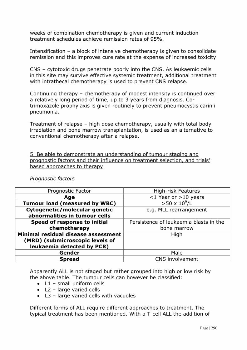
Page | 290
weeks of combination chemotherapy is given and current induction treatment schedules achieve remission rates of 95%. Intensification – a block of intensive chemotherapy is given to consolidate remission and this improves cure rate at the expense of increased toxicity CNS – cytotoxic drugs penetrate poorly into the CNS. As leukaemic cells in this site may survive effective systemic treatment, additional treatment with intrathecal chemotherapy is used to prevent CNS relapse. Continuing therapy – chemotherapy of modest intensity is continued over a relatively long period of time, up to 3 years from diagnosis. Co-trimoxazole prophylaxis is given routinely to prevent pneumocystis carinii pneumonia. Treatment of relapse – high dose chemotherapy, usually with total body irradiation and bone marrow transplantation, is used as an alternative to conventional chemotherapy after a relapse. 5. Be able to demonstrate an understanding of tumour staging and prognostic factors and their influence on treatment selection, and trials’ based approaches to therapy Prognostic factors
Prognostic Factor High-risk Features Age <1 Year or >10 years
Tumour load (measured by WBC) >50 x 109/L Cytogenetic/molecular genetic abnormalities in tumour cells
e.g. MLL rearrangement
Speed of response to initial chemotherapy
Persistence of leukaemia blasts in the bone marrow
Minimal residual disease assessment (MRD) (submicroscopic levels of
leukaemia detected by PCR)
High
Gender Male Spread CNS involvement
Apparently ALL is not staged but rather grouped into high or low risk by the above table. The tumour cells can however be classified:
• L1 – small uniform cells • L2 – large varied cells • L3 – large varied cells with vacuoles
Different forms of ALL require different approaches to treatment. The typical treatment has been mentioned. With a T-cell ALL the addition of

Page | 291
cyclophosphamide and intensive treatment with asparaginase is beneficial. Mature B-cell needs treating like a lymphoma with short-term intensive chemotherapy including high dose methotrexate. Iron Deficiency Anaemia 1. Know the normal physiological changes affecting blood count data from neonate to adolescent of all cell types (red, white and platelets) Haemopoiesis is the process which maintains lifelong production of haemopoietic blood cells. The main site of haemopoiesis in fetal life is the liver, whereas throughout postnatal life it is the bone marrow. All haemopoietic cells are derived from pluripotent stem cells which are crucial for normal blood production. The most important difference between fetal and postnatal life, in regards to haemopoiesis, is the changing pattern of haemoglobin. Fetal haemoglobin (HbF) is made of two alpha units and two gamma units and has a higher affinity for oxygen then adult haemoglobin HbA which is made of two alpha units and two beta units. HbF is gradually replaced by HbA in the first year of life and by one the percentage of remaining HbF is very low. At birth the Hb in term infants is high, 14-21.5 g/dl, to compensate for the low oxygen concentration in the fetus. The Hb falls over the first few weeks, mainly due to red cell production, reaching a value of around 10 g/dl at 2 months of age. Stores of iron, folic acid and vitamin B12 in term and preterm infants are adequate at birth. However these stores are lower in preterm infants so are quickly depleted in the first few months of life. White blood cell counts in neonates are higher (10-20 x 109/l compared with 4.5-13) than in older children but platelet count is similar to that of an adult (150-450 x 109/l). Anaemia is defined as an Hb level below the normal range and as these ranges vary with age the anaemia can be defined as:
• Neonate: Hb <14 • 1-12 months: Hb <10 • 1-12 years: Hb <11
Anaemia may result from reduced red cell production, increased red cell destruction or blood loss. 2. Be able to explain the reasons for the physiological changes in haemoglobin concentration with respect to growth, development and nutrition from neonatal period through to adulthood See above

Page | 292
3. Understand the importance of dietary factors affecting blood composition including iron, folic acid and vitamin B12 Iron deficient anaemia may be caused by inadequate intake, malabsorption or blood loss (rare). It is common in infants because addition iron is required for the increase in blood volume accompanying growth and to build up the child’s iron stores. Iron can come from breast milk (50% absorbed – by far the best source), formula, cow’s milk or solids. Iron deficiency may develop due to a delay in weaning beyond 6 months. Iron is best absorbed with vitamin c and without tannin from tea (or red wine!). Clinical features are usually not present until below 6-7 g/dl at which point the child will tire easily and feed more slowly than usual. They may appear pale but this is an unreliable sign unless confirmed by the conjunctiva, tongue or palmar creases. Some children have pica which is the inappropriate eating of non-food materials. Most management is dietary advice and oral supplementation if needed. If there is still not a gain then malabsorption should be investigated. The need of a blood transplant is incredibly rare. Folate is vital as it provides the constituents to produce red cells and without it then the body cannot make enough cells so a macrocytic megaloblastic anaemia occurs. B12 is also vital for DNA synthesis so will have a similar effect if there is a deficiency. 4. Be able to explain the changes in lymphocytes and neutrophil counts from neonatal period to adulthood and how they might be used to detect infection, immunodeficiency states and malignant disorders of the blood As mentioned above the counts will drop slightly with age. If they are below the reference values then there may be immunodeficiency. If they are above reference values then there may be an infection and there can be a malignant disorder of the blood with either of these presentations but it is generally a drop in white cells. ITP/Thrombocytopenia 1. Be able to describe the common ways that bleeding disorders present in childhood as congenital (e.g. congenital ITP, haemophilia) or acquired conditions (e.g. ITP or disseminated intra-vascular coagulation)

Page | 293
Thrombocytopenia is a platelet count <150 x 109/L. The risk of bleeding depends on the platelet level below this. At <20 the risk of bleeding spontaneously is high. At 20-50 the risk of excess bleeding during trauma or surgery is increased. At 50-150 there is a low risk of bleeding unless there is a major operation or serious trauma. This condition may result in bruising, petechiae, purpura and mucosal bleeding (epistaxis, bleeding gums etc). Major haemorrhage in the form of GI bleeds or intracranial bleeds are much less common. Haemophilia – will be covered in its own objective Immune thrombocytopenia (ITP) This is a condition that can be both congenital and acquired. It is the commonest form of thrombocytopenia in children with an incidence of 4 in 100,000 children per year. It is usually caused by the destruction of circulating platelets by anti-platelet IgG autoantibodies. The reduce platelet count may be accompanied by a compensatory increase in megakaryocytes in the bone marrow. Most children present between the ages of 2 and 10 years with onset often 1-2 weeks after viral infection. In the majority of children there is a short history of days or weeks. Affected children develop petechiae, purpura and/or superficial bruising. It can cause epistaxis and other mucosal bleeding but profuse bleeding is uncommon, despite the fact that the platelet count often falls below 10. Intracranial bleeding is a rare complication but serious, affected 0.1-0.5%. ITP is a diagnosis of exclusion and can also be caused by a congenital syndrome in children. Any atypical features such as anaemia, neutropenia, hepatosplenomegaly or marked lymphadenopathy should prompt a bone marrow investigation to exclude acute leukaemia or aplastic anaemia. SLE should also be considered. However if features are all typical then a bone marrow examination is not needed. In about 80% of children the disease is acute, benign and self-limiting, usually remitting spontaneously within 6-8 weeks. Most children can be managed at home and do not require admission. However treatment should be given if there is evidence of major bleeding or persistent minor bleeding. Treatment options include oral prednisolone, IV anti-D or IVIG. Platelet transfusions are reserved for life threatening haemorrhage as they only work for a few hours. Chronic IPT occurs in 20% and is where the platelet count remains low for over 6 months. Mostly supportive treatment is given unless bleeding is excessive. Again SLE should be screened for and finally a splenectomy can help if all else fails.

Page | 294
Disseminated intravascular coagulation See below section on DIC 2. Be able to outline the principles of acute and chronic management of bleeding disorders of childhood dependent upon their cause The management of most bleeding disorders (both acute and chronic) has been mentioned above or in the haemophilia section below. However I will mention von Willebrand disease (vWD) and how to differentiate the conditions from each other using the history. Von Willebrand disease Von Willebrand disease results from either a quantitative or qualitative deficiency in von Willebrand factor (responsible for platelet adhesion and as a carrier protein for factor VIII). This causes a defective plug formation and since vWF is a carrier protein for factor eight, patients with vWD also are deficient for this factor. There are many different mutations and the inheritance is usually dominant. The commonest subtype, type 1 (60-80%), is usually fairly mild and is often not diagnosed until puberty or adulthood. Clinical features are bruising, prolonged bleeding after surgery and mucosal bleeding. Spontaneous soft tissue bleeding is uncommon. Treatment depends on severity but can be treated with DDAVP which causes secretion of both factor eight and vWF into the plasma. Use in caution in under 1 year as it can cause hyponatraemia and seizures if fluid intake is not adequate. More severe forms need treating with plasma derived factor eight. IM injections, aspirin and NSAIDs should all be avoided. Acquired disorders of coagulation The main acquired conditions are haemorrhagic disease of the newborn (vitamin K deficiency), liver disease, ITP and DIC. Inadequate intake, malabsorption or vitamin K antagonists can all be a cause. Differentiating Age of onset
• Neonate – 20% of haemophilias present here • Toddler – haemophilias may present when starting to walk • Adolescent – vWD with menorrhagia
Family history
• If all boys then suggests haemophilia
Page | 295
Bleeding history • If bleeding is ok in some situations then it suggests a bleeding
tendency rather than an inherited disorder. • Drug history • NAI
Pattern of bleeding
• Mucous membranes and skin – platelet disorders or vWD • Bleeding into muscles or joints – haemophilia • Scarring and delayed haemorrhage – suggestive of disorders of
connective tissue 3. Offer children (or their carers) with low platelet counts health advice relating to their condition See above for specific information but it is generally to avoid injury, know the available treatments and have an action plan if severe haemorrhage occurs. Haemolytic Anaemia 1. Be able to describe the common presentation of haemolysis during childhood and the selection of tests to identify haemolysis as a phenomenon In haemolytic anaemia there is a reduced red cell lifespan due to increased intravascular and extravascular (spleen and liver) destruction of RBC, there is shortened RBC lifespan and when the bone marrow is unable to compensate any further there is anaemia. The signs of anaemia are reticuloendothelial hyperplasia (leading to hepatosplenomegaly), unconjugated bilirubin increase and increased urinary urobilinogen. In neonates autoimmune haemolytic anaemia is common but in children the most common cause are red cell membrane disorders, red cell enzyme disorders and haemoglobinopathies. Hereditary spherocytosis 1 in 5000 live births with 25% spontaneous and 75% autosomal dominant. The mutation in the genes encoding RBC skeletal proteins causes the RBC to lose some of its membrane as it goes through the spleen. The cell becomes spheroidal and is destroyed in the microvasculature of the spleen.
• Signs and symptoms – jaundice, anaemia, splenomegaly, gallstones (due to increased bilirubin) and may present with aplastic crisis
• Investigations – a blood film

Page | 296
• Management – folate supplementation in mild disease. In severe forms a splenectomy should be considered.
G6PD deficiency Commonest red cell enzyme problem worldwide and has a high prevalence in those from central Africa. It is an x-linked condition but female carriers have about 50% function and are clinically normal. G6PD usually stops oxidative damage to the cell.
• Signs and symptoms – neonatal jaundice in the first 3 days or with acute haemolysis precipitated by infection, drugs, broad beans etc. There may also be fever, malaise and haemoglobin in the urine.
• Investigations – Between episodes of haemolysis the patient is normal. Measurement of the G6DP activity in cells is diagnostic.
• Management – awareness to avoid certain drugs and foods. In some acute instances exchange transfusion may be needed.
Pyruvate Kinase Deficiency 2nd most common cause of haemolytic anaemia and is due to a lack of an enzyme vital in the final stage of glycolysis leading to a decrease in ATP. This causes the cell to become more rigid and be destroyed in the spleen. Thalassaemia and Sickle cell disease will be talked about in their own objectives Haemophilia 1. Understand the genetics, presenting features and management This is the commonest severe inherited coagulation disorder and consists of haemophilia A and B. Both have x-linked recessive inheritance. In haemophilia A there is a factor 8 deficiency with a frequency of 1 in 5000, and in haemophilia B there is a factor 9 deficiency with a frequency of 1 in 30,000 male births. Two-thirds have a family history. The disorder can be graded into mild, moderate or severe depending on the factor percentage (<1% severe, 1-5% moderate and >5-40% mild). The hallmark is recurrent spontaneous bleeding into joints and muscles which can lead to crippling arthritis if not properly treated. Most cases present at the end of the first year of life when walking starts. Almost 40% present in the neonatal period with intracranial haemorrhage, oozing from the heel prick or post circumcision. Treatment is recombinant factor 8 or 9 depending on type A or B. It is given my prompt IV infusion whenever there is bleeding. Raising the level

Page | 297
to 30% is sufficient to treat minor bleeds, but it needs to be 100% for surgery and maintained at 30-50% for 2 weeks after. Injections need to be given every 12 hours or by infusion. Parents and children can be taught to administer at home. Prophylactic factor 8 has been shown to be beneficial in children by giving better joint function in later life. Desmopressin may allow for mild haemophilia (stimulates factor 8 release) to be managed without the use of blood products. Specialised physiotherapy support is needed to preserve muscle strength and avoid damage from immobilisation. Lymphoma 1. Describe the organ/system of origin, the typical signs and symptoms and common clinical associations Lymphoma is the neoplastic proliferation of cells in the lymphatic system such as nodes, spleen and liver. They form solid tumours and can be divided into Hodgkin’s and non-Hodgkin’s lymphoma. NHL is more common in childhood whereas Hodgkin’s disease is more common in adolescence. Non-Hodgkin’s lymphoma predominately affects the lymph nodes. It typically affects younger children. Presentation depends on the type of disease, most T-cell malignancies can present as either NHL or ALL (thought to be a spectrum of disease) characterised by mediastinal mass (may cause superior vena cava obstruction) and bone marrow infiltration. B cell malignancies present more commonly as NHL with localised lymph node disease in the head, neck or abdomen (pain and intussusception). Treatment is multi-agent chemotherapy. The majority of patients do well and survival is over 80% for both T and B cell subtypes. Hodgkin’s lymphoma is defined as the presence of reed-sternberg cells. It is uncommon in those who are pre-pubertal and usually presents as painless lymphadenopathy (larger and firmer than benign lymphadenopathy seen in children), frequently in the neck. Clinical presentation is often long and systemic symptoms (anorexia, weight loss etc) are uncommon, even in advanced disease. Combination chemotherapy gives a cure rate of over 80% 2. Outline the main features and differences of Non-Hodgkin’s (NHL T and B cell) and Hodgkin’s lymphoma
• Hodgkin’s lymphoma has reed-sternberg cells • Hodgkin’s lymphoma often starts in the upper body

Page | 298
• Hodgkin’s spreads very slowly and is very receptive to chemotherapy and radiotherapy
3. Be aware of the long term complications of tumour treatment as a consequence of chemotherapy, radiotherapy and surgery including how development, growth, puberty and fertility can be affected. Side effects of chemotherapy include hair loss, anaemia, infection, bruising, sore mouth, nausea, vomiting, mood changes, irritability and weight gain. In the long term it can cause delayed puberty, reduced fertility, reduced growth. Neurotoxicity, hepatotoxicity, renal toxicity, cardiotoxicity, pulmonary toxicity, secondary cancer and psychological effects. Neuroblastoma 1. Describe the organ/system of origin, the typical signs and symptoms and common clinical associations Neuroblastoma and related tumours arise from neural crest tissue in the adrenal medulla and sympathetic nervous tissue. It is a biologically unusual tumour in that spontaneous regression sometimes occurs in very young infants. There is a spectrum of disease from the benign to the highly malignant. Neuroblastomas are most common before the age of 5. At presentation most children have an abdominal mass but the primary tumour can lie anywhere along the sympathetic chain from the neck to the pelvis. Classically the abdominal primary is of adrenal origin but at presentation the tumour mass is often large and complex, crossing the midline and enveloping major blood vessels and lymph nodes. Paravertebral tumours may invade through adjacent intervertebral foramen and cause spinal cord compression. Over the age of 2 years, clinical symptoms are mostly from metastatic disease, particularly bone pain, bone marrow suppression causing weight loss, and malaise. Other common symptoms include a limp and hepatomegaly. Prognosis is poor the metastatic disease and is little over 30%. Sickle Cell Disease 1. Be able to explain how the genetic mutations of haemoglobin lead to the signs and symptoms of homozygous and heterozygous states of sickle cell disease.

Page | 299
This is now the commonest genetic disorder in children in many European countries. Sickle cell is the collective name given to Haemoglobinopathies in which HbS is inherited. The mutation caused leads to a change in an amino acid from glutamine to valine. Sickle cell disease is most common in patients whose parents are black and originate from tropical Africa or the Caribbean. There are three main types of sickle cell disease and the sickle trait:
• Sickle cell anaemia (HbSS) – patients are homozygous for HbS, i.e. virtually all their Hb is HbS. They have small amounts of HbF and no HbA because they have the sickle mutation in both B-globin genes.
• HbSC disease (HbSC) – affected children inherit HbS from one parent and HbC from the other parents (HbC is formed as a result of a different point mutation in B-globin) so they also have no HbA because they have no normal B-globin genes
• Sickle B-thalassaemia – affected children inherit HbS from one parent and B-thalassaemia trait from the other. They have no normal B-globin genes and most patients can make no HbA and therefore have similar symptoms to those with sickle cell anaemia.
• Sickle trait – inheritance of HbS from one parent and a normal B-globin gene from the other parent, so approximately 40% of the haemoglobin is HbS. They do not have sickle cell disease but are carriers. They are asymptomatic and are only identified as a result of blood tests.
In all forms of sickle cell disease, HbS polymerises within red blood cells forming rigid tubular spiral bodies which deform the red cells into a sickle shape. Irreversibly sickled red cells have a reduced life span and may be trapped in the microcirculation, resulting in blood vessel occlusion and therefore ischaemia in an organ or bone. This is exacerbated by low oxygen tension, dehydration and the cold. Clinical manifestations vary, as does severity. HbSS is the most severe form. Clinical features:
• Anaemia – moderate (6-10 g/dl) with clinically detectable jaundice from chronic haemolysis
• Infection – increased susceptibility to pneumococci and H.influenzae. This is due to hyposplenism secondary to microinfarction in the spleen.
• Painful crises – vaso-occlusive crisis causing pain may affect many organs with varying frequency and severity but commonly the hands and feet. The bones of the limbs and spine are most common but this can also affect the chest and produce the need for ventilation. Exposure to cold, dehydration, excessive exercise, stress, hypoxia or infection can all exacerbate symptoms
• Acute anaemia – Sudden drop in haemoglobin from haemolytic crisis (infection), aplastic crisis (parovirus) and sequestration crisis

Page | 300
(sudden splenic or hepatic enlargement, abdominal pain and circulatory collapse from accumulation of sickle cells in the spleen
• Priapism – needs treating promptly or there may be later erectile dysfunction
• Splenomegaly – common in younger children than older Long term problems:
• Short stature and delayed puberty • Stroke and cognitive problems • Adenotonsillar hypertrophy • Cardiac enlargement – from chronic anaemia • Heart failure – from uncorrected anaemia • Renal dysfunction • Pigmented gallstones • Leg ulcers – uncommon in children • Psychosocial problems – education and behavioural difficulties
Management Prophylaxis with full immunisation and daily oral penicillin throughout childhood. Once daily folic acid is needed due to the increased demand. Vaso-occlusive crises should be avoided by avoiding the cold, dehydration, exercising excessively, undue stress or hypoxia. Treatment of an acute crises should be analgesia and good hydration with oxygen if needed. Prognosis varies due to infection and around 3% die during childhood from infection. Around 50% of patients with the most severe form will die before 40. 2. Understand how it presents through population screening, at symptomatic diagnosis and with intercurrent problems during chronic illness management Much of this is mentioned above but I will talk about prenatal diagnosis and screening. Many countries with a high prevalence of haemoglobinopathies, including the UK, perform neonatal screening on dried blood spots (Guthrie test) collected in the first week of life. Early diagnosis of sickle cell disease allows penicillin prophylaxis to be started in early infancy instead of awaiting clinical presentation, possibly due to infection. Prenatal diagnosis can be carried out by chorionic villus sampling at the end of the first trimester if parents wish to choose this option to prevent the birth of an affected child.

Page | 301
Thalassaemia 1. Be able to explain how the genetic mutations of haemoglobin lead to the signs and symptoms of homozygous and heterozygous states of thalassaemia with particular attention to how they present through population screening, at symptomatic diagnosis and with intercurrent problems during chronic illness management B-thalassaemias occur most often in people from the Indian subcontinent, Mediterranean and Middle East. In the UK most affected children are born to parents from the Indian subcontinent. In the past many were born to Greek Cypriots, but this has become uncommon through active genetic counselling within their community. There are two main types of B-thalassaemia, both of which are characterised by a severe reduction in the production of B-globin (and thereby reduction in HbA production). All affected individuals have a severe reduction in B-globin and disease severity depends on the amount of residual HbA and HbF production.
• B-thalassaemia major – this is the most severe form of the disease. HbA (alpha 2, beta 2) cannot be produced because of the abnormal B-globin gene.
• B-thalassaemia intermedia – this form of the disease is milder and of variable severity. The B-globin mutations allow a small amount of HbA and/or a large amount of HbF to be produced.
Clinical features:
• Severe anaemia, which is transfusion dependent from 3-6 months of age, and jaundice
• Failure to thrive/growth failure • Extramedullary haemopoiesis, prevented by regular blood
transfusions. In the absence of regular blood transfusions the patient develops hepatosplenomegaly and bone marrow expansion; the latter leads to the classical facies with maxillary overgrowth and skull bossing.
Management: this is fatal without treatment so all patients are given lifelong monthly blood transfusions. The aim is to keep haemoglobin above 10 g/dl in order to reduce growth failure and prevent bone deformation. Repeat transfusions can cause chronic iron overload which can cause cardiac failure, liver cirrhosis, diabetes, infertility and growth failure. Therefore all patients are treated with iron chelation from the age of 2-3 years. Patients who comply have a 90% chance of reaching 40 years. An alternative option is bone marrow transplant which is curative. This is reserved for patients with a compatible sibling where there is a 95% chance of success.

Page | 302
Prenatal diagnosis: in parents who are both heterozygous there is a 1 in 4 chance of passing this condition to their child and a 50% chance of giving them carrier status. B-thalassaemia trait – heterozygotes are usually asymptomatic. The red cells are hypochromic and microcytic. Anaemia is mild or absent with a disproportionate reduction in MCH and MCV. This can cause confusion with iron deficiency anaemia but here the ferritin stores will be normal. Alpha-thalassaemias involves the alpha-globin gene. Healthy individuals have 4 alpha-globin genes. Having none of these causes alpha-thalassaemia major, it mainly occurs in families of South East Asian origin and presents in mid trimester with fetal hydrops (oedema and ascites), from fetal anaemia, which is always fatal in utero, or within hours of delivery. The only chance of long term survival is intrauterine transfusion until birth and lifelong therapy after. With 3 deletions the anaemia is mild to moderate and with 1 or 2 deletions the disease is asymptomatic. Wilms’ Tumour 1. Describe the organ/system of origin, the typical signs and symptoms and common clinical associations This is also called a nephroblastoma , originates from embryonic renal tissue and is the commonest renal tumour of childhood. Over 80% of patients present before 5 years of age and it is very rarely seen after 10 years of age. Most children present with a large abdominal mass, often found incidentally in an otherwise well child. Other clinical features include abdominal pain, anorexia, anaemia, haematuria and hypertension. Investigations include an ultrasound and/or CT/MR and show a characteristic intrinsic renal mass distorting normal structures. Staging is to assess for distant metastasis (usually to the lung) along with initial tumour resectability and function of the contralateral kidney. In the UK children receive initial chemotherapy followed by delayed nephrectomy. After which the tumour is staged histologically and subsequent treatment is planned according to the surgical and pathological findings. Prognosis is good with more than 80% of all patients being cured. Cure rate is 60% if with metastasis but recurrence carries a poor prognosis. Wilms’ tumour is associated with overgrowth syndromes and trisomy 18.

Page | 303
Bone tumours 1. Be aware of the features of soft tissue sarcomas, osteosarcoma and Ewings tumour Soft Tissue Sarcomas Rhabdomyosarcoma is the most common form of soft tissue sarcoma in childhood. The tumour is thought to originate from primitive mesenchymal tissue and there are a wide variety of primary sites, resulting in varying presentation and prognosis. The head and neck are most commonly involved (40%) causing proptosis, nasal obstruction or blood stained nasal discharge. Genitourinary tumours may involve the bladder, paratesticular structures or the female genitourinary tract. Symptoms include dysuria and urinary obstruction, scrotal mass or blood stained vaginal discharge. Metastatic disease (lung, liver, bone or marrow) is present in around 15% of patient at diagnosis and is associated with a particularly poor prognosis. Biopsy and full radiological assessment of primary disease and any evidence of metastasis is needed. Management is multimodality treatment with chemotherapy, radiotherapy and surgery and varies depending on size, site and extend of disease. Overall cure rate is approximately 65%. Bone tumours Malignant bone tumours are uncommon before puberty. Osteogenic sarcoma is more common than Ewing sarcoma but Ewing sarcoma is seen more often in younger children. Both have a male predominance. The limbs are the most common site. Persistent localised bone pain is the characteristic symptom, usually preceding the detection of a mass, and is an indication for early x-ray. At diagnosis most patients are otherwise well. Plain x-ray is followed by MRI and bone scans. A bone x-ray shows destruction and variable periosteal new bone formation. In Ewing sarcoma there is often a substantial soft tissue mass. Chest CT is used to assess for lung metastases and bone marrow sampling to exclude marrow involvement. In both tumours the treatment involves the use of combination chemotherapy given before surgery. Whenever possible, amputation is avoided by using en bloc resection of tumours with endoprosthetic resection. In Ewing sarcoma radiotherapy is also used in the management

Page | 304
of local disease, especially when surgical resection is impossible or incomplete, e.g. in the pelvis or axial skeleton. DIC 1. Outline the main causes and the clotting abnormalities This describes a disorder of coagulation pathway activation leading to diffuse fibrin deposition in the microvasculature and consumption of coagulation factors and platelets. The commonest cause is severe sepsis or shock due to circulatory collapse e.g. meningococcal septicaemia or extensive tissue damage from trauma or burns. DIC can be acute or chronic. The predominant clinical features are bruising, purpura and haemorrhage. DIC should be suspected when there is thrombocytopenia, prolonged prothrombin time, prolonged APTT, low fibrinogen, raised fibrinogen degradation products and D-dimers and microangiopathic haemolytic anaemia. There is also usually a marked reduction in the naturally occurring anticoagulants, protein C and S and antithrombin. The most important part of management is to treat the underlying cause (usually sepsis) whilst providing intensive care. Supportive care may be provided by fresh frozen plasma, cryoprecipitate and platelets. Antithrombin and protein C concentrates have also been used. Retinoblastoma 1. Be aware of the common presenting features Retinoblastoma is a malignant tumour of the retinal cells and, although rare, it accounts for about 5% of severe visual impairment in children. It may affect one or both eyes. All bilateral tumours are hereditary, as are about 20% of unilateral cases. The retinoblastoma susceptibility gene is on chromosome 13, and the pattern of inheritance is dominant, with incomplete penetrance. Most cases present within the first 3 years of life. Children from families with the hereditary form of the disease should be screened regularly from birth. The most common presentation of unsuspected disease is when a white pupillary reflex is noted to replace the normal red one, or with a squint. MRI and examination under anaesthetic are requires and tumours are frequently multifocal. The treatment am is to cure, yet preserve vision. Biopsy is not undertaken and treatment is based on the ophthalmological findings. Enucleation of the eye may be necessary for more advances disease.
Page | 305
Chemotherapy is used, particularly in bilateral disease, to shrink the tumour(s), followed by local laser treatment to the retina. Radiotherapy may be used in advanced disease, but it is more often reserved for treatment of recurrence. Most patients are cured, although many are visually impaired. There is a significant risk of second malignancy (especially sarcoma) among survivors of hereditary retinoblastoma. Splenectomy 1. Be able to discuss the justification of and the risks and precautions necessary to take prior to splenectomy. A variety of chronic illnesses, such as hereditary spherocytosis, lymphoma or idiopathic thrombocytopenic purpura (IPT) may make it necessary to remove a spleen. While rare, trauma to the spleen with uncontrolled bleeding can create a situation where emergency spleen removal is necessary. Prior to splenectomy immunisations are given to prevent pneumococcus, Hib, meningococcus and influenza. Low dose antibiotics are likely to be needed for life after this operation and a high dose course should be kept around the house just in case. There is also a particularly high risk of developing malaria in patients without a spleen so caution should be taken when travelling abroad. It is recommended that the child wears a special bracelet or necklet containing this medical information. Endocrinology/Growth] Hypoglycaemia 1. Understand the diagnostic criteria and causes of hypoglycaemia from infancy through to adolescence This topic is broken up into neonatal hypoglycaemia and then hypoglycaemia after this period. Neonatal Hypoglycaemia is particularly likely in the first 24 hours of life in babies with IUGR, who are preterm, born to mothers with diabetes mellitus, are large for date, hypothermic, polycythaemic or ill for any reason. Growth restricted and preterm infants have poor glycogen stores, whereas the infants of a diabetic mother have sufficient glycogen stores but hyperplasia of the islet cells in the pancreas causes high insulin levels. Symptoms are jitteriness, irritability, apnoea, lethargy, drowsiness and seizures, sweating, tachycardia, tachypnoea and coma.

Page | 306
There is no agreed definition of hypoglycaemia in the newborn. Many newborn babies tolerate low blood glucose levels in the first few days of life, as they are able to utilise lactate and ketones as energy stores. Recent evidence suggest that blood glucose levels above 2.6 mmol/L are desirable for optimal neurodevelopmental outcome, although during the first 24 hours after birth many asymptomatic infants transiently have blood glucose levels below this. Long term symptomatic hypoglycaemia can cause permanent neurological disability. Children After the neonatal period hypoglycaemia is uncommon in non-diabetics. It is often defined as a plasma glucose <2.6 mmol/L, although the development of clinical features will depend on whether other energy substrates can be utilised. Clinical features include sweating, pallor, and CNS signs (irritability, headache, seizures and coma). If persistent then epilepsy or severe learning difficulties may develop. The risk is highest in early childhood when there is the most rapid brain growth. Infants have a high energy requirement and relatively poor reserves of glucose from Gluconeogenesis and glycogenesis. Infants should hence never be starved for more than 4 hours e.g. preoperatively. Causes in children after the neonatal period include:
• Fasting – probably the most common cause • Insulin excess – excess exogenous insulin (e.g. diabetes), beta-cell
tumours (insulinoma), drug induced (sulphonylurea), autoimmune (insulin receptor antibodies) and beckwith syndrome
• Without hyperinsulinaemia – liver disease, ketotic hypoglycaemia of childhood, inborn error of metabolism e.g. glycogen storage disorders, and hormonal deficiency (low GH, ACTH, Addison disease, and congenital adrenal hyperplasia)
• Galactosaemia • Fructose intolerance • Maternal diabetes • Hormonal deficiency • Aspirin/alcohol poisoning
Ketotic hypoglycaemia is a poorly defined entity in which young children readily become hypoglycaemic following a short period of starvation, probably due to limited reserves for gluconeogenesis. The child is often short and thin and the insulin levels are low. Regular snacks and extra glucose drinks are used as management and children should grow out of this. Transient neonatal hypoglycaemia is common in neonates who have been exposed to high insulin levels in utero. In contrast, recurrent severe

Page | 307
neonatal hypoglycaemia may be cause by persistent hypoglycaemic hyperinsulinism of infancy. This is due to a dysregulation of insulin release by the islet cells of the pancreas. 2. Outline the initial treatment and investigations of hypoglycaemia Hypoglycaemia can usually be prevented by early and frequent milk feeding. In infants at increased risk of hypoglycaemia, blood glucose is regularly monitored at the bedside. If an asymptomatic infant has two low glucose values (<2.6) in spite of adequate feeding, or one very low value (<1.6) or becomes symptomatic then glucose is given by IV infusion aiming to maintain glucose at 2.6 mmol/L. The concentration of IV dextrose may need to be up to 20%. Abnormal blood glucose results are confirmed in the laboratory. High IV infusion should be given via a central venous catheter to avoid extravasation. Usually IV dextrose 2-4ml/kg of 10% dextrose is given. Investigations should also include GH, cortisol, insulin, c-peptide, fatty acids, ketones, glycerol, lactate and Pyruvate. If there is a failure to administer therapy or a reduced response then IM glucagon can be given (0.5-1mg). Corticosteroids may also be used if there is a possibility of hypopituitarism or hypoadrenalism. IDDM/DKA 1. Outline the epidemiology of diabetes in children The incidence of diabetes in children has increased steadily over the last 20 years and now affects around 2 per 1000 children by the age of 16 years. It has been estimated that the incidence of childhood diabetes will double by 2020 in developed countries. There is considerable racial and geographical variation with the condition being more common in northern countries such as Scotland and Finland. Almost all children with type 1 DM require insulin from the outset. An identical twin of a diabetic has a 30-40% change of developing the disease and the risk is 1/40 if the father is affected or 1/80 if the mother is. 2. Be aware of the associated problems/diseases I will just talk about acute things here and more chronic problems in the complications section. Children usually present with only a few weeks of polyuria, excessive thirst and weight loss; young children may also develop secondary nocturnal enuresis. Most children are diagnosed at this early stage and

Page | 308
advanced diabetic ketoacidosis has become an uncommon presentation. Less common symptoms include enuresis, skin sepsis and candida infection. This is diagnosed by a glucose >11.1 mmol/L, glucosuria and ketonuria. If there is doubt then a fasting glucose should be >7 mmol/L to be positive, or a raised HbA1c can be checked. Type 2 diabetes should be suspected if there is a family history, the child is from the Indian subcontinent, and in severely obese children with signs of insulin resistance. 3. Understand the principles of non-medical and medical treatment Initial management depends on the patient’s condition but may require hospital admission and treatment. Most children are alert and can be managed by subcutaneous insulin alone. IV fluids are given if there is vomiting or dehydration. An intensive educational program is then given which includes the basic understanding, injection techniques, diet, adjustments of insulin for sickness or exercise, blood glucose checks, recognition of hypoglycaemia, where to get help, support groups and psychological support. Insulin Insulin is modified to be human and is in concentrations of 100units/ml. There are four main types of insulin:
• Rapid acting – rapid onset, short duration • Short acting – onset in 30-60 minutes and peak at 2-4 hours with
duration of 8 hours (given 15-30 minutes before a meal) • Intermediate acting – onset after 1-2 hours and peak at 4-12 hours • Predetermined preparations of short and intermediate acting insulin
Insulin can be administered by an infusion or by pump or injection. It is given subcutaneously in the upper arm, anterior and lateral thigh, buttocks and abdomen. To avoid complications (i.e. lipohypertrophy) the skin should be pinched and the needle inserted at 45 degrees, with sites being rotated frequently. Most infants are started on an insulin pump or 3-4 times/day injection regimen (basal bolus) with short acting insulin before snacks (bolus) and a long acting insulin in the evening (basal). Normally requirements are 0.5-1 U/kg in children but this can increase to over 2 U/kg/day in puberty. Diet Should be matched to the insulin regimen and the aim is to maintain control whilst getting good growth. High complex carbohydrates are

Page | 309
recommended and relatively low fat content (<30% of calories). The diet should be high in fibres and avoidance of foods that will cause rapid sugar highs. Blood glucose monitoring Regular monitoring is important as insulin regimens can be changed accordingly. The aim is to maintain blood glucose at 4-6 mmol/L but in practice (to avoid hypoglycaemia) this is 4-10 in children and 4-8 in adults. Subcutaneous glucose monitoring is being developed but is not yet universally used. Measuring HbA1c is useful to check long term control over 6-12 weeks and should be checked at least 3 times per year. The level is related to long term risk and this is an exponential relationship (i.e. a small increase in level equals a big increase in risk). The aim is to keep the level below 7.5% or <58 mmol/mol. Hypoglycaemia Children usually develop well defined symptoms when blood glucose drops below 4 mmol/L. These are the same as for hypoglycaemia (mentioned above) and will be treated in the same way. Firstly a hypo remedy should be tried such as a sugary drink (if possible) before moving onto IM glucagon. Once a sugary drink has given another complex carbohydrate should be given to maintain control. Adolescence Adherence is a massive problem here along with regular blood glucose monitoring. This can be due to smoking, alcohol, drugs or due to body image. This may be helped by establishing clear short-term goals, an enthusiastic effort to improve long term control, a united team approach and positive peer group pressure from activities. As mentioned previously, in puberty the level of insulin needed will rise due to antagonism by GH, oestrogen and testosterone. 4. Appreciate the need for multi-disciplinary team involvement Heavily involved in all areas and this should be clear from the information above and below. 5. Outline the short and long term complications The aims of long term management are:
• Normal growth and development • Maintaining a normal home and school life

Page | 310
• Good diabetic control through knowledge and technique • Encourage self reliance but with adult supervision initially • Avoidance of hypoglycaemia • Prevention of long term complications and maintenance of good
glucose control Problems in diabetic control:
• Eating too many sweets at parties or at school • Infrequent or unreliable blood glucose monitoring (sometimes made
up to impress doctor) • Illness – these are common in the young and can affect appetite as
well as increase insulin need. Therefore the dose needs titrating appropriately.
• Exercise – prolonged exercises requires a decrease in insulin and more glucose, especially before going to sleep
• Eating disorders – common in young girls • Family disturbances – divorce etc • Poor motivation and support
Prevention of long term complications Meticulous control when young helps reduce the risk of diabetic retinopathy or nephropathy and also slows progression. A good early control can also help compensate if control deteriorates later on in life (lower risk of complications). The complications that need assessment for later life are:
• Growth and pubertal development – some delay may occur and obesity is common, particularly in girls.
• Blood pressure – check once a year for hypertension • Renal disease – screen for microalbuminuria yearly • Eyes – retinopathy is rare in children but should be checked 5 years
after diagnosis or from the onset of puberty • Feet – encourage good care and avoid tight shoes or infections by
treating early • Others – coeliac disease and thyroid disease are commonly
associated with type 1 DM and are easily missed. There should also be a low threshold for investigating other autoimmune disorders.
6. Describe the presenting features and initial management of DKA Presentation is late with acetone on the breath, vomiting, dehydration, abdominal pain, hyperventilation due to acidosis, hypovolaemic shock, drowsiness, coma and death. Essential early investigations include blood glucose (bedside and laboratory), blood ketones, U&E’s (dehydration), blood gas (acidosis),

Page | 311
urinary glucose and ketones, evidence of precipitating cause i.e. infection (blood and urine cultures), cardiac monitor for t-wave changes, due to hypokalaemia, and weight. Management priorities are as follows:
• Fluids – If in shock then give initial resuscitation with normal saline. Dehydration needs correcting gradually over 48-72 hours as rapid rehydration can lead to cerebral oedema. Monitor fluid input/output, electrolytes, acid-base status, neurological state and consider central venous line and urinary catheter if shocked.
• Insulin – infusion of 0.05-0.1 U/kg/hour after 1 hour, titrating according to blood glucose. Do not give a bolus and monitor regularly. Aim for a reduction of 2 mmol/h of blood glucose as rapid reduction is dangerous. Change to 0.18% saline or 4% dextrose after 24 hours when the blood glucose has fallen to 14 mmol/L to avoid hypoglycaemia
• Potassium – although initially high, it will fall following treatment with insulin and rehydration. Replacement is needed as soon as urine is passed and continuous cardiac monitoring is important.
• Acidosis – avoid bicarbonate unless the child is shocked or not responding to therapy. This should correct with fluids and insulin but monitor capillary ketones.
• Re-establish oral fluids, diet and subcutaneous insulin – do not stop infusion until 1 hour after subcutaneous insulin has been given
• Identification and treatment of an underlying cause Constitutional Delay In Growth 1. Be able to exclude pathological causes of short stature Short stature is defined as a height below the 2nd centile or 0.4th centile. Most of these children will be normal, though short, with short parents, but the further the child is below these centiles, the more likely it is that there will be a pathological cause. However the rate of growth may be abnormal low before a child’s height falls below these values. Measuring height velocity is a sensitive indictor of growth failure. A height velocity persistently below the 25th centile is abnormal and children will eventually become short. The height centile must be compared to the weight centile and an estimate of their genetic target centile and range calculated from parental heights. There are many potential causes that will now be mentioned. Familial – most short children have short parents but care needs to be taken to ensure both parent and child do not have a genetic condition

Page | 312
IUGR and extreme prematurity – about a third of these children remain short and GH treatment may be indicated Constitutional delay of growth and puberty – these children have delayed puberty which is often familial, usually having occurred in the parent of the same sex. It is commoner in males and is a variation of the normal timing of puberty rather than an abnormal condition. It may be induced by dieting or excessive exercise. A child will have delayed sexual changes compared to peers and bone age would show moderate delay. The legs will be long in comparison to the back. Eventually the target height should be reached. Puberty can be started by androgens or oestrogen if needed. Endocrine – hypothyroidism, GH deficiency and steroid excess are uncommon causes of short stature. They are associated with children being relatively over weight, Hypothyroidism – this is usually caused by autoimmune thyroiditis during childhood. This produces growth failure, usually with excess weight gain. It can go undiagnosed for many years and cause short stature. When treated, catch-up growth rapidly occurs but often with rapid entry into puberty that can limit final height. Congenital hypothyroidism is usually noted shortly after birth so will not have any long term effects on stature. Growth hormone deficiency – this is an isolated defect or secondary to panhypopituitarism. Pituitary function may be abnormal in congenital mid-facial defects or as a result of Craniopharyngioma, a hypothalamic tumour or trauma such as head injury, meningitis and cranial irradiation. In growth hormone deficiency the bone age is markedly delayed. Laron syndrome is a condition due to defective growth hormone receptors resulting in GH insensitivity. Corticosteroid excess, Cushing syndrome – usually iatrogenic. This effect is reduced by alternative day therapy but some growth suppression may be seen at even relatively low doses. Non-iatrogenic Cushing syndrome is very rare in children and may be caused by pituitary or adrenal pathology. Growth failure can be severe with excess weight gain although these can resolve with withdrawal of treatment. If during puberty then the effects can be permanent. Nutrition/chronic illness – a relatively common cause of abnormal growth. These children are usually short and underweight. Inadequate nutrition includes insufficient food, poor appetite or restricted diet. It can also be due to an increased nutritional need due to disease. Chronic illnesses which may present with short stature include coeliac disease, Crohn disease and chronic renal failure.

Page | 313
Psychosocial deprivation – physical and emotional deprivation can cause shortness, delayed puberty and a child to be underweight. Children can show catch up growth if placed in a nurturing environment. Chromosomal disorder/syndromes – Down syndrome, Turner syndrome and Russell-Silver syndrome may present with short stature. Extreme short stature – there are a few rare conditions that cause extreme short stature in children. These include absolute resistance to GH and primordial dwarfism. Disproportionate short stature – confirmed by measuring sitting height, subischial leg length (sitting height minus standing height), and limited radiographic skeletal survey. Conditions with abnormal body proportions are rare and may be cause by disorders of the formation of bone (skeletal dysplasia). They include Achondroplasia and other short-limbed dysplasias. 2. Recognise the important features of other conditions presenting with short stature e.g. Russell-Silver syndrome Russell-Silver syndrome is a disorder present from birth that involves poor growth, low birth weight, short height and differences in the size of the two sides of the body. Symptoms include arms and legs length differences, cafe-au-lait spots, failure to thrive, delayed bone age, short height, swelling of the fingers/toes, GI reflux and kidney problems. 3. Be able to plot a growth chart and compare measurements of height with predicted height based on parental measures This is calculated as the mean of the father’s and mother’s height with 7cm added for the mid-parental target height for a boy and 7cm subtracted for a girl. The 9th-91st centile range of this estimate is given by +/-10cm in a boy and +/-8.5cm in a girl. Delayed Puberty 1. Define abnormalities with pubertal development First it is probably best to outline normal puberty. In females breast development shows its first signs at 8.5-12.5 years. Public hair growth and a rapid height spurt occur almost immediately after breast development. Menarche occurs on average 2.5 years after the start of puberty and signs that growth is coming to an end, with only around 5cm

Page | 314
height gain remaining. In males testicular enlargement occurs to >4ml volume and is the first sign of puberty. Public hair growth follows testicular enlargement usually between 10 and 14 years. A height spurt occurs when the testicular volume is 12-15ml, after a delay of around 18 months. The height spurt in males occurs late and is often a great magnitude than in females, accounting for the greater final average height of males than females. In both genders there is acne development, axillary hair, body odour and mood changes. Delayed puberty is often defined as the absence of pubertal development by 14 years of age in females and 15 years in males. In contrast to precocious puberty the problem is more common in males, in whom it is mostly due to constitutional delay in growth and puberty. This is often familial, usually having occurred in the parent of the same sex. It may also be induced by dieting or excessive exercise. These children have a delay in puberty, height and bone maturation. Eventually the target height will be reached as growth will continue for longer than their peers. Causes of delayed puberty include:
• Constitutional delay of growth and puberty • Low gonadotrophin secretion – systemic diseases (e.g. CF, asthma,
crohns, anorexia), acquired hypothyroidism or hypothalamo-pituitary disorders (intracranial tumours and growth hormone insufficiency).
• High gonadotrophin secretion – chromosomal abnormalities, steroid hormone enzyme deficiencies and acquired gonadal damage.
2. Outline basic investigations for delayed puberty In boys an assessment should include pubertal staging (especially testicular volume) and identification of chronic systemic disorders. In girls karotype should be performed to identify Turner syndrome, and thyroid and sex steroid hormones should be measured. The aims of management are to identify and treat underlying pathology, ensure normal psychological adaptation to puberty and adulthood, and to accelerate growth if necessary. If puberty is abnormally late or early then bone age measurements can be taken by obtaining an x-ray of the hand and wrist. Pelvic ultrasound may be used in females to assess uterine size and endometrial thickness. Obesity 1. Define obesity and be aware of the risk factors for it in children

Page | 315
In children the BMI is expressed as a BMI centile in relation to age and sex-matched population. In the UK the 1990 chart is used. For clinical use, overweight is a BMI>91st centile, obese is >98th centile, very severe obesity is >3.5 SD above the mean and extreme obesity is >4 SD above the mean. For children over 12 BMI is used and the above categories become ≥25, ≥30, ≥35, ≥40 kg/m2. The reason for the marked increase in prevalence is unclear but is thought to be due to changes in environment and behaviour relating to diet and activity. Energy dense foods are now widely consumed although there is no conclusive evidence that obese children eat more than normal children. Children’s energy expenditure has undoubtedly decreased. Fewer children walk to school, transport in cars has increased, there is less time in school doing physical activities and children spend more time in front of small screens rather than playing outside. Children from lower socioeconomic backgrounds are more likely to be obese. Females from these backgrounds are 2.5 times more likely to be obese than the highest quintile. Complications are multiple and affect later life as well as the immediate. Orthopaedic problems can occur include abnormal foot structure. There may be idiopathic intracranial hypertension (headaches, blurred optic disc margins). Hypoventilation syndrome can occur which is daytime somnolence, sleep apnoea, snoring, hypercapnia and heart failure. There is the risk of gall bladder disease, type 2 DM, hypertension, abnormal blood lipids, polycystic ovarian syndrome, psychological sequelae and other medical problems such as cancers and asthma. 2. Outline the management steps for obese children Most children are managed in primary care but specialist assessment is needed when complications occur or if an endogenous cause is suspected. Treatment should be considered when the child is above the 98th centile for BMI and the family are willing to make the necessary difficult lifestyle changes. Weight maintenance is a more realistic goal than weight loss and will result in a demonstrable fall in BMI on centile chart as height increases. This can be achieved by:
• Healthier eating – no sugar containing juices or soft drinks, decreased food portion size, increased protein and non-carbohydrate containing vegetables, discourage snacking and encourage family meals
• An increase in habitual physical activity to 60 minutes of moderate to vigorous daily physical activity
• Reduce physical inactivity during leisure time to less than an average of 2 hours per day.

Page | 316
Drug and surgical options are also available. Drug treatment has a part to play in children over the age of 12 who have extreme obesity (BMI>40) or have a BMI >35 and complications of obesity. It is recommended that drug treatment should only be considered after dietary, exercise and behavioural approaches have been started. Orlistat is a lipase inhibitor which reduces the absorption of dietary fat and thus produces steatorrhoea. Fat intake should be reduced to avoid unpleasant GI side effects. Metformin is a biguanide that increases insulin sensitivity, decreases gluconeogenesis and decreases gastrointestinal glucose absorption. Bariatric surgery is generally not considered appropriate in children or young people unless they have almost achieved maturity, have severe or extreme obesity with complications (e.g. Type 2 DM or hypertension) and all other interventions have failed to achieve or maintain weight loss. 3. List some common syndromes associated with obesity
• Prader-Willi syndrome • Psuedohypoparathyroidism • Laurence-Moon-Biedl syndrome • Cohen syndrome • Down syndrome • Turner syndrome
Precocious Puberty 1. Define normal puberty development and precocious puberty Normal puberty has been described in the delay in puberty section. Premature development of secondary sexual characteristics before 8 years old in females and 9 years old in males is defined as outside the normal range in the UK. It may be due to precocious puberty, premature breast development or premature pubic hair development. Precocious puberty (PP) may be categorised according to the level of pituitary derived gonadotropins, follicle-stimulating hormone and luteinising hormone as:
• Gonadotropin dependent from premature activation of the hypothalamic-pituitary gonadal axis
• Gonadotrophin independent from excess sex steroids Females This is usually idiopathic or familial and follows the normal sequence of puberty. Organic causes are rare and are associated with:

Page | 317
• Dissonance, when the sequence of pubertal change is abnormal e.g. isolated pubic hair with virilisation of the genitalia, suggests excess androgens from either congenital adrenal hyperplasia or an androgen-secreting tumour
• Rapid onset • Neurological symptoms and signs e.g. neurofibromatosis
Ultrasound examination of the ovaries and uterus is helpful in establishing the cause of precocious puberty. In the premature onset of normal puberty, multicystic ovaries and an enlarging uterus will be identified. Males This is uncommon and usually has an organic cause, particularly intracranial tumours. Examination of the testes may be helpful:
• Bilateral enlargement suggests gonadotropin release, usually from an intracranial lesion
• Small testes suggests an adrenal cause (e.g. a tumour or adrenal hyperplasia)
• A unilateral enlarged testis suggests a gonadal tumour Tumours in the hypothalamic region are best investigated by cranial MRI scan. 2. Outline initial investigations Investigations are described above for girls and boys. Blood tests to detect hormone levels are also very useful. The management of precocious puberty is directed towards:
• Detection and treatment of any underlying pathology, e.g. intracranial tumour in males, and reducing the rate of skeletal maturation if necessary. Skeletal maturation is assessed by bone age. An early growth spurt may result in early cessation of growth and a reduction in adult height.
• Addressing psychological/behavioural difficulties associated with early progression through puberty
Deciding whether to treat a girl who is simply going through puberty early needs further consideration. If treatment is required for gonadotropin-dependent disease gonadotropin-releasing hormone (GnRH) analogues are the treatment of choice. In gonadotropin independent cases the source of excess sex steroids needs to be identified. Inhibitors of androgen or oestrogen production or action may be used. Thyroid Disease 1. List the clinical features of hypo- and hyperthyroidism

Page | 318
The fetal thyroid predominately produces reverse T3 which is largely inactive. After birth there is a surge in the level of TSH which is accompanied by a marked rise in T4 and T3 levels. The TSH declines to the normal adult range within a week. Preterm infants may have very low levels of T4 for the first few weeks of life, while the TSH is within the normal range; under these circumstances additional thyroxine is not required. Detection of congenital hypothyroidism is important as it is relatively common (1/4000) and is one of the few preventable causes of severe learning difficulties. Causes include:
• Maldescent of the thyroid and athyrosis – the commonest cause of sporadic congenital hypothyroidism. This is where the thyroid fails to migrate or develop properly so remains as a small lingual mass.
• Dyshormonogenesis – an inborn error of thyroid hormone synthesis in about 5-10% of cases, although this is commoner in some ethnic groups and consanguineous marriages
• Iodine deficiency – the commonest cause of congenital hypothyroidism worldwide but rare in the UK
• TSH deficiency – rare and is usually due to panhypopituitarism. Clinical features include: Congenital
• Usually asymptomatic and found on screening • Failure to thrive • Feeding problems • Prolonged jaundice • Constipation • Pale, cold, mottled, dry skin • Coarse facies • Large tongue • Hoarse cry • Goitre • Umbilical hernia • Delayed development
Acquired • Females>males • Short stature/growth failure • Cold intolerance • Dry skin • Cold peripheries • Bradycardia • Thin, dry hair • Goitre • Pale puffy eyes with loss of eyebrows • Slow relaxing reflexes

Page | 319
• Constipation • Delayed puberty • Obesity • Learning difficulties
Hyperthyroidism is usually the result of Grave’s disease, secondary to the production of thyroid stimulating immunoglobulins. The clinical features are similar to those in adults, although eye signs are less common. It is most often seen in teenage girls. The levels of T4 and/or T3 are elevated and TSH levels are suppressed very low. Clinical features include:
• Anxiety, restlessness • Increased appetite • Sweating • Diarrhoea • Weight loss • Rapid growth in height • Advanced bone maturity • Tremor • Tachycardia, wide pulse pressure • Warm, vasodilated peripheries • Goitre • Learning difficulties • Psychosis • Eye signs (uncommon) – Exophthalmos, ophthalmoplegia, lid
retraction and lid lag 2. Outline the investigations and management of these conditions Hypothyroidism Most infants with congenital hypothyroidism are detected on routine neonatal biochemical screening (Guthrie test) by identifying a raised TSH in the blood. However thyroid dysfunction secondary to pituitary abnormalities may not be picked up at neonatal screening as they will have low TSH. In some countries T4 is also measured. Treatment with thyroxine is started at 2-3 weeks of age. Early treatment is essential to prevent learning difficulties. With neonatal screening the result of long term intellectual development has been satisfactory and intelligence should be in the normal range for most children. Treatment is lifelong with oral replacement of thyroxine, titrating the dose to maintain normal growth, TSH and T4 levels. Hyperthyroidism

Page | 320
The first-line treatment is medical, with drugs such as carbimazole or propylthiouracil that interfere with thyroid hormone synthesis. Initially B-blockers may be given for symptomatic relief of anxiety, tremor and tachycardia. There is a risk of neutropenia with anti-thyroid medication so families should watch for signs of infection. Medical treatment is given for 2 years which should control the thyrotoxicosis, but the eye signs may remain. When the treatment is stopped there is a 40-75% relapse. A second course of drugs may be given or surgery considered. Radioiodine is a simple treatment. With these procedures replacement thyroxine is likely to be needed for life. 3. Understand the importance of early diagnosis of hypothyroidism in children Detailed above Type 2 Diabetes 1. Be aware of the epidemiology, presenting features and early treatment in children. Type 2 diabetes is relatively rare in children but is becoming much more common. Most paediatric cases of type 2 diabetes currently belong to the minority communities. It tends to be commoner in girls and the range of onset is generally 12-16 years. Presenting features may include acanthosis nigricans, obesity and hypertension. They have a strong family history and there is no thirst or increased urination. Treatment should include activity, diet, metformin and potential insulin therapy where required. Cushing Syndrome 1. Appreciate this is rare in children and know the causes Glucocorticoid excess in children is usually a side-effect of long-term glucocorticoid treatment for conditions such as asthma, nephrotic syndrome or severe Bronchopulmonary dysplasia. Corticosteroids are potent growth suppressors and prolonged use in high dosage will lead to reduced adult height and osteopenia. This unwanted side-effect of systemic corticosteroids is markedly reduced by taking corticosteroid medication in the morning on alternate days.

Page | 321
Other causes of glucocorticoid excess are rare. It may be ACTH-driven, from a pituitary adenoma, usually in older children, or from ectopic ACTH-producing tumours, but these almost never occur in children. ACTH-independent disease is usually from corticosteroid therapy, but may be from adrenocortical tumours when there may also be virilisation. A diagnosis of Cushing syndrome is often questioned in obese children as most obese children are above average height where as Cushing syndrome children are short and have growth failure. Clinical features include growth failure, short stature, face and trunk obesity, red cheeks, hirsutism, striae, hypertension, bruising, carbohydrate intolerance, muscle wasting and weakness, osteopenia and psychological problems. Diabetes Insipidus 1. Be aware of the clinical features and at risk children Diabetes insipidus is the failure to produce ADH, this results in polyuria and polydipsia as the urine cannot be concentrated. The patient may become extremely dehydrated or may present with nocturnal enuresis. Treatment is with the ADH analogue Desmopressin, normally for life. This is a genetic condition so the children most at risk are those whose parents or family suffer from the same condition. Clinical features can be difficult in young children and are generally none specific such as failure to thrive, poor feeding and irritability. The earliest signs include vigorous suck with vomiting, fever without apparent cause, constipation and excessively wet nappies. Nocturia is common and signs of dehydration may be present. Growth Hormone Deficiency 1. Understand this is a rare condition but can be investigated and treated Much of this is discussed in the constitutional delay in growth section. The primary investigation would be a growth hormone provocation test using insulin, glucagon, clonidine or arginine. Growth hormone deficiency is treated with biosynthetic growth hormone, which is given by subcutaneous injection, usually daily. It is expensive and the management of this deficiency is undertaken at specialist centres. The best response is seen in children with the most severe deficiencies. Other indications include Turner syndrome, Prader-Willi syndrome, chronic renal failure and IUGR. Recently recombinant IGF-1 has been used to treat children with growth hormone resistance (Laron syndrome) and IGF-1 deficiency who would have previously not responded to GH treatment.

Page | 322
Gynaecomastia 1. Identify the clinical features, causes and investigations Gynaecomastia refers to male breast enlargement and occurs in around half of boys (12-16 years old) as they go through puberty. This is completely normal and will go away without treatment. Boys may notice a small, firm, tender mass under one or both nipples which eventually flatten out. The tenderness is only temporary and will go with time. In rare cases it may be caused by drugs, OTC medicines, illegal drugs (cannabis or steroids), tumours and certain genetic disorders (Klinefelter syndrome). Diagnosis is usually based on clinical examination and the knowledge that the child is in puberty. A review of medication may be necessary and tests can also be done although are rarely necessary. These might include LFT’s, plasma estradiol, plasma LH and plasma testosterone. Prader-Willi Syndrome 1. List the features of the syndrome and outline the genetic basis This occurs when a child has two copies of chromosome 15q11-13 inherited from the maternal side. Angelman syndrome occurs when there are two copies of the same region inherited from the paternal side. It currently affects 1 in 15,000-30,000 people but appears to be more sporadic than familial. There is obesity, poor linear growth (small stature), developmental delay, dysmorphic facial features (smooth philtrum, round face), Hypotonia, undescended testes (in the male), learning difficulties, behavioural problems and hyperphagia with excessive appetite or food obsession. Premature Thelarche/Pucarche 1. Define premature thelarche/pubarche and initial investigations Thelarche – premature breast development This usually affects females between 6 months and 2 years of age. The breast enlargement may be asymmetrical and rarely progresses beyond stage 3. It is differentiated from precocious puberty by the absence of

Page | 323
axillary and pubic hair and of a growth spurt. It is non-progressive and self limiting. Investigations are not usually required. Pubarche This occurs when pubic hair develops before 8 years of age in females and before 9 years in males but with no other signs of sexual development. It is most commonly caused by an accentuation of the normal maturation of androgen production by the adrenal gland (adrenarche). It is more common in Asian and Afro-Caribbean children. There may be a slight increase in growth rate. It is usually self limiting. An ultrasound scan of the ovaries and uterus and a bone age should be obtained to exclude central precocious puberty. A more aggressive course of virilisation would suggest late-onset non-salt losing congenital adrenal hyperplasia (CAH) or an adrenal tumour. Obtaining a urinary steroid profile helps differentiate premature pubarche from late onset CAH or an adrenal tumour. Children who develop premature pubarche are at an increased risk of developing polycystic ovarian syndrome in later life. Emergency Acute Life Threatening Event (ALTE) 1. Define the common causes of ALTEs These occur in infants and are a combination of apnoea, colour change, alteration in muscle tone, choking or gagging, which are frightening to the observer. They are most common in infants less than 10 weeks old and may occur on multiple occasions. They may be the presentation of a potential serious disorder, although no cause is often identified. Common causes:
• Infections – RSV, pertussis • Seizures • Gastro-oesophageal reflux • Upper airway obstruction – natural or imposed • No cause identified
Uncommon causes:
• Cardiac arrhythmia • Breath-holding • Anaemia • Heavy wrapping/heat stress • Central hypoventilation syndrome • Cyanotic spells from intrapulmonary shunting

Page | 324
2. Recognise and understand the immediate management of any acute illness in a child Management requires a detailed history and through examination to identify problems with the baby or in care giving. The infant should be admitted to hospital. Multi-channel overnight monitoring is usually indicated. In most, the episode is brief with rapid recovery and the baby is clinically well. 3. Have an understanding of the common investigations for ALTE to establish a diagnosis Baseline investigations and overnight monitoring of oxygen saturation, respiration and ECG are found to be normal. The parents should be taught resuscitation and will find it helpful to receive follow-up from a specialist paediatric nurse and paediatrician. Detailed specialist investigation and assessment will be required if clinical, biochemical or physiological abnormalities are detected. Investigations to be considered are:
• Blood glucose (ASAP) • Blood gas (ASAP) • Oxygen saturation monitoring • Cardiorespiratory monitoring • EEG • Oesophageal pH monitoring • Barium swallow • FBC • U&Es, LFTs • Lactate • Urine – microscopy, culture, metabolic studies and toxicology • ECG – for QT conduction pathway abnormality • Chest x-ray • Lumbar puncture
Anaphylaxis 1. Understand the pathophysiology of anaphylaxis in respect of the developing immune system Both IgE and non-IgE activation of mast cells and basophils ignites a cascade that results in the release and production of severe inflammatory and vasoactive substances. These include histamine, tryptase, heparin, prostaglandins, leukotrienes and cytokines. In anaphylaxis these

Page | 325
substances most commonly involve the skin, respiratory, cardiovascular and gastrointestinal systems. As a result there is angioedema, bronchospasm, bronchorrhea, laryngospasm, increased vascular permeability and decreased vascular tone and occasionally bloody diarrhoea. The most common cause of these mediators being released is an IgE mediated reaction. A previously sensitised B lymphocyte produces IgE against a specific antigen. The IgE resides on the mast cells and basophils. When the specific antigen, or one similar to it, binds to the high affinity receptor then degranulation occurs. 2. List the common agents that cause anaphylaxis in children and the risk factors children may have Foods are the most common cause of anaphylaxis in children with peanuts being the primary cause. These triggers can cause either an IgE mediated reaction or a non-IgE mediated reaction (generally less severe and with a delayed onset):
• Food – milk, eggs, wheat, soy, fish. Shellfish, tree nuts and legumes (peanuts)
• Medicine – antibiotics (penicillin, cephalosporins), local anaesthetic, analgesics (aspirin and NSAIDs), opiates (codeine and morphine) and radiocontrast media
• Biologicals – venoms, vaccines • Preservatives and additives – MSG • Others – latex, unknown/idiopathic
Risk factors for anaphylaxis (or a more serious attack) include being younger (smaller airway), having asthma, chronic GI symptoms (increases risk of vomiting), hypotension and bradycardia as well as a person history or family history of allergies and/or anaphylaxis. 3. Describe the common presenting features of anaphylaxis Anaphylaxis involves a range of signs and symptoms from hives with wheezing to cardiovascular collapse and death. It can occur with or without shock. More than 80% of patients will present with cutaneous symptoms. Generally at least 2 organ systems are involves however anaphylaxis can simply present with low SBP for age or a decrease of 30% in SBP after known allergen exposure. The primary clinical diagnostic criteria include the acute onset of skin and/or mucosal symptoms along with either respiratory compromise

Page | 326
and/or reduced blood pressure or associated symptoms of end-organ dysfunction e.g. hypotonia, syncope and incontinence. Usually cutaneous symptoms present first and a history of exposure to a known trigger is given. Symptoms may develop slowly and insidiously over several hours or may rapidly progress over several minutes. Parenteral agents generally have a faster onset of symptoms than ingested ones. Initial symptoms may also include itchiness and an awareness that something isn’t right. There can be a tingling mouth, runny nose, itchy eyes and the feeling of being flushed. Some children may be too young to show these symptoms (most anaphylaxis occurs in under 5s) but there can be general signs of irritability. 4. Detail the immediate management of anaphylaxis including ABC and medium term management Airway – Swelling, hoarseness, stridor Breathing – tachypnoea, wheeze, cyanosis, SpO2 <92% Circulation – pale, clammy, hypotension, drowsy, coma The above outline the signs that may be seen in anaphylaxis. The first step is to call for help before putting the patient in the supine position with the legs raised. Adrenaline (epinephrine) 1:1000 given IM (unless IV access is available); <6 years 150 micrograms, 6-12 years 300 micrograms and >12 years 500 micrograms to be given. Additionally an airway should be established and maintained and high flow oxygen given. IV fluids will help in addition to IM or slow IV hydrocortisone and chlorpheniramine. Pulse oximetry, ECG and blood pressure all need to be monitored. The medium-long term management of this condition should include the provision of an Epipen for on the go IM adrenaline if needed. There should clearly be an avoidance of the causative allergen in future. Antihistamines can be useful if there is a milder allergy. Steroids are particularly important in preventing a late phase reaction. Finally a person can be desensitised to certain allergens using immunotherapy. 5. List some common investigations for anaphylaxis Serum histamine levels rise quickly with the onset of symptoms but do not remain elevated after 30-60 minutes. Serum tryptase levels peak at 60-90 minutes after the onset of symptoms and remain raised for up to 5

Page | 327
hours. B-tryptase is released with degranulation of mast cells whereas a-tryptase is secreted constitutively by the mast cell. The ratio of total tryptase to b-tryptase can help distinguish systemic mastocytosis from anaphylaxis. Other useful tests include C1 inhibitor functional assay (C1INH) and urine vanillylmandelic acid (VMA) and serum serotonin levels. Radioallergosorbent test or cutaneous antigen testing can be used after recovery to try to identify the inciting antigen. 6. Understand the role of patient help medication for immediate out of hospital treatment for anaphylactic reactions An Epipen (epinephrine autoinjector) should be provided to all people who suffer from anaphylaxis. This can be administered IM when the signs and symptoms begin to manifest and can be life saving while the patient is transferred to hospital. Poisoning/Ingestion/Overdose 1. Detail the epidemiology of accidental poisoning in children and indentify those most at risk 90% of incidents happen in a child’s own home where supervision is inadequate or appropriate precautions have not been taken to prevent access to these chemicals. Most accidental poisonings occur in young children with a peak age of 30 months. There are around 32,000 telephone inquires about accidental poisoning in the UK each year but fortunately there are few deaths. Those children at risk may live in a house with poor parental input or where they are at risk of abuse or neglect. Toddlers who can walk and do not know the dangers of ingestion are those most at risk. 2. Detail the epidemiology of deliberate self harm through overdose or self injury These figures are hard to find as many children never present to services. It is thought that up to 7% of adolescents have engaged in self harming activity with the incidence being higher in teenage girls. For most people this will resolve before adulthood but it can persist in up to 10%. Anxiety, depression, heavy alcohol use, smoking and cannabis use have all been associated with self harm. Self cutting and burning are the commonest forms.

Page | 328
3. List the common presenting features of the main ingestion/overdose causes including paracetamol, NSAIDs, iron, methadone, alcohol and detergents Paracetamol – large ingestion is uncommon in young children as tablets are difficult to swallow and elixir is too sweet. Adverse affects include gastric irritation and liver failure after 3-5 days. Management is to check the plasma concentration after 4 hours ingestion. If >150mg/kg has been taken, or is plasma concentration is high, then start IV acetylcysteine. Monitor prothrombin time, liver function and plasma creatinine. NSAID’s – again depends on the amount ingested. Symptoms may be mild nausea, vomiting and electrolyte abnormalities. Large ingestion can lead to an altered level of consciousness, tachypnoea and even coma. There may be multiple organ failure and seizures. Tinnitus and nystagmus occur along with abdominal pain. Initially assess ABC and stabilise the patient. Next GI decontamination should begin with activated charcoal. Orogastric lavage may also be needed. Iron – initially there is vomiting, diarrhoea, haematemesis, melaena, acute GI ulceration. There is then a latent period of improvement. Hours later there is drowsiness, coma, shock, liver failure, hypoglycaemia and convulsions. Long term this can lead to gastric strictures. If serious toxicity (>60mg/kg elemental iron) then perform abdominal x-ray to count the number of tablets. Perform serum iron levels and consider gastric lavage (especially if severe and <1 hour ingestion time). Intravenous desferrioxamine for chelation may be used. Methadone – symptoms include pinpoint pupils, constipation, nausea, vomiting and spasms. There will be a low blood pressure, weak pulse and shallow slow breathing. Eventually this will lead to coma, drowsiness and peripheral shut down. If the patient lacks spontaneous respiration then intubate and give IV naloxone (antidote) to relieve some respiratory depression. Alcohol – cause hypoglycaemia, coma and respiratory failure. Management is to monitor blood glucose, check blood alcohol levels for severity and give IV dextrose if needed. Detergents – these agents are generally very caustic and present with dyspnoea, dysphagia, oral pain, cheek pain, abdominal pain, nausea and vomiting. Do not induce emesis or try to neutralise the agent. Dilutant may be used in some cases. The main treatment should be airway support and gastric emptying and decontamination (via NG tube).

Page | 329
4. Outline the immediate management of the common poisoning agents See above for specific information General management of poisoning is as follows:
• Identification of poison • Assessment of agents toxicity • Is removal of poison indicated (little evidence of this being effective
over 1 hour)? The options available are as follows: - Activated charcoal is the most effective available method and absorbs many drugs. It is ineffective for iron, hydrocarbons and insecticides. It is black, unpalatable and gritty. - Gastric lavage is rarely used in children and only consider for large quantities. A cuffed tracheal tube must be used if the patient is drowsy - Induced vomiting with ipecac is rare used as it is ineffective.
• Are investigations indicated – blood glucose for alcohol ingestion, blood levels of drug, toxicology screen
• Plan clinical management – determined by toxicity and if low then home, medium is observation then discharge and high is hospital admission.
• Assess social circumstances – required to prevent future incidents 5. Be aware of the resources available when dealing with children who have ingested/overdosed on a substance A&E CAMS (child and adolescent mental health services) 6. Recognise the importance of the social family factors in these children A lot of overdoses at home occur due to poor supervision (although not always) which increases the risk of these incidences. There should also be consideration of abuse and even the purposeful administration of these poisons. With the purposeful overdoses there may be a poorly cohesive family unit at home with little parental support. These factors are vital to protect against further incidents and need consideration. 7. Outline the associated risk factors for adolescents who overdose or self-harm Epidemiological risk factors:
• Men are more likely to complete suicide but women engage in more parasuicidal activity

Page | 330
• Rates highest in men 19-34 years old • Divorced>widowed>single • Social class V • Living alone
Clinical risk factors:
• Psychiatric illness • Previous deliberate self harm • Alcohol dependence • Physical illness – especially terminal illness and debilitating or
chronically painful conditions • Family history of depression, alcohol dependence or suicide • Recent adverse life-events (especially bereavement)
8. Be aware of the safeguarding issues in these children As mentioned above this event may have been due to neglect or may have been purposeful. These children need reviewing and potential safe guarding put in place. Sudden Infant Death Syndrome 1. Describe the epidemiology of SIDS including risk factors This is defined as the sudden and unexpected death of an infant or young child for which no adequate cause is found after a thorough post-mortem examination. There is a marked variation in the incidence of SIDS in different countries, suggesting that environmental factors are important. SIDS occurs most commonly at 2-4 months of age. The risk for subsequent children is slightly increased. In the UK the incidence of SIDS has dramatically fallen over the last 20 years, coinciding with the national ‘Back to sleep’ campaign which is detailed in objective 2. The incidence in the US in 2005 was 1 in 2000 births. Risk factors include:
• The infant – aged 1-6 months with a peak at 12 weeks, low birth weight and preterm, sex (male – 60%) and multiple births, GI reflux
• The parents – low income, poor or overcrowded housing, maternal age with <20 carrying three times the risk, single unsupported mother, high maternal parity, maternal smoking during pregnancy (>20 a day increases risk by 5 fold), and smoking after child’s birth.
• The environment – lying prone (face down), overheating

Page | 331
2. Give advice to parents about avoidance of SIDS and some of the evidence behind this
• Infants should be put to sleep on their back (not their front or side) • Overheating by heavy wrapping and high room temperature should
be avoided • Infants should be placed in the feet to foot position (feet at the foot
of the bed so they don’t slip under the covers) • Do not smoke during pregnancy or in the same room as the infant
3. Outline the common investigations and procedures that occur following the unexplained death of a child The following steps occur after a sudden infant death:
• Resuscitation if appropriate • Care of parents – history obtained • Baby pronounced dead – detailed clinical examination, remove
endotracheal tube, intraosseous needles but retain venous lines. Remove clothes and bedding to give to police. Investigations should include a nasopharyngeal aspirate for virology and bacteriology, blood toxicology, metabolic screen, blood culture, urine for biochemistry and toxicology. And lumbar puncture.
• Breaking the news to the parents – explain the police and coroner will be involved and a post-mortem is required. The option for organ donation is given and parents should be informed they are not being blamed (despite involvement of the police as it is protocol).
• Parents offered to see and hold their baby – this may occur immediately or within a few days (based on parents wishes) and a minister may be asked to attend by the parents
• Initial strategy discussion – social services asked to identify any concerns
• Home visit within 24 hours – police attend to get a detailed report • Postmortem • Case discussion • Follow up and bereavement counselling
Burns/Scalds 1. Describe the epidemiology, risk factors and presenting features of burns/scalds It is estimated than half a million children are admitted to hospital with burns each year, across the world. The majority of these occur in low to middle income countries. Low economic status and low education levels of the mother are the main demographic factors associated with a high risk

Page | 332
of burn injury. Other associated factors are a high population density, household crowding and psychological stress in the family. Single parents and younger mothers are more at risk, especially if they are unemployed. Worldwide the under 5’s account for half of all burns, the majority of which occur at home and are scolds. Scolds are more common in toddlers particularly as they scold at a lower temperature due to thinner skin. The parents usually bring the child with a clear history of a burn or scold. In addition to the clear area of damage there may be blisters, pain, peeling skin, shock, airway obstruction, wheezing and swelling. 2. Outline the first aid of burns/scalds out of hospital Initially the area should be run under cold water (not icy) for at least 5 minutes. A cold water compress may also be helpful from the perspective of pain. The person should be calmed and reassured and then the area wrapped in cling film or a sterile bandage. The burn should be protected from friction and pressure. Ibuprofen and paracetamol can be given to reduce the swelling and pain. Do not apply any creams or medicines to the burn. 3. Describe the immediate assessment and management of burns/scalds in the emergency department Firstly the severity of the injury must be assessed:
• Is the airway, breathing and circulation stable? • Was there any smoke inhalation? If this has occurred there is a
danger of subsequent respiratory complications and carbon monoxide poisoning. All affected children should be observed and managed in hospital with a low threshold to protect the airway before secondary problems develop.
• Depth of burn – in superficial burns the skin will be epithelialised from surviving cells. In partial thickness burns there is some damage to the dermis, with blistering and the skin is pink or mottled. Regeneration for superficial and partial thickness burns is from the margins of the wound and from the residual epithelial layer surrounding the hair follicles deep within the dermis. In deep (full thickness) burns the skin is destroyed down to and including the dermis and looks white or charred, is painless and involves hair follicles, hence skin grafting is often required. Deep burns need assessment and treatment in hospital
• Surface area of the burn is important and can be calculated from a surface area chart. Burns more than 5% full thickness and 10%

Page | 333
partial thickness need assessment by burns specialists. Involvement of greater than 70% of the body carries a poor prognosis.
• Special sites include the mouth and face which can cause disfigurement and also compromise the airway.
Treatment should be directed at:
• Relief of pain using a pain score and may require strong analgesia such as IV morphine
• Treating shock with IV fluids, preferable plasma expanders and close monitoring of haematocrit and urinary output. Children with more than 10% burns will require IV fluids.
• Providing wound care and covering the burns with plastic wrapping to protect against the air and infection. Blisters should be left alone and irrigation with cold water should be used briefly to superficial or partial thickness burns covering less than 10% of the body as it may cause excessive cooling otherwise. Tetanus immunisation status must be ascertained and a booster given if required. Ongoing care involves removal of dead tissue and replacement of sterile dressings.
If burns occur in specialist sites then a plastic surgeon may be needed for reconstruction. Psychological support may also be needed for the psychological sequelae of severe burns. 4. Be aware of the principles of burn/scald treatment See above 5. Recognise the importance of safeguarding in this area Not all burns are an accident and many will need investigating, particularly if severe or recurrent. Burns may also occur as an accident but in part due to neglect which is also a safe guarding issue. The site, depth and type of burn are all indications of accidental or deliberate injury. For example a cigarette burn may be accidental if very shallow but if deep then it implies a deliberate burn. 6. Be aware of preventative measures to reduce the prevalence of these injuries in children
• Install smoke alarms in the house • Teach a child about fire safety and the hazards of matches and
fireworks

Page | 334
• Keep children from being able to climb near stoves or from reaching and pulling pans, irons and oven doors
• Turn pot handles towards the back of the stove • Place fire extinguishers in key locations • Remove electrical cords from the floor and keep them out of reach • Use plug covers to prevent against electrical burns • Other common sense step
7. Recognise the importance of airway compromise with facial burns or smoke inhalation An airway burn has a significant impact on the survival of the patient. This leads to upper airway swelling, acute respiratory failure and carbon monoxide intoxication (if due to smoke). Oedema typically occurs 12-24 hours after injury and hence early intubation is recommended rather than observation. Oxygen at a high flow should be given. If respiratory effort fails due to damage then mechanical ventilation should be started in the aim to blow off any excess CO. 8. Appreciate the different between electrical burns Like before these primarily occur in the household and can vary from minor to multi organ involvement. Electrical injuries should be assessed according to the power source and the type of current. Most are low tension injuries (<110 volts) and occur on the hands or mouth. Electrocution generates heat and follows the path of least resistance. This can cause characteristic burns in children as well as muscle damage and cardiac anomalies. Injuries also often result from being thrown from the electrical source if it is AC. There may be tetanic contraction of muscles which results in muscle damage or tearing. Trauma/Injury 1. Describe the epidemiology of trauma/injury in children Trauma and injury is a broad topic and can include many things such as RTAs, head injuries, bike accidents and internal injuries. Injury is the leading cause of death in children older than 1 year and accounts for 65%. Road accidents make up the largest proportion of these, followed by homicide and drowning. Male children who are in their adolescence are most at risk. Blunt force is much more common than penetrative injuries.

Page | 335
2. Recognise the sick child as a result of injury/trauma and the immediate first steps of treatment Assessment Airway and breathing – assess for airway obstruction, work of breathing/effort, respiratory rate, stridor, wheeze, auscultation for air entry, cyanosis Circulation – heart rate, pulse volume, capillary refill time, blood pressure Disability – consciousness, posture (hypotonia, decorticate, decerebrate), pupil size and reactivity Management
• Airway and cervical spine – assume damaged and that neck movements may cause further damage. Only use jaw thrusts and chin lifts to open the airway and no neck extension. Secure the neck with a rigid cervical collar and sandbags. Discontinue immobilisation only after cervical spine x-rays and neurological examination are found to be normal
• Breathing – give high flow oxygen via face mask and if inadequate then start ventilation. If there is asymmetry of percussion note or breath sounds then consider pneumothorax/haemothorax or a misplaced endotracheal tube
• Circulation and haemorrhage – bleeding from a superficial wound? If there is shock then is there internal bleeding. Consider an x-ray of the chest and pelvis. Apply pressure to stop the bleeding if visible. Insert two large venous cannulae and take FBC, group and cross-match. Give crystalloid 20ml/kg and reassess. Seek surgical opinion as likely to be ruptured liver, spleen or fractured pelvis or long bone.
• Disability – assess consciousness and secure airway, provide respiratory support if GCS<8 or at P on AVPU (alert, voice, pain, unresponsive) scale. Assess pupil size and reactivity and if abnormal or unequal then assume serious head injury
• Exposure – examine all parts of the body, consider analgesia and also consider gastric tubing. Remove all clothing but avoid hypothermia and embarrassment.
Near Drowning 1. Be aware of drowning as a cause of death/morbidity in children and the basic epidemiology

Page | 336
Worldwide around 450,000 children drown each year and this is the leading cause of accidental death under 15 years of age. Drowning is three times more common in boys than girls and is generally more common in warmer countries, mostly due to domestic swimming pools. Babies are at risk of drowning in the bath, toddlers may wander into ponder or swimming pools and older children may get into trouble in swimming pools, rivers, canals, lakes and in the sea. Up to 30% of fatalities can be prevented by skilled on site resuscitation. Even children who are unconscious with fixed dilated pupils can survive near drowning episodes, particularly if the water is cold due to the protective effect of hypothermia against hypoxic brain injury. Therefore these children need full resuscitation until they have warmed up to nearly normal. Immediate management at the waterside is CPR with rescue breaths. Heat loss should be prevented by covering and warming. Children who have inhaled water need admitting to assess for respiratory distress from pulmonary oedema, 1-72 hours after due to secondary surfactant deficiency. Some aspirate water and develop pneumonia with secondary infection. There appears to be no difference in survival between salt and fresh water drowning. Infection/Immunology Meningococcaemia 1. Outline the incidence and demographics Firstly it is worth noting that two thirds of meningitis cases are viral. Over 80% of patients with bacterial meningitis in the UK are younger than 16 years. Only meningitis is present in 30-50% of cases of invasive meningococcal disease whereas 7-10% of cases only have features of septicaemia and 40% have both, 10% of patients being left with long term neurological impairment. Bacterial meningitis more commonly occurs in black and Hispanic children but may be due to socioeconomic factors. This occurs more commonly in males and peak incidence is 6-24 months, with most cases being under 4 years. Children under 6 months are generally protected by maternal antibodies. The incidence of meningococcaemia is 0.7-1.4 per 100,000 As many as 30% of teenagers and 10% of adults carry meningococci in their respiratory tract at any given time. The clinical different between septicaemia and meningitis is importance because patients who present with shock are treated differently than patients who present primarily with increased ICP.

Page | 337
2. Understand the pathophysiology of Neisseria meningitides and the serotypes that cause disease (reference to immunisation schedule) N.meningitides is the organism that causes meningococcaemia. There are around 13 serotypes identified but the most significant of these are A, B and C. Serotype A and C predominate in Asia and Africa and serotypes B and C predominate in Europe, North America and South America. In the UK most deaths used to be due to type C. Humans are the only known reservoir for N.meningitides and can transmit organisms by aerosol or nasopharyngeal secretions. Meningococcal infection is preceded by nasopharyngeal colonisation. Meningococci then enter the blood stream and spread to specific sites such as the meninges, joints or disseminate throughout the body. 5% of individuals become asymptomatic carriers. An increased risk of infection can be due to attending pubs/clubs, kissing and smoking (hence higher in university halls of residence). Meningococci have 3 importance virulence factors which are the polysaccharide capsule, their lipo-oligosaccharide endotoxin (mediates invasion and is the protein that our body responds to) and immunoglobulin A1 protease which cleaves membranes and helps the organism survive intracellularly. Meningitis C vaccine is given at 3 months, 4 months and at 1 year. Meningococcal group B vaccine is currently being developed. Much of the damaged caused by meningitis is as a result of the host response rather than the organism itself. The release of inflammatory mediators and activated leucocytes together with endothelial damage, leads to cerebral oedema, raised ICP and decreased cerebral blood flow. Other causative organisms include:
• Neonate to 3 months – Group B strep, E.coli, Listeria • 1 month – 6 years – N.meningitidis, Strep pneumoniae,
H.influenzae • >6 years – N.meningitidis, Strep pneumoniae
3. know how this disease presents clinically – reference to NICE guidelines on febrile children (description of purpura and relevant differential diagnoses) The younger the child the less likely he or she is to exhibit the classic symptoms of fever, headache and meningeal signs. Meningitis in the

Page | 338
neonatal period is associated with maternal infection or pyrexia on delivery. A child younger than 3 months may have very non-specific symptoms including hyperthermia/hypothermia, change in sleeping or eating habits, irritability or lethargy, vomiting, high pitched cry or seizures. Meningismus and a bulging fontanel may be observed. A child who is quiet at rest but who cries when moved or comforted may have meningitis. After three months of age the child may display symptoms more often associated with bacterial meningitis with fever, vomiting, irritability, lethargy or any change in behaviour. After 2-3 years the child may complain of headache, stiff neck and photophobia. The clinical course may be brief and fulminant or gradual with several days of URTI followed by more severe symptoms. A petechial rash (if N.meningitidis) is common and develops in 50-80% of patients and involves the axillae, flanks, wrists and ankles. They are usually located in the centre of lighter coloured macules. These are non-blanching and a sign of vasculitis. Opisthotonus is arching of the back with increased ICP and there may also be positive Brudzinski (flexion of the neck with the child supine causing flexion of the knees and hips)/Kernig (with the child lying supine and with the hips and knees flexed there is back pain on extension of the knee) signs. The classic signs are:
• Headache • Fever • Vomiting • Photophobia • Lethargy • Neck stiffness • Rash in over 50% • Seizures in 20% • Early non-specific symptoms
Septicaemia This syndrome results from the activation and continued stimulation of the immune system by proinflammatory cytokines, caused by endotoxin. The clinical spectrum is produced by 4 elements: capillary leak, coagulopathy, metabolic derangement and myocardial failure. Capillary leak – from presentation until day 2-4 the vascular permeability massively increases causing protein to enter the intravascular space and urine causing severe hypovolaemia. There is initial vasoconstriction to compensate but eventually there is decreased venous return and decreased cardiac output.

Page | 339
Coagulopathy – there is a severe bleed tendency in meningococcaemia and presents with severe thrombosis in the microvasculature of the skin, often in a glove and stocking distribution, sometimes requiring amputation. Metabolic derangement – profound acidosis occurs with severe metabolic abnormalities including hypokalaemia, hypocalcaemia, hypomagnesaemia and hypophosphataemia. Myocardial failure – function remains impaired even after the circulating volume is restored and metabolic abnormalities corrected. A gallop rhythm is often audible with elevated CVP and hepatomegaly. This is thought to result from direct damage with proinflammatory mediators, acidosis and hypoxia. Typical presentation includes:
• Fever • Rash (may initially be erythematous and may change to petechiae
and purpura) • Vomiting • Headache • Myalgia • Abdominal pain • Tachycardia/tachypnoea • Hypotension • Cool extremities • Initially normal consciousness level
NICE defines purpura as >2mm in diameter but does not give an exact definition. Differentials include sepsis, febrile seizures, measles, mumps, HSP, ITP or Reye’s syndrome 4. Understand and outline the acute management of fulminant meningococcal sepsis and meningitis Investigations
• FBC • Blood glucose and blood gas • Coagulation screen • U&E’s, LFT’s • Blood cultures, urine culture, stool culture, throat swab

Page | 340
• Rapid antigen testing for meningitis organisms • Lumbar puncture • Chest x-ray if suspected TB • Consider CT/MRI and EEG
Management With meningitis it is imperative that there is no delay in administering antibiotics and supportive therapy. The choice of antibiotics will depend on the likely pathogen but a third generation cephalosporin (e.g. ceftriaxone or cefotaxime) is preferred as it covers most common bacteria. Beyond the neonatal period dexamethasone administered with the antibiotics reduces the risk of long-term complications such as deafness. With meningococcal septicaemia the child needs rapid stabilisation and may require intensive care. Initially broad spectrum antibiotics should be given. Significant hypovolaemia is often present owing to fluid maldistribution which occurs due to the release of vasoactive mediators by host inflammatory and endothelial cells. There is a loss of intravascular proteins and fluids and circulating plasma volume is lost in the interstitial fluid. CVP and urinary catheterisation should be done to assess fluid balance. Capillary leak into the lungs may cause pulmonary oedema leadings to respiratory failure and the need for mechanical ventilation. Myocardial dysfunction occurs due to inflammatory cytokines and circulating toxins depressing the myocardial contractility, hence inotropic support may be needed. DIC may occur due to widespread microvascular thrombosis and consumption of clotting factors. This needs correcting with FFP and platelets. Prophylaxis Treatment with rifampicin to eradicate nasopharyngeal carriage is recommended in all household contacts. It is not required for the patient if they receive a third generation cephalosporin. Household contacts of patients with meningococcal meningitis type C should be immunised against group C. 5. Demonstrate awareness that this is a notifiable disease (and what this means) This is a notifiable disease meaning that the local public health department need telling about it. Patient’s cannot refuse this.

Page | 341
6. Understand that any child who is acutely septic with or without a purpuric rash may have meningococcaemia (presentation with maculopapular rash occurs in early disease) True. Sepsis is mostly commonly caused by meningococcus in children so should be suspected even if there is no rash. 7. Describe potential sequelae and the complications of meningococcaemia Despite early aggressive management the complications remain significant. In the neonatal period the mortality is 15-25% and this drops to 5% after this age. Focal neurological sequelae may occur in 10-15% of patients with meningitis and these problems are included below. Sequelae and complications can be divided into nervous system and others. Others
• DIC • Thrombocytopenia • Septic arthritis • Pericarditis • Bacterial endocarditis • Gangrene
Nervous System
• Visual field defects • Facial palsy • Hemiparesis • Hearing loss • Local vasculitis – may cause cranial nerve palsies or other focal
lesions • Local cerebral infarction – may result in focal or multifocal seizures
which may subsequently lead to epilepsy • Subdural effusion – particularly associated with H.influenzae and
pneumococcal meningitis • Hydrocephalus – results from impaired CSF reabsorption or
blockage of the ventricular outlets by fibrin. A ventricular shunt may be required
• Cerebral abscess – the child’s clinical condition deteriorates with the emergence of signs of a space occupying lesion. The temperature will fluctuate and this can be confirmed on CT.

Page | 342
8. Awareness of the rare underlying immunological deficits that may lead to recurrent meningococcaemia (complement deficiency) The prevalence is thought to be relatively rare for complement deficiency but up to 30% of people with recurrent meningococcaemia have it. Septicaemia 1. Be able to define ‘sepsis’ Sepsis refers to a bacterial infection in the blood stream or body tissues. This is a very broad term covering the presence of many types of microscopic disease causing organisms. 2. List the relevant causative organisms at various ages e.g. neonate, infant, toddler, pre-school/school child In patients with early onset neonatal sepsis (<48 hours after birth) there is an ascending infection from the birth canal and into the amniotic fluid. These infants have pneumonia and septicaemia. Causative organisms include group B streptococcus, E.coli, H.influenzae and listeria monoctogenes In patients with late-onset neonatal sepsis the source of infection is often the environment. Causative organisms include Staph.epidermidis, Staph.aureus, E.col, Kelbsiella, Psudonmonas, Enterobacter, Serratia and Candida In infants worldwide the most common causes of bacterial sepsis are H.influenzae type B, Strep.pneumoniae, N.meningitids and salmonella. In childhood the pathogens are similar to infancy although the presence of encapsulated organisms generally becomes less common. 3. Outline the main causative organisms in ‘at-risk’ groups’ e.g. immunocompromised, chronic respiratory illness Immunodeficiency predisposes to SIRS from various usual and unusual pathogens but particularly pneumococcus. Patients with chronic respiratory illness are particularly at risk from pseudomonas.
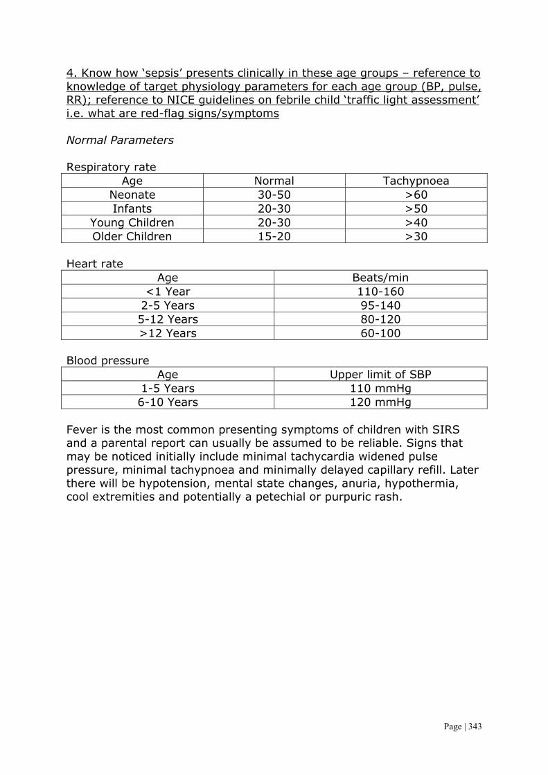
Page | 343
4. Know how ‘sepsis’ presents clinically in these age groups – reference to knowledge of target physiology parameters for each age group (BP, pulse, RR); reference to NICE guidelines on febrile child ‘traffic light assessment’ i.e. what are red-flag signs/symptoms Normal Parameters Respiratory rate
Age Normal Tachypnoea Neonate 30-50 >60 Infants 20-30 >50
Young Children 20-30 >40 Older Children 15-20 >30
Heart rate
Age Beats/min <1 Year 110-160
2-5 Years 95-140 5-12 Years 80-120 >12 Years 60-100
Blood pressure
Age Upper limit of SBP 1-5 Years 110 mmHg 6-10 Years 120 mmHg
Fever is the most common presenting symptoms of children with SIRS and a parental report can usually be assumed to be reliable. Signs that may be noticed initially include minimal tachycardia widened pulse pressure, minimal tachypnoea and minimally delayed capillary refill. Later there will be hypotension, mental state changes, anuria, hypothermia, cool extremities and potentially a petechial or purpuric rash.

Page | 344
5. Understand pathophysiology of ‘shock’ and how to recognise clinically the degree of shock Shock is defined physiologically as inadequate delivery of substrates and oxygen to meet the metabolic needs of the tissues. As cells are starved of oxygen and substrate they can no longer sustain efficient aerobic oxygen production. As oxygen delivery is impaired the cells must switch to the much less efficient anaerobic metabolic pathway which generates only 2 ATP per glucose rather than 36, with the resulting accumulation of lactic acid. Eventually cellular metabolism is no longer able to generate enough energy to power components of cellular homeostasis leading to disruption of the cell membrane ionic pumps, accumulation of intracellular sodium and efflux of potassium. The cells swell, membranes break down and cell

Page | 345
death ensures. Widespread cell death results in multiple system organ failure and even death. The clinical features of shock are manifestations of compensatory physiological mechanisms to maintain the circulation and the direct effects of poor perfusion of tissues and organs. In early compensated shock the blood pressure is maintained by increased HR and RR redistribution of blood from venous reserve volume and diversion of blood flow from non-essential tissues such as the skin in the peripheries, which becomes cold, to the vital organs like the brain and heart. In shock due to dehydration there is usually >10% loss of body weight and a profound metabolic acidosis which is compounded by failure to feed and drink while severely ill. Low BP is a late stage and indicates compensatory responses are failing. In late or uncompensated shock the BP falls and lactic acid increases.
Early (compensated) Late (decompensated) Tachypnoea Acidotic breathing (Kussmaul) Tachycardia Bradycardia
Decreased skin turgor Confusion/depressed consciousness Sunken eyes and fontanelle Blue peripheries Delayed capillary refill >2
seconds Absent urine output
Mottled, pale, cold skin Hypotension Core-peripheral temperature
gap >4oC
Decreased urinary output 6. Understand and demonstrate which antibiotics would be most appropriate empiric choice based on age and presentation of the child with septicaemia Newborns and infants in the first 6-8 weeks of life should generally receive ampicillin and gentamicin, ampicillin and cefotaxime or ampicillin and ceftriaxone unless a clear etiology is known. In older infants and children generally a third generation cephalosporin is given alone. Patients with indwelling lines are given vancomycin is MRSA is suspected. 7. Demonstrate awareness of escalation of treatment of septicaemia and shock and the role of appropriate airway management, inotropic support and intensive care management

Page | 346
The management of septicaemia has previously been discussed and largely revolves around the provision of adequate antibiotic cover in addition to managing any complication of metabolic derangement, cardiomyopathy, capillary leak and clotting. Septicaemia generally leads to shock so the treatment is along the same management plan. Initially, after basic life support and antibiotic therapy, oxygen should be delivered at 10-15 L/min by facial mask. Any patient with cool extremities, prolonged capillary refill time and a tachycardia should be considered to have shock. The initial therapy for shock is volume replacement at a rate of 20 ml/kg. In the UK the use of 4.5% albumin solution is generally recommended. A satisfactory response to volume replacement is a drop in heart rate and improved peripheral perfusion. The first bolus of fluid may be repeated to achieve this response. The patient's condition may stabilise with only volume

Page | 347
replacement, but the patient requires close monitoring and reassessment to detect further signs of shock or pulmonary oedema (due to capillary leak). The goal of circulatory support is to maintain tissue perfusion and oxygenation. Patients who do not response to initial volume replacement require further volume replacement and may need ionotropic support, such as the use of dopamine which may be administered via a peripheral vein until central venous access is established. Patients with persistent hypotension may need an adrenaline infusion administered centrally. Endotracheal intubation and ventilation are recommended in patients who still have signs of shock after they have received volume replacement of more than 40ml/kg. Even patients who are awake and alert have a high risk of pulmonary oedema. The mechanical ventilation helps to relieve metabolic work for the patient and may facilitate the removal of carbon dioxide. Biochemical correction of acidosis, hypoglycaemia, hypokalaemia, hypocalcaemia and hypomagnesaemia is required. Correct coagulopathy and anaemia with the use of fresh frozen plasma and blood as appropriate. 8. Know how to record information in patient notes to enable a relevant and structured handover when needed i.e. To ICU staff I’d hope so by this stage Allergy/Food Allergy 1. Understand the pathophysiology of allergic reactions Many genes have been linked to the development of allergic disease. Polymorphisms or mutations in these genes lead to susceptibility to allergy. Allergic diseases occur when individuals make an abnormal immune response to harmless environmental stimuli, usually proteins. The developing immune system must be sensitised to an allergen before an allergic immune response develops. However sensitisation can be ‘occult’ e.g. sensitisation to egg from exposure to trace quantities in maternal breast milk. Only a few stimuli account for allergic disease, which are inhalant allergens, ingestant allergens and insect stings/bits, drugs and latex. Proteins with an unstable tertiary structure may be rendered non-allergenic by heat degradation. The allergic immune response is classified as an IgE mediated or non-IgE mediated reaction. IgE mediated reactions have a clinical course:

Page | 348
• Early phase – within minutes of exposure, caused by histamine release and other mediators from mast cells. Causes urticaria, angioedema, sneezing and bronchospasm.
• Late phase – 4-6 hours later with nasal congestion in the upper airway and cough and bronchospasm in the lower airway.
The majority of life threatening events are IgE mediated as non-IgE mediated reactions have a delayed onset of symptoms and a more variable clinical course. The pathophysiology is as follows:
• This is a type 1 hypersensitivity reaction • Involves IgE and mast cells. The IgE is derived from B-cells that are
activated by exposed TH2 cells via the release of IL4. The initial exposure leads to the recruitment of eosinophils.
• The IgE molecules bound to the Fc receptors on mast cells are cross-linked by specific antigens
• This cross-linking leads to the release of preformed inflammatory mediators and the production and subsequent release of arachidonic acid derived inflammatory mediators
• The mediators have the effect of inducing inflammation and leads to marked vasodilation, smooth muscle contraction, increased small vessel permeability and increased secretion of mucus
2. List the common allergens and their presenting features in children Food intolerance has previously been discussed. Eczema – this can be atopic or non-atopic. Atopic eczema is classified as an allergic disease and many affected children will have a family history of allergy, at least 50% develop other allergic diseases and IgE antibodies to common allergens are present. There is a close relationship between eczema and food allergy, particularly in young infants with severe disease; up to 40% of them have an IgE mediated food allergy, in particular egg allergy. The core symptom is puritus with rash and excoriations. These areas are dry and may show lichenification. Allergic rhinitis and conjunctivitis – This can again be atopic or non-atopic. It can be classified as intermittent or persistent and mild or severe. It affects up to 20% of children and can severely disrupt their lives. It is associated with eczema, sinusitis and adenoidal hypertrophy and is closely associated with asthma. Asthma – affected children often have IgE antibodies to aeroallergens. Allergen avoidance is difficult to achieve.

Page | 349
Urticaria and angioedema – usually in response to an allergen or viral infection. It involves deeper skin tissue to produce swelling of the lips and soft tissue around the eyes and even anaphylaxis. Chronic urticaria >6 weeks is usually not allergic. It results from a local increase in the permeability of capillaries and venules. These changes are dependent on activation of skin mast cells. Drug allergy – only a minority who are labelled as allergic actually are. Insect sting hypersensitivity – this arises from bee or wasp stings but also from ant bites in the USA, Asia and Australia. This allergy can be mild (local swelling), moderate (generalised urticarial) or severe (systemic symptoms with wheeze or shock). Children with a previous mild reaction are unlikely to develop a severe reaction. 3. Outline initial investigations and management of common allergies Investigations are usually a detailed clinical history in addition to blood tests to check for markers of hypersensitivity. Other simpler tests such as introducing the stimulus into the patient in controlled conditions may work. Patch testing can be done for cutaneous allergies. Management depends on the allergy but is generally the use of antihistamines if needed along with steroids. An epipen must be provided if severe anaphylaxis occurs in patients. There is also the option for systemic desensitisation for a few common allergies. Drug Reaction 1. Recognise the importance of drug reactions and the common ones seen in children
• Antibiotics – penicillin and cephalosporins • Local anaesthetic (lidocaine) • Analgesics (aspirin, NSAIDSs – ibuprofen) • Opiates – codeine, morphine • Dextran • Radiocontrast media
2. Understand how to report severe reactions There is a form called the yellow card that should be submitted with the details of the incident to the medicines and healthcare products regulatory agency.

Page | 350
3. Outline the immediate treatment of drug reactions and be aware of resources available to assist management
• Stop using the drug that is thought to be the cause • Start a new drug if the existing condition needs urgent treatment • Consider altering the dose or temporarily stopping the drug
treatment • Consider the effects of drug-drug interactions • Consider the possibility of withdrawal effects if withdrawn too fast • Provide treatment for the allergy include ABC and medication as
necessary HIV 1. Outline the epidemiology both in the worldwide setting and the UK Globally HIV affects over 2.5 million children, mostly in sub-Saharan Africa (2.3 million) and there are still 380,000 children becoming infected each year. The major route is mother to child transmission which occurs during pregnancy, at delivery or through breast feeding. The virus can also be transmitted to children by infected blood products, contaminated needles or through child sex abuse, but this is uncommon. In comparison there are only 1400 children living with HIV across all of Western Europe. 2. Outline the short and long term risks Short and long term
• Opportunistic infections – TB, PCP, Toxoplasmosis, MAC, VZV, HSV, CMV, Candida
• Blood problems – thrombocytopenia, anaemia, neutropenia • Diet – high energy, high protein
Long Term
• Compliance • Failure to thrive • Risk of transmission • HIV encephalopathy • Neuropathy and myelopathy • Cancers – Kaposi’s sarcoma, Non-Hodgkin’s lymphoma
3. Understand the prevention and treatment options

Page | 351
Prevention is outlined in the neonatal section and involves reducing the mother’s viral load along with caesarean section and no breast feeding. This reduces the risk to 2%. Treatment of HIV revolves around the decision to start antiretroviral therapy (ART) which is based on a combination of clinical status, HIV viral load and CD4 count, except in infants who should start ART shortly after diagnosis, because they have a higher risk of disease progression. As in adults a combination of three or four drugs are used. Prophylaxis against PCP with co-trimoxazole is prescribed for infants who are HIV infected and for older children with low CD4 counts. Other aspects of management include:
• Immunisation which is important because of the higher risk of infections, and should follow the routine vaccination schedule, with the exception of BCG which should not be given as it is live and can cause disseminated disease. In addition to these a vaccine against influenza, hepatitis A, B, and VZV should be considered
• Multidisciplinary management of children in a family clinic with other infected members should be done
• Regular follow up with particular attention paid to weight, neurodevelopment and clinical signs of disease.
Infectious Mononucleosis 1. List the clinical features Acute infectious mononucleosis presents with a history of 1-2 weeks of fatigue and malaise, however onset may be abrupt. The incubation period in adolescents is 30-50 days but is shorter in younger children. Symptoms include a sore throat, headache, fever, myalgias, nausea and abdominal pain. Sore throat is the most frequent presenting symptoms, gradually worsening over the first week. It may be the most severe sore throat the patient has experienced. Headaches usually occur during the first week and may be retro-orbital. LUQ pain may be due to splenic enlargement and severe abdominal pain may indicate splenic rupture. Symptoms usually persist for 2-3 weeks but fatigue is often prolonged. Infants and young children with primary infection are usually asymptomatic. Physically EPV is characterised by pharyngitis, generalised lymphadenopathy and hepatosplenomegaly. Children younger than 4 years frequently have splenomegaly or hepatomegaly, rash and symptoms of an URTI. More than 90% of patients develop fever which is more severe in the afternoon, typically peaking at 38-39oC but may reach 40oC. Fever resolves over 10-14 days. Pulse is normal and tachycardia is unusual.

Page | 352
2. Be aware of the complications and treatment Infectious mononucleosis is a self-limiting illness that does not usually require specific therapy. Because of low transmissibility of EBV isolation is not indicated. Inpatient therapy for medical and surgical complications however may be required. Surgically the main problem is splenic rupture so the spleen may need removing. With the fatigue patients should avoid contact sport or heavy lifting for at least 2-3 weeks to avoid splenic rupture. Complications include:
• Hepatitis – 90% • Jaundice – 5% • Mild thrombocytopenia – 50% • Haemolytic anaemia – 0.5-3% • Upper airway obstruction due to tonsil hypertrophy – 0.1-1% • Splenic rupture – 0.1-0.2% • Neurological complications – 1% • Many neurological conditions including coma, meningitis,
encephalitis, cranial nerve palsies etc • Myocarditis and pericarditis • Reye syndrome • Chronic fatigue syndrome
Kawasaki Disease 1. List the diagnostic criteria NICE guidelines say a child has Kawasaki disease if they have a fever of 38oC or above for more than five days along with at least 4 of the following key symptoms:
• Conjunctival injection in both eyes • Change to the mouth or throat – such as dry cracked lips or a red
swollen tongue • Changes to the skin on the arms or legs such as swelling, redness
or peeling skin • A rash • Swollen lymph nodes of the neck
2. Recognise the presenting features Kawasaki disease is a rare condition that mainly affects children under 5 years of age. It causes a severe high fever that does not respond to

Page | 353
medication and a variety of virus like symptoms. The symptoms Kawasaki disease usually develop in three phases. Phase 1 – acute (weeks 1-2) During the acute phase the child’s symptoms will appear very suddenly and can often be severe. These are high fever, conjunctival injection, rash, changes in hands and feet, swollen lymph glands and changes to the lips, mouth and tongue (red, dry, cracked, peeling, swollen or bleeding) Phase 2 – sub-acute (weeks 3-4) During this phase the symptoms will become less severe but may last longer. The fever should subside but there may be persistent irritability and considerable pain. The symptoms during this stage may include peeling skin, abdominal pain, vomiting, diarrhoea, urine that contains puss, lethargy, headache, joint pain and jaundice. It is in this phase that complications such as coronary artery aneurism are likely to develop. Phase 3 – Convalescent (weeks 4-6) The child will begin to recover and all signs of illness should disappear. However the child may still lack energy and is easily worn out during this time. Occasionally complications can also occur in this phase. 3. Be aware of the investigations and treatment options There is no diagnostic test and the diagnosis is based on clinical findings. However there can be inflammation of the BCG injection site. They have high inflammatory markers (CRP, ESR, WBC) with a platelet count that rises typically in the second week of illness. An aneurysm is most likely to develop within the first 6 weeks (in 1/3 people) and can be visualised on echocardiography. Subsequent narrowing of the vessels from scar formation can cause an MI and sudden death, mortality is 1-2%. Prompt treatment with IVIG is given within the first 10 days to reduce the risk of aneurysm. Aspirin is used to reduce the risk of thrombosis and is given at a high anti-inflammatory dose until the fever subsides and inflammatory markers return to normal, and continue at a low dose until echo at 6 weeks is normal. If the platelet count gets very high then antiplatelet agents may be used. If there is a large aneurysm then long term warfarin therapy may be needed. If the fever recurs then a second dose of IVIG is needed. Persistent inflammation and fever may require treatment with infliximab, steroids or ciclosporin.

Page | 354
4. List the long term complications Aneurysm occurs in up to 33% which can lead to death. Up to 25% of untreated children may experience complications associated with their heart. These children are at a higher risk of developing cardiovascular complications later in life. Immunosuppression 1. Appreciate the causes of immunosuppression in children Immunosuppression involves an act that reduces the activation of efficacy of the immune system. In general there is deliberate suppression of the immune system to prevent the body from rejecting an organ transplant, treating graft versus host disease or after a bone marrow transplant. It can also be used when treating an autoimmune disease such as Crohn’s diseases or rheumatoid arthritis. This is typically done with drugs but may involve splenectomy or radiation. Immunosuppression can be divided into deliberate and non-deliberate: Deliberate immunosuppression includes the use of drugs and in ideal circumstances these should not cause immunodeficiency, but can lead to it and increase the likelihood of infection and decreased cancer surveillance. Common reasons to use these in children are a bone marrow transplant and any organ transplantation. Non-deliberate immunosuppression can occur due to malnutrition, aging, certain types of cancer (leukaemia, lymphoma, and multiple myeloma) and certain infections leading to AIDS. This can also be due to an undesirable effect of drugs used to treat other conditions. 2. Outline a strategy for prevention and treatment of infection Strategies should be similar to those outlined in the objective below. Immune levels need monitoring if deliberately suppressed and there should be prophylactic antibiotic cover as well as a decreased threshold for IV treatment. With non-deliberate immunosuppression there needs to be treatment of the cause, if possible, as well as antibiotic cover. Immunodeficiency

Page | 355
1. List the main causes of immunodeficiency This group of diseases refers to defective immune function, these disorders are characterised as either:
• Primary (uncommon) – an intrinsic defect in the immune system that is present at birth and is either inherited or congenital. Most commonly they are due to mutation on genes associated with immunological functions, most are autosomal recessive, with a few being autosomal dominant or x-linked.
• Secondary (more common) – caused by another disease or treatment, such as an intercurrent bacterial or viral infection, malignancy, malnutrition, HIV infection, immunosuppressive therapy, splenectomy or nephrotic syndrome.
Immunodeficiencies are characterised by infections that sent to be Serious, Persistent, Unusual and Recurrent (SPUR). The type of defect often relates to the infections seen in that disease. T-cell defects These are severe and/or unusual viral and fungal infections and failure to thrive in the first months of life e.g. severe bronchiolitis, diarrhoea, oral thrust and PCP.
• Severe combined immunodeficiency (SCID) – a heterogeneous group of inherited disorders of profoundly defective cellular humoral immunity altering both T and B cell lymphocytes. It is only treatable by bone marrow transplantation
• HIV infection – causes a progressive T cell deficiency • Wiskott-Aldrich – a triad with thrombocytopenia and eczema (x-
linked) • DiGeorge – with maldevelopment of the 5th brachial arch causing
heart defects, placental and facial defects, an absent thymus and hypocalcaemia
• Duncan syndrome – inability to make a normal response to EBV and child either succumbs to infection or develops secondary lymphoma
• Ataxia telangiectasia – defect in DNA repair, also increased risk of lymphoma. There is cerebellar ataxia and developmental delay
B-cell defects In the first 2 years (beyond infancy due to passive immunity) there are severe bacterial infections, especially of the ear, sinus’, skin and pulmonary system. There is often diarrhoea and failure to thrive. Recurrent pneumonias can lead to bronchiectasis; recurrent ear infections to impaired hearing.

Page | 356
• X-linked agammaglobulinaemia – abnormal tyrosine kinase gene, eseential for B-cell maturation
• Common variable immunodeficiency (CVID) – B cell deficiency, high risk of autoimmune disorders and malignancy. Later onset than above
• Hyper IgM syndrome – B cells produce IgM but prevented from switching to IgG and IgA
• Selective IgA deficiency – most common primary immune defect. Usually asymptomatic or recurrent ear, sinus and pulmonary infections
Neutrophil defects Recurrent bacterial infections – abscesses (skin, lymph nodes, lung, liver, spleen, bone), poor wound healing, perianal disease and periodontal infections; invasive fungal infections such as aspergillosis. Diarrhoea and failure to thrive. Granulomas from chronic inflammation.
• Chronic granulomatous disease – most are x-linked recessive, some autosomal recessive. Defect in phagocytosis as fail to produce superoxide after ingestion of micro-organism
Leucocyte function defects Delayed separation of umbilical cord, delayed wound healing, chronic skin ulcers and dee-seating infection.
• Leucocyte adhesion deficiency (LAD) – deficiency of neutrophil surface adhesion molecules leads to inability of neutrophils to migrate to sites of infection/inflammation
Complement defects Recurrent bacterial infections, SLE like illness, recurrent meningococcal infections with deficiency of the terminal complement components.
• Early complement component deficiency • Terminal complement component deficiency • Mannose-binding lectin (MBL) deficiency
2. Outline a strategy for prevention and treatment of infection Management options include:
• Antimicrobial prophylaxis – for T-cell and neutrophil defects give cotrimoxazole to prevent pneumocystis jiroveci infection and itraconazole or fluconazole to prevent other fungal infections. For B-

Page | 357
cell defects give antibiotic prophylaxis (e.g. azithromycin) to prevent recurrent bacterial infections
• Antibiotic treatment – prompt treatment of infections, appropriate choice of antibiotics and a generally longer courses with lower threshold for IV therapy.
• Screen for end organ disease e.g. CT scan in children with antibody deficiency to detect bronchiectasis
• Immunoglobulin replacement therapy – for children with antibody deficiency and can be given IV so a central venous line may be used
• Bone marrow transplantation – can be a matched sibling donor, matched unrelated donor or haploidentical transplant. Used for SCID and chronic granulomatous disease
• Gene therapy – for certain forms of SCID but associated with a risk of leukaemia
Typhoid Fever 1. Have a basic understanding of the clinical features and treatment A child with worsening fever, headaches, cough, abdominal pain, anorexia, malaise and myalgia may be suffering from infection with salmonella typhi or paratyphi. Gastrointestinal symptoms (diarrhoea or constipation) may not appear until the second week. Splenomegaly, bradycardia and rose-coloured spots on the trunk may be present. The serious complications of this disease include gastrointestinal perforation, myocarditis, hepatitis and nephritis. The recent increase in multi-drug resistant strains, particularly in the Indian subcontinent, means that treatment with cotrimoxazole, chloramphenicol or ampicillin may be inadequate. A third generation cephalosporin or azithromycin is usually effective. Typhoid is contracted from contaminated drinking water or food. Malaria 1. Outline the clinical features, including cerebral malaria, and the main treatment options Children are the worst affected, especially children aged 6 months to 5 years. In parts of the world where malaria is endemic it may causes as many as 10% of all deaths in children. The clinical features include fever (often not cyclical), diarrhoea, vomiting, flu-like symptoms, jaundice, anaemia and thrombocytopenia. Whilst typically the onset is 7-10 days after inoculation, infection can present many months later. Children are particularly susceptible to severe anaemia and the gravest form of the disease, cerebral malaria. The infection is diagnosed by examination of a thick film.

Page | 358
Cerebral malaria is a rapidly developing encephalopathy which only occurs in 20-50% of people who develop malaria. It occurs when parasites adhere to the cerebral microvasculature causing blockage. This loads to a shortage of oxygen to this site and therefore numerous complications. Around half these patients have elevated ICP and seizures. Treatment is usually quinine for plasmodium falciparum because of the emergence of chloroquine resistant strains worldwide. Travellers to endemic areas should always seek up to date information on malaria prevention. Prophylaxis reduces but does not eliminate the risk of infection. Prevention of mosquito bites with repellents and bed nets is also important. In many countries there has been a marked reduction in the incidence of malaria in children from insecticide-treated bed nets, indoor residual spraying of houses with insecticides, destruction of mosquito larvae and breeding areas and prompt treatment with artemisinin based combination therapy. Musculoskeletal Congenital Dislocated Hip (development dysplasia) 1. Be able to discuss the incidence, risk factors, screening tools, presentation and basic management This is a spectrum of disorders ranging from dysplasia to subluxation through to frank dislocation of the hip. Early detection is important as it usually responds to conservative treatment. Late identification is usually associated with hip dysplasia, which requires complex treatment often including surgery. Neonatal screening is performed as part of the routine examination of the newborn checking if the hips can be dislocated posteriorly out of the acetabulum (Barlow manoeuvre) or can be relocated back into the acetabulum on abduction (Ortolani manoeuvre). These tests are repeated at 8 weeks of age for routine surveillance. Thereafter presentation is usually with a limp or abnormal gait. It may also be identified from asymmetry of skin folds around the hip, limb abduction of the hip or shortening of the affected leg. On neonatal screening an abnormality of the hip is detected in about 6-10 per 1000 live births. Most will resolve spontaneously. The true birth prevalence of DDH is 1.3 per 1000 live births. Risk factors include being female (6 times more likely), a positive family history (20% of affect infants), if the birth is breech (30% of affected infants) or if the infants has a neuromuscular disorder.

Page | 359
Sometimes an examination may miss DDH so an ultrasound may be done in some centres where DDH is still suspected in the infant. If DDH is found then an ultrasound examination will reveal the quality of the hip and quantity of dysplasia. If the ultrasound is abnormal then the infant may be placed in a splint or harness to keep the hip flexed and abducted for several months. Progress is monitored by ultrasound or x-ray. In most instances a satisfactory response is obtained. Surgery is required if conservative measures fail. Osteomyelitis 1. Understand the clinical features, causative factors, investigations, immediate intervention and management In osteomyelitis there is infection of the metaphysis of long bones. The most common sites are the distal femur and proximal tibia but any bone may be affected. It is usually due to haematological spread of the pathogen, but may arise by direct spread from an infected wound. The skin is swollen directly over the affected site. Where the joint capsule is inserted distal to the epiphyseal plate, as in the hip, osteomyelitis may spread to cause septic arthritis. Most infections are caused by staph.aureus, but other pathogens include streptococcus and Hib if not immunised. Infection due to TB or sickle cell disease need to be considered. Presentation is usually with a markedly painful, immobile limb (pseudoparesis) in a child with an acute febrile illness. Directly over the infected site there will be swelling and exquisite tenderness, and it may be erythematous and warm. Moving the limb causes severe pain. There may be a sterile effusion of an adjacent joint. Presentation may be more insidious in infants, in whom swelling or reduced limb movement is the initial sign. Beyond infancy, presentation may be with back pain in a vertebral infection or with a limp or groin pain in infection of the pelvis. Occasionally there can be multiple foci. Investigations should include blood cultures, which are usually positive, and a white blood cell count and acute-phase reactants which should both be raised. X-rays are initially normal, other than showing soft tissue swelling; it takes 7-10 days for subperiosteal new bone formation and localised bone rarefaction to become visible. Ultrasound may show periosteal elevation at presentation. An MRI allows identification of infection in the bone and differentiation of bone from soft tissue infection. A radionuclide bone scan may be helpful if the site of infection is unclear. Treatment should be prompt with IV antibiotics for several weeks to prevent bone necrosis, chronic infection, a discharging sinus, limb

Page | 360
deformity and amyloidosis. Antibiotics are given IV until there is clinical recovery and the acute-phase reactants have returned to normal, followed by oral therapy for several weeks. Aspiration or surgical decompression of the subperiosteal space may be performed if the presentation is atypical or in immunodeficiency children. Surgical drainage is performed if the condition does not respond rapidly to antibiotic therapy. The affect limb is initially rested in a splint and subsequently mobilised. 2. Know about atypical presentations; subacute and chronic osteomyelitis Subacute osteomyelitis This is a distinct form of osteomyelitis and is difficult to diagnose because the characteristic signs and symptoms of the acute form of disease are absent. The disease has an insidious onset, mild symptoms, and lacks a systemic reaction, and supportive laboratory data are inconsistent. Subacute osteomyelitis can mimic various benign and malignant conditions which can delay diagnosis. The presenting symptoms include mild to moderate localised pain, usually exacerbated by unusual physical activity. Night pain is common, but relieved by aspirin, and there is minimal loss of function. On examination there is localised tenderness, occasionally associated with warmth, redness and soft tissue swelling. Pain may occur with movement of the adjacent joint and some joint effusion may be present. The surrounding muscle may show signs of wasting. The average duration of symptoms before diagnosis is 1-6 months. Chronic osteomyelitis If acute osteomyelitis is not treated it can progress to chronic osteomyelitis, producing permanent damage. Chronic osteomyelitis can also develop as a complication of pre-existing infection from syphilis. Multi organism infections are common with chronic osteomyelitis. Symptoms include bone pain, persistent fatigue, pus draining from a sinus, local swelling, skin changes, excessive sweating and chills. 3. Be aware of risks of undertreated/untreated infection Left untreated this condition can spread to other bones, causing widespread infection, sepsis and even death. With chronic disease there is destruction of bone which is permanent and may result in the need for amputation due to poor vascularisation of the remaining bone.

Page | 361
Septic Arthritis 1. Describe the epidemiology, aetiology, pathogenesis, clinical features, investigations and management Septic arthritis is a serious infection of the joint space, as it can lead to bone destruction. It is most common in children <2 years old but an actual incidence is not known. A higher incidence is seen in boys. It usually results from haematological spread but may also occur following a puncture wound or infection skin lesions, e.g. chickenpox. In young children it may result from spread from adjacent osteomyelitis into joints where the capsule inserts below the epiphyseal growth plate. Usually only one joint is affected with the hip being a particular concern in young children and infants. Beyond the neonatal period the most common organism is staph.aureus and usually only one joint is affected. Hib used to be an important concern and often presented at multiple sites, however the introduction of the Hib vaccine has seen these cases drop. Underlying illnesses such as immunodeficiency and sickle cell disease should be considered. Presentation is usually with an erythematous, warm, acutely tender joint with a reduced range of movement, in an acutely unwell, febrile child. Infants often hold their limbs still (psuedoparesis and pesudoparalysis) and cry if it is moved. A joint effusion may be detectable in peripheral joints. In osteomyelitis, although a symptomatic joint effusion may be present, the tenderness is over the bone, but in up to 15% there is coexistent septic arthritis. The diagnosis of septic arthritis of the hip can be particularly difficult in toddlers as the joint is well covered by subcutanoues fat. Initial presentation may be with a limp or pain referred to the knee. Investigations will show a raised white cell count and acute-phase reactants. Blood cultures must be taken. An ultrasound of the deep joints, such as the hip, is helpful to identify an effusion. X-rays are used to exclude trauma and other bony lesions. However, in septic arthritis, the x-rays are initially normal, apart from widening of the joint space and soft tissue swelling. A bone scan may be helpful and an MRI may demonstrate an adjacent osteomyelitis. Aspiration of the joint space under ultrasound guidance for organisms and culture is the definitive diagnosis. Ideally this is performed immediately unless it would significantly delay the administration of antibiotics. Treatment is a prolonged course of antibiotics, initially IV. Washing out of the joint or surgical drainage may be required if resolution does not occur rapidly, or if the joint is deep seated such as at the hip. The joint is initially immobilised in a functional position, but subsequently must be mobilised to prevent permanent deformity.

Page | 362
2. Be aware of special cases such as neonates, hip joint involvement, various organisms (such as tuberculosis), and septic arthritis in immunocompromised patients Hip involvement – see above Neonates – staph.aureus is most common but E.coli and group B strep also cause disease TB – a rare cause of chronic pyogenic arthritis and can affect the spine Immunocompromised Fractures 1. Understand common types of fractures and principles of management Firstly it should be noted that fractures can be a sign of NAI, especially in those below 30 months. The most common signs of NAI fractures are ribs (posterior), long bones such as the humerus (especially if the child is not yet mobile), those with multiple fractures and those with complex skull fractures. It is also important to rule out conditions such as osteogenesis imperfect and copper deficiency that lead to a higher chance of fractures. The epidemiology of fractures is different from adults. The risk of fracture increases with age and boys are more likely to sustain one. Trauma whilst playing sports or from playing events are the causes of the majority of fractures. The most common location is the upper extremities and includes:
• Distal forearm – 22.7% • Hand, phalanges – 18.9% • Carpal-metacarpal – 8.3% • Clavicle – 8.1%: immobilise with a sling for 4-6 weeks • Ankle – 5.5%
The management principles are to control haemorrhage, treat pain, prevent limb ischaemia and remove potential sources of contamination. Once this has been done the fracture should be reduced and the reduction maintained. These should then be immobilised and splinted before being cast. Juvenile Idiopathic Arthritis

Page | 363
1. Outline the classification/subtypes of JIA including differences between these This is the commonest chronic inflammatory joint disease in children and adolescents in the UK. It is defined as persistent joint swelling (of >6 weeks duration) presenting before 16 years of age and in the absence of infection or any other defined cause. It has a prevalence of 1 in 1000 children. There are numerous different subtypes and the disease is classified by the number of joints affected in the first 6 months: polyarthritis (>4) and oligoarthritis (4 or less) or systemic (with fever and rash). Features in the history are gelling (stiffness after periods of rest), morning joint stiffness and pain. Initially there may be only minimal evidence of joint swelling but subsequently there may be joint swelling and inflammation. Long term there can be bone expansion from overgrowth which can cause various deformities as well as causing advanced bone age. The types are as follows: Oligoarthritis persistent (49%)
• Age of onset – 1 to 6 years • Sex ratio (F:M) – 5:1 • Articular pattern – 1-4 (max) joints involved; knee, ankle or wrist
are the most common • Extra-articular features – chronic anterior uveitis in 20%, leg length
discrepancy. • Laboratory abnormalities – ANA+/- • Prognosis - Excellent
Oligoarthritis extended (8%)
• Age of onset – 1 to 6 years • Sex ratio (F:M) – 5:1 • Articular pattern – >4 joints involved after first 6 months. An
asymmetrical distribution of large and small joints • Extra-articular features – Chronic anterior uveitis in 20%,
asymmetrical growth. • Laboratory abnormalities – ANA+/- • Prognosis - Moderate
Polyarthritis RF negative (16%)
• Age of onset – 1 to 6 years • Sex ratio (F:M) – 5:1 • Articular pattern – Symmetrical large and small joint arthritis, often
with marked finger involvement. Cervical spine and temporomandibular joint may be involved
• Extra-articular features – Low-grade fever, chronic anterior uveitis in 5%, late reduction of growth rate.
• Laboratory abnormalities – None • Prognosis - Moderate

Page | 364
Polyarthritis RF positive (3%)
• Age of onset – 10 to 16 years • Sex ratio (F:M) – 5:1 • Articular pattern – Symmetrical large and small joint arthritis, often
with marked finger involvement • Extra-articular features – Rheumatoid nodules in 10%, similar to
adult RA. • Laboratory abnormalities – RF+ long term • Prognosis - Poor
Systemic arthritis (9%)
• Age of onset – 1 to 10 years • Sex ratio (F:M) – 1:1 • Articular pattern – Oligoarthritis or polyarthritis. May have aches
and pains in joints and muscles (arthralgia/myalgia) but initially no arthritis.
• Extra-articular features – Acute illness, malaise, high daily fever initially, salmon pink macular rash, lymphadenopathy, hepatosplenomegaly and serositis.
• Laboratory abnormalities – Anaemia, raised neutrophils and platelets, high acute-phase reactants
• Prognosis – Variable to poor Psoriatic arthritis (7%)
• Age of onset – 1 to 16 years • Sex ratio (F:M) – 1:1 • Articular pattern – Usually asymmetrical distribution of large and
small joints, dactylitis • Extra-articular features – Psoriasis, nail pitting or dystrophy, chronic
anterior uveitis in 20% • Laboratory abnormalities – None • Prognosis – Moderate
Enthesitis-related arthritis (7%)
• Age of onset – 6 to 16 years • Sex ratio (F:M) – 1:4 • Articular pattern – Lower limb, large joint arthritis initially, mild
lumbar spine or sacroiliac involvement later on • Extra-articular features – Enthesitis which is localised inflammation
at insertion of tendons or ligaments into bones, often at the feet (Achilles insertion). There is occasional acute uveitis
• Laboratory abnormalities – HLAB27+ • Prognosis – Moderate
Undifferentiated arthritis (1%)
• Age of onset – 1 to 16 years

Page | 365
• Sex ratio (F:M) – 2:1 (variable) • Articular pattern – Overlapping articular and extra-articular patterns
between ≥2 subtypes or insufficient criteria for sub-classification • Extra-articular features – Variable • Laboratory abnormalities – Variable • Prognosis – Variable
2. Outline the known associated conditions such as uveitis Chronic anterior uveitis – this is a common but asymptomatic condition and can lead to severe visual impairment. Regular ophthalmological screening using a slip lamp is indicated, especially for children with oligoarticular disease. Flexion contractures of the joints – these occur when the joint is held in the most comfortable position, thereby minimising intra-articular pressure. Chronic untreated disease can lead to joint destruction and the need for joint replacement. Growth failure – this may be generalised from anorexia, chronic disease and system corticosteroid therapy. May also be localised overgrowth such as leg length discrepancy due to prolonged active knee synovitis and undergrowth, such as micrognathia, usually seen in long-standing or sub optimally treated arthritis due to premature fusion of epiphyses. Constitutional problems – anaemia of chronic disease, delayed puberty Osteoporosis – multifactorial aetiology, including diet, reduced weight bearing, systemic corticosteroids and delayed menarche. Reduced risk by dietary supplements of calcium and vitamin D; regular weight bearing exercises; and minimise oral corticosteroids use and sometimes bisphosphonates. Amyloidosis – very rare now, causes proteinuria and subsequent renal failure and has a high mortality. 3. Appreciate the role of the multidisciplinary team members during the management of these patients All children suspected of having JIA should be managed by a specialist paediatric rheumatology multidisciplinary team, often working in shared care with local hospitals. 4. Understand common drugs used and importance of treatment

Page | 366
NSAIDs and analgesia – do not modify disease but help relieve symptoms during flares Joint injections – these are increasingly being done under ultrasound guidance as it is more effective. These are first-line treatment for oligoarticular JIA; in polyarticular disease multiple joint injections are used as bridging agents when starting methotrexate. These often require sedation with inhaled anaesthesia (Entonox) Methotrexate – early use reduces joint damage. It is effective in approximately 70% with polyarthritis, less effective in systemic features of JIA. It is given as a weekly dose (tablet, liquid or injection) and regular blood monitoring is required (for abnormal liver function and bone marrow suppression). Nausea is common. Systemic corticosteroids – avoid if possible, to minimise the risk of growth suppression and osteoporosis. Pulsed IV methylprednisolone often used for severe polyarthritis as an induction agent. It may be life-saving for severe systemic arthritis or macrophage activation syndrome. Cytokine modulators and other immunotherapies – Many agents (e.g. anti-TNF alpha, IL-1, CTLA-4 or IL-6) are now available and useful in severe disease refractory to methotrexate. Costly and given under strict national guidelines with registries for long-term surveillance. T-cell depletion coupled with autologous haematopoietic stem cell rescue (bone marrow transplant) is an option for refractory disease. The child should also be encouraged to take part in all activities apart from contact sports during active flares. With optimal care most children can be managed as outpatients. 5. Be aware of outcome measures used Long term studies have shown that at least 1 in 3 children will have ongoing active disease into adult years, with significant morbidity from previous inflammation, such as joint damage requiring joint replacement surgery, visual impairment from uveitis, or fractures from osteoporosis. Outcome measures include clinical damage, quality of life and measures of physical function. Perthe’s Disease

Page | 367
1. Outline the aetiology, presentation, investigations, prognosis and basic management This is an avascular necrosis of the capital femoral epiphysis of the femoral head due to interruption of the blood supply, followed by revascularisation and reossification over 18-36 months. It mainly affects boys (M:F = 5:1) of 5 to 10 years of age. Presentation is insidious, with the onset of a limp, or hip or knee pain. The condition may initially be mistaken for transient synovitis. It is bilateral in 10 to 20%. If suspected x-rays of both hips (including frog views) should be requested; early signs of Perthe’s include increased density in the femoral head, which subsequently becomes fragmented and irregular. Even if the initial x-ray is normal, a repeat may be required if clinical symptoms persist. A bone scan and MRI scan can be helpful in making the diagnosis. Prognosis is dependent on early diagnosis; if identified early and less than half the femoral head is affected, only bed rest and traction may be required. In more severe disease or late presentations, the femoral head needs to be covered by the acetabulum to act as a mould for the re-ossifying epiphysis and is achieved by maintaining the hip in abduction with plaster or calipers, or by performing femoral or pelvic osteotomy. In most children the prognosis is good, particularly in those below 6 years of age, with less than half the epiphysis involved. In older children or with more extensive involvement of the epiphysis, deformity of the femoral head and metaphyseal damage are more likely, with potential for subsequent degenerative arthritis in adult life. Reactive Arthritis 1. Understand the causes, different types, diagnostic criteria, lab investigations and radiological investigations Reactive arthritis is the most common form of arthritis in childhood. It is characterised by transient joint swelling (usually <6 weeks) often of the ankles or knees. It usually follows (or rarely accompanies) evidence of extra-articular infection. The enteric bacteria (salmonella, shigella, campylobacter and yersinia) are often the cause in children, but viral infections, sexually transmitted infections in adolescents (chlamydia, gonococcus), mycoplasma and borrelia burgdoferi (Lyme disease) are other causes. Rheumatic fever and post-streptococcal reactive arthritis are rare in development countries but are frequent in many developing countries. Reactive arthritis can be divided into post streptococcal, post infectious and classical reactive arthritis following GI/urinary tract infection.

Page | 368
• Post streptococcal – rarely seen in developed countries. Requires antibiotic treatment
• Classical reactive – inflammation in the absence of bacteria in the joint space.
• Post infective – includes most others Fever is generally low grade with inflammation of joints, skin, mucous membranes, urinary and GI tract. The eyes are also commonly affected. Acute-phase reactants are normal or mildly elevated and x-rays are normal. No treatment or only NSAIDs are required and complete recovery can be anticipated. HLA-B27 is positive in 65-96% of patients. Chronic cases may require steroids and methotrexate if there is no active infection. With regards to physical therapy the patient should rest and avoid using the affected joint. As the symptoms improve there should be a graded programme of exercise that is designed to strengthen affected muscle groups and improve the range of movement. 2. Know about pharmacological and physical therapy, along with course and prognosis See above for details Rickets 1. To be aware about pathophysiology, causes, diagnosis and management Rickets signifies a failure in mineralisation of the growing bone or osteoid tissue. Failure of mature bone to mineralise is called oestomalacia. The predominant cause of rickets during the early twenty-first century was nutritional vitamin D deficiency due to inadequate intake or insufficient exposure to direct sunlight. Nutritional rickets still remains the major cause in the developing world. In developed countries nutritional rickets has become rare due to formula milk and many breakfast cereals containing supplemental vitamin D. Nutritional rickets can sometimes be seen in black or asian infants who are totally breast fed in late infancy. It is also seen in extremely premature infants from dietary deficiency of phosphorus together with low stores of calcium and phosphorus. Children with malabsorptive conditions such as CF, coeliac disease and pancreatic insufficiency can develop rickets due to deficient absorption of vitamin D, calcium or both. Drugs, especially anticonvulsants such as Phenobarbital and phenytoin, interfere with the metabolism of vitamin D and may also cause rickets. Rickets may also result from impaired metabolic conversion or activation of vitamin D (hepatic and renal disease).

Page | 369
Other causes can be divided into:
• Nutritional – living in northern latitudes, dark skin, decreased sun exposure maternal vitamin D deficiency, diets low in vitamin D, phosphorus and calcium, vegan diets and prolonged parenteral nutrition in infancy
• Intestinal malabsorption – small bowel enteropathy (coeliac disease), pancreatic insufficiency, cholestatic liver disease, high phytic acids in diet
• Defective production of 25(OH)D2 – chronic liver disease • Increased metabolism of 25(OH)D3 – enzyme induction by
anticonvulsants • Defective production of 1,25(OH)2D3 – hereditary type 1 vitamin D-
resistant rickets, familial hypophosphataemic rickets, chronic renal disease, fanconi syndrome (renal loss of phosphate)
• Target organ resistance to 1,25(OH)2D3 – hereditary vitamin D-dependent rickets type 2
Before mentioning the clinical features it is worth covering the metabolism of vitamin D. The sunlight reacts with 7-dehydrocholesterol on the skin to form vitamin D3 (cholecalciferol). This enters the liver and is metabolised to 25(OH)D3. This then if free to enter the kidneys where it is hydroxylated again to form 1,25(OH)2D3, the most active form of the vitamin. The earliest sign of rickets is a ping-pong ball sensation of the skull (craniotabes) elicited by pressing firmly over the occipital or posterior parietal bones. The costochondral junctions may be palpable, wrists and ankles may be widened and there may be horizontal depression on the lower chest corresponding to attachment of the softened ribs and with the diaphragm (Harrison sulcus). The legs may become bowed. Other clinical features include:
• Misery • Failure to thrive/short stature • Frontal bossing of the skull • Delayed closed of the anterior fontanelle • Delayed dentition • Hypotonia • Seizures (late)
Diagnosis is made from:
• A dietary history for vitamin and calcium intake • Blood tests – serum calcium is low or normal, phosphorus is low,
plasma alkaline phosphatase activity is greatly increased, 25-hydroxyvitamin D may be low and parathyroid hormone elevated.

Page | 370
• X-ray of the wrist joint – shows cupping and fraying of the metaphyses and a widened epiphyseal plate.
Management is generally nutritional and involves giving advice about a balanced diet, correction of predisposing risk factors and by the daily administration of vitamin D3 (cholecalciferol). If compliance is an issue a single high dose of vitamin D3 can be given, followed by the daily maintenance dose. Healing occurs in 2 to 4 weeks and can be monitored from the lowering of alkaline phosphatase, increasing vitamin D levels and healing on x-rays, but complete reversal of bony deformities may take years. 2. Outline the role of vitamin D and its deficiency in health bone development Outlined above Slipped Upper Femoral Epiphysis 1. List the risk factors, age distribution, clinical presentation, and basic interpretation of radiological investigations This results in displacement of the epiphysis of the femoral head postero-inferiorly requiring prompt treatment in order to prevent avascular necrosis. It is most common at 10 to 15 years of age during the adolescent growth spurt, particularly in obese boys (2.4 times) and is bilateral in 20% (with the left hip being more commonly affected). There is an association with metabolic endocrine abnormalities, e.g. hypothyroidism with hypogonadism, generally in younger children (<10) where a bilateral presentation is more common. Presentation is with a limp or hip pain, which may be referred to the knee. The onset may be acute, following minor trauma or insidious. Examination shows restricted abduction and internal rotation of the hip. Diagnosis is confirmed on x-ray, and a frog lateral view should also be requested. Management is surgical, usually with pin fixation in situ. This should however be based on whether the condition is acute or chronic (>3 weeks) and whether the joint can bear weight or not. Analgesia including NSAIDs should also be provided. Following surgery a patient is given crutches for 6-8 weeks to reduce weight bearing, along with a course of physiotherapy. Flat Foot/Forefoot Adduction

Page | 371
1. Be aware of normality, causes and orthotic management Flat foot is known as pes planus whilst forefoot adduction is known as talipes equinovarus or position talipes. Flat foot Toddlers learning to walk usually have flat feet due to flatness of the medial longitudinal arch and the presence of a fat pad which disappears as the child gets older. An arch can usually be demonstrated on standing on tiptoe or by passively extending the big toe. Marked flat feet is common in hypermobility. A fixed flat foot, often painful, presenting in older children, may indicate a congenital tarsal coalition and requires an orthopaedic opinion. Symptomatic flat feet are often helped with footwear advice and, occasionally, an arch support may be required. In older children and adolescents a rigid flat foot is pathological. It is suggested by absence of a normal arch on tip-toeing. It may be due to an associated tendo-Achilles contracture, or tarsal coalition or inflammatory arthropathy. Talipes Positional talipes from intrauterine compression is common. The foot is of normal size, the deformity is mild and can be corrected to the neutral position with passive manipulation. Often the baby’s intrauterine posture can be recreated. If the positional deformity is marked, parents can be shown passive exercises by the physiotherapist. Talipes equinovarus is a complex abnormality. The entire foot is inverted and supinated, the forefoot adducted and the heel is rotated inwards and in plantar flexion. The affect foot is shorter and the calf muscle thinner than normal. The position of the foot is fixed, cannot be corrected completely and is often bilateral. The birth prevalence is 1 per 1000 live births, affects predominantly males (2:1), can be familial and is usually idiopathic. However, it may also be secondary to oligohydramnios during pregnancy, a feature of a malformation syndrome or of a neuromuscular disorder such as spina bifida. There is an association with developmental dysplasia of the hip (DDH). Treatment is started promptly with plaster casting and bracing (Ponsetti method), which may be required for many months. It is usually successful unless the condition is very severe, when corrective surgery is required. Ostegenesis imperfecta

Page | 372
1. Be aware of different types of inheritance This is a group of disorders of collagen metabolism causing bone fragility, with bowing and frequent fractures. There are many different forms but I will concentrate on types one to three. Type one is the most common form and is due to autosomal dominant inheritance. There are fractures during childhood and a blue appearance of the sclera with some children developing hearing loss. There is usually a dilated aortic root and thin heart valves. This accounts for 60% of all cases and is the mildest form. Fractures can occur at any time with these being 7 times more common than normal. There may be hypermobility of the joints and the teeth can be affected. Treatment is with bisphosphonates to reduce the fracture rate. The prognosis is variable but the best of the subtypes. Fractures require splinting to minimise joint deformity. Type two is a severe, lethal form with multiple fractures already present before birth. Many affected infants are still born. Inheritance is variable but mostly autosomal dominant or due to new mutations. In other types sclera discolouration may be minimal. Type 3 is a severely progressive form and the child will have various amounts of immature woven bone. The child may be born with fractures and, as age increases, there is a noticeable short stature and impaired dentition. Osteogeneis imperfecta occurs in roughly 1/20,000 to 1/50,000 live births and is the leading cause of lethal short limbed dwarfism. In almost all cases the mode of inheritance is autosomal dominant regardless of the clinical form. An autosomal recessive form has been identified in some families in South Africa. Diagnosis is mostly done prenatal but milder forms may not be picked up until much later. Treatment is complicated but involves bisphosphonates to reduce the risk of fractures. 2. Outline the clinical manifestations See above Polydactly/Syndactly 1. Know the common associations

Page | 373
Polydactyly is where there are more than 5 digits on the hands or feet and occurs in 1 in 1000. Common associated syndromes include trisomy 13, trisomy 21, tibial hemimelia and Ellis-van Creveld syndrome. Syndactyly is the fusion of two or more digits and occurs in 1 in 2500. It is also associated with several syndromes including Apert syndrome and Poland syndrome. Scoliosis 1. Be aware of the causes, significance, principles of interventional management Scoliosis is a lateral curvature in the frontal plane of the spine. In structural scoliosis there is rotation of the vertebral bodies which causes a prominence in the back from rib asymmetry. In most cases the changes are mild, pain free and primarily a cosmetic problems; however, in severe cases, the spinal curvature can lead to cardiorespiratory failure from distortion of the chest. Causes of scoliosis include:
• Idiopathic – the most common, either early onset (<5 years old) or, most often, late onset, mainly girls 10 to 14 years of age during their puberty growth spurt
• Congenital – from a congenital structural defect of the spine e.g. hemivertebra, spina bifida and syndromes e.g. VACTERL (vertebral, anorectal, cardiac, tracheo-oesophageal, renal and limb abnormalities).
• Secondary – related to other disorder such as neuromuscular imbalance (e.g. cerebral palsy or muscular dystrophy), disorders of bone such as neurofibromatosis or of connective tissues such as Marfan syndrome, or leg length discrepancy e.g. due to JIA of one knee.
Examination should start with inspection of the child’s back while standing up straight. In mild scoliosis there may be irregular skin creases and difference in shoulder height. The scoliosis can be identified on examining the child’s back when bent forwards. If the scoliosis disappears on bending forwards then it is postural although leg length should be checked. Mild scoliosis will resolve spontaneously, or progresses minimally. If more severe, the severity and progression of the curvature of the spine is determined by x-ray. Severe cases are managed in specialist spinal centres where the place of non-medical treatment such as bracing will be considered, with surgery indicated only if severe or there is coexisting pathology such as neuromuscular or respiratory disease.

Page | 374
Skeletal Dysplasia 1. Be aware of pathophysiology of skeletal dysplasia, broad classification, principles of physical and surgical management In general patients with a disproportionately short stature have skeletal dysplasia. These are a heterogeneous group of more than 200 disorders characterised by abnormalities of cartilage and bone growth resulting in abnormal shape and size of the skeleton and disproportion of the long bones, spine and head. The pathophysiology is complicated and varies depending on the condition. However it broadly involves the growth plate, most commonly affecting the zone of proliferation. This usually results in impaired strength along with those mentioned above. Intelligence remains normal. Classification is not clearly mentioned anywhere but these disorders can be classified by the area of the bone they affect or by whether they affect formation or structure (i.e. quality or quantity of the bone). The most commonly found conditions are:
• Achondroplasia – autosomal dominance inheritance but about 50% are new mutations. Clinical features are short stature from marked shortening of limbs, a large head, frontal bossing and depression of the nasal bridge. The hands are short and broad. A marked lumbar lordosis develops and hydrocephalus may occur.
• Thanatophoric dysplasia – this results in stillbirth. The infants have a large head, extremely short limbs and a small chest. The appearance of the bones on x-ray is characteristic. The importance of the correct diagnosis of this disorder is that, in contrast to Achondroplasia, its inheritance is sporadic. It can be identified on antenatal ultrasound.
• Cleidocranial dysostosis – this is an autosomal dominant disorder where there is absence of part or all of the clavicles and delay in closure of the anterior fontanelle and of ossification of the skull. The child is often able to bring the shoulders together in front of the chest to touch each other as a ‘party trick’. Short stature is usually present, intelligence is normal.
• Arthrogryposis – this is a group of disorders where there is stiffness and contracture of joints with an unknown cause. There are marked flexion contractures of major joints with dislocating hips, talipes and scoliosis.
• Osteogenesis imperfecta – mentioned before Medical care is mostly supportive with monitoring and correction of major problems caused by the deformities. Surgical care generally involves correcting major deformities such as scoliosis but these operations can be difficult. Physiotherapy and splints can be used in certain cases.

Page | 375
2. Understand roles played by multiprofessional team in management A multiprofessional team are involved. Torticollis 1. List the causes for acute presentation and chronic conditions Torticollis is a flexion, extension or twisting of the muscle in the neck that allows the neck to move beyond its normal position. This condition can develop slowly, especially if there is a family history, or with acute trauma or an adverse reaction to medication. The most common cause in infants is a sternocleidomastoid tumour. They can occur in the first few months of life and present with a mobile, non-tender nodule, which can be felt within the body of the muscle. There may be restriction of the head turning and tilting of the head. The condition usually resolves in 2-6 months but passive stretching can help. Torticollis presenting in later life can be due to muscular spasm or secondary to ENT infection, spinal tumours, cervical spine arthritis or malformation or posterior fossa tumour. 2. Outline the management of acute torticollis
• Exercise of the neck and remaining active are important to stop the neck stiffening
• Painkillers are useful and should include paracetamol and ibuprofen, possibly with a muscle relaxant such as diazepam but use caution in children
• Heat packs and good posture • If persistent then botox may help • Surgery is reserved for very severe cases and involve severing the
nerves around the muscle to force relaxation Transient Synovitis 1. Understand the causes, presentation, differential diagnosis and management This is the most common cause of acute hip pain in children. It occurs in children aged 2 to 12 years old. It often follows or is accompanied by a

Page | 376
viral infection. Presentation is with sudden onset of pain in the hip or a limp. There is no pain at rest, but there is decreased range of movement, particularly internal rotation. The pain may be referred to the knee. The child is afebrile or has a mild fever and does not appear ill. It can be difficult to differentiate transient synovitis from early septic arthritis of the hip joint, and if there is any suspicion of septic arthritis, joint aspiration and blood cultures are mandatory. Investigations should include white cell count (normal), acute-phase reactants/ESR (slightly high/normal), ultrasound (fluid in joint) and a radiograph (normal). In a small proportion of children (3%), transient synovitis precedes the development of Perthe’s disease. Management of transient synovitis is with bed rest, analgesia and, rarely, skin traction. It usually resolves in under a week. Specials ENT/Dermatology/Ophthalmology SEE SPECIALS REVISION GUIDE Clinical Skills Will not be explained here Understanding Normal Development 1. Know the four domains of development with the major developmental milestones in each
Social Hearing and communication
Fine motor and vision
Gross motor
Birth Startle to noise, quieten to voice
Fix Some limb flexion in ventral
suspension 6 weeks Smiling 3 months Laughing Single syllable Fix and follow,
4/12 hand regard, 5/12 reach & grab
Hold head in ventral
suspension, head control in
pull to sit 6 months No stranger
anxiety Double syllable Transfer, palmar
grasp Sit supported, weight bear,
7/12 unsupported sit
9 months Stranger anxiety Babble Raisin pincer grasp Crawl, pull to

Page | 377
stand 12 months
Clap, wave, pattacake, use
beaker
Mama, dada, few words in context
100s+1000s fine pincer grasp, turn
pages
Cruise, 13/12 walk
18 months
Use spoon and bowl
100 words, points to facial features
3 brick tower, scribbling
Walk and stoop
2 years Dry by day 2 words together Draw a line, hand dominance
Jump stairs, two feet per step
3 years Dry by night Full sentences, identify colours
Draw a circle, 8 brick tower
Tricycle, stairs one foot per
step 4 years Dress
themselves Name colours, know actions
Draw a cross Each foot 2 seconds
balance, hop 5 years Brush teeth Count to 5 Draw a square Each foot 5
seconds The above table tries to pick the most important mile stones that link with the developmental age. For example at 3 years old a child can ride a tricycle (3 wheels), at 5 years they can count to 5 etc. 2. Be able to identify if a child is developmentally delayed and appreciate the pattern of normal development All developmental milestones are an average point at which children should achieve a skill in a particular chronological order. However, as this is based on a normal distribution, there will be some children who fail to reach these milestones at the correct time but who are completely normal and this should be remembered. If the child is significantly lagging in particular domains then this will need further investigation and may indicate a problem. If they are delayed in all domains then this may be global developmental delay and also needs investigating. Particular limit ages for developing skills are listed near the start of this guide. This table gives values that are the mean age of attainment. If a child is premature then there age will need adjusting to corrected chronological age. 3. Be able to perform a developmental examination using the Denver II developmental assessment This doesn’t need explaining really. A Denver chart is simple to use and has instructions. It is important to make sure the child can get three in a row for each domain to reliably report their developmental age. The key is in how you word the questions and avoid giving the child clues. A child can have a maximum of three attempts at each task and should not be given the benefit of the doubt.

Page | 378
4. Understand the influence of environment and genetic potential on a child’s development All children are programmed genetically to reach certain potentials (e.g. height which can be predicted based on parental height). However these potentials need an appropriate environment, including love, education, nutrition and shelter, to help them be fully achieved. Hence two identical twins may be slightly different heights and intelligence. Normal Growth 1. Know the normal phases of growth and pubertal stages There are four main phases of human growth: Fetal This is the fastest period of growth, accounting for 30% of the eventual height. Size at birth is determined by the size of the mother and by placental nutrition, which in turn modulates fetal growth factors. Optimal placental nutrient supply is dependent on an adequate maternal diet. Size at birth is largely independent of the father’s height and of growth hormone. Severe IUGR and extreme prematurity can result in permanent short stature. Paradoxically low birth weight increases the risk of childhood obesity. The infantile phase Growth during infancy to around 18 months of age is also largely dependent on adequate nutrition. Good health and normal thyroid function are also necessary. This phase is characterised by rapid but decelerating growth rate, and accounts for about 15% of eventual height. By the end of this phase children have changed from their fetal length, largely determined by the uterine environment, to their genetically determined height. An inadequate rate of weight gain during this period is called failure to thrive. Childhood phase This is a slow, steady but prolonged period of growth that contributes to 40% of the final height. Pituitary growth hormone secretion acting to produce insulin-like growth factor 1 at the epiphyses is the main determinant of a child’s rate of growth, provided there is adequate nutrition and good health. Thyroid hormone, vitamin D and steroids also affect cartilage cell division and bone formation. Profound chronic unhappiness can decrease GH secretion and accounts for psychosocial short stature.

Page | 379
Pubertal growth spurt Sex hormones, mainly testosterone and oestradiol, cause the back to lengthen and boost GH secretion. This adds 15% to final height. The same sex steroids cause fusion of the epiphyseal growth plates and a cessation of growth. If puberty is early, which is not uncommon in girls, the final height is reduced because of early fusion of the epiphyses. Puberty Puberty follows a well defined sequence of changes that may be assigned stages. Over the last 20 years, the mean age at which puberty starts in girls has lowered. However the age at which menarche occurs remains stable. In female the signs of puberty:
• Breast development – the first sign usually starting between 8.5 and 12.5 years
• Pubic hair growth and rapid height spurt – almost immediately after breast development
• Menarche – on average it occurs 2.5 years after the start of puberty and signs growth is coming to an end with only around 5cm height gain remaining.
In males there is:
• Testicular enlargement to >4ml volume measured using an orchidometer. This is the first sign of puberty.
• Pubic hair growth – follows testicular enlargement usually between 10 and 14 years
• Height spurt – when the testicular volume is 12-15ml after a delay of around 18 months
Breast development BI – prepubertal BII – breast bud BIII – juvenile smooth contour BIV – areola and papilla project above breast BV – adult Pubic hair changes PHI – pre-adolescent (no hair) PHII – sparse, pigmented, long, straight, mainly along labia or base of penis PHIII – dark, coarser, curlier PHIV – filling out towards adult distribution PHV – adult in quantity and type with spread to medial thighs in males Male genital stages GI – preadolescent

Page | 380
GII – Lengthening of penis GIII – Further growth in length and circumference GIV – development of glans penis, darkening of scrotal skin GV – adult genitalia Menstruation has a wide range of normal variation. The normal cycle length varies between 21 and 45 days. The length of blood loss varies between 3 and 7 days and the average blood loss per cycle is <80ml. Passage of blood clots or the use of more than 6 pads per days indicates heavy bleeding and need evaluation. Rarely it can indicate a clotting disorder such as von Willebrand disease. 2. Know what and how to measure in order to monitor a child’s growth Weight – electronic scales and with a naked infants or a child dressed to only underwear and no shoes Height – if child is over 2 then standing height is measured otherwise a horizontal length is measured. Head circumference – the occipitofrontal circumference is a measure of head and hence brain growth. The maximum of three measurements is used. It is a good measure of developmental delay or hydrocephalus. This data should be plotted on a growth chart. The charts are based on babies’ breast fed to 6 months. Circulatory Changes at Birth 1. Know the anatomical and physiological differences of antenatal and postnatal circulation See the neonatal section for same objective 2. Know the major conditions resulting from delay/failure of normal neonatal circulatory adaptation (PDA, PFO). A patent ductus arteriosus has been discussed in the cardiac section. Major conditions include heart failure and pulmonary hypertension. A patent foramen ovale occurs when the normal shunt between the left and right atria fails to close. This produces the same results as an ASD which include recurrent chest infections. In later life this connection

Page | 381
increases the risk of blood clots causing strokes or TIAs. Major conditions include arrhythmias and heart failure in later life. Changes at Birth 1. Outline the anatomy and physiology of foetal respiratory system 2. Understand the changes in physiology to the respiratory system at birth See the neonatal section for same objective 3. Understand the implications of delay/failure of respiratory adaptation in a newborn (Transient tachypnoea of the newborn, RDS in prematurity) See the neonatal section for same objective 4. Know the APGAR score
Score 0 1 2 Heart Rate Absent <100bpm ≥100bpm Respiratory Effort Absent Gasping or irregular Regular, strong cry Muscle Tone Flaccid Some flexion of
limbs Well flexed, active
Reflex Irritability None Grimace Cry, cough Colour Pale/blue Body pink,
extremities blue Pink
This is a quick method of assessing newborns for any problems. Respiratory and Cardiac Physiology and Variation in Children 1. Have a general understanding/knowledge of the development, growth and differentiation of the respiratory tract and the cardiovascular system Feeding and Nutrition 1. Understand the nutritional requirements of children of different ages
Age Energy (kcal/kg/24h) Protein (g/kg/24h) 0-6 months 115 2.2

Page | 382
6-12 months 95 2.0 1-3 years 95 1.8 4-6 years 90 1.5 7-10 years 75 1.2
Adolescence (Male/Female) 11-14 years 65/55 1.0 15-18 years 60/40 0.8
2. Understand the varied effects of malnutrition and over nutrition in childhood See gastroenterology section for same objective 3. Be able to assess the nutritional status of a child See gastroenterology section for same objective Breast and Formula Feeding 1. Know the advantages and disadvantages of breast feeding See gastroenterology section for same objective Genetic Newborn Screening 1. Understand the importance of newborn screening and its purpose See neonatology section for same objective 2. Know the screening methods/tests involved and the conditions being checked for in routine newborn screening See neonatology section for same objective The Guthrie Test Screening 1. Understand the importance and justification of biochemical screening (Guthrie test) See neonatology section for same objective

Page | 383
2. Know the time frame and the conditions being tested for in Guthrie test See neonatology section for same objective Immunisation Schedule 1. Be able to list the current UK immunisation schedule In the newborn the BCG is given to those children at high risk of infection (usually family relatives from endemic countries or if they are being taken to such countries). At 2, 3 and 4 months the ‘5 in 1’ vaccine is given against diphtheria, tetanus, pertussis, H.influenzae type B and polio. The oral live polio vaccine has been replaced by killed vaccine given by injection due to the risk of transmission to unimmunised people and if immunocompromised. At 2, 4 and 13 month the pneumococcal conjugate vaccine (PCV) is given At 3 and 4 months the conjugate meningitis C vaccine is given At 12 to 13 months a booster of Hib is given, Men C and MMR are given At 12 to 13 years of age the HPV vaccine is given to girls BCG is no longer given to adolescents
Birth 1 month
2 months
3 months
4 months
12-13 months
3 years, 4 months+
12-13 years
13-18 years
BCG At risk Hep B At risk At risk At risk At risk 5 in 1 PCV MenC Hib/MenC MMR DTap/IPV HPV (girls)
Td/IPV 2. Be aware of the main contraindications to vaccinations

Page | 384
Following vaccination, there may be swelling and discomfort at the injection site and a mild fever and malaise. Some vaccines, such as measles and rubella, may be followed by a mild form of the disease 7-10 days later. More serious reactions, including anaphylaxis, may occur but are very rare. Vaccination should be postponed if the child has an acute illness; however a minor infection without fever or systemic upset is not a contraindication. Live vaccines should not be given to children with impaired immune responsiveness (except in children with HIV infection in whom MMR vaccine can be given). Children with an allergy to eggs should be immunised with MMR under medical supervision. There is a 10% failure rate from primary vaccination with MMR at 12-13 months of age, but the proportion of susceptible school-age children in the UK has been reduced by the introduction of a preschool booster of MMR. 3. Understand the principles of public health in relation to the vaccination program The main principle of the vaccination program is that of herd immunity. Immunisation uptake is never 100% for any disease, either due to personal choice or the inability to have the vaccine. However, if around 90% of the population is vaccinated (percentage varies according to disease) then this is high enough to prevent an epidemic and high spread rate of the pathogen. Hence the vaccination program not only aims to reduce the morbidity and mortality of these pathogens but also aims to drastically reduce their spread. Diphtheria – infection causes local disease with membrane formation affecting the nose, pharynx or larynx or systemic disease with myocarditis and neurological manifestations. Pertussis – causing whooping cough Hib – causes numerous problems Polio – most children are asymptomatic but some develop aseptic meningitis and develop paralysis. Meningococcal C – reduces the risk of meningitis or sepsis Pneumococcal – reduces the incidence of pneumonia HPV – used in girls before they are sexually active Health Surveillance and Screening

Page | 385
1. Know the main elements of the program (immunisation, health promotion, screening for physical and developmental problems) In the UK the healthy child programme (HCP) was introduced in 2009 and spans from pregnancy to 19 years old but the main emphasis is on ages 0 to 5. It offers families a programme of screening tests, immunisation, developmental reviews (including specific screening at 2 years) and health promotion. There is a universal programme as well as a progressive programme for families thought to be more at risk. Those in the program include infants and children with health or developmental problems, children at increased risk of obesity or families considered to be high risk e.g. drug and alcohol use. Screening
• Newborn – baby check • 5-8 days – heel prick and Guthrie • 6-8 weeks – physical exam • 3 and 4 months – reviews • 7-9 months – systematic assessment of physical, social and
emotional development • 12-13 months – review • 2 years – assessment of developmental progress • 3-5 years – review • 5 years or before – hearing test, growth assessment and orthoptist • 5-11 years – share information between school and health services • 11-16 years – health review • 16-19 years – sharing of information
Health promotion This includes various health advice and information throughout the child’s life. Examples include the birth to five book and the sexual health promotion campaigns. 2. Understand the importance and purpose of child health surveillance and promotion in context of public health This scheme is effective as it allows the important information to be given when it is most needed. It is targeted so avoids wasting money on expensive advertising campaigns. This information is very useful for parents and children to know and helps reduce the amount of consultations on such issues. Child Protection and Legal Issues

Page | 386
1. Know the different types of child abuse See the same objective above in community paediatrics 2. Know key features and possible presentations of child abuse and non-accidental injury See the same objective above in community paediatrics 3. Understand the role of a paediatrician in child protection and the need to follow safeguarding guidelines and legal protocols for management of suspected abuse or non-accidental injury. Each hospital will have a designated paediatrician who deals with all suspected abuse. They are specially trained to exam the child for forensic evidence as well as appropriately manage the legal and guideline side of things. The Multidisciplinary Team 1. Understand the purpose and importance of multidisciplinary approach to child health Should learn this from the placement without a problem 2. Be aware of the professionals involved in a multidisciplinary team in community paediatrics and the hospital setting and their area of expertise Should learn this from the placement without a problem 3. Understand the need for coordination of multidisciplinary care for an individual child/family and that the needs of these change with time Should learn this from the placement without a problem Common Haematological and Biochemical Abnormalities 1. Know the common causes, clinical features and initial management of anaemia in infants and children

Page | 387
See the anaemia section under haematology. 2. Be able to interpret basic FBC, U&E and LFT results Reference ranges will be given so I assume this objective means recognising patterns of abnormality. There are so many that it is beyond the scope of this section. Individual abnormalities will have been mentioned in their respective sections. The Use of LP/CSF analysis 1. Know the main indications and contraindications of LP/CSF analysis Indications These include suspected meningitis or encephalitis, a suspected subarachnoid haemorrhage (if CT is normal) and diagnosing or ruling out sepsis in the neonatal period as part of a septic screen. Other indications include the diagnostic work up of certain malignancies, seizures, metabolic disorders and other neurological disorders (e.g. MS, GBS). Contraindications There are several serious contraindications to conducting a lumbar puncture. These include cardiorespiratory instability, focal neurological signs, signs of raised ICP (e.g. coma, high BP, low heart rate or papilloedema), coagulopathy, thrombocytopenia, local infection at the site of the LP or if it causes an undue delay in starting antibiotics. 2. Know the typical changes in CSF values in meningitis (bacterial, viral, TB)
Appearance White Cells Protein Glucose Normal Clear 0-5/mm3 0.15-0.4
g/l ≥50% of blood
Meningitis Bacterial Turbid Neutrophils ++
Increased Decreased
Viral Clear Lymphocytes +
Normal / increased
Normal / decreased
TB Turbid/clear/viscous Lymphocytes +
Very high Very low
Electrolyte Disturbances

Page | 388
1. Be able to assess the hydration status of a child, recognise signs and symptoms of dehydration and estimate the fluid deficit This is covered in the gastroenterology section. The gold standard to assess dehydration is to use the child’s previous weight and compare this to their existing weight to get a percentage change. However these weights are rarely available and have to be relatively recent. Therefore the percentage dehydration is usually estimated on clinical signs as mild, moderate or severe. Now the amount of fluid replacement given for each of these categories seems to vary from source to source so hopefully the exam will give a percentage dehydration rather than mild, moderate or severe. However the lead surgical teacher at QMC (Mr Singh) recommends that mild dehydration is 5% dehydrated, moderate is 10% and severe is 15%. From this the following calculation can be used to estimate the deficit: Deficit in ml = % dehydration x weight (kg) x 10 I found it easier to remember that for every 1% dehydrated the child will require 10ml/kg so for example a 10% dehydration needs 100ml/kg replacement as well as maintenance fluids. As far as I am aware this fluid is given over 24 hours along with the maintenance fluids rather than as a bolus. The type of fluid is again difficult to choose as each clinical teacher will have their own preference. For the exam you should be given the name of the fluid so this shouldn’t be a problem. If required then I would give the answer of 0.9% NaCl with 5% dextrose to help the fluid remain in the intravascular space. 10% dextrose can be given if the patient is hypoglycaemia. 0.18% NaCl should be avoided as it can quickly lead to hyponatraemia. 2. Be able to calculate the maintenance fluids The first 10kg of weight is 100ml/kg The second 10 kg of weight is 50ml/kg The remaining weight is 20ml/kg These figures are only enough to cover normal losses e.g. urine. If there are excessive losses such as a high output stoma or diarrhoea then this needs to be measured for fluid volume and then this volume needs adding on top of the maintenance fluids. For example, if a high output stoma

Page | 389
produces 200ml of fluid then 200ml needs adding to the maintenance figure per 24 hours. 3. Understand the difference between isonatraemic, hyponatraemia and hypernatraemic dehydration and the implications for its clinical presentation and management Isonatraemia should be normal for most people who are unwell. Therefore there should be not obvious physical signs in relation to this and the management will just be 0.9% saline to replace normal losses. Hyponatraemia is noticed when NaCl drops <130 and produces mainly neurological signs. These include headaches, nausea, vomiting, lethargy, irritability, hyporeflexia, decreased consciousness, seizures and dry inelastic skin. Normally 0.9% saline with 5% dextrose is given to help draw water back into the intravascular space (that left due to the low salt) and then U&Es are measured every 4-6 hours. Hypernatraemia may present with jittery movements, increased muscle tone, hyperreflexia, convulsions, drowsiness or coma. Again normal 0.9% saline with or without 5% dextrose should be given but the deficit should be replaced over 48 hours (if possible), aiming for a fall in sodium of <0.5mmol/L/hour. Rapid correction can lead to cerebral oedema, convulsion and death. U&Es should be repeated every 4 hours and KCl should be added to 500ml IV fluid after urine has been passed and hyperkalaemia excluded. Feeding Problems 1. Understand the importance of establishing successful feeding in the early days in context of providing adequate nutrition and developing adequate relationship with the main carer Colostrum rather than milk is produced for the first few days. Colostrum differs from mature milk in that the content of protein and immunoglobulin is much higher. Volumes are low, but water or formula supplement are not required while the supply of breast milk is being established. The first breast-feed should take place as soon as possible after birth. Subsequent, frequent suckling is beneficial as it enhances the secretion of the hormones initiating and promoting lactation. If this is not done then the breast will cease to produce milk and this cannot be rectified. See the breast feeding section above for more details.

Page | 390
2. Understand feeding problems may arise due to medical conditions as well as behavioural difficulties, parental anxiety and disturbed parent-child interaction This is true. Medical conditions include a poor suck due to either a cleft palate, lip or other condition which prevents an adequate seal. Later on in life feeding can be difficult due to a whole host of behavioural problems but in early life I am not sure what this is getting at. Parental anxiety is often a cause for suspected feeding problems when actually everything is going fine. This commonly occurs with parents who have had their first child and either do not know what to expect or have not been taught the correct technique and times to feed their child. Finally if the child-parent relationship is disturbed then the child may receive less food for obvious reasons. Nutritional Support Including NGT 1. Understand that nutritional support of a child involves/requires a multidisciplinary approach Many disciplines are involves including a dietician 2. Know the difference between enteral and parenteral nutrition and some available methods used in each (NGT, enterostomy, TPN) Enteral nutrition is nutrition that is absorbed via the gut whilst parenteral nutrition is given IV. With less mature infants, such as preterms, enteral feeds may be needed due to a lack of suck reflex or ability. In very sick or premature children then parenteral nutrition is required. This is usually given through a central venous catheter inserted peripherally but these lines can carry a risk of sepsis along with thrombosis of major veins. Extravasation can cause skin damage if given into a peripheral vein. An enterostomy is where a hole is made through the abdominal wall and into the stomach. This can then be used for enteral feeding if, for some reason, an NG tube cannot be passed due to obstruction or malformation. These are also preferred if feeding is needed long term, i.e. in severe disability. Weaning

Page | 391
1. Know the timeframe when to introduce solids and how to proceed with weaning Solid foods are recommended to be introduced after 6 months of age, although they are often introduced earlier as parents often consider that their infant is hungry. It is done gradually, initially with a small quantity of pureed fruit, root vegetables or rice. If weaning takes place before 6 months of age then wheat, eggs and fish should be avoided. Foods high in salt and sugar should be avoided and honey should not be given until 1 year of age because of the risk of infantile botulism. After 6 months of age breast milk becomes increasingly nutritionally inadequate as a sole feed, as it does not provide sufficiency energy, vitamins or iron. 2. Understand that breast milk becomes increasingly nutritionally inadequate as a sole feed after 6 months See previous objective Hip Screening 1. Know the hips are to be check for developmental dysplasia/congenital dislocation as part of the newborn screening This is explained in the MSK section Inheritance and Genetic Screening 1. Know the main genetic principles of inheritance Autosomal dominance, recessive, x-linked etc etc 2. Understand the aims of genetic counselling, be aware of the topics that this needs to cover Explained in the genetics and syndrome section 3. Be aware of the possible antenatal genetic screening practices for trisomy 21 and its consequences (false positives and negatives) Trisomy 21 can be screened for antenatally in a number of ways which have previously been explained. Genetic sampling via chorionic villus or amniocentesis can be done to assess the genetic material for trisomy 21.

Page | 392
This is a fairly conclusive test. Other tests involve antenatal ultrasound to look for certain features but this is less diagnostic. Health promotion and Accident Prevention 1. Understand the importance of health promotion in childhood and adolescence for an individual Mentioned above 2. Understand the principles of public health in relation to health promotion and incident prevention Mentioned above 3. Know the main areas of health promotion for children of different ages and adolescents
• Newborn – birth to five book, prevention of SIDS • Baby review 14 days – feeding, promoting development and home
safety • 6-8 weeks – nutrition, immunisation, sleep problems, passive
smoking • 4 months – weaning information for 6 months • 7-9 months – prevention of choking, scolds, burns, safety gates,
nutrition, dental care and sunburn • 12-13 months – dental health • 2 years – obesity prevention and injury prevention • 3-5 years – health promotion • 11-16 years – sexual health and promote healthy weight • 16-19 – sexual health, physical activity, support
Health promotion is particularly important in the adolescent age group as this age group becomes more receptive to advice and can act on it themselves without parental involvement. This is a crucial period as it is when teenagers begin health-risk behaviours which can have a direct impact on later life. The main areas for health promotion here are health-risk behaviours (sex, drugs, alcohol, smoking), mental health, violent behaviour, physical activity, nutrition, obesity and parent-adolescent communication. Looking After and Vulnerable Children

Page | 393
1. Understand the need for multidisciplinary support of these children and their families Many services are involved in child protection 2. Know possible family/environmental risk factors for child abuse Risk factors have previously been outlined but here they are again:
• In the child – failure to meet parental expectations and aspirations e.g. wrong gender. Resulted from forced, coercive or commercial sex
• Parent/carer – mental health problems, parental indifference or intolerance or over-anxiousness, alcohol, drug abuse
• In the family – step parent, domestic violence, multiple closely spaced births, social isolation or perceived lack of support, young parental age
• Environment – poverty, poor housing, unsavoury neighbourhood The Use of EEG 1. Know the indications and limitations of EEG An EEG measures brain electrical activity through many electrodes placed onto the scalp. It is usually indicated when epilepsy is suspected (to diagnose the type of epilepsy or rule out this diagnosis) but can also be used to differentiate organic conditions (e.g. encephalopathy) from psychiatric symptoms (e.g. catatonia). It can generally be used to notice any change in brain activity, such as a space occupying lesion, as well as to locate brain death. EEGs can also be used in sleep studies or in surgery to measure the level of consciousness. Its major limitation is the poor spatial resolution that is achieved and this is further distorted by the CSF and meninges which blur the signal. The other significant limitation is that the results are usually useless unless an actual ‘event’ is recorded on EEG. 2. Understand the basic principles of EEG The electrodes pick up electrical activity of the brain in the general area of the electrode. There are 5 main waves to know about (not sure whether we need to?)
• Delta – 4hz • Theta – 4-8hz

Page | 394
• Alpha – 8-13hz • Beta - >13-30hz • Gamma – 30-100+hz
3. Be aware of some characteristic EEG traces (typical absence seizures, infantile spasms Firstly it is important to know what normal looks like: Typical absence seizures – there is a three per second spike and wave discharge which is bilaterally synchronous during and sometimes between attacks. More specifically there are 3hz spike and wave complexes:

Page | 395
West syndrome (infantile spasms) – EEG shows hypsarrhythmia, a chaotic pattern of high voltage slow waves and multi-focal sharp wave discharges. The Principles of CT/MRI/Cranial USS 1. Understand the differences in underlying physics of these investigations I doubt this will be examined. CT is radiation, MRI uses magnetic polarity and US uses sound waves. 2. Understand the limitations/risks and know some examples of indications for each imaging technique MRI does not have any real risks besides if the patient has metal in them or if it significantly delays treatment. CT scan gives high dose radiation so should be used with caution, especially in children. Ultrasound has no documented risks. MRI is much better at differentiating types of soft tissue than CT whilst a CT scan is much better for bony structures. With MRI there is a time issue when recording images that doesn’t occur with CT but this is rarely a problem. Ultrasound is useful as a quick and safe investigation for routine screening or bedside testing. It can be used for checking for structural abnormalities as well as helping guide many procedures. However the resolution is much poorer than MRI or CT. Cataract Screening

Page | 396
1. Understand the need for early detection and treatment of congenital cataract in terms of development of the optic pathways and visual cortex Visual development occurs throughout first few years of life. Any obstruction to light, especially within the critical period, will result in minimal stimulation to that optic nerve. Since no stimulation occurs then the fibres do not develop and vision is never gained. If this cataract is corrected in later life then vision will never develop. Therefore it is vital to remove these as soon as possible to prevent any permanent visual impairment (usually checked by assessing the red reflex at the neonatal baby check). Effects of Chemo-/Radio-therapy 1. Know the possible short-term and long-term side-effects of chemotherapy and radiotherapy in children and adolescents Chemotherapy is primarily used as a curative treatment, to control primary or metastatic disease before surgery or as an adjuvant treatment to deal with residual disease. Radiotherapy has a role in targeting certain tumours but this risk of damage to growth and function of normal tissue is greater in children than in adults. The limitation of both of these treatments is the risk of irreversible damage to normal tissue, particularly bone marrow. Sometimes a bone marrow transplant may be required after particularly intensive treatment. Cancer treatment produces frequent, predictable and often severe multisystem side-effects. These include:
• Bone marrow suppression – anaemia, thrombocytopenia, bleeding and neutropenia
• Immunosuppression – infection • Gut mucosal damage – infection and under nutrition • Nausea and vomiting – under nutrition • Anorexia – under nutrition • Alopecia
Supportive care is an important part of management and improvements in this aspect of cancer care have contributed to the increased survival rate. Due to both treatment and underlying disease the child often is immunocompromised and at risk of serious infection. Children with fever and neutropenia need to be admitted promptly for hospital cultures and treatment with broad spectrum antibiotics. Important associated infections include penumocytis jiroveci pneumonia, disseminated fungal infection and coagulase negative staphylococcal infections. Most common viral infections are no worse in children with cancer than in other children, but measles and varicella zoster may have atypical presentation and can

Page | 397
be life threatening. Aciclovir can be used to treat these viral infections. During chemotherapy and for 6-12 months after there should be an avoidance of live vaccines. After this period reimmunisation against childhood infections is recommended. Anaemia may require blood transfusion. Thrombocytopenia presents the hazard of bleeding, and considerable blood product support may be required, particularly for children with leukaemia, those undergoing intensive therapy requiring bone marrow transplantations and in the more intensive solid tumour protocols. Mouth ulcers are common, painful and, when severe, can prevent a child from eating adequately. Many chemotherapy agents are nauseating and induce vomiting, which may only be partially prevented by the routine use of anti-emetics. These two complications can result in significant nutritional compromise. Chemotherapy induced gut mucosal damage also causes diarrhoea and may predispose to gram-negative infection. Many individual drugs have specific side effects and the extent of these is not always predictable so patients require careful monitoring during, and in some cases, after treatment is complete. Some patient may be at risk of infertility as a result of their cancer treatment. Appropriate fertility preservation techniques may involve surgically moving a testis or ovary out of the radiotherapy field; sperm banking; and consideration of newer techniques such as cryopreservation of ovarian cortical tissue, although the long-term efficacy of this is still uncertain. We should not forget about the psychological impact that cancer and its treatment can have on a child, as well as the whole family. Most will benefit from counselling and practical support provided by health professions. Help with practical issues including accommodation, finance and travel can be useful and is an early priority. 2. Understand the importance of febrile neutropenia in oncology patients See above – signals the need for IV antibiotics and blood cultures Palliative Care 1. Understand the concept of palliative care and the need for multidisciplinary approach

Page | 398
When a child relapses, further treatment may be considered. A reasonable number can still be cured and others may have a further significant remission with good-quality life. However, for some children, a time comes when death is inevitable and the staff and family must make the decision to concentrate on palliative care. Most parents prefer to care for their terminally ill child at home, but will need practical help and emotional support. Pain control and symptom relief are a serious source of anxiety for parents, but they can often be achieved successfully at home. Health professionals with experience in palliative care for children work together with the family and local healthcare workers. After the child’s death, families should be offered continuing contact with an appropriate member of the team who looked after their child, and be give support through their bereavement. 2. Be aware of the resources for palliative care for children See above Allergy Testing 1. Understand that detailed clinical history is essential in order to diagnose allergy This is covered in the specials guide. Various sources of allergy need to be considered. 2. Know the available methods used to aid if diagnosis of allergy is unclear and their differences (skin prick test, serological testing, provocation testing) A skin prick test is where a solution of the suspected allergens is placed onto the skin and then a small prick (not piercing the entire skin thickness) is made and left for 10 minutes. A significant response to any of the allergens helps to diagnose a specific allergy. Serological testing is used to look at the IgE found in the blood. Specific antibodies to types of allergen can be measured and this is another indicator of whether a patient is allergic to something. Provocation testing is where the allergen is introduced to a patient in a controlled environment with resuscitation equipment available. This is used to assess the severity of a reaction as well as if the child is actually allergic to the reported allergen.

Page | 399
Oestrogen Effects on Neonates 1. Know the physiological effects of maternal oestrogen on the newborn (vaginal discharge, Gynaecomastia, increased size of labia majora in baby girls) Normal Postural Variants 1. Know the normal postural variants and the ages at which they may occur (bow legs, knock knees, flat feed, in-toeing, out-toeing, toe walking) Bow Legs The normal toddler has a broad base gait. Many children evolve leg alignment with initially a degree of bowing of the tibiae, causing the knees to be wide apart – best observed while the child is standing with the feet together. A pathological cause of bow legs is rickets; check for the presence of other clinical features. Severe progressive and often unilateral bow legs is a feature of Blount disease, an uncommon conditions predominantly seen in Afro-Caribbean children. Radiographs are characteristics with beaking of the proximal medial tibial epiphysis. Knock-Knees The feet are wide apart when standing with the knees held together. It is seen in many young children and usually resolves spontaneously Flat Feet Toddlers learning to walk usually have flat feet due to flatness of the medial longitudinal arch and the presence of a fat pad which disappears as the child gets older. An arch can usually be demonstrated on standing on tiptoe or by passively extending the big toe. Marked flat feet are common in hypermobility. A fixed flat foot, often painful, presenting in older children, may indicate a congenital tarsal coalition and requires an orthopaedic opinion. Symptomatic flat feet are often helpful with footwear advice and, occasionally, an arch support may be required. In-toeing and Out-toeing There are three main causes of in-toeing:
• Metatarsus varus – an adduction deformity of a highly mobile forefoot. This occurs in infants, is passively correctable, heel is held in the normal position and no treatment is required unless it persists over 5 years.
• Medial tibial torsion – at the lower leg, when the tibia is laterally rotated less than normal in relation to the femur. This occurs in

Page | 400
toddlers, may be associated with bowing of the tibiae and self corrects within 5 years.
• Persistent anteversion of the femoral neck – at the hip, when the femoral neck is twisted forward more than normal. This presents in childhood, usually self corrects by 8 years, may be associated with joint hypermobility, children sit between their feet with the hips fully internally rotated (W sitting) and most do not require treatment.
Out-toeing is uncommon but may occur in infants between 6 and 12 months of age. When bilateral it is often due to lateral rotation of the hips and resolves spontaneously. Toe walking Common in younger children and may become persistent, usually from habit; can walk normally on request. This needs to be distinguished from mild cerebral palsy or tightness of the Achilles tendons or inflammatory arthritis in the foot or ankle. In older boys Duchenne muscular dystrophy should be excluded. 2. Understand that these usually do not require treatment, but specialist assessment is needed if severe, painful, persistent or asymmetrical Self explanatory Refugees and Ethnic Minorities 1. Understand the possible need for additional medical and psychosocial support of such children and families (be able to consider immunisation status, infectious diseases, antenatal care etc) Most of this has been covered in previous sections. Certain diseases will need screening for if from endemic countries, certain immunisations may be needed (e.g. TB) and there are a whole host of issues regarding language, education and employment. 2. Appreciate the principles of public health in relation to this topic See above Parent and Family Mental Health Issues 1. Understand the possible impacts of parental and family mental health issues on a child’s health and development

Page | 401
This is discussed in depth in the psychiatry guide. Mental health covers a huge range of condition which may have no effect on a child or could lead to serious abuse. The spectrum is so wide that it is unrealistic to cover it here. 2. Understand the vulnerability and need for multidisciplinary support of such children and families Self explanatory The Family Unit 1. Understand the importance of family in terms of children’s developmental needs Again this is difficult to comprehensively cover. A child needs a loving family to adequately develop (children who do not receive this may not grow to their full potential) along with stimulation and play. 2. Understand the impact of parenting capacity as well as family environmental factors on child’s health and development There are many environmental factors that play a part in development. These include but are not limited to socio-economic status, employment, area of housing, quality of housing, siblings, pets, ethnicity, smoking, drugs etc. The Use of Muscle Biopsy 1. Understand the principle of muscle biopsy and its relevance in diagnoses of myopathy and/or tumours of the muscle tissue A muscle biopsy can be diagnostically useful in many situations when the muscle architecture is disrupted or altered. This is particularly relevant in myopathies, muscular dystrophies and muscular cancers. A hollow needle is generally used (can be done as an open procedure) and is inserted into the muscle where a biopsy is then taken. If a specific site is needed then ultrasound guidance may be helpful. This can be carried out under a general anaesthetic and pain relief is given for afterwards. CNS Anatomy

Page | 402
1. Have a general knowledge and understanding of the anatomical structures of CNS and their function The relevant anatomy has been described with each condition where applicable. Neurology should have been covered in CP1 and is too vast to cover here. I assume the important features will be the locations of haemorrhage, the ventricular arrangement and causes of CSF increase/decrease and the spinal anatomy regarding UMN and LMN lesions. Principles of Audit, Research and Evidence Based Medicine as it Applies to Child Health 1. Understand the general principles of evidence based medicine and its relevance to clinical practice We’ve been taught this enough already. 2. Understand the importance of audit in the field of child health Audits are useful in assessing current techniques and treatments and then seeing if improvement can be made. History Age, gender, full name
Presenting complaint and its history
• What prompted the referral and from where (i.e. GP, A&E) • onset, • duration, • previous episodes, • what relieves it/aggravates it, • time course of problem, • is it getting worse/better/the same, • are there associated symptoms, • what has been done so far • fears and concerns
General enquiry
• General healthy • Normal growth • Pubertal development

Page | 403
• Feeding/drinking/appetite • Recent changes is behaviour or personality
Systems review
• General rashes or fever • Respiratory – cough, wheeze, breathing problems • ENT – throat infections, snoring, noisy breathing • Cardiovascular – heart murmur, cyanosis, exercise tolerance • GI – vomiting, diarrhoea, constipation, abdominal pain • Genitourinary – dysuria, frequency, wetting, toilet-trained • Neurological – seizures, headaches, abnormal movements • Musculoskeletal – disturbance of gait, limb pain or swelling, other
functional abnormalities Past medical history
• Maternal obstetric problems, delivery, prolonged rupture of membranes, group B streptococcus status, maternal pyrexia
• Birth weight and gestation • Perinatal problems, whether admitted to special care baby unit,
jaundice etc • Immunisations • Past illness, hospital admissions and operations, accidents and
injuries Drug history
• Medications – why, dose etc • Known allergies to drugs and other things e.g. food
Family history
• Have family members or friends had similar problems or any serious disorder
• Any neonatal or childhood deaths • Draw a family tree • Is there consanguinity
Social history
• Parental occupation, economic status, housing, relationships, parental smoking, marital stress
• Is the child happy at home? What does the child prefer to do • Is the child happy at school and are they doing well • Is there a social worker involved • How has this illness affected the child and family
Development
• Vision and hearing – by parental report • Key milestones • Previous child health surveillance developmental checks

Page | 404
• Bladder and bowel control • Temperament and behaviour • Sleeping problems • Progress at school/nursery
Examination Peripheral examination:
• Cyanosis – extremities or around the lips • Clubbing – sign of chronic lung disease or cyanotic congenital heart
disease • Nail changes – pitting (psoriasis), splinter haemorrhages, dirt • Eyes – sclera for jaundice, conjunctiva for anaemia • Tongue/mouth – central cyanosis, beefy tongue (glossitis) (B12
deficiency), angular cheilitis (iron deficiency, celiac disease), ulcers (crohns/UC)
• Teeth – general dentition, bone deformities • Fontanelle – depends on age but a good check for ICP and
hydration • General appearance – dysmorphic features
Pulse:
• Rate • Rhythm – sinus arrhythmia • Volume – small in circulatory insufficiency or aortic stenosis,
increased in high-output states, collapsing in patent ductus arteriosus and aortic regurgitation
Temperature Capillary refill time (<2 seconds) Weight and height and head circumference Blood pressure PEFR (if over 4 to 5 years) Respiratory rate Respiratory specific parts Dyspnoea:
• Nasal flaring • Expiratory grunting • Use of accessory muscles, especially sternomastoids

Page | 405
• Recession of the chest wall from use of suprasternal, intercostal and subcostal muscles
• Difficulty speaking or feeding Chest shape:
• Hyper-expansion or barrel shape • Pectus excavatum (hollow chest) or pectus carinatum (pigeon
chest) • Harrison’s sulcus • Asymmetry of chest movements
Palpation:
• Chest expansion – 3-5cm in school aged children • Trachea – done selectively as seldom useful • Location of apex beat to detect mediastinal shift • Locate liver edge and border to check for hyper-expansion
Percussion:
• Compare like with like • Rarely useful in infants • Localised dullness: collapse, consolidation, fluid
Auscultation:
• Breathing sounds and added sounds • Harsh breath sounds from the upper airways are readily transmitted
to the upper chest in infants • Hoarse voice – vocal cord problem • Stridor – harsh, low-pitched, mainly inspiratory sound from upper
airway obstruction • Breathing sounds – vesicular or bronchial (higher pitched and the
length of inspiration and expiration are equal) • Wheeze – high pitched sound during expiration • Crackles – discontinuous ‘moist’ sounds from the opening of
bronchioles Bedside tests:
• Saturation monitor • Observations
Cardiovascular specific parts Inspection:
• Respiratory distress • Precordial bulge – causes by cardiac enlargement • Ventricular impulse – visible if thin, hyperdynamic circulation or left
ventricular hypertrophy • Operative scars – mostly sternotomy or left lateral thoracotomy

Page | 406
Palpation:
• Thrill – palpable murmur • Apex (4th-5th intercostal space, mid-clavicular line) – not palpable in
some normal infants, plump children or dextrocardia, heave from left ventricular hypertrophy
• Right ventricular heave at LLSE due to right ventricular hypertrophy Percussion:
• Rarely useful Auscultation:
• Heart sounds • Murmurs – timing (systolic, diastolic, continuous), duration (mid-
systolic, pansystolic, ejection systolic) • Loudness – systolic murmurs are graded 1-2 for soft, 3 is easily
audible with no thrill, 4-6 is loud with thrill • Site of maximal intensity (mitral, tricuspid, pulmonary, aortic) • Radiation – to neck in aortic stenosis, to back in coarctation of the
aorta or pulmonary stenosis • Listen for lung bases to exclude heart failure
Hepatomegaly:
• An important sign of heart failure. An infant’s liver is normally palpable 1-2cm below the costal margin.
Femoral pulses in coarctation:
• Decreased volume or may be impalpable in infants so try foot pulses
• Brachiofemoral delay in older children Bedside tests:
• Blood pressure • Saturations • ECG
Abdominal specific parts The patient must be relaxed to adequately examine the abdomen. If there is generalised distension then it is most often explained by the five F’s (fat, fluid, faeces, flatus and fetus (age dependent obviously)). Occasionally it may also be caused by a grossly enlarged liver and/or spleen or muscle hypotonia. Causes of local distension are: gastric dilatation from pyloric stenosis and hepato/splenomegaly (both upper abdomen), distended bladder and masses (last two from lower abdomen).

Page | 407
Other signs include dilated veins in liver disease, abdominal striae, operative scars and peristalsis. Are the buttocks normally rounded or wasted as in malabsorption. Palpation:
• Ask if it hurts • Palpate in a systematic fashion – liver spleen, kidneys, bladder and
all four abdominal quadrants • Ask about tenderness, watch the child’s face when palpating
Tenderness:
• Location – localised i.e. appendicitis, hepatitis and pyelonephritis; or generalised in mesenteric adenitis and peritonitis
• Guarding – pain on coughing, moving about, bumps in a car journey causing pain. Jumping on the spot can be incorporate into play to check for tenderness.
Hepatomegaly:
• Palpate from right iliac fossa • Locate edge with finger tips which may be soft or firm • Unable to get above it • Moves with respiration
Splenomegaly:
• Palpate from right iliac fossa, edge is usually soft, • Unable to get above it • Notch occasionally palpable if markedly enlarged • Moves on respiration • A palpable spleen is at least twice its normal size
Kidney:
• Not usually palpable beyond the neonatal period unless abdominal muscles are hypotonic
• Palpate by balloting bimanually • They move on respiration • One can get above them
Abnormal masses:
• Wilms’ tumour – renal mass that does not cross midline • Neuroblastoma – irregular firm mass which can cross midline and
child is generally very unwell • Faecal masses – mobile and non-tender • Intussusception – acutely unwell, mass may be palpable, most often
in RUQ Percussion:
• Liver, spleen and for ascites (shifting dullness)
Page | 408
Auscultation:
• Increased bowel sounds indicate intestinal obstruction and acute diarrhoea
• Reduce or absent bowel sounds indicate paralytic ileus and peritonitis
Other examinations:
• Genitalia • Rectal • Urinalysis
Neurological exam CNS – cranial nerves Before the age of four years you need to use some ingenuity to test for abnormal or asymmetric signs – make it a game and ask them to mimic you
• 1 – not done routinely • 2 – visual acuity – direct and consensual pupillary light reflex,
accommodation, visual fields if older • 3, 4, 6 – full eye movement through horizontal and vertical planes.
Is there a squint. Nystagmus can normally be induced at an extreme lateral gaze
• 5 – clench teeth, waggle jaw • 7 – close eyes tight, smile, show teeth • 8 – hearing – ask parents although unilateral deafness can be
missed • 9 – levator palate – saying aagh and looking for uvula deviation • 10 – recurrent laryngeal – listened for hoarseness and stridor • 11 – trapezius and sternomastoid power – shrug shoulders and turn
head • 12 – put out tongue and look for atrophy or deviation
PNS Inspection of limbs: Muscle bulk
• Wasting may be secondary to cerebral palsy, meningomyelocele, muscular disorder or from previous poliomyelitis
• Increased bulk of calf muscles may indicate DMD or myotonic conditions
Muscle tone: Tone in limbs

Page | 409
• Best assessed by taking the weight of the entire limb and bending and extending it around a single joint. Assess resistance to passive movement as well as a full range of movements
• Increased tone (spasticity) in adductors and internal rotators of the hips, clonus at the ankle or increased tone on pronation of the forearms at rest is usually the result of pyramidal dysfunction. This can be differentiated from extrapyramidal conditions which, if accompanies by a tremor, may be termed cog wheel rigidity.
• The posture of limbs may give a clue as to the underlying tone e.g. scissoring of the legs, pronated forearms, fisting and extended legs all suggest increased tone. Sitting in a frog like posture of the legs suggests hypotonia, while abnormal posturing and extension suggests fluctuating tone (dystonia)
Truncal tone
• In extrapyramidal tract disorders the trunk and head tend to arch backwards
• In muscle disease and some central brain disorders the trunk may be hypotonic. The child is floppy to handle and cannot support the trunk in sitting
Head lag
• This is best tested by pulling the child up by the arms from the supine position
Power This is difficult to test in babies. Watch for antigravity movements and note motor function. Both will tell you a lot about power. From 6 months onwards watch the pattern of mobility and gait. Watch the child standing up from lying and climbing stairs. From the age of 4 years, power can be tested formally against gravity and resistance, first testing proximal muscle and then distal muscle power and comparing sides Reflexes Test with the child in a relaxed position and explain what you are going to do. Brisk reflexes may indicate anxiety in the child or a pyramidal disorder. Absent reflexes may be due to a neuromuscular problem or a lesion within the spinal cord, but may also be due to inexpert examination technique. Children can reinforce reflexes if asked Plantar responses In children the responses are often equivocal and unpopular as it is unpleasant. They are unreliable under 1 year of age. Up going plantar responses provide additional evidence of pyramidal dysfunction. Sensation

Page | 410
Testing the ability to withdraw from tickle is usually adequate as a screening test. If loss of sensation is likely then more detailed assessment is requires as performed in an adult. In spinal and cauda equine lesions there may be a palpable bladder or absent perineal sensation. Coordination
• Depends on the age but includes doing up buttons and building bricks
• Hold out arms and close eyes and then observe for drift or tremor • Finger nose testing • Rapid alternate movements of hands and fingers • Touching tip of each finger in turn with thumb • Asking the child to walk heel-toe, jump and hop
Movements Observe walking as the pattern of gait can say a lot and an abnormal gait may be due to a neurological, skeletal or muscular condition. Walking on heels/toes/inverted feet can emphasise any abnormal gait. Observe from lying down to standing as children <3 turn prone to stand but beyond this age it indicated weakness. Climbing up their own legs with their arms to become standing is called Gowers sign. pGALS Screening questions:
• Do you (or your child) have any pain or stiffness in your joints, muscles or your back?
• Do you (or your child) have any difficulty getting yourself dressed without any help
• Do you (or your child) have any difficulty going up and down stairs
Posture and gait: Observe standing from the front, side and back. Observe walking normally, on tip-toes and on heels
Arms: Put hands out straight in front of you, turn your hands over and make a fist, pinch your index finger and thumb together, touch the tips of your fingers with your thumb in turn, squeeze the patient’s metacarpophalangeal joints for tenderness. Put your hands together palm to palm and then hands back to back, reach up and touch the sky, look at the ceiling, put your hands behind your neck.
Legs: Feel for effusion at the knee, bend and straighten your knee then do passively, passive movements of the hip

Page | 411
Temporomandibular joint: Open your mouth and put three fingers in your mouth
Neck and spine: Touch your shoulder with your ear, observe lateral flexion of the cervical spine, bend forward and touch your toes, observe curve of the spine.
Regional musculoskeletal exam
Look:
• For discomfort • Skin abnormalities • Limb alignment – length, muscle bulk and asymmetry • Bony deformity – soft tissue, joint swelling, muscle changes
Feel:
• Each joint, long bones and neighbouring tissue • Palpate along each bone and joint lines for tenderness • Feel for warmth • Delineate bony or soft tissue swellings • Check for joint effusion, most readily at the knee
Move:
• Active movement first • Observe for discomfort, symmetry and range of movements • Passively move the joint • Lateral and rotational movements as well as flexion and extension
Function:
• For lower limb joints check gait • For small joints such as hands then check grip
Don't forget to do an ENT examination – quick and easy

Page | 412

Page | 413
Otorhinolaryngology
Vertigo 1) The student will be able to demonstrate an understanding and management of:
• Vestibular neuronitis – Inflammation of the vestibular portion of the 8th nerve leads to vertigo with similar symptoms to Labyrinthitis. The major cause of this is thought to be viral infection and the hearing is usually not affected. Resolution gradually occurs over a period of weeks with slow compensation. Treatment is the same as Labyrinthitis and consists of vestibular sedatives and rest. The 8th nerve can be affected by disease at any point on its way from the cochlear and this can lead to hearing loss, vertigo and/or tinnitus.
• Labyrinthitis – this is an acute inflammation of the inner ear which usually follows a simple upper respiratory infection. However, infection can also spread from a middle ear infection, intracranial sepsis or via the blood stream. Vertigo is the main symptoms here and can be disabling. It can last for several days to weeks before beginning to settle. There may also be some residual vertigo occurring with rapid movements for some months after the initial episode. If severe then hearing loss can occur and may even lead to total vestibular destruction (dead labyrinth). Treatment is with antibiotics, vestibular sedatives and rest. Generally there is gradual labyrinthine compensation and this process of rehabilitation may be aided with special Cooksey-Cawthorne exercises.
• Benign paroxysmal positional vertigo (BPPV) – This is a condition characterised by episodic vertigo which occurs when the head is moved in a certain position, classically by turning in bed or looking up at an object. Each episode usually only lasts for a few minutes but can remain for hours. Episodes may occur for weeks or months before slowly settling. It can occur at any age and is probably one of the most common causes of vertigo. Diagnosis is clinical (Hallpike Manoeuvre – sitting up to lying down and tilting head down 30 degrees quickly). Nystagmus along with vertigo symptoms can be observed. The nystagmus is rotary

Page | 414
towards the underlying affected ear and has a latent period before starting. It then fatigues (slowly settles) and shows adaptation (lessens with consecutive tests). BPPV is thought to be due to dislodged otoliths settling in the posterior semicircular canal and causing irritation with particular movements. Treatment is to reassure patients that the condition will settle. Several sets of head movements may be helpful in speeding up the process (Epsley’s manoeuvre or cooksey-cawthorne exercises). Vestibular sedatives should be avoided as they will slow the compensation process. Very rarely surgery on the posterior semicircular canal is needed.
• Presbystasis – (aka disequilibrium) A momentary feeling of unsteadiness, particularly in elderly people. These symptoms are thought to be due to small vessel disease in the brain. It is usually self limiting and may improve but there are no satisfactory medical treatments for this. There is no associated nausea or vomiting.
• Vestibular migraine – This is basically a migraine with vestibular symptoms. Dizziness may occur before, during or after the migraine and this will vary depending if there is an aura or not. These symptoms usually last for 5-20 minutes. This is a diagnosis of exclusion and other problems such as BPPV should first be excluded. In basilar migraines there may also be ataxia, hearing loss and tinnitus. However auditory symptoms are generally rare. Diagnostic criteria include episodic vestibular symptoms, current or previous migraines, exclusion of all other causes and migraine symptoms during an attack of vertigo on at least two separate occasions. Treatment is the same as for treatment of migraines in general
2) The student should be able to demonstrate an understanding of and differentiate between vertigo originating within the inner ear and vestibular nerve, or originating in the central nervous system, and presyncopal symptoms originating in the cardiovascular system. Dizziness is a non-specific term encompassing many sensations e.g. vertigo, disequilibrium, light-headedness, ataxia, diplopia or even a psychological dissociative feeling. It is a symptom of many disorders and diseases and requires an especially thorough history in order to make a differential diagnosis. The first task is to determine which of the three types of dizziness the patient has. The causes of dizziness can be divided into three major groups: CENTRAL

Page | 415
• That is from vestibular nuclei, brainstem and upwards, can be of a central nervous system or cardiovascular origin (probably small vessel disease).
• A feeling of momentary disequilibrium or tending to veer when
walking is a common symptom in the over 60s. This is thought to be Ischaemic in origin and due to small vessel disease in the brain. It has been equated with presbyacusis and called presbystasis.
• Examples of central disease include space occupying lesions,
degenerative diseases, post-trauma, intoxication and vascular processes.
• Presentation is extremely varied but usually includes ataxia,
unsteadiness, a gradual feeling of being off balance, rarely: nausea, vomiting, hearing loss or tinnitus. There should be other symptoms and signs of cerebella or brainstem lesions or other signs and symptoms of a central lesion
CARDIOVASCULAR
• Feelings of faintness; ‘weak at the knees’. General syncopal symptoms and are not uncommon in the over 60s. They may be associated with, hypertension (or its therapy), cardiac arrhythmia's, vasovagal attacks.
• Common causes include postural hypotension, hypertension,
arrhythmias, vasovagal, drugs or hyperventilation.
• Presentation includes syncope, light-headedness, faints, unsteadiness etc
PERIPHERAL
• That is the labyrinth (semicircular canals, the saccule and utricle) and the vestibular nerve.
• These are characterised by sudden episodes of vertigo, almost always associated with nausea and vomiting. Hearing loss and tinnitus may be present and hence point towards a cochlear problem. Duration and presence or absence of hearing loss are clues towards diagnosis. Causes include BPPV, Meniere’s disease (triad of vertigo, tinnitus and hearing loss), vestibular neuronitis, drugs etc.
3) The student should understand the clinical significance of nystagmus and how to test for it.

Page | 416
Nystagmus is an involuntary conjugated rhythmic to and fro movement of the eyes and is a clinical sign of vestibular abnormality. Peripheral vestibular nystagmus can be rotary or horizontal whilst vertical nystagmus or nystagmus that changes direction is always a sign of a central lesion. If the lesion destroys the labyrinth then the nystagmus is always away from the damaged ear, if it is an irritative lesion then towards the affected ear. A cerebellar lesion results in a nystagmus to the ipsilateral side and therefore the direction cannot be used to localise a lesion. There are three degrees of nystagmus: The example is a sudden total vestibular failure in the right ear 3rd: nystagmus when looking left, right and straight ahead 2nd: when looking left and straight ahead 1st: when looking left Chronic nystagmus will gradually resolve due to compensation but can still be elicited if optic inputs are abolished (using Frenzel’s glasses). There is a 5 minute video on the NLE which demonstrates these techniques. The main components are checking the range of eye movements using the shape of the letter H for the patient to follow with their eyes whilst keeping their head still. The second is the Hallpike manoeuvre to try to elicit nystagmus. After setting the patient up you should immediately hold your finger about 60cm away from their face and get them to focus on that point to assess for nystagmus. 4) The student should know the basic management of the above causes of vertigo Management of the above conditions has already been mentioned but it also worth talking about Meniere’s disease. Meniere’s disease tends to be over diagnosed. The typical history is a middle aged person (aged 35-55) presenting with a classic triad of symptoms:
• Recurrent attacks of vertigo lasting from 10 minutes to 24 hours • Tinnitus • Fluctuating but deteriorating sensorineural hearing loss
The vertigo is often very acute in onset and can be disabling. Nausea and vomiting may also occur along with nystagmus. After an attack a patient may feel a bit off balance for a few days. Sensorineural hearing loss initially affects the lower frequencies. Tinnitus or a feeling of fullness in the ear may be felt just before an attack.

Page | 417
The condition is usually unilateral but may become bilateral with progression. Sensorineural hearing loss is initially recoverable but becomes permanent with recurrence. These attacks can occur in sporadic bursts or may only occur very occasionally. The cause is thought to be a distension of the membranous labyrinth but the aetiology is still unknown. The diagnosis is strongly suggested by the clinical history but it is important to exclude other causes of vertigo such as epilepsy, MS, tumours and vascular disease as well as Labyrinthitis and BPPV. Treatment consists of vestibular sedative acutely. In the long term betahistine (a vasodilator), diuretics and avoidance of caffeine and salt, along with reassurance, can reduce the number of attacks and increase coping. If the condition becomes debilitating then surgery may be considered. This can range from decompressing the endolymphatic sac to destroying the labyrinth or cutting the 8th nerve.
Otology 1) The student should be able to examine the pinna, external auditory meatus and ear drum, demonstrating knowledge of the normal anatomy. 2) The student should be able to recognise abnormal anatomy, signs of ulceration, inflammation, infection, discolouration of the tympanic membrane or perforation. I will use this section to talk about problems with the external ear.
• Otitis Externa – This can be acute or chronic and is a common generalised inflammation of the external acoustic meatus (EAM). The causes are often multifactorial and general skin conditions such as eczema can predispose to infection. Local factors such as trauma can also initiate this condition. The end result is a swollen and narrowed EAM which is itchy and often acutely tender. Typical symptoms include ache or pain, otorrhoea and hearing loss due to narrowing of the EAM. The inflammation can spread to the auricle or surrounding facial tissue (causing facial oedema). On examination the tragus is tender on movement and there maybe some tenderness behind the ear. The skin may crack and crust

Page | 418
which increases the likelihood of a fungal infection occurring. Chronically the skin may become thickened or fissured and permanently moist. Treatment consists of an aural toilet (basically washing out the ear) and local medication such as antibiotics or steroid ear drops. Antifungal agents and things such as glycerine (to withdraw moisture) can be used. Systemic antibiotics can be used if the condition is severe. When the problems begin to clear it is important to check for a middle ear infection as this is a common cause of otitis externa.
• Malignant otitis externa is a more aggressive form of otitis externa, usually seen in the elderly and diabetics. The causative organism is pseudomonas which spreads to the bone and causes osteomyelitis of the skull base. This can damage the facial nerve and those exiting through the jugular foramen (9, 10 and 11). This condition can be fatal and treatment needs to be prompt with high dose IV antibiotics and sometimes surgical debridement.
• Blunt trauma is important to consider as this can lead to a
haematoma. This blood clot, if not drained, can cause dense scarring and thickening of the ear. If infection occurs then necrosis of the cartilage and gross deformity may follow.
3) The student should be able to perform and interpret Rinne’s and Weber tuning fork tests, the common audiometric patterns on pure tone audiometry and tympanometry. When using tuning forks it is important to use the correct frequency for hearing (512Hz). Rinnes and Weber tests help us to determine whether a hear loss is uni or bilateral and whether it is conductive, sensorineural or mixed. Weber’s test – here the tuning fork is placed on the patient’s forehead, nasal bridge or upper teeth (not dentures!) and the patient is asked where sound is heard best. Results can be as follows:
• Unilateral or asymmetrical hearing loss - Conductive type = localizes to the affected (worse hearing) ear - Sensorineural type = localizes to non-affected (better hearing) ear
• Bilateral or symmetrical loss of either type where the sound is heard equally in both ears. This can also show hearing is normal.
Rinne’s test – This is to determine if sound is heard best through air conduction or bone conduction. The tuning fork is held next to the ear for a few second and then placed behind the ear on the mastoid process. The

Page | 419
patient is then asked which they can hear better. The results can be summarised as follows:
• Rinne positive (AC>BC) = normal response or the response for sensorineural hearing loss
• Rinne negative (BC>AC) = A conductive hearing loss in the test ear • Rinne false negative = if the test ear has profound sensorineural
hearing loss then the opposite ear may pick up some of the sound and give a false picture of conductive loss. Here a masking noise should be used.
Pure tone audiograms are the most commonly performed hearing test and help determine a patients hearing threshold. Tones are played through a set of headphones (one ear at a time with the other ear receiving white noise) at varying volumes and the patient is asked to respond when they hear a noise. Sound can also be delivered by a bone vibrating device to assess bone conduction. In the left ear air conduction is labelled x and bone conduction ] whilst in the right ear air conduction is O and bone conduction [. A normal picture is all frequencies between 0 and 20 db. A dip around 4000Hz indicates a noise induced loss whilst a progressive decline >4000Hz shows a sensorineural hearing loss associated with age. Tympanometry (ask impedance audiometry) measures the compliance/stiffness of the eardrum. The probe is inserted into the ear and has three channels; a speaker, a microphone and a device to vary pressure. Maximal sound energy passes through the ear drum when the pressure in the ear canal is the same as the middle ear and hence a peak should be seen on the graph at 0. A negative middle ear pressure forces the graph to the left and fluid in the middle ear gives a flat trace. An excessively tall peak can indicated a hyper mobile drum such as in ossicular discontinuity. 4) The student will be able to understand the basic pathophysiology, diagnosis and management of:
• Presbyacusis – A degenerative disorder and term used to describe the loss of hearing in old age. It is characterised with gradual hearing loss in both ears with or without tinnitus. Such a hearing loss usually affects the higher frequencies and gives a classical audiogram picture. There is no cure but a hearing aid can help by amplifying sound and masking the tinnitus. Atrophy of the labyrinth and cochlear nerve fibres cause this condition.
• Cholesteatoma – This is a cyst or sac of keratinizing squamous epithelium (skin) and most commonly occurs in the attic or epitympanic part of the middle ear. This will frequently cause a

Page | 420
chronic foul smell discharge and, as a result, it is classified as a subtype of CSOM (chronic suppurative otitis media). Signs and symptoms include the foul smelling discharge, a conductive hearing loss, an attic retraction filled with squamous debris, a discharging attic perforation and attic aural polyps. Patients may also present solely with a complication of Cholesteatoma such as facial palsy, vertigo and intracranial sepsis. Aetiology is unknown but it is not thought to be congenital. The most common theory is that a negative pressure in the middle ear has its maximal effect on the pars flaccida of the tympanic membrane. This causes it to balloon backwards forming a retraction pocket and trapping the outer layer of epithelium. This ball of squamous debris slowly enlarges and invariably becomes infected with pseudomonas. It tends to grow upwards into the attic region and backwards into the mastoid. This condition is able to erode bone and so all important structures in or around the middle ear and mastoid are at risk. These include the ossicles, the facial nerve, the labyrinth and the roof of the middle ear (causing intracranial sepsis). Treatment is surgical removal but the operation required depends upon the extent of the disease.
• Glue ear (otitis media with effusion) – Before going into this specific type of otitis media it is worth having an overview of the condition. Otitis media is an inflammation of the middle ear characterised by the formation of an effusion which can be sterile (as in glue ear) or suppurative (as in acute otitis media). Repeated attacks can lead to weaking of the ear drum and eventually a perforation which is non-healing. This is now chronic suppurative otitis media (CSOM). Acute (suppurative?) otitis media is common in children and is usually associated with an infection of the upper respiratory tract which spreads to the middle ear via the eustachian tube. An accumulation of pus in the middle ear leads to pressure on the tympanic membrane and hence pain. Rupture of the membrane leads to otorrhoea and a rapid reduction in otalgia. Other symptoms include hearing loss (conductive), pain, otorrhoea, pyrexia and systemic upset. Treatment is with antibiotics and simple analgesia. If a perforation occurs then the ear must be kept dry until it has healed. In a discharging ear the use of antibiotic and steroid ear drops can help. Nasal decongestants may help speed up recovery.

Page | 421
Otitis media with effusion (glue ear) – This is basically a secretory otitis media and the underlying basis is poor ventilation of the middle ear cavity which leads to sterile (non-purulent) and often thick and sticky effusion. Possible causes include a sequelae of acute otitis media, infection or allergy of the middle ear mucosa or eustachian tube dysfunction. Glue ear affects 70-80% of children at some point in their lives and mostly resolves spontaneously. In a small but significant number it may last months or years. It usually leads to a mild hearing loss of between 20-30dB and in the long term this can disrupt the child’s behaviour, development and schooling. A chronic effusion also predisposes to repeat attacks of acute otitis media as a result of infection spreading from the eustachian tube. If it does not resolve over 3 months and if it is symptomatic then treatment may be required. Currently the treatment consists of the insertion of grommets although hearing aids are an alternative. These are usually only needed for a few years as children tend to grow out of this. Chronic suppurative otitis media (CSOM) – prolonged and repeated bouts of otitis media in childhood can cause damage to the tympanic membrane and a non-healing perforation may occur. Symptoms can include hearing loss and otorrhoea. The hearing loss is usually 10-20dB if the tympanic membrane is involved but this can be up to 50-70dB if the ossicles are disrupted. Treatment here is regular aural toileting and a combination of antibiotic and steroid ear drops. Surgical repair of the ear drum may also be necessary.
• Otosclerosis – This is a disease of the otic capsule or bony labyrinth and causes hearing loss. The hard, compact bone of the labyrinth is replaced by patches of spongy bone. This abnormal bone is thought to produce toxins which can affect the cochlear and cause sensorineural hearing loss. However, more commonly the bony overgrowth affects the footplate of the stapes which results in its fixation and leads to conductive hearing loss. Aetiology is unknown but risks are high if a family member suffers from this. It is thought that up to 1/100 people have this condition but only a minor proportion of people are symptomatic. The hearing loss is bilateral and begins at around the age of 30 with symptoms being worse during pregnancy for women. An unusual

Page | 422
symptom called paracusis willsii (hearing better with background noise) can occur in these patients. Tinnitus and positional vertigo can also be additional problems. This diagnosis should be considered in patients who present with a conductive hearing loss and a normal ear drum. The only way to conclusively diagnose this is by surgical examination of the stapes footplate with the most common problem being adhesion/fixation. Treatment can be simply observation if mild or a hearing aid if symptomatic. A large conductive loss can be treated by replacing the stapes with a Teflon piston and this can give dramatic results.
• Noise induced hearing loss – A loud auditory stimulus can cause a mild hearing loss and tinnitus that quickly resolves. Repeated traumas of this type can cause permanent symptoms. A similar acoustic trauma can also arise from a very loud noise such as an explosion. Sensorineural hearing loss is most common but conductive loss should also be considered due to tympanic membrane rupture or middle ear damage. In noise induced hearing loss tinnitus is often a prominent feature and the audiogram has a classical appearance with the dip at 4kHz. The treatment is essentially supportive with tinnitus counselling and provision of a hearing aid where possible so prevention is most important.
• Obscure auditory dysfunction
• Tinnitus – This can exist with a hearing loss due to any cause but may occur even with normal hearing. However it is most often a feature of sensorineural loss. Most people will experience tinnitus at some point in their life and it is mostly transient and a minor problem. However in the long term it may trigger depression and even suicide. The noise heard by the patient is termed intrinsic as it is not heard by anyone else. Extrinsic tinnitus is indicative of a vascular origin such as a vascular bruit. Intrinsic causes include drugs, Labyrinthitis, trauma, vascular, presbyacusis, Meniere’s disease, noise induced, otosclerosis, idiopathic, temporal lobe epilepsy or 8th nerve tumours.
• Facial nerve – Neurological disease can both present with and cause otological symptoms. For example meningitis can cause profound deafness, especially in children. Vascular occlusion of the brainstem can lead to vertigo or hearing loss and MS can present with vertigo or facial nerve weakness. Therefore these should be considered

Page | 423
when there are unilateral otological symptoms and hence a neurological examination is indicated. The facial nerve is a motor nerve supplying the muscles of the face with its nuclear situated in the pons and emerging in the cerebellopontine angle (CPA). It is associated with the nervus intermedius which carries secretory motor fibres to the salivary glands of the head and neck (not parotid) from the superior salivary nucleus. The nerve also carries taste fibres from the anterior part of the tongue. The facial nerve enters the internal auditory meatus with the 8th nerve (vestibulocochlear) and travels through the petrous temporal bone to emerge on the medial surface of the middle ear. Here it turns posteriorly and then inferiorly to travel through the mastoid bone and exit the skull at the stylomastoid foramen. Facial nerve palsy – Any process that disrupts the nerve fibres of the facial nerve will result in a partial or total weakness of the facial muscles. UMN lesions are caused by damage above the level of the facial nucleus (motor cortex/pons) and are distinguished from LMN lesions by the sparing of the forehead muscle which receives innervation from the contralateral motor cortex as well. A LMN palsy causes total facial weakness. Bell’s Palsy – This is a viral infection that involves the 7th nerve and is possibly the most common cause of facial weakness (80%). It presents with facial palsy of sudden onset and is often preceded by an URTI. Increased pressure on the nerve due to swelling in its tight bony canal is thought to cause the dysfunction. If the patient presents within the first 48 hours then high dose oral steroid should be considered. The majority of cases completely resolve but some patients are left with residual facial weakness. Trauma – Temporal bone fractures are the biggest problem with trauma and tend to be longitudinal (80-90%) or transverse (10-20%). Transverse pose the biggest risk to both the facial nerve and to sensorineural hearing loss. The facial nerve is also at risk during surgery on the middle ear, mastoid and parotid gland so the integrity of this nerve must be check postoperatively. Infection – This may damage the nerve in the middle ear or more proximally. Treatment is by antibiotics and decongestants and occasionally a myringotomy to release the pus. It is important to exclude Cholesteatoma and malignant otitis externa here as they can both cause 7th nerve damage.

Page | 424
Ramsay Hunt syndrome – Herpes zoster virus causes this and it is characterised by a facial palsy, usually associated with facial pain and the appearance of vesicles on the ear drum, canal and pinna. Vertigo and deafness may also occur. Treatment is with acyclovir and is usually only effective if given early. The palsy is usually severe and often does not recover. Intracranial causes – these include cerebral ischemia, MS, CPA lesions and other neurological disorders. Facial nerve tumours – tumours of the facial nerve itself are rare but the nerve can be involved with a tumour anywhere along its course. Such sites include the parotid, CPA, external or middle ear, and petrous bone.
Rhinology 1) The student should be able to examine the external nose. 2) The student should be able to examine the internal nose. 3) The student should be able to examine the nasal airway. 4) The student will be able to demonstrate knowledge of:
• Nasal anatomy – a brief overview. The nose can be divided into several sections to make up this individual unit. These include the external nose, the nasal vestibule and valve, the septum, the lateral nasal walls and the nasopharynx (or post nasal space – PNS). The nose not only serves to help with respiration but also acts to warm and humidify the air we breathe. Stiff hairs on the vestibule help block large particles enter the nose whilst smaller particles are degraded by enzymatic destruction by the epithelium. It is also important to recognise that olfaction gives 85% of what we call taste so someone reporting a poor sense of taste may actually have a poor sense of smell. The external nose – the upper third is made of bone attached to the frontal bone and the lower two thirds are cartilaginous. The cartilage is then divided into the upper and lower lateral cartilages as well as the alar cartilage that sits behind the lower. This skeleton is covered in skin which is thin at the nasal bridge and thicker over the tip. Vestibule and valve – the vestibule is basically the nasal entrance and it is enclosed by the alar cartilages. The skin here bears stiff

Page | 425
hairs called vibrissae and the mucous membrane lies just behind these. BCC’s, SCC’s and benign papillomas can be found in the vestibule. The middle bit of skin connecting the lip to the nasal tip is called the columella. The narrowest part of the nasal cavity is the nasal valve which is at the upper border of the alar cartilage. Nasal septum – this is the midline division between each nasal cavity and is made of thin, flat bony sheets posteriorly (ethmoid and vomer bones) and cartilage anteriorly. The maxilla makes up the majority of the floor of the nasal cavities. The covering of the cartilage is mucoperichondrium and the covering of the bone is mucoperiostium. The septum has a particularly good blood supply, particularly anteriorly where four arteries anastomoses (Little’s area) and this is the most common site for nose bleeds. Lateral nasal wall – on the lateral walls are cigar-shaped ridges or swellings called the superior, middle and inferior turbinates. Each is made of a bone covered in vascular mucoperiostium. The space under each of these is called a meatus (inferior meatus below the inferior turbinate etc). The nasolacrimal duct and sinuses drain into these spaces and hence the middle meatus is clinically most important. The inferior turbinate can swell under autonomic control and blocks off each nostril (to some degree) for around 4 hours each to help prevent drying and allow regeneration. Nasopharynx or post nasal space (PNS) – the nasal cavities end as two oval spaces called the choanae. Rarely a congenital abnormality called choanal atresia can occur which is where a thin membrane blocks these two choanae. As children are born obligate nasal breathes then death will quickly follow if an oral airway is not given. The eustachian tube enters the PNS on each side so a tumour or infected enlarged adenoids can cause ear problems. In fact this can be the sole presenting feature of a nasopharynx carcinoma. The dividing line between the nasopharynx and oropharynx is the soft palate.
• Nasal symptoms – there are many conditions which all produce a limited number of symptoms. These include bleeding, obstruction, pain, rhinorrhoea (running), trauma, itch, irritation, sneezing and apnoea.
5) The student will be able to demonstrate knowledge of the diagnostic features of: Rhinitis can broadly be defined as an inflammation of the nasal lining. Most causes of rhinitis lead to similar symptoms which include nasal

Page | 426
congestion, rhinorrhoea, postnasal drip, sneezing and nasal irritation. History and allergy testing give the best indication of the cause of rhinitis. Its diagnosis is based on inflammation of the nose or Paranasal sinuses with two or more of: blockage/congestion, anterior or postnasal discharge, facial pain/pressure, reduction or loss of smell and either endoscopic signs of polyps/mucopurulent discharge/oedema or mucosal obstruction of the middle meatus and/or CT mucosal changes within the ostiomeatal complex and/or sinuses
• Infective rhinitis – this is basically the nasal effects of the common cold. It is usually viral in origin, spread by droplet transmission and is mild and self-limiting. Occasionally secondary effects of colds can persist after the infection has past, such as a middle ear infection or long running sinusitis. Other specific infections include syphilis, TB, and scleroma.
• Infective/allergic sinusitis – this can be acute (systemic upset, pyrexia, rhinorrhoea with pus etc) or chronic (otherwise well, postnasal drip, muzzy head and poor concentration) and presents with facial pain, headache, nasal obstruction, anosmia/cachosmia and halitosis. Acute sinusitis is commonly infective in origin and the primary infection has the effect of reducing ciliary function, causing oedema of the nasal mucosa and sinus ostia and increasing nasal secretions. These stagnant secretions within the sinuses may become secondarily infected by bacterial, commonly streptococcus or haemophilus. Any condition which blocks the ostia of sinuses or interferes with nasal air flow can predispose to sinusitis. The roots of the teeth often project into the maxillary sinus so dental infection can be another route of entry. The diagnosis of acute sinusitis is usually made on the clinical history and examination. X-rays are confirmatory but generally CT scans give far more information. Treatment of acute sinusitis is to reduce inflammation of the sinus ostia using topical nasal decongestants such as ephedrine and also to combat bacterial infection with antibiotics. Analgesics, such as paracetamol, are also often required in addition. If the above fail then aspiration and washout of the maxillary antrum will usually speed recovery. Chronic sinusitis is the result of chronic nasal mucosal inflammation which is commonly infective, allergic or failure of the mucocilia in origin. Frequently combinations of both are found. Such long-standing mucosal inflammation induces cystic or polypoid changes

Page | 427
in the lining of the nose and sinuses. The swollen mucosa further narrows the ostia and so a vicious cycle results. Treatment of chronic sinusitis is aimed at treating the cause (infection/allergy) and entails the use of a topical steroid and/or antibiotics. If this fails then surgery may be required to improve drainage.
• Allergic rhinitis – this is the second most common type after infective rhinitis and here the nasal lining becomes sensitised to particular tiny particles known as allergens. These allergens cause a hypersensitivity reaction which is IgE mediated and leads to mast cell degranulation and release of histamine. Nasal effects include vascular congestion, oedema, rhinorrhoea and irritation. Some patients are allergic to allergens which are only present for a particular season (seasonal) whilst others are affected all year around (perennial). Examination of the nose in patients affected will show a damp, pale nasal lining with swollen oedematous turbinates. In long-standing allergy these turbinates become hypertrophied and permanently enlarged and lose much of the erectile ability. Management include avoidance of allergens, drug therapy and occasionally turbinate surgery. The main drugs used should include steroid preparations as topical sprays or drops the take on a long term basis and control symptoms; antihistamines either orally or topically; and sodium cromoglycate nasal spray which helps stabilised mast cells for 4-6 hours and is used topically.
• Non-allergic rhinitis – this is unspecific but includes problems such as vasomotor rhinitis, rhinitis medicamentosa, atrophic rhinitis and many more. Vasomotor rhinitis is where patients fail to test positive for any allergens and is a diagnosis of exclusion. A small group of people may response to position or weather and can give a convincing history. Thankfully the treatment is much the same as with allergic rhinitis. Rhinitis medicamentosa is where there is an acquired sensitivity of the nasal lining in response to the prolonged use of topical nasal decongestant substances. The problem is that as the decongestant wears off there is a rebound vasodilation so further decongestant is taken. This causes a cycle and leads to turbinate hypertrophy with chronic unresponsive nasal obstruction. Treatment involves the cessation of the decongestant with instigation of topical nasal steroid, and possible turbinate surgery.

Page | 428
Atrophic rhinitis is less common now and is more often seen in developing countries. It is associated with abnormal patency of the nostril, usually as a result of surgery, and a loss of ciliated epithelium. Thickened secretions form which then dry and leads to a large crust with an unpleasant odour. Bleeding is frequent and a nasal toilet is required regularly to clear debris. Steam inhalation as well as glycerine should be used to soften the crusts. The most effective treatment is to surgically close off the affected nostril but this is poorly tolerated by patients. With the cessation of airflow the normal nasal lining returns but this is lost when the airways is re-opened. Rhinitis can also occur in pregnancy, with sexual arousal, in hot conditions and with old age.
6) The student will be able to demonstrate knowledge of the complications of rhinosinusitis. Complications of acute sinusitis include chronic sinusitis, facial cellulitis, peri-orbital cellulitis, osteomyelitis, meningitis, brain abscess and mucocoele formation.
• Frontal sinusitis is important to recognise as it can be life and sight threatening. It presents with tenderness over the forehead, especially on percussion and can give a severe frontal headache, becoming worse on bending. The infection can easily spread to the orbits and cause blindness without warning. Another danger is the spread to the cranial cavity with the formation of an extradural or intracranial abscess. These patients should be treated aggressively with broad spectrum antibiotics and decongestants. Surgical intervention, with drainage, may be necessary if there are complications or a slow recovery.
• Peri-orbital cellulitis may be the presenting feature with ethmoidal
sinus infection as the infection almost always spreads from the ethmoid sinus. These patients should be managed by ENT and not ophthalmologists. This is the most common complication and spread may be direct or blood-borne. Treatment is high dose antibiotics and observation. This is sight threatening due to pressure on the optic nerve.
• Mucocoeles are a late complication of acute sinusitis. They are a
collection of sterile mucous occupying an obstructed sinus (usually frontal or ethmoid). Over years the sinus expands due to mucous that is trapped under pressure within it. This is usually

Page | 429
asymptomatic unless infection occurs or the patient complains of facial swelling. Treatment is by surgical drainage.
• Intracranial complications can occur by direct spread, by venous thrombophlebitis or along the perineural tissue of the olfactory nerve. Meningitis is the most common complication. Others include: - Cavernous sinus thrombosis = decreased venous return from the eye causes the orbit to swell and symptoms include fever, rigors, severe headache and reduced consciousness. Signs include 3rd, 4th and 6th nerve palsies and treatment is with high dose antibiotics - Brain abscess = occurs most commonly in the frontal lobe and can cause headaches, convulsions and changes in personality. Treatment is neurological drainage or aspiration. - Extradural abscess = secondary to frontal sinusitis and are usually drained into the frontal sinus - Subdural abscess = again secondary to frontal sinusitis and is difficult to diagnose. Early symptoms include general malaise, headache and some neck stiffness and signs of raised ICP. The prognosis here is poor.
7) The student will be able to demonstrate knowledge of treatment of:
• Rhinosinusitis – mentioned above
• Deviated nasal septum – This may have resulted from trauma at birth or later in life. This can lead to nasal obstruction of air flow in a particular nostril. The treatment here, if required, is surgery to straighten the septum. This is done by either removing the deviated cartilage/bone or by mobilising and repositioning the deviated cartilaginous septum (septoplasty). In reality it is often necessary to do both of these. It is important not to excise the anterior or dorsal septum since this provides support for the nose and ugly cosmetic deformities may result.
• External nasal skeleton – trauma to the external nose is common and a fractured nose is a common reason for referral to the ENT department. Firstly it is important to take a holistic approach and consider if the patient is suffering from cervical spine injury, any head injury and any other facial injury or fracture. It should also be considered if any legal action may occur (in which case do an x-ray) and is there a possibility of a septal haematoma. It is now important to consider whether there actually is a cosmetic deformity or respiratory blockage and does this actually bother the patient. If yes then manipulation under local or general anaesthetic should occur within 2 weeks.

Page | 430
• Obstructive sleep apnoea – snoring and obstructive sleep apnoea
syndrome (OSAS) are described together since all patient who have OSAS snore but the reverse is not true. Patients on the borderline of OSAS can be pushed over by ingestion of alcohol and other sedatives. Sleep apnoea is defined as 30 or more episodes of cessation of breathing, each with a minimum duration of 10 seconds, occurring in a 7 hour period of sleep. Obstructive sleep apnoea is sleep apnoea due to collapse of the upper airway. As a result the chest movements continue but to no avail and blood oxygen saturations fall. When critically low a central reflex causes the patient to waken slightly and take a deep breath in order to overcome the obstruction. Over the long term this may lead to pulmonary hypertension and right ventricular strain and finally cor pulmonale. Central sleep apnoea is less common than the obstructive type and is due to a loss of central respiratory drive. Signs and symptoms – adult patients are often overweight with a large neck and they frequently indulge in alcohol. In children the syndrome is almost without exception due to adenotonsillar hypertrophy. Management can be through lifestyle changes, medical intervention and surgery. Lifestyle changes include losing weight and reducing alcohol consumption as well as any other sedative taken. Medically the patient may use drugs that reduce the amount of REM sleep, where these episodes are more common, or respiratory stimulants to maintain an increased effort. Continuous positive airway pressure (CPAP) involves wearing a mask that gives a higher end pressure than normal and this acts as a splint to keep the airway open. This is often poorly tolerated. The surgical option for children is adenotonsillectomy. In adults surgery has to be focussed on the area that is actually responsible for the collapse. This can vary from nasal polyps and a deviated nasal septum to the soft palate or lateral pharyngeal bands. A tracheostomy is an effective treatment but is reserved for extreme cases which are unresponsive to other treatments.
• Epistaxis – firstly consider provoking factors such as trauma, hypertension, NSAIDs, anticoagulants, URTIs and clotting disorders. Initial first aid should include getting the patient to sit forward and
Page | 431
pinch the fleshy part of the nose (not the bridge) for 10 minutes. The patient should avoid swallowing the blood and an ice pack on the nasal bridge may be helpful. Resuscitation may be needed in severe epistaxis such as assessing blood loss, pulse, BP, getting IV access, setting up an infusion, FBC, coagulation and group and save. Further management includes examining the nose and looking for a point of bleeding. If visible then spray the area with 5% cocaine or another topical local anaesthetic and attempt nasal cautery. If severe then the nose may need packing. Cauterising the nose – apply cotton buns soaked in 1:200,000 adrenaline or 5% cocaine solution to the area and apply pressure for at least 2 minutes. Take a silver nitrate cautery stick which should be applied for 1-2 seconds at a time. Start a few mm away from the bleeding point and work in a circle to cauterise the feeder vessels before attempting to cauterise the main point. If unsuccessful then reapply pressure and pack the nose. Nasal packing – when cautery cannot be done (i.e. a posterior bleed) then the nose needs to be packed. The idea is to put pressure on the blood vessels to prevent active haemorrhage and allow thrombotic mechanisms to act. These packs are usually left in for 24-48 hours and are secured anteriorly to prevent them prolapsing backwards into the airway. Most doctors will give prophylactic antibiotics whilst the packing is in place. The packing can be uncomfortable so the patient may be admitted and lightly sedated. These can be used unilaterally at first but bilateral may help by applying pressure to the other side.
Neck 1) The student should be able to examine the neck structures abutting the lateral and inferior to the oral cavity Mouth 1) The students should be able to examine the lips, oral cavity and salivary glands Facial Nerve 1) The student will have knowledge of anatomy of the facial nerve and how to test its function

Page | 432
The facial nerve attaches to the lateral surface of the brainstem between the pons and medulla oblongata. The roots cross the posterior cranial fossa and leave the cranial cavity through the internal acoustic meatus. The roots enter the facial canal in the petrous part of the facial temporal bone where the geniculate ganglion is formed. At this ganglion the greater petrosal nerve is given off which stimulates the secretormotor activity in the lacrimal, submandibular and salivary glands. The facial nerve continues along the canal and gives off the nerve to stapedius as well as the chorda tympani (provides taste to the anterior 2/3 of tongue). The facial nerve then emerges on the medial surface of the middle ear before turning posteriorly to exit the skull through the stylomastoid foramen. It gives of the posterior auricular nerve (skin behind ear) and then passes into the deep substance of the parotid gland where it usually divides into its upper and lower trunks. Five groups of branches are created which are (from superior to inferior) the temporal, zygomatic, buccal, marginal mandibular and cervical. The facial nerve provides motor innervation for most of the facial muscles so it is important to test these groups. Such tests may include:
• Getting the patient to close their eyes tight • Raise their eyebrows • Whistle/pout out their lips • Clench their teeth
2) The student will have knowledge of symptoms and signs of Bells Palsy Bell’s palsy is thought to be a viral infection of the 7th nerve and it is thought that up to 80% of facial palsies are due to a viral infection. It is characteristically sudden in onset and is often preceded by a URTI. Increased pressure on the nerve due to swelling in its tight bony canal is thought to be the cause of the dysfunction. This is a LMN cause so results in complete hemi-facial paralysis with no CNS or ear pathology. If the patient presents within the first 48 hours then treatment with high dose oral steroids should be considered. However some patients may be left with residual facial weakness. Recovery usually begins within two months. It must be remembered that this condition is a diagnosis of exclusion. 3) The student will have knowledge of symptoms and signs of facial paralysis due to parotid disease

Page | 433
Cancers and disease of the parotid are uncommon. The most common tumours here are usually benign pleomorphic adenomas. Other more concerning tumours such as squamous cell carcinomas and adenocarcinomas may also occur. Since the facial nerve enters the parotid as one fibre, and splits into five branches within, there are many places where the nerve may be affected. A large tumour or mass may compress on the entire nerve and cause a complete LMN facial palsy of that side. However, smaller masses may only cause paralysis of certain muscle groups depending on which branches are taken out. Similar problems may occur if the nerve, or its branches, are damaged during treatment or surgical removal. Weakness may also occur firstly and progress to a complete palsy. 4) The student will have knowledge of symptoms and signs of skull base pathology Skull base pathology can affect the facial nerve around the cerebellopontine angle (CPA) and cause a complete LMN facial palsy. Depending on the pathology this also has the potential to affect the taste fibres to the anterior 2/3 of the tongue, and the posterior auricular nerve. The 8th nerve (vestibulocochlear) also emerges at the CPA and there may hence be associated unilateral hearing loss and tinnitus. Transverse skull fractures (10-20% of fractures) have the potential to affect the facial nerve as well as hearing so may present in a similar way. Nerves 9, 10, 11 and 12 may be affected. 5) The student will have knowledge of the management of facial nerve paralysis Paralysis of the facial nerve can also be due to an UMN lesion in the motor cortex or pons. This will result in contralateral facial weakness with sparing of the muscles of the forehead. Such pathologies include vascular (TIA), iatrogenic, tumour or neurological. Other pathologies include (LMN): Ramsay Hunt syndrome which is a herpes zoster infection. This causes facial palsy and pain with the appearance of vesicles on the ear drum, canal and pinna. Vertigo and deafness may occur here and acyclovir treatment is needed. This facial weakness is usually severe and often does not recover. Middle ear damage as the facial nerve runs across the medial surface and can be compromised during surgery, trauma or infection.

Page | 434
Treatment: this is always of the underlying cause of the paralysis. The actually paralysis also needed managing. If it is complete then the conjunctiva may be permanently exposed and ulcerate leading to blindness. Hence artificial tear eye drops are needed along with potential surgery. There are also several operations to sling the joints in patients with a limited degree of movement. Most importantly, with anyone presenting with facial paralysis, a full otoneurological examination is required without delay. Nasal and Paranasal Disease 1) The student will have knowledge of how to take a focused clinical history 2) The student will be able to examine the external and the anterior part of the nasal cavity 3) The student will know how to make a differential diagnosis between infections, allergic, non-allergic rhinosinusitis and structure defects Acute rhinosinusitis is inflammation of the mucosa of the upper respiratory tract for up to 4 weeks and usually follows an URTI (usually strep pneumonia or haemophilus influenzae. Typical symptoms include severe unilateral pain over the infected sinus, malaise, pyrexia, nasal blockage, mucopurulent discharge rhinorrhoea and poor smell. Acute facial pain without nasal symptoms is unlikely to be rhinosinusitis. Chronic rhinosinusitis may follow ARS or be more insidious in onset. Symptoms are nasal obstruction and a discoloured discharge for more than 12 weeks. There may also be a loss of smell and some facial pain (although it is usually painless). Allergic rhinosinusitis may still have the obstruction and loss of smell but the mucosa will be pale and oedematous and only a slightly pale mucous with sneezing and irritation. 4) The student will be able to make a differential diagnosis of the complications of rhinosinusitis Mentioned above 5) The student will understand the medical and surgical treatments for rhinosinusitis and structural nasal defects Spoken about above but to reiterate:

Page | 435
Acute rhinosinusitis – simple analgesia, nasal decongestants (max of 5 days) and steam inhalation. Antibiotics are rarely needed but patients may expect them. If there are signs of complication or no improvement then surgical drainage of the sinus may be indicated. Chronic rhinosinusitis – principles are to ventilate the sinuses and restore mucociliary clearance. A broad course of antibiotics is given for 3 weeks and topical nasal steroid drops for 2 months followed by a nasal steroid spray. Any allergic element should also be treated as usual. Surgery can help those who do not improve with this treatment and involves opening the sinuses to encourage drainage. Snoring 1) The students will be able to differentiate between social snoring and sleep apnoea Snoring is common and is caused by vibration of one or more areas of the upper airway. Sleep apnoea is where there is increased upper airway resistance resulting in sleep disruption and resultant daytime sleepiness. Obstructive sleep apnoea syndrome (OSAS) is a particular subtype of sleep apnoea where there is repeated collapse of the upper airway, usually associated with desaturation. When hypoxia occurs a central impulse causes a slight lightening of consciousness and an increased respiratory effort to overcome the obstruction. Not all people who snore have sleep apnoea but all people who have sleep apnoea will snore. Sleep apnoea is broadly defined as 30 or more episodes of cessation of breathing, each lasting at least 10 seconds, occurring over a 7 hour period of sleep. Symptoms associated with OSAS:
• Excessive daytime sleepiness • Impaired consciousness • Snoring • Unrefreshing sleep • Choking episodes during sleep • Witnessed apnoea • Restless sleep • Irritability/personality change • Nocturia • Decreased libido
Aetiology of snoring and sleep apnoea a) Age – increased chance with older age up to 7th decade

Page | 436
b) Sex – males are 2-5 times more likely to suffer. Oestrogen has been shown to be protective as there is a rise in women after the menopause but not if on HRT c) Obesity – the most important risk factor with 70% of people with a BMI over 40 suffering. However neck and hip measurements are better d) Obstructed upper airway – obvious e) Social habits – smoking and alcohol don’t help f) Other risk factors – a family history, sedating drugs, neuromuscular disease, chronic lung disease Consequences a) Neurocognitive effect – daytime sleepiness causes a cognitive deterioration and can increase the risk of an accident b) Cardiovascular – can contribute to systemic hypertension as well as pulmonary hypertension and eventually ventricular failure and cor pulmonale. Assessment There are many different ways to assess a patient but the main reasons are to assess where the cause is and what the actual diagnosis is. Methods include sleep studies with various monitoring, or invasive endoscopies with probes to detect vibrations. Sleep studies should be conducted in patients with COPD or respiratory problems, people who work as drivers or with dangerous machinery and patients who are being considered for surgery. Children Unlike adults the incidence is similar in both sexes and does not increase with age. Peak occurrence is between the age of 2 and 5 when the adenoid and tonsils are largest (almost exclusively the cause). Children often show signs of failure to thrive rather than obesity and may be over active rather than sedated. 2) The students will understand the treatment of snoring and sleep apnoea There are a huge number of treatments and the choice is determined by an accurate diagnosis as well as an accurate location of the offended lesion/area. Treatments include:
• Behavioural changes – ear plugs for partner, sleeping on side etc • Weight loss – very effective in the majority • Lifestyle changes – stop alcohol, smoking and sedatives • Continuous positive airway pressure (CPAP) – the gold standard
treatment which involves wearing a mask that delivers a positive pressure. This keeps the airway open after expiration to prevent the

Page | 437
obstruction. This is extremely effective but poorly tolerated by patients for obvious reasons.
• Intra-oral appliances – devices are available to open the airway or fix the mandible in place but these have side effects such as drooling, joint discomfort etc
• Pharmacological treatments – there are medications available to either reduce the amount of REM sleep (when these episodes occur most often) or to increase respiratory effort. Both can be effective in selected patients.
• Upper airway surgery – this includes extreme measures such as tracheostomy (very effective as it bypasses the problem) or others such as nasal surgery, uvulopalatopharyngoplasta (UPPP) or radiofrequency.
Salivary Glands 1) Students should demonstrate knowledge of salivary gland inflammatory disease Non-cancerous disease of the salivary glands can be divided into viral infection, sialadenitis, sialolithiasis, granulomatous disease and Sjogren’s syndrome. It is also important to remember that the two main symptoms of salivary gland disease are pain and swelling. There may also be lacrimal involvement which needs to be assessed with systemic disease.
• Systemic viral infections: Mumps (caused by paramyxovirus) is the most common cause of bilateral parotid gland enlargement. The submandibular gland can also be affected but this is rare. It occurs mainly in children and presents with a systemic upset, swelling and pain which are due to the stretched parotid capsule. Infection with HIV can also be associated with infection of the major salivary glands.
• Sialadenitis: This is an acute infection of the parotid or submandibular gland that presents with pain and swelling of the gland. Acute parotitis commonly occurs in older debilitated patients who may be dehydrated and have poor oral hygiene. Local symptoms are pyrexia and systemic upset with a swollen and tender gland with visible pus coming from the opening of the parotid duct into the mouth. With the submandibular gland the tissues of the floor of the mouth are swollen and oedematous. Treatment is with high-dose antibiotics, rehydration and oral hygiene. Citrus mouthwashes will also improve saliva flow. If untreated a parotid abscess may occur and need surgical drainage.

Page | 438
A chronic form of this can also occur with recurring inflammation and pain that may follow an acute infection or be more insidious in onset. Pain and swelling are common symptoms that occur episodically or transiently after meals. With repeat infections there is scarring of the architecture of the gland and surgical excision may be necessary.
• Sialolithiasis: This describes the formation of stones (calculi) within
the salivary glands and often occurs alongside chronic sialadenitis. Most stones occur in the submandibular gland (maybe because the secretions are thicker) but they can occur in the parotids. These calculi usually present with postprandial swelling (after eating) and pain in the affected gland or in association with repeat infection. On examination the gland may be tender and swollen and, if the calculi has migrated into the submandibular duct, then it may be palpated in the floor of the mouth. The calculi can be seen on x-ray or when injecting radio-opaque dye into the duct. Initial treatment is with oral fluids and sialogogues (e.g. lemon drops which stimulate secretions) as sometime stones pass by themselves. If the situation becomes worse the stone or the gland can be surgically removed.
• Granulomatous disease: Both tuberculosis and non-tuberculous disease can affect the submandibular and parotid glands and will be seen as a cold abscess of the lymph nodes adjacent to the gland.
• Sjogren’s Syndrome: This syndrome affects many organ systems and is thought to be autoimmune in cause. Xerostomia (dry mouth) and keratoconjunctivitis sicca (dry eyes) are characteristic. Many of these patients will also have diffuse parotid gland enlargement. Minor and major salivary glands can be affected leading to a reduced saliva flow. Diagnosis is made by biopsy and treatment is symptomatic relief.
2) Students should demonstrate knowledge of salivary gland malignant disease Malignant tumours are relatively uncommon but present as a rapidly growing swelling, often with pain and the involvement of other structures. Facial nerve palsy with a parotid tumour is almost diagnostic of malignancy. Local lymph node metastases may occur so a neck examination should be included. Malignant tumours are more common in the sublingual and minor salivary glands than in the parotid gland therefore swellings in these areas must be treated with a higher index of suspicion. Since minor salivary glands are dispersed throughout the oral and nasal cavities tumours can occur anywhere.

Page | 439
Muco-epidermoid tumours have a range of malignancy from low to high and treatment depends on their grade. Low grade can be treated by excision whilst higher grades may need radical resection and radiotherapy Adenoid cystic carcinoma is the commonest salivary gland malignancy. It grows gradually and local spread can be extensive, often with infiltration along the nerves. Treatment is with radical local excision with radiotherapy. Patients can live with this disease for many years but the long term prognosis is poor. 3) Students should demonstrate knowledge of salivary gland benign disease Neoplastic disease of the salivary glands in uncommon and 80-90% of salivary neoplasms will arise in the parotid gland. A similar proportion of these tumours will be benign. Those arising in the submandibular or minor glands are uncommon but more likely to be malignant. FNA and excision biopsies should be used for diagnosis (not incisional as this can spread the tumour). Benign tumours classically present as slow growing, painless masses that may have been there for a long time. Facial or other nerve palsy does not tend to occur and examination usually reveals a smooth, subcutaneous swelling with no attachment to the skin. Pleomorphic adenomas are the most common salivary gland tumour and usually arise in the parotid gland. These are benign but, if present for several years, then they may start to show some malignant changes. Treatment is by surgical excision taking care to remove it completely as well as taking a healthy margin to prevent recurrence. Warthin’s tumour or adenolymphoma also tend to arise in the parotid gland (mostly the tail) and are most often found in older men. Occasionally they occur bilaterally and treatment is by excision. 4) Students should demonstrate knowledge of the diagnostic features of salivary gland disease See above 5) Students should be able to competently examine the parotid glands This is the largest of the salivary glands and is a serous gland which produces watery saliva. It is situated in the cheek lying in the space between the mastoid process and the mandible. Deep to this is the styloid process, its attached musculature and the carotid sheath. Laterally the gland is flat and enclosed in parotid fascia lying close to the skin. The

Page | 440
secretions drain into the mouth via the parotid duct that opens at the level of the second upper molar tooth. The facial nerve emerges from the stylomastoid foramen that lies at the posterior/deep border of the gland. As it passes through the gland it divides into its five branches. Lying within the deep lobe is the external carotid artery and several parotid lymph nodes. 6) Students should be able to competently examine the submandibular glands This is a mixed serous and mucous gland that lies in a triangular space bounded by the mylohyoid muscle, the mandible and roofed by the deep cervical fascia that is attached to the mandible and hyoid bones. The gland is composed of a superficial lobe that lies on the mylohyoid muscle and a deep gland lobe that wraps around the free posterior edge of the muscle to lie in the floor of the mouth. The submandibular duct runs from the deep lobe to open into the mouth as a papilla next to the frenulum of the tongue. Three important nerves are related to the gland. The hypoglossal and lingual nerves that are associated with the deep lobe and duct, and the marginal mandibular branch of the facial nerve that running in the skin overlying the gland. 7) Students should be able to competently examine the sublingual glands This is the smallest of the paired glands and lies in the floor of the mouth along the course of the submandibular duct. It is oblong in shape and is mucus secreting. It drains its secretions by 10-15 ducts either directly into the mouth or into the submandibular duct. It has similar relations to those of the submandibular gland. 8) Students should be able to competently examine appropriate investigations for salivary gland disease Mentioned above but largely from history, FNA and excision biopsy 9) Students should be able to competently examine the treatment of salivary gland disease Mentioned above but mostly excision for benign or low grade malignant and more radical resection with radiotherapy for high grade malignant tumours. Surgeons should be careful when operating on the parotid as the facial nerve lies within the gland. Other complications include a haematoma, salivary fistula and rare Frey’s syndrome (a condition where

Page | 441
secretormotor fibres redirect to the sweat glands and activate during meals leading to sweating over the parotid area). Neck Masses and H&N Cancer 1) Students should be able to demonstrate the surface anatomy of the trachea From CVI to TIV/V where it bifurcates. Surface anatomy is from the anterior inferior margin of the cricoids cartilage to the manubriosternal angle. 2) Students should be able to demonstrate the surface anatomy of the thyroid gland This is located in the anterior triangle of the lower neck on either side of the airway and digestive tract inferior to the position of the oblique line of the thyroid cartilage. Find it by palpating the thyroid prominence and arch of the cricoids cartilage and then feeling posterolateral to the larynx. The isthmus crosses the anterior to upper end of the trachea and is palpable in the midline, inferior to the arch of the cricoids. 3) Students should be able to demonstrate the surface anatomy of the laryngeal cartilage The laryngeal cartilages include the cricoids, thyroid, epiglottis, arytenoids, corniculate and cuneiforms. The most relevant of which are the cricoids and thyroid cartilages. Thyroid cartilage is the largest laryngeal cartilage and is located in the midline of the neck at CIII/IV (upper margin). The thyroid notch is palpable and the thyroid prominence usually visible. 4) Students should be able to demonstrate the surface anatomy of the hyoid bone The hyoid bone is located at CIII superiorly to the thyroid cartilage 5) Students should be able to demonstrate the surface anatomy of the carotid arteries The right common carotid originates from posterior to the right sternoclavicular joint. The left common carotid begins in the thorax and enters the next near the left sternoclavicular joint. Both ascend lateral to the trachea and oesophagus within the carotid sheath.

Page | 442
Near the superior edge of the thyroid cartilage each divides into the internal and external carotid arteries (in the carotid triangle). Here the carotid body and sinus are found along with cranial nerves 9, 10 and 12. The internal carotid heads to the base of the skull and enters through the carotid canal. It gives off no branches. The external carotid gives off 8 different branches which are: superior thyroid, ascending pharyngeal, lingual, facial, posterior auricular, superficial temporal and maxillary. 6) Students should be able to demonstrate the surface anatomy of the cricoids cartilage Located immediately below the thyroid cartilage at level CVI and marks the superior end of the trachea and oesophagus. This structure is important as it allows for the identification of the cricothyroid ligament through which a surgical airway can be created. 7) Students should be able to demonstrate the surface anatomy of the sternocleidomastoid muscle This muscle consists of two parts, the sterna head and the clavicular head. The sterna head originates from the upper part of the anterior surface of the manubrium of the sternum and inserts along the lateral half of the superior nuchal line. The clavicular head arises from the superior surface of the medial third of the clavicle and inserts along the lateral surface of the mastoid process. This muscle is evident when the patient twists their head. 8) Students should be able to demonstrate the surface anatomy of the cervical lymph nodes There are two types of cervical lymph nodes, those which are superficial and those which are deep. The superficial are collected along the course of the external jugular vein on the superficial surface of the sternocleidomastoid. These primarily receive drainage from the posterior and posterolateral regions of the scalp through the occipital and mastoid nodes and send lymphatic vessels in the direction of the deep cervical nodes. The deep cervical lymph nodes form a chain along the internal jugular vein (basically the same surface anatomy as the superior nodes). They are divided into upper and lower groups by the omohyoid muscle when it crosses the common carotid artery and internal jugular vein. The most superior node in the upper group is the jugulodigastric node that receives drainage from the tonsils and surrounding region. Another large node, this time of the lower group, is the jugulo-omohyoid node which drains the tongue.

Page | 443
The deep cervical lymph nodes eventually receive lymphatic drainage from the head and neck either directly or through regional groups of nodes. From these nodes the vessels form a right and left jugular trunk which empty into the right lymphatic duct on the right or the thoracic duct on the left. 9) Students should be able to demonstrate the anatomical site where brachial cysts develop Brachial cysts are congenital and tend to present before the age of 30, occurring in a characteristic position. They present as a lump in the neck situated in the region of the middle third of the sternocleidomastoid muscle and can be painful if infected. It is thought they result from epithelial inclusions within a lymph node which later undergoes a process of cystic degeneration. FNAC will result in a pus-like aspirate which is rich in cholesterol crystals. Treatment is by surgical excision. 10) Students should be able to demonstrate the anatomical site where thyroglossal duct cysts develop These lesions are congenital but tend to present in childhood or adulthood rather than at birth. They result from a defect in the development of the thyroid gland. The thyroid develops at the tongue base and in embryo it descends downwards around or through the hyoid bone, and through the tissues of the neck, to eventually overlie the trachea and thyroid cartilage. As a result of this a tract is left which runs from the foramen caecum of the tongue to the thyroid gland. The tract usually resorbs but it can remain and hence cyst or fistula formation of the tract can result. The lesions are almost exclusively present in the midline and will move upwards when the patient sticks out their tongue (due to the attachment with the hyoid and tongue base). The patient may notice a swelling (or a discharge if a fistula) at the front of the neck. Treatment consists of surgical excision of the whole tract, including the body of the hyoid bone. Excision of small parts is usually ineffective as the condition can recur and this is why complete excision is recommended. 11) Students should be able to demonstrate the anatomical site where thyroid masses develop Thyroid masses can develop over the thyroid, anywhere between the base of the tongue and thyroid gland, or in surrounding lymph nodes. There are two broad categories of thyroid enlargement, benign and malignant.

Page | 444
Goitre: can be separated into a diffuse enlargement and a nodular enlargement. A diffuse enlargement can occur as a direct result of iodine deficiency or even in pregnancy. It is due to excess stimulation by TSH and hence Grave’s disease is another major cause. A nodular enlargement (with single or multiple nodules) must raise the question of malignancy. Multiple nodules may result from alternating episodes of deficiency of iodine or TSH hyper secretion. In such cases thyroidectomy is only necessary from a cosmetic or compressive point of view. Malignant conditions – Tumours of the thyroid may arise from follicular cells (papillary, follicular and anaplastic carcinomas) or parafollicular cells (medullary carcinoma). 50% are papillary, 25% of follicular, 20% are anaplastic and 5% are medullary. Papillary – most common between 40 and 50 years old and tend to be multifocal with 60% of patients have neck nodes involves at presentation. There is a 90% survival if the tumour is confined to the gland and 60% if it is not. Since it is multifocal a total thyroidectomy is the treatment of choice with neck dissection (if necessary) and radioactive iodine given after to ablate any viable thyroid left. Follicular – more common between 50 and 60 years old and tend to have a well defined capsule. These tumours tend to spread haematologically rather than by invasion. Treatment is similar to papillary carcinomas. Anaplastic – this is a deadly tumour of which 92% of patients will die within one year despite treatment. It tends to affect elderly women who has long term thyroid enlargement. Patient presents with a rapidly enlarging mass, pain, referred otalgia and symptoms due to invasion of the larynx, trachea or oesophagus. Radical radiotherapy offers the only cure but early recurrence is common. Medullary – this tumour arises from the parafollicular cells which secrete calcitonin. As a result the plasma level of these hormones is raised but the level of calcium remains constant. Regional lymph nodes are affected in 30% of cases and total thyroidectomy plus radiotherapy is recommended. Benign adenoma – a benign tumour may or may not secrete thyroxine. An actively secreting tumour will take up radioiodine or technetium and is known as a ‘hot’ nodule. Symptoms of thyrotoxicosis may develop and, if suppressant treatment fails, then surgery or radioiodine may be required. Hot nodules are rarely malignant. Non-functioning adenomas also occur which do not take up iodine. These are referred to as ‘cold’ nodules and 10-20% will, in fact, represent malignant rather than benign tumours.

Page | 445
12) Students should be able to demonstrate the diagnostic features and presentation of head and neck malignancies. This is pretty broad so I will just cover features that may indicate malignancy in general. Most head and neck cancers manifest as an enlarging mass that may be asymptomatic, painful, ulcerating or visible. Subsequent symptoms depend on its location but these include pain, paresthesia, nerve palsies, trismus and halitosis. Otalgia is also a symptom that is often overlooked and may represent referred pain. Weight loss and dysphagia may be other symptoms. The tumour may also encroach on other structures and cause local damage by invasion or compression. 13) Students should be able to demonstrate knowledge of leukoplakia Leukoplakia is white patches adhering to the oral mucosa that cannot be removed by rubbing. They are usually asymptomatic and represent a hyperkeratosis of the oral keratosis. This is usually associated with local irritation e.g. smoking, alcohol, dentures and strong spices. 3% of these lesions will undergo malignant change over 5 years. The problem is found in less than 1% of people and most commonly between the ages of 50 to 70. It is more common in men than women with a ratio of 2:1. The patches are most commonly found on the tongue and are bright when with sharply defined edges. The patches are slightly raised above the normal mucosa. Erosions or ulceration can occur and are a sign of malignant change. Management is first by general measures such as stopping smoking and drinking. Retinoids have been shown to be effective in some cases and surgical excision is also an option. Another form of this condition is hair leukoplakia that is associated with HIV and EBV. It is often asymptomatic but some symptoms may include mild pain, dysaesthesia, alteration of taste and cosmetic impacts. Treatment is via antiviral therapy, topical retinoids and surgery. Vigorous brushing off the tongue can also help remove these. 14) Students should be able to demonstrate knowledge of erythroplakia These are red patches in the mouth that cannot be attributed to any other pathology. It is often associated with dysplasia and hence a precancerous lesion. Most are found in the floor of the mouth, the tongue and the soft palate. It is red and macular/papular with well defined borders and has a

Page | 446
soft/velvety texture. An adjacent area of leukoplakia may be noticed. Treatment involves biopsy and surgical excision. 15) To demonstrate a competent examination technique of the neck 16) To demonstrate a competent examination technique of the oral cavity 17) To demonstrate a competent examination technique of the oropharynx 18) To demonstrate a competent examination technique of the cranial nerves:
• V – Trigeminal: Sensation and pain of the face plus muscles of mastication, corneal reflex
• VII – Facial: Facial muscles and taste of anterior 2/3 of tongue
• IX and X – Glossopharyngeal and Vagus: Taste to posterior 1/3 of tongue and sensation of soft palate, swallowing, gag reflex uvula displacement (to normal side), vocal cords.
• XI – Accessory: Trapezium and sternocleidomastoid muscles
• XII – Hypoglossal: Muscles of the tongue with deviation towards the
affected side, articulation Voice Disorders 1) To demonstrate knowledge of the anatomy and function of the larynx The main function of the larynx is to act as a sphincter to protect the lower airways from contamination by food, liquid and secretions. It also allows for a effective cough and provides the ability to produce speech. The larynx is essentially a tube made up of a series of cartilage and bone which are held together by interconnecting membranes, ligaments and muscles. Superiorly it connects to the pharynx and inferiorly to the trachea. The larynx consists of the cricoids cartilage, the thyroid cartilage, the epiglottis and the arytenoids cartilages (plus a few others). The cricoids cartilage is the most inferior cartilage of the larynx and is signet ring in shape. It provides an attachment for the arytenoids cartilages and the thyroid cartilage.

Page | 447
The thyroid cartilage is the largest of the laryngeal cartilages and connects with the cricoids cartilage and hyoid bone. It also provides a site of attachment for the epiglottis and the vocal ligament. The epiglottis is attached to the posterior aspect of the thyroid cartilage at the angle and projects posterosuperiorly from its attachment. It main function is to protect the airway when swallowing. The arytenoids cartilages are pyramidal shaped cartilages and attach to the vocal cords. These are responsible for adduction and abduction of the ligaments. These also attach the vestibular ligament superiorly which is the false cord and this attached anteriorly to the thyroid cartilage. There are many muscles that control these ligaments that I won’t go into. During swallowing the food bolus is propelled backwards by the tongue and from here it enters two channels called the pyriform fossa. These are groves that run downwards and backwards around the laryngeal inlet and lead into the oesophagus. During this process the larynx is drawn upwards and this has the function of tilting the laryngeal inlet and bringing it closer to the tongue base and epiglottis, which act a bit like a lid. The vocal folds are sometimes called the glottis and lie suspended in the airway by the arytenoids and thyroid cartilages. The vocal cords themselves have a complex structure consisting of many layers which allows the superficial coverings to be relatively mobile while the body remains stiffer. The glottis divides the larynx into two; the supraglottis and the subglottis. The supraglottis has sensation supplied by the internal branch of the superior laryngeal nerve whilst the external branch carries motor fibres to the cricothyroid muscles. These helps adjust the tension of the vocal cords and is the only laryngeal muscle on the outside of the larynx. The recurrent laryngeal nerve carries sensation to the subglottis and supplies all other laryngeal muscles. This is a branch of the vagus nerve and has a relatively long course, especially on the left side. Because of this it is prone to injury in the neck or chest. There are several laryngeal muscles and they are all involved with adjustment of the vocal cord position and tension. The posterior crico-arytenoid muscle is the only muscle to move the cords apart (abduct) and is hence often described as the most important muscle in the body (without it you can’t get air in!). Lymphatic drainage is also important to cover here. The glottis acts as a sort of watershed in the larynx; supraglottis drains to nodes in the neck whilst subglottis drains into paratracheal nodes. The vocal cords themselves have very limited drainage.

Page | 448
The supraglottis comprises of: epiglottis, false cords, ventricles, aryepiglottic folds and arytenoids. It extends from the epiglottis to the level of the hyoid bone and superior aspect of the vocal cords The glottis comprises of: true vocal cords The subglottis comprises of: the region between the vocal cords and trachea. Other important surface anatomy is covered in a section further up. 2) To demonstrate knowledge of the diagnostic features and causes of laryngeal malignancy The most common malignant tumour of the larynx is a squamous cell carcinoma. Here the most important aetiological factor is smoking. The greater the number smoked and the longer the time period equate to a higher risk. Heavy alcohol intake can increase this risk even further. Symptoms: The primary symptom of carcinoma of the vocal cords is hoarseness. Since a small lesion here will cause symptoms early, and because of this areas poor lymphatic drainage, the prognosis is good (5 years = 95%). Always consider cancer in a patient with persisting hoarseness for greater than 6 weeks. Cancers in the supraglottis and subglottis unfortunately do not have such definite and early symptoms. They may cause irritation in the throat, a cough, referred otalgia or may present with a node in the neck. It is usually not until late when the airway or voice is compromised. Signs: Raised, thickened, irregular mass with leukoplakia and redness. There will also be narrowing of the airway and potentially fixation of the vocal cord on visual examination. Treatment: The primary options are endoscopic removal, radiotherapy and radical surgical excision. However it must be noted that surgery can have a significant impact on the voice and may result in a tracheostomy being needed after the laryngectomy. 3) To demonstrate knowledge of the diagnostic features and causes of recurrent laryngeal nerve palsy Firstly it is important to mention that the larynx can be affected by a more central pathology such as a CVA, trauma or tumour to the brain. In these circumstances phonation is less important than protection of the airway. Many of patients who have extensive brain injury die of pneumonia as a result of aspiration.

Page | 449
The recurrent laryngeal nerve is a branch of the vagus nerve and has an unusually long course, especially on its left side. On the left it runs around the arch of the aorta before passing upwards over the pleura and into the neck where it runs in a groove between the trachea and oesophagus before finally entering the larynx. Because of this long route it is commonly damaged by disease of, or surgery to, any structures close to it (lungs, oesophagus, thyroid). There is generally a rule of thirds for vocal cord palsy: 1/3 idiopathic, 1/3 surgery and 1/3 neoplasia. Vocal cord palsies affect the left side 75%, the right side 15% and both 10%. Of the malignant disease the most common is cancer of the bronchus. Symptoms include a hoarse/weak voice with tires after prolonged talking. There may be choking with fluids, high pitched voice, diplophonia and a weak bovine cough. When investigations of a vocal nerve palsy are done the cause should be assumed to be malignant unless proven otherwise. A chest x-ray is mandatory and also a CT scan if the x-ray shows nothing abnormal. Other investigations may include an ultrasound of the thyroid gland and rigid endoscopy if the aerodigestive tract under GA. It is also important to consider that the vocal cords may be paralysed for other reasons. For example the crico-arytenoid joint may become fixed due to severe RA or reflux. Here direct laryngoscopy is recommended under GA. 4) To demonstrate knowledge of the diagnostic features and causes of muscular tension dysphonia Muscle tension dysphonia is the commonest cause of voice disorders seen is secondary care. It is caused by an imbalance of ‘pull’ in the cords which results in excessive tension of the paired laryngeal muscles. There are many multifactorial aetiologies for this and these include: stress, anxiety, depression, conversion disorders, neck/back problems, poor vocal hygiene, lifestyle (vocal abuse, not enough fluids, too much tea/coffee/fizzy drinks, eating late/fatty food) or a secondary mechanism such as excessive tension required to overcome a deficiency in the voice producing mechanism e.g. structural defect of cords or poor respiratory function. It presents as a variable hoarseness and can range from normal to no voice. Voice usually worsens with use and may be a little deeper or higher for the expected age and sex. The voice is unstable and there is a dryness/uncomfortable sensation in the throat.

Page | 450
Signs include a croaky/hoarse, breathy, bizarre or aphonic voice. The voice quality can vary and may sometime be normal. The cough is often normal, even with aphonia. Vocal folds appear normal in appearance and movement but either constriction of false cords, antero-posterior constriction or extreme sphincter closure when the vocal folds disappear from view beneath the false cords. Treatment involves vocal hygiene and lifestyle advice, voice therapy and addressing the underlying causative factors. 5) To demonstrate knowledge of the diagnostic features and causes of benign vocal cord lesions There are many benign vocal cord lesions and the following will be discussed: nodules, polyps, Reinke’s oedema, cysts and papillomata. Papillomata of the larynx are the least rare benign tumours and are most often seen in children, although they can manifest in adults. The underlying cause is infection with HPV. The extent of the disease is variable and may only affect a small area of the larynx or may be widespread and involve the entire respiratory tree. The most common site to be affected are the vocal cords and hence hoarseness is a common symptoms. In severe cases stridor may develop. Treatment here should be to preserve the airway without damaging the larynx. Modern treatment often involves removing the papillomata, often using a laser. Usually several removals are required over many years as they can re-grow at an alarming rate. Very rarely the papillomata can occlude the airway and hence a tracheostomy or laryngectomy is required. Modern treatment has involved using immune regulation with steroids or interferon but mixed results have been seen. Malignant transformation to squamous cell carcinomas has been described in adults so histological examination is always required. Vocal cord nodules are often called singers or screamer’s nodules as these are usually the result of vocal abuse. They cause hoarseness and gruffness of the voice. No treatment is required unless the patient is unhappy with the changing quality of their voice. Examination will show small, usually white, nodular thickenings of the vocal cords bilaterally. These form at the area of maximal forceful glottic closure (junction of the anterior third and posterior two thirds of the cords). Initially they are soft and probably the result of a small haemorrhage into the vocal cords. With time fibrosis occurs and the nodules become firm. Treatment consists of speech therapy which is often successful, especially in the soft variety. If the nodules are resistant to conservative management then surgical excision may be required.

Page | 451
Vocal cord polyps and cysts: these lesions will present with a hoarse voice. The inflammation of the cords will lead to oedema in Reinke’s space causing the whole length to become oedematous, known as Reinke’s oedema. When the inflammation is localised to one region of the cord then cysts or polyps may develop. A cyst forms when the oedema localises under the covering of the cord and remains contained within it. A vocal polyp results from oedema more superficially in the cords which then prolapses into the airway. These can be difficult to recognise when small and removal is necessary in order to submit for a biopsy. Polyps: causes = shouting when suffering from a cold or extra-oesophageal reflux. Men>women 30-50s. Symptoms include a husky voice and voice cutting out when speaking. Signs are a unilateral grey or haemorrhagic swelling arising in the middle. Treatment is surgical, medical or voice therapy. Reinke’s Oedema: causes = smoking and talking a lot, reflux and men=women. Symptoms are a deep gravelly voice with episodes of choking if severe. Signs are usually a bilateral grey or erythematous swelling along the whole length. Treatment is to stop smoking, surgical reduction, medical treatment for reflux and voice therapy. Cysts: causes = not known but thought to be due to laryngeal inflammation, men=women. Symptoms include a husky voice, pitch breaks, loss of range of voice and increased effort to produce voice. Signs are a unilateral nodular swelling, localised bulge or stiffness of cords. Treatment is voice therapy to reduce tension and surgical excision. 6) To demonstrate knowledge of the diagnostic features and causes of reduced and absent vocal cord mobility The final position that the cords adopt is important since, if lateral, then the voice will be poor and the airway good, and vice versa. The major causes have already been discussed but in summary they are viral infection, cancers (benign or malignant) of the cord/joint or a tumour of the nerve, damage from intubation or laryngeal reflux, functional dysphonia and laryngitis. I will now discuss laryngitis and functional dysphonia since the other conditions are covered above. Functional dysphonia is a diagnosis that includes a wide variety of non-organic voice problems. The patient may present with a weak of hoarse voice that tires easily and is abnormally pitched. These problems can be attributed to laryngeal dysfunction resulting from vocal strain, stress and psychological or psychiatric problems. The patient may have experience

Page | 452
some form of stress or life event at the time of onset and it is not infrequent that a family member/friend has recently developed a serious throat condition. Firm reassurance is necessary and speech therapy can relieve laryngeal tension. On rare occasions the help of a psychiatric may be needed. Acute laryngitis: the larynx can become inflamed in isolation or as part of a general infective process. When only the larynx is affected it may be due to vocal abuse or voice strain as well as due to irritant substances such as cigarette smoke or alcohol fumes. A hoarse voice is the most common compliant but occasionally complete aphonia may occur. The patient may also complain of pain on speaking and swallowing. If there is an infective element then general malaise and slight pyrexia may be present. The vocal cords appear red and oedematous and, in fact, often the whole larynx is inflamed with swelling of the arytenoids and false cords. Movement of the cords is restricted here but symmetrically and without paralysis. Treatment is largely supportive with steam inhalation, voice rest, simple analgesia and gentle warmth applied to the anterior neck. Cough suppressants may help if this is a prominent feature. Forced vocalisation of an already inflamed larynx can lead to haemorrhage into the vocal fold and the resulting fibrous reaction can lead to permanent vocal disorders. Epiglottitis: although this doesn’t necessarily affect the vocal cords it is still important to know about. This is an acute and life threatening condition and must always be considered in pyrexia children with a sore throat. It can start with features of an URTI and rapidly progress to total airway obstruction within hours. This usually occurs in children but can also affect adults where the whole supraglottis is usually involved. Features include difficulty in swallowing, drooling, a change in voice or change in child’s cry. Avoid lying the patient down as this can lead to a collapse of the airways. The patient will usually be sitting up and using their accessory muscles for respiration. No intra-oral investigations should be performed without the equipment for intubation or emergency tracheostomy. The agent responsible is commonly H.influenzae so the condition rapidly responds to IV antibiotics. Croup: this is usually viral in origin and again due to H.influenzae. Here in causes diffuse inflammation of the airways and not just the supraglottis. It tends to have a longer course then Epiglottitis and can be serious or even fatal. A low grade URTI is common followed by a rise in temperature and present of stridor. A generalised deterioration ensues and the child quickly becomes toxic. Treatment is with IV antibiotics and nebulised adrenaline. A period of ventilation may be necessary is serious cases.

Page | 453
Laryngeal diphtheria: extremely rare in the UK but early treatment with antibiotics and antitoxin is needed. The symptoms are a hoarse voice, cough and later stridor which can progress to total airway obstruction. It can also affect the oral cavity and pharynx causing erythema and swelling. Damage to the myocardium and peripheral nerve from the toxin can occur. Chronic laryngitis: this is often multifactorial but the most important aetiological factor is smoking. Patients can often trace things back to a nasty URTI and since this they have been hoarse. Once inflammation has occurred it is sustained due to a combination of factors such as vocal abuse, chronic bronchitis, sinusitis leading to post nasal drip, environmental pollutants, acid reflux and alcohol fumes. Rarely TB, syphilis and fungal infections can be a cause. The patient complains of a hoarse voice and examination will show erythematous cords which may be thickened and oedematous. Even a small amount of oedema in Reinke’s space will be slow to resolve due to the poor lymphatic drainage of the area. Chronic inflammation can lead to dysplasia and carcinoma in situ so this must be watched. Management consists of intensive speech therapy and the removal of causative factors. Surgery is more diagnostic now so has been largely abandoned. 7) To demonstrate knowledge of the diagnostic features and causes of treatment of common voice disorders Dysphonia – any impairment in the voice or difficulty speaking Dysarthria – imperfect articulation of speech due to disturbances of muscular control incoordination Dysphasia – impairment of speech and verbal comprehension (sensory dysphasia) or speech and verbal production (expressive dysphasia) especially when associated with brain injury Hoarseness – a perceived rough, harsh or breathy quality to the voice Voice problems can be categorised into one of the following causes: inflammation, muscle tension imbalance, structural/Neoplastic or neuromuscular. One of the may lead to another and more than one may be present at once. Assessing a voice problem should include a history, listening to the voice (if rough then implies a problem with the way the vocal folds are vibrating or false cords being used. If breathy then implies problem bringing vocal folds together e.g. vocal cord palsy), visualise the larynx, palpate the neck and identify any contributing factors.

Page | 454
Voice problems include: laryngitis, muscle tension dysphonia, nodules, polyps, Reinke’s oedema, cysts, papillomatosis, CARCINOMA or cord palsy. Treatment depends on the condition causing the dysfunction and most have been mentioned in their respective objectives. Here I will talk about a lateralised cord and a medialised cord. Injecting materials to change the position of the cords and laryngeal framework surgery (thyroplasty) are used to medialise a lateralised cord. Firstly it must be mentioned that surgeons usually like to wait 6 months before operating (unless urgent) as sometimes vocal cord function can return. Thyroplasty – performed under local anaesthetic so voice can be assessed during operation. This involves a small neck incision and exposure of the thyroid cartilage. An oblong window is cut into the cartilage below the level of the vocal cord and then a silastic shim is inserted to manually reposition the cords. Alternatively a thick fluid can be injected lateral to the cord and hence push it closer to the midline. If the cords are medialised and need lateralising then the cords can either be manually repositioned as above or a section of cord can be removed to improve breathing. Upper Airway 1) To demonstrate knowledge of the diagnostic features and causes of:
• Clinical signs and symptoms of upper airway obstruction The symptoms of upper airway obstruction include noisy breathing, increased shortness of breath, change in voice (hoarse), potentially local pain and maybe even dysphagia. Signs of UAO include stridor, SOB (increased RR, suprasternal retraction, use of accessory muscles for inspiration and restlessness), voice change, drooling and subcutaneous emphysema.
• Management of upper airway obstruction First aid measured should firstly be applied and, if in respiratory arrest, then clear mouth and oropharynx of vomit, dentures or foreign bodies by suction of sweeping the airway with a gloved finger. If cyanosed and still breathing give heliox (80% helium and 20% oxygen which is easier to breathe).

Page | 455
Then consider which management option is most suited to the situation. The options are an airway (nasopharyngeal, oral or laryngeal mask), intubation, cricothyroid puncture or tracheostomy under local anaesthetic. Using a brown IV cannula palpate the thyroid cartilage and find the cricothyroid groove beneath the lower edge of the thyroid cartilage. Insert the cannula through the cricothyroid membrane and aspirate air to confirm it is in the lumen. Remove syringe and place high flow oxygen over the cannula. A second one can be inserted if necessary. Call for help! A percutaneous cricothyroidectomy kit can be used. If time then anaesthetise the skin and use a scalpel blade to make an incision in the midline of the airway over the cricothyroid membrane. Inset the stillette into the airway and thread the narrow endotracheal tube over this before removing the stillette. Attach an ambu bag and oxygen supply and ventilate as necessary. Endotracheal intubation vs tracheostomy: the choice should depend on the nature of the obstruction, the severity, the expertise available, the equipment available and the anxiety of the patient.
• Treatment of upper airway obstruction
Treatment mostly consists of fixed the upper airway obstruction once the patient has been stabilised and an airway secured.
2) To demonstrate knowledge of the diagnostic features and differentiate between stertor and stridor Stridor is defined as noisy breathing which can be on inspiration, expiration or both. The Inspiratory phase is most likely to be at the laryngeal level whilst expiratory is the wheeze of asthma in the bronchi or bronchioles. A mixed type can also occur which involves the trachea or laryngeal and lower airways. Stridor is typically high in pitch and is a sign of respiratory obstruction such as croup, a foreign body, abscess or allergic reaction.

Page | 456
Stertor is a low pitched noise produced at the level of the oro/nasopharynx i.e. snoring. It can manifest as heavy snoring when in a coma or deep sleep and is sometimes due to obstruction of the upper airways such as enlarged tonsils or choanal stenosis. Stridor can have many different causes, the more common of which are:
• Congenital – laryngomalacia, vocal cord palsy, vocal cord web and subglottic stenosis
• Acquired – trauma, foreign body, angioedema, Epiglottitis, croup, vocal cord palsy, laryngeal carcinoma, subglottic stenosis, laryngeal papillomata, large laryngeal polyps/cysts and external compressed (i.e. a thyroid mass).
Management consists of taking a basic history and giving first aid as required. The severity of the stridor should be assessed and the airway should be improved or secured as necessary. Finally the underlying causes should be investigated and treated. The severity of stridor can be assessed by the patient’s general appearance and the following categories can be used:
• Only present on exertion • Only present on deep inspiration • Audible all the time but able to hold a conversation • Has to talk in short phases • Only able to get odd words out as concentrating on breathing • Unable to talk, using accessory muscles • Cyanosed • Respiratory arrest
Management of an upper respiratory obstruction is discussed above and applies here. 3) To demonstrate knowledge of the diagnostic features and causes of the indications, managements and complications of tracheostomy A tracheostomy is an operation where a small hole is made through the skin over the lower part of the neck and into the trachea. A breathing tube is then inserted to bypass any obstruction above and allow ventilation. However, by doing this, the humidifying and warming function of the nose and upper airway is removed. These functions can be replaced artificially and are very important. This tube also bypasses the vocal cords and hence prevents speech. Various valves can be fitted to allow expired air to pass through these. Indications: the following are the main reasons for a tracheostomy:
• To bypass an obstruction in the throat or voice – infections, cancers, anaphylaxis, foreign bodies and obstructive sleep apnoea

Page | 457
• To prevent breathing problems due to tissue swelling after an operation – the air passage through the larynx is relatively narrow so a small amount of swelling can cause a substantial obstruction
• To allow easier removal of secretions from the air passages and prevent scarring of the larynx from long-term artificial respiration – initially patients may be intubated but this means the patient is unable to cough up mucus and hence this may cause a chest infection. Although mucus can be removed by suction, it is much easier with a tracheostomy and these also protect the delicate lining of the larynx.
• To prevent overspill of secretions into the lungs: certain neurological conditions affect the ability to cough and swallow meaning aspiration is a risk. A cuffed tracheostomy prevents this.
• To provide an alternative means of air entry into the lungs after a laryngectomy (removal of larynx) – this is a special type of tracheostomy where the trachea is cut across and the lower end is brought into a permanent stoma. The swallowing passage is then recreated.
There are two main types of tracheostomy: a) end tracheostomy – performed as part of a laryngectomy b) side tracheostomy – here the larynx is left in place and an airway is put through the skin over the trachea A patient may only need to spend 5-10 day in hospital depending on why the tube was inserted. The inner tube will need to be changed at 5 days and the patient should be confident on how to look after the tube whilst at home. Risks and complications:
• Early – tube displacement, blocked tube from dry secretions, pneumothorax, local infection, dysphagia and surgical emphysema
• Late – tracheocutaneous fistula on removing the tube, trachea-oesophageal fistula and tracheal stenosis
Outcome – often the tracheostomy can be removed at a later date (depending on the reason for it). It is important to check the patient is able to breathe for themselves normally and the tube is often blocked off for 24-48 hours to check this. Later a large occlusive dressing is applied over the hole (it has to be air tight to heal). Element of a tracheostomy tube:
• Cuff – allows seal for ventilation and prevent aspiration

Page | 458
• Fenestration – allows air to pass from lungs up through larynx for speech
• Reservoir – allows cuff pressure to be estimate • Inner tube – can be removed regularly for cleaning • Speaking valve – flap valves which open and allow air flow to lungs
on inspiration and close on expiration, directing air through the larynx for speaking.
Temporary tubes are often plastic but long term tubes are made of an inert silver.

Page | 459
Ophthalmology Topics and Topic Outcomes
Basic Clinical Skills Ophthalmoscopy Cornea – 0.5mm thick and is the main refractive structure (40 dioptres). It has 5 distinct layers and it is the endothelial layer that maintains the dehydration. Its transparency is achieved by cell orientation and lack of vessels. Conjunctiva – translucent mucous membrane that starts at the limbus and covers the sclera and internal surface of the lids. It contains mucin-secreting goblet cells. Sclera – irregular hydrates collagen fibres form a 1mm thick opaque layer which is covered by highly vascular episclera. The posterior sclera foramen transmits the ON and central retinal vessels. Iris – two layers, the stroma and pigment epithelium. The stroma is a thin avascular layer which contains the sphincter pupillae muscle and determines the iris colour. The second layer is the pigment epithelium which contains the majority of the iris pigment and contains the dilator pupillae muscle.

Page | 460
Lens – held by zonules and the epithelium produces lens fibres throughout life. It has a high protein content and gets its nutrition and oxygen from the aqueous. Cataracts can affect all its components. Ciliary body – part of the uvea and functions include aqueous secretion, accommodation and influences aqueous outflow. Vitreous – a transparent gel which is firmly attached to the optic disc and pars plana. It contains thin collagen fibres and is 99% water. Its function is unknown and it has a higher rate of degeneration with age. Retina – has macular and peripheral regions. The macula provides the colour vision (cones) and the fovea lies at the centre of the macula. The peripheral retina provides side/night vision (rods). The retina also has two basic structures; the neural retina and the retinal pigmented epithelium (RPE). The neural retina is responsible for generation, amplification and transport of electrical signals whilst the RPE provides functional and metabolic support for the photoreceptors. Fovea – a 1.5mm diameter depression and is the region with the highest concentration of cones. Choroid – part of the uveal tract and is a vascular sheet between the retina and sclera. It is 0.25mm thick and contains an extensive network of fenestrated vessels. It provides the blood supply to the outer retina and it separated from the retina by Bruch’s membrane. Optic nerve – formed by 1 million ganglion cell axons and has a central clearing (cup) which is an empty space. It contains the central retinal vessels and the nerve is myelinated posterior to the sclera. It is covered by an extension of the dural sheath from the brain. Visual Field Examination Eye Movements Examination of Pupillary Reflexes Further down the light reflex and accommodation reflex pathways are discussed. Here the sympathetic pathway will be briefly mentioned. The sympathetic pathway begins in the hypothalamus and travels down the spinal cord to around the level of T1. These preganglionic sympathetic fibres arise from the upper segment of the thoracic spinal cord and enter the sympathetic chain through white rami commincantes, and ascend to the superior cervical ganglion where they synapse with postganglionic

Page | 461
sympathetic fibres. The postganglionic fibres are distributed along the internal carotid artery and its branches. The postganglionic sympathetic fibres destined for the orbit travel with the ophthalmic artery. Once in the orbit the fibres are distributed to the eye ball either by:
• Passing through the ciliary ganglion, without synapsing, and joining the short ciliary nerves, which pass from the ganglion to the eyeball, or
• Passing along the long ciliary nerve to reach the eye ball In the eyeball the postganglionic sympathetic fibres innervate the dilator pupillae muscle. Pupil abnormalities include conditions such as Horner’s (partial ptosis, apparent enophthalmos and small pupils). This is a sympathetic related condition and can be caused by a first order neuron disease (CNS disease, cervical region) or a second order neuron disease (cervical rib, pancoast tumour, aneurysm, lymphadenopathy, apical TB and neck trauma) or a third order neuron disease (ICA aneurysm, migraine and idiopathic). Another cause of pupil abnormalities is a third nerve palsy. This causes ptosis, eyes ‘down and out’ and an efferent pupil defect. Causes include brain stem (tumour. CVA or demyelination), skull base (posterior communicating artery, extra dural haematoma), cavernous sinus (tumour, inflammation or cavernous-carotid fistula), orbit (tumour, inflammation or trauma) or a vascular palsy (diabetes/BP, pupils usually spared, self-limiting). Adie’s pupil – enlarged pupils, absent light response and slow near response, may be associated with reduced tendon reflexes and usually affects younger women. An argyll-Robertson pupil is caused by neurosyphilis and causes small irregular pupils and light near dissociation. Traumatic mydriasis is due to blunt trauma and causes damage to the sphincter iris muscle. Posterior synechiae follows intraocular inflammation (iritis) and leads to adhesion of the iris pigment epithelium to the lens. It causes an irregularly shaped fixed pupil. Refractive Error Optometry Vision is the level of detail an eye can see without any aid whereas visual acuity is the best an eye can see with optical correction.

Page | 462
The eyes cornea and crystalline lens are powerful optical lenses with the cornea having 40D power and the lens have 20D. If the eye has exactly 60D power and an axial length of exactly 22.22mm then the eye has perfect vision (in theory) and is said to be emmetropic. Convergence is the bending of light inwards and this type of lens is a positive lens. Divergence is the bending of light outwards and is a negative lens. Ametropia means some form of refractive error and can be broken down into two main causes. a) Axial Ametropia: Myopia – the light comes into focus in front of the retina and the axial length is longer than average. This can be corrected with a concave negative lens that diverges light. Hypermetropia – the light comes into focus behind the retina and the axial length is shorter than average. This can be corrected by a convex positive lens that converges light. b) Refractive Ametropia: Myopia – light comes into focus behind the retina and the refractive power is too high. Again concave negative lenses are used. Hypermetropia – the light comes into focus in front of the retina and the refractive power is too low. Again convex positive lenses are used. Astigmatism: this is where an eye is not completely symmetrical and hence light rays are not evenly bent. This creates two points of focus on the retina. A combination of cylindrical and toric lenses can be used to correct this. Presbyopia is the inability to focus on near objects without glasses (loss of accommodation). This can be corrected by positive lenses.
Introduction to Ophthalmology 1. Describe the ocular anatomy and identify different ocular regions and its supporting structures with aid of diagrams and model of the eye The outer layer of the eye consists of the cornea anteriorly which is continuous with the sclera. The next deeper layer is the choroid which is a vascular layer and joins the ciliary body anteriorly (forms a 360 degree ring around the front of the eye). The ciliary body holds the lens in place via the suspensory ligaments. Anterior to the lens is the iris and pupil.

Page | 463
Deep to this layer is the retina that is continuous across the inner surface of the posterior of the eye and ends at the beginning of the ciliary body. The eye can be divided into anterior and posterior segments with the division coming at the posterior surface of the lens. The anterior segment can be divided into an anterior chamber (in front of the iris) and a posterior chamber (between the iris and lens). The eyelids cover the outer surface of the eye and the upper eyelid is much longer than the lower lid. The inner surface of the lids is covered by conjunctiva that is folded on to the sclera at its edges. When the conjunctiva reflects around superiorly and inferiorly is called the fornix. The conjunctiva does not attach to the cornea and the lacrimal glands drain into the superior fornix. Deep to the conjunctiva is the tarsal plate and superior to it is the levator palpebrae. The anterior surface of the cornea is stratified, squamous, non-keratinising epithelium and the posterior surface is endothelial cells. The bulk of the cornea is a collagen stroma with very few cells or water. This helps to maintain the clarity of the cornea. Surface anatomy: The lower lid should be at the junction between the sclera and iris (limbus) whilst the upper lid should cross the cornea by two or three mm. Retinal blood vessels originate from the centre of the optic disc. The larger vessels (darker) are the veins whilst the paler thinner vessels are the arteries. They branch superiorly and inferiorly and then again to give nasal and temporal branch. The veins are generally 1.5-2 times bigger than the arteries of the same area. Vessels tend to avoid the central area of the fovea and instead this area is nourished by diffusion from choroidal and retinal blood vessels. This prevents interference to the light resolution. Optic disc: should be described using the three C’s (colour, contour and cup). The cup is where optic nerve tissue is lost and hence the retinal blood vessels are unsupported so dip into it. The normal cup disc ratio is less than 0.4. It should also be noted that, as in human skin colour, the fundus too varies in colour and this is normal. 2. Describe the physiology and function of the eye and its sensory and motor pathways The pathway begins in the eye where the nasal side of the eye sees the temporal field and the temporal side of the eye sees the nasal field. The

Page | 464
information then passes back to the optic chiasm where 50% of fibres cross (from the inside of the eyes – temporal fields). The fibres then pass back towards the lateral geniculate ganglion and then through the optic radiation to the occipital cortex Pupil light reflex: here nerve fibres from retinal ganglion cells bypass the lateral geniculate ganglion and synapse in the pretactile nucleus. The signal then goes to the Edinger-Westphal nucleus (some fibres cross to the opposite side just before this to give a contralateral response). Finally fibres reach the inferior division of the 3rd nerve that stimulates the pupils. 3. Perform assessments of vision including visual acuity and visual fields 4. Understand the principles and various investigations for colour vision Ishihara plates are generally used as a popular and effective way of screening for colour vision defects. These are a series of plates which have numerous coloured dots printed on. The normal sighted person will see numbers on the majority of plates whereas a colour blind person may struggle to see many of them. Other tests are available for a more detailed analysis such as the city university test and the Farnsworth 100 Hue test. 5. Perform a competent clinical examination of an eye with a pen torch and a direct ophthalmoscope 6. Interpret clinical symptoms and signs in order to make a differential diagnosis Conjunctivitis is not painful and there should not be corneal involvement. The borders of the sclera can be very red and this usually originates from the edges of the eyes to centrally. Herpes simplex keratitis actually affects the cornea and limbus. It is generally painful and very red, especially around the cornea. There will be minimal discharge but substantial watering and blurring. Staining with Fluorescein dye will show dendrite deposits on the cornea. Corneal opacity with vascularisation can occur if HSV is treated with steroids or not at all. This poses problems as it increases the chance of a graft being rejected. Corneal vascularisation can also occur if contact lenses are worn for too long as oxygen is deprived to the cornea. Hence blood vessels grow to respond to the poor oxygen supply. Iridocyclitis: marked redness that is uniform across the eye. The pupil is oval and the iris is ballooned forward. This is because the pupil is stuck to

Page | 465
the lens posteriorly due to inflammation. Hence the aqueous humour cannot pass through adequately and therefore the iris is pushed forward. On slit lamp examination inflammation cells can be seen as well as a horizontal light beam. Congenital cataracts generally only affect the centre of the lens so some red reflex can be seen around the edges. However senile cataracts tend to obscure the entire lens. A pale (lack of blood supply) and swollen optic disc with poorly defined margins may be a sign of giant cell arteritis and needs treating instantly. The patient may only have perception of light and the other eye will be affected within two weeks if the first eye is not treated. An ESR or CRP will confirm the diagnosis and treatment is high dose steroids for several years. Drusen are small focal thickenings of Bruch’s membrane (the layer that the RPE – retinal pigmented epithelium lies) and is one of the earliest signs of dry macular degeneration. A widespread yellow discolouration deep to retinal blood vessels is more indicative of wet macular degeneration and there may be involvement of the RPE producing pigmented areas. Exudates appear as if salt/sugar has been sprinkled on the retina (well defined). They represent lipoprotein material that has been deposited on the retina due to fluid that has leaked from the vasculature. Cotton wool spots are poorly defined and are micro infarcts of the nerve fibre layer of the retina. Neovascularisation is when very fine, irregularly branching, blood vessels form on the optic disc or retina. These develop due to retinal ischemia and are quite fragile, hence they may bleed. This is seen in proliferative diabetic retinopathy. Central retinal vein occlusion will appear like a yellow surface with red splashed across it. There will be a very poorly defined, swollen optic disc as well as cotton wool spots, flame shaped haemorrhages and dilated tortuous veins. AV nipping is where arteries cross veins. When the arterial wall thickens this narrows the vein and causes some degree of occlusion. 7. Understand the importance of recognising common eye disorders and abnormalities of ocular function and describe their management

Page | 466
8. Understand the role and relationship of the diverse team of healthcare professionals involved in ophthalmic care 9. Intellect: Promote reflective learning, problem recognition and problem solving skills 10. Professional practice: Understand and prioritise investigations and management of eye diseases especially common ophthalmic emergencies and initiate subsequent referral 11. Transfer: Perform meticulous history taking and presentation and examination of eye disorders 12. Attitude: Involve patients in management decisions 13. Attitude: Demonstrate an awareness of visual impairment and its implications to the individual, family and society 14. Attitude: Appreciate the value and implications of registration of the visually impaired Gradual Visual Loss 1. Examine the patients with cataracts and understand the effect on a patient’s vision and lifestyle Cataracts are the leading cause of blindness in the world and are an opacity of the lens. Incidence increases with age and nears 100% at the age of 100. 2. Identify different forms of cataract and make appropriate diagnosis by inspecting the images of cataracts given in the lecture Common causes of cataracts include age, trauma, metabolic, toxic, secondary, maternal infection, maternal drug ingestion or hereditary. Long standing cataracts may have a wrinkled appearance. They are usually deeply clouded and are due to liquefaction of the lens contents. Total cataracts in a patient with atopic dermatitis, no eye lashes and swollen eyelids are due to fibrotic changes. Age related cataracts can be subcapsular (anterior or posterior) and these patients are particularly troubled by bright light with reading vision affected more than distance vision (for posterior cataracts). Posterior cataracts in particular are associated with steroid use and diabetes.

Page | 467
Another form is nuclear sclerotic cataracts where the patient suffers a myopic shift (up to 3 dioptres). These are linked to smoking, calcitonin and milk intake. Cortical cataracts (between capsule and nucleus of lens) are opacities that assume a radial spoke-like configuration and are usually not in the visual axis so may produce no problems. These are related to UV exposure, diabetes and drug ingestion. Blue dot cataract is congenital and does not affect a patient’s vision. These are small dots scattered throughout the field and can progress. Approximately 1/3 of congenital and juvenile cataracts are inherited. Diabetes can manifest through a cataract. In type two diabetes age related cataracts tend to occur much earlier than normally expected. Removing them may also give less of a good result due to some background retinopathy. A true diabetic cataract is when there is osmotic over hydration of the lens causing the lens capsule to become leaky. This lets in fluid from the anterior chamber and can produce a posterior or anterior snowflake opacity. Cataracts can also be classified according to their maturity. These can be immature, mature (cortex completely opaque) or hyper mature (small and wrinkled lens material due to leaking out of material). Traumatic cataracts are where the iris is torn away from its normal insertion causing shrinking and damage. This needs to be sutured and allowed to repair before reconstruction occurs. Causes include electric shock, IR radiation, x-ray, ultraviolet and microwaves. Other metabolic causes: galactosaemia (oil drop cataract), Wilsons disease (red brown deposition in the cortex beneath the anterior capsule gives a sunflower cataract), uveitis, high myopia, steroids Systemic diseases: myotonic dystrophy (Christmas tree), Marfan’s, Atopic dermatitis, neurofibromatosis type 2 etc. 3. Observe a cataract operation performed and be aware that implant surgery can also be used to correct refractive errors at the same time When considering surgery the following should be thought about:
• Degree of disability • Patient’s opinion • Best correct visual acuity • Coexisting ocular pathology • General health

Page | 468
• Age – not a contraindication • No need to wait until the cataract ‘matures’
It is also important to calculate the biometry of the lens needed for each patient. This is largely based on the corneal diopteric power, axial length of the eye and IOL formula. Most patients are made slightly myopic to enable some reading vision. Cataract operation is the most common operation done on the NHS and is mostly done by phacoemulsification (intra/extra capsular extraction are becoming rarer). This involves a corneal incision, capsular tear, trenching and removal of nucleus with ultrasound tip plus irrigation and finally insert lens and visco-elastic substance. Complications include posterior capsule opacification (20%), vitreous loss (4%), retinal detachment (1%) and endophthalmitis (0.1%). Acute bacterial endophthalmitis presents as pain and marked visual loss with an absent or poor red reflex. There is corneal haze, hypopyon and exudates and this is largely caused by staph epidermidis, staph aureus and pseudomonas sp. Investigations: Ocular B scan is requested when a posterior pole pathology is suspected but the view is obscured by a dense cataract. 4. Inspect the fundus images of macular degeneration and examine patients with different types of age related macular degeneration 5. To assess a patient with macular degeneration and evaluate the effects of this disease on a patient’s life and know how patients may be helped to cope with poor vision Macular degeneration typically affects patients over 70 but can occur at a younger age. There are 15 million sufferers in the USA and 2 million in the UK. Dry type: gradual reduction in central vision with problems reading and recognising faces. The presence of drusen and retinal pigmentary atrophy are common. There is no form of treatment to restore vision and patients are required to make use of low visual aids to cope with their disability. Wet type (10%): rapid loss of central vision and may start with distortion of central vision (metamorphopsia). This progresses to a central scotoma and this can cause a profound handicap. It occurs as a result of new vessels growing from the choroid into the macula region of the retina causing retinal elevation which may be associated with retinal haemorrhage and oedema. Treatment is largely unsatisfactory but laser

Page | 469
photocoagulation may help. PDT therapy looks promising and there are many other treatment under development. Glaucoma 1. Describe the clinical features of different types of glaucoma including the changes in the optic disc. Diagnose glaucomatous changes in the optic disc on ophthalmoscopic examination The aqueous fluid is produced in the ciliary body and secreted into the posterior chamber (50% diffusion and 50% active secretion). It enters the anterior chamber and drains via the trabecular meshwork (a 360 degree ring where blockage most often occurs) into the canals of Schlemm, then to the collecting channels and finally into the venous system. The average IOP is 15.5 with a maximum of around 21 before damage becomes a risk. However damage may occur from pressures within the normal range. Progressive open angle glaucoma is the commonest cause of treatable blindness after cataracts in the developed world. 1% of over 40s and 5% of over 75s are affected. Risk factors include raised IOP, family history (most important), myopia, black race and diabetes. Pathology: raised IOP +/- vascular factors lead to a loss of retinal nerve fibres and optic disc excavation (cupping). This leads to visual field defects, tunnel vision and blindness. This is an asymptomatic disease and doesn’t present until a patient is almost blind. Usually the condition is asymmetrical with one eye leading the way. Regular assessment should include IOP, visual fields and fundoscopy. Signs include a high level of cupping and optic disc atrophy (blurring of borders). The cup to disc ratio should be largest inferiorly then superiorly, nasal, temporal (ISNT). Acute angle closure: an acute high pressure eye with pain, blurred vision and vomiting. Signs include corneal oedema, red eye and a fixed mid-dilated pupil. Risk factors include hypermetropia and a family history. Treatment is pilocarpine + acetazolamide, laser iridotomy or trabeculectomy. Pathology: lens gradually grows bigger with age and pushes the iris closer to trabecular meshwork. At a critical point the iris will completely block off the trabecular meshwork so drainage will cease but production of aqueous will continue. Going to bed may help as the pupil constricts and the iris

Page | 470
pulls away from the trabecular meshwork. Hence patients may have symptoms for several weeks before the full blown attack. Rubeotic glaucoma – follows central retinal vein occlusion or diabetic retinopathy. New vessels form and occlude the angle although this is rarer now. Symptoms include pain and reduced vision whilst signs include red eye, corneal oedema, rubeosis and pupil distortion. 2. Interpret the visual fields and make appropriate diagnosis The bottom of the retina corresponds to the superior visual field. IOP >30 – blind in 3 years IOP 25-30 – blind in 6 years IOP 21-25 – blind in 15 years 3. Describe the different therapies for glaucoma and their side effects, the importance of patient compliance and the consequences of delayed diagnosis and treatment failure Aim of treatment is to lower IOP:
• Medically – use eye drops to lower IOP a) Prostaglandin analogues (latanoprost) b) Beta blockers (timolol) – slows down aqueous production c) Carbonic anhydrase inhibitor (dorzolamide) – slows down aqueous production d) Alpha agonist (brimonidine) – slows down production and increases outflow e) Cholinergic (pilocarpine) – increases outflow
• Tablets can be used (carbonic anhydrase inhibitors) • Laser – argon/selective laser trabeculoplasty • Surgery – trabeculectomy
Side effects can lead to poor compliance:
• Beta blockers – cardiac and respiratory effects • Alpha agonists – dizziness, syncope and allergy • Prostaglandin analogues – lash growth, pigmentation • Cholinergic – eye ache, dim vision • Carbonic anhydrase inhibitors – taste problems, acidosis
Laser trabeculoplasty is where 50-100 shots are delivered around the trabecular meshwork to try to increase drainage. This can be used in most age groups and even as a primary treatment. This requires an open angle.

Page | 471
Trabeculectomy creates a controlled fistula where aqueous leaks out under the conjunctiva. The success rate is 50-90% with an IOP <21mmHg on no Rx. The surgery results in a drainage bleb and creates a open pathway for infection to get into the internal aspect of the eye. This is a risk for the rest of the patient’s life and is an ophthalmic emergency. The biggest problem is failure of the treatment, especially within the first 6 months, but can occur at any stage. This is when tissue under the conjunctiva activates and forms scar tissue. Risk factors include previous eye surgery, black race, long term topical medications (esp. pilocarpine), coexisting or past uveitis and diabetes (esp. with retinopathy). Ocular Trauma 1. Conduct history taking and examination in patients with ocular trauma. To study the general mechanism by which ocular trauma is sustained A thorough history is important which includes the mechanism of trauma. A foreign body should be obvious from the history and will give sudden onset irritation and photophobia. It is important to check under the eyelid and always beware the penetrating injury. Chemical injury of the ocular surface can be serious and may cloud the cornea. Treatment is needed urgently which includes irrigation with saline (several litres). It is also important to determine what type of substance is involved and if acidic or alkaline (much worse than acid). Both eyes should have their pH checked for comparison and finally antibiotics, vitamin C, steroids and mydriatics (pupil dilation) may be used. Blunt trauma is common and can lead to peri-orbital haematoma and associated sub-conjunctival haemorrhages. There may also be hyphaema (blood in the anterior chamber) which should be taken seriously as it is an indication of serious trauma. Blood cells from the hyphaema can block the drainage angle and cause acute glaucoma (severe pain and loss of visual acuity). Treatment for hyphaema is topical steroids (reduce inflammation) and a mydriatic (dilate pupils). Other complications of blunt trauma are traumatic cataracts or even subluxation/dislocation of the lens due to rupture of the zonules. A retinal tear or detachment may also develop, if the trauma is severe, and require immediate surgical intervention (especially a detachment). Extremely severe blunt trauma can cause extensive retinal haemorrhages and potentially cause acute retinal necrosis. The haemorrhages may result in decreased visual acuity and permanent damage to the retina. There may also be the risk of a vitreous haemorrhage which can need surgical intervention if the blood does not clear.

Page | 472
Blow out fractures occur after blunt trauma. The globe is weakest at its orbital floor so increased force on the orbit may force the eye through the floor and into the maxillary sinus. This can cause restriction of eye movements, peri-orbital swelling and potentially ocular damage. The inferior rectus muscle can become trapped and become ischemic if pressure is not released. When an orbital floor fracture is suspected then an x-ray should be done to check for opacification of the maxillary sinus. However, a negative finding does not exclude an orbital floor fracture so a CT scan may be needed for conclusive evidence. Penetrating eye injuries may lead to an intraocular foreign body so a detailed history is very important to determine the exact mechanism of trauma. The patient may not have their vision affected if minor and foreign bodies are not always present. Most of the time patients present with irritation or foreign body sensation. The risk of infection is generally small but can still predispose to a sight threatening infection. An intraocular foreign body can usually be detected by an x-ray although a CT scan may be needed for positioning or if the foreign body is particularly hard to spot. 2. To know the relevant aspects of the ocular examination and relevant investigations required in a patient with ocular trauma Investigations will include visual acuity,fundoscopy, appropriate systemic investigations and imaging. Low Vision and Visual Rehabilitation 1. Explain optometric reports, spectacle prescriptions and have a basic understanding of the types of refractive errors and diagnostic tests Causes of visual impairment: 5% of patients are children. The main causes in children are congenital cataracts, optic atrophy, albinism, buphthalmos, myopia and retinopathy of prematurity. Congenital cataracts: these are harder to spot as the child cannot tell you they can’t see. However the parents should notice over the next few weeks and photos may help with this. With children the lens cannot be replaced so they lens is removed through a similar surgery to adults but nothing is replaced (aphakic). Hence glasses or contact lenses are needed to help stimulate visual development and the prescription will be fairly strong.

Page | 473
Buphthalmos is congenital glaucoma which may be present due to a congenital abnormality that that blocks the drainage of aqueous. The child’s eye is also more flexible so can expand slightly and this will appear bigger to an observer. Most infants are born slightly hyperopic but those born myopic tend to become more myopic with age as the eye tends to elongate with development. This may need managing with lenses or glasses but resolution will still be worse than others, even with correction. Adults make up the rest of the patients and 25% of these are in the employable age group. The main causes of poor vision in this group are: diabetic retinopathy, myopia, uveitis, corneal dystrophies, macular degenerations and retinitis pigmentosa (causes poor vision in dull lighting and a reduction in visual fields but acuity may be unaffected). The elderly make up 75% of patients and the major causes here are age related macular problems (dry and wet), glaucoma, inoperable cataracts and diabetic retinopathy/maculopathies. Visual assessment: record distance vision unaided (and aided), retinoscopy (holding lenses in front of eye), subjective refraction (patients opinion on lens power), near vision assessment and unit magnification assessment. Unit magnification: when holding a card at the focal length of the reading prescription in the lenses then there is no magnification so a value of 1 is given. The patient is assessed for refraction/correction and prescription is increased by +4.00DS. The patient views a chart at 25cm and the best visual acuity is determined at this range e.g. N10. The patient is then asked when size is needed for desired tasks e.g. N5. Hence a 2 times magnification is needed. Magnification = power / 4 With retinoscopy various lens are used to neutralise the reflection being shined in and this allows a prescription to be calculated. With children eye drops may be used to prevent accommodation so that child cannot change their focal depth whilst the examination is done. LogMAR and Snellen charts can measure visual acuity. The LogMAR test is fairer as there is an equal number of letters per line which decrease in a logarithmic fashion. With the Snellen chart the test becomes harder further down as there are more letters to read.

Page | 474
2. Explain the differences between soft and rigid gas permeable lenses and give reasons behind the specific use of these in the hospital eye service There are many indications for using contact lenses. These include cosmetic, visual, occupational, medical, psychological or other. Soft lenses:
• Advantages – flexible, good comfort, good VA if large level of astigmatism, large diameter incurs secure fit, safer for sport, easier to fit, suitable for extended wear
• Disadvantages – split, deposition of tears, more expensive, dehydrate if left out of solution
Rigid gas permeable lenses:
• Advantages – fixed shape and durable, fewer problems with deposits, easy to clean, good for all day wear, smaller diameter incurs less risk of hypoxia, creates smoother ocular surface therefore better visual result for irregular corneas and high astigmatism
• Disadvantages – poor initial comfort, small diameter so more prone to fall out
3. Explain the types and application of different types of LVAs (low vision aids) as issued in the hospital eye service. For children: bar magnifier, dome ‘bright’ magnifier, bifocals, and distance binoculars/monoculars For adults: hand magnifiers, stand magnifiers, illuminated HM/SM, high reading aids (strong glasses), binoculars/monoculars and spectacle mounted devices Electronic devices: CCTV, compact devices, software, voice activated devices, screen/scanner readers and Braille keyboard. Non-optical devices: talking books/watches, large print, typoscope (black card that masks background of page), bump-ons (sticky pad), illumination, tinted lenses, kitchen aids and reverse contrast text. GOS 18 form is used by optometrist to relay information to the GP and hospital eye service. The form can be used for referral or information purposes. The optometrist is legally meant to write to a GP, no matter what the findings, if an eye test has been performed. Section one is by the optometrist and section two by the GP.

Page | 475
Specific referrals: LVI (letter of visual impairment), RVI (referral of visual impairment) and CVI (certificate of visual impairment). Visual impairment: a person who is substantially or permanently handicapped by defective vision caused through congenital defect, injury or illness. General guidelines suggest VA 3/60 to 6/60 or up to 6/24 with moderate field contraction or VA 6/18 or better if severe visual field loss. Severe visual impairment: so blind they are unable to perform work for which sight is essential. VA less than 3/60 or VA 3/60 to 6/60 with constricted visual fields or VA 6/60 to 6/24 with very constricted fields. Acute Painless Loss of Vision 1. Perform examination, history taking and presentation of a patient with APLV Things to include in an ocular history of APVL:
• Previous ocular history • Cardiovascular disease • Family history of eye disease, drugs and eye drops • Symptoms – monocular or binocular • Time of event, method of becoming aware of symptoms • Change in symptoms and associated symptoms • Duration/recovery • Visual loss
Examination should include testing acuity in both eyes and visual fields. Also check pupil reactions, anterior segment, red reflex and fundoscopy. Investigations will vary but blood tests and imaging are the main forms. 2. Describe different types of APVL commonly found in clinical practice, their investigations and management Monocular causes Acute corneal disease is usually painful but can rarely be painless (HSV) and gives a cloudy cornea. Anterior chamber haemorrhages are rare and cause a hyphaema. These usually occurred previously with more primitive intra ocular lenses.

Page | 476
Uveitis glaucoma haemorrhage is another cause due to surgery but is rare. Acute cataract is rare but can occur overnight. This can occur due to being struck by lightning. An acute cataract can also occur over weeks if the lens becomes porous and takes in fluid. Vitreous haemorrhage is more common and will cause acute disturbance of vision with substantial visual loss if fairly dense. Common causes are proliferative diabetic retinopathy, retinal tears and posterior vitreous detachment. Optic neuritis or ischaemic optic neuropathy (sectoral or global) – the importance here is cranial arteritis (presents with acute visual loss, aged over 60, has a headache, pain on chewing and combing hair, raised ESR etc). In ischaemic neuropathy the visual defect never crosses the midline unless there are multiple pathologies. A loss of lower vision will show pallor in the superior aspect of the optic disc. Retinal causes:
• Branch vein occlusion • Central retinal vein occlusion • Central retinal artery occlusion • Branch retinal artery occlusion • Retinal detachment • Macular haemorrhage
Binocular causes The optic chiasm can have pathology which includes pituitary tumours (pituitary apoplexy – a rapidly expanding pituitary tumour) and these can be painless. On observation the eye may look fine but there should be a bilateral afferent pupil defect. The optic nerve can be affected by infiltrative diseases, severe papilloedema and optic neuritis (such as sarcoid). Cortex – migraine which is more common with age (35+) (temporary – scintillating scotoma just off to left or right of central vision) or CVA (occipital lobe – homonymous hemianopia). Retinal causes Branch retinal vein occlusion – is a variable degree of central vision on waking in the morning. Examination will show nothing except retinal signs (variable degree of haemorrhage with cotton wool spots that are limited

Page | 477
to one sector of the retina). Commonest causes are high BP and irregular things in the blood so test ESR, FBC and glucose. This is not an emergency so refer to outpatients. Diagnosis is generally not good, if extensive, and there is the risk of development of new blood vessels in the future (i.e. in diabetic retinopathy). If it is mild then there is a generally good prognosis resolution and development of collaterals. Central retinal vein occlusion – again is present on waking but affects all of vision and not just the central part. Acuity will vary from 6/6 to CF and may have RAPD if severe. There are variable retinal signs from a few haemorrhages to extensive haemorrhages. The opposite eye should be examined for the optic disc to check for raised IOP. Investigations include BP, bloods and IOP. Referral should be to eye casualty as new treatments are beneficial in the early stages. Complications: permanent severe visual loss and Rubeotic glaucoma (rubeosis occurs, which is the development of new vessels on the iris, and block the drainage angle). Pan retinal photocoagulation is needed to stop the new blood vessels forming. An RAPD is a good sign of this risk. Central retinal artery occlusion – can lead to an absolute loss of vision down to no perception of light. It is important to ask the patient if they have had previous events where the vision has temporarily gone. They may describe it as a curtain going down over the eye. The acuity will be CF to NPL and there will be an APD or RAPD depending on visual perception. Signs include retinal oedema, cherry red spot in the macula, emboli in the retinal arterioles and carotid bruits on neck. Investigations include BP. Treatment in primary care includes rebreathe into paper (not plastic) bag to raise CO2 and dilate vessels to move emboli. Ocular massage may help remove the emboli. In secondary care an ESR (for arteritis), carotid ultrasound and cardiac echo should be done. Treatment here is similar and includes using acetazolamide and paracentesis (needle to lower IOP suddenly). Refer to eye casualty as max 12 hours until retina dies. Branch retinal artery occlusion – occurs at any time and can be sectoral or central. Acuity is from 6/5 to CF and may have RAPD, carotid bruits, field defects and an embolus on fundoscopy. May also be signs of hypertensive retinopathy (AV nipping and flame shaped haemorrhages). Investigations include BP, carotid ultrasound, bloods and cardiac echo. Referral should be to eye casualty for confirmation and onward referral for investigations and treatment. Retinal detachment/vitreous haemorrhage – provides a history of floaters and flashes followed by field loss. Acuity is normal if macula is attached but may have a field loss of variable pattern depending on amount of retina detached. If sufficient retina is detached then there will be a RAPD.

Page | 478
The red reflex will be abnormal. Nothing should be done in primary care and referred directly to eye casualty. This all occurs due to the aging process, particularly in short sighted eyes. The vitreous becomes degenerate with age and breaks down into pockets of fluid (from gel) and these coalesce. At some point the vitreous detaches from the posterior of the eye (it is very well attached anteriorly so will not come off here). This brings with it some tissue from the optic nerve head and may cause a vitreous haemorrhage. In most people this is a benign event and the floaters will settle. However, in some cases the retina may be torn if there is an abnormal attachment. Here fluid from the vitreous can get through the tear and peel off the rest of the retinal epithelium. This occurs particularly quickly if they are superiorly (gravity) but may be slow if inferiorly (weeks to years). On the red reflex part of it will be disturbed by the retina. Macular haemorrhage (AMD, diabetic retinopathy and macroanneurysm) – will present with a history acute visual loss and distortion with a positive scotoma (black blob). Acuity can vary from good to poor and there will be no RAPD in the absence of other eye disease as the remaining retina can still fire. The peripheral field will be good and on fundoscopy there will be a variable amount (minor to massive) of central haemorrhage. There may also be signs of primary disease. Investigations should include BP and referral should be to eye casualty. Acute Red Eye 1. Recognise the different causes of ‘red eyes’ and their management The major two causes of red eyes are haemorrhage and congestion (localised or generalised – conjunctival and ciliary). Haemorrhage can be subconjunctival or retrobulbar. It is usually subconjunctival and the posterior edge of the blood patch is visible. Occasionally it may be retrobulbar and the posterior edge is not visible. Ptosis, restricted eye movements, raised pressure and pupil reaction are all complications of retrobulbar haemorrhage and can lead to optic nerve compression and blindness (emergency). Subconjunctival haemorrhage – usually innocuous and happen in all patients. They occur after surgery, during increased pressure (breathing disorders or URTI), patients on aspirin or warfarin and sometimes it is due to trauma. They are asymptomatic when it happens but a sharp pain may be felt. They can then tract down and shift with gravity.

Page | 479
Retrobulbar haemorrhage – very homogenous red discolouration and red vessels are not visible. The posterior border is not visible. The main cause is iatrogenic (injection of anaesthetic) and the second main cause is trauma (head injury or optic floor fracture). Haemorrhage due to congestion/vascular engorgement. This can be localised such as with episcleritis and phlyctenular conjunctivitis, or generalised as with conjunctivitis, keratitis, uveitis and acute glaucoma. Episcleritis is a localised inflammation of the episcleral tissue which is usually autoimmune / immune based in nature and is common in collagen vascular disease and rheumatoid arthritis. The pain is mild and does not affect sight. It can be treated with non-steroidal or steroidal eye drops or even NSAIDS. If pain is severe then the infection is more likely to be scleritis which is more severe. Conjunctival congestion generalised – so where is the redness? This can be split to two broad types, the conjunctival congestion and ciliary/circumcorneal congestion. Conjunctival congestion – in the conjunctival fornices and are superficial vessels. The colour is bright red and will blanch with topical vasoconstrictors. It will move with conjunctival folds and there is centripetal blood flow (from periphery to cornea). Ciliary/circumcorneal congestion – is predominantly around the cornea. It originates from deeper anterior ciliary vessels and hence is dusky red in colour. It will not blanch with vasoconstrictors or move with conjunctival folds. The blood flow is centrifugal (from cornea outwards). 2. To distinguish the different causes of a red eye and know e.g. different features between a viral, chlamydial and bacterial conjunctivitis. Conjunctivitis is mainly caused by a virus, bacteria or allergy.
• Viral – gritty eyes, watery discharge, follicles (non specific inflammation) and lymph nodes
• Bacterial – gritty eyes, purulent discharge and lymph nodes • Allergic – itchy eyes, stringy discharge, papillae (non specific
inflammation) and no lymph nodes Note that lymph drains to the pre auricular nodes from the lateral half of the eye and to the submandibular glands from the medial half of the eye. The commonest cause of neonatal conjunctivitis is gonococcal and is very serious. This can lead to blindness if not treated. In the western world Chlamydia is the commonest cause of this. Follicles are a collection of lymphocytes where as papillae are not.

Page | 480
3. Describe various common viral corneal infections such as herpes simplex keratitis and adenoviral conjunctivitis Any form of corneal involvement will give a triple response: blanching, dilation and exudation. The cornea is avascular but extremely sensitive so the vessels around the cornea will show dilation. This may be a foreign body, trauma or keratitis (viral, bacterial or immune mediated). A dendritic ulcer is squiggly and branching in appearance and is a sign of herpes simplex keratitis (99% of time). This is the most common cause of infectious corneal involvement in the western world. Treatment is with acyclovir ointment. The problem is the body mounts an immune response to the antigen so the condition keeps coming back. This is usually unilateral and needs treating with steroids. Chronic ulcers are associated with tissue necrosis in the epithelium or underlying stroma. They do not have to be infective but can be very red with blood vessels growing into the centre. Poor contact lens hygiene can cause this. Hypopyon is a level of sterile pus from the iris that is due to toxins released from the ulcer. This builds in the anterior chamber and is visible to the naked eye. Uveitis – inflammation of the iris and ciliary body. Produces keratic precipitates (deposits of cells on back of cornea), constricted pupil, synechiae (iris adhesion to lens or cornea) and is usually unknown aetiology. Treatment is to dilate the pupil here (opposite to what the disease does) along with a steroid (immune mediated). Dilation of the pupil may reveal papillary adhesions. Acute angle closure glaucoma – causes headache, nausea, vomiting, reduced vision, halos, red eye, corneal haze (oedema) and a fixed mid-dilated pupil.
Sign Conjunctivitis Keratitis Uveitis Acute Glaucoma Vision Normal Impaired Impaired Poor
Redness Conjunctival Ciliary Ciliary Ciliary Pupil Normal Constricted Constricted Dilated
Discharge Yes Yes No No Pain Discomfort Moderate to
severe Ache Severe

Page | 481
4. Understand the adverse effects if topical steroid therapy on herpetic corneal infections Treating the immune manifestations of the virus with steroids will dampen the immune system and increases the risk of herpes re-infection. Hence use acyclovir with steroids. Neuro-ophthalmology 1. List and discuss importance causes of optic disc swelling The disc margin is ill defined with haemorrhages at the edge of the disc. The disc itself remains pink and the cup is not enlarged but can be hard to see. Differentials of an optic disc swelling are optic neuritis, papilloedema (has to be bilateral), malignant hypertension, arteritic anterior ischaemic optic neuropathy (aion) and non-arteritic aion. Optic neuritis produces a swollen disc and the margin is blurred with a pink colour and normal cup. The patient (young to middle age) will complain of blurring of vision and a dull ache, especially on eye movement. On assessment visual will be reduced centrally along with para-central scotoma or an enlarged blind spot. If the inflammation of the optic nerve is further back then the optic disc may not be swollen (retrobulbar neuritis). There is a RAPD and a desaturation of red colour vision. There is the risk of other transient neurological symptoms such as an increase in blurring with exercise, or a tingling sensation in the fingers or toes. There is a risk of MS if this is a repeat episode so an MRI is needed. Papilloedema means swelling of the optic discs due to increased intracranial pressure (therefore must be bilateral). The only occasion that it may be unilateral is if the patient has developed optic atrophy in one eye previously. The patient will complain of transient blurring of vision and may also have headaches. Retinal signs may include splinter haemorrhages, exudates, cotton wool spots and retinal folds. There will be bilaterally enlarged blind spots (early) and a gradual progressive field loss (late) (generalised constriction). Eventually there are irreversible atrophic changes. Arteritic anterior ischaemic optic neuropathy (AION) means there is inflammation of the arteries to the optic disc which causes infarction. This is giant cell/temporal arteritis where inflammation of the temporal arteries causes occlusion of the vascular supply to the optic nerve and it hence gets infracted. Before this happens there will be a temporal headache and jaw claudication (due to jaw ischemia). The patient may lose weight and will have aches all over the body. Visual loss here is caused by an

Page | 482
inflammatory infarction of the posterior ciliary artery. ESR and CRP are significantly raised. Urgent high dose steroid (1-1.5mg per kg but usually 80mg) treatment is needed or the other eye will go in 2-3 weeks. A temporal artery biopsy is needed within one week of starting treatment to give a conclusive diagnosis. Treatment should continue for at least 2 years. On fundoscopy the disk is pale/white and the margins are blurred. The cup is obliterated and will not be seen. The rest of the fundus may also have some pallor. Non-arteritic aion is caused by a swollen artery, usually due to atherosclerosis. This causes obliteration of the lumen of the posterior ciliary arteries and the optic nerve gets infracted. However the swelling is not as gross as with giant cell arteritis and the visual impairment is usually not as extensive. Usually only half the disc gets infracted (top or bottom). ESR is not raised as it is non inflammatory. 50% of patients will be hypertensive and many other patients will be diabetic. There are no systemic symptoms. Treatment here is low dose aspirin. Optic atrophy means the optic nerve is atrophic and pale. There is loss of the surface capillaries of the optic disc and it is associated with a RAPD. Anything that causes a disruption of the blood supply to the optic nerve, or compression of it, will produce optic atrophy. 2. Describe the actions of the extraocular muscles and their involvement in cranial nerve palsies There are six extraocular muscles. Four are controlled by the third nerve (SR, MR, IR and IO), one is controlled by the fourth nerve (SO) and one is controlled by the sixth nerve (LR). Third nerve palsy: affect the SR, MR, IR, IO, levator palpebrae superioris and intraocular pupil muscles. The SO and LR are spared so the eye will look down and out. There will be ptosis, a dilated pupil (efferent defect) but no APD. Fourth nerve palsy: affects the SO. Eye is unable to look down and in on the affected side. Hence vertical diplopia is most marked on looking down and in. Bilateral cases may occur with head injury. Sixth nerve palsy: affects the LR. This causes an inability to abduct the affected eye so it may drift to the medial side. Seventh nerve palsy: this supplies the muscles of facial expression including those that close the eye. Hence a palsy here means the affected eye cannot be closed and tear coverage will be reduced. This causes a dry

Page | 483
cornea and exposure keratitis. Here corneal sensation should be tested along with Bell’s phenomenon (patient’s eyeballs roll up when eyes are closed to protect cornea - normal). 3. Outline the anatomy of the pupil reflex pathways and common abnormalities associated with the same Pupils can have various pathologies: RAPD/APD, adies pupil, Argyll Robertson pupil, third nerve palsy, Horner’s syndrome and light-near dissociation.
The pupils normally act together. Light stimulates the retinal ganglion cells after reaching the light receptors. This goes to the pre tactile nucleus and then signals are sent to the Edinger-Westphal nucleus on the same and contralateral side.

Page | 484
Similarly both pupils constrict when looking at something close. Information is sent from area 19 bilaterally to both EWN. Hence vision is not necessary for this near reflex to occur. APD: Here there is disruption of fibres travelling from the RGC to PTN and from the PTN to the same and contralateral sided EWN. The afferent pathway is from the retina up to the EWN. However pathology usually affects the retina or optic nerve. RAPD: This is similar to APD but is not complete so a minimal response will be noticed. A swinging torch test can be done here which will show maximal constriction in both eyes when shined on the good eye, and a slight dilation in both eyes when shined on the bad eye. In summary:
• APD: no consensual or direct response • RAPD: reduced light and consensual response, pupil dilates on the
swinging light test The efferent pathway starts from the EWN and includes the inferior division of the third nerve, pupil and ciliary body. A third nerve palsy here will mean the affected pupil is larger than normal, there will be an efferent papillary defect, the pupil inequality is more obvious in bright light, complete ptosis and the eye will look down and out.

Page | 485
Horner’s Syndrome means there is a lesion affecting the sympathetic supply to the eyes. The affect pupil is smaller than normal and there is some ptosis. The pupil inequality is more pronounced in the dark. The patient may have a neck scar (pathology to sympathetic chain), partial ptosis and their eyes may appear to be sunken in (apparent enophthalmos). Alternatively the patient may have had the sympathetic chain disrupted by an apical lung tumour (pancoast tumour). Argyll Robertson pupils is due to tertiary syphilis (neurosyphilis affecting the midbrain). The pupils are often small and irregular (both affected and maybe asymmetry between the two) and there is a sluggish response to light. There will be light-near dissociation and the patient may be blind from optic atrophy. This was usually seen in 60-70 year old patients but syphilis is now on the rise. Syphilis can present with uveitis. Light-near dissociation is a negative reaction to light but a positive reaction to accommodation. All pathologies causing this will be of the brain stem. Just to clarify. When the fibres leave the EWN and enter the inferior division they enter the ciliary ganglion. This is responsible for pupil constriction and for the ciliaris muscle which contracts, releasing the tension of the zonular fibres and making the lens more convex (accommodation). Hence any pathology before here would affect both the accommodation and pupil response. The long ciliary nerve controls the dilator papillary muscles. Adies Pupil is a unilateral dilated pupil in an otherwise health patient. It occurs in typically young women and is associated with a poor pupil response to light and a slow response to accommodation. It is thought to be due to a viral/bacterial infection of the ciliary ganglion and autonomic system. Common Medical Retinal Diseases 1. Inspect the fundus images of diabetic retinopathy and macular degeneration and diagnose different grades of the disease Age related macular degeneration typically affects those over 50 and leads to a progressive central visual loss. It is usually bilaterally, even if one eye is involved initially, and is the commonest cause of visual loss of over 50 year olds. There are two types of MD; wet and dry.
• Dry (atrophic) – due to atrophy of the photoreceptors in the retinal pigment epithelium. It starts with atrophy of the RPE and inner

Page | 486
choroid and leads to death of photoreceptors. Drusens (soft thickenings of Bruch’s membrane) occur and are responsible for the mentioned changes. This type is responsible for 90% of cases of AMD but only 10-20% of severe visual loss in AMD.
• Wet (neovascular) – due to abnormal vessels growing from the choroid (neovascularisation) and underneath the retina. These vessels bleed and leak fluid which subsequently leaks and result in scar formation. This subtype is responsible for around 10-20% of cases but accounts for 80-90% of severe visual loss in AMD.
Development of drusen: The RPE is located between the choroidal layer and the photoreceptors. Between the RPE and choroid is Bruch’s membrane. In drusens there is a focal thickening of Bruch’s membrane which separates the photoreceptors from the choroid and hence their blood supply. With wet AMD there is the development of abnormal vessels underneath the choroid that tend to bleed/leak under the photographic film. This is very aggressive and leads to significant scar formation. Blood vessels can be seen over the fibrous scar tissue. Here the vessels proliferate and penetrate the choroid, drusen and into the tissue under the RPE and photoreceptors. These new vessels are fragile so tend to bleed and it is this bleeding that causes the subsequent problems. Visual perception – there will be a blind spot in the central vision and a distortion of vision (clearly shown when looking at a grid of lines). Risk factors: age, smoking, CVD (hypertension or hyperlipidemia) and low antioxidants in the blood. Diabetic Retinopathy Diabetes affects about 4% of the UK population and is the commonest cause of blindness in the working population (20-65). Most of this blindness is preventable but sadly diabetics are 25 times more likely to go blind than someone who is not diabetic. Diabetic retinopathy is essentially a retinal vasculopathy and affects the retinal precapillary arterioles, capillaries and venules. Resulting retinal disease may be vascular leakage and/or closure and sequelae. Sequelae related to VEGF and other factors released into the retina. The incidence of DR is most related to the duration of DM. Risk factors include: duration of DM, age, smoking, hypertension, poor DM control, hyperlipidemia, renal impairment and pregnancy.

Page | 487
There are two types of DR: background or proliferative retinopathy. Background means there are no new blood vessels developing whereas in the proliferative type new blood vessels are growing. Diabetic maculopathy can occur with either type (where the central retina is affected). NPDR – asymptomatic and occurs after 8-10 years of DM whether well controlled or not. It can however be mild, moderate or severe depending on the other risk factors. It clinically manifests as microaneurysms, exudates, retinal haemorrhages, cotton wool spots, vascular dilatations and calibre variations and intraretinal microvascular abnormalities (IRMA).
• Microaneurysms are focal dilatations of retinal capillaries which may leak and are usually temporal to the macula.
• Haemorrhages can be dots (small) or blots (large) from the venous end of retinal capillaries, deep in the retina. Flame shaped haemorrhages (more from arterial side) located in the nerve fibre layer can also occur.
• Exudates are yellowish-white deposits with well defined edges and represent precipitation of leaking lipoproteins from diseased retinal vasculature.
• Cotton wool spots (CWS) are greyish white poorly defined fluffy edged lesions in the nerve fibre layer. They represent microinfarcts in the retinal nerve fibres (axoplasmic accumulations).
Severe NPDR consists of a large number of dark haemorrhages, irregular calibre variation (beading) and dilatation of retinal veins and intraretinal microvascular abnormalities (IRMA). Most patients with severe NPDR will progress to PDR within 12 months. Proliferative diabetic retinopathy occurs in about 5% of DM. It is more common in type 1 than type 2 DM. It is characterised by the development of new blood vessels on the optic disc or surface of the retina. It occurs as a response to significant retinal ischemia. Initially the neovascularisations (NV) appear as small tuffs of irregular ramifying vasculature arising from veins. They are initially flat but enlarge and move forward into the vitreous. The NVs are fragile and are likely to bleed with slight traction resulting in pre-retinal and/or vitreous haemorrhage. Late changes of PDR include retinal fibrosis and traction retinal detachment. This will lead to VEGF entering the anterior segment of the eye and causing rubeosis iridis and neovascular glaucoma. Diabetic maculopathy is a specific type of DR and affects the macula. It can occur in proliferative or non-proliferative DR. It is more common in type 2 DM but can occur in either. It leads to visual loss if untreated.

Page | 488
Three types may occur in different combinations: focal, diffuse or ischaemic. If it is focal leakage from microaneurysms or dilated capillaries then there will be focal retinal thickening and surrounding exudates. Diffuse on the other hand is leaking from dilated capillaries which results in a diffuse retinal oedema which may be associated with some retinal haemorrhages but usually no exudates. The ischaemic type is due to closure of the perifoveal capillary network and manifests as a diffuse oedema plus associated dark haemorrhages. Fluorescein angiography is important in confirming ischemia. Finally diabetes can affect other parts of the eye:
• Increased incidence of eyelid infections and cataracts • Cranial nerve palsies of 3,4 and 6 • Delayed healing of corneal abrasions and corneal ulcers • More severe post-operative intraocular inflammation • Abnormal wound healing
2. Perform examination of the patients with different grades of diabetic retinopathy for diagnosis and suggest management Managing AMD – firstly evaluate so measure VA, check reading speed (will be reduced), check contrast sensitivity (a bright environment will cause problems) and central visual field-Amsler. The location of the choroidal new vessels in relation to the centre of the fovea is important for laser treatment. Extrafoveal > 200u from centre, Juxtafoveal > 1-199u from centre or subfoveal. The leakage type can also be classified as classical (normal pattern), occult (not obvious) or mixed. The original treatment was burning the new vessels with laser therapy. Laser photocoagulation is a non-selective thermal laser which destroys the choroidal neovascular lesions and can also damage the overlying retina. Eligibility is for Extrafoveal or Juxtafoveal lesions, presence of classical CVN and well demarcated lesion boundaries. About 13-26% of patients of eligible for treatment but leakage persists/recurs in 50%. Alternative treatments for wet AMD includes surgery (submacular excision of CNV - didn’t work, and macular rotation – 80% recurrence with high rejection rate), radiotherapy (less used), PDT (currently used) and pharmacological agents. Modern treatment aim to antagonise VEGF (vascular endothelial growth factor) and drugs include Macugen (isoform 165) and Lucentis (all isoforms). However VEGF is neuroprotective so can’t be use permanently and whatever is injected into the eye won’t last too long (4-6 weeks). Another treatment is Triamcinolone (long acting steroid) which causes

Page | 489
anti-permeability and anti-inflammatory but only mildly anti-angiogenic. Side effects of the steroid include cataracts and increased IOP. Combination treatment can be used. Diabetic Retinopathy Management consists of controlling the diabetes and its risk factors. Laser photocoagulation can be used for NV and in the future anti-VEGF may be used. Focal lasers are used to stop focal leaks where as a grid laser is used in diffuse macular oedema but neither of these can be used for ischaemic subtypes. Mixed maculopathies require combined strategies and pan-retinal photocoagulation (PRP) is the recommended treatment for PDR. 3. Assess a patient with diabetic retinopathy and macular degeneration and evaluate the effects of this disease on a patient’s life and know how patients may be helped to cope with poor vision Problems with AMD: increased risk of falling, difficulty shopping; managing money; preparing meals; using phone; doing housework and many suffer from emotional distress and depression. There is a high use of healthcare and community health services. Many patients may feel suicidal and as a result there is a high use of anti depressants. More likely to be dependent, live alone and injure themselves. If patients cannot be treated then they can be registered as blind and referred to the LVA (low visual aid) clinic. Support groups and societies may be useful. Orthoptics 1. Understand the basic terminology relating to squints, refractive error, visual acuity, ocular movements and diplopia Binocular vision is the brains ability to perceive an image with each eye as a single image. The three principles of this are simultaneous perception, fusion and stereopsis (depth perception). Squints are also termed strabismus and there are two main subtypes. The first is a manifest squint (tropia) which is an obviously visible inward, outward, upward or downward deviation of one eye. The second is a latent squint (phoria) that the tendency of one eye to deviate under certain circumstances.

Page | 490
With manifest squints the majority of adults will get diplopia (double vision) because they are looking at two different things. Children are very adaptable so can learn to suppress the troublesome eye. Amblyopia is a reduction of vision in one eye due to a lack of stimulation during the critical period of visual development. This can be for three reasons: stimulus deprivation, strabismic (squint) or anisometropic (large different in refractive errors of over 1 dioptre – eye with highest refractive error is affected). Amblyopia can be treated and reversed if treated within the critical period (<7 years). Treatment is occlusion of the good eye and the most common form is using a patch. Squints are usually managed by the correction of refractive error. Convex lenses (long sighted) help convergent deviations and concave lenses (short sighted) help divergent deviations. If this fails then surgery is an option and this involves moving and changing the length of the extraocular muscles. Testing for strabismus is done by the cover and alternative cover tests. The cover test checks for manifest squints and involves covering one eye and then removing the cover. When the normal eye is covered the bad eye will correct itself (and under the card the good eye will deviate). When the card is removed the good eye will return to its normal place and the bad eye will deviate again. The alternative cover test is to check for latent squint and involves covering one eye and then the other in quick succession. The covered eye will deviate under the card and will return to the normal position when uncovered. 2. Explain in basic terms the visual development periods and why visual acuity is test or screened in young children Visual acuity is important to test for in children. However they usually cannot be tested by the conventional ways in which adults are due to a lack of speech or education. Here there are a variety of tests that can be used:
• Preferential looks – this is where a card is held up with a pattern on one side and a blank space on the other. This is useful for a very young child and shows they have some form of sight. The user can look though the centre dot to see where the child looks. The stripes also get thinner and thinner to give a form of grading.
• Cardiff cards – cards with the same picture on at the top or bottom of the card. The child will then look up or down. The picture will get smaller and less defined on a grading system.

Page | 491
• Kays pictures – this is a picture form of the Snellen chart which can be used when a child can verbalise what the picture are off. Again these get smaller in a graded way.
• LogMAR crowded and uncrowded – useful when a child can read letters. The crowded test is usually done as uncrowded tests can be easier. In the crowded test the letters are together in a line, surrounded by a box.
• Bailey Lovie – the proper LogMAR chart. This varies from Snellen as there is the same number of letters per line to make it a fairer test. With Snellen’s it is an easier test if you have poor vision and a harder test if you have good vision.
Snellens vision is recorded as a fraction with the numerator being the distance and the denominator being the line of text read. With LogMAR with visual acuity is recorded as a decimal with 0 being normal and 1 being 6/60 so higher is worse vision. A negative number is better than normal. 3. Understand the extraocular muscle anatomy and why the muscles have primary, secondary and tertiary actions Types of squints:
• Concomitant strabismus – a strabismus that remains the same in all positions of gaze
• Incomitant strabismus – a strabismus that changes in different positions of gaze and is due to an extraocular muscle imbalance.
Ocular movements:

Page | 492
It is important to remember that the extraocular muscles do not work in isolation and they have secondary and tertiary actions. But for simplicity it is only necessary to know the primary actions but we can be tested on secondary and tertiary actions.

Page | 493
• MR and LR insert horizontally onto the globe so their action is simple.
• SR inserts at an angle of 23o to medial wall of orbit and does elevation, intorsion and adduction
• IR inserts at angle of 23o to medial wall of orbit and does depression, extorsion and adduction
• SO inserts at an angle of 51o to medial wall of orbit and does intorsion, depression and abduction
• IO inserts at an angle of 51o to medial wall of orbit and does extorsion, elevation and abduction
Innervation:
• 3rd is SR, MR, IR and IO • 4th is SO • 6th is LR
Third nerve also innervates pupils and upper eye lid Managing diplopia: prisms, occlusion or surgery 4. Explain the effect of having a squint in a child and in an adult 5. Explain the features of third, fourth and sixth cranial nerve palsies Aetiology of nerve palsies: trauma, tumour, vascular, inflammatory or infection. With children nerve palsies are never normal and they either have a tumour or traumatic aetiology.

Page | 494
Causes of mobility defects: mechanical conditions such as blow out fractures or thyroid eye disease, or myogenic conditions such as tumour, inflammation, disease of EOM and myasthenia gravis. 6. Understand the role of the orthoptist The definition of orthoptics is straight eyes. The profession is involved with the diagnosis and management of binocular vision and ocular motility disorders. Orbital Disease (not very useful) 1. Describe the clinical features of orbital disease and investigations for the same The bony orbit is pyramidal in shape and can be separated into four zones:
• Roof (2 bones) – Frontal bone and less wing of sphenoid • Lateral wall (2 bones) – Zygomatic bone and greater wing of
sphenoid • Floor (3 bones) – Zygomatic bone, maxillary and palatine • Medial wall (4 bones) – Maxillary, lacrimal, ethmoid and sphenoid
bones There are a huge number of signs and symptoms that can affect the orbit and they include:
• Signs – soft tissue involvement, proptosis, enophthalmos, ophthalmoplegia, visual dysfunction, dynamic changes and fundus changes
• Symptoms – double vision, pain, discomfort and decreased vision Tissue involvement includes lid and periorbital oedema, ptosis and conjunctival swelling due to inflammation or vascular abnormalities. Proptosis is the abnormal protrusion of the globe externally and can be intra/extraconal. Pseudoproptosis may occur with high myopia or contralateral enophthalmos. Causes include thyroid eye disease, tumours, inflammation and infection. Enophthalmos is a condition in which the globe is recessed within the orbit. Causes include a small globe, structural abnormalities (e.g. blow out fracture) or atrophy of the orbital contents (irradiation or scleroderma).

Page | 495
Ophthalmoplegia is restriction or disability of the ocular muscles. Common causes include tumour, restrictive myopathy, ocular motor nerve lesions and trauma (blow out fracture). Dynamic properties of the eye included increased venous pressure (thyroid eye disease or vascular problems) and pulsation (AV communication or defect in orbital floor and CSF pulsation). Bruits may be heard with the ball of a stethoscope. Fundus changes include optic disc changes (disc swelling, atrophy, shunts), choroidal folds and retinal vascular changes. Thyroid eye disease is an autoimmune disorder that exhibits a wide range of ocular manifestations. Eye lid retraction and periorbital oedema are the most common clinical signs. Exophthalmos (proptosis) occurs in a third of patients. Diplopia occurs in 5-10% and compression of the optic nerve is rare. These conditions may lead to an exposure keratopathy. Soft tissue involvement includes eyelid erythema, conjunctival injection, chemosis, swelling of the caruncle and eyelid oedema. Lid lag is also seen when following a target from a superior to inferior position. Exophthalmos is the most common cause of uni and bilateral proptosis and is permanent in 70% of cases. Optic neuropathy affects about 5% of patients and can happen in the absence of significant proptosis. The doctor should perform a CT scan and assess VA, colour vision, papillary reactions, visual fields and fundoscopy. Up to 50% of patients will have a permanent diplopia caused by a restrictive myopathy. Oedema is the causes in active stages and fibrosis in later stages. The muscles affected in order of frequency are inferior rectus, media rectus, superior rectus and finally lateral rectus. Infectious and Inflammatory orbital conditions Orbital cellulitis is an infection located behind the orbital septum and patients present with severe malaise, fever and orbital signs. Signs include severe orbital oedema, redness, ptosis, painful ophthalmoplegia and optic nerve dysfunction if advanced. Complications include optic neuropathy and abscess formation. In severe cases this can lead to infection of the cranial cavity. Idiopathic orbital inflammatory disease (IOID) is a non-neoplastic, non-infectious orbital lesion and can involve any soft tissue component. Presentation is typically between 20-50 years and with an abrupt painful onset. It is usually unilateral with proptosis, chemosis, periorbital swelling and ophthalmoplegia. This is a diagnosis of exclusion.

Page | 496
Vascular Orbital Disorders Orbital venous abnormalities and carotid-cavernous fistula are the two main conditions. Orbital varices are congenital, and usually unilateral, which may bleed or become thrombosed. These patients will demonstrate intermittent proptosis accentuated by the valsalva manoeuvre. Direct - Fistulas are abnormal communications between the carotid artery and cavernous sinus. This is a high velocity flow shunt and causes include head trauma or spontaneous rupture. Features are ptosis, chemosis, conjunctival injection, ophthalmoplegia and raise IOP. These patients have a pulsatile proptosis with bruit and thrill. This can be abolished by ipsilateral carotid compression. There is retinal venous congestion and haemorrhage. Indirect – Fistulas are abnormal indirect communications between meningeal branches of the internal carotids and the cavernous sinus. These are mostly congenital or spontaneous rupture and patients present with dilated episcleral vessels, raised IOP and occasional ophthalmoplegia and mild proptosis. Encephaloceles are a herniation of intracranial contents through the congenital skull defect. A meningocele contains only dura and a meningoencephalocele contains dura and brain tissue. The CSF will cause a pulsatile proptosis without bruit. Tumours that may affect the orbit include vascular, lacrimal gland, neural and miscellaneous tumours. Capillary haemangiomas are the most common orbital tumour in children with 30% present a birth and 100% present by 6 months of age. These tumours may enlarge on coughing or straining. These are associated with systemic conditions (high output cardiac failure, Maffuci syndrome etc) and are treated with steroid injections, systemic steroids and local resection if possible. Growth is during the 1st year and 70% have resolved by age 7. Cavernous haemangiomas are the most common adult orbital benign tumour and are found just behind the globe. They are most common in women 40-60 and treatment is surgical excision. Pleomorphic lacrimal gland adenomas present in the 4th to 5th decades and are painless and slow growing. These tumours are well encapsulated so can be surgically removed.

Page | 497
Lacrimal gland carcinomas present in the 4th to 6th decades and have a very poor prognosis. They are painful and grow rapidly. Diagnosis is by biopsy and treatment is radical surgery and radiotherapy. Optic nerve gliomas typically affect young girls and are associated with NF-1. Present at end of 1st decade with gradual visual loss. Slow growing lesions can be observed but excision is necessary if affecting vision or cosmesis. Optic nerve sheath meningioma typically affects middle aged women and causes gradual visual loss to due optic nerve compression. Treatment depends on the tumour but excision and radiotherapy may be necessary. Finally metastatic tumours can spread to the orbit and the common sites are from the: breast, bronchus, prostate, skin melanoma, GI tract and kidney. 2. Evaluate the management of common orbital diseases Orbital cellulitis is managed with hospital admission and intensive systemic antibiotic therapy. It is important to monitor optic nerve function. Indications for surgery include resistance to antibiotics, orbital or subperiosteal abscess or optic neuropathy. IOID has a varying outcome from mild to severe and treatment involves steroid therapy, radiotherapy and cytotoxic usage. Severe prolonged activity leads to a frozen orbit.

Page | 498

Page | 499
Obstetrics and Gynaecology Topics and Topic Outcomes Gynaecology Chapter 3 – History and Examination Gynaecological history This should follow the usual model for history taking with questions about presenting complaint, its history and associated problems. It should include a past medical history and information about medication used and allergies. After questions about social circumstances and activities, and family history, the history is completed by a general systemic enquiry. However, during a gynaecological history there are certain areas that need expanding on. These include menstrual, fertility, pelvic pain, urogynaecological and obstetric histories. Menstrual history The pattern of bleeding – the simple phrase ‘tell me about your periods’ often provides all the useful information here. The bleeding pattern of the menstrual cycle is expressed as a fraction; such that a cycle of 4/28 means that woman bleeds for 4 days every 28 days. A cycle of 4-10/21-42 means the women bleeds for between 4 and 10 days every 21 to 42 days. Asking the shortest time between periods and the average time helps to determine the cycle characteristics. Bleeding too little – amenorrhoea is the absence of periods. Primary amenorrhoea is when someone has not started menstruating by the age of 16 and secondary amenorrhoea means that periods have been absent for longer than 6 months after previously being present. Oligomenorrhoea means the periods are infrequent, with a cycle of 42 days or more. The climacteric is the peri-menopausal time when periods become less regular and are accompanies by increasing menopausal symptoms. The menopause is the time after the last ever period and can only therefore be assessed retrospectively. Irregular periods, oligomenorrhoea or amenorrhoea, suggest anovulation or irregular ovulation. Specific questions about weight, weight change, acne, greasy skin, hirsutism, flushes or galactorrhoea may help identify the nature of the ovarian dysfunction. Bleeding too much – this is very difficult to measure but, if accurately assessed then the average blood loss each month in a normal woman is 35ml. Heavy menstrual bleeding (menorrhagia) is defined as loss of more than 80ml during regular menstruation. Some women will complain of a very heavy period with a normal blood loss while others will not complain in the presence of heavy bleeding. Asking how often tampons and pads

Page | 500
have to be changed can provide more objective information but is still misleading. Whether bleeding is excessive can largely be a subjective matter. Specific symptoms can also indicate abnormally heavy menstruation. Although small pieces of tissue are normal, blood clots are not. Flooding is when menstrual blood soaks through all protection. It is both abnormal and distressing. Symptoms of anaemia may be present. A history of the menstrual cycle since menarche can reveal changes in the bleeding pattern. However, an emphasis on the effect on lifestyle and treatments tried previously is particularly important. Bleeding at the wrong time – it is important to ask specifically about bleeding, brown or bloody discharge between periods or after intercourse. These symptoms can point to abnormalities of the cervix or uterine cavity. Postmenopausal bleeding is defined as bleeding more than 1 year after the last period. Fertility history Last menstrual period – this question is vital and should be followed with whether that period came at the expected time and was of normal character. As well as alerting to the possibility of pregnancy, the information is important because some investigations need to be performed at specific times of the menstrual cycle. Contraception It is useful to establish if the woman is sexually active before asking about contraception. A further discussion about fertility issues, unprotected intercourse and risk factors for certain diseases may be appropriate. A contraceptive history should include any problems with chosen contraceptives and why they were stopped. Questions to follow up with may be ‘are you planning a pregnancy’ if the situation is unclear. If there are any infertility issues, their duration and the results of any investigation or treatment may be relevant. If the woman is postmenopausal then enquiry should be made about past or current use of HRT and whether she has any symptoms attributable to the menopause. Cervical smears Women between the ages of 25 and 64 are invited for cervical screening every 3 to 5 years. The date of the woman’s last smear should be noted and when it was recommended that she have her next smear. Any previous abnormalities should be noted and whether she has had any

Page | 501
colposcopic investigations or treatment. If she is over 50 it may be relevant to discuss breast screening. Pelvic pain history Painful periods – dysmenorrhoea is a common problem and its effects on lifestyle are important. The cramping pain of dysmenorrhoea is at its most intense just before and during the early stages of a period. Young women are particularly affected and the pain has usually been present from the first period. It is not usually associated with structural abnormalities and may improve with age or pregnancy. Secondary dysmenorrhoea is more likely to indicate pelvic pathology. Progressive dysmenorrhoea, where the pain becomes worse throughout menstruation, may indicate endometriosis. Pelvic pain – the relationship of pelvic pain to the menstrual cycle is important. Pain immediately prior to or during a period is more likely to be gynaecological in origin. Mittelschmerz is a cramping pelvic pain that can be midline or unilateral, it occurs 2 weeks before a period and is caused by ovulation. Intermittent discomfort may suggest some scarring or ovarian pathology but it is more commonly non-gynaecological. It is vital to take a urinary and lower GI history as UTIs and IBS may present with pelvic pain. Any pain is likely to be worse if the patient is anxious, stressed or depressed. Chronic pelvic pain is particularly affected by psychosomatic factors and recognising this during history taking is important. Pain on intercourse – dyspareunia can be superficial or deep. Deep dyspareunia is associated with pelvic pathology such as scarring, adhesions, endometriosis or masses that restrict uterine mobility. Superficial dyspareunia can arise from local abnormalities at the introitus or from inadequate lubrication. It can also be due to voluntary or involuntary contracts of the muscles of the pelvic floor referred to as vaginismus. Vaginal discharge Discharge is commonly normal but may be associated with cervical ectopy and, particularly if offensive or irritant, can indicate infection. It can also suggest neoplasia of the cervix or endometrium. Enquire about the duration, amount, colour, smell and relationship to cycle. Urogynaecological history Urinary incontinence – a good initial question to ask is ‘Do you ever leak urine when you don’t intent to?’ If so then find out what provokes it, how it affects her lifestyle and what steps she takes to avoid it. Asking is she

Page | 502
sometimes does not make it to the toilet in time can help identify urge incontinence as can a history of frequency and small volume passed after desperation. Incontinence after exercise, coughing, laughing or straining can suggest stress incontinence. It can be difficult to differentiate these two types of incontinence and often they coexist. Other urinary symptoms – enquiry should be made about frequency and nocturia. If present, small volumes and an inability to interrupt the flow may suggest detrusor instability. If large volumes are passed, ask about thirst and fluid intake. A history of dysuria or haematuria may suggest infection or pathology. Strangury is the constant desire to pass urine and suggests urinary tract inflammation. Prolapse – this may be associated with vaginal discomfort, a dragging sensation, the feeling of something coming down and possibly backache. Although the uterus and anterior vaginal wall and posterior vaginal wall can prolapse, it is difficult to separate these in the history. Bladder and bowel function should be explored, including a question about the need to digitally manipulate the vagina in order to be able to void. Gynaecological examination Signs of gynaecological disease are not limited to the pelvic. A full examination may reveal anaemia, pleural effusions, visual defects or lymphadenopathy. However, passing a speculum, taking a cervical smear and performing a bimanual pelvic examination are the key skills to acquire. A great deal of sensitivity is required in their use. Passing a speculum Preparation – the patient should empty her bladder and remove sanitary protection. The examination room should be quiet and have a private area for the patient to undress. It should contain an examination couch with a modesty sheet and good adjustable lighting. A female chaperone should always be present. The examination requires full explanation and verbal consent. The patient should then lie back, bend their knees, put their heels together and let her knees fall apart. The light should be adjusted to give a good view of the vulva and perineum and the modesty sheet should cover the patient’s abdomen and thighs. Inspection – firstly inspect the hair distribution and vulval skin. Hair extending towards the umbilicus and onto the inner thighs can be associated with disorders of androgen excess, as can clitoromegaly. The vulva can be a site of chronic skin conditions such as eczema and psoriasis, specific conditions such as lichen sclerosis and warts, cysts of the Bartholin’s glands and cancers. Ulceration may imply herpes, syphilis, trauma or malignancy. Look at the perineum and gently part the labia to

Page | 503
inspect the introitus. Perineal scars are usually secondary to tears or episiotomy during childbirth. A red papule around the urethral opening is usually a prolapsed area of urethral mucosa. A white plaque like discharge may suggest thrust, and pale skin with punctuate red areas implies atrophic vaginitis. Asking the woman to cough may demonstrate stress incontinence or the bulge of a prolapse. Speculum examination – Ensure the speculum is warmed, working normally and lubricated with gel. Hold the speculum so that its blades are orientated in the same direction as the vaginal opening. Part the labia and slowly insert the speculum, rotating it gently until the blades are horizontal. If the patient is in the lithotomy position at the edge of the cough, the speculum can be turned downwards to avoid pressure on the clitoris. It should be inserted fully in a slightly posterior direction, before firmly, but gently, opening to visualise the cervix. The speculum can then be closed a little when the cervix pops into view. If the cervix is not visible it is often because the speculum is not inserted far enough before opening. If this is not the case, the cervix is either above or below the blades. As most ueri are anteverted, it is usually below the blades and the speculum should be angled more posteriorly before reopening. Otherwise gently insert a finger to demonstrate its position. Inspect the vaginal for atrophic vaginitis and discharge. A creamy or mucousy discharge is normal. A yellow-greenish frothy discharge is seen with trichomonas vaginalis and a grey-green fishy discharge is suggestive of bacterial vaginosis. There may be a purulent cervical discharge with gonorrhoea and an increased mucousy discharge may occur with chlamydial cervicitis. Swabs, if required, should be taken from the vaginal fornices (high vaginal) or the cervical canal (endocervix). The cervical os is small and round in the nulliparous and bigger and more slit-like in parous women. Threads from an IUCD may be present. Translucent lumps or cysts around the os are Nabothian follicles but warts and tumours can sometimes be seen. An ectopy is red, as the epithelium of the cervical canal extends onto the surface of the paler outer cervical epithelium. It varies across the cycle and should be looked on as normal, although it may be associated with contact bleeding or increased discharge. The speculum should be opened further and withdrawn beyond the cervix before rotating back again, closure and removal. Taking a cervical smear Smears should ideally be performed in the mid to late follicular phase and not during menstruation. A speculum should be inserted as above. The most commonly used technique for sampling involves liquid based cytology and a broom-type sampling device. Insert the central bristles of the device into the endocervical canal deep enough to allow the shorter

Page | 504
bristles to fully contact the ectocervix. Push gently and rotate the broom in a clockwise direction five times (1800 degrees). Immediately put the broom into the container of preserving solution and rinse as quickly as possible by rotating ten times whilst pushing against the side of the container (although now you can just snap the end off into the solution). A bivalve speculum holds open the vaginal walls and obscures any cystocele or rectocele. A univalve speculum can demonstrate these. The patient lies in the left lateral position with her knees drawn up. The blade of the speculum is then used to hold back the anterior vaginal wall. Coughing will show a bulge of the posterior wall if a rectocele is present. When the posterior wall is held back, coughing will demonstrate the bulge of a cystocele and/or uterine descent. Pelvic examination Apply lubricating gel to the gloved finger of the right hand. Part the labia with the index and middle fingers of the left hand. Gently slip the right index finger into the vagina. If comfortable slip the middle finger below the index finger, making room posteriorly to avoid the sensitive urethra. The cervix feels like the tip of a nose and protrudes into the top of the vagina. Feel the cervix to note irregularities or discomfort. Cervical excitation is when touching the cervix causes intense pain and it implies active pelvic inflammation. The dimple of the os can be felt and the firmness of the uterine body lies above or below the cervix. A vaginal cyst may be an embryological duct remnant, and vaginal nodules may represent endometriosis. Assess the position of the uterus. It is usually anteverted with the cervix posterior and the uterine body anterior. If the uterus is retroverted the cervix is anterior and the uterine body lies posterior. The fingers should be manipulated behind the cervix to lift the uterus. With the left hand above the umbilicus, feel through the abdomen for the moving uterus. If the uterus cannot be palpated the hand should be moved gradually down until the uterus is between the fingers. Assess the mobility, regularity and size of the uterus. The adhesions of endometriosis, infection, surgery or malignancy fix the uterus and make bimanual examination more uncomfortable. Asymmetry of the uterus may imply fibroids. Uterine size is often related to stage of pregnancy. A normal sized uterus feels like a plum. At 6 weeks a pregnant uterus feels like a tangerine and at 8 weeks an apple, at 10 weeks an orange and at 12 weeks a grapefruit. At 14 weeks the uterus can be felt on abdominal palpation alone. Feel for the adnexal masses in the vaginal fornices lateral to the cervix on each side. Push up the tissues in the adnexal and, starting with a hand a above the umbilicus, bring it down to the appropriate iliac fossa, trying to

Page | 505
feel a mass bimanually. In thin women the ovaries can just be felt, but a definite adnexal mass is abnormal and should be investigated further. As large adnexal masses tend to move to the midline, it can be difficult to differentiate a large ovarian cyst form a large uterus. Obstetric history An obstetric history follows the usual model for history taking. However, as with gynaecological histories, there are several unique things to be covered. A history from a pregnant woman starts with calculating the gestation and putting this pregnancy in the context of previous pregnancies. It is followed by the presenting complaint and then the complete history of this pregnancy and previous pregnancies. Establishment of the estimated day of delivery (EDD) Term is between 37 and 42 weeks gestation but the actual EDD is 40 weeks after day 1 of the LMP. This can cause confusion as gestation is calculated from the LMP, not conception. When someone is 12 weeks pregnant she conceived 10 weeks ago. To calculate the EDD without a wheel, subtract 3 months from the LMP and add 1 year and 10 days. These methods assume a regular 4 week cycle. If this is not the case then the EDD may require adjustment and with a regular 5 week cycle the true EDD will be 1 week later than calculated (the follicular phase is always the same length). An US scan is used to confirm the final EDD. However, scans have an associated error that increases with gestation, and in the early second trimester this is approximately plus or minus 1 week. In general the EDD from the LMP is used, unless the US dates differ by more than a week. Obstetric summary Parity is a summary of a woman’s obstetrical history and two numbers are used to document this. Added together the numbers give the number of previous pregnancies. Someone who is para 0+0 has not been pregnancy before. The first number is the total number of live births plus the number of stillbirths after 24 weeks gestation. The second number is the number of pregnancies before 24 weeks in which the baby was not born alive. A woman who is para 3+3 has been pregnant 6 times. The first ‘3’ may represent a normal term delivery, a live birth at 23 weeks (after which the baby may have died), and a stillbirth at 25 weeks gestation. The other three pregnancies may have been spontaneous miscarriages at 23 weeks, an early ectopic pregnancy and a first trimester pregnancy termination. The numbers related to pregnancies rather than babies so that the mother of twins would be para 1+0. A women who is primiparous is pregnant and para 0. A parous woman is pregnant and para 1 (or more).

Page | 506
History Remember that there are two patients and the fetus should be assessed by asking about movements and any recent tests for fetal wellbeing. Fetal movements are first felt at around 20 weeks; a time referred to as the quickening but can be as early as 16 weeks. Normally there are several movements each hour but they are more frequently noticed when concentrated on. Kick charts usually involve noting the time when 10 movements have occurred. The first question to ask is about Preconceptual folic acid, followed by the diagnosis of pregnancy and problems such as bleeding and vomiting or pain in the first trimester. The next thing to ask is about the booking appointment, results of investigations, including US and prenatal screening tests. Then cover subsequent antenatal care, including clinics and any day unit assessment. The reason for and outcome of any additional USS should be reported. Past obstetric history Each of the woman’s previous pregnancies should be discussed chronologically. Information required includes the date, the gestation and the outcome. If the pregnancy ended in the first or second trimester the diagnosis and management, including any operative procedures, should be recorded. For other pregnancies information about the method of delivery, the reason for an operative delivery, the sex, weight, health and method of feeding should be obtained. Any postnatal complications need highlighting. Medical history Gynaecological history Drug history Family history Social history Systemic enquiry Low risk versus high risk pregnancy The key to good antenatal care is to recognise which women are more likely to develop problems in pregnancy before they happen. Clearly all women can develop problems but women at the extremes of age and weight, those with pre-existing medical conditions like diabetes, hypertension and epilepsy, those with significant past or family histories of obstetric problems, and those who smoke heavily, misuse drugs or have poor social circumstances are all more likely to develop problems. In these high risk pregnancies, antenatal care should be tailored to meet the increased needs of the woman and fetus.

Page | 507
Obstetric examination The areas to focus on here should be guided by the clinical history. In the hyperdynamic circulation of pregnancy cardiac murmurs can be common. The vast majority of these are flow murmurs but previous unrecognised pathological murmurs occasionally become apparent. Likewise, in normal pregnancy, skin changes and increasing oedema are common. A systematic approach is preferable. Starting with the hands and working up to the head and down the abdomen and legs will avoid missing important signs. Examination of the skin, sclera, conjunctiva, retina, thyroid, liver and tendon reflexes may reveal important abnormalities. There are three key elements to the obstetric examination which are blood pressure, abdominal palpation and vaginal examination. Blood pressure assessment The pregnant woman should lie in a semirecumbent position at approximately 30 degrees angle and time should be taken to ensure that she is relaxed. Any tight clothing should be removed from her arm. The blood pressure should be taken from her right arm, supported at the level of her heart. Ideally the cuff bladder should cover 80% of the arm circumference and the width of the bladder should be 40% of the arm circumference. The difference here is that the diastolic blood pressure is the point where the Korotkoff sound first becomes muffled rather than where it disappears. Abdominal palpation The woman should be examined in the recumbent position and initially the abdomen inspected. During inspection look for the distended abdomen of pregnancy and note asymmetry, fetal movements and tense stretching. The skin may reveal old or fresh striae gravidarum, a midline pigmented linea nigra and any scars from old previous surgery. The next stage is palpation which begins with measuring the symphysiofundal height (SFH). The uterus is palpated with the palm of the left hand, moving it upwards and pressed with the lateral border. There is a ‘give’ at the fundus. Hold the end of a tape measure, measuring centimetre side down, at the fundus and mark the tape at the upper border of the pubic symphysis. At 20 weeks gestation the uterus comes up to around the umbilicus and the SFH is 20cm. Each week the uterus grows by 1cm so that at 28 weeks it is 28+/-2cm and at 32 weeks 32+/-2cm. The next stage is to feel the uterus using gentle pressure of both hands, noting any irregularities, any tender areas and the two fetal poles, head and bottom. The lie of the fetus refers to the axis of the poles in relation to the mother. It is usually longitudinal but can be transverse of oblique.

Page | 508
The presentation refers to the part of the baby that is entering the pelvis. Generally it is cephalic (head) or a bottom (breech) but it can be the back or limbs. In twins it should be possible to feel at least 3 fetal poles. The engagement of the head refers to how far into the pelvis it has moved. This may be palpated by turning to face the woman’s feet and pushing suprapubically, trying to ballot the head between the fingers. The descent can be likened to a setting sun and is recorded as fifths palpation. It is engaged when the maximum diameter of the fetus head has passed through the pelvic brim. Therefore at 3/5ths palpation it is not engaged by at 2/5ths palpable it is. An attempt should be made to get an impression of the liquor volume, particularly if the SFH is abnormal. In oligohydramnios, fetal parts can often be felt easily, while in polyhydramnios the uterus is tense and fetal parts are difficult to feel. Also feel for the back of the fetus. It is firmer than the limbs (the side of most movements) and lies to one side. This helps work out the position of the fetus and where to pick up the fetal heart, it runs at a rate of 110-150bmp and is heard over the anterior shoulder. The fetal heart is heard using an USS transducer or a Pinard stethoscope. To use a Pinard the funnel end should be placed over the anterior shoulder of the fetus and an ear at the other end, and listen carefully without holding on. It can be tricky and is rather like listening to a clock ticking behind a waterfall (interesting analogy?). At the end of the examination ensure that the woman is comfortable, cover her abdomen and help her to sit up. Obstetric vaginal examination VE is the cornerstone of intrapartum management but is also a key skill in antenatal assessment. It is used in the diagnosis of pre-labour rupture of membranes and to assess pre-labour cervical change. Although the diagnosis of membrane rupture is often made from the clinical history, a speculum examination is important for three reasons. The first is to look for evidence of liquor in the vagina. This is using the standard technique and a pool of fluid, sometimes containing white flecks of vernix, can be seen in the posterior vagina or coming out the cervix on coughing. The second is to allow a high vaginal swab to be taken, looking particularly for group B streptococci. The third is to allow a visual inspection of the cervix to avoid digital examination. Digital examination assesses pre-labour cervical change in preterm and post-term pregnancies. This helps determine those at risk of preterm delivery and is useful in the management of induction of labour. After abdominal palpation the vaginal examination is performed in the same way as a gynaecological examination. In addition, however, it is important

Page | 509
to note the ischial spines posterolaterally, as the station or degree of descent is made with reference to this point. Feel for the cervix and note its position. Is it anterior or posterior and difficult to reach? Note its length. The cervix shortens from 3-4 cm until it is flush with the fetal head and does not protrude the vagina. This process is called effacement. Next its consistence needs to be checked, as does the dilation of the os. The cervix is assessed by the modified Bishop’s score. As the cervix ripens it becomes softer, shorter and more anterior and dilated, and the fetal head descends. As the onset of spontaneous labour approaches, the Bishop’s score increases. Chapter 4 – Paediatric Gynaecology Normal puberty Puberty encompasses an adolescent growth spurt, the acquisition of secondary sexual characteristics, the onset of menstruation (menarche) and the establishment of ovulatory function. Puberty begins with the reactivation of the hypothalamo-pituitary-ovarian axis, which has lain dormant from the 3rd-4th month of postnatal life. During childhood the hypothalamus and pituitary are highly sensitive to suppression by low levels of gonadal steroids but, with the onset of puberty, this is lost. The first recognised endocrine event is the appearance of sleep-related pulsatile release of gonadotrophins (LH and FSH). As puberty progresses, these extend through the 24 hours of the day. These gonadotrophins lead to the production of ovarian oestrogen, which initiates the physical changes of puberty. Changes in the ovaries are evident from an early stage and, before any obvious external physical changes, ultrasound will show a progressively increasing size of ovarian follicles such that, after the age of 8 and a half years, a multicystic appearance of the ovaries can often be demonstrated (more than six follicles greater than 4mm in diameter). Signs of puberty – the external signs of puberty usually occur in a specific order. The onset of the adolescent growth spurt is first and this acceleration in growth is dependent on both growth hormone and gonadal steroids. Almost at the same time the subareolar breast bud appears (thelarche). Breast development, which is primarily under the control of ovarian oestrogens, is described in five stages. The appearance of the breast bud is followed shortly afterwards by pubic and axillary hair (pubarche), mainly under the influence of ovarian and adrenal androgens. Menarche is a late feature in the course of puberty.

Page | 510
Since the turn of the 20th century the average age for the onset of puberty has been decreasing, most likely due to better nutrition and health. It is currently at 12.8 years of age and has remained stable for the last 30 years. It is thought that menarche is closely related to body weight and occurs roughly at 48kg. The body fat percentage is also important and increases to around 22% during the adolescent growth spurt. Eventually there will be a 3:1 ratio of weight to fat by menarche. Factors which delay this often also delay menarche and include:
• Malnutrition • Slow growth before and after birth • Twins • Athletic training • Eating disorders – particularly anorexia nervosa • Environmental factors – urban/rural and social classes
95% of normal girls attain stage 2 breast development by the age of 13.2 years and 50% will complete all stages of puberty in 2-3 years. 97% will do so in 5 years. The bone age correlates closely with the menarche and can be measured by an x-ray of the hand. 80% of girls begin to menstruate at a bone age of 13-14 years. After the menarche the menstrual cycle tends to be irregular as ovulation is initially infrequent. Most girls take several months or even a year or so to establish regular periods. Delayed puberty This is defined as the absence of physical manifestations of puberty by the age of 13 years. Primary amenorrhoea is defined as no menstruation but the age of 14 years accompanied by the failure to develop secondary sexual characteristics. It is also defined as no menstruation by the age of 16 years in the presence of normal sexual development. Sometimes girls may enter puberty but then their progression may arrest. Constitutional delay – this means the girl is normal but inherently late in entering puberty. This is the commonest cause of delayed puberty and is often genetic with the mother also suffering. Although these individuals are usually of short stature, and have been compared to their peers for a long time, their height is generally appropriate for their bone age. All stages of development are delayed and there is a functional deficiency of gonadotrophin releasing hormone. In these patients bone age shows a better correlation with the onset and progression of puberty than does chronological age. At a bone age of 11-13 years they should enter puberty. Management here is often reassurance and continued observation. When psychological problems arise treatment may be indicated. This may take

Page | 511
the form of 3 months of low dose estradiol 2 micrograms daily and this can be repeated. Hypogonadotrophic hypogonadism (hypogonadism caused by a hypothalamus or pituitary problem) – this arises from a defect of GnRH secretion and consequently FSH and LH. It is associated with conditions affecting body weight, central nervous system tumours and isolated gonadotrophin deficiency (rare). Management is to restore weight if of a low weight and neurological treatment of those with CNS pathology. When the defect is at the hypothalamic level a pulsatile administration of GnRH via an infusion pump can be given. This results in progress through all puberty stages in 12 months. It is however a very demanding form of treatment and in most cases replacement therapy with estradiol is usually employed for physical maturation. Pulsatile GnRH is required if ovulation induction is required. Primary gonadal failure – this is most commonly assoiated with chromosomal abnormalities such as Turner syndrome or can be as a result of chemo/radiotherapy. It is a hypergonadotrophic hypogonadism form, i.e. the problem is at the level of the germ cell line. Treatment is usually hormone replacement throughout life until 50 but they will remain infertile. Initially estradiol 2 micrograms is given to progress through puberty and then higher doses can be given when breast development is adequate (10-20 micrograms). Progresterones are later added to avoid unopposed oestrogen stimulation of the endometrium. Commonly ongoing oestrogen replacement is with the OCP. General investigations for this condition include:
• Plasma FSH, LH, oestradiol, prolactin and TFT • Karyotype • X-ray for bone age • Cranial CT or MRI
Precocious puberty The appearance of sings of sexual maturation prior to the age of 8 years constitutes precocious puberty. The causes are as follows: Constitutional – 80% of cases, with the normal sequence of puberty occurring at an earlier age. The growth spurt is the most striking feature but it is usually menstruation that brings the girls to medical attention.

Page | 512
Intracranial lesion – this is the next most likely cause, particularly in younger girls. This can result from encephalitis, meningitis or hydrocephaly or a SOL and may trigger premature activation of the axis. Feminizing tumours - of the ovary or adrenals may give vaginal bleeding without signs of pubertal development Other causes – hypothyroidism and rare syndromes Investigations and management:
• FSH, LH, oestradiol and TFTs • X-ray of hands to determine bone age • Ultrasound of abdomen and pelvis • Radiological skeletal survey • Cranial CT or MRI
In constitutional and cerebral forms the ovaries will show multicystic changes previously described in normal puberty. It will also discriminate between follicular cysts and solid secreting masses. The aims with precocious puberty are to arrest or induce regression of the physical signs of puberty, mainly menarche, and to avert the rapid advance in bone age that could compromise final height. The introduction of GnRH agonists, which suppress gonadotrophin secretion, has revolutionised the treatment of constitutional and cerebral precocious puberty. Treatment can be continued for 2-3 years without significant side effects. Abnormal genital tract development Horizontal septae – there may be crytomenorrhoea with cyclical pain and a haematocolpos . If the obstruction is simply caused by the hymen ten a cruciate incision under general anaesthesia is done. If the membrane is in the middle or upper vagina then total excision and suturing is needed but can reduce pregnancy rates down to 25% (100% if low down). Vertical septae – associated with abnormal uterine development. Although presentation may be with dysparenunia, infertility or occasionally in advanced labour, the septum is often asymptomatic and can be surgically removed. Vaginal atresia – associated with an absent or rudimentary uterus and presents with amenorrhoea in the presence of normal secondary sexual characteristics. Uterus

Page | 513
There are many abnormal uterine shapes that are usually asymptomatic but can present with primary fertility problems, recurrent pregnancy loss and menstrual dysfunction. With a unicornuate uterus there is a higher miscarriage rate and with a bicornuate uterus this can be asymptomatic and hold a fetus for a longer time period. Paediatric vaginal discharge Such a symptom raises the possibility of but does not necessarily imply sexual abuse. Non-microbial causes include threadworms or foreign body insertion but this is rare. Microbial causes are difficult to detect as the commensal profile of children is not well established. In those with discharge group A strep is commonly found. Bowel flora is also common. Swabs should be taken to check for chlamydia and gonorrhoea. Vaginal bleeding always requires investigation in the young and may need a vaginal examination under general anaesthesia. Chapter 5 – Disorders of sex development (DSD) Genetic differentiation Hormone production by the testes normally determines the phenotypic sex. First Sertoli cells develop and produce anti-Mullerian hormone (AMH) which promotes the regression of Mullerian structures. Then, leydig cells appear and, at around 8 weeks, under the stimulation of hCG, start to secrete testosterone. This causes development of the Wolffian structures (vas deferens, seminal vesicles and epididymis). Peripheral conversion of testosterone to dihydrotestosterone requires the enzyme 5-alpha-reductase and virilises the external genitalia. At 12 weeks the fetus is recognizable male and masculinisation of the genitalia is said to be complete by 14 weeks. The penis, similar in size to the clitoris at 14 weeks, then enlarges from around 20 weeks until birth. The ovarian cortex develops at 12 weeks and by 13.5 weeks primordial follicles are present. As AMH is not produced, the Mullerian ducts develop into the uterus, fallopian tubes and upper portion of the vagina. Without androgens the urogenital sinus develops into the female external genitalia, forming the clitoris, labia and lower vagina. The Wolffian structures regress at around 10 weeks due to the absence of testosterone. By 15 weeks the urogenital and Mullerian parts of the vagina meet and fuse and this ‘vaginal plate’ develops a lumen around 20 weeks. Disorders of sex development (DSD)

Page | 514
Sex chromosome DSD – Turner syndrome results from complete or partial absence of one X chromosome. It is the commonest chromosomal anomaly in females, occurring in 1 out of 2500 phenotypic female births. Although there can be variation amongst affected women, most have clinical features falling into the following three categories:
• Short stature • Ovarian dysgenesis • Internal and external dysmorphic features which may be associated
with Lymphoedema Clinical features include IBD, sensorineural/conductive hearing loss, renal anomalies, cardiovascular disease, low bone density and endocrine dysfunction. The most common chromosome complement in Turner syndrome is monosomy 45X but mosaicism is also common (i.e. 45X and 46XX together). Any form of gonadal streak must be removed to prophylactically reduce the risk of tumours. The majority of patients are diagnosed during childhood or adolescence but about 10% are not diagnosed until adulthood. The focus of paediatric care is on short stature, whereas adult women are generally more concerned with oestrogen replacement and fertility prospects. Pregnancy is possible but generally egg donation is needed. 46XX (congenital adrenal hyperplasia) CAH – this is the commonest DSD with an incidence of 1 in 14,000 worldwide. It usually presents with ambiguous genitalia in the neonate. In this condition the adrenal glands have undergone hyperplasia due to the overproduction of steroids. Affected individuals have an enzyme block in the steroidogenic pathway in the adrenal gland, with over 90% being deficient in 21-hydroxylase; this enzyme converts progesterone to deoxycorticosterone. The resultant low levels of cortisol continue to drive the negative feedback loop, leading to increased levels of androgen precursors and, in turn, to elevated testosterone production. Excessive testosterone levels in a female fetus will lead to virilisation of the external genitalia. The clitoris is enlarged and the labia are fused and scrotal in appearance. The upper vagina joins the male-type urethra and opens as one channel onto the perineum. The chromosomes are XX and the ovaries are normal, as are the internal structures, including the fallopian tubes, uterus and upper vagina. Approximately 75% will have a salt losing variety which affects the ability to product aldosterone. This presents a life threatening situation. Affected individuals will require lifelong steroid replacement, such as hydrocortisone, with fludrocortisone for salt losers. Traditional management involves feminizing genital surgery during the first year of life to reduce the size of the clitoris and open up the lower vagina. CAH is an autosomal recessive condition and molecular genetics

Page | 515
now allow prenatal diagnosis in families where an affected child has already been born. Prenatal therapy is available with dexamethasone, as this crosses the placenta and should therefore reduce the drive mediated by the low cortisol levels. 46XY Complete androgen insensitivity syndrome (CAIS) – this is very rare (1 in 40,000 to 90,000) and is due to an abnormality of the androgen receptor, which is completely or partially unable to respond to androgen stimulation. In a fetus with CAIS the testes form normally due to the action of the SRY gene. At the appropriate time, these testes secrete AMH, leading to the regression of the Mullerian ducts. CAIS women do not therefore have a uterus. Testosterone is also produced at the appropriate time, however, due to the inability of the androgen receptor to respond, the external genitalia do not virilise and instead undergo female development. The result is a physically and psychological female with no uterus, and testes are found at some point in their line of descent through the abdomen from the pelvis to the inguinal canal. During puberty breast development will be normal but the effects of androgens are not seen and pubic and axillary hair growth is minimal. Around 2/3 of mutations are inherited from their mother. The commonest presentation is with amenorrhoea although children can also present before with an inguinal hernia with a testes in it. Diagnosis is based on clinical examination along with signs of XY karyotype. Psychological support is the initial mainstay of treatment with full disclosure. Gonadectomy is usually recommended post-puberty due to the small risk of malignancy associated with intra-abdominal testis. After this oestrogen replacement is necessary to maintain bone density and general well-being. The vagina is blind ending and usually short but generally responds well to self dilation. 5-alpha-reductase deficiency leads to a failure of peripheral conversion of testosterone to dihydrotestosterone. This condition has autosomal recessive inheritance and presentation can be with ambiguous genitalia at birth or with virilisation at puberty of a female child. Ovotesticular DSD is where there are both testis and ovarian tissue with the majority being 46XX karyotype. 46XY complete gonadal dysgenesis – the chromosomes are XY but the gonads do not function. Since there are no testes there is no AMH or testosterone production so the child is phenotypically female. The external genitalia is unambiguously female and the uterus, vagina and fallopian tubes are normal. The condition usually first presents in adolescents when there is delayed puberty and amenorrhoea. There is a high incidence of gonadoblastomas so this tissue should be removed. Management is induction of puberty with oestrogen and long-term

Page | 516
replacement therapy with oestrogen and progesterone. Pregnancy is possible with a donated egg. Summary DSD may present at:
• Birth – CAH and partial androgen insensitivity • Adolescence – CAIS and gonadal dysgenesis • Virilisation at puberty – 5-alpha-reductase deficiency
Chapter 6 – The normal menstrual cycle Endometrial changes are controlled by the ovarian cycle. The average duration of this cycle is 28 days and it is composed of a follicular phase, ovulation and a postovulatory/luteal phase. If the cycle is prolonged then the follicular phase lengthens but the luteal phase remains constant at 14 days. What is fundamentally important to the normal cycle is an intact hypothalamo-pituitary-ovarian endocrine axis, the presence of responsive follicles in the ovaries and a functional uterus. The hypothalamus controls this cycle but it itself can be influenced by higher centres which allows aspects such as emotion to have an effect. The hypothalamus acts on the pituitary gland by releasing GnRH which is secreted in a pulsatile manner. The pulses are approximately every 90 minutes. GnRH reaches the anterior pituitary via the small blood vessels and acts to stimulate the release of LH and FSH. FSH is a glycoprotein which stimulates growth of follicles during the follicular phase of the cycle. FSH also stimulates sex hormone secretion, predominantly oestradiol, by the granulosa cells of the mature ovarian follicle. LH is also a glycoprotein which stimulates sex hormone production (testosterone which is then converted to oestradiol by the actions of FH). LH plays an essential role in ovulation and it is the mid-cycle release of LH which triggers the rupture of the mature follicle with release of an oocyte. Postovulatory production of progesterone by the corpus luteum is also under the influence of LH. FSH, LH, TSH and hCG are all composed of the same alpha sub unit but the beta subunit differs. The cyclical activity within the ovary which constitutes the ovarian cycle is maintained by the feedback mechanisms which operate between the ovary, hypothalamus and the pituitary. The ovarian cycle

Page | 517
Follicular phase Day 1 – 8 – at the start of the cycle (beginning of menstruation), levels of FSH and LH rise in response to the fall of oestrogen and progesterone at menstruation. This stimulates the development of 10-20 follicles. The follicle which is most sensitive to FSH is the dominant follicle and is the one destined to reach full maturation and ovulation. This dominant follicle appears during the mid-follicular phase, whilst the remainder undergo atresia. With growth of the dominant follicle, oestrogen levels increase. Day 9 – 14 – As the follicle increases in size, localised accumulations of fluid appear among the granulosa cells (of the ovum) and become confluent, giving rise to a fluid-filled central cavity called the antrum, which transforms the primary follicle into a Graafian follicle in which the oocyte occupies an eccentric position, surrounded by two or three layers of granulosa cells termed the cumulus oophorus. Associated with follicular maturation there is a progressive increase in the production of oestrogen by the granulosa cells of the developing follicle. As these levels rise the release of both gonadotrophins is suppressed (negative feedback) which prevent hyper stimulation of the ovary and the maturation of multiple follicles. The granulosa cells also product inhibin which restricts the number of follicles undergoing maturation. Ovulation Day 14 – ovulation is associated with rapid enlargement of the follicle, followed by protrusion from the surface of the ovarian cortex and rupture of the follicle with extrusion of the oocyte and adherent cumulus oophorus. Some women may feel pain associated with this (mittelschmerz) just before ovarian rupture. The rise in oestradiol concentration is thought to be responsible for the subsequent mid-cycle surge of LH and FSH as a form of positive feedback. Immediately before ovulation there s a rapid drop in oestrogen and rise in progesterone. Ovulation follows within 18 hours of the mid-cycle LH surge. Luteal phase Days 15 – 28 – the remainder of the Graafian follicle, which is retained in the ovary, is penetrated with vascular material. These structures collectively form the corpus luteum. This is a major source of sex steroid hormones, oestrogen and progesterone, secreted by the ovary in the postovulatory phase. Establishment of the corpus luteum results in a marked increase in progesterone secretion and a second rise in oestradiol levels. Progesterone levels peak 1 week after ovulation (21 to 28 days). This is a sign of ovulation when investigating fertility. During the luteal phase gonadotrophins reach a low and remain low until regression of the corpus luteum, which occurs on day 26-28. In conception and implantation occur

Page | 518
then the corpus luteum does not regress as it is maintained by hCG secreted by the trophoblast. If this has no occurred then the corpus luteum regresses, progesterone levels fall and menstruation ensues. The consequent fall in levels of sex hormones allows the FSH and LH levels to rise and initiate the cycle. The uterine cycle The endometrium – this is composed of two layers; a superficial layer which is shed in the course of menstruation, and a basal layer which does not take part in the process but regenerates the superficial layer. The junction is denoted by the formation of spiral arteries. The proliferative phase occurs during the follicular phase in the ovary and is when the endometrium is exposed to oestrogen secretion. With ongoing oestrogen exposure there is ongoing growth and proliferation of glands and blood vessels. The secretory phase occurs after ovulation when progesterone production induces secretory changes in the endometrial glands, preparing the endometrium for implantation. The menstrual phase: normally the luteal phase lasts 14 days, at the end of which regression of the corpus luteum is associated with a decline in ovarian oestrogen and progesterone. This fall is followed by intense spasmodic contractions of the spiral section of the endometrial arterioles, giving rise to ischaemic necrosis, shedding of the superficial layer of the endometrium and bleeding. The vasospasm appears to be due to local production of prostaglandins. These may also account for the increased uterine contractions at the time of the menstrual flow. Cervical mucus The glands of the cervix secrete cervical mucus. This changes in quantity and character throughout the cycle in response to sex hormones from the ovary. Early in the follicular phase the cervical mucus is scant. Later in the follicular phase the increasing oestrogen levels induce changes in composition of the mucus. The water content increases so that just before ovulation the mucus is watery and easily penetrated. After ovulation the progesterone secreted by the corpus luteum counteracts the effects of the oestrogen, and the mucus becomes thick and impermeable. This prevents further implantation and is one of the ways the progesterone OCP works. Other cyclical changes

Page | 519
Basal body temperature – a rise in basal temperature of approximately 0.5oC occurs following ovulation and is sustained until the onset of menstruation. This is due to the thermogenic effect of progesterone acting at the hypothalamic level. Should conception occur, the elevation in basal body temperature is maintained throughout pregnancy. Breast changes – the human mammary gland is very sensitive to oestrogen and progesterone. Breast swelling is often the first sign of puberty, in response to the small increase in ovarian oestrogen. Oestrogen and progesterone act synergistically on the breast and, during the normal cycle, breast swelling occurs in the luteal phase (probably due to increasing progesterone). The swelling is thought to be vascular rather than glandular. Psychological changes – some women notice mood changes with an increase in emotional liability in the late luteal phase. These changes may be directly due to falling levels of progesterone, although mood changes are not always closely synchronized with hormonal fluctuations. Chapter 7 – Amenorrhoea Amenorrhoea can be primary (when menstruation has never occurred) or secondary (when menstruation ceases for 6 months or more). Primary amenorrhoea – failure to menstruate by the age of 16 is referred to as primary amenorrhoea. The likely cause depends on whether secondary sexual characteristics are present or not. If they are absent then the cause is most likely a delay in puberty. If pubertal development is normal then an anatomical cause should be suspected. The main anatomical causes include:
• Congenital absence of the uterus • Imperforate hymen causing cyclical lower abdominal pain each
month. An incision under anaesthesia should cure this Failure to menstruate may also be a physiological delay. There is often a family history of the same delay in the mother. A progesterone challenge test (given for 5 days then withdrawn and a bleed should occur) is done and offers reasonable reassurance that spontaneous menstruation will occur. An ultrasound can be useful to confirm normal structures. Low body weight and excessive exercise are also associated with primary amenorrhoea. Secondary amenorrhoea – the commonest causes here are weight loss, polycystic ovarian syndrome (PCOS) and hyperprolactinaemia. Physiological is the most common cause and of which pregnancy and lactation are the major components. Pregnancy should therefore always

Page | 520
be excluded. The high postpartum levels of prolactin, associated with breast feeding, suppress ovulation, probably due to decreased GnRH production. Amenorrhoea usually persists throughout the period of breast feeding. This hypo-oestrogenic state may lead to atrophic vaginitis and occasionally painful intercourse. Hypothalamic amenorrhoea is frequently associated with stress and usually resolves spontaneously. Physical stress in the form of athletic training can also result in suppression of the hypothalamo-pituitary-ovarian axis. There are low levels of pituitary gonadotrophins in association with low levels of prolactin and oestrogen. If the hypothalamic amenorrhoea is not related to low body weight then treatment depends on whether the women wants to conceive. If pregnancy is not desired then oestrogen replacement in the form of the OCP is used. If the woman wants to become pregnant then ovulation may be induced with pulsatile GnRH therapy or exogenous gonadotrophins. The hypothalamus is very sensitive to changes in body weight and even a 10-15% variation from the ideal may lead to amenorrhoea. Anorexia should be considered and restoration of body weight is vital to restore menstruation. Ovulation induction is not recommended before the restoration of weight as pregnancy could carry to risk of IUGR and mortality. Prolactin stimulates breast development and subsequent lactation. Prolactin is produced in the anterior pituitary and is inhibited by dopamine from the hypothalamus. High levels of prolactin suppress ovarian activity by interfering with the secretion of gonadotrophins. Sustained high levels can lead to amenorrhoea and galactorrhoea unrelated to pregnancy. The causes of this are commonly micro/macroadenomas, PCOS, primary hypothyroidism and drugs. Adenomas should be sought for if prolactin is very high and the visual fields need to be checked. One third regress spontaneously and only 5% of microadenomas become macroadenomas. Serum levels of prolactin correlate well with tumour size but may be distorted if the tumour is compressing the pituitary stalk. Treatment is usually with a dopamine agonist which suppresses prolactin levels and induces regression. Ovarian Premature ovarian failure - the menopause normally occurs around the age of 50 but is termed premature if the cessation occurs before the age of 40. As in natural menopause, the failure is usually due to a depletion of primordial follicles in the ovaries. This occurs in 1% of women and can be due to surgery, infections, cytotoxic drugs or radiotherapy. A low oestrogen level, very high FSH and the absence of any menstrual activity are very poor prognostic signs for recovery. Pregnancy by IVF may be possible. HRT is required to relieve postmenopausal symptoms and minimise the risk of osteoporosis.

Page | 521
Polycystic ovarian syndrome is associated with menstrual disturbance and is the most common form of anovulatory infertility. It is thought to affect up to 10% of women and is characterised by at least two of:
• Oligo or amenorrhoea • Ultrasound appearance of large ovarian and/or multiple small
follicles • Clinical evidence of excess androgens
The aetiology is unknown but is thought to be linked to insulin resistance, excess insulin and excess ovarian androgen production. Linked with this is the increased risk of NIDDM and cardiovascular disease in later life. Treatment depends on the presenting problem. The COCP has been used to regulate the menses. Hirsutism may also be treated with the COCP as it suppresses ovarian androgen production. Clomifene is used to induce ovulation in women with anovulatory infertility. The cornerstone of management however is weight reduction which reduces insulin resistance, corrects hormonal imbalance and promotes ovulation. Long term there is an increased risk of endometrial carcinoma. Other endocrine causes include thyrotoxicosis, primary hypothyroidism and late onset congenital adrenal hyperplasia. Uterine causes are mainly excessive uterine curettage at the time of miscarriage, termination or secondary to PPH. The basal layer of the endometrium may have been damaged and adhesions hence form. These can be separated surgically and maintained by the insertion of an IUD. Summary of clinical management
• Exclude pregnancy • Ask about postmenopausal symptoms • Take of a history of weight changes, drugs, medical disorders and
thyroid symptoms • Carry out an examination of height, weight, visual fields and
presence of hirsutism or virilisation. Also carry out a pelvic examination.
• Check serum FSH, LH, prolactin, testosterone, thyroxine and TSH • Arrange a transvaginal ultrasound scan, looking for polycystic
ovaries. • Review the results • If the tests are normal consider the following causes – weight loss,
depression, emotional disturbances, extreme exercise, asherman’s syndrome (the uterine adhesions) and idiopathic amenorrhoea.
If the ultrasound scan shows a large ovary or multiple cysts then consider PCOS If there is elevated PRL consider hyperprolactinaemia

Page | 522
If there is elevated FSH consider if early menopause Abnormal TFTs consider treatment for this. In the majority of patients with secondary amenorrhoea the investigations will fail to demonstrate any significant endocrine abnormalities. Here there is probably a disturbance of the normal feedback mechanisms of control. Undue sensitivity of the hypothalamus and pituitary to the negative feedback suppression of endogenous oestrogen may result in impaired gonadotrophin secretion which is inadequate to stimulate gonadotrophin development and results in cycle initiation failure. Chapter 8 – Infertility Infertility can be defined as the inability of the couple to conceive within 2 years of beginning regularly unprotected sexual intercourse. A couple can have primary infertility (no previous pregnancies) or secondary infertilities (had at least one previous pregnancy). Infertility is rarely absolute and most couples have some degree of fertility. Around 84% of the normal fertile population will conceive within one year and 92% by the 2 years. Fecundability is the percentage of women exposed to the risk of pregnancy for one menstrual cycle who will subsequently produce a live-born infant (15-28%). This usually diminishes slightly with each passing month of not conceiving. Normal fertility usually declines with age. A woman is born with a finite number of oocytes (1 million falling to 250,000 at puberty). The menopause is reached when this number falls to around 1000. The loss is relatively constant throughout life until around the age of 37 when the rate of loss increases. The average age of menopause is 57 and at this point there are no functioning oocytes. There is a small but noticeable fall in fecundity rates from the age of 31 years and this is more pronounced after 36, becoming very steep at 40. There is also an increase in spontaneous abortion as maternal age increases. The major causes of fertility are ovulation defects, a male factor, tubal disease, endometriosis, unexplained or other. History and examination – factors that provide a clue towards aetiology include height and weight (high or low BMI are bad), body hair distribution (hyperandrogenism), galactorrhoea (hyperprolactinaemia), uterine structural abnormalities and a fixed or tender uterus. Male factors include an abnormal scrotum (varicocele), size of testes (small may indicate oligospermia), position of testes (undescended) ad prostate (chronic infection). Other important factors include a woman’s age,

Page | 523
duration of infertility (generally the longer the period the poorer the prognosis). Examination of a women – height and weight should be recorded and any change in weight also assessed (>10% can lead to menstrual disturbances if over 1 year or less). Other examinations are mentioned in the previous paragraph Examination of a man – this is not essential in the absence of any relevant history. If, however, the semen analysis is abnormal then examination of the genitalia may be helpful, looking specifically at size, consistency and position of the testes, the outline of the epididymis and finally the scrotum itself. Investigations Baseline – Female:
• Early follicular phase LH, FSH and oestradiol • Rubella • Luteal progesterone level • Test of tubular patency
Male: • Semen analysis x 2
Male factors Classification – male factors infertility can be a problem of sperm production, function or delivery. Sperm production may be completely absent (azoospermia) but more commonly patients present with a reduced sperm count of normal appearance. Secondly a large amount of sperm may be poorly motile (asthenospermia) or morphologically defective (teratospermia). The current method to monitor sperm function is by measuring their movement, speed and progress but this does not assess their actions. Problems with delivery may be due to blockage, impotence or an inability to have sexual intercourse. Semen analysis – Even with a low sperm density 19% can father a child compared to 43% of those with sperm counts >5 million/ml. Only when motility is below 20% and morphology below 15% does the basic analysis have any predictive value. If an abnormal result is obtained then a second count should be taken 3 months later as this is the period of spermatogenesis. Samples should be from masturbation or sexual intercourse into a non-lubricate condom. Antibodies can be developed against sperm in response to injury or infection of the testis. Men who have a vasectomy are most commonly affected (if they want it reversing).

Page | 524
Female factors Causes of anovulation – ovarian failure is found in around 50% of patients with primary amenorrhoea and 15% of those with secondary amenorrhoea. Most patients with primary amenorrhoea have an established diagnosis before presentation (e.g. Turner syndrome). In secondary failure there may be an obvious cause such a surgery, radiotherapy or chemotherapy. In some patients there will be no explainable reason. Weight related anovulation – weight plays an important part in the control of ovulation and a minimum degree of body fat is required to maintain ovulation. Low weight leads to a reduction of LH from low LHRH production and as a result the ovaries develop a multifollicular appearance. Prolonged exercise can also cause this. Excessive weight can have an adverse affect due to increases oestrogen generated by the adipose tissue. Weight can reduce the chance of pregnancy, increase the risk of miscarriage and increase the incidence of obstetric complications. Polycystic ovarian syndrome – 50% of presenting patients with anovulation have PCOS. Luteinized unruptured follicle syndrome – in some patients the follicle may be maintained following the LH surge and the follicle hence persists into the luteal phase. Hyperprolactinaemia – makes up 10-15% of secondary amenorrhoea cases and about 1/3 of these will have galactorrhoea and occasionally visual impairment from pituitary involvement. Tests of ovulation The only true proof of ovulation is pregnancy. However there are numerous tests to suggest it is occurring. Firstly over 90% of women with a regular menstrual cycle will ovulate spontaneously. LH kits can be used to pick up the mid-cycle surge of LH that starts the reactions leading to ovulation. The most commonly used test is the mid-luteal phase progesterone. A luteal phase progesterone value of greater than 28 nmol/l is found in conception cycles and as a result this value is generally accepted as evidence of satisfactory ovulation. The blood sample should be taken 7-10 days before the next period. Ultrasound tests and measuring a woman’s temperature can also be done. Testing ovarian reserve Checking the level of FSH at the start of cycle is probably the most common test of this. A raised FSH over days 2-5 indicates impaired reserves. Other methods include an antral follicle count, measuring ovarian volume and measuring the concentration of anti-Mullerian hormone.

Page | 525
Further investigations: • Pelvic ultrasound may reveal ovarian pathology • Chlamydia serology may indicate tubal pathology • Serum testosterone is indicated if there is evidence of hirsutism • Thyroid hormone is useful to check • A progesterone challenge can be done in patient with a normal FSH
level but amenorrhoea. It helps determine if a patient is clinically oestrogenized and results in an induced withdrawal bleed.
Tubal patency The fallopian tube can become blocked anywhere along its length. A tubes patency can be assessed once there is an absence of positive history of pelvic pathology, a negative physical exam and negative chlamydia antibody titre. Hysterosalpingography and hysterosalpingo-contrast sonography are two investigations which both involve using cannulae to pass fluid through the cervix and into the uterus to demonstrate the outline of the fallopian tubes. A diagnosis laparoscopy remains the golden standard as it provides a direct view of the pelvic orders. Treatment Approximately 1/5 patients conceive whilst being investigated. For those who do not the appropriate treatment is needed to match the reason for infertility. Anovulation – there are several ways of inducing ovulation depending on the cause and this should be continued for 12 months to give the optimum chance for conception. Hyperprolactinaemia is treated with dopamine agonists and body weight should be corrected to within an ideal range. Anovulation in an oestrogenized patient is usually due to PCOS and so the first line treatment here is anti-oestrogen therapy, usually with clomifene citrate. This increases plasma FSH by competitive blocking the negative feedback effects of endogenous oestradiol on the hypothalamus. FSH is the principle hormone needed for follicular recruitment and development. A daily dose is given for 5 days at the start of a cycle and ovulation can be achieved in around 80% of cycles. There is however a risk of multiple pregnancy and several side effects including hot flushes, pelvic discomfort, nausea and breast discomfort. Gonadotrophin treatment is another option and involves given FH IM followed by hCG when the Graafian follicle reaches maturity, replacing the normal LH surge. Tubal disease

Page | 526
Tubal surgery is available for blockage but the patients need to be selected carefully. Even doing this only gives a success rate of 40%. IVF treatment is much more effective in these women. Male factors problems Azoospermia and a raised serum FSH – this signifies speramtogenic failure and this can be confirmed by testicular biopsy. Occasionally islands of spermatogenesis can be found and extracted for ICSI as part of the IVF treatment. Azoospermia and a normal FSH – this signifies a block of the vas deferens or epididymis, most commonly found in males with a vasectomy. Even if this is reversed the success rates are generally poor. If surgery is not an option then sperm aspiration is possible Assisted conception Intrauterine insemination – used to treat couples with unexplained fertility, a mild male factor, those with coital difficulties and those with mild endometriosis. Sperm is sampled and a good quality sample is produced before this is directly placed into the uterine cavity via catheter, either in conjunction with natural or induced ovulation. Birth rates are around 8-9% per cycle but multiple pregnancy rates are common if ovulation is induced. IVF – indications include male factor infertility, severe endometriosis, failed ovulation induction, long history of un-explained infertility, preimplantation diagnosis of genetic disease, surrogacy and egg donation. The technique involves recruiting a cohort of antral stage follicles. This is achieved by an injection of FH. A GnRH agonist is also used to block a surge of LH. Once done a hCG injection is given which is given 36 hours before oocyte recovery. During super ovulation each ovary enlarges to the size of a tennis ball and the eggs are collected through the vaginal vault under heavy sedation. Excess eggs are frozen. Sperm are collected and added to a test tube with the egg which s then left for 16 hours to check for fertilisation. It is then put in an incubator for 24-48 hours until it reaches the day 5 stage. A max of 2 embryos are recommended for implantation due to the high risk of multiple pregnancy but certain women may only be given one. The hCG can be continued at day 14 when the result of a pregnancy test is known. The success rate overall can be as high as 43% but the main prognostic factor is maternal age. The cumulative success rate after 6 attempts is 35-79%. Several other variants are available including:
• Gamete intrafallopian tube transfer – IVF but then the oocyte and sperm are returned to the fallopian tube

Page | 527
• Zygote intrafallopain tube transfer – same as above but the egg is fertilised
• Intracytoplasmic sperm injection – this is where sperm are recovered from the testis by a needle and injected into the egg.
Preimplantation genetic diagnosis – couples who have had a history of failures or have a significant genetic disease may benefit from this. It involves the use of genetic markers using FISH to detect high risk haplotypes. Unaffected embryos are then cultured and implanted. Side effects of assisted conception – approximately 25% of all IVF pregnancies are twins and there are a few triplets. Ovarian hyper stimulation syndrome can occur but is rare <1%. This is where this is maturation of more than 30 follicles causing discomfort along with very high oestrogen and progesterone. This can lead to a fluid shift and hypovolaemia. There is no evidence of increased fetal abnormalities in IVF children Chapter 9 – Contraception and sterilisation Hormonal methods of contraception Combined oestrogen-progesterone methods (pills and patch) The COCP contains both oestrogen and progesterone in various quantities. Progesterone can be a second, third or fourth generation subtype. The third generation were produced as they were thought to have fewer adverse affects on lipids than earlier preparations. However they were shown to slightly increase the risk of venous thromboembolism (VTE). This is thought to be that these progesterones are less effective at combating the thrombotic effect of the oestrogens in the pill. Although the risk of VTE is 3-5 times higher with COCP usage, the actual risk is very small (15-25 in 100,000 per woman year, the pregnancy value is 60!). A monophasic pill is recommended as a first line COCP. These have a lower risk of VTE and can be started up to and including day 5 of the cycle to provide immediate contraceptive cover. If started after that time then condoms or abstinence is recommended for the following 7 days. Most COCP packages contain 21 pills which are taken one each day followed by a 7 day pill free interval. A withdrawal bleed usually occurs in the period due to endometrial shedding. Women should be encouraged to take the pill at the same time each day. In general one pill can be missed at any time in a packet without loss in efficacy or the need to use condoms. If 3 or 4 pills are missed then it depends on when in the cycle they are

Page | 528
missed. If in with first week then use emergency contraception. If in week 2 then up to 7 can be missed and there is no need for EC. If in the third week then the pill free interval should be omitted. Risks Halves the risk of ovarian and endometrial cancer whilst reducing colorectal cancer risk. Increases the risk of cervical and breast cancers, Increases the risk of VTE, ischaemic stroke and coronary artery disease.. Drug interactions Liver enzyme inducing drugs (including some anti-epileptics, some anti-retrovirals and rifampicin) accelerate the hepatic breakdown of contraceptive steroids thus potentially reducing the efficacy of COC. The options for these women include increasing the dose of EE to 50-60 micrograms per day (usually two pills) although this is used off licence. In addition it is recommended to reduce the pill free interval and use condoms. Other contraceptives could be used as an alternative that are not affected by hepatic breakdown and these include progesterone-only injectables, the progesterone IUD and the copper IUD. The bacterial flora is involved in the breakdown of EE in the large bowel and this facilitates secondary absorption of EE. Non-liver enzyme inducing antibiotics alter this flora and reduce the secondary absorption of EE. The effectiveness of the COCP must be assumed to be reduced if used short term (<3 weeks) and for 7 days after course completion. If a pill free interval falls within these 7 days then it should be omitted and barrier contraceptives used also. If taken continuously for 3 or more weeks then the gut flora can be assumed to have returned. Condoms are needed for at least 4 weeks following the cessation of liver enzyme inducing drugs. Follow up A 12 month supply of COC can be given at the first visit but generally a review is had at 3 months to assess problems and to reinstruct if needed. BP should be assessed annually. The COC should be stopped and an alternative method used at least 6 weeks before any planned major elective surgery where immobilisation is expected, although realistically it is better to not be pregnancy than have a slightly higher risk of VTE. COC patch The risks and benefits are as above. The patch can be placed on the abdomen, buttock or thigh on the same day each week and lasts 7 days. Three patches are used followed by a week without one. A single patch can however provide cover for up to 9 days.

Page | 529
Progesterone only contraception These avoid the potential side effects attributed to oestrogen. All these products can be associated with a disturbance in bleeding patters and this is often the main reason for discontinuation of these otherwise very effective methods. The effect of liver enzyme inducing drugs is similar to that for oestrogens. The injection and IUD are not affected by liver enzyme inducing medication. Progesterone is not reabsorbed from the bowel so non-liver enzyme inducing antibiotics are not a problem. Progesterone only pills (POPs) They are suitable for most women but most often used in those where a COCP is contraindicated (breastfeeding, hypertensive, >35 years who smoke or have a migraine without aura or for women of any age with migraines with auras). All POPs thicken cervical mucus, thus preventing sperm penetration into the upper reproductive tract. Traditional POPs inhibit ovulation in up to 60% of cycles. A POP should be taken at around the same time every day without a pill-free interval and can be started up to and including day 5 of the cycle to provide immediate contraception. If started at another time then other contraceptive methods are needed for the first 48 hours. A traditional POP is late if 27 hours have lapsed. Progesterone only injectable contraception This is given IM every 12 weeks and is licensed for short term use only. Its primary mode of action is to inhibit ovulation. Bleeding is common in the initial months of use but usually settles with 70% of women being amenorrhoeic at 12 months. There is a delay in return to fertility after stopping but no longer term reduction in fertility. The delay can be up to 6 months after stopping. There is also a reversible loss in bone density associated with its use but there has not been shown to be an increased fracture risk. Progesterone only subdermal implants It is made from a non-biodegradable polymer which contains an active slow release progesterone formulation and is about the size of a matchstick. It provides contraception for up to 3 years. Changes in bleeding patterns are common and discontinuation because of this is common (up to 43% in 3 years). Up to 20% of users will have no bleeding but almost 50% have infrequent, frequent or prolonged bleeding. These patterns may not settle with time. There is no association between the implant and weight changes, mood changes, loss of libido or headache.

Page | 530
Progesterone IUD This is a highly effective reversible contraceptive method and is licensed for 5 years of use. Failure rates are very low at <1% at 5 years. The primary action is the effect on the endometrium and this prevents implantation. In addition effects of cervical mucus reduce sperm penetration. Ovulation may be inhibited in up to 25% of women. Irregular bleeding is common during the first few months after insertion. Intramenstrual bleeding occurs frequently but this usually settles after 5 to 6 months. Menstrual loss is reduced by 90% at 12 months and 20% experience complete amenorrhoea. Complications include expulsion, perforation and infection, similar to a copper IUD. Copper IUD Copper is toxic to the ova and sperm and works primarily by inhibiting fertilisation. In addition the endometrial inflammatory reaction has an anti-implantation effect and alterations in cervical mucus inhibit sperm penetration. Failure rates at 5 years are very low. Devices in the UK can be used for up to 10 years and there is no delay in the return of fertility after removal. Spotting, light bleeding or heavier or prolonged bleeding is common in the first 3 to 6 months or use but this ma settle. There are few contraindications. Insertion – this should be done by a trained individual. It can be uncomfortable so pain relief is needed. Emergency equipment is needed in case of perforation. Expulsion and perforation – the risk of expulsion is around 1 in 20 and is most common in the first year, particularly the first 3 months. The risk of perforation is 2 in 1000 and most occur during insertion. Pelvic infection – there is an increase in infection in the 20 days following insertion but after this the risk is the same as for the normal population. Pregnancy – an IUD failure pregnancy is rare but the chance of having an ectopic is higher. The risk is actually reduced compared to using no contraception but is proportionally higher here as the uterus is more protected by the IUD. If the pregnancy is inter-uterine then there is a considerable risk of spontaneous miscarriage and preterm labour. Removal decreases these risks and should be done ASAP. Barrier methods Male and female condoms – used consistently and correctly male condoms are up to 98% effective and female condoms 95%. They also reduce the risk of STIs although do not completely eliminate it. Women

Page | 531
using condoms as a sole contraceptive should be given provision of emergency contraceptive. Female diaphragms and caps – these require a high degree of user motivation and failure rates are comparable to those seen with male condoms. They are divided into two groups: the diaphragm and those that cover the cervix (caps). The diaphragm is a soft dome surrounded by a metal spring that presses against the vaginal wall. It lies across the vagina from the pubic symphysis to the posterior fornix but does not create a complete barrier. Caps depend on suction to hold them over the cervix or vaginal vault. When used correctly and with spermicide the success rate s 92-96%. There is very limited evidence to show these protecting against STIs. Spermicides – used with caps and diaphragms and is not a contraceptive by itself. The only version in the UK is N-9 but can cause epithelial disruption and damage. This can lead to genital lesions which increase the risk of acquiring an STI. Natural planning – Intercourse should be restricted to certain days of the cycle. The woman assesses her cervical mucus throughout the cycle. Prior to ovulation (fertile period) the mucus is clear and watery and is easily stretched. After ovulation it is thick and sticky. This knowledge can help plan where intercourse is ok. Lactational amenorrhoea is another very effective technique (98%) if the woman is breast feeding day and night on demand with no supplementary feeds, she is less than 6 months postpartum and she is amenorrhoeic. Another conceptive is needed if menses occur (bleeding within 56 days is not counted) or with the introduction of weaning or after 6 months. Emergency contraception – this can be used after unprotected intercourse or after accidence with a barrier method. It is also useful if the combined pill is missed or if the progesterone pill is late. The methods are progesterone only emergency contraception or the copper IUD. Progesterone emergency contraception – can be used up to 72 hours after unprotected intercourse and can prevent 84% if pregnancies. The success rates are higher the sooner it is given. Between 73 and 120 hours it can be given but there is limited evidence of efficacy. Women who vomit within 2 hours need another dose. EC does not provide cover for the remainder of the cycle so contraception is needed. Copper IUD – this can be inserted up to 5 days after first episode of unprotected sex or up to 5 days after the date of predicted ovulation and is almost 99% effective. Sterilisation

Page | 532
Female sterilisation – women should be informed that vasectomy carries a lower failure rate and that there is less risk related to the procedure. Women need to be informed of the risks of laparoscopy and the chances of laparotomy being needed, particularly if they are at a higher risk e.g. obese. Women need to be told the failure rate is around 1/200 lifelong. The overall risk of an ectopic is not increased compared to not using any contraception but if tubal occlusion does fail then the pregnancy may be ectopic. Although women requesting sterilisation should know it is permanent, they should also be given information on reversal success rates. Tubal occlusion is not associated with an increased risk of heavier or longer periods when performed after 30 years of age. Sterilisation can be performed during a caesarean section. Vasectomy – this has an associated failure rate of 1/2000 but pregnancies can occur years after. Men should be advised to use appropriate contraception until azoospermia has been confirmed on two separate samples 3 months apart. This is no increased risk of testicular cancer or heart disease but there may be chronic testicular pain. Testosterone concentrations are not affected. Chapter 10 – Miscarriage Miscarriage is common, occurring in as many as 25% of all pregnancies. Clinical management must be founded on two important points:
• Care must be taken not to advise uterine evacuation if there is any possibility of fetal viability. It should not be assumed that a pregnancy is non-viable simply because the gestation does not agree with the expected dates
• There should be a low threshold of suspicion for ectopic pregnancy. The absence of an ectopic on ultrasound does not mean there is not one there
Miscarriage can be defined according to the gestation or the weight of the fetus. The WHO definition is the expulsion from its mother of an embryo or fetus weighing 500g or less (approx the 50th centile at 20 weeks). In the UK a pregnancy loss before 24 weeks is regarded as a miscarriage and any fetus born at or after 24 weeks is registered as a stillbirth. However a loss can be considered a live birth if there are signs of life after delivery, even if before 24 weeks. Miscarriage is typically classified as:
• Threatened – vaginal bleeding and an ongoing pregnancy • Inevitable – the cervix is dilated • Incomplete – passage of some but not all the products of
conception

Page | 533
• Complete – all products of conception have been expelled from the uterus
• Missed (silent) – where the fetus has died in utero before 20 weeks but has not been expelled (anembryonic) is a type of missed miscarriage in which embryonic development fails at a very early stage in the pregnancy
• Septic – a complications of incomplete miscarriage or therapeutic abortion (usually illegal) where intrauterine infection occurs
• Recurrent – three or more consecutive miscarriages Incidence – around 10-25% of pregnancies end is miscarriage and the risk is highest in early pregnancy, falling with increasing gestation. The ratio may however be much higher as many pregnancies may abort before being recognised (up to 50-60%). The incidence also increases with maternal age such that someone over 40 is 10 times or likely than someone under 35. If a fetus is found to be viable on ultrasound then the chance of a successful outcome is high. Recurrence risk – very few couples have a specific reason for miscarriage so couples should be encouraged to continue trying and the outlook for future pregnancies is very good. A woman experiencing her first miscarriage generally does not have an increased risk of future miscarriages. Aetiology Fetal chromosomal abnormalities – about 50% of all clinically recognised first trimester losses are chromosomally abnormal, with half being autosomy trisomy, 20% 45XO monosomy, 20% polyploidy and 10% with various other abnormalities. In second trimester miscarriage the incidence of chromosomal abnormality is lower at about 20%. Endocrine factors – women with PCOS have an increased incidence of both sporadic and recurrent miscarriage and, although this has been attributed to high circulating levels of LH in the follicular phase of the cycle, there is no evidence of any effective therapy. Inadequate luteal function has been reported in association with recurrent miscarriage in 20-60% of cases. In women with DM who have poor control there is a higher incidence of miscarriage at around 45% but it is no higher than the general population if control is good. There is no clear association between thyroid dysfunction and miscarriage. Immunological causes Autoimmune disease – approximately 15% of women who are investigated for recurrent miscarriage are found to be positive for lupus anticoagulant, antiphospholipid antibodies, or both. Untreated they have a

Page | 534
subsequent fetal failure rate of 90%. Effective treatment is with low-dose aspirin or LMWH. These antibodies are also associated with arterial and venous thrombosis, fetal growth restriction, pre-eclampsia and thrombocytopenia. This lupus antibody is not associated with SLE. Alloimmune disease – immunological tolerance of pregnancy is partly related to the special properties of the fetomaternal interface. The lack of classic major histocompatibility antigen from the trophoblastic cells of chorionic villi and the present of antigen, encoded by paternal genes, which are thought to stimulate production of blocking antibodies are two key mechanisms. If this complex goes wrong then there may be maternal rejection of the fetus. Uterine anomalies – structural anomalies may cause miscarriage in a few instances, particularly if the loss is in the second trimester. Uterine fibroids may interfere with early pregnancy growth but the extent to which they cause miscarriage is difficult to determine because of other associated factors such as age, hormonal dysfunction and subfertility. Infections – any serious maternal infection causing high fever at any time during pregnancy may adversely affect the fetus and lead to pregnancy loss. There are a number of specific maternal infections which may precipitate miscarriage and these are rubella and CMV. These can also cause fetal anomalies. Malaria, trypanosomiasis, chlamydia, mycoplasma, listeria and syphilis can all be implicated in pregnancy loss. Environmental pollutants – smoking, both active and passive, and high alcohol consumption are associated with higher rates of spontaneous and recurrent miscarriage. Unexplained – at least 50% of either sporadic or recurrent miscarriage are unexplained. Clinical presentation There is usually a history of bleeding per vagina and lower abdominal pain. The passage of tissue is sometimes reported. The bleeding can vary from being life-threateningly severe, requiring urgent and aggressive resuscitation, to the smallest brown spotting. Occasionally there may be no symptoms at all and an empty gestational sac or fetal pole with absent fetal heartbeat. Empty gestational sac – this means the pregnancy is likely to be non-viable but the pregnancy needs to be 6+ weeks to be certain as this is when fetal poles would be expected.

Page | 535
Empty uterus – either there has been a complete miscarriage or the pregnancy is very early or there is an ectopic. An intrauterine sac will usually be seen on scan if the hCG is >1000IU and its absence raises the possibility of ectopic pregnancy. Serum levels of hCG usually rise by more than 66% in 48 hours if the pregnancy is viable and intrauterine with a smaller rise suggesting an ectopic. If the levels double and the women remains well then the US should be repeated in 1 week. If less than doubling but slowly increasing then a laparoscopy should be considered. Management of early pregnancy loss There are three main options available: surgery uterine evacuation, conservative management or medical uterine evacuation with mifepristone and a synthetic prostaglandin. With conservative management the women needs to be warned that the onset, duration and magnitude of the inevitable bleeding are unpredictable. Rhesus isoimmunisation – may occur if a rhesus negative woman has lost a rhesus positive fetus. As there is no practical way of determining the fetal blood group in miscarriage, all rhesus negative women should be offered anti-D Ig as appropriate.
• Confirmed miscarriage – given to non-sensitised women who miscarry over 12 weeks (complete or incomplete) and to those below 12 weeks where the uterus is evacuated
• Threatened miscarriage – given to all after 12 weeks and is not required before 12 weeks unless the bleeding is heavy or associated with abdominal pain.
Septic abortion – this is rare but there is pyrexia, tachycardia, malaise, abdominal pain, marked tenderness and a purulent vaginal loss. Endotoxic shock may develop and there is significant maternal mortality. The responsible organisms include gram-negative strep and other anaerobes. Recurrent spontaneous miscarriage – this is the consecutive loss of three or more fetuses weighing less than 500g. The incidence of 1% is greater than what would be expected by chance along (0.34%). Investigations need to include:
• Karyotype of both parents to check for a balanced translocation (3-5%)
• Maternal blood for lupus anticoagulant and anticardiolipin antibodies. When treated with aspirin there is a 40% success rate and this rises to 70% if LMWH is used.
• Thrombophilia screen • Pelvic ultrasound – to check for abnormalities of the uterus.

Page | 536
Amongst women with mid-trimester loss the possibility of cervical incompetence needs to be considered. This means the internal os of the cervix is unable to retain the pregnancy within the uterus and this is supported by a history of relatively painless cervical dilatation and prior cervix surgery. There is no good investigation to prove incompetency. A cervical suture can be of benefit but risks introducing infection. Transabdominal cerclage is also used where a vaginal approach either has been technically not possible or a vaginal placed suture has failed. Where no specific abnormalities are found counselling and reassurance are the mainstays of successful management and 60-75% of women who suffer recurrent miscarriages will have a successful pregnancy if there are no underlying problems. Chapter 11 – Therapeutic abortion There are over 900,000 pregnancies across the world each day with 50% of these estimated to be unplanned and 25% actually unwanted. 150,000 pregnancies are aborted every day totalling 50 million per year. Over 100,000 women die each year due to illegal abortions. In the UK 185,000 abortions are carried out each year with the highest rate being the 18-24 year old category. About ¾ are unmarried although many of these will have a regular partner. Almost half have already had a child and a third have previously had an abortion. Most terminations are carried out before 9 weeks (60%) with a smaller number occurring in the second trimester (13%). Legal aspects Abortion can only be carried out in the UK if certain criteria are met. The 1967 abortion act, amended in 1991, states that abortion can be performed if two doctors agree that the pregnancy should be terminated on one or more grounds:
• A – the continuance of the pregnancy would involve risk to the life of the pregnancy woman greater than that if the pregnancy was terminated
• B – the termination is necessary to prevent grave permanent injury to the physical or mental health of the pregnant woman
• C – the pregnancy has NOT exceeded its 24th week and continuance of the pregnancy would involve risk, greater than if the pregnancy was terminated, of injury to the physical or mental health of the pregnant woman.
• D – the pregnancy has not exceeded its 24th week and continuance of the pregnancy would involve risk, greater than that if the

Page | 537
pregnancy was terminated, of injury to the physical or mental health of existing child(ren) of the family of the pregnant woman
• E – there is a substantial risk that if the child were born it would suffer from such physical and mental abnormalities as to be seriously handicapped
95% of abortions are carried out under clause C with 3% being clause D. The abortion act does not apply to Northern Ireland where abortion is only legal to save the mother’s life. Current methods of termination are now so safe that it is safer to have an early abortion than to continue to term and have a delivery. Although uncommon, the abortion act also allows an abortion to be performed after a single doctor’s signature in an emergency. This may be to save a life or to prevent grave permanent injury to the physical or mental health of a woman. 65% of the general public and 80% of doctors have been shown to support abortion. If a doctor does not believe in abortion then they should promptly refer the patient to a colleague who does. Counselling before abortion A doctor needs to be a sympathetic but should not give directional support so that a woman may explore her own feelings. It is important to know the woman has not been coerced by another party such as the partner or parents. Psychological problems and depression are not increased after abortion but some women may experience coping problems and distress. A woman is particularly at risk if she has a history of mental health problems, is young, has low self esteem, is a member of certain cultures or religions, has no close support, undergoes a late abortion or feels there is no choice i.e. financial pressure. Agreeing a contraceptive plan with a woman following abortion can help give a sense of control. Pre-abortion investigations
• Blood tests – haemoglobin is measured and a sample sent for ABO and rhesus blood grouping. Women who are rhesus negative will require anti-D immunoglobulin after the abortion. HIV, haemoglobinopathy and other tests can also be performed if indicated.
• Estimation of gestation – done by clinical examination or ultrasound. Ultrasound is essential if there is the possibility of ectopic pregnancy or where gestation is unclear.
• Prevention of infection – infection can occur in about 10% following abortion but can be reduced with antibiotic usage. Some clinical treat all women where as other initially screen for infection
• Cervical cytology – if a woman is due cytology then this should be done at the same time

Page | 538
• Provision of information – verbal information should be supported with written information.
Methods of abortion Medical abortion – Mifepristone is a synthetic steroid which blocks the biological action of progesterone by binding to its receptor in the uterus and other organs. It is given orally under supervision. The woman is then allowed home and returns 24-72 hours later and is admitted for administration of prostaglandin, usually given vaginally. Bleeding usually starts within a few hours followed by contractions which expel the fetus and placenta. The woman normal goes home later that day. Most women experience period-like pains but there is much variation with some women needing no pain relief whilst others require opiates. Bleeding usually continues for about 10 days after medical abortion. Early medical abortion (up to 9 weeks) – when a woman is less than 7 weeks then medical abortion is the most effective technique with a lower failure rate than early surgical abortion. Medical abortion in the late first trimester (9-13 weeks) – in the past surgical termination alone was offered here but it is now recommended that medical abortion be offered as an alternative in the first trimester. Medical abortion in the second trimester (13-24 weeks) – traditionally this was done with prostaglandin alone but this took several days. Giving Mifepristone prior to prostaglandin has been shown to reduce the length of time for the abortion to occur. Surgical abortion Surgical abortion below 7 weeks gestation – has a higher failure rate than for later procedures and medical abortion. For these reasons it is usually delayed until week 7. This is performed using a narrow suction curette of 4 or 5mm diameter, inserted into the uterus under local paracervical block. The early pregnancy is aspirated using a 50ml syringe. It is very important to ensure the abortion is complete either by identifying the products of conception or by hCG follow-up. Surgical abortion at 7-14 weeks – this is performed by suction or vacuum aspiration using a flexible suction curette and a mechanical or electrical pump. This is inserted after cervical dilatation and the contents are aspirated. The procedure is usually done under GA but local or conscious sedation can be used. Complications increase with advancing gestation and some doctors do not offer surgical termination beyond 12 weeks. Cervical treatment with prostaglandin before surgical abortion reduces the risk of cervical trauma and uterine perforation and should certainly be

Page | 539
used when the woman is under 18 years old or gestation is above 10 weeks. Late surgical abortion 15-24 weeks – cervical preparation followed by dilatation and evacuation can be offered in the second trimester. It is the method of choice in the USA but in the UK it is limited. The advantages are that the woman is unaware of the procedure but the cervix does need extensively dilating up to 20mm. Complications The risk of complications is generally low and the termination is actually safer than continuing the pregnancy to term. Retained products of conception – this is the most common complication and occurs in 5%. It is more common after medical abortion and when surgical abortion is carried out at a very early gestation. Some women with retained products will require (further) surgical evacuation of the uterus, particularly if there is prolonged or heavy bleeding. Many women will pass the retained tissue spontaneously. Antibiotics can be given to reduce the risk of secondary infection until the tissue is passed. Failure of abortion – this is unusual but occurs in 2.3 per 1000 women for surgical abortion and 1-4 per 1000 for medical abortion. Women should be advised on the importance of return for follow up, particularly if there was doubt over completeness or continuing signs of pregnancy. Post-abortion infection – pelvic infection can occur in up to 10% but this has halved with pre-abortion STI screening and the use of antibiotics at the time of termination. Women should be advised about signs of infection such as pyrexia, pelvic pain and offensive vaginal discharge. Haemorrhage – occurs in around 1 in 1000 cases and is more common if abortion is done at a later gestation. Trauma to the genital tract – perforation of the uterus happens in 1 in 1000 surgical cases and is more common with later gestation abortions. Cervical trauma is also uncommon and is reduced by cervical preparation with prostaglandin. Future fertility – there is no association between previous termination and future infertility, ectopic pregnancy or placenta praevia. There may be a slight increase in the risk of miscarriage and preterm delivery with late abortions. Psychological sequelae – there is no evidence of lasting harm.

Page | 540
Abortion aftercare A follow up visit at about 2 weeks can be offered to all women to check for complications and encourage the use of contraception. This can be with the GP, family planning clinic or abortion service. Post abortion contraception All methods of hormonal contraception (COCP, POP, implant and injection) can be started on the day of surgical abortion, or on the prostaglandin treatment day of medical abortion with immediate contraceptive cover. IUDs can be fitted immediately following termination. Condoms can be used immediately following termination. Women are usually advised not to use a diaphragm for 6 weeks in case refitting is required. Decisions about sterilisation are best delayed until 6 weeks after termination as there are higher failure rates and a later regret is common. Chapter 12 The normal human sexual response can be regarded as having five phases: desire, arousal, orgasm, resolution and the refractory phase. Desire This refers to the general level of interest in sexuality. It is modulated by hormones – hence the change in sexual interest at puberty. The main modulator in both sexes is testosterone. Arousal This phase has three components: central arousal, genital response and peripheral arousal. Central arousal refers to the response to sexual stimuli, which may be visual or tactile or may result from internal imagery or from a relationship. These stimuli act through the cerebral cortex. The areas of the brain involved in sexual arousal are thought to be in the limbic system. There are thought to be excitatory centres with endorphins as the neurotransmitter, and inhibitory centres linked to the centres for pain and fear. Genital response: the spinal pathways leading to the genitalia are not precisely known but appear to be near the spinothalamic pathways for pain and temperature. Genital responses are due to vasocongestion and neuromuscular changes. Arteriolar dilatation is probably controlled by the parasympathetic sacral outflow at S2, 3, 4. The local neurotransmitters involved include vasoactive intestinal polypeptide, a potent vasodilator found in the penis and vagina. In the male engorgement of the corpora cavernosa is due mainly to arteriolar dilatation and probably a reduction

Page | 541
in the venous outflow. The scrotum tightens due to contraction of the dartos muscle and the testes are elevated due to contraction of the cremaster muscle. In the female there is engorgement of the venous plexus surrounding the lower part of the vagina, and of the erectile bulbs of the vestibule on either side of the introitus. There is reddening and pouting of the labia minora. The clitoris becomes erect and later is said to retract against the symphysis pubis. The vagina becomes lubricated by a transudate as the blood supply to the vaginal wall increases. The fluid is not the product of mucous glands. Mucus secretion from the cervix makes relatively little contribution to vaginal lubrication (therefore hysterectomy has little effect). Secretion from Bartholin’s glands is only moderate in amount and occurs relatively late during arousal. Relaxation of the woman’s pelvic floor muscles occurs after vaginal lubrication has started. In the later stages of arousal the uterus becomes engorged, increases in size and rises in the pelvis. The upper part of the vagina balloons and there may be slow irregular contractions of the lower third of the vagina. In both sexes, but particularly in males, the genital response interacts with the central response, so that arousal becomes self amplifying. Peripheral arousal: sexual arousal leads to a rise in SBP and DBP, general flushing of the skin, change in HR, respiratory changes and pupillary dilatation. Plateau phase: when arousal is complete there may be a plateau phase during which the couple prolong the pleasure of intercourse before orgasm. If this continues too long however, coitus may become painful for one or both partners. Orgasm This involves genital, muscular and sensory changes, as well as cardiovascular and respiratory responses. In the male – firstly there is smooth muscle contraction of the epididymis, vas deferens, seminal vesicle, prostate and ampulla, propelling seminal and prostatic fluid into the urethral bulb. The male becomes aware that orgasm is imminent and ejaculation usually follows within seconds. The internal bladder sphincter remains shut but the external sphincter relaxes and semen is propelled along the urethra by rhythmic contractions of the bulbospongiosus and ischiocavernosus muscles. In the female – a few seconds after the onset of the subjective experience of orgasm there is a spasm of the muscles surrounding the lower third of the vagina, followed by a series of rhythmic contractions, usually five to

Page | 542
eight in number. These do not expel fluid from the vagina. Uterine contractions may also occur. In both sexes – there is contraction of rectus abdominis, pelvic thrusting, contraction of the anal sphincter and sometimes carpopedal spasm. SBP and DBP rise by at least 25mmHg and hyperventilation occurs. There is a feeling of intense pleasure and an alteration of consciousness to a variable degree. Resolution The events of arousal are gradually reverse. In men there is a moderate immediate loss of erection, followed by a slower complete reversal. In women, if no orgasm has occurred, pelvic congestion may take hours to resolve and may be uncomfortable. In both sexes there is a subjective feeling of relaxation, though its duration may differ between the man and woman. Refractory phase There follows an interval during which further stimulation does not produce a response. In men this varies from minutes in young men to many hours in older men. Some women do not experience a refractory period, but only a minority of women (14%) can have multiple orgasms. The effect of age Normal sexual behaviour differs from couple to couple. It also alters with age and with evolution of a sexual relationship. Patients may present with problems due to difficulties adjusting to the change from one phase to the next phase of a relationship. Adolescence – this group has a high capacity for sexual arousal and a need to find out whether he or she is sexually attractive. Unsatisfactory sexual experiences at this time can lead to continuing problems later. The couple – this group needs to quickly learn how to alter their sexual behaviour to prevent dysfunctional patterns developing for example premature ejaculation. Early parenthood – it can take a year or more for sexual interest to return and there may be problems such as a painful episiotomy, tiredness or difficulty coping. Middle age – sexually activity becomes less frequent and this may cause anxieties. After the menopause there may be a reduction in sexual interest or a problem with vaginal dryness.

Page | 543
Old age – loss of erectile capability increases with age and may also be due to physical disease. History taking Some sexual problems may present disguised as another symptom such as pelvic pain, or are discovered apparently fortuitously, for example when a routine inquiry is made about contraception. Routine questions may include ‘Do you have any trouble with intercourse?’ or, if appropriate, ‘Is this symptom worse after intercourse?’ A sympathetic but matter of fact approach may help to reduce this embarrassing experience for the patient. Initially partners should be interviewed separately before considering seeing them together. Sexual problems This can be divided into sexual variations and sexual dysfunction. Sexual variations Homosexuality – problems encountered here are sexual dysfunction or dissatisfaction with their sexual orientation. Transsexuality – this is where the patient believes that they are of the opposite gender. Transvestism – dressing up as the other sex Sadomasochism – moderate pain inflicted during sexual arousal Fetishism – very rare to occur in women Sexual dysfunction This may be due to psychological or relationship problems or may have a medical or surgical cause. It is so common as to be almost physiological. Not everyone with sexual dysfunction considers that they have a problem. The incidence of dysfunction varies with age with 50% of women being anorgasmic (despite having sex) in their late teens but only 10% by the age of 35. Permanent erectile impotence becomes more common with age, the incidence rising from about 2% at age 40 to 25% at age 70 and 75% at age 80. The common disorders of sexual function can be classified according to the physiological stages described earlier:
• Impaired desire • Disorders of arousal (erectile dysfunction in men and vaginismus in
women)

Page | 544
• Disorders of orgasm (premature ejaculation in men, anorgasmic, usually in women)
• Dyspareunia (mainly women) The causes of sexual dysfunction can be classified into pathological or psychological factors but it is helpful to make a sharp distinction between the two as they often coexist. Pathological causes of sexual dysfunction include: acute/chronic illness, cancer, neurological problems, endocrine problems, cardiovascular problems, respiratory, arthritic, renal, gynaecological, amputation amongst others. There are also many drugs including anticholinergics, anticonvulsants, antihypertensives, NSAIDs, hormones, sedatives, opiates and alcohol. Psychological causes include poor sex education, sexual trauma, psychiatric illness, childbirth, infidelity, relationship problems, anxiety, poor communication, lack of foreplay, depression and poor information. Female sexual dysfunction Impaired desire – this is the commonest symptom presenting to specialist sexual medicine clinics, although it is not such a frequent symptom in routine gynaecological clinics. A loss of libido can be primary or secondary. Primary: some women have never felt interested in sex and in these cases there is usually impairment of arousal and orgasm as well. The underlying cause is often in an upbringing in which sex was regarded as dirty. The woman may choose a partner who also has an apparently low sex drive. Secondary: more commonly, loss of libido follows an interval of apparently normal sex drive, during the woman’s teens or early 20s. Loss of interest may occur after childbirth and postnatal depression may exacerbate this. Other causes include depression, bereavement, menopause, gynaecological investigation and loss of self esteem. Sometimes there is no obvious cause. A woman who has suffered sexual or physical abuse in childhood may present late with secondary impaired desire. Loss of desire is often due to problems with the relationship and counselling will be directed towards improving communication between the two partners. Orgasmic dysfunction

Page | 545
Inability to achieve orgasm is usually associated with a lack of interest in sex, but sometimes can be an isolated symptom in a woman who has an otherwise satisfactory sex drive and is able to experience normal arousal. Primary – this may be due to inexperience of the woman or her partner or unrealistic expectations. Sometimes the cause is more deep seated. Secondary – this follows a period of adequate sexual functioning and is usually associated with decreased desire. Situational orgasmic dysfunction – some women can achieve orgasm through masturbation but not coitus, or with one partner but not another. Vaginismus This is an involuntary spasm of the pelvic floor muscles and perineal muscles provoked by attempted penetration. It is also provoked by vaginal examination or by attempts to insert a tampon. When severe the conditioned reflex includes spasm of the adductor muscles of the thighs. Primary – this may be due to apprehension or simple due to a failure to control the pelvic musculature. Persistent attempts at penetration cause more pain and a vicious cycle is set up. This may also be due to more deep seated psychological problems such as an unwillingness to accept sexual maturity or sexual repression in childhood. Secondary – this may follow a physically painful experience such as a sexual assault, an obstetric problem at delivery or an insensitive VE. Management of vaginismus – the vulva and vagina should be examined for any painful lesion, though in most cases no such cause will be found. Most cases of both primary and secondary vaginismus respond well to simple treatment involving training in relaxation and the use of vaginal dilators. The woman should be helped to relax completely and the VE should not be attempted until the muscles have relaxed. The woman is then taught to insert a small dilator and once she is comfortable with this she can progress to bigger sizes. During treatment she is also taught pelvic floor muscle exercises. Attempts at intercourse should be discouraged until she is able to insert the larger sized vaginal dilator. Dyspareunia Pain on intercourse is the commonest sexual problem presenting to the routine gynaecological clinic. It is usually classified into superficial and deep but the distinction is not always easy to make. Causes of superficial dyspareunia include infection, atrophic changes, vulval dystrophy and vaginismus whilst causes of deeps dyspareunia include endometriosis,

Page | 546
PID, bowel dysfunction, pelvic mass and unexplained pelvic pain. With superficial dyspareunia there is pain at the vaginal introitus on attempted penetration, often making intercourse impossible. Deep dyspareunia is fairly self explanatory. On bimanual palpation there may be an area of tenderness but this can also be quite generalised. Examination of the vulva may reveal the inflammatory appearance of candidal infection, the lesions of herpes or the presence of atrophy or dystrophy. Swabs should be taken and treatment given if appropriate. Deep dyspareunia is often associated with other symptoms such as dysmenorrhoea or persistent pelvic pain. The history should include bowel habits and the timing of pain in relation to the menstrual cycle. The finding of a retroverted uterus is unlikely to be significant, as uterine retroversion is common. Occasionally however, a sharply retroverted uterus can be the only site of tenderness. High vaginal and cervical swabs should be taken if infection is suspected. In most cases laparoscopy is necessary to diagnose or exclude endometriosis or PID. If laparoscopy is negative then a high fibre diet may help even in the absence of obvious bowel symptoms. If no other causes are found then the deep dyspareunia may be due to too little foreplay leading to inadequate arousal and insufficient relaxation of the upper vagina. Male sexual dysfunction Male sexual dysfunction can be classified as impairment of desire, erection or ejaculation. Impaired desire – in the male, libido is dependent on normal testosterone levels and serum testosterone should be checked in men complaining of a lack of libido. Male libido also diminishes with age and this is not simply due to diminishing testosterone levels. Erectile dysfunction – inability to achieve or maintain a satisfactory erection is the commonest sex problem among men. It may be associated with impaired desire but desire is usually normal. Indeed the anxiety provoked by erectile dysfunction may increase his awareness of sexual stimuli. Erectile dysfunction is usually due to physiological factors but it is important to investigate all possible causes. If a cause is found it may indicate a general disease which is treatable. Physical causes of erectile failure include:
• Endocrine disorders – decreases testosterone or diabetes causing neuropathy or vascular disease
• Neurological disorders – MS or spinal injury • Vascular disorders – after and before an MI, hypertension • Drugs – antihypertensives, beta blockers
mzyjm1
Highlight
mzyjm1
Highlight
mzyjm1
Highlight
mzyjm1
Highlight
Such as age or unrealistic expectations about the refractory period.
mzyjm1
Highlight
mzyjm1
Highlight
mzyjm1
Highlight
mzyjm1
Highlight

Page | 547
• Psychiatric illness – severe depression • Surgery – prostatectomy need not cause impotence, particularly if it
is by the transurethral or retropubic route. A perineal prostatectomy for cancer is a more radical operation and may cause erectile dysfunction.
• Physiological – age or unrealistic expectations of the refractory period
In addition to a full sexual history, the man should be asked about the duration of the problems, whether it is primary or secondary and whether it is situational. Enquiry should be made about symptoms of general disease i.e. does exercise cause claudication. Clinical examination should include a check for signs of systemic disease. The genitalia should be examined for abnormalities of the penis (such as hypospadias) or abnormally small testes. Serum testosterone should be checked as a matter of routine and, if it is low, serum sent for FSH and prolactin assay. Drug treatment is now often used in addition to, or instead of, psychosexual counselling. Therapeutic options include:
• Phosphodiesterase inhibitors e.g. sildenafil (Viagra). Penile erection is due to relaxation of the smooth muscle around the cavernosal vascular space, allowing them to fill with blood. This is under the control of the autonomic nervous system, mediated by cyclic guanosine monophosphate (cGMP). These drugs are taken orally and enhance erection by blocking breakdown of cGMP. Alternatives to sildenafil are vardenafil (Levitra) which acts more quickly, and tadalafil (Cialis) which has a longer duration of action. They work best in psychogenic erectile failure and milder organic problems, in which the success rate is 85%. Side effects are generally mild and transient and include flushing, dyspepsia, headache and disturbance to colour vision. These drugs must not be used in men who use nitrates or have severe cardiac disease as they may lead to a life threatening profound drop in blood pressure.
• Alprostadil (PGE1) – this drug also relaxes cavernosal smooth muscle but has to be injected directly into the corpora cavernosa. It is more effective than sildenafil in erectile failure due to more severe organic problems. It is also available as a urethral pellet but this is much less effective
• Other treatments used include vacuum devices and penile implants, the latter only where no other treatment has been effective.
Ejaculatory dysfunction The most common of these is premature ejaculation. Retarded ejaculation is much less common and may be associated with other psychological problems. Painful ejaculation is relatively rare. Premature ejaculation is normal in early sexual experiences. It is difficult to define, and the best
mzyjm1
Highlight
mzyjm1
Highlight
mzyjm1
Highlight
mzyjm1
Highlight
Parasympathetic nervous system for Pee, Poo and Point Sympathetic nervous system for Shoots, and Secretes (scores).
mzyjm1
Highlight
mzyjm1
Highlight
mzyjm1
Highlight
mzyjm1
Highlight
mzyjm1
Highlight
mzyjm1
Highlight
mzyjm1
Highlight
mzyjm1
Highlight
mzyjm1
Highlight
mzyjm1
Highlight
mzyjm1
Highlight
mzyjm1
Highlight
As the systemic relaxation of vascular smooth muscle will cause a drop in BP which will reduce the ability of the circulation to force blood through an atherosclerotic artery in the heart leading to ischaemia and infarction.
mzyjm1
Highlight
mzyjm1
Highlight
mzyjm1
Highlight
mzyjm1
Highlight
mzyjm1
Highlight
mzyjm1
Highlight
mzyjm1
Highlight
mzyjm1
Highlight
mzyjm1
Highlight
mzyjm1
Highlight
mzyjm1
Highlight
mzyjm1
Highlight
mzyjm1
Highlight
mzyjm1
Highlight

Page | 548
guide is if the man feels he has insufficient control to satisfy himself and his partner. Sometimes ejaculation occurs before penetration or within a few seconds. The cause of premature ejaculation is usually psychological, with anxiety increasing each time the problem occurs. Treatment requires the cooperation of the partner and it is difficult to help a man who presents for treatment without his partner’s knowledge. Several techniques can be employed including the stop start technique where he gets close to orgasm and then stops. The second is apply firm pressure at the level of the frenulum to retard ejaculation. Treatment of sexual problems Treatment can broadly be divided into two categories: counselling (which should be within the scope of a general practitioner) and sex therapy (which requires special training). Counselling – Simple counselling may include:
• Permission giving – a patient may become very anxious about some activity such as masturbation, and may be helped to know that it is normal. Simply talking about sexual matters in a matter of fact way is helpful to many patients who feel they are unique in experiencing difficulties.
• Limited information – an explanation of normal anatomy or physiology may also be helpful. He or she may be reassured by an examination which shows the genitalia are normal.
• Advice – commonsense advice on sexual technique may be helpful. The importance of foreplay need to be emphasised. Many partners with busy lives expect to be able to switch on sexual activity for a brief interlude when it is convenient.
Sex therapy Some problems are resistant to simple counselling and require specialist referral. Specialist treatment usually involves an average of about 12 sessions. Vaginismus and premature ejaculation respond particularly well to this therapy but results for reduced desire are less good. The components of this therapy are a graduated behavioural programme and counselling. Graduated behavioural programme – this first involves banning intercourse or genital contact. Then the partners must make time to touch each other’s bodies (I swear this is from a text book), tell each other what feels enjoyable, and relax without feeling pressure to have sex. The third stage is to touch the genital area, though the ban on intercourse remains. After that stage (it gets worse), the couple progress to vaginal containment which means penetration without movement, and then to intercourse including movement within the vagina.
mzyjm1
Highlight
mzyjm1
Highlight
mzyjm1
Highlight
mzyjm1
Highlight
mzyjm1
Highlight
mzyjm1
Highlight
mzyjm1
Highlight
mzyjm1
Highlight
mzyjm1
Highlight
mzyjm1
Highlight
mzyjm1
Highlight
mzyjm1
Highlight
mzyjm1
Highlight
mzyjm1
Highlight
mzyjm1
Highlight
mzyjm1
Highlight
mzyjm1
Highlight
mzyjm1
Highlight
mzyjm1
Highlight
mzyjm1
Highlight
mzyjm1
Highlight
mzyjm1
Highlight
mzyjm1
Highlight
mzyjm1
Highlight
mzyjm1
Highlight

Page | 549
Counselling – the graduated behavioural programme usually takes several months, and while the couple are proceeding through it they attend regularly for counselling. The counsellor may help couples to reconsider their attitudes to sexual matters and may discuss the feelings they have for sex and for each other. Counselling also involves permission giving, education, reassurance, summing up feelings that the couple may not recognise and reinforcing positive aspects of the relationship. Chapter 13 – Female genital infections The world health organisation in 2002 identified unsafe sex as being the most important cause of ill-health in the world after malnourishment, causing 17% of all economic loss. It is estimated that there are over 340 million cases of the four most curable STIs (syphilis, gonorrhoea, chlamydia and trichomoniasis) in adults aged 15-49 throughout the world each year. Additionally there are thought to be more than 33 million adults and children living with HIV. In the UK the number of new HIV cases has doubled between 2000 and 2004 stabilising at around 78000 new cases per year. The number of chlamydia infections has increased by around 294% and gonorrhoea rates have been gradually declining since 2002. Syphilis has increased in prevalence between 2000 and 2006 and is largely attributed to cases in men, particularly MSM. Rises in STIs in women have been greatest in those aged 15-19 years. The factors which are linked to these increases include a change in sexual behaviour, use of non-barrier contraceptives, emergence of drug resistant strains, symptomless carriage, a highly mobile population, lack of public education and the reluctance of some patients to seek treatment factors which are linked to these increases include a change in sexual behaviour, use of non-barrier contraceptives, emergence of drug resistant strains, symptomless carriage, a highly mobile population, lack of public education and the reluctance of some patients to seek treatment. STIs including chlamydia, syphilis and genital herpes can cause long-term morbidity. In women, untreated infections can lead to chronic pain or infertility and may significantly increase susceptibility to sexual transmission of HIV. During pregnancy they may cause miscarriage, premature birth or infections of the new-born. The demographics of someone at increased risk of an STI are aged under 25, lack of barrier contraception, being single, separated or divorced, and having an occupation that involves staying away from home.
mzyjm1
Highlight
mzyjm1
Highlight
mzyjm1
Highlight

Page | 550
Most STIs are asymptomatic and only a small proportion of women who present with symptoms such as vaginal discharge have an STI. Many of the serious infections can cause no symptoms until complications arise. About 70% of women with chlamydia, 50% with gonorrhoea, 65% with PID, 30% with genital warts and 50% with genital herpes have no symptoms. Opt-out testing for HIV and syphilis antenatally is an effective screening tool. Partner notification is an essential part of the management of curable bacterial STIs to prevent reinfection. The three main strategies are patient referral (patient informs partners), provider referral (healthcare professional contacts partners, and conditional referral (healthcare provider will contact partners if the patient hasn’t by a set date). In provider referral the patient’s identity should be protected. Sexual history Questions should be sensitive, inclusive and appropriate, but direct and avoid euphemisms. As with all history taking, choice of words and appropriate facial expressions and body language in the questioner are important. Use open language to appear non-judgemental i.e. partner instead of boyfriend. After assessing the presenting complaint a full gynaecological and sexual history should be taken to assess the risk of STIs. Important questions about a most recent sexual exposure include:
• How long ago was it? • Was this with a regular or casual partner? • If a regular partner then how long has the relationship been? • Was it a man or woman? • What kind of contraception or protection was used? • If condoms were used were they used consistently and properly
without accidents? • Has the sexual partner got any genital symptoms?
It is also important to ask about other sexual partners rather than previous sexual partners as up to 9% of women may have more than one sexual partner in the same time period. Questions should include:
• When did she last have sex with a different partner? If within the past few months then the same details as above are needed
• How many different partners have there been over the past few months?
HIV risk should also be assessed and questions need to include whether the woman has even injected drugs and if her partner is bisexual, injects drugs or is from part of the world where HIV risk is high. Examination
mzyjm1
Highlight
mzyjm1
Highlight
mzyjm1
Highlight
mzyjm1
Highlight
mzyjm1
Highlight
mzyjm1
Highlight
mzyjm1
Highlight
mzyjm1
Highlight
mzyjm1
Highlight
mzyjm1
Highlight
mzyjm1
Highlight
mzyjm1
Highlight
mzyjm1
Highlight
mzyjm1
Highlight
mzyjm1
Highlight

Page | 551
Clinical symptoms are not helpful at indicating the site of infection and are of limited use in determining which infection is likely to be present. Therefore a genital examination and microbiological testing are useful and should include:
• Inspect the pubic hair and surrounding skin for pubic lice and any skin rashes
• Palpate the inguinal region for lymphadenopathy • Inspect the labia majora and minora, clitoris, introitus, perineum
and perineal area for warts, ulcers, erythema or excoriations • Inspect the urethral meatus and Skene’s and Bartholin’s glands for
any discharge or swelling • Insert a bivalve speculum into the vagina • Inspect the vaginal walls for erythema, discharge, warts and ulcers • Inspect the cervix for discharge, erythema, contact bleeding, ulcers
or raised lesions. Mucopurulent discharge from the cervix is not a reliable indicator of infection.
• Perform a bimanual pelvic examination to assess size and any tenderness of the uterus, cervical motion tenderness (cervical excitation) and adnexal tenderness or masses.
Taking swabs For urethral and endocervical specimens, cellular material needs to be obtained. To take a urethral specimen, a fine swab should be gently inserted in to the urethral opening rotated and then placed in the medium. For an endocervical swab the swab should be inserted about 1cm into the endocervical canal and it should be rotated for several seconds and placed in the medium. Nucleic acid amplification testing (NAAT) is increasingly available for chlamydia, gonorrhoea, HSV and less commonly syphilis and trichomoniasis. Other similar assessments may include:
• A urethral swab for culture for gonorrhoea • A first pass urine sample for chlamydia DNA amplifications testing • Observation of vaginal discharge to see if it is homogeneous • Swab of lateral vaginal walls and the pool of discharge in the
posterior fornix. This should be smeared into a glass slide and dried before looking for Clue cells, pseudohyphae and spores.
• Test the pH of the vaginal discharge either by touching the swab used to take the vaginal specimen onto narrow-range pH paper, or the paper can be pressed against the lateral vaginal walls with sponge holders. It is important to avoid cervical secretions here as they are pH 7 and can give a falsely high reading
• Any vaginal secretions should be wiped from the cervix • An endocervical swab for gonorrhoea is useful • An endocervical swab for chlamydia is useful • A blood sample for syphilis serology • A blood sample for hepatitis B and C
mzyjm1
Highlight
mzyjm1
Highlight
mzyjm1
Highlight
mzyjm1
Highlight
mzyjm1
Highlight
mzyjm1
Highlight
mzyjm1
Highlight
mzyjm1
Highlight
mzyjm1
Highlight

Page | 552
• A blood sample for HIV • If vesicles or fissures are seen then sample for herpes culture
should be taken from the base of the lesion Symptoms associated with genital infections None - Many infections are completely asymptomatic so testing should be considered on the basis of risk factors. Vaginal discharge – an increase in vaginal discharge can be due to a number of infective and non-infection causes. These include bacterial vaginal infections (vaginosis (BV), candida, trichomonas), cervical infections (chlamydia and gonorrhoea), physiological discharge (cervical ectopy) and other causes (retained tampon and retained products of conception). Questions that will help differentiate between causes include:
• Does the discharge have an offensive odour? • Is there any vulval itching or soreness? • Are there any symptoms such as dysuria, intermenstrual bleeding
or postcoital bleeding or abdominal pain? Dysuria – this is likely to be due to bacterial cystitis, urethritis (chlamydia and gonorrhoea) or vulvitis (HSV, candida, trichomonas and dermatological conditions). External dysuria, particularly absence of frequency or abdominal pain, indicates irritation at the urethral meatus. Questions to help distinguish causes include:
• Is the dysuria external i.e. is the pain when the urine comes into contact with the vaginal mucosa?
• Is there any urinary frequency, nocturia or haematuria? • Is there any vaginal discharge, postcoital or intermenstrual
bleeding? • Are there any vulva sores or itching?
Vulval lumps – raised lesions on the vulva can be due to infections, or anatomical variants. Genital warts are by far the most common cause. Other causes include viral (warts, molluscum contagiosum, cancers), bacterial (Skene’s or Bartholin’s bland abscesses due to chlamydia or gonorrhoea) and anatomical variants. Key questions to ask are:
• Are the lumps painful? • How many are there? • How long have they been present? • Are there any other symptoms such as dysuria, intermenstrual or
postcoital bleeding or abdominal pain? Vulval ulcers – Infective lesions are the most common of the vulval ulcers with genital herpes being the main infection in the UK. Other causes include syphilis or ulcers. Key questions to ask are the same as above.
mzyjm1
Highlight
mzyjm1
Highlight
mzyjm1
Highlight
mzyjm1
Highlight
mzyjm1
Highlight
mzyjm1
Highlight
mzyjm1
Highlight
mzyjm1
Highlight
mzyjm1
Highlight
mzyjm1
Highlight
mzyjm1
Highlight
mzyjm1
Highlight
mzyjm1
Highlight
mzyjm1
Highlight
mzyjm1
Highlight
mzyjm1
Highlight
mzyjm1
Highlight
mzyjm1
Highlight
mzyjm1
Highlight
mzyjm1
Highlight
mzyjm1
Highlight
mzyjm1
Highlight
mzyjm1
Highlight
mzyjm1
Highlight
mzyjm1
Highlight
mzyjm1
Highlight
mzyjm1
Highlight
mzyjm1
Highlight
mzyjm1
Highlight
mzyjm1
Highlight
mzyjm1
Highlight
mzyjm1
Highlight

Page | 553
Lower abdominal pain – this can be caused by a number of different conditions which include uterine (endometritis due to chlamydia, gonorrhoea or BV, and endometriosis), fallopian tubes (salpingitis due to trachomatis or gonorrhoea, and ectopic pregnancy), ovary (torsion, cyst or haemorrhage), urinary tract (cystitis) and bowels (acute appendicitis or IBS). Infective causes are particularly common in young (under 25 years) sexually active women. Important questions are:
• Is there any vaginal discharge, postcoital or intermenstrual bleeding or deep dyspareunia (pain during sex)?
• When was her last menstrual period (LMP), what contraception has she been using and is there a possibility of pregnancy?
• Has she any dysuria, urinary frequency, nocturia or haematuria? • Has she any nausea, vomiting, diarrhoea or constipation?
Specific infections Bacterial vaginosis (BV) BV is the most common cause of vaginal discharge in women of reproductive age, found in around 9% of women in general practice, 12% of pregnant women and 30% of women undergoing termination. BV is due to an overgrowth of anaerobic bacteria, genital mycoplasmas and Gardnerella vaginalis (all of which can be present in small numbers in a normal healthy vagina). It is not sexually acquired so sexual partners do not need to be treated. Symptoms and signs – about 50% of women with BV are asymptomatic and if symptoms are present then they are mainly of increased vaginal discharge and a fishy odour, often without any itch or irritation. The odour is often worse after sexual intercourse and during menstruation. On examination the discharge is milky white and adherent to the vaginal walls and may be frothy. There is no inflammation of the vulva or vagina. Diagnosis – a gram stained vaginal smear showing depletion of normal lactobacilli and the presence of mixed organisms is the preferred method of diagnosis.BV can also be diagnosed clinically if there is typical thin homogenous discharge, a vaginal pH of greater than 4.5 and an amine odour after adding 10% potassium hydroxide to the vaginal fluid, clue cells on microscopy. Menses, semen and infection with T.vaginalis can also give a raised pH and positive amine test. Culture of vaginal secretions has no place in diagnosis Treatment and management – recommended for all women with symptoms and those undergoing gynaecological surgery. The main treatment is:
• Metronidazole 400mg PO bd for 1/52
mzyjm1
Highlight
mzyjm1
Highlight
mzyjm1
Highlight
mzyjm1
Highlight
mzyjm1
Highlight
mzyjm1
Highlight
mzyjm1
Highlight
mzyjm1
Highlight
mzyjm1
Highlight
mzyjm1
Highlight
mzyjm1
Highlight
mzyjm1
Highlight
mzyjm1
Highlight
mzyjm1
Highlight
mzyjm1
Highlight
mzyjm1
Highlight
mzyjm1
Highlight
mzyjm1
Highlight
mzyjm1
Highlight
mzyjm1
Highlight
mzyjm1
Highlight
mzyjm1
Highlight
mzyjm1
Highlight
mzyjm1
Highlight
mzyjm1
Highlight
mzyjm1
Highlight
mzyjm1
Highlight
mzyjm1
Highlight
mzyjm1
Highlight
mzyjm1
Highlight
mzyjm1
Highlight
mzyjm1
Highlight

Page | 554
Complications – BV increases vaginal vault infection following hysterectomy, postpartum endometritis following c-section and post-abortal PID after surgical abortion. In pregnancy it may increase the risk of late miscarriage and early labour. It increases a woman’s risk of acquiring HIV infection by two-three fold. Candida infections 75% of all women experience at least one episode of symptomatic candida in their lifetime. About 20% of asymptomatic women have vaginal colonization with candida. Increased rates (30-40%) of colonization are found in pregnancy and uncontrolled diabetes. Recognised predisposing factors are pregnancy, diabetes, immunosuppression, antimicrobial therapy and vulval irritation/trauma. It is not sexually acquired so partners do not need treatment. Symptoms and signs – vulval itching is present in nearly all symptomatic women. Thick, white vaginal discharge, vulva burning, external dysuria and superficial dyspareunia may also be present. On examination there may be vulval erythema, sometimes with satellite lesions, fissuring, oedema and excoriations may be present. There may be typical white plaque on the vaginal walls but the discharge can be minimal. Diagnosis – Microscopy and culture are the most sensitive methods of diagnosis. Up to half of women who self-diagnose a candida infection have another problem. Treatment and management – there are a number of effective intravaginal and oral antifungal agents available:
• Topical – clotrimazole pessaries for 1, 3 or 6 nights • Oral treatment – fluconazole 150mg single dose
Chlamydia trachomatis This is the most frequently seen STI in the UK and affects at least 3-5% of sexually active women and as many as 14% of those aged under 20 years. The natural history of infection is not understood and in around 50% of cases the infections resolves spontaneously without complications. However it can cause PID, chronic pain and infertility so is considered to be a serious public health issue. Screening for, and treating asymptomatic chlamydia reduces the rate of PID and some countries have instituted population screening programmes. Symptoms and signs – the cervix is the primary site of infection bit the urethra is affected in about 50%. Approximately 70% of women with chlamydia are asymptomatic. If symptoms are present, they are usually non-specific, such as increased vaginal discharge and dysuria. Lower
mzyjm1
Highlight
mzyjm1
Highlight
mzyjm1
Highlight
mzyjm1
Highlight
mzyjm1
Highlight
mzyjm1
Highlight
mzyjm1
Highlight
mzyjm1
Highlight
mzyjm1
Highlight
mzyjm1
Highlight
mzyjm1
Highlight
mzyjm1
Highlight
mzyjm1
Highlight
mzyjm1
Highlight
mzyjm1
Highlight
mzyjm1
Highlight

Page | 555
abdominal pain and intermenstrual bleeding may be present if the infection has spread beyond the cervix. On examination there may be mucopurulent cervicitis and/or contact bleeding but the cervix can look normal. Diagnosis – The widely used method of choice is NAAT such as PCR with a sensitivity of over 90%. Cervical or vulvovaginal swab samples are suitable and first voided urine samples are also adequate. Self-taken vulva swabs are as good, if not better, than physician taken swabs. Enzyme immunoassay (EIA) tests have a sensitivity of only 60-70% and are outdated. Treatment – uncomplicated infection can be treated with:
• Azithromycin 1g single dose • Doxycycline 100mg bd for 1/52
If pregnant then erythromycin 500mg bd for 2/52 is used. A test to prove clearance is not needed but the patient should abstain from sex until the end of treatment. However reinfection rates are high 10-30% so retesting is indicated at 6-12 months. Complications – chlamydia can spread beyond the lower genital tract and cause Skene’s and Bartholin’s gland abscesses, endometritis, salpingitis and perihepatitis. Around 3-10% develop symptomatic ascending infections (PID) which may lead to tubal damage predisposing to chronic pain, ectopic pregnancy and infertility. Infection during pregnancy can cause miscarriage, preterm birth, postpartum infection and neonatal infection. Infection increases the risk of contracting HIV by three-to-four fold. Neisseria Gonorrhoea Gonorrhoea rates are around 10-fold lower than chlamydia in the UK. There has been a steady fall in rates since 2002 but these are highest in women aged 15-19 in urban areas and particularly in black minorities. This organism can have extensive antibiotic resistance. Symptoms and signs – the cervix is the primary site of infection but the urethra is affected in 70-90%. Around 50% of women have no symptoms. The most common symptoms are increased vaginal discharge, dysuria and postcoital bleeding. Lower abdominal pain and intermenstrual bleeding may also be present if the infection has spread beyond the cervix. On examination there may be a purulent or mucopurulent cervicitis and/or contact bleeding but again the cervix may look normal.

Page | 556
Diagnosis – NAAT offers greater sensitivity but lower specificity than cultures. Confirmation of an NAAT result by culture is recommended and allows testing for antibiotic sensitivity. Treatment and management –
• Cefixime 400mg po stat • Ceftriaxone 250mg i.m. stat
About 40% of females with gonorrhoea also have chlamydia so need testing and treating. Complications – can spread to cause Skene’s and Bartholin’s gland abscesses, endometritis, salpingitis and perihepatitis. About 10-20% of women will develop salpingitis resulting with damage predisposing to tubal pregnancies and tubal infertility. Infection in pregnancy can cause miscarriage, preterm birth, postpartum infection and neonatal infection. Rarely a septicaemia can occur. Gonorrhoea increases the risk of HIV by four-to-five fold. Pelvic inflammatory disease (PID) Results when infections ascend from the cervix or vagina into the upper genital tract. It includes endometritis, salpingitis, tubo-ovarian abscess and pelvic peritonitis. The main causes are trachomatis and gonorrhoea. The true incidence is not known but it is thought that two thirds of women are asymptomatic. Symptoms and signs – these vary from none to very severe. The onset of symptoms often occurs in the first part of the menstrual cycle. Women with chlamydial PID usually have clinically milder disease than women with gonococcal PID. Lower abdominal pain is the most common symptoms, with increased vaginal discharge, irregular bleeding, postcoital bleeding and deep dyspareunia also present in some women. The cervix may have a mucopurulent discharge with contact bleeding, indicative of cervicitis. Adnexal and cervical motion tenderness on bimanual examination is the most common sign, but pyrexia and a palpable adnexal mass may also be present. Diagnosis - No specific symptoms, signs or laboratory tests are diagnostic of PID and the diagnosis is often made on clinical findings. Non-specific tests for inflammation such as ESR, WBC and CRP may be raised and swabs taken only from the lower genital tract showing sexually transmitted pathogens are supportive evidence, however negative results do not exclude the diagnosis. The absence of pus cells on a cervical sample is a sensitive marker of the absence of PID. Differentials include appendicitis and ectopic pregnancy. Laparoscopy is considered gold standard.

Page | 557
Treatment and management – this should not be delayed while waiting for bacteriological test results, as early antibiotic therapy improves outcomes. Outpatient therapy with oral antibiotics is appropriate for clinically mild to moderate disease but hospitalisation with IV antibiotics may be needed for the first few days in severe disease. Recommended regimens include:
• Cefoxitin 2g IV tds plus doxycycline 100mg bd plus metronidazole 400mg bd for 14 days
Appropriate analgesia should be given and patients should abstain from sex until they and their partner have completed treatment. Complications – the main one is tubular damage with the risk of all complications increasing with the severity of infection. Tubal infertility occurs in 10-12% of women after one episode of symptomatic PID, 20-30% after two and 50-60% after three plus. The risk of an ectopic pregnancy is increased 6-10 fold. Abdominal or pelvic pain lasting longer than 6 months occurs in 18% of women. Women with a past history of PID are 5-10 times more likely to need hospitalisation and hysterectomy. About 1/3 women have repeat infections either due to relapse or reinfection. Trichomonas vaginalis (TV) TV is uncommon in the UK with less than 6000 cases per year, but in other parts of the world (Africa and Asia) it remains a major cause of vaginal discharge. It is sexually transmitted and only infects the urogenital tract. Unlike BV, TV causes significant inflammation. Symptoms and signs – it may be asymptomatic in 10-50% of women but the most common symptom is vaginal discharge with a malodour. There may also be vulval pruritus, external dysuria and dyspareunia. On examination there may be vulval erythema and excoriation, and the purulent discharge may be visible on the vulva. The vaginal mucosa is often inflamed with a yellow or grey discharge. Diagnosis – culture is the most sensitive method of diagnosis. Microscopy of a wet-mount preparation, in which the motile trachomonads can be seen, is about 70% sensitive compared to culture. Treatment and management – the recommended treatment is:
• Metronidazole 2g PO stat or • Metrionidazole 400mg PO bd 5-7 days
30% of women with TV have gonorrhoea and/or chlamydia so they need testing for other STIs. Patients should abstain from sex until their partner is also clear.

Page | 558
Complications – increases the risk of acquiring HIV infection and in pregnancy it is associated with a low birth weight and preterm delivery. Genital warts Genital warts are the most common viral STI in the UK. The prevalence of infection with HPV types 6 and 11 in women aged 19-23 years in one study was 23% and the incidence of genital warts is around 0.8% per annum. HPV is highly infectious and two-thirds of sexual partners will develop warts, and HPV infection is also seen in adolescents who have had only non-penetrative sexual contact. Infection causes painless benign epithelial tumours caused by HPV types 6 and 11. The incubation period of months to years means that warts may appear some time into an exclusively monogamous relationship. The Immunosuppression of pregnancy may cause warts to appear or recur. Vaccines against oncogenic HPV types 16 and 18 are available and some of these also protect against types 6 and 11. Symptoms and signs – genital warts are painless so in women they may be asymptomatic. If symptomatic it is usually that the woman has felt vulval lumps. On examination the flesh-coloured papules can be seen around the introital opening. They can spread on to the labia, perineum and perianal area. They may be single but are usually multiple. On the mucous membranes they are usually soft and cauliflower like (condylomata acuminata) and on dryer surfaces they are harder and keratinised. Diagnosis – they are diagnosed by their clinical appearance. Atypical lesions should be biopsied, particularly in older women as premalignant and malignant lesions can look similar. Treatment and management – warts usually resolve spontaneously and no one treatment modality has been shown to be effective in all cases. A small number (<5) or keratinised warts can be treated with ablative therapy such as cryotherapy, trichloroacetic acid, curettage or electrocautery. All of these can be used in pregnancy and a single treatment may be effective. Multiple, soft warts can be treated with podophyllotoxin solution or cream. It is a cytotoxic agent so is contraindicated in pregnancy. Imiquimod cream works my stimulating local cell-mediated immunity, resulting in clearance of the warts. It can be used on both soft and keratinised warts but should also not be used in pregnancy. All treatments can have recurrence rates of up to 25%, because of residual subclinical viral infection. Treatment failure should be followed by change of treatment and management algorithms improve outcomes. Women with genital warts should be tested for chlamydia and, depending on risk assessment, may require testing for other STIs. There

Page | 559
is evidence that condoms reduce the spread of HPV so patients should be advised to use condoms with new partners. Complications – genital warts are mainly a cosmetic problem. Psychological morbidity may arise because of their appearance, fears about cervical cancer or concerns about fidelity if they appear in a regular relationship. Physical complications are rare: HPV 6 and 11 are not associated with cervical cancer and vertical transmission is rare. Genital herpes Genital herpes can be caused by HSV type 1 (mouth) or type 2 (genitals). Over 50% of first episode genital herpes in the UK is due to type 1. It is initially an acute vesicular/ulcerating eruption, frequently followed by recurring lesions. HSV ascends the peripheral sensory nerves into the dorsal root ganglion where latent infection develops. This can reactivate giving recurrent lesions. These are no always noticeable; asymptomatic, subclinical, viral shedding occurs up to 20% of the time in HSV-2 infection. All of these reactivated episodes are potentially infectious and around 75% of first-episode infections are acquired from an asymptomatic partner. Symptoms and signs – these range from mild irritation and soreness to severe systemic illness and extensive, confluent anogenital ulceration. Genital lesions classically pass through erythematous, vesicular and ulcerative stages before resolution. The primary infection causes vulval soreness and external dysuria, but it can also be asymptomatic. As the symptoms are non-specific it may be misdiagnosed as either a UTI or candida. On examination there are multiple painful superficial ulcers. Tender inguinal lymphadenopathy is also usually present. Non-primary first episode genital herpes occurs with previous orolabial HSV1 who then acquire HSV2 infection. There is some cross protection from this prior infection resulting in a milder illness than an primary infection. These are more likely to be asymptomatic. Recurrent herpes may be asymptomatic and if symptoms are present then they are usually milder than the first infection. They may be proceded by a prodrome of tingling, itching or pain in the area. On examination there are usually just a few ulcers confined to a small area. 90% of people with HSV2 infection and 60% with HSV1 will develop recurrences within the first year. The median number of recurrences in 1 year is one with HSV1 and 5 with HSV2. Long term studies show symptomatic recurrences decrease with time. Diagnosis – NAAT of swabs from the lesions for HSV1 and HSV2 DNA is the most sensitive method of diagnosis. Viral culture for HSV may be

Page | 560
used. Type-specific testing can be useful in certain circumstances to assess a partner’s susceptibility. Treatment and management – primary and first episode genital herpes can be treated with anti-retroviral drugs to reduce the severity and duration of the symptoms. They do not prevent latency and have no effect on future recurrences. Recommended regimens are aciclovir 200mg five times daily for 5 days. Aciclovir and can be used in pregnancy and breast feeding. Analgesia and saline baths are recommended. Patients can be advised to pass urine in a bath or under a shower spray of warm water to ease external dysuria. Testing for other STIs should also be done at some point. If recurrent then supportive therapy is offered with severe recurrences being treated with episodic antiviral therapy. If started early these will reduce the severity and duration of an attack but not the number of recurrences. The treatment should be taken at home and is as above. For frequent recurrences (>6 per year) suppressive therapy may be considered. In about 80% of cases recurrences are stopped altogether. Therapy does not modify the natural history of the disease but after 12 months about 20% will have fewer recurrences due to natural decay in episode frequency. A standard regimen is aciclovir 400mg bd. Patients should be advised to avoid sexual contact during prodrome and recurrence and that condoms help reduce risk. Complications – women who acquire HSV during pregnancy, particularly in the third trimester, may transmit the infection to the baby at the time of delivery. The risk of transmission with recurrent HSV is low. Genital herpes increases the acquisition of HIV two-three fold. Many people with HSV develop psychological problems and fear rejection by future sexual partners. Syphilis This has made a comeback since the year 2000 and is screening for antenatally. Symptoms and signs – this can be asymptomatic. There are several stages to symptomatic syphilis:
• Primary – about 3 weeks after exposure a chancre appears which is usually a single, painless ulcer with rolled indurated edges, which usually goes unnoticed in women. Even without treatment it heals spontaneously. Syphilis serology may be negative at this point.
• Second – after several weeks a generalised illness develops with fever, malaise and skin and mucosal rashes. The rash is present on the trunk, limbs and palms and soles. Wart-like moist papules occur

Page | 561
on the vulva. Even if untreated these symptoms and signs will resolve after 3-12 weeks. Syphilis serology is strongly positive here
• Late – up to 40% of untreated patients will develop symptomatic late syphilis with neurosyphilis, cardiovascular syphilis or gummata.
Diagnosis – serological testing is done, usually with EIA being used first. If positive then the venereal disease research laboratory test or rapid plasma regain test are used. Treatment and management – the treatment at all stages requires long courses of antibiotics and long-term follow up. Penicillins remain the treatment of choice. Management should be undertaken by the department of GU medicine. Complications – late syphilis can cause serious problems. In pregnancy syphilis can cause miscarriage and stillbirth and can be transmitted to the infant. HIV infection Covered in great detail many times before but there are few symptoms initially except a mild systemic illness with fever, malaise and rash at the time of seroconversion 6-12 weeks after infection. This is rarely recognised as HIV infection. As immune function falls there may be oral candida and HZV occurring. Diagnosis – serological testing and routinely at antenatal screening Treatment and management – patients should have their CD4 count and HIV viral load performed about every 3 months. AVT with 3+ drugs should be started when the CD4 count drops to 350-400 x 109/l. Complications – opportunistic infections, transmission to fetus at birth and death. Chapter 14 – Heavy menstrual bleeding and dysmenorrhoea Heavy menstrual bleeding is defined, for clinical purposes, as bleeding that has an adverse impact on the quality of life of a woman and is often called ‘menorrhagia’. This is the commonest cause of iron deficiency anaemia in the developed world. Only 50% of those who complain of heavy loss actually fall outside of the normal limits (>80ml per month). This is a common cause for hysterectomy which can have its problems. However satisfaction rates are generally very high.

Page | 562
The main causes or menorrhagia are uterine pathology (common), dysfunctional uterine bleeding (very common) and medical disorders including clotting defects (very rare). Uterine pathology HMB is associated with benign pathology (e.g. fibroids) and extremely rare malignant pathology (e.g. endometrial cancer). Over half of those women with excessively heavy loss of >200ml per month will have fibroids. Endometrial polyps are common benign localised overgrowths of the endometrium that consist of fibrous tissue and are believed to be caused by disordered cycles of apoptosis. It is likely that intrauterine endometrial polyps do increase the likelihood of irregular bleeding but it is unlikely, however, that small endocervical polyps detected at routine screening can cause the same effect. Malignant changes are rare. Uterine fibroids (leiomyomas) are benign tumours of the myometrium which are present in approximately 20% of women of reproductive age. They are well-circumcised areas of smooth muscle cells with collagen and can be both singular and multiple. Size varies and these are more common in Afro-Caribbean ladies. Submucous fibroids project into the uterine cavity, intramural fibroids are contained within the wall of the uterus, and subserosal fibroids project from the surface of the uterus; cervical fibroids arise from the cervix. Many fibroids are asymptomatic but when they do occur they are often related to the site and size of the fibroid. Presenting symptoms include menstrual dysfunction, infertility, miscarriage, dyspareunia and pelvic discomfort. The mechanism for infertility is unclear but fibroids may affect implantation or uterine blood flow. Fibroids can also exert pressure affects on surrounding organs such as frequency of micturation with the bladder or even hydronephrosis due to ureteric compression. Fibroid growth is mostly due to oestrogen so they grow in pregnancy and shrink after the menopause. Dysfunction uterine bleeding (DUB) This is said to be the cause of HMB in the absence of recognisable pelvic pathology or systemic disease. It is hence a diagnosis of exclusion and is the commonest diagnosis reached after investigating a woman for HMB. Some clinicians further divide DUB into anovulatory and ovulatory DUB although this has no affect on treatment. The underlying cause is likely to lie in the endometrium although the precise nature is unknown. Medical disorders and clotting defects Very rarely HMB is associated with a medical disorder such as hypo/hyperthyroidism, hepatic disease and renal disease although HMB is

Page | 563
rarely the only symptom. Certain coagulation abnormalities and platelet defects are associated with a higher incidence of HMB. Assessment of HMB History – the number of sanitary towels used, duration of bleeding and passage of clots seem to have little correlation with the actual loss of blood volume. Complaints of flooding (leaking of blood onto clothing) and having to use double sanitary protection (pad and tampon) are indicators of HMB and are likely to have an impact on quality of life. It is important to ask about the degree of inconvenience experienced. A history of irregular bleeding, dyspareunia, pelvic pain or intermenstrual or postcoital bleeding may raise the suspicion of underlying pelvic pathology and often require additional investigations. The woman should be questioned about symptoms of anaemia. A history suggestive of a systemic disorder should prompt further investigation. The woman should also be questioned about risk factors for endometrial cancer such as unopposed oestrogen use, tamoxifen use, PCOS and family history of this or colon cancer. It is important to assess the risk of DVT as many treatments are hormonal and hence may be contraindicated. Examination – firstly examine for signs of anaemia. Then perform an abdominal, bimanual and speculum examination. An enlarged ‘bulky’ uterus suggests fibroids and tenderness suggests endometriosis, PID or adenomyosis. Investigations – Laboratory tests:
• FBC to exclude anaemia • TFTs – systemic problem • Coagulation
Ultrasound: if history or examination suggests a structural problem or if it is not possible to assess the uterus clinically because of obesity. This can help show the site and size of fibroids together with assessment of the ovaries. Endometrial assessment: performed in all women over 45 and in younger women with HMB despite treatment, in those with red flag signs and those who are at high risk of endometrial cancer. This takes the form of either an endometrial biopsy or hysteroscopy. Cervical cytology: performed if it is due or if the cervix looks suspicious Treatment of causes of HMB Focal uterine pathology

Page | 564
Benign intrauterine polyps will usually be removed by polypectomy using hysteroscopic techniques. Fibroids may be treated medically or surgically: Medical – the symptoms caused by fibroids respond poorly to treatments such as those used for DUB. Since the growth of fibroids is hormone dependent, gonadotrophin releasing hormone (GnRH) analogues (which result in hypo-oestrogenism) may be used to cause fibroid shrinkage. The fibroids can shrink by 50% over 3 months of treatment but regrowth occurs on cessation. During treatment hypo-oestrogenism can result in symptoms such as hot flushes and also bone loss. In view of the concern of osteoporosis GnRH analogues are used for <6 months. Add-back HRT is required to minimise the risk of osteoporosis and side-effects. Surgical – resection by hysteroscope is often possible which can lead to improve fertility and relief of menstrual problems. Ablation of small submucous fibroids can be achieved by microwave endometrial ablation. If a woman wants to preserve her fertility then myomectomy is an option and involves an incision of the pseudocapsule of the fibroid followed by enucleation of the bulk of the tumour and closure of the resulting defect. This is usually an open abdominal procedure and is associated with a similar morbidity to that of hysterectomy. There is a risk of bleeding and the need for an emergency hysterectomy. There is the possibility of adhesion formation affecting fertility and also fibroid regrowth. GnRh analogues are often used preoperatively to shrink the fibroids and this decreases intraoperative blood loss. Pregnancies are often delivered by planned caesarean section due to concerns of uterine rupture. Uterine artery embolisation (UAE) is an effective and safe technique that involves interrupting the blood supply to the fibroid by blocking the uterine arteries with coils or foam delivered through a catheter placed in the femoral artery. The healthy myometrium will regenerate from the formation of collaterals whilst fibroids will not and can shrink 50%. Pain following this is usually severe and requires opiates. Complications include infection, fibroid expulsion and the effect of radiation on the ovaries. The incidence is low and morbidity is lower than hysterectomy. If childbearing is complete then a hysterectomy may be considered. Dysfunctional uterine bleeding (DUB) Medical treatment Intrauterine progestogens – the levonorgestrel intrauterine system delivers progesterone directly to the uterus and is first line treatment for HMB, being particularly suitable for women requiring contraception. After 12 months menstrual blood loss is reduced by around 95% and many women are amenorrhoeic. The main problems with the IUS are the high

Page | 565
incidence of irregular bleeding, particularly in the first 3-6months after insertion, and an expulsion rate of 5%. Prostaglandin synthesis inhibitors – NSAIDs taken during menstruation reduce menstrual blood loss by around 25%, by reducing endometrial prostaglandin concentrations. The NSAID most commonly used for treatment of HMB is mefenamic acid, although other NSAIDs have similar efficacies. Side effects include GI complaints, dizziness and headache. These drugs are also of benefit for treating dysmenorrhoea. Antifibrinolytics – these include tranexamic acid and work by inhibiting plasminogen activator, thereby reducing the fibrinolytic activity in the endometrium. This increases clot formation in the spiral arterioles and reduces menstrual loss. Tranexamic acid taken during menstruation reduces blood loss by around 50%. GI side effects, nausea and tinnitus can occur. The drug should not be taken by those who are at risk of DVT. NSAIDs and Antifibrinolytics are the best options for women wishing to conceive since they are only taken during menstruation and do not suppress ovulation. COCP – this reduces blood loss by 50%, possibly due to its suppressant effects on the endometrium. Systemic progestogens – oral progestogens are widely prescribed for HMB but have not been shown to give a meaningful reduction in loss. Taken in a cyclical fashion they are useful in regulating otherwise irregular cycles. If given by depot injection then eventually amenorrhoea may occur, but during initial months bleeding can be heavy and unpredictable. Side effects include nausea, bloating, headache, breast tenderness, weight gain and acne. GnRH analogues – amenorrhoea occurs as a result of pituitary down regulation and this inhibition of ovarian activity. Women may experience problems, however, associated with the resultant hypo-oestrogenism, particularly hot flushes and vaginal dryness. They are usually reserved for use for up to 6 months. Danazol – this is a synthetic androgen with anti-oestrogenic and anti-progestogenic activity which reduces menstrual blood loss but is no longer recommended because of its poor side-effect profile which includes irreversible virilisation. Surgical treatment Endometrial ablation – this lessens or stops menstrual loss altogether. Since the endometrium regenerates it is necessary to ablate to the endo-

Page | 566
myometrial border. This offers a safer method of control and shorter hospital stay with shorter recovery than hysterectomy. This can be done under GA and direct vision using either a heated balloon or diathermy. Success rates are broadly similar for each technique and give a 70-80% satisfaction rate. Balloon treatment causes amenorrhoea in 20% and 50% for diathermy. Complications include uterine perforation, hyponatraemia and infection. Pregnancy is contraindicated after ablation. Hysterectomy – this guarantees amenorrhoea and has a high level of satisfaction. It is performed by an abdominal or occasionally vaginal route. A transverse abdominal incision will be made and complications of this procedure include haemorrhage, bowel trauma, damage to the urinary tract, infection, postoperative thromboembolism and risk of vaginal prolapse in later years. Complications are significantly greater in those with uterine fibroids. Women undergoing the vaginal procedure generally recover quicker but the incidence of complications is higher. For women with DUB who are having an abdominal hysterectomy of normal cervical cytology, there is a choice of having a subtotal hysterectomy. This removes the uterus whilst preserving the cervix meaning the operation is quicker with less risk of damaging surrounding structures. The disadvantage is that these women still need to enter the cervical screening programme. Removing the ovaries at the time of hysterectomy depends on factors such as the woman’s age, preferences, family history of breast or ovarian carcinoma and her attitudes to HRT. If over 50 then these should ideally be removed as a bilateral salpingo-oophorectomy will significantly reduce the incidence of later ovarian carcinoma and surgery for benign ovarian tumours. Medical disorders and clotting defects Referral should be made to the appropriate physician/haematologist to institute further investigation and treatment of the underlying condition. Dysmenorrhoea Excessive menstrual pain is a significant clinical problem. It is characterised by cramping lower abdominal pain, which may radiate to the lower back and legs and may be associated with GI symptoms or malaise. It has been estimated that this affects 33-50% of menstruating women and is one of the commonest causes for women being absent from school or work. This can be idiopathic (primary) or due to pelvic pathology (secondary). Primary dysmenorrhoea

Page | 567
This generally begins with the onset of ovulatory cycles, typically within the first 2 years of the menarche. Pain is usually most severe on the day of menstruation or the day preceding this. There is good evidence that prostaglandins are involved in the aetiology, with higher concentrations of PGE2 and PGF2alpha in the menstrual fluid of women who suffer from dysmenorrhoea. PGF2alpha increases the contractility of the myometrium and can lead to the dysmenorrhoea pain. Management – a pelvic examination may not be helpful here and is not appropriate when dealing with adolescents. A transabdominal ultrasound scan will reveal normal pelvic organs and provide reassurance. Discussion and reassurance are essential parts of the management. If dysmenorrhoea is unresponsive to standard medical therapy then consideration should be given to the possibility of underlying pathology and appropriate investigation instituted. Treatment –
• Prostaglandin synthesis inhibitors – NSAIDs reduce the uterine production of PGF2alpha and this dysmenorrhoea. Most NSAIs have been shown to be effective but mefenamic acid and ibuprofen are preferred in view of their favourable efficacy and safety profiles.
• COCP – suppression of ovulation reduces the severity of pain • Depot progestogens – suppresses ovulation • Levonorgestrel intrauterine system – in addition to reducing
menstrual blood loss these are effective at reducing dysmenorrhoea.
Secondary dysmenorrhoea This usually has its onset many years after menarche and common associated pathologies include endometriosis, adenomyosis, pelvic infection and fibroids. It may also be associated with the presence of an IUS device yet an IUS is generally associated with reduced dysmenorrhoea. Management – women who have no other complaints and no abnormalities on abdominal, pelvic or speculum examination may be treated safely without further investigation. Swabs from the genital tract, however, are helpful to exclude active pelvic infection, particularly chlamydia. If pelvic masses such as fibroids are suspected then a pelvic US may be helpful. A laparoscopy is indicated if endometriosis or pelvic inflammatory disease is suspected, or for those women in whom standard medical therapy has been ineffective. Treatment – depends on the underlying pathology

Page | 568
Chapter 15 – Pelvic pain and ectopic pregnancy Pain – this is a subjective phenomenon with many factors affecting this being centrally mediated such as psychiatric, psychological or social distress. The peritoneal cavity and organs within are sensitive to inflammation, chemicals and by stretching or distortion caused by specific stimuli such as gas or adhesions. The sensitivity of different organs to different stimuli is also important. The cervix and uterus are relatively insensitive, for example, whilst the fallopian tubes are exquisitely sensitive. Crushing of the bowel is associated with minimal discomfort, whereas stretching and distension cause severe pain. Localisation of these visceral pains is often difficult. History – particular attention must be paid to the time of onset of pain, the characteristics, the radiation, duration, severity, exacerbating/relieving factors, cyclicity and analgesic requirements. Associated symptoms of a GI, urological or MSK origin should be sought. It is also important to take a menstrual history, in particular the frequency and characteristic of vaginal bleeding, any intermenstrual bleeding or vaginal discharge and their relationship to the pain. Ectopic pregnancies can occur without recognisable amenorrhoea. A sexual history is useful and should include details of superficial or deep dyspareunia, contraception and STIs. There may be a family history of gynaecological disorders (e.g. endometriosis) and cervical cytology history needs to be recorded. With chronic pain it can be useful to record a family and social history including marital or relationship problems, pressure at work, financial worries and childhood or adolescent problems such as sexual abuse. Listening is very useful in helping patients and it can be useful to ask the patient what they think is wrong. If the history is suggestive of a non-gynaecological problem then referral to the appropriate specialty is needed. Examination – Firstly the general demeanour should be observed to assess the severity of pain. The pulse and temperature should be recorded and eye-witnessed accounts of the pain from other people can help. Abdominal examination should include inspection for distension or masses, palpation for tenderness, rebound and guarding, and abdominal auscultation if GI obstruction or ileus is suspected. Inspection of the vulva and vagina at speculum examination may show abnormal discharge or bleeding. A vaginal exam and rectal exam should also be need if permission is given. A bimanual examination may reveal uterine or adnexal enlargement suggestive of a pelvic mass, fibroids or an ovarian cyst. Cervical excitation (pain associated with digital displacement of the

Page | 569
cervix) is associated with ectopic pregnancy and pelvic infection. Tenderness or pain elicited by bimanual palpation of the pelvic organs themselves is suggestive of an ongoing inflammatory process which may be infection (i.e. chlamydia) or non-infective (e.g. endometriosis). A fixed immobile uterus suggests multiple adhesions from whatever cause and nodularity within the uterosacral ligaments can be a feature of endometriosis. Acute pelvic pain – There are many causes of this but the most important are ectopic pregnancy, miscarriage, PID and torsion/rupture of an ovarian cyst. Other causes include appendicitis, constipation, diverticular disease, IBS, UTI, calculus and MSK problems. If the urinary pregnancy test is negative (UPT) a high vaginal swab, endocervical swab and FBC should be performed for evidence of infection. All sexually active women below the age of 25 should be offered opportunistic chlamydia screening. An US scan is useful for identifying ovarian cysts. Whilst the results of investigations are awaited it is important to monitor vital signs and to provide analgesia. A diagnostic laparoscopy may even be warranted. The management of miscarriage, PID and ovarian cysts is discussed in separated chapters. An innocent pain experienced with ovulation in the mid-cycle is termed ‘mittelschmerz’. This pain is usually sudden onset, can be quite severe and, if persistent each cycle, will respond to ovulation suppression with COCP. Ectopic pregnancy – Although non-intrauterine pregnancies can be ovarian, cervical or intra-abdominal, the vast majority are tubal. The incidence is 1 in 200 and it remains one of the biggest causes of maternal mortality. The history and examination should particularly include the date of the last menstrual period (LMP), the date of any pregnancy tests and symptoms suggestive of PID. Pelvic examination should be gentle to avoid tubal rupture. If the UPT is positive and the woman is not shocked then a transvaginal ultrasound will be helpful to distinguish between an ectopic, intrauterine or miscarried pregnancy. A serum hCG level which does not increase by over 66% in 48 hours increases the likelihood of an ectopic. Management depends on the overall clinical picture, the scan result and the level of hCG. Tubal pregnancy can be managed by laparotomy, operative laparoscopy, medically or occasionally by observation alone. Management must be tailored to the clinical conditions and future fertility preferences of the woman.
• If a woman is shocked on admission then an immediate UPT is needed along with resuscitation and blood transfusion as required.
• If the UPT is positive with clinical signs of an ectopic pregnancy (pelvic tenderness and/or cervical excitation and/or shoulder tip pain due to diaphragmatic irritation from haemoperitoneum) and an empty uterus on ultrasound, a diagnostic laparoscopy should be carried out. In the presence of an ectopic pregnancy a laparoscopic

Page | 570
salpingectomy or salpingotomy is appropriate. In a haemodynamically stable woman a laparoscopic approach is preferred to an open approach. In the presence of a health contralateral tube there is no evidence that salpingotomy should be preferred to salpingectomy. Postoperative tracking of hCG is necessary following salpingotomy to identify the small number of cases complicated by a persistent trophoblast.
• In a well woman with a positive UPT and an empty uterus a serum hCG level is needed. If the level is >1500IU/L then a laparoscopy should be considered as an intrauterine sac is usually seen above this level. Otherwise hCG should be rechecked at 48 hours. If levels are not doubled, steady or only slightly reduced then laparoscopy should be considered.
• Medical therapy with methotrexate is an option for women with ectopic pregnancies who have minimal symptoms, are clinically stable and have a serum hCG of less than 3000IU/L.
• Expectant management is an option for clinically stable asymptomatic women with an US diagnosis of ectopic pregnancy and decreasing hCG, initially less than 1000IU/L.
When a serum hCG level is <1000IU/L and there is no evidence of pregnancy intra or extra uterine visible on transvaginal ultrasound then the pregnancy is described as being of unknown location (PUL). Women with no or minimal symptoms but who are at risk of an ectopic should be managed expectantly for 48 hours with active intervention if symptoms occur or is hCG is greater than 1500IU/L when rechecked. When managed expectantly it is important to check hCG levels until <15IU/L. Non-sensitised women who are rhesus negative with a confirmed ectopic pregnancy should receive anti-D immunoglobulin. Chronic pelvic pain – healthcare costs associated with CPP are very considerable and don’t even taken into account the disability and suffering of the woman never mind the loss of earnings and cost to the employer. Generally in these women the rate of dysmenorrhoea was 16.8-81%, the rate of dyspareunia was 8-21.8% and that of non-cyclical pain was 2.1-24%. The definitions of CPP are numerous but the best suited is ‘intermittent or constant pain in the lower abdomen or pelvis of at least 6 months duration, not occurring exclusively with menstruation or intercourse and no associated with pregnancy’.
Acute Chronic Well defined onset Ill-defined onset Short duration Unpredictable duration
Rest often helpful Reset usually not helpful Variable intensity Persistent Anxiety common Depression common Disease symptom Cause may not be found

Page | 571
The management of CPP is difficult as there are many possible causes and contributory factors. An association with dysmenorrhoea, dyspareunia, irregular menstruation, abnormal vaginal discharge, cyclical pain and infertility may all suggest an underlying gynaecological problem. Altered bowel habits, excess flatus, constipation or diarrhoea on the other hand point towards a GI cause. Psychiatric, urological and musculoskeletal causes are further possibilities. Physical and sexual abuse, as well as pelvic pathology such as endometriosis, adhesions and pelvic varices, predisposes women to CPP. Up to 40% of women with CPP do not have an identifiable biological cause despite extensive investigations. It is important to half extra investigations if no pathological is identified to limit iatrogenic damage. Pelvic infection Chronic pelvic infection is associated with a high incidence of tubal damage, and consequently an increased incidence of ectopic pregnancy, infertility or CPP. It may be due to relapse of infection because of inadequate treatment, re-infection, a different STI or post-infection tubal damage. The severity of the problem is related to the number of episodes of PID and pelvic adhesions. Ovarian cysts – the majority of these are benign, particularly those presenting with acute pain. Pain may occur because of torsion, cyst rupture or bleeding. Management depends on the situation but torsion requires removal. Other causes – If tests are negative then it may be worth considering a 3 month trial of ovarian suppression with a GnRH analogue if a gynaecological cause is still suspected. A strongly positive response indicates a possible gynaecological cause and the patient may benefit from a hysterectomy. No identified cause These women are hard to treat and the first step is to get the women to except chronic pelvic pain as a syndrome. Managements range from psychosocial therapy, analgesia, hormones, antidepressants and complementary therapies, to surgery and pelvic clearance. Encouragement to lead as normal life as possible whilst investigations and treatments are instigated is acknowledged to be very important in recovery. This includes return to work, exercise, healthy lifestyle, looking for alternatives to analgesia and use of complimentary therapies.

Page | 572
Chapter 16 – Endometriosis The term endometriosis refers to tissue resembling the endometrium lying outside of the endometrial cavity. It usually lies within the peritoneal cavity and predominantly in the pelvis, commonly on the uterosacral ligament behind the uterus. Rarely it can also be found in distance sites such as the umbilicus, abdominal scars, perineal scars and even the pleural cavity and nasal mucosa. Like the true endometrium it responds to cyclical hormonal changes and it bleeds at menstruation. Adenomyosis occurs when there is endometrial tissue within the myometrium of the uterus. The uterus is enlarged and feels boggy. There is painful and heavy menstruation. Adenomyosis is usually only apparent retrospectively with examination after hysterectomy. It is considered a different condition with a different population and aetiology. Incidence – occurs in 1-2% of women of reproductive age but among infertile women the incidence may be 20 times greater. As it is oestrogen dependent it is rarely diagnosed postmenopausal but recurrence has been associated with the use of HRT. Aetiology – the precise aetiology remains unclear. Sampson’s implantation theory suggests than endometrial fragments flow in a retrograde manner along the fallopian tube during menstruation and seed themselves in the pelvic peritoneum. Seeding is also observed onto scars such as after hysterectomy or c-section or on perineal scars after delivery. This cannot be completely true thought as endometriosis can occur in women with congenitally blocked tubes. Another theory is that cells of coelomic membranes transform into endometrial cells by metaplasia due to hormone stimulation or inflammation. Sometimes endometriosis may even be explained by neoplasia, particularly so when there is a solitary ovarian endometrioma. Clinical presentation In most instances clinical presentation occurs because of pelvic disease. Endometriosis is the commonest cause of secondary dysmenorrhoea. There is usually a continuous, non-spasmodic pain, which is worse immediately before and throughout menstruation, and colicky dysmenorrhoea may also occur in association with heavy menstrual loss and the passage of clots. There may also be dyspareunia, which may relate to endometriotic deposits in the pouch of Douglas or to ovarian endometriomas. Typically the pain will settle when the period ends but some women also describe a continuous lower abdominal pain that is not specifically related to their cycle or sexual activity.

Page | 573
There also seems to be a lack of correlation between the severity of symptoms and extent of disease. Sometimes extensive deposits can completely obliterate the pouch of Douglas and yet be completely asymptomatic whereas other women may only have lesions a few millimetres across which can cause debilitating pain. Menstrual disturbances can be associated with endometriosis and particularly adenomyosis. Where there is significant ovarian involvement the menstrual cycle may be erratic. Rarely postcoital bleeding occurs if the ectocervix is involved or where deposits in the pouch of Douglas invade the posterior fornix. Endometriosis at distant sites is rare but generates local symptoms such as cyclical epistaxis or monthly rectal bleeding. Examination The diagnosis of endometriosis is aided by findings of:
• Thickened pelvic ligaments, particularly the uterosacral ligaments • A fixed (immobile) retroverted uterus • Uterine or ovarian enlargement if these organs are involved
There may also be tenderness in the lateral and posterior fornices and with applied pressure on the uterosacral ligaments. Attempts to move the uterus may also provoke pain and this often resembles the presenting symptoms, particularly if dyspareunia. However none of these features are diagnostic of endometriosis and their absence does not exclude the disease. There are many other causes of pelvic pain which include IBS and recurrent UTIs which may confuse the differential diagnosis. Since symptoms of PID and pelvic venous congestion can mimic those of endometriosis it is important to perform a laparoscopy to make the diagnosis. Endometriosis and infertility Endometriosis is commonly diagnosed when investigating women laparoscopically for infertility. Although the relationship between endometriosis and infertility cannot be explained by chance, the exact mechanism is unclear. Possible mechanisms are classified by the system they affect:
• Coital function – dyspareunia • Sperm function – inactivation of spermatozoa by antibodies,
phagocytosis of spermatozoa by macrophages • Tubal function – fimbrial damage, reduced tubal motility with
prostaglandins • Ovarian function – anovulation, altered release of gonadotrophins,
luteolysis cause by prostaglandin It is recognised that severe disease can cause periovarian and peritubular adhesions but this is not thought to be a problem in milder disease.

Page | 574
Theoretically high prostaglandin production from endometriotic tissue could impede tubal motility. Another possibility is that infertility caused by some unrelated factor may predispose to endometriosis simply because, in the absence of pregnancy and Lactational amenorrhoea, there will have been more periods. An associated with luteinized unruptured follicle syndrome, in which follicular development proceeds along apparently normal lines but oocyte release does not occur, provides another explanation. Investigations Transvaginal ultrasound can detect gross endometriosis involving the ovaries and MRI can delineate the extent of active endometriosis lesions greater than 1cm in diameter in deep tissues. Laparoscopy remains the traditional diagnostic method. Active endometriotic lesions are classically described as red, puckered and inflamed. Inactive lesions look like scars. Little is known about the rate of progression of low-grade endometriosis but a proportion of untreated patients may deteriorate over as little as 6 months. Management Medical treatment with NSAIDs and/or simple analgesia is widely employed and many women will be self-prescribing prior to diagnosis. Medical treatment with ovulation suppression is most useful for symptomatic relief, but is of no value for the treatment of endometriosis in patients wishing to conceive. Treatment is usually limited to between 3 and 6 months. Surgical treatment may be conservative, with laser or diathermy ablation, or radical, involving hysterectomy and oophorectomy. Medical treatment Ovulation suppression limits the likelihood of conception but the use of barrier contraceptives is still advised. To avoid administration during pregnancies all therapies are started within the first 3 days of the start of a menstrual period. Medical treatment is founded on the observation that endometriosis improves during both pregnancy and the menopause; so, creating a pseudo-pregnancy with progestogens or COCP and a pseudo-menopause with gonadotrophin releasing hormone (GnRH) analogues is appropriate and frequently effective. For symptomatic endometriosis continuous progestogens therapy is most cost-effective, has fewer side effects and is more suitable for long-term use compared with more expensive alternatives. These have a direct effect on endometrial tissue by binding to progestogens receptors. This produces decidualisation of the endometrial tissue which leads to subsequent necrosis. The COCP is also effective if taken continuously to

Page | 575
produce amenorrhoea. The usual risk factors for suitability for using the COCP should be evaluated, bur if appropriate and symptoms are alleviated then it can be taken for several years or longer. Second-line drugs are GnRH analogues (nasal spray, implant or injection) and the orally administered androgen danazol. GnRH analogues and to the GnRH receptor in the pituitary, initially stimulating gonadotrophin release but rapidly desensitising the pituitary to GnRH stimulation, thereby in turn suppressing gonadotrophin release and hence ovarian steroid secretion. The profound hypo-oestrogenic state produced not only affects endometrial tissue but also carries side effects. Therapy is limited to 4-6 months and it is routine to prescribe add-back HRT to alleviate predictable menopausal side effects and negative effects on bone density. Danazol combines androgenic activity with anti-oestrogenic and anti-progestogenic activity, and it inhibits pituitary gonadotrophins. It has a high incidence of androgenic and perimenopausal side effects making it an infrequent treatment choice. Medical treatment can also be used as a diagnostic tool. By achieving amenorrhoea and symptom relief, then it is extremely likely that the symptoms were due to endometriosis. Surgical treatment When continued fertility is required, conservative surgery is appropriate. This is usually carried out laparoscopically and includes diathermy destruction, laser vaporisation or excision of endometriosis deposits. It may bring about symptoms of relief and has a role in subfertile women. Recurrence risks following surgery are as high as 30%. Hysterectomy with bilateral oophorectomy for women who has completed their childbearing is usually curative. HRT will be needed, although this may active residual disease, the possibility may be minimised by using some form of combined preparation rather than an oestrogen only form. Fertility treatment This is no evidence that the medical treatment of endometriosis is of any value in the management of subfertility. Surgical ablation or excision of minimal and mild endometriosis does improve fertility but whether surgery has a role in more extensive disease is less clear. Complications, prognosis and long-term sequelae Depending upon the severity of the disease, adhesions and fibrosis may distort bowel, bladder, ureters and other neighbouring viscera, leading to chronic problems with these systems. The physical and psychological morbidity from long-term pain can be considerable.

Page | 576
Chapter 17 – Premenstrual syndrome Premenstrual syndrome (PMS) can be defined as a condition manifesting with physical, behavioural and psychological symptoms in the absence of organic or psychiatric disease, which regularly occurs during the luteal phase of each ovarian cycle and which disappears or significantly regresses by the end of menstruation. PMS is considered severe if it impairs work, relationships or usual activities. It is thought that up to 95% of women suffer mild symptoms and 5-10% of women have symptoms severe enough to severely disrupt their lives, principally in the 2 weeks leading up to the start of menstruation. Over 150 symptoms have been attributed to PMS but particularly:
• Mood changes/irritability • Abdominal bloatedness • Breast tenderness (cyclical mastalgia) • Headaches • Oedema
Aetiology – this remains largely unknown for PMS. Ovulatory cycles are generally considered to be a necessary pre-requisite. Many hypotheses have considered whether there may be abnormal levels of particular hormones but none have been found (oestrogen, progesterone, vasopressin, LH, TSH and adrenocorticotrophic hormone). It is suggested that it is their changing pattern rather than the absolute levels which is important. There may be an abnormality in levels of neurotransmitter function, particularly serotonin. Clinical presentation – As there are no specific biochemical tests for PMS the diagnosis is dependent on a prospective charting of symptoms to confirm that there is a true exacerbation in the luteal phase when compared to the follicular phase of the cycle. A simple calendar to record the woman’s three key symptoms along with her cycle is adequate. Differentials – Symptoms that are worse at other times as well as premenstrually are not attributable to PMS. Other conditions such as endometritis, migraine headaches, depression and anxiety disorders are exacerbated premenstrually, but again should not be confused with PMS. Perimenopausal mood changes are usually non-cyclical and it may be worth checking FSH levels if this is suspected. A normal FSH does not exclude menopause but investigations may be particularly important in those who do not menstruate e.g. hysterectomy with ovarian conservation. The breast pain of PMS is usually cyclical, bilateral and poorly localised with lumpiness being common. By contrast, non-cyclical breast pain is precisely localised and rarely bilateral.

Page | 577
In those with abdominal swellings it is important to consider intra-abdominal pathology such as an ovarian cyst or ascites. The abdominal bloating of PMS should be rapidly relieved by menstruation, perhaps owing to the relaxing effect of prostaglandins on smooth muscle or to comparative stasis of the gut in response to morphine like endorphins. Hypothyroidism and anaemia should be considered in those complaining of fatigue. The characteristics of endogenous depression are different to those of the mood changes and irritability commonly observed in PMS but, since both are common, these can coexist in some women. Management Women with mild PMS usually do not need management. General health measures such as improved diet, increased exercise, self-relaxation and reducing smoking and drinking might be helpful. Symptomatic treatment – a number of treatments are in use here although there is limited evidence for them. With premenstrual bloating this can be treated like IBS whilst oedema may respond well to a diuretic. Breast tenderness can also be treated with diuretics as well as with bromocriptine or low-dose danazol. Treatment aimed at the hypothesised cause – treatments can be divided into probably effective, may be effective and probably not effective. Probably not effective:
• Progesterone or progestogens – the rationale for these are based on the unsubstantiated premise that there is a progesterone deficiency.
• Evening primrose oil – the hypothesis is that there is a deficiency in essential fatty acids leading to low prostaglandin levels. It has minimal side effects and is targeted primarily for mastalgia.
• Vitamin B6 – taken daily during the luteal phase but there are safety concerns and it has not been shown to help
May be effective:
• Diet – reduce salt, sugar, alcohol and caffeine whilst increasing carbohydrates to increase the serotonergic activity which in turn improves symptoms
• Exercise – aerobic activity improves endorphin levels which are recognised to improve mood
• Psychological approach – techniques aimed at reducing stress i.e. CBT and relaxation can be helpful. A clinical psychology service should be made available for those with severe disease.
• Complementary therapy – homeopathy, dietary supplementation, relaxation, massage, reflexology, chiropractic therapy and

Page | 578
biofeedback are thought to provide some benefit but this is not proven.
Probably effective:
• Selective serotonin reuptake inhibitors (SSRIs) – PMS often presents with symptoms similar to those of anxiety and depression and this association has resulted in treatment with a variety of antidepressants. Around 60% of those with severe PMS have reported a reduction in physical and behavioural symptoms when taking antidepressants compared to 30% of controls. This effectiveness is often apparent after only one or two cycles. Side effects include insomnia, GI disturbances, fatigue and loss of libido but these may be acceptable at low doses. Intermittent use in the luteal phase may be as effective as continued daily dosing. A gradual rather than abrupt withdrawal of SSRIs is appropriate if the SSRI has been taken on a continuous basis, in order to avoid symptoms of withdrawal.
• Ovarian suppression – since the majority of PMS symptoms can be
attributed to cyclical ovarian hormone productions, the suppression of ovulation is a logical treatment option. It seems logical to take the COCP continuously to avoid ovulation but the effectiveness of this is unclear. A long acting depot progesterone may also be effective. The synthetic androgen danazol suppresses ovulation and a relatively low dose of 200mg bd is effective in improving the symptoms of mastalgia. This drugs use is limited by its potential for irreversible virilisation. Effective contraception is needed whilst on it to prevent virilisation of a female fetus. Transdermal oestrogen by patches designed for HRT is associated with improved PMS symptoms. If the woman has not had a hysterectomy then progesterone is also required to prevent endometrial stimulation, hyperplasia and possible malignant transformation. The lowest dose of progesterone is appropriate and this is usually by the IUS. Gonadotrophin releasing hormone (GnRH) analogues are a highly effective way of suppressing ovarian function and are hence a highly effective treatment of refractive PMS. As oestrogen is suppressed to post-menopausal levels the symptoms of PMS may be replaced by those of menopause including hot flushes. These can be minimised by the continuous use of add back HRT. A therapeutic trial of this is often useful. Treatment cannot be continued too long due to the risk of osteoporosis and other side effects. GnRH analogues are licensed for use over 6 months only and are not specifically licensed for PMS.
• Bilateral oophorectomy – this is an effective treatment for PMS but has its surgical risks. There are also the long term risks of

Page | 579
premature menopause mentioned above. This procedure should only be considered in those who are likely to benefit significantly, who have had a definite response to GnRH analogues, who have finished their families and who are not close to their natural menopause.
Individual management strategy There are many different treatments and it can be hard to choose which one will provide the most benefit. It is important to consider side-effect profiles as they may be used over many years. It seems sensible to try those treatments with fewest side effects first and then build up. An SSRI should be the first line drug choice and is used initially just in the luteal phase, moving to continuous use if not effective in 2-3 cycles. The next stage is ovarian suppression with GnRH. Success with this leaves a dilemma as long term use is not an option. Long term suppression with progesterone injections 3 monthly and surgical oophorectomy are the main subsequent options to be considered. Chapter 18 – The Menopause Menopause literally means ‘last menstrual period’ but the word is often used to cover the physiological changes that occur around this time. The fluctuating levels of oestrogen resulting from declining ovarian function lead to changes in a number of systems, and may give rise to significant symptoms. Although physiological, the menopause has important adverse long-term effects o health which can, in part, be offset by HRT.
• Short term – headaches, flushes, night sweats, palpitations, poor concentration and mood
• Urogenital – urethral symptoms, uterine prolapse, stress/urge incontinence, dyspareunia, atrophic vaginitis
• Cutaneous problems – vaginal dryness, dry hair/skin, brittle nails • Arterial – cardiovascular disease, cerebrovascular disease • Skeletal – osteoporosis
Physiology The perimenopause (or climacteric) may begin months or years before the last menstrual period, and symptoms may continue for years afterwards. The median age of menopause in the UK is 50.8 years and it occurs when the supply of oocytes is exhausted. A newborn girl has over 500,000 oocytes at birth, a third of which are lost by puberty. The remainder are lost during reproductive live (around 20-40 per cycle with the rest being lost spontaneously). In premenopausal women oestradiol is produced by the granulosa cells of the developing follicle, but this become

Page | 580
variable as the menopause approaches. The proportion of anovulatory cycles increases and progesterone production declines. Pituitary production of FSH and LH rises because of diminishing negative feedback from oestrogen and other ovarian hormones, such as inhibin, but other pituitary hormones are not affected. Serum levels of FSH over 30IU/L can be used to clarify a diagnosis of menopause although these levels rise significantly around the age of 38 in normal women. Anti-Mullerian hormone is a better marker of follicular reserves. Circulating androstenedione, mainly of adrenal origin, is converted by fat cells intro oestrone, a less potent form of oestrogen than oestradiol. After the menopause this is the predominant circulating oestrogen rather than ovarian oestrogen. Signs and symptoms Vaginal bleeding – irregular periods before the menopause are usually the result of anovulatory menstrual cycles and, if irregular bleeding persists, endometrial assessment may be required to exclude endometrial cancer. The menopause can itself only be recognised in retrospect after an arbitrary length of amenorrhoea, usually taken as 6 months or a year. Approximately 10% of women having postmenopausal bleeding have a gynaecological malignancy. Hot flushes – this is an uncomfortable subjective feeling of warmth in the upper part of the body, usually lasting around 3 minutes. Approximately 50-85% of menopausal women experience such vasomotor symptoms, although only 10-20% seek medical advice. Flushes are sometimes accompanied by nausea, palpitations and sweating and can be particularly troublesome at night. They are thought to be of hypothalamic origin and may in some way be related to LH release. It is thought that a fall in oestrogen levels affects central alpha-adrenergic systems which in turn affect central thermoregulatory centres and LH-releasing neurones. About 20% of women start experiencing these whilst still menstruating regularly. Flushes slowly improve as the body adjusts to the new low oestrogen concentrations, but in approximately 25% of women they continue for more than 5 years. Exogenous oestrogen administration in the form of HRT is effective in relieving these symptoms in about 90% of cases. Genitourinary atrophy – the genital system, urethra and bladder trigone are oestrogen dependent and undergo gradual atrophy after the menopause. Thinning of the vaginal skin may cause dyspareunia and bleeding, and loss of vaginal glycogen causes a rise in pH which can predispose to local infection. Urgency of micturation may result from atrophic changes in the trigone. Unlike flushes, these symptoms may appear years after the menopause and do not improve spontaneously,

Page | 581
although they do respond well to a short course of local or system oestrogen. Other symptoms – some studies suggest that irritability and lethargy may also be improved by HRT Long term effects Breast cancer – although the risk of this increases with age, the rate of increase slows after the menopause. The risk of breast cancer is decreased if the menopause is premature and increased if it occurs late, such that a woman who has had a menopause in her late 50s has double the risk of a woman who has had a menopause in her early 40s. Cardiovascular disease – a premenopausal woman’s risk of developing CVD is 1/5 of that of a man of the same age but this difference disappears by the age of 85. It has been shown than unopposed oestrogen may reduce the risk of ischaemic heart disease but other studies have shown no protective effect. Osteoporosis – bone reabsorption by osteoclasts is accelerated by the menopause. Oestrogen receptors have been demonstrated on bone cells, and oestrogens have been shown to stimulate osteoblasts directly. Calcitonin and prostaglandins may also be involved. In the first 4 years after menopause there is an annual loss of 1-3% bone mass, falling to 0.6% per year thereafter. This leads to an increased risk of fracture, particularly of the distal radius. Women who are low weight have a higher risk of osteoporosis because of reduces peripheral conversion of androgens to oestrogen. Women of afro-Caribbean origin have a smaller risk as they start with a higher bone density. HRT has a very significant benefit in reducing the incidence of osteoporosis and osteoporotic fractures. Administration of oestrogen decreases fracture risk; however, it is not recommended as a first-line treatment, as the long term risks (mainly stroke) are considered to outweigh the benefits. Diagnosis – the menopause can be confused with PMS, depression, thyroid dysfunction, pregnancy and even phaeochromocytoma. Vasomotor symptoms may be caused by calcium antagonists and by antidepressant therapy, especially tricyclics. The diagnosis of menopause is usually clinical and can only be made in retrospect after 6-12 months f amenorrhoea. If there is some clinical confusion then there may be some value in checking FSH levels which should be >30IU/L postmenopausally. It should be noted that the FSH levels peak physiologically mid-cycle, making it worth rechecking a second time if high. If there is still some doubt then a therapeutic trial of HRT in women older than 45 may be considered.
Page | 582
Hormone therapy Oestrogen supplementation is the basis of replacement therapy. Although progestogens may have a small role in relieving vasomotor symptoms, they are added to oestrogen to protect the endometrium and reduce the hyperplasia that would otherwise result. The oestrogens may be systemically administered as daily oral tablets, twice-weekly or weekly transdermal patches or subcutaneous implants administered every 6-8 months. Daily nasal sprays, skin creams and three monthly vaginal rings are also used. Whatever the route, women who have not undergone hysterectomy should be placed on a regimen which includes a progestogen to minimise the risk of endometrial cancer associated with unopposed oestrogen therapy. Oral preparations The oral route may have more beneficial effect than parenteral therapy on lipid profiles, leading to higher HDL than LDL but is potentially thrombotic. The combined form of tablet may be given cyclically or continuous. Cyclical preparations, which usually lead to monthly withdrawal bleeds, are used perimenopausally, and the continuous combined preparations are an option for more than 2 years after the LMP. Alternatives to these include tibolone and raloxifene. Tibolone is a synthetic steroid with weak oestrogenic, progestogenic and androgenic effects, which may be started 2 years after the LMP. Raloxifene is a synthetic selective oestrogen receptor modulator, has oestrogenic effects on bone and lipid metabolism but has minimal effect on uterine and breast tissue. It is therefore ineffective for controlling perimenopausal symptoms but it has a useful role in protecting against osteoporosis and it does not cause vaginal bleeding. Transcutaneous administration – these are available as unopposed oestrogen or combined cyclical/continuous preparations. The advantage of this route is that it avoids GI side effects and minimises the effects on hepatic production of both lipoproteins and coagulation factors. Patches are applied to the buttock and last 3-7 days depending on formulation. Subcutaneous implants – estradiol may be implanted into subcutaneous fat, usually in the lower abdomen, at intervals of 6 months. The oestradiol levels do not always fall away to baseline before symptoms occur and there is the risk of tachyphylaxis (needing higher and higher oestrogen levels). Vaginal preparations – these include tablets, ring pessaries and vaginal creams. They are useful in treating atrophic vaginitis. Risks and side effects of hormone treatment

Page | 583
General – nausea and breast tenderness can occur in 5-10% of patients. Uterine bleeding is less common with low dose regimens and irregular bleeding needs investigating. Endometrial carcinoma – unopposed therapy increases the incidence of endometrial cancer fourfold and hence it should only be used in those who have had a hysterectomy. With opposed therapy the RR is less than 1. The IUS is effective progesterone to use in this situation. Breast cancer – a link between oestrogen treatment and breast cancer is plausible and there is a small increase in risk over 5 years use of HRT. There is no risk in those who stopped taking HRT more than 5 years previously. It may be that breast cancer diagnosed while on HRT is more curable. Other cancers – any evidence is equivocal and any effect is likely to be very small VTE disease – there is an increases risk in the first year with a RR of 4.0 in the first 6 months and 3.0 in the second 6 months. There is apparently no increased risk in those taking it beyond 1 year. Stroke – there is a significant increase in the likelihood of stroke in all age groups, although the impact is small in younger menopausal women as the baseline risk of stroke is so low. Contraindications to hormone treatment Pregnancy, VTE (personal or family history), liver disease and undiagnosed vaginal bleeding are all contraindications. Treated hypertension and other cardiovascular risk factors are probably not contraindications. Use of oestrogen containing HRT is widely considered to be contraindicated following breast carcinoma and following advances endometrial carcinoma. There are also theoretical reasons as to it being avoided in ovarian cancer. Duration of HRT When oestrogens are given for vasomotor symptoms, they are generally continued for 2 or 3 years and then stopped. Whether to continue therapy beyond this time depends on whether symptoms recur and on weighing up the risks of osteoporosis against the potential side-effects of breast cancer and VTE disease for the particular individual. Non-hormonal treatment

Page | 584
Drugs – vasomotor symptoms may be reduced by clonidine, which acts directly on the hypothalamus, but in practice it is of limited value. The SSRIs have been shown to be effective. Palpitations and tachycardia may be improved by beta-blockers. Sedatives, hypnotics and antidepressants may be helpful in the treatment of non-vasomotor symptoms. The first line treatment for osteoporosis is now a bisphosphonate and oestrogen is used only for those where this is inappropriate. In elderly women, supplementation with calcium, calcitonin and vitamin D reduces the risk of hip fracture. Moderate exercise may slow the rate of bone loss though compliance to these programmes is often poor. Psychological support Some women with menopausal symptoms need only reassurance. Others who have particular stresses in their lives may have their problems accentuated and here psychological support may be helpful. Chapter 19 –Genital prolapse Uterovaginal prolapse is described as the descent of some of the pelvic organs (urethra, bladder, uterus, small bowel and rectum) into the vagina. The structures lying immediately above the vagina are in close proximity to each other and, if the integrity of the pelvic fascia is disrupted, descent of a single organ seldom occurs in isolation. Aetiology This is multifactorial and the main predisposing factors include childbirth, menopause, congenital, genetic and suprapubic surgery. In addition obesity, chronic cough and constipation, which all raise intra-abdominal pressure, can aggravate the condition. Childbirth – this result in trauma to the pelvic floor and loss of tissue support to the female pelvic organs. Vaginal delivery and particularly multiparity may disrupt the fascia and cause ligament weakening. A prolonged labour, in particular a prolonged second stage, a large baby and perineal trauma, have all been implicated in causing direct damage to the fascia and neuromuscular tissue of the pelvic floor. Menopause – this state is characterised by an oestrogen deficiency and loss of connective tissue strength, both of which are causative factors in the development of a prolapse. This may be because oestrogen influences collagen formation.

Page | 585
Congenital – congenital weakness and neurological deficiency of the tissues account for prolapse in a small proportion of women. Anatomical variants may also make some women more susceptible. Gynaecological surgery – suprapubic surgical procedures for urinary continence alter the anatomy such that the bladder neck is behind the symphysis pubis. This increases gravitational effects on the pouch of Douglas, prolapse of which leads to enterocele. Prolapse of the vaginal vault is a not uncommon long-term consequence of a hysterectomy. Genetic – many genetic factors are implicated. For example it is uncommon for prolapse to occur in an African population. Classification Urethrocele/cystocele – A Urethrocele is descent of part of the anterior vaginal wall which is fused to the urethra. This is approximately the first 3-4cm of the anterior wall superior to the urethral meatus. Any descent of this tissue may alter the urethrovesical angle and disrupt the continence mechanism, predisposing to stress urinary incontinence (SUI). The bladder base lies immediately above this. Descent of this area is termed a cystocele. Urethroceles and cystoceles are often considered together and when both are present the term cystourethrocele is used. Uterus and cervix – the cervix occupies the upper third of the vagina and descends when there is uterine prolapse. Uterine prolapse can be classified as first, second or third degree.
• 1st – uterus and cervix descent into vagina but cervix does not reach introitus (entrance)
• 2nd – cervix reaches level of introitus • 3rd – cervix and uterus protrude out of the vagina
Procidentia is a term used when the cervix, uterus and vaginal wall have completely prolapsed through the introitus. Exposure of the cervix and vagina outside the introitus may lead to ulceration of the cervix and thickening of the vaginal mucosa. Rectocele – weakening of the tissue that lies between the vagina and rectum (rectovaginal fascia) allows the rectum to protrude into the lower posterior vaginal wall, causing a Rectocele. Laxity of the perineum may also be present which gives a gaping appearance to the fourchette (the posterior margin of the introitus). Enterocele – an enterocele is the only type of vaginal prolapse which is truly a hernia. It has a sac, neck and contents. The sac is a protrusion of the peritoneum of the pouch of Douglas and may contain small bowel or omentum.

Page | 586
Symptoms Prolapse may be asymptomatic and it may only be detected when women present for cervical cytology. If symptoms are present, they are usually non-specific but there may be features that are related to a specific type of prolapse.
• Urethrocele and cystocele – urinary symptoms (stress incontinence and urinary frequency)
• Cervix and uterus – bleeding and/or discharge from ulceration associated with Procidentia
• Rectocele and enterocele – bowel symptoms, particularly the feeling of incomplete evacuation and something having to press the posterior wall backwards to pass the stool
Non-specific symptoms may be attributed to the stretch effect on tissues. Women may describe an uncomfortable dragging felling or backache that characteristically improved when lying down. Women may also describe something ‘coming down’. Coital difficulties are uncommon. Anterior wall prolapse may cause urinary symptoms because it involves the bladder and urethra. Over 50% of women with SUI have a significant cystourethrocele. A large cystocele can cause problems of incomplete emptying of the bladder, and retained urine then predisposes to recurrent UTIs. Uterine prolapse does not usually present until the women feels a lump. Bowel symptoms related to a Rectocele involve a feeling of incomplete emptying. When straining occurs the Rectocele balloons forwards and some women need to digitally reduce the rectocele to pass stool. Enteroceles usually present as a lump but may be also associated with non-specific lower abdominal discomfort. Signs Examination for prolapse should form part of the general gynaecological examination. Abdominal examination focuses on the possibility of pelvic masses which may be pushing the pelvic organs downwards. Pelvic examination should then be performed, initially with the patient supine. On inspecting the vulva there may be atrophic changes. The woman is then asked to abduct her legs and strain. By gently parting the labia a prolapse may be seen at the introitus. Urinary leakage may be apparent and an assessment of the perineum can also be made. A bimanual examination may then be performed, and may give a useful indication of uterine descent. Examination in the left lateral position is also helpful and allows systematic examination of the entire vagina. A speculum should be used, a particular attention paid to the posterior wall when it is withdrawn. If the prolapse is not apparent when lying down then the woman may need to be examined whilst standing.

Page | 587
Management If a prolapse is not causing symptoms and the woman is unaware of it, then one must question whether treatment is necessary. Conservative This option may be considered if a woman does not want, or is not fit enough, for surgery. Conservative measures can also be used for temporary relief before surgery and even as a therapeutic test to see if reduction of the prolapse improves specific symptoms. Pelvic floor exercises are not effective when a prolapse is well established. They do have a role in the treatment of associated urinary incontinence, but their main value may be as prophylactic intervention, particularly postpartum and postoperatively. Pessaries are commonly used. A ring pessary is an inert plastic ring which is placed in the vagina so that one edge of the ring is behind the pubic symphysis and the other is in the posterior fornix. The ring tends to support the uterus and vault of the vagina. It may also help reduce a cystocele but it will not reduce a rectocele. Once a ring is fitted it needs to be changed every 4-6 months. At this examination the vagina is inspected for atrophic changes and ulceration due to pressure necrosis. Complications from the ring can include urinary symptoms (frequency and infection), vaginal discharge, bleeding or very rarely a fistula. If atrophy of the lower genital tract is noted in association with the prolapse then a course of oestrogen therapy (usually cream) may improve vaginal tissue thickness. This may improve some symptoms and it facilitates any planned vaginal surgery. Surgery Most corrective surgery is performed through the vagina. When considering a surgical technique particular attention should be given to preserving the calibre of the vagina if the woman wishes to remain sexually active. Anterior vaginal wall repair – anterior vaginal wall prolapse can be associated with stress urinary incontinence which may need to be investigated prior to surgery. The principle of this surgery is to make a midline incision in the vaginal wall and reflect the underlying bladder off the vaginal mucosa. Once this is achieved, lateral supporting sutures are placed into fascia in order to elevate the bladder and bladder neck. The remaining redundant vaginal skin that has been ballooning down is excised, and the vaginal skin is then sutured closed. Uterine descent (vaginal hysterectomy) – vaginal hysterectomy is commonly performed for uterine prolapse but it cannot be assumed that this is always an option, particularly if there are large fibroids. One should

Page | 588
also consider if the uterus is being pushed down by a mass above such as an ovarian cancer, or whether bowel is likely to be adherent to the uterus. Once the uterus is removed the supporting ligaments should be approximated so as to prevent further prolapse of the vaginal vault. Posterior vaginal wall repair – the principles are the same as for an anterior repair and the operation can be combined with a repair of the perineal body to support the perineum. As the procedure reaches the apex of the rectocele the surgeon must identify whether there is an enterocele and if present the peritoneum must be opened. The hernia sac must be transfixed and excised and supporting lateral tissue approximated in order to prevent recurrence. Total vaginal prolapse (after hysterectomy) – this is where the vagina undergoes complete eversion. It is effectively a Procidentia without the uterus. Surgical options include a sacrocolpopexy (suturing vaginal vault to body of sacrum), sacrospinous fixation (fixation to sacrospinous ligaments) and vaginal mesh insertion. Chapter 20 – Urinary incontinence It is thought that between 10% and 20% of the adult female population are incontinent of urine on one or more occasion per month. This rate changes little with age until over the age of 75 where it affects 25-50% of women. Certain conditions predispose to incontinence including faecal impaction, decreased mobility, confusional states and the presence of certain drugs, including diuretics and hypnotics. There is contradictory evidence on any relationship between incontinence and previous hysterectomy. Types of urinary incontinence The commonest types are: stress urinary incontinence (SUI), overactive bladder (OAB), retention with overflow and fistula. SUI is the commonest cause in women, accounting for between 60-70% of cases. SUI is a sign/symptoms but, if this is proven with urodynamic studies, then it is called urodynamic stress incontinence. SUI is leakage that occurs when there is a rise in intra-abdominal pressure, therefore pressure without a detrusor contraction. Women therefore notice leakage on coughing, laughing, sneezing etc. In severe cases it can even be on walking or rising from sitting. An overactive bladder, previously called detrusor instability, occurs when a woman is incontinent in response to an involuntary detrusor contraction. This accounts for around 30% of cases. The woman will

Page | 589
experience sudden urgency, and if the contraction persists then she will be incontinent. She will tend to complain of urinary frequency, nocturia and, in severe cases, nocturnal enuresis. Retention with overflow is only common in elderly patients or in those with a neurological problem. The denervated bladder continues to fill until it simply spills over, resulting in leakage. A fistula is an abnormal communication between two epithelial surfaces and, in the UK, is usually the result of surgery. In less affluent countries obstructed labour is the primary cause. A communication with the lower urinary tract and genital tract will result in continuous dribbling incontinence. Fistulae account for only 1 in 1000 cases in the UK. It is important to be aware that incontinent women may have more than one type of coexisting incontinence. An overactive bladder often coexists with both stress incontinence and with voiding difficulties or retention. Physiology In normal women continence is maintained at the level of the bladder neck. This is termed the proximal urethral sphincter although it is technically not a muscular sphincter as the muscle is longitudinal rather than circular. It is thought that this so called sphincter actually acts as a water tight seal which maintains the pressure in the urethra greater than the pressure in the bladder. This pressure difference is created by a series of arteriovenous anastomoses within the wall of the proximal urethra. Additionally the effect of any pressure around the periphery of a tube acts to close it, such as in this situation. However the highest pressure is actually found in the mid-urethra and this is due to a second sphincter in the distal urethra which is controlled by S2-4 and is voluntary. Further mechanisms that aid continence are the supportive tissue which holds the urethra in an intra-abdominal position so pressure exerted on the bladder is also exerted on the proximal urethra to keep it closed. The supporting tissues are characterised anatomically as the pubourethral ligaments, derived from the fascia of the pelvic floor and, to a lesser degree, the pelvic floor musculature, namely levator ani. It has been demonstrated that vaginal delivery may denervate both the pubourethral ligaments and levator ani. Thus vaginal births may predispose to SUI. With OAB the bladder will relax during filling but then contracts involuntarily causing urgency, and if a high enough pressure then incontinence. Aetiology SUI – this clearly requires some weakness of both the proximal and distal sphincter mechanisms. Whilst no single aetiological factor exists in all women, there are a series of predisposing factors which often explain the

Page | 590
condition. These include pregnancy, prolapse, menopause, collagen disorders and obesity. Vaginal delivery in pregnancy can result in denervation of the pudendal nerve and hence damage to the supporting tissue of the urethra. The first vaginal delivery is more likely to cause damage than subsequent deliveries and this can be prevented by caesarean section. There is also a transient incontinence that can occur in pregnancy which is a result of raised intra-abdominal pressure (related to uterine contents) together with smooth muscle relaxation due to progesterone. Prolapse is not a cause of SUI per se but the same physiology abnormality which causes incontinence may cause prolapse. Anterior vaginal wall prolapse is therefore often a surrogate indicator of a predisposition to SUI. Menopause is linked to low oestrogen which reduces the maximal urethral closure pressure which increases the risk of SUI. Collagen disorders can affect the pubourethral ligaments which are a major component of the continence mechanisms. OAB – this can be idiopathic, Neurogenic or psychogenic in origin. Voiding difficulties – the aetiology of this in females is the opposite of those in males. In women it is due to an underactive detrusor in 90% of case and only in 10% is it an anatomical obstruction. This is linked to aging and a natural reduction in muscle fibres and muscle strength. There is some evidence that young women who put off voiding are more prone to this problem in later life. Clinical presentation Stand alone symptoms are uncommon and so urinary incontinence usually presents as part of a symptom complex comprising SUI, frequency, urgency and nocturia. Voiding problems should also be enquired about and have similar symptoms to those in men (hesitancy, poor stream, intermittent stream, straining, feeling of incomplete emptying and post-micturition dribbling. Symptoms of haematuria or recurrent UTIs are concerning and merit urological assessment of upper and lower urinary tracts. Prolapse will coexist with SUI in up to 50% of cases so enquiry about symptoms is essential. Other symptoms such as anal incontinence and SUI during sex needed to be checked. A full medical and drug history are useful to assess potential causes. Nocturia and incontinence seriously affect quality of life and can leave some people housebound. It is the second commonest reason for a patient being unable to return to independent living.

Page | 591
Diagnostic evaluation Clinical examination – all women should undergo an abdominal and pelvic examination after she has emptied her bladder. Abdominal examination may reveal a palpable bladder suggesting urinary retention and infrequently a pelvic mass may be palpated. Pelvic examination may reveal pelvic organ prolapse or vaginal atrophy. Coexisting symptomatic prolapse will suggest a surgical solution for the incontinence, whilst atrophic changes require treatment with vaginal oestrogen. SUI may be demonstrated by a sharp cough and a brief S2, 3, 4 examination should be done. Further investigation Urinalysis – every woman with lower urinary tract symptoms should have this performed and the presence of leucocytes and nitrites suggests a UTI. Treatment with broad spectrum antibiotics may be required and an MSU should be sent in this case. The presence of haematuria should prompt cystoscopy and US of the upper renal tracts. Frequency volume chart – this is very useful to assess the type of incontinence and should be ideally done for 2-3 days. As a woman responds to treatment her frequency volume chart should improve. Cystoscopy – only required for the assessment of haematuria or recurrent UTIs. Ultrasound measurement of post-void residual volume – this is a simple non-invasive test which should be performed if there are voiding difficulties and in all elderly patients with incontinence. Quality of life questionnaires – should be part of every assessment Urodynamic studies – these tests are dynamic assessment of the lower urinary tract and offer objective information about bladder and urethral function. They are however invasive, embarrassing, expensive and time consuming. The aim is to differentiate between SUI and OAB and also to predict the success of surgery and any complications. Indications include voiding difficulties, neurological disease, when conservative treatment has failed, prior to surgery and when surgery has failed. Treatment Options include conservative treatment (lifestyle interventions and bladder retraining), physiotherapy, drug therapy and surgery. Treatment should start conservatively and work progressively up the scale.
mzyjm1
Highlight
mzyjm1
Highlight
mzyjm1
Highlight
mzyjm1
Highlight
mzyjm1
Highlight
mzyjm1
Highlight
mzyjm1
Highlight
mzyjm1
Highlight
mzyjm1
Highlight
mzyjm1
Highlight
mzyjm1
Highlight
mzyjm1
Highlight
mzyjm1
Highlight
mzyjm1
Highlight
mzyjm1
Highlight
mzyjm1
Highlight
mzyjm1
Highlight
mzyjm1
Highlight
mzyjm1
Highlight

Page | 592
Lifestyle interventions – this includes normalising fluid intake to around 1.5l per day, cutting down on alcohol and restricting caffeine, losing weight (BMI<30), stopping smoking, avoiding carbonated drinks and treating chronic constipation and cough. Bladder retraining – the objective here is to re-establish cortical control over voiding. The patient empties her bladder to a strict time schedule, usually hourly to begin with. The time interval is then increases until a normal pattern is achieved. The best results are seen when instructed by dedicated nurses. Physiotherapy – first line for incontinence caused by pelvic floor dysfunction but can benefit those with OAB. Treatment involves regular pelvic floor contractions and can take around 6 months to train muscles effectively. Biofeedback can be used in the form of digital palpation or electromyogenic feedback to recognise the strength of contractions. Cones can be used, of different weights, and the woman can try to retain them vaginally for 10-20 minutes each time. Success rates of 60% have been noted. Drug therapy – the mainstay of treatment here is anti-muscarinics. The best estimate of effectiveness is that 50% of women will have up to a 50% improvement in symptoms/ Side effects include dry mouth, dizziness, nausea and constipation. Surgery is a serious undertaking so most of the 7 different drugs should be trialled first. Medical therapy for SUI includes oestrogen or duloxetine. Duloxetine is a combined serotonin and noradrenaline reuptake inhibitor licensed for use in moderate to severe SUI. It causes increased stimulation of urethral striated muscles in the sphincter and enhances contracts. Surgery for SUI – there are many surgical methods but the main one used now is a tension-free vaginal tape (TVT) which involves inserting a tape vaginally and providing a mid-urethral support. It then exits suprapubically being left under no tension. Cure rates are up to 94% but complications include bladder and vascular injuries. A trans-obturator tape can be used to minimise the risk of damage. Surgery for OAB – this includes sacral nerve root stimulation, botox injections, detrusor myectomy and augmentation cystoplasty. Treatment of voiding disorders - primarily this is clean intermittent self catheterisation (CISC). This puts the woman in control of her voiding function and carries less risk of infection than does an indwelling catheter.
mzyjm1
Highlight
mzyjm1
Highlight
mzyjm1
Highlight
mzyjm1
Highlight
mzyjm1
Highlight
mzyjm1
Highlight
mzyjm1
Highlight
mzyjm1
Highlight
mzyjm1
Highlight
mzyjm1
Highlight
mzyjm1
Highlight
mzyjm1
Highlight
mzyjm1
Highlight
mzyjm1
Highlight
mzyjm1
Highlight
mzyjm1
Highlight
mzyjm1
Highlight
mzyjm1
Highlight
mzyjm1
Highlight
mzyjm1
Highlight
mzyjm1
Highlight
mzyjm1
Highlight
mzyjm1
Highlight
mzyjm1
Highlight
mzyjm1
Highlight
mzyjm1
Highlight

Page | 593
Chapter 21 – Ovarian neoplasms Ovarian cancer is the most common of the gynaecological malignancies in most affluent countries and the incidence is rising. In the UK there are around 5000 newly diagnosed cases each year and approximately 3700 deaths annually. The overall 5 year survival is 25%. Ovarian cancer occurs predominantly in the 5th, 6th and 7th decades of life, with the peak age being 75 years. Unlike cervical cancer there is no clearly defined pre-invasive ovarian lesion. Benign, borderline and invasive tumours are recognised but these are distinct pathological entities and there is little evidence to say one progresses to another. There is even doubt as to whether the cancer starts in the ovary and spreads or starts as multicentric disease de novo and spreads to the ovaries. Aetiology – there are thought to be different aetiologies for different neoplasms as, for example, germ cell tumours accounts for 25% of ovarian cancers and occur in much younger woman than do the epithelial tumours. Reproductive history – a nulliparous women has a higher risk than a parous woman and the risk is inversely related to parity. It is thought that the number of ovulatory events is the main risk factor. Hence early menarche and late menopause also increase the risk whereas oral contraceptive decrease the risk. Exogenous oestrogens – oral contraceptives have been shown to significantly reduce the risk of ovarian cancer in later life whereas there is less substantial evidence for HRT. Genetic factors – although there is an increased risk of ovarian cancer in those with a family history, this risk is small for most categories except those with early onset and those with more than one first degree relative affected. If one affected primary relative has ovarian cancer before 50 years then the risk to the woman is 5% which rises to 25% if there are two first degree relatives under 50 affected. Only 5-10% of cancers actually have a direct genetic association and this includes the BRCA1/2 mutations which convey a 10-50% lifetime risk. These women may want to consider prophylactic bilateral oophorectomy after the completion of their families. Pathology Neoplasms can arise from any element of the mature ovary including its surface serosal or mesothelial elements. Broadly this can be divided into
mzyjm1
Highlight
mzyjm1
Highlight
mzyjm1
Highlight
mzyjm1
Highlight
mzyjm1
Highlight
mzyjm1
Highlight
mzyjm1
Highlight
mzyjm1
Highlight
mzyjm1
Highlight
mzyjm1
Highlight
mzyjm1
Highlight
mzyjm1
Highlight
mzyjm1
Highlight
mzyjm1
Highlight
mzyjm1
Highlight

Page | 594
epithelial tumours which are the most common (70%), sex cord/stromal tumours, germ cell tumours and metastatic tumours. Borderline tumours – this group includes tumours which display the characteristics of malignant tumours but show no evidence of invasion and hence the prognosis is much better than those with frankly malignant tumours. Nevertheless survival is not very high and recurrence can occur up to 20 years later. The treatment is mainly surgical as they are resistant to chemotherapy. Epithelial tumours Serous tumours – these are the most common ovarian neoplasm and account for almost 50% of cases. They also account for 20% of all benign ovarian tumours and these cases occur primarily in women of reproductive age. They are bilateral in 20% of cases whereas the benign form involves both ovaries in 50% of cases. Mucinous tumours – these comprise 20% of tumours and less than 10% are malignant. They are usually the largest of the common epithelial tumours. Endometrioid tumours – usually malignant and closely mimic endometrial cancer is histological appearance. Clear cell tumours – virtually all malignant Urothelial-like tumours – uncommon, unilateral and rarely malignant. Sex cord/stromal tumours (rare neoplasms comprising 5% of cases) Granulosa cell tumours – most secrete sex hormones, usually oestrogen, which can cause precocious puberty, irregular menstrual bleeding and postmenopausal bleeding. Thecoma/fibroma – unilateral and rarely malignant Sertoli/Leydig cell tumours – amongst rarest tumours and occur in young women in their mid 20s. Germ cell tumours This heterogenous group of tumours affects mainly children and young women and comprises 20-25% of call ovarian tumours. Around 4% are malignant. They also represent the majority of tumours in children of which 33% are malignant.
mzyjm1
Highlight
mzyjm1
Highlight
mzyjm1
Highlight
mzyjm1
Highlight
mzyjm1
Highlight
mzyjm1
Highlight
mzyjm1
Highlight
mzyjm1
Highlight
mzyjm1
Highlight

Page | 595
Dysgerminoma – comprises at least 50% of this group and there may be a raised hCG Endodermal sinus or yolk sac tumour – usually affects teenagers and onset is with sudden pelvic pain and mass, Choriocarcinoma – secretes hCG and has a poor prognosis Teratoma – usually benign and contain elements of all three germ lines. Metastatic tumours Secondary tumours in the ovaries are surprisingly common. Spread from cervical cancer is rare but endometrial cancer is far more frequent. Breast cancer also spreads here so this needs assessing if either is found. GI tumours also metastasise here. Spread – ovarian cancers spread by seeding onto peritoneal surfaces and organs. Those who die do so from intestinal obstruction and cachexia as a consequence of widespread intraperitoneal disease. Intrahepatic metastases and malignant pleural effusions are seen, and para-aortic lymph nodes metastases are found in up to 18% of cases. Presentation – as a rule ovarian cancer tends to present at a late stage. In general the symptoms are diverse and non-specific and as a result symptoms will have been present for some time before presentation with GI problems. The most common complaint is abdominal distension due to either ascites or masses. Investigations and staging – if there is a low index of suspicion then an US with tumour markers is done. If there is a higher risk then an MRI can be used in addition to tumour markers. An x-ray or CT of the chest is important to look for pleural effusions or macroscopic chest disease. Tumour markers – around 80% are associated with elevated CA125 and this can also be used to monitor the effectiveness of treatment and relapse. However this is elevated in several benign conditions including endometriosis or peritoneal trauma. Conversely a negative result does not rule out cancer as 50% of stage 1 cancers will have normal CA125. About 65% of ovarian germ cell tumours produce elevated serum levels of hCG, alpha-fetoprotein or both and again these markers are useful to assess progress. Treatment Benign tumours – these require either excision or possibly drainage under laparoscopic control. There may be difficulty in determining whether the

Page | 596
cyst is benign or malignant but in the majority of young women they tend to be benign, although the risk of malignancy increases with age. A cyst is often assumed to be benign if it is unilateral and unilocular with smooth external and internal surfaces and no solid elements. Epithelial cancers – most people present late here and hence survival is poor. Because of this the modern approach to treatment is to consider ovarian cancer as chronic disease. This means the tumour is primarily treated with the aim of achieving remission but accepting the fact that the tumour may come back in the future. Treatment is usually a combination of surgery and chemotherapy which may include aggressive surgery, removing both ovaries, uterus and omentum. Sometimes complete excision is unrealistic so surgical debulking is done instead. The chemotherapy is platinum based and is given over 6 cycles, usually as an outpatient. If relapse occurs then further chemotherapy will be required. In younger women, who want to preserve fertility, it is reasonable to perform conservative surgery if the tumour is small and restricted to one ovary. Non-epithelial tumours – these frequently occur in young women where preservation of fertility is important. These types are also often very sensitive to chemotherapy and radical surgery is therefore inappropriate. Survival Overall 5 year survival (all stages) is 25% but varies from 95% to 10%. The only real way of improving these rates is earlier detection. Chapter 22 – Uterine neoplasia The uterus consists of both the cervix and the body of the uterus. For many reason, including their causative factors and their treatment, tumours arising from the corpus and the cervix are usually regarded as originating from separate organs so cervical tumours will be discussed in chapter 24. The majority of uterine tumours arise from the endometrium. The endometrium consists of both glandular and supporting elements and it is possible for either to undergo malignant change. The majority of uterine malignancies are adenocarcinomas arising from the endometrial glands. Sarcomas of the muscle of the uterus, the myometrium or the stromal tissue of the endometrium are much rarer. Incidence – endometrial cancer is the second commonest gynaecological cancer after ovarian cancer and there are approximately 4000 new cases in England and Wales each year. Its incidence is low in women under 40

Page | 597
but rises rapidly between the ages of 40 and 55 years, levelling off after the menopause. Approximately 5% will develop in women under 40 and 20-25% will be diagnosed before the menopause. There is evidence that the incidence is rising in developed countries. Aetiology – the majority of endometrial cancers are associated with conditions in where there is a relatively high level of oestrogen production and it is therefore thought that oestrogen has a role in the development of the disease. High levels of oestrogen can be physiological such as with obesity, nulliparity and late menopause. The relationship between diabetes/hypertension and endometrial cancer is the result of increased obesity in this group of women. Non-physiological causes include HRT which is unopposed (if opposed then RR<1.0) and oestrogen secreting tumours which are rare. Endometrial cancer is also seen less frequently in those who use the COCP, probably as this releases progestogens throughout the cycle. Women who smoke, and hence are likely to have an earlier menopause, are also at lower risk. Type 1 disease is oestrogen dependent and is more common than type 2. It is seen in women at the time of menopause or soon after and is generally diagnosed at an earlier stage so has a better prognosis. Type 2 endometrial cancer is probably not related to oestrogen production. It is seen in older women and progresses more rapidly. The chance of survival with this type is much lower. Clinical features and diagnosis Abnormal uterine bleeding is the cardinal symptoms of endometrial carcinoma. The bleeding is most commonly postmenopausal and women with this symptom should be regarded as having malignancy until proven otherwise. Around 5-10% of women with postmenopausal bleeding will have a primary or secondary malignancy, most commonly endometrial cancer (80%), cervical cancer or rarely an ovarian tumour. As endometrial cancer can occur in younger women, any irregular uterine bleeding in those over 40 should be investigated. A less common presentation is bloody, watery or purulent vaginal discharge. Pain is rarely associated with early disease and indicates late spread to involve bone or nerve roots. Endometrial cancer can also present with abnormal cells on a smear. Mode of spread is principally direct through the fallopian tubes and myometrium by lymphatic and haematogenous spread may also occur. There are four main methods of diagnosis: Ultrasound – transvaginal scanning can be used to measure the endometrial thickness in postmenopausal women. If the thickness is less than 4mm then endometrial cancer is very unlikely. Fluid in the endometrial cavity is associated with malignancy in 25% of cases.

Page | 598
Endometrial biopsy – samples are useful but the success of detecting the tumour varies depending on the area of the surface sampled. Is view of the relatively high false negative rates, endometrial biopsy alone is appropriate only for those at relatively low risk of carcinoma. Dilatation and curettage – this is usually carried out under GA. The cervix is dilated to allow introduction of the curette and a sample of endometrium is then taken. Tumours are missed in 10% of cases. Hysteroscopy – the inside of the uterine cavity is directly visualised and a biopsy and curette can be performed at the same time. Hysteroscopy with biopsy is considered gold standard. Pathology Endometrial pathology can be divided into hyperplasia, carcinoma and sarcoma. Endometrial hyperplasia – this is a potentially premalignant condition which is thought to result from persistent and prolonged oestrogenic stimulation of the endometrium. Simple hyperplasia often occurs in anovulatory teenagers and in the perimenopausal years. Atypical hyperplasia coexists with endometrial carcinoma in 5-10% of cases and many will progress to carcinoma. It is common to treat hyperplasia with progestogens in young women but to also consider hysterectomy, particularly if there are atypical changes. Endometrial carcinoma – has a variety of histological appearances depending on the cell type. Endometrial sarcoma – this is rare and tends to be a locally aggressive tumour that metastasises early and is generally characterised by poor prognosis. Prognostic factors Endometrial cancer is not less aggressive than other gynaecological tumours but tends to present earlier so has a much better prognosis. Stage for stage endometrial cancer has a similar prognosis to that of cervical cancer. There are many prognostic factors, the most obvious being the stage of disease. This is an indication of how far the cancer has spread as well as how aggressive the tumour is. The histological type is also important. Papillary serous cancer for example has a much poorer prognosis than other types. Other factors include myometrial invasion, peritoneal cytology, lymph node metastasis and adnexal metastasis. Treatment

Page | 599
Before any operation the patient should have a chest x-ray and liver function tests to look for evidence of metastases. Other possible preoperative investigations include US and MRI. US can determine the size of the tumour and predict the presence of myometrial invasion. It is also useful in determining the presence of advanced disease. MRI is able to assess the condition of the myometrium and determine the extent of invasion but is particularly useful for assessing cervical invasion. In addition to a hysterectomy and bilateral salpingo-oophorectomy, there is debate as to whether pelvic lymph nodes should be removed, sampled or left alone. If the tumour is found to have spread outside the uterus then the treatment needs individualising to the situation. Treatment of disease in the fallopian tubes or ovaries is relatively easy to manage and for stage 3 disease there is still a good prognosis. The treatment after surgery is related to disease stage. Radiotherapy may be used as adjuvant treatment if the tumour invades the myometrium deeply, as there is a higher risk of extrauterine disease. Local radiation to the vaginal vault may prevent recurrence in this area. If disease is widespread then chemotherapy may be considered. Recurrence Most relapses occur early (within 2 years) and are most common in the vault of the vagina but can be in the lungs, bone, vagina, liver and inguinal and supraclavicular nodes. 80% of those with recurrent disease will die within 2 years so quality of life needs to be optimised. Those with recurrence should be given radiotherapy if not treated with this before. For the remainder the choice is between chemotherapy and hormonal therapy. The main hormonal option is high dose progestogens. Chemotherapy can produce tumour shrinkage in some cases but toxicity is considerable, not least because these patients are frail and have severe coexistent medical disorders. Summary Endometrial cancer is often considered to be easily treated but, stage for stage, its survival is approximately that of ovarian cancer. It is fortunate however that most women present early with postmenopausal bleeding. Therefore it is vital that these women are picked up on a referred appropriately. Chapter 23 – Disorders of the vulva

Page | 600
The vulva consists of the mons pubis, labia majora, labia minora, clitoris and the vestibule. It is covered by keratinising squamous epithelium unlike the non-keratinised epithelium of the vaginal mucosa. The labia majora are hair bearing and contain sweat and sebaceous glands. Bartholin’s glands are situated in the posterior part of the labia, one on each side of the vestibule. The lymphatics of the vulva drain to the inguinal nodes and then to the external iliac nodes. The area is richly supplied with blood vessels. Simple vulva conditions Urethral caruncle – this is a polypoid growth from the edge of the urethra which is most commonly seen after the menopause. The tissue is soft, red and smooth and appears as an eversion of the urethral mucosa. Most women are asymptomatic but some experience dysuria, frequency, urgency and focal tenderness. If there are any suspicious features then an excision biopsy should be taken to exclude the extremely rare possibility of urethral cancer. Bartholin’s cysts – the bartholin glands lie in the subcutaneous tissue below the lower third of the labia majora and open via ducts to the vestibule between the hymen orifice and the labia minora. They secrete mucous particularly at the time of intercourse. If the duct becomes blocked then a tense retention cyst forms, and if there is superadded infection, a painful abscess forms. The abscess can be incised and drained, usually under GA. To prevent the cyst reforming the fistula is kept open by suturing its edges to the surrounding skin, a procedure known as marsupialisation. Small cysts – the commonest small vulva cysts are usually either inclusion cysts or sebaceous cysts. Inclusion cysts form because epithelium is trapped in the epidermis, usually following obstetric trauma or episiotomy. They are usually asymptomatic and need no treatment. Sebaceous cysts are usually multiple, mobile, non-tender, white or yellow, filled with a cottage cheese like substance and more common in the anterior half of the vulva. Excision may be requested by the patient. Cysts in an episiotomy scar can be tender and need excision. Infected cysts need to be excised and drained and recurrent infections should be treated by excision in their non-acute phase. Moles – usually asymptomatic but become more pigmented at puberty. There is a good case to remove these as 2% of malignant melanomas in women are vulval in origin. Fibroma, lipoma and hidradenoma – fibromas and lipomas are benign, mobile tumours of fibrous tissue and fat respectively. Hidradenomas are rare tumours of sweat glands near the surface of the labia.

Page | 601
Haematoma – the commonest cause is vaginal delivery but can also occur after any vulval operation or by falling astride accidents, particularly in children. The possibility of sexual assault should be borne in mind in this situation. Vulval haematomas usually present with severe pain, and evacuation under GA is often required. Simple atrophy – elderly women develop vaginal, vulval and clitoral atrophy as part of the normal aging process. In severe cases the thin vulval skin, terminal urethra and fourchette causes dysuria and superficial dyspareunia, the labia minora may fuse and bury the clitoris. Introital stenosis can make coitus impossible. A simple topical moisturises is effective but topical oestrogen creams can be used. As a small amount is absorbed systemically the maximum course length is 2-3 months without progesterone supplementation to prevent endometrial stimulation. Ulcers – these can be:
• aphthous (yellow base) • Herpetic (painful multiple ulcerations) • Syphilitic (indurated and painless) • Associated with Crohn’s disease (knife like cuts) • Malignant • Tropical
Infection Candida, vulval warts, herpes, lymphogranuloma venereum, scabies, granuloma inguinale, tinea, chancroid and syphilis can all have an effect. Hidradenitis suppurative is a chronic unrelenting infection of the sweat glands causing them to become obstructed and chronically inflamed. Long term antibiotics reduce attacks but the only cure is by excision. Dermatoses Lichen sclerosus – this chronic and recurrent condition can present at any age but is more common in elderly patients and usually presents with pruritus. Less commonly there may be dyspareunia or pain. It is an autoimmune condition associated with pernicious anaemia, thyroid disease, DM, SLE, primary biliary cirrhosis and bullous pemphigoid. Clinically the skin appears white, thin and crinkly but may be thickened and keratotic. There may also be clitoral or labial adhesions. Lichen sclerosus is non-neoplastic but is associated with vulval intraepithelial neoplasia and this occurs in 2-5% of cases. If treatment is required then firstly topical steroid cream is used, reducing gradually to a milder preparation as symptoms require. An emollient is symptomatically beneficial.

Page | 602
Squamous cell hyperplasia – thickened hyperkeratotic skin with white, itchy plaques. Pruritus is usually severe. Treatment is as for lichen sclerosus. Other Dermatoses Allergic/irritant dermatosis – this is either due to irritation or a true allergy and can be caused by cosmetics, perfumes, contraceptive lubricant, sprays and douches. Anything used to clean undergarments can also cause irritation. Women with contact dermatitis have a red inflamed vulva with features of eczema. Temporary relief may be gained by using moisturiser and topical corticosteroids. Psoriasis – this manifests as a dry red papular rash that is usually well circumscribed and extends to the thigh. The diagnosis is easier to make if bleeding occurs when the characteristic silvery scales are removed. Because the vulva is often moist is can be hard to differentiate this from candida or dermatitis. The lesions should be treated with coal tar preparations, UV light, steroid creams or other formulations. Intertrigo with candida – this is a moist inflammatory dermatitis which can occur in any body fold because of apposition and chaffing of skin surfaces. This is more common in those who are overweight or who wear occlusive clothing. The skin is sore, macerated and often red, inflamed and cracked. Weight loss, local hygiene and ventilation should be encouraged and barrier preparations may be helpful. Candida often complicates this condition and should be treated as normal. If candida is not present then steroid cream can be used to treat the inflammation. Lichen planus – a chronic papular rash with a dark blue hue, involving the vulva and flexor surfaces. It can also affect mucous membranes of the mouth and other flexor surfaces which help confirm the diagnosis. It is usually idiopathic and treatment is with potent topical steroids. Pruritus This is commoner in those aged over 40 and symptoms are often most severe under times of stress or depression. There are numerous aetiologies which include infection, eczema, dermatitis, irritation, lichen planus, lichen sclerosus, vulval cancer, medical problems and psychogenic. A biopsy and patch testing may help in determine the diagnosis. It is important to break the scratch itch cycle and strong short term topical steroids will reduce the local inflammation. Irritants and bath water additives should be avoided. Antihistamines may also be helpful along with wearing lose cotton clothing.

Page | 603
Vulvodynia – this is chronic vulval discomfort, especially that characterised by the complaint of burning, stinging, irritation or rawness. There may also be pruritus. No one factor has been identified as a specific cause. It may be associated with previous sexual abuse and can respond to low-dose TCAs. Vulval vestibulitis is a chronic clinical syndrome with erythema, severe pain to touch and tenderness to pressure. If symptoms are of less than 3 months then corticosteroids can be used. If chronic then treatment is symptomatic. Surgical resection is a last resort. Vulval intraepithelial neoplasia (VIN) – refers to the presence of neoplastic cells within the vulval epithelium and includes squamous, melanoma in situ and non-squamous. Squamous VIN – this is classified as grade 1, 2 or 3 and it is considered, much like CIN, that HPV may be important in the aetiology. Many are asymptomatic although pruritus may be present. Lesions may be papular and rough, resembling warts. Lesions tend to me multifocal in women under 40 and unifocal in women in the postmenopausal age group. Diagnosis is by biopsy and the cervix should be checked for CIN. Treatment includes surgical excision, laser therapy and imiquimod cream. Non-squamous VIN (Paget’s disease) – in this uncommon condition there is a poorly demarcated, often multifocal, eczematoid lesions associated in 10% with adenocarcinomas either in the pelvis or at a distant site. Treatment is by wide local excision and recurrences are common. Vulval carcinoma Relatively uncommon and squamous cell carcinoma accounts for 90% of these. Approximately 5% are malignant melanomas and the others include Bartholin’s gland cancer, BCC and sarcomas. It is usually a disease of older women (60+) and, like cervical cancer, is commoner in smokers and women who are immunosuppressed. Clinical presentation – usually there is a long history of vulval irritation or pruritus and some will have has previous lichen sclerosus. A lump or ulcer is common. As the disease advances the tumour grows and focal necrosis may cause discharge and pain. The diagnosis is confirmed by biopsy. Pathophysiology – SCC spreads to the inguinal nodes and from there to the external iliac nodes in the pelvis. Unless the lesion has only penetrated the basement membrane by <1mm, node involvement is common and may include both the superficial and deep inguinal lymph node systems. Clitoral lesions have extensive lymphatic drainage and cells may embolise along the inferior vesical vessels and drain directly to the internal iliac nodes.

Page | 604
Surgical management – treatment is with some form of surgical excision, either a wide local excision or vulvectomy. The decision on whether to remove lymph nodes depends on grade and depth. The commonest complication of radical vulvectomy is breakdown of the wound which may take weeks to heal. In addition these women are often elderly, immobile and have had surgery on their vessels close to the femoral vein, leaving them at high risk of VTE. Long term sequelae of surgery include vulval mutilation and lymphoedema. The 5 year survival is around 80% if groin nodes are negative and 40% if positive. Recurrence – an excised tumour at the primary side is unlikely to return providing a 10mm margin has been achieved. The epithelium is unlikely to be stable so new tumours may arise. Treatment of recurrence is surgical, although interstitial radiotherapy may be appropriate. Chapter 24 – Cervical neoplasia Cervical cancer is the most common cancer amongst women in many developing countries and worldwide there are over 450,000 cases each year. About 3000 cases are diagnosed each year in the UK and 1300 of these women will die from this disease. Fortunately cervical cancer has a premalignant phase and many of the criteria for a suitable screening programme are fulfilled. Both the incidence and mortality have fallen considerably since the introduction of this screening programme. Cervical intraepithelial and cervical cancer screening Transformation zone – cervical intraepithelial neoplasia (CIN) develops in the transformation zone of the cervix. The endocervix is lined by columnar epithelium and the ectocervix by squamous epithelium. Under the influence of oestrogen, part of the endocervix everts, thereby exposing part of the columnar epithelium to the chemical environment of the vagina. The change in pH, along with other factors, causes the delicate columnar epithelium to transform into squamous epithelium through the process of metaplasia. CIN develops in this transformation zone and hence this is where cytology is taken from in screening. Cellular abnormalities are classified into different degrees of ‘dyskaryosis’. Although dyskaryosis is a cytological diagnosis, the degree of dyskaryosis correlates to some degree with the degree of CIN which is a histological diagnosis. Cervical smears can also identify candidal, trichomonal or wart virus infection. The precise rate of progression and spontaneous resolution of the disease are unknown. Roughly a third of lesions will progress to the next stage (CIN 1 to CIN 2 etc), a third will remain unchanged and a third will regress. The duration of progression to

Page | 605
invasive carcinoma is variable. But the average is perhaps around 10 years. Screening – England current recommends screening from the age of 25 until 65. It is three yearly between the ages of 25 and 49 and then every five years between 50 and 64. Colposcopy – significant dyskaryosis on a smear is an indication for further assessment with colposcopy. Although moderate and severe dyskaryosis are absolute indications, there is more controversy about whether to examine mild dyskaryosis. With mild dyskaryosis the smear may be repeated before considering colposcopy. The patient is placed in the lithotomy position and a bivalve speculum is then inserted to allow visualisation of the cervix. It is important to identify the squamocolumnar junction (SCJ). Abnormal epithelium, such as CIN, contains an increased amount of protein and lower levels of glycogen than normal epithelium. If acetic acid is applied to the cervix then the protein coagulates and the abnormal cells appear ‘aceto-white’. There may also be a mosaic pattern with patches of aceto-white separated by areas of red vessels. The inter-vessel distance increases with more severe lesions and bizarre branching with coarse punctuations and atypical vessels suggests invasive disease. Lugol’s iodine stains glycogen mahogany brown and the abnormal cells will take up less stain so can be viewed in this way. Treatment of CIN High grade CIN (2 or 3) requires treatment. With CIN 1 there is more controversy and generally a period of cytological surveillance will be employed as many of these lesions will resolve spontaneously. If high grade CIN is suspected colposcopically, the options are to treat immediately using an excisional method or to biopsy to confirm high grade CIN and treat thereafter. The cervix is infiltrated directly with local anaesthetic and a loop diathermy excision or some other form of excision is performed. The alternative of ablating the area has the disadvantage that the histological assessment is less complete. As smoking is an aetiological factor, its cessation should be discussed with the patient.
Method Summary Pros Cons Loop excision Wire loop with high
frequency current Easy outpatient, tissue available
Cervical incompetence and stenosis sometimes
Radical electrodiathermy
Cervical cautery Easy outpatient No tissue and unknown depth
Cryotherapy Freezing cervix with nitrogen
Easy outpatient No tissue and unknown depth
Laser vaporisation Destruction with CO2 laser
Easy outpatient, known depth of
No tissue

Page | 606
destruction Cold coagulation Heating to 100oC Easy outpatient No tissue and
unknown depth Cone biopsy Surgical excision Large specimen for
pathology Cervical stenosis, incompetence and need for GA
Follow up Any woman with CIN, treated or not, continued to be at risk of developing cancer. Follow up is therefore important and this is usually carried out by repeat smears. Following treatment it is reasonable to have a smear after 6 months and then annually for 5-10 years before returning to the national screening programme if the smears remain negative. Cervical cancer Aetiological factor Sexual behaviour – cervical cancer is usually a disease of sexually active women and has been linked to HPV. Women with cervical cancer are likely to have had more sexual partners and to have started intercourse earlier, and are less likely to have used barrier methods of contraception. The disease is more frequent in parous women. Human papillomavirus – a strong association is observed between HPV serotypes 16 and 18, pre-invasive disease and invasive cervical cancer. It is thought that these affect the p53 responsible in DNA repair. HPV is present in around a third of all women in their 20s in the UK. COCP – prolonged use increases risk up to fourfold but only in women who carry HPV. It can be argued that this is due to a difference in sexual behaviour rather than the pill itself. Smoking – this increases the risk, probably due to a weakened immune system and carcinogenesis. Future prevention There are currently two different vaccines for HPV which are Cevarex and Gardasil. For these to be most effective a girl needs to be inoculated before she becomes sexually active so routinely this is at the age of 12-13. Presentation
Page | 607
Patients with cervical cancer may present with postcoital bleeding, intermenstrual bleeding, menorrhagia or an offensive vaginal discharge. In early cases there may be no symptoms but it is picked up during screening. Other symptoms such as backache, referred leg pain, leg oedema, haematuria or alteration in bowel habits are usually associated with advanced disease. General malaise, weight loss and anaemia are also late features. There are three categories of clinical appearance:
• The most common is an exophytic lesion which arises on the ectocervix and produces a large friable polypoid mass which bleeds easily
• An infiltrating tumour that shows little ulceration but produces a hard indurated cervix
• An ulcerative tumour which erodes a portion of the cervix and vaginal vault.
Pathology – the majority of cervical cancers are squamous. Around 10-25% are adenocarcinomas. Spread Cervical cancer spreads by direct extension into adjacent structures and via the lymphatics. Blood-borne metastasis is rare. Direct invasion beyond the cervix is usually into the upper vagina, parametrium and pelvic sidewall and this tumour may lead to ureteric obstruction. There may also be invasion of the bladder and rectum. Staging, investigation and prognostic factors Cervical cancer is staged by clinical examination and confirmed by biopsy. The assessment should include a rectovaginal examination to assess parametrial involvement. Although in developed countries a greater proportion of cases present with stage 1 disease, in worldwide terms with majority (>75%) of women with cervical cancer present with advanced stage (3/4) disease The prognosis of early disease is relatively good (up to 95%) but advanced disease has a survival or 15-40%. Management Stage 1A – cured by simple excision to preserve fertility or with aggressive radiotherapy or radical surgery. Stage 1B-2A – treated with radical hysterectomy or radical radiotherapy. Stage 2B-4 – radical radiotherapy in combination with cisplatin chemotherapy Recurrent disease

Page | 608
Those with recurrent have a 1 year survival of 10-15% and most recurrences are suitable for palliative care only. If not already treated with radiotherapy then this is an option but most patients will have previously had this. Chemotherapy can also be used but is more useful ot reducing symptoms than curing disease. Chapter 25 – Gestation trophoblastic disease GTD is a term used to describe a number of conditions characterised by an abnormal proliferation of trophoblastic tissue. There are premalignant and malignant forms: the premalignant form is subdivided into partial and complete hydatidiform moles, and the malignant form into invasive moles, choriocarcinoma, and placental site trophoblastic tumours. GTD has a number of differences from other forms of malignancy in its aetiology, genetic make-up, pathophysiology and responsiveness to treatment. Fortunately the malignant forms of the disease are extremely sensitive to chemotherapy, and treatment routinely results in cure, even in patients with widespread disease. Even molar pregnancy, which is the most common form of GTD, is a relatively rare condition with around 1-3 cases per 1000 births. The incidence is higher at the extremes of reproductive age, at approximately 1 in 30 in those aged under 15 and as high as 1 in 5 in those in their late 40s. However only a small proportion of women at this age range become pregnant so 90% of cases occur in women aged 18-40. Trophoblast cells in health and disease In a healthy pregnancy the trophoblast cells make up a key component of the placental tissue. Their role is to promote invasion of the conceptus into the lining of the uterus, invade into the uterine blood vessels, promote angiogenesis and produce human chorionic gonadotrophin (hCG). The malignant forms of GTD, both molar pregnancies and malignant transformation of cells, share many of these characteristics. In addition to the abilities to invade into the lining of the uterus and stimulate new blood vessels, malignant cells are also able to spread to other organs of the body and grow at a very fast rate without any limit on their division. Fortunately the production of hCG is always retained and this is extremely useful in establishing a diagnosis and in monitoring the response to treatment. Premalignant GTD

Page | 609
This is divided into partial and complete molar pregnancies. A hydatidiform mole appears as a shapeless watery vesicle when evacuated after the first trimester. Partial hydatidiform mole – molar pregnancies occur as the result of an error in either the production of the oocyte or at the time of fertilisation. Normally fertilisation combines a 23,X set of haploid chromosomes from the ovum with either a 23,X or 23,Y haploid set from the sperm, the result being a diploid 46,XX or 46,XY zygote which has the correct balance of maternal and paternal genes. In a partial molar pregnancy there are 69 chromosomes, 23 from the mother and the other 46 paternally derived, usually from the entry of two separate sperm into the ovum. In a molar pregnancy there is usually an embryo which can be seen on early ultrasound. Although structurally abnormal, there may be no obvious ultrasound features of this in the early first trimester and the diagnosis may therefore not become apparent until histological tissue examination is carried out after a failed pregnancy. The features are of focal hyperplasia and swelling of the villi, though many areas do not have these obvious changes and distinguishing a partial mole from a hydropic miscarriage can be difficult. Fortunately the risk of miscarriage after a partial molar pregnancy is less than 1% and few patients require chemotherapy. Complete hydatidiform mole – in contrast to partial molar pregnancies, a complete molar pregnancy has the correct number of chromosomes, with the majority having a 46,XX karyotype. In complete molar pregnancies however, all the nuclear genetic material is from the father and they are therefore termed androgenetic in origin. This can occur by two mechanisms:
• The maternal 23,X haploid set of chromosomes in the ovum may be lost at the time of fertilisation and the 23,X haploid paternal chromosomes from the fertilising sperm may duplicate themselves giving rise to a 46,XX cell
• Alternatively an empty ovum may be fertilise by two separate sperm which gives a paternal derived karyotype
In a complete molar pregnancy there is never any fetal material, and the placental tissue has marked hyperplasia and gross vesicular swelling of the villi. The classic macroscopic bunch of small grapes appearance of a complete molar pregnancy generally occurs only in the second trimester. In areas without ultrasound the presentation may be large for dates or with hyperemesis or thyrotoxicosis. Approximately 10-15% will become malignant after their removal and will need chemotherapy. Malignant GTD

Page | 610
Invasive mole – this is rare but occurs when the molar tissue invades predominantly into the myometrium. The clinical presentation is with a uterine mass and an elevated hCG level. As a result of myometrial invasion, the tumour can lead to uterine rupture and present with abdominal pain and bleeding. Histologically, invasive mole has a similar appearance to a complete molar pregnancy and usually responds well to chemotherapy. Choriocarcinoma – this is a highly malignant tumour arising from malignant transformation of the trophoblast cells, and histologically is characterised by haemorrhage, necrosis and intravascular growth. It lacks the normal villous structure of the normal trophoblast or molar pregnancy. This disease is very rare with approximately 1 case per 50,000 live births. This may become apparent straight after pregnancy or can present after an interval of up to 20 years. Presentation is usually with persistent vaginal bleeding and a markedly raised hCG (the serum hCG level, which is usually less than 100,000IU/l at the time of delivery, should fall to normal within 3 weeks postpartum). Diagnosis can also follow presentation of a metastasis in:
• The lung – haemoptysis or dyspnoea • The brain – neurological abnormalities • The GI tract – chronic blood loss or melaena • The liver – jaundice • The kidney – haematuria
The finding of an elevated hCG level in a woman with cancer is highly suggestive of a choriocarcinoma. In contrast to molar pregnancies, there do not appear to be any risk factors or high-risk groups for developing this condition. Placental site trophoblastic tumour (PSTT) – this is the least frequent of gestation tumours, with approximately 1 case for every 200,000 births. In contrast to a choriocarcinoma, PSTT is believed to arise from the intermediate trophoblastic cells, which have a lower capacity to invade and also make relatively less hCG than the syncytiotrophoblast cells that give rise to choriocarcinoma. The presentation is similar to that of choriocarcinoma, though it only occurs after the delivery of a female infant and is more likely to be associated with hCG induced amenorrhoea. It usually presents later than choriocarcinoma, tends to grow slower and is less chemosensitive. Management of molar pregnancies Following an ultrasound scan to confirm this diagnosis a uterine evacuation should be arranged. In a complete molar pregnancy, where there are no fetal parts, the evacuation should be performed by a suction procedure. The risk of bleeding and perforation are significant. Medical

Page | 611
evacuation may be appropriate for a partial mole, particularly if larger fetal parts are present, but this should be followed by a surgical evacuation of any retain products of conception. Oxytocin should be avoided until after uterine evacuation to minimise the risk of distant spread by uterine contractions. Following evacuation the diagnosis is confirmed on histological examination. Follow up There is a 10% chance of persistent disease and the development of malignancy after a complete molar pregnancy. In contrast there is only a 1% chance with a partial molar pregnancy. There is no good way of predicting this group but monitoring hCG levels after evacuation allows this group to be identified. In the UK a molar pregnancy is registered with a specialist centre that is responsible for follow up. This has lead to extremely high cure rate. Usually the hCG level will fall to normal within 2 months and relapse after this is rare. Current advice is to follow up for 6 months and to avoid getting pregnant in this time as the hCG will be masked. There is also advice to avoid the contraceptive pill and use barrier methods of contraception. For a woman with one molar pregnancy the future risk is 1:75 but with two molar pregnancies the risk if 1:10. Management of malignant GTD Following evacuation of a molar pregnancy there are a number of indications for further treatment which include:
• Brain, liver or GU metastasis • Histological evidence of choriocarcinoma • Heavy vaginal bleeding • Pulmonary, vaginal or vulva metastasis • Rising hCG in two consecutive samples • hCG >20,000 IU/l more than 4 weeks after evacuation • hCG plateau in three consecutive samples • Raised hCG level 6 months after evacuation (even if falling)
The most frequent of these is a rise or a plateau in the hCG levels. The majority of patients are treated with chemotherapy which has a high cure rate and is generally well tolerated. A few women who have completed their families and have no evidence of spread may opt instead for a hysterectomy. In contrast, all patients with choriocarcinoma occurring after pregnancy require chemotherapy with surgery rarely being used. As this tumour is so sensitive to chemotherapy they can often be treated with a low-toxicity single agent i.e. methotrexate. This is well tolerated, has minimal side effects, does not cause hair loss or significant sickness and there is minimal risk of neutropenia. Overall cure rate is 99% or 90% in those who develop choriocarcinoma after a normal pregnancy (require

Page | 612
stronger chemotherapy). The majority of patients who are difficult to cure will have had a long interval from their causative pregnancy. After chemotherapy most patients rapidly recover and fertility is almost always retained. An interval of 12 months from the completion of chemotherapy to the next pregnancy is recommended. OBSTETRICS Chapter 26 – The physiology of pregnancy Respiratory – oxygen consumption increases 15-20%. This is partially maternal to satisfy the increase in cardiac output, renal function and other metabolic requirements (including respiratory function, breast development and uterine development). Around 40% of this increased requirement is for the fetoplacental unit. To supply this the mother hyperventilates by increasing her minute volume by about 40% above the normal 7L/min. This increase is far greater than the required amount and hence produces a safety net. Tidal volume is primarily increased rather than respiratory rate as this is more efficient with less dead space movement. Maternal CO2 falls, favouring CO2 transfer from the fetus to the mother. These changes are thought to be mediated by progesterone. Dyspnoea is a common symptom in pregnancy and is generally perceptual rather than a reflection of inadequate gas exchange, and is often worse at rest. In late pregnancy the gravid uterus may restrict the diaphragm and exacerbate these feelings. Cardiovascular – In pregnancy there is an increase in cardiac output and a decrease in peripheral vascular resistance. Cardiac output rises about 40% from around 3.5L/min to 6L/min, from an increase in both stroke volume and cardiac rate. As with the respiratory changes these are disproportionate to what is required. The fall in vascular resistance is not quite compensated for by the increased cardiac output and hence there is a slight fall in blood pressure during the second trimester, sometimes as much as 5 mmHg systolic and 10 mmHg diastolic. The blood pressure can rise again in the third trimester and it can be difficult to differentiate this from the start of pre-eclampsia. This high blood flow maximises PO2 on the maternal side of the placenta and maximised O2 transfer to the fetal circulation. The vasodilation also aids with heat loss that helps with the increased metabolic rate of

Page | 613
pregnancy. Peripheral vasodilation may be a factor in palmar erythema and spider naevi of pregnancy. The cardiac output can rise a further 2L/min in established labour, potentially due to uterine contractions dispelling blood from the uterus and increasing venous return. Following labour cardiac output reduces to 15-25% above normal and gradually returns to the pre-pregnancy state over the next 6 weeks. Late in pregnancy the mass of the uterus is likely to press on and partially occlude the inferior vena cava which can lead to a reduced cardiac output and hypotension. Therefore women are supported on a left lateral tilt during labour or in emergency situations. Blood, plasma and extracellular fluid volume – On average the total red cell mass increases steadily throughout the pregnancy by 25%, from around 1300ml to 1700ml. The circulating plasma volume increases by around 40% from 2600ml to 3700ml. Because the plasma volume increases proportionately more than the red cell mass there is a haemodilution such that a haemoglobin of 10.5g/L would be normal in healthy pregnancy. Plasma colloid osmotic pressure falls in pregnancy and, as a result, fluid shifts into the extravascular compartment, causing oedema. Not only do all pregnant women have some dependent oedema, but so also do non-pregnant women in the postovulatory phase of the cycle. Blood constituents and anaemia –
Non-pregnant Pregnant Haemoglobin (g/dl) 12-14 10-12 Red cell count (x1012/l) 4.2 3.7 Haematocrit (venous) 40% 34% MCV 75-99 80-103 MCH 27-31 No change MCHC 32-36 No change WBC (x109/l) 4-11 9-15 Platelets (x109/l) 140-440 100-440 ESR (mm/h) <10 30-100 Iron requirements are increased to meet the requirements of the larger red cell mass, developing fetus and the placenta. Therefore the serum ferritin level falls. The fetus gains iron from maternal serum by active transport across the placenta, mostly in weeks 36-40. This can be minimised by supplementation of iron although studies have not shown this to improve adverse outcomes. Mortality does not rise until Hb levels

Page | 614
fall below 7g/dl. WHO recommends iron levels be supplemented if below 10.5g/dl in pregnancy. Folate metabolism – this requirement rises from 50mg to 400mg per day so daily supplementation before conception is recommended to reduce the risk of neural tube defects. Haemostasis – pregnancy is a hypercoagulable state with an increase in procoagulants (particularly fibrinogen but also platelets, factor 8 and vWF) and a reduction in naturally occurring anticoagulants (e.g. antithrombin 3). Fibrinolysis is also increased, so there is an increased net turnover of coagulation factors. Fibrinolytic activity returns to normal within 1 hour of placental delivery. This is thought to be an evolutionary advantage to stem excess blood loss at birth. Platelets on the other hand are not affected (although aggregate slightly more readily). Renal system – renal blood flow and GFR increase by about 60% from early in the first trimester to around 4 weeks postpartum. This causes a fall in plasma creatinine (73 to 47 mmol/l) and urea from 4.3 to 3.1 mmol/l). This should be considered when reviewing results. The increased GFR is not matched by increased tubular reabsorption but fortunately sodium is not lost due to a compensatory increase in aldosterone. Glucosuria is common because the filtered load of glucose is greater than tubular absorption capacity but this can also be pathological. Dilation of the renal pelvis and ureters is caused by both progesterone and local obstruction by the gravid uterus. It occurs early in the first trimester and results in urinary stasis which increases the likelihood of urinary tract infection. This may be further exacerbated by the presence of glucosuria. Thyroid – there is increased iodine uptake activity and the total serum levels of T3 and T4 are also raised. Only the unbound portion of thyroxine is metabolically active however, and as oestrogens also induce synthesis of thyroid-binding globulin, the levels of free T3 and T4 remain within the normal range or may even fall slightly. Pituitary function – oestrogen stimulates the release of prolactin which in turn stimulates breast growth antenatally but lactation is inhibited by progesterone until after delivery of the placenta. The prolactin level is around 10 times normal and hence the pituitary increases in size by 135%. GI system – there is a general reduction in gut motility and a slowing of transit times. This can benefit the fetus by increasing absorption time of certain nutrients. Delayed gastric emptying and gastric relaxation are features of pregnancy and are particularly marked in labour. This

Page | 615
becomes clinically relevant if a general anaesthesia is needed as they are at increased risk of aspiration and hence acid-reduction medication is given to elective c-section patients. Nausea and vomiting are common in early pregnancy. Most pregnant women also report increased appetite and thirst and can have cravings or aversions to certain foods. Gastric acid secretion is reduced in pregnancy and hence sometimes gastric ulcers will improve. Reflux oesophagitis however may worsen from a combination of reduced lower sphincter tone and increased intra-abdominal pressure. Many women report constipation in pregnancy and this is attributed to the relaxing effect of progesterone on gut smooth muscle. However there is little good evidence that constipation is more common in pregnancy. It should be managed with dietary fibre and stool bulking agents. Liver and bile ducts – pregnancy is a mildly cholestatic state. Liver tests however are in the normal range with the exception of alkaline phosphatase which is roughly double due to placental secretion rather than from the liver. Oestrogen increases the serum cholesterol and this is translated into bile salt production which supersaturates the bile. Since progesterone reduces gall bladder emptying, pregnancy predisposes to gall stones. Skin and appendages – Increased pigmentation is seen in the nipples and in the midline of the abdomen (linea nigra), as a result of placental melanocyte-stimulating hormone production. Striae gravidarum (stretch marks) occur in the presence of high oestrogen levels and in skin subject to stretching such as over the breasts and abdomen. It is initially red/purple but fades after delivery to a faint silvery colour. The cycle of hair growth also changes so that most hairs are in their growth state. After birth there can be too many over aged hairs so hair can fall out in clumps. Metabolic changes – extra energy is obviously required, not only for the developing fetus but also to fuel the increase in maternal physiological parameters. The resting metabolic rate is increased around 20% and weight increases on average by around 12kg (only about 3kg is maternal stores, 3kg is bloods and fluids and 6kg is uterine contents and uterus). Initially there is an increased sensitivity to insulin which leads to increased glycogen synthesis, increased fat deposition and an increase of amino acid transfer into cells. After mid pregnancy there is a degree of insulin resistance. At this stage the serum glucose level may rise and this aids the fetus. The insulin resistance also leads to an increase in serum lipids which can be used as a maternal energy source instead of glucose. Maternal amino acids will also fall but there is an increased transfer across the placenta.

Page | 616
Pregnancy is a diabetogenic state. Cortisol, progesterone, oestrogen and the placenta are all insulin antagonists and tend to increase glucose levels. If the pancreas is unable to produce enough insulin or if the cells are resistant to it then maternal glucose levels may rise pathologically. Immunology – most human cells have a gene on chromosome 6 that codes for HLA proteins. Each person has their unique HLA protein that codes cells as ‘self’ and not for destruction. Fortunately the placenta does not exhibit this type of differentiation and does not allow HLA proteins to pass through in large number, hence protecting the fetus from immune rejection. Chapter 27 – Strategies to Improve Global Maternal and Neonatal Health There are five main obstetric causes of direct maternal mortality which are haemorrhage, obstructed labour, sepsis, eclampsia and unsafe abortion. The leading killer in the developed world is a sixth category: maternal medical problems. Haemorrhage – this is the most common cause of deaths in resource poor areas. This may be because of antepartum bleeding (abruption of placenta), bleeding during delivery (ruptured uterus) or postpartum (atonic uterus or retained placenta). The risk of dying is higher in those women who are already anaemic. Oxytocics are effective in preventing postpartum haemorrhage as well as in treating uterine atony, but such oxytocics may not be routinely used (not available). Vaginal and surgical tears can be surgically repaired if materials, instruments and skilled staff are available. The ability to give IV fluids, safe blood transfusion and anaesthesia is extremely important when pregnancy/delivery is complicated by haemorrhage. Obstructed labour – the most common cause of this is true cephalopelvic disproportion, though malpresentation is also important. Sometimes the labour is not directly obstructed but there is failure of uterine contractions. In those with a longitudinal lie, timely intervention with oxytocic therapy can improve contractions and allow vaginal delivery. With late presentation the presenting part may be deeply impacted in the pelvis and an examination may demonstrate gross fetal caput and moulding. This is particularly associated with the mother being short, malnourished, pyrexial, dehydrated and exhausted. The mother may be in extreme pain from a tonically contracted uterus. Management depends on the state of the fetus and if alive then a c-section is needed. If dead then craniotomy and other destructive processes may be used.

Page | 617
With long term impaction pressure necrosis can result on the genital tract walls leading to fistulae formation. These women who are at risk should be treated with continuous bladder drainage and antibiotic therapy. These should heal in 6-8 weeks but may require surgical management. Sepsis – bacterial infection often follows prolonged rupture of the membranes or with retained products of conception which can lead to overwhelming septic shock, multisystem failure and death. Early antibiotic treatment is essential. If there are retained products of conception then manual vacuum aspiration can be life saving. Unrecognised rupture of membranes in pregnancy risks ascending infection and this can lead to chorioamnionitis, premature delivery and major systemic sepsis. Prophylactic antibiotics should hence always be given after prolonged rupture or with caesarean section. Untreated STIs are common during pregnancy in resource poor countries and may contribute to sepsis. This may be exacerbated by suppression of the immune system by HIV. Eclampsia – this can lead to death, especially if the fit is prolonged. As the hypertensive disorders of pregnancy affect many systems the exact cause of death is difficult to pinpoint. Cerebral haemorrhage is probably the most common but renal or hepatic failure, respiratory failure or coagulopathy may also contribute. Recognition of pre-eclampsia by measuring BP and assessing urine for protein should be available to all women. Magnesium sulphate can be used to reduce the incidence of seizures. The only real treatment for eclampsia is delivery whilst providing adequate blood pressure control and extensive monitoring. Unsafe abortion – abortion is illegal in many countries and hence can be carried out by unskilled practitioners in dirty conditions. Death may be due to uterine perforation, sepsis and haemorrhage. Unsafe abortions are strongly linked to the non-availability of contraception. Medical conditions contributing to maternal mortality and morbidity Anaemia – it is thought that over half the pregnant women in the world have a haemoglobin level indicative of anaemia. This is usually chronic so the mother is asymptomatic at rest but can quickly decompensate in labour and, in the event of haemorrhage, is much more likely to die. Malaria – causes death directly or by maternal anaemia. Symptoms of fever in a malaria prone area should be treated as if it were malaria. HIV/AIDS – There is a whole host of issues here that are largely discussed in the paediatric guide. These involve minimising viral load with ARV drug therapy, avoiding breast feeding where possible and delivery by c-section.

Page | 618
Chapter 28 – Antenatal Care The antenatal schedule of visits varies with the initial ‘booking’ visit ideally occurring at around 8 to 10 weeks. Subsequent visits are offered 4 weekly until 30 weeks then another visit at 32 weeks followed by weekly visits until birth. The booking visit – the purpose of this visit is to detect any risk factors that may indicate the necessity of extra surveillance above that provided to low-risk women. It is also an opportunity to identify any social difficulties and to discuss the parents’ own wishes for the pregnancy and delivery. It should include:
• Past obstetric history – detailed account of previous pregnancies and labours including gestational age at delivery, if labour was spontaneous or induced etc. The duration of labour, mode of delivery, birth weight, sex, neonatal outcome and any postnatal complications should also be noted.
• Medical and surgical history – previous operations (particularly gynaecological problems such as previous cone biopsy than may predispose to cervical incompetence) and include a history of whether blood transfusions have been received. Questions should be asked about relevant medical disorders such as hypertension, diabetes, heart disease, renal disease, epilepsy, asthma or abnormal thyroid function
• Family history – enquire about potentially inherited conditions such as thalassaemia, cystic fibrosis and sickle cell anaemia.
• History of present pregnancy – date of the first day of the last menstrual period and details of the menstrual cycle prior to conception should be noted. Correlation with early pregnancy ultrasound dating is important
• Social and drug history – note all drugs and medications taken. Alcohol, smoking and drug misuse should also be noted with referral to appropriate services. Evidence of socioeconomic deprivation as well as the potential for child protection needs referring to the social work department
• Examination – a general examination should include pulse rate, BP, baseline weight and height (BMI>30 indicates increased risk of complications). Abdominal examination provides an approximate indication of the uterine size and may identify abnormal masses and other abnormalities. There is no indication for a VE but it is sensible to perform cervical cytology if overdue.
• Ultrasound – establishes fetal viability, gestational age and identifies/excludes multiple pregnancies. It may also be an opportunity to measure nuchal translucency and to diagnose gross abnormalities.

Page | 619
• Urine analysis – checked for protein and glucose Booking blood samples:
• FBC – to exclude maternal anaemia and thrombocytopenia • Blood group – to determine ABO and rhesus status and to detect
the presence of any other red cell antibodies • Rubella status – to identify at risk mothers who can be immunised
after delivery • Haemoglobin electrophoresis – used to identify those mothers who
may be at carriers of sickle cell anaemia or thalassaemia • Hepatitis B – allows for counselling and neonatal vaccination • Syphilis serology – refer to GU medicine and treat with penicillin • HIV
Antenatal planning - Mothers at the extremes of reproductive age are at increased risk of obstetric complications, particularly hypertensive disorders, and they also carry an increased risk of perinatal mortality. -The incidence of proteinuric pre-eclampsia in a second pregnancy is 10-15 times greater if there was re-eclampsia in the first pregnancy compared to those with a normal first pregnancy, although it tends to be less severe. - Those who have had a previous instrumental delivery usually have a straight forward delivery the second time around but may request an elective c-section which needs careful consideration. - Those who had a previous c-section for a non-recurrent indication (i.e. breech, fetal distress etc) should be offered vaginal birth after caesarean (VBAC) but occasionally a c-section is needed. - Smoking is associated with low birth weight infants, probably due to fetal hypoxia and ischaemia from the CO and nicotine. Long term follow up has demonstrated intellectual and emotional impairment as well as an increased risk of placental abruption, pre-term labour and SIDS. - Work, as long as it is not in a hazardous environment, is ok to continue with and moderate exercise is also recommended unless there are significant complications i.e. hypertension. Antenatal surveillance – Subsequent visits are then used to identify obstetric complications. Gestational hypertension and pre-eclampsia – blood pressure and urinalysis should be checked at every visit, and there should be a low threshold for acting on any abnormality. Fetal growth restriction and small for gestational age – SGA describes a baby whose birth weight is below the centile for a specific gestation, most commonly the 10th centile. The term FGR describes a fetus which fails to reach its genetic growth potential. In practice it is difficult to differentiate

Page | 620
the two clinically but FGR carries significant risk of antenatal and intrapartum asphyxia, intrauterine death, neonatal hypoglycaemia, long term neurological impairment and perinatal death. Screening for babies is by clinical palpation and objective measurement of symphysis fundal height with a tape measure. Ultrasound can also be used to measure this but it is not done routinely for normal pregnancy. Impaired glucose tolerance and diabetes – some centres offer a glucose tolerance test for women who fulfil certain criteria such as a family history of diabetes, previous large for gestational age babies or persistent glucosuria. Others do random glucose tests to these women whilst other centres test everyone. Haemolytic disease – Maternal IgG antibodies to fetal red cell antigens cross the placenta and may lead to fetal haemolysis, anaemia and hydrops fetalis. Initial sensitisation usually occurs at a previous birth but can also occur at any stage including vaginal bleeding or amniocentesis. Rhesus antigen is the most significant so all women are screened at booking and again in the 3rd trimester. Rhesus negative women without sensitisation are recommended to received anti-D at 28 and 34 weeks. Breech presentation – the incidence declines with gestational age. At 20 weeks it is 40%, at 32 weeks it is 25% and it is 3% at term with spontaneous version after 28 weeks only being less than 4%. It is associated with multiple pregnancies, bicornuate uterus, fibroids, placenta praevia, polyhydramnios and oligohydramnios. A planned c-section is associated with a lower neonatal mortality. Breech can be suspected on abdominal palpation and confirmed by ultrasound. Anaemia – as pregnancy advances there is a physiological fall in Hb. Iron supplements may add to GI side effects so are only recommended with iron levels below 10.5g/dl, or if the MCV is <80fl. Oral iron is well absorbed. Polyhydramnios – in the 2nd and 3rd trimesters liquor is produced by the fetal kidneys and is swallowed by the fetus. Excess fluid is suspected if the uterus feels tight, fetal parts are hard to palpate and if the symphysis fundal height is above the 90th centile for gestational age. Polyhydramnios is diagnosed by ultrasound and is associated with maternal diabetes (20%) and congenital fetal abnormalities (5% - see paediatric notes). Only rarely is it necessary to aspirate liquor and this is quickly replaced. Increased surveillance and awareness is needed. A paediatrician should examine the child for abnormalities. Prolonged Pregnancy >42 weeks

Page | 621
This is defined as pregnancy beyond 42 weeks gestation and occurs in 10% of pregnancies. It is associated with increased perinatal mortality due to unexplained intrauterine death, intrapartum hypoxia and meconium aspiration syndrome. Pregnancies over 40 weeks are routinely monitored with ultrasound or cardiotocography (CTG). Sweeping membranes – this involves performing a vaginal examination and inserting a finger through the internal os to separate the membranes from the uterine wall, thus releasing endogenous prostaglandins. It can be uncomfortable but it increases the incidence of spontaneous labour. Induction of labour – after 41 weeks this reduces the incidence of fetal distress and meconium staining compared with pregnancies managed conservatively with monitoring; there is also a reduction in the c-section rate. Antenatal assessment of fetal well-being Fetal movement counting – this is used as a screening test for further investigations. The women is asked to choose a starting time (e.g. 9am) and record how long it takes to feel 10 separate movements. If there have been less than 10 movements by 5pm she is asked to contact the hospital for further tests. There is great variation in what is considered normal and a change in the usual movements may be more important than absolute numbers. Fetal cardiotocography (CTG) – this gives an indication of fetal well-being at a particular moment but have little long-term predictive value. The routine use of antenatal CTG in low-risk pregnancies is not associated with an improved perinatal outcome. Fetal biophysical profile (BPP) – in the standard score five parameters are assessed, each scored out of 2, and the total out of 10. Areas include CTG, fetal breathing, movements, tone and liquor. Of all these liquor is probably the most predictive of fetal well-being. A major disadvantage of the BPP is that it is time consuming and can take up to 30 minutes. Most babies with abnormal BPP scores also have abnormal umbilical artery Doppler flow. Doppler flow velocity studies – ultrasound studies of the umbilical arteries is used as an assessment of downstream placental vascular resistance. It semi-quantitatively assesses blood flow, and reduced blood flow in fetal diastole correlates with fetal compromise. In severe compromise diastolic flow may stop altogether or may even reverse. This probably has no use in low-risk pregnancy as a screening tool. This examination is useful in pregnancies considered at risk of hypoxia due to impaired placental function. In particular a normal waveform would suggest that a small for

Page | 622
gestational age fetus was constitutionally small rather than growth restricted. Abnormal waveforms are associated with an increased risk of structural and chromosomal abnormalities. Common antenatal problems Backache – this occurs as ligaments relax and a support brace, firm mattress and flat shoes may be of help. Symptoms are nerve involvement require extra examination. Pelvic girdle pain (PGP) – this is pregnancy associated pain, instability and dysfunction of the symphysis pubis joint caused by asymmetrical movement of the pelvic bones. It is common and affects around 14-22% of pregnant women. Referral to a physiotherapist along with advice on pain relief and positioning should be given. Carpel tunnel syndrome – oedema in the hands is common in pregnancy and can lead to compression of the median nerve. Treatment is with rest with the arm elevated, the use of splints, application of ice, therapeutic ultrasound, a local hydrocortisone injection or, in severe cases, surgical division of the retinaculum Constipation – this is a common complaint and can be exacerbated by iron therapy. Usually dietary advice is sufficient. Laxatives may be used but stimulants should be avoided due to the potential to stimulate the uterine smooth muscle. Haemorrhoids – the weight of the gravid uterus reduces venous return and his predisposes to haemorrhoids. Treatment is by avoiding constipation and local application of proprietary creams. Heartburn – relaxation of smooth muscle by high circulating levels of progesterone causes relaxation of the gastro-oesophageal junction and reduces lower oesophageal sphincter pressure. This can result in the passage of acidic gastric juice into the lower oesophagus. It is helpful to avoid large meals, spicy meals, fatty foods, alcohol and cigarette smoking. Sleeping in a more upright position can also help. Aluminium and magnesium based antacids appear to be safe in pregnancy and there have been no reported teratogenic effects of ranitidine. Itching – this can be localised to the peritoneum or may be generalised. Localised itching may be due to infection, particularly candida. Generalised itching may occur with eczema, urticaria or scabies. Leg cramps – this affects a third of all pregnant women and will be severe in 5%. Elevating the leg end of the bed by 20cm can help. Salt supplements are of unproven benefit and quinine should not be used,

Page | 623
Nausea and vomiting – this commonly starts at around 6 weeks gestation and settles at 12-16 weeks but can continue throughout pregnancy. It is referred to as morning sickness but often persists throughout the day. The sickness often takes the form of retching rather than true vomiting and seldom affects the mothers’ health. The cause is unknown but is thought to relate to hCG levels. Those admitted to hospital with this are said to have hyperemesis gravidarum with an inability to keep down fluids, losing weight, becoming dehydrated, electrolyte disturbances and rarely losing vitamin B. On admission the urine should be dipped for ketones and the blood sent for U&Es and haematocrit. Treatment with IV fluids is usually sufficient as the only management and no anti-emetics are licensed in pregnancy even though the risk of teratogenicity is probably very low. Vaginal discharge – physiological discharge may be heavier during pregnancy but pathological causes need to be excluded. This includes candida and swabs should be taken. Varicose veins – these, along with ankle oedema, are common as the weight of the gravid uterus impaired venous return. They seldom improve until after delivery. Symptomatic relief can be gained from rest, elevation of the legs and compression stockings. Patient education - Classes are available to pregnant women which gives guidance and information on pregnancy and parenthood as well as what to expect at delivery such as types of analgesia available. Drug misuse This is associated with socioeconomic deprivation and an increase in obstetric complications including miscarriage, antepartum haemorrhage, fetal growth restriction, intrauterine death and preterm labour. A drug history needs to be taken which should include:
• Type of drug – street drug or pharmacological • Pattern of use, dose, route, frequency and method of financing • Social support – other children, partner, family, friends, social work
involvement, clothing, food, shelter and transport • Impending legal problems • Risk of infection from HIV, hep B/C etc • Domestic abuse risk
Management should be broken into social factors and detoxification. - Social factors: addiction is expensive and addicts often become involved in theft or prostitution. Attendance at antenatal care may often compete with more immediate problems but should be given with open access. For

Page | 624
opiate users it is worth considering changing to methadone for more stable levels and less risk of fetal distress and preterm labour associated with sudden withdrawal. - Detoxification: there are theoretical risks from rapid detoxification but in theory these are practically very small. Despite this detoxification should be done on an obstetric unit if rapid. It is not necessarily good to aim for abstinence but rather achieving a sustainable consistent dose. Topic up with benzodiazepines is particularly dangerous. Neonatal complications – there is an increased incidence of low birth weight due to fetal growth restriction, preterm delivery and sudden infant death syndrome. Neonatal withdrawal syndrome is particularly associated with opiates and benzodiazepines and is worse when these are used together. Severity is dose related and time depends on the rate of drug metabolism. E.g. injected heroin gives signs in 1 days compared to 3-5 days with methadone. There is CNS excitement, GI dysfunction and respiratory distress. Replacement therapy is recommended except for benzodiazepines and withdrawal symptoms can be reduced by breast feeding. Fetal effects of drugs:
Drug Effect on Fetus Alcohol There is no clear dose relationship. Fetal alcohol
syndrome is rare. Even small amounts of alcohol can lead to a reduction in birth weight and intellectual
impairment Amphetamines No good evidence of fetal abnormality Benzodiazepines Neonatal withdrawal occurs at levels associated with
abuse, even after brief use. Floppy child syndrome may occur if given to a nondependent mother in the
15h before childbirth Ecstasy No increased risk has been shown Cannabis No demonstrated problems but almost definitely
causes fetal growth restriction like normal smoking Opiates Associated with fetal growth restriction and preterm
labour Cocaine Increased risk of placental abruption, premature
rupture of membranes and possible fetal growth restriction and SIDS
Nicotine Associated with fetal growth restriction, preterm labour, perinatal death and delayed development
LSD No increased risks Chapter 29 – Postnatal Care

Page | 625
The puerperium is defined as being from delivery of the placenta to the end of the sixth postnatal week. This is an arbitrary definition as there is no true physiological basis. Some pregnancy changes revert within minutes of birth whilst others never do. The uterus contracts within a few minutes of delivery from a cavity capable of containing 4 or 5 litres to a space barely able to contain an adult’s finger. It involutes over the next 4 weeks, its weight reducing from 1000g to just 50-100g, with the lochial discharge changing from red to brownish pink and finally to cream/white. Maternal weight decreases, plasma volume, red cell mass and haemostasis revert to normal, and the other systemic, endocrine and metabolic adaptations return to the pre-pregnancy state. Lactation, instigated by the falling progesterone levels and maintained by oxytocin, can inhibit the return of menstruation and fertility until weaning. Routine postnatal assessment is useful to help provide the mother with support as she cares for her baby, and to identify any puerperal complications at an early stage. Normal Puerperium – for those giving birth in hospital the postnatal stay should be tailored to each individual woman and depends on maternal wishes, clinical condition, maternal health problems, difficulty feeding/bonding and poor home support. A stay can vary from immediate discharge to a few days or longer. If the woman is rhesus negative then a Kleihauer test should be sent and the baby’s blood group established to check whether anti-D treatment is needed. The mother should be offered rubella vaccination if she is not immune. Early postnatal checks – In the UK the midwife sees the woman regularly after birth based on individual needs. She checks on:
• General emotional and physical well-being • Infant feeding and care – breastfeeding should be encouraged if
possible • Urinary and bowel function • Lochia – this may continue for up to 4-8 weeks • Contraceptive plans
On examination the following should be checked as a matter of routine:
• Pulse, blood pressure and temperature, looking for signs of haemorrhage, anaemia and or sepsis.
• Abdominal examination to ensure that the uterus is involuting and non-tender. On the first day after birth the uterine fundus should be palpable at the umbilicus and it gradually reduces in size until, by the 10th-14th day it is no longer palpable above the symphysis pubis.
• The perineum, looking particularly for evidence of wound breakdown in those who have had perineal trauma and/or sutures. Cool gel packs may be applied intermittently, although ice packs are

Page | 626
no advocated. Simple analgesia can be prescribed and local anaesthetic gels or sprays may sometimes be of help
The midwife will see a woman for a minimum of 10 days and up to 28 days. In some regions the duration is extended to 6 weeks to coincide with the 6-week examination. Late postnatal check – this usually takes place around 6 weeks after birth and should be a chance to review the birth, address any doubts or questions, and place these in context for future births. It is important to assess the baby and how well the mother is coping, looking particularly for tiredness or depression. The maternal haemoglobin may be checked and cervical cytology performed as appropriate. Contraception is discussed and enquires made about whether intercourse has resumed and whether there were any specific problems. Postnatal problems Anaemia – the incidence of postnatal anaemia is 25-30%. It is reasonable simple to treat non-symptomatic anaemia with oral iron, reserving transfusion for those with significant symptoms. Bowel problems – constipation may be due to a number of factors, including fear of defecation following perineal trauma, reduced mobility, oral medication such as iron or codeine, or narcotic analgesia in labour. Constipation is reported by up to 20% of women in the puerperium. Haemorrhoids also affect around 20% of women and these often persist for some time after birth. They are more common in primiparous women and after instrumental delivery. Breast problems – two thirds of women will have some problem including nipple pain, engorgement, mastitis, thrush, cracks, abscesses and bleeding. For women who are not breastfeeding, suppression of lactation is the main problem with engorgement being the main symptoms. For breastfeeding women, problems can largely be prevented by proper advice regarding positioning of the baby’s mouth and supportive counselling. Mastitis, if it occurs, is usually the result of a blocked duct, although it can occur secondary to infection (e.g. with staph aureus). Episiotomy breakdown – this is not uncommon, but long-term problems are rare. If the wound is clean, resuturing should be considered. If there is any suggestion of infection, however, it is probably better to allow healing by secondary intention and antibiotics should be considered. Incontinence – in the first year after birth 3-5% of women experience urinary tract infection and about 5% report urinary frequency for the first time. Low-grade urinary tract infection is possible especially after

Page | 627
catheterisation. At least 20% of women suffer from stress incontinence if assessed 3 months after birth. This is mostly from neurapraxia (a temporary loss of motor and sensory function of the PNS due to blocked nerve conduction) and commonly resolves spontaneously. A few women will still be incontinent a year later. Inability to control flatus or faeces occurs in around 5% of women after birth but is often not reported due to embarrassment. According to ultrasound studies almost 35% of primiparae (given birth to first child) have demonstrable damage to the anal sphincter although many do not have symptoms. Both perineal trauma and nerve damage following spontaneous or instrumental delivery contribute to the problem. Investigation and treatment of symptoms is warranted. Psychiatric problems in the puerperium – covered extensively in psychiatry The postnatal blues – this occurs in 50% of women, usually beginning on days 2-4, peaking at days 4-6 and lasting 2-7 days. It is a mood disturbance rather than a mood illness, which may have a hormonal basis, and it is unrelated to obstetric or cultural factors. There is emotional lability, tearfulness, sadness, sleep disturbance, poor concentration, restlessness and headaches. The mother may feel vulnerable and/or rejected, and may show undue concern for the baby. Treatment is with reassurance and support Postnatal depression – the incidence iis between 10-25% in the first postnatal year with the peak onset around weeks 3-4. In two-thirds the illness is self limiting but it one third it can be sustained and severe. There are the usual features of depression but particularly increased irritability, tiredness, decreased libido, guilt at not loving or caring enough for the baby, inability to cope with the baby, or undue anxieties over the baby’s health and feeding. There is no biochemical explanation but there are likely to be a number of social and psychological factors. It may be associated with a past medical history of depression. Encouraging women to talk about their feelings helps to increase recovery time. Treatment depends on severity but includes brief psychotherapy, supportive psychotherapy, counselling and antidepressants. The outcome is generally good Puerperal psychosis – this has an incidence of 1 in 500-800 deliveries, beginning around days 3-7 and peaking at 2 weeks. There may be serious risks to both the mother and baby with 5% of women killing themselves and 4% killing their baby. There are variable psychotic symptoms which are sometimes superimposed on postnatal blues. Mood abnormality is common and the mother may be suspicious, sometimes denying the pregnancy and baby. There may be delusions, hallucinations, confusion

Page | 628
and cognitive impairment. The condition is associated with a past history of psychosis, with being unmarried, c-section, developing an infection or suffering perinatal death. Mother and baby should be admitted to a special unit and prognosis is generally good but 20% will have a repeat episode in further pregnancy and 50% will have another psychotic episode later in life. Puerperal pyrexia – this is defined as a temperature above 38oC on any occasion in the first 14 days after birth or miscarriage (although a slight fever is not uncommon in the first 24 hours). Pyrexia is usually due to urinary or genital infection but may also be due to infection of the chest or breast. DVT and pulmonary thromboembolism should be considered. After a full clinical examination a midstream urine and endocervical/wound swabs should be sent for analysis. In general if the mother is well and only with a mild temperature elevation then she can be managed conservatively. An unwell mother may require broad spectrum antibiotics. Superficial thrombophlebitis – this affects about 1% of women and is a very painful erythematous and tender vein. Treatment is with support stockings and anti-inflammatory drugs. Chapter 30 – Medical disorders in pregnancy Diabetes may be diagnosed before pregnancy or during pregnancy. Discovery during pregnancy is rare for type 1 DM but not uncommon for type 2. In addition to these a transient self-limiting state of hyperglycaemia may occur in pregnancy as a result of maternal endocrine changes. Glucose homeostasis is maintained by the balance between insulin and other hormones such as glucagon and cortisol. In pregnancy the placenta produces additional cortisol as well as other insulin antagonists such as human placental lactogen, progesterone and hCG, all of which tend to increase maternal glucose levels. If the beta islet cells are unable to compensate, or if there is maternal insulin resistance, the mother may develop a state of hyperglycaemia referred to as gestational diabetes. A lesser rise in glucose is termed impaired glucose tolerance of pregnancy. Women with pre-existing diabetes may have high glucose levels during the first trimester at the time of organogenesis and consequently there is an increased incidence of congenital abnormalities. These are mainly cardiac defects, neural tube defects and renal anomalies. The mechanism is unclear but good glucose control is important.

Page | 629
Fetal glucose levels closely reflect those of the mother as glucose can cross the placenta. Maternal insulin does not cross the placenta and the fetus can produce this from around 10 weeks. Insulin is very important in fetal growth and, if glucose levels are high, this can lead to macrosomia (large baby) and organomegaly as well as increased erythropoiesis and neonatal polycythaemia. In addition to the risk of congenital abnormalities there is also a risk of unexplained intrauterine fetal death, possibly because fetal hyperinsulinaemia leads to chronic hypoxia and lactic acidaemia. Although growth restriction can occur, only 15% weigh less than the 50th centile and labour/delivery may therefore be complicated by dystocia. Neonates may go on to develop hypoglycaemia. Effects of pregnancy on diabetes – insulin requirements may be static or decreased during the first trimester but typically increase in the 2nd and 3rd up until just before 40 weeks where they reduce slightly. Pregnancy can exacerbate diabetic retinopathy so this needs regular assessment. Effects of diabetes on pregnancy – the incidence of pre-eclampsia is increased. This is also an increased incidence of maternal infection, particularly of the urinary tract. Polyhydramnios may result from fetal polyuria. Screening – a 75g GTT at 24-28 weeks is offered to women who have risk factors (FH, BMI>30, previous macrosomic baby or previous GDM). With previous GDM a test is offered at 16-18 weeks and again at 28 weeks. Normal fasting is <5.5mmol/l. The GTT should be a fasting sample and if >7.0 at one hour and >11.1 at two hours then it is diabetes. If it is <7.0 at 1 hour and 7.8-11.1 at two hours then it is impaired glucose tolerance. 10% of pregnancy women will have IGT. Management – treatment of IGT and GDM is dietary and adjustment with insulin only be considered if target levels are not achieved. There is some evidence for the use of metformin. Up to 70% of women with GDM go on to develop diabetes within 25 years. Antenatal management of established DM – at pre-pregnancy counselling advice should be given about good control, diet, smoking and high dose folate supplements. Blood glucose should be monitored at home with a tight control. HbA1c should be checked monthly, aiming for less than 6.1%. Insulin should be used as normal and care taken to avoid ketoacidosis as this can lead to perinatal mortality. Maternal renal function and optic fundi need examining in early pregnancy and a detailed anomaly scan offered at 18-22 weeks. The abdomen should be checked for polyhydramnios, macrosomia or fetal growth restriction. With regards to delivery each case should be considered individually. There is no need

Page | 630
for intervention before 38 weeks if there are no complications. If preterm then steroids should be given as normal but this will lead to a marked deterioration in diabetic control until insulin doses are increased appropriately. Delivery – the risk of shoulder dystocia and fetal macrosomia should necessitate a planned caesarean section. A fetal ultrasound is only accurate to 15% so only advice can be given. Immediately postpartum insulin requirements will rapidly return to pre-pregnancy levels and the previous regimen can be established. For women with GDM insulin should be discontinued following delivery. Venous thromboembolic disease Antenatally – during pregnancy the clotting system is altered towards clot formation. There are increased levels of prothrombin and other clotting factors, together with reduced levels of endogenous anticoagulants. In addition to this the gravid uterus causes a degree of mechanical obstruction to the venous system and leads to peripheral venous stasis in the lower limbs. The risk is much lower in Asian and African women. Most deaths (over 50%) occur antenatally and in the first trimester. Over 80% of DVTs are left sides compared to 55% in non-pregnant women. Also around 70% are iliofemoral compared with 9% of non-pregnant women. Thromboembolism usually presents with calf tenderness, cough and chest pain. It may also present with lower abdominal and groin pain. D-dimer testing is not useful in pregnancy. Duplex Doppler is particularly useful at identifying femoral vein thromboses although iliac veins are less easily seen. Pregnancy is not a contraindication to a chest x-ray in this case. Treatment is with IV or subcutaneous heparin which should be continued into labour. LWMH is most appropriate as it carries a lower risk of thrombocytopenia and osteoporosis. The woman may continue this therapy for 6-12 weeks after birth. Postnatal – the risk of all women needs assessing after birth and should include age>35, obesity >80kg, para >4, varicose veins, infection, pre-eclampsia, immobility, major illness, c-section and family history. LWMH should be offered to these women. Cardiac disease Heart disease complicates less than 1% of pregnancies but accounts for 16% of maternal deaths. Maternal mortality is highest in conditions where pulmonary blood flow cannot be increased to compensate for the increased demand during pregnancy, e.g. Eisenmenger syndrome. Unfortunately many of the signs and symptoms of heart disease are similar to those in normal pregnancy, making clinical diagnosis difficult. Breathless and syncope are present in 90% of normal pregnancies, atrial

Page | 631
ectopic beats are common and up to 96% of normal women may have an audible ejection systolic murmur. Further investigations are needed if the murmur is loud (>2/6), if a thrill is present, or if there are other suspicious features. With AF anticoagulants are required to prevent problems. If the maternal PO2 is decreased the fetus is at risk from hypoxia and fetal growth restriction and should be carefully monitored. MI is rare during pregnancy but is the commonest cause of maternal mortality. Peripartum cardiomyopathy is also rare (<1:5000) but carries a 5% mortality and is associated with hypertension in pregnancy, multiple pregnancy, high multiparity and increased maternal age. Epilepsy A seizure in pregnancy should be assumed to be eclampsia until proven otherwise. Around 1/3 of pregnant women with epilepsy have an increase in seizure frequency independent of the effects of medication. For women with epilepsy on treatment, the fall in anticonvulsant levels due to dilution, reduced absorption, reduced compliance and increase drug metabolism is partially compensated for by reduced protein binding. There is an increased risk of fetal abnormalities with antiepileptic drugs (AEDs). Single drug regiments are safer and sodium valporate carries the highest risk. Folate should be continued until ideally 12 weeks and anticonvulsant disease adjusted. Oral vitamin K is needed daily from 36 weeks as some anticonvulsants are vitamin K antagonists. Most seizures will be self limiting but rectal or IV diazepam can be used. The mothers can generally breast feed safely as the drug is only transferred in small amounts. Hepatic disorders There are many causes of liver disorders in pregnancy. A history of prodromal illness, overseas travel and high risk groups should be checked for viral hepatitis. An itch is suggestive of cholestasis and abdominal pain is associated with gall stones, HELLP syndrome or acute fatty liver. Renal disorders In pregnancy there is a physiological increase in the size of both kidneys as well as dilatation of the ureter and renal pelvis. This dilatation is greater on the right than on the left because of dextrorotation of the uterus. There is also an increase in creatinine clearance owing to the increased glomerular filtration rate. In pregnancy the normal urea is <4.5mmol/l and creatinine <75micromol/l. UTIs occur in 3-7% of pregnancies and, if untreated, may lead to septicaemia and preterm labour. Asymptomatic bacteriuria should be

Page | 632
treated since there is a 30-40% risk of developing a symptomatic UTI. Pyelonephritis should be treated aggressively. Acute hydronephrosis is characterised by loin pain, ureteric colic, sterile urine and a renal ultrasound scan showing dilatation of the renal tract greater than normal for pregnancy. Treatment is with ureteric stenting or nephrostomy. There may be no obvious cause of obstruction and complete resolution may occur following delivery. Renal tract calculi are associated with an increased incidence of UTIs but otherwise do not usually affect pregnancy. Chronic renal disease in pregnancy can still have a good outcome if maternal BP and renal function are optimised. Hypertension should be treated aggressively and urine tested on each visit. It is difficult to differentiate pre-eclampsia from renal compromise as both may present with hypertension and proteinuria. Pregnancy should be discouraged for women on dialysis as fetal prognosis is poor. Respiratory disorders Breathlessness due to the physiological increase in ventilation is a common symptom in pregnancy. Although there is an increase in tidal volume from early pregnancy, the exact cause of feeling breathless is unclear. Asthma as a disease is unchanged in pregnancy. Thrombocytopenia Maternal – in the second half of normal pregnancies there is a mild thrombocytopenia in 8% of women which is not associated with any addition risk to the mother or fetus. The platelet count may be reduced in pre-eclampsia. Autoimmune thrombocytopenic purpura is the commonest cause of thrombocytopenia in early pregnancy and may be acute or chronic. These Antiplatelet antibodies may occasionally cross the placenta and affect the fetus. No treatment is required in the absence of bleeding. Fetal – this is a rare disorder where maternal antibodies affect fetal platelets. This is similar to rhesus disease and, although maternal platelets may be normal, the fetus will have profound thrombocytopenia and intracranial haemorrhage. Thyroid disorders 1% of pregnancy women in the western world are affected by thyroid disease, with hypothyroidism being commoner than hyperthyroidism. The fetal thyroid gland produces hormone from the 12th week and is independent to maternal control.

Page | 633
Hypothyroidism – this presents with fatigue, hair loss, dry skin, abnormal weight gain, poor appetite, cold intolerance, bradycardia and delayed tendon reflexes. If untreated there is an increased risk of spontaneous miscarriage and still births as well as the risk of neurological impairment. Fetal hypothyroidism may occur if the mother carries anti-thyroid antibodies or is receiving anti-thyroid medication. Hyperthyroidism – thyrotoxicosis presents with weight loss, Exophthalmos, tachycardia and restlessness. It is usually due to Graves’ disease but can be secondary to a toxic thyroid adenoma or multinodular goitre. Untreated it is associated with a high fetal mortality and risk of maternal thyroid crisis at delivery. Well treated there are no risks to the baby but they tend to be born smaller. Carbimazole crosses the placenta and can potentially cause fetal thyroid suppression. Radioactive iodine is contraindicated in pregnancy and surgery is only indicated in large goitres or with poor oral compliance. Postpartum thyroiditis – this occurs following 5-10% of all pregnancies with initial hyperthyroidism followed by hypothyroidism and then recovery. Because this occurs at around 1-3 months it can be associated with postnatal depression. This usually resolves within 6 months and should be treated symptomatically. Chapter 31 – Prenatal Diagnosis The aims of prenatal diagnosis are fourfold:
• The identification at an early gestation of abnormalities incompatible with survival, or likely to result in severe handicap, in order to prepare parents and offer the option of termination of pregnancy (TOP)
• The identification of conditions which may influence the timing, site or mode of delivery
• The identification of foetuses who would benefit from early paediatric intervention
• The identification of foetuses who may benefit from inutero treatment
Screening Ultrasound screening – mothers should be offered a detailed ultrasound scan at around 18-21 weeks gestation. This has the advantage of allowing good identification of major anomalies whilst still giving the parents with option for TOP or altering the plans for delivery. Scanning has its limitations and many defects are not identified (e.g. 50% of cardiac defects are missed). Furthermore a soft marker may be uncovered for

Page | 634
which the significance is unclear and can lead to unnecessary worry for the parents. Such markers include choroid plexus cysts, renal pelvic dilatation and echogenic cardiac foci. If multiple are found then a chromosomal problems becomes more likely. Chromosomal abnormalities are harder to identify than structural problems (e.g. 2/3 Down syndromes foetuses will appear normal at 18 weeks). Serological screening – This is used almost exclusively to detect two abnormalities: spina bifida and Down syndrome. Alpha-fetoprotein is an alpha-globulin of similar molecular weight to albumin which is synthesized by the fetal liver. If there is a break in the fetal skin (i.e. with spina bifida) then it escapes into the maternal circulation and levels will be raised. Normal serum levels will rise with pregnancy so labs report results as multiples of the median level for unaffected pregnancies at the gestation of sampling. High levels indicate the need to check for spina bifida, twins, intrauterine death and gastroschisis. Secondly it has been found that the levels of alpha-fetoprotein are lower in children who have Down syndrome and this can be further modified by measuring the hCG (which should be raised) and the Oestriol (should be low). Nuchal translucency – this is used to screen for Down syndrome during the first trimester. The larger the measurement the higher the risk but the measurement is usually combined with first trimester biochemistry to give a better picture. CVS then may be used to establish an earlier diagnosis than amniocentesis thereby allowing surgical termination. Increasing nuchal translucency is also a marker for structural defects, particularly cardiac, renal, abdominal wall and diaphragmatic hernias. Diagnosing chromosomal abnormalities Amniocentesis – this can be used diagnostically after 15 weeks gestation. A 22-guage needle is inserted into the amniotic cavity under ultrasound control and 10-15ml of amniotic fluid is drawn off. Rhesus negative women are given anti-D immunoglobulin. The risk of miscarriage is around 1%. Karyotype results are usually available in 3 weeks but rapid FISH or PCR can give results in 72 hours. Chorionic villus sampling – can be performed at any time after 10 weeks. Either a flexible cannula is passed through the cervix or a needle is passed transabdominally, both under ultrasound control. Results are usually available in 72 hours with the full karyotype in 3 weeks. CVS carries the same complication rate as amniocentesis but with a 2% miscarriage rate. Structural and chromosomal abnormalities

Page | 635
Down Syndrome (trisomy 21) – the overall incidence is 1:650 live births but this increases with maternal age: Age Risk 20 1:2000 30 1:900 35 1:350 36 1:240 38 1:180 40 1:100 44 1:40 Despite these statistics most Down syndrome children are born to younger mothers as they have more children than older women. This condition is discussed in paediatrics. Edwards, Patau and Turner syndrome are all discussed in paediatrics Triploidy – (three sets of all chromosomes) these children rarely survive to birth and there is no survival beyond the neonatal period. XXX – the incidence here is 1:1000 but this is tripled in over 40 year olds. The phenotype and fertility are normal and the abnormality frequently goes unnoticed. There is however a risk of sex chromosome abnormalities and premature menopause. Dysmorphism and mental retardation are more common in this group. XXY (Klinefelter syndrome) – this is uncommon (1:700-2000). The individual is phenotypically male who is tall with a reduced IQ, sparse facial hair and Gynaecomastia. It causes male hypogonadism and is usually diagnosed in the investigations of male infertility. Cystic hygroma – these are fluid swellings at the back of the fetal neck which probably develop because of a defect in the formation of lymphatic vessels. Large hygromas are frequently divided by septae and may be associated with skin oedema, ascites, pleural and pericardial effusions and cardiac/renal abnormalities. There is an association with aneuploidy (an abnormal number of chromosomes). If fetal hydrops is present then prognosis is poor but if not then surgical removal is possible and prognosis is good. Congenital heart disease – the four chamber view of the heart can be used as a screening test and will identify 25-40% of all major abnormalities, particularly VSDs. The arteries and veins are also checked for Tetralogy of fallot and transposition of the great vessels.

Page | 636
Neural tube defects – the neural tube is formed from the closing of the neural folds with both anterior and posterior neuropores closed by 6 weeks gestation. Failure to close the anterior pore results in anencephaly/Encephalocele and failure to close the posterior pore results in spina bifida. Anencephaly is where the skin vault and cerebral cortex are absent so the infant is either still born or dies shortly after birth. With Encephalocele there is a bony defect in the cranial vault through which dura mater protrudes. This can be occipital or frontal and usually carry a good prognosis is small. Spina bifida can take the form of a meningocele or a Myelomeningocele. In a meningocele the meninges of the neural tissue bulge through a posterior spinal wall defect, whereas in a Myelomeningocele the central canal of the cord is also exposed. Those with a meningocele usually have normal lower limb neurology and 20% have hydrocephalus. With myelomeningoceles usually have abnormal lower limb neurology and many have hydrocephalus. Additionally there may be problems with immobility, mental retardation, urinary tract infections, bladder dysfunction, bowel dysfunction and social/sexual isolation. Daily folic acid taken before conception reduces the recurrence risk. Abdominal wall defects – exomphalos and gastroschisis are talked about in the paeds notes. Genitourinary abnormalities Multicystic dysplastic kidneys – the kidneys have large discrete non-communicating cysts with a central, more solid core and are thought to follow early developmental failure. If the cysts only affect one kidney, the other is normal and there is adequate liquor then the prognosis is good. Otherwise the prognosis is poor. Polycystic kidney disease – adult disease has an autosomal dominant inheritance and is relatively benign, often not producing symptoms until the 5th decade of life. Infantile disease has an autosomal recessive inheritance and cysts can range from microscopic to several millimetres across. Both kidneys are affected and there may also be cysts in the liver and pancreas. Ultrasound may show oligohydramnios, an empty bladder and large symmetrical bright kidneys. If the child survives then there may be problems relating to blood pressure and progressive renal failure. Long term survival is rare. Pyelectasis – renal pelvic dilatation may be unilateral (79-90%) or bilateral. It is probably cause by a neuromuscular defect at the junction of the ureter and the renal pelvis and presents with the presence of increasing pelvic dilatation with a normal ureter. It is associated with posterior urinary tract infections and reflux nephropathy. Some may require surgery to prevent VUR damaging the kidneys.

Page | 637
Posterior urethral valves – here folds of mucosa at the bladder neck prevent urine leaving the bladder. The fetus is usually male and there is often oligohydramnios with varying degrees of renal dysplasia. Potters syndrome – discussed in paediatrics Lung disorders Pulmonary hypoplasia and diaphragmatic hernia are discussed in paediatrics. Single gene disorders CF and fragile X - see paediatrics Tay-Sachs disease – the gene frequency is 1:30 in Ashkenazi Jews can leads to the build up of gangliosides within the CNS leading to mental retardation, paralysis and blindness. By the age of 4 the child is usually dead or in a vegetative state. Prenatal congenital infection Infections in general raise the maternal levels of immunoglobulin of both the IgG and IgM variety. Maternal IgG crosses the placenta whilst IgM does not. The fetus does not make IgM until beyond 20 weeks gestation and its presence in fetal or early neonatal blood implies infection. Infection does not necessarily mean this has caused a problem and absence of fetal or neonatal IgM at sampling does not completely exclude intrauterine infection. Chickenpox – severe and even fatal chickenpox can occur in neonates whose mothers develop chickenpox just before delivery as the baby is born before maternal IgG production has increased sufficiently to allow passive placental protection. If maternal infection occurs 1-4 weeks before delivery, up to 50% of babies are infected and approximately a quarter develop clinical varicella. Severe infections are most likely to occur if the child is born within 7 days of onset of the mother’s rash, when cord blood IgG is low. Id delivery occurs within 5 days of infection or if the mother develops chickenpox within 2 days of giving birth then neonates should be given passive VZV Ig and then monitored for around 2 weeks. Active infection needs treating with aciclovir. Hepatitis – hep A is not associated with any significant complications. All mothers are screened for hep B. The initial serological response produces HBsAg followed by HBeAg (a marker of high infectivity). Vertical transmission is likely to occur if HBeAg is high. The baby should be given

Page | 638
passive hep B Ig at birth as well as the active hep B immunisation. Hep C transmission is related to viral load but treatment in pregnancy seems to have no effect. Hep E is rare but carries a 30% maternal mortality rate and possible risk of fetal loss. HSV – an acute attack of genital HSV shortly before birth may lead to localised or systemic neonatal infection and encephalitis. The risk is greatest with primary infection but can occur with recurrences. Screening is of no proven benefit but C-section may be indicated in the presence of a primary infection. Rubella – antibodies for this are routinely checked at the first antenatal visit and postnatal vaccination is offered if those titres are low. During infection the mother can be asymptomatic or have a mild maculopapular rash. However it can be highly damaging to the fetus, especially if early gestation (<13 weeks) and problems include IUGR, low platelets, hepatosplenomegaly, deafness, jaundice, cerebral palsy and congenital heart disease. Erythrovirus (parvovirus) – infection in early pregnancy can cause aplastic anaemia in the fetus, leading to high output cardiac failure and fetal demise. Transfusion is very effective with an excellent prognosis. Listeria monocytogenes – this is found in soft cheeses, pate, cooked-chilled meats and partially cooked ready meals. Following ingestion there may be a fleeting bacteraemia resulting in this crossing the placenta and causing amnionitis, preterm labour or spontaneous miscarriage. Group B strep – about 5-20% of women carry this organism in the vagina. It is associated with premature SROM. About 50% of babies are colonised at delivery but only 1% develop an infection. The neonatal mortality from this infection is as high as 80% with 50% of the survivors having serious neurological problems. Screening is not recommended as carriage status can quickly change. However, if the woman is known to be infected then she should be actively treated. Syphilis – this is rare congenitally and those identified need penicillin treatment. Chapter 32 – Obstetric Haemorrhage Obstetric haemorrhage is one of the leading causes of maternal mortality worldwide and, even in more affluent societies, death still occurs. A vaginal examination should never be performed in the presence of vaginal bleeding without first excluding placenta praevia.

Page | 639
Vaginal bleeding associated with intrauterine pregnancy is divided into the following categories:
• Threatened miscarriage – up to 24 weeks gestation • Antepartum haemorrhage – from 24 weeks gestation until the onset
of labour • Intrapartum haemorrhage – from the onset of labour until the end
of the second stage • Postpartum haemorrhage – from the third stage of labour until the
end of the puerperium Antepartum haemorrhage Causes: Local – there may be local bleeding from the vulva, vagina or cervix. Bleeding from the cervix is not uncommon in pregnancy and may be provoked by sexual intercourse. A cervical ectropion is often found, and only very rarely is there a carcinoma. Later in pregnancy a show of mucus and blood may simply herald the onset of labour as the cervix becomes effaced. Placenta praevia – this is defined at the placenta encroaching on the lower segment, with the lower segment being arbitrarily defined on ultrasound scanning as extending 5cm from the internal os. This is commoner in older mothers and those with previous caesarean section. It can be either major or minor. With a major placenta praevia it is not possible to avoid haemorrhage in labour but it may be possible with minor placenta praevia. Realistically the engagement of the presenting part of the fetus is more important than the length the placenta is from the internal os. Those who are not even partially engaged should be delivered by c-section. A large blood loss should be anticipated. A low-lying placenta may be identified early in an asymptomatic woman at the time of ultrasound scan. As the uterus grows from the lower segment the placenta appears to move upwards with advancing gestation and hence 2% of those with low-lying placentas before 24 weeks go on to have placenta praevia at term (risk increases with identification at advancing gestation). A major PP is if it covers part of or the whole of the os. A minor PP is where it encroaches on the lower segment and if it reaches the margin of the internal os. The risk of placenta praevia is of sudden and unpredictable major haemorrhage and some clinicians recommend hospitalisation from 30-32 weeks gestation. Elective delivery is usually planned for 38-39 weeks but may be earlier if major haemorrhage. If the placenta invades the

Page | 640
myometrium then it is termed the placenta accrete and this markedly increases the chance of severe haemorrhage. Placenta abruption – defined as retroplacental haemorrhage and usually involves some degree of placenta separation. Its management depends on the amount of bleeding, the maturity of the baby and the fetal condition. It is also essential to realise that the amount of revealed bleeding from the vagina may not reflect the degree of internal retroplacental bleeding and indeed it can be considerable without any external loss (concealed). Maternal smoking is the principle risk factor for abruption. Light bleeding from the edge of a normally situated placenta does not normally compromise the fetus and can be treated by a short spell of rest with subsequent close supervision of fetal growth and placental function until labour. Major reveals haemorrhage is obvious and urgent delivery is usually required. A major concealed abruption is inferred from the degree of pain, uterine tenderness and evidence of shock. If there is no fetal heartbeat then vaginal delivery is indicated. Hypovolaemic shock may develop and may progress to multisystem failure if not corrected. In addition the release of thromboplastins from the damaged placenta may leads to DIC with depletion of platelets, fibrinogen and other clotting factors. Therefore waiting for a vaginal delivery carries risks and so a c-section may sometimes be chosen. A c-section can be difficult if there is DIC. Unknown causes – a specific explanation of bleeding is often not found, even after the pregnancy is over, and it is then presumed to have come from a normally situated placenta. Bleeding with no explanation is the commonest clinical scenario and, in the absence of maternal or fetal compromise, is managed expectantly. Clinical presentation – bleeding can be light, moderate or severe and with or without pain. Admission to hospital is advised as even light bleeding can be a sign of premature labour or a warning of further haemorrhage. Observation will tell if the mother is in pain, which suggests placental abruption or labour, and there may be visible blood on the bed, legs or floor. If she is pale, with low BP and a rapid pulse then there is most likely hypovolaemic shock. With an abruption the uterus is hard and tender and there may be no discernible fetal heartbeat. When the bleeding is from a placenta praevia the uterus is usually soft, the presenting part will be free and the fetal heartbeat is usually present. Subsequent management depends on the amount of haemorrhage. Light bleeding with a soft uterus and normal cardiotocography – an ultrasound scan should be arranged to check the placental site and, providing it is not low lying, a speculum examination should be performed

Page | 641
to look for cervical effacement, dilatation, an ectropion or a carcinoma. Women who are rhesus negative should be given anti-D if over 12 week’s gestation. Light bleeding but with a hard, tender uterus – this is probably a concealed abruption and management should be as above. Resuscitation will be needed and the route of delivery has to be considered. Heavy bleeding – whether placenta praevia or abruption, delivery is likely to be required irrespective of gestation. Resuscitation will be required. Intrapartum haemorrhage Uterine rupture – relatively rare and discussed later Vasa praevia – this is very rare and is where the cord vessels run in the fetal membranes and cross the internal os. These vessels may rupture in early labour and this leads to rapid fetal exsanguinations. It may be that the cord is inserted into the membranes rather than the placenta. Postpartum haemorrhage There is always some bleeding during the third stage of a normal delivery, usually around 200-300ml. A primary postpartum haemorrhage is defined as a blood loss of 500ml or more with 24 hours of the delivery of the baby. A secondary PPH is any significant loss between 24 hours and 6 weeks after the birth. Primary PPH This occurs in around 5% of all deliveries and is most common in grand multiparity, multiple pregnancy, women with fibroids, polyhydramnios, placenta praevia and those who have had a long labour. It can follow antepartum haemorrhage and is also more common in those who have a past history of PPH. It is important to treat any anaemia in the antenatal period, particularly if the woman is at high risk of PPH. It is usual to give a uterotonic such as Syntocinon 5IU or Syntometrine with delivery of the baby. Active management of the third stage of labour is associated with a lower incidence of PPH. Causes
• Atony – including retained placenta (90%). Normally contraction of the uterus in the third stage causes compression of the intramyometrial blood vessels, and bleeding usually settles promptly. If there is uterine atony this compression does not occur.

Page | 642
If part of the placenta is retained then this is more common as its physical presence prevents contraction occurring and partial placental separation allows bleeding.
• Trauma (7%) – bleeding may be due to an episiotomy, a vaginal or cervical laceration, or a rupture of the uterine wall. Lacerations of the genital tract are more common after an instrumental delivery.
• Coagulation problems – usually DIC (3%) • Multiple causes may be present
The bleeding is usually obvious but occasionally an atonic uterus can fill up without any obvious external loss. A less dramatic trickle of blood can go unnoticed. Management is to make a rough estimate of blood loss, check the pulse and BP and palpate the abdomen to assess the size and tone of the uterus. If the uterus is atonic then a contraction can be rubbed up by abdominal massage. IV access should be established with two wide bore cannulae and Syntocinon 10 IU stat should be given. Fluids should be put up and the placenta should be delivered if possible. Further oxytocics may be given and, if an ongoing loss, then a general anaesthetic allows assessment for vaginal and cervical lacerations. If haemorrhage continues a CVP line is needed and a blood transfusion commenced. DIC should be corrected with FFP and techniques to stop bleeding should be started. These include suturing, pressure balloons and a hysterectomy. Secondary postpartum haemorrhage – this is usually due to infection or retained products of conception or both, rarely to a vulva haematoma and exceptionally to trophoblastic disease. The pulse, BP and temperature should be checked, the uterus palpated for tenderness and an endocervical swap sent for culture. In practice the management is either conservative with antibiotics or surgical with the manual removal of retained products of conception. Ultrasound can be unhelpful here as many normal women will have some retained products. Chapter 33 – Small babies Accuracy of dating It is not possible to accurately diagnose SGA or IUGR without accurate knowledge if gestation. Menstrual dating has significant inherent inaccuracies. The date may be inaccurately recalled, the cycle may be irregular and bleeding in early pregnancy may be mistaken for menses. Gestation is most accurately determined by ultrasound before 20 weeks

Page | 643
gestation as it is presumed that all foetuses are of similar size up until this point. The most reliable measurements are based on crown-rump length between the 8th and 14th week and the biparietal diameter between the 16th and 20th week. The EDD is taken as 40 weeks after the date of the start of the last menstrual period providing the cycle is 28 days in length. Abdominal palpation is an inaccurate way of assessing gestational age, as is the date that fetal movements were noticed. Small for gestation age (SGA) – this describes the baby or fetus whose birth weight or estimated fetal weight is below a specified centile (usually the 10th). Intrauterine growth restriction (IUGR) – this indicates a fetus which fails to reach its genetic growth potential. IUGR presents as a fetus whose growth on serial ultrasound scanning’s falls below a certain threshold. This threshold is poorly defined but can be the crossing of centiles. Babies with IUGR appear thin (ratio of body weight to length) and have low skin fold thickness. There is clearly an overlap between IUGR and SGA but with SGA the fetus is constitutionally (normally) small as determined by the genetics. Fetal factors affecting growth – the genetic makeup of the fetus is the main determinant of the intrinsic drive and is related to a number of factors, including ethnicity. The intrinsic genetic drive is more related to the maternal genome than the genome of the father (large women have large babies but large dads do not necessarily). Many developmentally abnormal foetuses are small, presumably as a result of decreased intrinsic drive. This is particularly seen with chromosomal abnormalities including 18, 13 and 21. Small babies are also found in association with structural abnormalities of all the major organ systems as well as the fetal infection. These include CMV, rubella and toxoplasmosis but worldwide the commonest is malaria. Maternal factors affecting fetal growth – small variations in diet have little effect on growth but extreme starvation can cause significant growth impairment. There is no evidence that food supplementation above normal diet provides any benefit. Oxygen supply is important and hence babies born at high altitudes are smaller. This is also true of babies born to mothers with chronic hypoxia secondary to congenital heart disease. The fetus can partly compensate by placental hypertrophy. Drugs such as tobacco and alcohol may decrease fetal growth. Maternal chronic disease will also have an effect, especially renal problems. Placental factors affecting fetal growth – in the first trimester the trophoblast cells invade the maternal spiral arteries in the decidua. In the second trimester a secondary wave of trophoblast extends this invasion along the spiral arteries and into the myometrium. This results in the

Page | 644
conversion of thick-walled muscular vessels with a relatively high vascular resistance to flaccid thin-walled vessels with a low resistance to flow. In certain conditions such as pre-eclampsia it would appear that there is a failure of this second stage, the consequence being subsequent placental ischaemia, atheromatous changes and secondary placental insufficiency. Local placental blood flow is under the control of prostacyclin and thromboxane, with thromboxane causing vasoconstriction and prostacyclin causing vasodilation. There is a relative deficiency of prostacyclin in pre-eclampsia and an increase in the production of thromboxane. The net result is placental vasoconstriction. The production of thromboxane (released from platelets) can be suppressed by the low-dose aspirin therapy and this provides an option for treatment. To compensate for any hypoxia the fetus will increase erythropoiesis in order to increase its oxygen carrying capacity, and redistributes blood away from the peripheral circulation, gut and liver towards the brain, heart and adrenal glands. The result is a baby with normal growth in length and brain development but who is thin and has little or no subcutaneous fat. Glycogen stores are minimal. Screening and diagnosis Firstly a history may give some pointers towards the possibility of a small baby, particularly if there has been a previous small baby, an antepartum haemorrhage, or decreased fetal movements. The diagnosis should be considered in any mother with pre-eclampsia or a history of a relevant pre-existing medical disorder. Estimation of fetal weight from clinical examination is notoriously difficult. The fundus reaches the umbilicus by around 20-24 weeks and the xiphisternum by approximately 36 weeks. Some clinicians will try bimanual palpation whilst others will use a tape measure. After 20 weeks gestation the height of the uterus, measured from the uterine fundus to the symphysis pubis in centimetres, is approximately equal to the gestation in weeks. A measurement below the 10th centile is an indication for ultrasound examination. Ultrasound – can be used to support a diagnosis of SGA or IUGR and includes fetal head circumference, the abdominal circumference and the femur length. A measurement below the 10th centile for abdominal circumference gives a 80% sensitivity of finding SGA. SGA or IUGR – those fetuses less than the 10th centile include those who are constitutionally small (SGA) and those who have IUGR. The increased risk of still birth, birth hypoxia, neonatal complications and impaired neurological development are likely to be in the IUGR group only. Current clinical practice is to plot two or more fetal measurements on a chart of estimated fetal weight against gestational age. Specific charts are also available to plot fetal growth velocity. A detailed scan is also indicated to

Page | 645
check for any chromosomal and structural abnormalities. A normal structural scan however does not prove the absence of IUGR and it is necessary to consider the parameters that are discussed below. Management Fetal movement monitoring – a poorly nourished fetus will attempt to conserve energy by becoming less active and the mother therefore will perceive less movement. However most women experience decreased fetal movements towards the end of pregnancy but any sudden change is important. Fetal cardiotocography – the CTG gives a good indication of fetal well-being at a specific point in time but has less long term benefit. Biophysical profile (BPP) – this includes a scoring of the CTG, fetal breathing, movements, tone and liquor. The predictive value in IUGR is very low. The test takes a long time to do (up to an hour). Many fetuses with an abnormal BPP also have abnormal umbilical artery Doppler flow and it is more reliable to rely on these studies. Doppler ultrasound – this is used as an assessment of placental vascular resistance further downstream. A normal waveform indicates that an SGA fetus is constitutionally small rather than growth restricted because of impaired placental function. A reduction or loss of end diastolic flow identified a fetus at high risk of hypoxia and absent end diastolic flow has been shown to be a useful discriminator between those IUGR babies at high risk of perinatal death and those at a lower risk. Doppler studies of the fetal cerebral circulation can also provide additional useful information. As the growth restricted fetus redistributes its blood away from the less vital organs and towards the brain it is reasonable to expect an increased cerebral flow. As the hypoxia becomes more severe these flow will drop again. Finally Doppler scans of a fetal vein in the liver (ductus venosus) can be used to indirectly measure the function of the right side of the heart. Generally the sequence of changes noted with progressive fetal hypoxia is impaired growth, abnormal artery waveform, increased cerebral blood flow, abnormal ductus venosus flow and abnormal fetal heart pattern followed by fetal demise. Chapter 34 – Pregnancy-induced hypertension, pre-eclampsia and eclampsia Intro

Page | 646
Although pre-eclampsia is association with abnormal trophoblast invasion in the first half of pregnancy, it is not until later in pregnancy that the clinical syndrome of pre-eclampsia is seen. Pre-eclampsia is defined as hypertension with proteinuria. It is however a very heterogeneous condition such that the timing of onset and the clinical course are unpredictable. Some women only have hypertension and proteinuria whilst others have renal involvement and liver impairment and yet some may have IUGR secondary to placental disease. Eclampsia is a generalised seizure that occurs during pregnancy in association with the features of pre-eclampsia. In a proportion of women with eclampsia however, the features of pre-eclampsia are not evident at the time of the first seizure. The only cure for these conditions is delivery. Hypertension is common in pregnancy and affects up to 15% of pregnant women. Hypertension in pregnancy is classified into three groups depending on the timing of onset and the associated clinical features. These are:
• Pre-existing (essential) hypertension (identified <20 weeks gestation)
• Pregnancy induced hypertension (hypertension only, no proteinuria) • Pre-eclampsia (hypertension and proteinuria +/- multisystem
involvement) Hypertension In normal pregnancy the maternal blood pressure falls slightly during the first trimester, predominantly as a consequence of reduced systemic vascular resistance. Maternal blood pressure continues to fall during the second trimester and reaches a low at approximately 22-24 weeks. Thereafter maternal blood pressure gradually increases during the third trimester to reach pre-pregnancy levels. Maternal BP drops after delivery but then rises to a peak on the 4th day. When measuring blood pressure the Korotkoff sound V should be taken as when it disappears rather than becoming muffled.
• Hypertension in pregnancy is defined as BP of ≥140/90mmHg on two occasions more than 4 hours apart.
• A diastolic BP of ≥110mmHg on any one occasion or a systolic BP of ≥160mmHg on any one occasion is significant hypertension.
• A SBP of 30mmHg above the booking SBP or a DBP of 15-25mmHg above the booking DBP are alternative and widely used criteria for hypertension in pregnancy.
Hypertension can be pre-existing or related to pregnancy (PIH or pre-eclampsia). An early increase in BP before 20 weeks is usually due to pre-existing hypertension, most commonly essential hypertension. In a young woman with pre-existing hypertension a cause needs to be identified. This diagnosis can be made retrospectively if the maternal BP has not returned

Page | 647
to normal within 3 months of delivery. PIH and pre-eclampsia rarely occur before 20 weeks gestation unless associated trophoblastic disease or fetal triploidy. Pre-eclampsia usually resolves within 6 weeks of delivery. Essential hypertension – this is common in older women and the prognosis in pregnancy is generally good. Complications include superimposed pre-eclampsia, placental abruption and IUGR. Diuretics and ACEIs are contraindicated in pregnancy so alternative treatment may be needed. Pregnancy induced hypertension, pre-eclampsia and eclampsia Risk factors include: first pregnancy, family history, extremes of maternal age, obesity, renal disease, pre-existing hypertension, DM, previous pre-eclampsia etc. The exact method of causation is not known but it is thought to occur in two distinct phases. The first is inadequate trophoblast invasion during pregnancy and secondly, in later pregnancy, there is reduced placental perfusion and uteroplacental ischaemia. It has been suggested that there is a trigger which promotes widespread vascular endothelial dysfunction subsequently causing metabolic changes, an exaggerated maternal inflammatory response and reduced organ perfusion. Maternal susceptibility – the evidence for genotypic susceptibility is strong with a 3-5 times increase if a first degree relative was affected. No gene has been identified and it may be a combination of maternal, fetal and paternal. Women with insulin resistance and central obesity are at increased risk, as are those with connective tissue disorders such as SLE. Those with thrombophilias are also at an increased risk suggesting a mix of metabolic, immunological and coagulation processes. Phase 1 abnormal placentation – in normal pregnancy placentation occurs between 6 and 18 weeks gestation. During this process major structural changes occur to the spiral arteries allowing increased blood supply to the placenta. Trophoblast invasion causes these arteries to expand to almost 5 times their normal size and become low resistance, high flow. If this is inadequate then placental perfusion is poor and is associated with IUGR that occurs independently of pre-eclampsia. During pregnancy this invasion is regulated by factors expressed within the decidua. Inadequacy of any of these factors may lead to poor invasion and pre-eclampsia. Abnormal placentation may also be the result of maternal immune rejection of paternal antigens expressed by the fetus. Phase 2 endothelial dysfunction – the second phase of pre-eclampsia is characterised by widespread endothelial damage and dysfunction. This promotes platelet adhesion and thrombosis and disturbs the normal

Page | 648
modulation of vascular tone, further amplifying the response. Normal pregnancy is a state of systemic inflammation. In normal pregnancy there is leucocytosis and an increase in leucocyte activation. Women with pre-eclampsia seem to exaggerate this response. Other systemic metabolic changes associated with pre-eclampsia include hypertriglyceridaemia and a significant increase in free fatty acids. Many of the fetus of the second phase are a result of reduced organ perfusion caused by vasoconstriction, activation of the coagulation system and reduction of plasma volume. Normal pregnancy is associated with an increase in angiotensin 2 levels, a potent vasoconstrictor. Despite this, in pregnancy, peripheral vascular resistance falls due to resistance to AT2, a phenomena that seems to be lost in those with PIH and pre-eclampsia. Additionally, in pregnancy, prostacyclin and thromboxane usually increase in proportion to each other but with pre-eclampsia there seems to be a relative deficiency of prostacyclin leading to vasoconstriction. Screening and detection Pre-eclampsia is an unpredictable condition and extremely variable in its manner of presentation. The aim of screening is to detect it earlier enough to half its progression. Risk factors are usually identified at booking. Signs and symptoms that may indicate pre-eclampsia/PIH include:
• Unusual headaches, typically frontal • Visual disturbances • Restlessness/agitation • Epigastric pain, nausea and vomiting • Sudden severe hypertension and proteinuria • Fluid retention with reduced urine output • Hyperreflexia or ankle clonus • Retinal oedema, haemorrhages or papilloedema
Management of hypertension without proteinuria If the BP is found to be elevated then measurements should be repeated at 10-20 minutes. If elevated then further investigations are needed, usually in an antenatal day care unit. The women should be asked about the symptoms above and an ultrasound scan can be used to assess fetal size, amniotic fluid volume and fetal umbilical artery waveform. Serum urate (rises in pre-eclampsia), U&Es, liver enzymes and platelets (which fall with pre-eclampsia) should be checked. In the absence of severe hypertension (≥160/110), significant proteinuria or symptoms of pre-eclampsia, and if the biochemistry and haematological results are normal, then the woman can be managed as an outpatient. She should be seen two weekly for BP and urinalysis. The

Page | 649
woman must return if she feels unwell or has a headache, visual disturbances or epigastric pain. Treatment with antihypertensives controls the hypertension but does not alter the course of the pre-eclampsia. Treatment may allow prolongation of pregnancy and improve fetal outcome. Clinical management of pre-eclampsia In a lady with pre-eclampsia it is important to consider the overall picture. Indications for admission to hospital include:
• BP >170/110mmHg or >140/90mmHg with 2+ proteinuria • Significant symptoms listed above • Abnormal biochemistry or haematology results • Significant proteinuria • The need for antihypertensives • Signs of fetal compromise
The aim should be to prolong the pregnancy to reduce the risk to the baby but this must be balanced against the risk to the mother. There are usually fetal advantages to conservative management before 34 weeks if the BP, laboratory values and fetal condition are stable. The principles of management of pre-eclampsia are:
• To control maternal BP, reducing the DBP to <100mmHg using labetalol, nifedipine, hydralazine or methyldopa
• To assess maternal fluid balance. Pre-eclampsia is association with an increase in vascular permeability and a reduced intravascular compartment. Too few fluids can lead to renal failure whilst too much may cause pulmonary oedema. In severe pre-eclampsia the oxygen saturations need to be monitored along with serum U&Es, urate, LFTs, haemoglobin, haematocrit, platelets and coagulation. If there is marked oliguria then CVP monitoring can be helpful to differentiate intravascular fluid depletion from renal failure.
• To prevent seizures (eclampsia). The use of magnesium sulphate in severe pre-eclampsia halves the risk of subsequent eclampsia, and may reduce the risk of maternal death. If given to those who have had seizures then it can prevent further ones.
• To consider delivery. The timing depends on maternal condition, the fetal condition and the gestational age. If preterm delivery is being considered, corticosteroids should be administered to the mother to reduce the risks associated with prematurity.
Management of eclampsia – this is where there is a tonic-clonic convulsion in association with the features of pre-eclampsia. In the UK the incidence is 4.9/10,000 maternities with 38% of seizures occurring antepartum, 18% intrapartum and 44% postpartum. Over 1/3 occur before proteinuria and hypertension have been documented. The maternal mortality in the UK is 1.8% with neonatal death rates of 34/1000. During eclampsia the patient should but put on their left side to

Page | 650
avoid aortocaval compression and given high flow oxygen. Magnesium sulphate is given IV but tendon reflexes need to be checked regularly as this can lead to neuromuscular toxicity (affects patella reflex before lungs). Prevention – low dose aspirin and calcium supplementation appear to be of value but little else is. Aspirin inhibits prostaglandin synthesis and thromboxane. As thromboxane is affected first only a low dose of aspirin is used to reduce the prothrombotic effects whilst preserving prostaglandin. This can lead to a 15% reduction in the incidence of pre-eclampsia. It should be offered to those at high risk and given before 12 weeks gestation. Calcium supplementation may reduce the risk of hypertension by up to 30% and pre-eclampsia by 50%. Restricting salt, weight gain, increasing protein and taking vitamin C and E are of no proven benefit. HELLP syndrome – haemolysis, elevated liver enzymes and low platelets. This is a variant of pre-eclampsia and is more common in multiparous women. These women may present with epigastric pain, nausea and vomiting, and right upper quadrant pain. AST rises first, followed by lactate dehydrogenase. Platelet transfusion is only rarely helpful. HELLP syndrome is also associated with acute renal failure and DIC and there is an increased incidence of placental abruption. The management is to stabilise the mother, correct any coagulation disorders, assess fetal wellbeing and assess the need for delivery. Vigilance is requires for 48 hours postpartum as deterioration may occur. The risk of recurrence is roughly 20%. Chapter 35 – Prematurity Prematurity is defined as delivery between 24 and 37 weeks gestation and occurs in 6-10% of births. Preterm labour is associated with multiple pregnancy, antepartum haemorrhage, fetal growth restriction, cervical incompetence, amnionitis, congenital uterine anomaly, polyhydramnios and systemic infection but most oftenly there is no apparent predisposing cause. Almost a third of preterm births in the UK are iatrogenic following deliberate intervention when the risk of continuing the pregnancy for either the mother or the fetus outweighs the risk of prematurity. Morbidity and mortality rates are inversely proportional to the maturity of organ systems, especially the lungs, brain and GI tract and it is exceptional to survive if born before 24 weeks. Of the infants who survive prematurity, up to 10% will have some form of long term handicap. Definitions

Page | 651
• Preterm – a gestation of less than 37 completed weeks • Very preterm – a gestation of less than 32 completed weeks • Preterm labour – regular uterine contractions accompanied by
effacement and dilatation of the cervix after 20 weeks and before 37 completed weeks
• Preterm pre-labour rupture of the membranes – PPROM is rupture of the fetal membranes before 37 completed weeks and before the onset of labour
• Low birth weight – birth weight of less than 2501g. It is important to note that low-birth-weight infants may be preterm or growth restricted or both
• Very low birth weight – birth weight of less than 1501g • Extremely low birth weight – birth weight of less than 1000g
Approximately 1.5% of preterm births will deliver before 32 weeks and only 0.5% before 28 weeks yet this latter group is responsible for 2/3 neonatal deaths. The factors that trigger spontaneous preterm labour are largely unknown but may be mediated through cytokines and prostaglandins. Infection has been implicated and it may be that bacterial toxins initiate an inflammatory process in the chorioamniotic membranes, which in turn release prostaglandins. Identifying those at risk There are a number of strategies involve here and they include clinical risk scoring, bacteriological assessment of the vagina, cervical assessment and the measurement of fetal fibronectin (Ffn). The risk scoring is based on the maternal obstetric, gynaecological and medical histories together with smoking status, body weight and socioeconomic status. The strongest of these factors is previous preterm birth. Other associations include congenital abnormality, bleeding in the first/second trimester, antepartum haemorrhage, placenta praevia, intrauterine infection, PPROM, IUGR, polyhydramnios, pre-eclampsia, severe maternal disease, UTI and bacterial vaginosis. Screening for and treating vaginal infections has given conflicting results. Bacterial vaginosis doubles the risk of preterm labour for example but this is a fivefold increase in if the first trimester. However trials have shown that treatment has no effect on outcome. Transvaginal examination of the cervix is now a focus of research. A normal length is between 34 and 40mm with no funnelling at the internal os. A length of less than 15mm at 23 weeks occurs in <2% of pregnancies but accounts for 90% of those who deliver before 28 weeks. The risk of 78% if less than 5mm. Fetal fibronectin is involved in maintaining the integrity of the choroidecidual extracellular matrix and is not usually detectable in cervical secretions after 20 weeks. Its presence at 23 weeks predicts 60% of spontaneous preterm births at <28 weeks.

Page | 652
Prevention Antibiotics – evidence suggests that screening for and treating asymptomatic bacteriuria reduces the risk of preterm labour. Evidence for the screening and treatment of vaginal organisms is more controversial, possibly due to intrauterine colonisation already being established and therefore not susceptible to intervention. Progesterone – these supplements may reduce the risk of preterm labour. Potential mechanisms include oxytocin antagonism, maintenance of cervical integrity and anti-inflammatory effects. It is administered IM or vaginally and is started around 29 weeks. Cervical cerclage – there is some evidence that performing this early in the second trimester is of benefit in those who have a history of cervical incompetence. A purse string suture is done internally around the cervix under anaesthesia and removed electively after 38 weeks or as an emergency if labour establishes before that time. This procedure can also be used as ‘rescue’ therapy in early preterm labour. Diagnosis and management of preterm labour Preterm labour is regular painful contractions associated with progressive cervical dilatation. Diagnosis can be difficult at the early stages as painful contractions may not progress to established labour. Labour can also be insidious, herald by a show or bleeding or abruption. A negative Ffn test will mean <1% of women will deliver within the week. Maternal wellbeing should be assessed by seeking evidence of haemorrhage or infection. The white cell count and CRP may be raised and vaginal swabs plus urine should be sent for culture. The fetus can be assessed by CTG and then ideally ultrasound to evaluate liquor volume, presentation, placental site and any evidence of fetal abnormality. Corticosteroids should be given before 36 weeks if birth is considered likely as these stimulate surfactant production. This also decreases the risk of necrotising enterocolitis and periventricular haemorrhage. There is no increased risk of infection but steroids should be avoided in maternal sepsis. Caution should be used with IDDM as it may trigger ketoacidosis. In preterm labour intrapartum antibiotic therapy is recommended to reduce the transmission of group B strep. If there is pyrexia then a broad spectrum antibiotic is needed. Inhibition of preterm labour An attempt can be made to stop labour at gestations of less than 33 weeks. The use of drugs to suppress uterine activity (tocolytics), will

Page | 653
generate a delay of between 24 and 48 hours which can allow for steroid administration and in-utero transfer. If there is abruption, bleeding or infection then delivery may be needed. Contraindications include IUGR, pre-eclampsia, significant vaginal bleeding, fetal death, lethal congenital abnormality, chorioamnionitis and significant fetal distress. A wide variety of drugs can be used and these include calcium channel blockers, cyclo-oxygenase inhibitor, an oxytocin antagonist and beta-sympathomimetics. Calcium-channel blockers Nifedipine is a calcium channel blockers and has been shown to reduce the number of women delivering within 7 days of presentation and also reduced perinatal morbidity. Side effects include dizziness, flushing and headache, all due to peripheral vasodilation. Cyclo-oxygenase inhibitors These reduce prostaglandin production and have been shown to delay labour by 48 hours. It can however lead to GI irritation, thrombocytopenia, allergic reactions, headaches and dizziness but the main risks are to the fetus. This is because prostaglandins maintain the patency of the ductus arteriosus. These can also reduce fetal urine output and lead to reduced liquor volumes. Oxytocin antagonist Leads to inhibition of intracellular calcium release. Side effects are nausea and vomiting and it is not superior to beta-sympathomimetics. Beta-sympathomimetics Ritodrine and Salbutamol cause stimulation of beta-2-adrenergic receptors on myometrial cell membranes. This leads to a reduction in intracellular calcium concentrations and inhibition of the actin-myosin interaction necessary for smooth muscle contraction. An IV infusion can postpone deliveries by up to 48 hours. As these drugs stimulate the sympathetic nervous system side effects are common and include maternal (and fetal) tachycardia, visual disturbances, skin flushing, nausea, vomiting, hyperkalaemia, hyperglycaemia, pulmonary oedema, hypotension and arrhythmias. Calcium channel blockers would be used before considering these drugs. Delivery If labour continues then close monitoring is important as a preterm fetus is more susceptible to intrapartum hypoxia and acidosis. Complications such as abnormal lie, cord prolapse, abruption and intrauterine infection

Page | 654
are also more common. The mode of delivery also needs some consideration. Whilst c-section may be indicated for an apparently compromised fetus, there is no evidence to support any better outcome. In fact a c-section may lead to significant fetal trauma. Preterm cephalic presenting babies should be born vaginally and the same goes for breech babies under 26 weeks, with those above that gestation being born by c-section. Preterm pre-labour rupture of the membranes (PPROM) This occurs in 2-3% of pregnancies but in 20-50% of all spontaneous preterm deliveries and is more likely with polyhydramnios, twins and vaginal infection. If the mother does not establish in labour then the problem is one of balancing the risks of chorioamnionitis with attendant maternal and fetal morbidity against the risks of prematurity. The neonatal outcome if poorer the earlier membranes rupture, leading to pulmonary hypoplasia and severe skeletal deformities due to the absence of amniotic fluid. After 24 weeks only 3% of infants will develop pulmonary hypoplasia if there is PPROM. Chorioamnionitis is potentially extremely serious for both mother and baby as both may develop overwhelming septicaemia. Infection can occur after the membranes rupture in between 0.5% and 25% of cases and is more likely if a vaginal examination has been performed. A VE is hence contraindicated unless labour is suspected. This diagnosis is suggested by maternal pyrexia, abdominal pain, uterine tenderness and a raised WBC count. There may also be a proven vaginal or urinary tract infection. Management of PPROM Most mothers will establish in labour with around 75% of those at 28 weeks gestation delivering within 7 days. There is little evidence that tocolysis is beneficial. For those who do not establish in labour regular monitoring is needed and can be managed as an outpatient after an initial inpatient review. The woman is advised to take her temperature 4 times a day. Delivery around 34-35 weeks strikes the appropriate balance between maturity and risk of chorioamnionitis. As there is such a high risk of delivery corticosteroids should be given if PPROM occurs at less than 36 weeks. Prophylactic erythromycin has also been shown to improve fetal outcome. Chapter 36 – Multiple pregnancy The natural incidence of twinning has a large geographical variation ranging from 54/1000 in Nigeria to 4/1000 in Japan (UK is 12/1000). The difference is almost entirely due to the variation in the rate of non-identical twins whilst the incidence of identical twins remains remarkably

Page | 655
constant at around 3/1000. In developed countries the incidence of twins is actually slightly higher due to modern fertilisation techniques. Around 25% of twin pregnancies, 50-60% of triplet pregnancies and 75% of quadruplet pregnancies are as a result of assisted reproduction techniques. Overall the perinatal mortality in twin pregnancies is four to five times higher than for a singleton pregnancy, largely because of preterm delivery, fetal growth restriction, twin-twin transfusion syndrome and a slightly increased incidence of congenital malformations. Perinatal mortality rates rise exponentially with fetal number in higher-order pregnancies. The outcome of any pregnancy is also significantly affected by its chorionicity. Dizygotic twinning – accounts for about 70% of twins and is the process where two ova are fertilised and implant separately into the decidua. Each developing embryo with form its own outer chorion (chorionic membrane and placenta) and its own inner amniotic membrane. Dizygotic twins are described as dichorionic and diamniotic. Monozygotic twinning – 30% of twins are derived from the splitting of a single embryo, and the exact configuration of placentation depends on the age of the embryo when the split occurs. A split that occurs at or before the eight cell stage (3 days post fertilisation) will occur before the outer chorion has differentiated and will therefore give rise to two separate embryos that will each proceed to form their own chorion. These twin pregnancies, like dizygotic twins, will therefore be diamniotic and dichorionic. Embryo-splitting at the blastocyst stage (4-8 days post fertilisation) will occur after the chorion has started to differentiate and therefore the fetuses will share an out chorion. This is the most common form of monozygotic twinning. Finally division of the embryo at between 8 and 14 days will result in the inner amniotic cavity and membrane being shared (monochorionic, monoamniotic). Splitting beyond 14 days is extremely rare and gives rise to conjoined twins. In monochorionic twins the shared placenta means there are a number of vascular anastomoses between the two fetal-placenta circulations. The number and nature of these places monochorionic twins at risk of specific complications and at increased risk of perinatal loss and morbidity. Chorionicity is best determined in the first or early second trimester by ultrasound. Maternal complications The incidence of these increases with multiple pregnancy. The problems are:

Page | 656
Hyperemesis – the increases placental mass means an increased quantity of hCG and hence an increased and early incidence of hyperemesis. Anaemia – there is a slight increase in the incidence of anaemia which is not completely explained by the haemodilution effect of the increased plasma volume. The extra iron and folate requirement may justify routine supplementation. Antepartum haemorrhage – placenta praevia is more common as a result of the larger placental surface. The management is similar to that of a single pregnancy. Placental abruption also appears to be more common. Pre-eclampsia – this incidence in twin pregnancies is three to four times higher, tends to develop earlier and is more severe. The mother is also at increased risk of gestational diabetes, general discomfort, varicose veins and dependent oedema, delivery trauma, c-section, PPH and breastfeeding challenges. Fetal complications Structural defects – the incidence of structural fetal anomalies is no different per fetus in a dichorionic pregnancy compared to a singleton pregnancy but it is a 2-3 fold greater risk for monochorionicity. This risk is then doubled as there are two fetuses. In monochorionic twins it is thought the embryo division is the process which is inherently teratogenic. Characteristic anomalies include cardiac defects, neural tube deformities and other CNS defects, and GI atresia. It is important to offer all those with multiple pregnancy a mid trimester scan. The anomalies are usually confined to one twin and the other twin is normal in 85-90% of cases. Selective termination with intracardiac KCl is possible in dichorionic pregnancies only and is most safely carried out before 16-20 weeks. The procedure however does carry a 5% risk of miscarriage of both twins. In monochorionic twins specialised cord occlusion techniques may be considered but carry an increased risk of loss to the other twin due to the increased invasiveness of this procedure. Chromosomal abnormalities These are usually discordant in Dizygotic twins and almost always concordant in monozygotic twins. The maternal age related risk for carrying a fetus with Down syndrome is therefore approximately double in dichorionic twin pregnancies. Maternal serum screening for trisomy 21 also performs poorly in twins so NT needs to be measured. If amniocentesis is done then careful documentation is needed for which sample corresponds to which sac. CV sampling is difficult as it is hard to be certain that both placentas have been sampled.

Page | 657
Premature birth Twins typically deliver by 37-38 weeks and triplets by 32-34 weeks gestation. Twins account for 25% of all premature births despite making up only 2% of births each year. Preterm delivery is higher in monochorionic compared to dichorionic twins. Increased uterine distension, early myometrial contractility and TTTS may be causative factors. At present there is no known treatment so women are encouraged to present early for corticosteroid treatment. IUGR – twins typically follow singleton size charts until 28-30 weeks and then growth slows. Approximately 30% of twins are SGA according to a singleton chart and a significant growth difference between twins is seen in 12% of pregnancies. Placental dysfunction underlies IUGR. Abdominal palpation is not a reliable method to monitor growth with twins. If IUGR is diagnosed the increased surveillance is needed which include CTG and umbilical artery Doppler. Monochorionic twins are at a higher risk of IUGR and require a lower threshold for delivering due to the poorer prognosis if one of these twins dies in utero. Twins with one fetal death First trimester intrauterine death in a twin has not been sown to adversely affect the survivor. This also holds true for the early second trimester but loss any later commonly precipitates labour such that 90% will have delivered both twins within 3 weeks of the loss. Prognosis for a dichorionic fetus is then influenced primarily by its gestational age. When a monochorionic twin dies in utero there are additional risk factors of death (20%) or cerebral damage (25%) in the co-twin as a result of the shared fetal circulation and ensuing acute hypotension. Early delivery of the surviving twin is unlikely to improve survival. Antenatal problems specific to monochorionic twin pregnancies Twin to twin transfusion syndrome (TTTS) – this complicated 10-15% of monochorionic multiple pregnancies and accounts for around 15% of perinatal mortality in twins. In this condition there is a new blood flow from one twin to the other through arterial to venous anastomoses in the shared placenta. The circulation of the recipient becomes hyperdynamic with the risk of high output cardiac failure and polyhydramnios. Conversely the donor develops oligohydramnios and often suffers growth restriction. Without treatment over 80% of pregnancies will be lost. Two interventions have proven useful:
• Serial amniodrainage – 50% survival • Laser ablation of causative placental vascular anastomoses – 70%
survival

Page | 658
Laser therapy is also associated with a lower rate of significant neurological morbidity in surviving twins compared to amnioreduction. Monoamniotic twins – twins occupying the same amniotic sac are at risk of cord entanglement in utero. Frequent CTG monitoring is required once they reach viability and delivery is indicated if cord compression is diagnosed. Delivery is otherwise electively planned for 32 weeks gestation. Delivery should be by caesarean section as the risk of a cord accident is particularly high. Twin reversed arterial perfusion sequence – If the heart of one of the twins stops then it may continue to be perfused by the surviving twin if large arterial-to-arterial anastomoses exist. The dead twin undergoes atrophy of its upper body and heat due to the especially poor blood supply of these tissues. This is rare but carries a high rate of mortality in the donor twin due to intrauterine cardiac failure and prematurity. Management of pregnancy Initial visit – as many as 10% of twin pregnancies diagnosed in the first trimester will proceed only as singleton pregnancies and parents need to be made aware of this. It is important to ensure the chorionicity has been established at the first scan as it becomes increasingly difficult to do so with advancing gestation. It may also be worth starting iron and folate supplementation at this stage. Subsequent visits – these are ideally performed at dedicated twin clinics and timed to coincide with ultrasound assessments. The schedule depends on whether they are monochorionic or dichorionic. Monochorionic twins will require greater monitoring which include every 2 weeks from 16-24 weeks, a structural scan at 18 weeks, a cardiac scan at 20-22 weeks and every 2 weeks from 24 weeks to term. Dichorionic twins only require a detailed structural survey at 18 weeks and follow up every 2-4 weeks from 24 weeks to term. The mother needs to be monitored for complications such as pre-eclampsia and anaemia. A discussion about the risks and management of premature delivery are needed. In an uncomplicated pregnancy there is usually a discussion around 32 weeks about the mode of delivery. Management of twin delivery – the presentations at term are typically 40% cephalic/cephalic, 40% cephalic/breech, 10% breech/cephalic and 10% others e.g. transverse. It is common to induce labour at 38-40 weeks in those who are suitable for vaginal delivery and to carry out a c-section at 38 weeks in those who are not. With twins, providing the first twin is cephalic, evidence would suggest a vaginal birth to be appropriate. Significant growth discordance or prematurity may be an indication for c-

Page | 659
section. The first stage of labour is managed as normal with appropriate CTG monitoring, best achieved with one fetal scalp electrode and the other monitored abdominally. At delivery an experienced obstetrician, an anaesthetist, two paediatricians and two midwives should be present. A Syntocinon infusion should be ready in case uterine activity decreases after delivery of the first twin. It is often useful to have someone stabilise the lie of the second twin to longitudinal, by abdominal palpation, whilst a VE is performed to assess the presenting part. The membranes of the second twin should not be broken until descent into the pelvis has occurred. A maternal epidural is useful in the management of twins owing to the increased risk of obstetric intervention, particularly assisted delivery. Triplets and higher multiples In these cases the perinatal mortality rate is high, mostly due to premature labour and hence it may be appropriate to discuss decreasing the number of fetuses to twins at 12-14 weeks gestation. This increases the chance of survival, particularly with quadruplets or higher. Triplets and higher order multiple pregnancies require intensive antenatal monitoring and should be delivered by c-section. Chapter 37 – Fetal haemolytic disease Fetal haemolytic disease is likely to occur when maternal antibodies develop against fetal red blood cells. Red cells not infrequently cross from the fetus to the mother either antenatally or at some intrapartum event and, if they are antigenically different from the mother’s red cells, there may be a maternal immune response with antibody production. IgG antibodies may cross in the opposite direction, back to the fetus, leading to haemolysis, anaemia, high output cardiac failure and fetal death. There are numerous known red cell antigens but the rhesus D antigen accounts for approximately 85% of haemolytic disease. If the mother is ABO group O and the father is A or B then the mother may mount an immune response to this but it is rare and investigation is not needed. The rhesus system comprises of over 40 antigens but primarily C, D and E. Those who inherit D, which is autosomal dominant, are referred to as rhesus positive. If a mother is rhesus negative and has a rhesus positive baby then she may become sensitised to the D antigen and attack future pregnancies. There are several other systems of antigens but are generally poorly developed and hence only evoke a mild immune reaction. A noticeable exception is the anti-Kell antibody which causes fetal bone marrow aplasia rather than haemolytic disease and is therefore much more complex to manage.

Page | 660
Incidence In the UK 17% of the population are rhesus negative and, assuming random mating without intervention, around 2/3 rhesus negative mothers would expect to carry a rhesus positive fetus. Approximately 10% of pregnant women are therefore at risk of developing anti-D antibodies. Since the use of prophylactic anti-D the perinatal mortality from haemolytic disease has fallen from 46/100,000 to 1.9/100,000. Aetiology and predisposing factors Transfer of fetal erythrocytes to the maternal circulation during pregnancy may occur without an obvious predisposing event and about 75% of women may be found to have fetal red cells circulating at some stage during the pregnancy or delivery. Fetomaternal haemorrhage is more likely, however, with disruption of the placental bed and this can occur with:
• Miscarriage and ectopic pregnancy • Invasive intrauterine procedures • ECV • Abdominal trauma • Antepartum haemorrhage • Labour and delivery, particularly with placental delivery
An immune response may follow this but this depends on the volume of blood, its antigenic potential and on the maternal responsiveness. ABO incompatibility may paradoxically offer some protection as the transfused cells are likely to be haemolysed by circulating maternal antibodies, reducing the risk of immunisation. This indicates the clear need for anti-D. Pathophysiology Initial exposure leads to a small antigen-specific antibody response, largely of IgM which does not cross the placenta. On subsequent exposure (2nd pregnancy for example) the already primed B cells produce a much larger response, this time with IgG which does cross the placenta. In the fetal circulation it forms an antibody-antigen complex on the red cell membrane which provokes phagocytosis of the cell by the reticuloendothelial system and results in a reduction in fetal red cell numbers. This will lead to anaemia unless there is sufficient compensatory haemopoiesis from the marrow, spleen and liver. Increasing anaemia causes progressive fetal hypoxia and acidosis leading to hepatic and cardiac dysfunction. Generalised oedema of skin develops, as well as ascites, a pericardial effusion and pleural effusions. This syndrome is known as hydrops fetalis and it may be fatal.

Page | 661
With haemolysis there is also an increased proportion of bilirubin, most of which passes to the mother and is cleared. The fetus hence does not become jaundiced antenatally but after delivery its liver is unable to cope and bilirubin quickly rises. If untreated this can cause kernicterus. Prevention The most effective treatment is intramuscular anti-D to provide passive immunisation of a non-sensitised woman around the time of exposure. A rhesus negative woman who has a potentially sensitising event before 20 weeks should be given 250IU ASAP and certainly within 72 hours. At more than 20 weeks the dose is 500IU. At delivery a fetal cord sample should be rhesus grouped and, if positive, a film made of the mothers blood for Kleihauer testing. This estimates the volume of fetomaternal transfer and allows an appropriate dose of anti-D to be calculated. As immunisation can occur silently it makes sense to routinely immunisation all women in the third trimester. This is standard practice in most areas. Clinical presentation Clinical symptoms and signs of fetal haemolytic anaemia occur late, are easily missed and are of little help in management. In advanced disease fetal movements may become feeble or even absent and there may be fetal growth restriction. Polyhydramnios, associated with fetal hydrops, may be detected. Usually however this is detected by routine screening. All pregnancy women at their first visit have serum sent for ABO and Rhesus D grouping with screening for irregular antibodies. The maternal serum level of any antibody discovered is used as an initial screening test for further action. Fetal assessment This includes middle cerebral artery Doppler to check for the need for intrauterine transfusion. Clinical significance of the antibody The chance of significant problems is only substantial when over 15 IU/ml are detected of anti-D. A sudden rise in level is also likely to be significant. Non-invasive testing Historically serial amniocentesis and measurement of amniotic fluid bilirubin levels was the method of assessing at risk pregnancies. However this has been superseded by non-invasive middle cerebral artery Doppler studies. A hyperkinetic circulation correlates very well with the degree of

Page | 662
fetal anaemia and can be used to predict the requirement of future therapy. CTG is also used and may reveal unreactive patterns or even decelerations but this is in advanced disease. A reported reduction in fetal movements is also a sign of fetal anaemia. If there are abnormal parameters then fetal blood sampling may be indicated. Fetal blood sampling A needle is inserted into the baby’s cord at its point of insertion into the placenta and enables immediate haemoglobin estimation to be made. It also provides a route for blood to be transfused in utero. Group O rhesus negative blood is cross-matched to the mother’s own serum prior to the procedure and, if the haemoglobin is low, a calculated volume may be transfused. This carries the risk of cord haematomas, fetal bradycardia and intrauterine death as well as further sensitisation of the mother. Delivery All babies with haemolytic disease need to be delivered in a special unit with full neonatal intensive care facilities. If premature delivery is anticipated the maternal corticosteroid therapy is indicated. Babies with mild anaemia can be delivered vaginally. For cases managed with IUT there is induction at 35 weeks. If a hydropic fetus requires delivery this should be by c-section. Experienced paediatricians are vital and cord gases should be taken at birth to check for haemoglobin, platelets, blood grouping, bilirubin and direct Coombs’ testing. The neonate may require intensive support. Prognosis For mildly affected fetuses in whom IUT is unnecessary, the outlook in experienced units is excellent. Survival rates for non-hydropic fetuses undergoing IUT are ≥90% compared to 75% if hydrops is present. Long term sequelae Early reports suggest serious neurological impairments including CP, abnormal development and hearing problems, especially in those who were transfused. Recent experience is more reassuring and suggests few, if any, addition risks beyond the hazards of prematurity. Chapter 38 – Labour Primigravid compared to multigravid labour – there is a considerable difference between the labours of these two groups. A successful well-

Page | 663
managed vaginal delivery first time around usually leads to subsequent deliveries being relatively uneventful. Conversely a poorly managed first labour can add to subsequent obstetric problems, and have emotional ramifications far beyond any obstetrical complications that may have occurred. The main differences between first time and previous deliveries are: Primigravida –
• a unique psychological experience • inefficient uterine action is common and therefore labour is longer • The functional capacity of the pelvis is not known – cephalopelvic
disproportion is a possibility • Serious injury to the child is relatively more common, as is the use
of instruments • The uterus is virtually immune to rupture
Multigravida – • Uterine action is efficient and genital tract stretches more easily,
therefore labour is shorter • Cephalopelvic disproportion is rare • Serious injury to the child is rare and the risk of maternal injury
during birth is less • There is a small risk of uterine rupture, particularly if there is a pre-
existing caesarean section scar The uterus during pregnancy The uterus is a thick walled hollow organ, normally located in the lower pelvis in the non-pregnant state. The smooth muscle fibres undergo hypertrophy and hyperplasia (to a less extent) during pregnancy. From early pregnancy onwards the uterus contracts intermittently and the frequency and amplitude of these increases as labour approaches. These Braxton Hicks’ contractions are irregular, low frequency and high amplitude in character and are only occasionally painful. Intensity is maximal at the fundus where the muscle is thickest and least at the lower segment. Initiation of labour In humans the precise trigger mechanism of labour remains unclear. It seems that there is a balance between pro-pregnancy and pro-labour factors. Labour may be triggered when the pro-pregnancy factors become overwhelmed by increasing levels of pro-labour factors although why this should occur is uncertain. Pro-pregnancy factors These include progesterone, nitric oxide, catecholamines and relaxin. Progesterone is derived from the corpus luteum for the first 8 weeks or so

Page | 664
of pregnancy and thereafter from the placenta. It has the direct effect of decreasing uterine oxytocin receptor sensitivity and therefore promotes uterine smooth muscle relaxation. It clearly plays a key part in pregnancy as the progesterone antagonist Mifepristone increases myometrial contractility and has been successfully used to induce labour. Some studies have observed that a fall in nitric oxide synthetase activity occurs as pregnancy advances. Catecholamines act directly on the myometrial cell membrane to alter contractility and beta-sympathomimetics are used a tocolytics to suppress preterm labour. Pro-labour factors These include oestrogens, oxytocin, prostaglandins, prostaglandin dehydrogenase and inflammatory mediators. Oxytocin, a nonapeptide from the posterior pituitary is a potent stimulator of uterine contractility. Circulating levels however do not change towards labour. Oestrogens levels do increase and this increases oxytocin receptor expression within the uterus. Prostaglandin levels are increased prior to the onset of labour and are synthesised from arachidonic acid by cyclo-oxygenase enzymes in the fetal membranes. These double around the time of pregnancy. Prostaglandins promote cervical ripening and stimulate uterine contractility both directly and by upregulation of oxytocin receptors. Hypothesis A proposed mechanism for the onset of labour is that the uterus is under strong initial progesterone suppression but the rising oestrogen and CRH concentrations activate cell surface receptors and COX-2 activity. This increased myometrial activity is further promoted by an inflammatory reaction in both the myometrium and the cervix, a process which promotes cervical ripening. The timing may hence be determined b when oestrogen or CRH concentrations reach a sufficient level to overcome pro-pregnancy suppression. Mechanisms of normal labour The mechanism of labour involves effacement and then dilatation of the cervix, followed by expulsion of the fetus by uterine contraction. The lower part of the uterus is anchored to the pelvis by the transverse cervical (cardinal) ligaments as well as the uterosacral ligaments, allowing the shortening uterine muscle to drive the fetus downwards. Prostaglandins increase cervical ripening by inhibiting collagen synthesis and stimulating collagenase activity to break down the collagen. The fetus then needs to traverse the pelvis. The widest two points of the fetus are the head in the anteroposterior plane and the shoulders, laterally from one shoulder tip to the other. The head rotates from a

Page | 665
lateral position at the pelvic brim to the anteroposterior position at the outlet. This rotation has the advantage that by the time the head is delivering through the outlet, the shoulders will be entering the inlet in the transverse position, maximising the chance of successful delivery. The position of the head is described as the position of the occiput in relation to the mother’s pelvis. The head usually enters the pelvic brim in either the right or left occipitotransverse position. The contracting uterus causes the head to flex so that the minimum head diameter is presented for delivery. As the head descends it then reaches the ischial spines and this encourages the head to rotate to the occipitoanterior or occasionally occipitoposterior position. Once the head passes the ischial spines it extends, distending the vulva until it is eventually delivered. Meanwhile at the pelvic inlet the shoulders are now presenting in the transverse position. They descend to the pelvic floor and rotate to the anteroposterior position. By this time the head is completely delivered and it is free to rotate back to the transverse position along with the shoulders. The anterior shoulder can then be delivered with downwards traction of the head and the posterior shoulder then by upwards traction. The rest of the body usually follows without problem. The third stage of labour is from the delivery of the baby until the delivery of the placenta. The uterus contracts, shearing the placenta from the uterine wall, and this separation is often indicated by a small rush of dark blood and a lengthening of the cord. The placenta can then be delivered by gentle cord tractions, but caution is required to avoid uterine inversion. Summary 1. Head at pelvic brim in left or right occipitolateral position 2. Neck flexes so that the presenting diameter is small 3. Head descends and engages 4. Head reaches the pelvic floor and occiput rotates to occipitoanterior 5. Head delivers by extension 6. Descent continues and shoulders rotate into the anteroposterior diameter of the pelvis 7. Head restitutes (comes into line with shoulders) 8. Anterior shoulder is delivered with lateral flexion from downward pressure on the baby’s head. The posterior should is delivered after by lateral flexion upwards. Diagnosis of labour This is important but surprisingly difficult. The presence of palpable contractions does not necessarily mean that a woman is in labour, as Braxton Hicks contractions are common antenatally. For a diagnosis of labour there needs to be uterine contraction together with effacement and

Page | 666
dilatation of the cervix. Effacement has occurred when the entire length of the cervical canal has been taken up into the lower segment of the uterus, a process which begins at the internal os and proceeds downwards to the external os. With primips dilatation will not begin until effacement has occurred whereas, in multips this can occur simultaneously. If there are regular contractions and a fully effaced cervix, the woman can be said to be in labour. If not then a ‘show’ or SROM may give further evidence. Pre-labour rupture of membranes In 6-12% of labours the membranes will rupture prior to the onset of uterine contractions or cervical dilatation. I VE is not usually indicated here as this increases the risk of infection. If management is conservative then 70% of mothers will establish in labour spontaneously by 24 hours and 90% by 48 hours. This approach is appropriate if the mother is apyrexial, the baby is cephalic, the liquor is clear and the monitoring is normal. On the other hand there is evidence for the induction of labour here as this may reduce the incidence of infection and chorioamnionitis with no increase in rate of c-section. Clinical progress in labour Labour is divided into three stages of unequal length. There is no normal time for the length of labour. The mean length of established labour (i.e. from 4cm dilated with regular painful contractions) is 8 hours for primips but can be up to 18 hours. The mean length for a second labour and subsequent labours is 5.5 hours but up to 12 hours. Even after 40 hours the chance of a vaginal delivery is up to 50%. Fetal distress is only partly related to the length of labour and the highest incidence of c-section for distress is in the first 30 minutes. First stage – Progress in the first stage is measured in terms of dilatation of the cervix and descent of the fetal head. VEs should be performed every 4 hours depending on progress. The average rate of dilation in a primip is 1cm per hour but ½ per hour is accepted. Descent of the fetal head is measured by abdominal palpation and the amount of head above the pelvic brim is recorded in fifths. If only 2/5 or less of the head is palpable then the head is termed engaged. On VE the station of the fetal head with respect to the ischial spines is recorded. The ischial spines are designated zero. When the head is above the spines it is -1, -2, -3, -4cm and when below it is +1, +2, +3cm etc. If the head is at the level of the ischial spines then it must be engaged. It is also important to note the position of the head on VE i.e. occipitotransverse, occipitoanterior or occipitoposterior. Caput (oedema of the scalp) is measured as +, ++ or +++ and moulding is classified in the same way.

Page | 667
Second stage – this begins when the cervix is fully dilated and progress is measured in terms of descent and rotation of the fetal head on VE. There are two distinct phases:
• The propulsive/passive phase – this is from fully dilated until the head reaches the pelvic floor. During this time the head is occipitotransverse, the lower vagina is not stretched and the mother has no urge to push.
• The expulsive/active phase – this begins when the fetal head reaches the pelvic floor and the mother gets a strong desire to push. With pushing the head usually delivers, normally in the occipitoanterior position. From then on the head restitutes and the birth attendant delivers the shoulders as previously mentioned. At the point oxytocic is injected IM into the mother’s thigh to encourage prolonged uterine contraction and this minimises the chance of PPH. The umbilical cord is clamped at both ends and cut. If the baby does not need resuscitation then skin to skin should be offered.
Third stage – active management is recommended to reduce the risk of PPH. This involves the use of oxytocics and gentle cord traction. Whilst giving gentle cord traction the other hand should be guarding the fundus of the uterus to prevent inversion. Once the placenta is almost out it should be twisted gently to allow membrane separation. The uterus should then be rubbed abdominally to ensure it is well contracted. The labia, vaginal and perineum are inspected for tears and sutured for bleeding. Finally the placenta is examined to check it is complete. The normal blood loss at delivery is 300ml and the use of oxytocics helps reduce PPH by 60%. Episiotomies and perineal tears It was once thought that an episiotomy reduced the incidence of anal sphincter tears but there is now little evidence to support this and hence a routine episiotomy to prevent 3rd and 4th degree tears should not be done. Midline episiotomies are particularly bad for protecting against perineal damage and may even impair anal continence. If one has to be performed then a right posterolateral episiotomy is preferred. Possible indications for an episiotomy include:
• A rigid perineum which is preventing delivery • If a large tear is imminent • With most instrumental deliveries • Should dystocia • Vaginal breech delivery
Prior to an episiotomy local anaesthetic is injected into the subcutaneous tissues of the perineum and vagina (unless a block is being used). A right mediolateral cut is made and pressure on the fetal head is maintained so

Page | 668
that delivery is slow and the head remains flexed, minimising the possibility of the incision extending. Spontaneous tears can be divided into 4 types. An episiotomy is a second degree tear. Anterior perineum trauma is classified as any injury to the labia, anterior vagina, urethra or clitoris and is described as such.
• First degree – vaginal epithelium and vulva skin • Second degree – perineal muscles but not anal sphincter • Third degree – perineum and involving the anal sphincter complex • Fourth degree – perineum involving the anal sphincter complex and
anal/rectal mucosa Repair of episiotomies and perineal tears Repair should be with an absorbable synthetic material using a continuous subcuticular technique to minimise short and long term problems. With a first or second degree tear repair a local anaesthetic is injected. The apex of the vaginal incision or tear is identified and the first suture is placed just above this level. A continuous locking suture is used to close the vaginal wall until the hymeneal edges are opposed. The suture can then be tied, or simply locked, and the needle threaded between the opposed vaginal edges a few centimetres back, ready to close the perineal body. The perineal sutures should be interrupted and a continuous fine suture is then used for the skin. When repairing a third or fourth degree tear this should be done by an experienced clinician in a theatre with good analgesia, light and appropriate instruments. The anal mucosa is repaired using interrupted dissolvable stitches with the knot of each suture placed in the anal canal. The internal sphincter is then identified and sutured. Next the external sphincter is stitched and then the remaining damage is repaired as described above. Chapter 39 – Monitoring of the fetus in labour The purpose of monitoring the fetus in labour is to try and identify those which might be at risk of hypoxic injury so that delivery can be expedited and potential problems prevented. The process of monitoring usually involves some form of fetal heart rate assessment with either intermitted auscultation or by continuous electronic measurement (cardiotocography – CTG) and analysis of the fetal ECG waveform. Intermittent auscultation is most appropriate in low risk labours with CTG being used in higher risk labours or if there are concerns with intermittent auscultation. The CTG is good at identifying a normal health fetus but there is a high false positive

Page | 669
rate and many fetuses labelled as ‘distressed’ are actually not hypoxic. As a consequence the rate of obstetric intervention may be increases in return for no neonatal benefit. Analysis of the ECG alongside CTG can improve the predictability. Fetal physiology Fetal oxygenation depends on a number of factors. Maternal blood supply to the placenta – during a contraction the intramural vessels supplying the placenta are constricted by the smooth muscle fibres of the uterus. Providing these contractions are not too long or frequent, the placental blood supply has time to recover before the next contraction begins. In hyperstimulation, where the uterus is contracting too frequently, placental oxygenation may be impaired. In other circumstances there may be placental hypoperfusion, for example following the distal sympathetic blockade and associated hypotension which can occur with spinal or epidural anaesthesia. Functional capacity of the placenta – a small, poorly formed placenta is less capable of adequate oxygen transfer than a larger placenta. In this condition the fetus may already be growth restricted prior to the onset of labour and therefore more susceptible to a hypoxic stress. In abruption of the placenta it is obvious that the resulting partial placental separation leaves a reduced surface area for vascular communication, and is therefore less efficient at oxygen exchange. Fetal blood supply – this is dependent on adequate fetal cardiac output. The fetus responds to hypoxia with peripheral vasoconstriction and redistribution of the blood to the heart and the brain. Prolonged vasoconstriction may lead to damage in other organs, particularly the GI tract (necrotising enterocolitis), the lungs (respiratory distress) and kidneys (acute renal failure). Hypoxia also leads to anaerobic metabolism and acidosis. Acidosis is therefore a reflection of the degree of oxygenation, and this forms the basis of intrapartum fetal blood sampling. Risk assessment Babies at high risk of distress in labour include:
• Fetal growth restriction • Placental hypertension • Antepartum haemorrhage • Precipitate labour • Premature labour • Prolonged labour • Induced labour or those augmented with syntocinon

Page | 670
• Mothers with epidurals, a previous c-section or significant medial problems
• Those with meconium stained liquor Meconium staining of the liquor – this is present in 15% of all deliveries and in about 40% of those at 42 weeks. The mechanism may be stimulation of the vagus (parasympathetic) nerves in utero causing the fetal gut to contract and the anal sphincter to relax. This often occurs for no reason but may be in response to hypoxia. While often not of significance, the presence of meconium staining increases the likelihood that there is underlying fetal compromise. A normal CTG provides reassurance, but an abnormal CTG becomes more significant and should prompt a lower threshold for investigation and intervention. As well as being a sign of fetal distress, meconium is found below the vocal cords postnatally in about 1/3 of cases in which its presence may give rise to meconium aspiration syndrome. This is a form of neonatal pneumonitis and the clinical features range from mild neonatal tachypnoea to severe respiratory compromise. The incidence is probably unrelated to fetal hypoxia but the syndrome is more likely to be severe if there is associated hypoxia/acidosis. It is also more severe when the meconium is thick. There is no evidence to support early delivery in the absence of fetal distress and it is likely that aspiration occurred in utero rather than with delivery. Meconium staining can be graded as:
• Grade 1 – good volume of liquor stained lightly with meconium • Grade 2 – reasonable volume of liquor with heavy suspension of
meconium • Grade 3 – thick undiluted meconium of pea-soup consistency
Fetal heart recording It is recommended the auscultate the fetal heart every 15 minutes before and after contractions during the first stage of labour, and every 5 minutes between contractions in the second stage of labour. A baseline bradycardia/tachycardia and the presence of decelerations are indications for further evaluation with continuous CTG. Continuous monitoring This monitors contractions of the uterus measured by a pressure monitor strapped to the abdomen. The fetal heart is also monitored either by transabdominal ultrasonic Doppler or via a fetal scalp electrode which can also show an ECG trace. The later is usually used when transabdominal monitoring is unsatisfactory. Normally the baseline of the fetal heart is between 110 and 160 beats per minutes (bpm). This rate represents a balance between the sympathetic

Page | 671
and parasympathetic systems. Sustained tachycardia is associated with prematurity and the rate slows physiologically with increasing gestation. It may also be associated with fetal hypoxia, maternal pyrexia and the use of exogenous beta-sympathomimetics. Baselines bradycardia is associated with fetal acidosis but is more commonly found with hypotension and maternal sedation. Congenital heart block and cardiac dysrhythmias are rare. Baseline variability is the variation in the fetal heart rate from one beat to the next and is due to an imbalance between the sympathetic and parasympathetic nervous system. This variability is low in early gestation as the nervous system is poorly developed. Baseline variability is defined as normal, reduced or absent and gives a good indication of fetal wellbeing. It is usually between 10-25 bmp. This reduces during the sleep/quiet phases which can last up to 40 minutes. Loss of variability is associated with acidosis, prematurity and drugs. Fetal acidosis is most likely if there is loss of variability along with late decelerations. Accelerations of the fetal heart rate with contractions are a sign of a healthy fetus but their absence in advanced labour is not unusual. Antenatally there should be at least 2 accelerations per 15 minutes, each with an amplitude greater than 15 beats and lasting at least 15 seconds. Decelerations are of at least 15 bmp and last for more than 15 seconds. Early decelerations occur with contractions. Id decelerations occur more than 15 seconds after the contractions they are termed late. Variable decelerations vary in both timing and shape.
• Early decelerations reflect increased vagal tone and are probably physiological
• Variable decelerations may represent cord compression or acidosis. A small acceleration at the beginning and end of the deceleration suggests that the fetus is coping well with the stress of intermittent compressions. These may resolve with maternal position change.
• Late decelerations suggest acidosis. Shallow late decelerations may be particularly ominous.
A trace with minimal to no variability is rare but may represent fetal anaemia or be a feature of fetal physiological behaviour. It should be considered serious until proved otherwise. Fetal ECG Myocardial hypoxia leads to changes in the ECG waveform:
• An increased amplitude of the T wave – the radio between the height of the T wave and QRS complex amplitude gives an accurate measurement of changes in T wave height. This is a result of potassium release associated with mobilisation of store glucose.

Page | 672
• Changes in the shape of the ST segment – characteristically the appearance of a biphasic pattern thought to be the result of the depressant effect if hypoxia on myocardial function.
Fetal blood sampling This is known as fetal scalp sampling and is a diagnostic test for fetal acidosis. CTGs are used to screen for distress and are highly sensitive but poorly specific. Since their use along would lead to a fourfold increase in the c-section rate it is important to consider fetal blood sampling to identify a normal pH in the false positive cases. Using an amnioscope a tiny amount of blood is removed from the scalp and both pH and base excess are measured. Normal maternal pH is 7.38 but the normal range of fetal pH is broader, extending to as low as 7.20. In the presence of hypoxia the fetus compensates by anaerobic glycolysis. This leads to an accumulation of lactic acid and a fall in the pH. Indications for FBS –
• Persistent late or variable decelerations on CTG • Persistent fetal tachycardia • Prolonged and persistent early decelerations • Significant meconium stained liquor (grade 2/3) along with any CTG
abnormality • Prolonged loss of baseline variability.
FBS is contraindicated where there is a risk of infection transmitted from the mother, a fetal bleeding diathesis and before 34 weeks gestation. Methods of fetal blood sampling – the mother is placed in the lithotomy position. An amnioscope is inserted and the scalp is dried with a swab. The scalp is then sprayed with ethyl chloride to induce hyperaemia and the area is covered with a thin layer of paraffin jelly (so that the blood forms a blob and does not run). A blade is used to make a small nick in the scalp and the blob is touched with a capillary tube. Where possible three samples are taken. Interpretation of results – the scalp blood pH reflects the state of the fetus only at the time of the sample while the base excess reflects a change over a longer period. The correlation between CTG and scalp pH is not precise but as a general rule:
• If all four components of the CTG are normal the risk of a pH <7.20 is 2%
• If one or two components are abnormal the risk of pH <7.2 is 20% • If two to four components of the CTG are abnormal the risk of pH
<7.20 is 50%

Page | 673
Generally delivery will be started if pH is <7.20 but it depends on the rate of fall rather than the absolute value. A pH >7.25 is normal, 7.20-7.25 is borderline and should be repeated at 30-60 minutes if not delivered. A pH of <7.20 is abnormal and delivery by c-section, ventouse or forceps is appropriate. A base excess <-6 is normal, -6.1 to -7.9 is borderline and ≥ -8 is metabolic acidosis. Long term prognosis following delivery There are two main issues here: the first is whether a particular infant, born with apparent compromise, will later turn out to be neurologically normal i.e. prospective prediction. The second is whether an infant, later discovered to be affected by a cerebral abnormality, sustained its injury prior to the onset of labour or as the result of some intrapartum insult i.e. retrospective evaluation. Prospective prediction – the actual length of time and degree of hypoxia require to produce cerebral palsy in a previously health infant are unknown but there are specific mechanisms which protect the fetus for considerably longer than an adult with similar blood gas concentrations. Nevertheless hypoxia can cause brain injury and various studies have tried to correlate status at delivery with long term neurological outcome. CTG abnormalities, Apgar scores, neonatal behaviour and neonatal brain imaging have been evaluated. CTGs and Apgar scoring are of very limited value in assessing long-term prognosis. There is a higher incidence of non-reassuring CTGs and Apgar scores in what is later shown to be a normal infant. Low Apgar scores for example do not indicate a cause of fetal hypoxia but just reflect the immediate status. Prolonged low values however are a more useful guide and of those children scoring <3 at 10 minutes two-thirds will die within one year and 80% of the survivors will be normal. Abnormal neonatal behaviour, termed neonatal encephalopathy, is considerably more useful characterised by difficulty maintaining respiration, a depression in tone and reflexes, altered consciousness and seizures. There are three grades: 1. Hyper-alert and jittery with reduced tone – usually resolves in 24 hours 2. Lethargic with seizures and weak suck – 15-27% chance of sequelae 3. Flaccid, no suck, no Moro and prolonged seizures – nearly 100% sequelae. Radiological assessment is also of use in assessing long-term neurological function. The prognosis is good if the CT or MRI scans are normal. Any damage is most obvious in the periventricular areas which will show periventricular leukomalacia. Early cerebral oedema shows an acute and recent event, usually resolving in 4 days. EEG can also be used to assess neural function.

Page | 674
Retrospective evaluation – CP is not usually diagnosed until months or years after birth and it is often at this point that questions about delivery are asked. In many instances it is impossible to say if the insult occurred antenatally or in labour. Studies suggest that 90% of antenatal in origin and 10% occur in delivery. In particular there is a strong association with prematurity, fetal growth restriction, intrauterine infection, fetal coagulation disorders, antepartum haemorrhage and chromosomal or congenital abnormalities. CP should be of the spastic quadriplegic or Dyskinetic type as these are the only types cause by fetal hypoxia. Chapter 40 – Induction of labour Induction of labour is indicated when the risks of continuing the pregnancy are felt to be greater than the risks of ending the pregnancy. Induction is generally carried out in the interests of fetal wellbeing and less commonly for maternal reasons. Induction is different from augmentation and refers to the process of starting labour rather than helping it along. Indications for induction can be divided into fetal and maternal: Fetal
• Post dates – 41 to 42 weeks • Fetal growth restriction • Certain diabetic pregnancies • Deteriorating haemolytic disease of the newborn
Maternal • Pre-eclampsia • Deteriorating medical conditions • Antepartum haemorrhage
The decision to induce labour depends on the balance between the risks of continuing fetal surveillance and the risks of induction and preterm delivery. Induction risks are largely the use of oxytocics, the preparation that stimulates uterine activity. The side effect of greatest concern is hyperstimulation which carries the risk of fetal compromise. The process of induction is associated with increased obstetric intervention, particularly if carried out before 41+ weeks’ gestation. Finally induction may be unsuccessful and the obstetrician may feel compelled to undertake a c-section that would not have otherwise been necessary. Before induction the gestation should be confirmed, the presentation checked and any contraindications (e.g. placenta praevia) excluded. It is important to note that real caution is needed in those who have had a previous caesarean section or previous uterine surgery, as induction carriers an increased risk of uterine scar rupture, and may clinicians would consider these as contraindications unless the cervix was very favourable. In addition grand multiparity and a history of previous

Page | 675
precipitate labour also carry increased risks of hyperstimulation. The decision about which technique is the most appropriate depends on the cervix as assessed by the Bishop’s scoring system. Bishop’s score
Score 0 1 2 Cervical dilation (cm) <1 1-2 3-4 Length of cervix (cm) >2 1-2 <1
Station of presenting part (cm)
Spines-3 Spines-2 Spines-1
Consistency Firm Medium Soft Position Posterior Central Anterior
If the score is 6 or less then the cervix should be ripened with prostaglandins If >6 either prostaglandins or artificial rupture of the membranes +/- syntocinon may be considered. Unfavourable cervix Prostaglandins – these promote cervical ripening and stimulate uterine contractility. They have been administered by the oral, parenteral and vaginal route as well as directly through the cervix and infused into the extra-amniotic sac. The main side effect is GI upset with nausea, vomiting and diarrhoea, which may occur in up to 50% of cases depending on route of administration. Vaginal preparations have fewer side effects than oral or parenteral routes. Administration directly into the cervix has been associated with higher failure rates than other routes. Prostaglandin E2 is used in clinical practice and is a gel or tablet that is inserted into the posterior fornix and, if there is no uterine activity, the cervix is reassessed in 6 hours. If the Bishops score is <7 further prostaglandin is given and the cervix reassessed in another 6 hours. Further doses may be given or the patient can be left for 12-18 hours. If at any stage the Bishops score is >6 then AROM can be performed, reassessment made in 2 hours and syntocinon started if there are no further changes. Prostaglandin should not be given if there is regular uterine activity. Misoprostol can be given orally or vaginally but carries a higher incidence of hyperstimulation than does PGE2 preparations. Sustained release preparations are also available for 12 hour use. As these all cause uterine contractions there is the potential to reduce uterine blood flow and compromise the fetus. CTG monitoring is therefore indicated. Favourable cervix

Page | 676
If the cervix is favourable then there is the choice between prostaglandins, AROM or AROM with syntocinon. It remains unclear which of these is superior. AROM – Can be used for those with a sufficiently favourable cervix or as augmentation for those who are in active labour. It probably works by a combination of reduction in uterine pressure and a local prostaglandin release. It also allows for the colour of the liquor to be assessed. Before AROM a VE is performed. The fetal head should be well applied to the cervix to minimise the risk of cord prolapse. With asepsis, the tips of the index and middle finger of one hand should be laced through the cervix onto the membranes. The amniotomy hook should be allowed to slide along the groove between the fingers until the cervix is reached. The hook is then turned upwards and used to break the sac. Liquor is usually seen but it may be absent with oligohydramnios or a well applied head. Cord prolapse should be excluded before removing the fingers and the fetal heart should be re-checked. Syntocinon – this may be used for induction after AROM with a favourable cervix, or for augmentation of a slow, non-obstructed labour. It should only be started after membrane rupture, and continuous CTG monitoring is mandatory. The dose should be titrated against the contractions, aiming for not more than six to seven every 15 minutes. In induction the use of syntocinon immediately after AROM reduces the time to delivery, the rate of postpartum haemorrhage and the need for operative delivery. Nevertheless, without syntocinon labour will begins within 24 hours in nearly 90% of cases. Membrane sweep This involves performing a VE and inserting a finger through the internal cervical os to separate the membranes from the uterine wall, thus releasing endogenous prostaglandins. It is often uncomfortable for the mother. If a sweep is carried out once after 40 weeks’ gestation, it doubles the incidence of spontaneous labour over controls, especially in those with a low Bishop score. The risk of infection is considered to be minimal. Anti-progesterone Mifepristone, a progesterone antagonist, has been shown to increase uterine activity and lead to cervical softening. Extra-amniotic saline This involves putting a catheter through the cervix and infusing normal saline into the extra-amniotic space. This should be limited to 1500ml.

Page | 677
This is similar to use PGE2 in effect but is cheaper and is better for non-affluent countries. Failed induction Sometimes, despite the listed techniques, induction is unsuccessful. If the reason for induction was for some significant fetal or maternal indication then there is probably little choice except a c-section. If, on the other hand, the induction was for some less pressing reason then a more conservative approach can be taken. Chapter 41 – Pain relief in labour Factors influencing pain The severity of labour pain can vary depending on obstetric, psychological and emotional factors. Pain scores have been shown to be higher in Primigravid women than in multiparous women, especially if they have not had any antenatal preparation. Reports have also shown that Primigravid women generally experience more sensory pain during early labour compared to multiparous women, who experience intense pain much later in labour as a result of rapid descent of the fetus. Long labours are perceived to be more painful and labour is also reported to be more painful when there is fetal malposition and, in particular, a women whose baby is occipitoposterior may experience continuous backache. Physiology of labour pain There are two components to the pain of labour, visceral (relating to the organ) and somatic (relating to other tissues). Visceral labour pain occurs during the first stage of childbirth and is due to progressive mechanical dilatation of the cervix, distension of the lower uterine segment and contraction of the uterine muscles. Labour pain may also be as a result of the myometrial and cervical ischaemia that occurs during contractions. Severity of pain generally mirrors the intensity and duration of contractions. Visceral pain is transmitted by small unmyelinated ‘C’ fibres which travel with sympathetic fibres and pass through the uterine, cervical and hypogastric nerve plexuses into the main sympathetic chain. This then enters the white rami communications of T10 to L1 where they synapse with the dorsal horn of the spinal cord. The pain is generally dull in character and is sensitive to opioids. Somatic labour pain occurs during the late first stage and second stage of labour and is due to stretching and distension of the pelvic floor, perineum and vagina. It occurs as a result of descent of the fetus and

Page | 678
during this stage of labour the uterus contracts more intensely in a rhythmic and regular manner. Somatic pain is transferred by fine, myelinated, rapidly transmitting ‘A delta’ fibres. Transmission occurs via the pudendal nerves and perineal branches of the posterior cutaneous nerve of the thigh to S2 to S4 nerve roots. All resulting nerve impulses, both visceral and somatic, pass to dorsal horn cells and finally to the brain via the spinothalamic tract. Direct pressure of the fetus on the lumbosacral plexus also results in neuropathic pain during labour. Psychology of labour pain Maternal control makes labour a more positive experience. Attitudes to pain and pain relief in labour depend on personal aspirations, expectations, cultural factors, learned behaviours, peer group influences, desirability of pregnancy, previous experiences of pain, pre-existing anxiety or depression and preparation, education and communication. Methods of pain relief Non-pharmacological methods Maternal support – psychological support is extremely valuable and allows pharmacological interventions to be minimised. The continuous presence of a supportive companion or partner is recommended. This is enhanced by a positive attitude from midwives or other professionals involves in the labour. Environment – music and a light diet should be offered and the mother should be encouraged to be mobile and comfortable. Pain is generally increased by lying on the back so positions such as squatting may help. Birthing pools – the use of warm baths and pools has been shown to reduce pain and need for regional analgesia. These should not be used within 2 hours of the administration of opioids or if the mother is drowsy. Education – maternal education has some effect in engendering calm and making expectations realistic. Breathing, relaxation techniques, massage, acupuncture, acupressure and hypnosis are used by some women but there is limited evidence about their effectiveness. TENS has no analgesic benefit but its use is not associated with harm. Pharmacological methods Inhaled analgesics – Entonox (50:50 mixture of oxygen and nitrous oxide) is commonly used by labouring mothers. Despite its widespread

Page | 679
use, studies have shown that it is not a potent analgesic in labour; reassuringly, however, there is moderate evidence for its safety. Any pain relief it offers is limited and side effects include nausea, vomiting, drowsiness and light-headedness. Other anaesthetic gases can be used at sub-anaesthetic concentrations for improved analgesia but their availability is limited due to cost and the need for an anaesthetist. Systemic opioid analgesia – systemic opioids have limited effect irrespective of the drug, the route or the method of administration. There is the potential for maternal nausea, vomiting and drowsiness, and short-term respiratory depression and drowsiness in the neonate. Antiemetics should be administered when parenteral opioids are used. Pudendal analgesia – the pudendal nerve is derived from S2, 3, 4 and supplies the vulva and perineum. It crosses the sacrospinous ligament behind the ischial spine along with the pudendal artery and local infiltration at this point may provide useful perineal analgesia for a low-outlet forceps or ventouse delivery. A pudendal needle is inserted through the sacrospinous ligament and, after aspiration, to ensure that the injection is not intravascular, a local anaesthetic is injected behind the ligament on that side. The injection is repeated on the other side and it is usual to infiltrate the perineum directly at the same time. Regional analgesia – this refers to an intrathecal (subarachnoid space) or epidural space injection. In general epidurals are used for labour and spinals are used for other operative procedures. These techniques reduce the need for a GA when doing a caesarean section. Epidural analgesia for labour – there is good evidence that this is more effective than parenteral opioid administration. The main indication for an epidural is maternal request, but there may be an obstetric indication such as a twin delivery when manual manipulation of the second twin may be needed. Epidurals do not cause a prolonged first stage, do not lead to an increased rate of caesarean section and are not associated with long term backache. They are, however, associated with prolonged second stage and a higher rate of instrumental delivery. The addition of opioid to the epidural solution allows less local anaesthetic to be used and therefore greater mobility. Continuous electrical fetal monitoring is recommended for at least 30 minutes after the initial epidural injection and after administration of each bolus thereafter. Epidural anaesthesia should be continued until the third stage and for perineal repair if necessary. Side effects can arise from the mechanical nature of the technique, or from the drugs themselves. Neurological complications from mechanism insertion are rare. Side effects from the local anaesthetic include hypotension, urinary retention, pyrexia, pruritus, maternal respiratory

Page | 680
depression and neonatal respiratory depression. The hypotension results from sympathetic blockade and peripheral vasodilation and should be managed by lying the patient in the left lateral position, giving oxygen, and treating with IV fluids or vasopressin. Spinal anaesthesia – these are rarely used for primary analgesia but are commonly used for c-section and instrumental delivery or the surgical management of a postpartum complications e.g. tearing. These are invariably carried out with a single shot which provides a dense block for 2-4 hours. As with epidurals, the addition of an opioid allows some sparing of the local anaesthetic volume which spares some side effects. Complications are again considered in relation to the mechanism and the drugs. The mechanical problems are the same as for epidurals. Hypotension can occur and a higher block can lead to bradycardia which further compounds hypotension. General anaesthesia – this carries more risks in pregnancy as there is reduced gastro-oesophageal tone, increased intra-abdominal mass and reduced gastric emptying meaning regurgitation and aspiration become more likely. In addition the gastric contents are more acidic in pregnancy and any aspiration could lead to pneumonitis. Difficult and failed intubation is more likely too due to the pregnancy related obesity. Regional anaesthesia is therefore preferred. Chapter 42 – Precipitate labour and slow labour Abnormal uterine activity has no clear definition, partly because the range of normal uterine activity itself has no clear definition. It is tempting to refer to over activity as that which results in labour progressing too quickly, and ‘inadequate’ uterine activity as that which is insufficient to provide adequate progress, but the rate of progress has no precise definition either. In practice, over activity presents as rapid painful contractions often associated with fetal distress and inadequate uterine activity as absent or slow cervical dilatation. Precipitate labour results from uterine over activity. Slow labour may result from inadequate uterine activity, cephalopelvic disproportion or a combination of the two. Cephalopelvic disproportion refers to how well the fetal head fits through the pelvis and may occur if the fetal head is too big or if the pelvis is too small. It is subdivided into ‘true’ cephalopelvic disproportion if the head is in the correct position and ‘relative’ cephalopelvic disproportion if the obstruction is caused by the head presenting in some less favourable position.

Page | 681
Precipitate labour – spontaneous hypercontractility is rare (only 1:3000). The contractions may be excessively long or be excessively frequent and there is a risk of fetal hypoxia due to interference with the placenta blood supply. Uterine hyperstimulation occurs much more commonly and is caused by the use of oxytocics. Both syntocinon and prostaglandins may be implicated. Generally these are started on a low dose of 0.5-1mU/min and increased over 4-5 hours to 12mU/min. The licensed maximum dose is 20mU/min but some clinicians use up to 40mU/min. With prostaglandins hyperstimulation is also a significant risk but is less likely if their administration is intravaginal rather than oral, intracervical or directly extra-amniotic. Precipitate labour either resulting from spontaneous hypercontractility or uterine hyperstimulation may lead to fetal distress. Placental blood supply becomes interrupted during uterine contractions and, if excessively long or frequent, then the fetus has less chance to recover between. Precipitate labour also predisposes to uterine rupture in parous women, particularly if there is a previous c-section scar. Management is largely dependent on fetal condition. If syntocinon is being used then it should be stopped and a tocolytics used instead. If severe fetal distress is evident then it may be necessary to delivery instrumentally or by caesarean section depending on the dilation of the cervix. If a c-section is arranged it is worth performing a VE prior to the operation as multiparous women can quickly dilate. It is important to note that frequent uterine contractions are also a sign of placental abruption. Contractions with a frequency of more than 1 in every 2 minutes are highly suggestive of this problem and these contractions may increase the distress of a fetus already compromised by partial placenta separation. This diagnosis is even more likely if there is associated lower abdominal pain, backache and vaginal bleeding. Tocolytics are contraindicated in this condition as uterine relaxation may exacerbate the bleeding and precipitate further placental separation. Slow labour Slow labour is associated with eventual fetal distress and fetal hypoxic injury, an increased risk of intrauterine infection leading to fetal and maternal morbidity, maternal anxiety and long term psychological scarring and finally a loss of confidence in those providing maternity care. There are all associated with an increased chance of caesarean section or instrumental delivery. Slow labour can be divided into two main causes:
• Prolonged latent phase (cervical effacement and onset of contractions) – idiopathic

Page | 682
• Prolonged active phase and secondary arrest (when the cervix dilates) – caused by many things including a hypoactive or incoordinate uterus and cephalopelvic disproportion (true or relative).
Prolonged latent phase The latent phase is from the onset of contractions until the cervix is fully effaced and the active phase is when the cervix begins to dilate. The latent phase is most likely to be prolonged in those whose cervix is unfavourable and a prolonged latent phase is therefore much more common in primips. There are rarely any serious causes for a prolonged latent phase. Cephalopelvic disproportion usually presents at more advanced stages of cervical dilatation. Only vary rarely are these symptoms mimicked by dehiscence of a previous caesarean section scar. With a prolonged latent phase the woman can often become weary and exhausted from what can sometimes be discomfort over a number of days. It is important to resist the temptation to intervene by artificially rupturing the membranes or giving oxytocics, at least until the cervix is 2-3cm dilated, fully effaced and well applied. These measures may indeed increase the risk of further obstetric intervention in what might turn out to be an uneventful labour. Reassurance, encouragement and appropriate analgesia over this time are extremely important. Prolonged active phase and secondary arrest The active phase is usually prolonged due to inadequate uterine activity or cephalopelvic disproportion. Inadequate uterine activity – the uterus can either be hypoactive or incoordinate. A hypoactive uterus is one with low resting tone and only weakly propagated contractions. There is often a longer interval between contractions and the contractions are not particularly painful. Incoordinate uterine activity may occur because of inadequate fundal dominance. As previously mentioned the contraction should begin at the fundus of the uterus where the muscle is thickest. Here should be the strongest contraction with a moderate contraction in the mid-segment and a mild contraction in the lower segment. With an incoordinate uterine activity the intensity profile appears to be reversed. This is much less efficient. The resting tone is also found to be increased throughout and hence the threshold for pain is reached much quicker. Inadequate uterine activity has no specific cause but is much commoner in primips. It may simply be a developmental feature that resolves spontaneously given time. There is additional evidence that inadequate uterine activity is associated with cephalopelvic disproportion. This is because cervical dilatation itself may improve uterine activity, but is less

Page | 683
likely to occur if the presenting part is pressing less firmly on the cervix. If progress is satisfactory then there is no need to consider treatment. Most patients will respond well to oxytocics if used. If labour is long then care should be taken to ensure the mother does not become dehydrated or ketotic. Cephalopelvic disproportion (CPD) This can occur because: 1. The baby’s head is presenting in the optimal way but is too large relative to the pelvis (true disproportion). It is diagnosed if the head does not become engaged despite adequate uterine activity. It is not possible to predict, even with modern techniques. Even short stature women are not necessarily regarded as high risk as they tend to have smaller babies. 2. There is malpresentation or malposition of the baby’s head so that a wider part of the head is being presented to the pelvis. This is ‘relative’ disproportion and occurs most commonly when there is occipitoposterior presentation. The first and second stage progress more slowly and, although spontaneous delivery is quite possible with the face coming out face to pubis, secondary arrest is not uncommon. 3. There is some form of pelvic abnormality. These are uncommon in affluent societies and are associated with disease, injury or severe nutritional problems. The obstetric classification is based on the shape of the pelvic brim, as it is the pelvic inlet which seems to be the major determinant of successful delivery. The normal pelvis is termed gynaecoid and is the commonest, followed by a long oval anthropoid pelvis which is still relatively common but more associated with an occipitoposterior presentation. Minor defects such as a platypelloid or triangular android pelvis are usually nutritional or environmental in origin. The major deformations caused by nutrition and the environment are due to rickets or osteomalacia. Disease or injury is less common but main causes are kyphosis or scoliosis, tumours, fractures, childhood polio or congenitally dislocated hip. Management of slow labour When progress is slow it is important to determine which of the above causes are to blame. The strength of the contractions is difficult to assess reliably but some idea of strength can be gained through maternal observation and abdominal palpation. In the presence of cephalopelvic

Page | 684
disproportion there will be caput and moulding and malposition or malpresentation may be identified by careful VE. In a primip with slow progress or secondary arrest who do not have a prohibitive malpresentation it is reasonable to start syntocinon. This is not appropriate if there is fetal distress and should only be after the membranes have been ruptured or ruptured spontaneously. The aim is to get 3-4 contractions per minute. A VE is needed every 2-3 hours to ensure adequate progress. If still inadequate then an operative delivery will be needed. In parous women the decision is more difficult owing to the higher risk of uterine rupture. A sudden rupture into the peritoneal cavity can rapidly lead to fetal death. If the mother has had a previous vaginal delivery then true cephalopelvic disproportion is extremely unlikely. Chapter 43 – Malpresentations and malpositions In the third trimester of pregnancy abdominal palpation should aim to define the lie, presentation and position of the fetus in that order. The lie refers to the long axis of the fetus in relation to the long axis of the uterus. Usually the fetus is longitudinal but occasionally it may be transverse or oblique. The presentation is that part of the fetus which is at the pelvic brim, in other words the part of the fetus presenting to the pelvic inlet. Normal presentation is the vertex of the fetal head and the word ‘malpresentation’ describes any non-vertex presentation. This may be of the face, brow, breech or some other part of the body if the lie is transverse or oblique. The position of the fetus refers to the way in which the presenting part is positioned in relation to the maternal pelvis. This can refer to any part but generally it is cephalic. The head is usually occipitotransverse at the pelvic brim and rotates to occipitoanterior at the pelvic floor. Malposition is when the head, coming vertex first, does not rotate to occipitoanterior, presenting instead as persistent occipitotransverse or occipitoposterior. Malpresentation When the fetus has a cephalic presentation the presenting diameter is dependent on the degree of flexion or extension of the fetal head – deflexed and brow presentations offer a wide diameter to the pelvic inlet and hence cause more problems. As the fetal neurocranium is made up of individual bony plates (occipital, sphenoid, temporal and ethmoid bones) which are joined by cartilagenous sutures (frontal, sagital, lambdoid and coronal) there is the potential for the skull to be moulded during labour.

Page | 685
This allows the head to fit the birth canal more closely. Moulding is different from caput which refers to oedema of the presenting part of the scalp. Both can occur in any cephalic presentation but are more likely to occur in malpresentation. Face presentation This occurs in around 1:500 births and is associated with anencephaly (no top of head) but this is a rare cause. This presentation is usually only noticed at labour and, if the face is swollen, it is easily confused with a breech presentation. The position of the face is described with reference to the chin, prefix ‘mento’. The face usually enters the pelvis with the chin transverse and 90% rotate to mentoanterior so that the head is born with flexion. If mentoposterior then the extending head presents an increasingly wide diameter to the pelvis, leading to worsening relative cephalopelvic disproportion and impacted obstruction. A caesarean section is usually needed. Brow presentation This occurs in 1:1500 births and is the least favourable for delivery. The supraorbital ridges and the bridge of the nose will be palpable on VE. The head may flex to become a vertex presentation or extend to a face presentation in early labour. If the brow presentation persists then a caesarean section is indicated. Breech presentation This describes a fetus presenting bottom first. The incidence is around 40% at 20 weeks, 25% at 32 weeks and only 3-4% at term. The chance of a breech presentation turning spontaneously at 38 weeks is less than 4%. A breech presentation is associated with multiple pregnancy, bicornuate uterus, fibroids, placenta praevia, polyhydramnios and oligohydramnios. It may also rarely be associated with fetal anomalies, particularly neural tube defects, neuromuscular disorders and autosomal trisomies. At term, 65% of breech presentations are frank (extended) with the remainder being flexed or footling. Footling breeches carry a 5-20% risk of cord prolapse.
• Extended (frank) – babies legs by its ears • Flexed – knees up to chest • Footling – body extended as if almost standing
Mode of delivery The risk of vaginal delivery with a breech presentation is small but risks include intracranial haemorrhage, widespread bruising, damage to the internal organs, spinal cord transaction, umbilical cord prolapse and

Page | 686
hypoxia following obstruction of the after-coming head. The risks of c-section are largely maternal and related to surgical morbidity and mortality. There is now evidence that a planned c-section is associated with less perinatal mortality and less serious neonatal morbidity than planned vaginal birth at term. The maternal risks are about the same, possibly as these vagina deliveries often end with an intrapartum c-section. External cephalic version (ECV) All women with an uncomplicated breech pregnancy at term should be offered ECV. It is good practice to offer ECV from 36 weeks in nulliparous women and from 37 weeks in multiparous women. There is no point in attempting ECV with a significant placenta praevia as a c-section will still be required. Other contraindications include:
• When c-section is needed • Antepartum haemorrhage in last 7 days • Abnormal CTG • Major uterine anomaly • Rupture membranes • Multiple pregnancy (except after delivery of first twin) • Absence of maternal consent
Relative contraindications • Scarred uterus • Unstable lie • Small for gestation age • Proteinuric pre-eclampsia • Oligohydramnios • Major fetal anomalies
Procedure – a CTG and US should be performed and access to a theatre is useful if needed. ECV is most likely to be successful in multiparous women and when the presenting part is free, the liquor volume is normal, the head is easy to palpate and the uterus feels soft. A flexed breech is more likely to turn than an extended breech. Ask the mother to lie flat with a 30 degree lateral tilt. The use of tocolytics to soften the uterus is associated with an increasing success rate. Applying scanning gel to the abdomen allows easier manipulation and permits scanning during the procedure if needed. Disengage the breech with the scan probe or hand and then attempt to rotate in the direction in which the baby is facing. Check the fetal heart rate every 2 minutes. If unsuccessful then a backwards somersault can be tried. If only partially successful, i.e. a transverse lie, then the fetus should be returned to the breech position. Give anti-D 500IU i.m. if rhesus negative. Perform a CTG after the procedure is complete. The success rate is 30% in primips and 50% in multips.

Page | 687
Caesarean section for breech presentation The evidence above only considers term pregnancies. It is probably advisable to perform a c-section in preterm deliveries as there is the additional risk of the cervix closing around the neck after delivery of the breech. The appropriateness of c-section in extreme prematurity is difficult as the fetus will undergo trauma no matter which method is chosen. Vaginal delivery for breech presentation This may be appropriate if the fetal weight is <3.8kg and there is no fetal compromise, pre-eclampsia or placenta praevia. Ideally the onset of labour should be spontaneous, the breech frank or flexed and the liquor volume normal. The first stage is managed with caution. The role of epidural analgesia is controversial as it may facilitate manipulation of the fetus but inhibit the desire to push. Augmentation must only be used if disproportion has been excluded and even then with caution. There is no contraindication for a fetal scalp electrode to be used as long as the genitalia is avoided. At full dilatation the mother can be advised to push and the temptation to pull the child out should be resisted. The baby should ideally be left to deliver itself, taking care to ensure the back remains uppermost during delivery. If there is undue delay or concern then an assisted delivery can help encourage a more rapid delivery. One of the key risks for breech delivery is that pulling may lead the head to extend and therefore become stuck at the pelvic brim. The importance of maternal effort at this point rather than traction cannot be emphasised enough as it allows the head to flex and minimises the risk of it becoming stuck at the pelvic brim. Should the head become trapped in an undilated cervix it should first be flexed as much as possible and failing this a cervical incision at 4 and 8 o’clock may be needed but this risks massive and potentially fatal maternal haemorrhage. Alternatively the fetus can be pushed up and a caesarean section tried but this is very difficult. All babies who are breech should be examined for developmental dysplasia of the hips (DDH). Transverse lie and oblique lie These are uncommon and occur in less than 1%. Usually there is no specific cause but abnormal lie is more common in multiparous women, multiple pregnancies, preterm labour and polyhydramnios. It may also be associated with placenta praevia, congenital abnormalities of the uterus and lower uterine fibroids and other pelvic masses. If transverse then a

Page | 688
scan is needed to exclude the conditions just mentioned. ECV is usually possible and the mother should be reviewed a few days later to ensure the lie is stable. She should be advised to come into hospital if there are any signs of labour and ECV may be possible at this stage also. In view of the small risk of cord prolapse some clinicians advise a woman to be admitted at 38 weeks until birth. In the lie is transverse in active labour then a c-section is needed. Unstable lie An unstable lie is one that varies from examination to examination. The options are:
• Manage conservatively with repeat ECVs as required and await labour. Should membranes rupture in a non-cephalic presentation then there may be the risk of cord prolapse and should be managed as described above.
• Arrange to turn the baby to cephalic presentation and then induce labour (stabilising induction). The disadvantages here are that induction is not without its risks and the lie may become unstable again
• Carry out a caesarean section Malposition Normally the head engages at the pelvic brim in the occipitotransverse position, flexing as it descends into the pelvic cavity and rotating to occipitoanterior at the level of the ischial spines. The head then extends as it descends, distending the vulva until it is delivered. In about 10% of pregnancies the head enters an OP position or somewhere in-between OP and transverse. With this there are three main possibilities: 1. the occiput will rotate anteriorly to occipitoanterior and then delivery (65%) 2. it will partially rotate to occipitotransverse and not deliver (20%) 3. It will rotate more posteriorly to occipitoposterior (15%) Those that remain OP have greater difficulty negotiating the birth canal and are less likely to deliver spontaneously. This is because extension of the head is not possible in this position and a wider diameter is presented to the outlet. With malposition the first and second stages of labour are usually longer, partly because of a greater presenting diameter and partly because the head is less well applied to the cervix and therefore less able to encourage dilatation. Back pain in labour appears to be more common with the OP position. The mother is more likely to request an epidural and secondary arrest is more likely due to relative cephalopelvic disproportion. If the cervix does not reach full dilatation, despite syntocinon, then a c-section is needed. If full dilatation is reached then it is quite possible for a

Page | 689
baby to deliver in the OP position but, not commonly, manual rotation, rotational ventouse or forceps delivery may be required. ‘Hands off’ vaginal breech delivery As the breech descends with pushing it rotates to the antero-posterior and advances over the perineum. It then rotates with its back uppermost and any movement of the back posteriorly should be corrected. The legs will free themselves as the baby advances and will hang down. With pushing the arms will deliver. The breech should be allowed to hang in order for the head to flex, waiting for the nape of the neck to become visible. After delivery of the other arm, flexion of the baby’s head is encouraged by placing the second and third fingers of the lower hand over the malar bones on the face and pulling them towards you while the second and third fingers of the other hand are used to push the occiput of the head away from you. With maximum flexion, the head can then be delivered. An episiotomy can be used if necessary. Assisted vaginal breech delivery The knees can be flexed to deliver the legs. Once the legs are delivered it is important to wait for the body to advance further before holding the bony pelvis firmly. Rotation allows one arm to be freed, flexed and brought down while rotation the other way allows the other arm to be similarly delivered. After delivery of the other arm, flexion of the baby’s head is again encouraged by allowing the breech to hang down and the head is delivered as for the hands off vaginal breech delivery. Chapter 44 – Obstetric emergencies Principles of management Anticipation and preparation are essential as they may lead to prevention. For example a mother with a history of PPH should be given IV access and bloods sent for group and save. On an identifications of an emergency 1. Call for help using an emergency bleep or pull cord. The team should include a senior obstetrician and anaesthetist, the theatre team, a person skilled in neonatal resuscitation, the midwifery sister, a porter and the junior medical staff. 2. Ensure the environment is safe and that ABC is applied as appropriate.

Page | 690
A – Place patients head down, maintain airway, give O2 at 15l/min via facemask, and attach pulse oximetry. B – Assess, monitor RR and ventilate if required C – Insert two large bore grey or brown cannulae; take a full set of bloods (FBC, coagulation, cross match 6 units, U&Es, LFTs. In all cases of severe haemorrhage give 1 litre 0.9% saline or Hartman’s. 3. Check maternal observations as appropriate At this point see the appropriate management guidelines for the particular emergency. 4. Consider ECG, blood glucose measurements, CVP and an arterial line Resuscitation This should have a strong focus on the ABC of basic life support. The aim is to resuscitate the mother and then (and only then) to consider the welfare of the baby. Resuscitation is pregnancy has some differences from a normal adult but it is still essential to approach the problem using ABC before considering the possible causes. The causes can be divided into the four Hs and four Ts: Hs
• Hypoxia • Hypovolaemia • Hypo/hyperkalaemia • Hypothermia
Ts • Thromboembolism • Toxic • Tamponade • Tension pneumothorax
Also include eclampsia and amniotic fluid embolus. The key resuscitation differences are that:
• The aorta and vena cava are compressed by the gravid uterus and impede venous return, reducing cardiac output
• There is an increase risk of aspiration of stomach contents due to relaxation of the oesophageal-gastric junction and the pressure of the uterus
• Difficult intubation is more common in the pregnant woman compared to normal (1:300 to 1:3000) due to a shorter neck and laryngeal oedema
• Chemical pneumonitis is more likely due to the decreased pH of stomach contents and the increased chance of inhaling this
It is therefore important, in the early stages of resuscitation to:

Page | 691
• Tilt the patient to their left by 15-30o (increased cardiac output by 25% by reducing aortocaval compression)
• Apply cricoids pressure and intubate early to avoid aspiration and facilitate oxygenation
• Involve a senior obstetrician and anaesthetist immediately It is essential to perform a caesarean section early and the decision for peri-mortem caesarean section should be made by 4 minutes if there is no response to active resuscitation, and the delivery by 5 minutes. This is primarily to save the life of the mother and makes CPR more effective by:
• Improving venous return • Improving ease of ventilation • Allowing CPR to be carried out in the supine position • Reducing oxygen requirement after delivery
Amniotic fluid embolism This is one of the most catastrophic conditions that can occur in pregnancy but fortunately it is rare with an incidence somewhere between 1:8000 and 1:30,000. This is the third largest cause of maternal death within the UK and until recently the mortality at 30 minutes was 85%. Aetiology – this is still unclear but it is thought that there is some breakdown between the physiological barrier separating the mother and fetus, allowing a bolus of amniotic fluid to enter the maternal circulation. This bolus moves to the pulmonary circulation and produces massive perfusion failure, bronchospasm and shock. More recently it has been suggested that this may be an anaphylactoid reaction to fetal antigens. Risk factors – this condition can occur at any time during pregnancy but it most commonly occurs in labour (70%), after vaginal delivery (11%), and following caesarean section (19%). The main risk factors are: multiparity, placental abruption, intrauterine death, precipitate labour, termination of pregnancy, abdominal trauma, ECV and amniocentesis. Clinical features – this condition develops almost instantaneously and hence the diagnosis needs to be considered in all collapsed obstetric patients. Some symptoms may include chills, shivering, sweating, anxiety and coughing whilst signs include cyanosis, hypotension, bronchospasm, tachypnoea, tachycardia, arrhythmias, MI, seizures and DIC. Classically a woman in late stages of labour or immediately postpartum will start to gasp for air, starts fitting and may have a cardiac arrest. There is often a profound DIC with massive haemorrhage, coma and death. There are inevitably signs of fetal compromise. Diagnosis – definitively this is by autopsy and is made by confirming the presence of fetal squames in the pulmonary vasculature. This is also

Page | 692
possible in a surviving patient by bronchiole washings. In the acute setting there diagnosis has to be made by exclusion. Management – this is primarily supportive and needs to be aggressive. There is, however, so significant evidence that any specific type of intervention significantly improves maternal prognosis. Initial therapy is aimed at supporting CO and management of DIC. If the woman is undelivered, an immediate caesarean section may be appropriate providing the mother can be stabilised. A chest x-ray will often show pulmonary oedema and an increase in right atrial and right ventricular size. The ECG demonstrates right ventricular strain and there is a metabolic acidosis. Additional therapies may include:
• Aggressive fluid replacement • Maintenance of CO with a dopamine infusion • Treatment of anaphylaxis with adrenaline, Salbutamol,
aminophylline and hydrocortisone • Treatment of haemorrhage after delivery with syntocinon,
ergometrine, carboprost or misoprostol and uterine massage • Early transfer to an ITU for central monitoring, respiratory support
and other therapy as appropriate Prognosis – the outcome for the baby is very poor, with a perinatal mortality rate of approximately 60% and more survivors usually suffering neurological impairment. Maternal outcome in mothers who have suffered a cardiac arrest is complicated by the fact that many are left with serious neurological impairment. Prolapsed umbilical cord A cord presentation is defined as the presence of the cord between the presenting part and the membranes, prior to membrane rupture. Prolapse umbilical cord refers to the same situation after membrane rupture. The cord can remain in the vaginal (occult prolapse) or can prolapse through the introitus with loops lying outside the vagina. This is a true obstetric emergency requiring immediate action. Epidemiology – the incidence is related to presentation (vertex – 0.4%, frank breech – 0.5%, flexed breech – 4-6% and footling breech 15-18%). Any obstetric condition that precludes a close fit between the fetus and the pelvic inlet makes a cord prolapse more likely, particularly breech presentation, malposition, preterm gestation, polyhydramnios, fetal growth restriction and placenta praevia. Other predisposing factors include a long umbilical cord, artificial rupture of membranes and being a second twin. Clinical features and investigations – there are two types of insult to the cord, both of which may lead to cessation of fetal blood flow and fetal

Page | 693
death. Firstly there is direct compression by the fetal body against the maternal pelvis and secondly there is likely to be cord spasm from exposure to the cool external atmosphere or excessive handling of the cord. CTG usually indicates fetal compromise in the form of deep variable decelerations or a single prolonged deceleration. In some instances the cord is clearly visible protruding through the vagina. It is important to routinely exclude cord prolapse following artificial membrane rupture or in the presence of variable decelerations of acute onset. Management – it is important to act quickly provided the maternal condition is stable. If there is any possibility that a fetal heartbeat is still present then the baby should be delivered immediately. If the cervix is fully dilated this should be by forceps or ventouse or via c-section if not. To protect the cord from occlusion during the transfer to theatre the woman should be placed in the head-down position and a hand placed in the vaginal to lift up the presenting part of the cord and prevent cord compression. Another reasonable approach is to insert a catheter and fill the maternal bladder with 500ml fluid. The bladder should be emptied before starting the c-section. The cord should be kept within the vagina and handled as little as possible to avoid spasm. A tocolytics should be given to minimise contractions. If there is any doubt in fetal viability then it is important to establish this before embarking down surgical routes. The absence of cord pulsation does not necessarily indicate fetal death, particularly if the prolapse is acute. Prognosis – fetal mortality has been reduced over the years but still remains at 10%. Retained placenta This is defined as failure to deliver the placenta within 30 minutes of delivery of the fetus. A retained placenta increases the risk of PPH by a factor of 10 owing to the inability of the uterus to contract down completely. This risk appears to be maximal at 40 minutes after delivery. Such haemorrhage can be severe and life-threatening, particularly if there is a partial separation. Epidemiology – this occurs in 2-3% of all retained vaginal deliveries and is more likely with preterm gestations. If the baby is delivered before 37 weeks the incidence increases by a factor of three and if delivered at 26 weeks this risk is increased by a factor of 20. It is also more common after a previous caesarean section and rarely this is associated with a morbidly adherent placenta. Pathology – during childbirth 90% of placentas are usually delivered within the first 15 minutes. Placental delivery is usually preceded by signs of placenta separation i.e. lengthening of the cord, a sudden small gush of

Page | 694
dark blood and increased mobility of the uterus. Failure to deliver may occur because of an usually adherent unseparated placenta or because the placenta has separated successfully but is retained within the uterus by a partially close cervix. Failure of separation is the more worrying of these situations. An adherent placenta is the result of abnormal placental implantation during the first trimester. Normally the invading fetal trophoblast cells are arrested by the maternal decidual barrier. If the maternal decidual layer is ineffective then the trophoblast cells may invade further and even into the myometrium or serous layers. A cause of this can be a past c-section which leads the decidua thin and scarred. When over invasion occurs and the placenta is overly adherent this is termed placenta accreta. Other risk factors include a previous retained placenta, high parity, advanced age, placenta praevia and previous dilatation and termination. The degree of placental invasion can be divided into placenta accreta (75-78% - myometrium), placenta increta (17% - deeply into myometrium) and placenta percreta (5-7% - through myometrium and into outer serous layer, potentially even invading other organs). The placenta is unable to separate after delivery and partial separation or iatrogenic effect can lead to profound haemorrhage. Management – if the placenta is bleeding heavily a retained placenta is an obstetric emergency and treatment must be immediate. Aside for the initial resuscitation measures the patient should be transferred to theatre for manual removal of the placenta. If there is no bleeding an initial conservative approach can be adopted. IV access should be established and cross match arranged in case bleeding begins, and it is reasonable to wait an hour or so for spontaneous expulsion. In the interim the use of syntocinon, the ‘rubbing up’ of a contraction, or breastfeeding, with its resultant physiological release of oxytocin, may help aid expulsion. If the placenta is retained for over an hour then it needed to be surgically removed. A hand is placed through the cervix in order to identify the cleavage plane whilst supporting the fundus of the uterus with the other hand. The placenta can then be gently stripped off the uterine wall and delivered. Once it is out a contraction should be rubbed up and a bolus of syntocinon given IV to reduce the risk of PPH due to an atonic uterus. The procedure should be covered with antibiotics as there is a significant association between manual removal of the placenta and postpartum endometritis. If this procedure is not possible then the diagnosis is placenta accreta. If there is persistent bleeding then a hysterectomy is often required. It may be possible to stop the bleeding using Tamponade techniques (using balloons or material to pack the uterus). If there is no active haemorrhage suction curettage or conservative management are

Page | 695
options. If the placenta is left in situ to absorb over time then there is a significant incidence of major complications from infection and bleeding. Shoulder dystocia This is one of the most frightening and threatening obstetric emergencies. There is a need to act quickly in order to prevent serious fetal morbidity and mortality. Definition – the fetal anterior shoulder becomes impacted behind the symphysis pubis, preventing delivery. Clinically it is defined as difficulty delivering the shoulders requiring obstetric manoeuvres beyond episiotomy and moderate downwards traction. Although the incidence overall is around 0.2%, it rises to 0.5% with a fetal weight of over 3.5kg and 10% with a weight of over 4.5kg. Shoulder dystocia accounts for 8% of all intrapartum fetal deaths. Risk factors – these have very little predictive value and 50% of shoulder dystocia occurs in normal sized fetuses and 98% if large fetuses do not have dystocia. Risk factors include macrosomia, past history of dystocia, diabetes, post dates, obese mother, high parity, male fetus, prolonged first stage of labour, secondary arrest >8cm, mid cavity arrest, forceps/ventouse delivery and difficulty delivering the chin. Clinical features – the baby’s head is often delivered as far as the chin and the fetal body is in the pelvis. The head often retracts tightly against the perineum and vulva (called the turtle sign) and should raise the possibility of impending dystocia. The umbilical cord is trapped and occluded between the fetal trunk and the maternal pelvis, leading to rapid fetal hypoxia and death. The pH drops by an estimated 0.04 per minute and therefore takes around 7 minutes for it to fall below 7.00 in an uncompromised fetus. It is estimated that half of deaths occur in 5 minutes. Neonatal morbidity may result from brachial plexus damage due to excessive downward traction of the head during attempts at delivery. It is possible to damage nerve roots at C5-T1 (Erb’s palsy is C5-6). While the main concerns for shoulder dystocia relate to the fetus, there may also be maternal complications in the form of genital tract trauma and atonic postpartum haemorrhage. Uterine rupture is rare. Management – the aim is to disimpact the anterior shoulder and allow the fetus to be delivered. There are a series of manoeuvres with the pneumonia HELPERR which are used to help achieve this, with each being tried for a maximum of 30 seconds before moving to the next.
• Help • Evaluate – consider an episiotomy

Page | 696
• Legs – move to the McRoberts position. With one midwife on each leg the mothers legs are flexed hard against her abdomen and at the same time slightly abducted outwards. This is successful in 40-60% of cases and helps by rotating the symphysis towards the maternal head.
• Pressure – suprapubic pressure is applied to the posterior aspect of the anterior fetal shoulder at an angle of 45 degrees towards the fetal chest in an attempt to rotate the shoulder into the oblique and also to reduce the bisacromial diameter. A rocking movement may also be tried
• Enter – Wood’s and reverse Wood’s. The attendant aims to internally rotate the baby. Hands should enter the vagina at the 5 and 7 o’clock position, depending on where the fetal back is, The middle and index fingers are placed on the posterior aspect of the anterior shoulder and an attempt is made to rotate it forwards. If this fails then this hand is kept still and the other hand is used to try to rotate the baby the other way. If this fails then the hands are moved and the same is tried on the posterior shoulder.
• Remove – the posterior arm. The hand of the operator is passed into the hollow of the sacrum, fetal elbow identified, the forearm is flexed and then delivered by sweeping it across the fetal chest and face. Fractures of the humerus are not uncommon.
• Roll – over. It is possible to displace the anterior shoulder during the act of turning the mother over into the all fours position. If not, an attempt can be made to deliver the posterior shoulder first, i.e. the shoulder nearest the ceiling. It is possible to try all the manoeuvres again in this position (except suprapubic pressure),
If all else fails there are three last resort measures. These include a symphysiotomy (splitting the symphyseal joint with a scalpel), deliberate fracture of the clavicles of the fetus, and the Zavanelli manoeuvre (replacing the head with flexion and rotation and delivering by c-section). Uterine inversion This is rare but can quickly lead to maternal death and it is an extremely significant third stage complication. The uterus may undergo varying degrees of inversion and, in its extreme form, the fundus may pass through the cervix such that the whole uterus is turned completely inside out. As there is a rich vagal supply to the cervix, the inversion leads to profound vasovagal shock and this may be exacerbated by a massive PPH secondary to uterine atony. Pathology – this occurs with active management of the third stage, that is to say it is usually iatrogenic associated with cord traction before the uterus contracts. It is more likely with a fundal placenta along with a previous history, fundal placental implantation and uterine atony.
Page | 697
Clinical presentation – with complete inversion the uterus will appear as a bluish-grey mass protruding from the vagina, and in extreme cases there may also be vagina eversion. The placenta remains attached in around half of cases. If the inversion is partial then there may only be profound shock out of proportion to the blood loss. The diagnosis will require a VE although it is often suspected on abdominal palpation. Management – 90% of patients will have immediate, potentially major life threatening haemorrhage. In order to reduce the vasovagal induced shock and haemorrhage it is imperative to replace the uterus as quickly as is practical. Immediate resuscitation is required. No attempt should be made to separate the placenta as this may exacerbate the haemorrhage. One method of reduction is to grasp the uterine fundus with the fingers directed towards the posterior fornix and replace the uterus back into the vagina, pushing the fundus towards the umbilicus and allows the uterine ligaments to pull the uterus back into position. Alternatively the centre of the uterus may be indented with three or four fingers and only the centre of the fundus pushed up until it re-inverts. Once re-inversion has occurred the hand inside the uterus should maintain pressure on the uterine fundus until oxytocics have been given in order to maintain a contracted uterine state and prevent recurrence. Should this fail then 2 litres of warmed fluid should be placed into the vagina causing distension of the vault and allowing the uterus to return to its normal position. Should this all fail then a laparotomy is required. Uterine rupture Loss of integrity of the wall of the uterus may occur either suddenly, or more gradually during the progress of labour. The uterine cavity may communicate directly with the peritoneal cavity (complete) or be separated by the visceral peritoneum of the uterus (incomplete). A complete uterine rupture is a life threatening emergency often resulting in fetal death, and may lead to maternal death from massive intra-abdominal haemorrhage. Early recourse to c-section in high-risk parous labours with signs of obstruction is likely to reduce incidence. Epidemiology – this is rare in multiparous women who have had previous vaginal deliveries and virtually unheard of in primigravidae. It does, however, complicate 0.6% of deliveries in those who have had a previous caesarean section, with the rupture occurring at the side of the caesarean section incision. The risk increase further when oxytocin is used. Prostaglandin use is a particular risk and it is assumed that the consequent powerful contractions place a greater strain on the scar. The risk is increased yet again if the previous caesarean section was a classical incision rather than lower segment and up to a third of these

Page | 698
pregnancies may be complicated with rupture, even before term. Most of these women would be offered an early caesarean section. Pathology – with complete rupture the fetus may be extruded into the abdominal cavity. As the rupture can extend laterally into the uterine arteries or broad ligament plexus of veins, there is often severe haemorrhage. Rarely, rupture may occur following direct abdominal trauma, for example an RTA. In caesarean scar dehiscence the fetal membranes remain intact. There is usually minimal bleeding and the rupture does not usually involve the entire scar length. Occasionally these are found incidentally at c-section for other reasons. Risk factors – these can be divided into antepartum and intrapartum causes. Antepartum rupture (rare) risks include certain congenital malformations of the uterus, external trauma, classical c-section, previous uterine trauma/surgery and ECV. Intrapartum rupture risk factors include previous caesarean section, oxytocin in the multiparous mother, precipitate delivery, obstructed labour, operative vaginal delivery, shoulder dystocia, breech extraction and difficult manual removal of placenta. Clinical features – the most common sign of uterine rupture is that of fetal compromise indentified by acute onset of significant CTG changes. This occurs in 70%. Other features include maternal tachycardia, vaginal bleeding, abdominal pain and easily palpable fetal parts per abdomen. Occasionally the fetal head is felt to have risen higher on VE, Dehiscence or rupture may occasionally be identified as a VE for postpartum haemorrhage. Management – if uterine rupture is suspected then the initial drill is to summon help and provide resuscitation. This is followed by emergency laparotomy to deliver the baby. At the time of laparotomy it may be possible to repair the defect, especially if this is simple dehiscence of a previous caesarean section. If there is massive haemorrhage or it does not involve a previous scar then an emergency haemorrhage is likely to be required. Prognosis - with complete rupture and expulsion of the fetus into the abdominal cavity the perinatal mortality rate approaches 75%. If untreated most women would die for infection and haemorrhage. Chapter 45 – Operative delivery

Page | 699
This describes both caesarean section and instrumental deliveries. They may be used in the presence of fetal distress or for a delay or failure to progress despite good contractions and maternal effort. The choice of techniques depends largely on the stage of labour, with instrumental delivery only being possible in the second stage. Instrumental vaginal delivery The most common indications for instrumental delivery are presumed fetal distress and second stage delay. The following criteria must be fulfilled before the procedure can be carried out:
• Consent from the mother • Cervix fully dilated with membranes ruptured • The head at spines or below with no head palpable abdominally • The position of the head known • The bladder empty • Analgesia satisfactory
A very careful assessment is needed prior to instrumental delivery and should include an abdominal palpation. There should be no head palpable above the symphysis but occasionally 1/5 is acceptable in the occipitoposterior position. One of the biggest difficulties is being sure of the fetal position before applying the forceps or ventouse. If there is suspicion that the head is occipitotransverse then it can be useful to try to feel an ear near the symphysis pubis. In addition to an attending midwife, a practitioner experienced in neonatal resuscitation should be present and the anaesthetist is frequently involved in the provision of adequate analgesia. Umbilical artery and vein acid-base status should be routinely recorded immediately after delivery. The choice is between forceps and ventouse. Forceps delivery There are three main types of obstetric forceps:
• Low-cavity outlet forceps which are short and light, and are used when the head is on the perineum
• Mid-cavity forceps for use when the sagital suture is in the anteroposterior plane
• Kielland’s forceps for rotational delivery to occipitoanterior or occipitoposterior position. The reduce pelvic curve allows rotation about the axis of the handle
Low- or mid-cavity non-rotational forceps – the mother should be placed in the lithotomy position with her bottom just over the edge of the bed. Using an aseptic technique the perineum is cleaned and draped, the bladder emptied, and the vaginal examination findings rechecked. A pudendal nerve block and perineal infiltration are inserted if required, and

Page | 700
the forceps assembled discreetly in front of the perineum before application, care being taken to ensure that the pelvic curve will be sitting over the malar aspect of the baby’s head, convex towards to babies face. Traction is applied in conjunction with the uterine contractions and maternal effort. Rotational forceps – these lack the pelvic curve and can be applied directly to the baby’s head, if occipitoposterior, to allow gentle rotation to occipitoanterior. After rotation delivery is as for above. These forceps require considerable skill and may be associated with greater maternal injury than rotational ventouse. Manual rotation is sometimes possible, using the left hand for ROT positions and the right hand for LOT. If rotation is a success then it is necessary to hold the head in this new position to prevent it turning back. Ventouse The advantage here is that less pelvic space is required than with forceps. With forceps the diameter of the presenting part includes the fetal head and the width of the forceps whereas with ventouse it is only the diameter of the head which needs to be delivered. There is, however, a greater risk of failure but less anaesthesia is needed, there is less perineal or vaginal trauma, more cephalohaematomas, more retinal haemorrhages and more low Apgar scores at 5 minutes. The use of soft silastic cups is associated with a higher failure rate than with metal cups but a lower incidence of neonatal scalp injuries. Silastic cups are therefore often used for occipitoanterior deliveries and a metal occipitoposterior cup for transverse and posterior malpositions. Disposable cups are also available which are powered by a hand pump. The cup should be placed in the midline overlying, or just anterior to, the posterior fontanelle in order to encourage flexion of the head. Failure to correctly position the cup is the commonest reason for failure. Suction is applied, care being taken to ensure the vaginal skin is not included under the cup. Traction is also applied downwards, as for forceps, but delivery is much more likely to be successful if traction is timed with contractions and maternal effort. The risk of significant fetal injury is increased with the duration of application. Although it has been suggested that ventouse should not be used at gestations of less than 36 weeks because of the risk of cephalohaematoma and intracranial haemorrhage, a case control study suggests that this may be unnecessary. There is a minimal risk if used after fetal scalp blood sampling or the application of a fetal scalp electrode. Forceps delivery before full cervical dilatation is contraindicated and only in exceptional circumstances can ventouse be used.

Page | 701
Caesarean section The can be:
• Pre-labour – this can be electively, for example with placenta praevia, severe fetal growth restriction, severe eclampsia, transverse lie or breech presentation, or as an emergency
• In labour (i.e. emergency) – usually for the reasons listed for a forceps delivery, if the cervix is not fully dilated or if the mother is unsuitable for vaginal delivery.
Maternal mortality is higher for emergency caesarean section than elective. Overall there is also a significant morbidity from thromboembolic disease, haemorrhage and infection. Deaths from VTE have been dramatically reduced from the use of appropriate thromboprophylaxis. Lower uterine segment c-section is by far the most commonly used technique and has a lower rate of subsequent uterine rupture, together with better healing and fewer postoperative complications. A ‘classical’ c-section (vertical uterine scar) will provide better access for a transverse lie following ruptured membranes, or with very vascular placenta praevia, a very preterm fetus or with lower segment fibroids. The risk of scar rupture in future pregnancies is much higher. Preparations include getting consent, IV access, group and save, sodium citrate (+/- ranitidine), appropriate VTE prophylaxis, anaesthesia, antibiotic prophylaxis and bladder catheterisation. The woman is put on a left lateral tilt to avoid aortocaval compression and a lower abdominal transverse incision is made, cutting through the fat and the rectus sheath to open the peritoneum. The bladder is freed and pushed down and a transverse lower segment incision is made in the uterus. If the presentation is cephalic then the head is encourage through the incision with firm fundal pressure from the assistant. If the baby is breech then traction is applied to the baby’s pelvis to deliver the bottom first. If transverse then a leg should be identified and pulled to deliver the baby. Caesarean section on maternal request Women who have previously has a difficult delivery may occasionally request an elective caesarean section. Although in many cases a more straight forward delivery may be anticipated next time, careful consideration of the advantages and disadvantages is required. In general women with a previous caesarean section for a non-recurrent indication i.e. breech, should be offered a trial of labour (vaginal birth after c-section – VBAC) but a caesarean can still be considered. Some women may request a caesarean section for their first delivery where there is no obstetric or medical indication.

Page | 702
Chapter 46 – Stillbirth and neonatal death Definitions
• Stillbirth – Any fetus born with no signs of life, after 24 weeks of gestation
• Early neonatal death – death in the first 6 days of life • Late neonatal death – deaths from age 7 days to 27 completed days
of life • Perinatal deaths – All stillbirths, plus deaths in the first week of life • Perinatal mortality rate (PNMR) – the number of perinatal deaths
per thousand live and stillbirths • Post-neonatal deaths – deaths at and beyond 28 days but under 1
year • Infant death – deaths at age under 1 year
Stillbirth Immediate management of in-utero fetal demise In the absence of some obvious precipitating event, the diagnosis is often first suspected because of reduced fetal movements. Further suspicion is raised when the fetal heart cannot be heard and diagnosis is usually confirmed by ultrasound scan. Before going into management options the parents need to be given time by themselves to organise themselves and deal with the sudden shock. Labour will need to be induced. The technique is much the same as for any labour induction, although there is probably a useful role for 48 hours of pre-labour preparation with Mifepristone. Some parents may not want the delay while others may value a day or two at home before having to undergo such a physically and emotionally draining event. All appropriate analgesia should be offered and experienced supportive midwifery care is vital. In general it is preferable to delay membrane rupture as long as reasonably possible, as the risk of chorioamnionitis is probably increased in comparison to live births. After delivery the parents should be encouraged to see and hold their baby. Patients often find it useful to have photographs of the baby, as well as hand-prints, foot-prints and a lock of the baby’s hair. Some hospitals have a bereavement counsellor whose role is to explain the legal requirement of registration and discuss funeral arrangements. It is vital that the community midwife and GP are given the information about the loss of pregnancy. Investigation

Page | 703
All parents will be asked the question ‘why?’ and most will want a full range of investigations to be carried out in order to establish both diagnosis and prognosis. Maternal blood tests should be sent to look for:
• Evidence of congenital infection with toxoplasmosis, rubella, cytomegalovirus or human parvovirus.
• Lupus anticoagulant and antiphospholipid antibodies. These are associated with recurrent miscarriages, stillbirth, arterial and venous thrombosis, fetal growth restriction, pre-eclampsia and thrombocytopenia. There is evidence that giving low dose aspirin can help prevent this.
• Diabetes • Fetomaternal haemorrhage with Kleihauer test • Isoimmunisation, either rhesus or non-rhesus
A post-mortem examination is extremely important. It is very rare for parents to regret this being carried out and it is not possible to have a useful post-mortem at some later stage if the parents change their minds. The post-mortem will include measurements of the baby’s length and weight, and a detailed external inspection, particularly of the limbs and face. A systematic internal examination is then carried out to look for any malformations of the viscera, limbs or genitalia. Genetic advice may be sought and a karyotype is often checked using samples of skin or blood. Histology is usually carried out on significant organs and x-rays taken if there is a suspicion of skeletal dysplasia. Placental examination, both macro and microscopic, can provide useful information as to the cause of death. There may be a retroplacental clot indicating a placental abruption, or the aberrant vascular supply of vasa praevia. Atheromatous deposits throughout the placenta may be indicative of poor placentation and may explain fetal growth restriction or hypoxia as a cause of death. Causes of stillbirth Rates have been falling, probably because of improved overall maternal health, better nutrition and wider education. Currently the rate stands at 5.5/1000 births and has not changed for many years. The obstetric classification of stillbirth is as follows:
• Unexplained <2500g – often associated with prematurity (40%) • Unexplained >2500g – 27% • Antepartum haemorrhage – abruption and placenta praevia (11%) • Congenital anomaly – structural, genetic or biochemical (11%) • Hypertension of pregnancy – 4% • Maternal disorder – e.g. surgery, trauma, diabetes (4%) • Trauma/mechanical – e.g. cord prolapse (3%) • Miscellaneous – 1% • Isoimmunisation – 0%

Page | 704
Congenital abnormalities – these may or may not have been diagnosed antenatally. Despite antenatal screening for structural defects, many cardiovascular conditions remain unrecognised. Detailed US views of the heart can be difficult to assess with repeatable accuracy, and cardiac abnormalities make up the largest group of lethal congenital anomalies. The incidence of neural tube defects seems to be falling due to an increased number of early terminations following antenatal diagnosis and also a background reduction independent of this. Follow up and subsequent pregnancy – this should be 4-8 weeks after delivery. This allows the doctor to discuss all the factors involved and the likelihood of recurrence. In the absence of any identifiable cause however, it is difficult to monitor for problems. More frequent antenatal visits may also provide reassurance, particularly growth scans every few weeks from 24 weeks onwards. By 38-39 weeks many couples will be anxious and seek to have the labour induced. However this does carry risks of fetal distress and hyperstimulation whilst conservative management carries a very small risk of fetal demise. Neonatal mortality This is the loss of a live-born baby within the first 4 weeks of life. Perinatal mortality is the sum of still births and early neonatal mortality (within the first 7 days from birth). Western causes of neonatal mortality – the major causes include congenital anomaly (31%), lung immaturity (21%), anoxia/birth trauma (19%) and infection (13%). Although only about 8% of babies are born prematurely, this group contains almost 80% of perinatal deaths. The causes of death amongst this group are varied but the majority are from either RDS secondary to lung immaturity or due to neonatal infection. Advances in neonatal care have improved the survival of many premature infants, but particularly with the development of improved ventilation techniques and exogenous surfactant administration. Epidemiologists associated perinatal mortality with three factors: maternal age, parity and socioeconomic class. Maternal age – this safest time to have children is between 25 and 29 with the highest rates of neonatal mortality being in babies who are born to women under the age of 20 and over the age of 35. These increases at the extremes of reproductive life are due to the higher incidence of medical conditions and higher parity in women over 35, and the lack of antenatal care and support that often occurs in women under the age of 20.

Page | 705
Parity – the safest pregnancies appear to be the second and third. First pregnancies are associated with a higher incidence of problems such as Proteinuric hypertension and obstructed labour. Higher parity is often a reflection of increasing maternal age so medical complications are commoner. Social class – there is a steady increase in neonatal mortality as social class falls. Other factors – reproductive history is important as the risk of perinatal death is increased if the previous pregnancy ended in premature birth, miscarriage or perinatal death. Ethnic factors are also important. Some women may experience communication problems, may have inadequate antenatal care, may be at increased risk because of consanguinity and are more likely to decline termination of a diagnosed lethal anomaly. Chapter 47 – Neonatal resuscitation Clamping the cord after delivery leads to acute hypoxia. The later is thought to be the major stimulate for a baby to start breathing. Physical stimuli such as cold air, rubbing, or physical discomfort may also provoke respiratory efforts. If the baby fails to start breathing, the baby’s oxygen concentrations fall further; the baby loses consciousness and enters primary apnoea. After 5-10 minutes of primary apnoea the spinal centres, which are normally suppressed by higher centres, begin to cause shuddering of the baby’s body at a rate of approximately 12 per minutes (agonal gasps). Once this stops the baby enters secondary apnoea and without intervention the outcome is death. The only way of assessing whether the infant is in primary or secondary apnoea is by assessing its response to resuscitation. If in primary apnoea then nearly all will start breathing in a few minutes but if secondary then the baby will gasp for some time before starting regular respiration. However, both are initially managed in the same way. Practical aspects Most babies born in primary apnoea will resuscitate themselves within 60-90 seconds given a clear airway. The basic approach to all resuscitation is therefore airway, breathing and circulation, but there are a number of additions that will be considered in more detail. These include starting the clock, keeping the baby warm and dry, assessing colour, tone, respiration and heart rate and then commencing resuscitation.

Page | 706
Dry, wrap and keep the baby warm – dry the baby immediately and then wrap in a warm dry towel. A naked, wet baby can still become hypothermic despite a warm room, especially if there is a draught. Cold babies have increased oxygen consumption and are more likely to become hypoglycaemic and acidotic. They also have an increased mortality. Most of the heat loss is by evaporation and hence the baby should be dried first. Babies also have a large surface area to weight ratio so heat is lost quickly. Assessment – the Apgar score is a tool for evaluation the baby’s condition at birth. Although the score, calculated at 1 and 5 minutes, may be of some use retrospectively, it is usually recorded subjectively.
Feature 0 1 2 Colour White Blue Pink Tone None Poor Good
Heart Rate <60bpm 60-100bpm >100bpm Respiration None Gasping Vigorous Response to stimulation
None Minimal Vigorous
An acute assessment will usually classify the baby into one of the three colour groups:
• Pink – regular respirations, heart rate fast. These are healthy babies who should be kept warm and given to their mothers
• Blue – irregular or inadequate respirations, heart rate slow. If gentle stimulation does not induce effective breathing, the airway should be opened.
• Blue or white – apnoeic, heart rate slow. Resuscitate. Open the airway, and look to see whether the chest is rising or falling. A reassessment of any heart rate response then directs further resuscitation. Reassess heart rate and respirations regularly (every 30 seconds ideally). Airway Position the baby with the head in the neutral position (i.e. face parallel to ceiling). Overextension may collapse the newborn baby’s pharyngeal airway just as will flexion. A folded towel placed under the neck and shoulders may be helpful in maintaining this position whilst a jaw thrust is given to open the airway and bring the tongue forward, especially if the baby is floppy. Suction of the airways with a soft catheter should only be carried out under direct vision of the cords. Meconium stained liquor is relatively common. Meconium aspiration is a rare event and often occurs in utero before delivery. If the baby is

Page | 707
vigorous, no specific action is needed but if not then the oropharynx should be inspected with a laryngoscope and aspirate any particulate meconium using a soft catheter. If the baby is still not breathing then intubation may be needed with an endotracheal tube and then this can be used to suck out the trachea. Breathing The first five breaths should be inflation breaths. These should be 2 to 3 second sustained breaths using a continuous gas supply, a pressure-limiting device, and a mask. If no system is present then a 500ml self-inflating bag and a blow-off valve set at 30-40cm H2O pressure can be used. The mask should carry the baby’s nose and mouth. The use of oxygen probably carries no benefits over air on term babies. The chest may not move in the first one to three breaths as fluid is displaced. Once the chest is inflated reassess the heart rate. Assess air entry by chest movement, not by auscultation. In fluid-filled lungs, breath sounds may be heard without lung inflation. If the heart rate responds it is safe to assume that the chest has be inflated successfully. A Guedel airway may be used to help maintain the airway. It should be inserted under direct vision with a laryngoscope. The correct size of airway should reach from the middle of the chin to the angle of the jaw. Once the chest is inflated, ventilation is continued at a rate of 30-40 ventilations per minute. Circulation If the heart rate remains slow one the lungs are inflated, cardiac compressions must be started. Compress the chest briskly to one-third of its diameter using a few fingers or by wrapping your hands around the child’s chest and using your thumbs. The purpose of cardiac compression is to move a small amount of oxygenated blood or drugs to the coronary arteries in order to initiate cardiac recovery. There is therefore no point in compressions until the lungs have been inflated. Similarly compressions are ineffective unless interposed breaths are of good quality and inflate the chest. Once the heart rate is above 60bpm and rising, cardiac compression can be discontinued. Drugs If, after adequate lung inflation and cardiac compression, the heart rate has not responded, drug therapy should be considered. The most common reason for failure of the heart rate to respond is failure to achieve lung inflation so airway and breathing must be adequate before using drugs. Venous access will be required via an umbilical venous line, as drugs can be given centrally.

Page | 708
• Adrenaline – given for profound unresponsive bradycardia or circulatory standstill
• A bolus of dextrose if hypoglycaemic • Volume expansion if there is thought to be blood loss. This is with
saline or with non-cross matched O-ve. • If maternal opiates are causing respiratory depression with good
ventilation and adequate heart rate then IM naloxone may be needed.
Response to resuscitation Usually the first indicator of success is an increase in heart rate. Recovered of respiratory drive may be delayed. Babies in terminal apnoea will tend to gasp first as they recover, before starting normal respirations. Tracheal intubation Most babies can be adequately resuscitated using a mask. However endotracheal intubation remains the gold standard in airway management. It is especially useful in prolonged resuscitation, preterm babies and cases of meconium aspiration. It should be considered if mask ventilation has failed, although the most common reason for failure with mask inflation is poor positioning of the head with consequent failure to open the airway. Preterm babies The more preterm a baby is, the less likely it is to establish adequate respiration. Preterm babies (especially <32 weeks) are likely to be deficiency in surfactant and so the effort to breath is greater yet the muscles are less developed. Preterm babies are also more likely to become cold and hypoglycaemic. These babies need to be put in a plastic bag immediately under the resuscitaire, leaving the face exposed and covering the head with a hat. Action in the event of poor initial response to ventilation Consider the following:
• Is the baby in the neutral position? • Is there a good seal on the mask? • Do you need a jaw thrust? • Check for airway obstruction • Consider Guedel airway • Is mask ventilation effective • Is the endotracheal tube correctly placed • Does the baby have a pneumothorax

Page | 709
• Does the baby remain cyanosed despite breathing with a good heart rate?
Discontinuation of resuscitation The outcome of a baby with no cardiac output after 15 minutes of effective full resuscitation is likely to be very poor. The decision to discontinue resuscitation should be taken by a senior member of the team, ideally a consultant.