Comparative Toxicity of Arsine Gas in B6C3F 1 Mice, Fischer 344 Rats, and Syrian Golden Hamsters:...
12
FUNDAMENTAL AND APPLIED TOXICOLOGY 14,776-787 ( 1990) Comparative Toxicity of Arsine Gas in B6C3F1 Mice, Fischer 344 Rats, and Syrian Golden Hamsters: System Organ Studies and Comparison of Clinical Indices of Exposure PATRICIAC.BLAIR,* MORROW B. THOMPSON,* RICHARDE.MORRISSEY,~ MICHAELP.M~~RMAN,~RICHARDA.SL~ANE,~ANDBRUCEA.FOWLERS *Chemical Pathology and TSystemic To.xico1og.v Branches, National To.xico1og.v Program, NIEHS, Research Triangle Park. North Carolina 27709; and f University of Maryland School ofMedicine, 660 West Redwood Street, Baltimore, Maryland 21201 Received August 1, 1989: accepted December I, 1989 Comparative Toxicity of Amine Gas in B6C3F, Mice, Fischer 344 Rats, and Syrian Golden Hamsters: System Organ Studies and Comparison of Clinical Indices of Exposure. BLAIR, P. C., THOMPSON, M. B., MORRISSEY, R. E., MOORMAN, M. P.. SLOANE, R. A., ANDFOWLER, B.A. (1990). Fundam. Appl. To.xicol. 14, 776-787. In order to examine possible species differences in response to arsine exposure, multiple inhalation studies consisting of acute (l-day), subacute (14- and 28-day), and subchronic (90-day) exposures to this agent were conducted using three different species of rodents. Evaluations of hematopoietic organs and alterations in the heme biosynthetic pathway were the focus of these studies. Species used were B6C3F, mice (exposed 1. 14, or 90 days), Fischer 344 rats (exposed 14, 28, or 90 days), and Syrian Golden hamsters (exposed 28 days). All arsine exposures were at concentrations of0.52.5, or 5.0 ppm except for 90-day studies, in which concentrations were lowered to 0.02505, or 2.5 ppm. No changes in body weight gain were observed in either sex of mice or hamsters. The only decrease in body weight gain occurred in male rats exposed to 5.0 ppm arsine for 28 days. Significant exposure- related increases in relative spleen weights occurred in both sexes of mice and rats in the 0.5 (except 14-day female rats), 2.5, and 5.0 ppm exposure groups from all studies and in hamsters in the 2.5 and 5.0 ppm exposure groups. Generally, increases in relative liver weight occurred in fewer exposure groups and were of a lesser magnitude than increases in spleen weight. Other parameters affected included decreased packed cell volumes (mice, rats, and hamsters), hematol- ogy profiles (rats), and an increase in &aminolevulinic acid dehydratase activity in all species. Arsenic content was measured in livers of rats after 90 days of exposure. Concentrations in- creased in relation to atmospheric concentrations of arsine. Histopathological changes included increased hemosiderosis and extramedullary hematopoiesis in spleen and intracanalicular bile stasis (mice only) in liver. Additionally, bone marrow hyperplasia was observed in rats. Effects on other organs were not observed, suggesting that the hematopoietic system is the primary target for arsine. In conclusion, we have determined that the effects of arsine exposure upon mice. rats, and hamsters are similar. Most importantly, even though no effects on the hematopoi- etic system were observed following a single exposure to 0.5 ppm arsine which is 10 times the Threshold Limit Value (TLV) set by the American Conference of Governmental Industrial Hygienists, repeated exposure to 0.025 ppm (one-half the TLV) caused a significant anemia in rats. E.J 1990 Society of Toxicology. Arsine gas (ASH,) is a potent hemolytic agent dant renal failure (Fowler and Weissberg, which causes rapid red blood cell destruction 1974) following acute exposure to high con- (Hong et al., 1989) accompanied by atten- centrations. Recently, arsine has been uti- 0272-0590/90 $3.00 Copyright @ 1990 by the Society ofToxicology. All nghxs of reproduction in any form reserved. 176
-
Upload
independent -
Category
Documents
-
view
0 -
download
0
Transcript of Comparative Toxicity of Arsine Gas in B6C3F 1 Mice, Fischer 344 Rats, and Syrian Golden Hamsters:...
FUNDAMENTAL AND APPLIED TOXICOLOGY 14,776-787 ( 1990)
Comparative Toxicity of Arsine Gas in B6C3F1 Mice, Fischer 344 Rats, and Syrian Golden Hamsters: System Organ Studies and
Comparison of Clinical Indices of Exposure
PATRICIAC.BLAIR,* MORROW B. THOMPSON,* RICHARDE.MORRISSEY,~ MICHAELP.M~~RMAN,~RICHARDA.SL~ANE,~ANDBRUCEA.FOWLERS
*Chemical Pathology and TSystemic To.xico1og.v Branches, National To.xico1og.v Program, NIEHS, Research Triangle Park. North Carolina 27709; and f University of Maryland School ofMedicine, 660 West Redwood Street, Baltimore, Maryland 21201
Received August 1, 1989: accepted December I, 1989
Comparative Toxicity of Amine Gas in B6C3F, Mice, Fischer 344 Rats, and Syrian Golden Hamsters: System Organ Studies and Comparison of Clinical Indices of Exposure. BLAIR, P. C., THOMPSON, M. B., MORRISSEY, R. E., MOORMAN, M. P.. SLOANE, R. A., ANDFOWLER, B.A.
(1990). Fundam. Appl. To.xicol. 14, 776-787. In order to examine possible species differences in response to arsine exposure, multiple inhalation studies consisting of acute (l-day), subacute (14- and 28-day), and subchronic (90-day) exposures to this agent were conducted using three different species of rodents. Evaluations of hematopoietic organs and alterations in the heme biosynthetic pathway were the focus of these studies. Species used were B6C3F, mice (exposed 1. 14, or 90 days), Fischer 344 rats (exposed 14, 28, or 90 days), and Syrian Golden hamsters (exposed 28 days). All arsine exposures were at concentrations of0.52.5, or 5.0 ppm except for 90-day studies, in which concentrations were lowered to 0.02505, or 2.5 ppm. No changes in body weight gain were observed in either sex of mice or hamsters. The only decrease in body weight gain occurred in male rats exposed to 5.0 ppm arsine for 28 days. Significant exposure- related increases in relative spleen weights occurred in both sexes of mice and rats in the 0.5 (except 14-day female rats), 2.5, and 5.0 ppm exposure groups from all studies and in hamsters in the 2.5 and 5.0 ppm exposure groups. Generally, increases in relative liver weight occurred in fewer exposure groups and were of a lesser magnitude than increases in spleen weight. Other parameters affected included decreased packed cell volumes (mice, rats, and hamsters), hematol- ogy profiles (rats), and an increase in &aminolevulinic acid dehydratase activity in all species. Arsenic content was measured in livers of rats after 90 days of exposure. Concentrations in- creased in relation to atmospheric concentrations of arsine. Histopathological changes included increased hemosiderosis and extramedullary hematopoiesis in spleen and intracanalicular bile stasis (mice only) in liver. Additionally, bone marrow hyperplasia was observed in rats. Effects on other organs were not observed, suggesting that the hematopoietic system is the primary target for arsine. In conclusion, we have determined that the effects of arsine exposure upon mice. rats, and hamsters are similar. Most importantly, even though no effects on the hematopoi- etic system were observed following a single exposure to 0.5 ppm arsine which is 10 times the Threshold Limit Value (TLV) set by the American Conference of Governmental Industrial Hygienists, repeated exposure to 0.025 ppm (one-half the TLV) caused a significant anemia in rats. E.J 1990 Society of Toxicology.
Arsine gas (ASH,) is a potent hemolytic agent dant renal failure (Fowler and Weissberg, which causes rapid red blood cell destruction 1974) following acute exposure to high con- (Hong et al., 1989) accompanied by atten- centrations. Recently, arsine has been uti-
0272-0590/90 $3.00 Copyright @ 1990 by the Society ofToxicology. All nghxs of reproduction in any form reserved.
176
TOXICITY OF ARSINE IN MICE, RATS, AND HAMSTERS 777
lized as a “dopant” in the manufacture of sili- con and gallium arsenide semiconductors (Dupis, 1984). Use of arsine in these pro- cesses has raised concerns about the possible effects of repeated low-level exposure to workers in the semiconductor industry (Wald and Becker, 1986; Fowler and S&erg, in press). Both arsine and gallium arsenide may be present in manufacturing areas of III-V (referring to elements in the periodic table with those valence states) semiconductor plants. The current Threshold Limit Value (TLV) set by the American Conference of Governmental Industrial Hygienists ( 1988- 1989 publication) is 0.05 ppm. This repre- sents the average concentration of arsine an individual may be exposed to over an 8-hr pe- riod. In previous studies, no effects were seen in animals exposed to this concentration dur- ing a single 8-hr exposure (data not shown).
Previous studies from a number of labora- tories have shown marked differences be- tween the rat and other species with regard to the metabolism of arsenicals (Vahter, 198 1; Webb et al., 1987). The comparative inter- species effects of arsine have not been thor- oughly evaluated following prolonged inhala- tion exposure to low concentrations. The present study was undertaken to investigate possible species differences which are of obvi- ous interest in establishing exposure stan- dards for this agent. In the studies reported here, mice, rats, and hamsters were exposed to arsine for 1, 14,28, or 90 days and allowed to recover for different periods of time. Atten- tion was focused on the hematopoietic sys- tem with regard to the nature and degree of response as a function of exposure and recov- ery time. To confirm toxic effects of arsine on blood, packed cell volumes (PCVs) were measured on blood samples from all animals. Because earlier studies performed in our lab- oratory provided detailed descriptions of the toxic effects of arsine on blood collected from mice at different time points (Blair et al., 1990) hematological profiles were performed only on blood samples collected from rats.
Red blood cell b-aminolevulinic acid dehy- dratase (ALAD) activity was examined to de- termine the effects of amine on the heme syn- thetic pathway. Previous studies (Goering et al., 1988) have shown that acute exposure to gallium arsenide produces marked inhibition of this enzyme. Other routine anatomical pathological procedures included body weight gain, relative spleen and liver weights, and histological examinations of tissues, to fur- ther characterize the nature and severity of amine toxicity.
MATERIALS AND METHODS
Method of exposure. Stainless-steel and glass “Roches- ter type” inhalation exposure chambers with a 1330-liter volume were used throughout these studies. The inlet process air was conditioned with activated charcoal and filtered with a high-efficiency particulate absolute (HEPA) filter. Chamber air temperature ranged from 72 to 75°F; relative humidity ranged from 40 to 60%. Fluo- rescent light was on for 12 hr. then off for 12 hr each day. Arsine gas in argon was delivered from a high-pressure cylinder to a distribution manifold. The inlet of each ex- posure chamber was equipped with a convoluted, stain- less-steel, static mixing element, housed in a 2-in., type 3 16, stainless-steel inlet tube After amine was introduced into the process air stream, it flowed through the mixing element into the chamber. Exposure concentrations were 0.025,0.500,2.500, or 5.000 ppm.
Arsine concentrations were monitored by gas chroma- tographs (Photovac, Inc., Model 10570, Ontario, Can- ada) equipped with photoionization detectors. The col- umn was divided into two sections, a 6-in. precolumn and a 4-ft analytical column. Each column section was 0.125 in. in diameter and packed with Carbopak BHT 40/60. Because arsine is recognized as somewhat unsta- ble and may decompose on exposure to heat, light, or moisture, reactivity/sensitivity calibration standards were checked for degradation over a 7-hr period. Stan- dards ranging in concentration from 20 ppb to 5 ppm were prepared in separate 1.2-liter Teflon bags. Arsine was purchased from the Linde Division of Union Car- bide (Keasbey, NJ) as a certified standard. The lower de- tection limit for these instruments was determined to be 0.0 10 ppm. Chamber concentrations were measured ap- proximately every 4 min and logged by a digital com- puter.
Experimental design. Eight- to ten-week-old B6C3Fi mice (20-30 g). Fischer 344 rats (90-105 g), and Syrian Golden hamsters ( 130- 150 g) of both sexes were used.
778 BLAIR ET AL.
Rats and mice were purchased from Charles River Breed- ing Laboratories (Raleigh, NC) and hamsters were pur- chased from Frederick Cancer Research Center (Freder- ick, MD). Pelleted diet. NIH-31 (rats and mice), TEKLAD LM-485 rodent diet (hamsters), and water were provided ad libitum during nonexposure periods but withheld during exposure periods. Diets were pur- chased from Zeigler Bros.. Inc. (Gardner. PA). and TEKLAD. Inc. (Madison, WI), respectively. All animals were housed in polycarbonate cages during nonexposure periods and in stainless-steel mesh chamber cages during exposure periods. Male mice were housed individually while female mice were housed 10 per cage. In all other cases, animals were housed 3 per cage. Animals were ran- domly assigned to treatment groups and individually identified by ear tag.
For mice. the experimental regimen consisted of 6-hr exposures for (I) 1 day (females only); (2) 14 consecutive days: or (3) 90 days, 5 consecutive days each week for 13 weeks. Mice exposed for a single day were euthanized 0, 1, 2, 4, or 7 days after exposure. Spreading the termina- tions over a 7-day period made it possible to track the postexposure recovery time course. Mice exposed for 14 days were euthanized 1 or 2 days after the final exposure while mice exposed for 90 days were euthanized 3 or 4 days after the final exposure.
For rats, the experimental regimen consisted of 6-hr exposures for (1) 14 consecutive days; (2) 28 days, 5 con- secutive days per week for 4 weeks; or (3) 90 days, 5 con- secutive days each week for 13 weeks. Blood and tissue samples were collected 1 and 3 days after the final expo- sure for 14-day and 28-day rats, respectively. Samples were collected 3 and 4 days after the final exposure for 90-day rats. Additionally, interim time points at 1,3. and 1 1.5 weeks were used for the collection and analysis of blood for rats in the 90-day study to monitor the time course of hematological changes. For logistic reasons. one-half of this group was sampled immediately after ex- posure for 7, 22, and 80 days and the rest were sampled after 8,23. and 81 days.
For hamsters, the experimental regimen consisted of 6-hr exposures for 5 consecutive days each week for 4 weeks (28-day study). Blood samples were collected 3 and 4 days after the final exposure.
Clinical and anatomical procedures. Clinical observa- tions were recorded each day for each animal. Exposure concentrations were 0,0.025,0.5, and 2.5 ppm for all 90- day studies or 0, 0.5, 2.5, and 5.0 for all other studies. Blood was collected from the retroorbital sinus into EDTA-coated tubes using 100% CO2 as the anesthetic. Immediately following blood collection, the animals were euthanized ( 100% CO,) and gross necropsies per- formed. Whole body, liver. kidney, thymus. and spleen weights were recorded. Thirty-one tissues were collected from male animals and 29 from female animals. These
esophagus, lungs and bronchi, heart and aorta, lymph nodes (bronchial, mediastinal, mandibular, mesenteric), salivary gland, mammary glands, bone (femur), bone marrow, skin, spleen, liver, pancreas, kidneys, adrenal glands, pituitary gland, thyroid gland, parathyroid, thy- mus, gall bladder, stomach, small intestine (duodenum, jejunum, illeum), large intestine (cecum, colon, rectum), urinary bladder, prostrate, seminal vesicles, testes, epi- didymis, preputial gland, ovaries, and uterus. Tissues were fixed in 10% neutral buffered formalin, processed using routine procedures, stained with hematoxylin and eosin, and examined histopathologically by a veterinary pathologist. Packed cell volumes were performed using an IEC microhematocrit centrifuge (International Equipment Co., Needham Heights, MA). Capillary tubes were filled at least halfway with whole blood and spun between 10,000 and 12,OOOg for 5 min. Packed cell vol- umes were read manually using an IEC microcapillary reader. ALAD activity in red blood cells was assayed by the method of Mitchell (Goering et al., 1988) with deter- gent (0.2% Triton X- 100) present in all incubates. Livers were collected from rats that were allowed to recover for 4 days in the 90-day study and were homogenized and analyzed for arsenic content by atomic absorption.
Statistical analyses. Data were analyzed for statistical differences (p < 0.05) by analysis of variance after which Dunnett’s test was used to identify treatment groups that were significantly different from the appropriate control group. The latter is a multiple comparison test in which the error rate is on an experiment-wise basis. Because effects on body weights and relative spleen weights were expected to be in one direction (decrease and increase. respectively), one-tailed tests were used. For all other data, including relative liver weights. two-tailed tests were used.
RESULTS
No statistically significant differences in body weight gain were seen in male or female mice and hamsters and female rats. Decreases in body weight gain occurred only in male rats exposed to 5.0 ppm arsine for 28 days. However, changes in organ weights relative to body weight did occur and were time depen- dent.
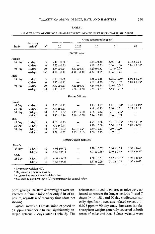
Liver weights. In 14- and 28-day studies, significant changes (increases) in relative liver weights were mild and occurred primarily in animals exposed to 2.5 and 5.0 ppm arsine (Table 1). In 90-day studies, significant
were brain, eyes. three cross sections of the nasal cavity, changes occurred only in high exposure (2.5
TOXICITY OF ARSINE IN MICE, RATS, AND HAMSTERS 779
TABLE I
-
Study Recovery periodh N 0.0
Amine concentration (ppm)
0.025 0.5 2.5 5.0
Female 14 Day
90 Day
Male 14 Day
90 Day
Female 14 Day 28 Day 90 Day
Male 14Day 28 Day 90 Day
Female 28 Day
Male 28 Day
(1 day) 5 (2 days) 6
(3 days) 10
(4 days) 5-6
(1 day) 5
(2 days) 6
(3 days) 10
(4 days) 5-6
( 1 day) (3 days) (3 days) (4 days)
5 3.87 20.15 - 3.83 kO.12 6 3.41 a0.21 - 3.18 k0.33
IO 3.09 * 0.22 3.19 + 0.20 3.28 f 0.12 6 2.82 4 0.16 2.86 + 0.20 2.94 f 0. I8
(1 day) (3 days) (3 days) (4 days)
5 4.63 kO.15 - 4.91 i 0.36 6 3.93 + 0.58 - 3.70 2 0.09
10 3.89 4 0.22 4.01 + 0.24 3.79kO.13 6 3.38 + 0.22 3.23 +- 0.05 3.30+0.15
(3 days) (4 days)
10 4.93 f 0.76 - 5.39 f 0.57 6 5.60 f 0.4 I - 5.01 +- 0.36*
(3 days) 10
(4 days) 6
B6C3F, mice
5.44 + 0.26” - 5.59 L!Z 0.36 5.33 + 0.33 - 5.16k0.23 4.64 f 0.24 4.47 + 0.2 1 4.80 +- 0.33 4.81 i 0.12 4.98 + 0.40 4.72kO.15
5.43 r 0.25 - 5.85 + 0.48 5.77 ?z 0.25 - 5.69 + 0.36 5.42 k 0.2 1 5.25 +0.15 5.46 f 0.24 5.13 a 0.25 5.28 2 0.30 5.29 + 0.3 1
Fischer 344 rats
Syrian Golden hamsters
4.58 zk 0.29 - 4.68 + 0.5 I
5.66 + 0.17 5.74 -+ 0.36 4.74 + 0.29 4.96 AZ 0.14
5.98 zk O.lO* 5.63 + 0.37 5.69 +- 0.24* 5.52 ?z 0.31*
4.11 +-0.18* 3.66 + 0.2 1 3.31 +0.18* 3.04 + 0.09
5.07 +- 0.13* 3.74kO.15 4.10 + 0.20 3.32 +- 0. II
5.60 + 0.75 5.40 rt 0.49
5.02 + 0.31*
5.75 f 0.55 5.86 2k 0.53*
-
6.00 -c 0.24* 6.08 -c 0.29:
-
4.28 f 0.07* 3.57 f0.12
- -
4.98 t 0.14* 3.83 + 0.20
-
5.36 f 0.48 4.87 f 0.37*
5.26 f 0.39* 4.68 f 0.24 - 4.77 f 0.29 5.1 I -r- 0.77 5.30 f 0.65
a Liver/body weight ( 100). ’ Days since last arsine exposure. c Expressed as mean + standard deviation. * Statistically significant (p < 0.05) compared with control value.
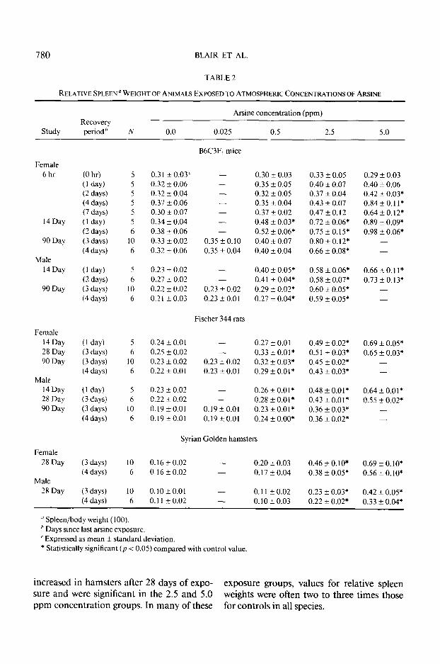
ppm) groups. Relative liver weights were un- spleens continued to enlarge as mice were al- affected in female mice after only 6 hr of ex- lowed to recover for longer periods (4 and 7 posure, regardless of recovery time (data not days). In 14-, 28-, and 90-day studies, statisti- shown). tally significant exposure-related (except for
Spleen weights. Female mice exposed to 0.025 ppm in 90-day study) increases in rela- 5.0 ppm arsine for 6 hr had significantly en- tive spleen weights generally occurred in both larged spleens 2 days later (Table 2). The sexes of mice and rats. Spleen weights were
780 BLAIR ET AL.
TABLE 2
RELATIVE SPLEEN’ WEIGHT OF ANIMALS EXPOSED TO ATMOSPHERIC CONCENTRATIONS OF ARSINE
Study Recovery
period ’
Arsine concentration (ppm)
N 0.0 0.025 0.5 2.5 5.0
B6C3F, mice
Female 6 hr
14 Day
90 Day
Male 14 Day
90 Day
Female 14 Day 28 Day 90 Day
Male
14 Day 28 Day 90 Day
Female 28 Day
Male 28 Day
(0 hr) 5
( 1 day) 5 (2 days) 5
(4 days) 5
(7 days) 5 (I day) 5 (2 days) 6
(3 days) 10 (4 days) 6
( 1 day) (2 days)
t 3 days) (4 days)
5 0.23 + 0.02 - 0.40 k 0.05* 6 0.27 k 0.02 - 0.4 1 + 0.04*
10 0.22 i- 0.02 0.23 f 0.02 0.29 i 0.02* 6 0.2 I + 0.03 0.23 +- 0.01 0.27 + 0.04*
( 1 day) 5 (3 days) 6
(3 days) 10
( 4 days) 6
( 1 day) 5
( 3 days) 6
(3 days) 10 (4 days) 6
(3 days) (4 days)
10 0.16 rt 0.02 - 0.20 f 0.03 6 0. I6 + 0.02 - 0.17 * 0.04
(3 days) 10
(4 days) 6
0.3 1 k 0.03’ 0.32 i 0.06 0.32 * 0.04 0.37 k 0.06 0.30 + 0.07 0.34 f 0.04 0.38 k 0.06 0.33 k 0.02 0.32 rt 0.06
- 0.30 * 0.03 - 0.35 + 0.05 - 0.32 + 0.05 - 0.35 + 0.04 - 0.37 !I 0.02 - 0.48 k 0.03* - 0.52 + 0.06*
0.35 f 0.10 0.40 rt 0.07 0.35 f 0.04 0.40 + 0.04
Fischer 344 rats
0.24kO.01 - 0.27 + 0.0 1 0.25 f 0.02 - 0.33 f 0.01* 0.23 f 0.02 0.23 f 0.02 0.32 -+ 0.03* 0.22 + 0.0 I 0.23 2 0.0 I 0.29 k O.Ol*
0.23 k 0.02 - 0.26 k O.Ol* 0.22 * 0.02 - 0.28 kO.01’ 0.19 to.01 0.19~0.01 0.23 tO.OI* 0.19c0.0l 0.19 * 0.01 0.24 + O.OO*
Syrian Golden hamsters
0.10+0.01 - 0.11 kO.02 0.11 * 0.02 - 0. IO + 0.03
0.33 f 0.05 0.40 k 0.07 0.37 * 0.04 0.43 f 0.07 0.47 f 0.12 0.72 f 0.06* 0.75 io.15* 0.80 f 0.12* 0.66 -+ 0.08*
0.58 f 0.06* 0.58 +- 0.07* 0.60 + 0.05* 0.59 -c 0.05*
0.49 t 0.02* 0.5 1 + 0.03* 0.45 f 0.02* 0.43 f 0.03*
0.48 + O.Ol* 0.43 t o.ot* 0.36 + 0.03* 0.36 f 0.02*
0.46 ?z 0. IO* 0.38 f 0.05*
0.23 f 0.03* 0.22 f 0.02*
0.29 + 0.03 0.40 k 0.06 0.42 + 0.03* 0.84 + 0.1 l* 0.64 + 0.12* 0.89 f 0.09* 0.98 + 0.06*
- -
0.66 + 0.11* 0.73 k 0.13*
0.69 f 0.05* 0.65 + 0.03*
-
0.64 + O.Ol* 0.55 * 0.02*
- -
0.69 + O.lO* 0.56 k O.lO*
0.42 f 0.05* 0.33 + 0.04*
u Spleen/body weight (100). ” Days since last arsine exposure. ’ Expressed as mean + standard deviation. * Statistically significant (p < 0.05) compared with control value.
increased in hamsters after 28 days of expo- exposure groups, values for relative spleen sure and were significant in the 2.5 and 5.0 weights were often two to three times those ppm concentration groups. In many of these for controls in all species.
TOXICITY OF ARSINE IN MICE, RATS, AND HAMSTERS 781
TABLE 3
Study Recovery period"
Arsine concentration (ppm)
N 0.0 0.025 0.5 2.5 5.0
Female 6 hr
14 Day
90 Day
Male 14 Day
90 Day
Female 14 Day 28 Day 90 Day
Male 14 Day 28 Day 90 Day
Female 28 Day
Male 28 Day
(0 W 5
( 1 day) 5
(2 days) 5
(4 days) 5
(7days1 5 (1 day) 5 (2 days) 5
(3 days) IO
(4 days) 6
( 1 day) 5 (2 days) 6 (3 days) 10
(4 days) 6
( 1 day) (3 days) (3 days) (4 days)
5 50.8 f 3.7 48.4 + 2. I 6 48.8 f 0.7 - 42.6 + l.l*
10 46.3 +- 0.9 46.2 f 0.9 42.6 f 1.3* 5-6 45.0 f I .4 44.4 * 1.7 42.5 + 0.8*
( I day) (3 days) ( 3 days) (4 days)
4-5 52.4 f 1.8 - 49.0 t 2.0* 6 48.2 f 0.8 44.0 f 1.3*
IO 47.8 + 0.8 47.6 + 1.2 46.2 f 1.2* 6 46.2 f 1.6 47.0 f 0.9 44.5 It 1.2
(3 days) (4 days)
10 52.3 rt_ 2.0 49.9k2.1’ 6 52.3 f 3.3 - 52.Ok 1.1
(3 days) (4 days)
10 57.0 f 2. I 53.1 zk 2.8* 6 55.3 + 2.7 - 55.5 f 0.6
B6C3F, mice
49.0 f l.5h - 46.0 f 0.6 -
46.9 k 0.6 47.7 f 0.5 - 48.7 + 0.9 - 45.8 + 1.3 -
44.8 + 2.3 44.5 + 1.8 45.4 + 2.9 44.3 + 2.3 39.7 * 2.0*
49.2 f 2.2 - 46.3 + 4.3 -
45.6 f 1.8 45.1 + 3.2 41.8 f 1.6 42.8 f 1.0
Fischer 344 rats
49.2 f 1.0 49.7 + 1.0 47.4 f 0.6 45. I kO.3 45.7 f 1.8 44.3 f 2.5* 47.2 f 2.0 45. I + 0.8* 47.8 f 1.7 45.2 f 1.4* 45.2 f 2.6 44.6 f 1.5 43.2 f 1.6 42.8 + 2.8 44.2+ 1.1 43.2 IL 0.9 39.3 f 0.8* 37.8 f 1.7*
47.5 I! 3.3 45.6 +- 1.1* 42.8 f 2.5 39.3 +_ 3.3* 45.6 + 2.5 41.0 f 1.8* 41.5 f 1.2 37.0 + 0.7*
46.2 f 2.4* 39.2 + 0.8* 43.0? 1.1* 41.7 f 1.6*
48.5 +_ 1.7* 41.0-t 1.2* 43.2 f 1.2* 43.0 f 1.9*
Syrian Golden hamsters
48.0 f 2.5* 48.0 k 1.7*
51.9 f 2.6* 52.8 f 1.3
49.5 f 2.1 41.6 f 1.9* 38.6 f l.l* 38.9 t 1.4* 43.0-+ 1.1* 41.0f3.1’ 39.0 f 3.3*
- -
41.2 f 2.5* 36.8 f 2.6*
- -
44.2 +_ 3.1* 40.0 It 0.7*
- -
46.8 + 1.1* 41.9,0.9*
- -
47.8 It 1.4* 49.0 k 2.1*
49.0 Tt 2.2* 50.5 f 3.s*
’ Spleen/body weight (100). ’ Expressed as mean f standard deviation. * Statistically significant (p < 0.05) compared with control value.
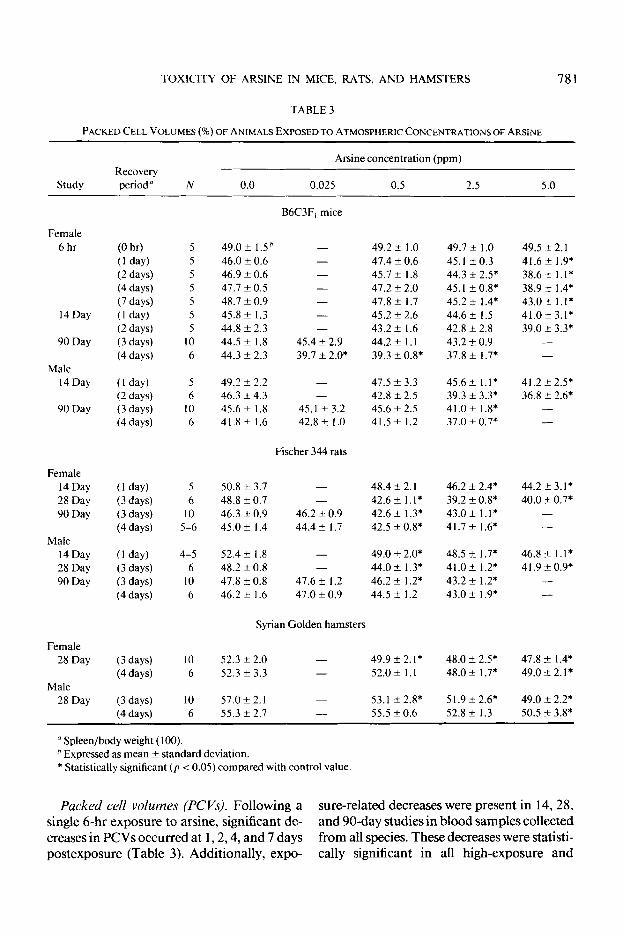
Packed cell volumes (PCVs). Following a sure-related decreases were present in 14,28, single 6-hr exposure to arsine, significant de- and 90-day studies in blood samples collected creases in PCVs occurred at 1,2,4, and 7 days from all species. These decreases were statisti- postexposure (Table 3). Additionally, expo- tally significant in all high-exposure and
782 BLAIR ET AL.
most mid-exposure groups. Occasionally, de- creases in low-exposure groups were signifi- cant.
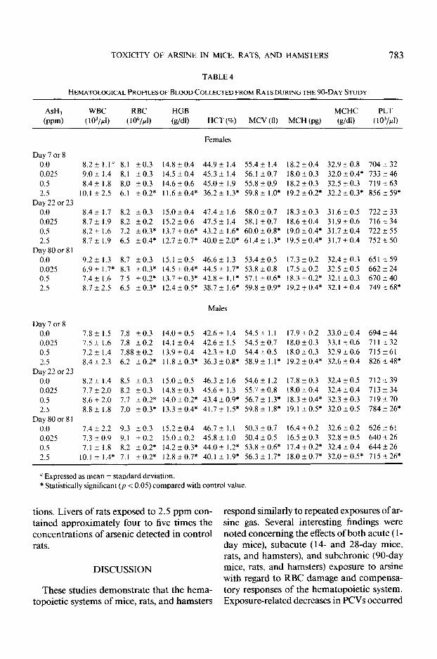
Hematological projles in rats. Results of hematological profiles performed on blood samples collected from both sexes of rats ex- posed to 2.5 ppm arsine included decreased red blood cell (RBCs) counts, hemoglobin concentrations (HGBs), and hematocrits (HCTs) at 7 and 8 days (Table 4). Concur- rently, increases were seen in mean corpuscu- lar volumes (MCVs), mean corpuscular he- moglobins (MCHs), and platelet counts. At 22 and 23 days of exposure, similar changes also were seen in rats in 0.5 ppm group. After 80 and 8 1 days, decreased RBCs, HGBs, and HCTs were present in blood samples col- lected from female rats in the low (0.025 ppm) exposure group. The anemia at 7 and 8 days caused by exposure to 2.5 ppm arsine was more severe than that at 22, 23, 80, and 81 days of exposure to the same concentra- tion.
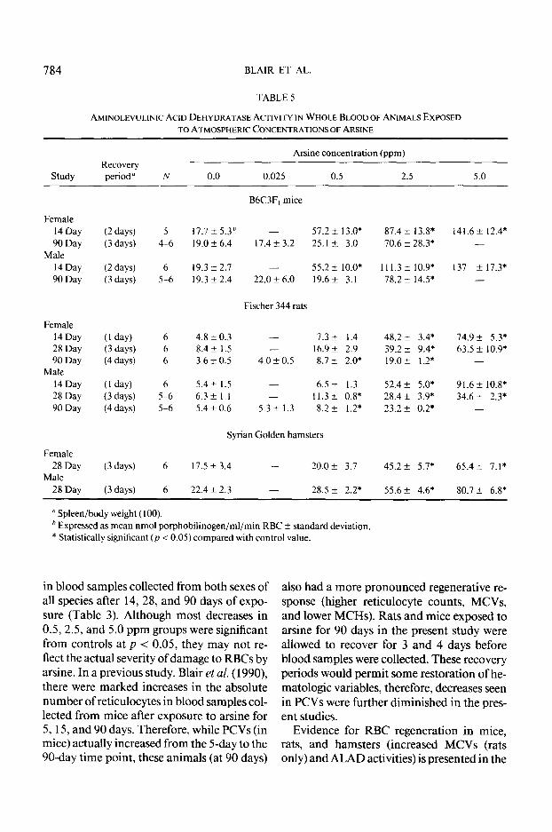
&Aminolevulinic acid dehydratase activity. ALAD activity was measured in whole blood collected from mice euthanized after 2 and 3 days of recovery in the 14- and 90-day stud- ies, respectively: in blood collected from rats after 1, 3, and 4 days of recovery in the 14-, 28-, and 90-day studies, respectively; and, fi- nally, in blood collected from hamsters after 3 days of recovery (Table 5). Significant in- creases in ALAD activity occurred in mice in all exposure groups after 14 days of exposure and only in the 2.5 ppm exposure group after 90 days. Results were similar for rats and hamsters.
Histopathological changes. No effects of arsine were observed on the nasal cavity, re- spiratory tract, or lungs. Treatment-related lesions were noted only in spleen, liver, and bone marrow. In mice, the spleen was the only organ in which gross effects (dark color and increased size) were observed. All other organs appeared normal. All mice from the 1 -day, acute study were necropsied. No gross effects were seen until after 4 days of recov-
ery, at which time the spleens were noticeably enlarged. After 7 days of recovery, spleens were dark as well. Similar gross observations were made in both sexes of mice in the 14- day (2.5 and 5.0 ppm) and 90-day (0.5 ppm (males only) and 2.5 ppm) studies. These splenic changes correlated well with micro- scopic changes which included increased hemosiderosis and extramedullary hemato- poiesis. Intracanalicular bile stasis in liver was observed in 3/ 10 female and 7/ 10 male mice in the 2.5 ppm group of the 90-day study.
Histopathological changes observed in or- gans collected from rats were similar to those seen in mice. Again, gross necropsy observa- tions were primarily limited to the spleen. These were enlarged in virtually all rats of both sexes exposed to 2.5 or 5.0 ppm arsine independent of the number of days of expo- sure (14, 28, or 90). At these concentrations, the spleen was also darkened in the 28-day and 90-day studies, but not in the 1Cday study. Lesions (increased hemosiderosis and extramedullary hematopoiesis) were present only in spleen and bone marrow of 2.5 ppm male and female rats in the 90-day study. Mi- croscopic lesions described above correlated well with macroscopic changes (darkening and enlargement) seen in all males and fe- males at this exposure level at necropsy. Hy- perplasia of the bone marrow was observed only in 2.5 ppm (90 day) male and female rats. No other exposure-related lesions were observed in rats.
Dark and enlarged spleens were observed in all hamsters exposed to 2.5 and 5.0 ppm arsine for 28 days. These observations corre- lated with increases in hemosiderosis and ex- tramedullary hematopoiesis. Increased he- patic hemosiderosis was observed in males and females of the same exposure groups.
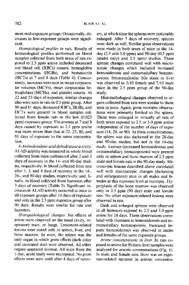
Arsine concentrations in liver. In rats ex- posed to arsine for 90 days, liver samples were analyzed for arsenic concentrations (Fig. 1). In male and female rats, there was an expo- sure-related increase in arsenic concentra-
TOXICITY OF ARSINE IN MICE, RATS, AND HAMSTERS 783
TABLE 4
ASH, WBC
(mm) ( 1 03//4
RBC
( 1 @/,I)
HGB
(g/W MCHC PLT
HCT (%) M-VU MCH(pg) Wdl) ( 1 031rl)
Females
Day7or8 0.0 8.2+1.1” 8.1 kO.3 14.8kO.4 44.9+ 1.4 55.4kl.4 18.2kO.4 32.9kO.8 704a32 0.025 9.0 f 1.4 8.1 kO.3 14.5 f0.4 45.3 + 1.4 56.1 k0.7 18.OkO.3 32.0+0.4* 733 -+ 46 0.5 8.4+ 1.8 8.0 k0.3 14.6 k0.6 45.Ok 1.9 55.8 kO.9 18.2kO.3 32.5 kO.3 719 f 63 2.5 10.1 k 2.5 6.1 f 0.2* 11.6 -cO.4* 36.2 + 1.3* 59.8 f l.O* 19.2 +0.2* 32.2 -+0.3* 856 f 59*
Day 22 or 23 0.0 8.4k1.7 8.2 kO.3 15.0+0.4 47.4kl.6 58.0k0.7 18.3kO.3 31.6kO.5 722233 0.025 8.7k1.9 8.2 kO.2 15.2kO.6 47.5fl.4 58.120.7 18.620.4 31.9kO.6 716k34 0.5 8.2kl.6 7.2 &0.3* 13.7+0.6* 43.2*1.6* 60.0?0.8* 19.0-+0.4* 31.7kO.4 722255 2.5 8.7rt 1.9 6.5 &0.4* 12.7r0.7* 40.0+_2.0* 61.4+ 1.3* 19.5*0.4* 31.7t0.4 752+50
Day80or81 0.0 9.2kl.3 8.7 k0.3 15.lkO.5 46.6kl.3 53.41-0.5 17.3kO.2 32.4kO.3 65lk59 0.025 6.9 f 1.7* 8.3 +0.3* 14.5 &0.4* 44.5 f 1.7* 53.8 kO.8 17.5 k0.2 32.5 zkO.5 662 +24 0.5 7.4kl.6 7.5 +0.2* 13.7+0.3* 42.8+1.1* 57.1 + 0.6; 18.3 + 0.2* 32.1 + 0.3 670 -t 40 2.5 8.7 + 2.5 6.5 +0.3* 12.4 +0.5* 38.7 f 1.6* 59.8 ?0.9* 19.2+0.4* 32.1 ? 0.4 749 2 68*
Males
Day 7 or 8 0.0 7.8kl.5 7.8 kO.3 14.0+0.5 42.6kl.4 54.5kl.l 17.9 + 0.2 33.0 + 0.4 694 2 44 0.025 7.5 + 1.6 7.8 kO.2 14.1 kO.4 42.6 k 1.5 54.5 k0.7 18.0k0.3 33.1 k0.6 711 ?32 0.5 7.2 f 1.4 7.88 + 0.2 13.9kO.4 42.3kl.O 54.4kO.5 18.0t0.3 32.920.6 715k61 2.5 8.4k2.3 6.2 kO.2* 11.8 &0.3* 36.3 ?0.8* 58.9 f l.l* 19.2 kO.4* 32.6 20.4 826 +48*
Day 22 or 23 0.0 8.2 f 1.4 8.5 kO.3 15.0 k 0.5 46.3 -t 1.6 54.6 + 1.2 17.8 k 0.3 32.4k0.5 712 + 39 0.025 7.7 f 2.0 8.2 kO.3 14.8k0.3 45.6+_ 1.3 55.7 kO.8 18.0 t 0.4 32.4 + 0.4 7 I3 k 34 0.5 8.6 k2.0 7.7 +-0.2* 14.0+0.2* 43.4*0.9* 56.7 f 1.3* 18.3 f0.4* 32.3 kO.3 719? 70 2.5 8.8? 1.8 7.0 kO.3* 13.3*0.4* 41.7+1.5* 59.8*1.8* 19.lf0.5* 32.OkO.5 784-c26*
Day80or81 0.0 7.4t2.2 9.3 to.3 15.250.4 46.7kl.l 50.3 + 0.7 16.4 ?z 0.2 32.6 + 0.2 626 + 61 0.025 7.3 * 0.9 9.1 f 0.2 15.0 f 0.2 45.8 + 1.0 50.4 f 0.5 16.5 k 0.3 32.8 + 0.5 640 2 26 0.5 7.1 i- 1.8 8.2 ?0.2* 14.2 f0.3* 44.Ok 1.2* 53.8 ?0.6* 17.4 kO.2* 32.4 + 0.4 644+26 2.5 10.1k 1.4* 7.1 ?0.2* 12.8f0.7* 40.1~1.9* 56.3?1.7* 18.0+-0.7* 32.0+0.5* 715?26*
u Expressed as mean f standard deviation. * Statistically significant (p < 0.05) compared with control value.
tions. Livers of rats exposed to 2.5 ppm con- respond similarly to repeated exposures of ar- tained approximately four to five times the sine gas. Several interesting findings were concentrations of arsenic detected in control noted concerning the effects of both acute ( l- rats. day mice), subacute (14- and 28-day mice,
rats, and hamsters), and subchronic (90-day
DISCUSSION mice, rats, and hamsters) exposure to arsine with regard to RBC damage and compensa-
These studies demonstrate that the hema- tory responses of the hematopoietic system. topoietic systems of mice, rats, and hamsters Exposure-related decreases in PCVs occurred
784 BLAIR ET AL.
TABLE 5
AMINOLEVULINICACIDDEHYDRATASEACTIVITYINWHOLEBLOODOFANIMALS EXPOSED TO ATMOSPHERIC CONCENTRATIONS OF ARSINE
Study Recovery
perioda N 0.0
Arsine concentration (ppm)
0.025 0.5 2.5 5.0
Female 14 Day 90 Day
Male 14 Day 90 Day
Female 14 Day 28 Day 90 Day
Male 14 Day 28 Day 90 Day
Female 28 Day
Male 28 Day
(2 days) 5
(3 days) 4-6
(2 days) 6 (3 days) 5-6
(1 day) 6
(3 days) 6
(4 days) 6
(1 day) 6
(3 days) 5-6 (4 days) 5-6
( 3 days) 6
(3 days) 6
B6C3F, mice
17.7 i 5.3* - 57.2 F 13.0* 19.0 + 6.4 17.4 + 3.2 25.1 +_ 3.0
19.3k2.1 - 55.2 k lO.O* 19.3 f 2.4 22.0 k 6.0 19.6+ 3.1
Fischer 344 rats
4.8k0.3 - 7.3f 1.4 8.4rt_ 1.5 - 16.9 f 2.9 3.6 t 0.5 4.0 f 0.5 8.7 I 2.0*
5.4+ 1.5 - 6.5 + 1.3 6.3kl.l - 11.3 f 0.8* 5.4 f 0.6 5.3 + 1.3 8.3+ 1.2*
Syrian Golden hamsters
17.5 f 3.4 - 20.0 f 3.7
12.4 f 2.3 - 28.5 + 2.2*
87.4 + 13.8* 70.6 f 28.3*
111.3 f 10.9* 78.2 ?I 14.5*
48.2 + 3.4* 39.2 f 9.4* 19.0+ 1.2*
52.4 f 5.0* 28.4 + 3.9* 23.2 + 0.2*
45.2 + 5.7*
55.6 f 4.6*
141.6 III 12.4* -
137 ?I 17.3* -
74.9 * 5.3* 63.5 + 10.9*
-
91.6 k 10.8* 34.6 + 2.3*
65.4+ 7.1*
80.7 k 6.8*
a Spleen/body weight (100). ’ Expressed as mean nmol porphobilinogen/ml/min RBC f standard deviation. * Statistically significant (p < 0.05) compared with control value.
in blood samples collected from both sexes of all species after 14, 28, and 90 days of expo- sure (Table 3). Although most decreases in O-5,2.5, and 5.0 ppm groups were significant from controls at p < 0.05, they may not re- flect the actual severity of damage to RBCs by arsine. In a previous study, Blair et al. (1990), there were marked increases in the absolute number of reticulocytes in blood samples col- lected from mice after exposure to arsine for 5, 15, and 90 days. Therefore, while PCVs (in mice) actually increased from the 5-day to the 90-day time point, these animals (at 90 days)
also had a more pronounced regenerative re- sponse (higher reticulocyte counts, MCVs, and lower MCHs). Rats and mice exposed to arsine for 90 days in the present study were allowed to recover for 3 and 4 days before blood samples were collected. These recovery periods would permit some restoration of he- matologic variables, therefore, decreases seen in PCVs were further diminished in the pres- ent studies.
Evidence for RBC regeneration in mice, rats, and hamsters (increased MCVs (rats only) and ALAD activities) is presented in the

TOXICITY OF ARSINE IN MICE, RATS, AND HAMSTERS 785
12 -
IO--
B-- ‘iii a 3 .o 6-- c 2 4--
2 --
T
0 Lid 0.0 0.5 Exposure Concentration (ppm)
FIG. 1. Mean arsenic concentration of livers from male and female rats at the end of 90 days of exposure to ar- sine.
current study. Hematological profiles of blood collected from rats at interim time points during the 90-day study revealed se- vere hemolytic anemia in high-dose animals with evidence of regeneration (increased MCVs) (Table 4). This finding (high MCVs) indicates an increase in the number of large immature RBCs. As previously described, the anemia at the earliest interim time point was more dramatic than that at later time points due to an increasingly active regenerative re- sponse. Additionally, a recent study by Hong et al. (1989) reported that mice exposed to ar- sine had an appropriate regenerative response despite a reduction in erythroid colony-form- ing units (CFU-E) in the bone marrow.
ALAD, an important enzyme in the path- way of heme synthesis, is normally present in immature RBCs. As the cells mature and he- moglobin production declines, ALAD activ- ity declines as well. Mature RBCs lack mito- chondria and are unable to synthesize heme. Therefore, high activity of this enzyme in
whole blood is consistent with an increase in the number of immature RBCs. The increase in ALAD activity in all three species of ani- mals exposed to arsine in the current study was exposure-related and consistent with a regenerative response initiated by arsine (Ta- ble 5). These results indicate that compensa- tory stimulation of the hematopoietic system with release of immature red cells (reticulo- cyte counts) must be evaluated when moni- toring worker populations exposed to this agent. Currently, hematological screens per- formed on human populations consist only of complete blood cell counts (CBCs). This routine hematological profile alone may be inadequate to effectively monitor the pro- longed effects of arsine.
The exposure-related splenomegaly ob- served in present studies is consistent with histopathological findings in the spleen. This effect is primarily the result of increased re- moval of damaged RBCs (fragments) with at- tendant hemosiderosis and, secondarily, in- creased splenic hematopoiesis. The func- tional aspects of these processes on normal splenic function are not completely known but are of obvious concern with regard to re- ported alterations in immunocompetency of mice exposed to arsine (Rosenthal et al., 1988). Further studies are needed to com- pletely evaluate the effects of arsine on this major target organ system.
Another major histopathological finding in this study was the development of hepatic in- tracanalicular bile stasis in male and female mice exposed to high concentrations of ar- sine. In rats, high deposits of arsenic were found in the liver after exposure (Fig. 1). Pre- vious studies from a number of laboratories (Hayes et al., 1982) have shown that liver is a target organ for inorganic arsenicals in both man (Morinaga milk incident, Frejaville et al., 1972) and laboratory animals but rela- tively little work has been done concerning arsine hepatotoxicity. Levvy ( 1947) and Hughes and Levvy ( 1947) studied the effects of arsine in vivo and in vitro. Hughes and
786 BLAIR ET AL.
Levvy (1947) found direct effects of arsine on liver slice respiration in vitro and demon- strated that the molar concentration of arsine required to produce decreases in tissue respi- ration was only one-fourth that required to produce similar effects by arsenite. The pres- ent investigations support these observations and suggest that prolonged exposure to arsine interferes with bile excretion or flow. Further studies are needed to delineate the mecha- nisms responsible.
In summary, several important conclu- sions can be made from these studies. There were no apparent differences in the types of effects produced by arsine in the three species. However, there was a difference in the degree of the effects between rats and mice according to hematologic profiles. In the study by Blair et al. (1990), in which both sexes of mice were exposed to the same concentrations of arsine for similar periods of time, the significant effects were limited to the 2.5 ppm exposure group. In the present study, rats in the 0.5 and 2.5 ppm exposure groups exhibited statisti- cally significant anemias by 22 and 23 days. By 80 and 8 1 days, females in the lowest ex- posure group, 0.025 ppm, had significant anemia. This indicates that rats are more sus- ceptible to arsine than mice. It may be that arsine produces similar effects in both spe- cies, but because of the greater capacity for splenic extramedullary hematopoiesis in mice, this species has increased capability for erythrocytic regeneration than rats. The over- all effects in hamsters were similar to those in mice and rats, but relative responses cannot be determined without complete hemato- logic profiles.
Most importantly, even though a single ex- posure to 0.5 ppm arsine (which is 10 times the TLV of 0.05 ppm) showed no significant effects on the hematopoietic system of mice, repeated exposure (80 or 81 days) to 0.025 ppm (which is one-half the TLV) produced significant anemia in rats. Therefore, the TLV may need to be reinvestigated and re- considered. Long-term exposures of labora-
tory animals to low levels of arsine produced significant hematologic toxicity. This pattern of repeated low-level exposures is more con- sistent with occupational exposures. In the semiconductor industry (or others), workers are more apt to be exposed to low levels of arsine over a number of days rather than to high or moderate levels for a single day. Addi- tionally, these data indicate that when work- ers who may have been exposed to low con- centrations of arsine are screened, PCVs or CBCs are not adequate for the detection of anemia due to arsine exposure. If workers have been exposed to low levels of arsine over a long period of time, decreases in RBCs, HCTs, or HGBs may be diminished from ac- tive regenerative responses. Additionally, the only indicator of regeneration included in this panel (CBC) is mean corpuscular vol- ume. MCVs are more variable in human populations than in controlled animal studies and mild increases may not be significant enough to alert the clinician. These hemato- logic profiles must be modified to include sensitive indicators of regeneration (reticulo- cyte counts, ALAD activities).
ACKNOWLEDGMENTS
We thank R. W. O’Conner. D. L. Crawford, N. F. Gage, II, R. C. Hamrick, A. K. Miller, C. R. Moorman, S. W. Lloyd, P. A. Sams, P. Y. Seabrook, B. Adkins, and J. W. Walters of NSI Technology Services Corp., G. Riley of Experimental Pathology Laboratories, Inc., and R. E. Wilson, H. Sanders, S. Eustis, H. Amyx. and J. Thigpen of NIEHS for assistance throughout the course of these studies. We thank Dr. B. A. Schwetz for helpful advice concerning these studies. These efforts were sup- ported in part by NIEHS Contract NOl-ES-5044.
REFERENCES
ACGIH (1988-89). Threshold Limit Values for Chemical Substances in the Work Environment. American Con- ference of Governmental Industrial Hygienists, Wash- ington, DC.
BLAIR, P. C., BECHTOLD, M.,THOMPSON, M. B., WIL- SON,R.E.,MOORMAN,M.P.. AND FOWLER, B. A.
TOXICITY OF ARSINE IN MICE, RATS. AND HAMSTERS 787
(1990). Evidence for oxidative damage to red blood cells in mice and rats induced by arsine gas. Toxicol- ogy, in press.
DUPIS. R. D. (1984). Metalorganic chemical vapor depo- sition of III-V semiconductors. Science 226,623-629.
FOWLER. B. A., MOORMAN, M. P., ADKINS, B.. JR., BLAKEWELL, W. E., JR., BLAIR, P. C., AND THOMP- SON, M. B. (1988). Arsine: Toxicity data from acute and short-term inhalation exposures. In Hazard As- sessment and Control Technology in Semiconductor Manufacturing, ACGIH, Cincinnati, OH.
FOWLER, B. A., AND SILBERGELD, E. K. Occupational diseases-New workforces, new workplaces. Ann. N. Y. Acad. Sci., in press.
FOWLER, B. A., AND WEISSBURG, J. B. (1974). Arsine Poisoning. N. Engl. J. Med. 291, 1 I7 1- 1 174.
FREJAVILLE, J. P., BESCOL, J.. LECLERC, J. P., GUILLAM, L., CRABIE, P., CONSO, F.. GERVAIS, P.. AND GAUT- LIER. M. (I 972). Acute poisoning with arsenic deriva- tives. Ann. Med. Int. (Paris) 123,7 13-722.
GOERING, P. L.. MARONPOT, R. R., AND FOWLER. B. A. ( 1988). Effect of intratracheal gallium arsenide admin- istration on b-aminolevulinic acid dehydratase in rats: Relationship to urinary excretion of aminolevulinic acid. To.xicol. Appl. Pharmacol. 92, 179-193.
HAYES, A. W., FEDOROWSKI, T., BALAZS, T.. CARLTON, W. W., FOWLER, B. A., GILMAN, M. R., HEYMAN. I., JACKSON, B. A.. KENNEDY, G. L., SHAPIRO. R. E., SMITH, C. C., TARDIFF, R. G.. AND WEIL, C. C. ( 1982). Correlation of human hepatotoxicants with hepatic damage in animals. Fundam. Appl. To.uicol. 2, 55-66.
HONG, H. L., FOWLER, B. A.. AND BOORMAN, G. A. (1989). Hematopoietic effects in mice exposed to ar- sine gas. To.xicol. Appl. Pharmacol. 97, 173- 182.
HUGHES, W., AND LEVVY, G. A. (1947). The toxicity of arsine solutions for tissue slices. Biochem. J. 41,8-l 1.
LEVVY, G. A. (1947). A study of arsine poisoning. Q. J. Exp. Physiol. 34,47-67.
ORLIC, D., Wu, J. M., CARMICHAEL, R. D.. QUAINI, F., KOBYLACK, M., AND GORDON, A. S. (1982). In- creased erythropoiesis and 2’5’-A polymerase activity in the marrow and spleen of phenylhydrazine-injected rats. E-up. Hematol. 10,478-485.
PERNIS. B.. AND MAGSTRETTI, M. (1960). A study of the mechanisms of acute hemolytic anemia from arsine. Med. LAVORO51,37-41.
ROSENTHAL, G. J., FORT, M. M., GERMOLEC, D. R., ACKERMANN. M. F.. LAMM, K. R., THOMAS, P. H.. BLAIR, P. C., FOWLER, B. A.. AND LUSTER, M. I. The effect of subchronic arsine inhalation on immune function and host resistance. Submitted for publica- tion.
SARGER, D. E. (1987, January 14). Pregnancy transfers by A.T. & T.-Miscarriages feared after study on chips. New York Times.
U.S. Environmental Protection Agency (USEPA) ( 1986). Air Quality Criteria@ Lead, Vol. 3, 9-20-9- 49 EPA-600/8/83, June 1986.
VAHTER, M. (198 1). Biotransformation of trivalent and pentavalent inorganic arsenic in mice and rats. Envi- ron. Rex 25286-293.
WALD, P. H., AND BECKER, C. E. (1986). Toxic gases used in the microelectronics industry. In Occupational Medicine: Microelectronic Industry. State of the Art Reviews. Vol. l., No. 1 (J. LaDou, Ed.), pp. 105-I 18. Hawley & Belfus. Philadelphia.
WEBB, D. R., WILSON, S. E., AND CARTER, D. E. (1987). Pulmonary clearance and toxicity of respirable gal- lium arsenide particulated intratracheally instilled into rats. Amer. Ind. Hyg. J. 48,660-667.