Comparative Analysis of End Point Enzymatic Digests of Arabino-Xylan Isolated from Switchgrass...
24
Metabolites 2012, 2, 959-982; doi:10.3390/metabo2040959 metabolites ISSN 2218-1989 www.mdpi.com/journal/metabolites/ Article Comparative Analysis of End Point Enzymatic Digests of Arabino-Xylan Isolated from Switchgrass (Panicum virgatum L) of Varying Maturities using LC-MS n † Michael J. Bowman 1, *, Bruce S. Dien 1 , Patricia J. O’Bryan 1 , Gautam Sarath 2 and Michael A. Cotta 1 1 U.S. Department of Agriculture, Agricultural Research Service, National Center for Agricultural Utilization Research, Bioenergy Research Unit, 1815 N. University Street, Peoria, IL 61604, USA; E-Mails: [email protected] (B.S.D.); [email protected] (P.J.O.); [email protected] (M.A.C.) 2 U.S. Department of Agriculture, Agricultural Research Service, Central-East Regional Biomass Center, 137 Keim Hall, East Campus, UNL, Lincoln, NE 68583, USA; E-Mail: [email protected] (G.S.) † Mention of trade names or commercial products in this article is solely for the purpose of providing scientific information and does not imply recommendation or endorsement by the U.S. Department of Agriculture. * Author to whom correspondence should be addressed; E-Mail: [email protected] Tel.: +1-309-681-6327; Fax: +1-309-681-6427. Received: 14 September 2012; in revised form: 30 October 2012 / Accepted: 9 November 2012 / Published: 19 November 2012 Abstract: Switchgrass (Panicum virgatum L., SG) is a perennial grass presently used for forage and being developed as a bioenergy crop for conversion of cell wall carbohydrates to biofuels. Up to 50% of the cell wall associated carbohydrates are xylan. SG was analyzed for xylan structural features at variable harvest maturities. Xylan from each of three maturities was isolated using classical alkaline extraction to yield fractions (Xyl A and B) with varying compositional ratios. The Xyl B fraction was observed to decrease with plant age. Xylan samples were subsequently prepared for structure analysis by digesting with pure endo-xylanase, which preserved side-groups, or a commercial carbohydrase preparation favored for biomass conversion work. Enzymatic digestion products were successfully permethylated and analyzed by reverse-phase liquid chromatography with mass spectrometric detection (RP-HPLC-MS n ). This method is OPEN ACCESS
-
Upload
independent -
Category
Documents
-
view
1 -
download
0
Transcript of Comparative Analysis of End Point Enzymatic Digests of Arabino-Xylan Isolated from Switchgrass...

Metabolites 2012, 2, 959-982; doi:10.3390/metabo2040959
metabolitesISSN 2218-1989
www.mdpi.com/journal/metabolites/
Article
Comparative Analysis of End Point Enzymatic Digests of Arabino-Xylan Isolated from Switchgrass (Panicum virgatum L) of Varying Maturities using LC-MSn †
Michael J. Bowman 1,*, Bruce S. Dien 1, Patricia J. O’Bryan 1, Gautam Sarath 2 and
Michael A. Cotta 1
1 U.S. Department of Agriculture, Agricultural Research Service, National Center for Agricultural
Utilization Research, Bioenergy Research Unit, 1815 N. University Street, Peoria, IL 61604, USA;
E-Mails: [email protected] (B.S.D.); [email protected] (P.J.O.);
[email protected] (M.A.C.) 2 U.S. Department of Agriculture, Agricultural Research Service, Central-East Regional Biomass
Center, 137 Keim Hall, East Campus, UNL, Lincoln, NE 68583, USA;
E-Mail: [email protected] (G.S.)
† Mention of trade names or commercial products in this article is solely for the purpose of providing
scientific information and does not imply recommendation or endorsement by the U.S. Department
of Agriculture.
* Author to whom correspondence should be addressed; E-Mail: [email protected]
Tel.: +1-309-681-6327; Fax: +1-309-681-6427.
Received: 14 September 2012; in revised form: 30 October 2012 / Accepted: 9 November 2012 /
Published: 19 November 2012
Abstract: Switchgrass (Panicum virgatum L., SG) is a perennial grass presently used for
forage and being developed as a bioenergy crop for conversion of cell wall carbohydrates
to biofuels. Up to 50% of the cell wall associated carbohydrates are xylan. SG was
analyzed for xylan structural features at variable harvest maturities. Xylan from each of
three maturities was isolated using classical alkaline extraction to yield fractions
(Xyl A and B) with varying compositional ratios. The Xyl B fraction was observed to
decrease with plant age. Xylan samples were subsequently prepared for structure analysis
by digesting with pure endo-xylanase, which preserved side-groups, or a commercial
carbohydrase preparation favored for biomass conversion work. Enzymatic digestion
products were successfully permethylated and analyzed by reverse-phase liquid
chromatography with mass spectrometric detection (RP-HPLC-MSn). This method is
OPEN ACCESS

Metabolites 2012, 2
960
advantageous compared to prior work on plant biomass because it avoids isolation of
individual arabinoxylan oligomers. The use of RP-HPLC- MSn differentiated 14 structural
oligosaccharides (d.p. 3–9) from the monocomponent enzyme digestion and nine
oligosaccharide structures (d.p. 3–9) from hydrolysis with a cellulase enzyme cocktail. The
distribution of arabinoxylan oligomers varied depending upon the enzyme(s) applied but
did not vary with harvest maturity.
Keywords: switchgrass; xylan; xylooligosaccharides; LC-MSn; biofuel
1. Introduction
Switchgrass (Panicum virgatum L., SG) is a warm season perennial grass that is native to the North
American prairies. Because of its high production yield and other favorable agronomic traits it is
grown for forage production and is being developed as a bioenergy crop [1]. Biochemical conversion
of switchgrass biomass into renewable biofuels and chemicals begins with the extraction of
carbohydrates using a combination of thermal, chemical, and enzymatic processing steps. The majority
of the carbohydrates are contained in the primary and secondary plant cell walls. The primary cell wall
of grasses are composed of polysaccharides cellulose (20%–30%), xylan (20%–40%), and pectin (5%)
with a minor polyaromatic lignin component; the secondary cell walls have an increased quantity of
cellulose (35%–45%), xylan (40%–50%), and lignin (20%), with a decreased pectin content
(0.1%) [2]. The high content of xylan within the grass cell wall material makes it an important factor in
the conversion process; as both an impedant to efficient cellulose conversion [3,4] and as a source of
pentose sugars [5]. Grass xylan is a heterogeneous polymer consisting of repeating (1→4)--linked
xylose residues with variability in acetyl or arabinose substitutions at the 2-O and/or 3-O positions of
backbone xyloses. Further complexity of the chains can arise from additional substitutions
(hexose, pentose, acetyl, and/or phenolics) on the arabinose side chains [6,7].
Lignocellulose is recalcitrant to enzymes and, therefore, pretreatment is required to open up the cell
wall structure. Numerous pretreatment strategies have been proposed in the literature [8]. The xylan
structure affects the processing of biomass at the pretreatment step. For example, when biomass is
pretreated with a mineral acid catalyst at relatively high temperatures (e.g., 160–200 °C); xylan
hydrolysis displays bimodal kinetics with fast and slow (20%–35% of total xylan) reacting fractions [9].
The slow kinetic fraction partially sets the minimal pretreatment severity. Following pretreatment, the
carbohydrate polymers are hydrolyzed to sugars with enzymes either prior to or simultaneously with
fermentation. Knowledge of the xylan structure can be directly used in formulating the xylanase
cocktail so as to obtain the highest yield with the lowest number of enzymes. Partially digested xylan
is a source of fermentation loss and interferes with cellulose hydrolysis, presumably by competitive
inhibition of cellulases [3].
Analyses of intact xylan samples are infeasible because of their size and heterogeneity. Therefore
xylan samples are hydrolyzed with enzymes (or acid catalysts) into arabino-xylooligosaccharides
(AXOs). The resulting oligomers can be fractionated by large-scale separation where oligosaccharide
structural assignments are made by linkage analysis and nuclear magnetic resonance (NMR) and

Metabolites 2012, 2
961
isolated products used as standards for high performance anion exchange chromatography with pulsed
amperometric detection (HPAEC-PAD) analysis for routine measurement [10–15]. Due to the
complexity of AXO products, alternative chromatographic techniques are necessary, as incomplete
separation has been shown for isomeric arabino-oligomers (i.e. oligomers from poly-arabinose) using
HPAEC-PAD [16]. Several reports employing the isolation and characterization strategy also use
infusion electrospray ionization mass spectrometry to further characterize the isolated materials either
as native oligosaccharides [13,17,18] or as permethylated derivatives [17,19–21]. Prior infusion mass
spectrometry (MS) reports are largely limited to studies of pericarp arabinoxylan as a source of
branched AXOs [13,17–19,22,23] because it is convenient to obtain; one prior study has reported on
endo-xylanase digested SG cell wall AXOs [20]. These prior studies are further limited to those
isolated AXOs that are typically the most abundant oligosaccharide products of the digestion and do
not consider low abundance sequences.
The isolation of AXO products is a time consuming process that is not conducive to identifying low
abundance sequences. Mass spectrometry is a sensitive analytical technique that can provide structural
information about carbohydrate analytes by variations in ionization [24], fragmentation chemistries [24],
and derivatization [24,25], that has been applied to characterize a variety of plant cell wall
polysaccharides [26]. As a complement to a purification and characterization strategy of the products,
a permethylation, reverse-phase high-performance liquid chromatography multiple stage mass
spectrometry (RP-HPLC-MSn) strategy is proposed herein. RP-HPLC-MSn can be an informative
technique for the identification, structural assignment, and relative quantification of the products of
endo-xylanase treatment of arabinoxylans from food or biomass sources. Hydrophilic-interaction
chromatography mass spectrometry LC-MS/MS has been used to identify reducing-end labeled
AXOs [22,23,27,28]; however the isobaric constituents of xylose and arabinose have identical neutral
losses in the resultant glycosidic fragment ions that prevent unambiguous MSn analysis.
This study had two goals. The first was to enhance current analytical methods for isolated xylans
from grasses to allow for rapid structure determinations. The second was to apply this method to
determine if SG alters its xylan structure as the plant grows. For the later purpose, SG samples were
analyzed from plants harvested at early, middle, and late maturities. Harvest maturity affects both
forage digestion and biofuel conversion kinetics and yields [29–31]. The isolated xylans from these
samples were enzymatically depolymerized, reduced, permethylated, and analyzed by C18-RP-HPLC-based
liquid chromatography with mass spectrometry (LC-MSn), Permethylation facilitates structural
assignments at branch points because the glycosidic cleavage fragments contain an identifiable mass
shift that is not present in native structures. C18-RP-HPLC-based LC-MSn was used to provide a more
complete picture of the AXO structures compared to HPAEC-PAD, because data can simultaneously
be collected on the degree of polymerization (d.p.) and fragmentation ions that identify structural
differences. In addition, the increased sensitivity of mass spectrometry permitted identification of low
abundance components within the digestion mixture.

Metabolites 2012, 2
962
2. Results and Discussion
2.1. Xylan Isolation
SG samples were obtained at different harvest dates because it is well known that plant maturity
affects biomass quality [29–31]. Xylan was isolated from SG at three harvest maturities, which were
pre-boot (MPV1), where leaf is in greater quantity than stems; anthesis (MPV2), where leaf and stem
are approximately equal; and post-frost (MPV3), where stem is the most prevalent. These specific SG
samples had been characterized for conversion yields by this laboratory and greater maturity was
determined to lower (on a specific carbohydrate basis) enzymatic release of free sugars following
dilute acid treatment [30] and ethanol conversion after pretreatment with ammonium hydroxide [29].
While the observed differences are likely the result of multiple factors, xylan is known to interfere with
cellulose accessibility [4], to inhibit cellulolytic enzymes [3] and to alter the kinetics of acid
catalyzed depolymerization [9].
Triplicate samples of each maturity were delignified with chlorous acid. Xylan was then separated
from cellulose by alkaline extraction, prior to xylan fractionation, based on low aqueous solubility
(xylan A) and subsequent precipitation by addition of organic solvent (xylan B), using well-established
techniques [32]. The low aqueous solubility of xylan A is due to a lower arabinose substitution,
therefore, xylan A may have a greater effect on cellulose conversion because long, unsubstituted
stretches of xylan have been proposed to bind more strongly to cellulose [4]. In contrast, the increased
substitution of xylan B, which makes it the more water-soluble fraction, was expected to have more
diverse structures because of the increased arabinose substitutions. The influence of ester bond groups
(e.g., acetyl- and feruloyl-esters) went unexamined because the alkaline extraction cleaves these bonds.
Slight changes in relative amounts of fraction less-substituted, water-insoluble xylan A was noted for
the early versus the later maturities samples. The preboot SG (MPV1) contained 7.25 ± 0.34%
(g/g dry mass) whereas 7.75 ± 1.10% and 7.71 ± 0.93% dry mass were measured for MPV2 and MPV3,
respectively. In contrast, the preboot SG (MPV1) contained a higher percentage of xylan B (23.2 ± 1.7%)
than MPV2 (20.2 ± 1.9%) and MPV3 (17.2 ± 2.3%). Overall, MPV1 was enriched with xylan while
MPV2 and MPV3 contained greater amounts of cellulose and lignin.
The monosaccharide compositions of the each isolated holocellulose and xylan fraction were
determined by complete acid hydrolysis using the National Renewable Energy Laboratory (NREL)
protocol 510-42618 [33]. The xylose to arabinose (Xyl:Ara) ratio is nearly constant for xylan A
fractions (X:A ratios of 10.4 ± 0.6, 10.3 ± 0.8, 10.6 ± 0.6), however the xyl:ara ratio in xylan B is lowest
for MPV1 SG (X:A ratios of 4.4 ± 0.4, 6.7 ± 0.7, 6.5 ± 0.2), thereby showing greater arabinose
substitution at early maturity. It is notable that the mass fractions of xylan A and xylan B present in SG
are similar to those previously reported for reed canary grass [34].
2.2. Enzymatic Depolymerization
Xylan needs to be depolymerized into oligomers prior to structural analysis. To this end, isolated
switchgrass xylan fractions were treated with endo-xylanase (E.C.3.2.1.8) from T. viride, a
commercially available and commonly used enzyme for structural analysis [10–13,15,20], to generate
a limited number of end point arabino-xylooligosaccharide products. This endo-xylanase is a member

Metabolites 2012, 2
963
of the glycohydrolase (GH) family 11, containing a characteristic “thumb” loop as part of the binding
pocket that provides high substrate selectivity for xylan. Due to the high selectivity of GH11
endo-xylanases for unsubstituted regions of the xylan chain, hydrolysis leaves side groups intact.
Additionally, GH11 xylanases have longer binding pockets than xylanase GH families 5, 8, and 10
(GH xylanases are extensively reviewed in [35–37]) leading to larger oligosaccharide products.
Previous reports characterized the specificity of T. viride xylanase to generate primarily branched
arabino-xylooligosaccharides, xylose, and xylobiose from wheat arabinoxylan [12,13]; therefore,
wheat arabinoxylan was included as a control for enzyme digestion, labeling, and chromatography. To
assess the activity of T. viride endo-xylanase on SG xylan a preliminary experiment was performed,
xylan A and xylan B of MPV-2 were incubated with enzyme, as described in Section 3.2., with
aliquots (1, 2, 8, 12, 16, 24, and 48 h) analyzed by HPAEC-PAD. Chromatographic peaks changed in
signal intensity from 1 to 12 h, after 16 h the product profiles remained constant. After
16 h, additional enzyme was added to confirm that inactivation of enzyme was not responsible for the
observed end of digestion. The temperature and buffer used were consistent with temperature and pH
optima for the endo-xylanase, as reported by the manufacturer. Enzyme loading was in excess of the
calculated amount needed to hydrolyze the xylan sample.
Treatment of the xylan samples was also generated using a commercial T. reesei cellulase
preparation commonly used for biomass conversion in the literature. While it may seem odd to apply a
cellulase mixture for xylan hydrolysis, commercial cellulase solutions are prepared from fungal
cultures and contain a wide spectrum of biomass related hydrolytic activities including: two
cellobiohydrolases (E.C.3.2.1.91), five endo-glucanases (E.C.3.2.1.4), two -glucosidases
(E.C.3.2.1.21), four xylanases (E.C.3.2.1.8), a mannanase (E.C.3.2.1.78), a -xylosidase
(E.C.3.2.1.37), a -mannosidase (E.C.3.2.1.25), three -arabinofuranosidases (E.C.3.2.1.55), an
-glucouronidase (E.C.3.2.1.139), and an -galactosidase (E.C.3.2.1.22). The xylanases of T. reesei
belong to GH families 5 (XynIV) [38], 10 (XynIII), and 11 (XynI and XynII) [39]. While the T. reesei
cellulase preparation represents a mixture of activities, its prevalent use in biomass conversion was
expected to provide insight into the depolymerization of xylan. Overall xylanase and xylan
debranching activities on model substrates for the specific cellulase used here, Celluclast, had been
characterized earlier [40,41]. For parallel processing of samples, the T. reesei enzyme cocktail loading
was kept consistent with T. viride endo-xylanase (i.e., 2U xylanase) for xylanase activity and the pH,
temperature, and buffer conditions were consistent with those reported for enzyme activity assays [41].
At this enzyme loading and reaction time auxiliary enzyme (i.e. -arabinofuranosidase) activity was
not expected to be limiting.
Both xylan fractions (A and B) from each SG maturity (MPV-1, MPV-2, MPV-3) were
independently digested and profiled. Xylan samples were normalized for xylose content to cancel out
effects related to differences in relative xylan contents at different harvest maturities [30]. It was
expected that changes in oligosaccharide structure would result in the appearance of new signals in
these xylose-normalized samples. The endpoint for enzymatic digestion, its reproducibility, and
preliminary enumeration of products (nine PAD peaks for T. viride endo-xylanase digestion and six for
T. reesei enzyme cocktail digestion; supplemental Figure 1 were determined by HPAEC-PAD analysis.

Metabolites 2012, 2
964
Figure 1. C18-LC-MS of enzymatically depolymerized MPV-2 xylan B (a) extracted-ion
chromatograms (EICS) of oligosaccharide products of T. viride endo-xylanase enzymatic
end point treatment of switchgrass xylan (d.p.2-d.p.8). (b) EICs of oligosaccharide
products of Celluclast end point treatment of switchgrass xylan (d.p.2-d.p.8).
EICs corresponding to: brown (Pent)2 PM (m/z 405); green (Pent)3 PM (m/z 535); blue
(Pent)4 PM (m/z 725); gold (Pent)5PM (m/z 885); purple (Pent)6 PM (m/z 1045);
aqua (Pent)7 PM (m/z 1205); grey (Pent)8PM (m/z 1365).
2.3. LC-MS
Permethylation of AXO samples enhanced chromatographic separation, increased signal strength,
and generated more informative fragmentation patterns, in a similar manner as reviewed for other
carbohydrates [24,25]. AXO structures have been determined by infusion MSn of enriched or purified
permethylated AXOs [17,19,20]; however, LC-MSn has not been applied to permethylated biomass
AXOs. Permethylation allows for semi-quantitative analysis due to the normalizing effect of
permethylation on ionization efficiencies [42] and allows for the use of chromatographic matrices that
are capable of isomeric separations of permethylated oligosaccharides from other sources [42–44]. To
account for losses during the liquid-liquid extraction step of the permethylation protocol, maltotetraose
was added prior to reduction and used as an internal standard.
Analysis by reverse-phase C18 HPLC-MSn in the positive mode provided strong singly-charged
([M+Na]+) peaks corresponding to the masses for the reduced, permethylated oligosaccharides (Table 1);
no higher charge states were observed. Using Xyl2-Xyl6 standards and structural variations determined
by LC-MSn (vida infra), the retention time trend for oligosaccharides increased with branching (linear,
single pentose branch, double pentose branch). There were differences in the oligosaccharide product
profiles in the extracted-ion chromatograms (EICs) from the two digestions (T. viride endo-xylanase
vs. T. reesei enzyme cocktail; Figure 1 and Figure S2) as expected because of quantitative and
qualitative differences between the enzyme solutions. Just as notable, there were no additional
structures observed with changes in maturity or between the differentially substituted xylan A and
xylan B fractions (14 for T. viride endo-xylanase, and nine for T. reesei enzyme cocktail, Figures 1
and 2). The oligosaccharides range from d.p. 2 to d.p. 9, each spaced by 160 daltons corresponding to
the mass of a permethylated pentose; no hexose or hexuronic acid containing oligosaccharides were
observed. Structures II and III were assigned by comparison of retention times and MS2 fragmentation
patterns to linear Xyl2 and Xyl3 standards (Figure 3). Unlabeled EIC peaks in Figure 1 represent

Metabolites 2012, 2
965
non-oligosaccharide coincidental mass peaks that have tandem mass spectra inconsistent with
permethylated oligosaccharides.
Table 1. Characteristics of Pentose Oligomers analyzed in this study.a
Structure
d.p.a
(RT,
min)
Underivatized
Molecular
Formula
Reduced,
Permethylated
m/z (M+Na)+
Assignment
by Present in
Additional
Information/
References
I 1 C5H12O5 245.1 Standards T. reesei
digestion
II 2 (10.9) C10H20O9 405.2 Standard T. viride
digestion
III 3 (16.3) C15H28O13 565.3 Standard T. viride
digestion
IV 3 (19.2) C15H28O13 565.3 MSn
Fragmentation
T. viride and T.
reesei digestion
Linear sequence
that does not
correlate with
Xyl3 standard
V 3 (19.8) C15H28O13 565.3 MSn
Fragmentation
T. viride and T.
reesei digestion
VI 4 (23.5) C20H36O17 725.4 MSn
Fragmentation
T. viride and T.
reesei digestion [12,13,18,45]
VII 4 (25.1) C20H36O17 725.4 MSn
Fragmentation
T. viride
digestion
Linear sequence
that does not
correlate with
Xyl4 standard
VIII 5 (28.5) C25H44O21 885.4 MSn
Fragmentation
T. viride
digestion [12,13,18,20,45]
IX 6 (32.4) C30H52O25 1045.5 MSn
Fragmentation
T. viride
digestion
Tentative
assignment may
be X
X 6 (33.6) C30H52O25 1045.5 MSn
Fragmentation
T. viride
digestion
[20] Peak may
be IX
XI 6 (35.5) C30H52O25 1045.5 MSn
Fragmentation
T. viride and T.
reesei digestion [12,18]
XII 7 (37.5) C35H60O29 1205.6 MSn
Fragmentation
T. viride
digestion
XIII 7 (39.9) C35H60O29 1205.6 MSn
Fragmentation
T. viride
digestion
XIV 7 (40.7) C35H60O29 1205.6 MSn
Fragmentation
T. viride
digestion
XV 8
(44.9) C40H68O33 1365.7
MSn Fragmentation
T. viride digestion
[18]
XVI 9
(48.6) C45H76O37 1525.7
Retention Time
T. viride digestion

Metabolites 2012, 2
966
Table 1. Cont.
Structure d.p.a (RT, min)
Underivatized Molecular Formula
Reduced, Permethylated m/z (M+Na)+
Assignment by
Present in Additional
Information/ References
XVII 5
(27.5) C25H44O21 885.4
MSn Fragmentation
T. reesei digestion
[27]
XVIII 6
(32.8) C30H52O25 1045.5
MSn Fragmentation
T. reesei digestion
XIX 7
(38.9) C35H60O29 1205.6
MSn Fragmentation
T. reesei digestion
XX 7
(40.9) C35H60O29 1205.6
MSn Fragmentation
T. reesei digestion
XXI 7
(42.9) C35H60O29 1205.6
MSn Fragmentation
T. reesei digestion
XXII 8
(43.8) C40H68O33 1365.7
Retention Time
T. reesei digestion
XXIII 8
(44.8) C40H68O33 1365.7
Retention Time
T. reesei digestion
XXIV 9
(47.5) C45H76O37 1525.7
Retention Time
T. reesei digestion
(Glc)4-Internal Standard
C24H44O21 901.5
a d.p. represents number of pentose units
Figure 2. (a) Graphical representation of differences in the quantities of oligosaccharide
products of switchgrass (SG) structural xylan from each maturity and xylan class from
T. viride endo-xylanase enzymatic end-point digestion. (b) Graphical representation of
differences in the quantities of oligosaccharide products of SG structural xylan from each
maturity and xylan class from Celluclast end-point digestion.

Metabolites 2012, 2
967
Figure 3. Structures present in T. viride endo-xylanase digested SG xylan.
Within the T. viride endo-xylanase-digested xylan set, SG xylan A showed an increased presence of
Structures II and III, as expected because xylan A is less substituted and more susceptible to
endo-xylanase enzymes for the generation of linear xylo-oligosaccharides. Comparison within the
T. viride endo-xylanase-digested xylan A set showed that EIC peak areas for all other measurable
oligosaccharide signals did not differ with maturity, which is reflective of the reduced substitution and
similarity between Xyl:Ara ratios for each maturity. This indicates that change in the amount of
xylan A is the greatest source of variability with maturity. Interestingly, no additional peaks from T.
viride endo-xylanase-digested xylan B fractions, compared to T. viride endo-xylanase -digested xylan
A, were detected by C18-LC-MS, where they may have been expected due to higher arabinose
substitution. The more substituted xylan B had a more divergent Xyl:Ara ratio in early maturity that
increased (i.e., became less substituted) with maturity. The LC-MS data are reflective of this trend as
substituted oligosaccharides had lower signals with increased maturity, and signals for unsubstituted
oligosaccharides II and III increased. Furthermore, the T. viride endo-xylanase-digested xylan B MS
data showed higher oligosaccharide signals compared to xylan A. This was not unexpected as the

Metabolites 2012, 2
968
higher arabinose substitution of the xylan B fractions was not the result of new structures present in
xylan B, but rather an increase in the prevalence of substituted structures. Xylan B has lower
measurable quantities of linear oligosaccharides II and III compared to xylan A, also consistent with
increased arabinose substitution. Interestingly, two d.p. 6 oligosaccharides, with nearly equivalent
signals (IX and X), and a third low-abundance d.p. 6 (XI) were observed in both SG xylan A and B
samples, in contrast to the previous report of a single hexasaccharide from Alamo SG containing an
ara-(1→2-)--ara (1→3-) disaccharide side-group [20], when fractionated by only size-exclusion
chromatography.
In contrast, Celluclast digestion of xylan fractions A and B were more complete (i.e., a greater
quantity of monosaccharide products were measured by HPAEC-PAD, supplemental Figure 1), which
was likely due to the presence of auxiliary debranching enzymes [40] providing greater substrate
access for endo-xylanases, while GH10 (XynIII) and GH5 (XynIV) further hydrolyzed the AXO
products of GH11 enzymes (XynI and XynII) [36,37]. Consistent with what was observed for the
T. viride endo-xylanase-digestion of xylan fractions, oligosaccharide peaks were the same for xylan A
and B, xylan A digestion profiles were similar across maturities, and the substituted oligosaccharide
signals for samples of xylan B decreased with greater maturity. However, AXOs from T. viride
endo-xylanase and T. reesei enzyme cocktail digestions had different retention times and
fragmentation patterns indicating the presence of isomers resulting from the altered substrate
recognition of the two xylanase sources. This demonstrates that reverse-phase- liquid chromatography
with mass spectrometry (RP-LC-MS) is suitable for differentiating isomeric AXOs from different
enzymatic treatments.
2.4. LC-MS2
Several reports characterize the end-point products of T. viride endo-xylanase treatment of xylan
using separation techniques (gel-filtration and/or semi-preparative HPAEC) to isolate oligosaccharide
products from various sources [13,17,18,20,45]. This method generates AXOs that can be readily
interrogated to determine structural features by permethylation and MSn. The use of this
monocomponent enzyme was expected to generate AXOs from SG xylan of similar structural
character to those previously reported for wheat pericarp xylan due to the enzyme’s substrate
selectivity. With the exception that grass cell wall xylan is expected to posses only 1→3)-arabinose
branches, as this is the major linkage reported for SG [20] and other grasses [7]. MSn is capable of
identifying the branch point and the linkage position of pentose substitutions, but differentiating
between arabinose and xylose substitutions relies on differences between the furanose for arabinose
and pyranose for xylose forms of the pentoses. Therefore, all structures are proposed based on the
previously reported prevalence of 1→3)-arabinose branches and disaccharide branches as either
(1→2)--ara-(1→3)--ara or (1→2)--xyl-(1→3)--ara for grasses [7,20].
Using LC-MSn, several structures (II, VI, VIII, X, XIII, XV, Figures 3 and 4) can be assigned for
analyte peaks that are consistent with the major T. viride endo-xylanase end-point digestion products
previously reported from biomass and pericarp sources [13,17,18,20]. The presence of these end-point
digestion products reflects the consistency of the product oligosaccharide selectivity of the T. viride
endo-xylanase treatment among xylan sources. Permethylation of the analyte facilitates structural
Metabolites 2012, 2
969
identification, as the tandem mass spectrometric data from LC-MS2 are informative in the
determination of branch points in the xylose backbone. Under the fragmentation conditions used,
tandem spectral data are dominated by Y- and B-type single fragmentations (in accordance with the
nomenclature of Domon and Costello [46]), with double B-/Y- and Y-/Y-fragmentation ions also present.
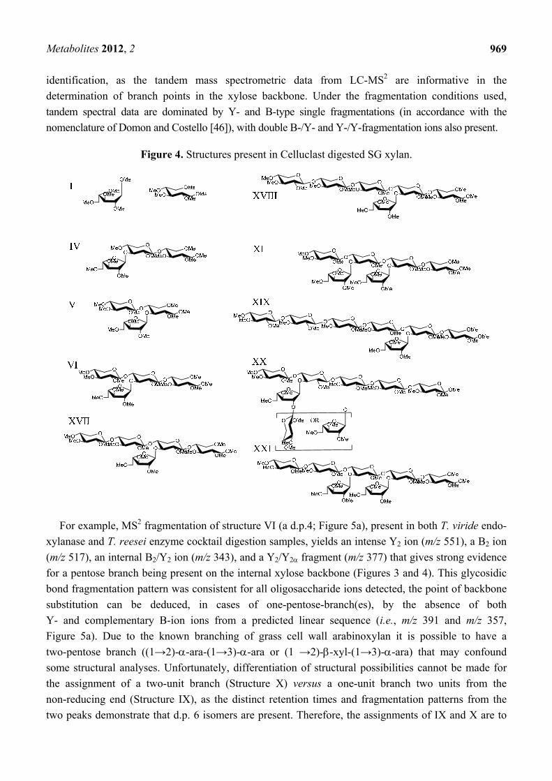
Figure 4. Structures present in Celluclast digested SG xylan.
For example, MS2 fragmentation of structure VI (a d.p.4; Figure 5a), present in both T. viride endo-
xylanase and T. reesei enzyme cocktail digestion samples, yields an intense Y2 ion (m/z 551), a B2 ion
(m/z 517), an internal B2/Y2 ion (m/z 343), and a Y2/Y2 fragment (m/z 377) that gives strong evidence
for a pentose branch being present on the internal xylose backbone (Figures 3 and 4). This glycosidic
bond fragmentation pattern was consistent for all oligosaccharide ions detected, the point of backbone
substitution can be deduced, in cases of one-pentose-branch(es), by the absence of both
Y- and complementary B-ion ions from a predicted linear sequence (i.e., m/z 391 and m/z 357,
Figure 5a). Due to the known branching of grass cell wall arabinoxylan it is possible to have a
two-pentose branch ((1→2)--ara-(1→3)--ara or (1 →2)--xyl-(1→3)--ara) that may confound
some structural analyses. Unfortunately, differentiation of structural possibilities cannot be made for
the assignment of a two-unit branch (Structure X) versus a one-unit branch two units from the
non-reducing end (Structure IX), as the distinct retention times and fragmentation patterns from the
two peaks demonstrate that d.p. 6 isomers are present. Therefore, the assignments of IX and X are to

Metabolites 2012, 2
970
be treated as tentative. Data collected from the T. reesei cellulase enzyme cocktail (Celluclast)
provided MS2 fragmentation patterns that can be used to identify alternate sites of branching, as the
selectivity of xylanase enzyme(s) gave an altered pattern of products (i.e., T. viride favors the
generation of oligosaccharides branched at the third residue from the reducing end (Figure 3), whereas
T. reesei xylanases produce oligosaccharides branched at the second residue from the reducing end
(Figure 4).) It should be noted that the enzyme cocktail may also have exo-enzymatic activities that
clip monosaccharide units (xyl and ara) from the initial T. reesei endo-xylanase product
oligosaccharides to smaller pieces; however the end-point products are constant with maturity and
xylan type, showing the fidelity of the enzyme cocktail treatment. The T. reesei AXO products have
direct significance to biomass conversion because of the prevalence of T. reesei-based enzyme
cocktails for biomass saccharification.
Figure 5. Mass spectrometry (MSn) fragmentation of permethylated oligosaccharide VI
(RT 23.5 min) (a) MS2 m/z 725. (b) MS3 fragmentation m/z 725→551. (c) MS4
fragmentation m/z 725→551→377.

Metabolites 2012, 2
971
Due to the complexity and low signals of the largest oligosaccharides detected, structural
assignments could not be made for Structures XVI, XXII, XXIII, and XXIV.
2.5. LC-MSn
In some cases MS2 was not sufficient for complete structural assignments; therefore, additional
fragmentation data from the segmented MSn method was used. Within the appropriate retention time
window for each d.p. oligomer, MS2 targeted the appropriate m/z for the AXO d.p. followed by an MSn
tree that targeted ions in the following manner: In MS3 the Y- and B-ions corresponding to the loss of
one pentose (Y(d.p.-1)-14; B(d.p.-1)-14), followed by subsequent MSn for fragmentations of pentose losses
(Y(d.p.-2)-14; B(d.p.-2)-14); the isolation and fragmentation of double losses were also included
(e.g. Y/Y(d.p.-2)-28), as these represent the information-rich fragments that show where branching has
been present. There were up to 13 MSn experiments within a time window, encompassing MS2 to MS6;
however data from MS5 and MS6 were typically limited in information. The time required to complete
the MSn tree was limiting; therefore, a traditional 250 mm HPLC column was used to provide
sufficient resolution of analytes while permitting the full MSn analyses of the targeted ions. Peak
elution profiles under ultra-performance liquid chromatography (UPLC) conditions were insufficiently
long to obtain detailed structural information from the MSn tree (data not shown); however UPLC may
be used once detailed structures are determined and MS2 patterns are sufficient to confirm the identity
of known analytes. MS2 data were ambiguous for the interpretation of smaller oligosaccharides (d.p. ≤
3); however the use of linear standards (Xyl2-Xyl6) and MSn provided sufficient data for assignments
of II and III, and excluded the presence of linear xylooligomers as d.p 4–6 products. T. viride endo-
xylanase digestion produces a linear xylotriose (Xyl3, III) indicating that the number of xylose repeats
between endo-xylanase product oligosaccharide structures within the xylan should be an odd number.
Xyl3 has been reported as a product of T. viride endo-xylanase digestion [45], but primarily generates
xylobiose as a product from Xyl4 and Xyl6 (data not shown), showing a strong preference for
generation of xylobiose. In addition to chromatographic and mass spectrometric comparisons to known
linear standards, assignments for additional d.p. 3 structures can be made based on MSn data. For
example, structure V can be identified by the presence of the sole ion at m/z 217 in its MS3 spectrum
(m/z 565→391) (Supplemental Figure 3), corresponding to a monosaccharide unit on the reducing end
that had a branching pentose [19]. This low-abundance product may be the result of T. reesei GH5
(XynIV) hydrolysis, as a GH5 xylanase has been shown to generate similarly reducing-end branched
structures [19]. Structure V was the only product oligosaccharide identified in this study that had a
substitution on the reducing end unit. Complete structural assignment could not be made on all d.p. 3
analytes, where Structure IV was proposed as the likely structure due to the evidence that it was not
substituted on the reducing end and the retention time and fragmentation pattern were not consistent
with the linear structure (III).
Longer oligosaccharides provide less ambiguous spectra due to the length of the oligosaccharide
backbone, permitting more targeted cycles of fragmentation of glycosidic fragment ions. Structure VI
can be determined by a combination of MS2 fragmentations (described in Section 2.4.), indicating the
location of branching on the chain, followed by MS3 and MS4. MS3 (m/z 725→551, Figure 5b) yields
an m/z 377 product ion that would be the same from either fragmentation pathway consistent with the

Metabolites 2012, 2
972
two potential fragmentation pathways of the proposed structure. The MS4 spectrum
(m/z 725→551→377, Figure 5c) gives an m/z 231 ion indicating an unsubstituted reducing end
structure. In addition, lower abundance, but structurally informative cross-ring cleavages showed the
linkage site of the branching substitution (1,4X to give m/z 273.2, 0,2X to give m/z 287.2, 2,5X to give
m/z 303.4). The lower energy fragmentation of the linear ion-trap (LTQ) requires the use of multiple
stages of fragmentation to obtain strong cross-ring cleavages that show the linkage position of the
branch; this is in contrast to the generation of diagnostic ions for arabinose branching in the positive
ion tandem mass spectrum using high energy CID MALDI-TOF/TOF for the analysis of reducing-end
labeled AXOs [22,23]. Each enzymatic condition resulted in only one d.p.5 peak (VIII and XVII)
having different retention times and likely different structures. Structure VIII from T. viride endo-
xylanase digestion can be assigned through a sequential fragmentation of the glycosidic bond cleavage
fragment ions (to MS5) to locate the site of branching on the third backbone residue from the reducing
end and the linkage information in the MS4 spectrum (Figure 6 a–c). Similarly, Structure XVII from
the T. reesei enzyme cocktail digestion can be assigned based on the same MSn tree, but the differing
spectra allow for the determination of the branch point on the second unit from the reducing end
(Figure 7a–c). Structure XVII is also consistent with a structure identified and purified from a SG
biomass fermentation using T. reseei-based enzyme cocktails [27]. MS5 spectra of VIII and XVII
(Figures 6 and 7, m/z of 391 and 377, respectively) show only an m/z 231 ion corresponding to an
unsubstituted reducing end of the oligosaccharide ion.
The combination of LC-MS, MS2, and MSn data provided diagnostic ions for the identification of
twelve branched oligosaccharide structures (Figures 3 and 4) in addition to the assignment of
xylobiose and xylotriose from T. viride endo-xylanase digestion and nine branched product oligomers
(five differing from T. viride endo-xylanase digestion) from T. reesei enzyme cocktail digestion. The
use of the two different enzyme conditions demonstrates that the use of LC-MSn is capable of
differentiating isomeric structures that vary only in their position and quantity of branch points on the
xylose backbone.
Figure 6. MSn fragmentation of permethylated oligosaccharide VIII (RT 28.5 min)
(a) MS2 m/z 885. (b) MS3 fragmentation m/z 885→711. (c) MS4 fragmentation m/z
885→711→537.

Metabolites 2012, 2
973
Figure 6. Cont.
Figure 7. MSn fragmentation of permethylated oligosaccharide XVII (RT 27.5 min)
(a) MS2 m/z 885. (b) MS3 fragmentation m/z 885→711. (c) MS4 fragmentation m/z
885→711→551. (d) MS4 fragmentation m/z 885→711→537.

Metabolites 2012, 2
974
Figure 7. Cont.
2.6. Discussion
Arabinoxylans can be conveniently depolymerized to manageable sized units by treatment with
xylanase. However new methods are needed to analyze the resulting AXOs for structure because those
reported in the past are laborious and insensitive to low-abundance species. The use of C18-LC-MSn
under a relatively wide elution gradient and online MSn monitoring allowed the separation and analysis
of AXOs with d.p. 3–9 with good resolution of peaks in a convenient manner. Due to the improved
resolution of RP-LC-MS, low abundance structures that would be difficult to separate in sufficient
quantities for full structural characterization can now be deduced. This new method has direct

Metabolites 2012, 2
975
applications to researching biochemical conversion of biomass into biofuels. Developing minimal
enzyme mixtures for converting xylan into fermentable monosaccharides is challenging because of the
complexity of the xylan chains [6,7,47]. This method to identify the unhydrolyzed oligosaccharides of
arabinoxylan after enzyme treatment will allow for a better understanding of the substrate selectivity of
currently marketed biomass saccharifying enzymes and can be used to screen for auxiliary enzymes
beneficial to saccharification. The use of permethylation is also amenable to alternate chromatography
techniques (e.g., porous graphitic carbon (PGC)) that could provide further benefits in chromatographic
resolution. Examples might include its use for more complex mixtures such as pericarp xylan for food
applications or whole biomass digestions that contain oligosaccharides derived from the diverse
carbohydrate biopolymers present within the cell wall.
In this study, RP-LC-MSn allowed for the differentiation of multiple components from SG xylanase
digestion mixtures. LC-MS allowed for the analysis of small components and separated AXOs with
higher resolution than other chromatographic techniques. In comparison to another report of SG xylan
characterization, xylan extracted from ball-milled Alamo variety SG was T. viride endo-xylanase-digested
followed by fractionation by size-exclusion chromatography; the report characterized three
arabinose-containing xylooligosaccharides (XO) structures of d.p 8, d.p. 6, and d.p.5, while leaving a
low-mass fraction uncharacterized [20]. In this LC-MSn study, the previously reported structures from
T. viride endo-xylanase-digested SG xylan are consistent with d.p. 5 (VIII) and d.p. 6 (X); however the
previously reported d.p. 8 was not observed. In contrast to the previous report, this method identifies
three d.p. 6 oligomers (IX, X, XI) as well as two oligosaccharides that could be components of the
previously reported d.p.8 (VIII + III). This study used Cave-in-Rock variety SG, where structural
variation could be attributed to inherent differences between the two varieties of SG (lowland
octoploid Alamo vs. upland tetraploid Cave-in-Rock) or differences in enzymatic treatment conditions
(i.e., temperature, enzyme loading, and buffer salts). In addition, the use of LC-MSn makes it
unnecessary to purify each component of the digestion prior to characterization, leading to the
identification of several components of lower d.p. (≤4) than were previously reported for SG xylan [20].
One area not previously reported on is the effect of harvest maturity on the xylan structure.
SG xylan fractions A and B were isolated from three harvest maturities. Interestingly, the structures
obtained from the endpoint enzymatic depolymerization of SG xylan A and B did not change with
maturity. While the diversity of AXO structures remained uniform with plant development, there are
differences in the relative occurrence of sequences as SG ages. This result bodes well for biorefineries
because it suggests that an enzyme mixture formulated with the activities necessary to hydrolyze all
SG xylan glycosidic bonds should be effective regardless of the harvest maturity of the biomass
delivered to the gate.
While this is the first structure analysis that has considered SG maturity as a variable, caution must
be taken in drawing conclusions from the use of endpoint enzyme-based characterization of xylan, as
substrate specificity of enzymes may shape the patterns observed, thereby creating a subset of analytes
that may not fully reflect the diversity of the entire xylan chain (i.e. arrangement of the detected
end-point oligosaccharides within the structure). Specifically, end-point digestions of T. viride
endo-xylanase released xylobiose and T. reesei enzyme cocktail released both xylose and arabinose,
thereby making extended structural determination impossible; therefore, less stringent digestion

Metabolites 2012, 2
976
conditions (e.g., 50% digestion) would provide a distribution of oligosaccharides that could provide
further structural information about the structural repeats of xylan.
It should be also noted that the methods used (e.g., alkali extraction and permethylation) would
hydrolyze ester bonds connected to the xylan structure (e.g., acetyl- and feruloyl-) that may vary during
the maturation of SG biomass. The degree of esterification may play a role in the varied response to
acidic conditions; however, ammonia-based pretreatments should remove the majority of the ester
groups [27,48]. Finally, this report used whole ground plant material because that is what will be
delivered to a biorefinery facility. Grass stems grow upwards by adding internodes of younger
maturity. It is possible that fractionation of stems into individual internodes would yield differences
hidden by using bulk samples. Future studies will focus on the use of xylan extraction techniques that
retain ester functionalities, investigation of samples fractionated by internodes, and samples processed
under less stringent hydrolysis conditions.
The use of LC-MSn to identify the structural characteristics of xylan can be an important tool for
biomass research due to the highly complex substrates proposed for conversion. While NMR will
remain an essential technique for absolute structural identification of purified products, the sensitivity
of mass spectrometry allows for the detection of many enzymatic products that are of low abundance
and would be difficult to obtain in sufficient quantities for traditional characterization. The influence of
low abundance structures in bioconversion is yet to be determined; however, the need for high solid
loadings required for economical biomass conversion processes means the quantity of unhydrolyzed
xylan structures will be in greater abundance than the enzymes potentially impeding the
depolymerization.
3. Experimental Section
3.1. Materials
Sodium chlorite, potassium hydroxide (KOH), 1-octanol, sodium hydroxide (NaOH) (50% w/v),
sodium acetate anhydrous, trifluoroacetic acid (TFA), acetonitrile (ACN), acetic acid (AcOH),
dimethylsulfoxide (DMSO), methyl iodide, sodium hydroxide powder, sodium borohydride, xylose,
and arabinose were purchased from Sigma-Aldrich Company (St. Louis; MO). Acetone was purchased
from Fisher Scientific (Pittsburgh, PA). Formic acid was purchased from Fluka Chemical
(Buchs, Switzerland). Purified xylobiose to xylohexaose standards, wheat arabinoxylan, and xylanase
M1 from T. viride were purchased from Megazyme (International Ireland Ltd (Wicklow,Ireland).
Celluclast 1.5 L (Novozymes) was purchased from Brenntag Great Lakes, LLC.
3.2. Xylan Sample Preparation
Delignification and xylan isolation. Triplicate samples of switchgrass (SG) Cave-in-Rock variety at
three stages (pre-boot, anthesis, and post frost) of maturity were delignified and the xylan was alkali
extracted as previously described [28]. The compositions of holocellulose and xylan components were
quantified using two-stage acid hydrolysis (NREL procedure LAP002) [33], as previously described [28].

Metabolites 2012, 2
977
T. viride xylanaseDigestion. Triplicate samples (normalized for xylose content to 1 mg of
MPV-1 xylan A) of each SG xylan maturity were exhaustively digested; in 50 mM sodium acetate
pH 5.0 (1 mL); by the action of 1 L of xylanase from T. viride (2000 U/mL; final loading (U/mL):
2 U xylanase activity; 7.8 g protein) solution was added in two portions 3 h apart, with a total
reaction time of 16 h at 50 °C.
CelluclastDigestion. Triplicate samples (1 mg) of each SG xylan maturity were exhaustively digested,
in 50 mM sodium acetate pH 5.0 (1 mL), by the action of 2 L of Celluclast 1.5 (solution was added in
two portions 3 h apart (final loading (U/mL): 2.2 U xylanase activity; 0.032 filter paper units (FPU);
1.9 U carboxymethyl cellusase activity; 0.030 U -glucosidase; 0.012 U -xylosidase;
0.062 U -L-arabinofuranosidase; 30 g protein, assayed as described in [41]), with a total reaction
time of 16 h at 50 °C.
Analysis by HPAEC-PAD. Twenty-five microliters of each sample (2.5 g) were analyzed by
HPAEC-PAD (Dionex ACS 3000, Sunnyvale, CA) utilizing a PA-100 column (Dionex) at 1 mL/min
running 100% A (A:100 mM NaOH) isocratically for 15 min followed by a gradient program to 12%
B (B:100 mM NaOH containing 1 M sodium acetate) over 20 min followed by washout and 15 min of
re-equilibration in 100% A, based on conditions reported in [15]. Extent of digestion was determined
by comparison of xylose, arabinose, and linear xylooligosaccharides (xyl2-xyl6) released by enzymatic
hydrolysis versus the amount of xylose and arabinose produced by acid hydrolysis (2M TFA) of
equivalent portion of xylan.
3.3. Permethylation Reaction of Enzymatic Products
An aliquot of each digestion (100 g) was removed, maltotetraose was added as an internal
standard, and dried in a Speed vac. After drying, samples were reduced with sodium borohydride
(500 mM) in 2 M NH4OH for 1 h at 45 °C. Excess borohydride was removed by repeated addition of
acidic methanol and dried by speed vac (3×). Permethylation (PM) was performed according to the
method of Ciucanu and Kerek [49], with modifications to limit oxidative degradation [50]. Due to the
presence of undermethylated material (<5%) in preliminary experiments, each sample was methylated
twice to ensure the reaction was complete. Samples were dissolved in 300 L methanol followed by
addition of 700 L water.
3.4. C18-LC-MSn.
LC-MS. Mass spectrometry samples (2 L injection, 0.2 g based on starting material) were
analyzed by LC-MS (Thermo Acella HPLC) through a narrow-bore (2.1mm × 250 mm, 3 m particle
size) C18 column (Inertsil, GL Sciences, Inc., Torrance, CA) running a gradient elution of 30% A:70%
B (buffer A 0.1% formic acid, buffer B 100% acetonitrile) to 70% A:30% B over 45 min at a flow rate
of 200 L/min, followed by a 5 min B washout and 10 min re-equilibration, while maintaining a
constant column temperature of 30 °C. Electrospray positive mode ionization data were collected with
a linear ion trap-Orbitrap mass spectrometer (Thermo LTQ-Orbitrap Discovery) under Xcalibur 2.1
control. Prior to LC-MSn experiments the instrument was tuned and calibrated using the LTQ tune
mix. The parent ion table function was used to isolate and fragment singly charged ions ([M+Na]1+)
Metabolites 2012, 2
978
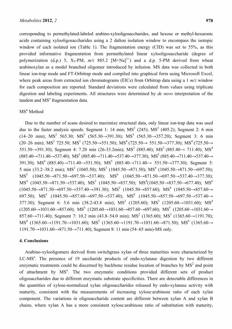
corresponding to permethylated-labeled arabino-xylooligosaccharides, and hexose or methyl-hexuronic
acids containing xylooligosaccharides using a 2 dalton isolation window to encompass the isotopic
window of each isolated ion (Table 1). The fragmentation energy (CID) was set to 55%, as this
provided informative fragmentation from permethylated linear xylooligosaccharide (degree of
polymerization (d.p.) 5, X5-PM, m/z 885.2 [M+Na]1+) and a d.p. 5-PM derived from wheat
arabinoxylan as a model branched oligomer introduced by infusion. MS data was collected in both
linear ion-trap mode and FT-Orbitrap mode and compiled into graphical form using Microsoft Excel,
where peak areas from extracted ion chromatograms (EICs) from Orbitrap data using a 1 m/z window
for each composition are reported. Standard deviations were calculated from values using triplicate
digestion and labeling experiments. All structures were determined by de novo interpretation of the
tandem and MSn fragmentation data.
MSn Method
Due to the number of scans desired to maximize structural data, only linear ion-trap data was used
due to the faster analysis speeds. Segment 1: 14 min; MS2 (245); MS2 (405.2); Segment 2: 6 min
(14–20 min); MS2 565.30; MS3 (565.30→391.30); MS3 (565.30→357.20); Segment 3: 6 min
(20–26 min); MS2 725.50; MS3 (725.50→551.50); MS4 (725.50→ 551.50→377.30); MS4 (725.50→
551.50→391.30); Segment 4: 7.20 min (26-33.2min); MS2 (885.40); MS3 (885.40→ 711.40); MS4
(885.40→711.40→537.40); MS5 (885.40→711.40→537.40→377.30); MS5 (885.40→711.40→537.40→
391.30); MS4 (885.40)→711.40→551.50); MS5 (885.40→711.40→ 551.50→377.30); Segment 5:
5 min (33.2–38.2 min); MS2 (1045.50); MS3 (1045.50→871.50); MS4 (1045.50→871.50→697.50);
MS5 (1045.50→871.50→697.50→537.40); MS6 (1045.50→871.50→697.50→537.40→377.30);
MS4 (1045.50→871.50→537.40); MS3 (1045.50→837.50); MS4(1045.50→837.50→677.40); MS6
(1045.50→871.50→697.50→537.40→391.30); MS3 (1045.50→857.60); MS4 (1045.50→857.60→
697.50); MS5 (1045.50→857.60→697.50→537.40); MS6 (1045.50→857.50→697.50→537.40→
377.30); Segment 6: 5.6 min (38.2-43.8 min); MS2 (1205.60); MS3 (1205.60→1031.60); MS4
(1205.60→1031.60→857.60); MS5 (1205.60→1031.60→857.60→697.60); MS5 (1205.60→1031.60→
857.60→711.40); Segment 7: 10.2 min (43.8–54.0 min); MS2 (1365.60); MS3 (1365.60→1191.70);
MS4 (1365.60→1191.70→1031.60); MS5 (1365.60→1191.70→1031.60→871.50); MS6 (1365.60→
1191.70→1031.60→871.50→711.40); Segment 8: 11 min (54–65 min)-MS only.
4. Conclusions
Arabino-xylooligomers derived from switchgrass xylan of three maturities were characterized by
LC-MSn. The presence of 19 saccharide products of endo-xylanase digestion by two different
enzymatic treatments could be discerned by backbone residue location of branches by MS2 and point
of attachment by MSn. The two enzymatic conditions provided different sets of product
oligosaccharides due to different enzymatic substrate specificities. There are detectable differences in
the quantities of xylose-normalized xylan oligosaccharides released by endo-xylanase activity with
maturity, consistent with the measurements of increasing xylose:arabinose ratio of each xylan
component. The variations in oligosaccharide content are different between xylan A and xylan B
chains, where xylan A has a more consistent xylose:arabinose ratio of substitution with maturity,

Metabolites 2012, 2
979
whereas xylan B has a decreasing amount of arabinose (i.e., it is less substituted with maturity). The
observation that the xylan structure appears uniform with maturity is beneficial showing that enzyme
cocktails capable of hydrolyzing xylan glycosidic bonds need not be altered depending upon harvest
maturity of the SG biomass; however, the quantity of xylan may be a factor for the loading of enzyme
cocktail to limit product inhibition of enzymes. The application of permethylation followed by
LC-MSn provides a useful platform for the identification of structural features of heteroxylan-derived
oligosaccharides for applications to extended structure determination and sampling of biomass-derived
fermentation residues to identify recalcitrant structures.
Supplementary Materials
Supplementary materials can be accessed at: http://www.mdpi.com/2218-1989/2/4/959/s1.
Acknowledgments
The authors thank Brenda Duppong for excellent technical assistance throughout this study.
Conflict of Interest
The authors declare no conflict of interest.
References
1. Walsh, M.E.; Ugarte, D.G.D.; Shapouri, H.; Slinksky, S.P. Bioenergy crop production in the
United States: Potential quantities, land use changes, and economic impacts on the agricultural
sector. Environ. Resource Econ. 2003, 24, 313–333.
2. Vogel, J. Unique aspects of the grass cell wall. Curr. Opin. Plant. Biol. 2008, 11, 301–307.
3. Qing, Q.; Yang, B.; Wyman, C.E. Xylooligomers are strong inhibitors of cellulose hydrolysis by
enzymes. Bioresour. Technol. 2010, 101, 9624–9630.
4. Kabel, M.A.; van den Borne, H.; Vincken, J.P.; Voragen, A.G.J.; Schols, H.A. Structural
differences of xylans affect their interaction with cellulose. Carbohydr. Polym. 2007, 69, 94–105.
5. Gírio, F.M.; Fonseca, C.; Carvalheiro, F.; Duarte, L.C.; Marques, S.; Bogel-Łukasik, R.
Hemicelluloses for fuel ethanol: A review. Bioresour. Technol. 2010, 101, 4775–4800.
6. Selinger, L.B.; Forsberg, C.W.; Cheng, K.J. The rumen: A unique source of enzymes for
enhancing livestock production. Anaerobe 1996, 2, 263–284.
7. Faik, A. Xylan biosynthesis: news from the grass. Plant. Physiol. 2010, 153, 396–402.
8. Mosier, N.; Wyman, C.; Dale, B.; Elander, R.; Lee, Y.Y.; Holtzapple, M.; Ladisch, M. Features of
promising technologies for pretreatment of lignocellulosic biomass. Bioresour. Technol. 2005, 96,
673–686.
9. Esteghlalian, A.; Hashimoto, A.G.; Fenske, J.J.; Penner, M.H. Modeling and optimization of the
dilute-sulfuric-acid pretreatment of corn stover, poplar and switchgrass. Bioresour. Technol. 1997,
59, 129–136.

Metabolites 2012, 2
980
10. Dervilly-Pinel, G.; Tran, V.; Saulnier, L. Investigation of the distribution of arabinose residues on
the xylan backbone of water-soluble arabinoxylans from wheat flour. Carbohydr. Polym. 2004,
55, 171–177.
11. Gruppen, H.; Kormelink, F.J.M.; Voragen, A.G.J. Water-Unextractable Cell-Wall Material from
Wheat-Flour .3. A Structural Model for Arabinoxylans. J. Cereal Sci. 1993, 18, 111–128.
12. Ordaz-Ortiz, J.J.; Devaux, M.F.; Saulnier, L. Classification of wheat varieties based on structural
features of arabinoxylans as revealed by endoxylanase treatment of flour and grain. J. Agric. Food
Chem. 2005, 53, 8349–8356.
13. Ordaz-Ortiz, J.J.; Saulnier, L. Structural variability of arabinoxylans from wheat flour.
Comparison of water-extractable and xylanase-extractable arabinoxylans. J. Cereal Sci. 2005, 42,
119–125.
14. Vietor, R.J.; Kormelink, F.J.M.; Angelino, S.A.G.F.; Voragen, A.G.J. Substitution Patterns of
Water-Unextractable Arabinoxylans from Barley and Malt. Carbohydr. Polym. 1994, 24, 113–
118.
15. Rantanen, H.; Virkki, L.; Tuomainen, P.; Kabel, M.; Schols, H.; Tenkanen, M. Preparation of
arabinoxylobiose from rye xylan using family 10 Aspergillus aculeatus endo-1,4--D-xylanase.
Carbohydr. Polym. 2007, 68, 350–359.
16. Westphal, Y.; Kuhnel, S.; de Waard, P.; Hinz, S.W.; Schols, H.A.; Voragen, A.G.; Gruppen, H.
Branched arabino-oligosaccharides isolated from sugar beet arabinan. Carbohydr. Res. 2010, 345,
1180–1189.
17. Fernández, M.L.E.; Obel, N.; Scheller, H.V.; Roepstorff, P. Differentiation of isomeric
oligosaccharide structures by ESI tandem MS and GC-MS. Carbohydr. Res. 2004, 339, 655–664.
18. Quéméner, B.; Ordaz-Ortiz, J.J.; Saulnier, L. Structural characterization of underivatized arabino-
xylo-oligosaccharides by negative-ion electrospray mass spectrometry. Carbohydr. Res. 2006,
341, 1834–1847.
19. Correia, M.A.; Mazumder, K.; Bras, J.L.; Firbank, S.J.; Zhu, Y.; Lewis, R.J.; York, W.S.; Fontes,
C.M.; Gilbert, H.J. Structure and function of an arabinoxylan-specific xylanase. J. Biol. Chem.
2011, 286, 22510–22520.
20. Mazumder, K.; York, W.S. Structural analysis of arabinoxylans isolated from ball-milled
switchgrass biomass. Carbohydr. Res. 2010, 345, 2183–2193.
21. Brown, D.M.; Goubet, F.; Wong, V.W.; Goodacre, R.; Stephens, E.; Dupree, P.; Turner, S.R.
Comparison of five xylan synthesis mutants reveals new insight into the mechanisms of xylan
synthesis. Plant. J. 2007, 52, 1154–1168.
22. Maslen, S.L.; Goubet, F.; Adam, A.; Dupree, P.; Stephens, E. Structure elucidation of
arabinoxylan isomers by normal phase HPLC-MALDI-TOF/TOF-MS/MS. Carbohydr. Res. 2007,
342, 724–735.
23. Ridlova, G.; Mortimer, J.C.; Maslen, S.L.; Dupree, P.; Stephens, E. Oligosaccharide relative
quantitation using isotope tagging and normal-phase liquid chromatography/mass spectrometry.
Rapid Commun. Mass Spectrom. 2008, 22, 2723–2730.
24. Zaia, J. Mass spectrometry of oligosaccharides. Mass Spectrom. Rev. 2004, 23, 161–227.

Metabolites 2012, 2
981
25. Ruhaak, L.R.; Zauner, G.; Huhn, C.; Bruggink, C.; Deelder, A.M.; Wuhrer, M. Glycan labeling
strategies and their use in identification and quantification. Anal. Bioanal. Chem. 2010, 397,
3457–3481.
26. Bauer, S. Mass spectrometry for characterizing plant cell wall polysaccharides. Front. Plant. Sci.
2012, 3, 45.
27. Bowman, M.J.; Dien, B.S.; Hector, R.E.; Sarath, G.; Cotta, M.A. Liquid chromatography-mass
spectrometry investigation of enzyme-resistant xylooligosaccharide structures of switchgrass
associated with ammonia pretreatment, enzymatic saccharification, and fermentation. Bioresour.
Technol. 2012, 110, 437–447.
28. Bowman, M.J.; Dien, B.S.; O'Bryan, P.J.; Sarath, G.; Cotta, M.A. Selective chemical oxidation
and depolymerization of switchgrass (Panicum. virgatum L.) xylan with oligosaccharide product
analysis by mass spectrometry. Rapid Commun. Mass Spectrom. 2011, 25, 941–950.
29. Dien, B.; O'Bryan, P.J.; Hector, R.; Iten, L.; Cotta, M.A. Conversion of switchgrass to sugars and
ethanol using dilute ammonium hydroxide pretreatment. In Proceedings of the 31st Symposium
on Biotechnology for Fuels and Chemicals, San Francisco, CA, USA, 3 May 2009; p. 93.
30. Dien, B.S.; Jung, H.J.G.; Vogel, K.P.; Casler, M.D.; Lamb, J.F.S.; Iten, L.; Mitchell, R.B.; Sarath,
G. Chemical composition and response to dilute-acid pretreatment and enzymatic saccharification
of alfalfa, reed canarygrass, and switchgrass. Biomass Bioenergy 2006, 30, 880–891.
31. Jung, H.J.G.; Vogel, K.P. Lignification of Switchgrass (Panicum Virgatum) and Big Bluestem
(AndropogonGerardii) Plant-Parts during Maturation and Its Effect on Fiber Degradability.
J. Sci. Food Agric. 1992, 59, 169–176.
32. Whistler, R.L.; Bachrach, J.; Bowman, D.R. Preparation and properties of corn cob holocellulose.
Arch. Biochem. 1948, 19, 25–33.
33. Sluiter, A.; Hames, B.; Ruiz, R.; Scarlata, C.; Sluiter, J.; Templeton, D.; Crocker, D.
Determination of structural carbohydrates and lignin in biomass; Technical Report NREL/TP-
510-42618, 2005. Available online: http://www.nrel.gov/biomass/pdfs/42618.pdf, accessed on 14
November 2012.
34. Packett, L.V.; Plumlee, M.L.; Barnes, R.; Mott, G.O. Influence of Hemicellulose A and B on
Cellulose Digestion Volatile Fatty Acid Production and Forage Nutritive Evaluation. J. Nutr.
1965, 85, 89–101.
35. Paës, G.; Berrin, J.G.; Beaugrand, J. GH11 xylanases: Structure/function/properties relationships
and applications. Biotechnol. Adv. 2012, 30, 564–592.
36. Pollet, A.; Delcour, J.A.; Courtin, C.M. Structural determinants of the substrate specificities of
xylanases from different glycoside hydrolase families. Crit. Rev. Biotechnol. 2010, 30, 176–191.
37. Collins, T.; Gerday, C.; Feller, G. Xylanases, xylanase families and extremophilic xylanases.
FEMS Microbiol. Rev. 2005, 29, 3–23.
38. Parkkinen, T.; Hakulinen, N.; Tenkanen, M.; Siika-aho, M.; Rouvinen, J. Crystallization and
preliminary X-ray analysis of a novel Trichoderma reesei xylanase IV belonging to glycoside
hydrolase family 5. Acta Crystallogr., Sect. D 2004, 60, 542–544.
39. Tenkanen, M.; Puls, J.; Poutanen, K. Two Major Xylanases of Trichoderma Reesei. Enzyme
Microb. Technol. 1992, 14, 566–574.

Metabolites 2012, 2
982
40. Berlin, A.; Maximenko, V.; Gilkes, N.; Saddler, J. Optimization of enzyme complexes for
lignocellulose hydrolysis. Biotechnol. Bioeng. 2007, 97, 287–296.
41. Saha, B.C.; Iten, L.B.; Cotta, M.A.; Wu, Y.V. Dilute acid pretreatment, enzymatic
saccharification and fermentation of wheat straw to ethanol. Process. Biochemistry 2005, 40,
3693–3700.
42. Costello, C.E.; Contado-Miller, J.M.; Cipollo, J.F. A glycomics platform for the analysis of
permethylated oligosaccharide alditols. J. Am. Soc. Mass Spectrom. 2007, 18, 1799–1812.
43. Hu, Y.; Mechref, Y. Comparing MALDI-MS, RP-LC-MALDI-MS and RP-LC-ESI-MS glycomic
profiles of permethylated N-glycans derived from model glycoproteins and human blood serum.
Electrophoresis 2012, 33, 1768–1777.
44. Huang, R.; Pomin, V.H.; Sharp, J.S. LC-MSn analysis of isomeric chondroitin sulfate
oligosaccharides using a chemical derivatization strategy. J. Am. Soc. Mass Spectrom. 2011, 22,
1577–1587.
45. Izydorczyk, M.S.; Biliaderis, C.G. Studies on the Structure of Wheat-Endosperm Arabinoxylans.
Carbohydr. Polym. 1994, 24, 61–71.
46. Domon, B.; Costello, C.E. A systematic nomenclature for carbohydrate fragmentations in FAB-
MS/MS spectra of glycoconjugates. Glycoconjugate J. 1988, 5, 397–409.
47. Jordan, D.B.; Bowman, M.J.; Braker, J.D.; Dien, B.S.; Hector, R.E.; Lee, C.C.; Mertens, J.A.;
Wagschal, K. Plant cell walls to ethanol. Biochem. J. 2012, 442, 241–252.
48. Chundawat, S.P.; Vismeh, R.; Sharma, L.N.; Humpula, J.F.; da Costa Sousa, L.; Chambliss, C.K.;
Jones, A.D.; Balan, V.; Dale, B.E. Multifaceted characterization of cell wall decomposition
products formed during ammonia fiber expansion (AFEX) and dilute acid based pretreatments.
Bioresour. Technol. 2010, 101, 8429–8438.
49. Ciucanu, I.; Kerek, F. A simple and rapid method for the permethylation of carbohydrates.
Carbohydr. Res. 1984, 131, 209–217.
50. Ciucanu, I.; Costello, C.E. Elimination of oxidative degradation during the per-O-methylation of
carbohydrates. J. Am. Chem. Soc. 2003, 125, 16213–16219.
© 2012 by the authors; licensee MDPI; Basel; Switzerland. This article is an open access article
distributed under the terms and conditions of the Creative Commons Attribution license
(http://creativecommons.org/licenses/by/3.0/).