COLOUR PICTURES OF FISHES FOR TAXONOMIC PURPOSES. THE PROBLEMS OF REFLECTIONS & BACKGROUND COLOUR...
13
Phuket mar. bioi. Cent. Spec. Publ. no. 12(1993) COLOUR PICTURES OF FISHES FOR TAXONOMIC PURPOSES. THE PROBLEMS OF REFLECTIONS & BACKGROUND COLOUR SMUDGING. by Jorgen Hylleberg l , Weera Pokapunt 2 and Pairoj Sirimontraporn 3 1. Phuket Marine Biological Center & Institute 0/ Biological Sciences,Aarhus University, Denmark 2. Oceanic Fisheries Division, Samutprakarn, Thailand 3. National Institute o/Coastal Aquaculture. Songkla. Thailand ABSTRACT 53 Colour of fresh fish is too variable to be of diagnostic value. Nevertheless, colour is part of a complete description of fish. Therefore, colour quality has been studied on exposures made on various types of back- grounds and films, in sunlight and in a photographic set-up. Emphasis was on smudging of background colour on the fish. Problems with reflections were solved by submersion of the fish in water. Generally, white, brown and black backgrounds resulted in reduced colour quality and made the fishes look "dead". Green, blue and greyish- blue backgrounds gave the best results. Blue backgrounds made the fishes look more fresh, but green was the most useful colour for versatile applications. Transparent fins received the colour of the background. However, background colour also smudged the colour of the solid fish body. Red background was especially bad in this respect. Smudging became more conspicuous when fish were cut free from the background to be mounted on a white background to yield plates. INTRODUCTION Descriptions offish usually contain a para- graph on the colour of the live fish. From such de- scriptions, e.g. Randall & Ben-Tuvia (1983), it is obvi- ous that colour varies considerably within regions as well as between habitats. The same species caught in shallow and deep water may look very different. Ifwe observe the colours only, it may sometimes be hard to believe that we are looking at the same species. Randall & Ben-Tuvia (1983) described groupers from the Red Sea. The diagnosis of individual species does not in- clude colour of the fresh fish. Presumably because this character is too variable to be of diagnostic value. But colour offresh fish is part of the subsequent de- scription. For example they describe the colour of Variola louti (ForsskiH, 1775) as follows: "yellowish to brown to organge-red (more red in deeper water), the head, body, and median fins with numerous small irregular spots which may be pale blue, lavender or pink; posterior edges of fins broadly yellow". Based on such range of colours it may seem unimportant to worry about the perfect colour picture as we have done it during the present workshop in Taxonomy and Biology ofFish from The Andaman Sea. However, we arrived at the conclusion, that if our fresh fish is red, the picture of it should also be red, even if another specimen of the same species may be yellowish or brown. In addition to the question of correct repro- duction of colour, we wanted to compare exposures made on various types offilms in sunlight and in the laboratory. We also wanted to study smudging ofback- ground colour on the shot, and we looked into tech- niques to minimize the problems of reflections, which in some cases may be slightly disturbing, but in se- vere cases may convey wrong information by hiding or changing colour patterns. MATERIALS & METHODS Fresh fish were obtained from fish markets. In the laboratory, fins were unfolded and fixed in ex- panded position with needles and 10 % formalin ap- plied to the fin area. Fish were placed on a raised glass plate in a photographic set-up (Fig. 1) and illuminated by two flood-lights, each 500 Watt. We used Pentax or NikonFM2 cameras mounted with 55 mm macro Nikkor, 1: 2.8, and loaded with Kodacolor Gold II film for colour
-
Upload
independent -
Category
Documents
-
view
1 -
download
0
Transcript of COLOUR PICTURES OF FISHES FOR TAXONOMIC PURPOSES. THE PROBLEMS OF REFLECTIONS & BACKGROUND COLOUR...
Phuket mar. bioi. Cent. Spec. Publ. no. 12(1993)
COLOUR PICTURES OF FISHES FOR TAXONOMIC PURPOSES.THE PROBLEMS OF REFLECTIONS & BACKGROUND COLOUR
SMUDGING.
by Jorgen Hyllebergl, Weera Pokapunt2 and Pairoj Sirimontraporn3
1. Phuket Marine Biological Center & Institute 0/Biological Sciences,Aarhus University, Denmark2. Oceanic Fisheries Division, Samutprakarn, Thailand3. National Institute o/Coastal Aquaculture. Songkla. Thailand
ABSTRACT
53
Colour of fresh fish is too variable to be ofdiagnostic value. Nevertheless, colour is part ofa completedescription of fish. Therefore, colour quality has been studied on exposures made on various types of backgrounds and films, in sunlight and in a photographic set-up. Emphasis was on smudging ofbackground colouron the fish. Problems with reflections were solved by submersion ofthe fish in water. Generally, white, brown andblack backgrounds resulted in reduced colour quality and made the fishes look "dead". Green, blue and greyishblue backgrounds gave the best results. Blue backgrounds made the fishes look more fresh, but green was themost useful colour for versatile applications. Transparent fins received the colour of the background. However,background colour also smudged the colour of the solid fish body. Red background was especially bad in thisrespect. Smudging became more conspicuous when fish were cut free from the background to be mounted on awhite background to yield plates.
INTRODUCTION
Descriptions offish usually contain a paragraph on the colour of the live fish. From such descriptions, e.g. Randall & Ben-Tuvia (1983), it is obvious that colour varies considerably within regions aswell as between habitats. The same species caught inshallow and deep water may look very different. Ifweobserve the colours only, it may sometimes be hard tobelieve that we are looking at the same species. Randall& Ben-Tuvia (1983) described groupers from the RedSea. The diagnosis of individual species does not include colour of the fresh fish. Presumably becausethis character is too variable to be ofdiagnostic value.But colour offresh fish is part of the subsequent description. For example they describe the colour ofVariola louti (ForsskiH, 1775) as follows: "yellowishto brown to organge-red (more red in deeper water),the head, body, and median fins with numerous smallirregular spots which may be pale blue, lavender orpink; posterior edges of fins broadly yellow". Basedon such range ofcolours it may seem unimportant toworry about the perfect colour picture as we havedone it during the present workshop in Taxonomy and
Biology ofFish from The Andaman Sea. However, wearrived at the conclusion, that ifour fresh fish is red,the picture of it should also be red, even if anotherspecimen of the same species may be yellowish orbrown. In addition to the question of correct reproduction of colour, we wanted to compare exposuresmade on various types offilms in sunlight and in thelaboratory. We also wanted to study smudging ofbackground colour on the shot, and we looked into techniques to minimize the problems of reflections, whichin some cases may be slightly disturbing, but in severe cases may convey wrong information by hidingor changing colour patterns.
MATERIALS & METHODS
Fresh fish were obtained from fish markets.In the laboratory, fins were unfolded and fixed in expanded position with needles and 10 % formalin applied to the fin area. Fish were placed on a raised glassplate in a photographic set-up (Fig. 1) and illuminatedby two flood-lights, each 500 Watt. We used Pentax orNikonFM2 cameras mounted with 55 mm macro Nikkor,1: 2.8, and loaded with Kodacolor Gold II film for colour
54 Taxonomy and biology of fishes from the Andaman Sea.
prints. Exposed films were developed and prints madeaccording to standard procedure by a shop in Phukettown. Slides were copied onto colour negative film ina Nikon Slide Copy Attachment & Nikon Bellows Focusing Attachment PB-4, using sunlight to illuminatethe slide. Individual fish were cut from the backgroundcolour by Jorgen Hylleberg, using a pointed SabreScalpel blade, shape E/II. Cutting took place under adissecting microscope at 6 times magnification. Thelayer of gelatine on the print was cut around the fishand similarly 5-10 mm away from the outline of thefish. The layer of gelatine could then be peeled offbetween the two cuts with the help of a pair of finelypointed forceps (Dumont & Fils No.5).
RESULTS & DISCUSSION
In the Workshop we compared slides andcolour prints shot on different backgrounds in thelaboratory and outdoors in sunlight, to judge howwell the colours ofthe pictures agreed with the coloursof the fresh fishes. Photos taken in sunlight on boarda research vessel, right after the catch, were generallyofhigh colour quality. The shots suffered from a minimum of reflections but uniform exposure was difficultto obtain. Shading of parts of the fish could not becontrolled because of movements ofthe vessel. In thephotographic set-up in the laboratory, the fish wasraised on a glass plate so shade would fall under thefish, eliminating this problem. On the research vessel,it could be a problem to fix the fins in expanded position with formalin and needles when sailing in roughweather. The fin problem was easier to tackle in thelaboratory, but then a very uniform artificial light sourcewas needed for the shots in order to avoid unwantedreflections from skin and fins. Only two flood-lightswere available during the workshop and this turnedout to be an inadequate number in many cases.
A slight amount of reflection, especially inthe corner ofthe eye, can give the impression offreshness to a dead fish, but too much reflection can eraseimportant characters and convey wrong informationfrom the picture. For example we experienced unwantedreflections from the dorsal fin rays of a black speciesof grouper. The rays appeared white on the pictureand could be mistaken to constitute a species specificcharacter. We discussed the possibility of using po-
larizing filters, but agreed that such filters wouldchange the colour tone of the fish. To get rid of unwanted reflections, the best solution turned out to beto submerse the fish in freshwater while shooting thepicture.
Our workshop participant SomchaiBussarawit developed the technique to take picturesof submersed fish. He found that gas in the stomachof dead fish had to be sucked out and the volumereplaced with water. Otherwise the fish would float orturn around. Lumps of plasticine were used to support the fish in the right position. Mucus from the skinwould loosen and drift to the surface ofthe aquarium,so it was necessary to skim the surface with a pipetteor change the water.
We analyzed the effect ofbackground colouron the colour ofphotographed fish. In general, white,brown and black backgrounds often gave poor colourquality, i.e. the fishes had dull colours. Green, blueand grayish blue backgrounds gave the best results.Blue backgrounds made the fishes look very fresh.Green turned out to be the most useful colour for versatile applications. However, there were many exceptions to the general application of green background.A yellow & purple fish fish came out very well on awhite background. We therefore recommend that different backgrounds should be tried in each case.
Of course it was unavoidable that transparent fins received the colour of the background. Butwe were surprised to discover that also the colour ofthe solid body became smudged according to thebackground colour. Red background was especiallybad in this respect. Some of this unwanted colourcould eventually be removed by the shop makingcolour prints, although it would imply processing ofindividual exposures, and not all shops are willing todo this extra work.
Our workshop participants PairojSirimontaporn and Weera Pokapunt have considerable experience in shooting colour pictures of freshfish. They brought a number of perfect pictures shotin the laboratory or on board a research vessel. Theseshots were compared with shots made during theworkshop, and we decided to communicate our find-
Phuket mar. bioI. Cent. Spec. Publ. no. 12(1993) 55
ings since it is difficult to find information about theseproblems in the literature. Actually we did not have asingle paper on the problems, when we decided tostudy the art ofpicture taking during the workshop. Itmay occur to be a very simple thing to make colourpictures offish. But we are never informed about thetrouble experienced by photographers, and how manyrolls of film an author used to obtain the good shotswe see printed in a given publication. In the followinganalysis of colour plates, we present the discussionswe had in the workshop with respect to the quality ofindividual pictures. The shots were made in variousways to allow comparisons. Some pictures were copied from slides, resulting in change of colour, and allprints have to go through one more phase of colourseparation and printing in these Proceedings. We dohope that printing of the plates will not change thecolours one more time, thereby invalidating our comments.
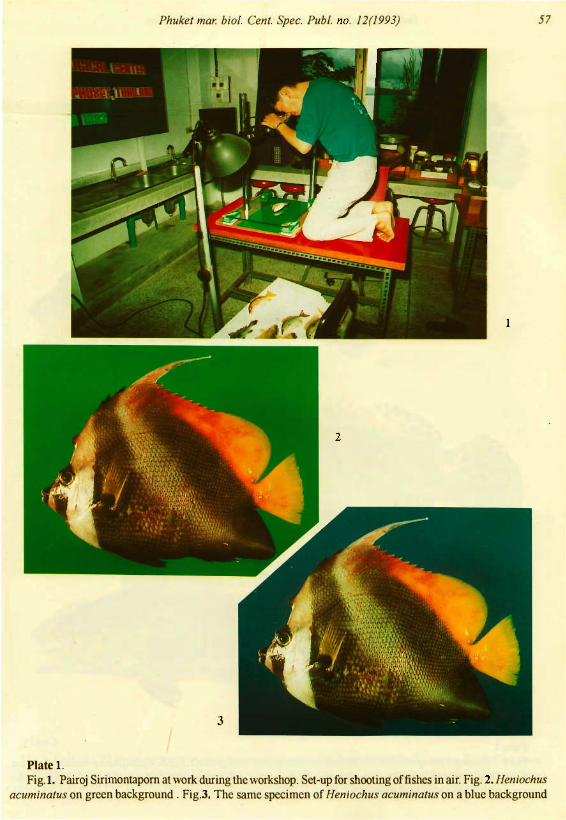
Plate 1.Fig.I. Pairoj Sirimontraporn at work during the
workshop. The fish is placed on a glass plate raisedabove the exchangeable background of uniformlycoloured sheets. In this case green. Photo by SomchaiBussarawit. Fig. 2. Heniochus acuminatus (L., 1758)on green background which brings off the darker tones(a bit more reddish) on the body of the fish. The resulting colour is brownish. The flood-light gave reflections in the skin concentrated around the eyes. Inthis shot the reflections are a bit too strong, eliminating the black colour ofthe stripe under the eye. Similarly, unwanted reflections produced white areas atthe root of the tail. Fig.3. The same fish on a bluebackground which brings out the lighter colour tones.The result is a grey-black body. Because of smudging, yellow parts, such as edges of fins tend to become more green than seen on a green background.Figs. 2 & 3 shot in laboratory by Pairoj Sirimontaporn.Glossy paper prints from colour negative film.
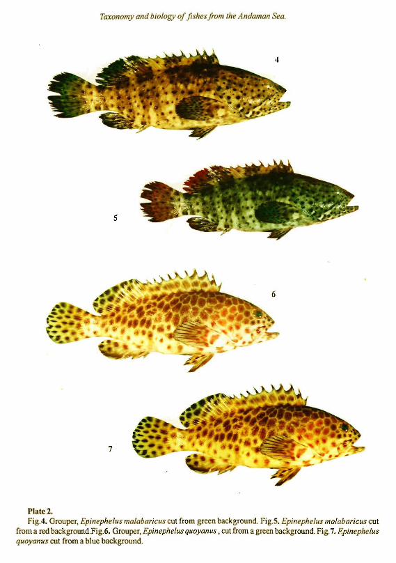
Plate 2.Fig.4. Grouper, Epinephelus malabaricus (Bloch
and Schneider, 1801), cut from green background. Thefins are tinted green by smudging, and colour of thebody becomes brownish. A disturbing area of reflections blurs the dorsal fin at the posterior part. The fishappears as if it were attacked by fungi, but it was actu-
ally a very fresh and healthy looking fish. Reflectionseasily developed in this particular area ofthe fish whenphotos were made in the photographic set-up of theworkshop. Fig.5. The same fish, but cut from a redbackground. The fins are heavily tinted by red, andthe black-grey colour tones of the body are stronglyset off. The colour of the body is most correct in thisparticular specimen, but the smudging of red is toostrong to warrant use of this picture for taxonomicpurposes. Fig.6. Grouper, Epinephelus quoyanus(Valenciennes, 1830), cut from a green background.This shot shows the most correct colour of the bodyof this particular specimen. There is only a slightsmudging of colour on the fins. In our opinion, theonly flaw in this picture is the disturbing reflectionson the posterior part of the dorsal fin. Fig.7. The samefish, but cut from a blue background. The spottedsurface ofthe body surface becomes more dark brown,i.e. increased contrast, on the blue background. Sincethe colours offish vary according to dominant coloursin the environment, the correct colour tone may beconsidered a minor problem. Spots on the surface ofEpinephelus quoyanus may be found even darker(nearly black) than shown on this photo. However, ifwe want a precise record of the colour, Fig.7 is notcorrect. Figs. 4-7. shot in lab. by Pairoj Sirimontaporn.Glossy paper colour prints from negative fIlm.
Plate 3.Fig.S. Grouper, Epinephelus quoyanus, cut from a
black background. This shot is without reflectionsbecause it was made ofa specimen submersed in water. There is only a slight smudging of colour on thefins. The black background resulted in a very pale fishcompared to the next picture. Fig.9. The same fish,but cut from a blue background. The spotted surfaceof the body surface becomes more dark brown, i.e.increased contrast, on the blue background. Reflections are absent (submersed) but some blue smudging occurs on the fins. Fig.tO. The same fish, but cutfrom a green background. The spotted surface of thebody surface becomes even darker than on a bluebackground. Reflections are absent (submersed) butsome green smudging occurs on the fins. Fig.H. Thesame fish, but cut from a green background. This timethe picture was taken out of the water. Reflectionsobscure the spots clearly seen at the root of the tailwhen the fish is submersed. A desirable reflection is
5 6 Taxonomy and biology of fishes from the Andaman Sea.
seen in the eye, but there are disturbing reflections onthe posterior part of the dorsal fin and on the body ,similar to the results obtained with the other specimenof Epinephelus quoyanus shown on Plate 2. Smudging of green occurs on the fins to the same degreewhether the fish is submersed or in the air. Figs. 8-11shot in lab. by Somchai Bussarawit. Semiglossy paperprints from negative film.
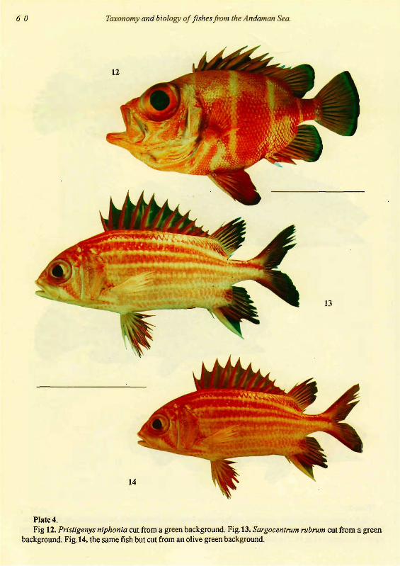
Plate 4.Fig 12. Pristigenys niphonia (Cuvier), cut from a
green background. Generally red fish are nicely set offby the green contrast. But smudging of green on thethin parts of fins can become a problem. Originally,the picture was a colour slide. It lost some details afterbeing photographed on colour negative film, butparticipants agreed that it still had sufficient qualityto be used for taxonomic purposes in spite of someloss of colour intensity. We found that slides weresuperior as far as colour-true photographic recordsare concerned, but they are too expensive to be usedfor illustrations in print, in general. From glossy print.Fig.13. Sargocentrum rubrum Forsskal, 1775, cut froma green background. This picture was copied from acolour slide to negative film as in Fig.12. From glossyprint. Fig.14. the same fish but eht from an olive greenbackground. This background gives a warm touch tothe body ofa red fish, and smudging ofthe fins is lessconspicuous compared to the bright green background. This picture has a more correct colour thanthe above shot. Colour negative film. Print onsemiglossy paper. Figs. 12-14 shot in natural sunlighton board the research vessel by Weera Pokapunt.
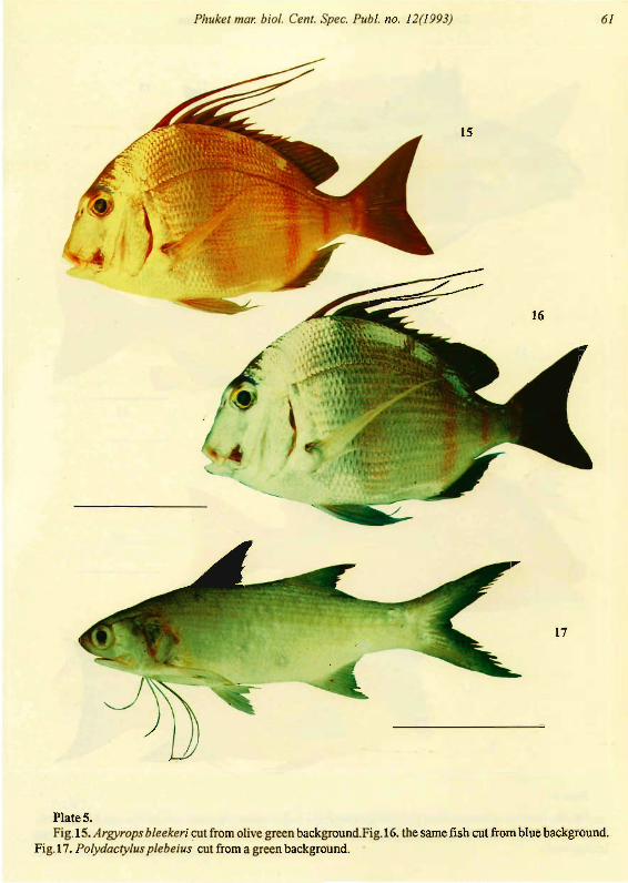
Plate 5.Fig.15. Argyrops bleekeri Oshima, cut from olive
green background. The colour tone is soft, and thecolour of the fish close to the live fish. Colour negative film. Fig.16. the same fish cut from blue background. Originally, this picture was a colour slide. Theslide lost some quality after being photographed oncolour negative film but when compared with Fig. 15, itis our opinion that a blue background is better forreproduction of silvery ·species. Species such asArgyrops bleekeri are difficult to photograph becauseof strong reflections from the scales. One part of thefish easily becomes overexposed and other parts underexposed. In this picture, the tail has become too
da~k. There is'heavy smudging of blue colour whichhas changed most of the red colour to blue. Glossyprint.' Fig.17. Polydactylus plebeius (Broussonet,1782), cut from a green background. In this case arather time consuming enterprise. About one houragainst less than halfan hour in a more "normal" fish.Originally, this picture was a colour slide showing moredetails on the ventral part. We found that a green background also is good for silvery species. Glossy print.Figs. 15-17 shot in natural sunlight on board the research vessel by Weera Pokapunt.
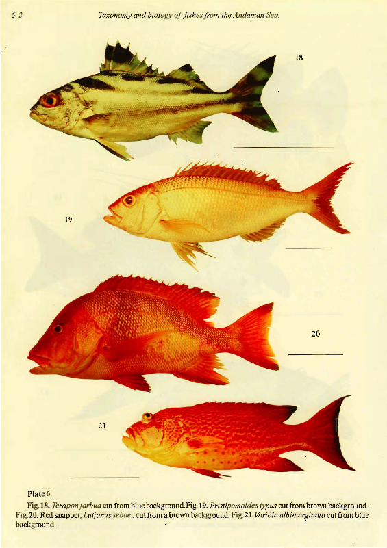
Plate 6.Fig.lS. Teraponjarbua (Forsskal, 1775), cut from
blue background, which sets off a good contrast inthe silvery body against the black pattern. But thefins do get a blue tinge, which is beautiful but incorrect. Originally, the picture was a colour slide. Glossypaper print. Fig.19. Pristipomoides typus Bleeker, 1852,cut from brown background. Smudging has becomesevere on fins and body of this silvery fish. Colournegative film; semiglossy print. Fig.20. Red snapper,Lutjanus sebae (Cuvier, 1828), cut from a brownbackground. The brown background gives a warm touchto the body of a red fish, and smudging of the fins isless conspicuous compared to the blue and green backgrounds. The contrast would presumably have beenstronger set offon a black background. Colour negative film. Print on semiglossy paper. Fig.21. Variolaalbimarginata Baissae, 1952, cut from blue background. Red colour is well reproduced on this background which only results in smudging of the edgesof fins. However, smudging has made the picture incorrect since it nearly caused the narrow white rim ofthe caudal fin to disappear. Originally, the picture wasa colour slide. Copied on colour negative film. Glossypaper print. Figs.18-2l shot in natural sunlight onboard the research vessel by Weera Pokapunt.
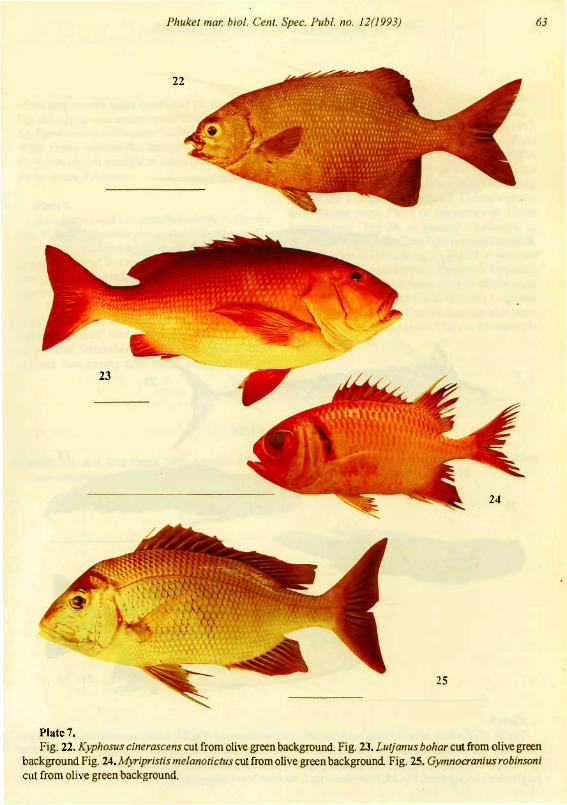
Plate 7.Fig. 22. Kyphosus cinerascens (ForsskaI, l775),cut
from olive green background. All pictures on this platehave been cut from this type of background (canvason board research vessel) which seems to give superior colour quality to brown and red fishes. We haveselected these photographs for this reason. There is aminimum ofsmudging and no disturbing reflections.Fig. 23. Lutjanus bohar (Forsskal, 1775). Movements
Phuket mar. bio/. Cent. Spec. Pub/. no. 12(1993) 57
Plate 1.Fig.I. Pairoj Sirimontaporn at work during the workshop. Set-up for shooting offishes in air. Fig. 2. Heniochus
acuminatus on green background. Fig.3. The same specimen of Heniochus acuminatus on a blue background
58 Taxonomy and biology of fishes from the Andaman Sea.
5
7
Plate 2.Fig.4. Grouper, Epinephelus malabaricus cut from green background. Fig.5. Epinephelus malabaricus cut
from a red background.Fig.6. Grouper, Epinephelus quoyanus ,cut from a green background. Fig.7. Epinephelusquoyanus cut from a blue background.

Phuket mar. bioi. Cent. Spec. Pub/. no. 12(1993) 59
10
9
11 •
Plate 3.
Fig.S. Grouper, Epinepheius quoyanus, submersed in water. Cut from a black background. Fig.9. The samefish submersed in water, but cut from a blue background. Fig. 1O. The same fish submersed in water, but cut froma green background.Fig. II. The same fish photographed out of water. Cut from a green background.
60 Taxonomy and biology of fishes from the Andaman Sea.
14
13
Plate 4.Fig 12. Pristigenys niphonia cut from a green background. Fig. 13. Sargocentrum rubrum cut from a green
background. Fig.14. the same fish but cut from an olive green background.
Phuket mar. bioi. Cent. Spec. Pub/. no. 12(1993) 61
17
PlateS.Fig.IS. Argyrops bleekeri cut from olive green background.Fig.I6. the same fish cut from blue background.
Fig.I7. Polydactylus plebeius cut from a green background.
62
19
Taxonomy and biology of fishes from the Andaman Sea.
21
18
20
Plate 6.
Fig.IS. Teraponjarbua cut from blue background.Fig. 19. Pristipomoides typus cut from brown background.Fig.20. Red snapper, Lutjanus sebae , cut from a brown background. Fig.21. Variola albimarginata cut from bluebackground. .
23
Phuket mar. bioi. Cent. Spec. Publ. no. 12(1993)
25
63
Plate 7.Fig. 22. Kyphosus cinerascens cut from olive green background. Fig. 23. Lutjanus bohar cut from olive green
background Fig. 24. Myripristis melanotictus cut from olive green background Fig. 25. Gymnocranius robinsonicut from olive green background.
64 Taxonomy and biology of fishes from the Andaman Sea.
27
28
29
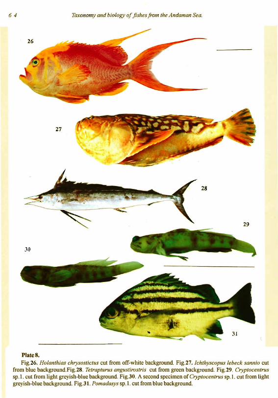
PlateS.Fig.26. Holanthias chrysostictus cut from off-white background. Fig.27. lchthyscopus lebeck sannio cut
from blue background.Fig.28. Tetrapturus angustirostris cut from green background. Fig.29. Cryptocentrussp.l. cut from light greyish-blue background. Fig.30. A second specimen of Cryptocentrus sp.l. cut from lightgreyish-blue background. Fig.31. Pomadasys sp.l. cut from blue background.
Phuket mar. bio/. Cent. Spec. Publ. no. 12(1993) 65
of the ship caused slight shading of the dorsal body.Fig. 24. Myripristis melanotictus Bleeker, 1853 Fig.25. Gymnocranius robinsoni (Gilchrist et Thompson,1908). Colour negative film. Semiglossy prints. Figs.2225 shot in natural sunlight on board the research vessel by Weera Pokapunt.
Plate 8.Rare species and new records from the Andaman
Sea. Fig.26. Holanthias chrysostictus Gunther, 1871,cut from off-white background which is a very goodcolour for fish of this type. Shot outdoors in naturalsunlight by Somchai Bussarawit. Originally, the picture was a colour slide. The slide did not lose qualityafter being photographed on colour negative film.Fig.27. Jchthyscopus lebecksannio Whitley, cut fromblue background. Shot outdoors in natural sunlightby Somchai Bussarawit. Copied from colour slide. Itdid not lose quality after being photographed on
colour negative film, but blue colour smudging wasaccentuated by the second shooting. Glossy print.
Fig.28. Tetrapturus angustirostris Tanaka, 1915,cut from green background. Shot in natural sunlighton the board research vessel by Weera Pokapunt.Glossy print. Fig.29. Cryptocentrus sp.l. cut from lightgreyish-blue background. There is virtually no smudging but the fish gets a dull colour. Colour negativefilm. Glossy print. Photo in laboratory by UkkritSatapoomin. Fig.30. a second specimen ofCryptocentrus sp.l. cut from light greyish-blue background. Photo made as in Fig.29. Fig.3!. Pomadasyssp.l. cut from blue background. Originally the picturewas a colour slide which was copied on colour negative film. The picture did not loose much in quality.Smudging of blue colour on the fins was also heavyon the slide. Semiglossy print. Photo in laboratory byUkkrit Satapoomin.
REFERENCES
Randall, J. E. & A. Ben-Tuvia. 1983. A review ofthe groupers (pisces: Serranidae:Epinephelidae) ofthe Red Sea,with description ofa new species ofCephalopho/is. Bulletin o/Marine Science, 33:373-426.
Addendum
The species identified on p. 64 no. 26 as Holanthias chrysosticus was subsequently identified asMeganthiasfiliferus n.sp. by Randal & Hemstra, 2007.
Meganthias filiferus, a new species of anthiine fish (Perciforrnes : Serranidae), from theAndaman Sea off southwestern Thailand. - Phuket mar. bioI. Cent. Res. Bull. 68: 5-9 (2007).