COLOUR AND LIFE
159
Symposia if the Institute if Biology, No. 1 2 COLOUR AND LIFE Edited by W. B. BROUGHTON
-
Upload
khangminh22 -
Category
Documents
-
view
1 -
download
0
Transcript of COLOUR AND LIFE

Symposia if the Institute if Biology, No. 1 2
COLOUR AND LIFE
Edited by
W. B. BROUGHTON

THIS BOOK presents an up-to-date account of
colour in living things. Chapters deal with the
production of colours by physical processes; the
nature of chemical colours in plants and animals;
the significance of colour for the purpose of classi
fication; the role of pigments in those essential
processes, photo'synthesis and respiration; colour
perception by invertebrates and man; the part played
by colour in animal behaviour. The final chapters
are concerned with man's use of colour and the
problems of the reproduction of colour.
Colour has an important role in man's affairs and
it is hoped that the contents of this book will interest
not only the teacher of biology but all those who
seek a knowledge of recent advances in the field of
natural colours.
Price 25s.

COLOUR AND LIFE

Symposia of the Institute of Biology (Published by the Institute save in the cases indicated)
BIOLOGICAL HAZARDS OF ATOMIC ENERGY Oxford University Press 1952
FREEZING AND DRYING 1952
THE BIOLOGY OF DESERTS 1954
THE NUMBERS OF MAN AND ANIMALS Oliver and Boyd 1957
BIOLOGICAL ASPECTS OF THE TRANSMISSION OF DISEASE Oliver and Boyd 1957
THE BIOLOGY OF AGEING 1957
THE BIOLOGICAL PRODUCTIVITY OF BRITAIN 1958
THE EFFECTS OF POLLUTION ON LIVING MATERIAL 1959
BIOLOGICAL PROBLEMS ARISING FROM THE CONTROL OF PESTS AND DISEASES
1960
THE BIOLOGY OF SPACE TRAVEL 1961
THE BETTER USE OF THE WORLD'S FAUNA FOR FOOD 1963
COLOUR AND LIFE 1964

COLOUR AND LIFE
Symposia of the Institute of Biology No. 12
EDITED BY
W. B. BROUGHTON
LONDON The Institute of Biology 41 Queen's Gate, S.W.7
1964
712163

Printed by F. J. Milner & Sons Ltd., Brentford & London

CONTENTS
Introduction By W . B. BROUGHTON
Structural Colours of Biological Material By W. D. WRIGHT
Chemical Colour in Plants By J. B. HARBORNE
The Chemistry of Animal Colours By T. w. GOODWIN
Colour and Classification of the Lower Plants By G. E. FoGG
The Photosynthetic Pigments By C. P. WHITTINGHAM
Respiratory Pigments By H. MuNRO Fox
Colour Change in Animals By D. B. CARLISLE
The Physiology of Colour Perception in Invertebrates By J.D. CARTHY
Colour and Animal Behaviour .. . By U. WEIDMANN
Human Mechanisms of Colour Perception By KATHARINE T ANSLEY
The Acceptability of Colour Reproduction By R. W. G . HUNT
Applications of Colour in Everyday Life By MISS KATHLEEN A. BATTERSBY
Adornment by Colour in Man and Other Animals By H . GWYNNE VEVERS
Author Index
Subject Index
v
Page
vii
13
25
41
47
55
61
69
79
101
115
125
133
141
142


INTRODUCTION
By
W. B. BROUGHTON
./ sometimes think that never blows so red The Rose, as where some buried Caesar bled ...
OMAR KHAYYAM, as a poet and a contemporary of William the Conqueror, may perhaps be excused for stretching his biology some way beyond the action of conventional soil chemicals upon indicator pigments of flowers as we know them to-day. But if those outsiders, the Scientists, have taken the mystique out of living colour, at least they have spared, indeed heightened, the romance of it. Hard it may be to pierce through the calculated and necessary meiosis of scientific expression to the wonder and elegance of the mechanisms that generate colour out of structure, be it physical or chemical; yet wondrous and elegant they are, and he of either Culture who can read the essays of Wright, Harborne and Goodwin (pp. 1, 13, 25) without a thrill, is a sad fish indeed. As these fruits of only the last decade or so of research show, this is yet another field where biological phenomena are at last proving to have a basis as logical and beautiful in its precision as the self-styled exact sciences. Of perhaps particular interest among the work undertaken by Wright specifically for this Symposium is that which follows up Mason's study of 1927 on the beetle Heterorrhina elegans F. (pp. 7-8); only two years after Mason's failure to detect laminations, Stegemann found, in the elytra of other beetles, the now familiar "Balken" which are presumably the morphological basis of the multilayer interference now established by Wright.
Of the pigments of plants, we still have much, it seems, to learn about the form in which they actually occur, even though, as Harborne says, our knowledge of the chemistry itself is tolerably complete-sufficiently so for this author to give sound advice to those eccentrics, the would-be growers of blue roses and yellow sweet peas. In this context, even Hunt's recipe (p. 120) for the production of cyan cats on pink carpets may not fall upon wholly deaf ears.
It had been intended originally to include an entire paper devoted to the genetics and (non-trophic) physiology of plant colours; this was one of those aspects of which a few inevitably get omitted from symposia such as this, in which the physical presence of the contributor is required. Harborne, however, gives a useful brief review of the subject (pp. 21-22). The other, and much studied, sector of colour physiology in plants, photosynthesis and its allied processes,
Vll

is reviewed by Whittingham (p. 47); from this paper again, the excitement of living through the last two decades, and of having seen the pieces of this fantastic jigsaw put together, can be recaptured by those who have known it, and learned by those who were too young -this has indeed been a rewarding age for the biologist.
Goodwin, Munro Fox and Carlisle (pp. 25, 55, 61) have done for animals what the foregoing authors have done for plants: Goodwin with a monumental review of the structure and function of the nonrespiratory, Fox on the respiratory, pigments, and Carlisle on pigment deployment in colour change. A recently discovered respiratory metabolite, ubiquinone, is referred to on pp. 33-4. Though it remains clear that many pigments are excretory products (amongst new data, see e.g. ommatins, p. 29), both Goodwin's essay and the discussion sound a note of caution for those who believe that the complex pigment reaction-systems originated from excretory mechanisms; the same discussion, indeed, emphasizes the enormous number of questions of all types that remain open in animal chromochemistry. Carlisle takes up the dynamic aspects of this subject, which again have changed out of all recognition in the last decade or two; his treatment necessarily carries him into the domain of neurophysiology, behaviour and evolutionary theory, with the discussion centering mainly on the last.
If the foregoing topics have seen some exciting advances in recent years, no less striking is the impact upon taxonomy of the relation between colour and chemistry, a theme developed by Fogg (p. 41) in relation to the lower plants, where, as he shows, the most reliable taxonomic use of colour is to be found. Elsewhere in both kingdoms, colour may be at once a pitfall for the unwary and a short cut for the initiated; perhaps further advances in the chemistry of these higher groups will bring this paradox nearer to solution.
In the field of colour perception and related behaviour, Carthy's comprehensive review of invertebrate mechanisms (p. 69) opens appropriately with a clear exposition of the limits within which inferences can safely be drawn from either physiological or behaviour studies. Hunt's paper (p. 115), though concerned with the human observer, is not without relevance here, since it makes explicit some of the many other factors implicit in earthy's phrase: "the modifying influences of the central nervous system" -factors besides the mere physiological capacity for colour vision that for animals, as for man, must enter into the assessment of colour signs in the environment (see also earthy's remark on the Land effect in pigeons, p. 110).
In the vertebrate, and especially the human, field, where we can draw conclusions more safely and more easily, Tansley examines the current position of the principal theories of colour vision, and points out the need to see the available experimental results, so long thought contradictory, against their proper background-the actual "wiring diagram" of the visual apparatus. This, together with recent work
viii

tending to confirm the existence of opponent mechanisms (even if not precisely those envisaged by Hering) bids fair to resolve conflicting hypotheses into a single unified theory that will explain both colour phenomena per se and simultaneous contrast, hitherto one of the difficulties for the trichromatic theory. The actual opponent mechanisms now postulated-the interplay of nervous excitation and inhibition- accord not only with the classical notion of reciprocal inhibition, but also with the whole basis of current ethological theory. Points of particular interest in the discussion are an analogy with colour television, and Dartnall's hypothesis of the behaviour of cone pigments.
The relation of colour to behaviour is a vast subject which is introduced by Weidmann (p. 79) and developed in particular aspects, human or animal, by the remaining three authors. The underlying theme of Weidmann's first section is the interrelation of colour signals and natural selection, for whose proper understanding a discussion of many of the fundamentals of ethology has necessarily been brought in. The stereotypy of colour signals smacks almost of plant rather than animal organization, a point driven home by the main topic of his second section, the angiosperm-insect relationship, which includes a review of Daumer's work on ultra-violet honeyguides. Both from his discussion of the idea of conflicting selection pressures in social communication and from that of the concept of "guild-signs" and Eibl-Eibesfeldt's fascinating procryptic mimic, Aspidodontus taeniatus, he makes abundantly clear how much behaviour study has to contribute to the understanding of animal coloration and its evolution. Two other important loci among many in this paper are the discussion on p. 90 of a relation between inhibition and habituation which may be of much wider application than has yet been generally realized, and the underlining on p. 95 of the intensity of selection for mimicry (both cryptic and aposematic). In the discussion, attention is once again focused on the importance of such factors as spectral exactness in colour ethology.
Hunt (p. 115) and Battersby (p. 125) are concerned wholly, and Gwynne-Vevers (p. 133) principally, with the impact of extraneous colour on the human race. The features controlling acceptability of colour reproductions to human judgment may seem far removed from the sign-stimuli that elicit the begging response of a gull chick: yet essentially the same subjective phenomenon appears to be at work in both cases-recognition by approximation, as Hunt's conclusion implies. The parallel may extend even to supranormal stimuli: "Better than the real thing", as we say; or "teaching the sheets a whiter hue than white", as Shakespeare and the soap firms say. It is interesting to find that the quality of greys is one of the most important and constant criteria of acceptability; discussion of the Land effect throws interesting light on this.
These considerations, and the content of the two final papers, show
ix

how well justified is the Institute's practice of seeking to carry the final sessions of its Symposia some way beyond the strict confines of biology. In this way, the "two cultures" are brought a little closer for a little time, and new lines of thought may be catalyzed by the contact. Thus in two complementary parts of the same topic- the use of colour by man-we have the artist commenting on the phylogeny of traffic lights and the zoologist discoursing on the history of an important branch of graphic art. Miss Battersby's account of the "chromology" of the last few decades, even to one who has lived through them, is a justly astonishing document; Vevers' "chromography" is equally astonishing : both, if highly entertaining, are no less serious contributions for that.
This Symposium was built, like its predecessors, upon the outstanding contributions of the last few years, many of which were produced by our present contributors, as a glance at the bibliographies will show. But this is perhaps the first attempt to compass the whole field in a single volume; insofar as it may have succeeded, we have to thank the chairmen and speakers for giving their time, the authorities at Birkbeck College for giving space and facilities, and not least, the Institute's own unflagging permanent staff. It is a quaint, endearing habit of the Council of this Institute to reward those whose bright idea they accept as the theme of a Symposium, by thrusting upon them the honour of organizing it and editing its proceedings. This is far from the back-handed compliment it might seem, because nearly all the hard labour is taken out of it by the diligence and devotion of Mr. D. J. B. Copp, the Institute's General Secretary, to whom I offer my most sincere thanks.
X

STRUCTURAL COLOURS OF BIOLOGICAL MATERIAL
By
W. D. WRIGHT
Technical Optics Section, Imperial College of Science and Technology
Types of Surface Reflection
SuRFACE reflection can be of two main kinds, specular and diffuse. In specular reflection, the light is reflected according to the laws of reflection as applied to a mirror. In diffuse reflection, on the other hand, each ray of light is split into many parts by multiple reflection, refraction and scattering, so that the fine structure of the surface diffuses the light in all directions.
The majority of surfaces exhibit both specular and diffuse reflection, and the total amount of light reflected is in general significantly less than the total amount of light incident on the surface. Except for one or two instances where the reflection is nearly 100 per cent, for example highly specular surfaces like silver or very white matt surfaces like smoked magnesium oxide, some of the incident light is either absorbed in the surface or transmitted through it. If the sample is partially transparent or translucent, its appearance as seen by reflection will depend to an important extent on the background or backing behind the surface.
A surface will appear coloured if it reflects some parts of the spectrum more highly than others. If the coloration is due to a dye or pigment, this variation of reflection with wavelength occurs because some of the energy is removed by absorption. The coloration may, however, be due to optical effects such as diffraction, scattering or interference, in which a preferential reflection of some wavelengths occurs through a re-direction of the energy without loss by absorption. Colours of biological material produced in this way are known as structural colours.
The reflection factor p of a surface is defined as the ratio of the total luminous flux reflected by the surface to the total luminous flux incident on it. The spectral reflection factor p;, is this ratio obtained when the surface is illuminated by monochromatic light of wavelength A. One way to describe the colour of the surface is then to plot the curve of p;, against A. This is often a useful curve to know, but it has the disadvantage that the reflected light which it

Wright
measures includes both the specular and the diffuse components. It cannot, therefore, distinguish the colour of the one from the colour of the other. The directional variations of hue which occur in iridescent colours will also be averaged out in the integrated reflection recorded by p;..
An alternative quantity which can be measured is the luminance factor (J. This is defined as the ratio of the luminance (photometric brightness) Ls of a surface when seen from a given direction and under defined conditions of illumination to the luminance Lw of a perfect white diffuser (in practice a white magnesium oxide surface) under the same conditions of illumination and viewing ; that is
fJ = Ls Lw"
The corresponding quantity measured under monochromatic illumination is known as the spectral luminance factor {J;.. {J;. has the advantage over p;. that it can be measured for different directions of view. The curves of {J;. against A for these various directions will then reveal any differences in colour corresponding to the specular and diffuse reflections or to iridescence.
Reflection at a polished metal surface differs from that at a polished surface of an insulating material such as glass, in two important respects. In the first place the reflection factor of a metal is very much higher than that of glass and secondly, in the case of a coloured metal, the specular reflection itself is coloured, whereas that from coloured glass is uncoloured. The metal is also, of course, opaque. In general, the top surface reflection from most nonmetallic materials is uncoloured, as, for example, in the highlights of a glossy paint surface. Metallic lustre or bronzing does, however, occur with some pigments due to very high absorption and specular re-radiation of rays of a particular waveband in the spectrum. We shall see that multilayers of non-metallic material can also, under some conditions, give rise to a high specular reflection that is coloured and which for this reason is described as "metallic reflection".
One other type of surface needs to be mentioned, namely the so-called "enamelled surface". In this the colour originates below the top surface, as indicated, for example, by movement of the illuminated area when the observer moves his head.
The Optical Basis of Structural Colours
(a) Scattering. When very fine particles are illuminated they become new sources of light and radiate in all directions. If the particles are concentrated into a thick layer, the multiple scattering produces a matt white reflection. Surface whiteness also arises from the random reflections and refractions in finely divided, optically transparent material, such as snow. The whiteness of much
2

Structural Colours of Biological Material
biological material originates in this way. (For a general account of structural colours, see D. L. Fox, 1953, and Munro Fox and Vevers, 1960.)
Scattering of light by particles in a dispersed suspension will also be white if the particles are large relative to the wavelength, but when the particle diameter falls below about 0·6f-l, the short-wave blue light is scattered more intensely than the long-wave red light. The dependence of the scattering on the size of the particles was first studied experimentally by Tyndall in 1869 and the effect is commonly known among biologists as Tyndall scattering. Rayleigh followed in 1871 with a quantitative study, in which he established that the intensity of the scattered light is proportional to fr when the particle size is less than the wavelength A.
For scattering to occur, a difference in refractive index must exist between the minute scattering bodies and the surrounding medium, but this means that it can take place not only at solid particles in air but also at minute air cavities in a solid medium. The Tyndall blue of the feathers of the blue jay or of the macaw, for example, is attributed to light-scatter at the minute air cavities in the alveolar or box cells in the barbs of the feathers. The cavities here are probably less than 0·3/-l in diameter. Mason (1923) summarized the characteristics of Tyndall blues and noted that the blue colour is only visible _by reflection, no major changes of colour occur over wide variations in the angle of reflection, and no blue dye or pigment is extractable.
Diffraction, in which the spreading of the light is due to the finite wavelength of the radiation and is particularly marked for light incident on a fine periodic structure, is not considered by most authorities to be responsible for any of the more striking examples of biological colouring. Yet Fox (1953, p. 39) reports that M. G. M. Pryor maintains that the blue colours of feathers are not produced by Tyndall scattering but by diffraction from surface structures acting as a grating.
Since the wavelength range over which Tyndall scattering occurs extends well into the green part of the spectrum, green colours will be produced through the combined operation of Tyndall scattering and absorption in a yellow pigment. For example, if the light scattered in the box cells of a feather has also to pass through a yellow pigment in the cuticle, the pigment will absorb the short-wave blue light but transmit the green, thus producing a residual green colour.
Tyndall scattering does not produce a very intense colour because the fraction of the incident light that is scattered is fairly low. It will in fact be easily masked by the light reflected from the other structures of the specimen unless there is a substratum of some dark absorbing medium such as melanin or haemoglobin . To that extent absorption is necessary for the effective observation of Tyndall scattering.
3

Wright
(b) Interference. The most striking structural colours arise from thin film interference. This is a very well known phenomenon in physics in which the partial reflection at the two surfaces of a thin layer of a transparent medium, e.g. a soap film or an oil layer, produces two beams having a difference of optical path D given by D =2nd cosO, where dis the thickness of the film, n the refractive index of the film and 0 is the angle of incidence of the light within the film. This path retardation gives rise to a difference of phase t5 between the two vibrations where
b = 2n.2nd cosO, A
but there will be an additional phase retardation of n for a reflection at a denser medium.
Since in the above equation b is dependent on A, the two beams will be in phase for some wavelengths, out of phase for others and in some intermediate phase relation for the remainder, producing varying degrees of reinforcement and interference. The intensity of the reflected light will therefore vary through the spectrum, hence the production of interference colours. The spectral composition of the reflected light can be measured by recording the curve of {1;. against A exactly as if the coloration had been produced by a pigment. If the film varies in thickness, a series of interference fringes can be seen which, in white light, will have the appearance of a succession of spectra, strongly coloured in the first order spectrum where the film is thinnest but becoming paler and desaturated in the higher order spectra.
For a soap film or oil film, only some 3 or 4 per cent of light will be reflected at each surface, so the intensity of the reflected light is relatively low. The interference colours only show up strongly, therefore, when the film is seen against a dark background as with an oil film on a road surface. The intensity of the reflection becomes much higher, however, in a structure consisting of a series of thin laminations of transparent media. The reflected light then consists of a series of multiple reflections of diminishing intensities and increasing phase retardations. The intensity of the component reflected from any given boundary will depend on the refractive index change at that boundary and on the attenuation due to losses by partial reflection at the other boundaries as the light passes in and out of the laminated layer.
The deposition of multiple layers is now an important optical technique for the production of mirrors and filters having defined spectral reflection and transmission characteristics. These characteristics may include very high reflection in some parts of the spectrum and high transmission in others. The reflection is therefore coloured and specular and hence has the characteristics of metallic reflection, even though produced by layers of transparent material. The
4

Structural Colours of Biological Material
theoretical analysis of multilayer interference is complex (Weinstein, 1954; Greenewalt, Brandt and Friel, 1960). In general it may be said that for the case of a periodic structure consisting of a two-layer sequence repeated many times, the wavelength of maximum reflection is the same as that for one two-layer element, but the multiple beams unite to give a very much higher reflection.
The spectral reflection curve consists of a central band of high reflection with subsidiary reflection bands at wavelengths on either side of the main band. The width of the central band is a function of the difference in refractive index of the alternating layers, the smaller the index difference the narrower the bandwidth. The intensity and distribution of the subsidiary bands is, however, affected to a marked extent by the nature of the bounding media of the lamination.
Since the path difference D between the beams reflected from the two surfaces of a thin film is given by 2nd cosO, then as the angle of incidence increases the path difference decreases. This means that if the reflected beams are in phase for wavelength A. when the light is incident normally, so that there is a reflection maximum at this wavelength, the wavelength of maximum reflection wiJl move to a shorter value A.' for a more oblique direction of illumination and viewing. A film will thus change, say, from green to blue or violet in passing from normal to grazing incidence. · The corresponding effect with multilayer films is very striking and it is for this reason that these colours are known as iridescent colours. In biological material, however, although the structure is deposited in closely parallel layers, their general contour is likely to be curved or irregular rather than flat. Hence when looking, for example, at a beetle which exhibits metallic reflection, the iridescence may manifest itself by apparent variations in colour over the surface because its curvature introduces variations in the angle of incidence and reflection of the light.
Although the light reflection from a multilayer is much higher than for a single layer and certainly more intense than Tyndall scattering, the vividness of the colour is likely to be enhanced when an underlying absorbing layer of melanin is present. The iridescent colours of beetles are also especially striking because the scales provide a more or less extended and continuous area of metallic reflection. On the other hand, when multilayer reflection occurs in feathers, the colour is derived from small areas of local iridescence interspersed with much non-iridescent material. The colour of an extended area of feather, impressive as it may seem, is thus a rather degraded version of the colour of the individual sources of iridescence. A detailed study of the iridescence spectrum would therefore require the projection of these small areas of iridescence through a microscope on to the slit of a spectroscope, as was done in a recent study of hummingbird feathers by Greenewalt, Brandt and Friel (1960).
5

Wright
Preliminary Data on Some Structural Colours
As a contribution to this symposium, some measurements have been made on certain structural colours using the spectrophotometric equipment available in our Technical Optics Section. These include a Beckman DK2 recording spectrophotometer and a nonrecording spectrophotometer of our own design (Wright, 1958). The curves must be regarded as provisional but they may be of interest in showing the problems involved and the kind of results that can be obtained.
The Beckman instrument has the great advantage that a complete spectral reflection curve can be recorded in 2-5 minutes but it has the disadvantage that the reflected light is collected in an integrating sphere. (The specular reflection can, however, be excluded if desired.) The instrument therefore records the reflection factor p, instead of the luminance factor fJ, and it is impossible to record the iridescent colours at various angles of incidence and reflection. Nevertheless, the p;. curve can provide very useful information.
In our non-recording instrument, the specimen is illuminated at a given angle of incidence i and the luminance factor {3;. measured for a given direction e to the normal. Both i and e can be varied over a wide range of angles. The measurements are, however, rather timeconsuming and it is often useful, therefore, to record p;. on the Beckman instrument to get a first general idea of the shape of the spectral reflection curve.
Figure 1 shows the {3;. curve for the Tyndall blue (curve A) of a macaw feather with light incident at 50° to the normal and collected at 10° on the opposite side of the normal. There is a maximum in the blue at wavelength 0·455,u and the curve does not show a variation of luminance factor proportional to f. Perhaps this is due to absorption in the cuticle at the violet end of the spectrum. As a matter of interest the curve was also measured for the underside of the feather (curve B) which has a strong reddish-orange colour. The light is again diffusely scattered but presumably from pigmented structures in the feather.
Figure 2 shows the spectral luminance curve for the blue-green area of the peacock's eye feather, measured at 10° incidence and 10° reflection. No doubt the curve is broader and the values of {3;. lower than if individual iridescent points on the feather had been measured.
Figure 3 shows two spectral luminance curves for the beetle Chrysochroafulminans F. (family Buprestidae), one for 10° incidence and 10° reflection (curve A) and the other for 45° incidence and 45° reflection. These curves are typical of multilayer interference and the shift in the wavelength of maximum reflection from 0·573,u to 0·524,u with increase in angle of incidence would correspond to the shift to be expected in a film of refractive index of about 1·4.
6

11,
Structural Colours of Biological Material
FIG. I. Spectral reflection characteristics of Macaw feather as measured by the spectral luminance factor {J'A.
Curve A-blue coloration due to Tyndall scattering. Curve B-reddish-orange coloration of underside of feather due to pigment scattering.
r 1\ ' !/ \ v ~
0
(\ 0
I' \ lr 1\'
0
'J 1\ \ IL 1..-J \_ \ t--
0
FIG. 3. Spectral luminance factor {J'A for beetle Chrysochroa fulminans F. (family Buprestidae). Coloration due to multilayer interference. Curve A-angle of incidence and reflection 10°. Curve B-angle of incidence and reflection 45°.
o,
,.,..
. ' /
'~ I V"--
• -:-----L_j
FIG. 2. Curve of spectral luminance factor {J'A for Peacock's eye feather. Coloration due to multilayer interference.
I
5
0 LJ ~
0•55
Wavelength
Flg. 4
FIG. 4. Spectral luminance factor {J'A for beetle Heterorrhina elegans F. (family Cetoniidae).
Coloration due to multilayer interference.
Angle of incidence 30°, normal reflection.
070"'
Figure 4 shows the spectral luminance curve for the beetle Heterorrhina elegans F. (family Cetoniidae), which is an example of a green enamelled reflection, the colour appearing to originate from well below the surface. There is a white top surface reflection which
7

Wright
has to be avoided in the measurement and the curve was obtained for about 30° incidence and normal viewing. Figure 4 is remarkable for the narrowness of the spectral reflection band, implying a very small refractive index difference between the layers. The enamelled effect is attributed by Mason (1927) to fine rod like structures normal to the surface, but he could not detect any laminations under the microscope. This may well be a consequence of the small index difference but an interference or phase contrast microscope might well reveal them.
The final specimen that has been studied is the remarkable South American Morpho butterfly whose wings are a brilliant iridescent blue. There has been much discussion as to the origin of the colour, which was thought by some authorities to be due to diffraction. This appears to be ruled out by the fact that the specular reflection is coloured (although this objection would not apply if the structure behaved like an echelette or blazed grating, (Sawyer, 1951) ), and by the fact that the change in colour is in the direction from blue to violet as the angle of incidence and reflection is increased.
However, the architecture of the scale on the wing is quite extraordinary and the scales possess some very unusual optical properties. Figure 5(a) has been reproduced from photomicrographs taken in our microscope laboratory by Dr. W. N. Charman, in which the rows of scales on the wing can be seen ; Figure 5(b) is an enlargement of one of the scales showing the existence of very fine ribs spaced about It-t or Jess apart. The structure of these ribs has been further
FIG. 5. (a) Photomicrograph of scales on wing of Morpho butterfly. Mag. x 45 .
8

Structural Colours of Biological Material
FIG. 5. (b) Enlarged photomicrograph of part of scale showing fine ribs spaced about l!J. apart. Mag. x 700. (Photographs by Dr. W. N . Charman).
elucidated by some electron microscope studies by Anderson and Richards (1942), as illustrated in Figure 6.
FIG. 6. Structure of ribs on scale of Morpho butterfly wing, as deduced from electron microscope studies. (Anderson
and Richards, 1942.)
Examination of the direction in which the main specular reflection occurs suggests that the ribs provide reflecting strips which are inclined to the plane of the wing at an angle of about 10°. For normal incidence the reflected light is distributed in a semi-circular band in a plane normal to the ribs, the colour changing fairly suddenly from blue to deep violet as the reflection approaches the grazing angle. In certain directions around the normal, no light is reflected other than the scattering from the brown substratum of melanin. Not surprisingly, in view of its elongated structure, the layer is also doubly refracting.
The spectral luminance curves that have been obtained so far on the Morpho butterfly are shown in Figures 7(a) and (b) for various angles of incidence and reflection. Their general shape is consistent with multilayer interference, but it is not at all clear how to explain
9

Wright
--, i/ 1\ r
I \ I \
·I \ \
R,
. I \ "
\ 0
0
j\J
I \ .
.\ h--. r---... ~ '\., ~ ~ 0 , . ., O"
FIG. 7. Spectral luminance factor fJ1. for Morpho butterfly wing. (a) Angle of incidence 40°, normal reflection. (b) Angle of incidence zoo. Curve A-normal reflection. Curve B- angle of reflection zoo.
Curve C-angle of reflection 40°.
(Angles of incidence and reflection measured relative to plane of wing. Measurements made with ribs on scales running perpendicular to
plane of incidence.)
" '
the polar distribution of the reflected light. Clearly a complete interpretation of the optical properties of this most interesting material must await more detailed and extensive measurements.
Acknowledgments
I would like to express my sincere thanks to Mr. J. P. Doncaster and Dr. E. B. Britton of the British Museum (Natural History) and to Dr. H. G. Vevers of the Zoological Society of London for the supply of specimens and for some helpful discussions with Dr. Britton. I am also very grateful to Dr. W. N. Charman for taking the photomicrographs reproduced here and to Dr. W. T. Welford for some valuable discussions on the theory of multilayer interference.
References Anderson, T. F ., and Richards, A. G. 194Z. J. appl. Phys. 13, 748. Fox, D. L. 1953. Animal Biochromes and Structural Colours, Cambridge
University Press. Fox, H . Munro, and Vevers, H. Gwynne. 1960. The Nature of Animal Colours,
Sidgwick and Jackson, London. Greenewalt, C. H., Brandt, W. , and Friel, D . D . 1960. J . Opt. Soc. Amer., 50,
1005. Mason, C. W. 19Z3. Mason, C. W. 19Z7. Sawyer, R . A. 1951. Weinstein, W. 1954. Wright, W. D. 1958.
J. Physical Chern., 27, ZOI , 400. J. Physical Chern., 31, 1856. Experimental Spectroscopy, Chapman & Hall , London. Vacuum, IV, 3. The Measurement of Colour, Hilger & Watts, London.
10

Discussion
DISCUSSION
0 . V. S. Heath . Does Professor Wright know of any instances of plants producing structural colours?
W. D. Wright. There are, of course, a good many examples of light scatter occurring in plants and flowers and since these affect the surface appearance and colour, they might be quoted as instances of structural colours. I would be surprised if there were not some examples also of interference colours in plants but I am not sufficiently a botanist to be able to name any.
B. H. Crawford. The curve shown in Fig. 4 suggests an analogy with a Lippmann-Ives type of interference filter in which the reflecting laminae are tilted relative to the outer surface. Could this explain the apparent asymmetry of incidence and reflection?
W. D. Wright. Yes, if the reflecting laminae are tilted relative to the plane of the surface, this will cause the lack of symmetry between incidence and reflection.
11


CHEMICAL COLOURS IN PLANTS
By
J. B. HARBORNE
John Innes Institute, Hertford, Herts.
Introduction
THE colours of plants are due to the presence in the plastids or cell vacuoles of organic substances (pigments) which are capable of absorbing, transmitting and reflecting white light. These pigments selectively absorb light in one region of the visible spectrum and the human eye will see this by the appearance of colours complementary to those that have been absorbed . Colours produced by the reflection or diffraction of light from cell surfaces, phenomena common in the animal kingdom, are almost completely absent from plants.
The subject of plant pigments is a very considerable one. Thus, there are many thousands of species of plants and each may contain a considerable number of pigments, some of which may be organspecific, other which occur in most tissues. In addition, intraspecific flower colour variation is a common feature of cultivated plants. Fortunately, there is unity in diversity and a relatively small number of pigments account for the majority of plant colours. For example, green colours in plants are produced universally by a mixture of two closely related porphyrin pigments-chlorophylls a and b. Plant pigments can, in fact, be classified on an arbitrary chemical basis into four main types: carotenoid, nitrogenous, quinonoid and flavonoid. Within each type, series of pigments of closely related structure are known. In the present discussion, emphasis will be given to the commonly occurring pigments, which all fall into one of these four categories. It is not possible to mention more than a few of the rarer pigments of which some hundreds are known.
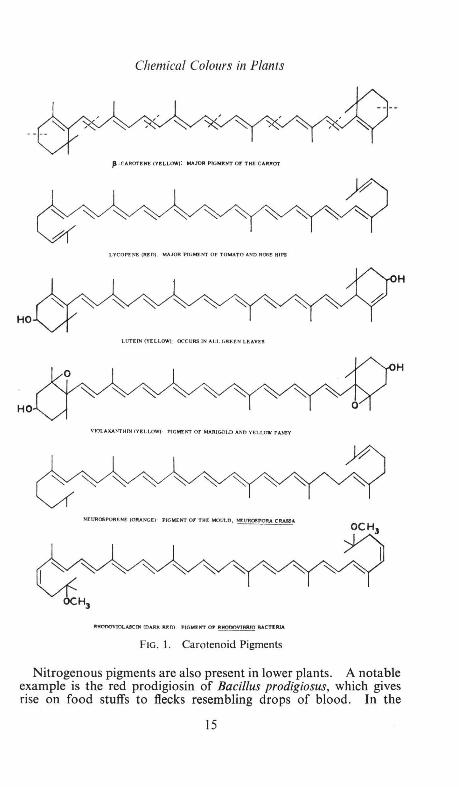
The main requirement for a compound to function as a pigment is that it should contain a conjugated system of alternating double and single carbon-carbon bonds. Such a system may include a number of chromophoric or "colour-producing" groups (such as carbonyl and azomethine groups) and will be a resonating structure, in which electrons can move around freely. Representative pigment structures are illustrated in Figs. 1-4.
13

Harborne
Carotenoid Pigments
Carotenoids are found in the plastids of plants and are lipid-soluble pigments, being yellow, orange or red in colour. They occur in the leaves of all higher plants, but their presence is usually masked by the green of the chlorophyll. They are conspicuous pigments in many yellow flowers, e.g. the dandelion, buttercup, pansy, tulip, and in some vegetables and fruits, notably the carrot and tomato. Carotenoids also occur in lower plants; they have been isolated from fungi, bacteria and algae. Only plants can synthesize carotenoids; thus, the many carotenoid pigments found in animals are derived directly or indirectly from plants eaten as food (see p. 61 ).
In structure, carotenoids are terpenoids, being built up from eight five-carbon isoprene units (Fig. 1). There are many opportunities for cis- and trans-isomerism in their conjugated systems of double and single bonds. Trans-structures are usually more deeply coloured than the related cis- forms. P-Carotene is one of the simplest of the group; other carotenoids differ mainly in having more or fewer double bonds (e.g. lycopene) and in having hydroxyl (e.g. lutein), epoxy (e.g. violaxanthin) and methoxyl groups (e.g. rhodoviolascin).
Nitrogenous Pigments
These pigments will be dealt with rather briefly, since the most important group of this class are the chlorophylls, and these substances will be discussed later in this Symposium in relation to photosynthesis. The structures of chlorophylls a and b are shown in Fig. 2. The basic porphyrin molecule consists of four pyrroles linked together in a planar structure and is also present in animal pigments (e.g. haemoglobin*) as well as in plants. The green colour of chlorophyll is due to the presence of covalently bound magnesium atom in the centre of the molecule. Chlorophyll also differs from animal porphyrin pigments in having a long aliphatic side chain (the phytyl residue) attached.
Other nitrogenous pigments which are common in animals and which probably occur in plants are the black insoluble polymers, the melanins. The colours in the fruiting body of the inkcap fungus and in the black spot on broad bean flowers are probably melanoid, although definite proof of this is still lacking. Some dark brown and black colours in plants, such as that of ebony wood, are almost certainly due to quinones rather than melanins.
Very few natural plant pigments are used as dyes because of their chemical instability. However one nitrogenous pigment, indigo, appealed so much to our forefathers that they coated their bodies with it. Indigo 9ccurs in Indigofera and /satis (woad) plants but since it is present in the leaves in a colourless form as the glucoside, indican, it is not a conspicuous plant colour. * See also p. 55.
14
Chemical Colours in Plants
fl · CAROTE~ CYlL LOW): MAJOR PIGJ«NT OP' THf: CARROT
LYCOPEI'"f: (RED) MAJOR P IGMENT OF TOMATO AND ROOE HIPS
H
HO
LUTEI'N (YELLOW) OCCURS IN ALL GREEN LEAVES
H
HO
VIOLAXANTHfN fY!:LLOW') PIGMt:NT OF MARIGOLD AND YELI.OW PANSY
NEUR06PORENE (ORA NGE:) PIGMENT 0 , T HE MOULD, N!:UR08roRA CRA!BA.
JII:HOOOVIOLA.!ICrtri {D4RJ< ~01 PIGM:t l'n OF ~BACT! RIA
FIG. 1. Carotenoid Pigments
Nitrogenous pigments are also present in lower plants. A notable example is the red prodigiosin of Bacillus prodigiosus, which gives rise on food stuffs to flecks resembling drops of blood. In the
15

Harbornc
Middle Ages, the presence of such flecks on sacramental wafers led to the belief that they had been defiled by Jews and were bleeding; many innocent men lost their lives as a consequence. In its structure (shown in Fig. 2), prodigiosin is related to the porphyrins.
CHLOROPHYLLS A and B: GREEN PIGMENTS OF PLANTS
INDIGO: PURPLE DYE FROMINDIGOFERA
BETANIN (MAUVE) : PIGMENT OF BEETROOT [~¢o] n
MELANIN: ONE POIIBIBLE STRUCTURE
PRODIG108111 (111!0) : PIGMENT OF BACn.Lill PRODIOI081l8
FIG. 2. Nitrogenous Pigments
16

Chemical Colours in Plants
Finally, the water-soluble betacyanins may be mentioned under the heading of nitrogenous pigments, although they are also phenolic in nature. These pigments occur exclusively in plants of the Centrospermae order; in cactus and bougainvillaea flowers and beetroots for example. Since they occur in the place of anthocyanins in Centrospermae and are similar in colour, betacyanins have long been considered to be of related structure. Recently, however, betanin, the major beet pigment, has been shown to have a structure (Fig. 2) more closely related to the alkaloids than the anthocyanins. The beet pigment betanidin, not only occurs in beet as the glucoside, betanin, but is also present in many mauve and red cactus flowers in other glycosidic or isomeric forms. A related group of yellow pigments, betaxanthins are also present in Centrospermae but these have not yet been characterized.
Quinone Pigments
The qui nones occur in both plants and animals; however of the 190 pigments that have been characterized, 160 are plant constituents. In higher plants, quinones are not important as pigments, since they frequently occur in roots (e.g. emodin) or under the bark of trees (e.g. lucidin). Two exceptions are carthamone and dunnione, which occur in flowers of Safflower and on leaves of Streptocarpus respectively. Quinones contribute significantly to pigmentation in fungi (two examples are helminthosporin and fumigatin) and in lichens (e.g. physcion). Although there is a rather bewildering variety of substituent groups (e.g. hydroxyl, methyl, etc.) which may be attached to quinones, the basic chromophore of the group is the simple p-benzoquinone nucleus, as in fumigatin (Fig. 3).
Flavonoid Pigments
Flavonoid pigments occur mainly in higher plants. The anthocyanins are the most important group of colouring matters here, since they provide nearly all the scarlet, apricot, pink, red, maroon, mauve and blue colours. They are present in leaves (e.g. red cabbage, begonia), in many fruits (e.g. blackberry, apple, plum) in tubers (potatoes) and in roots (radishes) but are seen to their best advantage in petals (most garden plants). Anthocyanins are cations and are isolated as their chlorides; they occur naturally in association with organic acid anions. Three common anthocyanins are: pelargonin, first isolated from the scarlet Geranium; cyanin , occurring typically in the crimson rose; and delphin, present in blue delphiniums. Methylated derivatives are known : the pink peonin (from peony), the rose rosinin (from Primula rosea) and the mauve pigments petunin (from Petunia), malvin (from Malva), hirsutin (from Primula
17

HO
HO 0
0
OH
Harborne
CH 3
HO 0
HO 0
OH
CH 3
EMODIN (ORANGE) : IN RHUBARB ROOTS HELMINTHOSPORIN (DARK MAROON) :
0
DtTNNIONE (ORANGE·RED): ON
LEAVES OF STRE PTOCARPUS DIJNNII
0 OH
0 LUCIDIN (YELLOW): IN
BARK OF COPROSMA LUCIDA
0
IN THE ' RUST' ON BAJU..EY
CARTHAMONE (BRICK- RED) :
IN FLOWERS OF SAFFLOWER
FUMIGATIN (MAROON): IN THE
FUNGUS, COPRINUS SJMILill
PHYSCJON (ORANGE-YELLOW): IN
THE LICHEN XANTHORJA PARIETANA
FIG. 3. Quinone Pigments
hirsuta) and capensinin (from Plumbago capensis). There are two other anthocyanins, gesnerin and luteolinidin 5-glucoside, which occur only in the Gesneriaceae; as they do not have hydroxyl
18

Chemical Colours in Plants
groups in the 3- position, their colours (yellow and orange) are different from the others (Fig. 4).
The flavones are a group of pale yellow or ivory pigments, closely related in structure to the anthocyanins. They may provide some
HO OH
Glu-0 Pti.AROONIN (SCARLET):
IN~FLOW!RS
HO
0-Giu OH Glu-o
DELPHIN (MAUVE) ·
IN DELPH~'lUM FLOWERS
Glu-0 0-Giu OCH
3 HlRSUTIN (MAU\'El :
HO
HO 0
Fl.AVONt:S
OH
OH
Glu-0 CYAHIN CMACEI'n"A).
I" CRIMSOl4 ROSES
Glu-0 ROSJNIN (ROSEl :
IN PAlMULA J{08EA
HOO)OO ~ ~ OH I + -~ §
G lu-o cEsNERlN (YELLOW):
HO~
~
IN CESNERlA CARDINALlS
0 OH
CH-0 0 J A 'ZALEIN : IN RHOOODENDROHS
AND PLUWBAGO FLOWERS
OH
CKALCONtS ANl.l AURONES
OH
GI,-O~OH 0
COREOPSJ N (YELLOW! : IN
O OH
Glu-o()CI "<==cHO'\: OH ~ / -c
II HO 0
AL: REUSIN {OOLOEN YELLOW)
YE LLOW Am'IRHHINUMS
FIG. 4. Flavonoid Pigments
19

Harborne
of the yellow colour seen in primroses and crocuses but more frequently, they are" copigments" to the anthocyanins; that is, the flavones form loose complexes with anthocyanins in vivo and have a blueing effect on these mauve and crimson pigments. Flavones are also present in most ivory and cream flowers; they are pigments here in the sense that they absorb light in the near ultra-violet and hence are visible to insects. Aurones and chalcones are also phenolic pigments; they are deeper yellow in colour than flavones but are less widely distributed. Chalcones, for example, occur in gorse and yellow dahlia blooms; aurones are present in Oxalis, Antirrhinum and Coreopsis flowers.
Flavonoids occur in vivo as glycosides and there is a remarkable variation in the nature and position of the attached sugars. While glucose is a common constituent, arabinose, xylose, galactose, rhamnose and various di- and tri- saccharides have been found linked to these pigments. The sugars do not themselves contribute to the colour of the pigments. They are important in preventing the pigments from fading (the anthocyanidins are unstable to light) and in increasing their sap solubility.
Factors Modifying Flower Colour
The chemical structures of the pigments present are the major factors in determining flower colour. Thus, if chlorophyll is the only pigment, as in the flowers of some hellebore species and certain tulip varieties, the petal is always geen in colour. If only flavones are present, the flower is usually cream or ivory. True white (albino) or pigment-less flowers are rather rare. If carotenoids are present, petal colour may be yellow, orange or red, depending on which and how much of a number of pigments are present. At least five pigments, namely ,8-carotene, a-carotene, lycopene, lutein and violaxanthin, have been found in the deep orange flowers of the composite Calendula officina/is. Mixtures of anthocyanins are also common. Thus, scarlet, pink, crimson, mauve and purple shades of verbenas contain appropriate mixtures of pelargonidin, cyanidin and delphinidin glycosides. Again, colour varieties of sweet peas may each contain up to six or seven different anthocyanins. The colours of anthocyanins in flowers are frequently modified by other factors, some of which are listed below:
(1) Mixtures of different types of pigment may co-occur. The brown colours of Primula polyanthus and wallflowers are the result of superimposing yellow carotenoid (in the chloroplasts) on to a purple anthocyanin background.
(2) Trace metals present in petals may complex with pigments which have o-dihydroxylic groupings and alter flower colour. The pigment of the blue cornflower is a magnesium and iron
20

Chemical Colours in Plants
complex of the magenta anthocyanin, cyanin. Many other blue flowers (e.g. Hydrangea, Comme/ina and blue lupins) also contain anthocyanin-metal complexes. It should be remembered, however, that methylated anthocyanins cannot form metal complexes and also that some blue colours (in Primu/a, roses, etc.) are produced by co-pigmentation of anthocyanins by flavones .
(3) The concentration of pigment may vary within wide limitsvery dark shades being produced by a high concentration of anthocyanin in the cell sap. The intense purple-black varieties of pansy and tulip are produced in this way; in the pansy, 30 % of the dry weight of the petal is said to consist of anthocyanin.
(4) Variation in the pH of the cell sap may alter flower colour, since anthocyanins are natural indicators. For example, in flowers of the Chinese primrose, a change in pH from 5·4 to 6·2 brings about a colour shift from magenta to blue.
It is important to bear these colour modifying factors in mind when considering the production of new flower colour varieties. Many attempts have been made to breed a blue rose. The chemical evidence indicates that one source of blueness- the purple pigment, delphinidin-is absent from the Rosaceae so that there is no chance of raising a blue petalled rose by this means. Blueness, however, can also be produced by either metal-complexing or co-pigmentation and breeders might consider using varieties with this metal ion or high flavone concentrations in their petals. Co-pigmentation of the crimson rose pigment, cyanin, by unidentified flavone materials is certainly responsible for the mauve and purple shades now available (in, for example, the variety "Reine de Violette"). As well as the blue rose, breeders have been searching for a yellow sweet pea. The barrier here is of a different nature and is the difficulty of hybridizing the wild yellow flowered (carotenoid containing) Lathyrus pratensis with (flavonoid containing) L. odoratus.
Other Aspects of Plant Colour
Coloration in plants is inherited in a simple Mendelian fashion and much work has been done on flower colour genetics. Genes controlling the type and quantity of pigment and the patterning and distribution of pigment have been described in a number of cultivated plants. Studies of the pigments in mutant forms (particularly of mutants in the tomato, Dahlia and Antirrhinum) have provided valuable information about the biogenesis of carotenoids and anthocyanins. Mutations from coloured to white flowers have been recorded in many plant species. Under natural conditions the recessive white forms are usually at a selective disadvantage and die
21

Discussion
out. Flower colour thus appears to be related to plant vigour in these species; or else corolla pigments are required for the purpose of attracting insects to pollinate the flowers. The function of pigmentation in roots and tubers is at the moment more obscure.
The physiology of pigment production has been much studied, particularly with regard to anthocyanin formation. Environmental factors which individually produce high anthocyanin concentration are low temperatures, high light intensities and nitrogen or phosphate starvation. A light controlled reaction is a necessary step in anthocyanin formation in leaves and petals; presumably this step can occasionally be by-passed, since pigment is also formed in the roots and tubers of some species.
Pigment biosynthesis has been extensively studied in recent years. Many of the early precursors have been identified. Thus, carotenoids are derived from acetate and mevalonate and y-aminolevulinic acid is readily incorporated into chlorophyll synthesis. Anthocyanins and other flavonoids are derived from two sources: acetate units and aromatic precursors such as phenylalanine or phenylpyruvic acid. Many of the later steps in these biosynthetic pathways remain to be elucidated and only a few of the enzymes catalysing pigment synthesis have been isolated so far.
To summarize, it can be said that our knowledge of chemistry of plant colours is now fairly complete. The structures of a few pigments remain to be elucidated, particularly the yellow betaxanthins and the brown and black polymeric pigments, but it is unlikely that any radically new type of pigment remains to be discovered. By contrast, our knowledge of the form in which pigments occur in living cells is still very superficial and much remains to be learnt about the distribution and function of plant pigments.
Bibliography Chemistry of Natural Coloring Matters, by F. Mayer and A. H. Cook, Reinhold ,
1943. Comparative Biochemistry of the Carotenoids, by T. W. Goodwin, Chapman &
Hall , 1952. Naturally Occurring Quinones, by R. H. Thomson, Butterworths, 1957. Chemistry of Flavonoid Compounds, edited by T. A. Geissman, Pergamon, 1962. Comparative Biochemistry. Vats. III and IV, edited by H. S. Mason and
M. Florkin, Academic Press, 1962.
DISCUSSION
0. V. S. Heath. If the colouring in yellow snapdragons is due to a flavone pigment, is the lack of colour in white snapdragons due to a difference in pH of the cell sap?
J. B. Harborne. The colouring in yellow snapdragons is due to the aurone pigment aureusin (see Fig. 4 for its structure), and not to flavones,
22

Discussion
which are present in ivory and cream varieties. Thus, there is no question of a difference of pH being involved in flower coloration in this plant. There is no reason why differences of pH should not alter the colour of flavones in white flowers, since flavones change to yellow when dissolved in alkaline solution; however, no such examples are known.
L. Broadbent. Has Dr. Harborne any information on the processes that lead to "colour breaks" in the flowers of some plants infected with some viruses ?
J. B. Harborne. The best known "colour breaks" are those in tulip, in which virus infection inhibits anthocyanin synthesis in some parts of the petal to give a "striping" effect. However, virus infection in some flowers can lead to the production of red anthocyanin in an otherwise white flower ; an example .here is the garden stock Matthiola incana. Unfortunately, nothing is known about the mechanism by which virus infection regulates anthocyanin synthesis in these plants.
23


THE CHEMISTRY OF ANIMAL COLOURS
By
T. w. GOODWIN
Department of Biochemistry and Agricultural Biochemistry, University College of Wales, Aberystwyth
Introduction THE major groups of pigments which are concerned with animal coloration are (a) carotenoids, (b) ommochromes, (c) melanins, (d) pteridines and fiavins, (e) quinones and (f) porphyrins and metalloporphyrins. As haem pigments are to be dealt with later in the symposium they will not be considered here; neither will the many miscellaneous pigments the chemical structures of which remain obscure (see D. L. Fox (1953); H. M. Fox and Vevers, 1960).
Carotenoids The general structures of the carotenoid pigments have already
been discussed in the preceding paper and need no further elaboration here. It is, however, important to emphasise that as far as we know all carotenoids in animals are of dietary origin. The ability of plants to synthesize these tetraterpenoids de novo has not extended to the animal kingdom, although, with the exception of some insects, they can synthesize steroids, which are triterpenoids and which share with the carotenoids a number of common biosynthetic steps (see Goodwin, 1960). However, animals, especially invertebrates, do possess the ability to oxidize dietary carotenoids to produce pigments which are often characteristic of the species.* The pigments have frequently been termed "animal carotenoids", but this is probably confusing, because many carotenoids first thought to be unique to animals have now been found in plants (e.g. echinenone, 4-oxo-fJ-carotene).
The Distribution of Carotenoids (a) Invertebrates.
The distribution of cartenoids in invertebrates is summarized in Table 1 (see Goodwin, 1952a, 1962). It should be emphasized that the Porifera, Echinoidea and Gastropoda accumulate mainly carotenes (hydrocarbons) and echinenone. Of particular interest is the discovery of the unique pigments renieratene (I) and isorenieratene (II) in the sponge Reniera japonica (Yamaguchi, 1958); these
• seep. 61.
25

Goodwin
TABLE 1
Classification Porifera Coelenterata Echinodermata
Carotenoid Distribution in Invertebrates Predominant pigments
Carotenes, echinenone
Asteroidea Echinoidea
Mollusca Lamellibranchiata Cephalopoda Gastropoda
Arthropoda Crustacea
Insecta
Xanthophylls and acidic carotenoids (not astaxanthin)
Mainly astaxanthin Mainly ~-carotene and echinenone
Xanthophylls and acidic carotenoids (not astaxanthin) Carotenoids present only in traces ~-Carotene and echinenone, mainly
Astaxanthin, with traces of ~-carotene, almost universally distributed
~-Carotene, xanthophylls, occasionally astaxanthin
pigments contain aromatic rings in contrast to the usual cyclohexenyl rings present in pigments such as fJ-carotene (III). Another exceptional case is the occurrence of astaxanthin (IV) in the eggs
OH
HO
of the fresh-water gastropod Pomacea canalicu/ata australis (Cheeseman, 1955); no other gastropod has been reported to contain astaxanthin.
The remaining phyla are characterized by the presence of highly oxygenated pigments which include astaxanthin in addition to other less well characterized pigments with acidic properties. Although many Crustacea and Asteroidea resemble each other in accumulating relatively large amounts of astaxanthin in both the free form
26

HO
The Chemistry of Animal Colours
(dissolved in lipids) and as chromoproteins, they differ in the fact that astaxanthin is virtually the only pigment present in Crustacea (it is sometimes accompanied by small amounts of P-carotene) (Fisher eta/., 1952, 1954, 1955; Goodwin, 1960), whereas a complex mixture of carotenoids intermediate in oxidation state between ~-carotene and astaxanthin exists in the Asteroids (de Nicola, 1954, 1956).
P-Carotene is widely distributed in insects which accumulate carotenoids (Goodwin, 1952 b). Plant xanthophylls [e.g. lutein (V) and violaxanthin (VI)] are frequently encountered and the more highly oxygenated taraxanthin and astaxanthin also occur. In locusts the last named pigment is almost certainly synthesized from dietary P-carotene (Goodwin, 1952 b).
Carotenoids play an important part in colour changes during the onset of sexual maturity in the male Locusta migratoria; during this phase of development males turn from brownish purple to bright yellow owing to the transfer of P-carotene from the body fat to the integument (Goodwin, 1952 b). The green colour of many insects is due to the combined effect of two chromoproteins, one with a carotenoid as prosthetic group (yellow) and the other with a bile pigment as prosthetic group (blue).
(b) Vertebrates (i) Fish. Not all fish accumulate significant amounts of caro
tenoids but those that do fall into a general group of xanthophyll (hydroxycarotenoid) producers. The variety of xanthophylls encountered is small, the pigments being mainly lutein and taraxanthin; astaxanthin is also found in fish but is generally confined to the Salmonidiae.
The pigments are found mainly in the skin and ovaries, except in the Salmonidae, where astaxanthin is found in the muscles. In the skin, carotenoids are localized in specialized cells, the chromatophores, and thus play an important part in producing colour patterns in fish (Steven, 1948). Although the ovaries of fish can accumulate large quantities of carotenoids the pigments rarely occur in male gonads.
As with aquatic invertebrates astaxanthin can exist in the skin as blue and purple chromoproteins (Abolins, 1957). Carotenoids also frequently play an important part in sexual colour differences in fish (e.g. Labrus mixtus and Crenilabrus parvo) (Abolins, 1957).
27
oH

0
Goodwin
(ii) Birds. Like most of the animals discussed so far, birds accumulate mainly xanthophylls, which are found in yolk, body fat, liver, eyes and feathers (Goodwin, 1952a; VOlker, 1960). Lutein is a reasonably constant component of bird carotenoids and is present in many yellow feathers along with canaryxanthophyll (unknown structure) and taraxanthin. The colour of red and pink feathers is frequently the result of the accumulation of astaxanthin [Laniarius atrococcineus, (VOlker, 1955)], rhodoxanthin (VII) [Phoenicirens nigricol/is, (Volker, 1953)] and canthaxanthin (VIII)
[Ajaia ajaia, (D. L. Fox, 1962)]. The discovery of canthaxanthin in birds is important in connection with the pathway of formation of astaxanthin in animals.
As in fish and insects, carotenoids can play an important part in sexual colour changes in birds. (See also pp. 133-4.)
(iii) Mammals. The carotenoid pigments of mammals play little part in their coloration; they can be divided into three main groups according as they accumulate (a) carotenes and xanthophylls, (b) primarily carotenes or (c) no carotenoids. Apart from very occasional exceptions as in genetic variants of the rabbit (see Goodwin, 1952) and the sheep (Hill, 1962), mammals do not accumulate xanthophylls exclusively. Unlike lower animals mammals do not appear to possess the ability to transform dietary carotenoids into more oxidized products.
Ommochromes
Ommochromes are pigments which contain the phenoxazine ring system and are so-called because they were first found in the ommatidia of the insect compound eye. They are widely distributed in the Arthropoda, (Crustacea, Chelicerata, Insecta) and in the Cephalopoda (Linzen, 1958, 1959). The ommochromes, which occur in granules attached to proteins, are subdivided into ommatins, which are of low molecular weight and alkali-labile and ommins which are high molecular weight and are alkali-stable (Becker, 1942). In spite of considerable technical difficulties Butenandt and his colleagues have brilliantly worked out the structure of three ommatins and one ommin (see Scott, 1962). Xanthommatin (IX) occurs in
28

The Chemistry of Animal Colours
trace amounts in the eyes of crustaceans e.g. Ligia oceanica, Neomysis integer, Porce/lio scaber and Leander serratus; the main pigment in these eyes is an ommin. Xanthommatin is universally distributed in insect eyes but ommins are again the major pigments, except in the case of many Diptera (e.g. Calliphora erythrocephala), where xanthommatin is the only ommochrome present. Rhodommatin (X) and ommatin D (XI) are found in the wings of the Nymphalidae (e.g. Aglais urticae); xanthommatin is found in the epidermis of the larvae and pupae of Nymphalidae but never, apparently in the wings. The large amounts of xanthommatin previously reported in postpupal secretions were derived as artifacts from ommatin D; it now appears that xanthommatin does not occur in fresh secretions.
Ommatins of the post-pupal secretion are synthesized just before pupation and are localized in the alimentary canal. They are also synthesized in the fat body of Cerura vinula. Prepupal secretions of some Lepidoptera contain ommatins which are synthesized in the Malphigian tubules.
Ommin A (XII) has been separated from the ommin mixture present in the eyes of Bombyx mori, Crangon vulgaris and Sepia officina/is, where it represents about 75 % of the total fraction (Butenandt et al., 1959). Studies on the distribution of "ommin", which were carried out before the separation of crude ommin into its components had been accomplished, indicated that these pigments are present in the eyes of all orders oflnsecta and Crustacea examined, and are also present in the eyes of Arachnids and Cephalopods; except in the Diptera (quoted above) "ommin is the major ammochrome present (Butenandt eta/., 1958). Although they are present in the eyes and skin of Sepia officina/is ommins are not present in the ink (Schwink, 1953).
{xuJ
It is clear from genetical and [l4C] studies that ommochromes are synthesized from tryptophan (XIII) via 3-hydroxykynurenine (XIV) (Butenandt and Neubert, 1955; Butenandt eta/., 1958, 1959).
29

Goodwin
Furthermore tyrosinase from Calliphora will produce xanthommatin from 3-hydroxykynurenine in the presence, but not the absence, of small amounts of 3, 4-dihydroxyphenylalanine (DOPA) (Butenandt et al., 1956). Thus the oxidation of 3-hydroxykynurenine can be coupled with the redox system DOPA!:; DOPA quinone.
Although ommochromes have never been reported in vertebrates, rat liver mitochondria can synthesize xanthommatin from 3-hydroxykynurenine in the presence of cyrochrome c (Y oshi and Brown, 1959).
Melanins The term melanin has been loosely applied to pigments of high
molecular weight formed by the enzymatic oxidation of phenols. A more precise definition of a melanin is: a pigment formed by the action of tyrosinase on tyrosine (XV) or closely related compounds such as DOPA or tyramine (XVI) (Thompson, 1962). However, a definition must inevitably remain unsatisfactory until the structures
•o OC.H,CHNHJ. COOH
(X\1)
of natural melanins are known. One of the main difficulties in dealing with this highly intractable material is in separating it from attendant proteins. The best preparation of Sepia melanin has the elementary analysis C, 64.08; H, 3.00; N, 8.52; S, 0.2 %. The trace of sulphur remaining suggests that the melanin was originally hound to a protein by an S-linkage, (Nicolaus et al., 1959).
A further problem in studying melanin is the lack of specific tests for melanin; Thompson (1962) clearly points out that alkali solubility and reversible reduction merely indicate phenolic and quinonoid properties, respectively, and he also states that none of the histochemical tests so far described are specific. The best criteria for considering a pigment a melanin are: (a) insolubility in usual solvents; (b) decolorization by oxidizing agents such as H20 2 ; (c) reduction of ammoniacal AgN03 ; (d) association in vivo with tyrosinase; and (e) occurrence in the form of granules.
In mammals the granules are present in special cells and electron microscope studies reveal a composite structure consisting of a colourless matrix on which is deposited an envelope of melanin (Laxer et al., 1952).
30
The Chemistry of Animal Colours
The colour of melanin can be gradually reduced to a light tan by reducing agents; thus the existence of melanin in nature in different states of oxidation can account to some extent for the different shades of melanin encountered in animals, although perhaps the number and disposition of melanin granules in the melanocytes may be of greater importance.
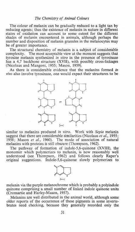
The structural chemistry of melanin is a subject of considerable complexity. The most acceptable view at the moment suggests that tyrosine melanin synthesized in vitro in the presence of tyrosinase has a 4,7 backbone structure (XVII), with possibly cross-linkages (Nicolaus and Mangoni, 1955; Mason, 1959).
As there is considerable evidence that the melanins formed in vivo also involve tyrosinase, one would expect their structures to be
[xvu]
similar to melanins produced in vitro. Work with Sepia melanin suggest that there are considerable similarities (Nicolaus et al., 1955; 1958; Mason et al., 1960). The mode of association of natural melanins with proteins is still obscure (Thompson, 1962).
The pathway of formation of indole-5,6-quinone (XVIII), the monomer which polymerizes to melanin, is now reasonably well understood (see Thompson, 1962) and follows clearly Raper's original suggestions. Indole-5,6-quinone slowly polymerizes to
[m•)
melanin via the purple melanochrome which is probably a polyindole quinone comprising a small number of linked indole quinone units (Cromartie and Harley-Mason, 1957).
Melanins are well distributed in the animal world, although some older reports of the occurrence of these pigments in some invertebrates need checking, because they generally recorded only the
31

Goodwin
presence of black pigment together with histochemical tests of doubtful authenticity. Melanins are, however, clearly present in the plumose anemone Metridium senile, in the tropical sea-urchin Diadema anti/larum and in the ink of Sepia officina/is.
The pigment generally accumulates in special cells, melanophores and melanocytes. The melanophores which occur in poikilothermal vertebrates, are branched cells, and melanin can be aggregated into the middle of the cells or dispersed in the branches, which allows the animal rapidly to change colour. Mammals and birds possess branched melanocytes, which are much smaller than melanophores, and in which rapid colour changes do not occur.
Melanins in the integument are also responsible indirectly for Tyndall blues etc. (Seep. 3.)
Albinism, the absence of melanin from melanocytes and melanophores, is due to the failure of the animal to synthesize the key enzyme, tyrosinase.
Care must always be taken to differentiate between melanin formation in insects and darkening of the cuticle caused by quinone tanning; the latter is the cross linking of proteins by quinones derived from dihydric phenols such as protocatechuic acid (XIX). The darkening is due to the formation of aminoquinones (see Mason, 1955; Dennell, 1958 a; Karlson, 1960). The two processes are quite separate, as clearly demonstrated by Dennell (1958 b), who found that inhibition of melanin synthesis in blowfly (Calliphora vomitoria) larvae by phenylthiourea did not inhibit hardening of the puparium.
Q. {x•JII.}
Pteridines and F1avins
These two groups of pigments are considered together because biosynthetically they are both derived from purines (Goodwin, 1963) Furthermore, members of both groups are coenzymes in essential metabolic reactions, probably in most living systems; in this function they play no role in coloration. For example, flavin adenine dinucleotide (XX) is an essential coenzyme for many dehydrogenases; and tetrahydrofolic acid (XXI) is a co-factor in the metabolism of 1-carbon compounds. The only known case in which a flavin plays any part in animal coloration is that of the bush baby Galago, in which crystals of riboflavin constitute the tapetum lucidum behind the retina; this augments eye shine (Pirie, 1958).
32

The Chemistry of Animal Colours
The compounds now known as pteridines were first isolated from butterfly wings by Gowland Hopkins in 1889. Much later Wieland's school isolated and determined the structure of xanthopterin (XXII) and leucopterin (XXIII) (see Ziegler-Gi.inder, 1956). Erythopterin (XXIV) was also obtained.
In insects, pteridines are confined, in the Pieridae, to the wings (Ford, 194 7); but they also occur in the integument of many Hymenoptera, for example, the common wasp (Becker, 1937). They are also present in the eyes of wild type Drosophila me/anogaster along with ommochromes.
Pteridines are well distributed in Amphibia; and amongst reptiles isoxanthopterin (XXV) is present in the skin of the green mamba, Dendroaspis viridis (Blair, 1957). (See H. M. Fox and Vevers, 1960. for a full discussion of distribution).
fxxv]
Quinones Benzoquinones, naphthoquinones, anthraquinones and polycyclic
quinones all occur in animals, but the first named will not be considered because, as far as is known, they play no part in animal coloration. However, it should be emphasized that the recently discovered
33

Goodwin
ubiquinones (XXVI) play a key part in the respiratory chain in all animals so far examined.
H1col)
0
'"• u.,
H,co (<:H,cw-t-tH,),. H 0
[x>M]
(1) Napthoquinones. The main group of naphthoquinones which make a marked
contribution to animal coloration are the polyhydroxy compounds echinochrome A (XXVII) and spinochromes A, E, M, N, P (XXVIIIXXXV). With but one exception these pigments are confined to the Echinoidea (Table 2). Echinochrome A is well distributed throughout the tissues of a number of sea urchins, whilst the spinochromes
TABLE II. Distribution of spinochromes and echinochrome A in Echinoidea
Species
Anthocidaris crassisiphina Arbacia lixula Diadema antillarum Diadema setosum Echinus escu/entus Hemicentrotus pu/cherrimus Heterocentrotus mammi/atus Paracentrotus lividus
(North Atlantic) Paracentrotus lividus
(Mediterranean) Pseudocentrotus depressus Scaphechinus mirabilis Strongylocentrotus purpuratus
CaW,(OOH o OH
H0 OH
OH 0 [n...,o]
OH 0
CH1CO~OH
Ho~OH OH 0 [x.x]
(Thompson, 1962) Spinochromes Echinochrome A
ABCDEFGMNP
t
t t t
t t
t t t
t
c .. Jr:.WOH 0 Ol-4
HO OH OH [xli.VIII)
CHJWOH 0 OH HO OH
OH
[Jtxxt]
t
t t
t
t
t t t
' t
t
t t
HO~OH
HO~OH O~XJI:XIIt
(X)OH O OH HowO Oil CH.r:(r)OH 0
HO-
rc.W=CHC.OOH OH
OHoOH OHo oH O
[xxxuo] [-.xov] [xxo<VJ
34

The Chemistry of Animal Colours
have so far only been reported in spines and tests; this apparently specific distribution may only be because spinochromes have not been sought in other regions of the animals. Different relative amounts of spinochromes can cause differences in colour as in the case of violet and olive-green spines from Paracentrotus lividus (Goodwin and Srisukh, 1950). There appears to be no relationship between naphthoquinone pigments and food habits of sea urchins.
The one exception which has been recorded of the occurrence of a polyhydroxynaphthoquinone outside the Echinoidea is the presence of namakochrome (XXVI) (spinochrome E monomethyl ether) in the sea cucumber (Polycheira rufescens (Brandt) Holothurioidea) (Mukai, 1960).
GHJO~oH
HO~OH OH 0
[< .. vo)
The bones of the sea otter (Enhydra /utris), which feeds on sea urchins, are bright purple owing to the accumulation of a pigment closely resembling echinochrome A (Thompson, 1962). This situation is, however, different from that in sea urchins, which appear to synthesize their echinochrome de novo.
(ii) Anthraquinones. The Coccidae produce three anthraquinones which are still used
as dye stuffs. Carminic acid (XXXVII) the colouring matter of cochineal, is extracted from the dried bodies of Dacty/opius coccus and related species such as D. tomentosus and D. indicus (Ali and Haynes, 1959). It is unusual in that it is a C-glycoside. Kermesic acid (XXXVIII) is the main constituent of Kermes, a very ancient dyestuff, obtained from Kermes ilicis, which infests the Kermes oak
~COC.H, HO~OH
COOH 0 OH
()(~)C.VIII)
35

Goodwin
Quercus coccifera (Dimroth and Fick, 1916); laccaic acid, the colouring matter of lac dye, is obtained from lac, the secretion of various coccids especially Laccifer /acca; it is probably a mixture of materials similar in structure to carminic acid (Venkataraman, 1957).
Anthraquinone pigments are also present in crinoids, and two of those present in the australian crinoid Comatula pectinata have recently been identified as the 6-monomethylether and 1,6-dimethylether of rhodocomatulin (XXXIX) (Sutherland and Wells, 1959); very similar pigments occur in C. cratera (J. W. Wells, quoted by Thompson, 1962).
OH 0
0 OH (XL)
OM 0
OM 0
OH 0
(XLII]
36

The Chemistry of Animal Colours
(iii) Polycyclic quinones Complex polycyclic quinones, the aphins, have been investigated
by Todd and his group at Cambridge. A yellow water-soluble pigment in the haemolymph of many insects (aphids have been mainly investigated, but not all aphids contain aphins and the pigments do exist in other families, e.g. Phylleroxidae) has been termed protoaphin; this compound is converted enzymatically into a fatsoluble pigment xanthoaphin (XL), which gradually changes into orange chrysoaphin (XLI) and red erythroaphin (XLII); these pigments are derivatives of 4,9-dihydroxyperylene-3,10-quinone but the exact nature of the parent protoaphin is still unknown. The conversion of protoaphin into erythroaphin represents a gradual aromatization of the molecule. The structures quoted are for aphins obtained from Aphis fabae and are usually denoted by the suffix ~fb, because aphins from different sources have slightly different structures (Human eta/., 1950; Duewell eta/., 1950 a, b).
(iv) Conclusion Aromatic rings are synthesized de novo in plants and micro
organisms, but not in mammals (see Goodwin, 1960). It would appear that the invertebrates which accumulate large quantities of aromatic quinones synthesize them de novo thus exhibiting an important biosynthetic pathway lacking in higher animals.
References
Abolins, L., 1957. Acta Zoo/. (Stockholm), 38, 223. Ali, M. A. & Haynes, L. J., 1959. J. Chern. Soc., p. 1033. Becker, E., 1937. Z. Morph. Okol., 32, 672. Becker, E., 1942. Z. Verebungslehre, 80, 157. Blair, J. A., 1957. Nature Lond., 180, 1371. Butenandt, A. & Neubert, G., 1955. Hoppe-Seyl. Z., 301, 109. Butenandt, A., Biekert, E. & Linzen, B., 1956. Hoppe-Sey/. Z., 305, 283. Butenandt, A., Biekert, E. & Linzen, B., 1958. Hoppe-Seyl. Z., 312, 227; 313,
251. Butenandt, A., Neubert, G. & Baumann, U., 1959. Hoppe-Sey/. Z., 314, 15. Cheeseman, D. F., 1955. J. physiol (London), 131, 3 P. Cromartie, R. I. T. & Harley-Mason, J., 1957. Biochem. J., 66, 713. de Nicola, M., 1954. Biochem. J., 56, 555. de Nicola, M., 1956. Expt. Cell. Res., 10, 441. Dennen, R., 1958a. Proc. Roy. Soc., 158B, 176. Dennen, R., 1958b. Bioi. Revs., 33, 178. Dimroth, 0. & Fick, R., 1916. Liebigs Ann., 411, 315. Duewell, H., Human, J.P. E., Johnson, A. W., MacDonald, S. F. & Todd, A. R.,
1950a. J. Chern. Soc., P. 3304. Duewell, H., Johnson, A. W., MacDonald, S. F. & Todd, A. R., 1950b. J. Chern.
Soc., P. 485. Fisher, L. R., Kon, S. K. & Thompson, S. Y., 1952. J. marine Bioi. Assoc.
U.K., 31, 229. Fisher, L. R., Kon, S. K. & Thompson, S. Y., 1954. J. marine Bioi. Assoc.
U.K., 33, 589.
37

Discussion
Fisher, L. R., Kon, S. K. & Thompson, S. Y., 1955. J. marine Bioi. Assoc. U.K., 34, 81.
Ford, E. B., 1947. Proc. Roy. Ent. Soc. Lond., 22, 72. Fox, D . L., 1953. Animal Biochromes. Cambridge University Press. Fox, D . L., 1962. Comp. Biochem. Physiol., 6, 1-40. Fox, H. M. & Vevers, G., 1960. The Nature of Animal Colours. London,
Sidgwick & Jackson. Goodwin, T. W., 1952a. Comparative Biochemistry of Carotenoids. London,
Chapman & Hall. Goodwin, T. W., 1952b. Bioi. Revs. Cambridge Phil. Soc., 27, 439. Goodwin, T. W., 1960. Recent Advances in Biochemistry. London, Churchill. Goodwin, T. W., 1962. In Comparative Biochemistry. Ed. M . Florkin &
H. S. Mason. Academic Press, Vol. 4. Goodwin, T. W., 1963. The Biosynthesis of Vitamins. Academic Press. Goodwin, T. W. & Srisukh, S., 1950. Biochem. J., 47, 69. Hill, F., 1962. Human, J. P. E., Johnson, A. W., MacDonald, S. F. & Todd, A. R., 1950.
J. Chern. Soc. P. 477. Karlson, P., 1960. Hoppe-Seyl. Z ., 318, 194. Laxer, G., Sikorski, J., Whewell, C. S. & Woods, H. J. Biochim. Biophys. Acta,
15, 174. Linzen, B., 1958. Ver. Deutsch. Zool. Gess in Frankfurt a.M. P. 154. Linzen, B., 1959. Hoppe-Seyl. Z., 314, 12. Mason, H. S., 1955. Advances in Enzymol., 16, 105. Mason, H . S., 1959. In Pigment Cell Biology (M. Gordon ed.). Academic
Press. p. 563. Mason, H. S., Ingram, D. J. E. & Allen, B., 1960. Arch Biochem. Biophys.
86,225. Mukai, T., 1960. Bull. Chern. Soc. Japan., 33, 453. Nicolaus, R. A. & Mangori, L., 1955. Gazz. chim. ita!., 85, 1397. Nicolaus, R. A., Vitale, A. & Piattelli, M., 1958. Rend. accad. sci. fis. e nat.
(Soc. naz. sci. Napoli), 4, 25. Nicolaus, R. A., Piattelli, M. & Narni, G. ,1959. Tetrahedron Letters., 21, 14. Pirie, A., 1958. Nature, Lond., 183, 985. Schwink, I., 1953. Naturwiss, 40, 365. Scott, T. A., 1962. Ann. Reports Chern. Soc. , 58, 378. Steven, D . M., 1948. J. exp. Biol., 25, 369. Sutherland, M. D. & Wells, J. W., 1959. 'Chern. & Ind. Lond., p. 291. Thompson, R. H., 1962. In Comparative Biochemistry. Ed. M. Florkin &
H. S. Mason. Academic Press. 3, 727. Venkataraman, K., 1957. In "Festschrift Arthur Stoll". p. 360. Birkhauser,
Basel. Volker, 0 ., 1953. Hoppe-Seyl. Z., 292, 75. Volker, 0., 1955. J. Ornithol., 96, 50. Volker, 0 ., 1960. Fort. Chern. org. Naturstoffe, 18, 177. Yamaguchi, M., 1958. Bull. Chern. Soc. Japan, 31, 51, 739 (1958). Yoshi, S. & Brown, R. R., 1959. Fed. Proc., 18, 255. Zeigler-Giinder, I., 1956. Biol. Revs., 31, 313.
DISCUSSION
P. F. Holt. I have been fascinated by the chemical reaction Professor Goodwin mentioned by which sponges appear to be able to convert the alicyclic rings of carotenoids into aromatic rings, at the same time shifting methyl groups. The methyl groups in the product are differently disposed
38

Discussion
in the two rings. This presumably involves two types of attack by methyl radicals and one would expect simultaneous formation of symmetrical compounds. Have such compounds been isolated?
T. W. Goodwin. As far as I know no symmetrical compounds have yet been isolated.
J. D. Carthy. Would Professor Goodwin care to speculate about the origin of these complex pathways of pigment formation? It seems very difficult to account for them as having originated from excretory mechanisms.
T. W. Goodwin. In the present state of knowledge it is impossible to speculate sensibly, but I agree that pigments must have originated other than as excretory mechanisms.
H. J. A . Dartna/1. Certain crustacea, in particular those that inhabit deep waters, are red in colour instead of the normal green. I would like to ask Professor Goodwin whether the biochemistry of this considerable colour difference is known. The normal green colour, I understand, is due to a carotenoid-protein complex, e.g. the astaxanthin-protein ovoverdin that is responsible for the green colour of lobster eggs. Presumably the red colour of certain deep sea crustacea is also due to a carotenoidprotein complex, but in what way does this differ from that giving rise to the green colour?
The reason I ask this question is because of an apparently analogous problem concerning the visual pigments. These are haplo-carotenoidprotein complexes, the haplo-carotenoid being either retinene1 or retinene2• The visual pigments based on retinene 1 have !.max ranging from 430 mu to 562 mu while those based on retinene2 have !.max extending from 510 mu to 620 mu. No hypothesis has been advanced explaining the structural principles underlying this great range of colour.
T. W. Goodwin. Nothing is known of the structural differences which make one astaxanthin-protein complex green, another red and another deep purple. It ought to be possible to extend the current exciting work on the structure of the active sites of enzymes to this problem.
L. R. Fisher. With reference to the deep red colour of deep-sea prawns, Nicol, working at Plymouth, has shown this to be due, in Acanthephyra, to the presence of free astaxanthin.
I should like to ask Professor Goodwin if he can explain why the carotenoid pigmentation of crustaceans in mountain pools increases in intensity with altitude.
T. W. Goodwin. I can only suggest a possible explanation, that at higher altitudes the light reaching the pools is richer in those wavelengths which stimulate pigmentation.
J. B. Davis. I would like to refer to the work of Dr. Umebachi in Japan on the yellow pigments in the Papilionidae. These are derived from tryptophane by way of kynurenin, but are evidently not ommochromes. He has found knurenin present in the scales of the yellow areas in the wings of Papilio xuthus.. I have found considerable quantities of kynurenin in the wings of male P. dardanus in addition to other yellow
39

Discussion
substances. Does Professor Goodwin know of any further work relating to such pigments?
T. W. Goodwin. No, but Professor Thomson probably does.
R . H. Thomson. No. The structure of these Papi/io pigments is not yet known. From the published work they appear to be chemically related to the ommochromes although they are definitely not ommochromes.
G. E. Fogg. Since carotenoids appear always to originate in plants I think it is rather a pity that plant names for them should have to give way to animal names. An instance I have in mind is that of myxoxanthin, a characteristic pigment of the blue-green algae (myxophyceae), which is now generally known as echinenone. Have you any comment?
T. W. Goodwin. It is generally accepted by organic chemists that the man who first describes a compound has priority in naming it. I think echinenone was described before myxoxanthin.
R. H. Thomson. As Professor Goodwin has pointed out "animal" carotenoids are derived from plant carotenoids. Dr. Harborne has also pointed out that flavonoid pigments occur very widely in plants. It follows that animals which eat plants also eat flavones, and there are a number of reports in the literature suggesting the presence of flavones in animal tissues, but the evidence is very flimsy. The best authenticated case is the work of D. L. Thomson on the Marbled White butterfly (Melanargia galathea). Dr. Morris and I have recently re-investigated this insect and find that at least a dozen flavonoid pigments are present. One has been isolated as a crystalline compound and identified as the flavone, tricin, and another (not isolated) is a glycoside of lutexin. This type of compound appears to occur widely in grasses and it seems likely that the butterfly pigments originate in the food of the larvae. The amount of flavonoid material present is very small and so their contribution to the colouring of the butterflies is also very small.
40

COLOUR AND THE CLASSIFICATION OF THE LOWER PLANTS
By
G. E. FoGG
Department of Botany, Westfield College, London
IT is tempting to use colour in the classification of living organisms and it may sometimes be as useful a taxonomic character as any other. Comparative biochemistry shows that particular chemical substances may be characteristic of a species or other taxonomic group and if such substances happen to be pigments then colour may be a reliable guide in classification. It is not of much use as a taxonomic character with mosses, ferns, gymnosperms and flowering plants. The leaf pigments of these forms are extremely uniform and while flower colour may often be useful as an ancillary character in the identification of species it is not otherwise of much use in classification. Colour has, however, been used from early times in the classification of lower plants and modern knowledge shows this to have considerable justification.
The distinction between the two major groups of the lower plants (Thallophyta), the Algae and Fungi, was certainly made before it was realized that differences in colour might imply a fundamental distinction between them but following the recognition of the essential role which chlorophyll plays in photosynthesis it became clear that the chief distinguishing feature of the fungi is that they lack this green pigment. Bacteria, most of which have no photosynthetic pigments, are put alongside the fungi in many schemes of classification. In practice, therefore, the major subdivision of the Thallophyta, is made on a basis of colour, with provisos that in some algae the green of chlorophyll may be masked by other pigments and that some fungi may contain green pigments 0ther than chlorophyll. On the whole, this biochemical classification agrees well with that which can be made on purely morphological grounds and, while there is no doubt that they each include organisms with widely differing phylogenies, there is equally no doubt that the Algae, Fungi and Bacteria are reasonably " natural " plant groups. It should be noted that although the presence or absence of chlorophyll is the chief criterion in this classification it is not of over-riding importance. .Prototheca, a genus of colourless organisms which in all morphological and most biochemical features resembles the green Chlorel/a, is classed with the algae whereas the photosynthetic bacteria, which contain chlorophylls similar but not identical to
41

Fogg
those in algae and which morphologically resemble other bacteria, are regarded as true bacteria.
Within the Fungi colour is only of importance in classification at the genus and species level. Thus it is of outstanding value in the taxonomy of the genus Aspergillus in which a wide range of colony colours is exhibited, these colours being comparatively stable and correlated with morphological and biochemical characteristics. Penicillium shows a contrasting state of affairs, for the colour of most species belonging to this genus is some shade of green during the growing period and is not stable, changing with age and conditions of culture, especially trace element supply. Colour is, of course, of great value in the recognition of individual species of higher fungi . It has also been used as a basis for the subdivision of the Agaricales into five series, with white, pink, brown, purple and black spores respectively. However, this seems to result in a purely artificial division, separating genera which on morphological grounds are obviously closely related. Even within a single genus of Agaricales there may be a range of spore colour which makes nonsense of classification by spore colour, e.g. Lepiota echinata has dull green spores, changing to reddish brown and then to red, L. georginae has pink spores and L. eyrei has green spores. The chemical constitution of some of the fungal pigments has been established, many of them proving to be quinone derivatives. Pigments play generally the same role in the classification of the lichens as they do in that of the fungi-indeed they usually seem to be produced by the fungal rather than by the algal partner. As an example we may take the genus Xanthoria which is characterized by the production of the bright yellow anthraquinone pigment parietin. This pigment is the only character separating the genera Xanthoria and Physica and the separation appears to be an artificial one made purely for convenience.
The fungal pigments seem to be mainly by-products of metabolism and of only secondary importance for the life of the organism which produces them. Hence it is not surprising that they should vary from species to species and be of little use in characterizing major groups. Pigments which play a more fundamental role in metabolism may be expected to provide a basis for taxonomic groupings of a higher order. The photosynthetic bacteria afford an example here. One family, the Chlorobacteriaceae, is distinguished from the others, the Thiorhodaceae and the Athiorhodaceae, the members of which are purple, by comprising green species. These colour differences denote definite biochemical differences in the photosynthetic pigments, the chlorobium ch/orophylls of the Chlorobacteriaceae are chemically and spectrophotometrically distinct from the bacteriochlorophyll of the purple bacteria, the characteristic colour of which is due to specific accessory photosynthetic pigment, acyclic carotenoids (two carotenes and at least three xanthophylls), not
42

Colour and the Classification of The Lower Plants
present in Chlorobacteriaceae, which instead contain cyclic carotenoids (y-carotene and rubixanthin).
However, it is with the algae that pigmentation plays the most important part in classification. To anyone who looks at the seaweeds on a rocky shore in temperate regions it is immediately obvious that there are three main kinds :-green, brown and red, and almost from the beginnings of the science of phycology the major groups have been distinguished on this basis. In Harvey's Phycologia Britannica (1846-51) seaweeds were divided into Chlorospermae (greens and blue-greens), Rhodospermae (reds) Melanospermae (browns). Later, as it was realized that microscopic forms were related to the seaweeds, other algal groups were distinguished and here again colour was taken as an important taxonomic character.
Following the pioneer work of Willstlitter and Stoll (1913) it became clear that these colour differences denote definite differences in photosynthetic pigments. All the algal groups have been found to contain chlorophyll a as their principal photosynthetic pigment, a feature which differentiates them clearly from the photosynthetic bacteria. In the Myxophyceae this is the only chlorophyll but other groups have characteristic accessory chlorophylls :-chlorophyll b (Chlorophyceae and Eugleninae) chlorophyll c (Cryptophyceae, Chrysophyceae, Bacillariophyceae, Phaeophyceae) and chlorophyll d (Rhodophyceae). There is doubt about chlorophyll e, which has been reported as occurring in the Xanthophyceae. Among the carotenoids ~-carotene is almost universally characteristic of the algae, although in one order of the Chlorophyceae, the Siphonales, it is quantitatively less important than ex-carotene. The xanthophylls of the algae are extremely diversified and almost every group has its characteristic kinds. Thus the Myxophyceae, have myxoxanthophyll; the Chrysophyceae, Bacillariophyceae and Phaeophyceae, fucoxanthin ; and the Chlorophyceae and Eugleninae, lutein, which is also characteristic of higher plants. Finally, water-soluble bile protein pigments are found in three groups, Myxophyceae, Rhodophyceae and Cryptophyceae. These are of two main types, phycocyanins, which are blue with a red fluorescence, and phycoerythrins, which are red with an orange fluorescence, both of these occurring in all three algal groups. There are, however, spectrophotometric, and therefore presumably chemical, differences between the biliproteins from the different algal classes.
It should be realized that this picture of the occurrence of photosynthetic pigments in the algal classes is based on evidence which is not as extensive as one could wish. While many members of the Chlorophyceae have been examined, in other groups, e.g. the Xanophyceae and Chrysophyceae, pigments have been studied in only one or two representatives. It is evident that there may be variation
43

Fogg
in pigmentation among representatives of the same class and it is quite possible that our present generalizations about certain classes are based on results obtained with atypical species. One class, the Chloramonadinae, has scarcely been studied from this point of view as yet, the difficulty being in obtaining sufficient amounts of microscopic species free from contaminating organisms.
It is nevertheless clear that, by and large, the differences in colour shown by algae do denote consistent chemical differences in components of the photosynthetic apparatus. Differences in such basically important components seem to be conservative and thus be taxonomically useful characters. This conclusion is borne out by the remarkable way in which pigmentation is correlated with other taxonomic characters, both biochemical and morphological. Only two examples of this correlation can be given. The Myxophyceae are clearly demarcated from the other algal classes, not only by lack of a chlorophyll other than chlorophyll a and by the possession of characteristic xanthophylls and biliproteins, but by cytological characteristics such as their anomalous nuclear structure, lack of flagella and lack of a membrane separating the chromatophore from the rest of the protoplasm. That the Xanthophyceae should be separated from the Chlorophyceae, in spite of the close morphological parallels between them, is evident, not only because chlorophyll b is absent from the former and present in the latter, but because there are differences in flagella and in carbohydrate reserve products.
Taken in conjunction with other criteria, the colour of an alga may thus be a most useful indication of its taxonomic position. In fact it is probably the first feature of an alga of which the phycologist, consciously or unconsciously, takes note. Certain snags must however be borne in mind. One is that the colour of the photosynthetic pigments may be masked by others. Many algae accumulate carotenoid pigments when their growth is halted through deficiency of a mineral nutrient or water. This accumulation is in fat droplets and not in the chromatophores and the carotenoids are usually different from those associated with photosynthesis, e.g~ in Haematococcus ~-carotene and lutein are the most important carotenoids in the chloroplasts whereas it is astaxanthin which accumulates in quantity when growth slows down. Such accumulation results in algae becoming yellow, orange or red. Certain species, e.g. of Haematococcus, Trentepohlia and Botryococcus in the Chlorophyceae, of Euglena in the Eugleninae, and of various genera of Dinophyceae, are most familiar in this state. However, if they are grown in culture they show the colour characteristic of their class while active growth is taking place. Many Myxophyceae normally have sheaths containing brown, yellow or, sometimes, red or violet pigments, which mask the colour of the cells themselves. Little is known of the chemical nature of such pigments. Another
44
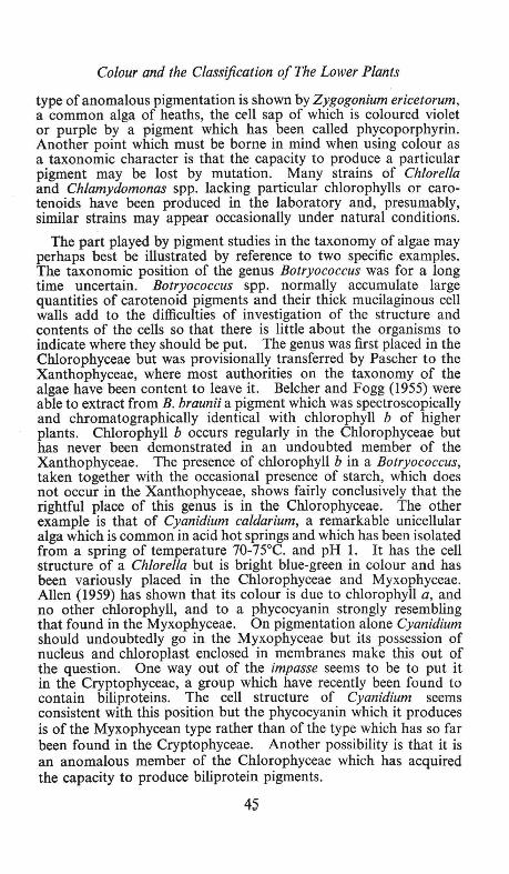
Colour and the Classification of The Lower Plants
type of anomalous pigmentation is shown by Zygogonium ericetorum, a common alga of heaths, the cell sap of which is coloured violet or purple by a pigment which has been called phycoporphyrin. Another point which must be borne in mind when using colour as a taxonomic character is that the capacity to produce a particular pigment may be lost by mutation. Many strains of Chlorella and Chlamydomonas spp. lacking particular chlorophylls or carotenoids have been produced in the laboratory and, presumably, similar strains may appear occasionally under natural conditions.
The part played by pigment studies in the taxonomy of algae may perhaps best be illustrated by reference to two specific examples. The taxonomic position of the genus Botryococcus was for a long time uncertain. Botryococcus spp. normally accumulate large quantities of carotenoid pigments and their thick mucilaginous cell walls add to the difficulties of investigation of the structure and contents of the cells so that there is little about the organisms to indicate where they should be put. The genus was first placed in the Chlorophyceae but was provisionally transferred by Pascher to the Xanthophyceae, where most authorities on the taxonomy of the algae have been content to leave it. Belcher and Fogg (1955) were able to extract from B. braunii a pigment which was spectroscopically and chromatographically identical with chlorophyll b of higher plants. Chlorophyll b occurs regularly in the Chlorophyceae but has never been demonstrated in an undoubted member of the Xanthophyceae. The presence of chlorophyll b in a Botryococcus, taken together with the occasional presence of starch, which does not occur in the Xanthophyceae, shows fairly conclusively that the rightful place of this genus is in the Chlorophyceae. The other example is that of Cyanidium caldarium, a remarkable unicellular alga which is common in acid hot springs and which has been isolated from a spring of temperature 70-75°C. and pH 1. It has the cell structure of a Chiarella but is bright blue-green in colour and has been variously placed in the Chlorophyceae and Myxophyceae. Allen (1959) has shown that its colour is due to chlorophyll a, and no other chlorophyll, and to a phycocyanin strongly resembling that found in the Myxophyceae. On pigmentation alone Cyanidium should undoubtedly go in the Myxophyceae but its possession of nucleus and chloroplast enclosed in membranes make this out of the question. One way out of the impasse seems to be to put it in the Cryptophyceae, a group which have recently been found to contain biliproteins. The cell structure of Cyanidium seems consistent with this position but the phycocyanin which it produces is of the Myxophycean type rather than of the type which has so far been found in the Cryptophyceae. Another possibility is that it is an anomalous member of the Chlorophyceae which has acquired the capacity to produce biliprotein pigments.
45

Discussion
Finally it may be noted that, besides being of use in distinguishing between the algal classes, pigmentation may indicate affinities between them and so give clues to phylogeny. The occurrence of chlorophyll a in all algae is perhaps an indication that they arose from a common stock. Various cytological and biochemical features point to an affinity between the Chrysophyceae, the Bacillariophyceae and the Phaeophyceae and this idea is supported by the occurrence of chlorophyll c and fucoxanthin in all three of these classes. The production of biliproteins by the Myxophyceae, the Rhodophyceae and the Cryptophyceae, perhaps indicates a common origin for these classes, but, in view of profound cytological differences between them, the connexion must be considered a remote one. For a fuller discussion of these problems the reader is referred to the paper by Dougherty and Allen (in Allen, 1960).
References
Allen, M.B. 1959. "Studies with Cyanidium caldarium, an anomalously pigmented chlorophyte ". Arch. Mikrobiol., 32, 270-277.
Allen, M. B. editor. 1960. Comparative biochemistry of photoreactive systems. Academic Press, New York.
Belcher, J. H . and Fogg, G . E. 1955. "Biochemical evidence of the affinities of Botryococcus ". New Phyto/.,~54, 81-83.
DISCUSSION
T. W. Goodwin. It is clear from the recent work of Krinsky and Goldsmith (Arch. Biochem. Biophys. 1960, 91, 271) and B. H. Davies (Ph.D. Thesis, University of Wales, 1961) that the main pigment of the Eugleniniae is not lutein but the very similar compound antheraxanthin. Earlier work had failed to distinguish between these pigments, but this is now possible using ,modern techniques.
46

THE PHOTOSYNTHETIC PIGMENTS
By
C. P. WHITTINGHAM
Department of Botany, Queen Mary College, London
THE autumn colour of leaves marks the end of the annual growing season; the processes leading to the appearance of these colours being associated with the return of materials from the leaf to the rest of the plant and the subsequent death of the leaf. The changes in colour follow a definite pattern. In some leaves the initial change is an increase in red coloration followed by a subsequent development of yellow, whereas other leaves do not produce a red colour at all . The yellow coloration occurs in all plants to a greater or lesser degree. Shortly after the stage when yellow has become predominant the leaf becomes brown and dies. The changes in colour indicate the differential loss of three distinct groups of pigment. The normal green colour is due to the presence of chlorophylls. The yellow colour is due to the presence of the xanthophylls and carotenes. Both the green and yellow pigments are insoluble in water and are confined to particulate bodies in the cells, the chloroplasts. The red pigments, present only in some plants, are due to anthocyanins. These are soluble in water and are present in the plant sap. These play no part in photosynthesis. The brown colour is associated with death and is due to oxidation ; it is not normally present in the living leaf.
The lower plants show a greater diversity of green pigmentation. The green algae, like the higher plants, eontain chlorophylls a and b. The blue-green and red algae do not contain chlorophyll b. Certain of the red algae contain a different form of chlorophyll, chlorophyll d, although the precise nature of this form is not yet known. The brown algae also lack chlorophyll b but contain chlorophyll c. The structure of chlorophyll c is not fully known but it is believed to lack phytol. In the photosynthetic bacteria the purple bacteria contain bacteriochlorophyll whereas the green bacteria contain bacterioviridin (or chlorobium chlorophyll).
All higher plants have the same four major carotenoids, ~-carotene, lutein, violoxanthin and neoxanthin. These are also present in most groups of the algae, but in the brown algae, fucoxanthin replaces lutein as the major xanthophyll, and in the blue-green algae echinenine and myxoxanthophyll are present. Exceptional carotenoids are found in various algal mutants which have been produced by irradiation with either X-rays or ultraviolet light.
47

Whittingham
In addition to chlorophyll and the carotenoids, the red and bluegreen algae contain phycobilin pigments which like chlorophyll are tetrapyrrolic but have a linear instead of cyclic arrangement of the pyrrole rings. The red algae, containing phycoerythrin, are found predominantly in the deeper layers of sea water. Engelmann proposed that these additional pigments might have been developed during evolution to complement the wavelength characteristics of the radiation reaching the deeper layers. There are, however, certain green algae which occur in deep waters whilst some red algae live in relatively shallow waters.
Action spectra
It is possible to determine experimentally which pigments contribute absorbed light energy to photosynthesis. This can be done by determining the relative quantum yield for photosynthesis in monochromatic light at different wavelengths. The resulting " action spectrum " can be compared with the absorption spectrum. From the latter the proportion of light at each wavelength absorbed by individual pigments can be estimated. The green pigments, the chlorophylls, predominantly absorb in the red and blue whereas the carotenes and xanthophylls absorb wavelengths shorter than 500 mp. Nevertheless the action spectrum shows that excitation in the yellow region of the spectrum in brown algae results in an equivalent photosynthetic activity to excitation in the red. Hence the energy absorbed by fucoxanthin must be used in photosynthesis as efficiently as that absorbed by chlorophyll. In the higher plants the yellow carotenoids are less effective contributors to photosynthesis. Also, in the green algae they result in about half the activity compared with equivalent excitation of chlorophyll, and in the red algae the efficiency may be as low as 20 % of that of chlorophyll. The red pigment phycoerythrin, and the blue pigment phycocyanin are almost as effective as chlorophyll in photosynthesis. Since all the so called accessory pigments are more or less efficient in activating photosynthesis it has been proposed that the simplest hypothesis is that all these pigments transfer their energy to chlorophyll which alone initiates the chemical process of photosynthesis.
This view was supported by observations of the fluorescence of living cells. For example, in a suspension of diatoms illuminated with light of a wavelength which is predominantly absorbed by fucoxanthin the fluorescence is similar in quantity and intensity to that emitted when the exciting light is absorbed only by chlorophyll. Clearly energy must have been transferred in the cell from the fucoxanthin which absorbed the light to chlorophyll which then emitted fluorescence. A similar transfer of energy has been demonstrated from chlorophyll b to chlorophyll a in green algae and from phycobilins to chlorophyll in the red and blue-green algae. This
48

The Photosynthetic Pigments
transfer of energy takes place by a process called inductive resonance which can have a high efficiency. It should be noted that energy transfer can only take place from a pigment absorbing at a shorter wavelength to one absorbing at a longer wavelength and not vice versa.
Different forms of chlorophyll
Thus until a few years ago it appeared that the main problem remaining in photosynthesis was to determine the manner in which excitation of the chlorophylls resulted in photochemistry. But a detailed study by Brown and French (1959) of the absorption band of green plants in the red region of the spectrum indicated the presence of more than two chlorophyll components. These workers developed an instrument to determine the first derivative of the optical extinction as a function of wavelength, a method which is more sensitive than measuring the extinction itself. The observed derivative spectrum is then analysed into the summation of a number of normal probability curves representing constituents each with single absorption maxima. With this technique it was shown that the absorption in the red region of the spectrum for a leaf of a higher plant could be analysed in terms of the presence of at least four distinct absorption bands. One of these with a maximum at 650 mfL was attributed to chlorophyll b, the other three to different forms of chlorophyll a having absorption maxima at 673, 683 and 695 mfL. It has been since suggested that these different forms may represent combinations of the pigment with different proteins. The proportion of the different forms varies from organism to organism and in the case of micro-organisms may be modified according to the conditions of culture.
Confirmation of the existence of more than one form of chlorophyll has come from the study of etiolated plants. When a higher plant is grown in the dark its leaves are yellow-green and contain the pigment proto-chlorophyll. When it is placed in the light it begins to become green and there is a mole for mole change to chlorophyll. Before and after this initial change the chromatophore is combined with a protein on a molar basis. The chlorophyll first formed from proto-chlorophyll has an absorption maximum at 682 mfL. Subsequently, after some minutes at room temperature, the chlorophyll maximum shifts to 670 mfL and now several molecules of chlorophyll are present for each molecule of protein.
The existence of these different forms of chlorophyll clarified an earlier observation of Emerson and Lewis (1943) who found that the quantum efficiency of photosynthesis in the green alga Chlorella decreased very rapidly in the region between 680 and 700 mfL where there was still appreciable absorption. The effect was even more striking in certain red algae where the quantum yield declined beyond 650 although there was appreciable absorption up to 680 m!J.. It
49

Whittingham
now appears that the forms of chlorophyll at 683, 695 mfJ. which absorb in the far red are ineffective in photosynthesis.
Emerson and co-workers subsequently showed (1957) that absorption in the far red could be made effective if it was supplemented by simultaneous absorption at a shorter wavelength. The action spectrum for the increased rate of photosynthesis resulting from a second wavelength superimposed on monochromatic light of 697 mfL showed two characteristic peaks, one at 650 mfL and one at 670 mfL. The peak at 650 mfL is characteristic for absorption by chlorophyll b and Emerson concluded that the simultaneous excitation of chlorophyll b must improve the photosynthetic efficiency of the light absorbed in the far red by chlorophyll a. At that time Emerson made no comment on the second peak in the enhancement action spectrum at 670 mfL. An increased photosynthetic activity (enhancement effect) due to simultaneous illumination by two different wavelengths has now been found in a large number of organisms (Haxo, 1960). In the red alga Porphyridium, Brody showed a very marked increase in the activity of light absorbed in the far red, when this was supplemented by light absorbed by phycocyanin. In Ch/orella the full action spectrum for the effectiveness of the second light showed two peaks, one at 480 and one at 658 mfL giving a curve resembling the absorption spectrum of chlorophyll b; in Porphyra the action spectrum showed a single peak at 550 mfL resembling the absorption spectrum of phycoerythrin. In the blue-green alga Anacystis maximum enhancement was obtained by illumination at 600 mfL implicating phycocyanin. In the diatom Navicula enhancement was obtained at 540 mfL and 645 mfL implicating fucoxanthin and chlorophyll c. It should be noted that the enhancement effect results from excitation of a second pigment throughout its spectrum. For example, excitation in Chlorella of either the blue or the red absorption bands of chlorophyll b is equally effective for enhancement. The general conclusion must be that it is necessary to have simultaneous excitation of two pigment systems for efficient photosynthesis. In studies with monochromatic light, the relative inefficiency of absorption by chlorophyll a alone, appears only at the far red end of the spectrum because this is the only region in the visible where chlorophyll a is the sole absorbing pigment. This is the reason why this phenomenon has only been discovered in recent years, more than 20 years after the first precise measurements of action spectra for photosynthesis.
The two photochemical reactions required for efficient photosynthesis have been called the long-wave chlorophyll reaction and the accessory-pigment reaction. French and Fork (1961) have suggested that both chlorophyll a 683 and chlorophyll a 695 are capable of a single photochemical step referred to as the long wavelength chlorophyll reaction. The accessory pigment reaction can be effected by absorption by chlorophyll a 673 and chlorophyll b in
50

The Photosynthetic Pigments
Chlorella, by chlorophyll b in the green plant, by chlorophyll c in fucoxanthin in the brown algae and by phycoerythrin and phycocyanin in the red algae. Franck has suggested that the 673 form chlorophyll a may differ from the 683 and 695 forms in that in the former the chlorophyll is associated with an aqueous phase and in the latter associated with lipids. He suggests this because the form associated with lipids might be expected to show little or no fluorescence. Excitation of any of the accessory pigments results in energy transfer to the shorter wavelength form of chlorophyll associated with water molecules.
The chemical nature of the two light reactions
The intensive studies of animal biochemistry followed by rather more fragmentary studies of plant biochemistry have elucidated the main biochemical reactions of respiration. The process of respiration is now seen to consist essentially of the oxidation of carbon substrates by oxygen with a resulting flow of electrons from carbon to oxygen. The oxide-reduction reactions take place for the greater part in the mitochondrion, a highly organized cell organelle. In this structure electron flow via the cytochromes is accompanied by a concomitant phosphorylation of adenosine diphosphate to form adenosine triphosphate. In a subsequent process the A TP can be hydrolyzed liberating free energy which can be utilized to drive biosynthetic reactions or to do work in an osmotic or mechanical system. Thus an essential feature of respiration is the formation of ATP as an intermediate which can be utilized subsequently in energy-requiring reaction systems.
Recent work on photosynthesis suggests that an analogous biochemical mechanism operates in photosynthesis. The fundamental difference is that whereas in respiration the free energy utilized in the formation of ATP is derived from the oxidation of carbohydrate, in photosynthesis the free energy is obtained directly from radiation. By analogy with respiration an electron flow in an ordered structure, presumably containing cytochromes, could result in the formation of ATP.
In photosynthesis in higher plants the synthesis of carbon compounds from carbon dioxide utilizes ATP produced by light energy; but in order to effect this synthesis, in addition to ATP, a supply of electrons (in the form of reduced pyridine nucleotide) is also necessary. In the photosynthetic bacteria these may be supplied from an external reagent, like hydrogen gas, which can reduce pyridine nucleotide in the dark. In this case light energy is utilized solely for the production of ATP. But in the green plant no additional substrate is required for photosynthesis other than carbon dioxide and water and the green plant must utilize radiation to produce an electron donor from water. Because of this, oxygen becomes a
51

Whittingham
product of photosynthesis in the green plant whereas it is not in the bacteria. Summarizing, in green plant photosynthesis, electrons are transferred from water via cytochromes to pyridine nucleotide, the necessary energy being obtained from radiation. Accompanying this flow of electrons, A TP is formed. In the bacterium the electron donor may be supplied exogenously. The light-generated electron flow is then cyclic as shown by the dotted line in Fig. 1 and the sole product of light energy is ATP .
... ~
[e"":----+81 .. ~
I
I I I
Chi :!:~cY'f•A-c;;:
LIGHT
Chi = A form of chlorophyll a. Pigment = Another form of chloro
phyll or an accessory pigment.
Cyt = Cytochrome. P = Inorganic phosphate. ~p = Organic phosphate in
adenosine triphosphate. A and B are intermediates, the
nature of which is not definitely established.
Flo. 1 Patterns of electron flow in photosynthesis.
Both the photosynthetic bacteria and the leaves of higher plants contain cytochromes which can be distinguished from those of nonphotosynthetic tissue by their characteristic absorption. In the reduced form cytochromes show a prominent absorption band in the yellow region of the spectrum. Therefore by observing changes in absorption at this particular wavelength it is possible to determine how far the cytochromes present in a living organism are in the reduced form. Indeed by comparing the absorption spectrum of a photosynthetic plant in a weak light intensity, resulting in little photosynthesis, and a stronger intensity, when photosynthesis is active, it is possible to observe whether any change in the state of the cytochromes occurs during photosynthetic activity. Duysens and others have used this technique (called difference spectroscopy) and shown that in general a cytochrome component becomes oxidized as a result of illumination.
In Porphyridium cruentum, Duysens (1961) showed that the action spectrum for the oxidation of the cytochrome (probably cytochrome f) showed a maximum at 680 mfl whereas the action spectrum for
52

The Photosynthetic Pigments
oxygen production in photosynthesis was known to have a maximum at 560 mfL where phycoerythrin absorbs. He proposed that two pigment systems were present-system (1) containing the phycobilins in association with that part of chlorophyll a to which energy transfer can readily take place from the phycobilins and represented in higher plants by chlorophyll b 650 and chlorophyll a 673; system (2) containing the remainder of the chlorophyll a which is not in association with phycobilins and represented in higher plants by chlorophyll a 683 and chlorophyll a 695. Excitation at 680 mfL which primarily excites chlorophyll a in system (2) resulted in an oxidation of cytochrome. If, in addition, light of 560 mfL absorbed by system (1) was added the cytochrome again became reduced. This effect of the addition of light of 560 mu was inhibited by DCMU (3 : 4 dichlorophenol 1 : 1 dimethyl urea). Hence system (2) can result only in the oxidation of cytochrome; by itself it cannot result in photosynthesis. System (1) must also be excited for efficient photosynthesis and sensitizes, by reaction with water, the reduction of the cytochrome which has become oxidized as a result of reaction with system (2).
Witt has extensively observed the changes in optical absorption consequent upon illumination of photosynthetic tissue with extremely intense short flashes (Witt eta/., 1961). He has found five different types of absorption change according to the duration of the flash and the wavelength observed. These have been related to sequential changes in constituents of an electron transport chain which is believed to operate in photosynthesis. A characteristic decrease in absorption at 420 mfL (neg. peak) was observed when the long wavelength chlorophyll form was excited alone.
With excitation of the 670 mfL absorbing form of chlorophyll additional changes were observed at 475 mfL (negative peak) and 515 mfL (positive peak). This second type of change is due to the presence of a substance whose reduction potential is about zero and which can be extracted from chloroplasts by petrol ether. Witt has proposed that this is probably identified with plastoquinone or a component directly connected with plasto-quinone. (Plastoquinone itself does not show these absorption changes). Fig. 2 shows the reaction pattern of photosynthesis proposed by Witt involving the two different forms of chlorophyll, the compound related to plastoquinone, and a cytochrome.
It is now generally believed that photosynthesis involves two photochemical processes sensitized by different pigments. The products of the one process via a thermal dark reaction become the substrate of the second. (Whittingham and Bishop, 1961). The free energy change of the two photochemical processes is likely to be equal and the dark thermal stage in between involves a loss of free energy which is probably coupled to phosphorylation. Additional phosphorylation (cyclic) may also occur by a separate reaction
53

Whittingham
FIG. 2 Reaction pattern of photosynthesis.
This diagram shows the complexity of the electron chain according to Witt. The substances Y, X, C, Z are unspecified intermediates. ADP = Adenosine diphosphate. A TP = Adenosine triphosphate. PMS = Phenazine methosulphate.
i~~Hz} = The oxidised and reduced forms of co-enzyme 2.
The figure is intended to give a view of the probable complexity of the process, rather than a detailed statement of the intermediates.
mechanism involving the most reduced of the constituents of the electron transport chain, and it is probable that this reaction requires excitation only of pigment system (2). Indophenol dye can be reduced by the products of the first photochemical reaction alone (pigment system (1)). It is therefore possible to separate for the purposes of study in vitro the two photochemical processes on the one hand by studying photophosphorylation and on the other by studying dye reduction. This type of investigation can be expected to add to our knowledge of the mechanism for these two processes in the immediate future.
References Brown, J. S. and French C. S. 1959. Plant Physiol., 34, 305. Duysens, L. M. N., Amesz, J. and Kamf, B. M. 1961 . Nature, Lond., 190, 510. Emerson, R., Chalmers, R. F. and Cederstrand, C. 1957. Proc. Nat. Acad.
Sci., Wash., 43, 133. Emerson, R. and Lewis, C. S. 1943. Amer. J. Bot., 30, 165. French, C. S. and Fork, D. C. 1961. International Biochemical Congress,
Moscow. Haxo, F. T. 1960. In Comparative Biochemistry of Photoreactive Systems
(Ed. by Allen, M. B.), 339-360. New York, Academic Press. Whittingham, C. P. and Bishop, P. M. 1961. Nature, Lond., 192, 426. Witt, H. T., Muller, A. and Rumberg, B. 1961. Nature, Lond .• 191, 194.
54

RESPIRATORY PIGMENTS
By
H. MuNRO Fox
Queen Mary College, London
THERE are five respiratory pigments. Three of them, haemoglobin, chlorocruorin and haemocyanin-red, green and blue respectivelycarry oxygen in the blood stream to the tissues. Haemoglobin is found in all vertebrates except two (the eel larva and another fish) and in many invertebrates, especially annelid worms and entomostracan crustacea, but some phyla, such as coelenterates, quite lack it. Chlorocruorin is confined to a few polychaete worms, while haemocyanin occurs only in cephalopod and gasteropod molluscs, in decapod and stomatopod crustacea, and in arachnids. Haemoglobin alone of these three respiratory pigments is not only in the blood stream but also fixed in various tissues where it stores oxygen ready for immediate use. In vertebrates the only tissue with this stationary haemoglobin is muscle (and so it is known as myoglobin) but in various invertebrates all kinds of other tissues contain it.
The fourth respiratory pigment, again a red one, is haemerythrin; it occurs in very few animals, namely the sipunculids, Priapulus, Lingula and one polychaete. This pigment has recently been re-investigated by D. Keilin (1960). The fifth respiratory pigment is cytochrome. As its name implies, it is in the cells and it is immensely important in their respiration. Since the time of its discovery in 1929 by D. Keilin literally thousands of papers have been published about it, yet I shall not consider it here as this symposium is on Colour and Life, whereas cytochrome is present in such small quantities that it rarely colours a tissue and never an animal.
Haemoglobin consists of haem (Fig. 1) joined to a protein, globin. Its colour is due to the haem. The figure shows that haem is composed of four pyrrol rings, with certain side chains, united by methene bridges into a super-ring, in the middle of which is a ferrous iron atom . . The oxygen (or water) is attached to the iron. There are many haemoglobins, specific to animals and tissues; their differences reside in the globin, not the haem. A large amount of most important research has been done in the last few years on the amino acid differences between haemoglobins and on their genetics (Ingram, 1960; Perutz, 1962), but as we are concerned here with colour, this is not the occasion to enlarge on them.
Chlorocruorin is dichroic: green in dilute, red in concentrated solution. In structure it is very similar to haemoglobin although
55

Fox
CH 1 CH1
CH3 ~H CH3 gH I I I I C=C C--C
6 6=~-g g /
~N-<.. ! ".N/~ HC ···.F~--~ CH
'/N~~·······~N". / C C=C-C C
6=6 H 6=6 tH, tHa tH1 tH,
tH~ tH 1
I I COOH COOH
FIG. I. The haem of haemoglobin.
differing both in its haem and in its protein. In the haem one of the two vinyl side chains shown in Fig. I is substituted by a formyl group. Chlorocruorin may be considered as a mutant form of haemoglobin. It is less useful as an oxygen carrier in that a higher oxygen pressure is needed to saturate it. Had the mutation occurred in an active animal it would probably have been eliminated by natural selection, but sabellids lead a quiet life. Serpulids have chlorocruorin but it is a remarkable fact that their blood also contains haemoglobin, and in consequence its colour is brown. This simultaneous presence of two respiratory pigments is unique. It must again have been a mutation that was not worth selecting out (Fox, 1949).
Cytochrome too is composed of haem and protein, but it is really a mixture of a number of similar compounds, each with its own type of haem. One of these is like the haem of haemoglobin (Fig. 1) and another is similar to the haem of chlorocruorin. The remaining two respiratory pigments-haemerythrin and haemocyanin-contain no haem (in spite of their names, given before the term haem had acquired its present significance) although they have a metal atom and a protein. In haemerythrin the metal again is iron, in haemocyanin it is copper. As these two pigments are oxygen carriers, like haemoglobin and chlorocruorin, one wonders of what use is the haem group.
To what extent is the external coloration of animals due to respiratory pigments? Not very much. Among the vertebrates the
56

Respiratory Pigments
so-called white races of man are coloured by haemoglobin, but otherwise it is seen only in tongues, wattles or gills. In the invertebrates various annelids are red with haemoglobin and so is the bloodworm. The redness of Daphnia varies immensely in inverse proportion to the oxygen in the water (Fox, 1955). The green colour of chlorocruorin is visible from without only in the chlorhaemid worms. The pink of haemerythrin tints Sipunculus, but haemocyanin colours no animal.
There are derivatives of haemoglobin, but not of the other respiratory pigments, which contribute to animal colours. These derivatives are not respiratory substances and no physiological function is known for them. They seem to be either breakdown products of haemoglobin or by-products of synthesis, and some may come from plants. They are retained in the animal body and have sometimes been made use of for protective or sematic coloration. These pigments are of two kinds: porphyrins and bilins.
If the iron atom is removed from haem (Fig. I) we have a porphyrin. Such compounds are quite widespread in animal tissues, but usually in amounts too small to give a coloration and detectable only by their strong red fluorescence in ultraviolet light before a Wood's glass screen. The porphyrin of haemoglobin is known as protoporphyrin; a rare instance of an animal colour due to it is the purplish anterior end of the earthworm. There are other porphyrins differing in the side chains on the four pyrrol groups. Only one of them contributes to animal coloration: this is uroporphyrin with four acetic and four propionic acid groups in place of the side chains in Fig. 1. We meet it in two places. It colours certain marine gasteropod shells, among which the most intense and beautiful red colour is in the Indian Ocean species Clanculus pharaonis. The other uroporphyrin coloration is seen as an intense red in wing feathers of African touraco birds. The pigment, known as turacin, is a copper compound of uroporphyrin. The artificial formation of the pigment from copper and uroporphyrin has recently been studied by Joan Keilin and McCosker (1961).
Chlorophyll too has a porphyrin structure, but it does not occur in any animal tissue. Yet in just a few instances among marine worms derivatives of chlorophyll contribute to animal coloration. The bright green integument of Bonellia viridis is coloured by a chlorin, while in Owenia (Kennedy and Nicol, 1959) and Chaetopterus (Dales, 1957) green colours are due to phaeophorbides. These pigments are derived from plants.
If the super-ring of four pyrrols constituting a porphyrin is broken at one methene link, so that there is no longer a ring but a chain of pyrrols, we have a bilin or bile pigment. These contribute much more than porphyrins to animal coloration. The following are the outstanding instances: the green and blue eggs of birds, the blue colour of fish such as wrasse, the green of insects (due to a blue
57

Discussion
bilin with a yellow carotenoid), the purple secretion of the sea mouse, Aplysia, the green base of the common sea anemone, Actinia, and the blue of the coral Heliopora. Quite lately it has been shown by Green (1961) that there is biliverdin in the eyes of the cladoceran Polyphemus pediculus but not in those of Daphnia, and Barbara Gilchrist and he have found the fairy shrimp, Chirocephalus diaphanus, to be coloured by this pigment. Dales (1961) has found that the deep green colour of the Pacific polychaete worm Eupolymnia heterobranchia is due to mesobiliverdin, while Mangum (1962) describes green populations of another polychaete, Clymene/la torquata, coloured by mesobiliverdin.
What is the origin of these bilins? The pigments of vertebrate bile come from haemoglobin breakdown and Dales and Kennedy (1954) have shown that this is the source of biliverdin in Nereis diversico/or. We guess that egg-shell pigments have a similar origin without knowing. It has, however, recently been found that bilin may arise as a by-product of the synthesis of haem (Gray and Scott, 1959; Stewart, 1960) and this could contribute to coloration. The purple of Aplysia may derive from bilins which colour red and bluegreen algae, but we do not know. Anemones and corals are carnivores; they lack haemoglobin but both they and their prey possess haem compounds which could give bilins.
This brief account shows in what diverse ways respiratory pigments and their derivatives may be responsible for animal coloration. A fuller treatment is given in a recently published book by Dr. Gwynne Vevers and myself.
References Dales, R. P. 1957. J. mar. bioi. Ass. U.K., 36, 81-89. Dales, R. P. 1961. Nature, Lond., 190, 168. Dales, R. P. and Kennedy, G. Y. 1954. J. mar. bioi. Ass. U.K., 33,699-708. Fox, H. M. 1949. Proc. roy. Soc. B., 136, 378-388. Fox, H. M. 1955. Bull. Soc. zoo/. Fr., 80, 288-298. Fox, H. M. and Vevers, G. 1960. The nature of animal colours. London:
Sidgwick & Jackson. Gilchrist, Barbara and Green, J. 1962. Comp. Biochem. Physiol., 7, 117-125. Gray, C. H. and Scott, J. J. 1959. Biochem. J., 71, 38-42. Green, J. 1961. Nature, Lond., 189, 227-228. Ingram, V. M. 1960. Haemoglobin and its abnormalities. Oxford: Blackwell. Keilin, D. 1960. Acta biochim. polonica, 8, 415-423. Keilin, Joan and McCosker, P. J. 1961. Biochim. biophys. Acta, 52, 424-435. Kennedy, G. Y. and Nicol, J. A. C. 1959. Proc. roy. Soc. B., 150, 509-538. Mangum, C. P. 1962. Nature, Lond., 195, 198. Perutz, M. F. 1962. Nature, Lond., 194, 914-917. Stewart, C. P. (Ed.). 1960. Proceedings of the symposium on the formation
and breakdown of haemoglobin. Amsterdam: Elesvier.
DISCUSSION
C. Ellenby. I agree with Professor Munro Fox that blood pigments usually play little part in animal coloration for they are usually screened
58

Discussion
by overlying tissue and respiratory organs are frequently tucked away inside. But I wonder whether they may have played a more important part at an earlier stage of evolution. The possession of a highly coloured respiratory pigment may make an animal more conspicuous and this may, at an earlier stage, have placed a premium on the development of obscuring pigments. These may have continued to have a function even after morphological changes have made their hypothetical initial function unnecessary.
59


COLOUR CHANGE IN ANIMALS
By
D. B. CARLISLE
Anti-Locust Research Centre, London, W.8.
COLOUR change in animals may be brought about by either of two processes :
I. Existing pigment may be removed from the integument and replaced by new pigment, different in colour, or,
2. The pigments in the integument may become redistributed, thus presenting an abrupt (or relatively abrupt) change of colour with little or no metabolism of the pigments.
It is obvious that the former process is relatively slow and may be characterized as a morphogenetic colour change.
Morphogenetic colour changes may be brought about in response to many factors. Heat and light, diet, the colour of the background and the degree of crowding of the animals may all play a part. Thus the stick insect, Carausius morosus, is green if reared under cool conditions with alternating light and darkness, but if it is transferred to warm conditions the green pigment gradually disappears and the animal takes on a grey-brown to black colour. Gregarious hoppers of the desert locust, Schistocerca gregaria, have a yellow body with black markings. If the temperature is raised the amount of black pigmentation is reduced at the next moult and at very high temperatures (up to 40oC) it may be almost non-existent, so that the hopper appears almost uniformly yellow. If the temperature is lowered the amount of black pigmentation increases until after a couple of moults at 26°C the animal is in effect black with yellow markings.
Diet may affect colour in several ways. Perhaps the most direct may be seen in the flamingo, where the carotenoids from the diet are deposited directly in the feathers, more or less unchanged. When a flamingo's diet changes from one containing a high carotenoid content (e.g. crustaceans) to one low in these pigments, the pink colour of the plumage fades slowly until it is almost white. The pink colour of the plumage may be restored by a return to the former diet. A less direct effect of diet may be seen in those animals which deposit pterins or purine bases in the integument. The area of the white patches and the intensity of the whiteness depends then on the amount of nucleic acid in the diet and it seems plausible that the loss of white patches as the shore crab (Carcinus maenas) grows older reflects a change in the diet with approaching maturity.
61

Carlisle
As an example of response to the colour of the background we may take nymphs of the locust Locusta migratoria. Like the desert locust this shows aposematic colouration-black and orange in this species when the nymphs are reared in crowds. But when they are reared in isolation from each other (a scattered low density population in the wild, or in separate cages in the laboratory) they take on the colour of the background. As with crowded hoppers the amount of black pigment is dependent on temperature, but it is less than at the same temperature in crowded animals. Accordingly, an animal feeding on lush green grass at a high temperature will be almost completely green, fed on hay in a light brown cage the hopper will be fawn; kept in suitable individual cages in the laboratory Locusta nymphs may be blue, black or brown. And if we change a nymph from one background to another, within one or two moults it will have changed to the new colour, by removal of one pigment from the integument and the substitution of fresh pigments. But this ability of the solitary locust hopper to match its background is lost if it comes into contact with its own kind for any length of time. Hoppers forced into association with one another by the nature of the terrain, or in the laboratory, soon learn to aggregate socially and to "prefer" one another's company. Once they are banded together in this way their coloration undergoes a change so that after one or at the most two moults they have assumed· the aposematic black and orange or black and yellow pigmentation of the crowded hoppers. Such change of colour in response to population density is by no means rare in the Orthoptera, though perhaps less common in other groups of animals.
As a final type of morphogenetic colour change we may mention the breeding dress of many animals. The drake mallard at the end of winter dons a fine new colourful plumage for the breeding season and the female prawn, Palaemon serratus, adorns herself with a row of white spots down the side of the abdomen when she is ready to breed.
To turn now to the kinetic colour changes-that is to say those which take place within a period of less than a day by means of redeployment of pigments within the integument-we find that these may be responses to just as wide a variety of stimuli. Copepods may respond to unwelcome actinic radiation by expanding the chromatophores covering the viscera, just as we respond to similar unwelcome radiation by tanning. Prawns can change colour to match the background-pattern as well as colour-within about half an hour, and cephalopods can complete a cryptic change of colour within a second. The three-spined stickleback, the cuckoo wrasse and the black sea bream can adopt a breeding dress quite as colourful as that of the drake mallard, by kinetic means. High temperatures and bright light can stimulate a prawn to change colour. A Sepia can change from aposematic colouration to cryptic colouration in a
62

Colour Change in Animals
flash and produce "eye-spots" which seem to serve to scare off predators. Even the chamaeleon, which changes colour so slowly, can show more than a mere albedo response, for its response to background colour is modified by the temperature.
So much for the stimuli which provoke colour change. But what of the effectors themselves, which bring about the change of colour? The pigment is largely intracellular, and usually in granular or droplet form, rarely dissolved in the cytoplasm of the cells. An exception is the carotenoid-protein complex which is dissolved in the cytoplasm of the integument of the lobster or in the shell itself, to produce the blue colour. More commonly we find the pigment in the general cells of the integument, in special cells known as chromatophores or in dead cells, such as those which go to make up feathers and hair. In addition melanin in particular may be found in extra-cellular layers, such as the shell of arthropods. It should be obvious that where the greater part of the body is covered by dead structures such as feathers or hair, any colour change can only come about by shedding these structures and growing fresh ones of a different colour. Where the pigment is contained in living cells it may be destroyed or removed and replaced by fresh pigments. Both these kinds of processes are classed under morphogenetic colour change. Chromatophores may be of three main kinds, and any of the types may possess a variety of pigments, so that they are often named for the colour they show-melanophores, erythrophores, etc. The first type may be exemplified by the melanophores of the mammalian skin, including our own. In these the melanin granules are merely present in greater or lesser amount, not under any form of control of their distribution, so that any colour change is brought about by a diminution or increase in the amount of contained pigment. The second type may be illustrated by the chromatophores of the prawn. They consist of fixed channels or chromorrhizae, radiating from a centre and branching irregularly amongst the surrounding cells. Within these channels the pigment granules may flow about, dispersing to occupy the entire network of chromorrhizae or concentrating into the central mass as a small dot. The pigment contained within the chromatophores should not be regarded as fixed in any way. New pigment is constantly being formed or added and pigment is being removed. In particular, when the carotenoid granules are concentrating rapidly towards the central reservoir of the cell some of the pigment enters into a complex with protein, to become water-soluble and blue, so diffusing out of the cell. The third type of chromatophore is found in the cephalopods. Here the pigment is contained in a spherical sac surrounded by a halo of muscle fibres. When these contract the sac is pulled out until it is a thin plate occupying more than a hundred times the area of the sphere.
The final topic which I propose to consider is the question of the mediation between stimulus and response. In the simplest situation
63

Carlisle
the effector organ responds directly to the stimulus- the cells of the human skin produce melanin as a direct response of each individual chromatophore to the ultra-violet radiation; a single chromatophore of a prawn may react to a spot of light, and Yoshida has shown, using a spot of light 3ft in diameter, that a mere sector of a chromatophore of the sea urchin Diadema can respond; the cells of the feather follicles of flamingos lay down carotenoids in the feather they are forming in direct ratio to the amount supplied by the blood. In these examples of colour change-morphogenetic or kinetic-there is no involvement of any central control mechanism. The stimulus is received and the response is carried out by the effector cell itself.
More often, however, the stimulus is received through the sense organs and the response is mediated via the central nervous system. Even chromatophores which can respond directly to illumination, such as those of the prawn, are often also under central control. The most direct and complex central control is found in the cephalopods where the muscles which expand the chromatophores are under direct nervous control and a separate lobe of the brain regulates colour change. In an octopus the chromatophores of the skin are never at rest; blushes of colour pass over the surface and the individual chromatophores expand and contract all the time. Since the mechanism is nerve-and-muscle, colour change is a matter of fractions of a second. In the lower vertebrates too the chromatophores of the skin are under nervous control, but here this is supplemented by hormonal control. In an unstimulated condition the pigment in the chromatophores is more or less completely dispersed (producing a dark skin) and nervous stimulation serves to cause the pigment granules to migrate towards the centre of the chromatophore, leading to paling. This is a slower process than contraction of muscles and may take two or three minutes. An opposing innervation will reverse the process, if this is to take place rapidly, but if the initial nervous stimulus to concentrate the pigment merely ceases, the pigment starts to disperse to the fine branches quite slowly. This nervous control is reinforced by hormones from the pituitary, which, slower to act in the first place, may maintain the response for far longer. The sexual colour changes of fish seem to be mediated entirely by hormonal control. The strong concentration of all the chromatophores across the shoulders of the male cuckoo wrasse, which produces the white patch of the breeding male, seems to be mediated entirely by hormonal means, though whether the hormone concerned is a hormone of the pituitary, or one of the gonadal hormones we do not know.
In the Crustacea the chromatophores are activated entirely by endocrine control; they are not innervated in any way. In the prawn there are at least twenty different types of chromatophores which respond differentially, and six or more hormones. As a result
64
Discussion
the prawn can take on much of the colour and pattern of the background, though not perhaps with such precision as the octopus . In addition at least five centres- groups of neurosecretory cells-are known to produce different assemblages of chromactivating hormones. It is interesting to speculate upon the reason for this anatomical complexity. It seems most likely that the different centres are related to the different levels of association and integration of the sensory input, so that at the most peripheral level of visual input the neurosecretory centre receives information on the number of visual cells receiving stimuli. At a second level the incident illumination may be compared with the albedo. At a third level the pattern of things seen may result in a message passed to a third neurosecretory centre, while the centre in the tritocerebrum may be concerned with a total integration of all stimuli and central effects, visual or otherwise, which may lead to a colour change. Thus the red tail which prawns always show at nightfall can be shown to depend in no way upon visual stimulation and to be mediated via the tritocerebral neurosecretory centre, not via the centres in the eyestalk.
DISC USSION
C. Ellenby. Although so much has been achieved in the field of colour change there is surprisingly little evidence as to its value. I believe that Hogben always maintained that there was sufficient hormone circulating in the blood of the frog to keep its melanophores in the fully dispersed state. One wonders also, about the function of the melanophores in the peritoneum of the frog and of those enveloping, even on the ventral side, its sympathetic ganglia. Dr. Carlisle points out that copepods "open" their chromatophore "umbrellas" when they rise to the surface waters of the sea and suggests that this protects them against the harmful effect of the light. But they rise to feed on the organisms of the phytoplankton which, apparently can manage without such protection.
D. B. Carlisle. Dr. Ellenby is, I think, mistaken when he says that phytoplanktonic organisms can manage without the protection of pigments. In a number of plants mutants are known which lack carotenoids from the chloroplasts. Such organisms, after emergence from the seed or spore, can survive in the dark as long as their food reserves last, but if they are exposed to the light they die of "sunstroke". In other words the carq.tenoids are needed to protect them against the damaging effect of the light. Phytoplanktonic organisms then have a permanent "parasol" of pigment to protect them against solar radiation. Sea water absorbs ultra-violet light extremely quickly so that protection is needed against visible light, not against the ultra-violet.
A. T. Thomson. The possibility that the black and yellow coloration of gregarious locust hoppers has the aposematic function ascribed to it by Dr. Carlisle was suggested and discussed by Key (1957: p. 281). I have not seen locusts in the field, but I should have thought that, in the normal
65

Discussion
environment (taking the Desert Locust as an example) of sandy soil with scattered vegetation and numerous insects of the same species, that coloration could as well be considered cryptic as can the green colour of the solitary hopper against its normal background-the green food plant. It is relevant to add that the gregarious hoppers are eaten in large numbers by a wide range of vertebrate predators, which suggests that they are not distasteful; while the possibility that there is a mimetic resemblance to distasteful animals appears to be ruled out by the fact that they are usually by far the most abundant creatures in their particular habitats. Key, K. H. L., 1957. "Kentromorphic phases in three species of Phasmatodes". Aust. J. Zoo!., 5, 247-284.
D. B. Carlisle. I was using the word aposomatic in a very loose sense. There can be no doubt that hopper bands frighten small predators. Huddleston (1958) for instance reports that many small birds are frightened of hopper bands and only eat the stragglers. It is possible of course that in the .normal environment, as Mr. Thompson says, the coloration could be considered as cryptic, but I rather doubt it. Possibly when the bands are roosting on bushes they may be difficult to observe but when they are marching, as they are most of the day, they are very clearly visible. The ease with which hopper bands are spotted by aircraft reconnaissance is sufficient evidence for this, I think.
Huddleston, J. A., 1958. " Some notes on the effects of bird predators on hopper bands of the Desert Locust (Schistocerca gregaria Forskal)." Ent. mon. Mag., 942, 210-214.
W. B. Broughton. I am delighted to witness the heresy of Mr. Thomson in questioning the sacred concept of the Aposema. I am sure we too readily attribute warning significance as in every case the only or even the most likely explanation of this striped type of coloration; and in this case I endorse Mr. Thomson's suggestion of crypsis as the more likely function -but combined with episematic signalling. When the same colouring has to be used for recognition as well as concealment, clearly the potentially conspicuous disruptive rather than the blending type is demanded. I too have seen hopper bands only through the medium of film, but this striping is common enough among our own acridids, and it certainly does, as Mr. Thomson said, conceal.
Dr. Carlisle in his reply stressed the conspicuousness of the band as nullifying the cryptic effect of individual pattern. This is surely to miss the whole point of the argument-that the more individuals massacred by predators, the higher the selection in favour of those individuals whose pattern and behaviour is just that shade better in hiding them (like the householder whose slightly better burglar-proofing induces the thief to go next door). On the other hand, the high importance of visual stimulation for the marching of hoppers, and the known potency of striped patterns for the optomotor reflex, clearly put a premium on striped, rather than neutral, camouflage.
D. B. Carlisle. I would agree with Mr. Broughton as to the episematic signalling function of the colour of locust hoppers. I would not agree, however, that the striping of gregarious hoppers serves to conceal them.
66

Discussion
W. D. Wright. In considering the colour of locusts and other creatures possessing fine patterns of colour, I think it should be pointed out that quite drastic changes in colour appearance may occur through effects of contrast and other visual phenomena in the eye and brain of the observer. These, of course, have nothing to do with the locust itself but may affect the classification of its colour. We are engaged in our laboratory at the Imperial College in measuring these subjective effects and I think it might be of interest to the meeting if Mr. Gindy, who is particularly concerned with this project, showed some of his colour effects.
T. W. Goodwin. Dr. Carlisle's comment that certain crustacea living at high altitudes increase their pigment concentration in order to protect themselves from visible radiation, raises an interesting paradox. If visible light is the true source of the damage then the animals could logically protect themselves by becoming transparent. There can be no biological effect of light without a photoreceptor, which must be pigmented.
D. B. Carlisle. Is it in fact possible for an animal to become completely transparent? Agreed that there can be no biological effect of light without a pigmented photoreceptor, but every constituent of living matter absorb somewhere in the visible or ultra-violet spectrum, every single one of them. And even if all the tissues of the body can be made completely transparent to visible light, the animal must feed upon pigmented material and so have a coloured gut.
T. W. Goodwin. Dr. Carlisle, do I take it that your observations indicate that the chromatophore is the site of astaxanthin synthesis in Crustacea?
D. B. Carlisle. No, sir, I have no information on that point.
67


THE PHYSIOLOGY OF COLOUR PERCEPTION IN INVERTEBRATES
By
J.D. CARTHY
Department of Zoology, Queen Mary College, London
EVIDENCE for colour perception by animals has to be gathered largely by inferences from physiological experiments, for often direct tests of the animals' colour sensitivity have not as yet been carried out. Extraction of the eye pigments may show that these have absorption spectra with a peak or peaks in particular parts of the spectrum; when the spectrum shows such a peaked curve, and particularly when the difference spectrum gives this appearance it is tempting to consider the animal as endowed with colour sensitivity. Again, records of changes of potential in the eye, accepted as associated with the primary visual process, may show that various wavelengths stimulate the sense organ to different extents. Such measures of the electro-retinograms may yield graphs which follow those of the absorption spectra. Nevertheless this sort of evidence is not positive evidence for the existence of colour sensitivity for it deals with the abilities of the sense organ and does not take into account the modifying influences of the central nervous system.
Direct evidence can only come from behaviour experiments designed to test the reaction of the whole animal to colours. The interpretation of these experiments has all the difficulties usually associated with behavioural experiments. Is a negative result a real one, for example? Colour choice by the butterfly, Pier is brassicae demonstrates the problem. When feeding the insects will not choose green papers, but when egg laying green becomes highly attractive (lise, 1937). Thus, a negative result may simply mean that the test is meaningless for the animals in that situation and when motivated by those particular drives. But in addition the greatest care must be taken to ensure that the test involves a choice between colours of different wavelength only and that differences in brightness are eliminated. * Naturally, similar care has to be taken with tests of the electro-retinograms. But here it has always to be remembered that brightness as measured with an instrument may not be the same as the subjective brightness to the animal for various reasons, among them that the optical apparatus of the eye may differentially absorb light.
The classical experiments of Lubbock (1881) on the honeybee consisted of training bees to seek food on various coloured papers. *for other potential factors see p. I I 5.
69

Carthy
He showed that they can be trained to a colour. But it was von Frisch's (1915) elaboration of the experimental conditions which supplied firm proof of their colour sensitivity. He offered food on either yellow or blue squares placed among squares of fifteen different shades of grey. All the papers were covered with glass so that any scent traces could be wiped off. On each square a watchglass was placed, sugar water being put only in the one on the training square. When tests were made all the watchglasses were empty. The bees continued to visit, say, the blue square to which they had been trained ignoring the grey squares among which were ones of equal brightness to the blue. Later Kuhn (1924) used projected spectral colours matched in intensity to demonstrate, without further question, the ability of honeybees to see three major "colours": yellow (650-500 m,u), blue-green (500-480 m,u) and blue (480-400 m,u). In addition to these colours in the visible spectrum bees can detect ultra-violet (400-300 m,u) invisible to the human eye. This last ability explains why certain red poppies are attractive to bees though the insects are red-blind; the flowers have a high reflectance for ultra-violet. Kuhn also demonstrated that honeybees exhibit the same colour-contrast behaviour as man, thus, a bee trained to blue will go to a grey paper surrounded by yellow; such a juxtaposition gives rise to a subjective impression of blue in the human mind.
However, colour relationships are not organized in the bee as they are in man. Using an arena whose translucent floor could be illuminated from below with coloured light, Daumer (1956) showed that the bee has three complementary colours as shown below:
IILUf - GRU:N
HO NEY -BEE
FIG. 1. Charts to show the relationships of the complementary colours in honeybee and man (after Daumer, 1956).
Subjective white light for a honeybee can be produced by mixing 55% yellow+ 30% blue-violet + 15% ultra-violet light.
No other insect has been investigated in such detail, but the ability to distinguish colour has been tested in a number of insects. Insects will react to the movement of vertical stripes by moving in such a
70

Physiology of Colour Perception in Invertebrates
way as to counteract their movement (optomotor reflex). If the insect reacts in this way it can be taken that it distinguishes the stripes (Schlegtendal, 1934). To use this reaction for testing spectral sensitivity, stripes of a colour and of a grey shade are rotated past the insect; the grey stripes are altered in reflectance until no reaction is observed. The insect is now seeing the two kinds of stripes as of equal brightness. The grey is then matched against various intensities of a second colour until again no reaction occurs. The final hue of the second colour is, therefore, subjectively equal in brightness to the first colour for the insect. If the insect shows an optomotor reaction when confronted with stripes of these two colours, it must be distinguishing them by their spectral differences and is truly able to distinguish light of different wavelengths. Such experiments show that many insects are colour blind, that few can distinguish red as a colour and that green is rarely distinguished except by some leaf-eaters like the beetle Chrysome/afastuosa.
Not all parts of an insect's compound eye are necessarily equally sensitive to colour. The antero-ventral areas of the eyes of the backswimmer Notonecta glauca are colour blind, while the posterodorsal area is sensitive. (Rokohl, 1942). Recordings of the electroretinograms from the upper and lower parts of the divided eyes of the whirligig beetle Gyrinus marinus suggest that the two parts differ in their sensitivity to different parts of the spectrum (Carthy, unpublished). The ommatidia of the upper part of the eye of the blowfly, Calliphora, are uniformly sensitive with a peak at 489 mfl while the visual cells in the lower part show three ranges of sensitivity, a green receptor (similar to that of the dorsal part of the eye) with a maximum at 491 mfl, a blue receptor maximum 468 mfl, and a yellow-green maximum 524 mfl (Autrum and Burkhardt, 1961). In addition all these have a further peak of sensitivity at 345 mfl.
In many insects the retinulae cells of the ommatidium are arranged in a cluster contiguous with the central rhabdome, but with one cell on the outskirts of the group. This eccentric cell has been labelled the colour sensitive one for no reason, it seems, than that it is the odd man out. However, Autrum and Burkhardt's recordings show that their green, blue and yellow-green receptors are present in the ratio 18:4:3. Now if five of the seven retinulae cells were sensitive to green, one to blue and one to yellow-green, a ratio very close to the observed one would be produced. It thus appears that in fact all the retinulae cells of the ommatidium may be effective in colour sensitivity. This fits in with the evidence that each retinula cell has its own axon, unlike the arrangement in the compound eye of Limu/us, where in the adult, at least, it appears that only the axon from the eccentric cell carries impulses from the stimulated eye.
The problem in receptor "design" is to ensure that while one cell can signal a change of wavelength by a change of frequency of discharge this does not become confused by changes of intensity which
71

Carthy
also will produce changes in frequency. An array of receptor cells with different spectral sensitivities will give rise to an overall pattern of discharge which can be interpreted centrally as light of a particular colour and a particular brightness.
The visual pigments of insect eyes have not been extensively studied though Goldsmith has extracted from the head of honeybees a retinene whose photosensitive pigment has an absorption maximum at 440 m,u. This, however, is not the only pigment in either compound eyes or ocelli. There are several peaks of sensitivity in different regions of the spectrum which indicate the presence of other pigments and reflect Daumer's three colour system. The sensitivity peak at 345 m,u common to all systems in the Calliphora eye is strongly reminiscent of the {3 absorption of vertebrate retinene.
The existence of a double receptor system, similar to that of vertebrate cones and rods, is proposed for the eyes of the crustacean Daphnia pulex (Heberdey and Kupka, 1942). The sensitivity of these water fleas to differences between two white lights is shown by their distribution in a trough illuminated by a light from each side The animals are made positively phototactic by raising the carbondioxide content of the water. They will move towards the light which is subjectively the brightest. The curve of the least detectable differences at different intensities shows a break at 400 lux. This could represent the change from one receptor system to another, though the transition point is much higher than that for man (about 0·03 lux). In addition it is clear that whichever of the systems are functioning the animals are colour sensitive, a further difference from the vertebrate condition. Blue sensitivity is the same whether light- or dark-adapted, but sensitivity to green and yellow is about three times greater in the dark-adapted animal than in the lightadapted (Heberdey 1949). The range of colour sensitivity is very much akin to that of the honeybee, even extending into the ultraviolet.
There are indications also that two-receptor systems may be present in the eye of Calliphora erythrocephala, for the sensitivity to colour (measured by the height of the on-effect of the electroretinogram) shows a movement of the peak from 620 m,u to 490 m,u with reduction of intensity by 10,000 times (Fig. 2) (Autrum 1955), reminiscent of the Purkinje shift found in the human eye.
There is considerable evidence of the differing stimulating efficiency of various wavelengths in the phototaxic behaviour of a number of other crustacea. Provided that care is taken to measure responses in the same way at all wavelengths and to ensure controlled spectral energies, such action spectra can be accepted as proving spectral discrimination. Some results of such experiments are given below (Waterman 1961):
72

Physiology of Colour Perception in Invertebrates
Animal
Pa/aernonetes sp. Nototrops swarnrnerdarnii Daphnia pulex Balanus amphitrite Balanus irnprovisus
Maximum sensitivity (m,u)
470-510 520-540 530-540 530-545 530-545
These animals can be expected typically to aggregate mainly in light of green or yellow-green wavelength.
FIG. 2. Initial potential changes in the eyes of normal red-eyed flies, Calliphora erythrocephala, on stimulation by equalquantum stimuli at various wavelengths and different quantum numbers. Abscissa : light wavelength; ordinate : millivolts ; parameter : log. relative quantum number. (A utrum, and Stumpf, 1960).
Occasionally light of different wavelength may produce different behaviour. Light of wavelength 500 m,u or less causes a number of freshwater cladocera (Daphnia, Ceriodaphnia, Bosrnina), stomatopod larvae and some marine copepods to swim downwards (Baylor and Smith, 1957). While yellow light of wavelength more than 500 m,u stimulates them to swim upwards, in red light they tend to oscillate various distances in an upright position but with little horizontal movement. Such behaviour would hold the crustacea in areas where their phytoplankton food is, for the phytoplankton will have the effect of filtering out the shorter wavelengths. Were these shorter wavelengths present, they would move away either vertically or horizontally.
The extraction of pigment from arthropod eyes has been difficult because of the problem of the removal of the masking photostable pigment. However, Wald and Hubbard (1957) have succeeded in
73

Carthy
extracting the photosensitive pigment from the eye of the lobster, Homarus americanus. This pigment is a rhodopsin, bleaching in light to a retinene and an opsin. Its difference spectrum, derived by subtracting the absorption spectrum of the final bleaching product from that of the rhodopsin shows a maximum near 515 m,u, a little higher than vertebrate rhodopsin (Fig. 3). In contrast the maximum for the pigment from Meganyctiphanes (Fisher and Goldie, 1959) and Euphausia pacifica (Kampa, 1955) is at 460-465
-O ·t
300 <00 500
Wave-leng th {m;.t)
600
FIG. 3. Difference spectra of lobster rhodopsin and of an early product of its irradiation (photoproduct), obtained by subtracting from the absorption spectra of the two compounds the absorption spectrum of the final bleaching product. As this final product has virtually no absorption spectrum above 450 m!J., the difference spectra are equivalent to true absorption spectra above this wavelength, marked in the figure by a vertical line. (Wald and Hubbard, 1957).
m,u. This shift may be an adaptation to life well below the surface where much of the longer wavelengths are filtered out. A similar but smaller shift has been shown in a comparison of shallow water and bathypelagic fish (Denton and Warren, 1957). Retinene was detected after bleaching the Meganyctiphanes pigment; it appears to be similar to that from vertebrate rhodopsin but with a different opsin, the effect of which is to shift the maximum towards a shorter wavelength. No retinene was detected in Euphausia and hence the pigment has been called "euphausiopsin". However, in general , it can be said that the photosensitive pigments of both crustacea a nd insects are similar to vertebrate rhodopsin.
Both insect and crustacea eyes contain pterine pigments (Viscontini, Kuhn and Egelhaaf, 1956; Viscontini, Schmid and Hadorn, 1955). Often such pigments are photo-labile (Autrum and Langer, 1958) but there is no evidence thus far that they play any part in light sensitivity. They are also fluorescent and could act as an intermediary between the incident light and the energization of the photochemical system, but sensitivity to ultra-violet light in planaria, at least, does not depend upon an intermediate fluorescent pigment (Merker and Gilbert, 1932).
74

Physiology of Colour Perception in Invertebrates
Outside the arthropoda, very few critical investigations of spectral sensitivity have been made. The frequency of the discharge in the optic nerve of the octopod Eledone moschata is correlated with the wavelength of the light (Fig. 4) (Frohlich, 1914). A limited success has been achieved with training another cephalopod (Octopus) to colours (see discussion in Carthy, 1958). It may seem evident from their ability to change their body colour to match the colour rather than the brightness of the background that both these animals must have spectral sensitivity.
,.. so
Lo "
whi\thght
10 1000 lnttnslty of stimulus (arbitrary uniU}
FIG. 4. Relation between frequency of discharge in the retina of E/edone moschata and the intensity ofillumination for white light and two monochromatic lights (after Frohlich 1914).
A rhodopsin has been extracted from the retina of both squids and octopus (Sepia officina/is, Loligo pealii and Octopus vulgaris). These rhodopsins do not partake in quite the same reactions as those in the vertebrate retina for after bleaching through a lumirhodopsin to a meta-rhodopsin further breakdown to retinene and opsin by hydrolysis is prevented by the stability of the meta-rhodopsin, though this substance shows reversible change from the acid red form to the alkaline yellow form (Kropf, Brown and Hubbard, 1959). The maximum absorption for the rhodopsins are squid 500 m,u and octopus 483 m,u.
There have been many experiments to ascertain the effect of coloured light on the behaviour of other invertebrates but few of the results can be accepted as the experimental conditions were not critical. However, a double receptor system has been reported in the earthworm (Unteutsch, 1937). One system is responsible for the shadow reaction and is mainly sensitive in the yellow region of the spectrum. The receptors for this are scattered uniformly over the body. On the other hand the receptors for the other reaction
75

Car thy
which involves changes in speed of locomotion are best developed at either end of the body and are mainly sensitive to blue light.
There are cells in the epidermis of the earthworm which appear to be specialized for light sensitivity but in a number of invertebrates a general dermal light sensitivity has been reported. The receptor system responsible is not known but the sensitivity of planaria to ultra-violet light remains in conditions where the eyes can have no part. It is also claimed that much of the spectral sensitivity of Daphnia is due to dermal senses and not to the eyes (Schaeffer, Robert and Medioni, 1958). Few protozoa have light receptor organelles but many appear to be light sensitive and to respond differentially to light ofdifferent wavelengths. Amoeba reacts most strongly to light of 430-490 mf-l wavelength, but only slightly to longer or shorter wavelengths. The light reactions of the urchin Diadema antillarum are due to the direct stimulation of the nervous system by light rays. There is spectral sensitivity for the maximum stimulating power of monochromatic light producing spine movement in response to shadow lies in the region 455-460 mf-l (Yoshida and Millott, 1960). This result was obtained by illuminating the radial nerve by carefully matched coloured lights.
No pigments have been detected associated with the nerve endings in the wall of the sea-anemone, M etridium senile, yet on illumination the longitudinal muscles contract. This reaction shows light- and dark-adaptation, with a maximum sensitivity to light of 490-520 mf-l wavelength, and another peak at 550-600 m,u. At longer wavelengths, sensitivity decreases rapidly. These figures are based on observations on white individuals; those coloured red or brown exhibit maxima which suggest that the pigment acts as a filter (North and Pantin, 1958). (Another coelenterate, Hydra sp., has been claimed to have no spectral sensitivitywhatsoever(Haug, 1933)). Dermal light sensitivity in other invertebrates may similarly be due to direct stimulation of nerve endings in the skin.
There are many parts of the invertebrate sub-kingdom in which work on colour sensitivity remains to be done. We have little information on the general distribution of this ability among the families of the Annelids and Molluscs, for example, but where pigments have been isolated they suggest that the processes lying behind the colour sensitivities of their possessors are very like those of vertebrates.
References Autrum, M., 1955. "Die spektrale Empfindlichkeit der Angenmutation white
apricot von Calliphora erythrocephala". Bioi. Zentralbl. 74, 515- 524. Autrum, M. and Burkhardt, D., 1961. "Spectral sensitivity of single visual
cells". Nature Lond., 190, 639. Autrum, M. and Langer, M., 1958. "Photolabile Pterine im Auge von Calli
phora erythrocephala". Bioi. Zentr, 77, 196-201. Autrum, M. and Stumpf, M., 1960. in Mechanisms of Colour Discrimination.
Pergamon Press, London.
76
Physiology of Colour Perception in Invertebrates
Baylor, E. R . and Smith, F. E., 1957. "Diurnal migration of plankton crustaceans" . Jn : Recent Advances in Invertebrate Physiology . Univ. of Oregon, Pubns.; Eugene.
Carthy, J . D., 1958. An introduction to the behaviour of invertebrates. Allen & Unwin, London.
Daumer, K. , 1956. "Reizmetrische Untersuchung des Farbensehens der Bienen". Z . vergl. Physiol. , 38, 413 .
Denton, E. J. and Warren, F. J., 1957. "The photosensitive pigment in the retinae of deep-sea fishes". J. mar. bioi. Ass. U.K. , 36, 651 - 662.
Fisher, L. R . and Goldie, E. M., 1959. "The eye pigments of a Euphausid crustacean, Meganyctiphanes norvegica". XV Int. Congr. Zoo!., London, 1958, Proc., 533- 535.
Frisch, K. von, 1915. "Der Farbensinn und Formensinn der Biene". Zoo!. Jb., Abt. allq. Zoot. Physiol., 35, 1- 182.
Frohlich, W., 1914. "Beitrage zur allgemeinen Physiologic des Sinnesorgane". Z . Sinnesphysiol. , 48, 28- 164.
Goldsmith, T. C., 1958. "On the visual system of the bee (Apis mellifera)". Ann. N . Y. A cad. Sci., 74, 223- 229.
Haug, G ., 1933. "Die Lichtreaktionen der Hydren". Z. vergl. Physiol., 19, 246-303.
Heberdey, R . F., 1949. " Das Unterscheidungsvermogen von Daphnia fiir Helligkeiten farbiger Lichter". Z . vergl. Physiol., 31, 89- 111.
lise, D., 1937. "New observations on responses to colours in egg-laying butterflies" . Nature Land., 140, 544- 545.
Kampa, E. M., 1955. "Euphausiopsin, a new photosensitive pigment from the eyes of euphausiid crustacea". Nature Land., 175, 996-998.
Kropf, A., Brown, P. K. and Hubbard, R., 1959. "Lumi- and meta-rhodopsins of squid and octopus". Nature Land., 183, 446-448.
Kiihn, A., 1924. "Versuche uber das Unterscheidungsvermogen des Bienen und Fische fiir Spektrallichter." Nachr. Ges. Wiss. Gottingen, 1924, 66-71.
Lubbock, Sir J ., 1881. Ants, bees and wasps. Kegan Paul, London. Merker, E. and Gilbert, H., 1932. "Das Sehvermogen unserer Susswasser
planarien im langwelligen Ultraviolett". Zoot. Jahrb., Abt. allg . Zoot. Physiol., 50, 441 - 504.
North, W. J . and Pantin, C. F. A., 1958. "Sensitivity to light in the sea-anemone, Metridium senile: adaptation and action spectra". Proc. roy. Soc. Land., B., 148, 385- 396.
Rokohl, R ., 1942. "Uber die regionale Verscheidenheit des Farbentuchtigkeit im Zusammengesetzen Auge von Notonecta glauca". Z . vergl. Physiol., 29, 638- 676.
Schlegtendal, A., 1934. "Beitrag zum Farbensinn des Arthropoden". Z. vergl. Physiol., 20, 545-581.
Schaeffer, D . and Robert, P. and Medioni, J., 1958. "Reactione oculomotrices de Ia Daphine (Daphnia pulex) en reponse a des lumieres monochromatiques d'egale energie. Sensibilite visuelle et sensibilite dermatoptique". Compt. rend. soc. bioi., 152, 1000- 1003.
Unteutsch, W. , 1937. " Uber den Licht und Schattenreflex des Regenwurm". Zoo!. Jahrb., Abt. allg. Zoot. Physiol., 58, 69- 112.
Viscontini, M., Kiihn, A. and Egelhaaf, A., 1956. "Isolierung fluorescierender Stoffe aus Ephestia Kiihniel!a". Z . Naturforsch, llb, 501- 504.
Viscontini, M. , Schmid, H. and Hadorn, E., 1955. "Isolierung fluorescierender Stoffe a us Astacus /fuviatilis" . Experimentia, 11, 390- 392.
Wald, G . and Hubbard, R., 1957. "Visual pigment of a decapod crustacean; the lobster". Nature Land., 180, 278-280.
Waterman, T. H., 1961. "Light Sensitivity and Vision". in The Physiology of Crustacea. Vol. ll, Ed. by T. H. Waterman, Academic Press, London.
77

Discussion
White, G . M., 1924. "Reactions of the larvae of the shrimp, Palaemonetes vulgaris, and the squid, Loligo pealii, to monochromatic light". Bioi. Bull. 47, 265.
Yoshida, M. and Millott, N., 1960. "The shadow reaction of Diadema antillarum Philippi. IIL Re-examination of the spectral sensitivity" . J . exp. Bioi., 37, 390-397.
DISCUSSION
U. Weidmann. I should like to supplement Dr. earthy's remark by referring to the beautiful work of Daumer (1958) on the appearance of flowers to bees. He took photographs of flowers through various filters corresponding to the primary colour sensations of bees and demonstrated the presence of colour patterns invisible to us but visible to bees. For instance, the petals of many flowers do not reflect Ultraviolet in the more central regions near the sites where nectar is hidden (e.g. see Fig. 5 on p. 92). Daumer showed that the pattern of UV reflectance functions as a honey guide, for even untrained bees, foraging for the first time, extend their tongue and search for nectar as soon as they move into an UV-free area of a flower (Daumer, K., 1958. Z vergl. Physiol. 41, 49).
P. F. Mattingly. Are there any records of die! changes in colour sensitivity?
J. D. Carthy. I know of no work which shows such changes.
L. R. Fisher. In the euphausiid crustaceans the eyes are very rich in vitamin A which exhibits a yellow-green fluorescence in ultra-violet light, but this substance would appear to have no function in providing an increased amount of light in the eye like that of riboflavin in the eye of Galago, because ultra-violet light does not penetrate into the sea to the depth atiwhich euphausiids normally live.
78

COLOUR AND ANIMAL BEHAVIOUR
By
U. WEIDMANN
Dept. of Zoology, Birkbeck College, London
THIS contribution deals with the role which colour plays in the behaviour of animals. It includes the responses of animals to colour and colour patterns as well as the significance of animal coloration for the relationships between animals of the same or of different species. The field is an exceedingly large one and no more can be done in the space available than discuss some of the more recent findings ; for further information the reader is referred to the books and papers of Armstrong (1949), Baerends (1950, 1959), Cott (1940), v.Frisch (1950), Hingston (1933), Huxley (1934), Marler (1961), Meeuse (1961), Morris (1956), de Ruiter (1955, 1958), Russell (1943), Stephenson and Steward (1955) and Tinbergen (1948, 1952, 1953).
At the outset I should like to draw attention to a widespread ambiguity in the use of the terms colour and coloration. In everyday language we contrast coloured objects and colourless ones, which may be white, grey or black. A similar distinction is made when we speak of" colour" vision as opposed to the mere perception of brightness differences by a colour-blind organism. However, in the literature on adaptive coloration it is quite customary to include such topics as countershading or melanism, or to refer to the black and white pattern of a zebra as an example of animal coloration, though these refer essentially to the distribution of dark pigments, or structures seen as white and not to colour in the first sense. This is so for various reasons : there is no sharp boundary between black and brown pigments; the mechanisms governing the state of expansion of the black melanophores and the red erythrophores are the same and, most important of all, the overall " colouring " of an animal is the combined outcome of the state of concentration of all pigments, whether black or coloured . In the following I shall however confine myself mainly to examples involving true colour.
The use of colour in social communication
Research of the past decades gives ample support for Lorenz's (1935) contention that the relations between animals e.g. during courtship, during fights or between parent and young are largely · settled by the exchange of signals: one individual, the actor, producing stimuli which release appropriate responses in another
79
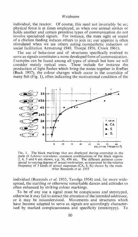
Weidmann
individual, the reactor. Of course, this need not invariably be so; physical force is at times employed, as when one animal strikes or holds another and certain primitive types of communication do not involve specialised signals. For instance, the mere sight or sound of a chicken feeding induces others to join in; our appetite is often stimulated when we see others eating (sympathetic induction or social facilitation Armstrong 1949, Thorpe 1956, Crook 1961).
The use of behaviour and of structures specifically evolved to serve as signals constitutes a more developed form of communication. Examples can be found among all types of stimuli but here we will consider mainly optical ones. These include for instance the production of light flashes which bring the mates together in fireflies (Buck 1937), the colour changes which occur in the courtship of many fish (Fig. 1), often indicating the motivational condition of the
10 15 10 JS
I . "'fAN
e. CA • . s • • S;
)0 35 "" R[LATIV[ FAEOUENCv%
FIG. I. The black markings that are displayed during courtship in the male of Lebistes reticula/us; common combinations of the black regions 2, 4, 5 and 6 are shown, e.g. 56, 456 etc. The . different patterns correspond to varying degrees of sexual motivation, as measured by the relative frequency of 3 kinds of sexual responses (CA, S, Si) shown by the male.
After Baerends et at. 1955
individual (Baerends eta/. 1955, Tavolga 1954) and, far more widespread, the startling or otherwise remarkable dances and attitudes so often enhanced by striking colour markings.
To be of any use a signal must be conspicuous and sterotyped, otherwise it may fail to attract the attention of the intended addressee, or it may be misunderstood. Movements and structures which have become adapted to serve as signals are accordingly characterised by marked conspicuousness and specificity (stereotypy). To
80

Colour and Animal Behaviour
emphasise their common function Lorenz (1935) called them "releasers"; nowadays they are often referred to as "social releasers". Allaesthetic character is an older term (Huxley 1934) for the features of an animal which influence the behaviour of others from a distance; to some extent the two terms overlap.
The characteristics of releasers and the methods for their study have been discussed at length in the review by Tinbergen (1948). Evidence that a colour pattern or a conspicuous movement or posture of an animal functions as a visual releaser may be circumstantial; thus we may repeatedly observe that a colour marking is displayed to other members of the species. A correlation between conspicuous marking and display at least suggests that they could function as releasers; to prove it, experiments are needed in which the behaviour of an animal is compared when subjected to stimulus situations containing and lacking the colour pattern in question.
A classical example concerns the signal function of the orange-red breast of the robin (Lack 1943). This region is regularly displayed by the owner of a territory towards an intruding robin who does not retreat at once when approached. The display, which is usually followed by the hasty departure of the intruder thus saves the territory owner from engaging in actual combat. The postulated effect of the red area depends however on the context, eliciting escape or attack accordingly. Thus when stuffed robins or parts of a robin were fixed on a branch near a nest containing young they were furiously attacked as long as part of the red breast was present. Typical displays were even shown to a mere bundle of red feathers, whereas a stuffed adult whose breast had been stained with brown ink, or a stuffed juvenile which has a speckled brown breast, released hardly any attacks at all. Of course, there can be no question that the robin is able to distinguish such objects from a real robin; his eyesight is sufficiently acute to recognise his mate. What these tests rather show is that the response of attack can be brought about by a very limited number of features which characterise the situation normally presented by an intruder. Other features of the normal situation are less important in eliciting an attack, and though normally perceived can be omitted in an experiment, without diminishing the strength of the response.
This dependence of a response on only a few characteristic properties of a stimulus situation is exceedingly common, especially for responses which are elicited for the first time. The relevant stimuli are called key, sign or releasing stimuli to distinguish them from the other features of a complex situation (Russell1943, Tinbergen 1951). Features, which appear striking to us, may not be involved in eliciting a given response; the stimulus situation must therefore be analysed in each case to find its constituent sign stimuli. A few examples may serve to illustrate such an analysis.
81

Weidmann
The sexes of budgerigars differ in the colour of the cere above the beak, which is blue in the male and brown in the female. Experiments show that budgerigars, too, rely on these features to recognise the sexes. Thus males which had their cere stained brown were courted by other males, while females with cere painted blue were attacked as rivals (Cinat-Tomson 1926). Similar results were obtained by disguising females as males in several species of lizards and in chaffinches (Marler 1955). Female chaffinches with underparts dyed red, were avoided by others, including some hand-reared females who had never seen adult males. As a result the red females won most aggressive encounters with other females and came to dominate them in the social hierarchy. The intimidatory effect of red was not due to the fact that it was unusual. Females dyed with green were not treated differently from normal ones. .
Crane (1955) analysed the stimulus situation which elicits courtship in the tropical butterfly Heliconius erato. Painted pieces of canvas were fastened to a thread and moved up and down near a male or female. The features which mattered most were type of motion and colour, red and orange models being far more effective than models painted in other colours. The butterflies have orange-red badges on their otherwise black wings, and models with patches of colour on a dark background were on the whole more successful than uniformly coloured ones, 'Yith the surprising exception of an all-red dummy whose releasing value exceeded that of a live specimen. Other experiments showed that size, shape and pattern of the colour badge were of minor importance. Similar results were obtained by Magnus (1958) with the fritillary Argynnis paphia raised in captivity. He used a kind of merry-go-round on which he mounted flapping dummies or rotating cylinders with alternate stripes of colour and black. The males were attracted by orange, and preferred models of 4 times natural size to normal ones. The flapping or flickering of a dummy increased its effectiveness up to the flicker-fusion frequency of the eye, as determined with electrophysiological methods. On the other hand, the natural shape and the normal black pattern of the female wing were found to be irrelevant for this response.
Newly-hatched Black-headed gulls peck at the tip of the red beak of their parents when they are hungry, a response which induces the parent to regurgitate food for them. Experiments with inexperienced, incubator-hatched young showed that this " begging " response can be elicited by a variety of dummies, such as rods, flat discs and other shapes which entirely lack such features of the normal sitmi.tion as e.g. the brown mask on the head of the parent, the shape of head, neck and body, the eyes etc. If a dummy was red, or of a contrasted black and white pattern, of small size (narrow rod or small disc) and was slowly moved to and fro in front of the chick, it was far more effective than dummies which lacked one or more of
82

Colour and Animal Behaviour
these characteristics (Weidmann & Weidmann 1958, 1961, and in prep.). These results agree well with those obtained by Tinbergen & Perdeck (1951) in the Herring gull.
Properties that are important in releasing one response often do not influence another, so that each response is susceptible to its own unique combination of stimuli. For example, inexperienced Heliconius, as mentioned above, react to orange-red :during courtship, but prefer yellow paper flowers to orange ones when feeding (Crane 1955). Pieris brassicae hatched in a cage approached red, yellow, blue or violet paper models when searching for food, but alighted on green models when depositing eggs (lise 1937).
The foregoing conclusions apply especially to responses which are elicited for the first time. Subsequently, they may become conditioned (imprinted) to additional features of the releasing situation or the inborn preferences may become altered (Thorpe 1956). Thus, many responses of birds, mammals or fish are only elicited by visual stimuli coming from one particular individual, e.g. the mate or the parent, and not by other members of the species; even so, communication may still depend on the releasers which are typical for the species. Some cichlid fish stop reacting to dummies after they have met conspecifics (Seitz 1940). Honey bees, foraging for the first time, are particularly attracted by blue and yellow (Butler 1951), but later the colour preference becomes conditioned to the colour of available crops, remaining the same as long as a favoured source of nectar lasts (flower constancy). The begging response of the Black-headed gull chicks can be conditioned to a grey disc by rewarding each peck with food; in an experiment, two days of conditioning to this dummy (which lacked such important qualities as " redness " and " thinness ") were sufficient to make it superior to the hitherto optimal dummy, a thin red rod (Weidmann 1961).
However, the innate susceptibilities of a response are not necessarily changed by experience. Young cichlids of the spp. Apistogramma reitzigi and A.borelli follow any slowly moving object which is yellow or yellow and black respectively, regardless of shape and size. The selectivity of this response was not increased even after the young had stayed with their mother for three weeks. Attempts at conditioning inexperienced young to other colours were unsuccessful (Kuenzer & Kuenzer 1962).
Some of the examples mentioned show that different aspects of a visual situation, e.g. movement, brightness, hue, pattern or size of a colour mark may each contribute to its releasing value. A curious mosaic type of interaction obtains in such instances: a stimulus situation deficient in one or two sign stimuli still elicits a typical response, albeit a weaker one, irrespective of which of the stimuli are missing (heterogeneous summation, Seitz 1940). For instance, a Black-headed gull chick will also peck at models which
83

Weidmann
lack one or several releasing stimuli, e.g. at a grey disc. However, a better response is elicited by a grey rod, providing the feature "thinness ", or alternatively by a red disc, which adds " redness " to the previous situation. A red rod is still better, suggesting that the releasing effects of thinness and redness contribute independently to the overall releasing value (Fig. 2).
HETEROGENEOUS SUMMATION
0 GREY DISC GREY ROO RED DISC RED ROD
GO~------------~GR R~D __________ ~RR
0 5 10 15 20 25 NUMBER OF PECKS IN 30 SEC .
FIG. 2. Average number of pecks given by newly hatched Black-headed gull chicks to 4 different dummies, presented in
succession, one at a time. See text.
These conclusions, i.e. the importance of sign stimuli and heterogeneous summation lead to the hypothesis, already stated by Lorenz (1935), that there must be afferent mechanisms between the receptors and the motor centres of the various responses, which deal with the information received by the sense organs and activate the appropriate motor centre. The integrating and filtering properties of these postulated " releasing mechanisms " would thus account for the fact that one response is given to one set of sign stimuli, another one to a different set, etc. Where such selectivity exists prior to conditioning one speaks of innate releasing mechanisms. The concept of a releasing mechanism is at present the subject of controversy (Baerends 1959, Hinde 1956 Schneirla 1956) into which we need not enter here. Suffice it to say that whether or not a particular feature of an object is involved in eliciting a response depends on the physiological make-up of the reactor, on the properties of its eyes as well as on more centrally situated mechanisms which sort and select the information provided (Marler 1961, Magnus 1958, Weidmann & Weidmann 1958).
84
Colour and Animal Behaviour
Signals from colour patterns are not only involved in eliciting responses: they often serve to direct or orientate a reaction. An example is the bright marking inside the throats of many nestling songbirds, which forms an easy target for the parents' beak when feeding the young. A young Herring gull chick aims particularly at the red spot on the lower mandible of its parent when begging (Goethe 1937, Tinbergen & Perdeck 1950). The contrasting patterns on the hindquarters of deer and antelope facilitate contact among a group of individuals following one another in the dark. Many birds and some fish, e.g. the 3-spined stickleback have special displays to guide the mate to the nest site (Tin bergen 1953).
Display does not always elicit an overt response at once but rather tends to change the reactor's mood, making it ready for some future action. Many of the striking and often repeated courtship displays of birds serve such a function, preparing the mate for copulation by gradually overcoming its fear or aggressive tendencies and by stimulating it sexually. Another function of these displays is of course the prevention of interspecific matings (see below).
This leads to a consideration of how releasers have evolved. Comparative studies suggest that displays are evolutionary elaborations and fusions of intention and ambivalent movements, redirection and displacement activities and reactions of the autonomic nervous system, which regularly accompany conflicts of one kind or another (Daanje 1950, Eibl-Eibesfeldt 1956, Lorenz 1951, Morris 1956, Moynihan 1955a, Tinbergen 1952). The evolutionary changes have been such as to make these movements at the same time more conspicuous and less variable, and thus to increase their effectiveness as signals (ritualisation; Blest 1961 , Huxley 1923, Morris 1956, 1957, Tinbergen 1952).
Far less is known about the origin and evolution of colour patterns. Often a display has later been enhanced by the devlopment of striking colours. Such a sequence of events is suggested by the numerous instances in which the same display movement is used in a group of closely related species to exhibit different colour markings. Examples include the red and blue-and-orange breasts of robin and bluethroat (Cyanosylvia svecica) (Peiponen 1960), the different head and breast patterns of tits (Hinde 1952) or of ducks (Lorenz 1941). The brown mask of the Black-headed gull emphasises various of its threat displays (Moynihan 1955b) which the species shares with other non-" hooded " species. The mask is thus less primitive than the displays with which it is now associated. However, the reverse development, viz. evolutionary change of a movement following the acquisition of a colour pattern is not unknown. Indeed, the mask of the Black-headed gull is probably responsible for the extremely emphasised form of head-flagging in this species. This display which consists of turning the head and bill away from another bird, occurs in many other gulls where it has for long been
85

Weidmann
overlooked because it is far less obvious and pronounced in those species (Fig. 3). Being in many ways the opposite of a threat display
liK ;'1.~:2~~ c~ :f;r+~ A~~
\, ....._. f' I(< s . -FIG. 3. Head flagging or facing away in various species of gulls. a, c. Kittiwake (adult intruder), e. Kittiwake (young), b. Lesser Black-backed gull (pair); d. Common gull (bird on the left); f Hartlaub's gull (pair); g. Black-headed gull (pair), note mask. From Tin bergen 1959, after various authors.
(e.g. the bill is removed from sight) it may help to inhibit attack or escape tendencies in the reactor (Tinbergen 1959). To be effective in the Black-headed gull this must clearly involve the removal from sight of the whole mask, and this may explain why the movement has become so much more stereotyped in this species.
Some colour signals have probably arisen from autonomic reactions and were at first independent of movements which later displayed them: examples are the vasodilation of bare skin patches (blushing, flushing, wattles of turkey cock) or inflation which led to such bizarre signals as the scarlet bladder of the male frigate bird. By the sudden contraction of the yellow iris in the eye a male bearded tit can produce a yellow flash which is supposed to function as a signal in courtship (Konig cit. in Eibl-Eibesfeldt 1956).
86

Colour and Animal Behaviour
In yet other instances it is li.kely that both movements and structural features have been elaborated side by side; thus a courtship movement, derived from displacement preening, and consisting of touching an area of the closed wing is very widespread in male ducks, but nowhere is it so emphasised as in the Mandarin drake (Aix galericulata) which points to its gaudily coloured and oversized feather in this region, which is clearly a releaser evolved in association with this movement.
What constitutes an improvement in a signal depends, firstly on the responding individual's sensitivity and preferences, and secondly on the background against which the signal has to be perceived. Thus, in several studies it has been possible to make dummies which surpass the natural object in releasing value. Magnus' super models for eliciting the sexual pursuit of the male fritillary have been mentioned above. Eggs or egg models placed on the rim of the nest of a Herring gull are retrieved when the incubating bird returns to the nest. In a choice experiment the largest dummy is rolled in first , even if it is several times larger than a normal egg. The specks on the surface of the eggs are also important; the more numerous, the smaller and darker they are, the better; dummies with particularly " dainty " specks are preferred to normal ones (Baerends 1957). Such "supernormal" stimuli (Tinbergen & Perdeck 1951) have been found in many species and suggest directions in which releasers might possibly be improved by further evolution.
The so called "egg-dummies" (Wickler 1962) of some Cichlid fish provide a striking example of the evolution of a releaser to fit the susceptibilities of the reacting animal. A large group of cichlid fish, the so called mouth-breeders, hatch their young in the mouth, the eggs being collected by the female as soon as they are laid. In some species the interval between laying and swallowing is so short (presumably for protection) that the eggs are not fertilized in the water, and sperms have to be swallowed to allow fertilization within the mouth cavity. The uptake of sperm is assured by the displaying by the male of bright-coloured patches (annular spots) on its anal fin, resembling in size and colour the eggs of the species (Fig. 4) When the female tries to swallow these "eggs", the male discharges spermatozoa which are "inhaled" by the female. In other species the males possess long genital tassels which bear bright orange blobs (Fig. 4); these, too are believed to function as egg dummies.
Innate preferences might also explain some differences in colour patterns between related species, serving similar functions. For example some gulls have red spots on yellow beaks while the beak of others is uniformly red. J. P. Hailman (pers. comm.) showed that there is a good correlation between beak width and presence of spots; spots being found almost exclusively in species with large beaks. This makes sense when the importance of the stimulus "narrowness" is remembered. As has already been explained a
87

Weidmann
FIG. 4. "Egg-dummies" in cichlids. a. Original pattern of markings on median fins in substrate-brooding cichlids. b. Annular spots (eggdummies) on anal and dorsal fin of the male of some Haplochromis species, e.g. H. wingatii. c. Fully ripe male of Tilapia karomo to show the development of the long bifid genital tassel. a, b after Wickler
1962, c after Lowe 1956.
narrow beak model elicits begging far more efficiently than a wide one, and other experiments have shown that the same applies for stripes or dots on a disc; there is, for Black-headed gulls an optimum diameter somewhere around 6.5 mm (Weidmann, in prep.) and roughly similar optima apply to some of the other species. While the beaks of smaller species approach this optimum closely this is not so for the larger ones. A large bill of uniform colour would therefore be a far less effective releaser than one with a small red spot. Indeed, there is some direct evidence that this speculation is true; in the experiments of Tinbergen & Perdeck with Herring gulls, an all-red beak was about twice as good as a yellow one, whereas a yellow bill with a red spot was over three times as efficient.
However, natural selection also operates on the susceptibilities of the responding individual and leads to adjustments in the releasing mechanisms parallel to the evolution of releasers. We know far less about such changes than about the more easily studied releasers.
88

Colour and Animal Behaviour
An example is again provided by the begging response. In many species of birds, including moorhens, gulls and terns of several kinds, newly hatched young respond particularly to red. In the Sandwich tern, whose beak is black with a yellowish tip the young show a slight preference for black (Weidmann 1961) and in the Wideawake tern, whose beak is black too, the young have no preference either way, suggesting at least a loss of the red preference which may be expected to have existed in its ancestors (Cullen 1962). It is not known, whether the change of beak colour preceded that of the preference of the young, though this appears more likely than the alternative.
TABLE I
Unlearnt colour preferences of some gulls, terns and other birds
Species Chick has un/earnt Author preference for Adult Bill
Red Black
HERRING GULL t red spot on yellow Tinbergen & Perdeck
BLACK-HEADED GULL t crimson R. & U. Weidmann
KlTIIWAKE t yellow with red E. Cullen gape
CASPIAN TERN t scarlet Bergmann
ARCTIC TERN t blood red D. A. Quine
SANDWICH TERN t black with yellow U. Weidmann tip
WIDEAWAKE TERN neither black J. M. Cullen
OYSTER CATCHER t orange red Lind
MOORHEN t red and yellow U. Weidmann
CooT t white U. Weidmann
The background against which a signal has to be perceived is another factor which determines the course of further improvements (Marler 1957, Crook 1958). Indeed the initial " choice " of a character for a signal function may have depended on this. In a species which posesses auditory, olfactory and visual organs any one of these modalities might have been employed to convey a particular message. That they would all be possible choices is shown by the fact that the same message, e.g. " territory occupied, keep out if conspecific male, approach if conspecific female " is transmitted by song in many birds, by its red belly in the 3-spined
89

Weidmann
stickleback and by scent marks in some mammals. We know as yet too little, to explain in every case why song is the vehicle for transmission in one species and display in another. But suggestions can be made, see e.g. Marler (1957), Crook (1958).
In the case of signals which play a role in courtship and pairformation the " background " includes the signals given by other sympatric species. Natural selection puts a premium on the evolution of differences, particularly on those releasers which initiate pairformation. We thus find that sympatric and closely related species differ especially in their courtship displays, and usually the plumage patterns are even more distinct than the displays (Mayr 1942). This is so in the ducks (Lorenz 1941), Parus species (Hinde 1952) and many other groups, though exceptions do exist. The astonishing complexity of many of the releasers used in courtship may on closer study prove to be no more than what is required by the demands of specificity. This suggestion is made by Morris (1954) for the complex colour markings of the Zebra finch and 22 related estrildines in Australia; while many markings are shared, each species has a unique combination of markings which distinguishes it from all others.
The divergence in colour and courtship display is usually greater in males than in females; it has been suggested that divergence in females is suppressed by selection for crypsis, or that male colour differences are also selected in the context of fighting; this in turn is related to the evolution of sexual dimorphism and cannot be discussed here (see Hinde 1959).
Further it should be borne in mind that sexual isolation depends as much on divergence in the " preferences " of the responding mate as on changes in the signals used. Such differences may be inborn, or acquired by early learning (imprinting) (Dilger & Johnsgard 1959). Preferences may also be adjusted in the presence of even unrelated species who, owing to chance resemblance, might be mistaken for conspecifics. A particularly instructive case has been described by Stride (1958) in the African butterfly Hypolimnas misippus. Males approach cardboard models of various colours but in addition to a preference for yellow and red there is a marked reluctance to court a model which contains a large area of white, even if it is otherwise attractively coloured. According to Stride the significance of this "white inhibition", which is also found in many nymphalidae and satyridae, lies in the following : by ignoring the numerous white pierids a male saves energy, risks less exposure to predators and avoids habituation, thus maintaining its readiness to follow a conspecific. The white inhibition might well explain a further peculiarity of the coloration of this species. In West Africa the females of H. misippus mimic the alcippus form of Danaus chrysippus, but unlike these they have no white hindwings, at most the brown is at times somewhat suffused with white scales. This
90

Colour and Animal Behaviour
discrepancy between mimetic form and model can be explained as the result of a balance between opposing selection pressures. Mimetic selection tends to evolve white hindwings similar to the danaid model; sexual selection by the male discriminates against white hindwings as a result of the white inhibition, itself maintained for the advantages mentioned above.
The idea of conflicting selection pressures is basic to an understanding of adaptive coloration. In most species a compromise is reached between the conflicting advantages of conspicuous signals and cryptic appearance for concealment from predators and prey alike. The importance of these factors varies from species to species and different " solutions " are encountered. Many species appear cryptically coloured for most of the time but posesss bright colour patterns which are only exposed for brief moments: e.g. the wing specula of female ducks are exposed only during flight . Some colour patterns which are conspicuous from nearby such as the brilliant greens and blues of the male peacock are apparently fairly cryptic in the jungle when seen from a distance (Huxley 1934). Species whose intraspecific releasers make them particularly conspicuous to predators are usually protected in some other way: e.g. the spines of the three-spined stickleback (Hoogland, Morris & Tinbergen 1957), and the habit of roosting in inaccessible places of many gallinaceae and anatidae. (See also pp. 65-66.)
Use of colour in communication between symbionts
By far the best known symbiotic relationship is that between insects and flowers. In 1793 Christian Sprengel suggested that colour and scent attract insects to the flowers and he rightly interpreted the coloured dots and lines on the petals of many flowers as honey guides, i.e. marks which guide an insect to the hidden nectar and the nearby pollen. The effectiveness of honey guides has recently been demonstrated in experiments with bumble bees (Manning 1956). When offered a blue paper disc the bees tend to hover or alight on the edge more often than in the centre, but when the disc contains yellow spots in the middle, or converging yellow lines, more bees alight in the centre. Manning concludes that the presence of honey guides is advantageous to the flower. On a flower lacking honey guides bees might stay near the edge and leave without having found the way to the nectar and pollen. For other recent work in this field reference is made to Meeuse (1961), Manning (1956b, c, 1957) and Tinbergen (1953). The remarkable correlations between flower structure, scent and colour on the one hand and insect behaviour and sensitivity on the other, must be the outcome of mutual adaptations, but we do not know how they evolved. The fact that so few bee-pollinated flowers are purely red
91

Weidmann
and that there are ultra-violet honeyguides, invisible to us (Daumer 1958), makes sense, when it is realized that bees are red blind, but can see ultra-violet. From a recent study of Daumer (1958) it appears that, owing to the ultra-violet emitted, many flowers are much more conspicuous to a bee than to us (Fig. 5).
A
B
centre corners centre corners
I 64 46
FIG. 5. A. Oenothera biennis, from a photo taken through UV-filter; UV -free regions appear black. B. UV-free regions act as honey guides eliciting searching reactions in inexperienced bees. left: normal arrangement; right: reversed arrangement of appropriately cut-up petals 0 . biennis under ultraviolet transparent glass. Numbers of probing reactions of bees recorded in an experiment are given. After Daumer
1958.
The blue, yellow or red colours of many fruits are probably an adaptation providing colour contrast against the green foliage, thus facilitating their discovery by the many fruit-eating birds which disperse their seeds. While it is true that many birds are particularly sensitive to red (Walls 1942), there appears to be no conclusive evidence of any inborn preference for red or blue in fruit-eating animals.
Another symbiosis involving communication by means of colour is that between some marine fish and their cleaners. The cleaners are small fish which habitually clean others of ectoparasites. The big fish invite cleaning by adopting special postures and regularly visit special places among the corals where the cleaners are likely to
92

Colour and Animal Behaviour
be found. On the other hand, some cleaners attract the attention of possible clients by swimming in characteristic attitudes across their path. Cleaners, of several unrelated families, are characterized by black longitudinal stripes or by a yellow and blue pattern, and Eibl-Eibesfeldt (1959) suggests that this may be a "guild sign" advertising the fishes "occupation" of being a cleaner. This fantastic suggestion gains support from the discovery by EiblEibesfeldt of a mimic Aspidontus taeniatus (Fig. 6). This species has
B
c FIG. 6. Coloration of fish which habitually rid others of ectoparasites (symbiotic cleaners). B. Labroides dimidiatus (Labridae) and C. Elacatinus oceanops (Gobiidae) and of a mimic A. Aspidontus taeniatus (Blenniidae). Eibl-Eibesfeldt
1959.
the same colour pattern and behaviour as the cleaner Labroides dimidiatus, and is thus able to approach other fish closely, whereupon he suddenly bites a chunk out of their body and flees.
93

Weidmann
Animal coloration and behaviour in predator-prey relationships
All types of adaptive coloration (concealment, advertisement or disguise) which are of significance in predator-prey encounters come under our theme, in as far as their major evolutionary cause "has had eyes", as one author put it. In addition, colour patterns considered to be adaptive are often associated with behaviour which is essential for the full effectiveness of the coloration: Selection of suitably coloured or patterned backgrounds (e.g. of cryptic and melanic moths, Kettlewell 1955), adoption of correct position with regard to countershading (de Ruiter 1955) or disguise as a twig (de Ruiter 1953), the mimicking of model species also in behaviour, or startling movements exposing striking colours (Fig. 7) come to
FIG. 7. Defensive display of the man tid Acanthops falcata . Wings yellow and black, abdomen rosy red and black, colour otherwise
brown. After Crane 1952.
mind in this context (Crane 1952, Chance & Russell 1959, Blest 1957a, b).
Recent studies have mainly been concerned with the experimental verification of some of the previously suggested functions of colour. The Browers' (1958, 1960a, b) work clearly demonstrates the effectiveness of mimetic resemblances, and this topic is discussed from an ecological and genetical angle by de Ruiter (1958) and Sheppard (1959). Blest (1957a) studied the significance of eyespot patterns in Lepidoptera and found that their intimidatory effect, particularly
94

Colour and Animal Behaviour
on inexperienced small passerines, depends both on colour and pattern ; circular patterns are more effective than non-circular ones (Fig. 8). He suggests that the coloured ocelli act by eliciting responses normally given to large avian predators. In many small passerines such responses are inborn, and the eyes of the predator form an important feature of the stimulus situation (Hinde 1954).
It has been alleged that some of the resemblances of animals to leaves or twigs etc. are too good to be accounted for by selection. Such arguments were based on the assumption that a bird only reacts to a few features of a total situation, as indeed we found to be the case in many intraspecific relationships. However, de Ruiter's (1952) studies with jays and stick-caterpillars showed that the chance discovery of one caterpillar was followed at once by an intensive search for similar objects (searching image), twigs of similar shape and size being turned over, too, until the bird became discouraged. Only the " best " caterpillars living on the tree from which the sticks were derived had any chance of surviving this examination undiscovered.
An interesting suggestion is that a moderately distasteful species is subjected to two opposing selection pressures which result in its being neither too cryptic nor too conspicuous (Prop 1959). The idea is that in years of plenty it is an advantage to be recognized on sight by a predator as inedible and thus be spared; however, in lean years moderate inedibility is no protection and then it pays not to be found too easily. Prop adduces evidence supporting this suggestion in several species of tenthredinid larvae.
The sofar intractable "coral-snake problem", viz. the use of warning coloration in a species whose bite is deadly, has been brought nearer to a solution by the observations of Klopfer (1957) that some birds learn to avoid an object after seeing it cause harm to a fellow member.
For an exhaustive review and exposition see Cott (1940).
Other behaviour-colour relationships
Finally, there are some examples which may suggest that some animals have the rudiments of aesthetic appreciation. A number of animals show a liking for particular colours or colourful objects, which in themselves are not of any (known) biological significance. An amusing example concerns the preference of Adelie penguins for red stones (Levick 1914). During its courtship the male presents the female with stones for nest building, more being stolen later from neighbouring nests. When stones were painted in different colours and placed at one side of the nesting area, the penguins collected these too, and during the next few days the stones "travelled" across the colony (by theft!), the red ones far ahead of the others.
95
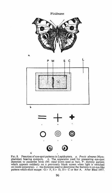
Weidmann
b
-:---- + +
0 @)
G
FIG. 8. Function of eye-spot patterns in Lepidoptera. a. Precis a/mana (Nymphalidae) bearing eyespots. b. The apparatus used for presenting eye-spot dummies to passerine birds (M: meal worm used as bait, P: dummy pattern which appears suddenly on a previously blank screen when light is switched on inside apparatus). c. the dummies used to determine the features in an eye-like pattern which elicit escape: G> F, E> D, D> Cor BorA. After Blest 1957.
96
Colour and Animal Behaviour
The strange fact is that there seem to be no red objects in the penguins' environment, yet they prefer this colour to all others. Another example of the use of colourful objects is provided by the bower birds, whose " art-galleries " (tunnels of twigs adorned with collections of pretty feathers, shells, flowers and other objects, neatly arranged and typical for the species) serve to attract a mate, who presumably is impressed by this gorgeous exhibit (Marshall 1954). Results with captive monkeys and apes (Rensch 1957, 1959) are even more explicit: thty reveal clear preferences for certain colours which however change from time to time ("aesthetic-fashions"). Differently coloured cubes which can be stuck together are assembled into rows of uniform colour. Morris (1962) studied the patterns painted by captive chimpanzees provided with paper and pencil or crayons. Remarkably enough, these are far from random; e.g. if a dot is provided, the first scribbles are placed in clear spacial relation to it and in general there is a marked tendency to balance the " design ". With regard to colour there is less discrimination, though sometimes certain of the colours offered are refused. Both Rensch and Morris conclude that the rules of " creative activity " displayed by these animals parallel the elements of aesthetic appreciation in man.
References
Armstrong, E.A., 1949. Bird display and behaviour. London. Baerends, G. P., 1950. Symp. Soc. exp. Bioi., 4, 337-60 Baerends, G. P., 1957. Anat. Rec.,128, 518-519 Baerends, G. P., 1959. Proc. XVth int. congr. zoo!. London. Baerends, G. P., Brower, R. & Waterbolk, H. T., 1955. Behaviour, 8, 249-334. Blest, A. D., 1957a. Behaviour,ll, 209-256. Blest, A. D., 1957b. Behaviour,ll, 257-309. Blest, A. D. 1961 in W. H. Thorpe & 0. L. Zangwill: Current problems in animal
behaviour. Cambridge. Brower, J. VZ., 1958. Evolution, 12, 32-47. Brower, J. VZ., 1960. Amer. Nat., 94, 271-282 Brower, L. P., Brower J.VZ., & Westcott, P. W., 1960. Amer. Nat., 94, 343-356. Buck, J. B., 1937. Physiol. Zoo/., 10,412-419. Butler, C. G., 1951. Proc. roy. Soc. B.,138, 404-413. Chance, M. R. A., & Russell, W. M.S., 1959. Proc. zoo!. Soc. Lond., 132, 65-70. Cinat-Tomson, H.,1926. Biol.Zbl., 46,543-52 Cott, H. B., 1940. Adaptive coloration in animals. London. Crane, J., 1952. Zoologica, 37,259-293. Crane, J., 1955. Zoologica, 40, 167-95. Crook, J. H., 1958 cit. in Hinde, R. A., 1959. Bioi. Rev., 34, 85-128. Crook, J. H., 1961 in W. H. Thorpe, & 0. L. Zangwill. Current problems in
animal behaviour. Cambridge. Cullen, J. M., 1962. Ibis, 103b, 162-173. Daanje, A., 1950. Behaviour, 3, 48-98. Daumer, K., 1958. Z . vergl. Physiol., 41, 49. Dilger, W. C. & Johnsgard, P. A., 1959. Wilson Bull., 71,46-53. Eibl-Eibesfeldt, I., 1956. Z.f. Siiugetierk., 21,29-43. Eibl-Eibesfeldt, 1., 1959. Z.f. Tierspycholog., 16, 19-25.
97

Weidmann
Frisch, K . von., 1950. Bees, their vision, chemical senses and language. Ithaca, New York.
Goethe, F., 1937. J. f Ornithol., 85, 1-119. Hailman, J. P. (Personal communication). Hinde, R. A., 1952. Behaviour, supplement 2, 1-201 . Hinde, R . A. ,1954. Proc. roy. Soc. B., 142, 306-331. Hinde, R. A., 1956. Brit. J. Philos. Sci., 6, 321-331. Hinde, R. A., 1959. Bioi. Rev., 34, 85-128. Hinde, R. A., 1961 in W. H. Thorpe & 0. L. Zangwill: Current problems in
animal behaviour. Cambridge. Hingston, R . W. G ., 1933. The meaning of animal colour and adornment.
London. Hoogland, R. , Morris, D . & Tinbergen, N ., 1957. Behaviour,10 ,.205-236. Huxley, J. S., 1923. J. Linn. Soc. Lond., 25, 253-292. Huxley, J. S., 1934. Proc. 8th int. ornithol. congr. Oxford. lise, D ., 1937. Nature, 140, 544-545. Kettlewell, H. B. D., 1955. Nature, 175, 943. Klopfer, P. H., 1957. Amer. Nat., 91, 61-63. Kuenzer, E. & Kuenzer, P., 1962. Z.f Tierpsycholog., 19, 56-83. Lack, D ., 1943. The Life of the Robin. London. Levick, G. M., 1914. Antarctic Penguins. London. Lorenz, K ., 1935. J. Ornithol., 83, 137-214, 289-413. Lorenz, K ., 1941. J. Ornithol., 89, 194-294. Lorenz, K., 1951. Naturwiss., 38, 113-116. Magnus, D . B. E., 1958. Proc. JOth int. congr. entomol. 1956. Manning, A., 1956a. Behaviour, 9, 114-139. Manning, A., 1956b. Behaviour, 9, 164-201. Manning, A., 1956. New Biology, 21. Manning, A., 1957. Proc. Roy. Phys. Soc., 25, 67-71. Marler, P. , 1955. Brit. J. Anim. Behav., 3, 137-146. Marler, P., 1957. Behaviour, ll, 13-39. Marler, P., 1961 in W. H. Thorpe & 0 . L. Zangwill: Current problems in animal
behaviour. Cambridge. Marshall, A. J., 1954. Bower Birds. London. Mayr, E., 1942. Systematics and the origin of species. New York. Meeuse, B. J.D., 1961. The story of pollination. New York. Morris, D., 1954. Behaviour, 6, 271-322. Morris, D ., 1956. Behaviour, 9, 75-113. Morris, D ., 1957. Behaviour,ll, 1-12. Morris, D., 1962. The Biology of Art. London. Moynihan, M., 1955a. Auk., 72, 240-246. Moynihan, M., 1955b. Behaviour, supplement 4, 1-201. Peiponen, V. A., 1960. Ornis Fennica, 37, 69-83. Prop, N ., 1959. Arch. m!erl. Zoo/., 13, Rensch, B., 1957. Z.f Tierpsycholog., 14,71-99. Rensch, B., 1959. Proc. 15th int. congr. zoo/. London. Ruiter, L. de., 1952. Behaviour, 4, 222-232. Ruiter, L. de., 1955. Arch. neerl. Zoo/. ,11, 3-57. Ruiter, L. de., 1958. Arch. neerl. Zoo/., 13, suppt. 1, 351-368 Russell, E. S., 1943. Proc. Lirm. Soc. London, 154, 194-216. Schneirla, T. C., 1956 in L'instinct dans le comportement des animaux et de
l'homme. Paris. Seitz, A., 1940. Z . f Tierpsycholog., 4, 40-84. Sheppard, P. M., 1959. Cold Spring Harbour Symp. Quant. Bioi., 24, 131-140. Stephenson, E. M. & Steward, Ch., 1955. Animal camouflage. London. Stride, G . 0., 1958. Animal Behav., 6, 224-230. Tavolga, W. N., 1954. Bull, Amer. Mus. Nat. Hist., 103, 427-460. Thorpe, W. H., 1956. Learning and instinct in animals. London.
98
Discussion
Tin bergen, N., 1948. Wilson Bull., 60, 6-52. Tin bergen, N., 1951. The study of instinct. Oxford. Tinbergen, N., 1952. Quart. Rev. Bioi., 27, 1-32. Tinbergen, N., 1953. Social behaviour in animals. London. Tinbergen, N., 1959. Behaviour, 15, 1-70. Tinbergen, N. & Perdeck, A. C., 1950. Behaviour, 3, 1-39. Walls, G . L., 1942. The vertebrate eye. Michigan. Weidmann, R. & Weidmann, U ., 1958. Anim. Behav., 6, 114. Weidmann, U., 1961. Anim. Behav., 9, 115-116. Wielder, W., 1962. Z .f.Tierpsycholog ., 19, 129-164.
DISCUSSION
Sir Julian Huxley. Dr, Weidmann stressed the importance of the background. This was well demonstrated when James Fisher and I were trying the effect of dummies on blackheaded gulls. One dummy head that we had on a knitting needle was attacked by the parent bird returning to the nest, and fell off; thereupon the bird brooded it!
Dr. Weidmann also underlined the specific effect, as in, for example, the care of the budgerigar; but this is sometimes overridden by another stimulus. In zebra finches, the young resemble in most respects the female; if the begging young is painted to complete the resemblance, the male courts it, but the begging continues, and then he feeds it. This sort of thing has been much neglected in many of the earlier studies stressing fixity of response.
U. Weidmann. Yes, I agree; the importance of learning in social communities is dealt with in my written paper, but, in selecting the material for oral presentation this was one of the things I had to leave out.
In some other cases, imprinting is involved.
Sir Julian Huxley. In the light of the views you quoted on head-flagging in blackheaded gulls, one is led to realize that human beings are not the only animals that "count 10 when angry" !
J. D. Carthy. I have no doubt that physical measurements of spectral reflectance are needed in ethological work. The use of models to analyse releaser systems is now widespread but I believe that little has been done to compare spectral reflectances of the models coloured to match to the human eye and the actual features of the animal which they imitate. In one case a high reflectance for ultraviolet light from patches on a butterfly's wing was shown to be of considerable behavioural importance, though their existence had not been suspected before physical methods were used. (The paper by Hunt, p. 115, discusses other factors in the subjective assessment of colour which may well need to be taken into account in interpreting colour behaviour.-Eo.).
U. Weidmann. I agree ; the work of Daumer on ultraviolet honeyguides provides another example of the successful use of measurements of spectral reflectances in this kind of work. Even in groups such as birds where, judging from previous studies, the occurrence of sensitivity to ultraviolet seems unlikely, little is known about the laws of colour matching; two colours which may be indistinguishable for the human observer
99

Discussion
may not beso for his experimental animals. Moreover, as slight differences in hue sometimes are found to have profound effects on behaviour, comparison between the results of different workers only becomes meaningful when the colours used are standardized and when their spectral reflectances are known. It may be worth mentioning that many of the more troublesome uncertainties attending experiments in this field can be avoided by using narrow-band spectral filters (and neutral filters to control intensity) instead of paints or coloured papers.
P. H. Silver. I understand that D . Backhaus has given evidence that some of the ungulates, red deer, pigmy goat, antelope and giraffe, have colour vision. (Backhaus D. (1959). "Experimentelle Untersuchungen iiber die Sehscharfe und das Farbensehen einiger Huftiere". Z .f. Tierpsycho!., 16, 445-467. Backhaus D. (1959). "Experimentelle Priifung des Farbsehvermogens einer Massai Giraffe". Z.f. Tierpsychol., 16, 468-477.
U. Weidmann. Some degree of colour vision seems to be much more widespread in the animal kingdom than had been suspected until recently; this applies to a number of fishes, amphibia, reptiles and among the mammals to ungulates and rodents in particular. For a recent review see Autrum 1958, Tabulae Biologicae, 22, 33-42. What is usually not known is the significance of colours in the life of these animals.
R. Swinfen. I should like to refer to the comments passed by Dr. Weidmann on the sequence of colour changes produced during the courtship display of the male guppy, Lebistes reticulatus. From the transparency shown on the screen it was apparent that these colour changes were observed on both the caudal fin and body of the fish. If this is so perhaps Dr. Weidmann could explain the courtship and successful mating of the black veil-tailed guppy, as this strain has a uniformly pigmented caudal fin . This pigmentation also extends over the majority of the caudal peduncle in some individuals and does not appear to impair their reproductive powers.
U. Weidmann. In the absence of detailed work we can only speculate. Assuming that the colour changes of the guppy during courtship have indeed a signal value one might sutmise that the mutant strain mentioned may have come to rely on other signals. Observations on closely related strains or species of Drosophila show that they may differ considerably in their evaluation of visual and chemical signals during courtship . How such differences arise in evolution is a fascinating problem, and might well repay closer study in the instance mentioned.
100

HUMAN MECHANISMS OF COLOlJR
PERCEPTION
By
KATHARINE T ANSLEY
Institute of Ophthalmology, London
I SHOULD like to remind you briefly of the contents of the two major theories of colour vision and then to examine some of the recent physiological work bearing on the subject in order to see how far these studies support one or other of the great theories or whether they need modification.
Before we start on theories, however, let us recall the points that are generally accepted by all respectable colour vision theories. Very many animals including man, have two sorts of visual cell in their retina, the rods and the cones. The rods are generally supposed to mediate night vision, that is vision at low illumination levels (levels below about 0·1 foot candle, roughly the level provided by a threequarter moon). Vision at these low levels is colourless and we will not, therefore, be further concerned with the rods at present. Day vision, that is vision at the higher illumination levels, is thought to be mainly due to the cones. In man we have colour vision atthese levels and, in addition, those animals which have been shown by properly conducted behaviour experiments to have colour vision all have a high proportion of cones in their retinae. Colour vision, therefore, is considered to be a property of the cones.
The first and best known of the classical theories of colour vision is that first suggested by Thomas Young in 1801 and later elaborated by Helmholtz (1867). It is the trichromatic theory. This theory is based on the fact that all known colours can be matched by some mixture of three primary colours taken from the blue, green and red parts of the visible spectrum. It postulates three retinal mechanisms, one maximally sensitive to blue radiation, one to green and the last one to red. The proportions in which these three mechanisms are stimulated by a given coloured light determines the resultant colour sensation (Fig. 1). Young simply spoke of retinal mechanisms but Helmholtz went further and suggested that there are three different types of cone with different spectral sensitivities and that colour vision was in effect a recognition by the brain of the proportions in which these cone types were stimulated under any given stimulus conditions. The Young-Helmholtz theory provides a
101

Tans ley
FIG. I. Suggested set of fundamental sensation curves. These curves are thought to represent the spectral sensitivities of the three retinal mechanisms
postulated by the trichromatic theory.
satisfactory explanation of the laws of colour mixing and of many of the forms of colour blindness. There are, however, other colour phenomena such as colour contrast and some experimental results with colour adaptation which cannot be explained by this theory in any simple way. Since Helmholtz' day such difficulties have led to the formulation of many other colour theories designed to overcome them. Unfortunately most of these theories are, in their turn, unable to explain the undoubted trichromacy of colour vision as revealed by colour mixing experiments so that they are not altogether acceptable either.
The most successful rival to the Young-Helmholtz theory is that propounded by Hering in 1878. Hering was a psychologist and he started from what he called the "psychological purity" of black, white, blue, green, yellow and red. Every other hue appears to our consciousness as some mixture of these sensations. These six visual sensations can be divided into three opponent pairs, white and black, yellow and blue, red and green. Hering agreed with Helmholtz that the colour mechanisms are situated in the retina and he suggested three retinal substances the breakdown of which produced the first
102

Human Mechanisms of Colour Perception
sensation of each pair, white, yellow and red, while their synthesis produced the second, black, blue and green. There is much in Hering's theory which is not very easy to accept, for instance the conception of "psychologically pure" sensations being necessarily the direct result of fundamental physiological mechanisms is not very satisfactory. Colour mixture experiments show that two of Hering's "pure" sensations, yellow and white, can in fact be produced by mixed stimuli apparently activating at least two retinal mechanisms at the same time. All the same, the notion of opponent mechanisms does go far to explain many colour phenomena which are hard to understand on the basis of the trichromatic theory and that alone.
Now let us turn to the results of some more recent investigations on animals and see if they give any help in choosing between these two theories or whether it now seems necessary to formulate another.
The simplest way in which we can imagine light to affect a structure such as the retina so as to start off a discharge in its optic nerve fibres is through the mediation of sensitive substances which can absorb and transform its energy, since light cannot directly stimulate a nerve. We know a good deal about one set of such sensitive substances, those which are contained in the rods and which, on good evidence, are believed to mediate night vision. If we accept that stimulation of the rods and night vision is due to the presence of a sensitive pigment it is a fairly safe assumption that stimulation of the cones and colour vision is achieved in the same way. If we believe with the trichromatists that there are three sorts of cone with three different sensitivity curves like those shown in Fig. 1 the logical conclusion is that each cone contains a different pigment with an absorption curve corresponding to one of the sensation curves. What evidence have we that this is indeed the case? Now the rod pigments can readily be obtained in solution by extraction of a darkadapted rod-containing retina with certain detergents, but so far the cone pigments have generally not been susceptible to such methods, so that other means of attack have had to be used.
One of the most recent and interesting of these is concerned with the light sensitive pigments present in the human eye. These can be studied in situ by an elegant method whereby the amount of several monochromatic lights reflected back through the pupil from behind the retina is measured, first in the dark-adapted eye and then in the same eye after treatment with a bright white light. In these experiments the retina acts as a colour filter. If the effect of light is to remove or change a retinal pigment, let us say a red pigment, more green light (since this is the colour most heavily absorbed by such a pigment) will be reflected after light adaptation.
It is possible by this means to measure the difference curve of a retinal pigment in situ. The most easily obtained curve in man is that of the rod pigment rhodopsin which is present in relatively large amounts in the peripheral human retina and also in the retinae of
103

Tans ley
the common laboratory animals. For any attempt to examine the cone pigments which might be the basis for colour vision the measurements must either be made on a predominantly cone retina or, in man, on the rod-free central spot or fovea, for in these cases the result should not be complicated by the presence of rhodopsin. Such an examination, undertaken by Rushton (1958) both on normal and on partially colour-blind human subjects, has yielded two curves, one with its maximum at 540 m,u in the green, the other at 590 m,u in the orange. These maxima are very close to those shown in Fig. 1 for the suggested sensitivity curves of the human green and red retinal mechanisms. So far no curve has been described that could mediate the blue mechanism. Incidentally the human curve at 540 m,u is almost idential with that reported for the pure-cone eye of the common American grey squirrel by Weale (1955) using essentially the same technique. Part of the spectral sensitivity curve of the eye of the same squirrel measured by means of the electroretinogram is again very similar (Arden & Tansley, 1955) and it did, therefore, appear as though some cones contained a green-sensitive pigment with a maximum spectral absorption and, therefore, sensitivity at about 540 m,u. The possible red-sensitive pigment was not so well characterized since it has only been found in the human eye where it is apparently contaminated by the green-sensitive one even in colour deficient subjects.
Unfortunately some even later work (Dartnall, 1960) has suggested that the situation may not be so simple as this. Very recently detergent extracts have been made from the grey squirrel retina. These extracts appeared to contain a very little rhodopsin and no other pigment that could be identified. When this rhodopsin solution was bleached it produced a photosensitive yellow breakdown product with a maximum absorption at 480 m,u. It has been calculated that interactions between these two, rhodopsin and its yellow bleach product both light sensitive, could give the squirrel spectral sensitivity curve and also the apparent absorption curve of a greensensitive pigment. This work, however, has not yet been repeated and confirmed.
In the 1940s the well-known neurophysiologist, Ragnar Granit in Stockholm made intensive observations on the reactions of the retinae of a number of animals to stimulation by light of different colours (Granit, 1947). Briefly he opened the eye in a decerebrate or anaesthetized animal, removed the cornea, lens and part of the vitreous and placed a platinum micro-electrode on the inner surface of the retina in contact with or in the neighbourhood of one of the retinal ganglion cells. Such an electrode picks up the discharges from a ganglion cell and these can be amplified and reproduced either in a loud speaker or on a cathode ray tube. Using coloured stimuli he recorded the intensity necessary for the production of a just perceptible response for a series of wave-lengths throughout the
104

Human Mechanisms of Colour Perception
spectrum. By this means he found that all ganglion cells in a given retina do not have the same spectral sensitivity curve, but that the total number of different curves which can be recorded is limited. In the course of an extensive investigation on many different vertebrates including fish, amphibia, reptiles, birds and mammals he found that most ganglion cells gave one or other of four broad sensitivity curves, the particular curve obtained depending on the species and the state of adaptation, whether dark-adapted or lightadapted, of the eye. These broad curves he called dominator curves and they are not thought to be of importance in colour vision. In addition to the dominator curves Granit managed, sometimes directly and sometimes by means of various experimental tricks, to record about seven much narrower curves which turned up again and again in many different species (Fig. 2). These Granit called modulator curves and they are thought to represent, probably in rather an indirect way, the spectral sensitivities of specific retinal colour mechanisms.
FIG. 2. Modulator curves recorded by Gran it in a variety of vertebrate retinae.
Looking at this figure and remembering the much simpler collection of three curves shown in Fig. 1 it certainly does not look as
105

Tans ley
though Granit's results provide much support for a trichromatic theory. However if we ignore the curve at 500 m,u which is probably due to rhodopsin, the rod pigment, we can see that the remaining six curves tend to fall into three groups, one with its maximum sensitivity in the blue, one in the green and one in the red or orange. In Fig. 3 the mean curves for each group are plotted with an indication of their variability, and it can be seen that the retinal mechanisms investigated by Granit could be the basis of a trichromatic theory of
100 II Jli JF 7111 1\
% I IU J \\ I II I 1\ :-[7
I \ \ I ~
J I ' \ n 1\ \ 80
I I I \ II II I
I I , I , .) 1/ (J \ 60
I \ '
40 r- IJI~l
'-J \ ~ ...II. 11\
II J 1/ II ~, 1\ ' v ) '17 ~ ~•a J II 'I " 1'- ' I/ II { ) "' 1\. \...J ~1'1 :.1 ' IU
20
J .. 1\. -"' ~
I IJ' v I" . .,. T ·400 ·450 •500 ·550 • 600 •650 .,. • 700
FIG. 3. Set of mean modulator curves suggesting the way in which the curves of Fig. 2 may fall into three main groups.
colour vision. In any case Granit's work showed for the first time that different parts of the retina do have different wave-length sensitivities and that, whatever the visual mechanism for wave-length analysis may be, it is apparently situated in the retina as both Helmholtz and Hering foretold. Incidentally, the middle curve in Fig. 3 also has its maximum at about 535 m,u and is, in general,very like that found in the squirrel and the human fovea.
If we take a closer look at the site from which Granit obtained his curves in the light of the actual retinal structure of the animals on which he did his experiments, we shall see that it is really rather unreasonable to expect three clear-cut curves like those which have been suggested for human colour vision. If it is true that the retinal colour mechanisms are based on three types of cone with three well-defined spectral sensitivity curves we could only expect any one
106

Human Mechanisms of Colour Perception
of these curves to be faithfully reproduced if the ganglion cell from which we were recording were directly connected to a single cone or to a group all of the same type, and was not under any other influence. Such an experimental situation might be obtained in the human fovea where each midget ganglion cell has a direct pathway to a single cone, but in the retina of the usual laboratory animals this is far from being the case. Fig. 4 shows a much simplified "wiring diagram" of a typical vertebrate retina containing both rods and cones. This diagram is based on results obtained with specific nerve stains. If one studies this figure one is unable to find one ganglion cell which makes connection with a single cone without some likelihood of interference from the reactions of other visual cells both
FIG. 4. The vertebrate retina. Left : Histological section through the retina. Right: "Wiring diagram" of the nervous connections of the retina based on
preparations treated with specific nerve stains.
rods and cones. If one bears this in mind I think it not surprising that Granit probably never recorded the responses of a single cone nor that he did not find three immutable curves in his experiments.
107

Tans ley
A moment's reflection will tell us that if we accept that colour vision is fundamentally based on the reactions of three primary mechanisms we must also accept a great deal of elaboration of these reactions in the course of their transfer into colour sensations. Granit's work simply indicates that some of this elaboration takes place in the retina.
Let us now see what, if anything, recent animal research has done for Hering's theory of opponent mechanisms. The results of some of the investigations of the electrical responses of the retina to stimulation by light seem to point directly to a system of opponent reactions. First of all black does appear to be a definite sensation and not just the result of a complete absence of stimulus. Many optic nerve fibres signal the cutting off of a light stimulus by a renewed burst of activity which, one would suppose, must convey a positive message to the brain. This "off-response", as it is called, is thought to be associated with inhibitory activity in the retina. Then there is much evidence for a mechanism which signals changes in brightness (whiteness) and which is independent of the retinal colour mechanisms. Granit's dominator curves are thought to be a manifestation of such brightness mechanisms. Lastly there is some more recent work (MacNichol and Svaetichin, 1958) in which the responses from within the retina of several species of fish were investigated using differently coloured light stimuli Here again there was evidence of an independent brightness mechanism with a broad spectral sensitivity covering most of the visible spectrum. There were also in some fish other responses grouped in two pairs red-green and yellow-blue. These responses took the form of a series of steady potential changes of opposite sign, generally negative following red or yellow stimuli and positive for green or blue (Fig. 5). These responses were not recorded from the visual cells themselves but from the region of the horizontal and bipolar cells lying between them and the ganglion cells. It is not at all clear how these responses are related to those which can be recorded from the ganglion cells and optic nerve fibres, but they do strongly suggest a set of opponent retinal mechanisms concerned with colour similar to those postulated by Hering.
The phenomenon of simultaneous contrast has always been one of the most difficult to explain by means of the trichromatic theory. If a small patch of colour is viewed against a neutral background it soon appears to be surrounded by a ring of the opponent or complementary colour. If the patch is black or white its immediate surround will appear lighter or darker. Hering explained these observations by assuming that a breakdown or synthesis of colour mediating substance induced the opposite reaction in the surrounding retina. We now know that retinal interaction does indeed take place although not apparently in the way he suggested. This interaction seems to be of great importance in vision. If a very small area of the retina is stimulated by white light and the reactions of the nerve fibre
108

4
Human Mechanisms of Colour Perception
,.,.,,,. ,.,,,,,,,.,,~ .. . ..
I ',i, I ,•'
'I,, I'' .
lflllllllhllltttltl! I ! 1111 I
1111 II I ltl···
soo
I ACHROMATIC VISION L·CONES
2 DICHROMATIC VISION Y·B CONES L·CONES
l DICHROMATIC VISION R·G CONES L·CONES
' TETRACHROMATIC VISION R·G & Y·B CrN£5 L·CONES
t!P""IIIIIh I. ....... --.~---..~-...-1 ...
600 • 700• '"II ,00 soo 600
FIG. 5. Opposite electrical responses to different coloured stimuli recorded from various fish retinae. Spikes below the line represent positive electrical potentials,
those above the line represent negative potentials.
serving the area recorded it is found that simultaneous stimulation of the surrounding retina can inhibit or reverse the original response (Barlow, Fitzhugh and Kuffier, 1957). This immediately suggests a possible explanation of simultaneous contrast, in any case between black and white. The experiments have not yet been done with colour.
And so we come to a fresh idea with regard to Hering's theory. The opponent sensations may be mediated not by the breakdown and synthesis of chemical substances but by some interplay of nervous excitation and inhibition. In this connection it is worth recalling that, in the spinal cord under conditions such that a positive potential from within a nerve cell indicates excitation, a negative one indicates inhibition. There is still controversy as to whether the potentials from the fish retina shown in Fig. 5 are intracellular or not, but if this relationship of positivity with excitation and negativity with inhibition also holds in the retinal experiments, the potentials consequent on stimulation with complementary colours may in fact indicate excitations and inhibitions.
Although Hering definitely considered his theory to be a true alternative theory of colour vision and as such antogonistic to Helmholtz' views it has not been discussed here in any way as a
109

Discussion
substitute for the trichromatic theory. It seems that any complete theory of colour vision will probably have to include both the ideas that have sometimes seemed so hopelessly opposed . There is no doubt that colour vision is fundamentally trichromatic but it also seems necessary to accept a thesis of opponent mechanisms in some form or other. It is possible that the three fundamental mechanisms will turn out to be three photopigments or perhaps combinations of photopigments situated in the retinal cones and that the opponent reactions are the result of an elaboration, possibly by means of excitations and inhibitions, of their responses. Some of this elaboration apparently takes place in the retina.
References Arden, G . B., and Tansley, K. 1955. J. Physiol., 127, 592. Barlow, H . B., Fitzhugh, R., and Kuffier, S. W. 1957 . J . Physiol., 137, 338. Dartnall, H . J . A. 1960. Nature, 188, 475. Granit, R. 1947. Sensory Mechanisms of the Retina, Oxford University Press
London. Helmholtz, H . von. 1867. Handbuch der physiologischen Optik., Leipzig. Hering, E. 1878. Zur Lehre vom Lichtsinne, Wien. MacNichol, E. J. , and Svaetichin, G. 1958. Amer. J. Ophthal., 46, 26. Rushton, W. A. H . 1958. Nature, 182, 690. Weale, R. A. 1955. J. Physiol. , 127, 587. Young, T. 1801. Phil. Trans., 92, 23.
DISCUSSION
0. V. S. Heath. Is the bush baby able to detect ultraviolet light?
H. J. A. Dartnall. I can confirm that the tapetum of the bush baby (Galago crassicaudatus agisymbamus) is composed of riboflavin crystals. These give it a gold colour, and the whole tapetum reflects light specularly. The normal function of a tapetum is that of reflecting light that has passed through the retina back into the retina again. By this means the amount of light absorbed by the retina is practically doubled.
I have extracted the visual pigment from the retina of the bush baby. It is a homogeneous retinene pigment of '-max= 502 mfL . The spectral sensitivity of the bush baby has been measured by Ikeda, Rosenberg and Arden, and I understand that although it agrees in form with the spectrum of the visual pigment in the long wave part of the spectrum the sensitivity is unexpectedly enhanced at short wavelengths. Since riboflavin fluoresces strongly this suggests that light of short wavelengths is converted by the tapetum into more visible light.
J. R. Busvine. I would like to ask Dr. Tansley whether the Land Effect (seep. 120) favours either of the two aspects of colour vision ; namely, the absorption peaks of visual pigments and the peaks of maximum nervous response of the retinal cells.
J. D. Carthy. It does not seem that the Land Effect requires the sophistication of the human brain and its memory. Some work on pigeons
110

Discussion
showed that they, too, are susceptible to the effect. They were trained to coloured patterns. When offered similar patterns by Land's method they accepted them as equivalent to those that they had learned.
W. D. Wright. (a) I should like to ask Dr. Tansley whether she thinks the yellow macular pigment is photo-sensitive. (b) I think there is no difficulty in postulating a process of colour perception which embraces the main features of both the Young-Helmholz three-components theory and the Hering opponent colours theory. I do not necessarily think we need to do this, but the system of transmission being used in colour television forms an interesting analogy. Thus the television camera itself consists, in effect, of three cameras, one red sensitive, one green sensitive and one blue sensitive, giving red, green and blue signals. For the actual transmission of the information, however, it is technically advantageous to transform the signals into a luminance signal and two colour-difference signals. The luminance signal can then, if desired, be received on a black and white receiver to give the normal black and white picture. With a colour receiver, however, the signals are unscrambled again to give the appropriate modulation of the red, green and blue images in the receiver. Perhaps something similar occurs in the neutral pathway between the retina and the visual cortex.
K. Tansley. There is no evidence to suggest that the macular pigment is photosensitive.
C. Ellenby. We are all raised on the belief that the vertebrate retina is the "wrong way round" and that this is due to its mode of origin. But anyone who knows even a little about development cannot help being a little sceptical about this since the powers of adjustment during development seem to be almost unlimited. I wonder whether there may be an advantage in having the retina in the "reversed" position?
H. J. B. Lowe. It seems to me that in view of what we were told yesterday about the transparency of tissues, the fact that the layers of the retina are apparently "the wrong way round" may not be any great disadvantage.
J. D. earthy. I take it, that evidence from recordings from single neurones in the brain would not be acceptable in trying to determine the number of components in the visual system? Some of these results have suggested as many as seven components but they are separated from the retinal events by many synapses.
J. D. earthy. In Malacostracan Crustacea it appears that acuity is increased by movement of the proximal pigment sleeve around the ommatidium. In light this spreads to cover the reflecting tapetal layer at the base of the eye and thus cuts down the reflection of light back into the ommatidium and its neighbours.
M. A. Tribe. There has been much literature published about the genetic differences between normal and colour blind people. I wonder if you could enlighten me on the biochemical and biophysical differences between the two types, and whether in fact there has been much work done in this particular field?
111

Discussion
H. J. A. Dartnall. When a visual pigment in solution is exposed to light the initial photochemical events (conversion of the parent chromoprotein to the all trans isomer or, perhaps, to a mixture of isomers) are followed by rapid thermal reactions that may culminate with the production of retinene. Since the spectrum of retinene is centred in the near ultraviolet there is generally not much overlap between the spectra of the original pigment and its final product. Consequently, the difference spectra obtained in solution have a positive portion that approximates quite closely to the spectrum of the original pigment, and negative portion that approximates to the spectrum of the relevant retinene.
Difference spectra obtained from the living retina by the method of fundus reflectometry resemble, in certain cases, the difference spectra obtained in solution. These cases are the pure-rod or the predominantlyrod retinas, and the implication is that in such retinas bleaching· of the visual pigment must follow a similar course to that in solution.
When, on the other hand, the bleaching of a cone retina is observedwhether grossly as in the all-cone squirrel retina or particularly as in the rod free area of the central human fovea-the difference spectra obtained have a narrowness that is quite incommensurate with the broad curves obtained by bleaching rod retinae or pigments in solution.
One possible explanation of this difference is that the pigments present in cones differ from those found in rods. But this explanation is unlikely for iodopsin, which is generally regarded as a cone pigment, has the same broad spectrum as pigments of rod origin. Moreover, I have recently extracted from the pure cone retina of the squirrel a retinene pigment with :Amax at 502 mf.'lwhich is indistinguishable from the pigment of the all rod retina of the hamster.
I therefore prefer to explain this difference between the bleaching of rod and cone retinas by supposing that bleaching does not proceed to retinene (as in the rod retina or in solution) but is arrested at an orange photoproduct stage. Thus the shape and :Amax (535 filL) of the difference spectrum obtained for the living squirrel retina may be explained by supposing that the 502 filL visual pigment bleaches to a product absorbing maximally at 480 filL in the visible, thus yielding a narrow difference spectrum maximal at 535 ffif.L. The "chlorolabe" of the human fovea may be similarly explained if it is supposed that some foveal cones contain visual purple (rhodopsin) similarly bleaching to a 480 filL product.
Support for this hypothesis is also provided by the fact that the difference spectrum of "erythrolabe", a narrow curve maximal at 590 filL has a negative portion maximal at 470 filL, again suggestive of bleaching to an orange photoproduct. The "erythrolabe" difference spectrum can thus be accounted for by supposing that a pigment of :Amax about 560 filL (i.e. in the iodopsin range) bleaches to a 480 miL product.
The fact that the spectral sensitivity of the grey squirrel, as measured by the ERG, is also in tolerable agreement with the retinal difference spectrum suggests that spectral sensitivity functions of cones may be mediated by a mechanism dependent on the difference between the light absorbed by the parent pigment and that absorbed by the chromoproteins isomeric with it (the orange photoproduct). It is known from flash photolysis work that shortly after irradiation of a visual pigment there may be present all six of the isomeric chromoprotein forms. Moreover,
112

Discussion
subsequent irradiation of any of these isomers can result in conversion of any other.
Thus the hypothesis implies that under the conditions of normal photopic vision due to cones, the operative "visual pigment" is in fact a mixture of chromoproteins in a dynamic equilibrium determined by the light conditions and not the single 11 cis chromoprotein (parent pigment) to which all these forms revert in the dark, and which is consequently extracted from the dark adapted retina.
Sir Julian Huxley. How many placental mammals have colour vision?
K. Tansley. Most are indeed colour blind but the scivrids have a purecone retina and therefore should be colour-perceptive.
Sir Julian Huxley. In such a case the character would, presumably, have been reacquired convergently with the primates.
113


THE ACCEPTABILITY OF COLOUR
REPRODUCTION
By
R. W. G. HUNT
Research Laboratories, Kodak Limited
Introduction
FoR most of the history of man, reproductions of original scenes have depended for their colour content on the artist. With the invention of optics, however, man was able to look at images of the outside world in telescopes and camera obscuras. To capture these images in some permanent form is the object of the activities generally referred to to-day as "colour reproduction".
At first sight it might seem that a purely optical image, as seen for instance in a telescope, is bound to be acceptable as a colour reproduction, because the colours in the original and reproduction are physically almost exactly the same. But on closer examination certain modifying effects can be seen to be operating even in this simple case. Even if the aberrations of the optical system have been sufficiently well corrected for the image to appear sharp and without colour fringes, it is impossible to eliminate flare light completely, and the image will always be of restricted angular subtense. The effects of flare are most noticeable in dark areas of scenes where a general lightening, a loss of detail contrast, and a desaturation of colour, occur. The restricted angular subtense (the image of the scene almost invariably having a very dark surround) tends to lighten all colours by reason of simultaneous contrast, and to reduce the amount of flare light in the eye to a level lower than that which occurs under normal light-surround conditions (Walsh, 1958). In spite of the operation of these two factors, however, images seen in telescopes and binoculars are generally entirely acceptable.
In a camera obscura, or when a scene is viewed on the ground glass focusing screen of a camera, another factor is operating. In these cases, the luminance of the reproduction is much lower than that of the original and, while the eye compensates for a large part of this change by adaptation, the compensation is not complete, and such reproductions look dimmer than bright originals, even when full adaptation has taken place (Craik, 1940); however, the dark surround (Judd, 1940) in the reproduction, by in effect "subtracting grey" from
115
Hunt
each element of the picture, helps to overcome this effect to some extent and brightens the reproduction very usefully. But it is not only luminosity that is lost when the luminance level is low; colour saturation also decreases (Hunt, 1950, 1952, 1953), and the dark surround is unable to make good this loss. Whether the image is acceptable or not depends on the magnitude of this loss, and hence on the luminance level; generally speaking, however, such images are considered to be of good acceptability.
Colour Television
The most modern form of colour reproduction is in the medium of television, but a few moments thought show that the viewing conditions are not unlike those of the camera obscura. Thus the image is of limited subtense, is generally of lower luminance than the original (although the limitations are not nearly so severe as in the camera obscura), and is viewed against a surround that is darker than the reproduction. There are in addition, however, a further formidable list of differences from the original.
The colours in practically all the colour television sets so far made are formed by the mixture of various amounts of red, green and blue light. The mixture is usually achieved by composing the picture of dots or lines of red, green, and blue light, and arranging that the width of the dots or lines is so small that at normal viewing distances they are not resolved. To ensure that each triad of dots or lines emits the correct amount of light of the appropriate colour consistently at all times is a major difficulty, and failure in this respect leads to coloured fringes appearing round objects or some regions of the picture taking on false colours. Even when these defects are absent, however, there is a basic limitation in that the red, green, and blue sensitivity curves of the receptors of the human eye overlap (Hunt, 1959), as shown in Fig. l(a), and hence each dot or line of the television reproduction (Fig. l(b)) stimulates more than one of the three types of receptors. These unwanted stimulations always result in the three types of receptor producing signals more nearly equal than they should be, and hence the colours in the reproduction show a general loss of saturation and some errors in hue. It is possible to compensate for this effect by a technique known as matrix ing* but this is not generally done, because the resulting improvement in colour reproduction is too small to justify the extra cost in the receiver.
It is generally agreed that a modern colour television set, even without matrixing, when correctly adjusted and performing according to specification, gives a level of colour reproduction which is entirely * Matrixing consists of obtaining from one set of three signals another set related
to the first set in a particular way ; the relationship usually involves only linear equations and in television systems is carried out by electrical circuits.
116

A
The Acceptability of Colour Reproduction
Wavtle"CJth
Wovelenqth
(A) Spectral sensitivity curves typical of those believed to be operating in human colour vision.
(B) Spectral energy emissions of red, green, and blue phosphors typical of those used in colour television.
(c) Spectral transmission curves of cyan, magenta, and yellow dyes typical of those used in colour photography.
The ove~;lapping of the visual curves p, y, and ~ means that the corresponding receptors cannot be stimulated separately; but the emissions of typical colourtelevision phosphors, R, G, and B, have narrower peaks, and are therefore more selective, than the absorptions of typical colour-photography dyes, C, M, andY.
FIG. 1.
acceptable; it should be noted, however, that, unlike optical images, even when the luminance level of the reproduction and original are identical, a certain amount of colour desaturation and hue distortion occurs. But the greatest hindrance to the widespread use of colour television is not the level of colour reproduction attainable, but the difficulty of avoiding defects in the receiving sets which result in the actual level of quality achieved being well below the potentially possible.
117

Hunt
The search for more reliable colour television tubes is therefore being pressed forward, and one approach is to use only one electron gun to excite the red, green, and blue phosphors, instead of a separate gun for each. In such systems, however, the gain in reliability and simplicity is sometimes only attained at the expense of a further loss of colour saturation and an increase in hue errors (for example, because of incomplete discrimination by the gun between the three phosphors), and the point can soon be reached where the result is unacceptable for colour reproduction.
Colour Transparencies and Films
When colour transparencies and films are projected in a dark room, we have similar viewing conditions to those for the camera obscura and television set: limited angular subtense, usually reduced luminance, and a dark surround. In this case, however, the colours in the reproduction are produced not by minute triads of red, green, and blue areas, but by superimposed layers of cyan (blue-green), magenta, and yellow dyes. The difficulties of colour fringing and variation in colour balance over the picture area, which are serious problems in colour television, are usually fairly easily avoided in colour photography, but the maintenance of consistent colour rendering at all luminance levels, so that a scale of greys covering the range from white to black is rendered correctly, is achieved only with a great deal of care. Even when this is achieved, however, the colour transparency suffers a general loss of colour saturation, and some distortion of hue: this is because, as each dye varies in concentration, the red, green, and blue receptors of the eye are not affected independently, as can be seen from Fig. l(a) and (c). It can be seen by comparing Figs. l(b) and l(c) that this problem is actually more acute with the cyan, magenta, and yellow dyes used in colour photography than with the red, green and blue phosphors used in colour television. In addition the dyes often have unwanted absorptions in parts of the spectrum where they should transmit all the light. In colour photography, therefore, procedures analogous to matrixing are sometimes used.
If the dyes have to be used twice, as when a colour negative is first produced from which subsequently a colour print is made, a technique known as masking is employed: in its most elegant form this comprises forming in each layer of the negative, not only the normal negative dye image, but also a positive dye image of such colour as to counteract the unwanted absorptions of the negative dyes (Hanson, 1950). Since negatives are "viewed" not by the eye, but by a print material which can be made without appreciable overlapping of its red, green, and blue sensitivities. the masking of colour negatives can provide almost complete compensation for the unwanted absorptions of the negative dyes.
118

The Acceptability of Colour Reproduction
In positives, which are usually intended for viewing by the eye, subsidiary negative dye-images cannot be used to counteract the unwanted absorptions of the positive dyes, because they would result in the darkening or colouring of light parts of the picture to such an extent that it would be completely unacceptable. The masking therefore usually takes the form of "interlayer effects", in which by rather intricate technical mechanisms the amount of dye in one layer affects the amount in an adjacent layer in such a way as to lighten the colours and make them more saturated (Hanson and Horton, 1952).
The widespread use of colour transparencies and films certainly indicates that in general a very satisfactory level of colour reproduction is reached.
Reflection Prints
When the colour reproduction is in the form of a reflection print, the viewing conditions differ considerably from those considered thus far. The picture still has limited angular subtense, and is probably usually viewed at a lower luminance level than the original, but the surround is generally no longer dark and on the average is probably equivalent to a medium grey. Under these circumstances the surround does not appreciably "subtract grey" from the picture and hence for this reason, if for no other, reflection prints generally lack the brilliance of transparencies. ·
But there are other factors operating in addition. The blackest black obtainable on a reflection print is limited by its surface texture, and hence the luminance range available is smaller than that for a transparency. For scenes of high luminance range a reflection print must therefore either lose some shadow or highlight detail or operate at lower contrast throughout its whole scale; lowering of contrast, however, causes loss of colour saturation unless achieved by masking. Another factor is that with a reflection print the surround provides reference levels of both luminosity and colour, and as a result the acceptability of a print falls off much more quickly than that of a transparency as variations in colour balance and lightness occur. Another factor of importance is that because inter-reflections take place between the base and the top surface of a reflection print, the unwanted absorptions of dyes are accentuated and their colours are therefore darkened and shifted in hue relative to the characteristic of the same dyes used in transparencies (Evans, Hanson and Brewer, 1953). The only way of avoiding this difficulty is by breaking the optical contact between the dye layers and the reflecting base; this can be done by making the print as a transparency of half the normal contrast and then laying it on the reflecting base. That this technique is not usually adopted, although it reduces appreciably the loss
119

Hunt
of colour saturation and darkening caused by unwanted dye absorptions, is evidence that these degradations are not so great as to render the reproductions unacceptable. Of more importance in practice is the control of the colour balance and density of prints by keeping the relative and absolute weights of the cyan, magenta, and yellow images at or near to their optimum values. In the bulk production of colour photographic prints this is greatly facilitated by automatic printing equipment which exposes the three layers of the print material in proportion to the average opacities of the negative to red, green, and blue light (Evans patents). This method gives unacceptable results if the subject matter has a great preponderance of one colour : thus a small white cat on a large area of red carpet will be reproduced as a cyan cat on a less-red carpet; it is found in practice, however, that "colour failure" of this sort is sufficiently rare to make the method well worth while.
Half-Tone Colour Reproduction
When large numbers of colour reproductions are required, the most economic method of producing them is by means of "half-tone" processes, in which the image is broken up into dots and printed in cyan, magenta, and yellow inks. The dot-structure of these images still further emphasizes the unwanted absorptions of the cyan, magenta, and yellow colorants (Pollak and Hepher, 1956), so that half-tone colour reproductions suffer from all the problems described in the previous section together with this further degradation; there is also the ever-present problem of maintaining the exact registration of the three images during printing. On the other hand, because large numbers of prints are involved it is economic to adopt elaborate masking procedures, many of which are now carried out electronically, and also to correct individual areas of the printing plates by hand. The acceptability of the results is generally governed by the amount of effort and care which is expended on preparing and printing the plates, but clearly the majority of the results are acceptable to the public or they would not continue to be produced.
Two-Colour Reproductions
Considerable interest has been aroused recently in two-colour reproductions, particularly when the method used is to project two black-and-white transparencies in register in a dark room with a red filter over one of them and no filter over the other (Land, 1959). A remarkable property of these projections is that if the two images are in good registration quite acceptable colour reproduction can be obtained for some subject matter; if the registration is slightly out, the appearance is slightly impaired; but if the registration is grossly out, the observer sees only reds, pinks and whites. It is clear from
120

The Acceptability of Colour Reproduction
these registration effects that more is involved than a general adaptation to the pink colour of the light, for this would be largely independent of registration; what apparently happens is that if the registration is good enough for the two images to give the appearance of a single meaningful scene, then the visual mechanism instantaneously largely discounts the average pink colour, and discerns the objects in the scene as though they were illuminated with a whitish light (Wilson and Brocklebank, 1961).
The acceptability of two-colour reproductions, whether of the red-plus-white type, or of the more usual type in which blue-green and orange dyes are used, is markedly dependent on the subject matter. Indoor scenes are often very realistic, probably because light sources very deficient in blue content, such as candles and yellowish tungsten lamps, are commonly experienced, and the low level ofthe blue signal tends to reduce vision to nearly two variables. Outdoor scenes, on the other hand, are generally less acceptable, and the inability to render the hue difference between blue sky and green foliage is a serious draw-back.
The Basis of Acceptability
It is clear from the above discussion that exact colorimetric fidelity is not necessary for a colour reproduction to be acceptable. In fact, some workers have reported that optimum reproduction of some well-known colours, such as flesh, is achieved when a definite difference exists between the original and reproduction colours (MacAdam, 1951; Bartleson and Bray, 1962). Perhaps the most important feature for acceptability, once a modest level of colour saturation has been attained, is that the picture must look as the scene could have looked (Hunt, 1951). Scenes vary so much in contrast and colour saturation, as the lighting varies in direction and intensity, that so long as reproductions fit somewhere into the realm of the possible, departure from the precise is unimportant, and indeed often undetected. One has only to think of the difference in appearance of the same scene on a bright sunny day and on a drab overcast day to realize the very large variations that can occur.
There is one property of the appearance of original scenes, however, which remains remarkably constant, and that is the colour of greys. A grey scale, seen in a very wide range of conditions, continues to look grey (Evans, 1943) : this is partly because of the general adaptation of the eye to the prevailing illuminant colour, but also because of the instantaneous "discounting of illuminant" effect which plays so important a part in the red and white two-colour reproductions. If, therefore, in a reproduction, greys look coloured, the result is immediately unacceptable. This is why, in colour photographic systems, accurate matching of the contrasts of the
121
Hunt
t!u:ee layers of the material is so important; for adaptation and illuminaLJ.t discounting are once and for all adjustments applying to the picture as a whole and cannot operate differently at different density levels. Thus, if a colour photographic transparency material, balanced for daylight use, is exposed to tungsten lighting, the result looks too yellow, because such materials always increase in contrast with density and therefore the yellowness increases with density and cannot be discounted sufficiently when viewed.
In a television picture, although colour balance is usually fairly well matched at different density levels, as has been explained, it is difficult to maintain it constant at all areas in the picture; here again a bias cannot be discounted in a part of the picture only, and hence the effect is objectionable.
Acceptability, then, depends upon the scene looking as it might have looked, and although this only requires a modest level of colour saturation it is very important that the scene be in harmony with itself as regards colour balance, both from one density level to another and also from point to point in the picture area. It is also desirable that the level of colour saturation from one hue to another should be reasonably uniform, but this is less important. Other factors, such as density level, sharpness, and graininess, also affect acceptability, of course, but they are not specific to reproductions in colour.
It is perhaps as well to point out that our subject has been the a.cceptability of colour reproduction; but pictures which are acceptable can often be greatly improved: thus while it is certainly true that widely acceptable results can be obtained with a modest level of colour saturation, generally speaking, better results are obtained if the saturations of the colours in the reproduction are similar to those in the original. This is particularly true in the case of reflection prints and becomes most apparent if an original and its reproduction are compared side by side.
References
Bartleson, C. J., and Bray, C. P. 1962. · Phot. Sci. and Eng., 6, 19. Craik, K. J. W. 1940. Proc. Roy. Soc. , 128B, 232. Eva.ns, R. M., Brit. Pat. 660,099, U.S. Pat. 2,571,697. Evans, R. M. 1943. J. Opt. Soc. Amer., 33, 579. Evans, R. M. , Hanson, W. T., and Brewer, W. L. 1953. Principles of Colour
Photography, Chapman and Hall, p. 365. Hanson, W. T. 1950. J. Opt. Soc. Amer., 40, 166. Hanson, W. T., and Horton, C. A. 1952. J. Opt. Soc. Amer., 42, 663. Hunt, R . W. G. 1950. J. Opt. Soc. Amer., 40, 362. Hunt, R . W. G. 1951. Phot. J., 91B, 107. Hunt, R. W. G. 1952. J. Opt. Soc. Amer., 42·, 190. Hunt, R. W. G. 1953. J. Opt. Soc. Amer., 43, 479. l;l,unt, R. W. G. 1959. The Reproduction of Colour, Fountain Press, p. 24. Judd, D. B. 1940. J. Opt. Soc. Amer. , 30, 2. See Fig. 4, p. 24. Land, E. H. 1959. Proc. Nat . Acad. Sci . . 45, 115, 636.
122

Discussion
MacAdam, D . L. 1951. J . Soc. Mot. Pic. Tel. Eng., 56, 502. Pollak, F., and Hepher, M. 1956. Penrose Annual, 50, 106. Walsh, J. W. T. 1958. Photometry . Constable and Co., p. 66. Wilson , M. H., and Brocklebank, R. W. 1961. Contemporary Physics, 3, 19.
DISCUSSION
B. H . Crawford. Dr. Hunt appears to regard the required performance of photographic colour reproduction as based on accuracy of imitation of the original. On the other hand, in relation to flesh tints, foliage green and sky blue, some workers have determined what they call "preferred colours" which differ significantly from the originals and which they suggest as targets for reproduction rather than the originals. Can Dr. Hunt resolve this apparent discrepancy?
R. W. G. Hunt. My paper includes the statement: "Some workers have reported that optimum reproduction of some well-known colours, such as flesh, is achieved when a definite difference exists between the original and reproduction colours. Perhaps the most important feature for acceptability . . . is that the picture must look as the scene could have looked".
I do not therefore regard exact imitation of the original as optimum in colour reproduction; but an understanding of the many and varied factors which lead to departure from exact imitation is usually a necessary prelude to obtaining a reproduction sufficiently near to the original for a consideration of subtle differences such as those between preferred and identical colours to be relevant. "Preferred colours" are presumably an abstraction from memories of how similar scenes looked, so that the criterion that the picture must look as the scene could have looked will include an allowance for "preferred colours".
123


APPLICATIONS OF COLOUR IN EVERYDAY LIFE
By
MISS KATHLEEN A. BATTERSBY
Studio Director, British Colour Council
IN dealing with the subject of this paper it will be convenient to divide people into two groups, the first composed of people who work with colour and use it deliberately, the second composed of people who accept colour sub-consciously.
In the first group come the artists, painters and designers, the textile printers and poster artists, the designers of packaged goods the chemists and physicists, the dyer and colour printer. In the second group come all those who accept colour as part of the thing they see. The two groups are distinct only so far as they react professionally or as amateurs. When someone in the first group goes to a colour film, his critical faculties are probably no more alert than those of his neighbour. When someone in the second group undertakes some do-it-yourself house decoration he passes into the first group as a deliberate colour user.
When the British Colour Council was set up in 1930 to establish a means of communicating and co-ordinating colour for those who make and use it, it became concerned with both groups of people.
The Council was formed when colour was becoming a matter of great importance in the industrial world. Colour makers and colour users urgently required standard references whereby materials could be ordered, checked and recorded. To fill this need the British Colour Council produced a Dictionary of Colour Standards of over 200 colours, named, numbered and illustrated. Until this was published, colour nomenclature was very vague and the description, Sky Blue, for example, might cover anything from the palest to the deepest blue.
When I joined the British Colour Council to work with Robert F. Wilson on the Dictionary of Colour Standards in 1931, it was our intention, as authors, to base this Dictionary on a scientifically graded spectrum of colours to which all derivatives could be related. The finished Dictionary does contain six colours described as spectrum colours, but it is not presented in spectrum order. The specification for this Dictionary was that it should contain the majority of colours in general use in the textile and allied trades and the bulk of the colours submitted for classification were quite unsuitable for an orderly arrangement of the kind at first envisaged.
125

Battersby
This Dictionary includes such standards as Posf Office Red, Oxford and Cambridge Blue, colours for Heraldry supplied by the College of Heralds, Bunting Colours provided by the Admiralty, Tartan colours and such colours as Magenta (1859) and Celadon Jade (1920), which were the result of dyestuffs discoveries. In cases where no precise authority could be invoked and where there was a variety of alternatives, a colour name was attributed to the general representation of samples submitted by textile and other colour using organizations. This Dictionary is in world-wide circulation, provides a compact reference work and is affectionately known as "the shippers' bible". It serves as a valuable means of communication in overseas trade since reference to the name, number or code word enables accuracy of colour description and time saved in ·exchange of patterns.
Robert Wilson continued to work on the compilation of a comprehensive spectrum of colours, and in 1938, in conjunction with the Royal Horticultural Society, produced a colour chart of 200 colour plates based on a spectrum of 64 colours. Each of these was separately printed on a high quality white paper which gives the brilliance of colour necessary for assessment of colour in flowers .
To these 64 full hues, all colours can be visibly related. Brief notes are included in the first volume of the charts giving definitions of the terms used in relation to colour and these terms are the ones the Council regularly uses in referring to colours in everyday use.
To each colour is attributed three dimensions :-hue, tone and intensity. The hue describes the dimension by which one colour is distinguishable from another by its predominance: as violet, blue, green, yellow, orange, red. The tone describes the dimension by which a colour can be perceived by the normal eye as holding a ·position in a light to dark scale. Tones lighter than full hues in the chromatic circle are called " tints ", tones darker than tlie chromatic circle are called "shades". The intensity describes the dimension or attribute by which the brilliance or dullness of a colour is revealed or assessed.
It was upon the basis of the Wilson spectrum of 64 hues shown in this Horticultural colour chart that I based the colour ranges shown in the next Dictionary of Colours published by the British Colour Council in 1949. This work, called the British Colour Council Dictionary of Colours for Interior Decoration, illustrates 378 colours arranged six or three to a colour group. Each colour is illustrated on a matt and gloss surface and all but a few of exrrethe oelicacy of colour are shown on a pile fabric as well. Each pattern measures 1-!-" xi" and only one name is given to the three interpretatiohs of the colour. Over 100 of the "standard" colours are cross-referenced with these colours. The three surfaces matt, gloss and 'pile fabric, are designed to give facility in matching a variety of ·materials and are found to be of practical use by designers, chemists, dyers and
126

Applications of Colour in Everyday Life
painters working in the fields of painting, decorating and furnishing. This dictionary is also in world-wide circulation and is used in
many fields by research workers who require visible patterns against which to make colour assessments for reproduction and recording. When colours from this dictionary are used for measuring experiments, it is of course necessary to quote the surface selected for experiments to ensure that the colour patches give similar reflectance value.
In addition to these standard reference works there is a continual demand from colour makers and colour users for smaller ranges designed for specific purposes. In the t.rades concerned with decoration and furnishing, for example, it is a matt~r of economy to produce furnishing fabrics and wallpapers in colours which will harmonize with carpets. In the trades concerned with fashion there is an obvious need for co-ordination between related merchandise, between men's ties and suits for example, and between women's gloves, dresses, coats and hats. Dress colours in turn affect cosmetic colours and all these things affect the colours used in making colour films, in theatrical productions, in colour printing for magazines and packaged goods in posters and display materials. For all these purposes the British Colour Council now issues periodic charts a.s guidance in the application of colour.
Although colour has always existed in the natural scene to be pl(rceived by the discerning eye, colour in works of art and manufactured articles was, until quite recently, the pleasure of the privileged few.
The availability of the pres~nt wealth of coloured materials, pigments and dyes, stems from the nineteenth century discovery of man-made colouring matter.
When, in 1856, William Henry Perkin, seeking a substitute for quinine, observed a pale violet stain which he subsequently reproduced. and called Mauve, he and workers in similar fields opened up the way to the present dyestuffs industry, and led to a revolution in colour use in which we to-day are still taking part. For Perkin was more than the discoverer of the first synthetic dye, he led the way in technological development, which to-day brings colour in some forrr;1 within everyone's reach and experience.
Until the discovery of man-made dyestuffs, colour users, artists, painters, potters, weavers and tanners were dependent on" natural" resources for their colouring matter. The painter had to be his own chemist and as his ingredients were costly he usually had to rely on a wealthy patron for supplies, as well as the commission for his wm;k. Schools in the sense of modern schools of art and technology are a very recent development and learning came by apprenticeship and practicaJ experience. The preparation of materials besides being costly, was a long and arduous process; dyestuffs were obtained from plants, lichens, roots and tree barks, which
127

Battersby
varied with climate, locality, conditions of transport and storage. There were no " standard " colours, each supply of coloured matter varied, and methods of manufacture and dyeing were closely guarded secrets.
At the beginning of the twentieth century the new man-made dyestuffs began to yield a full range of reliable colours, but at first comparatively few people benefited from them. There was a wide gulf between high-quality, high-priced merchandise and the cheaper goods produced for the mass of the people at prices within their means, and different ranges bore quite different types of colour. Fashions in colour were set, as in the eighteenth century, by Royalty or ladies of fashion, later by stage or screen personalities and, in the years between the wars, by the fashion houses and manufacturers of goods, rather than by their patrons.
The idea of new clothes or house furnishings each season or each year was unthinkable for the ordinary household. Clothes were " handed down " the family or from one class to another. Few firms supplied uniforms or overalls to their employees, and workers in many industries were often incongruously dressed in the shabbygenteel city clothes immortalized by Charlie Chaplin. A street scene on a rainy winter evening of this period would have to be painted in the gloomiest colours. Even well-to-do children wore black oilskins and boots. Clogs and shawls were a familiar sight in the Lancashire cotton towns and everyday clothes for women were in dark browns, navy, bottle green, maroon and black, with black or brown stockings and shoes.
Nor was there any greater charm of colour in the domestic surroundings. The hand-made furniture and pottery of an earlier period had given place to cheap factory made goods ; carpets or rugs were a rare luxury and floors were covered in crudely printed " oilcloth " ; white bed-linen and coverlets contrasted coldly with the blackness of iron bedsteads; shiny black horse-hair added a further note of greyness and there was much brown or dark green paint. Mass produced furnishing textiles were deliberately designed to " go with anything ", and the cheaper qualities were usually printed or woven with a beige ground, which, being dull to start with, tended to smother the brightness of any applied design. Unshaded gas lighting disfigured white ceilings, while the almost universal use of coal for domestic heating further clouded the industrial towns with a pall of smoke.
Conditions in factories , schools and public buildings were equally depressing, little calculated to inspire designers and colourists living and working in these surroundings to make the best use of the newfound colour available through the discovery of chemists.
Between the two World Wars, manufacturers began to appreciate the selling power of colour to a public becoming more colour conscious. Cruising holidays were popular and were responsible for
128
Applications of Colour in Everyday Life
starting the fashion for cheerful holiday clothes; travel aboard and abroad further stimulated the appetite of ordinary, not particularly well educated people, for still more colour and some of the textiles and furnishings made in this age of jazz bear witness to the lamentable lack of colour appreciation and knowledge at this stage.
During this period the discovery of man-made fibres began to increase the vehicles of colour available in the textile field. To the classic fibres which are limited by cultivation and growth, such as silk, wool, cotton, linen and leather, may now be added such manmade materials as plastics, viscose and acetate rayons, nylon, Terylene and the Acrilic fibres which have many of the valuable properties of wool. Many of these new materials are woven with the classic fibres and their different affinities for dyeing have opened up still more fascinating possibilities for colour application through " crossdyeing".
It was out of the need of advice on colour, its determination and co-ordination, that the British Colour Council was formed, and a major part of its work to-day is concerned with ranges of colour suitable for the wide range of products produced in colour to-day.
During the 1939-45 war the application of colour to articles of civilian life were limited. The British Colour Council undertook to interpret the edicts of the Dyestuff Controller and issued colour ranges planned to give the widest possible variety with economy of the chemical and colouring materials required for such military purposes as uniforms, camouflage and safety equipment.
It was during the last war that the Council developed a service of practical advice on the use of colour and correct lighting in factories, offices and canteens to combat fatigue of mind and muscle, overcome eye strain and relieve the monotony of long working hours, blackout and war-time diet. This has developed to a peace-time service on colour and lighting in industrial premises, which has far reaching consequences. Each colour scheme is planned in relation to the conditions and location of work and workers, and there is no blue-print for this kind of advice, though the British Colour Council's booklet, Colour and Lighting in Factories and Offices, gives basic principles, colours recommended for use as safety colours, examples of specimen schemes and colours suggested for the painting of machinery. The application of colours used as a safety code, however, is only effective if the viewer has normal colour vision, and if the colours are isolated from conflicting colours in the same field of vision or have distinguishing shapes and positions.
Some of the restraining influence resulting from rationing and the self-imposed disciplines of war in the United Kingdom were of lasting benefit. It was a time for cutting out the inessentials and many designers produced colour schemes for china, pottery and textiles with an economy of colour and design which gave a beautiful simplicity. Distributors found a ready acceptance for coloured
129

Battersby
textiles based on two or three colours in place of the multicoloured effects of the jazz period, and many of these simpler schemes are acceptable to-day.
There was also an upsurge of creative effort fostered by the civilian population as a result of the necessity " to make do and mend ", and organ~ations like the Women's Institutes and Townswomen's Guilds supplying expert instructions and guidance discovered many women with a talent for colour which they did not know they possessed.
The great hunger for colour after the war was a natural reaction to the days of fear and anxiety, austerity and monotony, black-out and a population clad in uniform or utility clothing. Home decoration by do-it-yourself methods caused an enormous increase in the demand for paint and wallpaper, and as home supplies flowed more smoothly the demand for coloured goods of every description increased rapidly. With controlled demobilization unemployment was at a low figure and a new buying public began to emerge.
Another war-time condition of lasting influence was the unprecedented mixing up of all classes through the evacuation of crowded city dwellers to country surroundings, and through the recruitment of men and women from widely differing homes into the ranks of the fighting services. Officers and men alike used every type of restaurant, for the Ritz and the Corner House both served 5s. meals, and from that time onward, the word " exclusive " has ceased to have much meaning either as applied to a colour or to a restaurant.
Immediately after the war the effect of the education acts began to play their part on colour usage. Every child in the new schools was to be supplied with the school uniform, and the teaching of painting and crafts in colourful school surroundings engendered an awareness of colour at an early stage.
Until children are given some opportunity and encouragement to handle colour themselves, they are likely to grow up with little appreciation of colour and those arts in which the attraction of colour leads on to closer study of pattern, texture and form.
Most young children of my acquaintance connect colour with food or flowers. Until subsequent impressions replace them, first impressions hold good. If red is the name given to strawberries, then ired will remain strawberry red until some other variety of red supplants it. It has been found that even spastic children who are also deaf and dumb react instantly and co-operatively to colour signals. How much more may be expected from normal children in the colourful age of the future.
Now that colour is more universally available, a study of the human response to colour may yield calculable results. As yet we know little about the emotional appeal of colour, although it seems reasonable to suppose that the association of certain ideas with
BO

Discussion
definite colour sensations is traceable to an inherited racial memory. Colour must surely have played an important part in primitive man's choice of food, the place he chose to live in and the materials he selected for tools for the improvement of his conditions. Blue with the sky of night and day; green with growing things, the fertile season; yellow with the sun and a promise of warmth and long days; red with fire and bodily heat-blood-passions aroused to spill blood, and so danger. That we use certain of these colours to convey similar meanings to-day suggests that they are siill capable of arousing their primitive message.
Red Stop ! Danger ! Orange . . . between red and yellow-Caution ! Green . . . Safety ! Go !
Until lately the tendency for each specialist to use his own colour nomenclature and terminology was a cause of confusion both amongst colour users of special kinds and between specialist and layman. The physicist's reference to spectrum colours, ·red, blue and green, puzzles the painter whose essential palette is composed of red, blue and yellow, whose world is composed of vermilion, orange-cadmium, yellow-ochre, co bait and ultramarine: the photographer and colour printer comes close to the dyer and painter with his magenta, yellow and cyan and with the advent of colour television it seems likely that we may soon all be turning knobs labelled "B " , " R " , " G ",to obtain a satisfactory picture on the viewer.
To-day, apart from textiles and plastics, colour is to be seen everywhere, in public buildings and transport, on the new housing estates and in public gardens and street decorations: it is to be seen in colour films and cartoons and in periodicals on every bookstall.
The choice of colour as the subject for this symposium indicates the wide interest it has for people to-day, and it has been a privilege to bring to the notice of so distinguished a gathering of speakers some of the applications of colour in everyday life.
DISCUSSION
W. B. Broughton. May I ask under what lighting the comparison with the Dictionary colours is done? Is it invariably or ultimately daylight?
Miss K. A . Battersby. At the time that the colours of the Dictionary were prepared (1940 onwards) there was no adequate artificial lighting for colour matching and north daylight was used throughout the 10 year period of preparation. Today, when matching colours of different media, we still use north daylight a great deal, but when matching colours on the same material, wool to wool, gloss paint to gloss paint etc., we now make considerable use under controlled conditions, of A.E.I. Kolor-Rite fluorescent tubes.
131

Discussion
B. H. Crawford. In view of the inevitable impermanence of any material standard of colour, would Miss Battersby say what steps have been taken to ensure the reproducibility in the future of the Colour Dictionary?
Miss K. A. Battersby. I have regretfully to say that the formulae for each colour pattern in the Colour Dictionaries have not been retained. Reproducbility would necessitate] fresh matching with up-to-date materials, and it is likely that the c.i.e. co-ordinates which were made both of the colours in the Dictionary of Colour Standards, and of the gloss surface shown in the Dictionary of Colours for Interior Decoration would prove valuable in checking visual colour matches.
132

ADORNMENT BY COLOUR IN MAN AND OTHER ANIMALS
By
H. GwYNNE VEVERS
Zoological Society of London
ADoRNMENT is defined as the action of adorning or embellishing. The present paper is restricted to some remarks on the adornment by colour of the human or animal body, in the sense of adding some extraneous colour to the surface of the body, or, if this colour is produced by the animal itself, to the transfer of this colour from its original site of production to another part of the body. This excludes the production of such ornamental features as coloured spines, hairs, horns, antlers and display feathers, and also the colours of clothing in man. Within this purposely restricted definition there are several interesting cases . of adornment, and the attention drawn to them here may perhaps lead to the report of further examples.
Among the coelenterates there are some sea-anemones which attach pieces of gravel or shell to the outside of the body, but these appear to be random gatherings from their environment and are not necessarily coloured. Polychaete worms do not normally attach colour in any form to their bodies, although some build tubes of sand and mud. Among the crustaceans, on the other hand, many species adorn their bodies with extraneous material which is often brightly coloured. In European waters the spider-crabs Inachus and Hyas attach pieces of algae to the carapace, which is thus effectively masked. Other crabs, such as Dromia, hold pieces of sponge over the back.
Some littoral sea-urchins also attach pieces of shell or algae to the test, probably as a protection against light. Dubois (1914) has shown that, if given a choice, Paracentrotus will pick up discs of opaque glass in preference to clear, and coloured glass in preference to colourless. He also found a tendency for other colours to be discarded in favour of red.
Among birds there is evidence of active adornment by colour, which is quite separate from the production of brilliant ornamental feathers and naked skin patches in situ. The most remarkable case is that of the Great Indian Horn bill which adorns itself with colour in a special way. The greatly developed bill is rubbed against the outlet of the uropygial or preen gland, which produces an oily yellow secretion. The colour-loaded bill is then rubbed on the
133

Vevers
white feathers at the angle of the wings and also on the rump feathers. This behaviour can easily be observed in zoos and was recorded by Elliot (1882) in his monograph on the Bucerotidae. The pigment from the preen gland of a Great Indian Hornbill has recently been extracted and found to contain a high content of carotenoid (Vevers, 1962). This has some bearing on the fact that the painting activity is continuous, for when exposed to air and light the thin film of carotenoid on bill and feathers will be quickly oxidized and bleached, only to be progressively renewed by repeated applications from the preen gland. It is noteworthy that the pigment is not applied indiscriminately, but is smeared on to conspicuously placed areas of white feathers.
Although there is no evidence that other horn bills paint in this way, it appears that other birds also do so, although less obviously. Stegmann (1956) has drawn attention to the pink suffusion on the breast of the Black-headed Gull, Larus ridibundus, and of the White Pelican, Pelecanus onocrotalus, and has suggested that this also is produced by rubbing the beak first on the preen gland, and then on the breast feathers. Witherby et a/ (1938-41) note that individual Black-headed Gulls are suffused with pink " more often in summer than in winter and more conspicuously on the under-parts than on the upper-parts; this pink tinge fades to a great extent in dried skins, but is sometimes quite strong in skins even 30 years old". Similarly in their description of the summer plumage of the Roseate Tern they say " whole under-parts white with strong rose blush in life (varying in strength and disappearing in most skins)".
The transient nature of these tints is again exactly what would be expected from a carotenoid smear exposed to light and air, and the pink and rose hues, as opposed to the yellow of the Hornbill, strongly suggest the presence of astaxanthin or related acidic carotenoids.
There are similar observations of pink suffusions or rose-tipped feathers among one or two other gulls, such as the Common Gull and Little Gull, and waders, such as knot, turnstone, sanderling, purple sandpiper and godwits. It is possible that these are also produced by pigment taken from the preen gland and painted on to the plumage. Other cases of transient cosmetic coloration may also exist but may be difficult to observe unless they occur in birds with white feathers.
In the mammals the evidence for cosmetic adornment is, for the most part, very scanty. They would either need, like the hornbill , to produce a coloured secretion and use it as a paint, or would have to collect and use coloured material from the outside world.
Pocock (1915) investigated the perfume glands of a small number of viverrines and found that in the Feline Genet, Genetta felina, the hairs lining the inner faces of the lobes of the gland were stained by a yellow secretion, but there is no evidence that this secretion is
134

Adornment by Colour in Man and other Animals
spread to other parts of the body. Similarly in a more extensive study of the pedal, inguinal and preorbital glands of ruminants the same writer (1910) cites many cases where the secretion of these glands is coloured. In general the secretion from inguinal and pedal glands, if coloured at all, is yellowish, whereas that from the preorbital gland is black. These secretions are usually applied to trees, branches or grasses, where their scent probably helps in sex recognition or in keeping the herd or family together. Here again, however, there is no evidence that the secretion is actively spread to other parts ofthe body, and its colour may have no functional significance in the life of the animal.
The African False Vampire Bat, Lavia frons, produces a yellow dust which it spreads over the upper surface of the body, of which Lang and Chapin (1917) say "Adult males have peculiar yellow glands in the skin of the whole lower back, over which the long hair, otherwise slate-gray, assumes a brownish tint, apparently coloured by the slightly odorous excretion ". It has been suggested that this is done to exhibit threat.
There are other glands, well-known to the mammalian anatomist, which may produce a secretion that slightly alters the colour of the fur. Such secretions may well account for the changes in colour of mammalian skins between capture and museum preservation.
In man we come to the prime example of adornment by colour in the sense in which it is used in the present paper. Here we are dealing neither with the incidental colouring of hairs by a gland primarily satisfying a sense other than the visual, nor, in normal circumstances, with the painting of the body with one of its own secretions, whether indiscriminately as in gulls and waders or in defined areas as in the horn bill. In man the adornment of the body by colour is a planned and deliberate process going back to the very roots of humanity. If we exclude the use of jewellery and clothing the methods of adorning the human body with colour may be classified as being either temporary, such as war paint and cosmetics (rouges, hair dye and nail varnish), or permanent, involving tattooing or scarification.
The art of tattooing (Tahitian tatu, from ta, mark) involves puncturing the skin and introducing various indelible colouring matters. It has been widely used by Polynesians, Maoris, Burmese, Chinese and Japanese, and also by some of the American Indians, in addition to the international sailors who have brought the art to Europe. Among the Polynesians tattooing is started at the age of about 12 and is evidently a mark of puberty, and the same is probably true of the American Indians. The most complete tattooing is done in the Marquesas Islands, where the whole body may be covered. Apart from black they also tattoo in red, derived from cinnabar. The actual puncturing of the skin is usually done by needles, although some Polynesians use a gadget with sharp teeth mounted in a handle.
135

Vevers
This tool is dipped in charcoal and hammered into the skin with a mallet-cries being drowned by songs and drums.
As a sideline to normal tattooing the Eskimos practise kakina, in which a thread dipped in soot is drawn under the skin, leaving black lines.
Australian aborigines and some African negroes practice scarification. This involves cutting the flesh and filling the wound with coloured clay or cinders in order to raise a scar.
It is difficult to trace the exact origin of tattooing and scarification. In some races it may have been purely ornamental, but in others it probably originated as part of the initiation and puberty rites of the young male. From the viewpoint of colour, however, both processes were relatively restricted, and it is not until we come to the adornment of the human body by temporary colour that we find any great variety of colours in use.
It would be possible to discuss the face and body paints of primitive man from the viewpoint of time or geographical area, but in the present company of biologists it seems more appropriate to deal primarily with the nature of the pigment used, whether animal, vegetable or mineral.
Plant pigments have been used in cosmetics from the earliest times. Every British schoolboy knows-or used to know in the days when science and art were one culture-that some of his ancestors painted their bodies with woad, an extract of the leaves of the cruciferous plant lsatis tinctoria. There are no contemporary works of art to show what we looked like in woad, but Julius Caesar has recorded that it made us look more horrific in war. Brazilian Indians use coloured fruit juices for painting the face, obtaining violet from lana and orange from annatto, and in western Asia henna derived from the plant naphthaquinone, lawsone, has been widely used as a dye for hair and nails.
Many other races employ coloured clays, usually red, blue or black. For instance certain Australian aborigines paint stripes of red ochre on their faces; they also used yellow ochre, charcoal, soot and chalk. One of the most persistent of these war paint patterns . is the circling of the eyes or mouth, as done by the Japanese, Maoris and Papuans. Among the latter the men paint themselves with a mixture of red clay and rancid pig fat, whilst the women have to make do with the pigment only, as pig fat is taboo to them. In some parts of New Guinea the men paint well-defined patterns on the face, either an ellipse of red over the forehead, cheeks and chin, or only horizontal stripes running from the corners of the eyes to the ears.
Although many of the colours and patterns used by primitive races are clearly derived from the necessities of war and threat there may be some which give aesthetic or sexual pleasure to their
136
Adornment by Colour in Man and other Animals
fellow tribesmen. This is a borderline field which only the anthropologist is qualified to discuss. When we come, however, to the use of paint in the Western world we find that men drop almost entirely out of the picture and women come into their own in a field where aesthetic pleasure and sexual attraction has taken the place of war and threat. At the same time there is a development from the use of naturally occurring pigments such as coloured earths or organic extracts to complicated chemical mixtures requiring the skill of the chemist. Incidentally the prophet Enoch thought that it was the angel Azariel who taught women to paint, some time before the flood.
One of the earliest of cosmetics in the Near East was the eye shadow known as kohl, which could be either green or black. In Ancient Egypt green kohl was used in pre-dynastic times, and was usually malachite, although some may have been chrysocolla, a locally found copper silicate. Most of the black kohl was powdered galena (lead sulphide), although there is evidence that oxides of iron, manganese and copper were also used (Lucas, 1934).
The most widespread colour used in cosmetics is undoubtedly red, particularly on the lips, and this was certainly common in Ancient Egypt, Greece and Byzantium, as well as further east. Among plant pigments many races have used extracts of rose petals, grapes and the root of alkanet (A/cana tinctoria) which give a rather violet tint, and in the 15th and 16th century lichen dyes, then known colloquially as " fucus " were much used in cosmetics. Among animal pigments they used cochineal from bugs and tyrian purple from muricid molluscs. Cochineal, an anthraquinone derivative, could not have been used until the 16th century, as the bug, Dactylopius cacti, from which it is derived is of American origin, but kermes, another anthraquinone, derived from the related bug, Lecanium ilia, was well known in the Mediterranean world.
The Romans brought cosmetic recipes from the Near East, and these probably spread rapidly throughout western Europe. De Bligny (1698) gives a process of making lip rouge, using grape juice and powdered alkanet (A/cana tinctoria) as colorants:
" Raisin: faites fondre quatre onces de eire jaune dans une terrine de terre vernissee sur petit feu et, quand elle sera fondue, ajoutez-y demi-livre de beurre frais et, incontinent a pres, les grains de trois grappes de raisin noir et une once d'orcanette bien pulverisee; puis, ayantfait bouillir letoutunmoment, passez-lepar un linge fin sans l'exprimer; le coulature se congelera quand elle sera froide. Elle est admirable pour les crevasses des levres et des seins ".
From the 18th century onwards cosmetic coloration of the face has been accentuated. Naturally-occurring materials were still used, usually with a basis of olive oil and white wax to which balsam of Peru or something similar was added to prevent rancidity. It was
137

Vevers
not until the end of the 19th century that use was made of mineral waxes and oils which did not go rancid, e.g. vaseline and paraffin wax. These substances are, however, not particularly good solvents, but they were coloured by precipitating lakes on to an inert white base, such as titanium oxide, which was then blended in with the mineral oil and wax, thus producing an opaque rouge.
Within the present century further experiments have been made, of which the best known is the " rouge baiser " or kissproof lipstick of P. Baudecroux. Here the main components are white wax and almond oil emulsified in an aqueous phase of rose water saturated with a soluble colour such as potassium eosinate. The reverse process-an emulsion of aqueous rouge in a continuous oily phasehas been more recently developed in America. An even more refined method is to take powdered silk, with a particle size of 5[L, dye it with eosin and then incorporate this in a waxy base. Other pigments used in lip rouges are alizarin lake, cadmium yellow and rhodamine myristate (Gattefosse, 1948), and there are even lip rouges which change colour with the pH of the labial mucosa.
Some mention has already been made of the use of henna as a hair dye. This and certain other dyestuffs, particularly those of the paraphenylene diamine group, stain the entire hair shaft. On the other hand, metallic dyes (with silver or copper) only form a film round the shaft and give the hair a brassy or metallic sheen.
There are, of course, other coloured cosmetics used by man, such as face powder, mascara, and nail varnish. Powder is usually a mixture of talc, precipitated chalk, kaolin, zinc oxide and magnesium stearate, suitably coloured and perfumed. Mascara is an oil-based mixture containing bone black or other opaque pigment, whilst eye shadow, which may be regarded as the modern descendant of kohl, is a variant of lip rouge, with a far greater range of colour, and the frequent addition nowadays of copper or aluminium powder to give an exciting brilliance.
The coloration of the nails by varnish is largely a modern development. In the Orient the keratin of nails was directly stained with henna in the same way as hair, but it was not until the development of quick-setting varnishes that an even glossy coat could be achieved satisfactorily. As in all modern cosmetics each manufacturer has his own formulae and methods of working, but basically most nail polishes contain collodion, butyl alcohol, ethyl acetate and propyl alcohol, with pigment to taste. Some are also enriched with an ammoniacal suspension of the scales of the freshwater fish Alburnus a/burnus (the bleak), to give a pearly finish.
With the resources of a chemical laboratory at his disposal the modern cosmeticologist has indeed an almost infinite range of possibilities, and in rather less than a hundred years has come a long way from the cocoa butter and grape juice of earlier generations.
138

Discussion
The direct adornment of the integument is, therefore, a widespread phenomenon in the animal kingdom. Among the invertebrates it is probably related in most cases to camouflage or to a masking of the body from certain wavelengths of incident light. The almost complete absence of cosmetic coloration in the largely olfactory world of sub-human mammals is perhaps not surprising. Colour adornment must appeal primarily to the visual sense, and whatever its function-whether love or war-it will be more important among animals, such as birds and man, which rely largely on sight.
References De B!igny, N. 1698. Les secrets concernant Ia beaute et Ia sante. Paris. Dubois, R. 1914. "Action de Ia lumiere sur les echinodermes ". C.R.
Congr. Int. Zoo/., 9, 148-51. Elliot, D. G. 1882. A monograph of the Bucerotidae. London. Gattefosse, R. M. 1948. "Les raisins". Parfumerie moderne, No. 8. Lang, H. and Chapin, J. P. 1917. "Field notes on African Chiroptera ".
Bull. Amer. Mus. nat. Hist., 37, 497-563. Lucas, A. 1934. Ancient Egyptian materials and industries. London. Pocock, R. I. 1910. " On the specialized cutaneous glands of ruminants ".
Proc. zoo/. Soc. Lond., 840-985. Pocock, R. I. 1915. "On the feet and glands and other external characters
of the Viverrinae, with the description of a new genus". Proc. zoo/. Soc., Lond., 131-49.
Stegmann, B. 1956. "Ueber die Herkunft des fluchtigen rosenroten Fedetpigments ". J. Orn., 97, 204-5.
Vevers, H. G. 1962. Unpublished observations. Witherby, H. F., et a/. 1938-41. The handbook of British birds. London.
DISCUSSION
Sir Gerard Thornton. On the subject of tattooing, I am reminded of a very interesting exhibit that I saw in the Hermitage Museum in Leningrad: the entire contents of a chieftain's tomb, dating I think from about 250 B.C. found at Pasyrik in Siberia, where it had been preserved by frost. It included the chieftain himself whose skin was covered with complicated tattooing-surely the oldest existing example of the art.
139


AUTHOR INDEX
The following index does not include the names of persons whose papers have been used as references.
Battersby, Kathleen A., 125-132 Broadbent, L., 23 Broughton, W. B., 66, 131 Busvine, J. R., 110
Carlisle, D. B., 61-66 Carthy, J.D., 39, 69-78, 99, 110, 111 Crawford, B. H., 11, 123, 132
Dartnall, H. J. A., 39, 110, 112 Davis, J. B., 40
Ellenby, C., 58-59, 65, Ill
Fisher, L. R., 39, 78 Fogg, G. E., 40, 42-46 Fox, H . Munro, 55-59
Goodwin, T. W., 25-40, 46, 66
Harborne, J. B., 13-23 Heath, 0. V. S., 11, 22, 110
141
Holt, P. F., 38, 39 Hunt, R. W. G., 115-123 Huxley, Sir Julian S., 99, 113
Lowe, H. J. B., 111
Mattingley, P. F., 78
Silver, P. H ., 99 Swinfen, R., 100
Tansley, Katherine, 101-113 Thomson, A. T., 65 Thomson, R. H., 40 Thornton, Sir Gerard, 139 Tribe, M. A., 111
Vevers, H. Gwynne, 133-137
Weidmann, U ., 78,79-100 Whittingham, C. P., 47-54 Wright, W. D ., 1-11,66, 111

SUBJECT INDEX
Acanthephyra, 39 A cant hops falcata, 94 Actinia, 58 Aesthetic appreciation in animals,
95-97 Agaricales, 42 Aglais urticae, 29 Albinism, 20, 32 Algae, 14,40,41,43,45, 47,48,49,50 Amphibia, 33, 65, 79, 105 Anacystis, 49 Animal colours, Chemistry of, 25-40 Annelida, 55, 56, 57, 76, 133 Antheraxanthin, 46 Anthocidaris crassiphina, 34 Anthocyanins, 17, 18, 19, 20, 21, 22,
23,47 Anthraquinones, 34, 35, 137 Antirrhinum, 19-22 Aphins, 37 Aphisfabae, 37 Apistogramma borelli, 83 Apistogramma reitzigi, 83 Aplysia, 55, 58 Arbacia lixula, 34 Argynnis paphia, 82 Arthropoda, 26, 27, 29, 39, 55, 57, 61,
62, 71, 72, 73, 74, 76,111,133 Aspergillus, 42 Aspidontus taeniatus, 93 Asteroidea, 26-27 Athlorhodaceae, 42 Aureusin, 19,22 Aurones, 19,20,22 Azalein, 19 Azomethine group, 13
Bacillariophyceae, 43, 45 Bacillus prodigiosus, 15, 16 Bacteria, 14, 15, 41, 42, 43, 47, 51, 52 Bacteriochlorophyll, 42, 47 Bacterioviridin, 47 Bark, 17 Bees, 69-71,83,91 Beetroot, 16, 17 Begonia, 17 Benzoquinones, 34 Betacyanin, 17 Betanidin, 17 Betanin, 16, 17 Betaxanthins, 17, 22 Bile pigments, 43-46, 57, 58 Biosynthesis, 22, 25, 37
142
Birds, 3, 5, 6, 7, 27, 28, 32, 51, 57, 61, 64, 81-87, 90-92, 95, 97, 105, 110, 133, 134
Blackberry, 17 Blue jay, 3 Bombyx mori, 29 Bonellia viridis, 57 Bones, 35 Bosmina, 73 Botryococcus braunii, 45 Bougainvillaea, 17 British Colour Council, 125-129 Broad bean, 14 Budgerigars, 82, 99 Bush baby, 32, 110 Buttercup, 14
Cacti, 17 Calendula officina/is, 20 Calliphora, 71-72 Calliphora erythrocephala, 29, 30, 72 Calliphora vomitoria, 32 Canaryxanthophyll, 28 Canthaxanthin, 28 Capensinin, 17, 18 Carausius morosus, 61 Carbonyl group, 13 Carcinus maenas, 61 Carminic acid, 35, 36 Carotenes, 25-28,42, 43,44, 47,48 Carotenoid pigments, 13, 14, 15, 20,
21,22,25-28,38, 39,40,42,43,44, 45,47,48,58,61,63, 134
Carrot, 14-15 Carthamone, 17-18 Centrospermae, 17 Cephalopods, 26, 28, 29, 55, 63-65 Ceriodaphnia, 73 Cerura vinu/a, 29 Cetoniidae, 7 Chaetopterus, 57 Chaffinches, 82 Chalcones, 19, 20 Chelicerata, 28 Chinese primrose, 21 Chirocephalus diaphanus, 58 Chlamydomonas, 45 Chloramonadinae, 43 Chlorella, 41, 45, 49, 50, 51 Chlorobacteriaceae, 42, 43 Chlorobium chlorophylls, 42, 4 7 Chlorocruorin, 55-57 Chlorophyceae, 43, 44, 45
Subject Index
Chlorophyll, 13, 14, 16, 20, 22, 41, 43, 44,45,47,48,49,50,53,57
Chromatophores, 17, 27, 44, 63, 64 Chromophoric Groups, 13 Chromoprotein, 27 Chryosochroafulminans F, 6, 7 Chrysomelafastuosa, 71 Cichlidae, 83, 87 Clancu/us pharaonis, 57 Clymene/la torquata, 58 Chrysophyceae, 43, 45 Cochineal, 35, 137 Coelenterata, 26, 32, 55, 57, 76, 133 Colour and animal behaviour, 79-100 - and classification, 41-46 - and communication, 79c94 - and sex, 27, 28, 64, 80, 82, 85-87,
89,90,91,99 - blindness, 101, 102, 104, Ill -change, 61-66 -, Chemical, 13-40 -Dictionary, 125-127, 132 -,Factors modifying, 20, 21 -inheritance, 21 - perception, 101-ll3 -,Physical, 1-11 - Physiology of perception, 69-78 - , Structural, in biological material,
1-11 -reproduction, 115-123 Comatula cratera, 36 - pectinata, 36 Commelina, 21 Coot, 89 Copigrnents, 20, 21 Coprinus simi/is, 18 Coprosma Iucida, 18 Corespsin, 19 Coreopsis, 19,20 Cornflower, 20 Cosmetics, 135-139 Cosmos, 19 Crangon vulgaris, 29 Crenilabrus parvo, 27 Crocusesm, 20 Crustacea, 26, 27, 28, 29, 39, 55, 61,
62, 63, 64, 65, 66, Ill, 133 Cryptophyceae,43,45,46 Cyanidin glycoside, 20 Cyanidium caldarium, 45 Cyanin, 17, 19, 21 Cyanosylvia svecica, 85 Cytochrome, 52, 55,56
Dactylopius coccus, 35 - tomentosus, 35 Danaus chrysippus, 90 Dandelion, 14
143
Daphnia pu/ex, 72, 73 Delphin, 17, 19 Delphinidin, 21 Delphinidin glycoside, 20 Dendroaspis viridis, 33 Diadema antillarum, 32, 34, 76 Diadema setostum, 34 Diatoms, 48, 40 Dictinary of Colour Standards,
125-127, 132 Dinophyceae, 44 Diptera, 29 Drosophila, 100 Drosophila melanogaster, 33 Dromia, 133 Ducks,85,87,90,91 Dunnione, 17, 18 Dyestuffs, 127-128, 136, 138
Ebony, 14 Echinenone,25,26,40,47 Echinochrome, 34 Echinodermata, 25, 26, 33, 34, 36, 64,
76, 133 Echinoidea, 25, 26, 34 Echinus esculentus, 34 Elacatinus oceanops, 93 Eledone mosclzata, 75 Emodin, 17, 18 Enhydra lutris, 35 Etiolation, 49 Euglena,44 Eugleninae, 43, 46 Euphausia pacifica, 74 Eupolymnia hetero-branchia, 58 Eyes, 28-29,33,71-76, 101-113, 116
Feathers, 5, 6, 7, 27, 57, 61, 63, 64, 133, 134
Ferns, 41 Films, ll8-119, 122
91-93,99,100,105,109,138 Fish, 27, 55, 57, 64, 74, 83, 85, 87, 89 Flamingo, 61,64 Flavins, 25, 32-33 Flavones, 19,20,21,22,23,40 Flavonoid pigments, 13, 17-20,21,22,
40 Flowering plants, 11, 13-23 Fluorescence,43,48,51, 74 Frigate bird, 86 Fucoxanthin, 43, 46, 47,48 Fumigation, 17, 18 Fungi, 14, 15, 17,18,41
Galago, 32, 78 Gastropoda, 25, 26, 55 Genes, 21

Subject Index
Genettafelina, 134 Geranium, 17 Gesneria cardinale, 19 Gesneriaceae, 18 Gesnerin, 18, 19 Grasses, 40 Great Indian horn bill, 133, 134 Gulls, 82-89,99, 134 Gymnosperms, 41 Gyrinus marin us, 71
Haematococcus, 44 Haemerythrin, 55-57 Haemocyanin, 55-58 Haemoglobin, 3, 14, 55-58 Heliconius erato, 82 Heliopora, 58 Hellebore, 20 Helminthosporin, 17, 18 Hemicentrotus pulcherrimus, 34 Heterocentrotus mammilatus, 34 Heterogeneous summation, 83, 84 Heterorrhina elegans F, 7 Hirsutin, 17, 19 Holothurioidea, 35 Homarus american us, 73 Hyas, 133 Hydra, 76 Hydrangea, 21 Hymenoptera, 33 Hypolimnas misippus, 90
Inachus, 133 Indican, 14 Indigo, 14, 16 Indigofera, 14, 16 Insects, 5, 7, 8, 10, 25, 26, 27, 28, 29,
30, 32, 33, 35, 37, 40, 57, 61-66, 69-73,78,82-83,90,91,94,95,99
Interference, 4-5, 6, 7, 8, 9, 11 Invertebrates, 25-27, 28, 31, 32, 34,
35, 36,38,39,55,56,57,58,69-78 Iridescence, 2, 5, 6 Iron, 20 Isorenieratene, 25 Isoxanthopterin, 33
Kamperol-3-Triglucoside, 19 Kermes, 35-36, 137 Kittiwake, 89
Labroides dimidiatus, 93 Labrus mixtus, 27 Lac, 36 Laccifer lacca, 36 Lamellibranchiata, 26 Land Effect, 110,111 Laniarius atroccineus, 28
144
Lathyrus odoratus, 21 Lathyrus pratensis, 21 Laviafrons, 135 Leander serratus, 29 Lebistes reticulatus, 100 Lepidoptera,8-10,29,33,40 Lepiota echinata, 42 Lepiota eyrei, 42 Lepiota georginae, 42 Leucopterin, 33 Lichens, 17,18,42 Ligia oceanica, 29 Limulus, 71 Lingula, 55 Locusts, 27, 61, 62, 65, 66 Locusta migratoria, 27, 62 Loligo pealii, 75 Lucidin, 17, 18 Luminance, 2, 6, 7, 9, 10, 111, 115,
119 Lupins, 21 Lutein, 14, 15, 20, 27, 28, 43, 44, 46,
47 Luteol Inidin 5-Giucoside, 18 Lutexin, 40 Lycopene, 14,15,20
Macaw, 3, 7 Magnesium, 14, 20 Malphigian tubes, 29 Malva, 17 Malvin, 17 Mammals, 28, 30, 32, 35, 37, 39, 83,
85, 89, 97, 99, 104-106, 110, 112, 134, 135
Marigold, 15 Matthiola incana, 23 Meganyctiphanes, 73 Melanargiagalathea, 40 Melanin, 3, 5, 8, 14, 16, 25, 30-32, 63,
64,94 Melanocytes, 32 Melanophores, 32, 65 Mendelism, 21 Mesobiliverdin, 58 Metallo-porphyrins, 25 Metridium senile, 32, 76 Mimicry, 90-94 Mitochondrion, 51 Mollusca, 26, 29, 30, 31, 32, 55, 57,
62, 75, 137 Moorhen, 89 Morpho Butterfly, 8, 9, 10 Mosses,41 Myoglobin, 55 Myxophyceae,40,43,44,46 Myxoxanthin, 40 Myxoxanthophyll, 43, 47

Subject Index
Naphthoquinones, 34, 35 Navicula, 49 Neomysis integer, 29 Neoxanthin, 47 Nereis diversicolor, 58 Neurospora crassa, 15 Neurosporene, 15 Nitrogenous pigments, 13, 14, 15, 16,
17 Notonecta glauca, 71 Nototrops swammerdamii, 73 Nymphalidae, 29, 90, 96
Octopus, 64, 65, 75 Octopus vulgaris, 75 Oenothera biennis, 92 Omrnatins, 28 Omrnins,28 Omrnochromes, 25, 28-30, 33, 40 Owenia, 57 Oxalis, 20
Palaemon serratus, 62 Palaemonetes, 73 Pansy, 14, 15, 21 Papilio dardanus, 39 Papilio xuthus, 39 Paracentrotus, 133 Paracentrotus lividus, 34, 35 Parietin, 42 Peacock,6, 7 Pelargonidin glycoside, 20 Pelargonin, 17,19 Pe/ecanus onocrota/us, 134 Penguins, 95, 96 Pelargonium, 19 Penicillium, 42 Peonin, 17 Peony, 17 Petunin, 17 pH. variation, 21, 22,23 Phaeophorbides, 57 Phaeophyceae,43,46 Phoenicirens nigrico/lis, 28 Photosynthesis, 14, 41-44, 47-54 Photosynthetic pigments, 42, 43,47-54 Phycobilin pigments, 48, 53 Phycocyanins,43,45 Phycoerythrins, 43, 48, 51 Phycoporphyrin, 44 Phycoxanthin,48,51 Phylleroxidae, 37 Physcion, 18 Physica,42 Physiology of colour perception, 69-
78 Pieridae, 33, 90
145
Pier is brassicae, 69, 83 Plant pigments, 13-23 Plastoquinone, 53 Plum, 17 Plumbago, 19 Plumbago capensis, 18 Polycheira rufescens, 35 Polyphemus pediculus, 58 Pomacea canalicu/ata Australis, 26 Porce/lio scaber, 29 Porifera, 25, 26, 38 Porphyra, 49 Porphyridium, 50 Porphyridium cruentum, 52 Porphyrin, 13, 14, 16, 25, 57 Potatoes, 17 Precis A/mana, 96 Predator-prey relationship, 94-95 Priapulus, 55 Primrose, 19,20 Primula, 21 Primula hirsuta, 17, 19 Primula polyanthus, 20 Primula rosea, 17, 19 Prodigiosin, 15, 16 Prototheca, 41 Protozoa, 76 Pteridines, 25, 32-33
Quercus coccifera, 36 Quinones, 25, 31, 32, 34, 35, 36, 37 Quinonoid pigments, 13, 14, 17, 18,
25, 32, 33-37,42
Rabbit, 28 Radishes, 17 Rats, 30 Red cabbage, 17 Reflection, I, 2, 4, 5, 6, 7, 8, 9, 13 Reflection prints, 119-120 Releasers, 81-86,90,99 Renierajaponica, 25 Renieratene, 25 Reptilia, 33, 62, 63, 82, 99, 105 Respiration, 51,55-59 Rhodocomiatulin, 36 Rhododendron, 19 Rhodomrnatin, 29 Rhodophyceae, 43, 46 Rhodopsin, 74, 75, 103, 104 Rhodovibrio bacteria, 15, 16 Rhodoviolascin, 14, 15 Rhodoxanthin, 28 Riboflavin, 32, 78 Robin, 81, 85 Roots, 17,22 Rose hips, 15 Roses, 17, 19, 21

Subject Index
Rosinin, 17, 19 Rubixanthin, 43 Ruminants, 134
Safflower, 17 Salmonidae, 27 Satyridae, 90 Scaphechinus mirabilis, 34 Scarification, 136 Schistocercagregaria, 61,65 Sepia, 30, 31, 62 Sepia officina/is, 29, 75 Sheep, 28 Siphonales, 43 Spectrophotometer, 6-10,51,52 Spinochrome, 34, 35 Steroids, 25 Streptocarpus, 17, 18 Strongylocentrotus purpuratus, 34 Structural colours, 1-11 Sugars, 20 Sulphur, 30 Sweet Peas, 20, 21 Symbiotic relationship and commun
ication, 91-93
Tapetum, 32, 110 Taraxanthin, 27, 28 Tattooing, 135-136, 139 Television, 111,116-118, 122 Terns, 89, 134 Thiorhodaceae, 42 Threat display, 86 Tits·, 85, 86 Tomato, 14, 15, 21 Touraco, 57
146
Trace metals, 20, 21 Transparencies, 118-119, 122 Trentepohlia, 44 Trichromatic Theory, 101-112 Tubers, 17,22 Tulip, 14, 20, 21, 23 Turacin, 57 Turkey, 86 Tyndall Blue, 3, 6, 32 Tyndall scattering, 3, 5, 7
Ubiquinones, 34 Ungulates, 100 Uroporphyrin, 57
Verbena, 20 Violaxanthin, 14, 15, 20, 27,47 Viruses, 23 Vitamins, 39
Waders, 134 Wallflowers, 20 Wavelengths, 1, 3, 4, 5, 6, 39, 48, 49,
50,53, 76,106 Woad, 136
Xanthommatin, 28-30 Xanthophyceae, 43, 44, 45 Xanthophylls, 26, 27, 28,42-44,47,48 Xanthopterin, 33 Xanthoria, 42 Xanthoria parietana, 18
Zebra finches, 99 Zygogonium ericetorum, 45

SYMPOSIA OF THE INSTITUTE OF BIOLOGY (Published by the Institute save in the cases indicated)
BIOLOGICAL HAZARDS OF ATOMIC ENERGY Oxford University Press, 1952
FREEZING AND DRYING 1952
THE BIOLOGY OF DESERTS 1954
THE NUMBERS OF MAN AND ANIMALS Oliver and Boyd, 1957
BIOLOGICAL ASPECTS OF THE TRANSMISSION OF DISEASE
Oliver and Boyd, 1957
THE BIOLOGY OF AGEING 1957
THE BIOLOGICAL PRODUCTIVITY OF BRITAIN
1958
THE EFFECTS OF POLLUTION ON LIVING MATERIAL
1959
BIOLOGICAL PROBLEMS ARISING FROM THE CONTROL OF PESTS AND DISEASES
1960
THE BIOLOGY OF SPACE TRAVEL 1961
THE BETTER USE OF THE WORLD'S FAUNA FOR FOOD
1963
COLOUR AND LIFE 1964
Institute of Biology 41 Queen's Gate London, S.W.7.