
Cognitive Psychology - Boston University
41
Neural dynamics of object-based multifocal visual spatial attention and priming: Object cueing, useful-field-of-view, and crowding Nicholas C. Foley, Stephen Grossberg ⇑ , Ennio Mingolla Center for Adaptive Systems, Department of Cognitive and Neural Systems, Boston University, 677 Beacon Street, Boston, MA 02215, United States Center of Excellence for Learning in Education, Science and Technology, Boston University, 677 Beacon Street, Boston, MA 02215, United States article info Article history: Accepted 2 February 2012 Keywords: Surface perception Spatial attention Transient attention Sustained attention Object attention Object recognition Parietal cortex Prefrontal cortex Attentional shroud Crowding abstract How are spatial and object attention coordinated to achieve rapid object learning and recognition during eye movement search? How do prefrontal priming and parietal spatial mechanisms inter- act to determine the reaction time costs of intra-object attention shifts, inter-object attention shifts, and shifts between visible objects and covertly cued locations? What factors underlie individ- ual differences in the timing and frequency of such attentional shifts? How do transient and sustained spatial attentional mecha- nisms work and interact? How can volition, mediated via the basal ganglia, influence the span of spatial attention? A neural model is developed of how spatial attention in the where cortical stream coordinates view-invariant object category learning in the what cortical stream under free viewing conditions. The model simulates psychological data about the dynamics of covert attention priming and switching requiring multifocal attention without eye move- ments. The model predicts how ‘‘attentional shrouds’’ are formed when surface representations in cortical area V4 resonate with spatial attention in posterior parietal cortex (PPC) and prefrontal cortex (PFC), while shrouds compete among themselves for domi- nance. Winning shrouds support invariant object category learn- ing, and active surface-shroud resonances support conscious surface perception and recognition. Attentive competition between multiple objects and cues simulates reaction-time data from the two-object cueing paradigm. The relative strength of sustained 0010-0285/$ - see front matter Ó 2012 Elsevier Inc. All rights reserved. doi:10.1016/j.cogpsych.2012.02.001 ⇑ Corresponding author at: Center for Adaptive Systems, Department of Cognitive and Neural Systems, Boston University, 677 Beacon Street, Boston, MA 02215, United States. Fax: +1 617 353 7755. E-mail addresses: [email protected], [email protected] (S. Grossberg). Cognitive Psychology 65 (2012) 77–117 Contents lists available at SciVerse ScienceDirect Cognitive Psychology journal homepage: www.elsevier.com/locate/cogpsych
-
Upload
khangminh22 -
Category
Documents
-
view
0 -
download
0
Transcript of Cognitive Psychology - Boston University
Author's personal copy
Neural dynamics of object-based multifocal visual spatialattention and priming: Object cueing,useful-field-of-view, and crowding
Nicholas C. Foley, Stephen Grossberg !, Ennio MingollaCenter for Adaptive Systems, Department of Cognitive and Neural Systems, Boston University, 677 Beacon Street, Boston, MA02215, United StatesCenter of Excellence for Learning in Education, Science and Technology, Boston University, 677 Beacon Street, Boston, MA02215, United States
a r t i c l e i n f o
Article history:Accepted 2 February 2012
Keywords:Surface perceptionSpatial attentionTransient attentionSustained attentionObject attentionObject recognitionParietal cortexPrefrontal cortexAttentional shroudCrowding
a b s t r a c t
How are spatial and object attention coordinated to achieve rapidobject learning and recognition during eye movement search?How do prefrontal priming and parietal spatial mechanisms inter-act to determine the reaction time costs of intra-object attentionshifts, inter-object attention shifts, and shifts between visibleobjects and covertly cued locations? What factors underlie individ-ual differences in the timing and frequency of such attentionalshifts? How do transient and sustained spatial attentional mecha-nisms work and interact? How can volition, mediated via the basalganglia, influence the span of spatial attention? A neural model isdeveloped of how spatial attention in the where cortical streamcoordinates view-invariant object category learning in the whatcortical stream under free viewing conditions. The model simulatespsychological data about the dynamics of covert attention primingand switching requiring multifocal attention without eye move-ments. The model predicts how ‘‘attentional shrouds’’ are formedwhen surface representations in cortical area V4 resonate withspatial attention in posterior parietal cortex (PPC) and prefrontalcortex (PFC), while shrouds compete among themselves for domi-nance. Winning shrouds support invariant object category learn-ing, and active surface-shroud resonances support conscioussurface perception and recognition. Attentive competition betweenmultiple objects and cues simulates reaction-time data from thetwo-object cueing paradigm. The relative strength of sustained
0010-0285/$ - see front matter ! 2012 Elsevier Inc. All rights reserved.doi:10.1016/j.cogpsych.2012.02.001
! Corresponding author at: Center for Adaptive Systems, Department of Cognitive and Neural Systems, Boston University, 677Beacon Street, Boston, MA 02215, United States. Fax: +1 617 353 7755.
E-mail addresses: [email protected], [email protected] (S. Grossberg).
Cognitive Psychology 65 (2012) 77–117
Contents lists available at SciVerse ScienceDirect
Cognitive Psychology
journal homepage: www.elsevier .com/locate/cogpsych

Author's personal copy
surface-driven and fast-transient motion-driven spatial attentioncontrols individual differences in reaction time for invalid cues.Competition between surface-driven attentional shrouds controlsindividual differences in detection rate of peripheral targets in use-ful-field-of-view tasks. The model proposes how the strength ofcompetition can be mediated, though learning or momentarychanges in volition, by the basal ganglia. A new explanation ofcrowding shows how the cortical magnification factor, amongother variables, can cause multiple object surfaces to share a singlesurface-shroud resonance, thereby preventing recognition of theindividual objects.
! 2012 Elsevier Inc. All rights reserved.
1. Introduction
Typical environments contain many visual stimuli that are processed to varying degrees for percep-tion, recognition, and control of action (Broadbent, 1958). Attention allocates visual processing re-sources to pursue these purposes while preserving reactivity to rapid changes in the environment(Kastner & Ungerleider, 2000; Posner & Petersen, 1990). If, for example, a car is coming towards usat high speed, the immediate imperative is to get out of the way. Once it has passed, we may wantto determine its make and model to file a police report. However, to succeed at either task, the visualsystem must first learn what an object is. Under unsupervised learning experiences, nothing tells thebrain that there are objects in the world, and any single view of an object is distorted by cortical mag-nification (Daniel & Whitteridge, 1961; Drasdo, 1977; Fischer, 1973; Polimeni, Balasubramanian, &Schwartz, 2006; Schwartz, 1977; Tootell, Silverman, Switkes, & De Valois, 1982; Van Essen, Newsome,& Maunsell, 1984). Yet the brain somehow learns what objects are. This article develops the hypoth-esis that information about an object may be accumulated as the eyes foveate several views of an ob-ject. Similar views learn to activate the same view category, and several view categories may learn toactivate a view-invariant object category. The article proposes how at least two mechanistically dis-tinct types of attention may modulate the learning of both view categories and view-invariant objectcategories.
While we can easily describe the subjective experience of attention (James, 1890), a rigorousdescription of the underlying units or mechanisms has proved elusive (Scholl, 2001). This may in partbe due to the emphasis on describing attention as acting on the visual system rather than acting in thevisual system. Early models, such as the ‘spotlight of attention’ (Duncan, 1984; Posner, 1980; Scholl,2001) and biased competition types of models (Grossberg, 1976, 1980b; Grossberg, Mingolla, & Ross,1994; Itti & Koch, 2001; Treisman & Gelade, 1980; Wolfe, Cave, & Franzel, 1989), suggested that atten-tion could enhance and suppress aspects of a visual scene, but most of these early models did not con-sider what attention must accomplish to lead to behavior, notably the learning and recognition of theobjects that are situated in a scene. A notable exception is Adaptive Resonance Theory, or ART, whichpredicted how learned top-down expectations can focus attention upon critical feature patterns thatcan be quickly learned, without causing catastrophic forgetting, when the involved cells undergo asynchronous resonance (Carpenter & Grossberg, 1987, 1991, 1993; Grossberg, 1976, 1980b). Since thattime, the modeling of Grossberg and his colleagues have distinguished at least three distinct types of‘‘object’’ attention, and described different functional and computational roles for them:
(1) Surface attention (Cao, Grossberg, & Markowitz, 2011; Fazl, Grossberg, & Mingolla, 2009; Gross-berg, 2007, 2009): Spatial attention can fit itself to an object’s surface shape to form an ‘‘atten-tional shroud’’ (Cavanagh, Labianca, & Thornton, 2001; Moore & Fulton, 2005; Tyler &Kontsevich, 1995). Such a shroud is formed and maintained through feedback interactionsbetween the surface and spatial attention to form a surface-shroud resonance.
78 N.C. Foley et al. / Cognitive Psychology 65 (2012) 77–117
Author's personal copy
(2) Boundary attention (Grossberg & Raizada, 2000; Raizada & Grossberg, 2001): Boundary attentioncan flow along and enhance an object’s perceptual grouping, or boundary (Roelfsema, Lamme, &Spekreijse, 1998; Scholte, Spekreijse, & Roelfsema, 2001), even across illusory contours (Moore,Yantis, & Vaughan, 1998; Wannig, Stanisor, & Roelfsema, 2011). Both boundary and surfaceattention can facilitate figure-ground separation of an object (Grossberg & Swaminathan,2004; Grossberg & Yazdanbakhsh, 2005).
(3) Prototype attention (Carpenter & Grossberg, 1987, 1991, 1993; Grossberg, 1976, 1980b): Proto-type attention can selectively enhance the pattern of critical features, or prototype, that is usedto select a learned object category (Blaser, Pylyshyn, & Holcombe, 2000; Carpenter & Grossberg,1987; Cavanagh et al., 2001; Duncan, 1984; Grossberg, 1976, 1980b; Kahneman, Treisman, &Gibbs, 1992; O’Craven, Downing & Kanwisher, 1999).
Early ART models focused on prototype attention and its role in the learning of recognition catego-ries. In particular, ART predicted how prototype attention can help to dynamically stabilize the mem-ory of learned recognition categories, notably view categories. Surface attention, represented as an‘‘attentional shroud’’, has more recently been proposed to play a critical role in regulating view-invari-ant object category learning. In particular, the ARTSCAN model (Cao et al., 2011; Fazl et al., 2009;Grossberg, 2007, 2009) predicted how the brain knows which view categories, whose learning is reg-ulated by prototype attention, should be bound together through learning into a view-invariant objectcategory, so that view categories of different objects are not erroneously linked together. The ART-SCAN model hereby proposed how spatial attention in the where cortical processing stream could reg-ulate prototype attention within the what cortical processing stream.
The current article builds on this foundation by developing the distributed ARTSCAN, or dARTSCAN,model. ARTSCAN modeled how parietal cortex within the where cortical processing stream can regu-late the learning of invariant object categories within the inferior temporal and prefrontal cortices ofthe what cortical processing stream. In ARTSCAN, a spatial attentional shroud focused only on one ob-ject surface at a time. Such a shroud forms when a surface-shroud resonance is sustained between cor-tical areas such as V4 in the what cortical stream and parietal cortex in the where cortical stream. ThedARTSCAN model extends ARTSCAN capabilities in three ways.
First, dARTSCAN proposes how spatial attention may be hierarchically organized in the parietal andprefrontal cortices in the where cortical processing stream, with spatial attention in parietal cortexexisting in either unifocal or multifocal states. In particular, dARTSCAN suggests how the span of spa-tial attention may be varied in a task-sensitive manner via learning or volitional signals that are med-iated by the basal ganglia. In particular, spatial attention may be focused on one object (unifocal) tocontrol view-invariant object category learning; spread across multiple objects (multifocal) to regu-late useful-field-of view, and may vary between video game players and non-video game players;or spread across an entire visual scene, as during the computation of scenic gist (Grossberg & Huang,2009). This enhanced analysis begins to clarify how, even when spatial attention seems to be focusedon one object, the rest of the scene does totally vanish from consciousness.
Second, dARTSCAN does not rely only on the sustained spatial attention of a surface-shroud reso-nance. It also proposes how both sustained and transient components of spatial attention (Corbetta,Patel, & Shulman, 2008; Corbetta & Shulman, 2002; Egeth & Yantis, 1997) may interact within parietaland prefrontal cortex in the where cortical processing stream. Surface inputs to spatial attention arederived from what stream sources, such as cortical area V4, that operate relatively slowly. Transientinputs to spatial attention are derived from where stream sources, such as cortical areas MT andMST, that operate more quickly.
This analysis distinguishes two mechanistically different types of transient attentional compo-nents. ARTSCAN already predicted how a shift of spatial attention to a different object, that can occurwhen a surface-shroud resonance collapses, triggers a transient parietal signal that resets the cur-rently active view-invariant object category in inferotemporal cortex (IT), and thereby enables thenewly attended object to be categorized without interference from the previously attended object.In this way, a shift of spatial attention in thewhere cortical stream can cause a correlated shift of objectattention in the what cortical stream. Chiu and Yantis (2009) have described fMRI evidence in humansfor such a transient reset signal. This transient parietal signal is a marker against which further exper-
N.C. Foley et al. / Cognitive Psychology 65 (2012) 77–117 79
Author's personal copy
imental tests of model mechanisms can be based; e.g., a test of the predicted sequence of V4-parietalsurface-shroud collapse (shift of spatial attention), transient parietal burst (reset signal), and collapseof currently active view-invariant category in cortical area IT (shift of categorization rules).
The transient parietal reset signal that coordinates a shift of spatial and object attention across thewhere and what cortical processing streams, respectively, is mechanistically different from transientattention shifts that are directly due to where stream inputs via MT and MST, which are predicted be-low to play an important role in quickly updating prefrontal priming mechanisms and explaining two-object cueing data of Brown and Denney (2007).
Third, dARTSCAN models how prefrontal cortex and parietal cortex may cooperate to control effi-cient object priming, learning, and search.
This dARTSCAN analysis provides a neurobiological explanation of how attention may engage,move, and disengage, and how inhibition of return (IOR) may occur, as objects are freely explored witheye movements (Posner, 1980; Posner, Cohen, & Rafal, 1982; Posner & Petersen, 1990).
These three innovations are described in greater detail in Section 3 and beyond. Given these en-hanced capabilities, the dARTSCAN model is able to explain, quantitatively simulate, and predict datafrom three experimental paradigms: two-object priming, useful-field-of-view and crowding. The twoobject priming task, first used by Egly, Driver, and Rafal (1994), investigates object-based attentionby measuring differences in reaction time (RT) between a validly cued target, an invalidly-cued targetthat requires a shift of attention within the cued object, and an invalidly-cued target which requires ashift of attention between two objects. Two adaptations of the original task are examined, an extensionof the original that includes location cues and targets (Brown & Denney, 2007) and a version of the taskthat examines individual differences between subjects (Roggeveen, Pilz, Bennett, & Sekuler, 2009). Theuseful-field-of-view (UFOV) task, first used by Sekuler and Ball (1986) measures the ability of a subjectto detect the location of an oddball amongmany distracters over a wide field of view in a display, whichis masked after a brief exposure. Crowding is a phenomenon in which a letter which is clearly visiblewhen peripherally presented alone, is unrecognizable when surrounded by two nearby flanking letters(Bouma, 1970, 1973; Green & Bavelier, 2007; Levi, 2008). These data and concepts about the coordina-tion of spatial and object attention are more fully explained in the subsequent sections.
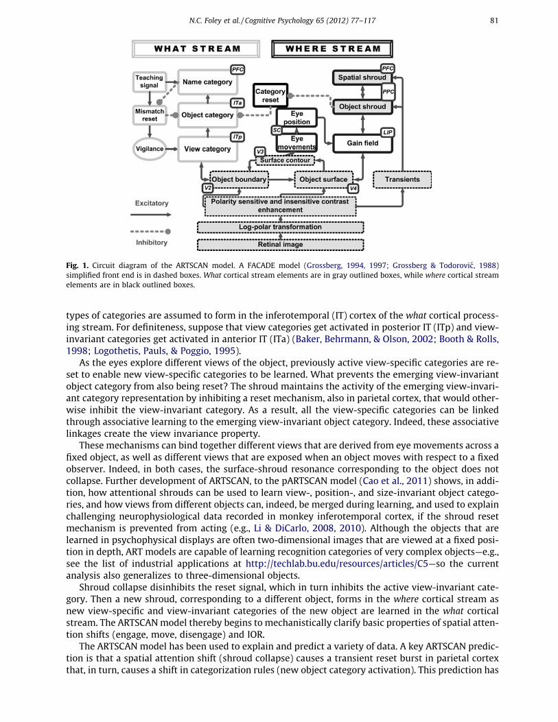
2. Spatial attention in the regulation of invariant object category learning
How does the brain learn to recognize an object from multiple viewpoints while scanning a scenewith eye movements? How does the brain avoid the problem of erroneously classifying parts of dif-ferent objects together? How are attention and eye movements coordinated to facilitate object learn-ing? The dARTSCAN model builds upon the ARTSCAN model (Fig. 1; Cao et al., 2011; Fazl et al., 2009;Grossberg, 2007, 2009), which showed how the brain can learn view-invariant object representationsunder free viewing conditions. The ARTSCAN model proposes how an object’s pre-attentively formedsurface representation in cortical area V4 generates a form-fitting distribution of spatial attention, or‘‘attentional shroud’’ (Tyler & Kontsevich, 1995), in the parietal cortex of the where cortical stream. Allsurface representations dynamically compete for spatial attention to form a shroud. The winningshroud remains active due to a surface-shroud resonance that is supported by positive feedback be-tween a surface and its shroud, and that persists during active scanning of the object with eyemovements.
A full understanding of the structure of surface-shroud resonances will require an analysis of howan object’s distributed, multiple-scale, 3D boundary and surface representations in prestriate corticalareas such as V4 (e.g., Fang & Grossberg, 2009; Grossberg, Kuhlmann, & Mingolla, 2007; Grossberg,Markowitz, & Cao, 2011; Grossberg & Yazdanbakhsh, 2005) activate parietal cortex in such a way thatdifferent aspects of the boundary and surface representations can be selectively attended. The simplevisual stimuli used in the psychophysical experiments that are explained in this article can be simu-lated with correspondingly simple surface-shroud resonances. Once such a shroud is activated, it reg-ulates eye movements and category learning about the attended object in the following way.
The first view-specific category to be learned for the attended object also activates a cell populationat a higher processing stage. This cell population will become a view-invariant object category. Both
80 N.C. Foley et al. / Cognitive Psychology 65 (2012) 77–117
Author's personal copy
types of categories are assumed to form in the inferotemporal (IT) cortex of the what cortical process-ing stream. For definiteness, suppose that view categories get activated in posterior IT (ITp) and view-invariant categories get activated in anterior IT (ITa) (Baker, Behrmann, & Olson, 2002; Booth & Rolls,1998; Logothetis, Pauls, & Poggio, 1995).
As the eyes explore different views of the object, previously active view-specific categories are re-set to enable new view-specific categories to be learned. What prevents the emerging view-invariantobject category from also being reset? The shroud maintains the activity of the emerging view-invari-ant category representation by inhibiting a reset mechanism, also in parietal cortex, that would other-wise inhibit the view-invariant category. As a result, all the view-specific categories can be linkedthrough associative learning to the emerging view-invariant object category. Indeed, these associativelinkages create the view invariance property.
These mechanisms can bind together different views that are derived from eye movements across afixed object, as well as different views that are exposed when an object moves with respect to a fixedobserver. Indeed, in both cases, the surface-shroud resonance corresponding to the object does notcollapse. Further development of ARTSCAN, to the pARTSCAN model (Cao et al., 2011) shows, in addi-tion, how attentional shrouds can be used to learn view-, position-, and size-invariant object catego-ries, and how views from different objects can, indeed, be merged during learning, and used to explainchallenging neurophysiological data recorded in monkey inferotemporal cortex, if the shroud resetmechanism is prevented from acting (e.g., Li & DiCarlo, 2008, 2010). Although the objects that arelearned in psychophysical displays are often two-dimensional images that are viewed at a fixed posi-tion in depth, ART models are capable of learning recognition categories of very complex objects—e.g.,see the list of industrial applications at http://techlab.bu.edu/resources/articles/C5—so the currentanalysis also generalizes to three-dimensional objects.
Shroud collapse disinhibits the reset signal, which in turn inhibits the active view-invariant cate-gory. Then a new shroud, corresponding to a different object, forms in the where cortical stream asnew view-specific and view-invariant categories of the new object are learned in the what corticalstream. The ARTSCANmodel thereby begins to mechanistically clarify basic properties of spatial atten-tion shifts (engage, move, disengage) and IOR.
The ARTSCAN model has been used to explain and predict a variety of data. A key ARTSCAN predic-tion is that a spatial attention shift (shroud collapse) causes a transient reset burst in parietal cortexthat, in turn, causes a shift in categorization rules (new object category activation). This prediction has
Fig. 1. Circuit diagram of the ARTSCAN model. A FACADE model (Grossberg, 1994, 1997; Grossberg & Todorovic, 1988)simplified front end is in dashed boxes. What cortical stream elements are in gray outlined boxes, while where cortical streamelements are in black outlined boxes.
N.C. Foley et al. / Cognitive Psychology 65 (2012) 77–117 81

Author's personal copy
been supported by experiments using rapid event-related functional magnetic resonance imaging(fMRI; Chiu & Yantis, 2009). Positive feedback from a shroud to its surface is predicted to increasethe contrast gain of the attended surface, as has been reported in both psychophysical experiments(Carrasco, Penpeci-Talgar, & Eckstein, 2000) and neurophysiological recordings from cortical areasV4 (Reynolds, Chelazzi, & Desimone, 1999; Reynolds & Desimone, 2003; Reynolds, Pasternak, & Desi-mone, 2000). In addition, the surface-shroud resonance strengthens feedback signals between the at-tended surface and its generative boundaries, thereby facilitating figure-ground separation of distinctobjects in a scene (Grossberg, 1994; Grossberg & Swaminathan, 2004; Grossberg & Yazdanbakhsh,2005; Kelly & Grossberg, 2000).
In particular, surface contour signals from a surface back to its generative boundaries strengthenconsistent boundaries, inhibit irrelevant boundaries, and trigger figure-ground separation. Whenthe surface contrast is enhanced by top-down spatial attention as part of a surface-shroud resonance,its surface contour signals (which are contrast-sensitive) become stronger, and thus its consistentboundaries become stronger as well, thereby facilitating figure-ground separation. This feedbackinteraction between surfaces and boundaries via surface contour signals is predicted to occur fromV2 thin stripes to V2 pale stripes.
Corollary discharges of these surface contour signals are predicted to be mediated via cortical areaV3A (Caplovitz & Tse, 2007; Nakamura & Colby, 2000) and to generate saccadic commands that arerestricted to the attended surface (Theeuwes, Mathot, & Kingstone, 2010) until the shroud collapsesand spatial attention shifts to enshroud another object.
Why is it plausible, mechanistically speaking, for surface contour signals to be a source of eyemovement target locations, and for these commands to be chosen in cortical area V3A and beyond?It is not possible to generate eye movements that are restricted to a single object until that objectis separated from other objects in a scene by figure-ground separation. If figure-ground separation be-gins in cortical area V2, then these eye movement commands need to be generated no earlier than cor-tical area V2. Surface contour signals are plausible candidates from which to derive eye movementtarget commands because they are stronger at contour discontinuities and other distinctive contourfeatures that are typical end points of saccadic movements. ARTSCAN proposed how surface contoursignals are contrast-enhanced at a subsequent processing stage to select the largest signal as the nextsaccadic eye movement command. Cortical area V3A is known to be a region where vision and motorproperties are both represented, indeed that ‘‘neurons within V3A. . .process continuously movingcontour curvature as a trackable feature. . .not to solve the ‘‘ventral problem’’ of determining objectshape but in order to solve the ‘‘dorsal problem’’ of what is going where’’ (Caplovitz & Tse, 2007, p.1179).
Last but not least, ARTSCAN quantitatively simulates key data about reaction time costs for atten-tion shifts between objects relative to those within an object (Brown & Denney, 2007; Egly et al.,1994). However, ARTSCAN cannot simulate all cases in the Brown and Denney (2007) experiments.Nor was ARTSCAN used to simulate the UFOV task or crowding.
3. Sustained and transient attention, spatial priming, and useful-field-of-view
The dARTSCANmodel incorporates three key additional processes to explain a much wider range ofdata about spatial attention:
(1) The breadth of spatial attention (‘‘multifocal attention’’) can vary in a task-selective and learning-responsive way (Alvarez, Horowitz, Arsenio, Dimase, & Wolfe, 2005; Cavanagh & Alvarez,2005; Cave, Bush, & Taylor, 2010; Franconeri, Alvarez, & Enns, 2007; Green & Bavelier, 2003;Jans, Peters, & De Weerd, 2010; McMains & Somers, 2004, 2005; Muller, Malinowski, Gruber,& Hillyard, 2003; Pylyshyn & Storm, 1988; Pylyshyn et al., 1994; Scholl, Pylyshyn, & Feldman,2001; Tomasi, Ernst, Caparelli, & Chang, 2004; Yantis & Serences, 2003). The current model pro-poses how spatial attention can be distributed across multiple objects simultaneously, whilestill being compatible with the strictly unifocal attention in ARTSCAN.
82 N.C. Foley et al. / Cognitive Psychology 65 (2012) 77–117

Author's personal copy
Below we illustrate how flexibly altering the maximal distribution of spatial attention can be voli-tionally regulated by the basal ganglia using an inhibitory mechanism that is predicted to behomologous to the one that regulates visual imagery (Grossberg, 2000b) and the storage ofsequences of items in working memory (Grossberg & Pearson, 2008).(2) Both sustained surface-driven spatial attention and transient motion-driven spatial attention inter-
act to control maintenance and shifts of spatial attention (Fig. 2). A large experimental literatureattempts to anatomically and functionally differentiate components of sustained and transientattention (Dosenbach, Fair, Cohen, Schlaggar, & Petersen, 2008; Dosenbach et al., 2007; Gee,Ipata, Gottlieb, Bisley, & Goldberg, 2008; Gottlieb, Kusunoki, & Goldberg, 2005; Hillyard, Vogel,& Luck, 1998; Ploran et al., 2007; Reynolds, Alborzian, & Stoner, 2003; Yantis & Jonides, 1990;Yantis et al., 2002). The ARTSCAN model only incorporates sustained, surface-driven attentionnecessary for view-invariant category learning. On the other hand, as noted in Section 1, ART-SCAN also posits a transient reset signal that coordinates shifts of spatial attention with shifts ofcategorization rules.
(3) In addition to parietal cortex, prefrontal cortex plays a role in priming spatial attention (Fig. 2).Many experiments document such a role for prefrontal cortex (Boch & Goldberg, 1989; Gold-man & Rakic, 1979; Kastner & Ungerleider, 2000; Ungerleider & Haxby, 1994; Zikopoulos & Bar-bas, 2006). The ARTSCAN model is agnostic about the role of PFC in deploying spatial attention.
The above three sets of processes, working together, enable our model to explain a much largerrange of data about how attention and recognition interact, notably to better characterize howmultifocal attention can help to track and recognize multiple objects in familiar scenes, and focalattention can support view-invariant object category learning for unfamiliar objects.
The large cognitive literature about multifocal attention (Cavanagh & Alvarez, 2005; Pylyshyn et al.,1994) produced concepts such as Fingers of Instantiation (FINST; Pylyshyn, 1989, 2001), Sprites (Cav-anagh et al., 2001), and situated vision (Pylyshyn, 2000; Scholl, 2001) which have in common an ideathat objects which are not being focally attended are nonetheless spatially represented in the atten-tional system (Scholl, 2001). This is necessary to allow rapid shifts of attention between objects, totrack several identical objects simultaneously, and to underpin visual orientation by marking the allo-centric coordinates of several objects in a scene (Mathot & Theeuwes, 2010a; Pylyshyn, 2001). Suchconcepts are consistent with the daily experience that scenic features outside our focal attention donot disappear.
One challenge to extending the ARTSCAN shroud architecture is how these ideas might be inte-grated into a system that continues to allow the learning of view-invariant object categories over sev-eral saccades, which requires that attention be object-based and unifocal. Multiple shrouds cannot
Fig. 2. The recurrent, feedforward and feedback connections of object and spatial shrouds. Hemifield separation is not shown.
N.C. Foley et al. / Cognitive Psychology 65 (2012) 77–117 83

Author's personal copy
exist during multi-saccade exploration because saccades from one attended object to another wouldfail to reset the active view-invariant object category, causing distinct objects to be falsely conflated asparts of a single object. Moreover, since multiple surfaces would be recipients of contrast gain fromshroud-to-surface feedback, peripheral parts of the object being learned and other objects nearbycould similarly be conflated. This suggests that there are at least two model states in which stableshrouds can form. In one, unifocal attention can be maintained on a single surface over multiple sac-cades, allowing the learning of view-invariant object categories. In the other, multiple shrouds cansimultaneously coexist, at a lower intensity not sufficient to gate multi-saccade learning, but allowingrapid recognition of familiar objects and attention to be deployed on multiple objects, regardless offamiliarity, between saccades. As noted in item (1) above, volitional control of inhibition in the atten-tion circuit, likely mediated by the basal ganglia (Brown, Bullock, & Grossberg, 2004), allows unfamil-iar objects that have weak shrouds to be foveated, followed by an increase in competition to create asingle strong shroud that can support learning of a view-invariant object category.
As noted in item (3) above, another challenge, for both the ARTSCAN model, as well as other biasedcompetition models (Itti & Koch, 2001; Lee & Maunsell, 2009; Reynolds & Heeger, 2009; Treisman &Gelade, 1980; Wolfe et al., 1989), is to understand the mechanism of attention priming. Brief transientcues orient attention to an area of a visual scene, which leads to improved behavioral performance if atask-relevant stimulus is presented at the same position within several hundred milliseconds (Desi-mone & Duncan, 1995; Posner & Petersen, 1990). If the experiment continues and a second task-rel-evant stimulus appears at the same position after the first has disappeared or is no longer task-relevant, it takes longer for that area to be attended again, due to inhibition-of-return (Grossberg,1978a, 1978b; Koch & Ullman, 1985; Posner et al., 1982; see Itti and Koch (2001) for a review). ART-SCAN and biased competition models can account for IOR, but they do not incorporate a mechanismthat can explain how a brief bottom-up input onset can prime visual attention in the absence of inter-vening visual stimuli for several hundred milliseconds. But such priming seems to be necessary to ex-plain an extension by Brown and Denney (2007) of the two-object cueing task first used by Egly et al.(1994); see Fig. 3. Priming by prefrontal cortex is incorporated into the model as a natural complementto allowing multifocal attention within the model parietal cortex (Fig. 2).
We hereby propose that a hierarchy of attentional shrouds in parietal and prefrontal cortex (Figs. 1and 2) can smoothly switch between behavioral modes, while being sensitive to both transient eventonsets and offsets, as well as to sustained shroud-mediated spatial attention, and which we will em-ploy in order to explain key properties of cognitive models of multifocal attention, such as FINST (Pyly-shyn, 1989, 2001).
The idea of a hierarchy of attention is consistent with accumulating anatomical and physiologicalevidence that magnocellular pathways play a role in priming object recognition in inferotemporal cor-tex through orbitofrontal cortex (Bar et al., 2006; Zikopoulos & Barbas, 2006). Evidence for multipleattention representations has also been found by mapping retinotopy in the visual system using fMRI,which has shown multiple, attention-sensitive maps in connecting areas of the intra-parietal sulcus(Silver, Ress, & Heeger, 2005; Swisher, Halko, Merabet, McMains, & Somers, 2007), as well as in reti-notopic and head-centric representations in other areas of parietal cortex and areas of prefrontal cor-tex such as the frontal eye fields and dorsolateral prefrontal cortex (Saygin & Sereno, 2008).
The lower shroud level in the hierarchy, the object shroud layer, behaves similarly to the shroudsfound in the ARTSCANmodel (Fazl et al., 2009; PPC in Figs. 1 and 2). Neurons in this layer can resonatestrongly with a single surface to gate learning in the what stream to allow the formation of view-invariant object categories. Multiple ensembles of neurons in the object shroud layer can also weaklyresonate with several surfaces, allowing multifocal attention and rapid recognition of familiar scenes.Object shroud neurons can thus exist in two different regimes, which alternate reactively in responseto changing visual stimuli, or can be volitionally controlled by modifying the inhibitory gain throughthe basal ganglia (Brown et al., 2004; Matsuzaki, Kyuhou, Matsuura-Nakao, & Gemba, 2004; Xiao, Zik-opoulos, & Barbas, 2009). The first regime was studied in the original ARTSCAN model, where a singlehigh-intensity shroud covers an object surface and gates learning during a multi-saccadic explorationof an unfamiliar object. The second regime allows multiple low-intensity object shrouds to existsimultaneously, supporting rapid recognition of a familiar scene’s ‘‘gist’’. Gist was modeled in the ART-SCENE model (Grossberg & Huang, 2009; Huang & Grossberg, 2010) as a large-scale texture category.
84 N.C. Foley et al. / Cognitive Psychology 65 (2012) 77–117

Author's personal copy
Since object shroud neurons gate learning regimes lasting several seconds, they provide sustainedattention and are slow to respond to changes in a scene.
In contrast to this property, reaction times in response to scenes that contain several objects arereduced if a task-relevant stimulus appears at one of the object positions within a few hundred mil-liseconds (Desimone & Duncan, 1995; Posner & Petersen, 1990). The attentional shrouds of the ART-SCAN model require a surface to be present to sustain a shroud-surface resonance. If that surfacedisappears, its corresponding shroud will collapse and another shroud will form over the next mostsalient object in a scene, which will start a new learning regime. This means that if a location is brieflycued, attention will shift once the cue disappears and the cued location will immediately be subject toIOR, which is inconsistent with attentional priming. Also in the ARTSCAN model, spatial attention isnot preferentially sensitive to the appearance of a new object of equal contrast to the existing objectsin a scene, unless the other objects in a scene had already been attended. Thus both attentional prim-ing and fast reactions to cue changes are not adequately represented in the ARTSCAN model.
These ARTSCAN properties derive from that model’s exclusive consideration of focal sustainedattention and how it shifts through time. The dARTSCANmodel also incorporates, and elaborates func-tional roles for, inputs that are sensitive to stimulus transients (Fig. 2, MT; also see Section 5.6 and
Fig. 3. Brown and Denney (2007) data and reaction time (RT) simulations. Each experimental case is shown, with the fourcolumns corresponding to what is seen in each display segment. Experimental RTs (black) and simulated RTs (gray) are shownat the far right. [Data reprinted with permission from Brown and Denney (2007).]
N.C. Foley et al. / Cognitive Psychology 65 (2012) 77–117 85

Author's personal copy
Appendix A.6), consistent with models of motion perception (Berzhanskaya, Grossberg, & Mingolla,2007) and models that combine transient and sustained contributions to spatial attention (e.g., Gross-berg, 1997). Moreover, the model proposes how such transient inputs can contribute both to the for-mation of shrouds in the parietal cortex, and to the development of top-down priming from theprefrontal cortex.
Accordingly, in the higher level of the hierarchy, the spatial shroud level is formed by ensembles ofneurons primarily driven by transient signals from the where cortical processing stream (see Sections5.8 and Appendix A.8), notably signals due to object appearances, disappearances, and motion (PFC,Fig. 2). Unlike object shrouds, several shrouds in the spatial shroud layer can coexist at all times. Thisallows objects unattended in the object shroud layer to maintain spatial representations, allowing ra-pid (between saccades) shifts of attention, transient interruption of multiple-view learning, and atten-tional priming of locations at which an object recently disappeared or was occluded. Recurrentfeedback allows a spatial shroud to stay active at a cued location for several hundred milliseconds,regardless of whether a surface is present at the location. Spatial shroud neurons also improve reac-tion times to transient or otherwise highly salient stimuli through top-down feedback onto objectshroud neurons.
Top-down priming feedback enables quantitative simulation of reaction time differences in severalcases presented in Brown and Denney (2007), particularly the LVal case (Fig. 3, row 5) where there is avalid location cue. As described in greater detail in Section 6.1.1, without priming, there would be noattentional representation of the cue through the interstimulus interval (ISI). In particular, as notedabove, the original ARTSCAN model requires a surface to be present to sustain a shroud-surface reso-nance. This means that if a location is briefly cued, attention will shift once the cue disappears, and thecued location will immediately be subject to IOR. Prefrontal attentional priming overcomes this defi-ciency by sustaining an attentional prime throughout the ISI.
4. Individual differences and the basal ganglia: useful-field-of-view and RT
There are systematic individual differences in how attention is deployed and maintained (Green &Bavelier, 2003, 2006a, 2006b, 2007; Richards, Bennett, & Sekuler, 2006; Scalf et al., 2007; Sekuler &Ball, 1986; Tomasi et al., 2004). Green and Bavelier have, in particular, compared how video gameplayers (VGPs) and non-video game players (NVGPs) perform in spatial attention tasks. Another lineof research examined the differences in performance in the same individual under different condi-tions, such as before and after a training session (Green & Bavelier, 2003, 2006a, 2006b, 2007; Richardset al., 2006; Scalf et al., 2007; Tomasi et al., 2004). Finally, on psychophysics tasks such as the two ob-ject-cueing task first used by Egly et al. (1994), bootstrapping methods have been used to study be-tween-subject differences on the same task, rather than the average population response todiffering stimuli (Roggeveen et al., 2009). The dARTSCAN model proposes how these multiple modesof behavior and performance differences between individuals may arise from differences in inhibitorygain, mediated by the basal ganglia. We suggest how volitional or learning-dependent variations inthe strength of inhibition that governs attentional processing may explain individual differences. Asnoted above, variations of this mechanismmay be used in multiple brain systems to control other pro-cesses, such as visual imagery (Grossberg, 2000b) and working memory storage and span (Grossberg &Pearson, 2008).
VGPs have been found to have superior performance in several tasks involving visual attention,including flanker recognition, multiple object tracking (MOT), useful-field-of-view (UFOV), attentionalblink, subitizing, and crowding (Green & Bavelier, 2003, 2006a, 2006b; Sekuler & Ball, 1986). Addition-ally, VPG’s have better visual acuity than NVGPs (Green & Bavelier, 2007). Most of these performanceimprovements have also been found when a group of NVGPs trains on action video games for variousperiods of time typically 30–60 h; e.g., Green and Bavelier (2003). This indicates that playing actionvideo games causes substantial changes in the basic capacity and performance of the visual attentionsystem that is not specific to any individual or subpopulation. In some tasks, such as UFOV, similarchanges have been found as the result of aging (Richards et al., 2006; Scalf et al., 2007; Sekuler & Ball,1986).
86 N.C. Foley et al. / Cognitive Psychology 65 (2012) 77–117

Author's personal copy
As described above, the object shroud layer can switch between unifocal and multifocal attentionthrough volitional control of inhibitory gain mediated by the basal ganglia. The model predicts thattraining through action video games increases the range of volitional control available in both shroudlayers. Similarly, aging reduces the range of inhibitory control in both shroud layers. This allows VGPs(and the young) to spread their attention more broadly in the spatial shroud player, and increases thecapacity of the object shroud attention layer so that the same object causes less inhibition than itwould in a NVGP. We test this prediction using the UFOV task. The results are shown in Section 6.2and Fig. 6 below. Roggeveen et al. (2009) revisited the two-object cueing task of Egly et al. (1994),who found that people respond faster when an invalid cue is presented on the same object as the tar-get, than when the cue is presented on a different object. Roggeveen et al. (2009) reported that, whilepart of their subject pool showed the same effect, another part showed the opposite effect and re-sponded faster when the invalid cue was on the other object. They also found that a valid cue improvesreaction time in nearly all subjects. These data are challenging to explain in an object-based attentionparadigm because, if the object is attended, how can a target on an unattended object lead to a fasterresponse? The current model is able to produce both effects by varying the relative strength of theslower, surface-(object shroud) resonance, and the faster, transient-driven response of spatial shrouds.The model predicts that the rate with which attention spreads on a surface varies for each individual,which is attributed to different relative gains between the surface-(object shroud) resonance and the(object shroud)-(spatial shroud) resonance. For those individuals who respond faster when the invalidcue is on the other object, attention in the object shroud layer has not yet completely covered the cuedobject. Since inhibition in the object shroud attention layer is distance-dependent, the area of a surfaceimmediately beyond the leading edge of the spread of attention is actively suppressed relative to theun-cued object (see Figs. 6 and 7, Section 6.1). This parametric modification leads to the model predic-tion that varying the cue and ISI duration will shift the proportion of subjects who exhibit same-objectpreference. The model also predicts that altering the visual geometry of the display or the strength ofthe cue will alter the width and slope of the distribution of individual differences.
5. Model description
The dARTSCAN extension of the ARTSCAN model focuses on the where stream side of the originalmodel (Fig. 1). Both models share similar boundary and surface processing. The current model uses asimpler approximation of cortical magnification to reduce the computational burden in simulationdue to adding transient cells, prefrontal priming, and variable field-of-view. As briefly summarizedabove, both parietal and prefrontal shrouds are now posited: object shrouds, which are similar tothe original ARTSCAN shroud representation and are primarily driven by surface signals, and spatialshrouds, which are primarily driven by transient signals (Fig. 2).
5.1. Cortical magnification
Visual representations in the early visual system are subject to cortical magnification, which hasoften been simulated using a log-polar approximation (Fazl et al., 2009; Polimeni et al., 2006). Work-ing with models that include cortical magnification creates several complications. Because corticallymagnified images are spatially variant, depending on the center of gaze, fixed convolutions or cen-ter-surround processing create effects that vary with eccentricity (Bonmassar & Schwartz, 1997). Inaddition, log-polar transformations do not fit neatly into the neighborhood relations of a matrix,which is the most convenient data structure for representing images and layers of neuronscomputationally.
It is possible, however, to maintain cortical magnification as a function of eccentricity while alsokeeping the neighborhoods of the matrix form, by approximating the central portion of the visual fieldusing radial symmetric compression instead of a log-polar mapping. This has the advantage of simpli-fying computation of the model without ignoring the basic geometry reflected in the anatomy of thevisual system (see Appendix A.1).
N.C. Foley et al. / Cognitive Psychology 65 (2012) 77–117 87
Author's personal copy
Given that model preprocessing before cortical activation is highly simplified, it is assumed that themodel retina, rather than cortical area V1, already samples input images in a spatially-variant manner,using symmetric log compression to approximate cortical magnification, so that objects near the foveahave magnified representations and objects near the periphery have compressed representations. Thisis important in the model to bias attention towards foveated stimuli, which have correspondingly lar-ger representations. Model retinal cell activities are normalized by receptive field size, and serve asinput to the model lateral geniculate nucleus (LGN).
5.2. Hemifield independence
Model interactions also exhibit hemifield independence, which is consistent both with the anatom-ical separation of processing as well as behavioral observations (Alvarez & Cavanagh, 2005; Luck, Hill-yard, Mangun, & Gazzaniga, 1989; Mathot, Hickey, & Theeuwes, 2010; Swisher et al., 2007; Youakim,Bender, & Baizer, 2001). Hemifield independence is implemented by using different sets of connectionweights to control the strength of connections between neurons in the same hemifield, and neurons ofopposite hemifields (see Appendix A.2, Eqs. (10)–(13)). Left and right hemifield representations useone set of distance-dependent connection weights between neurons that are in the same hemifieldof the respective layer, or projecting layers. They use a different set of distance-dependent connectionweights for neurons that are in the opposite hemifield of the same layer, or projecting layers. The con-nection strength between neighboring neurons is weighted near network boundaries to normalize thetotal input for each neuron (Grossberg & Hong, 2006; Hong & Grossberg, 2004). Thus, there are noboundary artifacts, either near the vertical meridian or the edges of the visual field.
5.3. LGN polarity-sensitive ON and OFF cells
The model LGN normalizes contrast of the input pattern using polarity-sensitive ON cells that re-spond to input increments and OFF cells that respond to image decrements. ON and OFF cells obey cellmembrane, or shunting (Eqs. (1)–(3)), equations that receive retinal outputs within on-center off-sur-round networks that join other ON and OFF cells, respectively (Eqs. (14) and (15)). These single-oppo-nent cells output to a layer of double-opponent ON and OFF cells in the what cortical processingstream (Eqs. (18) and (19)), as well as to transient cells in the where cortical processing stream(Eqs. (30)–(32)).
5.4. Boundaries
The model omits oriented simple cell receptive fields and various properties of ocularity, disparity-sensitivity, and motion processing that are found in primary sensory cortex, since no inputs used forthe current model simulations require it. Instead, polarity-insensitive complex cells are directly com-puted at each position as a sum of rectified signals from pairs of polarity-sensitive double-opponentON and OFF cells (Eq. (20)).
Object boundaries (Eq. (21)) are computed using bottom-up inputs from complex cells (Eq. (20))that are amplified through a modulatory input from top-down inputs from surface contour cells(see Appendix A.4 and Eq. (23)). Surface contour signals are generated by surfaces that fill-in withinclosed boundaries. They select and enhance the boundaries that succeed in generating the surfacesthat may enter conscious perception, thereby assuring that a consistent set of boundaries and surfacesare formed, while also, as an automatic consequence, initiating the figure-ground separation of objectsfrom one another (Grossberg, 1994, 1997).
Surface contours are generated by contrast-sensitive networks at the boundaries of successfullyfilled-in surfaces; that is, surfaces which are surrounded by a connected boundary and thus do not al-low their lightness or color signals to spill out into the scenic background. During 3-D vision and fig-ure-ground separation, not all boundaries are connected (Grossberg, 1994). However, in response tothe simplified input images that are simulated in this article, all object boundaries are connectedand cause successful filling-in. As a result, surface contours are computed at all object boundaries,and can strengthen these boundaries via their positive feedback. Moreover, when the contrast of a sur-
88 N.C. Foley et al. / Cognitive Psychology 65 (2012) 77–117
Author's personal copy
face is increased by feedback from an attentional shroud, the surface contour signals increase, so thestrength of the boundaries around the attended surface increase also.
More complex boundary computations, such as those in 3D laminar visual cortical models (e.g., Cao& Grossberg, 2005; Fang & Grossberg, 2009) can be added as the model is further developed to processmore complex visual stimuli, without undermining the current results.
5.5. Surfaces
Bottom-up inputs from double-opponent ON cells (Eq. (18)) trigger surface filling-in via a diffusionprocess (Eq. (26)) which is gated by object boundaries (Eq. (28)) that play the role of filling-in barriers(Grossberg, 1994; Grossberg & Todorovic, 1988). The ON cell inputs are modulated by top-down feed-back from object shrouds (Eq. (33)) that increase contrast gain during a surface-(object shroud) reso-nance. Such a resonance habituates through time in an activity-dependent way (Eq. (29)), therebyweakening the contrast gain caused by (object shroud)-mediated attention. Winning shrouds will thuseventually collapse, allowing new surfaces to be attended and causing IOR.
Filled-in surfaces generate surface contour output signals through contrast-sensitive shunting on-center off-surround networks (Eq. (23)). As noted above, surface contour signals provide feedback toboundary contours, which increases the strength of the closed boundary representations that inducedthe corresponding surfaces, while decreasing the strength of boundaries that do not form surfaces.
Although surface filling-in has often been modeled by a diffusion process since computationalmodels of filling-in were introduced by Cohen and Grossberg (1984) and Grossberg and Todorovic(1988), Grossberg and Hong (2006) have modeled key properties of filling-in using long-range hori-zontal connections that operate a thousand times faster than diffusion.
5.6. Transient inputs
Where stream transient cells in cortical area MT are modeled using a leaky integrator (Eq. (30)).Transient cells receive bottom-up input from double-opponent ON cells (Eq. (18)) proportional tothe ratio of the contrast increment between previous and current stimuli at their position (Eq. (31))for a brief period (Eq. (32)) after any change. Such ratio contrast sensitivity is a basic property of re-sponses to input increments in suitably defined membrane, or shunting, equations (Grossberg, 1973,1980a, 1980b). Any increment in contrast will trigger a transient cell response. After the period of sen-sitivity ends, transient activity quickly decays. OFF channel transient cells were omitted since onlystimuli brighter than the background were simulated.
5.7. Object shrouds
The model where cortical stream enables one or several attentional shrouds to form in the objectshroud layer, thereby supporting two different modes of behavior. The first, in which only a singleshroud forms, allows an object shroud to perform the same role as in the original ARTSCAN model,gating learning when a sequence of several saccades explores a single object’s surface to learn aview-invariant object category. The second, where multiple, weaker, shrouds can simultaneouslycoexist, supports conscious perception and concurrent recognition of several familiar object surfaces.
Object shroud neurons (Eq. (33)) receive strong bottom-up input from surface neurons (Eq.(26))and modulatory input from transient cells to help salient onsets capture attention during sustainedlearning (Eq. (30); see Fig. 2). Object shroud neurons also receive top-down habituating (Eq. (38))feedback from spatial shroud neurons (Eq. (39)), as well as recurrent on-center off-surround (Eqs.(35) and (36)) habituating (Eq. (37)) feedback from other object shroud neurons. Recurrent feedbackamong object shroud neurons habituates faster than spatial shroud feedback, which in turn habituatesfaster than feedback onto the surface layer. This combination of feedback produces several importanteffects. The first loop, surface-(object shroud)-surface (Fig. 2), enables a local cue on a surface that hassuccessfully bid for object shroud attention to trigger the filling-in of attention along the entire surface(Eqs. (26)–(28)). Fully enshrouding an object which attracts attention through a local cue is slow com-pared to transient capture of highly salient objects, since it depends on slower surface dynamics. The
N.C. Foley et al. / Cognitive Psychology 65 (2012) 77–117 89

Author's personal copy
second loop, recurrent on-center off-surround feedback in the object shroud layer, allows objectshrouds to compete weakly in the multi-focal case, to provide contrast enhancement, and stronglyin the unifocal case, so that view-invariant object categories may be learned. Once a shroud haswon in the unifocal case, surface-(object shroud) resonance dominates until habituation occurs. Thelevel of competition between object shrouds depends on the inhibitory gain (Eq. (36)), which canbe volitionally controlled through the basal ganglia. The third loop, (object shroud)-(spatialshroud)-(object shroud) enhances responses to salient transient signals, facilitates the spread of objectshroud attention along surfaces, and helps maintain an (object shroud)-surface resonance over thewhole surface, as parts of the object shroud start to habituate, by up-modulating the bottom-up sur-face signal. Once an object shroud that has habituated is out-competed and collapses, it is difficult fora new object shroud to form in the same position until the habituating gates recover, leading to IOR.
5.8. Spatial shrouds
When object shrouds are supporting view-invariant category learning, they must be stable on theorder of seconds, to allow multiple saccades to explore an object (Fazl et al., 2009). The visual systemhowever, is also capable of considerably faster responses, in particular to transient events (Desimone& Duncan, 1995). Spatial shrouds allow the model to respond quickly to transient stimuli when theyare present, without compromising the stability required to support view-invariant object categorylearning in more stable environments (see Fig. 2). This basic fast-slow dynamic also underpins themodel’s explanation of individual difference data in the two-object cueing task, and allows the modelto successfully simulate cases in the Brown and Denney (2007) data which require rapid responses totransient cues and targets, as is explained in Section 3.1.
Spatial shrouds (Eq. (39)) receive bottom-up input from transient neurons (Eq. (30)), and weakerbottom-up input from object shroud neurons (Eq. (40)). Spatial shroud neurons interact via a recurrenton-center off-surround network (Eqs. (41) and (42)) that does not habituate. As a result, multiple spa-tial shrouds can survive for hundreds of milliseconds in relatively stable environments unless they areout-competed by new transients. Spatial shroud cells are always sensitive to salient environmentalstimuli and can mark multiple objects simultaneously, even if these objects are not being activelylearned or recognized, allowing maintenance of allocentric visual orientation consistent with situatedvision (Pylyshyn, 2001). The spatial shroud layer has non-habituating recurrent feedback capable ofmaintaining spatial shroud activity through time, so that an active spatial shroud can persistentlyprime object shroud formation over a surface presented at the corresponding location, unless the ob-ject shroud neurons are deeply habituated, causing IOR. There is no volitional attention from planningareas in the model, although we hypothesize that feedback to the spatial shroud layer might comefrom planning and executive control areas.5.9. Computing model behavioral data
The data sets that are simulated use reaction time and detection thresholds to assess behavioralperformance. We simulate these behavioral outputs by measuring activity levels at regions of interest(ROIs) important for the experimental display. To measure reaction time, we integrate activity in theobject shroud layer over time until it reaches a threshold (chosen for best fit in the 2Val condition),then assume a constant delay between detection and motor output. To measure detection perfor-mance in the UFOV, we use the size of a Weber fraction comparing the level of response in the objectshroud layer for the target and distracters at the end of the masking period as a direct proxy forperformance.
6. Results
6.1. Two-object cueing
The two-object cueing task (Egly et al., 1994) is a sensitive probe to examine the object-based ef-fects of attention. Two versions of the experiment extend the basic two-object task. One version in-cludes one object and non-object positions with the same geometry (Brown & Denney, 2007). The
90 N.C. Foley et al. / Cognitive Psychology 65 (2012) 77–117

Author's personal copy
second version shows that the general population effect found in Brown and Denney (2007) for same-object vs. inter-object attention switches is not uniform among all subjects. In both experiments, pre-sentation occurs in four states (see Fig. 3). The first stage, called ‘‘prime,’’ displays two rectangles, equi-distant from the fixation point, equal in size and such that the distance between the two ends of arectangle is the same as the distance between the rectangles (Fig. 3, column 1, rows 1–3). In the casesof one object with possible position cues and targets, only one of the two rectangles is shown (seeFig. 3, column 1, rows 4–9). The rectangles can either cross the vertical meridian, or be presentedin separate hemifields. In the second stage of the experiment (Fig. 3, column 2), one end of a rectangle(or the equivalent location, if there is only one rectangle) is cued, which is followed by the ISI (Fig. 3,column 3). Finally, a target is presented at one of the four possible cue positions (Fig. 3, column 4). Thiscue can be valid to the target or invalid.
The original ARTSCAN model could simulate the order of reaction times in four of the main casespresented in Brown and Denney (2007); namely, the primary cases that illustrate the object cueingadvantage (cases 2Val, InvS, InvD and OtoL in Fig. 3). The hierarchy of attentional interactions betweenPPC and PFC in the dARTSCAN model can simulate all nine cases successfully. This can be done due tothe addition of transient cells, which shorten reaction times for targets, and by replacing the singleshroud layer with the PPC-PFC hierarchy, which allows attention priming and balancing betweenthe dynamics of the fast spatial shroud layer and the slower object shroud layer.
The following cases explain how the model fits the entire data set.
6.1.1. Valid cuesThere are three display conditions in which the cue is valid. From fastest to slowest reaction times,
these are:
(1) One rectangle is presented throughout the experiment, with the cue and the target at the sameposition on the rectangle (1Val; Figs. 3 and 4).
(2) Two rectangles are presented throughout the experiment, with the cue and target at the sameposition (2Val; Figs. 3 and 5).
(3) One rectangle is presented throughout the experiment, with the cue and the target presentedoutside the object (LVal; Figs. 3 and 4).
The 1Val condition has a faster reaction time than the 2Val condition, because the presence of thesecond rectangle bidding for attention adds to the inhibition that the cued object must overcome toresonate with an object shroud. In both the object-valid conditions, there is a surface visible at thelocation of the cue throughout the experiment, and a resonant object shroud is maintained fromcue presentation through target detection. In the LVal case, on the other hand, this does not occur:only a spatial shroud corresponding to the cued location endures through the ISI. While the spatialshroud primes the object shroud when the target appears, the object shroud cannot resonate untila new surface representation is formed. This in turn cannot take place until new boundaries haveformed, something unnecessary when changing the contrast of an existing visible surface. If therewere no spatial shroud present to prime the object shroud, the process would take longer, since afterthe surface representation formed, it would then have to bid for attention against a weak shroud rep-resentation on the rectangle visible throughout the experiment, substantially delaying the formationof a resonant shroud.
6.1.2. Invalid cues: one objectThere are four display conditions in which one rectangle is visible throughout the experiment, but
the cue is invalid. From fastest to slowest in human and model reaction times, these are:
(1) The cue is presented at one end of the (only) rectangle, and the target appears at the far end ofthe rectangle (1Inv; Figs. 3 and 4).
(2) The cue is presented at a position outside the rectangle, and the target is presented at anotherlocation outside the rectangle consistent with the spacing of 1Inv (LtoL; Figs. 3 and 4).
N.C. Foley et al. / Cognitive Psychology 65 (2012) 77–117 91

Author's personal copy
Fig. 4. Simulations of Brown and Denney (2007) one-object cueing data showing the dynamics of the surface, object shroud,and spatial shroud layers. The top row in each panel shows a schematic of input presentations for the one-object cases used tofit the Brown and Denney (2007) data, with colors corresponding to each region of interest (ROI) indicated at the upper leftcorner (not all positions are shown in each plot). In all simulations, the temporal dynamics of surface (S, Eq. (26)), object shroud(AO, Eq. (33)), and spatial shroud (AS, Eq. (39)), variables are shown in the first, second and third columns respectively, for eachROI. Solid, dashed and dotted lines show the target conditions that are defined in the top row. The bottom row of each panelshows the distribution of activity in the same layers at the times (!) and (^). (A) The three cases where a position is cued. Notethat while the object shroud over the cued location falls to zero, the spatial shroud is maintained, thereby priming object shroudformation when the target is valid. (B) The three cases where there is one object, which is cued.
92 N.C. Foley et al. / Cognitive Psychology 65 (2012) 77–117

Author's personal copy
Fig. 5. Simulations from Brown and Denney (2007) two-object cueing data, and individual difference data from Roggeveen et al.(2009) showing surface, object shroud, and spatial shroud dynamics. The top row shows a schematic of input presentations forthe three two-object cases used to fit the Brown and Denney (2007) data, with colors corresponding to each ROI indicated at theupper left corner (not all positions are shown in each plot). In all simulations, the temporal dynamics of surface (S, Eq. (26)),object shroud (AO, Eq. (33)), and spatial shroud (AS, Eq. (39)), variables are shown in the first, second and third columnsrespectively, for each ROI. (A) The surface activity for the 2InvS case (dark blue) starts to increase slightly before targetpresentation, as does the activity of the object shroud. This indicates that the surface is about to be completely enshrouded, andthat active inhibition has been released from the end of the rectangle opposite the cue because it has passed the edge separatingthe inhibitory surround from the excitatory center of the resonating shroud. This set of simulations corresponds to thebehavioral results shown in middle simulation of Fig. 6B (a = 5, Eq. (33)). (B) The surface activity of the case in which 2InvDresponse is faster than 2InvS (green vs. dark blue), corresponding to the far left simulations in Fig. 6B (a = 3, Eq. (33)). Note thatthe bubble of inhibition (blue curve, object shroud layer is less than 0 and decreasing) corresponding to the 2InvS target isforming when the target is presented. (C) The surface activity of the case in which 2InvS response is faster than 2InvD (dark bluevs. green), corresponding to the far right simulations in Fig. 6B (a = 7, Eq. (33)). Note that surface-(object shroud) resonance hasenshrouded the entire object prior to the InvS target being presented. (For interpretation of the references to color in this figurelegend, the reader is referred to the web version of this article.)
N.C. Foley et al. / Cognitive Psychology 65 (2012) 77–117 93

Author's personal copy
(3) The cue is presented at a position outside of the rectangle, and the target within it (LtoO; Figs. 3and 4).
(4) The cue is presented in the rectangle, and the target is presented at a position outside of it(OtoL; Figs. 3 and 4).
Condition 1Inv has the quickest reaction time because a strong object shroud has spread over thecued object, facilitating detection. It is slower than the valid case because un-cued portions of the at-tended object lack a strong spatial shroud in addition to the strong object shroud. The more interestingcases are the middle two: why should a target at an invalid position be detected faster than an inval-idly-cued target on an un-cued, but visible, object that has a weak object shroud formed over it? It isbecause the model is sensitive to transient events. An existing (object shroud)-surface resonance,especially one supporting view-invariant object category learning, is difficult to break. It can be brokenthrough inhibition created by a competing shroud, or by exhausting its habituating gates. The modelresponds locally to transient effects as a function of the contrast ratio in the surround. This contrast-sensitive response is larger when a target appears against the background of the display, than on arectangle of the display. This implies that a relatively intense spatial shroud forms on the target inthe LtoL case, which allows a rapid where stream transient input to create a strong (object shroud)-(spatial shroud) resonance. This process can occur faster than more modest contrast increment onthe weakly attended surface in the LtoO, which will continue to be supported by a slower surface-(ob-ject shroud) resonance. This transient-activated shroud hierarchy does not provide the same level ofbenefit in the OtoL case, however, since in the OtoL case a strong object shroud (rather than a weakone) has formed over the object because it was cued, thereby creating a much higher hurdle toovercome.
6.1.3. Invalid cues: two-objects and individual differencesThe classic finding of Egly et al. (1994) supporting object-based attention is that, when there are
two identical rectangles presented at a distance equal to their length throughout the experiment,and one end of one rectangle is cued, reaction times occur in the following order:
(1) 2Val, described above (Figs. 3 and 5).(2) A target appearing on the other end of the same rectangle (InvS; Figs. 3 and 5).(3) A target appearing at the same end on the other rectangle (InvD; Figs. 3 and 5).
Brown and Denney (2007) replicated this finding by measuring mean reaction times for 30 subjects(Fig. 3). Roggeveen et al. (2009) re-examined the paradigm using a variant of this task (Moore et al.,1998) and focused on the object-based attention for each individual, rather than over the populationas a whole. They found that about 18% of individuals had significantly better reaction times for InvSthan InvD, while another 18% of individuals had a significant reverse effect, preferring InvD to InvS.The rest of the subjects showed smaller differences in both directions, creating a fairly smooth distri-bution. Nearly all (96%) of subjects reacted more quickly to a valid cue.
This variant of the task requires discrimination between a target and distracters, rather than simpledetection (Moore et al., 1998), which means that in all invalid trials, there is a distracter at the cuedlocation. This is the likely cause of the comparatively large size of reaction time differences, about200 ms faster for a valid cue, compared to the detection paradigm used in Brown and Denney(2007), which showed 40 ms differences for the same comparison. However, this does not explainwhy some subjects reacted more quickly to an invalidly-cued target on the same rectangle, and somereacted faster to an equidistant invalidly-cued target on the other rectangle.
As noted in Section 4, our model predicts that the difference between individuals who react fasterfor an invalidly-cued target on the same object, and those that do the opposite, is the relative gain be-tween the faster dynamics of (spatial shroud)-(object shroud) resonance, and the slower dynamics ofsurface-(object shroud) resonance. As can be seen in Fig. 6, as the relative strength of the spatialshroud resonance is increased (from left to right on the bottom row), reaction time decreases slightlyacross the board, but massively for InvS. This is because a strong object shroud can spread over therectangle before the target and distracters appear (see Fig. 5C vs. B), which diminishes the effect of
94 N.C. Foley et al. / Cognitive Psychology 65 (2012) 77–117

Author's personal copy
the distracter at the cued location, where there is also a strong spatial shroud. If the shroud has nothad time to spread over the entire bar (as in the left hand case) then there is a bubble of inhibitionat the far end of the cued bar, suppressing attention to the target, while enhancing the distracter. Thispredicted difference between the slower parietal-V4 vs. faster prefrontal–parietal resonances may betestable using rapid, event-related fMRI or EEG/MEG.
Goldsmith and Yeari (2003) employ a variation of the Egly et al. (1994) task with two cue condi-tions: in the ‘exogenous’ condition, the cues appear at one of four locations at the ends of the two rect-angles, while in the ‘endogenous’ task a third rectangle, smaller and oriented towards one of the fourtarget locations appears near the fixation point. Since the model does not explicitly consider the ef-fects of orientation, or learning that an oriented bar may cue a distant location, it is beyond the pur-view of the model to account for the endogenous-target-valid case. However, given the interference ofa highly transient third object (the ‘endogenous’ cue), and the model’s clarification of how spatialattention may learn to be focused or spread, depending on task conditions, it is consistent with thefindings of the current model that there is little reaction time difference between the invalid-same ob-ject and invalid-different object cases, since attention has been drawn away from both of them.
6.2. Useful-field-of-view
The UFOV paradigm measures how widely subjects can spread their attention to detect a briefstimulus that is then masked (Fig. 7A; Green & Bavelier, 2003; Sekuler & Ball, 1986). The task startswith a fixation point, which is followed by a brief (10–30 ms presentation) appearance of 24 additional
Fig. 6. Roggeveen et al. (2009) individual difference data and modeled behavior. (A) The experimental paradigm and individualpreferences for InvS, and InvD targets. (B) Simulated reaction times for two-object conditions (left plot), and reaction timedifferences (right plot) between InvS and InvD conditions, as the (spatial shroud)-(object shroud) resonance changes fromweakest (left, a = 3, Eq. (33)) to strongest (right, a = 7, Eq. (33)) by increasing the response gain to changing inputs in the objectshroud layer. This shifts preference from InvD (bottom) to InvS (top). [Data reprinted with permission from Roggeveen et al.(2009).]
N.C. Foley et al. / Cognitive Psychology 65 (2012) 77–117 95
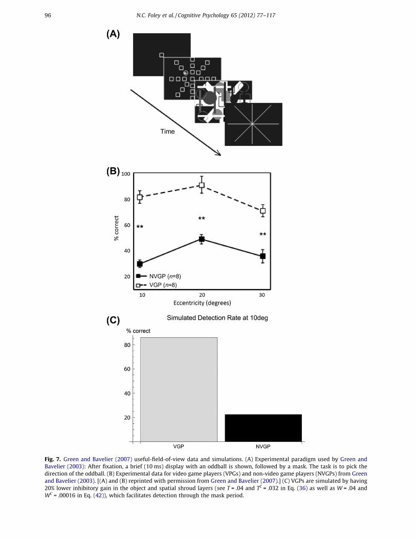
Author's personal copy
Fig. 7. Green and Bavelier (2007) useful-field-of-view data and simulations. (A) Experimental paradigm used by Green andBavelier (2003): After fixation, a brief (10 ms) display with an oddball is shown, followed by a mask. The task is to pick thedirection of the oddball. (B) Experimental data for video game players (VPGs) and non-video game players (NVGPs) from Greenand Bavelier (2003). [(A) and (B) reprinted with permission from Green and Bavelier (2007).] (C) VGPs are simulated by having20% lower inhibitory gain in the object and spatial shroud layers (see T = .04 and TC = .032 in Eq. (36) as well as W = .04 andWC = .00016 in Eq. (42)), which facilitates detection through the mask period.
96 N.C. Foley et al. / Cognitive Psychology 65 (2012) 77–117

Author's personal copy
elements, all but one of which is identical to the fixation point. The elements are arranged in eightspokes, at equally spaced angles and at three eccentricities. A mask then appears, followed by aneight-spoke display of lines. The subject then indicates the direction along which the oddball ap-peared. VGPs perform better at this task than non-VGPs (Fig. 7B).
We simulated a simplified version of this display with a contrast increment oddball, with videogame players having a 20% lower inhibitory gain in the object shroud and spatial shroud layers (seeEqs. (36) and (42)). The results show that just this small change in inhibitory gain in both the PPCand PFC attentional networks has a large effect on the detection performance.
This occurs because the inhibitory gain in the attention layers of the model serves as a resourceconstraint, which helps VGPs detect the location of the oddball in two distinct ways. The spatialshroud layer receives strong transient input, which is excited by the appearance of the target anddistracters. Initially, all the targets and distracters are represented in the spatial shroud layer beforerecurrent feedback causes competition. The initial signal that each element can project to the spatialshroud layer through transient response is determined by the inhibitory gain in that layer. Therefore,VGPs have an early spatial shroud response to the target which is less likely to be washed out by com-petition from distracters and the mask. Inhibitory gain also serves as a resource constraint in the ob-ject shroud layer. However, since shrouds in the object shroud layer require surface resonance, andobject shrouds will expand over any surface that begins resonating with its corresponding shroud,the resource constraint is how many objects can be represented in the object shroud layer. Decreasingthe inhibitory gain the object shroud layer increases the chance that an object shroud can begin res-onating with the target before the mask, rendering its location detectable.
6.3. Crowding
In the crowding paradigm, an object, such as a letter, is visible and recognizable when presented byitself, but it is not recognizable when the letter is presented concurrently with similar flanking letters(Green & Bavelier, 2006a; Levi, 2008; Toet & Levi, 1992). The distance between the target letter and theflanking letters at which the target letter becomes unrecognizable is called the crowding threshold, andis a function of the eccentricity of the target and the flankers, and their relative position (Bouma, 1970,1973; Levi, 2008). A related concept is the spatial resolution of attention (Intriligator & Cavanagh,2001), which is the minimum distance between several simple identical objects, like filled circles, thatallow an observer to covertly move attention from one circle to another based on a set of instructions,without losing track of which circle they should attend. The spatial resolution of attention is also pro-portional to eccentricity, and falls off faster than acuity loss due to cortical magnification (He, Cava-nagh, & Intriligator, 1996; Intriligator & Cavanagh, 2001).
Crowding has been attributed both to pre-attentive visual processing in early visual areas, as wellas to attentional mechanisms (see Levi (2008) for a review). It is likely that it is a combination of thetwo. Some models of early vision, such as LAMINART, predict how flankers can cause either facilitationor suppression of stimuli between them, depending on distance and contrast (Grossberg & Raizada,2000) using interacting pre-attentive grouping and competitive mechanisms. A second proposal isthat larger receptive fields in the what stream capture features from multiple objects and conflatethem. A third proposal is that observers confuse the location of the target and the flankers due to posi-tional uncertainty, but that object-feature binding remains intact. Yet another proposal is that crowd-ing is the result of failed contour completion (Levi, 2008). However, because of the similaritiesbetween crowding and the spatial resolution of attention, measured without the use of flanking ob-jects (He et al., 1996; Intriligator & Cavanagh, 2001; Moore, Elsinger, & Lleras, 2001), it is unlikely thatcrowding is solely due to pre-attentive factors in early visual processing.
Our model has a natural explanation for crowding due to its linkage of where stream spatial atten-tional processes to what stream object learning and recognition processes. In particular, the modelpredicts that when at least three peripherally presented similar objects are nearby, and subject tothe cortical magnification factor, an object shroud forming over one object can spread to another ob-ject, confounding recognition when several objects are covered by a single shroud (see Fig. 8). Said inanother way, when a single shroud covers multiple objects, it defeats figure-ground separation andforces the objects to be processed like one larger unit. This means that there is both featural and posi-
N.C. Foley et al. / Cognitive Psychology 65 (2012) 77–117 97

Author's personal copy
tional confusion, but this is the result of how the shroud forces the failure of object recognition, ratherthan being due to large receptive fields or simple positional uncertainty. This does not occur with justtwo peripheral objects, since the highest intensity part of each object shroud can shift to the most dis-tant extrema of the two objects, and can thereby inhibit the gap between the objects.
In summary, our proposed explanation of crowding exploits the dARTSCAN predictions of howwhere stream processing of attentional shrouds influences what stream learning and selection ofobject recognition categories, and how cortical magnification influences attention within and betweenobject surfaces.
Fig. 8. Green and Bavelier (2007) crowding data and qualitative behavioral simulations. (A) Top panels show a crowding displayand data from Green and Bavelier (2007), which show lower thresholds for VGPs. [Data reprinted with permission from Greenand Bavelier (2007).] (B) Bottom panels show simulations of the object shrouds that form in response to three displays. Thethree inputs are all at the same eccentricity, but the gap between the target and flankers shrinks from top to bottom. At thesmallest gap, a single object shroud forms over multiple objects, so that individual object recognition is compromised.
98 N.C. Foley et al. / Cognitive Psychology 65 (2012) 77–117

Author's personal copy
7. Discussion
The current article introduces three innovations to further develop the original ARTSCAN modelconcept of how where stream spatial attention can modulate what stream recognition learning andattention (Cao et al., 2011; Fazl et al., 2009; Grossberg, 2007, 2009): First, we show how both wherestream transient inputs from object onsets, offsets, and motion can interact with what stream sus-tained inputs from object surfaces to control spatial attention. Second, we show how both parietal ob-ject shrouds and prefrontal spatial shrouds can help to regulate the priming, onset, persistence, andreset of spatial attention in different ways and at different speeds. Third, we show how basal gangliacontrol of inhibition across spatial attentional regions can control focal vs. multifocal attention, eitherthrough fast volitional control or slower task-selective learning. The interactions among these pro-cesses can explain a seemingly contradictory set of data, notably how both same-object and differ-ent-object biases are both compatible with a model founded on object-based attention.
7.1. Comparison with other models
Recent models of attention can be split into several groups. Successors to Feature Integration the-ory (Treisman & Gelade, 1980), such as Guided Search (Wolfe, 2007; Wolfe et al., 1989) and Itti andKoch (2001), rely on biased competition between parallel visual representations, whereby each pre-sentation highlights a specific feature of the visual input. The ARTSCENEmodel, which uses attentionalshrouds in addition to saliency maps, can explain similar data using object-based, rather than pixel-based attention, and regional, rather than local, competition (Grossberg & Huang, 2009; Grossberget al., 1994; Huang & Grossberg, 2010), as well as data that cannot be explained by saliency mapsalone. Using objects and regions as the key substrate of competition allows the model to learn gistsas large-scale texture categories (Grossberg & Huang, 2009) and to explore their importance in pro-cessing natural scenes.
All of these models incorporated attentional circuits defined by top-down, modulatory on-center,off-surround networks whose cells obey shunting dynamics with activity-dependent gain control thatdivisively self-normalizes cell responses (Bhatt, Carpenter, & Grossberg, 2007; Carpenter & Grossberg,1987, 1991, 1993). Related models of how attention interacts with neurons in the visual system, espe-cially V4, used similar properties of gain control, contrast enhancement, and divisive normalization tothe response characteristic of neurons under varying attention conditions (Ghose, 2009; Lee & Maun-sell, 2009; Reynolds & Heeger, 2009). Finally, cognitive theories of attention and object recognitionhave introduced seminal concepts such as FINSTs and Sprites, but are not computational models (Cav-anagh et al., 2001; Pylyshyn, 1989, 2001; Scholl, 2001), with the notable exception of Logan (1996).The ARTSCAN model and its extension in the current article offer computational rigor and new expla-nations of challenging data as manifestations of emergent properties of multiple brain mechanisms asthey interact on multiple time scales in a hierarchy of attentionally-modulated cortical processingstages.
7.2. Transients, sustained resonance, and priming
In the current model, transient inputs can activate sustained priming activity in the spatial shroudlayer. On the other hand, surface inputs slowly create object shroud attention, but sustaining thatattention requires a surface-shroud resonance until eye-movement-contingent inhibition-of-returnand habituative gates cause it to collapse. When such an object shroud collapses, spatial shroudscan continue to prime lower levels, thereby maintaining sensitivity to future salient events.
7.3. Gain control, normalization, and capacity
As noted above, several recent models have focused on describing the effect of attention on neuro-nal responses in visual cortex, particularly V4. In particular, the models of Reynolds and Heeger(2009), Ghose (2009), and Lee and Maunsell (2009) explore the mechanism through which attention
N.C. Foley et al. / Cognitive Psychology 65 (2012) 77–117 99
Author's personal copy
enhances the processing of selected areas of the visual field. They conclude that divisive normalizationusing center-surround processing is the most reasonable model for the effects of attention on V4 neu-rons. The recurrent shunting on-center off-surround networks that form the basis of the object andspatial shroud layers produce divisive normalization. Indeed, the exact form factor for doing this thatwas proposed by Reynolds and Heeger (2009) was earlier used by Bhatt et al. (2007), and variants of ithave often been used to explain attentional dynamics in models by Grossberg and his colleagues (e.g.,Berzhanskaya et al., 2007; Carpenter & Grossberg, 1987, 1991; Gove, Grossberg, & Mingolla, 1995;Raudies & Neumann, 2010) since the original proposal of how shunting dynamics can normalizethe activities of neurons in center-surround networks of neurons was demonstrated in Grossberg(1973). Furthermore, the current model can explain individual differences in attentional capacityand performance, as in individual differences in useful-field-of-view, by varying inhibitory gain ofattention via the basal ganglia.
As with the general theme of normalization, volitional control of inhibitory gain, and thus thestrength of normalization, seems to use a similar mechanism across many parts of the brain. Indeed,such volitional control is predicted to determine whether top-down attention is modulatory or can eli-cit visual imagery in visual cortex (Grossberg, 2000b), and whether bottom-up inputs are stored or notin prefrontal working memory (Grossberg & Pearson, 2008). This latter property may help to explainindividual differences in working memory capacity (Vogel & Machizawa, 2004). Thus, the capacity ofspatial attention and that of visual working memory may share an underlying neural mechanism, al-beit one that is expressed in different brain regions. In all these cases, the simplest hypothesis (Gross-berg, 2000b) is that inhibitory interneurons in cortical layer 4 of the target area are inhibited by thebasal ganglia. This disinhibitory effect can convert a modulatory on-center response, due to balancedexcitation and inhibition, into a driving suprathreshold response. The anatomy and control dynamicsof this predicted volitionally-sensitive gain control mechanism require more experimental study.
7.4. Normalization controls attentional capacity
Cognitive theories of attention and short-term memory fall into two groups, the first, invoked bythe phrase ‘‘the magical number 7,’’ (Miller, 1956) posits a certain number of ‘slots’ which objectsor some other fundamental unit fill (Fukuda, Awh, & Vogel, 2010). The second posits that resource con-straints variably filled by different traits, details, and features limit capacities (Alvarez & Cavanagh,2005; Alvarez & Franconeri, 2007; Alvarez et al., 2005; Verghese & Pelli, 1992). In the latter, objectsare not the fundamental unit, because individuals can track or remember several simple objects,but only a few complex ones.
Objects are an important basis of attention (Fallah, Stoner, & Reynolds, 2007; Kahneman et al.,1992; Lamy & Egeth, 2002; Martinez et al., 2006; Mitchell, Stoner, & Reynolds, 2004; O’Craven, Down-ing, & Kanwisher, 1999; Pylyshyn et al., 1994; Roelfsema et al., 1998; Scholl, 2001; Scholl et al., 2001;Serences, Schwarzbach, Courtney, Golay, & Yantis, 2004; Shomstein & Yantis, 2004). It is difficult totrack one end of a tumbling symmetrical object, attention spreads over surfaces slanted or curvedin depth (He & Nakayama, 1995; Moore & Fulton, 2005; Moore et al., 1998; Scholl et al., 2001), andone of two co-localized objects can be distinguished and tracked solely through different features (Bla-ser et al., 2000). Neural correlates of object-based attention have also been found in fMRI and EEG/MEG work by a number of labs (Egeth & Yantis, 1997; Martinez et al., 2006; O’Craven et al., 1999; Ser-ences et al., 2004; Theeuwes et al., 2010).
However, objects are not the whole story. Limited information is retrievable about any given objectwhen attention is split during unique object tracking (Horowitz et al., 2007). It is also possible to trackobjects while also performing a visual search task (Alvarez et al., 2005), suggesting that attention canalso be spatially deployed without destroying object-based attention representations. Behavioral andimaging studies have shown sensitivity to the number of objects (Culham, Cavanagh, & Kanwisher,2001; Culham et al., 1998; Franconeri et al., 2007; Tomasi et al., 2004), speed, size, proximity, and ob-ject mutation (Alvarez & Franconeri, 2007; Alvarez & Scholl, 2005; McMains & Somers, 2004, 2005;Scalf & Beck, 2010).
The dARTSCAN model uses normalization in the two shroud layers to produce a dual resource con-straint that makes objects the dominant, but not exclusive, unit of attention. The primary constraint
100 N.C. Foley et al. / Cognitive Psychology 65 (2012) 77–117

Author's personal copy
on the number of objects that can be attended at any given time is the inhibitory load in the objectshroud layer, which varies based on the size, complexity and salience of objects in a scene, as wellas the center of gaze, due to divisive normalization and variations in basal ganglia inhibitory modu-lation. The number of spatial positions that can simultaneously be primed is dependent on the resul-tant inhibitory load in the spatial shroud layer, where attended objects enjoy considerable competitiveadvantage but are not preeminent. An excellent example of non-object attentional factors occurs dur-ing scene understanding, including the perception of a scene’s gist, which has been modeled as a large-scale texture category (Grossberg & Huang, 2009). Object categories in the what cortical stream, andposition categories in the where cortical stream can define distinct, but often cooperating, contexts fordriving search, learning, and recognition, as is well-studied in the contextual cueing literature (e.g.,Chun & Jiang, 1998; Grossberg, 1972, 1980a, 1980b; Olson & Chun, 2001), and modeled by neuralmechanisms that are compatible with the current analysis (Huang & Grossberg, 2010).
7.5. Prefrontal priming of what and where
The ARTSCAN model showed how complementary object and spatial properties (Grossberg, 2000a)of the what and where streams (Haxby et al., 1991; Ungerleider & Haxby, 1994) produce view-invari-ant object category learning, while the ARTSCENE model (Grossberg & Huang, 2009; Huang & Gross-berg, 2010) showed how the same type of architecture, extended to include working memory andcategorizing roles for perirhinal, parahippocampal, and prefrontal cortices, can support object and spa-tial context priming and learning, including gist learning, that is capable of driving efficient visualsearch, learning, and recognition of objects in complex scenes; see Section 7.7.
An emerging body of anatomical and imaging experiments suggests rapid magnocellular pathwaysto prefrontal cortex can prime object categories or contexts that aid in rapid object and scene recog-nition (Bar et al., 2006; Gronau, Neta, & Bar, 2008; Kveraga, Boshyan, & Bar, 2007). Recent electrophys-iology studies in V4 have found that, in addition to contrast gain due to spatial attention, there is alsoindependent feature-based attention that modifies spectral tuning of preferred spatial frequencies(David, Hayden, Mazer, & Gallant, 2008; Hayden & Gallant, 2005, 2009). The dARTSCAN model showshow multifocal spatial attention that allows view-invariant object category learning leads to prefron-tal priming. This suggests how broadly distributed, multifocal attention in the where stream may con-textually prime thewhat stream for rapid recognition in familiar scenes, facilitating the later volitionaldeployment of unifocal attention to novel or behaviorally salient objects.
7.6. Gain fields and predictive remapping
Stable visual orientation and object constancy require that the visual system keep track of the rel-ative positions of objects in a scene during saccades (Mathot & Theeuwes, 2010a, 2010b). The corti-cally magnified, retinotopic map of the visual field found in early visual areas creates highlydissimilar representations of the same scene for different centers of gaze. This suggests that the visualsystem needs either a visual area with a coordinate system that is insensitive to gaze location, or thatsome of the retinotopic areas in the visual system have their activity remapped by saccades (Duhamel,Colby, & Goldberg, 1992; Gottlieb, Kusunoki, & Goldberg, 1998; Mathot & Theeuwes, 2010a; Melcher,2007, 2008, 2009; Saygin & Sereno, 2008; Tolias et al., 2001). Attentionotopic maps of the visual sys-tem produced using fMRI suggest that many dorsal stream areas sensitive to visual attention haveretinotopic coordinates (Saygin & Sereno, 2008; Silver et al., 2005; Swisher et al., 2007). However,there is at least one area in anterior parietal cortex has been found to show head-centered coordinates(Sereno & Huang, 2006). Electrophysiological studies have shown perisaccadic (around the time of thesaccade) remapping of receptive fields in frontal eye fields (Goldberg & Bruce, 1990) and parietal areas,including LIP (Andersen, Bracewell, Barash, Gnadt, & Fogassi, 1990; Duhamel et al., 1992), as well asmore modest remapping in V4 (Tolias et al., 2001). In particular, attended targets do not cause newtransient activity in these regions after saccades (see Mathot and Theeuwes (2010a) for a review).
Behavioral results are consistent in finding a brief retinotopic facilitation (priming) effect and a sus-tained spatiotopic IOR effect (Posner & Petersen, 1990). More recent evidence also finds a longer livedspatiotopic facilitation along with the short term retinotopic facilitation in certain task conditions (Go-
N.C. Foley et al. / Cognitive Psychology 65 (2012) 77–117 101

Author's personal copy
lomb, Chun, & Mazer, 2008; Golomb, Nguyen-Phuc, Mazer, McCarthy, & Chun, 2010; Golomb, Pulido,Albrecht, Chun, & Mazer, 2010). The original ARTSCAN model used a head-centric attention layer thatinteracts with retinotopic surface and eye-movement related areas of the model through an LIP gainfield (Fig. 1). The current dARTSCAN model uses a retinotopic representation of attention in both theobject and spatial shroud layers, so that stimulus eccentricity biases competition. Since the presentstudy focuses on data which were collected while subjects were fixating, remapping was notimplemented.
7.7. Where’s Waldo? From where to what and from what to where
The current article focuses on brain mechanisms whereby spatial attention in the where stream caninfluence prototype attention in the what stream. It is also the case that prototype attention in thewhat stream can influence spatial attention in the where stream, as when a human or animal triesto solve the Where’s Waldo problem; that is, to efficiently search for a desired target object in a clut-tered scene. For this to work well, a desired target category in the what stream needs to be primed andused to discovery the location in space to which spatial attention in the where stream should be direc-ted. Towards this goal, Huang and Grossberg (2010) have modeled how a human can learn to moreefficiently search for a target in a scene by learning to accumulate both object and spatial contextualevidence from scenic targets and distractors with which to direct the search. This analysis proposeshow the perirhinal and parahippocampal cortices, interacting with ventrolateral and dorsolateral pre-frontal cortices, among other brain regions, help to accumulate object and spatial evidence for targetlocation as the eyes scan a scene. This ARTSCENE model quantitatively simulates a large body of psy-chophysical data about contextually cued visual search, notably data of Marvin Chun and his col-leagues; e.g., Chun (2000), Chun and Jiang (1998), Jiang and Chun (2001), and Olson and Chun (2002).
7.8. Other model extensions
There are many databases that an extension of the current model could naturally explain in a uni-fied way. Extending towards cognitive and planning areas, including explicit basal ganglia gating cir-cuits, and prefrontal working memory, planning, and performance circuits, as developed in otherrecent models (Brown, Bullock, & Grossberg, 1999; Grossberg & Pearson, 2008; Huang & Grossberg,2010; Srihasam, Bullock, & Grossberg, 2009), such an enhanced model could use view-invariant objectrepresentations to choose novel objects while exploring complex scenes. Extension in these directionswould further probe the limits and consequences of volitional gain control and the effects of training,such as are found in VGPs or the aging.
The model can also naturally be extended back into the early visual areas, to explore the interactionbetween attentional shrouds, figure-ground separation, and laminar cortical processing (Cao & Gross-berg, 2005; Grossberg & Swaminathan, 2004). Similarly, there is an extended literature on multiple-
Fig. 9. Schematic showing the primary stages of log-polar processing (see Section 5.1 and Appendix A.1). (A) Original inputimage. (B) Original input image projected into polar coordinates. (C) Transformed image from (B) compressed logarithmicallyalong the r-dimension. (D) Compressed image from (C) projected back into Cartesian coordinates.
102 N.C. Foley et al. / Cognitive Psychology 65 (2012) 77–117

Author's personal copy
object tracking that would be available if early motion perceptual mechanisms, including the object-sensitive dynamics of transient cell responses, were integrated into the model (Berzhanskaya et al.,2007; Grossberg, Mingolla, & Viswanathan, 2001). This would allow motion-sensitive spatial shroudcells to help drag object shroud-surface resonances along with tracked objects in multiple-objecttracking, providing an implementation of the FINST concept (Pylyshyn, 1989, 2001).
Acknowledgments
Supported in part by CELEST, an NSF Science of Learning Center (SBE-0354378), the SyNAPSE pro-gram of DARPA (HR0011-09-03-0001, HR001-09-C-0001), the National Science Foundation (BCS-0235398), and the Office of Naval Research (N00014-01-1-0624).
Appendix A. Model equations
The model is a network of point neurons with single compartment membrane voltage, V(t), thatobeys the shunting equation:
Fig. 10. Identical bars oriented to cross the vertical meridian (A) or stay within one hemifield (B). Top row: Input, I. BottomRow: On-center processing with hemifield independence, v(2, 1, 5, 2, I) (see Eq. (13)).
N.C. Foley et al. / Cognitive Psychology 65 (2012) 77–117 103

Author's personal copy
CmdV"t#dt
$ %&V"t# % Eleak'cleak % &V"t# % Eexcite'cexcite"t# % &V"t# % Einhib'cinhib"t# "1#
(Grossberg, 1973; Grossberg, 1980a, 1980b). There are two constants: Cm controls the membranecapacitance and the constant conductance cleak controls membrane leakage. The time varying conduc-tances cexcite and cinhib, respectively, represent the total excitatory and inhibitory inputs to the neuronas specified by the model architecture. The three E terms represent reversal potentials. Solved at equi-librium, the above equation can be rewritten as:
V $ Eexcitecexcite ( Einhibcinhib ( Eleakcleakcexcite ( cinhib ( cleak
"2#
Thus, increases in excitatory conductance depolarize the membrane while increases in inhibitory con-ductance hyperpolarize it. All conductances contribute to divisive normalization of the membrane, asshown in the denominator. This divisive effect includes a special case of pure ‘‘shunting’’ inhibition,when the reversal potential of the inhibitory channel is close to the neuron’s resting potential(Borg-Graham, Monier, & Fregnac, 1998). Equation (1) can be re-written as:
dXdt
$ %AxX ( "Bx % X#cexcite % "Cx ( X#cinhib "3#
by setting X = V, Ax = cleak, Eleak = 0, Bx = Eexcite, and Cx = %Einhib. Sometimes there are signal functionsused in cexcite or cinhib, which are denoted by f(a) or h(a). Often signals are half waved rectified, suchthat:
dae( $ max"0; a# "4#
A.1. Log polar processing
Cortical magnification is approximated using radial symmetric compression proportional to a logtransform in the following algorithm:
1. Let IInt be the cubic interpolation (Press, 1992) of the input I (Fig. 9A).2. Set n to be the smallest distance between our fovea, (x0, y0) = (1024, 1024), and an edge of the
image.3. Let M = Iint[x0 + r ⁄ Cos(h), y0 + r ⁄ Sin(h)] for r = [ - n, n], g = [0, 2p] in 2m steps.4. Since this creates mirror representations of I, discard the lower half ofM so that it is n )m (Fig. 9B).5. Create a k ) n logarithmic compression matrix, C, with non-zero elements using the following
recurrent formula:
R"i# $ R"i% 1# ( "2i% 1#
and
R"0# $ 1
such that for the jth row of C,
C"j# $1
R"i# for j 2 &R"i#;R"i( 1# % 1'0 for j R &R"i#;R"i( 1# % 1'
(
with k ) n to compress the data in the r dimension. This causes smaller weights as j increases and,for each row j, there exists a proportionally longer section of non-zero elements so that C com-presses M / eaj to approximate a log-polar map (Fig. 9C).
6. Let MC = C.M, and use the map inverse to step 3 to create the symmetric log compressed image Ic(Fig. 9D)
104 N.C. Foley et al. / Cognitive Psychology 65 (2012) 77–117
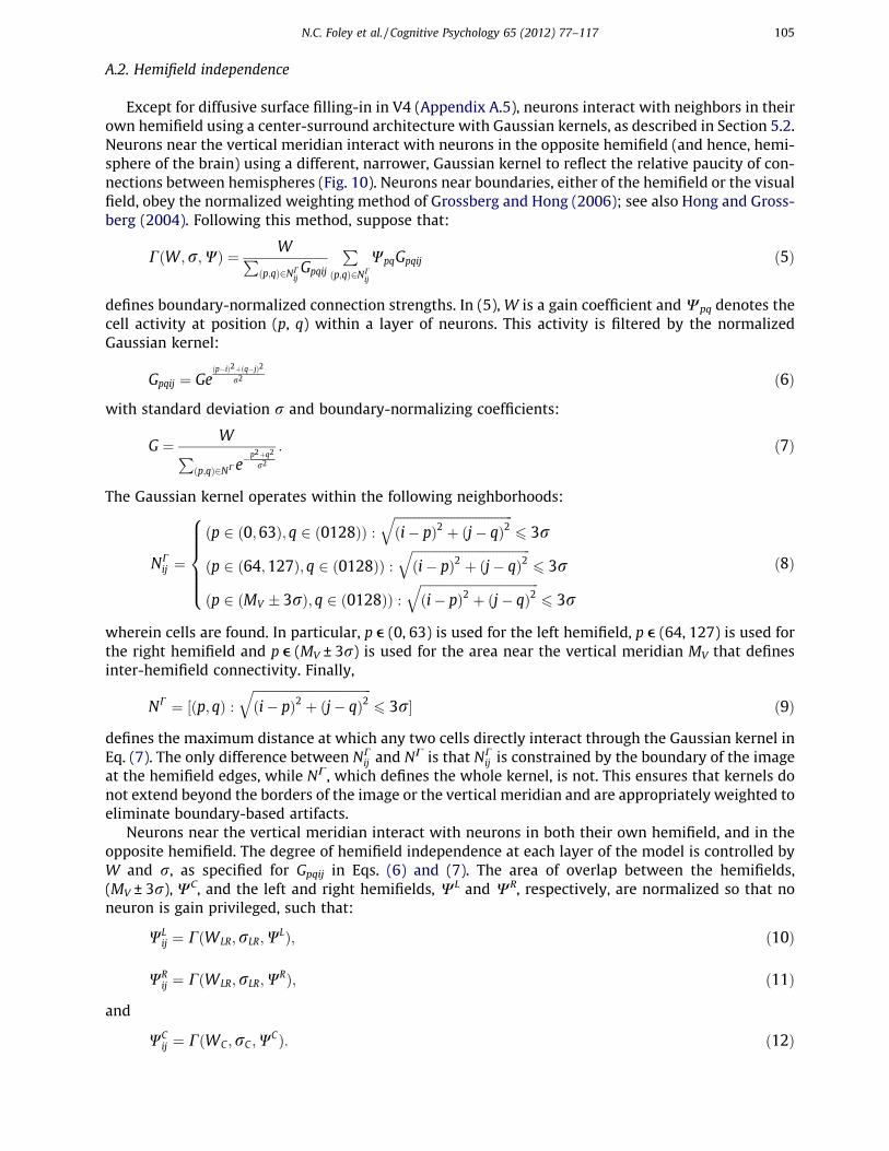
Author's personal copy
A.2. Hemifield independence
Except for diffusive surface filling-in in V4 (Appendix A.5), neurons interact with neighbors in theirown hemifield using a center-surround architecture with Gaussian kernels, as described in Section 5.2.Neurons near the vertical meridian interact with neurons in the opposite hemifield (and hence, hemi-sphere of the brain) using a different, narrower, Gaussian kernel to reflect the relative paucity of con-nections between hemispheres (Fig. 10). Neurons near boundaries, either of the hemifield or the visualfield, obey the normalized weighting method of Grossberg and Hong (2006); see also Hong and Gross-berg (2004). Following this method, suppose that:
C"W;r;W# $ WP"p;q#2NC
ijGpqij
P
"p;q#2NCij
WpqGpqij "5#
defines boundary-normalized connection strengths. In (5), W is a gain coefficient andWpq denotes thecell activity at position (p, q) within a layer of neurons. This activity is filtered by the normalizedGaussian kernel:
Gpqij $ Ge"p%i#2("q%j#2
r2 "6#
with standard deviation r and boundary-normalizing coefficients:
G $ WP
"p;q#2NCe%p2(q2
r2
: "7#
The Gaussian kernel operates within the following neighborhoods:
NCij $
"p 2 "0;63#; q 2 "0128## :!!!!!!!!!!!!!!!!!!!!!!!!!!!!!!!!!!!!!!"i% p#2 ( "j% q#2
q6 3r
"p 2 "64;127#; q 2 "0128## :!!!!!!!!!!!!!!!!!!!!!!!!!!!!!!!!!!!!!!"i% p#2 ( "j% q#2
q6 3r
"p 2 "MV * 3r#; q 2 "0128## :!!!!!!!!!!!!!!!!!!!!!!!!!!!!!!!!!!!!!!"i% p#2 ( "j% q#2
q6 3r
8>>>><
>>>>:
"8#
wherein cells are found. In particular, p e (0, 63) is used for the left hemifield, p e (64, 127) is used forthe right hemifield and p e (MV ± 3r) is used for the area near the vertical meridian MV that definesinter-hemifield connectivity. Finally,
NC $ &"p; q# :!!!!!!!!!!!!!!!!!!!!!!!!!!!!!!!!!!!!!!"i% p#2 ( "j% q#2
q6 3r' "9#
defines the maximum distance at which any two cells directly interact through the Gaussian kernel inEq. (7). The only difference between NC
ij and NC is that NCij is constrained by the boundary of the image
at the hemifield edges, while NC, which defines the whole kernel, is not. This ensures that kernels donot extend beyond the borders of the image or the vertical meridian and are appropriately weighted toeliminate boundary-based artifacts.
Neurons near the vertical meridian interact with neurons in both their own hemifield, and in theopposite hemifield. The degree of hemifield independence at each layer of the model is controlled byW and r, as specified for Gpqij in Eqs. (6) and (7). The area of overlap between the hemifields,(MV ± 3r),WC, and the left and right hemifields, WL and WR, respectively, are normalized so that noneuron is gain privileged, such that:
WLij $ C"WLR;rLR;WL#; "10#
WRij $ C"WLR;rLR;WR#; "11#
and
WCij $ C"WC ;rC ;WC#: "12#
N.C. Foley et al. / Cognitive Psychology 65 (2012) 77–117 105

Author's personal copy
These functions allow us to define a general function, v, that describes the complete set of connectionweights to any neuron in layer W:
v"WLR;WC ;rLR;rC ;W# $
WLij for i 2 &0;VM % 3rC#; j 2 &0128'
WRij for i 2 "VM ( 3rC ;127'; j 2 &0128'
tr"WLRpqij#W
LRij (tr"WC
pqij#WCij
tr"WLRpqij#(tr"WC
pqij#for i 2 &VM % 3rC ;VM ( 3rC '; j 2 &0128'
8>>><
>>>:; "13#
where tr(a) is the trace of a,WLR is the intra-hemifield gain,WC is the inter-hemifield gain, rLR controlsmaximal extent of intra-hemifield connectivity, and rC controls the maximal extend of inter-hemifieldconnectivity. Figs. 10A and B illustrate input horizontal and vertical bars and the corresponding func-tion v(2, 1, 5, 2, I). The bar in 10A crosses the hemifield, whereas the bar in 10B does not.
A.3. LGN polarity sensitive cells: On and Off Channels
There are four types of cells in the model LGN: single opponent ON-cells (x(ij , Eq. (14)), single oppo-nent OFF-cells (x%ij , Eq. (15)), double opponent ON-cells (X(
ij , Eq. (18)) and double opponent OFF-cells(X%
ij , Eq. (19)).Single opponent cells are computed using feed-forward shunting networks solved at equilibrium
(Eq. (3)). At position (i, j), the output signal from the single opponent ON-cell is:
x(ij $Q( ( Eij % Dij
1( Eij ( Dij
" #(
"14#
and the output signal from the single opponent OFF-cell is:
x%ij $Q% ( Dij % Eij
1( Eij ( Dij
" #(
"15#
where Q+ = 5 and Q% = 1 represent tonic bias factors, while
Eij $ v"1:5;1:5; :2; :2; IC# "16#
and
Dij $ v":2; :2;1:5;1:5; IC# "17#
compute the center–surround sampling of the compressed input image. The parameters were chosento satisfy the property of featural noise suppression; that is, the LGN ON cells have 0 response to con-stant luminance of any intensity; they only respond in the vicinity of luminance gradients. Due to thefact that Q% > Q+ in Eqs. (14) and (15), OFF cells are tonically active in the presence of uniform inputs,including in the dark. However, they do not project to the surface layer in the model, and thus do notaffect filling-in.
The output signal from the double-opponent ON-cell at position (i, j) is:
X(ij $ dx(ij % x%ij e
( "18#
and from the double-opponent OFF-cell is:
X%ij $ dx%ij % x(ij e
( "19#
Note that the output signals from all four types of LGN cells are half-wave rectified, see Eq. (4).
A.4. Boundaries
Because the model inputs are simple, the double opponent LGN outputs require little additionalprocessing to create effective boundaries. Simplified complex cell outputs such as those found in pri-mary visual cortex are modeled by polarity-insensitive cells, Zij, at position (i, j) that add ON "X(
ij , Eq.(18)) and OFF (X%
ij , Eq. (19)) channel inputs from double-opponent cells:
106 N.C. Foley et al. / Cognitive Psychology 65 (2012) 77–117

Author's personal copy
Zij $ d1:5X(ij ( X%
ij % 1e( "20#
The ON-channel is scaled to be stronger than the OFF-channel so that the model responds robustlyover a wide range of local contrasts. Object boundary contours, Bij, at position (i, j) are defined bythe equation:
:11dBij
dt$ %1:5Bij ( "1% Bij#Zij"1( 25F(
ij #: "21#
Eq. (21) says that an object boundary Bij is activated by complex cell outputs Zij that are modulated byfeedback signals "F(
ij , Eq. (22)) from the contours of successfully filled-in surfaces that are fed back totheir inducing boundaries. These feedback signals are accordingly derived from surface contours (Cij,Eq. (23)). In (21), the feedback signal to position (i, j) of the boundary is:
F(ij $ v"3:5;3;2;1;Cij#; "22#
where function v is defined by Eq. (13). The surface contour output signal, Cij, at position (i, j) is com-puted by ON and OFF shunting on-center off-surround networks that are sensitive to the contrast offilled-in surface activity (Sij, Eq. (26)). They are therefore positive at the positions of successfully filled-in object boundaries:
Cij $K(
ij % K%ij
3( K(ij ( K%
ij
& ’(
(K%
ij % K(ij
3( K(ij ( K%
ij
& ’(
; "23#
where
K(ij $ v"3;3;1;1; Sij# "24#
and
K%ij $ v"3:7;3:7;2:5;2:5; Sij# "25#
compute the ON and OFF representations of the filled-in surfaces, respectively, at position (i, j).Surface contours are computed at the boundaries of successfully filled-in surfaces; that is, surfaces
which are surrounded by a connected boundary and thus do not allow their lightness or color signalsto spill out into the scenic background. Grossberg (1994, 1997) analyzed how connected boundariesplay a special role in 3-D vision and figure-ground separation. In this more general situation, not allboundaries are connected. However, in response to the simplified input images that are simulatedin this article, all object boundaries are connected and cause successful filling-in. As a result, surfacecontours are computed at all object boundaries, and can strengthen these boundaries via their positivefeedback in Eq. (21). When the contrast of a surface is increased by feedback from an attentionalshroud (see term Lij in Eqs. (26) and (27), the surface contour signals increase, so the strength ofthe boundaries around the attended surface increase also.
A.5. Surfaces
Object surface activity, Sij, at position (i, j) obeys the boundary-gated diffusion equation:
dSijdt
$ %:8Sij ( X(ij "1( Lij# (
P"m;n#2&fi%1;jg;fi(1;jg;fi;j%1g;fi;j(1g'
"Smn % Sij#Pijmn "26#
solved at equilibrium, where X(ij (Eq. (18)) provides bottom-up double-opponent ON-cell input and
Lij $ v":08; :08;8;4; fS"dAOije
(ySij## "27#
modulates this bottom-up input with attentional feedback from object shroud neurons (Eq. (33)) med-iated by the sigmoid signal function fS"a# $ a6
:366(a6. Diffusive surface filling-in between the cell at (i, j)
and its nearest neighbors (m, n) e [{i % 1, j}, {i + 1, j}, {i, j % 1}, {i, j + 1}] is gated by the interveningboundaries via the boundary-sensitive permeability:
N.C. Foley et al. / Cognitive Psychology 65 (2012) 77–117 107

Author's personal copy
Pijmn $ 401( 150"Bmn ( Bij#
; "28#
where Bij and Bmn are the boundary contours (Eq. (21)) at positions (i, j) and (m, n) respectively (Gross-berg & Todorovic, 1988). Eqs. (26) and (27) show how boundaries form filling-in barriers that divi-sively gate, or inhibit, the spread of surface lightness and color. Eqs. (26) and (27) show how top-down feedback from object shrouds can increase the filled-in contrast of a surface and, due to the dis-tributed on-center in Lij, also spill out over the defining boundary of a surface. This latter property isimportant in our explanation of crowding.
The attentional feedback from active object shrouds (see Sections 5.7 and Appendix A.7) habituatesslowly over time in an activity-dependent way, thereby weakening the corresponding surface-(objectshroud) resonance. The rate of habituation between object shroud neurons and surface neurons at po-sition (i, j) in Eq. (27) is controlled by the habituative gate equation (Grossberg, 1972, 1980a, 1980b):
dySijdt
$ 9 ! 10%8"2% ySij % 3 ! 106ySijdAOije
(#; "29#
where the habituative gate ySij recovers to the value 2 and habituates at a rate that depends on the ob-ject shroud activity AO
ij Habituation occurs at slower rate than either of the shroud habituative gates(Eqs. (37) and (38)) and should be thought of as a mechanism underlying adaptation.
A.6. Transient inputs
Several model simulations use sequential, static displays as stimuli but no simulation uses stimuliin motion. Thus, a simplified model of MT cells in the where cortical stream is employed to producetransient inputs to model attentional processes. The activity, Rij, of the transient cell at position (i, j)accordingly responds briefly to contrast increments between successive displays:
:2dRij
dt$ %10Rij ( :7C"t; t0#Q"t; t1# "30#
where
Cij"t0; t1# $ fCX(
ij "t1#X(
ij "t0#
!"31#
is activated by contrast increments between the input to the model at time t0, which is the last timestep for which a prior input was being presented, and time t1, which is the first time step for which anew input is presented, as modified by the sigmoid signal function fC"a# $ a3
103(a3to prevent unbounded
transient responses. A switch function,
Q"t; t1# $1 for t 2 &t1;n( t1'0 for t R &t1; n( t1'
$; "32#
controls the duration of transient cell sensitivity to changes in input, where n = 30 time steps.
A.7. Object shrouds
Object shroud neurons are similar to the shrouds in the ARTSCANmodel. If an object shroud neuroncorresponding to a visible surface reaches a threshold level of activity, then attention will spread overthe entire surface. The shroud intensity is a function of surface activity, previous attention activity inthe object shroud and spatial shroud layers, and inhibitory gain, which is volitionally controlled insome simulations. There are two stable dynamical modes for the object shroud layer: focal, or win-ner-take-all, to support view-invariant object category learning; and multifocal to support multipleshrouds and surface perception. The activity of the object shroud neuron, AO
ij , at position (i, j) obeys:
adAO
ij
dt$ %:05AO
ij ( "1% AOij #Vij&1( 2Rij ( Oij' % "AO
ij ( :2#Tij; "33#
108 N.C. Foley et al. / Cognitive Psychology 65 (2012) 77–117
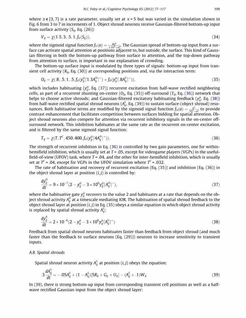
Author's personal copy
where a e [3, 7] is a rate parameter, usually set at a = 5 but was varied in the simulation shown inFig. 6 from 3 to 7 in increments of 1. Object shroud neurons receive Gaussian-filtered bottom-up inputfrom surface activity (Sij, Eq. (26)):
Vij $ v"1:5;3; :5;1; fV "Sij##; "34#
where the sigmoid signal function fV "a# $ 4a6
:1:556(a6. The Gaussian spread of bottom-up input from a sur-
face can activate spatial attention at positions adjacent to, but outside, the surface. This kind of Gauss-ian filtering in both the bottom-up pathway from surface to attention, and the top-down pathwayfrom attention to surface, is important in our explanation of crowding.
The bottom-up surface input is modulated by three types of signals: bottom-up input from tran-sient cell activity (Rij, Eq. (30)) at corresponding positions and, via the interaction term:
Oij $ v":8; :5;1; :5; fO"yOijd1:3AOije
(# ( fO"yAijd:8ASije
(##; "35#
which includes habituating (yOij , Eq. (37)) recurrent excitation from half-wave rectified neighboringcells, as part of a recurrent shunting on-center (Oij, Eq. (35)) off-surround (Tij, Eq. (36)) network thathelps to choose active shrouds; and Gaussian-filtered excitatory habituating feedback (yAij , Eq. (38))from half-wave rectified spatial shroud neurons (AS
ij, Eq. (39)) to sustain surface-(object shroud) reso-nances. Both habituative terms are modified by the sigmoid signal function fO"a# $ a5
:555(a5to provide
contrast enhancement that facilitates competition between surfaces bidding for spatial attention. Ob-ject shroud neurons also compete for attention via recurrent inhibitory signals in the on-center off-surround network. This inhibition habituates at the same rate as the recurrent on-center excitation,and is filtered by the same sigmoid signal function:
Tij $ v"T; TC ;450;400; fO"yOijd4AOije
(##: "36#
The strength of recurrent inhibition in Eq. (36) is controlled by two gain parameters, one for within-hemifield inhibition, which is usually set at T = .05, except for videogame players (VGPs) in the useful-field-of-view (UFOV) task, where T = .04, and the other for inter-hemifield inhibition, which is usuallyset at TC = .04, except for VGPs in the UFOV simulation where TC = .032.
The rate of habituation and recovery of recurrent excitation (Eq. (35)) and inhibition (Eq. (36)) inthe object shroud layer at position (i, j) is controlled by:
dyOijdt
$ 9 ! 10%7"2% yOij % 3 ! 106yOijdAOije
(#; "37#
where the habituative gate yOij recovers to the value 2 and habituates at a rate that depends on the ob-ject shroud activity AO
ij at a timescale mediating IOR. The habituation of spatial shroud feedback to theobject shroud layer at position (i, j) in Eq. (35) obeys a similar equation in which object shroud activityis replaced by spatial shroud activity AS
ij:
dyAijdt
$ 2 ! 10%6"2% yAij % 3 ! 106yAijdASije
(# "38#
Feedback from spatial shroud neurons habituates faster than feedback from object shroud (and muchfaster than the feedback to surface neurons (Eq. (29))) neurons to increase sensitivity to transientinputs.
A.8. Spatial shrouds
Spatial shroud neuron activity ASij at position (i, j) obeys the equation:
:3dAS
ij
dt$ %:05AS
ij ( "1% ASij#&5Rij ( Gij ( Uij' % "AS
ij ( :1#Wij "39#
In (39), there is strong bottom-up input from corresponding transient cell positions as well as a half-wave rectified Gaussian input from the object shroud layer:
N.C. Foley et al. / Cognitive Psychology 65 (2012) 77–117 109

Author's personal copy
Gij $ v"1; :5;3;1; d1:5AOije
(# "40#
Recurrent feedback is received from a shunting on-center (Uij, Eq. (41)) off-surround (Wij, Eq. (42)) net-work with on-center excitation:
Uij $ v"1:6; :5;3; :75; fU"d1:5ASije
(## "41#
and off-surround inhibition:
Wij $ v"W;WC ;200;150;Gij ( fU"d1:5ASije
(##: "42#
Both on-center and off-surround recurrent signals are modified by the sigmoid signal functionfU"a# $ a5
:45(a5. The inhibitory gain in (42) is controlled within hemifields by W = .005, except for the
VGP UFOV simulation, where W = .004; and between hemifields by WC = .002, except for the VGPUFOV simulation, where WC = .00016.
In all, spatial shroud neurons receive bottom-up input from transient cells and object shroud neu-rons while exciting their nearby neighbors and inhibiting their distant neighbors. Small spatialshrouds can hereby survive for several hundred milliseconds in the absence of bottom-up input beforedecaying if there is little new transient input or activity causing competition. Recurrent competitioncan attract or repel spatial shrouds without bottom-up support, causing them to move slightly fromtheir initial positions over time. Spatial shrouds cover the extent of a surface only if that surface res-onates with an object shroud. When this occurs, however, the coverage is often not uniform in inten-sity, and hotspots of activity will form at salient areas of objects such as corners.
A.9. Parametric sensitivity
The behavior of recurrent neural network models, especially those with several interacting recur-rent loops, can exhibit different modes of behavior depending on how parameters of the model arechosen. Some of these parameters can incrementally change model behavior, such as with the UFOVsimulations (see Sections 6.2, Appendices A.7 and A.8). Further changes in those same parameters cancause a state change in model behavior, where attention goes from being multifocal to unifocal (seeSection 3). Other parameters are set to obey specific properties, such as suppression of uniform inputpatterns (see Appendix A.3) and otherwise support a wide range of possible parameters without mod-ifying the model behavior. Finally, some parameters depend on the simulation environment (such asthe number of neurons in each layer, the array size of the inputs), notably the spatial extents of theGaussian kernels controlling feed-forward, feedback and lateral communication between differentmodel neurons, have to be rescaled for larger or smaller simulation environments. All of the param-eters in the front end of the model, in Appendices A.2-A.4 and A.6, fall into the latter two categories.
More interesting are the parameters that control the recurrent circuits between the surface, objectshroud and spatial shroud layers of the model. These equations fall into three general categories: thefirst is the lateral inhibition between model neurons, as well as the kernels controlling the strength ofinter-layer connections ((Eqs. (27), (34)–(36), and (40)–(42)). The second is the various sigmoid signalfunctions (f(a)) that control three important qualities: size, range of sub-linear, linear and super-linearresponse, and the steepness of the linear response. Finally, there are habituating gates (Eqs. (29), (37),and (38)) that control the rate of habituation for different types of feedback within the model.
The primary loop in the model, is the surface-(object shroud)-surface loop, controlled by Eqs. (27),(29), and (34)–(37) and related signal functions. There are several important parts of this loop: thebottom-up connections from the surface to the object shroud layer (Eq. (34)) controls how objectsof different luminance and size bid for object-based attention. Increasing the gain of the kernels inEq. (34) increases response of objects across the board (while also increasing the competition in theobject-shroud layer, since it increases the denominator of the divisive inhibition), while decreasingit has the converse effect. Increasing the half-max point (1.55) or exponent (6) of the correspondingsignal function means that perceptually brighter (or attended) objects are accorded a better bid thandimmer objects, while decreasing it closes the gap. While these parameters are all important, and themodel behavior (though not the ability to form shrouds) is sensitive to relatively small changes (10%)
110 N.C. Foley et al. / Cognitive Psychology 65 (2012) 77–117

Author's personal copy
in any of them, in practice, these provided a baseline for detailed fitting in the feedback and compe-tition related parameters of the model. Competition between shrouds in the model is controlledthrough the balance of lateral excitation and inhibition (Section 6.2 and Eqs. (35)–(37)), and feedbackto the surface layer (Eqs. (27) and (29)) which makes attended objects brighter, thereby increasingtheir bids through the bottom-up connections previously described. The strength of the effects ofincreasing lateral inhibition relative to excitation are determined by the corresponding signal func-tion, and is important in every simulated fit (shrouds will still form after a 10% perturbation of anyof these parameters, but simulated RTs will change).
The secondary loop in the model is the (object shroud)-(spatial shroud)-(object shroud) loop. Thetime constant in the habituating gate between spatial and object shrouds (Eq. (38)) controls the dura-tion of priming, while the strength of feedback between spatial and object shrouds (Eq. (35)) directlycontrols the strength of priming. Changes in parameters for either equation will change the RTs in thelocation cued fits. Another important aspect of the spatial shroud layer is how closely the contrast ra-tio between transient stimuli is preserved, in the balance of lateral excitation and inhibition (Eqs. (41)and (42)) which helps successful detection in the UFOV simulations. Other parameters in the spatialshroud layer can be perturbed by 10% without significantly affecting current results. The dynamics inthis layer would become more sensitive if the model was extended to simulate multiple-object track-ing, or experiments with endogenously cued attention.
References
Alvarez, G. A., & Cavanagh, P. (2005). Independent resources for attentional tracking in the left and right visual hemifields.Psychological Science, 16(8), 637–643. doi: PSCI1587 [pii] 10.1111/j.1467-9280.2005.01587.x.
Alvarez, G. A., & Franconeri, S. L. (2007). How many objects can you track? Evidence for a resource-limited attentive trackingmechanism. Journal of Vision, 7(13), 10–11. doi: 14 10.1167/7.13.14/7/13/14/ [pii].
Alvarez, G. A., Horowitz, T. S., Arsenio, H. C., Dimase, J. S., & Wolfe, J. M. (2005). Do multielement visual tracking and visualsearch draw continuously on the same visual attention resources? Journal of Experimental Psychology: Human Perception andPerformance, 31(4), 643–667. doi: 2005-09705-001 [pii] 10.1037/0096-1523.31.4.643a.
Alvarez, G. A., & Scholl, B. J. (2005). How does attention select and track spatially extended objects? New effects of attentionalconcentration and amplification. Journal of Experimental Psychology: General, 134(4), 461–476. doi: 2005-14939-002 [pii]10.1037/0096-3445.134.4.46.
Andersen, R. A., Bracewell, R. M., Barash, S., Gnadt, J. W., & Fogassi, L. (1990). Eye position effects on visual, memory, andsaccade-related activity in areas LIP and 7a of macaque. Journal of Neuroscience, 10(4), 1176–1196.
Baker, C. I., Behrmann, M., & Olson, C. F. (2002). Impact of learning on representation of parts and wholes in monkeyinferotemporal cortex. Nature Neuroscience, 5, 1210–1216. doi: 10.1038/nn960.
Bar, M., Kassam, K. S., Ghuman, A. S., Boshyan, J., Schmid, A. M., Dale, A. M., et al (2006). Top-down facilitation of visualrecognition. Proceedings of the National Academy of Sciences of the United States of America, 103(2), 449–454. doi: 0507062103[pii] 10.1073/pnas.0507062103.
Berzhanskaya, J., Grossberg, S., & Mingolla, E. (2007). Laminar cortical dynamics of visual form and motion interactions duringcoherent object motion perception. Spatial Vision, 20(4), 337–395. doi: 10.1163/156856807780919000.
Bhatt, R., Carpenter, G. A., & Grossberg, S. (2007). Texture segregation by visual cortex: Perceptual grouping, attention, andlearning. Vision Research, 47(25), 3173–3211. doi: S0042-6989(07)00309-4 [pii] 10.1016/j.visres.2007.07.013.
Blaser, E., Pylyshyn, Z. W., & Holcombe, A. O. (2000). Tracking an object through feature space. Nature, 408(6809), 196–199. doi:10.1038/35041567.
Boch, R. A., & Goldberg, M. E. (1989). Participation of prefrontal neurons in the preparation of visually guided eye movements inthe rhesus monkey. Journal of Neurophysiology, 61(5), 1064–1084.
Bonmassar, G., & Schwartz, E. L. (1997). Space-variant Fourier analysis: The exponential chirp transform. IEEE Transactions onPattern Analysis and Machine Intelligence, 19(10), 1080–1089.
Booth, M. C., & Rolls, E. T. (1998). View-invariant representations of familiar objects by neurons in the inferior temporal visualcortex. Cerebral Cortex, 8, 510–523.
Borg-Graham, L. J., Monier, C., & Fregnac, Y. (1998). Visual input evokes transient and strong shunting inhibition in visualcortical neurons. Nature, 393(6683), 369–373. doi: 10.1038/30735.
Bouma, H. (1970). Interaction effects in parafoveal letter recognition. Nature, 226(5241), 177–178.Bouma, H. (1973). Visual interference in the parafoveal recognition of initial and final letters of words. Vision Research, 13(4),
767–782.Broadbent, D. E. (1958). Perception and communication. New York: Pergamon Press.Brown, J., Bullock, D., & Grossberg, S. (1999). How the basal ganglia use parallel excitatory and inhibitory learning pathways to
selectively respond to unexpected rewarding cues. Journal of Neuroscience, 19(23), 10502–10511.Brown, J. M., & Denney, H. I. (2007). Shifting attention into and out of objects: Evaluating the processes underlying the object
advantage. Perception and Psychophysics, 69(4), 606–618.Brown, J. W., Bullock, D., & Grossberg, S. (2004). How laminar frontal cortex and basal ganglia circuits interact to control planned
and reactive saccades. Neural Networks, 17(4), 471–510. doi: 10.1016/j.neunet.2003.08.006S0893-6080(03)00252-1 [pii].Cao, Y., & Grossberg, S. (2005). A laminar cortical model of stereopsis and 3D surface perception: Closure and da Vinci stereopsis.
Spatial Vision, 18(5), 515–578.
N.C. Foley et al. / Cognitive Psychology 65 (2012) 77–117 111
Author's personal copy
Cao, Y., Grossberg, S., & Markowitz, J. (2011). How does the brain rapidly learn and reorganize view- and positionally-invariantobject representations in inferior temporal cortex? Neural Networks, 24, 1050–1061.
Caplovitz, G. P., & Tse, P. U. (2007). V3A processes contour curvature as a trackable feature for the perception of rotationalmotion. Cerebral Cortex, 17(5), 1179–1189.
Carpenter, G. A., & Grossberg, S. (1987). A massively parallel architecture for a self-organizing neural pattern recognitionmachine. Computer Vision, Graphics, and Image Processing, 37(1), 54–115.
Carpenter, G. A., & Grossberg, S. (1991). Pattern recognition by self-organizing neural networks. Cambridge, Mass.: MIT Press.Carpenter, G. A., & Grossberg, S. (1993). Normal and amnesic learning, recognition and memory by a neural model of cortico-
hippocampal interactions. Trends in Neurosciences, 16(4), 131–137. doi: 0166-2236(93)90118-6 [pii].Carrasco, M., Penpeci-Talgar, C., & Eckstein, M. (2000). Spatial covert attention increases contrast sensitivity across the CSF:
Support for signal enhancement. Vision Research, 40(10–12), 1203–1215.Cavanagh, P., & Alvarez, G. A. (2005). Tracking multiple targets with multifocal attention. Trends in Cognitive Sciences, 9(7),
349–354. doi: S1364-6613(05)00149-X [pii] 10.1016/j.tics.2005.05.009.Cavanagh, P., Labianca, A. T., & Thornton, I. M. (2001). Attention-based visual routines: Sprites. Cognition, 80(1–2), 47–60.Cave, K. R., Bush, W. S., & Taylor, T. G. (2010). Split attention as part of a flexible attentional system for complex scenes:
Comment on Jans, Peters, and De Weerd (2010). Psychological Review, 117(2), 685–696. doi: 2010-06891-014 [pii] 10.1037/a0019083.
Chiu, Y. C., & Yantis, S. (2009). A domain-independent source of cognitive control for task sets: Shifting spatial attention andswitching categorization rules. Journal of Neuroscience, 29(12), 3930–3938. doi: 29/12/3930 [pii] 10.1523/JNEUROSCI.5737-08.2009.
Chun, M. M. (2000). Contextual cueing of visual attention. Trends in Cognitive Sciences, 4, 170–178.Chun, M. M., & Jiang, Y. (1998). Contextual cueing: Implicit learning and memory of visual context guides spatial attention.
Cognitive Psychology, 36(1), 28–71. doi: S0010-0285(98)90681-8 [pii] 10.1006/cogp.1998.0681.Cohen, M. A., & Grossberg, S. (1984). Neural dynamics of brightness perception: Features, boundaries, diffusion, and resonance.
Perception and Psychophysics, 36, 428–456.Culham, J. C., Brandt, S. A., Cavanagh, P., Kanwisher, N. G., Dale, A. M., & Tootell, R. B. (1998). Cortical fMRI activation produced by
attentive tracking of moving targets. Journal of Neurophysiology, 80(5), 2657–2670.Culham, J. C., Cavanagh, P., & Kanwisher, N. G. (2001). Attention response functions: Characterizing brain areas using fMRI
activation during parametric variations of attentional load. Neuron, 32(4), 737–745. doi: S0896-6273(01)00499-8 [pii].Corbetta, M., Patel, G., & Shulman, G. G. (2008). The reorienting system of the human brain: From environment to theory of
mind. Neuron, 58, 306–324. doi: 10.1016/j.neuron.2008.04.017.Corbetta, M., & Shulman, G. L. (2002). Control of goal-directed and stimulus-driven attention in the brain. Nature Reviews
Neuroscience, 3, 201–215.Daniel, P., & Whitteridge, D. (1961). The representation of the visual field on the cerebral cortex in monkeys. Journal of
Physiology, 159, 203–221.David, S. V., Hayden, B. Y., Mazer, J. A., & Gallant, J. L. (2008). Attention to stimulus features shifts spectral tuning of V4 neurons
during natural vision. Neuron, 59(3), 509–521. doi: S0896-6273(08)00570-9 [pii] 10.1016/j.neuron.2008.07.001.Desimone, R., & Duncan, J. (1995). Neural mechanisms of selective visual attention. Annual Review of Neuroscience, 18, 193–222.
doi: 10.1146/annurev.ne.18.030195.001205.Dosenbach, N. U., Fair, D. A., Cohen, A. L., Schlaggar, B. L., & Petersen, S. E. (2008). A dual-networks architecture of top-down
control. Trends in Cognitive Sciences, 12(3), 99–105. doi: S1364-6613(08)00027-2 [pii] 10.1016/j.tics.2008.01.001.Dosenbach, N. U., Fair, D. A., Miezin, F. M., Cohen, A. L., Wenger, K. K., Dosenbach, R. A., et al (2007). Distinct brain networks for
adaptive and stable task control in humans. Proceedings of the National Academy of Sciences of the United States of America,104(26), 11073–11078. doi: 0704320104 [pii] 10.1073/pnas.0704320104.
Drasdo, N. (1977). The neural representation of visual space. Nature, 266(5602), 554–556.Duhamel, J. R., Colby, C. L., & Goldberg, M. E. (1992). The updating of the representation of visual space in parietal cortex by
intended eye movements. Science, 255(5040), 90–92.Duncan, J. (1984). Selective attention and the organization of visual information. Journal of Experimental Psychology: General,
113(4), 501–517.Egeth, H. E., & Yantis, S. (1997). Visual attention: Control, representation, and time course. Annual Review of Psychology, 48,
269–297. doi: 10.1146/annurev.psych.48.1.269.Egly, R., Driver, J., & Rafal, R. D. (1994). Shifting visual attention between objects and locations: Evidence from normal and
parietal lesion subjects. Journal of Experimental Psychology: General, 123(2), 161–177.Fallah, M., Stoner, G. R., & Reynolds, J. H. (2007). Stimulus-specific competitive selection in macaque extrastriate visual area V4.
Proceedings of the National Academy of Sciences of the United States of America, 104(10), 4165–4169. doi: 0611722104 [pii]10.1073/pnas.0611722104.
Fang, L., & Grossberg, S. (2009). From stereogram to surface: How the brain sees the world in depth. Spatial Vision, 22, 45–82.Fazl, A., Grossberg, S., & Mingolla, E. (2009). View-invariant object category learning, recognition, and search: How spatial and
object attention are coordinated using surface-based attentional shrouds. Cognitive Psychology, 58(1), 1–48. doi: S0010-0285(08)00036-4 [pii] 10.1016/j.cogpsych.2008.05.001.
Fischer, B. (1973). Overlap of receptive field centers and representation of the visual field in the cat’s optic tract. Vision Research,13(11), 2113–2120.
Franconeri, S. L., Alvarez, G. A., & Enns, J. T. (2007). How many locations can be selected at once? Journal of ExperimentalPsychology: Human Perception and Performance, 33(5), 1003–1012. doi: 2007-14662-001 [pii] 10.1037/0096-1523.33.5.1003.
Fukuda, K., Awh, E., & Vogel, E. K. (2010). Discrete capacity limits in visual working memory. Current Opinion in Neurobiology,20(2), 177–182. doi: S0959-4388(10)00043-7 [pii] 10.1016/j.conb.2010.03.005.
Gee, A. L., Ipata, A. E., Gottlieb, J., Bisley, J. W., & Goldberg, M. E. (2008). Neural enhancement and pre-emptive perception: Thegenesis of attention and the attentional maintenance of the cortical salience map. Perception, 37(3), 389–400.
Ghose, G. M. (2009). Attentional modulation of visual responses by flexible input gain. Journal of Neurophysiology, 101(4),2089–2106. doi: 90654.2008 [pii] 10.1152/jn.90654.2008.
112 N.C. Foley et al. / Cognitive Psychology 65 (2012) 77–117

Author's personal copy
Goldberg, M. E., & Bruce, C. J. (1990). Primate frontal eye fields. III. Maintenance of a spatially accurate saccade signal. Journal ofNeurophysiology, 64(2), 489–508.
Goldman, P. S., & Rakic, P. T. (1979). Impact of the outside world upon the developing primate brain: Perspective fromneurobiology. Bulletin of the Menninger Clinic, 43(1), 20–28.
Goldsmith, M., & Yeari, M. (2003). Modulation of object-based attention by spatial focus under endogenous and exogenousorienting. Journal of Experimental Psychology: Human Perception and Performance, 29, 897–918. doi: 10.1037/0096-1523.29.5.897.
Golomb, J. D., Chun, M. M., & Mazer, J. A. (2008). The native coordinate system of spatial attention is retinotopic. Journal ofNeuroscience, 28(42), 10654–10662. doi: 28/42/10654 [pii] 10.1523/JNEUROSCI.2525-08.2008.
Golomb, J. D., Nguyen-Phuc, A. Y., Mazer, J. A., McCarthy, G., & Chun, M. M. (2010a). Attentional facilitation throughout humanvisual cortex lingers in retinotopic coordinates after eye movements. Journal of Neuroscience, 30(31), 10493–10506. doi: 30/31/10493 [pii] 10.1523/JNEUROSCI.1546-10.2010.
Golomb, J. D., Pulido, V. Z., Albrecht, A. R., Chun, M. M., & Mazer, J. A. (2010b). Robustness of the retinotopic attentional traceafter eye movements. Journal of Vision, 10(3), 11–12. doi: 19 10.1167/10.3.19/10/3/19/ [pii].
Gottlieb, J., Kusunoki, M., & Goldberg, M. E. (2005). Simultaneous representation of saccade targets and visual onsets in monkeylateral intraparietal area. Cerebral Cortex, 15(8), 1198–1206. doi: bhi002 [pii] 10.1093/cercor/bhi002.
Gottlieb, J. P., Kusunoki, M., & Goldberg, M. E. (1998). The representation of visual salience in monkey parietal cortex. Nature,391(6666), 481–484. doi: 10.1038/35135.
Gove, A., Grossberg, S., & Mingolla, E. (1995). Brightness perception, illusory contours, and corticogeniculate feedback. VisualNeuroscience, 12(6), 1027–1052.
Green, C. S., & Bavelier, D. (2003). Action video game modifies visual selective attention. Nature, 423(6939), 534–537. doi:10.1038/nature01647nature01647 [pii].
Green, C. S., & Bavelier, D. (2006a). Effect of action video games on the spatial distribution of visuospatial attention. Journal ofExperimental Psychology: Human Perception and Performance, 32(6), 1465–1478. doi: 2006-22004-012 [pii]10.1037/0096-1523.32.6.1465.
Green, C. S., & Bavelier, D. (2006b). Enumeration versus multiple object tracking: The case of action video game players.Cognition, 101(1), 217–245. doi: S0010-0277(05)00187-3 [pii] 10.1016/j.cognition.2005.10.004.
Green, C. S., & Bavelier, D. (2007). Action-video-game experience alters the spatial resolution of vision. Psychological Science,18(1), 88–94. doi: PSCI1853 [pii] 10.1111/j.1467-9280.2007.01853.x.
Gronau, N., Neta, M., & Bar, M. (2008). Integrated contextual representation for objects’ identities and their locations. Journal ofCognitive Neuroscience, 20(3), 371–388. doi: 10.1162/jocn.2008.20027.
Grossberg, S. (1972). A neural theory of punishment and avoidance, II: Quantitative theory. Mathematical Biosciences, 15,253–285.
Grossberg, S. (1973). Contour enhancement, short-term memory, and constancies in reverberating neural networks. Studies inApplied Mathematics, 52, 213–257.
Grossberg, S. (1976). Adaptive pattern classification and universal recoding, II: Feedback, expectation, olfaction, and illusions.Biological Cybernetics, 23, 187–202.
Grossberg, S. (1978a). Decisions, patterns, and oscillations in nonlinear competitive systems with applications to Volterra-Lotkasystems. Journal of Theoretical Biology, 73(1), 101–130.
Grossberg, S. (1978b). Do all neural models really look alike? A comment on Anderson, Silverstein, Ritz, and Jones. PsychologicalReview, 85(6), 592–596.
Grossberg, S. (1980a). Biological competition: Decision rules, pattern formation, and oscillations. Proceedings of the NationalAcademy of Sciences of the United States of America, 77(4), 2338–2342.
Grossberg, S. (1980b). How does a brain build a cognitive code? Psychological Review, 87(1), 1–51.Grossberg, S. (1994). 3-D vision and figure-ground separation by visual cortex. Perception and Psychophysics, 55(1), 48–121.Grossberg, S. (1997). Cortical dynamics of three-dimensional figure-ground perception of two-dimensional pictures.
Psychological Review, 104(3), 618–658.Grossberg, S. (2000a). The complementary brain: Unifying brain dynamics and modularity. Trends in Cognitive Sciences, 4(6),
233–246. doi: S1364-6613(00)01464-9 [pii].Grossberg, S. (2000b). How hallucinations may arise from brain mechanisms of learning, attention, and volition. Journal of the
International Neuropsychological Society, 6(5), 583–592.Grossberg, S. (2007). Towards a unified theory of neocortex: Laminar cortical circuits for vision and cognition. Progress in Brain
Research, 165, 79–104. doi: S0079-6123(06)65006-1 [pii] 10.1016/S0079-6123(06)65006-1.Grossberg, S. (2009). Cortical and subcortical predictive dynamics and learning during perception, cognition, emotion and
action. Philosophical Transactions of the Royal Society of London. Series B: Biological Sciences, 364(1521), 1223–1234. doi: 364/1521/1223 [pii] 10.1098/rstb.2008.0307.
Grossberg, S., & Hong, S. (2006). A neural model of surface perception: Lightness, anchoring, and filling-in. Spatial Vision, 19(2–4), 263–321.
Grossberg, S., & Huang, T. R. (2009). ARTSCENE: A neural system for natural scene classification. Journal of Vision, 9(4), 1–19. doi:6 10.1167/9.4.6/9/4/6/ [pii].
Grossberg, S., Kuhlmann, L., & Mingolla, E. (2007). A neural model of 3D shape-from-texture: Multiple-scale filtering, boundarygrouping, and surface filling-in. Vision Research, 47, 634–672.
Grossberg, S., Markowitz, J., & Cao, Y. (2011). On the road to invariant recognition: Explaining tradeoff and morph properties ofcells in inferotemporal cortex using multiple-scale task-sensitive attentive learning. Neural Networks, 24, 1036–1049.
Grossberg, S., Mingolla, E., & Ross, W. D. (1994). A neural theory of attentive visual search: Interactions of boundary, surface,spatial, and object representations. Psychological Review, 101(3), 470–489.
Grossberg, S., Mingolla, E., & Viswanathan, L. (2001). Neural dynamics of motion integration and segmentation within andacross apertures. Vision Research, 41(19), 2521–2553.
N.C. Foley et al. / Cognitive Psychology 65 (2012) 77–117 113

Author's personal copy
Grossberg, S., & Pearson, L. R. (2008). Laminar cortical dynamics of cognitive and motor working memory, sequence learning andperformance: Toward a unified theory of how the cerebral cortex works. Psychological Review, 115(3), 677–732. doi: 2008-09896-006 [pii] 10.1037/a0012618.
Grossberg, S., & Raizada, R. D. (2000). Contrast-sensitive perceptual grouping and object-based attention in the laminar circuitsof primary visual cortex. Vision Research, 40(10–12), 1413–1432. doi: S0042-6989(99)00229-1 [pii].
Grossberg, S., & Swaminathan, G. (2004). A laminar cortical model for 3D perception of slanted and curved surfaces and of 2Dimages: Development, attention, and bistability. Vision Research, 44(11), 1147–1187. doi: 10.1016/j.visres.2003.12.009S0042698903007995 [pii].
Grossberg, S., & Todorovic, D. (1988). Neural dynamics of 1-D and 2-D brightness perception: A unified model of classical andrecent phenomena. Perception and Psychophysics, 43(3), 241–277.
Grossberg, S., & Yazdanbakhsh, A. (2005). Laminar cortical dynamics of 3D surface perception: Stratification, transparency, andneon color spreading. Vision Research, 45(13), 1725–1743. doi: S0042-6989(05)00035-0 [pii] 10.1016/j.visres.2005.01.006.
Haxby, J. V., Grady, C. L., Horwitz, B., Ungerleider, L. G., Mishkin, M., Carson, R. E., et al (1991). Dissociation of object and spatialvisual processing pathways in human extrastriate cortex. Proceedings of the National Academy of Sciences of the United Statesof America, 88(5), 1621–1625.
Hayden, B. Y., & Gallant, J. L. (2005). Time course of attention reveals different mechanisms for spatial and feature-basedattention in area V4. Neuron, 47(5), 637–643. doi: S0896-6273(05)00614-8 [pii] 10.1016/j.neuron.2005.07.020.
Hayden, B. Y., & Gallant, J. L. (2009). Combined effects of spatial and feature-based attention on responses of V4 neurons. VisionResearch, 49(10), 1182–1187. doi: S0042-6989(08)00320-9 [pii] 10.1016/j.visres.2008.06.011.
He, S., Cavanagh, P., & Intriligator, J. (1996). Attentional resolution and the locus of visual awareness. Nature, 383(6598),334–337. doi: 10.1038/383334a0.
He, Z. J., & Nakayama, K. (1995). Visual attention to surfaces in three-dimensional space. Proceedings of the National Academy ofSciences of the United States of America, 92(24), 11155–11159.
Hillyard, S. A., Vogel, E. K., & Luck, S. J. (1998). Sensory gain control (amplification) as a mechanism of selective attention:Electrophysiological and neuroimaging evidence. Philosophical Transactions of the Royal Society of London. Series B: BiologicalSciences, 353(1373), 1257–1270. doi: 10.1098/rstb.1998.0281.
Hong, S., & Grossberg, S. (2004). A neuromorphic model for achromatic and chromatic surface representation of natural images.Neural Networks, 17(5–6), 787–808. doi: 10.1016/j.neunet.2004.02.007S0893-6080(04)00053-X [pii].
Horowitz, T. S., Klieger, S. B., Fencsik, D. E., Yang, K. K., Alvarez, G. A., & Wolfe, J. M. (2007). Tracking unique objects. Perceptionand Psychophysics, 69(2), 172–184.
Huang, T. R., & Grossberg, S. (2010). Cortical dynamics of contextually cued attentive visual learning and search: Spatial andobject evidence accumulation. Psychological Review, 117(4), 1080–1112. doi: 2010-22285-001 [pii] 10.1037/a0020664.
Intriligator, J., & Cavanagh, P. (2001). The spatial resolution of visual attention. Cognitive Psychology, 43(3), 171–216. doi:10.1006/cogp.2001.0755S0010-0285(01)90755-8 [pii].
Itti, L., & Koch, C. (2001). Computational modelling of visual attention. Nature Reviews Neuroscience, 2(3), 194–203.James, W. (1890). The principles of psychology. New York: H. Holt and Company.Jans, B., Peters, J. C., & De Weerd, P. (2010). Visual spatial attention to multiple locations at once: The jury is still out.
Psychological Review, 117(2), 637–684. doi: 2010-06891-013 [pii] 10.1037/a0019082.Jiang, Y., & Chun, M. M. (2001). Selective attention modulates implicit learning. Quarterly Journal of Experimental Psychology, 54A,
1105–1124.Kahneman, D., Treisman, A., & Gibbs, B. J. (1992). The reviewing of object files: Object-specific integration of information.
Cognitive Psychology, 24(2), 175–219. doi: 0010-0285(92)90007-O [pii].Kastner, S., & Ungerleider, L. G. (2000). Mechanisms of visual attention in the human cortex. Annual Review of Neuroscience, 23,
315–341. doi: 10.1146/annurev.neuro.23.1.315.Kelly, F., & Grossberg, S. (2000). Neural dynamics of 3-D surface perception: Figure-ground separation and lightness perception.
Perception and Psychophysics, 62(8), 1596–1618.Koch, C., & Ullman, S. (1985). Shifts in selective visual attention: Towards the underlying neural circuitry. Human Neurobiology,
4(4), 219–227.Kveraga, K., Boshyan, J., & Bar, M. (2007). Magnocellular projections as the trigger of top-down facilitation in recognition. Journal
of Neuroscience, 27(48), 13232–13240. doi: 27/48/13232 [pii] 10.1523/JNEUROSCI.3481-07.2007.Lamy, D., & Egeth, H. (2002). Object-based selection: The role of attentional shifts. Perception and Psychophysics, 64(1), 52–66.Lee, J., & Maunsell, J. H. (2009). A normalization model of attentional modulation of single unit responses. PLoS ONE, 4(2), e4651.
doi: 10.1371/journal.pone.0004651.Levi, D. M. (2008). Crowding – An essential bottleneck for object recognition: A mini-review. Vision Research, 48(5), 635–654.
doi: S0042-6989(07)00556-1 [pii] 10.1016/j.visres.2007.12.009.Li, N., & DiCarlo, J. J. (2008). Unsupervised natural experience rapidly alters invariant object representation in visual cortex.
Science, 321, 1502–1507.Li, N., & DiCarlo, J. J. (2010). Unsupervised natural visual experience rapidly reshapes size invariant object represent in inferior
temporal cortex. Neuron, 67, 1062–1075.Logan, G. D. (1996). The CODE theory of visual attention: An integration of space-based and object-based attention. Psychological
Review, 103(4), 603–649.Logothetis, N. K., Pauls, J., & Poggio, T. (1995). Shape representation in the inferior temporal cortex of monkeys. Current Biology,
5, 552–563. doi: 10.1016/S0960-9822(95)00108-4.Luck, S. J., Hillyard, S. A., Mangun, G. R., & Gazzaniga, M. S. (1989). Independent hemispheric attentional systems mediate visual
search in split-brain patients. Nature, 342(6249), 543–545. doi: 10.1038/342543a0.Martinez, A., Teder-Salejarvi, W., Vazquez, M., Molholm, S., Foxe, J. J., Javitt, D. C., et al (2006). Objects are highlighted by spatial
attention. Journal of Cognitive Neuroscience, 18(2), 298–310. doi: 10.1162/089892906775783642.Mathot, S., Hickey, C., & Theeuwes, J. (2010). From reorienting of attention to biased competition: Evidence from hemifield
effects. Attention, Perception, and Psychophysics, 72(3), 651–657. doi: 72/3/651 [pii] 10.3758/APP.72.3.651.
114 N.C. Foley et al. / Cognitive Psychology 65 (2012) 77–117

Author's personal copy
Mathot, S., & Theeuwes, J. (2010a). Evidence for the predictive remapping of visual attention. Experimental Brain Research,200(1), 117–122. doi: 10.1007/s00221-009-2055-3.
Mathot, S., & Theeuwes, J. (2010b). Gradual remapping results in early retinotopic and late spatiotopic inhibition of return.Psychological Science, 21(12), 1793–1798. doi: 0956797610388813 [pii] 10.1177/0956797610388813.
Matsuzaki, R., Kyuhou, S., Matsuura-Nakao, K., & Gemba, H. (2004). Thalamo-cortical projections to the posterior parietal cortexin the monkey. Neuroscience Letters, 355(1–2), 113–116. doi: S0304394003012746 [pii].
McMains, S. A., & Somers, D. C. (2004). Multiple spotlights of attentional selection in human visual cortex. Neuron, 42(4),677–686. doi: S0896627304002636 [pii].
McMains, S. A., & Somers, D. C. (2005). Processing efficiency of divided spatial attention mechanisms in human visual cortex.Journal of Neuroscience, 25(41), 9444–9448.
Melcher, D. (2007). Predictive remapping of visual features precedes saccadic eye movements. Nature Neuroscience, 10(7),903–907. doi: nn1917 [pii] 10.1038/nn1917.
Melcher, D. (2008). Dynamic, object-based remapping of visual features in trans-saccadic perception. Journal of Vision, 8(14),1–17. doi: 2 10.1167/8.14.2/8/14/2/ [pii].
Melcher, D. (2009). Selective attention and the active remapping of object features in trans-saccadic perception. Vision Research,49(10), 1249–1255. doi: S0042-6989(08)00145-4 [pii] 10.1016/j.visres.2008.03.014.
Miller, G. A. (1956). The magical number seven plus or minus two: Some limits on our capacity for processing information.Psychological Review, 63(2), 81–97.
Mitchell, J. F., Stoner, G. R., & Reynolds, J. H. (2004). Object-based attention determines dominance in binocular rivalry. Nature,429(6990), 410–413. doi: 10.1038/nature02584nature02584 [pii].
Moore, C. M., Elsinger, C. L., & Lleras, A. (2001). Visual attention and the apprehension of spatial relations: The case of depth.Perception and Psychophysics, 63(4), 595–606.
Moore, C. M., & Fulton, C. (2005). The spread of attention to hidden portions of occluded surfaces. Psychonomic Bulletin andReview, 12(2), 301–306.
Moore, C. M., Yantis, S., & Vaughan, B. (1998). Object-based visual selection: Evidence from perceptual completion. PsychologicalScience.
Muller, M. M., Malinowski, P., Gruber, T., & Hillyard, S. A. (2003). Sustained division of the attentional spotlight. Nature,424(6946), 309–312. doi: 10.1038/nature01812nature01812 [pii].
Nakamura, K., & Colby, C. L. (2000). Visual, saccade-related, and cognitive activation of single neurons in monkey extrastriatearea V3A. Journal of Neurophysiology, 84(2), 677–692.
O’Craven, K. M., Downing, P. E., & Kanwisher, N. (1999). FMRI evidence for objects as the units of attentional selection. Nature,401(6753), 584–587. doi: 10.1038/44134.
Olson, I. R., & Chun, M. M. (2001). Temporal contextual cuing of visual attention. Journal of Experimental Psychology: Learning,Memory, and Cognition, 27(5), 1299–1313.
Olson, I. R., & Chun, M. M. (2002). Perceptual constraints on implicit learning of spatial context. Visual Cognition, 9, 273–302.Ploran, E. J., Nelson, S. M., Velanova, K., Donaldson, D. I., Petersen, S. E., & Wheeler, M. E. (2007). Evidence accumulation and the
moment of recognition: Dissociating perceptual recognition processes using fMRI. Journal of Neuroscience, 27(44),11912–11924. doi: 27/44/11912 [pii] 10.1523/JNEUROSCI.3522-07.2007.
Polimeni, J. R., Balasubramanian, M., & Schwartz, E. L. (2006). Multi-area visuotopic map complexes in macaque striate andextra-striate cortex. Vision Research, 46(20), 3336–3359. doi: S0042-6989(06)00142-8 [pii] 10.1016/j.visres.2006.03.006.
Posner, M. I. (1980). Orienting of attention. Quarterly Journal of Experimental Psychology, 32(1), 3–25.Posner, M. I., Cohen, Y., & Rafal, R. D. (1982). Neural systems control of spatial orienting. Philosophical Transactions of the Royal
Society of London. Series B: Biological Sciences, 298(1089), 187–198.Posner, M. I., & Petersen, S. E. (1990). The attention system of the human brain. Annual Review of Neuroscience, 13, 25–42. doi:
10.1146/annurev.ne.13.030190.000325.Press, W. H. (1992). Numerical recipes in FORTRAN: The art of scientific computing (2nd ed.). Cambridge, England; New York, NY,
USA: Cambridge University Press.Pylyshyn, Z. (1989). The role of location indexes in spatial perception: A sketch of the FINST spatial-index model. Cognition,
32(1), 65–97. doi: 0010-0277(89)90014-0 [pii].Pylyshyn, Z., Burkell, J., Fisher, B., Sears, C., Schmidt, W., & Trick, L. (1994). Multiple parallel access in visual attention. Canadian
Journal of Experimental Psychology, 48(2), 260–283.Pylyshyn, Z. W. (2000). Situating vision in the world. Trends in Cognitive Sciences, 4(5), 197–207. doi: S1364-6613(00)01477-7
[pii].Pylyshyn, Z. W. (2001). Visual indexes, preconceptual objects, and situated vision. Cognition, 80(1–2), 127–158. doi: S0010-
0277(00)00156-6 [pii].Pylyshyn, Z. W., & Storm, R. W. (1988). Tracking multiple independent targets: Evidence for a parallel tracking mechanism.
Spatial Vision, 3(3), 179–197.Raizada, R., & Grossberg, S. (2001). Context-sensitive bindings by the laminar circuits of V1 and V2: A unified model of
perceptual grouping, attention, and orientation contrast. Visual Cognition, 8, 431–466.Raudies, F., & Neumann, H. (2010). A neural model of the temporal dynamics of figure-ground segregation in motion perception.
Neural Networks, 23, 160–176.Reynolds, J. H., Alborzian, S., & Stoner, G. R. (2003). Exogenously cued attention triggers competitive selection of surfaces. Vision
Research, 43(1), 59–66. doi: S0042698902004030 [pii].Reynolds, J. H., Chelazzi, L., & Desimone, R. (1999). Competitive mechanisms subserve attention in macaque areas V2 and V4.
Journal of Neuroscience, 19(5), 1736–1753.Reynolds, J. H., & Desimone, R. (2003). Interacting roles of attention and visual salience in V4. Neuron, 37(5), 853–863. doi:
S0896627303000977 [pii].Reynolds, J. H., & Heeger, D. J. (2009). The normalization model of attention. Neuron, 61(2), 168–185. doi: S0896-
6273(09)00003-8 [pii] 10.1016/j.neuron.2009.01.002.
N.C. Foley et al. / Cognitive Psychology 65 (2012) 77–117 115

Author's personal copy
Reynolds, J. H., Pasternak, T., & Desimone, R. (2000). Attention increases sensitivity of V4 neurons. Neuron, 26(3), 703–714. doi:S0896-6273(00)81206-4 [pii].
Richards, E., Bennett, P. J., & Sekuler, A. B. (2006). Age related differences in learning with the useful field of view. Vision Research,46(25), 4217–4231. doi: S0042-6989(06)00382-8 [pii] 10.1016/j.visres.2006.08.011.
Roelfsema, P. R., Lamme, V. A., & Spekreijse, H. (1998). Object-based attention in the primary visual cortex of the macaquemonkey. Nature, 395(6700), 376–381. doi: 10.1038/26475.
Roggeveen, A., Pilz, K., Bennett, P., & Sekuler, A. (2009). Individual differences in object based attention. Journal of Vision, 9(8),143.
Saygin, A. P., & Sereno, M. I. (2008). Retinotopy and attention in human occipital, temporal, parietal, and frontal cortex. CerebralCortex, 18(9), 2158–2168. doi: bhm242 [pii] 10.1093/cercor/bhm242.
Scalf, P. E., & Beck, D. M. (2010). Competition in visual cortex impedes attention to multiple items. Journal of Neuroscience, 30(1),161–169. doi: 30/1/161 [pii] 10.1523/JNEUROSCI.4207-09.2010.
Scalf, P. E., Colcombe, S. J., McCarley, J. S., Erickson, K. I., Alvarado, M., Kim, J. S., et al. (2007). The neural correlates of anexpanded functional field of view. Journals of Gerontology Series B – Psychological Sciences and Social Sciences, 62(Spec No. 1),32–44. doi: 62/suppl_Special_Issue_1/32 [pii].
Scholl, B. J. (2001). Objects and attention: The state of the art. Cognition, 80(1–2), 1–46. doi: S0010-0277(00)00152-9 [pii].Scholl, B. J., Pylyshyn, Z. W., & Feldman, J. (2001). What is a visual object? Evidence from target merging in multiple object
tracking. Cognition, 80(1–2), 159–177. doi: S0010-0277(00)00157-8 [pii].Scholte, H. S., Spekreijse, H., & Roelfsema, P. R. (2001). The spatial profile of visual attention in mental curve tracing. Vision
Research, 41, 2569–2580.Schwartz, E. L. (1977). The development of specific visual connections in the monkey and the goldfish: Outline of a geometric
theory of receptotopic structure. Journal of Theoretical Biology, 69(4), 655–683.Sekuler, R., & Ball, K. (1986). Visual localization: Age and practice. Journal of the Optical Society of America A. Optics and Image
Science, 3(6), 864–867.Serences, J. T., Schwarzbach, J., Courtney, S. M., Golay, X., & Yantis, S. (2004). Control of object-based attention in human cortex.
Cerebral Cortex, 14(12), 1346–1357. doi: 10.1093/cercor/bhh095bhh095 [pii].Sereno, M. I., & Huang, R. S. (2006). A human parietal face area contains aligned head-centered visual and tactile maps. Nature
Neuroscience, 9(10), 1337–1343. doi: nn1777 [pii] 10.1038/nn1777.Shomstein, S., & Yantis, S. (2004). Configural and contextual prioritization in object-based attention. Psychonomic Bulletin and
Review, 11(2), 247–253.Silver, M. A., Ress, D., & Heeger, D. J. (2005). Topographic maps of visual spatial attention in human parietal cortex. Journal of
Neurophysiology, 94(2), 1358–1371. doi: 01316.2004 [pii] 10.1152/jn.01316.2004.Srihasam, K., Bullock, D., & Grossberg, S. (2009). Target selection by the frontal cortex during coordinated saccadic and smooth
pursuit eye movements. Journal of Cognitive Neuroscience, 21(8), 1611–1627. doi: 10.1162/jocn.2009.21139.Swisher, J. D., Halko, M. A., Merabet, L. B., McMains, S. A., & Somers, D. C. (2007). Visual topography of human intraparietal
sulcus. Journal of Neuroscience, 27(20), 5326–5337. doi: 27/20/5326 [pii] 10.1523/JNEUROSCI.0991-07.2007.Theeuwes, J., Mathot, S., & Kingstone, A. (2010). Object-based eye movements: The eyes prefer to stay within the same object.
Attention, Perception, and Psychophysics, 72(3), 597–601. doi: 72/3/597 [pii] 10.3758/APP.72.3.597.Toet, A., & Levi, D. M. (1992). The two-dimensional shape of spatial interaction zones in the parafovea. Vision Research, 32(7),
1349–1357.Tolias, A. S., Moore, T., Smirnakis, S. M., Tehovnik, E. J., Siapas, A. G., & Schiller, P. H. (2001). Eye movements modulate visual
receptive fields of V4 neurons. Neuron, 29(3), 757–767. doi: S0896-6273(01)00250-1 [pii].Tomasi, D., Ernst, T., Caparelli, E. C., & Chang, L. (2004). Practice-induced changes of brain function during visual attention: A
parametric fMRI study at 4 Tesla. Neuroimage, 23(4), 1414–1421. doi: S1053-8119(04)00425-2 [pii] 10.1016/j.neuroimage.2004.07.065.
Tootell, R. B., Silverman, M. S., Switkes, E., & De Valois, R. L. (1982). Deoxyglucose analysis of retinotopic organization in primatestriate cortex. Science, 218(4575), 902–904.
Treisman, A. M., & Gelade, G. (1980). A feature-integration theory of attention. Cognitive Psychology, 12(1), 97–136. doi: 0010-0285(80)90005-5 [pii].
Tyler, C. W., & Kontsevich, L. L. (1995). Mechanisms of stereoscopic processing: Stereoattention and surface perception in depthreconstruction. Perception, 24(2), 127–153.
Ungerleider, L. G., & Haxby, J. V. (1994). ‘What’ and ‘where’ in the human brain. Current Opinion in Neurobiology, 4(2), 157–165.Van Essen, D. C., Newsome, W. T., & Maunsell, J. H. (1984). The visual field representation in striate cortex of the macaque
monkey: Asymmetries, anisotropies, and individual variability. Vision Research, 24(5), 429–448.Verghese, P., & Pelli, D. G. (1992). The information capacity of visual attention. Vision Research, 32(5), 983–995. doi: 0042-
6989(92)90040-P [pii].Vogel, E. K., & Machizawa, M. G. (2004). Neural activity predicts individual differences in visual working memory capacity.
Nature, 428(6984), 748–751. doi: 10.1038/nature02447nature02447 [pii].Wannig, A., Stanisor, L., & Roelfsema, P. R. (2011). Automatic spread of attentional response modulation along Gestalt criteria in
primary visual cortex. Nature Neuroscience, 14, 1243–1244. doi: 10.1038/nn.2910.Wolfe, J. M. (Ed.). (2007). Guided search 4.0: Current progress with a model of visual search. New York: Oxford.Wolfe, J. M., Cave, K. R., & Franzel, S. L. (1989). Guided search: An alternative to the feature integration model for visual search.
Journal of Experimental Psychology: Human Perception and Performance, 15(3), 419–433.Xiao, D., Zikopoulos, B., & Barbas, H. (2009). Laminar and modular organization of prefrontal projections to multiple thalamic
nuclei. Neuroscience, 161(4), 1067–1081. doi: S0306-4522(09)00641-1 [pii] 10.1016/j.neuroscience.2009.04.034.Yantis, S., & Jonides, J. (1990). Abrupt visual onsets and selective attention: Voluntary versus automatic allocation. Journal of
Experimental Psychology: Human Perception and Performance, 16(1), 121–134.Yantis, S., Schwarzbach, J., Serences, J. T., Carlson, R. L., Steinmetz, M. A., Pekar, J. J., et al (2002). Transient neural activity in
human parietal cortex during spatial attention shifts. Nature Neuroscience, 5(10), 995–1002. doi: 10.1038/nn921nn921 [pii].
116 N.C. Foley et al. / Cognitive Psychology 65 (2012) 77–117

Author's personal copy
Yantis, S., & Serences, J. T. (2003). Cortical mechanisms of space-based and object-based attentional control. Current Opinion inNeurobiology, 13(2), 187–193. doi: S0959438803000333 [pii].
Youakim, M., Bender, D. B., & Baizer, J. S. (2001). Vertical meridian representation on the prelunate gyrus in area V4 of macaque.Brain Research Bulletin, 56(2), 93–100. doi: S0361-9230(01)00608-6 [pii].
Zikopoulos, B., & Barbas, H. (2006). Prefrontal projections to the thalamic reticular nucleus form a unique circuit for attentionalmechanisms. Journal of Neuroscience, 26(28), 7348–7361. doi: 26/28/7348 [pii] 10.1523/JNEUROSCI.5511-05.2006.
N.C. Foley et al. / Cognitive Psychology 65 (2012) 77–117 117