Cloning, characterization, and chromosomal localization of rec1.3, a member of the G-protein-coupled...
9
Introduction Dinoflagellates, which can be heterotrophic or autotrophic, free living or parasitic, are widely distributed in the phytoplankton. Both toxic and non-toxic dinoflagellates can proliferate in sea water, and they can cause important economic and health problems. Dinoflagellates are eukaryotes that have kept several prokaryotic features, and have some original characteristics such as a permanent nuclear envelope, chromosomes condensed throughout the cell cycle and the lack of histones and nucleosomes. The mitotic microtubule spindle is extranuclear, and passes through the nucleus via cytoplasmic channels (for review, see [27]). All species of dinoflagellates show a high content of DNA in the nucleus, which ranges from 7.0 pg/cell in Crypthecodinium cohnii, whose nucleus is haploid [16, 18], to 200 pg/cell in Gonyaulax polyedra [3] compared to a range of 0.046–3 pg/nucleus in other unicellular eukaryotes [16]. DNA filaments are packaged in a various number (from 4 to 200, depending of the species [28]) of morphologically identical chromosomes without longitudinal differentiation at either the Q, G or C banding [27]. An unusual base, the hydroxymethyluracil has been found to replace thymine in dinoflagellate genomes at different rates from 37% in C. cohnii [14] to 63% in Prorocentrum micans [11]. Furthermore, in C. cohnii, hydroxymethyluracil is not found uniformly interspersed with thymine, and about 10% of its genomic DNA displays a low hydroxymethyluracil content [14]. Renaturation kinetic studies demonstrated the presence of 55 to 60% of repeated, interspersed DNA in C. cohnii genome [1]. This proportion of repeated sequences was confirmed later [12], and their organization in the genome was determined. Half of the genome is composed of unique sequences interspersed with repeated sequence elements with a period of around 600 bp, representing around 95% of the total number of interspersed unique elements [12]. Dinoflagellate chromosomes kept permanently condensed throughout the cell cycle, and in ultrathin sections, nucleofilaments appear in a series of arches [25]. Such a condensation of DNA raises the problem of gene transcription, and the accessibility of the transcription machinery to the coding Hervé Moreau Marie-Line Géraud Yvonne Bhaud Marie-Odile Soyer-Gobillard Observatoire Océanologique de Banyuls, Laboratoire Arago, Banyuls-sur-Mer, France Received 19 October 1997 Accepted 15 December 1997 Correspondence to: Hervé Moreau. Observatoire Océanologique de Banyuls. Laboratoire Arago. UMR CNRS 7628. BP44. F-66651 Banyuls-sur-Mer. France. Fax: +33-468887398. E-mail: [email protected] RESEARCH ARTICLES INTERNATL MICROBIOL (1998) 1: 35-43 © Springer-Verlag Ibérica 1998 Cloning, characterization and chromosomal localization of a repeated sequence in Crypthecodinium cohnii, a marine dinoflagellate Summary Genomic DNA of Crypthecodinium cohnii has been extracted in the presence of cetylmethylammonium bromide and hydrolysed by 13 restriction enzymes. No typical ladder-like pattern or isolated band of satellite sequences were found with any of these enzymes. A “mini” genomic DNA library had been made and screened by reverse hybridization to isolate highly repeated sequences. Seven such DNA fragments were sequenced. The copy number of one of them (Cc18), 226 bp long, was estimated at around 25,000, representing 0.06% of the total genome. Cc18 was found to be included in a higher fragment of 3.0 kb by Southern blot analysis after cleavage by PstI. This higher molecular weight fragment could be composed either of tandemly repeated Cc18 sequences, or by only one or a very low copy number of Cc18. In this latter case, these fragments, also repeated 25,000 times would represent 1 to 2% of the total genome. Genomic localization of Cc18 by in situ hybridization on squashed C. cohnii cells showed that it was widely distributed on the different chromosomes. All the chromosomes observed displayed Cc18 labeling, which appeared homogeneously distributed. The ability of Cc18 to be a specific molecular marker to distinguish sibling C. cohnii species is discussed. Key words Crypthecodinium cohnii · Repeated sequence · Dinoflagellates · Chromosomal localization · Genomic organization
-
Upload
independent -
Category
Documents
-
view
2 -
download
0
Transcript of Cloning, characterization, and chromosomal localization of rec1.3, a member of the G-protein-coupled...
35
Introduction
Dinoflagellates, which can be heterotrophic or autotrophic,free living or parasitic, are widely distributed in thephytoplankton. Both toxic and non-toxic dinoflagellates canproliferate in sea water, and they can cause important economicand health problems. Dinoflagellates are eukaryotes that havekept several prokaryotic features, and have some originalcharacteristics such as a permanent nuclear envelope,chromosomes condensed throughout the cell cycle and the lackof histones and nucleosomes. The mitotic microtubule spindleis extranuclear, and passes through the nucleus via cytoplasmicchannels (for review, see [27]).
All species of dinoflagellates show a high content of DNA in the nucleus, which ranges from 7.0 pg/cell inCrypthecodinium cohnii, whose nucleus is haploid [16, 18], to200 pg/cell in Gonyaulax polyedra [3] compared to a range of0.046–3 pg/nucleus in other unicellular eukaryotes [16]. DNAfilaments are packaged in a various number (from 4 to 200, depending of the species [28]) of morphologically identical
chromosomes without longitudinal differentiation at either the Q, G or C banding [27]. An unusual base, thehydroxymethyluracil has been found to replace thymine indinoflagellate genomes at different rates from 37% in C. cohnii[14] to 63% in Prorocentrum micans [11]. Furthermore, inC. cohnii, hydroxymethyluracil is not found uniformlyinterspersed with thymine, and about 10% of its genomic DNAdisplays a low hydroxymethyluracil content [14].
Renaturation kinetic studies demonstrated the presenceof 55 to 60% of repeated, interspersed DNA in C. cohniigenome [1]. This proportion of repeated sequences wasconfirmed later [12], and their organization in the genome wasdetermined. Half of the genome is composed of uniquesequences interspersed with repeated sequence elements witha period of around 600 bp, representing around 95% of the totalnumber of interspersed unique elements [12].
Dinoflagellate chromosomes kept permanently condensedthroughout the cell cycle, and in ultrathin sections,nucleofilaments appear in a series of arches [25]. Such acondensation of DNA raises the problem of gene transcription,and the accessibility of the transcription machinery to the coding
Hervé MoreauMarie-Line Géraud Yvonne Bhaud Marie-Odile Soyer-Gobillard
Observatoire Océanologique de Banyuls,Laboratoire Arago, Banyuls-sur-Mer, France
Received 19 October 1997Accepted 15 December 1997
Correspondence to:Hervé Moreau. Observatoire Océanologique deBanyuls. Laboratoire Arago. UMR CNRS 7628. BP44. F-66651 Banyuls-sur-Mer. France. Fax: +33-468887398. E-mail: [email protected]
RESEARCH ARTICLESINTERNATL MICROBIOL (1998) 1: 35-43© Springer-Verlag Ibérica 1998
Cloning, characterization andchromosomal localization of a repeated sequence inCrypthecodinium cohnii, a marinedinoflagellate
Summary Genomic DNA of Crypthecodinium cohnii has been extracted in thepresence of cetylmethylammonium bromide and hydrolysed by 13 restriction enzymes.No typical ladder-like pattern or isolated band of satellite sequences were found withany of these enzymes. A “mini” genomic DNA library had been made and screenedby reverse hybridization to isolate highly repeated sequences. Seven such DNAfragments were sequenced. The copy number of one of them (Cc18), 226 bp long,was estimated at around 25,000, representing 0.06% of the total genome. Cc18 wasfound to be included in a higher fragment of 3.0 kb by Southern blot analysis aftercleavage by PstI. This higher molecular weight fragment could be composed eitherof tandemly repeated Cc18 sequences, or by only one or a very low copy numberof Cc18. In this latter case, these fragments, also repeated 25,000 times wouldrepresent 1 to 2% of the total genome. Genomic localization of Cc18 by in situhybridization on squashed C. cohnii cells showed that it was widely distributed onthe different chromosomes. All the chromosomes observed displayed Cc18 labeling,which appeared homogeneously distributed. The ability of Cc18 to be a specificmolecular marker to distinguish sibling C. cohnii species is discussed.
Key words Crypthecodinium cohnii · Repeated sequence · Dinoflagellates ·Chromosomal localization · Genomic organization

sequences. Sigee [24] showed that transcription occurs on extrachromosomal filaments and not on DNA in the main body ofthe chromosome. He suggested that a large part of geneticallyinactive DNA should locate inside the chromosome and shouldplay an major role by stabilizing chromosome structure inassociation with protein matrix. Furthermore, a specialconformation of DNA (zDNA) has been described at theperiphery of the chromosomes of C. cohnii, in accordance withthe loops protruding from the chromosome masses [26].
More direct evidence of a different chromosomallocalization of coding and non coding sequences in C. cohniihas been provided by Anderson et al. [2]. These authors showedthat coding sequences in intact nuclei were preferentiallydigested by restriction enzymes, and that bulk chromosomalDNA, which was inaccessible to these enzymes, contained few,if any, coding sequences. This work confirmed two previousobservations which had shown that only 10 to 13% of the DNAin isolated dinoflagellate nuclei was accessible to micrococcalnuclease [5, 10].
In prokaryotes, the great majority of the DNA present innucleoids codes for proteins. The presence of large part ofnon coding DNA sequences in dinoflagellates is a majordistinction from prokaryotes. In eukaryotes, non coding DNAis an important part of the genome, and it is usually organizedas repeated sequences interspersed or tandemly repeated(satellite sequences), in more or less special regions of thechromosomes, as telomeres and centromeres [6, 20]. Thisorganization is rather similar to the dinoflagellate genomicorganization described from renaturation kinetic experiments[12]. However, in eukaryotic cells, the non coding DNA doesnot appear as structural DNA, located into the body of thechromosome [2]. Our present investigations focuse on theidea that this peculiar organization of the dinoflagellategenome could be associated with the permanently condensedstate of chromosomes.
C. cohnii had been studied for a long time, and its cell cyclecourse well described [3, 4]. This species presents manyinteresting characteristics such as the availability of pure axeniccultures of high level of synchronization rate potentiality, highcell rate growth, relative small genome among dinoflagellates,typical dinoflagellate behavior and worldwide distribution. Tofurther characterize the genomic organization of C. cohnii, wecloned and characterized a repeated 226 bp length sequence,which appeared interspersed in the genome, and was foundincluded in a larger DNA fragment. Its genomic localizationby in situ hybridization was widely and homogeneouslydistributed on the whole chromosomes.
Material and methods
Biological Models C. cohnii Biecheler 1938, strain Whd(Woods Hole), a heterotrophic dinoflagellate, was grown in
MLH medium [29] in the dark at 27°C. Under these conditions,the cell cycle lasts 8 h [3].
Genomic DNA extraction The isolation of genomicDNA, was performed following the method described byRogers and Bendich [19], and slightly modified by Lee etal. [13], utilizing cethylmethylammonium bromide (CTAB)to remove polysaccharides. In the end, the nucleic acid wasprecipitated in 60% ethanol, and recovered by centrifugation(500 g). The pellet was air dried, and resuspended in Tris-HCl 10 mM (pH 8.0) and EDTA 1 mM buffer at aconcentration around 1 mg/ml.
Genomic Southern blots Five µg of genomic DNA ofC. cohnii were restricted in a volume of 20 µl, and run on an1% agarose gel, and blotted onto a Hybond N+ nylonmembrane (Amersham) according to manufacturer’srecommendations. The membrane was probed with the Cc18DNA fragment randomly labeled with biotin, using the high-prime kit (Boehringer). Prehybridization and hybridizationsteps were performed under high stringency conditions:NaPO4 buffer (pH 7.2) 0.2M, EDTA 1mM, bovine serumalbumin (BSA) 1%, SDS 7% and formamide 15%.Prehybridization was done at 65°C for one hour, andhybridization at the same temperature overnight. Washeswere performed three times at 65°C in NaPO4 (pH 7.2) 40mM, 1 mM EDTA and 1% SDS.
Biotinylated probe was revealed according toBoehringer’s protocol, by using streptavidin conjugated withperoxidase (dilution 1/20,000), and the luminol aschemiluminescent substrate. After development, membraneswere Saran wrapped into saran, and exposed to Kodak X-OMAT films for 5 to 20 min.
Genomic DNA library and screening 10 µg of CTABpurified genomic DNA of C. cohnii were incubated 120 minat 37°C with 20 units of PstI. DNA was then extracted withphenol, and phenol/chloroform/isoamylalcohol (25/24/1), andfinally precipitated with ethanol. 0.4 µg of this DNA wereligated for 120 min at room temperature with 0.2 µg ofBluescript vector previously cleaved by PstI anddephosphorylated. Efficiency of ligation was measured aftertransformation and plating of E. coli bacteria on LB agarmedium containing X-gal and IPTG. White colonies, consideredas positive clones, were numbered.
One hundred white colonies were then seeded on eachHybond N+ (Amersham) membrane, and were hybridizedovernight with total genomic DNA of C. cohnii previouslyrestricted by PstI, and random labeled with biotin (high-primekit, Boehringer). Prehybridization, hybridization and washingconditions were as described above for genomic southernblots.
Amplification of clones All clones were obtained in theBluescript vector, which is flanked by primers T7 and T3.Polymerase chain reactions (PCR) were performed in avolume of 50 µl, with denaturation 1 min at 94°C, annealing1 min at 55°C and extension 1 min at 72°C (30 cycles of
36 INTERNATL MICROBIOL Vol. 1, 1998 Moreau et al.

amplification). DNA was added under a suspension (5 µl) ofexponentially growing bacteria containing the vector. PrimersT3 and T7 were present at a concentration of 5 pM, and 2units of Taq polymerase (EuroTaq, Eurogentec) were added.Amplification products were analyzed on 1 or 2% agarosegels, and size of DNA determined by comparison withstandard MW markers.
Copy number of Cc18 sequence Increasing amounts oftotal genomic DNA of C. cohnii and of purified Cc18 bandwere immobilized on Hybond N+ nylon membrane (Amersham)with a manifold device. DNA samples were previouslydenatured by boiling 5 min, followed by rapid cooling on ice10 min. Then, after loading onto the membrane, fixation of theDNA to the membrane was performed under alkaline conditions(NaOH 0.4 M, 15 min), and prehybridization, hybridizationand revelation steps were identical to those described abovefor Southern blots.
In situ hybridization Cell squashes were preparedaccording to Soyer-Gobillard et al. [26]. Slides were thentreated with 100 µg/ml RNase A (Boehringer) in 2 × SSC(0.15 M sodium chloride and 0.015 M sodium citrate indistilled water, pH 7.0) for 1 h at 37°C and washed twicein 2 × SSC. A third wash was in Tris-CaCl2 buffer (20 mMTris-HCL, 2 mM CaCl2, pH 7.4). Samples were treated with0.5 µg/ml proteinase K (Sigma, preincubated for 3h at 37°Cto eliminate possible DNase activity) in Tris-CaCl2 bufferfor 10 min at 37°C. Slides were washed in PBS containing50 mM MgCl2. C. cohnii slides were then incubated 30 minat room temperature with 4 N HCl, and washed three timesin PBS-MgCl2 buffer. Cells were then fixed for 10 min in4% paraformaldehyde, washed again in PBS-MgCl2 andfinally washed three times in bidistilled water. Slides werewashed in PBS-MgCl2, fixed for 10 min in 4%paraformaldehyde, washed again in PBS-MgCl2 and finallywashed three times in bidistilled water and air-dried. Thehybridization mixture (2 × SSC, 50% deionized formamide,10% dextran sulfate) contained 3 ng/µl of the biotinylatedprobe. 25 µl of the mixture were placed on the slide under acoverslip. Denaturation of the probe and the cell sampleswas done simultaneously by putting the slides into anincubator at 90°C for 10 min. The slides were then transferredovernight in moist chambers to an incubator at 37°C, andthen washed three times for 10 min in 50% formamide in 2× SSC at 42°C followed by two washes in 2 × SSC and onewash in 4 × SSC containing 0.05% Tween 20 (SSC-Tween)at room temperature.
Slides were incubated first in 4 × SSC, 0.05% Tween 20and 5% non-fat dry milk (SSC-milk) for 10 min at roomtemperature and then in the above mixture containing 5 µg/lavidin-FITC (Sigma) for 30 min at room temperature andwashed three times (5 min each) in SSC-Tween. After 30min incubation in SSC-milk containing 10 µg/ml biotynylatedgoat anti-avidin (Sigma), slides were washed three timesin SSC-Tween. Finally slides were incubated again in SSC-
milk containing 10 µg/ml avidin-FITC for 30 min at roomtemperature and washed three times in SSC-Tween. Slideswere then washed in 4 × SSC alone, mounted in 4×SSC/glycerol (1:1) containing 5% antifading compound N-propylgallate and observed under fluorescent light with aReichert Polyvar.
Results
Extraction of genomic DNA Genomic DNA isolated fromC. cohnii by conventional methods such as cell lysis followedby phenol/chloroform extraction was refractory to cleavageby restriction enzymes. This has also been reported in othersystems and notably for another dinoflagellate: Gonyaulaxpolyedra [13]. This has been attributed to the presence ofpolysaccharides and/or pigments co-purified with DNA [13].The protocol used in this work, using CTAB as selectiveprecipitating reagent [19], yielded more cleavable DNA.The extracted DNA appeared on electrophoresis in 1%agarose gel as a unique band between 20 and 25 kb, whichis usually described as the limit of electrophoretic mobilityof long DNA fragments in such gels. A very low diffusebackground or any was visible below this band after ethidiumbromide staining, indicating that minimal shearing had takenplace.
13 different restriction enzymes have been tested on thisDNA (Fig. 1), and of which, 3 enzymes were found to be themost effective to cleave the DNA: NcoI, PstI and PvuII. Sevenothers (ApaI, BamHI, BglII, EcoRI, HindIII, SalI and XbaI)
37Repeated sequence in a marine dinoflagellate INTERNATL MICROBIOL Vol. 1, 1998
Fig. 1 Agarose gel electrophoresis of C. cohnii genomic DNA extracted bythe CTAB protocol and cleaved with various restriction enzymes: ApaI (a),NcoI (b), NotI (c), PstI (d), PvuII (e), BamHI (f), BglII (g), EcoRI (h),molecular weight markers (i), HindIII (j), SalI (k), SmaI (l), XbaI (m), XhoI(n) and not digested (o). 4 µg of genomic DNA were incubated 60 min at37°C (except for SmaI: 25°C) in the presence of 10 units of restrictionenzyme. DNA bands were revealed by ethidium bromide staining, andmolecular weight markers were from lambda DNA phage digested by EcoRIand HindIII. Sizes were: 21,226 bp; 5,148 bp; 4,973 bp; 4,268 bp; 2,027 bp;1,904 bp; 1,584 bp; 1,375 bp; 947 bp, 831 bp and 564 bp

were less efficient, and three of the enzymes displayed noactivity or a very weak one (NotI, SmaI and XhoI). No obviousrelation could be found between the composition of theirrecognition sites, their efficiency to cleave the genomic DNAof C. cohnii and the replacement of around 37% of thymineresidues by the modified nucleotide 5’-hydroxymethyluracilin this organism [14].
Cloning of repeated sequences Bands representing laddersof oligomer did not repeat, nor were isolated bands observedafter digestion of the DNA with any of the enzymes used. Thisindicated that no satellite/repeated sequence could be directlyisolated from agarose gels, as is usually done to isolate satellitesequences in eukaryotes [20].
To overcome this difficulty, the reverse hybridization systemwas used [21]. Genomic DNA fragments generated by PstIdigestion were ligated into bluescript vector, and aftertransformation in bacteria and plating, a total of 10,000independent positive (white) colonies were obtained. Twelve
clones randomly picked gave an average size between 100 and1000 bp per clone (data not shown). Assuming a quantity ofDNA per C. cohnii cell of around 7.0 pg [16], this “mini”genomic library would represent between 0.01 and 0.1% of thetotal haploid genome. Although it is a low rate, it appearedrepresentative of the total genome to isolate the most repeatedsequences present in the genome of C. cohnii. Around 300 whiteclones were plated on three different membrane lifts and wereanalyzed by reverse hybridization with biotin-labelled totalgenomic C. cohnii DNA previously cleaved by PstI. Intensityof hybridization signal was deemed to be representative ofthe repetitiveness of the insert in the genome. Hybridizationand subsequent washes were performed under high stringencyconditions. Film exposure showed that clones could be classifiedinto three groups, as a function of the intensity level of thehybridization signal (Table 1). Group I presented the higherlevel of hybridization, and was found in 6% of total clones,representing 20 independent clones on the three membranes.
38 INTERNATL MICROBIOL Vol. 1, 1998 Moreau et al.
Table 1 Reverse hybridization on Crypthecodinium cohnii DNA genomic library
Plate I II III
Total number of clones 115 121 123Number of clones presenting a strong hybridization signal 7 (6.0%) 9 (7.4%) 4 (3.2%)Number of clones presenting an intermediate hybridization signal 11 (9.6%) 10 (8.3%) 15 (12.2%)Number of clones presenting a low hybridization signal 62 (53.9%) 54 (44.6%) 72 (58.5%)Number of clones presenting no hybridization signal 5 (30.5%) 48 (39.7%) 32 (26.1%)
On each plate, 2 clones containing only bluescript without any insert as negative control were seeded, and did not display any hybridization signal.
Table 2 Sequences of clones selected for their strong signal after reverse hybridization
Cc18 TGCAGTGGCA TCAAAGATCC AGTTCCTGAA GTTATTCGGCTCCCCTACTTCGCTGGCGTC TGCATGACCT TGAAGCTCCA CCACGGCACC TGCAGGAACAGGTACTTCAA GGCTGGAGGG GCGAACCCGA GGTGGCTGCT GGAGTGGTTCCCAGAGGACC CCAGCGACAT CCCAGAGCAC TTTGGCACCC CCCATGTCTCCCTGGTGTCC TACAGGGAGT TCCTGCLength: 226 bp - 72C; 63G; 47T; 44A. GC ratio = 60%
Cc19 TGCAGCCTCT TGGTCAGACT TCTTCTCGGG GACCTTCCGC TCTCGGGCTG GTTCLength: 52 bp - 18C; 15G; 17T; 4A. GC ratio = 63%
Cc20 TGCAGACTGA ATCCTTTGGC TATATGGGTC CCGACTCCTG TAGTAGTCTCTCCAAAAGC Length: 59 bp - 16C; 13G; 17T;13A. GC ratio = 49%
Cc21 TGCAGTGCGC GCCAAGCGCT TTGGCATGCC AAAGCAGGGC ACAGTGTGTCLength: 50 bp - 14C, 17G, 9T; 10A. GC ratio = 62%
Cc22 TGCAGTGCGC GCCAAGCGCT TTGGCATGCG AAAGCAGGGC ATAGTGTGTCLength: 50 bp - 12C; 18G; 10T; 10A. GC ratio = 60%
Cc23 TGCAGGCACT GATGCGCCC Length: 19 bp - 7C; 6G; 3T; 3A. GC ratio = 68%
Cc24 TGCAGGCACT GATGCGCCC Length: 19 bp - 7C; 6G; 3T; 3A. GC ratio = 68%

Inserts of the seven clones of the first membrane reactingstrongly with the probe were amplified by PCR and visualizedon agarose gel. Their size was low, below 50 bp for Cc23 andCc24, around 60 bp for Cc19, Cc20, Cc21 and Cc22, and 250bp for Cc18. 16 other clones were taken randomly from thelibrary, amplified by PCR and their size was determined in anagarose gel. The average size of these inserts of the same rangeas those obtained with clones selected after reversehybridization.
The seven clones from group I of the first membrane werefurther analyzed and totally sequenced (Table 2). Theirmolecular weights, previously determined electrophoretically,were confirmed by the determination of the sequences. Theseseven clones had been isolated independently, but sequencesof four of them appeared highly related two by two. Cc23and Cc24 were very short (i.e., 19 bp), and totally identical.Cc21 and Cc22 displayed 96% of homology on theirsequences of 50 bp length for both inserts. Furthermore, forthese two sequences, a palindromic sequence, composedof 18 nucleotides, was seen in Cc21 DNA, and with onedifferent nucleotide in Cc22. Fragments Cc11, Cc17 andCc18, Cc19 and Cc20 were unique, and no obvious repeat,inversion or other structural particularity could be observed,except in an inverse repeat of 8 bp in Cc19. A PstI site hadbeen sequenced inside the Cc18 fragment, indicating thatthis site was not accessible to PstI enzyme in the genomicDNA of C. cohnii. The GC content (60%) found in all theclones, except Cc20, was high if compared with the averagepreviously reported for genomic DNA of C. cohnii (40%)[14]. No clear homology could be found in genbank andEMBL data bank, using the blast homology research system
for the seven clones. Only very partial homologies werefound on limited portions of these sequences, with variouscoding or non coding DNA sequences, without any clearsignificance.
Characterization of Cc18 Cc18 was the longer isolatedsequence (226 bp), allowing a high rate of biotinincorporation by random priming method, and was furthercharacterized.
To estimate the relative number of copies of Cc18 sequencesin C. cohnii genome, the intensity of the hybridization signalwas compared between increasing amounts of both total C.cohnii DNA, and Cc18 DNA. DNA samples were spot-blottedonto nylon membranes and subsequently hybridized to Cc18insert restricted and purified from its vector. 2,500 ng ofunrestricted genomic DNA gave a hybridization signalequivalent to 1.5 ng for Cc18 (Fig. 2). This result has beenconfirmed three times. Since the C. cohnii cell contains around7.0 pg of DNA (around 8.0 × 10⁹ bp) (16), this correspondedto a value of about 20,000 copies of Cc18 per cell, andrepresented 0.06% of the total genome of C. cohnii.
On each dot blot performed to determine the copy numberof Cc18 in C. cohnii genome, the same increasing amountsof genomic DNA from another dinoflagellate species P. micanswere spotted onto the membranes (Fig. 2), to determine thespecificity of Cc18 sequence among the dinoflagellate group.No hybridization signal with labelled Cc18 could be detectedat any quantity of P. micans DNA tested (up to 5 µg).
To further characterize Cc18 sequence, a kinetic digestionof C. cohnii genomic DNA by PstI was analyzed by Southernblot and hybridized with Cc18 probe (Fig. 3). After 5 minutesof digestion (Fig. 3b), Cc18 signal was confined to a major
39Repeated sequence in a marine dinoflagellate INTERNATL MICROBIOL Vol. 1, 1998
Fig. 2 Estimation of the copy number of Cc18 in the genome of C. cohnii andP. micans. Different amounts of Cc18 DNA (A), C. cohnii genomic DNA (B)or P. micans genomic DNA (C) were spotted on Hybond N+ membrane(Amersham) and hybridised with biotinylated Cc18 probe. Values reported oneach spot on the figure represent the quantity of DNA (in ng) spot-blottedon the nylon membrane
Fig. 3 Southern blot analysis of C. cohnii genomic DNA digested by PstI. 2.6µg of genomic DNA were digested during various times, and hybridized withCc18 biotinylated probe: not digested (a), 5 min (b), 10 min (c), 30 min (d),60 min (e), 120 min (f), 240 min (g) and 360 min (h). Molecular weight markersare similar to those described in Fig. 1

40 INTERNATL MICROBIOL Vol. 1, 1998 Moreau et al.
Fig. 4 Genomic localization of Cc18 sequences on C. cohnii squashes, by in situ hybridization of Cc18 biotinylated probe. a, a’ Not hybridized C. cohniisquashed cell in cytodieresis, where nuclei were DAPI stained. Cytoplasmic limit (a, arrows) and nucleolar regions (nu) are visible. b-f The Cc18 probe wasdetected in the nuclei with FITC-conjugated avidin as small bright spots either in non dividing (b) or in cytodieresis (c) cells, and either on compacted (d, f, at left) or in squashed nuclei (d, f, at right). The nucleolus regions (b’ arrows) were not fluorescent (b). Note the bright autofluorescent point (emptyarrows), usually seen in C. cohnii cells. All filaments were brigtly fluorescent (e) where the chromatin was spread out from the nuclei (e’). g-h Negative controlsperformed either by omitting the nucleic acid probe (g), or by replacing Cc18 by an irrelevant probe (h). b’-h’ are the corresponding DAPI stained nuclei. a, a’, e, e’, g, g’, h, h’: × 3,200; d, d’: × 2,800; b, b’, c, c’, f, f’: × 4,400
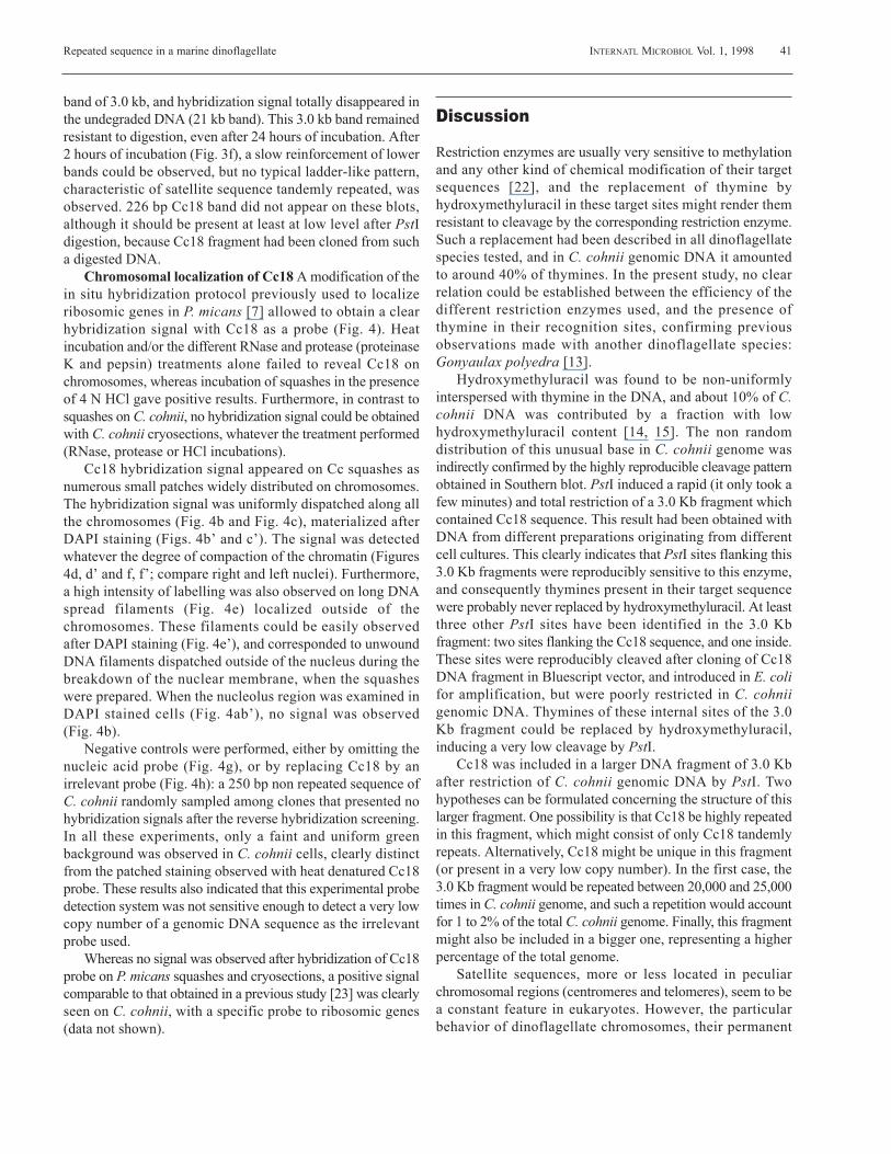
band of 3.0 kb, and hybridization signal totally disappeared inthe undegraded DNA (21 kb band). This 3.0 kb band remainedresistant to digestion, even after 24 hours of incubation. After2 hours of incubation (Fig. 3f), a slow reinforcement of lowerbands could be observed, but no typical ladder-like pattern,characteristic of satellite sequence tandemly repeated, wasobserved. 226 bp Cc18 band did not appear on these blots,although it should be present at least at low level after PstIdigestion, because Cc18 fragment had been cloned from sucha digested DNA.
Chromosomal localization of Cc18 A modification of thein situ hybridization protocol previously used to localizeribosomic genes in P. micans [7] allowed to obtain a clearhybridization signal with Cc18 as a probe (Fig. 4). Heatincubation and/or the different RNase and protease (proteinaseK and pepsin) treatments alone failed to reveal Cc18 onchromosomes, whereas incubation of squashes in the presenceof 4 N HCl gave positive results. Furthermore, in contrast tosquashes on C. cohnii, no hybridization signal could be obtainedwith C. cohnii cryosections, whatever the treatment performed(RNase, protease or HCl incubations).
Cc18 hybridization signal appeared on Cc squashes asnumerous small patches widely distributed on chromosomes.The hybridization signal was uniformly dispatched along allthe chromosomes (Fig. 4b and Fig. 4c), materialized afterDAPI staining (Figs. 4b’ and c’). The signal was detectedwhatever the degree of compaction of the chromatin (Figures4d, d’ and f, f’; compare right and left nuclei). Furthermore,a high intensity of labelling was also observed on long DNAspread filaments (Fig. 4e) localized outside of thechromosomes. These filaments could be easily observedafter DAPI staining (Fig. 4e’), and corresponded to unwoundDNA filaments dispatched outside of the nucleus during thebreakdown of the nuclear membrane, when the squasheswere prepared. When the nucleolus region was examined inDAPI stained cells (Fig. 4ab’), no signal was observed (Fig. 4b).
Negative controls were performed, either by omitting thenucleic acid probe (Fig. 4g), or by replacing Cc18 by anirrelevant probe (Fig. 4h): a 250 bp non repeated sequence ofC. cohnii randomly sampled among clones that presented nohybridization signals after the reverse hybridization screening.In all these experiments, only a faint and uniform greenbackground was observed in C. cohnii cells, clearly distinctfrom the patched staining observed with heat denatured Cc18probe. These results also indicated that this experimental probedetection system was not sensitive enough to detect a very lowcopy number of a genomic DNA sequence as the irrelevantprobe used.
Whereas no signal was observed after hybridization of Cc18probe on P. micans squashes and cryosections, a positive signalcomparable to that obtained in a previous study [23] was clearlyseen on C. cohnii, with a specific probe to ribosomic genes(data not shown).
Discussion
Restriction enzymes are usually very sensitive to methylationand any other kind of chemical modification of their targetsequences [22], and the replacement of thymine byhydroxymethyluracil in these target sites might render themresistant to cleavage by the corresponding restriction enzyme.Such a replacement had been described in all dinoflagellatespecies tested, and in C. cohnii genomic DNA it amountedto around 40% of thymines. In the present study, no clearrelation could be established between the efficiency of thedifferent restriction enzymes used, and the presence ofthymine in their recognition sites, confirming previousobservations made with another dinoflagellate species:Gonyaulax polyedra [13].
Hydroxymethyluracil was found to be non-uniformlyinterspersed with thymine in the DNA, and about 10% of C.cohnii DNA was contributed by a fraction with lowhydroxymethyluracil content [14, 15]. The non randomdistribution of this unusual base in C. cohnii genome wasindirectly confirmed by the highly reproducible cleavage patternobtained in Southern blot. PstI induced a rapid (it only took afew minutes) and total restriction of a 3.0 Kb fragment whichcontained Cc18 sequence. This result had been obtained withDNA from different preparations originating from differentcell cultures. This clearly indicates that PstI sites flanking this3.0 Kb fragments were reproducibly sensitive to this enzyme,and consequently thymines present in their target sequencewere probably never replaced by hydroxymethyluracil. At leastthree other PstI sites have been identified in the 3.0 Kbfragment: two sites flanking the Cc18 sequence, and one inside.These sites were reproducibly cleaved after cloning of Cc18DNA fragment in Bluescript vector, and introduced in E. colifor amplification, but were poorly restricted in C. cohniigenomic DNA. Thymines of these internal sites of the 3.0Kb fragment could be replaced by hydroxymethyluracil,inducing a very low cleavage by PstI.
Cc18 was included in a larger DNA fragment of 3.0 Kbafter restriction of C. cohnii genomic DNA by PstI. Twohypotheses can be formulated concerning the structure of thislarger fragment. One possibility is that Cc18 be highly repeatedin this fragment, which might consist of only Cc18 tandemlyrepeats. Alternatively, Cc18 might be unique in this fragment(or present in a very low copy number). In the first case, the3.0 Kb fragment would be repeated between 20,000 and 25,000times in C. cohnii genome, and such a repetition would accountfor 1 to 2% of the total C. cohnii genome. Finally, this fragmentmight also be included in a bigger one, representing a higherpercentage of the total genome.
Satellite sequences, more or less located in peculiarchromosomal regions (centromeres and telomeres), seem to bea constant feature in eukaryotes. However, the particularbehavior of dinoflagellate chromosomes, their permanent
41Repeated sequence in a marine dinoflagellate INTERNATL MICROBIOL Vol. 1, 1998

condensed state throughout the cell cycle, non banding and theabsence of morphologically discernable centromeric structures,reveal an unusual genomic organization. By cleavage of C.cohnii genomic DNA with any of the enzymes tested, neitherbands representing ladders of oligomer repeats nor isolatedbands could be seen; consequently, no satellite tandemlyrepeated sequences could be directly isolated. Severalpossibilities can explain the absence of such restriction patterncharacteristic of satellite sequences in this work: (i) none ofthe target sequences of the 13 restriction enzymes used werepresent in such hypothetical satellite sequences; (ii) in manysites of these sequences, a high proportion of thymines wasreplaced by hydroxymethyluracil, and was resistant to cleavage,hiding such a repeated structure; and (iii) such tandemlyrepeated sequences do not exist in C. cohnii genome. Althoughno clear indications of the formal existence of such sequencesin C. cohnii genome can be found in previous studies, the highpercentage of repeated sequences [1] makes the last hypothesisvery unlikely.
The GC content of the repeated DNA sequences isolatedin this study was high for 5 sequences. This was markedlydifferent to the average GC content of genomic DNA of C.cohnii, which had been found around 40% [14]. But thispercentage seems variable among dinoflagellate species; 60%of GC content had been determined in P. micans [11]. Satellitesequences in eukaryotes display a high GC content, and thisexplains their different sedimentation in CsCl gradients.
In situ hybridization experiments showed that classicalprotocols used to denature the genomic DNA (heat treatment)were inefficient with the C. cohnii genome, although they hadbeen previously successfully used for P. micans ribosomicgenes [7]. Dinoflagellates display a particular genomicorganization, and their chromosomes contain neithernucleosomes nor histones [10, 16, 23]. Several models of DNApackaging into dinoflagellate chromosomes have been proposedthat were essentially based on electron microscopy studies [17, 28], although more data are needed to find a model thatsolves this intriguing DNA organization. However, from thesestudies, it was obvious that the DNA packaging in P. micans[8] was different than in C. cohnii [9]. In C. cohnii, whose DNAwas very densely packed into small chromosomes, targetsequences for in situ hybridization experiments cannot beaccessible to the nucleic acid probes used. By now, the onlypossibility to label these sequences would be to elongate thechromosome as in the squash technique, with a signal intensityproportional to the unstacking of DNA filaments.
Cc18 labelling appeared uniformly distributed on allchromosomes, and all along each chromosome. This indicatesthat Cc18 is widely interspersed in the C. cohnii genome, andnot restricted to specfic areas of the chromosomes.
This sequence was not detected in P. micans by Southern-blot, nor by in situ hybridization techniques, P. micans beinga dinoflagellate species very different to C. cohnii. Research isin progress to test the presence of this sequence in different
sibling species of C. cohnii, to evaluate the degree of specificityof Cc18, which could constitute a good tool to discriminate, bymolecular hybridization techniques, between sibling or closelyrelated species with no discernable morphological differences.
Acknowledgements The authors wish to thank M. Albert andP. Llaona for their excellent technical assistance, and Dr. T. M.Preston for critical reading of the manuscript. This work wassupported by the CNRS (UMR 7628).
References
1. Allen JR, Roberts TM, Loeblich III AR, Klotz LC (1975) Characterizationof the DNA from the dinoflagellate Crypthecodinium cohnii andimplications for nuclear organization. Cell 6:161-169
2. Anderson DM, Grabher A, Herzog M (1992) Separation of codingsequences from structural DNA in the dinoflagellate Crypthecodiniumcohnii. Mol Mar Biotech 1:89-96
3. Bhaud Y, Salmon JM, Soyer-Gobillard MO (1991) The complex cellcycle of the dinoflagellate protoctist Crypthecodinium cohnii as studiedin vivo and by cytofluorimetry. J Cell Sci 100:675-682
4. Bhaud Y, Barbier M, Soyer-Gobillard MO (1994) A detailed study ofthe complex cell cycle of the dinoflagellate Crypthecodinium cohniiBiecheler and evidence for variation in histone H1 kinase activity. J EukMicrobiol 41:519-526
5. Bodansky S, Mintz LB, Holmes DS (1979) The mesokaryote Gyrodiniumcohnii lacks nucleosomes. Biochim Biophys Res Commun 88:1329-1336
6. Elder JF, Turner BJ (1995) Concerted evolution of repetitive DNAsequences in eukaryotes. Quart Rev Biol 70:297-320
7. Géraud ML, Herzog M, Soyer-Gobillard MO (1991) Nucleolarlocalization of rRNA coding sequences in Prorocentrum micans Ehr.(dinomastigote, kingdom Protoctist) by in situ hybridization. BioSystems26:61-74
8. Haapala OK, Soyer MO (1973) Structure of dinoflagellates chromosomes.Nature New Biol 244:195-197
9. Haapala OK, Soyer MO (1974). Absence of longitudinal differentiation ofdinoflagellate (Prorocentrum micans) chromosomes. Hereditas 78:146-150
10. Herzog M, Soyer MO (1981) Distinctive features of dinoflagellatechromatin. Absence of nucleosome in a primitive species Prorocentrummicans E. Eur J Cell Biol 23:295-302
11. Herzog M, de Marcillac GD, Soyer MO (1982) A high level of thyminereplacement by 5-hydroxymethyluracil in nuclear DNA of the primitivedinoflagellate Prorocentrum micans E. Eur J Cell Biol 27:151-155
12. Hinnebusch AG, Klotz LC, Immergut E, LoeblichIII AR (1980)Deoxyribonucleic acid sequence organization in the genome of thedinoflagellate Crypthecodinium cohnii. Biochemistry 19:1744-1755
13. Lee DG, Mittag M, Sczekan S, Morse D, Hastings JW (1993) Molecularcloning and genomic organization of a gene for luciferin-binding proteinfrom the dinoflagellate Gonyaulax polyedra. J Biol Chem 268:8842-8850
14. Rae PMM (1973) 5-Hydroxymethyluracil in the DNA of a Dinoflagellate.Proc Nat Acad Sci USA 70:1141-1145
15. Rae PMM (1976) Hydroxymethyluracil in eukaryote DNA: a naturalfeature of the Pyrrhophyta (dinoflagellates). Science 194:1062-1064
16. Rizzo PJ, Nooden LD (1973) Isolation and chemical composition ofDinoflagellate nuclei. J Protozool 20:666-672
17. Rizzo PJ (1987) Biochemistry of the dinoflagellate nucleus. In: TaylorFJR (ed), The Biology of Dinoflagellates. London: Blackwell Sci Pub,pp 143-173
18. Roberts TM, Tuttle RC, Allen JR, Loeblich III AR, Klotz LC (1974)New genetic and physicochemical data on structure of dinoflagellatechromosomes. Nature 248:446-447
42 INTERNATL MICROBIOL Vol. 1, 1998 Moreau et al.

43Repeated sequence in a marine dinoflagellate INTERNATL MICROBIOL Vol. 1, 1998
19. Rogers SO, Bendich AJ (1988). Extraction DNA from plant tissues.In: Gelvin SB, Schilperoort RA, Verna DPS (eds) Plant Molecular BiologyManual, A6. Dordrecht: Kluwer Academic Publishers, pp 1-10
20. Ruiz-Lara S, Prats E, Sainz J, Cornudella L (1992) Cloning andcharacterization of a highly conserved satellite DNA from the molluscMytilus edulis. Gene 117:237-242
21. Sainz J, Prats E, Ruiz S, Cornudella L (1992) Organization of repetitivesequences in the genome of the echinoderm Holothuria tubulosa.Biochimie 74:1067-1074
22. Sambrook J, Fritsch EF, Maniatis T (1989) Molecular Cloning: ALaboratory Manual, 2nd ed, Cold Spring Harbor, NY: Cold Spring HarborLaboratory Press, 5.1-5.95
23. Shupe K, Rizzo PJ (1983) Nuclease digestion of chromatin from theeukaryotic algae Olisthodiscus luteus, Peridinium balticum, andCrypthecodinium cohnii. J Protozool 30:599-606
24. Sigee DC (1984) Structural DNA and genetically active DNA inDinoflagellate chromosomes. BioSystems 16:203-210
25. Soyer MO, Haapala OK (1974) Division and function of dinoflagellatechromosomes. J Microsc 19:137-146
26. Soyer-Gobillard MO, Géraud ML, Coulaud D, Barray M, Théveny B,Révet B, Delain E (1990) Location of B- and Z-DNA in thechromosomes of a primitive eukaryote Dinoflagellate. J Cell Biol111:293-308
27. Soyer-Gobillard MO (1996) The genome of the primitive eukaryoteDinoflagellates: organization and functioning. Zoological studies 35:78-84
28. Spector DL (1984) Dinoflagellate nuclei. In: Spector DL (ed)Dinoflagellates. New York: Academic Press, pp 107-147
29. Tuttle RC, LoeblichIII AR (1975) An optimal growth medium for thedinoflagellate Crypthecodinium cohnii. Phycologia 14:1-8