Cloning and Characterization of Human WDR10 , a Novel Gene Located at 3q21 Encoding a WD-Repeat...
12
DNA AND CELL BIOLOGY Volume 20, Number 1, 2001 Mary Ann Liebert, Inc. Pp. 41–52 Cloning and Characterization of Human WDR10, a Novel Gene Located at 3q21 Encoding a WD-Repeat Protein That Is Highly Expressed in Pituitary and Testis COLEMAN GROSS, 1,2 ELFRIDE DE BAERE, 3 ANNIE LO, 1 WENHAN CHANG, 1,2 and LUDWINE MESSIAEN 3 ABSTRACT Members of the steroid–thyroid-retinoid receptor superfamily regulate a spectrum of cellular functions, in- cluding metabolism and growth and differentiation. We sought to isolate novel members of this family by us- ing degenerate oligonucleotide primers directed to sequences encoding the AF-2 domain of these molecules in a PCR-based approach. The AF-2 domain serves a critical function in recruiting coregulatory molecules and in transcriptional activation. We report the cloning and initial characterization of a novel gene, WDR10, which encodes a 140-kD protein that is highly expressed in pituitary and testis. This protein, WDR10p, contains an AF-2 domain as well as seven N-terminal WD repeats and is highly conserved through evolution. Chromoso- mal localization studies placed WDR10 at 3q21, near a locus for the Moebius syndrome, Hailey-Hailey dis- ease, and rhodopsin, which is involved in several forms of retinitis pigmentosa. The expression pattern of WDR10 and its chromosomal location makes this novel gene a candidate gene for the hypogonadism associ- ated with some forms of retinitis pigmentosa and the Moebius syndrome. 41 INTRODUCTION M EMBERS of the steroid–thyroid-retinoid receptor super- family and their ligands regulate diverse physiologic processes, including metabolism, cell growth, and differentia- tion. Recently, an increasing number of novel members of this family have been discovered and have provided valuable in- sights into basic physiologic processes and opportunities for the development of new therapeutics (Mangelsdorf et al., 1995). Ligands of class III members of this superfamily (retinoic acid receptor, RAR; retinoid X receptor, RXR; vitamin D receptor, VDR; peroxisome proliferator–activator receptor, PPAR) have potent antiproliferative and differentiating activities. The iden- tification of related molecules represents an avenue for a greater understanding of the endocrine, paracrine, and autocrine regu- lation of cellular growth. The nuclear hormone receptors share a high degree of struc- tural homology, particularly within the DNA-binding and lig- and-binding domains. Within the C-terminal portion of the lig- and-binding domain, the activation function-2 (AF-2) domain is functionally important in transcriptional activation (Barettino et al., 1994; Danielian et al., 1992). The AF-2 domain folds into an amphipathic a-helix and folds inward on ligand bind- ing, thus providing a surface for the binding of coregulatory molecules (Freedman, 1999; Nolte et al., 1998; Westin et al., 1998). This sequence of events appears to be crucial for the transactivation process. The AF-2 domain is highly conserved among the nuclear re- ceptors and contains a core motif defined by ffXEff, where f represents a hydrophobic amino acid. In an effort to identify molecules related to nuclear receptors that might play a role in the transcriptional control of growth and differentiation, we used a PCR-based approach to isolate novel cDNAs that con- tain sequences encoding potential AF-2 domains using oligonu- cleotides directed to this region of class III nuclear receptors. These studies resulted in the isolation of a novel gene, WDR10, which encodes a protein of 1242 amino acids that has an AF- 2 like domain and seven N-terminal WD-repeat motifs. The WD-repeat motif is a 44- to 60-amino acid unit that variably ends in Trp-Asp and folds into four antiparallel b sheets (Neer et al., 1994; Smith et al., 1999). Although the exact function of this motif is poorly understood, it appears that multiple WD- 1 Endocrine Section, Department of Veterans Affairs Medical Center, San Francisco, California. 2 Department of Medicine University of California, San Francisco, San Francisco. 3 Department of Medical Genetics, Ghent University Hospital, Ghent, Belgium.
-
Upload
ua-birmingham -
Category
Documents
-
view
4 -
download
0
Transcript of Cloning and Characterization of Human WDR10 , a Novel Gene Located at 3q21 Encoding a WD-Repeat...

DNA AND CELL BIOLOGYVolume 20, Number 1, 2001Mary Ann Liebert, Inc.Pp. 41–52
Cloning and Characterization of Human WDR10, a NovelGene Located at 3q21 Encoding a WD-Repeat Protein That Is
Highly Expressed in Pituitary and Testis
COLEMAN GROSS,1,2 ELFRIDE DE BAERE,3 ANNIE LO,1 WENHAN CHANG,1,2
and LUDWINE MESSIAEN3
ABSTRACT
Members of the steroid–thyroid-retinoid receptor superfamily regulate a spectrum of cellular functions, in-cluding metabolism and growth and differentiation. We sought to isolate novel members of this family by us-ing degenerate oligonucleotide primers directed to sequences encoding the AF-2 domain of these molecules ina PCR-based approach. The AF-2 domain serves a critical function in recruiting coregulatory molecules andin transcriptional activation. We report the cloning and initial characterization of a novel gene, WDR10, whichencodes a 140-kD protein that is highly expressed in pituitary and testis. This protein, WDR10p, contains anAF-2 domain as well as seven N-terminal WD repeats and is highly conserved through evolution. Chromoso-mal localization studies placed WDR10 at 3q21, near a locus for the Moebius syndrome, Hailey-Hailey dis-ease, and rhodopsin, which is involved in several forms of retinitis pigmentosa. The expression pattern ofWDR10 and its chromosomal location makes this novel gene a candidate gene for the hypogonadism associ-ated with some forms of retinitis pigmentosa and the Moebius syndrome.
41
INTRODUCTION
MEMBERS of the steroid–thyroid-retinoid receptor super-family and their ligands regulate diverse physiologic
processes, including metabolism, cell growth, and differentia-tion. Recently, an increasing number of novel members of thisfamily have been discovered and have provided valuable in-sights into basic physiologic processes and opportunities for thedevelopment of new therapeutics (Mangelsdorf et al., 1995).Ligands of class III members of this superfamily (retinoic acidreceptor, RAR; retinoid X receptor, RXR; vitamin D receptor,VDR; peroxisome proliferator–activator receptor, PPAR) havepotent antiproliferative and differentiating activities. The iden-tification of related molecules represents an avenue for a greaterunderstanding of the endocrine, paracrine, and autocrine regu-lation of cellular growth.
The nuclear hormone receptors share a high degree of struc-tural homology, particularly within the DNA-binding and lig-and-binding domains. Within the C-terminal portion of the lig-and-binding domain, the activation function-2 (AF-2) domainis functionally important in transcriptional activation (Barettino
et al., 1994; Danielian et al., 1992). The AF-2 domain foldsinto an amphipathic a-helix and folds inward on ligand bind-ing, thus providing a surface for the binding of coregulatorymolecules (Freedman, 1999; Nolte et al., 1998; Westin et al.,1998). This sequence of events appears to be crucial for thetransactivation process.
The AF-2 domain is highly conserved among the nuclear re-ceptors and contains a core motif defined by ffXEff, wheref represents a hydrophobic amino acid. In an effort to identifymolecules related to nuclear receptors that might play a role inthe transcriptional control of growth and differentiation, weused a PCR-based approach to isolate novel cDNAs that con-tain sequences encoding potential AF-2 domains using oligonu-cleotides directed to this region of class III nuclear receptors.These studies resulted in the isolation of a novel gene, WDR10,which encodes a protein of 1242 amino acids that has an AF-2 like domain and seven N-terminal WD-repeat motifs. TheWD-repeat motif is a 44- to 60-amino acid unit that variablyends in Trp-Asp and folds into four antiparallel b sheets (Neeret al., 1994; Smith et al., 1999). Although the exact functionof this motif is poorly understood, it appears that multiple WD-
1Endocrine Section, Department of Veterans Affairs Medical Center, San Francisco, California.2Department of Medicine University of California, San Francisco, San Francisco.3Department of Medical Genetics, Ghent University Hospital, Ghent, Belgium.

repeats form a donut-like structure or b propeller that gener-ates a scaffold which acts as a platform for protein–protein in-teractions (Neer et al., 1994; Smith et al., 1999; Wall et al.,1995). The WDR10 gene encodes a 140-kD protein (WDR10p)that has been highly conserved throughout evolution, with or-thologs in C. elegans and D. melanogaster, suggesting a criti-cal function for this protein. WDR10 is highly expressed in testisand pituitary gland and may therefore play an important role intheir physiology. Chromosomal localization studies placeWDR10 at 3q21, near the rhodopsin gene (RHO), the Hailey-Hailey disease (HHD) gene, and the Moebius syndrome(MBS2) locus.
MATERIALS AND METHODS
Rapid amplification of cDNA ends and PCR
The cDNA was synthesized from poly(A)1 RNA either ex-tracted from cells or obtained commercially (Clontech, PaloAlto, CA). The cDNA ends were ligated to the AP-1 adaptorincluded in the Marathon cDNA Amplification kit (Clontech).Initial 59-RACE was carried out using the supplied AP-1 primerand a downstream degenerate AF-2 domain oligonucleotideprimer (59-ACBCTTCGAGCBCAAGGGGCHTTBGCTT-3 9).Initial 59-RACE thermocycling parameters were 94°C for 1 minand 28 cycles of 94°C for 30 sec, 60°C for 30 sec, and 68°Cfor 4 min followed by 8 min at 68°C. Additional 59 and 39sequence-directed RACE primers were used to complete am-plification of the WDR10 cDNA (primers 18CA1: 59-CAGTG-GCTAGCAGAGAACGATCGCTTG-39 and 18CB2: 59-CAAA-GCGATCGTTCTCTGCTAGCCACTG-3 9). Thermocyclingparameters employed a modified Touchdown protocol (Don etal., 1991) to limit artifacts (94°C for 1 min; five cycles of 94°Cfor 30 sec, 72°C for 4 min; five cycles at 94°C for 30 sec, 70°Cfor 4 min; and 25 cycles at 94°C for 20 sec, 68°C for 4 min).Full-length WDR10 cDNA was amplified using FL-7 and FL-4 primers (FL-7: 59-TTTTCTGGCTTTCCCTTTCGGACAT-GCG-39 and FL-4: 59-TTTCGACAGTTTAACTCTTTATTC-TCCTTCACAGC-3 9) under the following thermocycling para-meters: 95°C for 3 min; 10 cycles of 94°C for 20 sec, 64°C for30 sec, 68°C for 6 min; 20 cycles of 94°C for 20 sec, 64°C for30 sec, 68°C for 20 sec 1 5 sec per cycle; and 72°C for 10 min.The amplified 4-kb WDR10 cDNA was cloned into the pCR-XL-TOPO cloning vector (Invitrogen, Carlsbad, CA).
DNA sequencing
DNA sequencing was carried out at the Biomolecular Re-source Center core sequencing facility at UCSF on an ABI 3700automated sequencing machine using BigDye Terminators (Ap-plied Biosystems, Foster City, CA).
Tissue culture and transfection
All cell lines were obtained from the UCSF tissue culture fa-cility or ATCC (Manassas, VA) except for aT3-1 gonadotropecells (Windle et al., 1990), which were the generous gift of Dr.P. Mellon (University of California-San Diego). All media andtissue culture reagents were obtained from GIBCO/BRL(Rockville, MD). The HEK293 human embryonal kidney,HeLa, and mouse aT3-1 pituitary gonadotrope cells were grown
in DMEM supplemented with 10% fetal bovine serum (FBS);MCF-7 human breast cancer and LNCaP human prostate can-cer cells were grown in RPMI-1640 supplemented with 10%FBS; and rat GH-3 somatolactotropes were grown in F-10Ham’s Nutrient Mix with 15% horse serum, 2.5% FBS. Celllines were cultured under standard conditions.
Cell cultures were transfected with 2 to 7 mg of cDNA plas-mids in 100-mm dishes at a cell density of 0.5 to 2 3 106 perdish using Effectene transfection reagent (Qiagen, Valencia,CA) at a 10:1 Effectene:DNA ratio following the manufac-turer’s recommendations. For expression studies, a plasmidcontaining the WDR10 cDNA in pcDNA 3.1/hygro1 was em-ployed (pcDNA 3.1-WDR10). In cellular localization studies,cells transfected with a plasmid (WDR10-pGFP) containing theWDR10 cDNA fused in-frame to the 59 end of the EGFP cDNAof pEGFP N3 (Invitrogen) were visualized on a TCS SP con-focal microscope (Leica, Heidelberg, Germany).
Western blot analysis
Western analysis was carried out as previously describedwith modifications (Gross et al., 1998a,b). Cell lysates wereprepared in RIPA buffer (50 mM HEPES, pH 7.4; 1% deoxy-cholate, 1% Triton X-100, 0.1% SDS, 150 mM NaCl, 1 mMEDTA) using standard conditions. Cleared aliquots of the su-pernatant liquids were incubated at ambient temperature, 37°C,or 100°C prior to SDS-PAGE. Membranes were probed withan affinity-purified rabbit antibody that was raised to a peptidecorresponding to the first 25 amino acids of human WDR10p(5A2) at a dilution of 1:1250. In some experiments, the 5A2antibody was preadsorbed with excess peptide antigen. An anti-green fluorescent protein (GFP) antibody (Clontech) was usedas the primary antibody (1:150) in some experiments.
Northern blot analysis
Northern analysis was done as previously described (Grosset al., 1998a,b). The membranes were hybridized with the 32P-labeled 18C cDNA probe complementary to the 39 portion ofthe WDR10 cDNA. The human multiple tissue RNA blot waspurchased from Clontech.
In situ hybridization
Frozen sections from various rat tissues were used for in situhybridization experiments. Slides were washed in phosphatebuffered saline (PBS; 80 mM Na2HPO4, 20 mM NaH2PO4, 100mM NaCl), fixed in 4% paraformaldehyde, and rinsed in PBStwice. The tissue sections were incubated in the presence ofproteinase K for 5 min and then washed in 4% paraformalde-hyde (PFA) for 10 min. After a 1-min wash in PBS and deion-ized H2O, the sections were dehydrated with three sequential1-min washes in ethanol (70%, 90%, 100%) and air drying. Thesections were prehybridized at 60°C in hybridization buffer(50% formamide, 300 mM NaCl, 20 mM Tris, pH 8; 5 mMEDTA, 13 Denhardt’s solution, 10 mM NaH2PO4, 10% dex-tran sulfate, yeast tRNA 10 mg/ml) for 1 h. Fresh hybridiza-tion solution (50 2 100 ml/tissue section) containing 10 ng ofa digoxigenin–11-UTP-labeled 0.4-kb antisense riboprobe ofhuman WDR10 was added to the sections, which were incu-bated overnight. The antisense riboprobe was labeled using a
GROSS ET AL.42

DIG RNA labeling kit (Roche Molecular Biochemicals, Indi-anapolis, IN) according to the manufacturer’s instructions. Theslides were washed following standard procedures. Hybridiza-tion signals were detected using anti-digoxigenin-AP Fab frag-ments and NBT/BCIP substrate (Roche).
Filter screening of the human RPCI1 PAC andchromosome 3 LLNL cosmid libraries
For hybridization against the human RPCI1 and chromosome3 cosmid libraries (UK-MRC), 20 ng of the 4-kb WDR10 cDNAwas randomly labeled with 32P-dCTP using the MegaprimeDNA Labeling System (Amersham Phamacia Biotech, Uppsala,Sweden) according to the manufacturer’s instructions. The pre-hybridization, hybridization, and wash steps were carried outfollowing standard procedures (De Baere et al., 1999).
Fluorescence in situ hybridization
Probes were biotinylated (biotin-16-dUTP; Roche) or digox-igenated (digoxigenin-11-dUTP; Roche Molecular Biochemi-cals, Mannheim, Germany) by standard nick translation. Slideswere treated for R-banding as described (Cherif et al., 1990).A 50-ng sample of PAC DNA (70-I 11; GenBank AccessionNo. AC000380) was cohybridized with a probe (p`3.5) for thecentromeric region of chromosome 3 (Delattre et al., 1987) onR-banded metaphase chromosomes to determine the map posi-tion and with a YAC (Y919H6) mapping to 3q23 (De Baere etal., 1999). The FISH was performed as described (De Baere etal., 1999; Speleman et al., 1992), and slides were observed un-der a standard fluorescence microscope equipped with a 100 WHg lamp. Images were recorded with the ISIS digital imagingsystem (Meta Systems, Altlussheim, Germany).
CLONING AND CHARACTERIZATION OF WDR10 43
FIG. 1. WDR10 cDNA and deduced amino acid sequence. The seven N-terminal WD-repeat motifs are underlined. The AF-2-like domain is indicated by broken underline. Two potential nuclear localization sequences are shown in gray highlight. Fourcysteines conserved in WDR10p and its two orthologs are shown in reverse highlight. The polyadenylation signal is boxed.

RESULTS
Cloning of WDR10
We chose to screen human cDNA libraries from tissues thatdemonstrate an antiproliferative or differentiation response toclass III nuclear receptor ligands (vitamin D, retinoids). There-fore, we screened human HL60 leukemia and prostate and coloncancer libraries with degenerate oligonucleotides directed to se-
quences encoding the AF-2 domains of these receptors that con-tained 81 permutations along with a common upstream adap-tor primer. In addition to several known nuclear receptors, weidentified a novel 1.2-kb cDNA that demonstrated strong ex-pression in testis and pituitary (see below). On the basis of the1.2-kb cDNA sequence, 59- and 39-RACE was used to gener-ate the full-length WDR10 cDNA. The 59- and 39-RACE frag-ments disclosed an ORF of 3726 bp. The 39 fragment contained
GROSS ET AL.44
FIG. 2. Multiple sequence alignment of WDR10p and its orthologs. The WDR10p deduced amino acid sequence and that ofits orthologs in D. melanogaster (CG7161), C. elegans (f23B2.4), and mouse (GenBank Accession No. AF296075) are shownin a multiple sequence alignment (Corpet, 1988; http://www.toulouse.inra.fr). Numbering is relative to the longest sequence(f23B2.4). Residues sharing identity between sequences are highlighted.

an in-frame stop codon, a polyadenylation signal, and a poly(A)tail, indicating that it represented the 39 end of the WDR10cDNA. Although the 59 cDNA fragment contained a start codon(ATG) with a good Kozak sequence (Kozak, 1996) and an up-stream stop codon, we performed additional rounds of 59-RACEto extend the cDNA sequence from the coding region into the59 untranslated region (UTR) of WDR10. Multiple 59-RACEcDNA clones obtained from testis and pituitary libraries alldemonstrated the same 59-UTR sequence and start codon.
Using RT-PCR in a human testis cDNA library with oligonu-cleotide primers directed to the 59 and 39 UTR (FL-7 and FL-4), we cloned the entire WDR10 cDNA (Fig. 1, GenBank Ac-cession No. AF244930). In addition, we isolated a longervariant of the WDR10 cDNA, which appeared to be attributableto incorporation of an additional 153-bp exon near the 59 endof the gene (GenBank Accession No. AF244931). The geneticnotation WDR10 was chosen on the basis of the recommenda-tions of the HUGO Nomenclature Committee.
Analysis of the WDR10 cDNA and the WDR10p protein
BLAST (Altschul et al., 1997) searches against the expressedsequence tag (EST) database (dbEST) identified full-lengthcDNA orthologs of WDR10 in C. elegans (f23B2.4; GenBankAccession no. CAB05177) and D. melanogaster (CG7161;GenBank Accession No. AAF50423). The translated sequencesof the cDNA orthologs are 60% homologous and 27% identi-
cal and 68% homologous and 36% identical to WDR10p re-spectively, over their entire length (Fig. 2). Two overlappingmouse ESTs were found (GenBank Accession Nos. AL023084,542 bp, and AA002413, 344 bp) that have a high degree of ho-mology (84% each) with the 39 end of the WDR10 cDNA se-quence (bp 3123–3654). We completed sequencing of the lat-ter clone (AA002413), which extended the sequence data to atotal of 1031 bp (GenBank Accession No. AF296075). Joiningthis EST sequence to EST AL023084 generated 1207 bp of con-tiguous cDNA sequence containing an ORF that encodes 273amino acids. This amino acid sequence has 83% identity withWDR10p from residue 978 to 1242. The multiple sequencealignment (Corpet, 1988) is shown in Figure 2.
CLONING AND CHARACTERIZATION OF WDR10 45
FIG. 3. Human multiple tissue RNA blot. A blot containingpoly(A)1 RNA from multiple human tissues was probed witha 32P-labeled WDR10 cDNA probe. A1–8: whole brain, amyg-dala, caudate, cerebrum, frontal lobe, hippocampus, medulla,respectively; B1–7: occipital lobe, putamen, substantia nigra,temporal lobe, thalamus, subthalamic nucleus, spinal cord;C1–8: heart, aorta, skeletal muscle, colon, bladder, uterus,prostate, stomach; D1–8: testis, ovary, pancreas, pituitary,adrenal, thyroid, salivary gland, breast; E1–8: kidney, liver,small intestine, spleen, thymus, leukocyte, lymph node, bonemarrow; F1–4: appendix, lung, trachea, placenta; G1–7: fetaltissues—brain, heart, kidney, liver, spleen, thymus, lung; H1–8:negative controls: yeast RNA, yeast tRNA, E. coli rRNA, E.coli DNA, poly r(A), human C0t1 DNA, human DNA (100 ng),human DNA (500 ng).
FIG. 4. Northern analysis of WDR10. Blots were probed witha 32P-labeled WDR10 cDNA probe. (A) poly(A)1 RNA (1mg/lane) from human testis, pituitary, heart, and prostate. (B)HL60 human leukemia cells (1 mg poly(A)1 RNA) and MCF-7 human breast cancer cells (20 mg of total RNA). (C) TotalRNA (30 mg/lane) from rat GH3 and mouse aT3 pituicytes,LNCaP human prostate cancer cells, and MCF-7 cells.

GROSS ET AL.46
FIG. 5. In situ hybridization of WDR10 in rat tissues. (A) Pituitary (a, anterior; i, intermediate; p, posterior lobe). (B) Testis.(C) Ovary. (D) Dorsal prostate from adult rats. (E–H) Sense controls (no hybridization signals). Expression in testis (arrow: sper-matogonia; arrowhead: spermatids) and ovarian follicles appears cell-stage specific (arrowhead: early stage, arrow: midstage fol-licle).

Analysis of the deduced WDR10p sequence of 1242 aminoacids using the Protein Sequence Analysis (PSA) system(http://bmerc-www.bu.edu) disclosed that WDR10p has a 95%to 100% probability of being a WD-repeat protein with sevenWD repeats in its N terminus (see Fig. 1) (Stultz et al., 1993;White et al., 1994). An AF-2-like domain is present (aminoacids 719–732), bearing homology to the core AF-2 domainsof nuclear receptors (Fig. 1). Similar to the prototypical AF-2domain of nuclear receptors, this region of WDR10p was pre-dicted to fold into an a-helix by the Network Protein SequenceAnalysis server (http://pbil.ibcp.fr), which models secondaryprotein structure (Garnier et al., 1996). In addition to the WD-repeats and the AF-2 like domain, WDR10p contains two po-tential nuclear localization sequences (NLS) (Hicks andRaikhel, 1995), one of which is similar to a recently reportedNLS (PPXR) for the VDR (Michigami et al., 1999). Finally,the C-terminus contains four cysteines, which are conservedamong the orthologs of WDR10p and may serve an importantfunction such as coordination of divalent cations (e.g., zinc;Figs. 1 and 2).
Although the WD-repeat structure does not suggest a func-tion for WDR10p, this portion of the molecule is homologousto the WD-repeat motifs in the yeast transcriptional repressorTup-1 (Komachi and Johnson, 1997; Zhang et al., 1991) and arelated fungal protein, RCO-1 (Yamashiro et al., 1996). InBLAST searches against the nonredundant (nr) and Swiss-Protdatabases, rat selective LIM binding protein (SLB) (Howardand Maurer, 2000) was the only known protein to demonstratesimilarity with the C-terminal portion of WDR10p. Both mol-
ecules have seven N-terminal WD repeats, and they share 23%amino acid identity in the C terminus from amino acids 656 to885 of WDR10p. Interestingly, SLB is expressed at high lev-els in pituitary and testis, as is WDR10p (see below).
Expression of WDR10
Northern analysis. We performed a tissue survey to deter-mine the pattern of WDR10 expression. A 1.2-kb midregionWDR10 cDNA probe was hybridized with a human multipletissue RNA blot. Expression of WDR10 mRNA was found tobe very high in testis and moderately high in pituitary (Fig. 3).The WDR10 mRNA was expressed at significantly lower lev-els in several other tissues. Northern blot analysis with theWDR10 cDNA probe revealed a single 4-kb band in testis andpituitary (Fig. 4). A smaller, 3.5-kb, WDR10 transcript was de-tected in HL60 leukemia cells; however, the 4-kb transcript ap-peared to be expressed in most other tissues. In pituitary cells,we found WDR10 expression to be greater in mouse aT 3 go-nadotropes than in rat GH3 somatolactotropes. However, wecannot discount species variation as a confounder.
In situ hybridization. On the basis of our initial data thatdemonstrated high expression of WDR10 in the pituitary andtestis, we carried out our in situ survey on reproductive tissuesin the rat using a digoxigenin-labeled antisense WDR10 ribo-probe (Fig. 5). Specific hybridization was observed in late-stagespermatids and spermatogonia within the testis. The testis ofthe rat displays synchronization of spermatogenesis such thatonly certain cell types occur together in any one section of an
CLONING AND CHARACTERIZATION OF WDR10 47
FIG. 6. Combined genetic and physical map of human chromosomal region 3q21-3q23. (A) Positions of critical regions of fourgenetic disorders: retinitis pigmentosa 4 (RP4), Hailey-Hailey disease (HHD), Moebius syndrome 2 (MBS2), and blepharophi-mosis syndrome (BPES). The position of WDR10 with respect to the rhodopsin gene (RHO; RP4 gene), marker WI-9557, ATP2C1(HHD gene), D3S1587, D3S1292, D3S1576, S0X14, D3S3586, and D3S1316 is shown. The location of these syndrome genesand markers is derived from the NCBI Human GeneMap99 (Deloukas et al., 1998; http://www.ncbi.nlm.nih.gov/genemap/) andpublished data (Hu et al., 2000; Sudbrak et al., 2000; De Baere et al., 1999; Kremer et al., 1996). On the right, the position ofWDR10 with respect to RHO and WI-9557 in a human PAC clone (pDJ70i11; GenBank Accession No. AC000380) is shown.(B) Ideogram of human chromosome 3 showing cytogenetic banding. The black bar to the left of the chromosome indicates cy-togenetic band 3q21. (C) Mapping of WDR10 to 3q21 by FISH analysis: cohybridization of the WDR10-containing PAC pDJ70i11(lower yellow signal) and a centromeric chromosome 3 probe (pa3.5; upper yellow signal) on R-banded metaphase chromosome3. (D) Cohybridization of the WDR10 containing PAC pDJ70i11 (3q21) (red) and Y919H6 (green) from the BPES critical re-gion at 3q23 on unbanded metaphase chromosome 3, confirming the large distance between WDR10 and the BPES locus.
GROSS ET AL.48
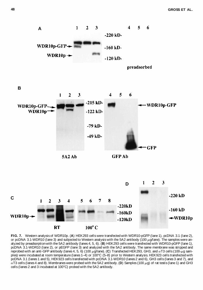
FIG. 7. Western analysis of WDR10p. (A) HEK293 cells were transfected with WDR10-pGFP (lane 1), pcDNA 3.1 (lane 2),or pcDNA 3.1-WDR10 (lane 3) and subjected to Western analysis with the 5A2 antibody (100 mg/lane). The samples were an-alyzed by preadsorption with the 5A2 antibody (lanes 4, 5, 6). (B) HEK293 cells were transfected with WDR10-pGFP (lane 1),pcDNA 3.1-WDR10 (lane 2), or pEGFP (lane 3) and analyzed with the 5A2 antibody. The same membrane was stripped andreprobed with an anti-GFP antibody (lanes 4, 5, 6) (100 mg/lane). (C) Transfected HEK293, GH3, and aT3 cells (100-mg sam-ples) were incubated at room temperature (lanes 1–4) or 100°C (5–8) prior to Western analysis. HEK923 cells transfected withpcDNA 3.1 (lanes 1 and 5), HEK923 cells transfected with pcDNA 3.1-WDR10 (lanes 2 and 6), GH3 cells (lanes 3 and 7), andaT3 cells (lanes 4 and 8). Membranes were probed with the 5A2 antibody. (D) Samples (100 mg) of rat testis (lane 1) and GH3cells (lanes 2 and 3 incubated at 100°C) probed with the 5A2 antibody.
A
B
C
D

individual spermatic tubule. Granulosa cells of the ovaryshowed specific hybridization signals in a cell stage-specificfashion as well. Early-stage follicles had no hybridization withthe WDR10 riboprobe, whereas signal was clearly seen in later-stage follicles. Selective hybridization was observed in the an-terior portion of the pituitary gland and in prostate epithelium.
Chromosomal localization of human WDR10 and therhodopsin gene
Comparison of the WDR10 cDNA sequence with the HighThroughput Genome Sequences (HGTS) database at the Na-tional Center for Biotechnology Information (NCBI;http://www.ncbi.nlm.nih.gov/) revealed .99% identity withPAC pDJ70i11 (GenBank Accession No. AC000380), a ge-nomic clone corresponding to chromosome 3q. This compari-son indicated that the WDR10 gene has at least 28 exons spreadover approximately 65 kb of genomic DNA.
To confirm the chromosomal location of WDR10, a humanPAC and a chromosome 3 cosmid library (UK-MRC) werescreened using a WDR10 cDNA probe. The following PACsand cosmids were identified: PAC 70-I 11, PAC 172-B 10, AC7-F 2, AC 8-I 7, AC 7-P 6, AC 11-E 14, AC 12-N 17, AC 12-P 7, AC 38-D 6, AC 38-F 16, AC 48-A 5, AC 48-C 7, AC 41-F 11, AC 41-N9, AC 54-C 1, AC 51-H 12, AC 53-L 22, andAC 53-N 18. The PAC 70-I 11 was mapped to chromosome3q21 by FISH analysis on R-banded metaphase chromosomes(Fig. 6B, C, and D).
Sequence analysis of PAC 70-I 11 (GenBank Accession No.AC000380) showed the presence of marker WI-9557 and RHO,located 35 kb and 8 kb telomeric, respectively, from WDR10(Fig. 6A). RHO is involved in an autosomal dominant and re-cessive form of retinitis pigmentosa (RP4; MIM 180380) (Dryjaet al., 1990) and was previously mapped to 3q21–q24.
Several genetic syndromes map to the chromosomal regionof WDR10 (Fig. 6A): retinitis pigmentosa (RP4; MIM 108380),HHD (MIM 169600), and MBS2 (MIM 601471) at 3q21 andthe blepharophimosis–ptosis-epicanthus inversus syndrome
(BPES; MIM 110100) at 3q23. For the mapping of WDR10 rel-ative to the BPES locus, PAC 70-I 11 was cohybridized on nor-mal metaphase chromosomes with a YAC spanning the BPEScritical region at 3q23 (De Baere et al., 1999). This procedureconfirmed that there is a distance of one chromosomal band be-tween WDR10 and the BPES locus (Fig. 6D).
Western analysis
We developed an antibody (5A2) to a peptide that corre-sponded to the first 25 amino acids of the predicted WDR10psequence. Using this antibody, a 140-kD protein was detectedon Western analysis in HEK239 cells transfected with aWDR10p expression plasmid (pcDNA3.1–WDR10; Fig. 7).This is the predicted molecular weight of WDR10p based onthe deduced amino acid sequence.
The specificity of the 5A2 antibody was confirmed using twoapproaches. First, the antibody was adsorbed with the peptideto which it was raised prior to exposure to the membrane. Thisstep eliminated the 140-kD signal as well as a higher molecu-lar weight signal present in nontransfected cells (Fig. 7A). Sec-ond, Western analysis was performed on HEK293 cells trans-fected with a WDR10-GFP cDNA (WDR10-pGFP), whichlengthened the translated product by 27 kD (Fig. 7B). Theseexperiments indeed confirmed that the fusion protein was of ahigher molecular weight. The blot was also probed with an anti-GFP antibody, which recognized the WDR10p-GFP protein butnot WDR10p (Fig. 7B). Western analysis on protein extractsmade from rat testis and aT3 gonadotropes also demonstratedthe 140-kD WDR10p band (Fig. 7C). Detection of WDR10pwith the 5A2 antibody in GH3 somatolactotropes was prob-lematic. When samples were not heated at 100°C prior to West-ern analysis, it was difficult to detect a specific band at 140 kD(Fig. 7C, lane 3) even over a range of concentrations of 20 to100 mg/lane (data not shown). When samples were heated to100°C, WDR10p was not visualized in HEK293 cells trans-fected with a WDR10p expression plasmid, GH3 cells, or aT3cells (Fig. 7C, lanes 6–8). A high molecular weight band (ap-
CLONING AND CHARACTERIZATION OF WDR10 49
FIG. 8. Cellular localization of WDR10p. A full-length WDR10-GFP cDNA (WDR10-pGFP) expression plasmid was trans-fected into HEK293, MCF 7, and HeLa cells. Cellular localization of the fusion protein was determined with a confocal micro-scope. The fusion protein appears to localize to the cytoplasm in all three cells, with little if any localization in the cell mem-brane and nucleus.

proximately 180 kD) demonstrated crossreactivity with the 5A2antibody but is likely nonspecific, as it is present in cells thatdo not express WDR10 transcripts on Northern analysis.
Cellular localization
To determine the cellular localization of WDR10p, we gen-erated expression plasmids that contained the entire WDR10coding sequence fused in-frame with, and upstream of, the GFPcDNA (WDR10-pGFP). This construct contained the 59 UTRof WDR10. It was transfected into several cell types, includingHEK293, HeLa, and MCF-7 cells. As shown in Figure 8, flu-orescence was located primarily within the cell cytoplasm: therewas little if any nuclear accumulation.
DISCUSSION
WDR10 is a novel gene that appears to have a complex struc-ture and gives rise to several transcripts, as indicated by North-ern analysis and by the isolation of a 153-bp longer alternativetranscript. Northern analysis indicated a 4-kb transcript in mosttissues and a 3.5-kb transcript in HL60 leukemia cells.
The WDR10 gene is located on chromosome 3q21, nearRHO, which is responsible for an autosomal dominant and arecessive form of retinitis pigmentosa (RP4), the HHD gene(MIM 169600), and a locus for Moebius syndrome (MBS2;MIM 601471) and relatively far from the BPES locus at 3q23.Two recent physical maps of the HHD region show the close-ness of WI-9557 and RHO centromeric to the recently identi-fied HHD gene, ATP2C1 (Hu et al., 2000; Sudbrak et al., 2000).In a recent linkage analysis of a Dutch kindred with Moebiussyndrome (Kremer et al., 1996), a maximum LOD score wasobtained for STS marker D3S1292. On the map published byHu et al. (2000), this marker is placed in the HHD critical re-gion telomeric to the HDD gene, which implies that the MBS2locus is located relatively close to WDR10 (see Fig. 6).
Interestingly, Moebius syndrome (Baraitser and Rudge,1996; Brackett et al., 1991; Kawai et al., 1990), as well as BPEStype 1 (Jones and Collin, 1984; Smith et al., 1989; Zlotogoraet al., 1983), is associated with hypogonadism. Rarely, retini-tis pigmentosa, which is genetically heterogeneous, is associ-ated with hypogenitalism/hypogonadism such as in the Lau-rence-Moon (MIM 245800) and Bardet-Biedl (MIM 209900)syndromes. To our knowledge, HHD is not associated with hy-pogonadism. Given the expression pattern of WDR10 at highlevels in pituitary gland and in testis, this gene might be a can-didate for the hypogonadism associated with MBS2 and certaincases of retintis pigmentosa. Its chromosomal localization on3q21 excludes WDR10 from being a candidate for the cause ofhypogonadism in BPES type 1 (Fig. 6). Our data may supporta role for WDR10 in hypogonadism associated with individualsyndromes whose genetic loci have not yet been determined.
The homology of WDR10p to orthologs in C. elegans, D.melanogaster, and rat SLB suggests that WDR10p is a novelmember of a new class of proteins. Moreover, its high conser-vation through evolution suggests an important role for this pro-tein. At present, we can only speculate about the functional roleof WDR10p, but its high expression in certain endocrine tis-sues, including the testis and pituitary, may indicate an impor-tant functional role in these tissues. Its sequence similarity to
yeast Tup-1 and rat SLB suggests that WDR10p interacts withtranscription factors. The SLB protein was recently identifiedas a specific binding partner of the LIM homeodomain proteinLhx3, which plays a role in pituitary glycoprotein hormone se-cretion and pituitary organogenesis (Howard and Maurer,2000). Like WDR10, SLB has high levels of expression in pi-tuitary and testis.
It is interesting to speculate that WDR10p interacts withother proteins to modulate transcription, similar to the actionof SLB. Indeed, we isolated WDR10 based on the presence ofa sequence encoding an AF-2 domain that may function in pro-tein–protein interaction. The finding that WDR10p is localizedto the cytoplasm does not exclude its functioning as a tran-scriptional modulator, as SLB also resides primarily in the cy-toplasm. However, with increased levels of Lhx3, SLB translo-cates into the nucleus (Howard and Maurer, 2000). A similarphenomenon has been reported for the WD-repeat-containingp48 subunit of the damaged DNA binding protein complex(DDB) (Shiyanov et al., 1999), which interacts with the E2F1transcription factor.
The stage-specific expression of WDR10 within developingsperm and ovarian follicles suggests that this gene has a role indifferentiation within the gonads. The process of differentia-tion, particularly for developing sperm, results from a complexseries of transcriptional events that result in highly specializedfunction (Berruti, 1998). It is not possible to say at this pointwhether WDR10 regulates these events or is merely a markerof them. Expression of WDR10 in the testis occurs in germ cells,whereas in the ovary, its expression is limited to somatic gran-ulosa cells. The implications of this finding are unclear and willbe the subject of further study.
In summary, we have identified a novel gene that is ex-pressed abundantly in pituitary and testis. Its high degree ofevolutionary conservation suggests an important physiologicfunction. Its expression pattern and sequence homologies sug-gest a role in gonadal differentiation, a hypothesis that needsto be confirmed by further functional studies. According to thechromosomal location at 3q21, WDR10 may be a candidate genefor certain genetic forms of hypogonadism, which may be elu-cidated by the mapping of cytogenetic rearrangements with re-spect to WDR10, by mutation studies or by the construction ofa WDR10 ortholog knockout mouse.
ACKNOWLEDGMENTS
We thank Drs. Y. Hirokawa and H. Ingraham for technicalassistance with the in situ hybridization studies. We thank ScottMunson for assistance with production of the figures. This studywas funded by National Institutes of Health Grant DK 02459(to CG) and the Fund for Scientific Research-Flanders FWOGrant 315-18000 (to EDB).
REFERENCES
ALTSCHUL, S.F., MADDEN, T.L., SCHAFFER, A.A., ZHANG, J.,ZHANG, Z., MILLER, W., and LIPMAN, D.J. (1997). GappedBLAST and PSI-BLAST: A new generation of protein databasesearch programs. Nucleic Acids Res. 25, 3389–3402.
GROSS ET AL.50

BARAITSER, M., and RUDGE, P. (1996). Moebius syndrome, an ax-onal neuropathy and hypogonadism. Clin. Dysmorphol. 5, 351–355.
BARETTINO, D., VIVANCO RUIZ, M.M., and STUNNENBERG,H.G. (1994). Characterization of the ligand-dependent transactiva-tion domain of thyroid hormone receptor. EMBO J. 13, 3039–3049.
BERRUTI, G. (1998). Signaling events during male germ cell differ-entiation: Bases and perspectives. Front. Biosci. 3, D1097–D1098.
BRACKETT, L.E., DEMERS, L.M., MAMOURIAN, A.C., ELLEN-BERGER, C. JR., and SANTEN, R.J. (1991). Moebius syndrome inassociation with hypogonadotropic hypogonadism. J. Endocrinol. In-vest. 14, 599–607.
CHERIF, D., JULIER, C., DELATTRE, O., DERRE, J., LATHROP,G.M., and BERGER, R. (1990). Simultaneous localization of cos-mids and chromosome R-banding by fluorescence microscopy: Ap-plication to regional mapping of human chromosome 11. Proc. Natl.Acad. Sci. USA 87, 6639–6643.
CORPET, F. (1988). Multiple sequence alignment with hierarchicalclustering. Nucleic Acids Res. 16, 10881–10890.
DANIELIAN, P.S., WHITE, R., LEES, J.A., and PARKER, M.G.(1992). Identification of a conserved region required for hormone de-pendent transcriptional activation by steroid hormone receptors.EMBO J. 11, 1025–1033.
DE BAERE, E., VAN ROY, N., SPELEMAN, F., FUKUSHIMA, Y.,DE PAEPE, A., and MESSIAEN, L. (1999). Closing in on the BPESgene on 3q23: Mapping of a de novo reciprocal translocationt(3;4)(q23;p15.2) breakpoint within a 45-kb cosmid and mapping ofthree candidate genes, RBP1, RBP2 and b9-COP, distal to the break-point. Genomics 57, 70–78.
DELATTRE, O., BERNARD, A., MALFOY, B., MARLHENS, F.,VIEGAS-PEQUIGNOT, E., BROSSARD, C., HAGUENAUER, O.,CREAU-GOLDBERG, N., VAN CONG, N., and DUTRILLAUX,B. (1987). Isolation and characterization of an alphoid DNA sequencerecently amplified on human chromosome 3. Nucleic Acids Res. 15,8561.
DELOUKAS, P., SCHULER, G.D., GYAPAY, G., BEASLEY, E.M.,SODERLUND, C., RODRIGUEZ-TOMÉ, P., HUI, L., MATISE,T.C., MCKUSICK, K.B., BECKMANN, J.S., BENTOLILA, S., BI-HOREAU, M.-T., BIRREN, B.B., BROWNE, J., BUTLER, A.,CASTLE, A.B., CHIANNILKULCHAI, N., CLEE, C., DAY, P.J.R.,DEHEJIA, A., DIBLING, T., DROUOT, N., DUPRAT, S.,FIZAMES, C., FOX, S., GELLING, S., GREEN, L., HARRISON,P., HOCKING, R., HOLLOWAY, E., HUNT, S., KEIL, S., LIJN-ZAAD, P., LOUIS-DIT-SULLY, C., MA, J., MENDIS, A.,MILLER, J., MORISSETTE, J., MUSELET, D., NUSBAUM, H.C.,PECK, A., ROZEN, S., SIMON, D., SLONIM, D.K., STAPLES, R.,STEIN, L.D., STEWART, E.A., SUCHARD, M.A., THANGARA-JAH, T., VEGA-CZARNY, N., WEBBER, C., WU, X., HUDSON,J., AUFFRAY, C., NOMURA, N., SIKELA, J.M., POLY-MEROPOULOS, M.H., JAMES, M.R., LANDER, E.S., HUDSON,T.J., MYERS, R.M., COX, D.R., WEISSENBACH, J., BOGUSKI,M.S., and BENTLEY, D.R. (1998). A physical map of 30,000 hu-man genes. Science 282, 744–746.
DON, R.H., COX, P.T., WAINWRIGHT, B.J., BAKER, K., andMATTICK, J.S. (1991). “Touchdown” PCR to circumvent spuriouspriming during gene amplification. Nucleic Acids Res. 19, 4008.
DRYJA, T.P., MCGEE, T.L., REICHEL, E., HAHN, L.B., COWLEY,G.S., YANDELL, D.W., SANDBERG, M.A., and BERSON, E.L.(1990). A point mutation of the rhodopsin gene in one form of re-tinitis pigmentosa. Nature 343, 364–366.
FREEDMAN, L.P. (1999). Increasing the complexity of coactivationin nuclear receptor signaling. Cell 97, 5–8.
GARNIER, J., GIBRAT, J.-F., and ROBSON, B. (1996). GOR sec-ondary structure prediction method version IV. Methods Enzymol.266, 540–353.
GROSS, C., KRISHNAN, A.V., MALLOY, P.J., ECCLESHALL, T.R.,ZHAO, X.Y., and FELDMAN, D. (1998a). The vitamin D receptor
gene start codon polymorphism: A functional analysis of FokI vari-ants. J. Bone Miner. Res. 13, 1691–1699.
GROSS, C., MUSIOL, I.M., ECCLESHALL, T.R., MALLOY, P.J.,and FELDMAN, D. (1998b). Vitamin D receptor gene polymor-phisms: Analysis of ligand binding and hormone responsiveness incultured skin fibroblasts. Biochem. Biophys. Res. Comm. 242, 467–473.
HICKS, G.R., and RAIKHEL, N.V. (1995). Protein import into the nu-cleus: An integrated view. Annu. Rev. Cell. Dev. Biol. 11, 155–188.
HOWARD, P.W., and MAURER, R.A. (2000). Identification of a con-served protein that interacts with specific LIM homeodomain tran-scription factors. J. Biol. Chem. 275, 13336–13342.
HU, Z., BONIFAS, J.M., BEECH, J., BENCH, G., SHIGIHARA, T.,OGAWA, H., IKEDA, S., MAURO, T., and EPSTEIN, E.H. JR.(2000). Mutations in ATP2C1, encoding a calcium pump, cause Hai-ley-Hailey disease. Nature Genet. 24, 61–65.
JONES, C.A., and COLLIN, J.R.O. (1984). Blepharophimosis and its association with female infertility. Br. J. Ophthalmol. 68,533–534.
KAWAI, M., MOMOI, T., FUJII, T., NAKANO, S., ITAGAKI, Y.,and MIKAWA, H. (1990). The syndrome of Mobius sequence, pe-ripheral neuropathy, and hypogonadotropic hypogonadism. Am. J.Med. Genet. 37, 578–582.
KOMACHI, K., and JOHNSON, A.D. (1997). Residues in the WD re-peats of Tup1 required for interaction with alpha2. Mol. Cell. Biol.17, 6023–6028.
KOZAK, M. (1996). Interpreting cDNA sequences: Some insights fromstudies on translation. Mamm. Genome 7, 563–574.
KREMER, H., KUYT, L.P., VAN DEN HELM, B., VAN REEN, M.,LEUNISSEN, J.A., HAMEL, B.C., JANSEN, C., MARIMAN, E.C.,FRANTS, R.R., and PADBERG, G.W. (1996). Localization of a genefor Mobius syndrome to chromosome 3q by linkage analysis in aDutch family. Hum. Mol. Genet. 5, 1367–1371.
MANGELSDORF, D.J., THUMMEL, C., BEATO, M., HERRLICH,P., SCHUTZ, G., UMESONO, K., BLUMBERG, B., KASTNER, P.,MARK, M., and CHAMBON, P. (1995). The nuclear receptor su-perfamily: The second decade. Cell 83, 835–839.
MICHIGAMI, T., SUGA, A., YAMAZAKI, M., SHIMIZU, C., CAI,G., OKADA, S., and OZONO, K. (1999). Identification of aminoacid sequence in the hinge region of human vitamin D receptor thattransfers a cytosolic protein to the nucleus. J. Biol. Chem. 274,33531–33538.
NEER, E.J., SCHMIDT, C.J., NAMBUDRIPAD, R., and SMITH, T.F.(1994). The ancient regulatory-protein family of WD-repeat proteins.Nature 371, 297–300.
NOLTE, R.T., WISELY, G.B., WESTIN, S., COBB, J.E., LAMBERT,M.H., KUROKAWA, R., ROSENFELD, M.G., WILLSON, T.M.,GLASS, C.K., and MILBURN, M.V. (1998). Ligand binding and co-activator assembly of the peroxisome proliferator-activated receptor-gamma. Nature 395, 137–143.
SHIYANOV, P., HAYES, S.A., DONEPUDI, M., NICHOLS, A.F.,LINN, S., SLAGLE, B.L., and RAYCHAUDHURI, P. (1999). Thenaturally occurring mutants of DDB are impaired in stimulating nu-clear import of the p125 subunit and E2F1-activated transcription.Mol. Cell. Biol. 19, 4935–4943.
SMITH, A., FRASER, I.S., SHEARMAN, R.P., and RUSSELL, P.(1989). Blepharophimosis plus ovarian failure: A likely candidate fora contiguous gene syndrome. J. Med. Genet. 26, 434–438.
SMITH, T.F., GAITATZES, C., SAXENA, K., and NEER, K.J. (1999).The WD repeat: A common architecture for diverse functions. TIBS24, 181–185.
SPELEMAN, F., VAN ROY, N., WIEGANT, J., VERSCHRAE-GEN-SPAE, M.R., BENOIT, Y., GOVAERT, P., GOOSSENS, L.,and LEROY, J.G. (1992). Detection of subtle reciprocal translo-cations by fluorescence in situ hybridization. Clin. Genet. 41,169–174.
CLONING AND CHARACTERIZATION OF WDR10 51

STULTZ, C.M., WHITE, J.V., and SMITH, T.F. (1993). Structuralanalysis based on state-space modeling. Protein Sci. 2, 305–314.
SUDBRAK, R., BROWN, J., DOBSON-STONE, C., CARTER, S.,RAMSER, J., WHITE, J., HEALY, E., DISSANAYAKE, M., LAR-REGUE, M., PERRUSSEL, M., LERACH, H., MUNRO, C.S.,STRACHAN, T., BURGE, S., HOVNANIAN, A., and MONACO,A.P. (2000). Hailey-Hailey disease is caused by mutations in ATP2C1encoding a novel Ca(21) pump. Hum. Mol. Genet. 9, 1131–1140.
WALL, M.A., COLEMAN, D.E., LEE, E., INIGUEZ-LLUHI, J.A.,POSNER, B.A., GILMAN, A.G., and SPRANG, S.R. (1995). Thestructure of the G protein heterotrimer Gi alpha 1 beta 1 gamma 2.Cell 83, 1047–1058.
WESTIN, S., KUROKAWA, R., NOLTE, R.T., WISELY, G.B.,MCINERNEY, E.M., ROSE, D.W., MILBURN, M.V., ROSEN-FELD, M.G., and GLASS, C.K. (1998). Interactions controlling theassembly of nuclear-receptor heterodimers and co-activators. Nature395, 199–202.
WHITE, J.V., STUTZ, C.M., and SMITH, T.F. (1994). Protein classi-fication by stochastic modeling and optimal filtering of amino-acidsequences. Math. Biosci. 119, 35–75.
WINDLE, J.J., WEINER, R.I., and MELLON, P.L. (1990). Cell linesof the pituitary gonadotrope lineage derived by targeted oncogene-sis in transgenic mice. Mol. Endocrinol. 4, 597–603.
YAMASHIRO, C.T., EBBOLE, D.J., LEE, B.U., BROWN, R.E.,BOURLAND, C., MADI, L., and YANOFSKY, C. (1996). Charac-terization of rco-1 of Neurospora crassa, a pleiotropic gene affect-ing growth and development that encodes a homolog of Tup1 of Sac-charomyces cerevisiae. Mol. Cell Biol. 16, 6218–28.
ZHANG, M., ROSENBLUM-VOS, L.S., LOWRY, C.V., BOAKYE,K.A., and ZITOMER, R.S. (1991). A yeast protein with homologyto the beta-subunit of G proteins is involved in control of heme-reg-ulated and catabolite-repressed genes. Gene 97, 153–161.
ZLOTOGORA, J., SAGI, M., and COHEN, T. (1983). The ble-pharophimosis, ptosis and epicanthus inversus syndrome: Delin-eation of two types. Am. J. Hum. Genet. 35, 1020–1027.
Address reprint requests to:Dr. Coleman Gross222 Gravatt Drive
Berkeley, CA 94705
E-mail: [email protected]
Received for publication September 22, 2000; accepted Octo-ber 16, 2000.
GROSS ET AL.52