
Clones of Normal Keratinocytes and a Variety of Simultaneously Present Epidermal Neoplastic Lesions...
8
1998;58:2449-2455. Cancer Res Cecilia Williams, Fredrik Pontén, Afshin Ahmadian, et al. Gene Mutations in a Xeroderma Pigmentosum Patient p53 Present Epidermal Neoplastic Lesions Contain a Multitude of Clones of Normal Keratinocytes and a Variety of Simultaneously Updated version http://cancerres.aacrjournals.org/content/58/11/2449 Access the most recent version of this article at: E-mail alerts related to this article or journal. Sign up to receive free email-alerts Subscriptions Reprints and . [email protected] Department at To order reprints of this article or to subscribe to the journal, contact the AACR Publications Permissions . [email protected] Department at To request permission to re-use all or part of this article, contact the AACR Publications on July 16, 2014. © 1998 American Association for Cancer Research. cancerres.aacrjournals.org Downloaded from on July 16, 2014. © 1998 American Association for Cancer Research. cancerres.aacrjournals.org Downloaded from
-
Upload
independent -
Category
Documents
-
view
0 -
download
0
Transcript of Clones of Normal Keratinocytes and a Variety of Simultaneously Present Epidermal Neoplastic Lesions...
1998;58:2449-2455. Cancer Res Cecilia Williams, Fredrik Pontén, Afshin Ahmadian, et al.
Gene Mutations in a Xeroderma Pigmentosum Patientp53Present Epidermal Neoplastic Lesions Contain a Multitude of Clones of Normal Keratinocytes and a Variety of Simultaneously
Updated version
http://cancerres.aacrjournals.org/content/58/11/2449
Access the most recent version of this article at:
E-mail alerts related to this article or journal.Sign up to receive free email-alerts
Subscriptions
Reprints and
To order reprints of this article or to subscribe to the journal, contact the AACR Publications
Permissions
To request permission to re-use all or part of this article, contact the AACR Publications
on July 16, 2014. © 1998 American Association for Cancer Research. cancerres.aacrjournals.org Downloaded from on July 16, 2014. © 1998 American Association for Cancer Research. cancerres.aacrjournals.org Downloaded from
[CANCER RESEARCH 58, 2449-2455, June ]. 1998]
Clones of Normal Keratinocytes and a Variety of Simultaneously Present Epidermal
Neoplastic Lesions Contain a Multitude ofp53 Gene Mutations in a XerodermaPigmentosum Patient1
Cecilia Williams, Fredrik Fönten,Afshin Ahmadian, Zhi-Ping Ren, Gao Ling, Ola Rollman, Anders Ljung,Nicholaas G. J. Jaspers, Mathias Uhlén,Joakim Lundeberg,2 and Jan Pontén
Department of Biochemistry and Biotechnology, Royal Institute of Technology (Kungliga Tekniska Hogskolan), S-IOO 44 Stockholm. Sweden [C. W., A.A.. M. U., J.LI:Departments of Pathology [F. P., Z-P. R., G. L, J. P.]. Dermatology ¡O.RJ, and Plastic Surgery ¡A.LI, University Hospital. 5-757 85 Uppsala. Sweden; and Department of Cell
Biology and Genetics, Erasmus University, 3000 DR Rotterdam, ¡heNetherlands [N. G. J. J.J
ABSTRACT
A patient with xeroderma pigmentosum group C was extensively examined for mutations in the p53 gene in normal skin exposed to varyingdegrees of sunlight and in excisional biopsies of basal cell cancer, squa-mous cell cancer, and squamous cell dysplasia. Seventy-three samples
were analyzed by microdissection of small cell clusters, followed by PCRand direct DNA sequencing. In skin taken from areas that most likely hadnever been exposed to the sun, no mutations were found. However, in skinexposed to the sun, we observed a multitude of mutations in the p53 gene.UV light-induced mutations were found in all types of lesions, as well as in
clusters of morphologically normal epidermal cells. Twenty-nine distinctmutations were found in exons 5-8, all missense or nonsense, of which 27(93%) were UV-specific C —¿�>T or CC —¿�>TT transitions at dipyrimidine
sites of the nontranscribed strand. Two types of normal skin areas containing p53 mutations were observed: areas that stain strongly with p53antibody (p53 patches) and those that do not stain. Because no silent orintron mutations were found in these cell clusters, the alterations in thep53 gene of morphologically normal cells are likely to have resulted in aselective growth advantage. The poor correlation between mutations andmorphological phenotypes demonstrates that p53 mutations alone do notdetermine the phenotypes observed.
INTRODUCTION
XP3 is caused by inherited defects in NER, resulting in a complex
skin pathology, including pigment disturbances, epidermal hyperpla-
sias, BCCs, SCCs, and malignant melanomas triggered by exposure tothe sun. This entire spectrum of lesions can be described as anexaggerated response to UV light, with early onset of vastly increasednumbers of skin lesions, which otherwise occur in small numbers latein life. Due to the deficient DNA repair, the rate at which skin cancersdevelop in young XP patients is increased by at least 1000-fold (1).
The interplay between mutations in the p53 gene caused by UVlight and the morphological responses has been partly analyzed by usand others. The choice of the p53 gene as a marker has been motivatedby the high frequency of DNA lesions characterized by transitions atdipyrimidine sites and the hypothesis that mutations of this tumorsuppressor gene could be etiologically important for the growth disturbances observed in cancer.
SCC and BCC are invasive keratinocyte-derived tumors with dif
ferences in biological behavior. SCCs grow relatively rapidly, developstepwise through dysplasia and cancer in situ, and may metastasize.BCCs are typically slow growing, develop without any known pre-
Received 11/25/97; accepted 3/30/98.The costs of publication of this article were defrayed in part by the payment of page
charges. This article must therefore be hereby marked advertisement in accordance with18 U.S.C. Section 1734 solely to indicate this fact.
1This work was supported by the Swedish Cancer Foundation.2 To whom requests for reprints should be addressed. Phone: 46 8 790 8758; Fax:
46 8 24 54 52; E-mail: [email protected] abbreviations used are: XP, xeroderma pigmentosum; NER. nucleotide excision
repair; BCC, basal cell carcinoma; SCC, squamous cell carcinoma; UDS, unscheduledDNA synthesis; IH. immunohistochemistry.
cancerous lesions, do not show signs of progression, and virtuallynever metastasize. Both SCCs and BCCs frequently exhibit mutationsin the p53 gene (2-5).
Previous studies have established that mutations in XP skin tumorsare targeted at dipyrimidine sites and >50% are tandem CC —¿�»TTtransitions, considered veritable signatures of UV-induced lesions (6).
Nearly all mutations (95%) observed in XP skin tumors are due tounrepaired lesions remaining on the nontranscribed strand (7),whereas no strand bias is seen in skin tumors from normal individuals(6).
A recently described epidermal abnormality, denoted p53 patch, iscommonly observed in the sun-exposed skin of elderly people (8-10).
These p53 patches are defined as clones of morphologically normalkeratinocytes, which stain strongly with p53 antibodies. Sequencinghas shown that the p53 gene is mutated in at least 70% of cases (2, 3).It has not been resolved if p53 patches represent precancerous lesions.
Valuable information about progression and the relationship between different skin lesions has previously been obtained throughanalysis of coexisting histológica! patterns in people without knownDNA repair deficiency (3, 11). Here, we take advantage of the rapidevolution of genetic events in XP, which has given us opportunity toanalyze multiple samples from normal skin, hyperplasias, p53patches, squamous cell dysplasias, SCCs, and BCCs from a singleindividual. The goal is to gain insights into the genetic events incancer development, accelerated here by deficient NER.
MATERIALS AND METHODS
Patient. The patient is an 18-year-old Iranian man whose parents arecousins. Two of his siblings are healthy, but a 16-year-old sister has skin
symptoms indicative of XP. Since he was 6 months of age. increased numbersof freckles on sun-exposed skin areas, photophobia, and conjunctivitis have
been noted. From the age of 7, he has continuously developed multiple skintumors of the face and scalp, despite avoiding outdoor activities during the dayand etritinate treatment at 0.5-0.75 mg/kg/day from age 8 to 10 years. Since
1995, at which time he moved to Sweden for climate reasons, about 10 BCCsand 15 SCCs of the face and scalp have been excised. Also, a fast-growingexophytic scalp tumor, histologically diagnosed as a well-differentiated SCC
or keratoachanthoma. has been removed surgically. Shave biopsies, as detailedin Table 1, were obtained with the patient's permission under local anesthesia.
Determination of the XP Complementation Group. Fibroblasts werecultured in Ham's F-10 culture medium with 15% fetal bovine serum and
antibiotics. DNA damage was induced by germicidal UV-C rays of predomi
nant wavelength 254 nm. Cell survival and NER activity (UDS) were measured as described (12). For complementation analysis, fibroblasts of twostrains were preloaded with distinct types of plastic beads and then fused usinginactivated Sendai virus and seeded on coverslips. One day later, coverslipcultures were UV irradiated, labeled with tritiated thymidine for 3 h. fixed, andprocessed for autoradiography to measure UDS in homo- and heterodikaryons,
discriminated by their cytoplasmic content of beads.Sample Preparation. Biopsies were sliced immediately after excision,
snap-frozen, and cryosectioned. The 16-^im cryosections were air-dried at
room temperature for 1 h. IH was performed as described (13). DO-7 (DAKO.
2449
on July 16, 2014. © 1998 American Association for Cancer Research. cancerres.aacrjournals.org Downloaded from
p53 GENE MUTATIONS IN XERODERMA PIOMENTOSUM
code M7(X)I) was used as the primary antibody (dilution. 1:200; incubationtime = 30 min), and biotinylated rabbit antimouse (DAKO. code E354) wasused as a secondary antibody (dilution, 1:200; incubation time = 30 min). Theimmunoreaction was visualized by avidin-biotin complex (DAKO, code K355)using diaminobenzidine as a chromogen. Mayer's hematoxylin was used forcounterstaining. Two patterns of positivity were distinguished. The "dispersed" pattern has been interpreted as reactive, i.e., transient accumulation of
p53 caused by acute or chronic sun exposure. The compact pattern has beeninterpreted as constitutive, caused by accumulation of mutated p53 (14).Microdissection from p53-immunostained cryosections and preparation for
DNA analysis were performed as published previously (13). Immunostainedslides, immersed in water, were microdissected using a small scalpel (AlconOphthalmic knife 15°).Seventy-three small clusters, ranging from 50 to 500
cells, were collected, 20 samples were collected from normal epidermis, 16were from p53 patches. 7 were from epidermal hyperplasias, 14 were fromsquamous cell dysplasias, and 16 were from SCCs and BCCs. Samples weretransferred to tubes containing 50 fj.1of PCR buffer 110 mM Tris-HCl (pH 8.3)
and 50 mM KG]. Cells were lysed by addition of 2 ^1 of freshly preparedproteinase K solution (20 mg/ml, dissolved in redistilled water) at 56°Cfor 1 h,followed by heat inactivation (95°Cfor 5 min). Five fj.\ of this mixture were
used for outer PCR amplification.Sequence Analysis. PCR amplification of chromosomal DNA was per
formed as described previously (15). Primers were situated in intronic sequences covering the human p53 gene and the HLA-DQB1 locus. These
fragments were amplified simultaneously in a multiplex outer amplification,performed in one tube for 30 cycles, using AmpliTaq and Stoffel FragmentAmpliTaq polymcrases (Perkin-Elmer Corp., Norwalk, CT) or, alternatively,the Pfu DNA polymerase (Stratagene, La Jolla, CA). After dilution (25-fold forexons 5, 7, and 8 and 100-fold for exon 6), inner region specific amplificationswere performed for exons 5-8 (30 cycles). One of the inner primers for eachfragment was labeled with biotin to permit solid-phase sequencing of PCRtemplates by use of paramagnetic beads (Dynabeads M-280 streptavidin;Dynal AS. Oslo, Norway). Solid-phase sequencing was performed essentially
as described previously (15. 16) with automated laser fluorescent analysis(ALF; Pharmacia Biotech AB. Uppsala, Sweden). The sequences were compared with wild-type p53 sequence, and all alterations were confirmed by a
repeated analysis of lysed cells. Six samples were additionally confirmed by anew microdissection from parallel slides. Several negative controls wereincluded for each set of 10 samples. The detection limit for the assay requiresat least 20% of cells in a sample to harbor the alteration.
RESULTS
Excision Repair Characteristics
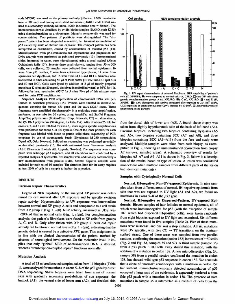
Degree of NER capability of the analyzed XP patient was determined by cell survival after UV exposure and by specific excisionrepair activity. Hypersensitivity to UV exposure was intermediatebetween normal and XP group A cells and comparable to a cell strainfrom XP group C (Fig. 1, left). NER activity, measured as UDS, was—¿�20%of that in normal cells (Fig. 1, right). For complementationanalysis, the patient's fibroblasts were fused to XP cells from groups
A, C, and D. Only after fusion with XP group C cells did repairactivity fail to return to normal levels (Fig. 1, right), indicating that thegenetic defect is caused by a defective XPC gene. This assignment isin line with the clinical characteristics of the patient, marked byabsence of neurological involvement. On the molecular level, it implies that only "global" NER of nontranscribed DNA is affected,whereas "transcription-coupled repair" functions normally.
Mutation Analysis
A total of 73 microdissected samples, taken from 11 biopsies (Table1), were analyzed for mutations in exons 5-8 of thep5J gene by direct
DNA sequencing. Shave biopsies were taken from areas of normalskin with gradually increasing probabilities of sun exposure: thebuttock (Al), the ventral side of lower arm (A2), and freckled skin
Cell survival NER complementation analysis
NXAC X x A X x C X x DFig. 1. UV repair characteristics of cultured fibroblasts. NER capability of patient's
cells (X. XPIUPS; •¿�)were compared to normal cells (N. C5RO; D) and XP cells fromgenetic complementation groups A (A. XP25RO: 0), C (C. XP2IRO; Q ), and D (D,XP6BE; fl|). Left, clonogenic cell survival measured after exposure to 2.5 J/nr. Right.UDS expressed as grains per nucleus (GpN). induced by 10 J/m2. ^5?, heterodikaryons of
neighboring fused partners.
from the dorsal side of lower arm (A3). A fourth shave-biopsy was
taken from slightly hyperkeratotic skin of the back of left hand (A4).Excision biopsies, including two biopsies containing dysplasia (A5and A6), two biopsies containing SCC (A7 and A8). and threebiopsies containing BCC (A9-A11) from the face and scalp were
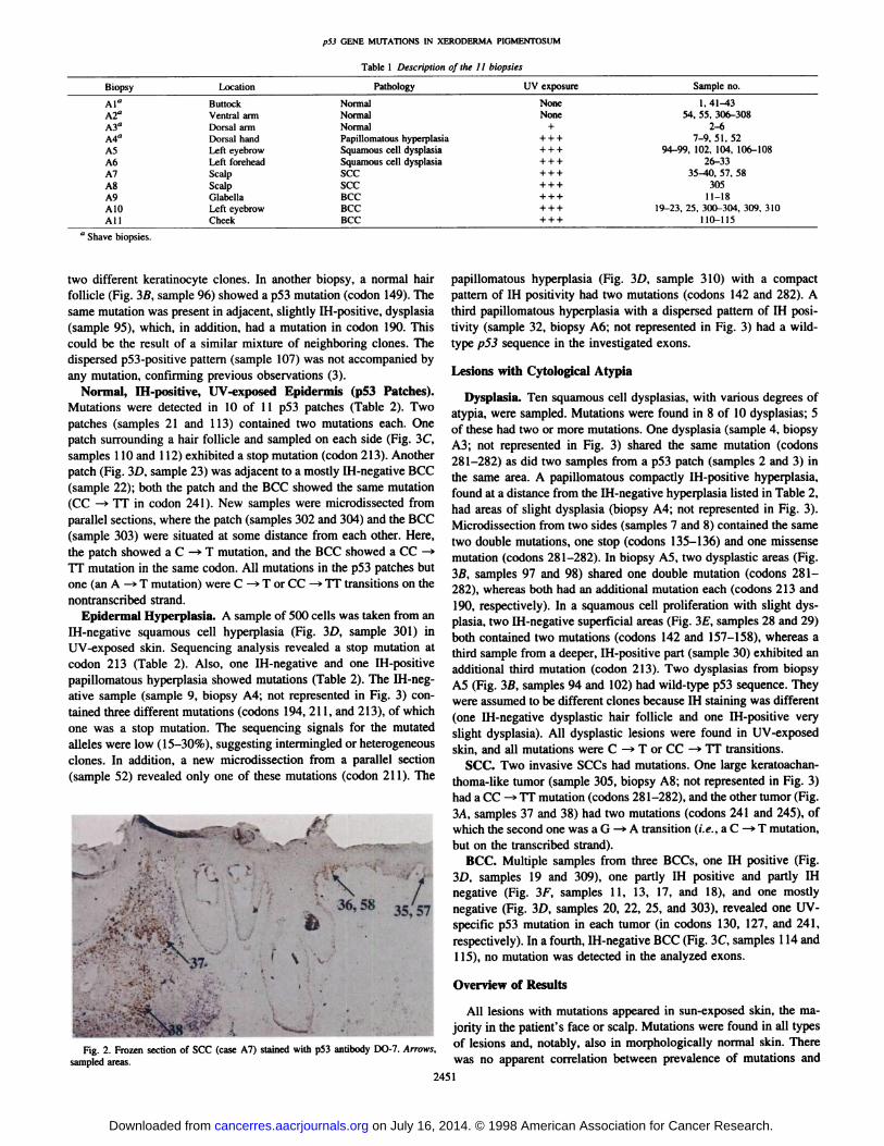
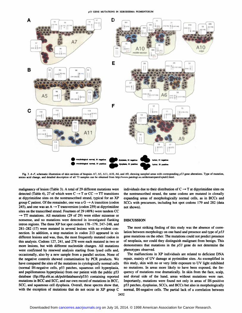
analyzed. Multiple samples were taken from each biopsy, as exemplified in Fig. 2, showing an immunostained cryosection from biopsyA7 (arrows, sampled areas). A schematic overview of results forbiopsies A5-A7 and A9-A11 is shown in Fig. 3. Below is a descrip
tion of the results, based on type of lesion. A lesion was consideredmonoclonal when multiple samples from different parts of the lesionhad identical mutation(s).
Samples with Cytologically Normal Cells
Normal, IH-negative, Non-UV-exposed Epidermis. In nine samples taken from different areas of normal, IH-negative epidermis from
skin that was not exposed to UV light (Al and A2), we found nomutations in exons 5-8 of the p53 gene.
Normal, IH-negative or Dispersed-Pattern, UV-exposed Epi
dermis. Eleven samples of hair follicles or normal epidermis, all ofwhich were immunonegative for p53 (with the exception of sample107, which had dispersed IH-positive cells), were taken randomly
from eight biopsies exposed to UV light and examined. Six differentmutations were found in five samples (Table 2). Five of these mutations were missense, and one was a stop mutation. All six mutationswere UV specific, with five CC —¿�»TT transitions on the nontran
scribed strand. One of these areas was sampled from two parallelsections, confirming the mutation (codon 152) in two sets of —¿�50cells
(Fig. 2 and Fig. 3/4, samples 35 and 57). A third sample (sample 36)from a p53 patch —¿�100cells away shared this mutation, with the
addition of a mutation in codon 138. A new microdissection (Fig. 3A,sample 58) from a parallel section confirmed the mutation in codon138, but showed wild-type p53 sequence in codon 152. We concludethat a clone of "normal" keratinocytes with a mutation in codon 152
but without immunohistochemically detected accumulation of p53occupied a large part of the epidermis. It apparently bordered a bonafide p53 patch with a mutation in codon 138. The presence of bothmutations in sample 36 is interpreted as a mixture of cells from the
2450
on July 16, 2014. © 1998 American Association for Cancer Research. cancerres.aacrjournals.org Downloaded from
p53 GENE MUTATIONS IN XERODERMA PIGMENTOSUM
Table 1 Description of the ¡I
BiopsyAl"tafA3"A4"A5AhA7ASA9AIOAllLocationButtockVentral
armDorsalarmDorsalhandLefteyebrowLeftforeheadScalpScalpGlabellaLeft
eyebrowCheekPathology
UVexposureNormal
NoneNormalNon«Normal+Papillomatous
hyperplasia + ++Squamouscell dysplasia + ++Squamouscell dysplasia + ++SCC
+ ++SCC+ ++BCC+ ++BCC+ ++BCC+ + +Sample
no.1,41-4354.
55,306-3082-67-9,
51,5294-99.102. 104.106-10«26-3335-tO,
57.5830511-1819-23,
25, 300-304,309.110-115310
' Shave biopsies.
two different keratinocyte clones. In another biopsy, a normal hairfollicle (Fig. 3ß,sample 96) showed a p53 mutation (codon 149). Thesame mutation was present in adjacent, slightly IH-positive, dysplasia
(sample 95), which, in addition, had a mutation in codon 190. Thiscould be the result of a similar mixture of neighboring clones. Thedispersed p53-positive pattern (sample 107) was not accompanied by
any mutation, confirming previous observations (3).Normal, IH-positive, UV-exposed Epidermis (p53 Patches).
Mutations were detected in 10 of 11 p53 patches (Table 2). Twopatches (samples 21 and 113) contained two mutations each. Onepatch surrounding a hair follicle and sampled on each side (Fig. 3C,samples 110 and 112) exhibited a stop mutation (codon 213). Anotherpatch (Fig. 3D, sample 23) was adjacent to a mostly IH-negative BCC
(sample 22); both the patch and the BCC showed the same mutation(CC —¿�>TT in codon 241). New samples were microdissected from
parallel sections, where the patch (samples 302 and 304) and the BCC(sample 303) were situated at some distance from each other. Here,the patch showed a C —¿�>T mutation, and the BCC showed a CC —¿�»
TT mutation in the same codon. All mutations in the p53 patches butone (an A —¿�>T mutation) were C —¿�»T or CC —¿�>TT transitions on the
nontranscribed strand.Epidermal Hyperplasia. A sample of 500 cells was taken from an
IH-negative squamous cell hyperplasia (Fig. 3D, sample 301) inUV-exposed skin. Sequencing analysis revealed a stop mutation atcodon 213 (Table 2). Also, one IH-negative and one IH-positivepapillomatous hyperplasia showed mutations (Table 2). The IH-neg
ative sample (sample 9, biopsy A4; not represented in Fig. 3) contained three different mutations (codons 194, 211, and 213), of whichone was a stop mutation. The sequencing signals for the mutatedalÃeleswere low (15-30%), suggesting intermingled or heterogeneous
clones. In addition, a new microdissection from a parallel section(sample 52) revealed only one of these mutations (codon 211). The
. \' 3557
^37-
, . , ,Fig. 2. Frozen section
sampledareas., . . .,, , , j r>m Aof SCC (case A7) stained with p53 antibody DO-7. Arrows,
papillomatous hyperplasia (Fig. 3D, sample 310) with a compactpattern of IH positivity had two mutations (codons 142 and 282). Athird papillomatous hyperplasia with a dispersed pattern of IH positivity (sample 32, biopsy A6; not represented in Fig. 3) had a wild-
type p53 sequence in the investigated exons.
Lesions with Cytological Atypia
Dysplasia. Ten squamous cell dysplasias. with various degrees ofatypia, were sampled. Mutations were found in 8 of 10 dysplasias; 5of these had two or more mutations. One dysplasia (sample 4, biopsyA3; not represented in Fig. 3) shared the same mutation (codons281-282) as did two samples from a p53 patch (samples 2 and 3) inthe same area. A papillomatous compactly IH-positive hyperplasia.found at a distance from the IH-negative hyperplasia listed in Table 2,
had areas of slight dysplasia (biopsy A4; not represented in Fig. 3).Microdissection from two sides (samples 7 and 8) contained the sametwo double mutations, one stop (codons 135-136) and one missensemutation (codons 281-282). In biopsy A5. two dysplastic areas (Fig.3ß,samples 97 and 98) shared one double mutation (codons 281-
282), whereas both had an additional mutation each (codons 213 and190, respectively). In a squamous cell proliferation with slight dysplasia, two IH-negative superficial areas (Fig. 3£,samples 28 and 29)both contained two mutations (codons 142 and 157-158), whereas athird sample from a deeper, IH-positive part (sample 30) exhibited an
additional third mutation (codon 213). Two dysplasias from biopsyA5 (Fig. 3ß,samples 94 and 102) had wild-type p53 sequence. They
were assumed to be different clones because IH staining was different(one IH-negative dysplastic hair follicle and one IH-positive veryslight dysplasia). All dysplastic lesions were found in UV-exposedskin, and all mutations were C —¿�>T or CC —¿�»TT transitions.
SCC. Two invasive SCCs had mutations. One large keratoachan-thoma-like tumor (sample 305, biopsy A8; not represented in Fig. 3)had a CC -»TT mutation (codons 281 -282), and the other tumor (Fig.
3/4, samples 37 and 38) had two mutations (codons 241 and 245), ofwhich the second one was a G —¿�»A transition (i.e., a C —¿�*T mutation,
but on the transcribed strand).BCC. Multiple samples from three BCCs, one IH positive (Fig.
3D, samples 19 and 309), one partly IH positive and partly IHnegative (Fig. 3F, samples 11, 13, 17, and 18), and one mostlynegative (Fig. 3D, samples 20, 22, 25, and 303), revealed one UV-
specific p53 mutation in each tumor (in codons 130, 127, and 241,respectively). In a fourth, IH-negative BCC (Fig. 3C, samples 114 and
115), no mutation was detected in the analyzed exons.
Overview of Results
All lesions with mutations appeared in sun-exposed skin, the majority in the patient's face or scalp. Mutations were found in all types
of lesions and, notably, also in morphologically normal skin. Therewas no apparent correlation between prevalence of mutations and
2451
on July 16, 2014. © 1998 American Association for Cancer Research. cancerres.aacrjournals.org Downloaded from
pS3 GENE MUTATIONS IN XERODERMA PIGMENTOSUM
•¿�N-H^lsit"«
) morphological normal. H negative
morphological normal. IH positive
' dyspiasia. H negative
dysptasta, H positive
Fig. 3. A-F, schematic illustration of skin sections of biopsies A7, A5, All. AIO. A6. and A9. showing sampled areas with corresponding p53 gene alterations. Type of mutation,amino acid change, and detailed description of all 73 samples can he obtained from http://www.patologi.uu.se/dermatopatol/xptab2.html.
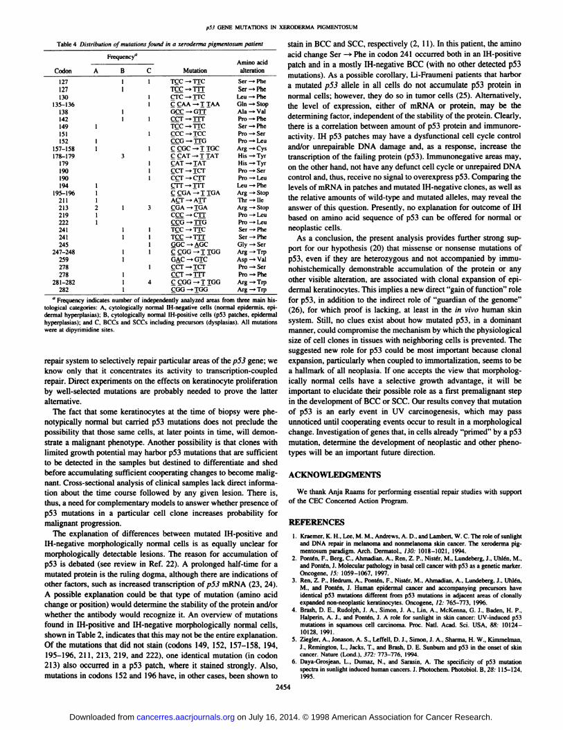
malignancy of lesion (Table 3). A total of 29 different mutations weredetected (Table 4), 27 of which were C -»T or CC -»TT transitions
at dipyrimidine sites on the nontranscribed strand, typical for an XPgroup C patient. Of the remainder, one was a G —¿�*A transition (codon245), and one was an A —¿�»T transversion (codon 259) at dipyrimidine
sites on the transcribed strand. Fourteen of 29 (48%) were tandem CC—¿�»TT mutations. All mutations (29 of 29) were either missense or
nonsense, and no mutations were detected in investigated flankingintron regions. The three XP hot spot codons 178-179, 247-248, and281-282 (17) were mutated in several lesions with no evident con
nection. In addition, a stop mutation in codon 213 appeared in sixdifferent lesions and was, thus, the most frequently mutated codon inthis analysis. Codons 127, 241, and 278 were each mutated in two ormore lesions, but with different nucleotide changes. All mutationswere confirmed by renewed analysis starting from lysed cells and,occasionally, also by a new sample from a parallel section. None ofthe negative controls showed contamination by PCR products. Wehave compared the sites of the mutations in cytologically normal cells(normal IH-negative cells, p53 patches, squamous cell hyperplasia,
and papillomatous hyperplasia) from our patient with the public p53database (ftp://ftp.ebi.ac.uk/pub/databases/p53/) containing reportedmutations in BCC and SCC, and our own record of mutations in BCC.SCC, and squamous cell dysplasia. Overall, these spectra show that,with the exception of mutations that do not occur in XP group C
individuals due to their distribution of C —¿�>T at dipyrimidine sites on
the nontranscribed strand, the same codons are mutated in clonallyexpanding areas of morphologically normal cells, as in BCCs andSCCs with precursors, including hot spot codons 179 and 282 (datanot shown).
DISCUSSION
The most striking finding of this study was the absence of correlation between morphology on one hand and presence and type of p53gene mutations on the other. The mutations could not predict presenceof neoplasia, nor could they distinguish malignant from benign. Thisdemonstrates that mutations in the p53 gene do not determine thephenotypes observed.
The malfunctions in XP individuals are related to deficient DNArepair, mainly of UV damage at pyrimidine sites. As exemplified inthis study, skin with no or very little exposure to UV light exhibitedno mutations. In areas more likely to have been exposed, the frequency of mutations rose dramatically. In skin from the face, scalp,and dorsal side of the hand, areas without mutations were rare.Importantly, mutations were found not only in areas of IH-positive
p53 patches, dysplasias, SCCs, and BCCs but also in morphologicallynormal, IH-negative cells. The partial lack of a correlation between
2452
on July 16, 2014. © 1998 American Association for Cancer Research. cancerres.aacrjournals.org Downloaded from
p53 GENE MUTATIONS IN XERODERMA PIGMENTOSUM
Table 2 Sun-exposed akin with cytologically normal ceils
The distribulion of mutations of the p53 gene is related to the pattern of p53 IH staining.
BiopsyIHnegative or dispersedpattern''A3A4A4A5ASA5A5A6A6A7A9A9AIOAllIH
positive (compactpattern'')A3A3A6A6A7A7A9AIOAIOAIOAllAllSample6519%99104107263235.57163003011112,3108273136.
58391523,
302.30421310113110,
112Codon195-196219194211213149wrWTWTWTWT152WTWT213157-158222281-282278127259138WT178-179241178-179247-248282142127178-179213Mutation"SMMMSMMSMMMMMMMMMMMMMMMSDescription
ofsampleNormal
epidermisNormalepidermisPapillomatoushyperplasiaNorma
hairfollicleNormaepidermisNormaepidermisNormaepidermis'Norma
epidermisPapillonatoushyperplasia'Norma
epidermisNormaepidermisNormalepidermisSquamous
cellhyperplasiaNormalepidermisp53
patchp53patchp53patchp53patchp53patchp53patchp53patchp53patchp53patchPapillomatous
hyperplasiap53
patchp53
patch" Type of mutation: M. missense mutation: S. stop mutation.
See "Materials and Methods."'' WT, wild type.
' Dispersed pattern.
mutation of the p53 gene and immunopositivity has been notedpreviously (18).
The sensitivity of the method did not permit detection of mutatedcells if their number was <20% of the total number of sampled cells.Therefore, fairly large areas of sun exposed epidermis contain clandestine clonal expansions, originally derived from a single mutatedcell. These clones resemble p53 patches, in that the cells are morphologically normal and clonally expanding and harbor p53 mutations. Inp53 patches, which are immunopositive, the p53 protein is obviouslyoverexpressed. In the clusters discovered here, the protein does notaccumulate and goes unnoticed in pathological investigations. Theseclones appear to be of sizes similar to those of p53 patches. In oneexample shown in Fig. 3A, such a clone seems to be made up of atleast a few hundred cells. This is larger than the conjectured size of aproliferative epidermal unit (19). With 45% (5 of 11) of randomlysampled normal. IH-negative, sun-exposed epidermis being mutated,a large proportion of this individual's normal epidermis is predicted toharbor mutations in the p53 gene. The area of the patient's face that
is covered with p53 patches at present is probably not too different
Table 3 Frequency of p53 munitions
Type ofhistologySamples
with cytologically normalcellsNormal,IH-negative, non-UV-exposedepidermisNormal,IH-negalive, UV-cxposedepidermisIH-positive
p53patchesEpidermal
hyperplasiaLesionswith cytologicalatypiaDysplasiasecBCCMutated0/95/1112/133/48/10W3/4
from that of a normal 70-year-old individual, who is calculated,assuming a lifelong accumulation of mutations, to have ~30% of his
face covered by mutated p53 patches (20). To reveal if this mutabilityis specific for the p53 gene or is general for the whole genome, similaranalyses of other genes should be performed. Because all mutationswill not be selected for and be displayed as clones, analysis at thesingle cell level would be required, as outlined recently (21).
The significance of clonal proliferations of otherwise normal kera-
tinocytes carrying a p53 mutation is unknown. The two major alternatives are that (a) the mutations are indicators of UV exposure, withno functional significance, or (b) they, because of selective growthadvantage, "drive" or permit clonal expansion of epidermal cells. The
fact that no silent p53 mutations were found favors the latter hypothesis. To investigate this further, the distribution of mutations wasstatistically analyzed. The number of possible, XP group C typicalalterations that theoretically can occur in the gene sequence analyzedin this study (including flanking introns) is 254 (taking into accountthat a CC photoproduct can result in three possible alterations: CC —¿�»CT, CC ^TC, and CC --TT). Of these alterations, 161 (63%) would
result in missense or nonsense mutations, and 93 (37%) would besilent (including intron mutations). We found 27 of 161 (17%) possible XP specific missense and nonsense mutations and should, thus,theoretically have detected 16 (17% of 93) silent mutations, but nonewas found. This strongly suggests that only cells with missense ornonsense p53 mutations are selected for clonal expansion. This argument is only valid if one can exclude that differential repair is thecause of the skewed distribution of the mutations; however, it seemsunlikely that differential repair would specifically correct silent mutations. As shown in Fig. 1, our XP patient had residual 20% overallexcision repair capacity. We do not know the properties of his defunct
2453
on July 16, 2014. © 1998 American Association for Cancer Research. cancerres.aacrjournals.org Downloaded from
pSÕ GENE MUTATIONS IN XERODERMA PIGMENTOSUM
Table 4 Distribution of mutations found in a \eniderma pigmentosum patient
Codon127127130135-136138142149151152157-158178-179179190190IM195-196211213219222241241245247-248259278278281-282282Frequency"ABCMutation1
I TCC ->TTC1
TCC—TTT1CTC—TTC1C CAÕ -»TTAA1GCC -»GTT1
1CCT—TTT1TCC-»TTC1CCC—TCC1CCG ->TTC1
1 CCGC—TTGC3C CAT -> TTAT1CAT-»TAT1CCT —¿�TCT1CCT —¿�CTT1CTT —¿�TTT1CCGA —¿�TTGA1ACT ->ATT2
1 3 CGA —¿�TGA1CCC—CTT1CCG ->TTC1TCC—TTC1TCC—TTT1GGC —¿�AGC1CCGG—TTGGGAC
—¿�GTC1CCT —¿�TCTCCT
—¿�TTT4CCGG—TTGGCOG—
TGGAmino
acidalterationSer
—¿�PheSer—¿�PheLeu—¿�PheGin—¿�StopAla—¿�ValPro—¿�PheSer—¿�PhePro—¿�SerPro—¿�LeuArg—¿�>CysHis—¿�TyrHis—
TyrPro—¿�SerPro—¿�LeuLeu—¿�PheArg—¿�*StopThr—¿�IleArg—¿�StopPro—¿�*LeuPro—¿�LeuSer—¿�PheSer—¿�PheGly—¿�*SerArg—
TrpAsp—¿�ValPro—¿�SerPro—¿�PheArg—¿�TrpArg—¿�Trp
" Frequency indicates number of independently analyzed areas from three main his
tológica! categories: A, cytologically normal IH-negative cells (normal epidermis, epidermal hyperplasias); B, cytologically normal IH-positive cells (p53 patches, epidermal
hyperplasias); and C. BCCs and SCCs including precursors (dysplasias). All mutationswere at dipyrimidine sites.
repair system to selectively repair particular areas of the p53 gene; weknow only that it concentrates its activity to transcription-coupled
repair. Direct experiments on the effects on keratinocyte proliferationby well-selected mutations are probably needed to prove the latter
alternative.The fact that some keratinocytes at the time of biopsy were phe-
notypically normal but carried p53 mutations does not preclude thepossibility that those same cells, at later points in time, will demonstrate a malignant phenotype. Another possibility is that clones withlimited growth potential may harbor p53 mutations that are sufficientto be detected in the samples but destined to differentiate and shedbefore accumulating sufficient cooperating changes to become malignant. Cross-sectional analysis of clinical samples lack direct informa
tion about the time course followed by any given lesion. There is,thus, a need for complementary models to answer whether presence ofp53 mutations in a particular cell clone increases probability formalignant progression.
The explanation of differences between mutated IH-positive andIH-negative morphologically normal cells is as equally unclear for
morphologically detectable lesions. The reason for accumulation ofp53 is debated (see review in Ref. 22). A prolonged half-time for a
mutated protein is the ruling dogma, although there are indications ofother factors, such as increased transcription of p53 mRNA (23, 24).A possible explanation could be that type of mutation (amino acidchange or position) would determine the stability of the protein and/orwhether the antibody would recognize it. An overview of mutationsfound in IH-positive and IH-negative morphologically normal cells,
shown in Table 2, indicates that this may not be the entire explanation.Of the mutations that did not stain (codons 149, 152, 157-158, 194,195-196, 211, 213, 219, and 222), one identical mutation (in codon
213) also occurred in a p53 patch, where it stained strongly. Also,mutations in codons 152 and 196 have, in other cases, been shown to
stain in BCC and SCC, respectively (2, 11). In this patient, the aminoacid change Ser —¿�»Phe in codon 241 occurred both in an IH-positivepatch and in a mostly IH-negative BCC (with no other detected p53mutations). As a possible corollary, Li-Fraumeni patients that harbor
a mutated p53 alÃelein all cells do not accumulate p53 protein innormal cells; however, they do so in tumor cells (25). Alternatively,the level of expression, either of mRNA or protein, may be thedetermining factor, independent of the stability of the protein. Clearly,there is a correlation between amount of p53 protein and immunore-
activity. IH p53 patches may have a dysfunctional cell cycle controland/or unrepairable DNA damage and, as a response, increase thetranscription of the failing protein (p53). Immunonegative areas may,on the other hand, not have any defunct cell cycle or unrepaired DNAcontrol and, thus, receive no signal to overexpress p53. Comparing thelevels of mRNA in patches and mutated IH-negative clones, as well asthe relative amounts of wild-type and mutated alÃeles,may reveal the
answer of this question. Presently, no explanation for outcome of IHbased on amino acid sequence of p53 can be offered for normal orneoplastic cells.
As a conclusion, the present analysis provides further strong support for our hypothesis (20) that missense or nonsense mutations ofp53, even if they are heterozygous and not accompanied by immu-
nohistchemically demonstrable accumulation of the protein or anyother visible alteration, are associated with clonal expansion of epidermal keratinocytes. This implies a new direct "gain of function" rolefor p53, in addition to the indirect role of "guardian of the genome"
(26), for which proof is lacking, at least in the in vivo human skinsystem. Still, no clues exist about how mutated p53. in a dominantmanner, could compromise the mechanism by which the physiologicalsize of cell clones in tissues with neighboring cells is prevented. Thesuggested new role for p53 could be most important because clonalexpansion, particularly when coupled to immortalization, seems to bea hallmark of all neoplasia. If one accepts the view that morphologically normal cells have a selective growth advantage, it will beimportant to elucidate their possible role as a first premalignant stepin the development of BCC or SCC. Our results convey that mutationof p53 is an early event in UV carcinogenesis, which may passunnoticed until cooperating events occur to result in a morphologicalchange. Investigation of genes that, in cells already "primed" by a p53
mutation, determine the development of neoplastic and other pheno-
types will be an important future direction.
ACKNOWLEDGMENTS
We thank Anja Raams for performing essential repair studies with supportof the CEC Concerted Action Program.
REFERENCES
1. Kraemer. K. H.. Lee. M. M.. Andrews, A. D-. and Lambert, W. C. The role of sunlightand DNA repair in melanoma and nonmelanoma skin cancer. The xeroderma pig-mentosum paradigm. Arch. Dermatol., 130: 1018-1021, 1994.
2. Ponten, F., Berg, C., Ahmadian, A.. Ren, Z. P.. Nistér,M.. Lundeberg. !.. Uhlén,M.,and Pontén,J. Molecular pathology in basal cell cancer with p53 as a genetic marker.Oncogene. /5: 1059-1067, 1997.
3. Ren. Z. P., Hedrum. A.. Pontén.F., Nistér.M.. Ahmadian. A.. Lundeberg. J.. Uhlén,M., and Pontén.J. Human epidermal cancer and accompanying precursors haveidentical p53 mutations different from p53 mutations in adjacent areas of clonallyexpanded non-neoplastic keratinocytes. Oncogene, 12: 765-773, 1996.
4. Brash, D. E., Rudolph. J. A., Simon. J. A.. Lin, A., McKenna, G. J.. Baden, H. P.,Halperin, A. J., and Pontén.J. A role for sunlight in skin cancer: UV-induced p53mutations in squamous cell carcinoma. Proc. Nati. Acad. Sci. USA, 88: 10124-10128, 1991.
5. Ziegler. A.. Jonason, A. S., Leffell, D. J., Simon. J. A.. Sharma. H. W.. Kimmclman.J.. Remington. L.. Jacks. T.. and Brash. D. E. Sunburn and p53 in the onset of skincancer. Nature (Lond.), 372: 773-776. 1994.
6. Daya-Grosjean. L.. Dumaz. N.. and Sarasin. A. The specificity of p53 mutationspectra in sunlight induced human cancers. J. Photochem. Photobiol. B. 28: 115-124,1995.
2454
on July 16, 2014. © 1998 American Association for Cancer Research. cancerres.aacrjournals.org Downloaded from
pS3 GENE MUTATIONS IN XERODERMA PIOMENTOSUM
7. Dumaz. N.. Stary, A., Scussi, T., Daya-Grosjean. L., and Sarasin. A. Can we predict
solar ultraviolet radiation as the causal event in human tumours by analysing themutation spectra of the ¡>53gene? Mutât.Res., 307: 375-386. 1994.
8. Ponten. F., Berne, B., Ren, Z. P., Nistér,M., and Pontén.J. Ultraviolet light inducesexpression of p53 and p21 in human skin: effect of sunscreen and constitutive p21expression in skin appendages. J. Invest. Dermatol.. 105: 402-406, 1995.
9. Urano, Y.. Asano, T.. Yoshimoto. K.. Iwahana. H.. Kubo. Y.. Kalo. S., Sasaki, S..Takeuchi, N., Uchida, N., and Nakanishi, H. Frequent p53 accumulation in thechronically sun-exposed epidermis and clonal expansion of p53 mutant cells in theepidermis adjacent to basal cell carcinoma. J. Invest. Dermatol., 104: 928-932, 1995.
10. Jonason. A. S.. Kunala, S., Price. G. J., Restifo, R. J.. Spinelli. H. M., Persing. J. A..Leffell. D. J., Tarone. R. E., and Brash. D. E. Frequent clones of p53-mutated kcratino-cytes in normal human skin. Proc. Nati. Acad. Sei. USA. 93: 14025-14029. 19%.
11. Ren, Z. P., Ahmadian. A.. Pontén,F., Nister, M., Berg, C., Lundeberg, J., Uhlen. M.,and Pontén.J. Benign clonal keratinocyte patches with p53 mutations show no geneticlink to synchronous squamous cell precancer or cancer in human skin. Am. J. Pathol..ISO: 1791-1803. 1997.
12. Hamel. B. C. J.. Raams, A., Schuitema-Dijkstra. A. R., Simons. P.. van der Burght.I., Jaspers. N. G. J., and Kleijer. W. J. Xeroderma pigmentosum-Cockayne syndromecomplex: a further case. J. Med. Genet., 33: 607-610, 1996.
13. Hedrum, A., Pontén,F., Ren, Z. P.. Lundeberg. J.. Pontén.J.. and Uhlen, M.Sequence-based analysis of the human p53 gene based on microdissection of tumorbiopsy samples. BioTechniques. 17: 118-129, 1994.
14. Ren. Z. P.. Pontén.F.. Nistér.M., and Pontén.J. Two distinct p53 immunohisto-chemical patterns in human squamous-cell skin cancer, precursors and normal epidermis. Int. J. Cancer. 69: 174-179, 1996.
15. Berg, C., Hedrum, A., Holmberg, A., Pontén,F., Uhlén,M., and Lundeberg. J. Directsolid-phase sequence analysis of the human p53 gene by use of multiplex polymerasechain reaction and a-thiotriphosphate nucleotides. Clin. Chem., 41: 1461-1466, 1995.
16. Hultman, T., Stahl. S.. Homes. E., and Uhlén,M. Direct solid phase sequencing ofgenomic and plasmid DNA using magnetic beads as solid support. Nucleic AcidsRes.. 17: 4937-4946. 1989.
17. I 'inn.i/. N.. Drougard. C.. Sarasin, A., and Daya-Grosjean, L. Specific UV-inducedmutation spectrum in the p53 gene of skin tumors from DNA repair-deficientxeroderma pigmenlosum patients. Proc. Nati. Acad. Sci. USA, 90: 10529-10533,1993.
18. Hall. P. A., and Lane, D. P. p53 in tumour pathology: can we trust immunohisto-chemistry? Revisited!. J. Pathol., 172: 1-4. 1994.
19. Potten. C. S. Identification of clonogenic cells in the epidermis and the structuralarrangement of the epidermal proliferative unit (EPU). In: A. B. Cairnie (ed.I. StemCells of Renewing Cell Populations, pp. 91-102. New York: Academic Press, 1976.
20. Ren. Z-P. Role of mutations in the p53 gene for the development of human squamous
cell skin cancer (Dissertation no. 605). Uppsala, Sweden: Uppsala University, 1996.21. Pontén.F.. Williams, C. Gao. L.. Ahmadian. A., Nisler. M.. Lundeberg. J.. Pontén,
J., and Uhlén,M. Genomic analysis of single cells from human basal cell cancer usinglaser-assisted capture microscopy. Mutât.Res., 3K2: 45-55, 1997.
22. Lane, D. P. p53 and human cancers. Br. Med. Bull., 50: 582-599, 1994.
23. Williams. C.. Norberg. T.. Ahmadian, A., Pontén.F.. Inganäs.M.. Bergh, J., Lundeberg. J., and Uhlén,M. Assessment of sequence-based p53 gene analysis in human
breast cancer: mRNA in comparison with genomic DNA targets. Clin. Chcm.. 44:455-462. 1998.
24. EI-Mahdani, N., Vaillant. J-C. Guiguet, M., Prévôt.S.. Bertrand. V.. Bernard, C..
Parc. R.. Bérézial.G., and Hermelin. B. Overexpression of p53 mRNA in colorectalcancer and its relationship to p53 gene mutation. Br. J. Cancer. 75: 528-536. 1997.
25. MacGeoch. C.. Turner. G.. Bobrow, L. G.. Barnes, D. M.. Bishop, D. T., and Spurr.N. K. Heterogeneity in Li-Fraumcni families: p53 mutation analysis and immuno-histochemical staining. J. Med. Genet.. 32: 186-190, 1995.
26. Lane, D. P. Cancer. p53. guardian of the genome. Nature (Lond.), 358: 15-16. 1992.
2455
on July 16, 2014. © 1998 American Association for Cancer Research. cancerres.aacrjournals.org Downloaded from