CLINICAL STUDIES ON IRON KINETICS
26
Nagoya ]. med. Sci. 30: 465-490, 1968 CLINICAL STUDIES ON IRON KINETICS I. IRON KINETICS STUDIES IN BLOOD DISORDERS HIDEO YAMADA 1st Department of Internal Medicine, Nagoya University School of Medicine ( l)irector: Prof Susumu Hibino) ABSTRACT Ferrokinetics data on patients with blood disorders of diverse etiology were presented and the clinical implication of this test was discussed. Ferrokinetics data on normal adults as well as on the aged were also presented. Ferrokinetic patterns of ineffective erythropoiesis were obtained on patients with pernicious anemia, erythroleukemia, myelofibrosis, paroxysmal nocturnal hemoglobinuria and refractory anemia as well. Ferrokinetic studies on patients with portal hypertension with congestive splenomegaly have disclosed that the erythropoietic activity in this condition is rather increased and the liver damage does not necessarily exert inhibitory effect on erythropoiesis in these patients. The studies on patients with acute leukemia did not reveal the patterns typical of each cell type of leukemia, and the results indicate that the myelophthisic effect due to the encloachment of leukemic cells is a major factor in the develop- ment of anemia in acute leukemia. Finally, it is stressed that the ferrokinetic measurements with body surface counting are of great use in diagnosis and treatment of patients with myelopro- liferative syndrome. I. INTRODUCTION Since the pioneering work of Huff and associates, ferrokinetic studies have had much application in clinical medicine as well as experimental works. In this paper, the author presents ferrokinetics data on patients with blood disorders of diverse etiology and discusses the clinical significance of ferrokinetic studies as well as on some basic problems of iron kinetics. II. MATERIALS AND METHODS Materials Normal controls: Seven normal adults (from 26 to 56 years old) and seven aged subjects (from 63 to 75 years old) who were all in good health, with normal hematolo- WI E8 #i: tiE Received for publication September 16, 1967.
-
Upload
khangminh22 -
Category
Documents
-
view
0 -
download
0
Transcript of CLINICAL STUDIES ON IRON KINETICS

Nagoya ]. med. Sci. 30: 465-490, 1968
CLINICAL STUDIES ON IRON KINETICS
I. IRON KINETICS STUDIES IN BLOOD DISORDERS
HIDEO YAMADA
1st Department of Internal Medicine, Nagoya University School of Medicine ( l)irector: Prof Susumu Hibino)
ABSTRACT
Ferrokinetics data on patients with blood disorders of diverse etiology were presented and the clinical implication of this test was discussed. Ferrokinetics data on normal adults as well as on the aged were also presented.
Ferrokinetic patterns of ineffective erythropoiesis were obtained on patients with pernicious anemia, erythroleukemia, myelofibrosis, paroxysmal nocturnal hemoglobinuria and refractory anemia as well. Ferrokinetic studies on patients with portal hypertension with congestive splenomegaly have disclosed that the erythropoietic activity in this condition is rather increased and the liver damage does not necessarily exert inhibitory effect on erythropoiesis in these patients.
The studies on patients with acute leukemia did not reveal the patterns typical of each cell type of leukemia, and the results indicate that the myelophthisic effect due to the encloachment of leukemic cells is a major factor in the development of anemia in acute leukemia.
Finally, it is stressed that the ferrokinetic measurements with body surface counting are of great use in diagnosis and treatment of patients with myeloproliferative syndrome.
I. INTRODUCTION
Since the pioneering work of Huff and associates, ferrokinetic studies have had much application in clinical medicine as well as experimental works. In this paper, the author presents ferrokinetics data on patients with blood disorders of diverse etiology and discusses the clinical significance of ferrokinetic studies as well as on some basic problems of iron kinetics.
II. MATERIALS AND METHODS
Materials Normal controls: Seven normal adults (from 26 to 56 years old) and seven aged subjects
(from 63 to 75 years old) who were all in good health, with normal hematolo-
WI E8 #i: tiE Received for publication September 16, 1967.

466 H. YAMADA
gical values and without history of chronic diseases or blood loss were studied
for controls.
Patients with blood disorders of diverse etiology:
Eighty four patients with blood disorders of diverse etiology on whom the
diagnosis was confirmed by clinical and hematological examinations and no
treatment were done before and during examinations, were studied for fer
rokinetic measurements. The numbers of patients with blood disorders were as follows: 8 of iron
deficiency anemia, 18 of portal hypertension with splenomegaly, 13 of hemolytic
anemia, 3 of paroxysmal nocturnal hemoglobinuria, 2 of pernicious anemia, 11
of aplastic anemia, 5 of polycythemia vera, 3 of Di Guglielmo's syndrome, 3
of myelofibrosis, 14 of acute leukemia and 4 of chronic leukemia patients.
Body surface counting studies were simultaneously performed on some of
these cases.
M ethods Ferrokinetic studies : 5 to 10 p.c of 59Fe in the form of ferric citrate was
diluted in 10 ml of isotonic saline and injected intravenously without incubation
with plasma. Plasma iron disappearance rate ( PID, T ! ), plasma iron turnover rate (PIT),
percent red cell utilization of 59Fe ( %RCU), red cell iron turnover rate (RCIT)
and mean red cell life span ( MRCLS) were calculated according to the method
of Huff and associates1' with minor modification.
Plasma volume was measured by 59Fe dilution method. PIT was expressed
in mgjkgjday. And, to exclude the error inherent to the measurement of
plasma volume, PIT was also expressed in mg/ 100 ml plasma/day by using
the formula as follows:
Plasma iron turnover rate (PIT) (mg/100 ml plasma/day)
Serum iron ( tlg I 100 ml) T ! (min)
The 9~ RCU was calculated by the formula as follows :
Percentage of red cell utilization of 59Fe ( %RCU)
Counts/ min/ ml hemolyzed whole blood 100 Countsjmin jml plasma at 0 time x 100- Ht x 100
(Ht = Hematocrit 9~ )
This formula made calculation simpler and excluded the error inherent to
the counting of the injected dose. Serum iron was measured by the method of Matsubara using o-phenanthro
line as a colour reagent and hydroxylamine hydrochloride as a reducing agent2'.
The unsaturated iron-binding capacity was measured by a modification of the
ra(,iiQi:ron m~thoQ. of Ta,uxe 3',

CLINICAL STUDIES ON IRON KINETICS (l) 467
Body surface counting was performed over the precordium, liver, spleen and the bone marrow (sacrum) hourly during the first day and with 2 to 3 days interval for 2 weeks until the completion of JC RCU studies. The directional scintillation counter was placed on the skin and appropriate oblique placements were chosen to minimize the influence from the vertebrae of the sternum. As to the activities, gross body surface counting was used without respects to body background counting. The results were plotted with activities (count/min/ p.c) on a linear ordinate scale against time or day on a linear absicca scale.
III. RESULTS
A. Ferrokinetic data (Turnover indice)
Il Normal subjects Ferro kinetic data in 7 healthy adults were as follows : PID ( T ~ ) being
96.1 ± 193 minutes, PIT 0.45 ± 0.07 mgjkgjday, JC RCU 92.1 ± 8.79C, and RCIT being 0.41 ±0.06 mg/kgjday. On the other hand, ferro kinetic data in 7 normal aged subjects were as follows: the mean values of PID ( T~), PIT, JC RCU and RCIT were 89.9 ± 19.5 minutes, 0.49 ± 0.07 mgj kgjday, 84.6 ± 6.5 9C and 0.42
± 0.06 mgjkgjday respectively. The mean values of PID ( T ~), PIT, 9C RCU and RCIT show no statistically significant differences between normal adults and aged. ,
II) Blood disorders 1) Iron deficiency anemia Table 1 depicts the hematological and ferrokinetic data in 8 patients with
TABLE 1. Hematological and Ferrokinetics Data on Patients with Iron Deficiency Anemia
C se I Hemo- Reti-~ulo-rfo Age Sex, globin cyte
. i ( ,% ) (%o)
1 2 3 4 5 6
7 8
mean S.D.
25 M 14 F 15 F 20 M 18 M 47 M
23 F 34 F
31 28 50 3 49 21 58 14 18 8 55 19
45 21 34 8
42.5 15.3
Serum Iron
(pgfdl)
32 55 35 20 14 42
20 21
29.9
PID = Plasma Iron Disappearance. PIT = Plasma Ir on Turnover. RCU = Red Cell Utilization of 59 F e.
PIT I PID n RCU (min) mg/ mg/100 ml (%)
Remarks
kg/day plasma/day
14 1.58 2.28 100 gastrectomy 20 2.02 2.75 109 idiopathic 16 1.30 2.19 100 II
18 0.68 1.11 105 rr 14 0.66 1.00 110 II
14 1.45 3.00 100 gastrointestinal bleeding
13 1.21 1.54 100 idiopa thic 13 1.32 1.61 99 1 hypermenorrhea
15.3 2.4
1.28 0.42
1.94 0.69
102.911
4.2
468 H. YAMADA
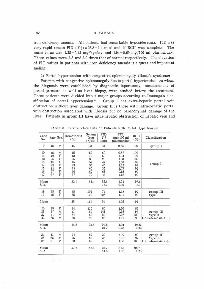
iron deficiency anemia. All patients had remarkable hyposideremia. PID was very rapid (mean PID (T~)=l5.3±2.4 min) and JC RCU was complete. The mean value was 1.28±0.42 mgjkg/day and 1.94±0.69 mg/100 ml plasma/day. These values were 2.8 and 2.0 times that of normal respectively. The elevation of PIT values in patients with iron deficiency anemia is a queer and important finding.
2) Portal hypertension with congestive splenomegaly (Banti's syndrome l Patients with congestive splenomegaly due to portal hypertension, on whom
the diagnosis were established by diagnostic laporatomy, measurement of portal pressure as well as liver biopsy, were studied before the treatment. These patients were divided into 3 major groups according to Imanaga's classification of portal hypertension 1>. Group I has extra-hepatic portal vein obstruction without liver damage. Group II is those with intra-hepatic portal vein obstruction associated with fibrosis but no parenchymal damage of the liver. Patients in group III have intra-hepatic obstruction of hepatic vein and
TABLE 2. Ferrokinetics Data on Patients with Portal Hypertension
Case Age Se I Hematocrit Serum 1 PID PIT
RCUI Iron I Ti< mg/100 ml Classification No. X L%') ( 7/dl) (min) plasma/day (%)
------------ ---~-----~-----------
9 18 Ml 46 93 I 33 2.83. 100 group I
10 15 Mi 15 52 60 0.87 100 11 42 F! 42 72 69 1.04 99 12 26 Fl 26 30 20 1.50 100 13 45 F 45 52 47 1.10 98 group II 14 43 F 43 55 45 1.22 99 15 13 F 13 60 35 1.72 96 16 57 F 36 60 68 0.88 90 17 37 F 37 70 45 1.56 99
Mean 32.1 64.4 55.6 1.24 97.61 S.D. 17.1 0.30 3.1
~ ----' -~ 18 65 F 35 102 74 1.38 92 group III 19 45 F 35 120 108 1.11 90 i type 1
Mean 35 111 91 1.25 91 I I _________ ._ ____ -
20 18 F 34 120 80 1.50 85 I 21 27 M 41 82 141 0.58 95 group III
22 41 M 34 80 82 0.98 100 1 type 2 23 35 Ml 38 92 83 1.11 99
1Hematemesis ( -- )
Mean 35.8 93.5 96.5 1.04 94.8[ S.D.
i 24.7 0.33 5.931
--·--
HI 24 41 24 82 20 4.10 99 I group III 25 68 26 82 38 2.16 97 type 2 26 21 30 86 55 1.56 100 Hematemesis ( + )
Mean I 27.7 83.3 37.7 2.61 98.71 S.D.
I 14.3 1.08 1.25

CLINICAL STUDIES ON IRON KINETICS (I l 469
portal vein with liver cirrhosis. Group III was subdivided further into groups with hematemesis and without hematemesis.
To exclude an error inherent to the measurement of plasma volume in
these patients with massive splenomegaly, PIT was expressed in mg/ 100 ml plasma/day. The results were shown in Table 2. One patient in group I (No. 9) showed rapid PID ( T ~) with complete %' RCU and increased PIT. However, this finding does not necessarily represent ferrokinetic changes of this group
because of the paucity of the cases studied. Patients in group II (No. 10-17) showed remarkable hyposideremia and rapid PID ( T!) (mean value: 55.6 ± 17.1 minutes). Group II and group III (No. 18- 23) differ significantly in the values of serum iron and PID ( T!). The mean value of PIT on patients in group II
was L24 mg/ 100 ml plasma which was significantly higher than normal.
Percent RCU in this group was rapid and full (97.69ti) . On the other hand, patients in group III showed normal range of serum iron and PID ( T ~ ) and increased rates of PIT (mean value: L 11 mg/ 100 plasma). In group III, patients with hematemesis or melena (No. 24-26) showed accelerated PID ( Tt)
and remarkably increased PIT values (mean value: 2.61 mg/100 ml plasma).
3) Hemolytic anemias i) Congenital hemolytic anemia In hereditary spherocytosis, PID ( T!) was accelerated (less than 30 minutes)
and 96 RCU has varied between 11 and 84. The PIT has been found to be
between L72 and 7.64 mg/kgjday (mean value: 3.50 mg/kgjday) (Table 3), The patient No. 34 was the case of congenital hemolytic anemia which was different hematologically from the other cases and simulated the thalassemia. However, in this case alkaline resistant hemoglobin was 2.696 and abnormal hemoglobin was not demonstrated. This patient showed the pattern of high degree of ineffective erythropoiesis.
ii) Autoimmune hemolytic anemia Ferrokinetic data in patients with autoimmune hemolytic anemia were
shown in Table 3. All 5 patients were cases of idiopathic autoimmune hemolytic anemia, having positive Coombs test and no underlying disease. Two cases (No. 35 and No. 36) were examined before treatment and the others (No. 37-39) during t reatment. Three patients under treatment were in remission from the anemia by steroid hormone. Non-treated patients showed rapid PID (T~) and increased rates of PIT. After treatment, PIT tends to improve rapidly but remain at relatively high levels.
iii) Paroxysmal nocturnal hemoglobinuria The results of ferrokinetic study in 3 patients with paroxysmal nocturnal
hemoglobinuria were shown in Table 3. The case No. 40 was studied at the early stage of non-treatment and the others were studied after prolonged hemoglobinuria and varieties of treatments. The case No. 41 was studied

TA
BL
E 3
. H
emat
olo
gic
al a
nd
Fer
rok
inet
ics
Dat
a o
n P
ati
en
ts w
ith
Hem
oly
tic
An
emia
an
d P
ati
en
ts w
ith
Per
nic
iou
s A
nem
ia
. -
s I P
ID
.. -
PIT
..
--I
RB
C
Cas
e D
iag
no
sis
Hem
og
lob
in
Ret
icu
locy
te
1eru
m
T 1
R
CU
R
CIT
L
ife
Sp
an
No.
A
ge
Sex
(%
) (%
) ( .7~1)
(mi~
) m
g/k
g/d
ay
m
g/1
00
ml
(%)
(mg
jkg
fday
) 51
Cr T
~ fl
g
pla
sma/
dal
(d
ays
) 27
20
28
18
29
27
30
40
31
19
32
17
33
11
~~
--
---
Mea
n
S.D
. 3
4
27*
M
Here
dit
ary
77
sp
her
ocy
tosi
s M
"
61
M
" 83
M
"
69
F II
67
F
II
I 68
F
II
45
---------
--
------
67.1
79
120
142
110
26
158
26
120
186
95
144
76
101
118
100.
6 11
3.9
I 22
15
i
40
I 24
I 23
15
i
14
21.9
8.
4 5 4
2.5
3
3.97
1.
72
3.95
2.
26
2.45
7.
64
3.50
1.
57
8.59
0.49
5.45
7.33
3.
95
5.00
4.
13
5.07
8.
43
62
. --
1.57
50
1.99
50
0.
86
70
2.77
74
1.
68
84
2.05
50
3.
82
--
----
---3
5 ___
17
F I
IAH
A
I' 40
11
8 90
I 2
7 1.
66
3.33
99
-i
.65
36
39
F
II
46
245
71
62
1.65
1.
15
70
1.16
Mea
n
I I
43
181.
5 80
.5 I 4
4.5
1.65
5 2.
24
84.5
1.
41
I S
.D.
17.5
0.
005
1.09
14
.5
0.24
11 6.5
7 11
37
*
28
F I
IAH
A
I 75
20
50
1
30
0~8:
i 1.
67 __
_ 13--
~0.6_1 _
__ 1
__
_ _
38*
14
F
" 95
10
66
I
47
0.64
1.
40
96
0.62
3
9'
16
F
!I
84
18
220
90
0.87
2.
44
49
0.43
I
40
34
M
I.
PN
H
I 35
81
18
7 I
47
2.00
3.
98
47
0.78
I
41
26
M I
II
33
317
65
36
0.97
1.
81
86
0.83
11
42
32
F
II
35
30
60
48
0.
85
1.25
37
0.
31
12
Mea
n
'I 1
34.3
14
2.7
104.
0 I
43.7
1.
27
2.35
56
.7
0.64
I
S.D
. I
5.44
0.
52
1.18
21
.1
0.23
43
44
M
ean
S
.D.
50
F 1
Per
nic
iou
s an
emia
[
15
M
I II
'
I
62
41
51.5
7 22
14.5
IAH
A =
Idio
pat
hic
au
toim
mu
ne
he
mo
lyti
c an
emia
. P
NH
=P
aro
xy
smal
no
ctu
rnal
hem
og
lob
inu
ria.
C
ase
No.
27*
: P
ost
sp
len
ecto
my
.
167
153
160.
0
30
3.11
5.
57
50
1.56
32
2.
54
4.78
35
0.
89
31.0
2.
83
5.18
42
.5
1.23
I
1.0
0.28
0
.39
7.5
0.32
Cas
e N
o. 3
7*,
38*
and
39*
: D
uri
ng
tr
eatm
en
t o
f st
ero
id h
orm
on
e o
r 6
MP
.
~ "' 0 ;:r: >< :> ;;:: :>
tj :>

CLINICAL STUDIESON IRON KINETICS (I) 471
while under the steroid hormone maintenance therapy. This patient had blood transfusions and sodium bicarbonate therapy several months prior. Ferro kinetic study in case No. 42 was carried out during the non-treatment period 6 months after the onset of the disease. Previous therapies were the washed erythrocytes transfusion and administration of steroid hormone. In general, these patients showed an accelerated PID, increased PIT and depressed 9.~ RCU. Case No. 41, studied after prolonged massive hemoglobinuria, exhibited the ferrokinetic pattern similar to that in patients with iron deficiency anemia.
4) Pernicious anemia As shown in Table 3, the studies on two patients with pernicious anemia
had the rapid PID in spite of hypersideremia and depressed 9& RCU, averaging 43 9&. Values of PIT were 3.11 mg/kgjday and 2.54 mg/kgjday (mean value: 2.83 mg/ kgjday). These were 6.9 and 5.6 times normal.
5) Aplastic anemia Table 4 depicted the hematological and ferrokinetic data on 7 patients with
panmyelophthisis, 3 patients with erythroblastophthisis and one patient with refractory anemia. In patients with panmyelophthisis, PID (TD was markedly retarded and 9& RCU was depressed (mean value: 31.3 %).
PIT values have been normal or subnormal except high values in case No. 45 and No. 51. Percent RCU was moderately depressed in patients with
TABLE 4. Hematological and Ferrokinetics Data on Patients with Aplastic Anemia
-~ (/)
:.a .._, ..c: "" 0
Ql p,
s ~
IHemo- . Reticulo· Serum PID PIT RCU RIT I E/M
Case Age Sex globin cyte Iron T ~ / 1100 1 (% ) (mgj Bone ___ .~) __ (~g/dl~ (min ) ~=L~L~~sma/~a_y-_ _: -~g/day )Marro"'
45 42 M 35 18 270 198 0.88 1.36 64 0.56 1,000 46 21 M 69 10 195 150 0.58 1.30 54 0.56 1,030 47 30 F 25 65 238 520 0.32 0.46 34 0.11 665 48 65 M 55 2 120 164 0.25 0.73 42 0.13 740 49 37 F 58 0 168 175 0.57 0.96 10 0.06 (1,520 )' 50 41 F 38 0.5 134 346 0.27 0.39 2 0.01 I( 581 )' 51 55 M 30 3 199 154 0.81 1.29 13 0.11 I 156
~ - - - -------Mean
~ S.D. 44.3 14.1 189.1 243.9
129.2 0.53 0.24
0.93 0.38
31.3 0.22 II
21.9 0.22
.~ 0.01 0 ·"' 52 47 M 45 0 200 440 0.34 0.45 3 0'" ,_.,.c: 53 53 F 24 0.5 182 348 0.46 0.52 3 0.01 0 ..C:'"' ..,..c: 54 74 M 35 14 145 160 0.54 0.91 47 0.25 154 p,l'l.
~~ I _____ ! __________
p:j::O Mean 34.7 4.8 175.7 316 0.45 0.6:1 17.7 0.09
o[ 55 62 M ! I 44 54 82 22 2.76 3.72 64 1.76 [ 1,~~ --
Case No. 51: Hypoplastic anemia associated with secondary hemochromatosis. Case No. 52 and No. 53: Pure red cell anemia associated with thymoma (mediastinal). C =Refractory anemia with erythroid hyperplasia in bone marrow. ( )' = Scarce cell count of bone marrow aspirate.

472 H. YAMADA
erythroid norma- or hypercellular marrow (No. 45-48), but was markedly
decreased in patients with severely hypocellular bone marrow (No. 49-51).
Case No. 51 was the patient of hypoplastic anemia who had secondary hemo
chromatosis due to parenteral iron therapy and blood transfusions. This case
showed relatively fast PID ( T ! ) in spite of markedly depressed 'X RCU. PIT
was 0.81 mg/kg/day which is significantly higher than normal.
Two of 3 cases with erythroblastphthisis were pure red cell anemia patients
associated with thymoma. On ferrokinetic studies, both patients showed almost
0 in reticulocyte count and complete erythroid aplasia in the bone marrow.
Case No. 52 was studied after extirpation of thymoma and case No. 53 before
thymoma extirpation. These two patients showed conspicuously retarded PID
(T ~ ) and almost 0 in 9-G RCU. PIT rates were 0.34 and 0.46 mgjkgjday
respectively. Case No. 54 had presented cellular bone marrow with erythroid
hypoplasia until death and thymoma was not found by autopsy. Erythroid
cells in bone marrow remained at a relatively high percentage at the time of
examination, so % RCU was relatively high.
Case No. 55 in Table 4 was diagnosed as refractory normoblastic anemia,
based on the following findings, that is. persistent erythroid hyperplasia in
bone marrow, the presence of many ringed sideroblasts, absence of hemolytic
factors in serum and refractoriness to treatments with iron, Bs, B12. and folic
acid. In this case, PIT was 2.76 mg/kgjday with RCU of 649C.
6) Myeloproliferative disorders
a) Polycythemia vera The results in 5 patients with polycythemia vera were shown in Table 5.
No treatment were given to case No. 56, 58 and 60 before and during fer
rokinetic studies. Case No. 57 was diagnosed polycythemia vera 9 years ago,
and treated with venesections, 32P and Thio TEPA for the last 7 years except
the last two years. Case No. 59 had venesection of up to 2 litre 3 months
prior to examination. On case No. 60 the diagnosis was made 20 years ago
but no treatment was done what so ever. At the time of examination, this
patient <No. 60) showed a moderate degree of myelofibrosis of the bone marrow
and massive splenomegaly in which on body surface counting and spleen
biopsy the presence of extramedullary erythropoiesis was revealed. The
average half-time of PID in patients with polycythemia vera was 26 minutes
and 9C RCU was rapid and complete (mean value: 99 %). The mean value of
PIT rates was 1.29 mgfkg/ day, ranging from 0.68 to 2.22 mg/kg/day.
b) Myelofibrosis All 3 patients with myelofibrosis showed dry bone marrow tap and massive
splenomegaly. On all of 3 cases extramedullary erythropoiesis was demonstrated
by 59Fe in vivo counting. The bone marrow biopsies revealed the myelofibrosis
in all cases. Two of them were primary myelofibrosis and the other (No. 63)

TA
BL
E 5
. H
emat
olo
gic
al a
nd
Ferr
ok
ineti
cs
Data
on
Pati
en
ts w
ith
My
elo
pro
life
rati
ve
Sy
nd
rom
e
Cas
e N
o.
~·
·--···
-··-
1 P
IT
-~~-
---~
I S
eru
m I
PID
R
CU
_ R
CIT
H
emo
glo
bin
Re
tic~
locy
te
Iro
n
T ~
1
mg
/10
0 m
l (,
%)
( mg
jkg
/day
) (%
) (%
) (p
g/d
l)!
(min
) m
g/k
g;d
ay
p
lasm
a/d
ay
A
ge
Sex
D
iag
no
sis
56
57
58
59
60
43
54
48
48
44
M
Po
lycy
them
ia v
era
F "
M
" F
" F
"
114 96
132 70
117
I
2 12
4 6 12
Mea
n
S.D
.
-~--~~-
--I
105.
8 7
61
63
F P
rim
ary
m
yel
ofi
bro
sis
~
~
F "
63
34
F S
eco
nd
ary
Mean
S
.D.
64
65
66
Mea
n
S.D
.
54
34
24
Mi
F .
F
my
elo
fib
rosi
s
CM
L
II
II
52
73
43
56.0
74
65
82
73.7
45
31
82
52.7
15 5 8 9.3
~---'---~
-~~-~--~-
67
48
M
I C
LL
92
2
60
40
0.83
1.
50
85
31
1.60
2.
74
106
24
2.22
4.
41
38
16
1.12
2.
37
30
17
0.
68
1.77
64
50
50
62
54.0
87
35
80
67.3
26 9.0
36
22
30
29.3
5.
7
66
30
79
58.3
20
.7
1.29
0.
18
0.76
2.16
1.
10
1.34
0.
60
0.69
0.
77
0.46
0.64
0.
13
2.56
1.
02
1.32
2.2'
/ 2.
07
1.89
0.
41
1.32
1.
17
1.01
1.17
0.
13
100
100 98
99
98
99 0.9
63
48
75
62.0
11
.1
68
100 95
87.7
14
.1
0.83
1.
60
2.21
1.
12
06
7
1.29
0.
56
0.46
1.04
0.
82
0.77
0.
24
0.47
0.
77
0.44
0.56
0.
15
',---------------------
138
83
0.85
1.
66
92
0.78
~~~-~i~
-!~~f"
'Y'"';
;f'"""
" ----~~-
21
32
33
12
19 8
202
177
113
2.08
2.
37
1.46
2.40
3.
54
2.26
84
50
50
9.6
9.0
2.8
0.21
0.
21
0.13
Mea
n
S.D
. 28
.7
-'----
---~--
CM
L =
Ch
ron
ic m
yel
og
eno
us
leu
kem
ia.
CL
L =
Ch
ron
ic l
ym
ph
ati
c l
euk
emia
.
13.0
16
4.0
61.3
16
.0
1.97
0.
38
2.73
0.
57
7.1
3.1
0.18
0.
04
0 t'" z (')
~
t'"
{f)
,...,
c: 8 t:rl
{f)
0 z :;5
0 z ~ z d 0 {
f) "" .....
, w

474 H. YAMADA
was secondary to stomach cancer. Scores of alkaline phosphatase in neutrophiles
were high in the primary ones and normal in the secondary one. As a common
finding in this disease, the PID ( T ~ ) was rapid and JC RCU was moderately
depressed (mean value: 62 5!C). The PIT was high in two cases (No. 62 and 63) and
moderately raised in one (No. 61), with an average of 1.34 mgfkgfday ('fable 5).
c) Chronic leukemia In the studies of 3 patients with chronic myelogenous leukemia, PIT was
raised in two (No. 64 and 65) and normal in one (No. 66), witn an average
of 0.64 mg/kgfday. Percent RCU varied between 68 and 1009,;' (Table 5).
In a patient with chronic lymphatic leukemia having no anemia (No. 67),
PIT was 0.85 mgfkg/day, with % RCU of 92.
d) Erythroleukemia (Di Guglielmo's syndrome)
Table 5 also depicts the results in 3 patients with DiGuglielmo's syndrome.
All of the data were obtained from the patients in the initial eryhtremic stage
prior to medication. An average halftime of PID was 61.3 minutes and an
average of JC RCU was 7.1. PIT was 4.4 times normal, with an average of
1.97 mgfkgfday. The patterns showed the intense degree of ineffective erythro
poiesis in this condition. Case No. 68 and 69 died after transformation to
acute myelogenous leukemia, while case No. 68 showed an interesting chronic
course of 3 years and 3 months which was divided into 3 stage, that is,
erythremic stage, erythroid hypoplastic stage and myeloid leukemic stage.
Ferrokinetics measurements obtained in each stage of this case were shown in
Table 6. At the stage of erythroid hypoplasia in bone marrow, the patient
showed the ferrokinetic pattern of aplastic anemia. And in the terminal stage
complicating secondary hemochromatosis after c.a. 60,000 ml blood transfusion,
patient showed relatively fast PID and high value of PIT which is believed to
be due to the increase of non-erythroid tissue iron turnover.
7) Acute leukemia Table 7 depicted the ferrokinetic results in patients with acute leukemia
of lymphatic and myelogenous type. In patients with acute myelogenous
leukemia, PID retarded generally and PIT rates were slightly elevated in 4
TABLE 6. Transitional Changes of Ferrokinetic Data in a Patient with
Erythroleukemia (Case No. 82)
Serum I PIT RCIT I· E/ M Data Iron UIBC IPIDH RCU
(pg/dl) (minf mg j kgj day mg/ 100ml <%> . m Bone
(pgjdl) (mg j kg; day ) Marrow plasma/day
Aug. '63 202 32 84 2.08 2.40 10 0.21 2,178 (onset) Dec. '63 185 24 156 0.67 1.19 17 0.11 796 June '66 200 13 140 1.13 1.43 1 0.01 29
UIBC =Unsaturated Iron-binding Capacity.

TA
BL
E 7
. H
emat
olo
gic
al a
nd
Fer
rok
inet
ics
Data
on
Pat
ien
ts w
ith
Acu
te L
euk
emia
···-
---~---
~
~-
--
Ag
e S
ex[
Ser
um
I
PID
P
IT
RC
IT
Cas
e D
iag
no
sis
Hem
og
lob
in
Hem
ato
crit
R
etic
ulo
cyte
Ir
on
T~
RC
U
(mg
/
i
(%)
(%)
( %
o)
(pg
/dl)
1 (m
in)
mg
fkg
/day
m
g/1
00
ml
( %)
kg
/day
) p
lasm
a/d
ay
71
50
M
AM
L
34
16
1 19
5 16
6 0.
78
1.17
27
0.
21
0 t-<
72
17
M
II
71
33
2 18
0 16
5 0.
61
1.09
52
0.
32
>--<
73
50
M
II
20
12
15
159
172
0.73
0.
92
63
0.46
z >--
< 74
50
M
. II
38
16
19
14
6 25
0 0.
40
0.58
34
0.
14
0 :>
75
22
~r
II
85
42
3 18
0 17
7 0.
51
1.02
65
0.
33
t-<
76
17
II
43
18
-19
0 19
0 0.
60
1.00
39
0.
23
Ul
----
------------~-----------
---~----
o-J
Mea
n
I 48
.5
8.0
175.
0 I
186.
7 0.
61
0.96
46
.7
0.28
c:::
S
.D.
29.5
0.
13
0.19
14
.4
0.10
t:l
[:;;
U
l 77
22
M
A
ML
*
91
39
12
115
90
0.78
1.
28
100
0.78
0
78
32
M
AM
L*
11
6 42
1
0
295
180
0.82
1
.6t
68
0.56
z
79
12
M
AM
L*
83
36
11
20
1 60
0.
52
3.35
85
0.
44
>--<
80
49
M
AM
L0
53
25
3 24
0 72
0 0.
20
0.33
3.
1 0.
01
:>;1
81
34
F C
ML
--->
AM
L'
63
29
2 12
8 15
5 0.
44
0.86
43
0.
19
0
_If
z 82
22
~I A
LL
73
31
2
135
I
154
0.50
0.
88
12
0.11
~ z
83
17
II
75
31
5 17
8 16
2 0.
58
1.10
43
0.
47
ti:I
84
26
II
I 43
22
9
295
368
0.53
0.
80
16
0.13
o-J
>--
<
I
I 2
02
.71
0
Mea
n
63.7
2
80
5.
3 22
8 0.
54
0.93
23
.7
0.24
U
l
S.D
. I
99.0
0.
03
0.13
13
.8
0.17
--
··------
AM
L =
Acu
te m
yel
og
eno
us
leu
kem
ia.
AL
L=
Acu
te l
ym
ph
atic
leu
kem
ia.
*=
Rem
issi
on
aft
er
trea
tmen
t.
0 =
En
d s
tag
e o
f A
ML
. 1
=A
cu
te t
ran
sfo
rmati
on
of
CM
L.
.... _, (Jl

476 H. YAMADA
cases (No. 71, 72, 73 and 76) and close to normal in 2 cases (No. 74 and 75). Percent RCU in these cases varied between 27 and 65 9&, averaging 46.7 90. Three patients with acute lymphatic leukemia (No. 82-84) showed also retarded PID ( T ~), depressed 9C RCU and almost normal levels of PIT. Three patients with acute myelogenous leukemia (No. 77-79) who were under complete hematological remission by chemotherapy, showed a ferrokinetic data close to normal.
B. Body surface counting by 59Fe Body surface counting for tissue localization of radioiron were performed
on 33 patients with various hematological diseases. 1) Fig. 1 shows the normal distribution of 59Fe over the bone marrow
(sacrum), liver, spleen and heart with daily counting. The maximum uptake over the marrow was reached in 12 to 24 hours and then showed progressive diminution after the release of 59Fe incorporated red blood cell into circulation. The count over the liver usually remained constant and was slightly higher than that over the spleen. In Fig. 1 is also shown the curves of a patient with polycythemia vera (case No. 56).
2) Fig. 2 shows body surface counting curves in patients with portal hypertension. The countings were preformed in 2 patients of group II (No. 11 and 13) and 2 patients of group III (No. 19 and 22). These 4 patients showed a similar pattern of the curve on each organ. The counting curves over the liver and bone marrow showed normal patterns. The counts over the spleen showed a progressive increase 2 or 3 days after 59Fe injection, paralleling to the increase of the counts over the heart (secondary spleen curve). This secondary spleen curves were obtained on all the patients of both group II
Normal subject Polycythemia v era
A. A. Male , Age 21 (Case no. 56)
cpm/JLC cpny' JLC
Bone Marrow
0 5 lO days 15 0 10 days 15
FIG. 1. Body surface patterns on a normal subject and a patient with
polycythemia vera.

CLINICAL STUDIES ON IRON KINETICS (I)
80
60
40
20
0
cpm/f'c
80
60
40
20
0
H. F. Female, Age 42. (GroupJ,Case 11)
5 10 days 15
I, 0. Female, Age 45.
(Group I, Case no. 19 )
5 10 days 15
--... Bone marrow ( Sac rum)
~Liver
CPm,/f'C
80
60
40
20
0
cpm/f'c
8
T. Y. Female, Age 45.
(Group I, Case no. 13 )
5 10 days 15
M. H. Male, Age 41. (Group I, Case no. 22)
o'----....,.---.,.-----r-5
...___..... Spleer1
~Heart
10 days 15
FIG. 2. Body surface patterns on patients with portal hypertension having congestive splenomegaly.
477
and III examined. Any different pattern in body surface counting was not observed between group II and group III.
3) The body surface countings were done on 4 patients with congenital hemolytic anemia (case No. 27, 29, 30 and 34) and a patient with autoimmune hemolytic anemia (case No. 37*). The characteristic picture is the progressive increase of counts over the spleen which was also observed on patients with portal hypertension. This secondary spleen curve was obtained on all of the 4 patients with hereditary spherocytosis (Fig. 3). The secondary spleen curve in case No. 27 disappeared after splenectomy. The uptake of 5~Fe by the bon~

478
cpfll/p.c K. F. Male, Age 20
(Case no. Zl) 80
40
20
0
cpf1l/p.c
80
60
40
20
5 10 days 15
K . I. Male, Age 16
( Case no. 34)
0 ~-----r----~------~ 5 10 days 15
......----4 Bone marrow ( Sacrum)
~Liver
H. YAMADA
cpm/p.c K. F. Male, Age 20 (Case no. 27)
After ~;~plenctomy 80
20
0 5 10 days 15
cofll/p.c F . I. Female , Ag e 28 .
80 (Autoimmune hemo lyti c anemia, Case no. 37* )
60
40
20
0 ~-----r------~-----r--5 10 days 15
--.spleen
o-o--.00 Heart
FIG. 3. Body surface patterns on patients with hemolytic anemia.
marrow and liver were normal in this disorder. However, patient No. 34 showed an unusual picture such as depressed rise in radioactivity on the bone marrow and the progressive increase of counts on the liver which was supposed to reflect aplastic crisis.
4) Fig. 4 shows the body surface counting curves in 3 patients with aplastic anemia (No. 49, 51 and 52) and a patient with acute leukemia (No. 81). These patients all having pronounced erythroid hypoplasia in bone marrow presented a typical aplastic pattern which shows a maximum uptake on the liver, but little or none on the bone marrow or on the spleen.
5) Body surface countings in all 3 patients with myelofibrosis (No. 61-63) showed the evidence of extramedullary erythropoiesis in the spleen (Fig. 5). These patients showed a marked initial rise and fall over the spleen (primary spleen curve) and low or no marrow uptake. These splenic curves resemble the normal marrow curves but do not necessarily fall at the same rate as marrow, as ~een in c~se No. 63. ln this stlldies, the extramedullary patterns

CLINICAL STUDIES ON IRON KINETICS (I)
cpm/Jlc
80
60
40
20
0
T. K. Female, Age 37.
(Aplastic anemia, case no. 49)
5 10 days 15
T. K. Male ,Age 55. (Case no. 51)
(Ap l astic anemia associated w it h
secondary hemochromatosis)
5 10 days 15
S. S. Female ,Age 34. M. T. Male, Age 49 . (Acute myelogenous leukemia
(Pure red cell anemia, Case no. 52) Case no. 81)
cpm/JlC
80 so
60 60
40 40
20 20
0 5 10 days 15
0 5 10 days 15
A------A Bone marrow (Sacrum) _____...Spleen
~Liver o----oHeart
FIG. 4. Body surface patterns on patients with erythroid hypoplasia.
479
were also obtained in a patient with burnt out polycyhthemia vera (No. 60) and a patient with chronic myelogenous leukemia (No. 64 ).
C. Integrative analysis of ferrokinetic data
In ferrokinetic studies of blood disorders of diverse etiology, some diseases present a consistent pattern, but others are highly heterogeneous in ferrokinetic

480 H. YAMADA
cpm/ p.c W. M. Female, Age 63 .
180
120
60
0
cpm/p.c
240
180
120
60
0
Primary myelcfi b ros is
(Caseno.61)
5 10 d ays 15
K. Female, Age 34.
Secondary myelofibrosis
(Case no. 63)
5 10 days 15
.,___.Bone marrow (Sacrum)
b-----ALi ver
cpm/p.c
180
120
60
0
cpm/ p.c
240
180
120
60
0
Y . I. Female, Ag e 29 .
Primary myelofibrosis
(Case no . 62)
5 10 days 15
A . Male, Age 54.
Chronic myelogenous
leukemia
(Case no . 64)
5 10 days 15
----.Spleen
o----<>Heart
FIG. 5. Body surface patterns on patients with extramedullary erythropoiesis.
measurements. Perspective review and integrative analysis on ferrokinetic
data descrived above are beneficial for the correct interpretations of these
data and for an application of this technique in clinical practice. Fig. 6
presented the average of PIT with their standard deviations in various blood
disorders. PIT and RCIT rates in these hematopoietic disorders were visualized
in Fig. 7. Between hematological and ferrokinetic parameters the correlation
between serum iron and PID ( T ~ ) is the highest. Correlation coefficient in
whole subjects (normal subjects and patients with blood disorders) was +0.687.
A significantly high correlations between PID ( T ! ) and serum iron value were
observed in normal subjects as well as patients with portal hypertension and
patients with acute leukemia lFig. 8). Correlation coefficients were +0.785
jn nQp:naJ subjects, +0.678 in patients with portal hypertension and +0.782 in

FIG. 7.
CLINICAL STUDIES ON IRON KINETICS (I)
0 PIT
mg/kg/day
4.0
3.0
2.0
1.0
0
~ ~ n 0 ,
~
0 , .. e
;; o= ,n .~
;; 0
FIG. 6.
0 I PIT &RCIT
mg/kg/day
4.0
3.0
2.0
1.0
0
z ~ ~ n a i
0 , ... ~ ..
•= ,n ... • ;;;
Plume lrjon Turnovt1' Ratti
E 'l
~ ~ "' I :f
~ ; ~ ~
Q ~ ~ !!. . ~ . 2. a:
~ i. n ~· . . 0 .. .. . • .
-~ 0 ~- 3 :- .. ;; , ~~ ... . , .. • c ; ;-g , . ,
; .. . ;.~ •• .. - 0 ;;· -·. ~--· n ·~ •.
D~ • = ~ Q
Q:-
Plasma iron turnover rates in blood disorders.
Plama Iron Turnover and Rod Cell Iron Turnover Rates
"' ~ 'l ~ i "' ~ f
~
% ... g i §: !
, !. . ~
a: ~ .. ! ,... i . 0 .. • . . P· ; : Q ;; 0
l: .. o'1l-
~~ Q ;;- • < -· ..
=~ " ii . . , • ; ;.g ii lg ;i .. i.
i'~ ·= Q ;; ~ .. ..
;;:-
Plasma iron turnover and red cell iron turnover rates in blood
481
disorders.

482 H. YAMADA
S.l.
l"AJI S.l. Norma! Subjects ~/dl. Portal Hypertension
I SOt 150
,. 100 10
r = +o.785
so 50 r" t0.678
patients with acute leukemia respectively. Significant correlations between them were also observed in iron deficiency anemia ( r = + 0. 569) and myeloproliferative disease ( r= +0.677), but absent in hemolytic anemia (r= -0.238 ). The significant correlation was not observed in patients with aplastic anemia (r= +0.334).
The relationship between PID ( T~) and erythroid/myeloid ratio in bone marrow in 48 patients with blood disorders was signifcant. The correlation coefficient was -0.435, while it was -0.484 in 29 patients excluding patients with pernicious anemia, paroxysmal nocturnal hemoglobinuria, erythroleukemia, polycythemia vera and patients with aplastic anemia. Significant relationship between erythroid/myeloid ratio in bone marrow and PIT was also noted in 47 patients with blood disorders. The correlation coefficient was +0.378. But, when the patients with hemolytic anemia, pernicious anemia, erythroleukemia and polycythemia vera were excluded from them, the correlation coefficient was +O 524. Otherwise, cerrelation between hemoglobin concentration and PID ( T ~), that of hemoglobin concentration and 9C RCU and that of PID ( T ~) and 9C RCU were not significant or low in patients with blood disorders described.

CLINICAL STUDIES ON IRON KINETICS (I ) 483
IV. DISCUSSION
Normal values of ferrokinetic measurement in normal adults were reported by Wakisaka 5l and Sekiya 61 • The mean values of PIT in normal adults presented by author were slightly less than those reported by W akisaka and Sekiya but close to the data of these two workers using 59Fe-globulinate. The ferrokinetic data of present study in the aged had fallen within the range which had been obtained in normal adults. PIT and RCIT rates in the aged showed no statistically significant difference to the results in normal adults. Similar results on the aged subjects were reported by Takaku 7l and Maekawa8l.
Thus, it will be said that so far as ferrokinetic studies are concerned. the erythropoietic activity of the aged is within normal range. Results in 8 patients with iron deficiency anemia gave values for PIT higher than normal, though normal or decreased values might be anticipated in this condition. The mean value of PIT was 1.28 mg/kgjday and 1.94 ·mg/100 ml plasma/day. Several investigators 9l 10 l111 have also reported high values of PIT in this condition, while PIT values between 20 and 60 percent of normal were reported in the study of Finch 12J. Bothwell and Finch 13 l speculate two possible reasons for elevated PIT values in iron deficiency anemia. The one is the inaccuracy in measurement of serum iron at low levels found in iron deficiency anemia. The other relates to the finding that the line of clearance of iron from the plasma tends eventually to deviate from a straight line. Pollycove141 disclosed by means of complete iron kinetics including in vivo counting that the increased PIT in iron deficiency anemia is related to the intramedullary hemolysis of maturing erythrons. However, it is doubtful whether this could be a significant feature and reach to a considerable amount, since the 9~ RCU is complete. Moreover, it should be considered that the elevated values of PIT can be related to the hemorrhage occurring occasionally in this condition. In the present study, the remarkable elevation of PIT were observed in patients with portal hypertension associated with acute massive blood loss. This effect of hemorrhage on PIT value was shown in the experiment using rats by Girvin and associates 15l.
In the present study, patients of portal hypertension with congestive splenomegaly were grossly divided into two groups, that is, the non-cirrhotic group (group II) and the cirrhotic group (group III) according to the hemodynamic findings of intrahepatic and portal blood flow. The ferrokinetic studies disclosed that the erythropoietic activity on patients in both group is increased rather than depressed, and the liver damage does not necessarily show inhibitory effect on the red cell production in this disorder. However, it is evident that the bone marrow in this condition is still unable to compensate for the decrease of red blood cells in spite of an increase of erythropoiesis.
In body surface counting, the spleens in patients of both groups showed

484 H. YAMADA
the progressive uptake of 59Fe which reflects the splenic sequestration of labeled young red cells.. However, it may be that sequestrated red cells are not necessarilly damaged cells and are exchanging with circulating blood, since the JC RCU is rapid and full. On the other hand, it was confirmed by several workers 16 l17 l that a large quantity of blood was sequestrated in the enlarged spleen of the patients with portal hypertension. And it may be that this finding has to do with the pathogenesis of the anemia in this condition. Summing up these results, it is possible to speculate that red cells trapped within the spleen are actually still within circulating blood and therefore the stimulus to increase red cell production may not reach the marrow.
In congenital hemolytic anemia, there are a marked increase of PIT values and moderate degree of depression in JC RCU. PID curve is not purely exponential in this condition, therefore PIT calculated according to the method of Huff and associates is over-estimated. Interpretation of the decreased s;; RCU is difficult because of the random destruction of newly formed red cells and the possible effect of increased iron stores on the utilization of radioiron.
In hereditary spherocytosis, the ineffective erythropoiesis was not demonstrated except rare cases 18l 19 '. Meanwhile, it has been reported that there is a marked degree of ineffective erythropoiesis in thalassemia 20 l.
Ferrokinetic studies on patients with autoimmune hemolytic anemia has been reported by several investigators 21 ' 22 l. Kuroyanagj22l demonstrated the presence of ineffective erythropoiesis in some of patients with autoimmune hemolytic anemia.
The present data in ferrokinetic study on patients with pernicious anemia have also shown a marked increase of PIT and a marked depression of ;•;; RCU. The PID curve is not purely exponential and PIT is also over-estimated in this disease. The ferrokinetic results similar to those presented have been reported in literatures 23 lHl. These ferro kinetic patterns are one of ineffective erythropoiesis. In recent study of Myhre 2j) were presented the erythrokinetic findings suggesting that great destruction of young erythroid cells occurs either within the bone marrow or shortly after delivery of the red cells into the peripheral blood in this condition.
Only a few ferrokinetic studies 25 l 26 > in patients with paroxysmal nocturnal hemoglobinuria have known to the author. The results presented by the author suggest the presence of ineffective erythropoiesis in this disorder. However, it is uncertain, as Kummer 21 l stated, that a marked increase of PIT is due to the presence of second red cell population with very short survival time, as postulated by many investigators.
Patients with aplastic anemia form a heterogeneous group in ferrokinetic patterns due to a variety of the marrow cellularity and different pathological conditions but one point in common is the decrease of production of viable red cells. It is obvious that PIT and 9C RCU, as they are, do not reflect the

CLINICAL STUDIES ON IRON KINETICS (I l 485
rate of erythropoiesis in this condition, because a large quantity of iron shifts into iron stores. Bothwell and associates 27 ' reported PIT values twice normal with 39 JC RCU in patients with hemochromatosis. Thus, the increased iron stores showed a considerable effect on PIT in spite of the presence of normal erythropoiesis. In the present study, the tendency to increased PIT due to iron overload was demonstrated in 2 patients with secondary hemochromatosis (case No. 51 and case No. 68 at the end stage). Erythroid cells in the bone marrow being absent in two patients with erythroblastphthisis at the time of study, the PIT of these patients represent purely the tissue iron exchange other than erythroid marrow. The value for PIT in these cases is twice normal, for non-erythroid iron turnover rate in normal subject is about 0.2 mg/ kg/day13!.
Several results 1 ' 28 ' have been reported in patients with the condition labeled ··refractory anemia with hyperplastic marrow". According to these reports, PIT rates are usually considerably elevated and JC RCU are depressed to less than 50 %. These ferrokinetic patterns are also the type of ineffective erythropoiesis and are the same as seen in patients with untreated pernicious anemia and Di Guglielmo's syndrome.
The elevation of PIT in polycythemia vera has been recognized by Bothwell9',
Huff 1' , Wasserman 11 ' and Kiely 29 '. From a practical standpoint, it has been reported that the ferrokinetic measurements are useful in the differential diagnosis of the polycythemias 30' and in assessing the effect of therapy in patients with polycythemia vera 31 '. However, it is difficult to differentiate, by ferrokinetic measurements polycythemia vera and secondary polycythemia due to associated tumor. The reason why the high values of PIT were obtained in polycythemia vera and polycythemia associated with a variety of tumours has not been solved, although a few explanations have been presented by Huff'' and Sharney. 32 ' Clinically, it should be kept in mind that the stage of the disease and the complications of iron deficiency anemia and myeloirl metaplasia can influence the ferrokinetic measurements.
The results in 3 patients with myelofibrosis gave values of PIT 3 times normal and diminished JC RCU. It is apparent that quite various patterns can be obtained depending on the stage of the disease. Therefore, ferrokinetic measurements are useful in defining the different patterns present in individual patients. The erythrokinetic studies of our cases 33 ' have demonstrated the presence of ineffective erythropoiesis in this disorder.
In the present study of Di Guglielmo's syndrome, the mean value of PIT was 4.4 times normal and Jt: RCU was 7%. Similar results to those were reported by Baldini341 and other investigator35'. These results show the relatively uniform pattern as one of the ineffective erythropoiesis and they are quite similar to those of untreated pernicious anemia. True mechanism of the anemia in Di Guglielmo's syndrome has not been made clear but the presence

486 H. YAMADA
of high degree of ineffective erythropoiesis has been demonstrated by erythro
kinetic studies 19 133 1.
In the present study on 4 patients with chronic myelogenous leukemia,
the mean value of PIT was 1.4 times normal and that of JC RCU was 889.;'.
But lower 9C RCU were reported by Huff 11 • The present study on a patient
with chronic lymphatic leukemia showed PIT 1. 9 times normal and 92 ?;S in
RCU, while some workers 101111 reported normal or low PIT with lower JC RCU.
In the present study on 6 patients with acute myelogenous leukemia and 3
patients with acute lymphatic leukemia, the PIT was normal or slightly
increased and JC RCU was depressed between 12 to 65SC. A high correlation
between serum iron and PID ( T!) was obtained in patients with acute leukemia
and the correlation coefficient was so high as seen in normal subjects and
patients with non-hematological disorders. This finding may indicate that
myelophthisic effect due to the encloachment of leukemic cells is a major
factor in the mechanism of anemias in patients with acute leukemia studied.
However, it is generally accepted that the anemia in leukemia is not only due
to erythropoietic failure by the encloachment of leukemic cells but also due
to hemolytic process. Wetherley-Mein and associates 36 1 demonstrated the role
of hemolysis in leukemia by a combined 59Fe and 51Cr techniques. In the
present studies and several other reports 11'110111 >37 >38 >, the ferro kinetic patterns
typical of each cell type of leukemia can not be obtained on both acute and
chronic leukemia. Body surface counting tests do not always show a consistent patterns in
certain hematological disorders but is useful in obtaining anatomic distribution
of erythropoietic activity. Based on the results presented, three main types
of surface patterns have been obtained in terms of erythropoiesis. They are
(1) the normal (2) the hypoplastic and (3) the extramedullary pattern. On
the other hand, there are two types of surface patterns as to the spleen. That
is, primary spleen . curve and secondary spleen curve. Surface patterns of
extramedullary erythropoiesis have been reported in patients with polycythemia
vera, myelofibrosis and chronic myelogenous leukemia 38 >3'1' 01 • However, it does
not necessarily mean that the absence of extramedullary patterns in body
surface counting indicates the absence of extramedullary hematopoiesis. These
false negative cases have been reported in literatures 411 ' 2>. Thus, viewed from
clinical practice, body surface counting can be useful to obtain evidence of
extramedullary erythropoiesis and give confirmatory evidence of hypoplasia or
iron overload.
In clinical practice, there are three main types of parameters in ferro
kinetic studies. They are measurement of PIT, measurement of JC RCU and
body surface counting. PIT is one of the most important parameters which
reflects total erythroid activity. In clinical practice this parameter is of great
use as a reproducible and rapid measure of erythropoiesis. Another important
CLINICAL STUDIES ON IRON KINETICS (I) 487
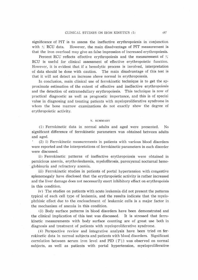
significance of PIT is to assess the ineffective erythropoiesis m conjunction with JC: RCU data. However, the main disadvantage of PIT measurement is that the iron overload may give an false impression of increased erythropoiesis.
Percent RCU reflects effective erythropoiesis and the measurement of Yt: RCU is useful for clinical assessment of effective erythropoietic function. However, it is evident that if a hemolytic process is involved, interpretation of data should be done with caution. The main disadvantage of this test is that it will not detect an increase above normal in erythropoiesis.
In conclusion, main clinical use of ferrokinetic technique is to get the approximate estimation of the extent of effective and ineffective erythropoiesis and the detection of extramedullary erythropoiesis. This technique is now of practical diagnostic as well as prognostic importance, and this is of special value in diagnosing and treating patients with myeloproliferative syndrome in whom the bone marrow examinations do not exactly show the degree of erythropoietic activity.
V. SUMMARY
( 1) Ferrokinetic data in normal adults and aged were presented. No significant difference of ferrokinetic parameters was obtained between adults and aged.
(2) i) Ferrokinetic measurements in patients with various blood disorders were reported and the interpretations of ferrokinetic parameters in each disorder were discussed.
ii) Ferrokinetic patterns of ineffective erythropoiesis were obtained in pernicious anemia, erythroleukemia, myelofibrosis, paroxysmal nocturnal hemoglobinuria and refractory anemia.
iii) Ferrokinetic studies in patients of portal hypertension with congestive splenomegaly have disclosed that the erythropoietic activity is rather increased and the liver damage does not necessarily exert inhibitory effect on erythropoisis in this condition.
iv) The studies on patients with acute leukemia did not present the patterns typical of each cell type of leukemia, and the results indicate that the myelophthisic effect due to the encloachment of leukemic cells is a major factor in the mechanism of anemia in this condition.
( 3) Body surface patterns in blood disorders have been demonstrated and the clinical implication of this test was discussed. It is stressed that ferrokinetic measurements with body surface counting are of great use both in diagnosis and treatment of patients with myeloproliferative syndrome.
( 4) Perspective review and integrative analysis have been tried on ferrokinetic data in normal subjects and patients with blood disorders. Significant correlation between serum iron level and PID ( T ~) was observed on normal subjects, as well as patients with portal hypertension, myeloproliferative
488 H. YAMADA
syndrome and patients with acute leukemia. Significant correlations were
observed between PID ( T ~) and erythroid/myeloid ratio in bone marrow and
between PIT and erythroid/myeloid ratio in bone marrow in patients with blood disorders.
ACKNOWLEDGMENT
The author wishes to express cordial appreciation to Professor S. Hibino
and assistant Professor K. Takikawa for their sincere guidance and encourage
ment throughout this study and also to Dr. H. Ohta and Dr. M. Kubo for their
cooperation throughout this work. Finally, the author thanks to Dr. H. Takagi for his cooperation in the study of patients with portal hypertension.
REFERENCES
1) Huff, R. L., Hennessy, T. G., Austin, R. E., Garcia, J. F., Roberts, B. M., and Lawrence,
- J. H., Plasma and red cell iron turnover in normal subjects and in patients having
various hematopoietic disorders, ]. Clin. Invest., 29, 1041, 1950.
2) Matsubara, T., Studies on the method for determination of iron in biological materials,
especially serum and whole blood-a new proposal for the standardization of the
method, Acta Haemat. ]ap., 24, 434, 1961 (in Japanese).
3) Yamada, H, Studies on the radioisotopic determination of the unsaturated iron-binding
capacity of serum, ]ap.]. Nucl. Med., 2, 146, 1965 (in Japanese).
4) Imanaga, H., Portal hypertension, Kyorinshoin, Tokyo, 1962.
5) Wakisaka, G., Dynamic studies on iron metabolism, Acta Haemat. ]ap., 23, 400, 1960
(in Japanese). 6) Sekiya, T., Studies on iron metabolism using radioactive iron. Report II. Iron meta
bolism in renal diseases, Acta Haemat. ]ap., 29, 850, 1966 (in Japanese) .
7) Takaku, F., Studies on the anemia in the aged-with a special reference to the pro
duction and destruction of erythrocytes, Acta Haemat. ]ap., 2.2, 464, 1959.
8) Maekawa, T. and K inukasa, K., Hematologic studies on the aged, Acta Haemat. ]ap.,
20( 3) Suppl., 105. 1957. 9) Bothwell, T. H., Callen dar, S., Mallett, B., and Witts, L. J.. The study of erythropoiesis
using tracer quantities of radioactive iron, Brit. ]. Haemat., 2, 1, 1956.
10) Bothwell, T. H., Hurtado, A. V., Donohue, D. M., and Finch, C. A., Erythrokinetics IV.
The plasma iron turnover as a measure of erythropoiesis, Blood, 12, 409, 1957.
11) Wasserman, L. R., Rashkoff, I. A., Leavitt, D., Mayer, J .. and Port, S., The rate of
removal of radioactive iron from the plasma-an index of erythropoiesis, ]. C/in. Invest.,
31, 32, 1952.
12) Finch, C. A., Unpublished data, 1958.
13) Bothwell, T. H. and Finch, C. A., Iron Metabolism pp. 208, Boston Little, Brown and
Company, 1962. 14) Pollycove, M. and Lawrence, J. H., Marrow hemolysis of immature red cells and
increased hemoglobin synthesis in iron deficiency anemia demonstrated with 59Fe.
IIIrd Congress of International Society of Haemat. in Mexico, 1962.
15) Girvin, E. C., Ooi, S. K., and Wong, S. P. S., The effect of hemorrhage on plasma iron
turnover, Blood, 17, 225, 1961.
16) Toghill, P. ]., Red-cell pooling in enlarged spleens, Brit. ]. Haemat., 10, 347, 1964.
17) Takagi, H., A study on the hematologic findings in portal hypertension, ]. ]ap. Sur g.
Soc., 67, 1990, 1966 (in Japanese).

CLINICAL STUDIES ON IRON KINETICS (I ) 489
18) Huff, R. L., Elmlinger, P. J., Garcia, J. F., Oda, J. M., Cockrell, M. C., and Lawrence, J. H ., Ferrokinetics in normal persons and in patients having various erythropoietic disorders, }. Clin. Invest., 30, 1512, 1951.
19) Haurani, F. I. and Tocantins, L. M., Ineffective erythropoiesis, A mer. ]. Med. , 31, 519, 1961.
20) Malamos, B., Belcher, E. H., Gyftaki, E., and Binopoulos, D., Simultaneous radioactive tracer studies of erythropoiesis and red-cell destruction in thalassemia, Brit.}. Haemat., 7, 411, 1961.
21) Giblett, E. R., Coleman, D. H., Pirzio-Biroli, G., Donohue, D. M., Motulsky, A. G., and Finch, C. A., Erythrokinetics: quantitative measurements of red cell production and destruction in normal subjects and patients with anemia, Blood, 11, 291, 1956.
22) Kuroyanagi, T., Some problems on immunology, jap. }. Clin. Med., (Tokyo) 22, 341, 1964 (in Japanese).
23) Finch, C. A., Coleman, D. H., Motulsky, A. G., Donohue, D. M., and Reiff, R. H., Erythrokinetics in pernicious anemia, Blood, 11, 807, 1956.
24) Myhre, E., Studies on the erythrokinetics in pernicious anemia, Scand. ]. Clin. Lab. Invest., 16, 391, 1964.
25) Nakao, K., Maekawa, T., Yaginuma, M., Wada, T., and Kenjo, T., A case report of paroxysmal nocturnal hemoglobinuria, with special reference to the mechanism of hemolysis, Acta Haemat. jap., 24(6), 59, 1961.
26) Kummer, H., Radioisotopenuntersuchungen bei der paroxysmalen naechtlichen Hemoglobinurie, Schweiz. Med. Wschr., 42, 1506, 1963.
27) Bothwell, T. H., Ellis, B. C., van Doorn·Wittkampe, H. van W., and Abrahams, 0. L., Radioiron studies in hemochromatosis. The effects of repeated phlebotomies, }. Lab. Clin. Med., 45, 167, 1955.
28) Najean, Y., Meeus-bith, L., Bernard, C., Bairon, M., Bousser, ]., and Bernard, J., Exploration isotopique de L'erythrocinetique dans 31 cas de pancytopenie idiopathique chronique a moelle histologiquement normale kou riche, Sang, 30, 101, 1959.
29) Kieley, J. M., Stroebel, C. F., Hanlon. D. G., and Owen, C. A., Jr., Clinical value of plasma-iron turnover rate in diagnosis and management of polycythemia, }. Nucl. Med., 2, 1, 1961.
30) Telfer, N. and Schiffman, N. L., The differential diagnosis of the polycythemias by plasma iron turnover determination, Nuclear·medizin, 3, 137, 1963.
31) Brodsky, I., The use of ferrokinetics in the evaluation of busulphan therapy in polycythemia vera, Brit. ]. Haemat., 10, 291, 1964.
32) Sharney, L., Schwartz, L., Wasserman, L. R., Port, S., and Leavitt, D., Pool systems in iron m etabolism; with special reference to polycythemia vera, Proc. Soc. Exp. Bioi. Med., 87, 489, 1954.
33) Yamada, H., Tanaka, M., and Kubo, M., Erythrokinetic studies of ineffective erythropoiesis in patients with anemias, Acta Haemat. j ap., 29, 254, 1966.
34) Baldini, M., Fudenberg, H. H., Fukutake, K., and Dameshek, W., The anemia of the Di Guglielmo syndrome, Blood, 14, 334, 1959.
35) Resegotti, L., La ferrocinetica nella mielosi eritremica, Minerva Med., 54, 2048, 1963. 36) Wetherley·Mein, G., Epstein, I. S., Foster, W. D., and Grimes, A. J., Mechanism of
anemia in leukemia, Brith. }. Haemat., 4, 281, 1958. 37) Sekiya, T., Studies on iron metabolism using radioactive iron. I. Iron m etabolism in
blood diseases, Acta Haemat. j ap., 25, 33, 1962. 38) Matsubara, T., Ferrokinetics in leukemia, jap. }. Clin. Haemat. 4, 133, 1963 (in Japanese ). 39) Kuba, ]., Wiedermann, B., und Wiedermann, M., Zur diagnostischen Auswertung von
Oberflaechemaktivitaetsmessungen bei Untersuchungen der Eryt hrokinetik mit Radio· f;~en \lnd Radiochrom1 Zscf!r. Gt;s. !11n. Med., l 9, 465, 1964,

490 H. YAMADA
40) Oettgen, H. F. und Pribilla, W., Die Erythrokinetik bei Osteomyelofibrose, Klil'l. Wschr.,
10, 483, 1964. 41) Nakai, G. S., Craddock, C. G., and Figuera, W. G., Agnogenic myeloid metaplasia.
A survey of twenty-nine cases and a review of the literature, Ann. Inter. Med., 57,
419, 1962. 42) Szur, L. and Smith, M. D., Red-cell production and destruction in myelosclerosis,
Brit. ]. Haemat., 7, 147, 1961.