Chapter 09: Experimental Growth in Animals
41
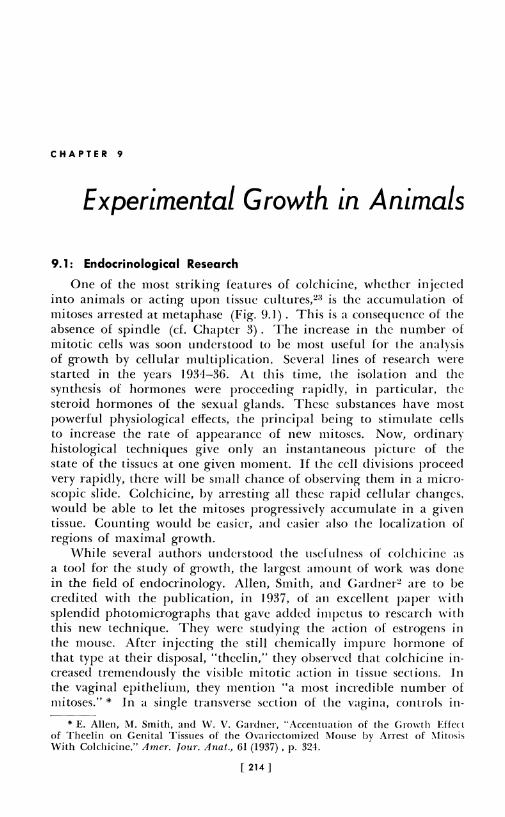
CHAPTER 9 Experimental Growth in Animals 9.1: Endocrinological Research One of the most striking features of colchicine, whether injected into animals or acting upon tissue cultures?: 1 is the accumulation of mitoses arrested at metaphase (Fig. 9.1). This is a consequence of the absence of spindle (cf. Chapter 3). The increase in the number of mitotic cells was soon understood to be most useful for the analvsis of growth by cellular multiplication. Several lines of research were started in the years 1934-36. At this time, the isolation and the synthesis of hormones were proceeding rapidly, in particular, the steroid hormones of the sexual glands. These substances have most powerful physiological effects, the principal being to stimulate cells to increase the rate of appearance of new mitoses. Now, ordinary histological techniques give only an instantaneous picture of the state of the tissues at one given moment. If the cell divisions proceed very rapidly, there will be small chance of observing them in a micro- scopic slide. Colchicine, by arresting all these rapid cellular changes, would be able to let the mitoses progressively accumulate in a given tissue. Counting would be easier, and easier also the localization of regions of maximal growth. While several authors understood the usefulness of colchicine as a tool for the study of growth, the largest amount of work was done in the field of endocrinology. Allen, Smith, and Gardner~ are to be credited with the publication, in 1937, of an excellent paper with splendid photomicrographs that gave added impetus to research with this new technique. They were studying the action of estrogens in the mouse. After injecting the still chemically impure hormone of that type at their disposal, "theelin," they observed that colchicine in- creased tremendously the visible mitotic action in tissue sections. In the vaginal epithelium, they mention "a most incredible number of mitoses."* In a single transverse section of the vagina, controls in- * E. Allen, M. Smith, and \V. V. Gardner, "Accentuation of the Growth Effect of Theelin on Genital Tissues of the Ovaricctomizcd !\louse by Arrest of :\litosis With Colchicine," Amer. /our. Anal., 61 (1937), p. 324. [ 214]
-
Upload
khangminh22 -
Category
Documents
-
view
1 -
download
0
Transcript of Chapter 09: Experimental Growth in Animals
CHAPTER 9
Experimental Growth in Animals
9.1: Endocrinological Research
One of the most striking features of colchicine, whether injected into animals or acting upon tissue cultures?:1 is the accumulation of mitoses arrested at metaphase (Fig. 9.1). This is a consequence of the absence of spindle (cf. Chapter 3). The increase in the number of mitotic cells was soon understood to be most useful for the analvsis of growth by cellular multiplication. Several lines of research were started in the years 1934-36. At this time, the isolation and the synthesis of hormones were proceeding rapidly, in particular, the steroid hormones of the sexual glands. These substances have most powerful physiological effects, the principal being to stimulate cells to increase the rate of appearance of new mitoses. Now, ordinary histological techniques give only an instantaneous picture of the state of the tissues at one given moment. If the cell divisions proceed very rapidly, there will be small chance of observing them in a microscopic slide. Colchicine, by arresting all these rapid cellular changes, would be able to let the mitoses progressively accumulate in a given tissue. Counting would be easier, and easier also the localization of regions of maximal growth.
While several authors understood the usefulness of colchicine as a tool for the study of growth, the largest amount of work was done in the field of endocrinology. Allen, Smith, and Gardner~ are to be credited with the publication, in 1937, of an excellent paper with splendid photomicrographs that gave added impetus to research with this new technique. They were studying the action of estrogens in the mouse. After injecting the still chemically impure hormone of that type at their disposal, "theelin," they observed that colchicine increased tremendously the visible mitotic action in tissue sections. In the vaginal epithelium, they mention "a most incredible number of mitoses."* In a single transverse section of the vagina, controls in-
* E. Allen, M. Smith, and \V. V. Gardner, "Accentuation of the Growth Effect of Theelin on Genital Tissues of the Ovaricctomizcd !\louse by Arrest of :\litosis With Colchicine," Amer. /our. Anal., 61 (1937), p. 324.
[ 214]
Experimental Growth in Animals 215
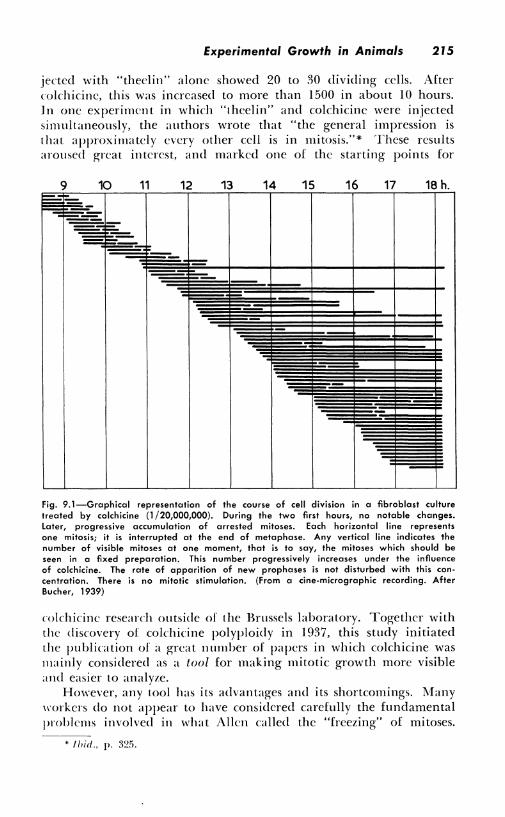
jected with "theclin" alone showed 20 to 30 dividing cells. After colchicine, this was increased to more than 1500 in about IO hours. Jn one experiment in which "theelin" and colchicine were injected simultaneously, the authors wrote that "the general impression is that approximately every other cell is in mitosis."* These results aroused great interest, and marked one of the starting points for
9 10 11 12 13 14 15 16 17 18 h. E:::::, -
-=-= =-
=== =-,_ ~
-
~ ~ -:~
--
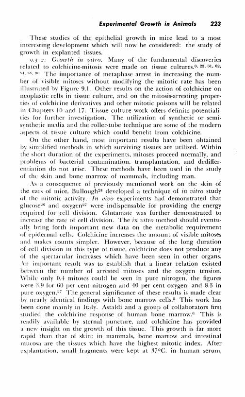
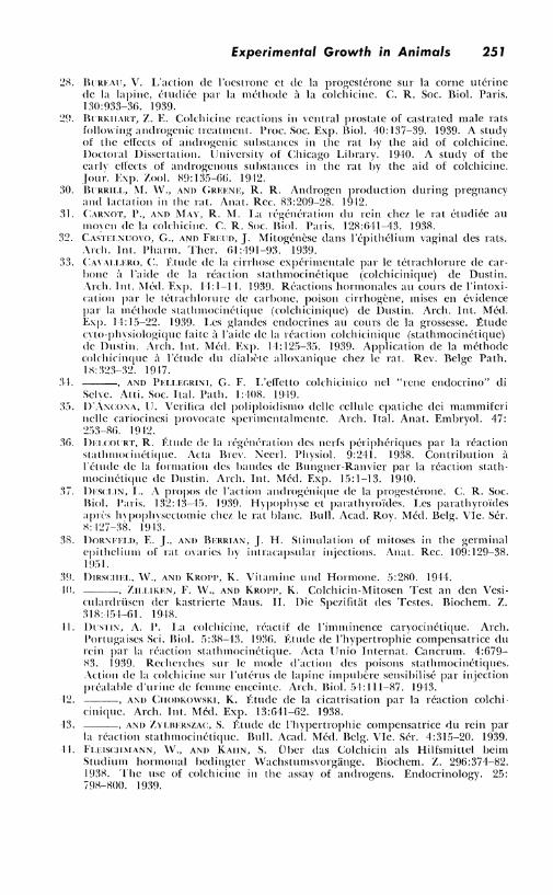
Fig. 9.1-Graphical representation of the course of cell division in a fibroblast culture treated by colchicine (1 /20,000,000). During the two first hours, no notable changes. Later, progressive accumulation of arrested mitoses. Each horizontal line represents one mitosis; it is interrupted at the end of metaphase. Any vertical line indicates the number of visible mitoses at one moment, that is to say, the mitoses which should be seen in a fixed preparation. This number progressively increases under the influence of colchicine. The rate of apparition of new prophases is not disturbed with this con• centration. There is no mitotic stimulation. (From a cine-micrographic recording. After Bucher, 1939)
colchicine research outside of the Brussels laboratory. Together with the discovery of colchicine polyploidy in 1937, this study initiated the publication of a great number of papers in which colchicine was mainly considered as a tool for making mitotic growth more visible and easier to analyze.
However, any tool has its advantages and its shortcomings. Many \\"orkers do not appear to have considered carefully the fundamental problems involved in what Allen called the "freezing" of mitoses.
216 Colchicine
Some of the complexities have already been scrutinized in the first chapters of this book. A few more considerations about this particular problem of multiplying the numbers of mitoses by destroying their spindle will be useful for future workers in this field. While the number of papers published about the rnlchicine method appears to be on the decrease, so far as can be assessed, for colchicine is not always mentioned in the titles, much work remains to be done. This chapter will point out several unexplored fields.
9.2: Theoretical Considerations
l\fost of the American authors, following the first papers of Allen, those of Brues1!l, 20 , 21 , 22 on liver regeneration, and the tissue culture work of Bucher23 and Ludford,"2 considered colchicine simply as a means of stopping any mitosis at metaphase. The complexities of colchicine pharmacology (Chapter 7) should alone call for more caution.
A. P. Dustin, Sr., in a paper published in 19%, but which could not have received much publicity, demonstrated the utility of colchicine as a tool. 41 He had noticed the increased number of divisions in the wall of a parasitic cyst in a mouse, a fact which was the starting point for experiments on the healing of wounds, reviewed further on in this chapter. In his own words, "colchicine enables the detection of the otherwise invisible state of preparedness to mitosis."'~ It throws into an abortive division all the cells which arc ready to
divide, or had been prepared for mitosis, for instance, under the influence of endocrine or other stimuli. This was in agreement with the line of thought which had led to the discovery of colchicine's action in 19'.H, and which was the study of the regulation of mitotic growth.
The theories of "mitotic arrest" or "arrest after mitotic stimulation" arc conflicting. ln work where the location of mitoses is the main purpose and where no quantitative data are required, colchicine is useful whatever the opinion one has about a possible stimulation of mitosis. This problem. however, should not he overlooked. For instance, several authors have thought it possible to calculate from the number of mitoses found after colchicinc, the average duration of these mitoses, had they not been arrested. This duration is, of course, an indication of the rapidity of cellular growth in the tissues studied. It should be clearly realized that such calculations imply several unknown factors, and they have a precise signification only if the following conditions are fulfilled: l. Colchicine arrests all mitoses, shortly after it has been injected
and until the end of the experimental period.
* A. P. Dustin, "La Colchicine, Rcactif de !'Imminence Caryocinctique," ,-1rcl,. Portugaises Sci. Biol., 5 (1936) , p. ,JI.
Experimental Growth in Animals 217
2. The intermitotic period is much longer than the duration of the experiment, and is not modified by the experiment.
3. The arrested mitoses arc not destroyed before the moment the tissues are fixed and examined.
J. The tissue is homogeneous from the point of view of mitosis, that is to say, mitotic rates and intcrmitotic periods do not vary from one region of the tissue to another.
5. The mitotic rate does not vary during the experimental period, in control animals.
Such conditions arc not often fulfilled. One type of experiment in ,rhich they are is liver regeneration; this will be considered further. J n mammals, cellular destruction is a factor which cannot be ignored. If, however, the above-mentioned causes of error do not exist, the average duration of mitosis can be found by the formula A= Mt/X, in which M is the mitotic index before colchicinc, and X the index found t hours after the injection of the alkaloid.
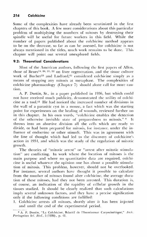
1£ this formula is applied to the results obtained in the experiments referred to in the previous paragraph,2 it is found that after "theelin" stimulation, the average duration of mitoses would be 10 minutes. This is a remarkably short period, and it may be questioned ,rhcther mitoses can be completed so rapidly. However, results obtained by A. P. Dustin, Sr., in the uterus of the rabbit after stimulation by chorionic gonadotropic hormones, are rather similar.41 The increase in the number of mitoses was observed in repeated biopsies. Figure 9.2 shows that it was considerable, and that in one animal, the calculated duration of each mitosis, had it not been arrested by colchicine, would be 12 minutes. These results bring some evidence for mitotic stimulation, for the prophase mitotic index increased also. This indicates that more cells were undergoing prophase than expected; that is to say, a true stimulation took place. This index rose from 7.56 to 14.8 in 2 hours, and from 4.8 to 24.4 in 7 hours. It must, of course, be supposed here that the duration of each prophase was not affected by colchicine.
Such results are rather complex, for the mitotic index could have been modified by the traumatisms of the biopsies themselves, and also by the continued action of the hormone. The possibility of a syncrgic action of hormones and colchicine cannot be ruled out6n (cf. Chapter 7).
The following results2n are all the more interesting, for while they apparently could demonstrate such a synergism, a much simpler explanation is possible. Table 9.1 gives the results of mitotic counts in the seminal vesicles, after stimulation by a single large dose of testosterone. There appears to be a veritable "explosion" of mitoses, to use the expression coined by A. P. Dustin, Sr. Does this give evidence of mitotic stimulation by the alkaloid? The counts of the con-
218 Colchicine
trol animals demonstrate that it does not, for it can be seen that between the thirtieth and thirty-fifth hours after the hormone injection the mitotic index rises sharply. If colchicine had been injected at the thirty-first hour, a mitotic increase from 2.92 to I 08.60 would have been observed, and this could not be explained by the theory of metaphase arrest. This increase is, however, not only the result of mitotic
X 35
x30
X 25
X 20
X )5
X )0
x5
xi
mitotic index
0 = CALCULATED DURATION OF
MITOSES ==A== M.t X
hours: 2 3
/
/ .•
/
/ /
4
/ /
/
/
/ /
/ /
/
/ /
_________ /'®
5 6
/ /'®
7 8
Fig, 9.2-Progressive increase of the numbers of mitoses, in repeated biopsies from the rabbit's uterus, after stimulation by chorionic gonadotropins and injection of colchicine, Calculated duration of mitoses on the assumption that colchicine does nothing more than arrest them at metaphase, (From original data of A, P, Dustin, 1943n)
stasis, but also of the progressive action of testosterone, demonstrated by the fact that in untreated animals the mitotic count rises about threefold. Therefore, colchicine alone has increased the mitoses only from about JO (2.92 X 3) to 108.GO within -1 hours, which means that the average mitotic duration must be about 25 minutes or less, This agrees with knowledge of mitotic duration in mammals.
Such an example dem(>nstrates the intricacies of quantitative
Experimental Growth in Animals 219
work with colchicine. Others will be found in this chapter. Here, as in other fields of colchicine work, problems must not be oversimplified, and here especially, the greatest care should be taken in all quantitative estimations. It is striking that it is when colchicine is considered as a tool that the need for fundamental knowledge is the most apparent.
9.3: Cellular Multiplication in Normal Growth
Growth patterns in the organs of adult animals can be revealed far better after colchicine than with ordinary tissue sections. The alkaloid may do more than simply locate the germinative zones of organs; under strict experimental conditions, it may solve some quantitative problems of growth. Another method, which has brought excellent results, is to study the growth of explanted tissues. This has been done by the ordinary methods of tissue culture,2 :1, 62 , 88 or
TABLE 9.1
MITOTIC ACTIVITY IN THE SEMINAL VESICLES OF CAS
TRATED 80-DAY-OLD RATS TREATED WITH 0.3 MG. OF
TESTOSTERONE PROPIONATE
(Abridged from Burkhart29 )
19 .. 0 0.24
23. 0.28 0.20
27. 5.00 2.04
31 ... 2.02 7.60
35 ... 10.68 108.60
·---~-
by a modified technique in which cellular multiplication was obsened only for a few hours after explantation.5 - 8 , 24 - 27 Some of the results demonstrating how useful colchicine may be as a tool in such work will be summarized here.
q.3-1: Studies in vivo. Some of the early work in this field was done on the ovary. Colchicine, by increasing from 11 to 35 times the number of mitoses that could be observed in the germinal epithelium of the ovary of mice, demonstrated that this was a region of active growth. 1 - :rn, 14 , ,o Similar facts were observed in guinea pigs. •"· 77 The relation between the mitotic activity in the ovarian follicles
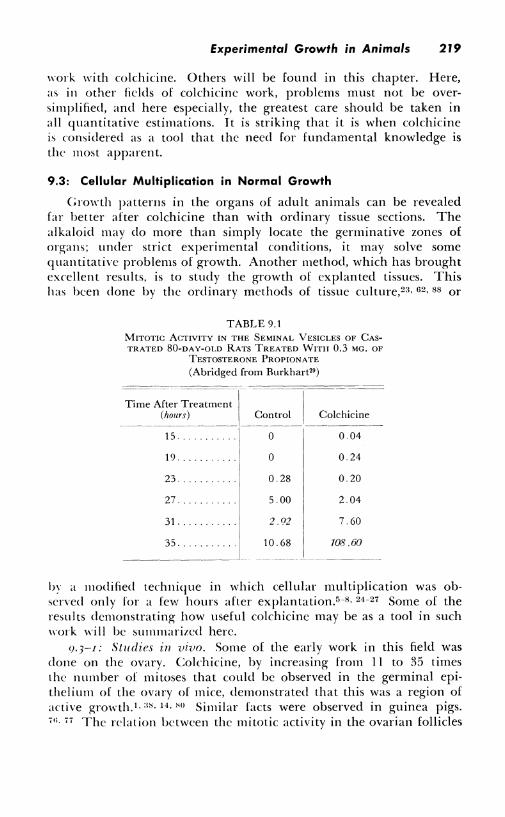
220 Colchicine
and the estrus cycle were carefully analyzed (Fig. 9.3). In the endothelial cells of the theca interna of the ovarian follicles, immediately before ovulation, the karyokineses were found to increase about sixtyfold. Arrested mitoses of follicular cells in the rat can be found around eggs after they have reached the uterus (Fig. 9.4) .4 Some follicles are found to be growing rapidly while others are quiescrn t.
140
130
120
110
JOO
90
80
II) 70 UJ
II)
~ 60 ~ lL 50 0 ci 40 z
30
20
10
0
16 2
MITOTIC PROLIFERATION IN THE DEVELOPING AND
RETROGRESSING CORPUS LUTEUM
-endothelium
_______ ~ luteal cells
__ ~ -· ____ connce:ctive: tissue:
th<ca utuna
3 4 5 6 7 S 9 JO II 12 13 14 JS 16 2 3 4 DAY OF ESTROUS CYCLE
Fig. 9.3-Mitoses in the corpus luteum of the ovary of a normal mature guinea pig, studied by the colchicine method. (After Schmidt")
This fact is not evident in control animals, because the number of mitoses is too small.
In the pituitary glands of mice, colchicine increases the number of mitoses about threefold. This is an indication that these mitoses are normally of long duration. Many data have been gathered about the mitotic activity in this organ in various physiological conditions. 11 , 52 Table 9.2 shows how evident is the action of age on mitotic activity when the number of metaphases has been artificia!IY 111-
creased by spindle poisoning.'12
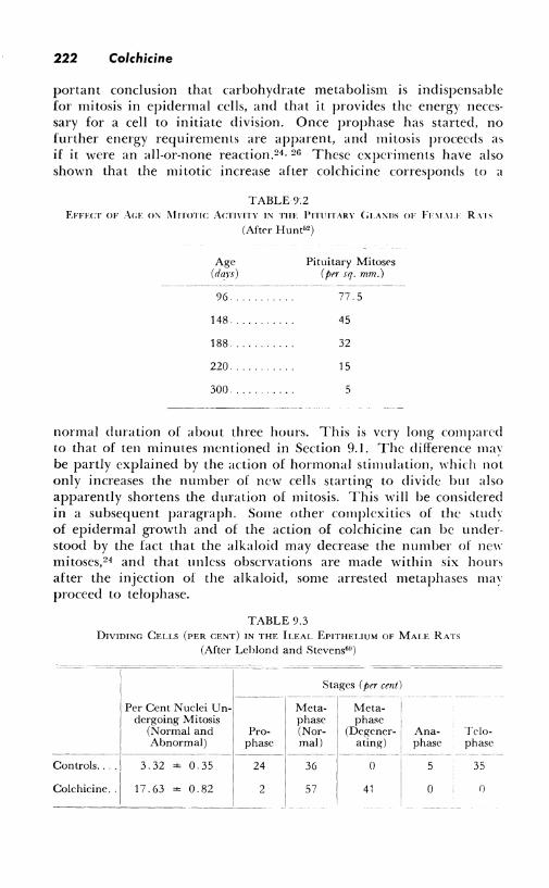
A quantitative study of cell regeneration in the mucosa of the intestine in rats has been made possible by colchicine. It was known that the intestinal cells are continuously shed, but how long it took for the whole epithelial lining to be replaced was not known. Table
Experimental Growth in Animals 221
9.,l gi,e., the results, with the percentages of dividing cells and of mitotic stages in control and colchicinized animals.0° From these results. it is apparent that mitotic arrest at metaphase has increased in six hours the number of cell divisions by 17.63/3.32. The mitotic duration, calculated as indicated in Section 9.2, is 3.32 X 6.0/17.63 = 1. l!l = I hr. 8 min. 1 t can be calculated from this result that in 3 7 .7 hours ( 1.57 days) , I 00 per cent of the cells will have divided; that is to say, a complete renewal of the epithelium will have taken place. This is, of course, only statistically correct, for there must remain a certain number of stem cells so that growth may persist. These cells will divide into one differentiating cell and one stem cell identical to the first. A great discrepancy between results obtained ,rith radio-phosphorus on the nucleic acid turnover and the figures ?;i,en by the colchicine method as used by the same authors has been cliscmered.' 1 This may throw more light on the complex problems of gnmth in differentiating tissues.
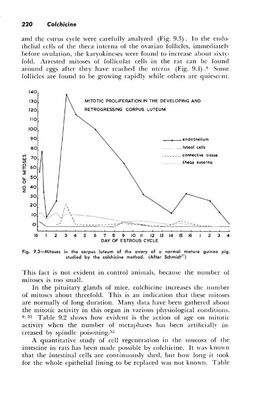
The ,kin of small rodents has been excellent testing material for the study of growth as analyzed by colchicine. A very extensive series
Fig. 9.4~Colchicine-mitoses (black dots) in an ovarian follicle {left), and in follicular cells surrounding an egg found in the uterus in the rat. (After Allen et al.')
of experiments has been carried on, especially by Bullough.24- 27 This has pro,ided ample material for a precise analysis of growth and the fundamental mechanisms of mitosis. Further reference will be made to some of these papers in the section on hormonal stimulation of mitosis. Di urn al variations, the action of sleep. the effects of bloodsugar len·l and of injections of starch, have led to the most im-
222 Colchicine
portant conclusion that carbohydrate metabolism is indispensable for mitosis in epidermal cells, and that it provides the energy necessary for a cell to initiate division. Once prophase has started, no further energy requirements are apparent, and mitosis proceeds as if it were an all-or-none reaction.24 , 26 These experiments have also shown that the mitotic increase after colchicine corresponds to a
TABLE9.2 EFFECT OF Au: o:\/ l\1noT1c AcnvnY 1, TIIE l'nunARY G1.A,ns oF Fi-,uu R,1s
(Aftc-r Hunt62 )
Age (dars)
Pituitary Mitoses (prr sq. mm.)
-------------- --- ------ ---
96. 77.5
148. 45
188. 32
220. 15
300. 5
normal duration of about three hours. This is very long compared to that of ten minutes mentioned in Section 9.1. The difference may be partly explained by the action of hormonal stimulation, which not only increases the number of new cells starting to divide but also apparently shortens the duration of mitosis. This will be considered in a subsequent paragraph. Some other complexities of the ~tuch of epidermal growth and of the action of colchicine can be understood by the fact that the alkaloid may decrease the number of new mitoses,24 and that unless observations are made within six hours after the injection of the alkaloid, some arrested metaphases ma, proceed to telophase.
TABLE 9.3 DIVIDING CELLS (PER CENT) IN THE ILEA!. EPITHEI.IUM OF MALE RATS
(After Leblond and Stevens'°) -- -----------
Controls .... I I
Colchicine. · [
Per Cent Nuclei Undergoing Mitosis
(Normal and Abnormal)
3. 32 ± 0. 35
17. 63 ± 0. 82
Prophase
24
2
Stag es ( per cent)
-Met~~-1-M~:- 1 -
phase phase (Nor- (Degener- ' ma!) ating)
36
57
0
41
Anaphase
5
0
Telophase
35
0
Experimental Growth in Animals 223
These studies of the epithelial growth in mice lead to a most interesting development which will now be considered: the study of growth in explanted tissues.
9.3-2: Growth in vitro. Many of the fundamental discoveries related to colchicine-mitosis were made on tissue cultures.6 , 23, 610 62 ,
' 4 · "· "" The importance of metaphase arrest in increasing the number of visible mitoses without modifying the mitotic rate has been illustrated by Figure 9.1. Other results on the action of colchicine on neoplastic cells in tissue culture, and on the mitosis-arresting properties of colchicine derivatives and other mitotic poisons will be related in Chapters IO and 17. Tissue culture work offers definite potentialities for further investigation. The utilization of synthetic or semisvnthetic media and the roller-tube technique are some of the modern aspects of tissue culture which could benefit from colchicine.
On the other hand, most important results have been obtained bv simplified methods in which surviving tissues are utilized. Within the short duration of the experiments, mitoses proceed normally, and problems of bacterial contamination, transplantation, and dedifferentiation do not arise. These methods have been used in the study of the skin and bone marrow of mammals, including man .
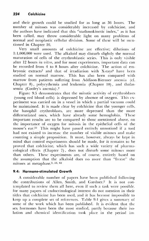
. -\s a consequence of previously mentioned work on the skin of the ears of mice, Bullough24 developed a technique of in vitro study of the mitotic activity. In vivo experiments had demonstrated that glucose~,; and oxygen27 were indispensable for providing the energy required for cell division. Glutamate was further demonstrated to increase the rate of cell division. The in vitro method should eventually bring forth important new data on the metabolic requirement of epidermal cells. Colchicine increases the amount of visible mitoses and makes counts simpler. However, because of the long duration of cell division in this type of tissue, colchicine does not produce any of the spectacular increases which have been seen in other organs . . \n important result was to establish that a linear relation existed between the number of arrested mitoses and the oxygen tension. \Vhik only 0.'1 mitoses could be seen in pure nitrogen, the figures were :l.9 for liO per cent nitrogen and 40 per cent oxygen, and 8.3 in pure oxygeri.~7 The general significance of these results is made clear IJ\ nearly identical findings with bone marrow cells.5 This work has been done mainly in Italy. Astaldi and a group of collaborators first studied the cokhicine response of human bone marrow.U This is readily available by sternal puncture, and colchicine has provided a new insight on the growth of this tissue. This growth is far more rapid than that of skin; in mammals, bone marrow and intestinal mucosa are the tissues which have the highest mitotic index. After cxplantation, small fragments were kept at 37°C. in human serum,
224 Colchicine
and their growth could be studied for as long as 36 hours. The number of mitoses was considerably increased by colchicine. and the authors have indicated that this "stathmokinetic index," as it has been called, may throw considerable light on many problems of normal and neoplastic cellular division. Some of these will be mentioned in Chapter 10.
Very small amounts of colchicine are effective; dilutions of 1: 1,000,000 were used. The alkaloid may disturb slightly the normal maturation of cells of the crythroblastic series. This is only visible after 12 hours in vitro, and for most experiments, important data can be recorded from 4 to 8 hours after colchicine. The action of embryonic extracts7 and that of irradiation with X-rays8 have been studied on normal marrow. This has also been compared 11·ith marrow from patients suffering from Addison-Bienner anemia (cf. Chapter 8), polycythemia and leukemia (Chapter 10), and thalassemia (Cooley's anemia) .7
Figure 9.5 demonstrates that the mitotic activity of erythroblasts (young red blood cells) is depressed by absence of oxygen. This experiment was carried on in a vessel in which a partial vacuum rnuld be maintained. It is made clear by colchicine that the younger cells, the basophil erythroblasts, arc more depressed than the more differentiated ones, which have already some hemoglobin. These important results are to be compared to those mentioned above, on the importance of oxygen for mitosis in the epithelial cells of the mouse's ear.27 This might have passed entirely unnoticed if a tool had not existed to increase the number of visible mitoses and make counting a simple proposition. It must, however, always be kept in mind that control experiments should be made, for it remains to be proved that colchicine, which has such a wide variety of pharmacological effects (Chapter 7) , docs not disturb some mitoses more than others. These experiments are, of course, entirely based on the assumption that the alkaloid does no more than "freeze" the mitoses at metaphase.2 - 23, u2
9.4: Hormone-stimulated Growth
A considerable number of papers have been published follm1·ing the contributions of Allen, Smith, and Gardner.2 It is not contemplated to review them all here, even if such a task were possible. for many papers of cndocrinological interest do not mention in their titles that colchicine has been used, and it has become impossible to keep up a complete set of references. Table 9.1 gives a summary of some of the work which has been published. It is evident that the sex hormones have been the most studied, partly because their 1,0-
lation and chemical identification took place in the period 1111-
MITOSES 100
90
80
0
0
0
70 . 0
8
0 0
60 0 8
0
50 0
0
40 0
30
20
10
PRESSION: 760mm. 660 Hg.
. . .
. . • .
•
0 0
0
0
0
0
560
•
• . . . •
0
s 0 I
0 • 0 0 0 0 0 . 0 0
0 . 0 0 0
• 8 d 0 0
460 360 260 160
Fig. 9.5-Linear relation between pressure of atmospheric air and mitoses in bone-marrow erythroblasts studied by culture in vitro. The results are expressed as percentages of the maximum mitotic rate, i.e., that of basophil erythroblasts at atmospheric pressure. (After Astaldi et al. 8)
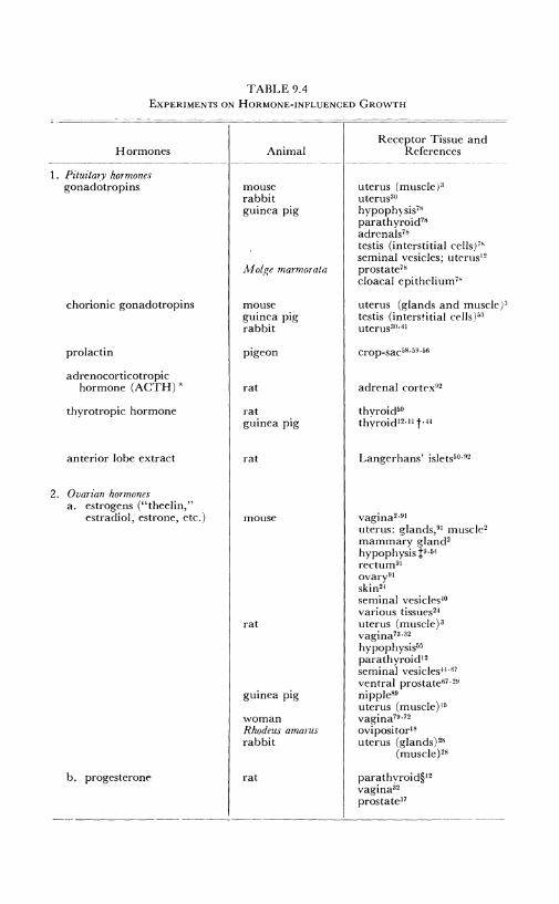
TABLE9.4 EXPERIMENTS ON HORMONE-INFLUENCED GROWTH
Hormones --------~-------------
1. Pituitary hormones gonadotropins
chorionic gonadotropins
prolactin
adrenocorticotropic hormone (ACTH)•
thyrotropic hormone
anterior lobe extract
2. Ovarian hormones a. estrogens ("theelin,"
estradiol, estrone, etc.)
b. progesterone
Animal
mouse rabbit guinea pig
Molge marmorata
mouse guinea pig rabbit
pigeon
rat
rat guinea pig
rat
mouse
rat
guinea pig
woman Rhodeus amarus rabbit
rat
Receptor Tissue and References
uterus (muscle )3
uterus30
hypophysis78
parathyroid78 adrenals78
testis (interstitial cells/' seminal vesicles; ut<>rus 12
prostate78
cloaca] epithelium"
uterus (glands and muscle)' testis (interstitial cells) 53
uterusao,.11
crop-sac58 · 59 ·56
adrenal cortex92
thvroid50
thyroid12 •11 t·"
Langerhans' islets50 · 02
vagina2 •91
uterus: glands, 91 muscle' mammary gland2
hypophysis f9•54
rcctum9l
ovary91
skin24
seminal vesicles 10
various tissues2·1
uterus (muscle)' vagina73 · 32
hypophysis55
parathyroid12
seminal vesicles 11 •67
ventral prostate67 ·"'
nipple89
uterus (muscle)I,s vagina79 ·72
ovi posi tor18
uterus (glands) 28
(muscle) 28
parathvroid§12
vagina32
prostate37
--------------~---------
Experimental Growth in Animals 227
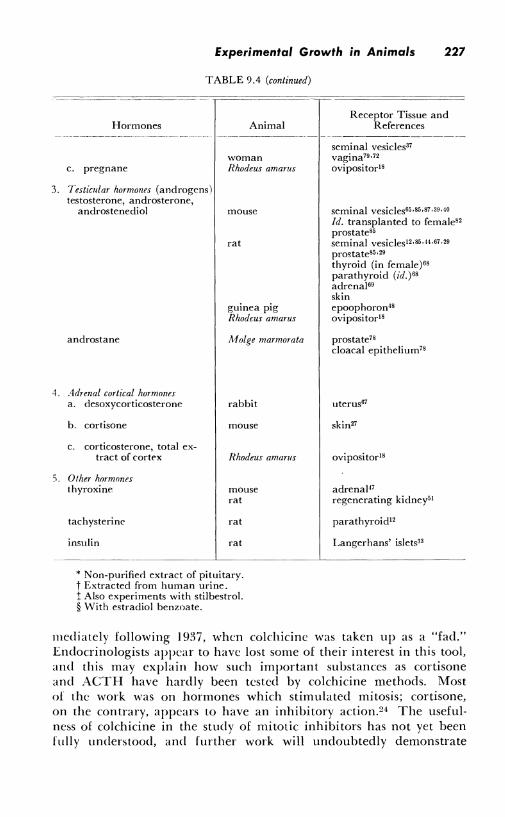
TABLE 9.4 (continued)
Hormones Animal
woman c. pregnane Rhodeus amarus
3. Testicular hormones (androgens) testosterone, androsterone,
4.
5.
androstenediol mouse
rat
guinea pig Rhodeus amarus
androstane Molge marmorata
Adrenal cortical hormones a. desoxycorticosterone rabbit
b. cortisone mouse
C. corticosterone, total ex-tract of cort('x Rhodeus amarus
0 I her hormones thyroxine mouse
rat
tachysterinc rat
insulin rat
* Non-purified extract of pituitary. t Extracted from human urine. ! Also experiments with stilbestrol. § With estradiol benzoate.
Receptor Tissue and References
seminal vesicles37
vagina79 •72
ovipositor18
seminal vesicles65,85,s7 ,39,10
Id. transplanted to female82
prostate85 seminal vesicles 12 •85 ·'"'· 67 ·29 prostate85 •29
thyroid (in female) 68
parathyroid (id.)•s adrenal69
skin epoophoron18 ovipositor18
prostate78
cloaca! epithelium78
uterus67
skin27
ovipositor18
adrenal47
regenerating kidney51
parathyroid12
Langerhans' islets33
mediately following l 937, when colchicinc was taken up as a "fad." Endocrinologists appear to have lost some of their interest in this tool, and this may explain how such important substances as cortisone and ACTH have hardly been tested by colchicine methods. Most of the work was on hormones which stimulated mitosis; cortisone, on the contrary, appears to have an inhibitory action.24 The usefulness of colchicine in the study of mitotic inhibitors has not yet been fully understood, and further work will undoubtedly demonstrate
228 Colchicine
that this is a tool for the study of mitotic activity, whether stimulated or depressed. Results reported in Chapter IO support this opinion.
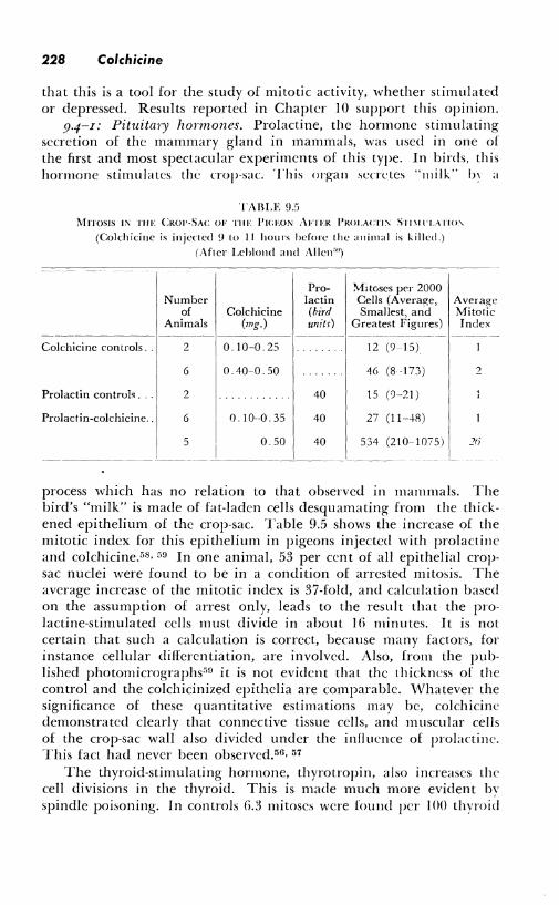
9.4-I: Pituitary hormones. Prolactine, the hormone stimulating secretion of the mammary gland in mammals, was used in one of the first and most spectacular experiments of this type. In birds, this hormone stimulates the crop-sac. This organ secretes "milk'' b, a
TABLE 95 MITOSIS IN THE CROP-SAC OF TIH: J'ICEON AFTFR l'ROLACTI, S1n1 l'L\TIO'\
(Colchicine is injected 9 to 11 hours before the animal is killed.) (After Leblond and Allen.-")
Colchicinc controls ..
Prolactin control~ ...
Prolactin-colchicine ..
Number of
Animals
2
6
2
6
5
Colchicine (mg.)
0. 10-0. 25
0.40-0.50
0.10--0. 35
0.50
Prolactin (bird uni tr)
40
40
40
Mitoses per 2000 Cells (Average, Average Smallest, and Mitotic
Greatest Figures) I ndcx
12 (9 15)
46 (8 173)
15 (9-21)
27 (11-48)
534 (210-1075)
2
process which has no relation to that observed in mammals. The bird's "milk" is made of fat-laden cells desquamating from the thickened epithelium of the crop-sac. Table 9.5 shows the increase of the mitotic index for this epithelium in pigeons injected with prolactine and colchicine.580 59 In one animal, 53 per cent of all epithelial cropsac nuclei were found to be in a condition of arrested mitosis. The average increase of the mitotic index is 37-fold, and calculation based on the assumption of arrest only, leads to the result that the prolactine-stimulated cells must divide in about 16 minutes. It is not certain that such a calculation is correct, because many factors, for instance cellular differentiation, are involved. Also, from the published photornicrographs"n it is not evident that the thickness of the control and the colchicinized epithclia are comparable. \,Vhatever the significance of these quantitative estimations may be, colchicine demonstrated clearly that connective tissue cells, and muscular cells of the crop-sac wall also divided under the influence of prolactine. This fact had never been observed.56, 57
The thyroid-stimulating hormone, thyrotropin, also increases the cell divisions in the thyroid. This is made much more evident by spindle poisoning. In controls 6.3 mitoses were found per I 00 thyroid
Experimental Growth in Animals 229
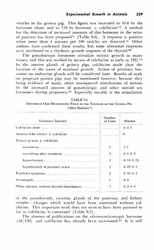
\'esicles in the guinea pig. This figure was increased to 16.8 by the hormone alone, and to 119 by hormone + colchicine.12 A method for the detection of increased amounts of this hormone in the urine of patients has been proposed11 (Table 9.6). A response is positive when more than 4 mitoses per 100 vesicles are detectecl. 11 Other authors have confirmed these results, but some abnormal responses were attributed to a rhythmic growth response of the thyroid.50
The gonadotropic hormones stimulate mitotic growth in many tissues, and this was studied by means of colchicine as early as 1937.12
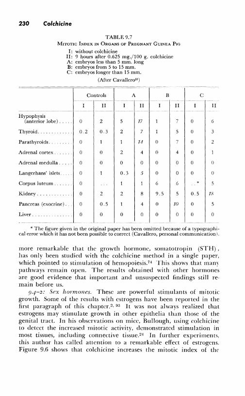
In the uterine glands of guinea pigs, colchicine made clear the location of the zones of maximal growth. Action of pituitary horrnones on endocrine glands will be considered later. Results of work on pregnant guinea pigs may be mentioned however, because they bring evidence of many, often unsuspected stimulations of mitosis by the increased amount of gonadotropic and other steroid sex hormones during pregnancy.33 Especially notable is the stimulation
TABLE 9.6 MITOSES IN ONE MICROSCOPIC FIELD IN THE THYROID OF THE GUINEA Pm
(After Basteniell)
Substance Injected
Colchicinc alone ........ .
Anterior lobe extrart + colchicinc.
Extracts of urine + colchicine:
myxedema ....
myxcclcma aft<'r treatment.
hypcrthyroidy .
hypothyroicly of pituitary origin.
Froelich's syndrome .. .
A.,Tomegaly .......... .
Other diseases, without thyroid disturbances ...
Number of Cases
5
3
3
3
2
3
Mitoses
0.5-1
1)
1-7
0.1-0. 5
0.15-0.25
0.05-0.4
0. 05-0. 1
0.2
0.2-0.4
------ ·-
of the parathyroids, exocrine glands of the pancreas, and kidney t11lmles - changes which would have been unnoticed without colchicine. This irnportant work does not seem to have been pursued so far as colchicine is concerned (Table 9.7).
The absence of publications on the adrenocorticotropic hormone (.\CTH) and colchicine has already been mentioned. 24 It is still
230 Colchicine
TABLE 9.7 MITOTIC INDEX IN ORGANS OF PREGNANT GUINEA Pw
I: without colchicine II: 9 hours after 0.625 mg./100 g. colchicine A: embryos less than 5 mm. long B: embryos from 5 to 15 mm. C: embryos longer than 15 mm.
(After Cavallero33 )
Controls A B ---~--- -------- -· --- ---
II II II ---------- -·---- ------··
Hypophysis (anterior lobe) ... 0 2 5 17 7
Thyroid .. 0.2 0 3 2 7 5
Parathyroids. 0 14 () 7
Adrenal cortex .. 0 0 2 4 0 4
Adrenal medulla . 0 0 0 0 0 0
Langerhans' islets. 0 0.3 5 0 0
Corpus luteum .. 0 6 6
Kidney ........ 0 2 2 8 9.5 5
Pancreas (exocrine) ... 0 0.5 4 0 1()
Liver ...... 0 0 0 0 0 0
------ -- ---- ---- -------- ·---
C -·--
II
0 6
() 3
() 2
0
0 0
0 0
* 5
0.5 18
0 5
0 0
* The figure given in the original paper has been omitted because of a typographical error which it has not been possible to correct (Cavallero, personal communication I.
more remarkable that the growth hormone, somatotropin (5TH), has only been studied with the colchicine method in a single paper, which pointed to stimulation of hemopoiesis. 74 This shows that many pathways remain open. The results obtained with other hormones are good evidence that important and unsuspected findings still remain before us.
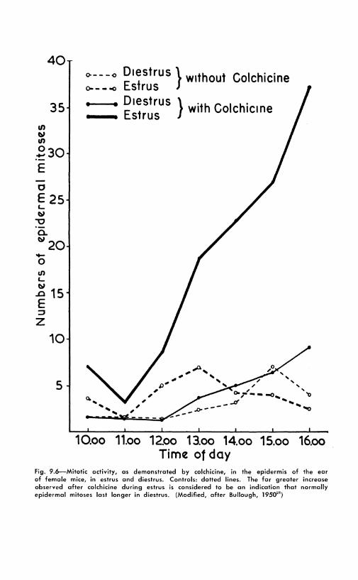
9.4-2: Sex hormones. These are powerful stimulants of mitotic growth. Some of the results with estrogens have been reported in the first paragraph of this chapter. 2 , 03 It was not always realized that estrogens may stimulate growth in other epithelia than those of the genital tract. In his observations on mice, Bullough, using colchicine to detect the increased mitotic activity, demonstrated stimulation in most tissues, including connective tissue.24 In further experiments, this author has called attention to a remarkable effect of estrogens. Figure 9.6 shows that colchicine increases the mitotic index of the
40
35 II> ~ II>
230 E 0
E 25 L. ~
""O a. ~20 -0 .,, L. ~
15 .a E ::,
z 10
5
<>-- -<> Die5trus } without Colchicine o- - - "° Estrus I • D1estrus } ·th C I h' __ Estrus w1 o c 1c1 ne
,, ,, 0- ,-t - .. -o....,
_Ji' ....... .1>--- "'O
10.oo 11.oo 12~o 13.oo 14.oo 15.oo 16.oo Time of day
fig. 9.6--Mitotic activity, as demonstrated by colchicine, in the epidermis of the ear of female mice, in estrus and diestrus. Controls: dotted lines. The far greater increase observed after colchicine during estrus is considered to be an indication that normally epidermal mitoses last longer in diestrus. (Modified, after Bullough, 1950'")
232 Colchicine
epidermis of the ear considerably more during estrus than during diestrus. The mitoses were counted hour by hour by clipping small fragments of the ear. This difference can be explained by a shortening of the time taken for one division, from about 2 hours in diestrus to ¾ hour in estrus. This significant result is not discussed; other possible hypotheses arc, for instance, synergic action of colchicine and hormone, or changes in the duration of interphase. The alkaloid is simply considered to stop metaphases. 2 , 2 '1· n2
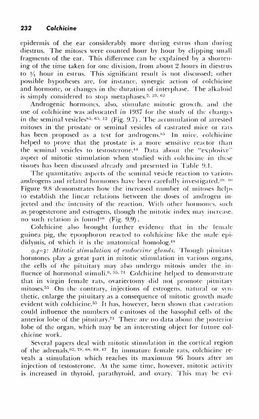
Androgenic hormones, also, stimulate mitotic growth. and the use of colchicine was advocated in I 9:l7 for the study of the changes in the seminal vesicles8", n:., 12 (Fig. 9.7). The acrnmulation of arrested mitoses in the prostate or seminal vesicles of castrated mice or rats has been proposed as a test for androgens;'" In mice. colchicinc helped to prove that the prostate is a more sensitive reactor than the seminal vesicles to testostcronc.44 Data about the "explosive·· aspect of mitotic stimulation when studied with colchicinc in these tissues has been discussed already and presented in Table (),I.
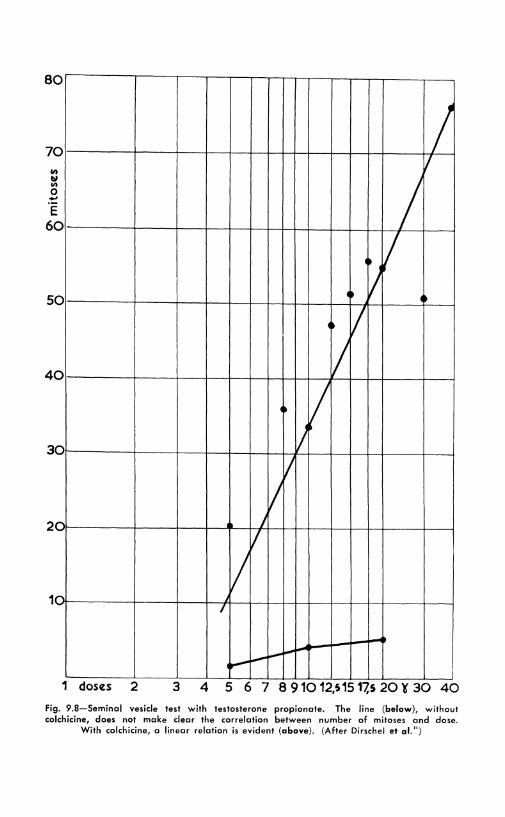
The quantitative aspects of the seminal vesicle reaction to variou, androgens and related horlllones have been carefully investigated_:w. 4"
Figure 9.8 dclllonstrates how the increased number of mitoses helps lo establish the linear relations between the doses of androgen injected and the intensity of the reaction. \Vith other horllloncs, such as progesterone and estrogens, though the lllitotic index may increase. no such relation is found 10 (Fig. 9.!}) .
Colchicine also brought further evidence that in the female guinea pig, the epoophoron reacted to colchicine like the male epididymis, of which it is the anatomical homolog. 4H
9.4-3: 1Witotic stimulation of emlocrinr' glands. Though pituitan hormones play a great part in mitotic stimulation in various organs, the cells of the pituitary may also undergo mitosis under the influence of hormonal stimuliY, r,r,, 71 Colchicine helped to demonstrate that in virgin female rats, ovariectomy did not promote pituitan mitoses."" On the contrary, injections of estrogens, natural or s,·nthetic, enlarge the pituitary as a consequence of mitotic growth made evident with colchicine."" It has, however, been shown that castration could influence the numbers of c:-mitoses of the basophil cells of the anterior lobe of the pituitary. 71 There arc no data about the posterior lobe of the organ, which may he an interesting object for future colchicine work.
Several papers deal with mitotic stimulation in the cortical region of the adrenals.02 , 78 , GH, mi, 47 ln immature female rats, colchicine reveals a stimulation which reaches its maximum 96 hours after an injection of testosterone. At the same time, however, mitotic acti,·itY is increased in thyroid, parathyroid, and ovary. This ma\' he cvi-
Fig . 9.7- Mitotic stimulation by testosterone propionate in the seminal vesicles. Above. Hormone alone. Below. Hormone + colchicine. (Original photomicrographs from
Bastenie and Zylberszac")
8 0
7 0 Ill
"' Ill 0 ., E
6 0
5 0
4 0
3 0
2 0
1 C
1 dOSIZS 2 3
/ I
I It I
It J ◄~
It I I
t I J
J
J , .. J
7
I/ j
7
---I~
i--
4 5 6 7 8 910 12,S 15 17,S 20 ¥ 30 40
Fig. 9.8-Seminal vesicle test with testosterone propionate. The line (below), without colchicine, does not make clear the correlation between number of mitoses and dose.
With colchicine, a linear relation is evident (above). (After Dirschel et al.")
Experimental Growth in Animals 235
BO / t11:stostcron11:
:
70 tutost11:ron11:pro~ionol11: /
I :
I I androsl11:ndion11:, .
50
40
I .' : I
I I
/2 I ·, ;
I? "·. I I '-..._,:
_I I
/ /
V/ I. .,
dihydroondrostuon11: /,/' / I
I
30
20
10
/ -I _,✓-:..-- androst11:y
I -----~ / -· - / / ,,--· / --I ,- _/ /,_: / - ,
'· / / '\ / v/ \
/
'\ / / ~ / ~
/ / // --~ - --------~ --- --- --- -- - d11:hydroondrost11:ron11: ----- ----- - -- -------
I
10 20 dos11:s 50 100 200 500 1000~ 2000
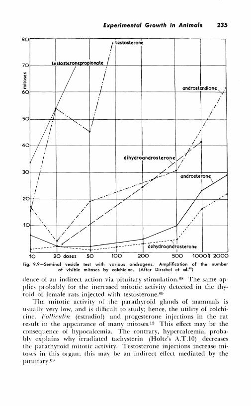
fig. 9.9-Seminal vesicle test with variaus andragens. Amplificatian of the number of visible mitoses by colchicine. (After Dirschel et al.'0 )
dence of an indirect action via pituitary stimulation.u8 The same applies probably for the increased mitotic activity detected in the thyroid of female rats injected with testosterone.6!!
The mitotic activity of the parathyroid glands of mammals is usually very low, and is difficult to study; hence, the utility of colchicine. Folliculin (estradiol) and progesterone injections in the rat result in the appearance of many mitoses.12 This effect may be the consequence of hypocalcemia. The contrary, hypercalcemia, probably explains why irradiated tachysterin (Holtz's A.T.10) decreases the parathyroid mitotic activity. Testosterone injections increase mitoses in this organ; this may be an indirect effect mediated by the pituitary.Gu
236 Colchicine
In the Langerhans' islets of the pancreas, pituitary stimulation 92 , 50 and pregnancy increase the number of mitoses, as detected b\' colchicine.
It is surprising to find no paper dealing with mitotic stimulation in the interstitial (Leydig) cells of the testes. In guinea pigs injected with chorionic gonadotropins, these cells increase in number, but colchicinc failed to detect mitoses. It was concluded that the hormone-secreting cells originated from ordinary connective cells_:.:i Further work on this tissue is obviously needed.46
9.5: Regeneration and Hypertrophy
The problem which was under study in the laboratory of .\. P. Dustin, Sr., since about I 920 and which led to the discovery of the properties of colchicine was that of the regulation of growth and mitotic activity in pluricellular animals. In vertebrates, for instance, cell division takes place only in some tissues, and then in an orderly way. \1/hilc in the adult, nerve cells become incapable of any mitosis, other organs, such as the liver and the kidney, while nearly devoid of any mitotic activity in normal conditions, may grow rapidly by cellular multiplication after surgical excision. In the rodents, and in particular the rat, large portions of the liver may be removed surgically. The remaining cells start to divide at once, and regeneration of the normal liver mass is remarkably rapid. 21 The exact determinism of this cellular growth is unknown. This was one of the first subjects to be studied with the help of colchicine as a tool for a better analysis of mitotic activity.19 , 20 , 21 • 22 Hence, the work which had been initiated in order to understand better such problems as regenerative growth led indirectly to the discovery of a new tool, colchicine, which was rapidly put to use in several countries.rn, :H, 41 • 4 :i
The problems of cellular division in wound healing, which is closely related to regeneration, will be considered in the next section of this chapter. This work deserves special attention, for important results appear to have been often overlooked. Once again, colchicinc was taken up with enthusiasm as a new tool; new discoveries were made possible, but only in a few instances was the study pursued long enough to come near a solution of the problems."1 This field appears today as one of the most promising for future research.
9.5-I: Liver. In the rat, as much as 68 per cent of the liver parenchyma may be removed surgically. After an initial period of edematous swelling lasting about 2·1 hours, cell division takes place. This type of growth has been extensively studied, for it lends itself to quantitative estimations of the numbers of new cells formed each day.2 The duration of mitosis was found to be between 18 and 53 minutes. After colchicine, many arrested mitoses arc visible. Their
Experimental Growth in Animals 237
number can be explained on the basis of mitotic arrest.HJ, 20 . 21 Some show only slight abnormalities, but most are of the exploded type (Fig. 2.5). \Vhen up to one-fifth of all the liver cells are in this
condition, s,vollen and their chromosomes dispersed, the liver becomes extremely friable. 22 The various stages of restitution after the injection of colchicine have been described and illustrated in Chapter 2. J t is surprising that the regeneration is only slightly slowed down by se\'cral injections of the sublethal dose of 50 mg. This has been e:-.:plained by the fact that the exploded metaphascs, after building cells ,l'ith many micronuclci, regained normal nuclei by the fusion of the minonuclei (Figs. 2.7. 2.8, 2.9). These facts remain rather dilhndt to understand from a cpiantitative point of view .
. \part from this work, li\'er regeneration studied with colchicine has prm·ided some material for counting the chromosomes. This is done readily in the exploded rnetaphases. Diploid, tetraploid, and octoploid nuclei were observed, a fact which agrees with karyometric data.:::, . \bout the analysis of the differential growth of various liver constituents - li\'er cells, Kupffer cells, bile canaliculi, blood vessels - hardly anything is known, and there remain ample opportunities for further colchicine researd1.·':1. n1. 7 :, The biochemical stimulus to mitotic growth after hcpatectomy is also unknown; some unpubIi,hed results obtained at Brussels indicate that the ligature of bile ducts may increase mitoses, as observed in the liver by the colchicine method.
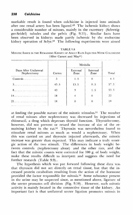
<1.5-2: Kidncy. The increase of the volume of one kidney after re1nmal of the ~ther is closely related to regeneration. It proceeds ll\ mitotic growth. This is particularly dillicult to analyze in such a complex organ as the kidney, and any tool increasing the number of Yisible mitoses is most helpful. 41 • .i;;, :n The great number of mitoses obsened in rats injected with 2.5 mg/kg alter unilateral nephrectomy and killed IO hours later is apparent from Table 9.8.
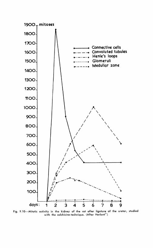
The problems of kidney mitoses in this condition and in other expcriments carried on to throw light on the causal factors have been the object of se,·eral publications From the Brussels school. After uniLtteral nephrectomy, the maximal number of mitoses is found during the first four days in the convoluted tubules, then in the glollleruli, and on the seventh day in Henle's loops and the Schweigger-Scidel tubules. 41 - 1'1 No mitoses are to be found in the epithelium of the renal pelvis. Exploded c-mitoses are the most frequent in the convoluted tubes. ][ a partial nephrectomy is added to the ablation of the other kidney, the remaining tissue shows mitoses in all locations, including the pelvis. Ligation of the ureter, without nephrcctomy, also stimulates kidney cells to divide, a fact which may prO\e of great experimental importance"1 (Fig. 9.10). Another re-
238 Colchicine
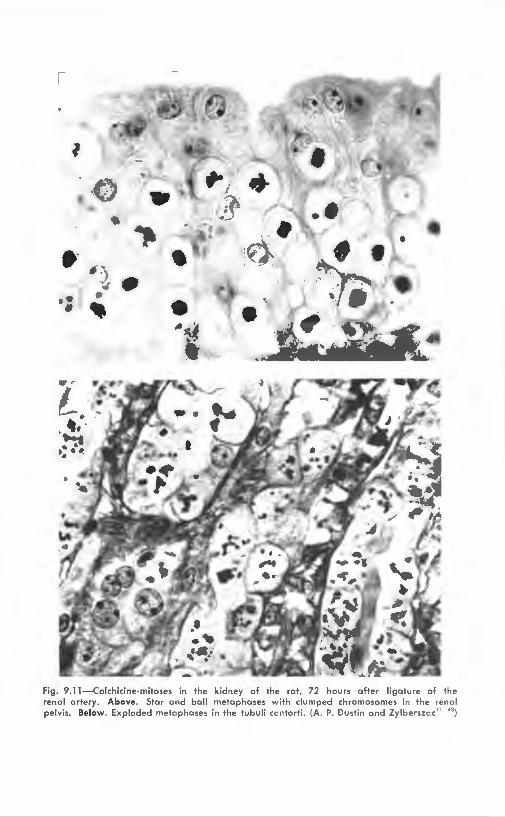
markable result is found when colchicine is injected into animals after one renal artery has been ligated.43 The ischemic kidney shows a considerable number of mitoses, mainly in the excretory (Schweigger-Seidel) tubules and the pelvis (Fig. 9.11) . Similar facts have been observed in kidneys made partly ischemic by the endocrine kidney operation of Selye.:i3 The following experiments were aimed
TABLE 9.8 MITOTIC INDEX IN THE REMAINING KIDNEY oF ADULT RATS INJECTED WITH CoLCHICI~E
(After Carnot and May31 )
Days After Unilateral Nephrectomy
Controls.
3 ...
8 ..
14 ..
21 ..
Cortex
3
43.5
43
18
18
Medulla -------~-
External Internal Zone Zone
- ------- -- ~-----
.5 0
13. 5 0. 5
9 1.5
~ 0
0 0
Total
4. 5
57. 5
53. 5
22
lR
at finding the possible nature of the mitotic stimulus.51 The number of renal mitoses after nephrectorny was decreased by injections of thiouracil, a drug which depresses thyroid function. Thyroidertomy, however, did not prevent or retard the increase of size of the remaining kidney in the rat.51 Thyroxin was nevertheless found to stimulate renal mitoses as much as would a nephrectomy. \Vhen this was carried on and thyroxin injected afterwards, the mitotic increase was greater than expected. This may indicate a truly synergic action of the two stimuli. The differences in body weight between controls (nephrectomy alone) and the other rats, and the fact that the mitotic counts were corrected for 100 g. of body "·eight, make these results difficult to interpret and suggests the need for further research (Table 9.9).
The hypothesis which was put forward following these data ,ras that thyroxin did not act directly on renal tissue, but that the increased protein catabolism resulting from the action of the hormone provided the factor responsible for mitosis. 51 Some substance present in the urine may be suspected since, as mentioned above, ligature of the ureter promotes cell division (Fig. 9.10). However, such mitotic activity is mainly located in the connective tissue of the kidney. An important fact is that unilateral ureter ligation promotes mitosis in
1900
1800
1700
1600
1500
1400
1300
1200
1100
1000
900
800
700
600
500
400
300
200
100
mitos«s
days: 1
----➔ •·----•
►----•
...... .....
2 3 4 5
Connective cells Convoluted tubul«s Henle's loops Glomeruli Medullar zone
\ \
\
\
\ \
\ \
\ \
\ \ ..... \
--- .. ...... ......
6 7 8 9 Fig. 9.10-Mitotic activity in the kidney of the rot ofter ligature of the ureter, studied
with the colchicine-technique. (After Herlont51 )
•• {
# ) • .,; • ... f •
# ~ •
Fig. 9. 11 -Colchicine-mitoses in the kidney of the rat, 72 hours after ligature of the rena l ar tery. Above. Star and ba ll metaphases with clumped chromosomes in the renal pe lvis. Below. Exploded metaphases in the tubuli ccntorti . (A. P. Dustin and Zylberszc c"· ")
Experimental Growth in Animals 241
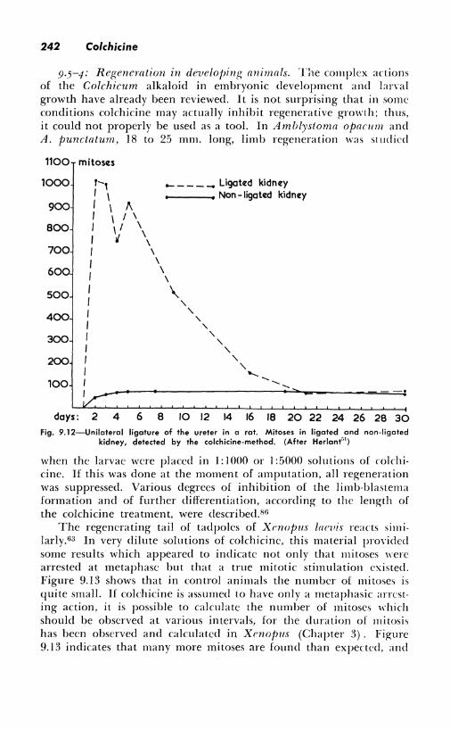
the other kidney also; this resembles closely the changes of compensatory hypertrophy (Fig. 9.12). Substances reabsorbed from the urine may promote division first in the ligated kidney and later in the other one. Research by other workers has suggested that xanthopterin or substances of that chemical constitution may initiate the kidney hypertrophy. The problems arc far from being solved, but the utility of colchicine for the observation of mitotic growth has been amply demonstrated.
9.5-3: Other organs. The following results give an indication of the multiple uses of rnlchicine as a tool. In the Langerhans' islets of the pancreas, alloxan brings about a selective destruction of the so-called fi-cells, which secrete insulin. Regeneration and mitoses of these cells are prevented if the animals receive insulin. This probably acts through a pituitary mechanism, for extracts of the pituitary gland increase considerably the number of cell divisions in islet regeneration. Colchicine-mitoscs are also observed in the anterior lobe of the pi tuitary:1:1 (Table 9.10) . The regeneration of the adrenal cortex after unilateral adrcnalectomy in rats has also benefited from the use of mitosis arrest. 10 In rats also, colchicine helped to demonstrate that compensatory hypertrophy of parathyroids after partial parathyroidectomy does not take place in hypophysectomized animals:17 and that testosterone inhibited the epithelial mitoses in thymic regeneration following X-irradiation.49
TABLE 9.9 ACTION OF THYROXIN ON RENAL HYPERTROPHY AFTER UNILATERAL NEPHRECTOMY
NUMBER OF MITOSES IN A MEDIAN SECTION OF THE WHOLE KIDNEY, 9 HOURS AFTER COLCHICINE
(Abridged from Herlant51 )
Mitoses ------ ------ ------
Convo- Conncc-luted Henle's Glo- Me- tive
Experiment Tubules Loops meruli dulla Tissue Total -- ------------- -----------
1. Unilateral nephr.-ctomy ( 4 rats)* ...... 61-125 8-19 0 3 26-87 44-62 163-210
2. Thyroxin alone (4rats)t ...... .. 173-252 3-5 0-2 2-5 3-7 186-250
3. Unilateral ncphrectomy
315-589 i + thyroxin
(7 rats)t ..... ... 35-65 2-15 25-152 31-132 523-722
* Animals weighing 260-360 gm. t Six daily doses of 0.25 mg. thyroxin; killed the seventh day after 2 mg/kg col
chicine. Animals weighing 120-220 gm.
242 Colchicine
9.5-4: Regeneration in develojJing animals. T:1e complex actions of the Colchicum alkaloid in embryonic development and larYal growth have already been reviewed. It is not surprising that in some conditions colchicine may actually inhibit regenerative growth; thus, it could not properly be used as a tool. In Amblystoma oparnm and A. punctatum, 18 to 25 mm. long, limb regeneration was studied
1100 mitoscts
1000 ,-, _____ Ligated kidney
I \ Non - ligated kidney 900 t \ !\
I I / \
BOO I I / \ \ I ,J \
700 I \
t \ 600 I
\ \
500 t \ I ' 400 I ' ' I ' 300 I ' ' I ' 200 I ' ' I ........
100 I ---- ---- ----I ---·
days: 2 4 6 B 10 12 14 16 18 20 22 24 26 28 30 Fig. 9.12-Unilateral ligature of the ureter in a rot. Mitoses in ligated and non-ligated
kidney, detected by the colchicine-method. (After Herlant''1)
when the larvae were placed in I: l 000 or l :5000 solutions of rnlchicine. If this was done at the moment of amputation, all regeneration was suppressed. Various degrees of inhibition of the limb-blastema formation and of further differentiation, according to the length of the colchicine treatment, were described.86
The regenerating tail of tadpoles of Xrnopus larvis reacts similarly.63 In very dilute solutions of colchicine, this material proYided some results which appeared to indicate not only that mitoses "·ere arrested at metaphase but that a true mitotic stimulation existed. Figure 9.13 shows that in control animals the number of mitoses is quite small. lf colchicine is assumed to have only a metaphasic arresting action, it is possible to calculate the number of mitoses which should be observed at various intervals, for the duration of mitosis has been observed and calculated in Xcnopus (Chapter 3). Figure 9.13 indicates that many more mitoses are found than expected, and
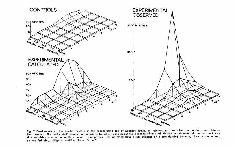
CONTROLS
30 MITOSES
20
EXPERIMENTAL CALCULATED
60 MITOSES
50
40
30
20
EXPERIMENTAL OBSERVED
150 MITOSES
100
50
Fig. 9.13-Analysis of the mitotic increase in the regenerating tail of Xenopus laevis, in relation to lime after amputation and distance from wound. The "calculated" number of mitosis is based on data about the duration of one cell-division in this material, and on the theory that colchicine does no more than "arrest" metaphases. The observed data bring evidence of a considerable increase, close to the wound, Qn the fifth day. (Slightly modified, from Liischer63}
244 Colchicine
that instead of a gradual rise, there is a steep increase on the fifth day. However, the experimental conditions are complex and stimuli from other growth-promoting substances cannot be excluded. These data with those given in Section 9.2 comprise the best evidence to date of possible mitotic stimulation of animal cells by rnlchicine.
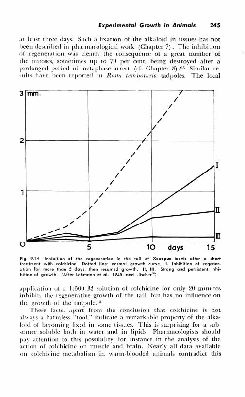
In Xenopus, a short treatment, one hour in a 1:2000 solution, ni;tv completely inhibit growth. However, regeneration often proceeds normally during the first three days after this "colchicine shock" because cellular migration is not disturbed. On the fifth day, on the contrary, when divisions should be taking place, regeneration was completely inhibited (Fig. 9. I 4). Some pharmacological conclusions are important to mention; they arc the results of an extensive series of experiments on this favorable material. Colchicine was demon-
TABLE 9.10 INFLUENCE OF ALLOXAN DIABETES ON PANCREATIC, PITUITARY, A'.'ld SuPRARE:--,\L
MITOSES; lNHIIHTION BY INSULIN; STIMULATION BY PITUITARY EXTRACTS
Days
1 ....
2 ....
3 ....
4 ..
5 ....
9 ....
12 ....
I: rats injected with 150 mg/kg alloxan II: id. + 10 to 20 units insulin per da)
III: id. + pituitary extract (about 32 mg. dry powder per day) (After Cavallero")
Mitoses ------------ - -----------
Anterior Loh<" Langerhans' Islets of I I ypophysis Adrenal Medulla
-- ·---~ --~ --------
II lll II III ll II I
8 0 3 15 15 24 2 ~
7 24 24 1 ') 16 2 7 3
44 2 n2 54 2 64 0 ()
81 0 185 81 9 1(,4 3 2 0
31 86 15 14 12 () ') (I
2 0 8 27 8 10 0 0 ()
7 7 27 22 8 0 7 :?.
strated to act locally, for 110 inhibition was observed when only the anterior part of the larva was immersed in the solution. This is also evidenced by the absence of inhibition if cokhicine is applied to another wound close to the amputation. Experiments in which the tail blastcrna was amputated and growth resumed, demonstrated that cokhicine did not penetrate more than 2 mm. from the 1\'0tmd. These also showed that colchicine was fixed in the tissues of the "·omHl for
Experimental Growth in Animals 245
at least three days. Such a fixation of the alkaloid in tissues has not been described in pharmacological work (Chapter 7). The inhibition of regeneration was clearly the consequence of a great number of the mitoses, sometimes up to 70 per cent, being destroyed after a prolonged period of mctaphase arrest (cf. Chapter 3) .63 Similar results have been reported in Rmrn temjwraria tadpoles. The local
3 mm.
I I
I I
/
2i----------+--------"------,--------------l
I /
I
/ I
I
1i---------+---~-----+-----------------l /
/
0 5 10 days 15
I
m
fig. 9.14-lnhibition of the regeneration in the tail of Xenopus laevis after a short treatment with colchicine. Dotted line: normal growth curve. I. Inhibition af regeneration for more than 5 days, then resumed growth. II, Ill. Strong and persistent inhibition of growth. (After Lehmann et al. 1945, and Luscher"")
application of a 1 :500 M solution of colchicine for only 20 minutes inhibits the regenerative growth of the tail, but has no influence on the growth of the tadpole. 1:i
These facts, apart from the conclusion that colchicine is not always a harmless "tool," indicate a remarkable property of the alkaloid of becoming fixed in some tissues. This is surprising for a substance soluble both in water and in lipids. Pharmacologists should pav attention to this possibility, for instance in the analysis of the action of colchicine on muscle and brain. Nearly all data available on colchicine metabolism in warm-blooded animals contradict this
246 Colchicine
idea of a fixation of the alkaloid. One of the purposes of this book is being fulfilled whenever similar contradictions between work done in widely separated fields of research are brought to light.
9.6: Wound Healing
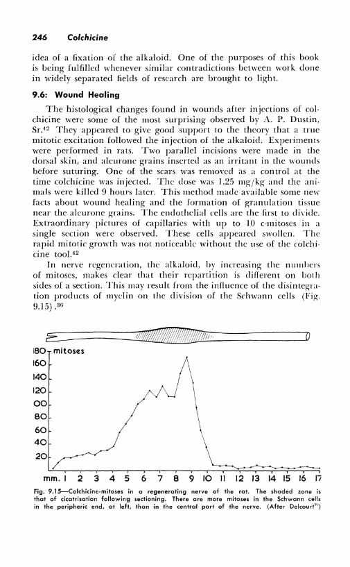
The histological changes found in wounds after injections of colchicine were some of the most surprising observed by A. P. Dustin, Sr.42 They appeared to give good support to the theory that a true mitotic excitation followed the injection of the alkaloid. Experiments were performed in rats. Two parallel incisions were made in the dorsal skin, and alcurone grains inserted as an irritant in the wounds before suturing. One of the scars was removed as a control at the time colchicine was injected. The dose was 1.25 mg/kg and the animals were killed 9 hours later. This method made available some new facts about wound healing and the formation of granulation tissue near the aleurone grains. The endothelial cells are the first to di\'ide. Extraordinary pictures of capillaries with up to 10 c-mitoses in a single section were observed. These cells appeared swollen. The rapid mitotic growth was not noticeable without the use of the colchicine tool. 42
In nerve regeneration, the alkaloid, by increasing the numbers of mitoses, makes clear that their repartition is different on both sides of a section. This may result from the influence of the disintegration products of myelin on the division of the Schwann cells (Fig. 9.15) .30
E 180 mitoses
160
140
120
00
80
60 40
20
mm. 2 3 4 5 6 7 8 9 10 II 12 13 14 15 16 17
Fig. 9.15-Colchicine-mitoses in a regenerating nerve of the rat. The shaded zone is that of cicatrisation following sectioning. There ore more mitoses in the Schwann cells in the peripheric end, at left, than in the central part of the nerve. (After Delcourt'")
Experimental Growth in Animals 247
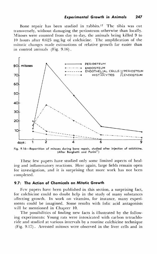
Bone repair has been studied in rabbits.17 The tibia was cut transversely, without damaging the pcriosteum otherwise than locally. :\fitoses were counted from day to day, the animals being killed 9 to IO hours after 0.G25 mg/kg of colchicinc. The amplification of the mitotic changes made estimations of relative growth far easier than in control animals (Fig. 9.1 G) .
80 mitoses
70
60
50
40
30
20
10 ...
days: 2
--------- ... ... ------•
4
PERIOSTEUM
ENDOSTEUM ENDOTHE;:;,l0AL CELLS }{PERIOSTEUM
HISTIOCYTES ENDOSTEUM
6 9
Fig. 9.16-Repartition of mitoses during bone repair, studied after injection of colchicine. (After Borghetti and Parini 17)
These few papers have studied only some limited aspects of healing and inflammatory reactions. Here again, large fields remain open for investigation, and it is surprising that more work has not been completed.
9.7: The Action of Chemicals on Mitotic Growth
Few papers have been published in this section, a surprising fact, for colchicine could no doubt help in the study of many substances affecting growth. In work on vitamins, for instance, many experiments could be imagined. Some results with folic acid antagonists will be mentioned in Chapter 10.
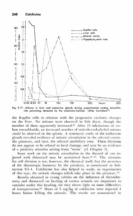
The possibili tics of finding new facts is illustrated by the following experiments: Young rats were intoxicated with carbon tetrachloride and studied at various intervals by a routine colchicine technique (Fig. 9. I 7). Arrested mitoses were observed in the liver cells and in
248 Colchicine
80 2:4
I '
I X .. -0
I .!:
-~ 2
602 ! ·e '
1,6 '
40 1,2
'
0,0
20
0;4
i: 0 49 2123 57 81
I
I
I
130
_ Kupffer cells _____ - Liver ce:lls ___ . _ Adre:nol cortrz:>C
178
.. --• Hypophysis,anter. lobe
" ·. " .. \o-
250 hours 370
Fig. 9.17-Mitoses in liver and endocrine glands during experimental carbon tetrachloride poisoning, detected by the colchicine-method. (After Cavallero"")
the Kupfier cells in relation with the progressive cirrhotic changes on the liver. No mitoses were observed in bile ducts, though the number of these apparently increased.:i:i After 15 inhalations of carbon tetrachloride, an increased number of reticulo-cndothclial mitoses could be observed in the spleen. A systematic: sl udy of the endocrine glands revealed evidence of mitotic stimulation in 1he adrenal cortex. the pituitary, and later, the adrenal medullary zone. These divisions do not appear to be related to local damage, and may be an e\·idence of a pituitary stimulus arising from "stress" (cf. Chapter 7).
Some work on the mitotic stimulation in the thyroid of rats injected with thiouracil may be mentioned here. 70 • -i;; The stimulus for cell division is not, however, the chemical itself, hut the secretio:1 of the thyrotropic hormone by the pituitary, as mentioned in Subsection 9.4-1. Cokhicine has also helped to study, in experiments of this type, the mitotic changes which take place in the pituitan·. 1'3
Results obtained in young rabbits on the influence of thyroidectomy and thiouracil on healing of cornea wounds are important to consider under this heading, for they throw light on some difficulties of interpretation.45 Doses of 5 mg/kg of cokhicine were injected 4 hours before killing the animals. The results are summari,ed in
Experimental Growth in Animals 249
Table 9.11. It is evident that the mitotic index is more depress::d bY thiouracil than by thyroidectomy, but it seems surprising that this fact is not at all noticeable without colchicine, thiouracil-inject::d animals having a slightly higher mitotic count than the controls. The authors think that the count after thiouracil results from a douc!e effect, i.e., a decrease of the mitotic rate, which would decrease the mitotic index, and a simultaneous lengthening of the duration oL mitosis, which would have the opposite effect.
TABLE 9.11 CORNEAL MITOTIC COUNTS IN A RABBIT
(After Fleischmann and Breckler'15 )
Controls ..
Thiouracil-treated .
Thyroidectomiz('d .
, • · I
I
I
Without Colchicine
92 ± 35
100 ± 17
REFERENCES
With Colchicine
393 ± 59
168 ± 42
228 ± 41
I. .\1.1,i-:x. E., A:\ll GREAI>ICK, R. N. Ovoge11csis during sexual maturity. The first stage of mitosis in the germiual epithelium, as shown by the colchidne technique. :\11at. Rec. (i<J: l!H-95. 1937.
'' ---, S.\IITII, :\I., A:\ll GARll:\FR, \V. U. Affcntuation of the growth effect of theelin 011 genital tissues by arrest of mitosis with colchicine. Anal. Rec. 67: Suppl. I: 19. I !J'.11>. Accentuation of the growth effect of theelin on genital tissues of the o\'aricnomi,ed mouse by arrest of mitosis with colchicine. Amer. .four . .-\nat. (il:'.121-12. I<J,l7. A short test for !l\'arian follicular hormone and other estrogens. Endoni11ology. 21:'112-1:l. 1937.
:L ---, ,\'Ill Ri-:YN01.ns, S. R. \I. Hyperplasia of uterine muscle, as studied by the rnlchici11e method. !'roe Soc. Exp. Biol. and Med. :17:257-59. I !l'.li.
I. T110~1.\s. T. B., \V11.s0N, J. C., Al\n H1-:ss10N, D. Differential growth in the maries and genital tract near the time of O\'t1latio11 in rats treated with rnlchiciue. Amer. Jour. Anat. 72:291-3:17. 1913 .
. ·,. ,\sL\I.lll, C., B1•.RNAR1>F1.1.1, E., AM) Ru1Al!llo, G. Research on the proliferation acti\'it\' of c1sthroblasts at low atmospheric pressure. Experientia. 8: I 17-19. l'l:i2. La prnliferation de I'crythroblaste en depression. Le Sang. 23:293-3]0_ ] ~):)'..?.
Ii. ---, A:S:D \fAIJRI. C. La nlutazione cle!Litti\'itii proliferativa delle cellulc midollari. Stu,lio cli un "test statmocinetico." Haematologica. 33: 1-16. 1919.
, . ---, ANll ---. New criteria for the evaluation of the bone-marrow cells mitotic acli\'ity. Le Sang. 21:378-82. l<J 0,o.
~ ---. ---, AND D1 Cu1,1.1E1.Mo, L. L'effetto dci raghi roentgen sull'attivita proliferati\'a dcgli eritroblasti stucliati 11d midollo osseo umano in cultura. Hacmatologica. 35:8fi7. 1950.
9. BAFR, F. Ober das Vorkommen vo11 1\Jitoscn im vorcler- um! zwischenlappen der Hvpophyse. Acta Neer!. Morph. 3:97-128. J<J39.
10. J\\KFR., D. D., .-\.Nil BAIi.I.IF, R. N. Role of capsule in surrenal regeneration studied with aid of colchicinc. Proc. Soc. Exp. Biol. and Med. 10:117-21. 1939.
250
11.
12.
13.
11.
15.
16.
17.
18.
19.
20.
21.
22.
23.
24.
25.
26.
27.
Colchicine
BASTFNIE, P. Detection de ]'hormone thyreotropc dans lcs urines. :\lcthode ct resultats. Arch. Int. Med. Exp. 14:111-22. 1939. ---, AND ZYLBERSZAC, C. Misc en evidence des stimulations hormonales par la colchicine. I. Detection de stimulation thyro"idicnne par I'extrait antchypophysaire. C. R. Soc. Biol. Paris. l 26+J6. 1937. I I. Iktccl ion de 1 'a<l ion stimulatricc du propionate de testosterone sur lcs vesicules seminales. C. R. Soc. Biol. Paris. 126:891. 1937. !JI. Action de I'cxtrait ant{:-lnpoplnsaire "" l'appareil genital du cobayc impubcrc. C. R. Soc. Biol. Paris. 126: l 2K2. I 'l'.li. IV. Doses croissantcs de propionate de testosterone sur I'apparcil gt'·nital du cobayc impubc:re. C.R. Soc. Biol. Paris. 12li:12K,I. 19'.l7. \'. Stimulation de L, parathyro'ide. C.R. Soc. Biol. Paris. 127:882. 1938. Misc en c,idcncc de stinrnlations honnonalcs par la mcthode colchiciniquc de Dustin .. .\rd1. Int. \lc-d. Exp. 13:183-203. 1938. Influence de la tachystcrinc irradicc. (AT IO de Holtz) sur la parathyro'itlc de la rate. C, R. Soc. Biol. Paris. 1,l2:%-%. 19'.l!I. BFRNIIARn, \V. Rcgcncrationshcmmung und Ausliisung epithelialer \\'ucherungcn durch Colchicin am Schwanz \On Rana-Lan-en. Re,·. Suisse Zoo!. 'i 1 :i I '.l-~7. 1917. BERRIAN, J. H., AND DORNFELD, E. J. Cellular proliferation in the germinal epithelium of immature rat ovaries. An in vitro method for the studv of mitotic rate. Jour. Exp. Zoo!. 115:49,l-512. 1950. The effects of rihonucleotides on mitosis in the germinal epithelium of immature rat o,aries cultured i11 1•itm . .Jour. Exp. Zoo!. 115:513-20. 19:,0. BIMES, C. Mitoses clans le myomctrc chcz la femclle du cobaye hyperfolliculiniscc. C. R. Assoc Anat. 3·1: 18-5:i. 1917. BORDO:S.ARO, F. I.a corrclazionc ipofiso-tiroidea ncl ratto a trattamento tiouracilo. Studio citofunzionalc a rne,zo de] metodo cokhicinico. Re,·. "l.'(hpedalc Magg." 3:i (6) Giugno, 1917. BoRGHETTI, U., A:--:D PARINI, I'. 1-'ratture sperimentale studiate con L1i11to de un mctodo cokhicinico. Med. Sper. Arch. Ital. K:G(i!,-K-1. Ell 1. BRETSCHNElllER, L. H., A:S.D DUYVENE rm \V1T, J. J. Histophysiologische .\nahse der sexuallendokrinen Organisation des BitterlingweilKhens. 1 /(/1(),/1·11.1 11111(1-
rus). Z. Zellforsch. 31 :227-:l'l-l. 1911. BRUFS, A. ;\f. The effect of colchicine on regenerating Ii,er. (Proc. Plnsiol. Soc.) .Jonr. Physiol. 86:63-64. 193li. ---, AND Corn::-., A. Effects of colchicine and related substances on cell di,i sion. Biochem, .Jour. 30:136,Hi8. 19%. ---, AND MARBI.F, B. B. An analysis of mitosis in lin·r restoration . .Jour. Exp. l\led. 65:15. 1937. , ---. A:--:n .JACKSO~. E. B. ;\'uclear ahnonnalitics resulting from inhibition of mitosis hy colchicinc and other substances. Amer. .Jour. Cancer. ,w::·,01-11. 1!1'.li. BlJCHER, 0. Zur \Virkung einiger Mitosegifte auf die Ce1,chckultur. l.e Sang. 21:382-89. 1950. l.c r(,lc de la culture des tissus i11 vitro clans l'c'•tude des poisons de la mitosc. M{:m. Soc. Vaucloise Sci. Nat. 10:2 l'i-70. I !)'i I. Buu,0uc;rr, \V. S. Mitotic activitv in the adult fcrnalc n10use M11.1 1111,sn,/11.1
L. A study of its relation lo the ~)estrus cycle in normal and ahnonnal conditions. Phil. Trans. Roy. Soc. B 231 :435. 19-lli. Epidermal thickness follm,in~ oestrone injections in the mouse. Nature. l!i9:IOl-2. 1917. The artion of colchicine in arresting epidermal mitosis. Jour. Exp. Biol. 26:287-91. 1919. The mitogcnic actions of starch and oestrone on the epidermis of the adult mouse . .Jour. Endocrin. 6:3:iO-Gl. 1950. Epidermal mitotic ani,·it, in the adult female mouse . .Jour. Endoc:rin. (i:310-19. 19:iO. Stress and epidermal mitotic activity. I. The effects of the adrenal hormones. .Jour. Endocrin. 8:265-74. 1952. ---, AND VAN OoRDT, .J. The mitogenic actions of testosterone propionate and of oestronc on the epidermis of the adult rnale mouse. Ana Endocrin. Copenhaguc. 4:291-305. 1950. ---, AND ElsA, E. A. The effect of a graded series of restricted diets on epidermal mitotic activity in the mouse. Brit. Jour. Cancer. -1:'121-28. 1'150 ---, ANB .JoHNSON, M. Epidermal mitotic activity and oxygen tension. :S:aturc. 167:-188. 1951.
Experimental Growth in Animals 251
2H. lltrRF.\1:, V. 1:action de l'oestrone ct de la progesterone sur la corne uterine de la lapinc, etudic:c par la methodc i1 la colchicinc. C. R. Soc. Biol. Paris. I '.l0:933-36. 1939.
2'l. l\t'RKIIART, Z. E. Colchicinc reactions in ventral prostate of castrated male rats following androgenic treatment. Proc. Soc. Exp. Biol. 40: 137-39. 1939. A study of the effects of androgenic substances in the rat by the aid o[ colchicine. Doctoral Dissertation. University of Chicago Library. 1940. A study of the earlv effects of androgenous substances in the rat by the aid of colchicine . .Jour. Exp. Zoo!. 89: l'.l!i-6(i. J9,t2.
'.lO. llt'RRILL, :\f. ,v., A"ID CREE:--!!·:, R. R. Androgen production during pregnancy and lactation in the rat. Anal. Rec. 8:1:209-28. 1912.
'.ll. C:ARXOT, I'., Al\ll :\IAY, R. :\f. I.a n\;{·n{:ration du rein rhcz le rat ctudiee au 1110,en de la colchicine. C.R. Soc. Biol. Paris. 128:611-43. 1938.
32. CA<Tnr-;uovo, G., AND FR1-11n, J. Mitogencse clans l'{·pithclium vaginal des rats. ,\rch. Int. Phann. Thcr. 61:191-93. 1939.
3'.L L\\AI.I.FRO, C. i-:tude de la cirrhosc cxp{:rimentalc par le tctrachlorurc de carhone ,1 !'aide de la reaction stathmocinetique (colchicinique) de Dustin. Ard1. Int. :\kd. Exp. H:1-H. 1939. R{:actions hormonales au cours de !'intoxication par le tetrarhlorure de carbone, poison cirrhogi:ne, mises en evidence par la m{:thode statlnnocin{·tique (colchicinique) de Dustin. Arch. Int. Med. Exp. 11: l!i-22. 1939. I.cs glandes endocrines au cours de la grossesse. Elude cvto-plnsiologique faite ;'t !'aide de la rbction colchicinique (stathmocinetique) de Dustin. Arch. Int. Med. Exp. 11:12"1-:l!i. 1939. Application de Ia mcthode cokhirinquc a l'{:tudc du diabi:te alloxaniquc chez le rat. Rev. Belge Path. IK:'12:Hl2. 1917.
31. ---, A:--1D 1'1-1.1.Er.R1:s11, G. F. L'effctto cokhicinico nel "rcnc endocrino" di Sel\c. Atti. Soc. Ital. Path. J:'108. l!ll!l.
3,i. IL\xcoxA, lJ. Verifica de! poliploidismo dellc cellule epatiche dei mammifcri nellc cariocincsi provocatc sperimentalmente. Arch. Ital. Anal. Embryo!. 47: '.!'i3-Rti. l!ll:!.
3(i. Ih1.cot'RT, R. 1-:lllde de la r{,g{,n{·ration des nerfs p{:ripheriques par la reaction stathmorini·tique. Acta Brev. l\ccrl. Phvsiol. 9:2°!!. 1938. Contribution ;1 J'etude de la formation des handes de Bungner-Ranvier par la reaction stathmocin{:tique de Dustin. Arch. Int. :\fed. Exp. l:i:1-13. 1910.
'.Ii. lhsu.1:s1, L. Apropos de !'action androgenique de Ia progesterone. C. R. Soc. Biol. Paris. ];12: 1:l-Vi. 19:l!l. Hypophysc ct parathyro'iclcs. I.es parathyroYdes aprc·s lnpophvsectomie chcz le rat blanc. Bull. Acad. Roy. Med. Belg. \Ile. Sfr. R: 1'.!7-38. I!) 13.
'.l8. ])oR:-.:FFI.D, E . .J.. AND BFRRIAN, .J. H. Stimulation of mitoses in the germinal epithelium of rat ovaries by intrarapsular injections. Anat. Rec. 109:129-38. I 'Fi I.
3!1. ])mscnu., ,v., ANll KROi'!', K. Vitaminc und Hormone. "1:280. 1944. 10. ---. ZII.UKF:--1, I'. ,v., A'.'ID KROPP, K. Colchicin-Mitosen Test an den Vesi
rnlardrlisen der kastriertc Maus. II. Die SpezifiUit des Testes. Biochem. Z. 318:l">l-61. 1918.
11. llt·sn:-.:. A. I'. I.a cokhicinc, rcactif de !'imminence caryocinctique. Arch. Portugaiscs Sci. Biol. ",:38-13. l!l31i. f:tudc de I'hypertrophie compensatrice du rein par la r<:action stathmocin{:tique. Acta Unio Internal. Cancrum. 4:679-83. 19:19. Recherches sur le mode d'action des poisons stathmocin<'.:liques . . \ction de la colchiciue sur l'utfrus de lapinc impuhi:re sensibilise par injection prblahlc d'urine de femme cnccinte. Arch. Biol. "11:111-87. 1913.
12. ---, A:-.:n C11onKowsK1, K. F.tude de la cicatrisation par la reaction colchi cinique. Arch. Int. :\Jed. Exp. 13:641-62. 1938.
13. ---, AND Zn.BFRSZAc, S. f:llldc de l'hypertrophie compensatrice du rein par la r{:action stathmocinetique. Bull. Acad. :\1i·d. Belg. Vle. Ser. 1:315-20. 1939.
,I I. F1.uscHMAI\N, \V., AND KA11:s1, S. 0 her das Colchicin als Hilfsmittel beim Studium hormonal hcdingter ,vachstumsvorgange. Biochem. Z. 296:374-82. I !l3H. The use of colchicinc in the assay of androgens. Endocrinology. 25: 798-HOO. 19:19.
252 Colchicine
45. }"1.nsn1,1ANN, \V., AND BRFCKLFR. I. A. ;\litotic and "·ou11d-heali11g acti, itie, of the corneal epithelium in thiouracil treated and thnoidc<10111i1ed rats. Endocrinology. ,JI :2(i(i-68. I !l li.
46. GATZ, A. J. The cellular changes induced in the testes of the albino rat ll\ artificial cryptorchiclism aide,! by the arrest of mitosis with colchicin .. \nat. Rec. 70:Suppl. I :87. 19:17.
47. GINFSTF, D. J. Rechcrd1es sur L, rc·g{·11{·ration des {·li·mcnts de la glandc cor· tico-surrcnale par la rn{:thodc colchici11iq11c .. \dion de din:rs factl'tirs. C. R. Soc. Biol. Pans. 110:221-22. 1 !l 16.
48. G1ul\EI., F. La sensihilitc de l'c·poophorc it la tc.stosti·rnne. Rbction wkhiciniquc. C.R. Soc. Biol. Paris. 1:ll: l2'i0- 0,(i. 1!13!1.
49. (;Ri:GOJRF., C. Recherches sur Jes relations entrc thymus ct sunc·nales. 11. Le, reactions des ccllulcs du reticulum i·pithi·lial thy111iq11c ;'t J'ahlation des surrc:nales. Arch. Int. Pharmacodn1. hi:IHi-(i;l. 1!)12. Sur le 1ni·,a11ismc de !"atrophic tll\mique d{:dandifr 1;ar des honnones scxuelles. Arch. Int. Ph,1n11acodyn. 70:-l'i-7i. l!l l!i.
50. (;frnrun, H. Der Einfluss von Hypopll\scmorde1lappc11e'dractc11 und Colchicin auf KerngTiisse um! Kernteilung in der Schilddri"isc. \'inhm,s \rdi. 307:37-70. 1!)10. Die Einfluss von Hypophyscnnmlcrlappenextractcn und Cokhicin auf die L111gerhansd1cn lnseln des Pankreas. \'ircho\\s .\1d1. '.\07: 17:i-99. 1910.
:,l. HERi.ANT, :\I. Jnfl11ence dn thiouracyl sur l'hypcrtrnphie co111pe11satri,c dt1 1ei11. Bull. Acad. Rm. Belg. Classe. Sci. 'ie Ser. 'l'.l::ili7-7fi. 1')17. \,ti,itt' mitotique des cell11les 1h1alcs au cours de l'hydroni·phrosc 1111iL1ti-r:de. 1\1111. Acad. Roy. l\frd. Belg. Ge S{-r. l'.l:'ll'i-,lO. 1918. Experimental hydronephrosis studied l,y the cokhicine method. '.\Jatmc. l(i2:2'il-'i2. 1'118.
52. Htr'-:T, T. ·E. .\litotic acti,ity in the anterior hypoplnsis of female rats of clillcrc111 age groups and at different periods of the da,. E11doni11olog,. 32:331-3'1. 1913.
5'l. JAIUR, .J. \V. ;\fitotic index of hypnpl;istic interstitial cells of the guinea-pi~. Proc. Soc. Exp. Biol. ancl \fed. :l!J:281-83. 19:l8.
'i·l. KFRR, T. i\litotic acti,ity in the female mouse pituitary . .Jo11r. Exp. Biol. 20:il-iH. I'll'!.
50. Kt:ZFLL, \\'. c., AND CIJTTINC, \\". C. Pituitary mitotic changes ,tfter the adtninislralion of oestrogen a11d after oy;iriccto111y. Endorrinol<)gy. '...:l)::~>~Vi·-:1,-..:. 1910.
5(i. LAIIR, E. L .. AND Rmnu-:, 0. Prolifnation of nop-sac epithelium in i11nd,ati11~ and in prolactin-injectcd pigeons studied with the colchici11c-methocl. .\mer. Jour. Physiol. 12,l:(il 1-l!l. l!l'.l8.
57. ---, AI.WELL, L. H., AND Rrnnu:, 0. :\!itosis ohscned under cold1ici11c in crop-sac tissue after s11hn1ta11co11s and i11tr;11111isc11L1r injection of proL1t tin. Arch. Int. Phannacoch11. 65:278-82. I!) 11.
58. Ll:BLO:\ll. C. I'. Action' de Lt proladi11e s11r le jahot du pigeon misc en hick-11,c par l'arri-1 des mitoses ;'t ]'aide de la cokhicinc. C.R. Assoc. des .\nat. '.l'.!:'.211-47. 19'l7.
5!). ---, ANll A1.1.FN, E. Emphasis of the growth effect of prnL1r1i11 011 the crop gland of the pig·eon b, arrest of mitoses ,,ith cold1ici11. Fndoninolog,. '.!I: 455-60. 1937.
60. ---, ANll STl•:n:,o;s, C. E. The constant renewal of the intestinal cpithclit1m in the albino rat. Anal. Rec. 100:'l'i7-78. 1()18.
61. LFTTRf, H. Uber l\litosegifte. Ergclm. Physiol. ·16:,l79-l'i'.!. El',O. 62. LtmFoRn, R. J. The action of toxic sul,stanccs upon the di,ision of 1101111;,I and
111alig11a11t cells i11 ,,itro and i1111i,,o. Arch. Exp. Zcllforsch. 18:111-11. 1'1:lli. 63. Liisc1u:R, M. Hemmt oder fiirdert Colchicin die Zcllteil1111g im regencrierenden
Schwanz der Xenopns-Larve? Rev. Suisse Zoo!. 5:l:·181-H(i. l'll(i. Die Hemmung den Reg·cneration d11rd1 Colchicin heim Schwan, dcr Xc1101rns-L1ne u11d ihre cntwicklungsphysiologisc:hc \\'i1ku11gsa11alyse. Heh·. Physiol. ct Phann. Acta. ·1:165-94. 19-16.
64. \IALINSKY, .J., AND LAN<:, B. J-Iypcrplasie du foic de rat apri·s ld·patectomic particlle ct infl11c11ce des corps colchicin{·s sur celle-ci. C. R. Soc. Biol. Paris. 115:609-12. El'iL
Experimental Growth in Animals 253
ti,-,. \hxL·s, \I. II. C:. Zaadblaastest met hchulp van colchicine. :'siederl. Tijdschr. Ccncesk. 81: 1128-29. Samcnhlascntcsl mit rnld1icin. Acla Brcv. Neer!. Physiol. 7:17">. l!J37.
li!i. \lcl'1L\IL, .\I. K .. AXll \\'11.lll'R, K. :\1. Ahscnce of potentiation of g-onadotropin and steroid function in mammals hy colchicinc. Endocrinology. 35:196-97. 1911.
Ii,. \loR.-\TO·.\IAXARo, J. An:ion dcl acctato de desoxvcorticosteronc sohre el 11tero de la coneja infantil cstu,liato por el metodo c;ilchicinico. Arch. Soc. Biol. .\lonte,·ide<J. 10:110-11. EMO. Accion de los androgenos sohre la vcsicula seminal de la rata, estudiata por cl mcto,lo colchicinico. Arch. Soc. Biol. .\lontcvideo. 10: 193-201. 1911.
(iK. :'\ATIL\XSOX, I. T., IIRIJI•:s, A. ;\I., AND RAWS();\;, R. \\'. Effect of testosterone propionate upon thyroid and parathyroid glands in intact immature female rat. !'roe. Soc. Exp. Biol. and \led. 13:7'.li-l0. 1910.
h'I. ---, A1':ll ---. Effect of testosterone propionate upon the mitotic activity of the adrenals in the intact immature female rat. Endocrinology. 29:397-401. l 911.
70. l'.\SC!lhlS, K. E., C:A1':TAROW. A., RAhOFF, :\. E., AXD R<HIIF1'11ER(;, \f. s. Mitosi~
7 I.
7 I.
1,.
s I.
stimulation in the tlnroi,l gland ind11<Cd hy thiouracil. Endocrinology. 37: 133-3">. 1915. . l'o\IFRAT, (;_ R . .\litotic aclivitv in the pituitary of the white rat following castration. Amer. .Jour. Anat. fi9:R9-121. 1911. PL·xnn., \1. I'. f:tndc des r{·actions vaginalcs hormonalcs chez la femme par la mi-th ode rnkhiciniq11c. ,\nn. E11donin. 2:h59-(i I. l !l'iO. Roc;1-Rs, l'. V., AND Au.t-N. E. Epithelial g-rowth caused hv stimulation with ,arious smear methods as demonstrated hv mitotic stasis witl.1 colchicine. Endo-crinology. 21 :G29-32. 1937. · S\cc11t•Tr. C ... \Xll J\L\l\CJt1x1, E. Action dire<le de la S. T. H. sur les activites de la moi'llc ossc11sc hurnainc nonnalc. Le Sang. 2 l::l-11-51. 1953 . . Su11rnt.FY, C. H., .\:SD HH;c;1,-;s, (;_ \I. Effect of administration of colchicine after panial 1-crnmal of the lin:r. Proc. Mayo Clin. l:i:5,lG. 19-10. Su1.\111>T. I. C., ,\'\ll HoFF\l.\:\, F. C. Proliferation and on>genesis in the gerrrtinal epitlieliurn of the normal mat11rc g·uinea-pig o\'an·. as shown hy the col-chicine tcclrniquc. ,\tncr. Jo11r. .\nal. liH:2(i:l-72. 1911. Sclt\IlllT, I.(;_ \litolic prolifnation in the o,·arv of the normal mat11re g-uineapig treated with rnlchicinc .. \mer. Jom. :\nal. 71:215-70. 1!)12 . . s,:, t-i1x, P., Al\D T11c11M,\l\N-llt•l't.1-ss1s, II. \lise en e,idencc de mitoses clans l'ln popll\se d11 cohaye par !'action de la colchicine. Variation de l'activitc di,isionnclle ;, U·tal normal ct apri·s injections d'honnonc gonadolrope. l\lontpcllicr \lc'-d. 2:l-21:l(i'l-(il. 19l'l. Sur la pn\scncc des mitoses rnlchiciniques dans le cloaquc ct la prostate du Triton marhre (l\folge marmorata Latr.) soumis ;, ]"action des hormones scxucllcs cl hypophysaircs. \lontpellier !\-It'd. !.''.l-'.!l:210-12. 1!11,l. Sur quelqucs particularitcs d'action de la colchicine surles glandes endocrines du cohayc inject{, d'honnonc gonadotropc. :\lontpellier \l{·d. 29-'.l0:I,l3-35. 1915 . . S1toRR, F., A:Sll C:0111-:N, E. l ·se of colchicine in detecting- hormonal effects on ,aginal epithelium of menstruating and castrate women. Proc. Soc. Exp. Biol. and \led. l(i:,l:l0-,l5. 1'111. SI ITS, K. F., A'\D F<>RDIAX, D. Effect of thyroid substances in the ovarian cap· ,.11lc 11pon mitosis in the germinal cpitheli11m. Anal. Rec. 10:i:G-l'.l-5G. 1949. Sn:nss, C. E., JhmrsT, R., A'.\D Li-:111.0,ll, C. I'. Rate of synthesis of desoxyri-1,onucleic acid and mitotic rate in Ji\'cr and intestine. Jour. Biol. Chem. 202: I 77-H(i. I 95,l. L\hl·W.\kl, K. :\litotic acti\itv in seminal vesicle cells transplanted to female
mice . .Jo11r. Fae Sci. Tobo t"rniv. 5:2!)1. 19-11. T1-:rn. H. Cokhicinc-tests for the p11rposc of ascertaining cell-division regencrati,e conditions in the )iYer of the rat. Acal. Path. l\licroh. Scand. 25:45-51. 191tL
~ I. Ti xx \'.\T, R., AXD L1i-:11ow, .\. A. The actions of colchirine and ethvlcarhylamine on tissue rnltures. Yale Jour. Biol. and .\led. 13:39-l<J. 1910. ' ·
254 Colchicine
85. THALES-MARTINS. Test rapido para o hormonio masculino: mitoses 1u genitalia accessoria. Brasil Med. 51:717-19. 1937. Test rapide de I"hormone masculine: mitoses clans Jes genitalia accessoires de mftles ctstrcs. C. S. Soc. Biol. Paris. 126:131-34. 1937.
86. THORNTON, C. S. The effect of colchicine 011 limh regeneration in !anal Amblystoma. ]our. Exp. Zool. 92:281-93. 19·13. Colchicine and limb regeneration in larval Amblystoma. Anat. Rec. 81:512. 19-12.
87. T1s1.owrrz, R. Oher die Latenzperiode von Testosterone uml Testosteronepropionat. Kongressber. JG. Internal. Physiol. Kongr. 19:Hl. The colchicine test as a method for determining the time of onset and the duration of action of male substances. Endocrinology. 25:719-5:l. 1939. The action of estrogn1s in inducing mitoses in the muscle, connective tissue. and epithelium of the prostate and seminal vesicle as determined by the colchicine technique. Anal. Rec. 75:265-74_ 1939.
88. TiiRo, E., AND VADAsz, J. l/ntersuclrnng-en iiber die ,virkung von Colchicin um! Corhormon in Gewebekulturen mit Hilfc von filmaufnahmen. Arch. Exp. Zellforsch. 23:277-98. 1939.
89. UEI.INGER, E., JADASSOIIN, ,v., ANIJ Fn:Rz, l-1. E. :\litoscs OfflllTing in the actnthosis produced I,y hormones. Jour. Invest. Denn. ·1:,l:ll-,l:i. 19-11.
90. VERNE_, J., AND Vn.TER, V. Elude de !'action de la rnlchicine sur Jes mitoses des fibrohlastes cultivcs in vitro. Concentrations dites fortes. C. R. Soc. Biol. Paris. 133:618-21. 1940. Mccanisme d'action de la colchicine, employee en concentrations faiblcs, sur )'evolution de la mitose dans les cultures de fibrohlastcs in vitro. C. R. Soc. Biol. Paris. l:l:l:621-21. 1910.
91. \VILLIAMS, W. L., STEIN, K. F., ANIJ AI.I.FN, E. Reaction of genital tissues of the female mouse to the local application of colchicine. Yale Jour. Biol. and \led. 13:8·11-16. 19·11.
92. \VOLi', 0. Mitotic activity of stimulated rat adrenals and spleen 111e1s11red ll\ colchicin technic. Anal. Rec. 70:Suppl. I :86. 1937. \Iitotic activity of the islands of Langerhans and parathyroids of rats following· pituitarv extract and colchicine injections. Biol. Bull. 75:'.!77-78. l'l'lH.
93. ,voRTIIINGTON, R. V., AND AI.I.I'N, E. Crml"th of genital tissues in response to estrone as studied by the colchicinc technique. Yale .Jour. Biol. and \led. 12:137-53. 1939.