CCM3/PDCD10 stabilizes GCKIII proteins to promote Golgi assembly and cell orientation
Frank SicheriandAvijit Chakrabartty, Anne-Claude Gingras
Goudreault, Ian C. Scott, W. Brent Derry,Khipple Mulligan, Michelle J. Kean, Marilyn Derek F. Ceccarelli, Rob C. Laister, Vikram CCM3 HomodimerizationProteins Using a Mechanism Analogous toGerminal Center Kinase III (GCKIII) CCM3/PDCD10 Heterodimerizes withSignal Transduction:
doi: 10.1074/jbc.M110.213777 originally published online May 11, 20112011, 286:25056-25064.J. Biol. Chem.
10.1074/jbc.M110.213777Access the most updated version of this article at doi:
.JBC Affinity SitesFind articles, minireviews, Reflections and Classics on similar topics on the
Alerts:
When a correction for this article is posted•
When this article is cited•
to choose from all of JBC's e-mail alertsClick here
http://www.jbc.org/content/suppl/2011/07/11/M110.213777.DCAuthor_profile.htmlRead an Author Profile for this article at
http://www.jbc.org/content/286/28/25056.full.html#ref-list-1
This article cites 25 references, 8 of which can be accessed free at
by guest on August 1, 2013http://www.jbc.org/Downloaded from
CCM3/PDCD10 Heterodimerizes with Germinal Center KinaseIII (GCKIII) Proteins Using a Mechanism Analogous to CCM3Homodimerization*�
Received for publication, December 17, 2010, and in revised form, April 26, 2011 Published, JBC Papers in Press, May 11, 2011, DOI 10.1074/jbc.M110.213777
Derek F. Ceccarelli‡1, Rob C. Laister§1, Vikram Khipple Mulligan¶, Michelle J. Kean‡�2, Marilyn Goudreault‡,Ian C. Scott�**, W. Brent Derry�**, Avijit Chakrabartty¶‡‡, Anne-Claude Gingras‡�3, and Frank Sicheri‡�4
From the ‡Centre for Systems Biology, Samuel Lunenfeld Research Institute at Mount Sinai Hospital, Toronto, Ontario M5G 1X5, the§Division of Stem Cell and Developmental Biology, Ontario Cancer Institute, Toronto, Ontario M5G 1L7, the ¶Campbell FamilyInstitute for Cancer Research, Ontario Cancer Institute/University Health Network, Department of Biochemistry, University ofToronto, Toronto, Ontario M5G 1L7, the �Department of Molecular Genetics, University of Toronto, Toronto, Ontario M5S 1A8, the**Program in Developmental and Stem Cell Biology, The Hospital for Sick Children, Toronto, Ontario M5G 1X8, and the‡‡Department of Medical Biophysics, University of Toronto, Toronto, Ontario M5G 1L7, Canada
CCM3 mutations give rise to cerebral cavernous malforma-tions (CCMs) of the vasculature through a mechanism thatremains unclear. Interaction of CCM3 with the germinal centerkinase III (GCKIII) subfamily of Sterile 20 protein kinases,MST4, STK24, and STK25, has been implicated in cardiovascu-lar development in the zebrafish, raising the possibility that dys-regulated GCKIII function may contribute to the etiology ofCCM disease. Here, we show that the amino-terminal region ofCCM3 is necessary and sufficient to bind directly to the C-ter-minal tail region of GCKIII proteins. This same region of CCM3was shown previously to mediate homodimerization throughthe formation of an interdigitated �-helical domain. Sequenceconservation and binding studies suggest that CCM3 maypreferentially heterodimerize with GCKIII proteins througha manner structurally analogous to that employed for CCM3homodimerization.
Cerebral cavernous malformations (CCMs)5 are vascularabnormalities in the brain characterized by focal dilations ofcranial vasculature that can progress to hemorrhages andstroke (OMIM ID 116860). Mutations have been identified in
three distinct genes, denoted CCM1–3, that are causative forthe formation of most familial CCM lesions (1, 2). CCM3, alsonamed PDCD10, is a 212 amino acid protein conserved amongmetazoans (3, 4). Knockdown of CCM3 in zebrafish causes anenlarged heart phenotype (5), whereas targeted deletion ofCCM3 in the mouse results in defects of early angiogenesis andearly embryonic lethality, a phenotype also observed followingtissue-specific deletion in the vascular endothelium (6). A non-cell autonomous role for CCM3 in neuroglia on the vasculaturehas also been uncovered in mouse recently, indicating thatCCMs may arise in the central nervous system by the loss ofCCM3 signaling in neural as well as endothelial populations (7).CCM3 has been detected in complex with CCM1 and CCM2
proteins, suggesting that the three proteins may share a com-mon biochemical function (8–10). Yeast two-hybrid, co-im-munoprecipitation, and GST pulldown experiments from celllysates demonstrated that CCM3 also readily interacts withMST4, STK24, and STK25, a grouping of protein kinasestermed the germinal center kinase class III (GCKIII) family(10–13). CCM3 and the GCKIII proteins have also beendetected as part of a large multiprotein complex termed STRI-PAK (striatin-interacting phosphatase and kinase; see Refs. 14,15). The knockdown of GCKIII proteins in zebrafish gives riseto the same cardiovascular defects as CCM3 knockdown, sug-gesting the CCM3-GCKIII protein interaction is important forproper CCM3 function (5).6
The GCKIII proteins (STK24, STK25, and MST4) are mem-bers of the larger Sterile 20 kinase family and are characterizedby highly conserved catalytic domains and a 100–120 residuecarboxyl-terminal tail, whose function is not currently known.The closely related GCKII proteins MST1 and MST2 possesscompletely distinct C-terminal tails that mediate homotypicand heterotypic interactions (16), raising the possibility that ananalogous functionmight be served by the tail region of GCKIIIproteins, albeit through an unrelated structural mechanism.Crystal structures of the CCM3 protein revealed an architec-
ture consisting of two distinct structural domains (17, 18). TheN-terminal helical domain of CCM3 mediates homodimeriza-
* This work was supported in part by Canadian Institutes of Health ResearchGrants MOP-36399 (to F. S.) and MOP-84314 (to A.-C. G.).
� This article was selected as a Paper of the Week.1 Both authors contributed equally to this work.2 Supported by the Canadian Institutes of Health Research through a Banting
and Best Canada graduate scholarship.3 Canada Research chair in Functional Proteomics and the Lea Reichmann
chair in Cancer Proteomics. To whom correspondence may be addressed:Samuel Lunenfeld Research Institute at Mount Sinai Hospital, 600 Univer-sity Ave., Toronto, ON M5G1X5, Canada. Tel.: 416-586-5027; Fax: 416-586-8869; E-mail: [email protected].
4 Canada Research chair in Structural Principles of Signal Transduction. Towhom correspondence may be addressed: Samuel Lunenfeld ResearchInstitute at Mount Sinai Hospital, 600 University Ave., Toronto, ONM5G1X5, Canada. Tel.: 416-586-8471; Fax: 416-586-8869; E-mail: [email protected].
5 The abbreviations used are: CCM, cerebral cavernous malformation; GCKIII,germinal center kinase group III; STRIPAK, striatin-interacting phosphataseand kinase; TEV, tobacco etch virus; SEC-MALS, size exclusion chromatog-raphy-multiangle light scattering; FAT, focal adhesion targeting; PP4C, cat-alytic subunit of the serine/threonine protein phosphatase 4; mAU, milliabsorbance units. 6 B. Yoruk, B. S. Gillers, N. C. Chi, and Ian C. Scott, submitted for publication.
THE JOURNAL OF BIOLOGICAL CHEMISTRY VOL. 286, NO. 28, pp. 25056 –25064, July 15, 2011© 2011 by The American Society for Biochemistry and Molecular Biology, Inc. Printed in the U.S.A.
25056 JOURNAL OF BIOLOGICAL CHEMISTRY VOLUME 286 • NUMBER 28 • JULY 15, 2011 by guest on August 1, 2013http://www.jbc.org/Downloaded from
tion. The C-terminal four-helix bundle, termed the focal adhe-sion targeting (FAT) homology domain (17), functions as a lin-ear peptide binding module that mediates direct interactionswith CCM2, paxillin, and the striatin component of STRIPAK(17).7 Of note, the N-terminal region of CCM3 has also beenimplicated in the interaction with GCKIII proteins in cells andmodel organisms (5, 13, 19). Given the critical role for CCM3and GCKIII proteins in maintaining vascular integrity, we haveprobed the basis for their interaction in close detail. The resultspresented here demonstrate that the amino terminus of CCM3interacts directly with the C-terminal regions of GCKIII pro-teins. Based on sequence similarity between the interactingregions of CCM3 and GCKIII proteins, we propose that het-erodimerization of the two proteins is achieved through ananalogous structural mechanism to that reported for thehomodimerization for CCM3 and present data indicating thatheterodimerization may be favored over homodimerization.
EXPERIMENTAL PROCEDURES
Cloning of Expression Constructs—PDCD10/CCM3,STK25/SOK1, MST4, STK4/MST1, and STRN3 were ampli-fied by PCR and cloned into the modified bacterial expres-sion vectors pGEX-TEV and/or ProEX-TEV. CCM3 pointmutations were generated by PCR using standard tech-niques, and all clones were sequence verified. The N-terminal mutant of CCM3 (LAIL-4D) comprises L44D,A47D,I66D,L67D and theC-terminalmutant (K4A) comprisesK132A,K139A, K172A,K179A. CCM3 pointmutants were sub-cloned into the pcDNA5-FRT-GFP vector (20) for expressionin mammalian cells. pcDNA3-FLAG-MST4 was reported pre-viously (14).Protein Expression and Purification—BL21-Codon� cells
(Agilent Technologies) were transformed and grown to A600 of0.8 and induced by addition of 0.25 mM isopropyl 1-thio-�-D-galactopyranoside for 12–18 h at 18 °C. Bacterial cell pelletswere harvested and stored at �20 °C. Full-length STK25 andMST4 were also cloned into pFastBACGST-TEV (Invitrogen),and recombinant baculoviruses were generated for expressionin SF9 cells. Infection of SF9monolayer cells with amultiplicityof infection of�5were performed for 72 h, followed by harvest-ing of cell pellets and storage at �80 °C.Bacterial or SF9 cell pellets of His-tagged proteins were
thawed and resuspended in nickel-loading buffer containing 20mM HEPES, pH 7.5, 400 mM NaCl, 5 mM imidazole, and 5 mM
�-mercaptoethanol. Cells were lysed in the presence of 1 mM
phenylmethylsulfonyl fluoride by passage through a cellhomogenizer (Avestin, Inc.). Supernatant following centrifuga-tion at 20,000� gwas applied to a nickel-chelating column (GEHealthcare) and eluted over a gradient to 300 mM imidazole.Fractions containing the protein of interest were incubatedovernight with an aliquot of tobacco etch virus (TEV) proteaseand 1mMDTT. Protein was dialyzed into nickel-loading bufferand flowed over a 1-ml nickel chelating column to removeuncleaved protein, concentrated, and injected onto a 120-ml
S-75 size exclusion column (GE Healthcare) in 20 mM HEPES,pH 7.5, 100 mM NaCl, and �-mercaptoethanol. Fractions con-taining purified protein were concentrated, flash frozen, andstored at�80 °C. Bacterial pellets of GST-tagged proteins werelysed in 20mMHEPES, pH 7.5, 400mMNaCl, and 5mM �-mer-captoethanol, and the supernatant was applied to glutathione-Sepharose resin (GE Healthcare). The protein of interest wasseparated from the GST affinity tag following overnight incu-bationwith TEV protease and further purified by size exclusionchromatography as described above. The heterodimeric CCM3and MST4 complex was obtained by mixing bacterial lysatesexpressingGST-MST4 andHis-CCM3proteins. Protein elutedfrom a nickel-chelating column was applied directly to gluta-thione Sepharose resin and subsequently purified as describedabove for GST-tagged proteins.In Vitro Binding Studies—Bacterial or SF9 cell lysates con-
taining GST fusion proteins were prepared as above, howeverthe supernatant was applied to glutathione-Sepharose resin(GE Healthcare) in GST-loading buffer containing 20 mM
HEPES, pH 7.5, 400 mM NaCl, and 5 mM �-mercaptoethanoland washed extensively. The protein-bound affinity resin wasequilibratedwith interaction buffer (20mMHEPES, pH 7.5, 150mM NaCl, and 5 mM �-mercaptoethanol). Previously purifiedbinding partner proteins were added to the protein-boundaffinity resin and incubated on ice with gentle mixing for 20min. The affinity resin was thenwashed three times with 500�lof interaction buffer and aliquots of the binding reactions wereseparated by SDS-PAGE with proteins visualized followingCoomassie staining.Light Scattering—Size exclusion chromatography-multi-
angle light scattering (SEC-MALS) was performedwith 200�M
protein samples injected onto a 24-ml S200 Superdex columnand measured using a three-angle (45, 90, and 135°) miniDawnlight-scattering instrument equipped with a 690-nm laser andanOptilab rEXdifferential refractometer (Wyatt Technologies,Inc.) as described in Ref. 21. Molecular weights were calculatedby using ASTRA software (Wyatt Technologies, Inc.) based onZimm plot analysis and by using a protein refractive indexincrement, dn dc�1 � 0.185 liters g�1.Analytical Ultracentrifugation—Sedimentation equilibrium
experiments were carried out on samples containing CCM3alone, MST4 alone, and a CCM3-MST4 complex obtained bydual affinity tag purification. Samples were loaded at concen-trations yielding initialA280 values of 0.2, 0.4, and 0.8. Ultracen-trifugation was performed at 25 °C in 20mM sodium phosphatebuffer, pH7.0, with 100mMNaCl and 5mM �-mercaptoethanolusing an Optima XL-I analytical ultracentrifuge (BeckmanInstruments, Palo Alto, CA) with a AN50-Ti rotor, quartz win-dows, and standard six-sector charcoal-filled Epon center-pieces. Absorbance profiles at 280 nm were collected at spinspeeds of 3,000, 10,000, 15,000, 20,000, and 25,000 rpm after24 h of equilibration at each spin speed. Data were analyzed bynonlinear least-squares fitting using Origin software (version7.0, OriginLab Corp., Northampton, MA). Global fits wereobtained to Equation 1, representing a single-species model.Data from CCM3 and MST4 samples were also fit to Equation2, representing a monomer-homodimer equilibrium model.
7 M. J. Kean, D. F. Ceccarelli, M. Goudreault, S. Tate, B. Larsen, M. Sanches,L. C. D. Gibson, W. B. Derry, I. C. Scott, L. Pelletier, G. S. Baillie, F. Sicheri, andA.-C. Gingras, submitted for publication.
CCM3 Interacts Directly with GCKIII Proteins
JULY 15, 2011 • VOLUME 286 • NUMBER 28 JOURNAL OF BIOLOGICAL CHEMISTRY 25057 by guest on August 1, 2013http://www.jbc.org/Downloaded from
A280 � A280,F e�2
2RTM�1 � v����r2 � F2� � C (Eq. 1)
A280 � � �monCmon,F e�2
2RTMmon�1 � v����r2 � F2�
� 2�mon
Cmon,F2
KDe
�2
RTMmon�1 � v����r2 � F2��l � C (Eq. 2)
In the equations shown above, � is the spin speed, �mon is the280-nmmolar extinction coefficient of a monomer, Cmon,F isthe concentration of monomers at the reference radius (F), ris the radius from the spin axis, v is the partial specific vol-ume of the protein, � is the density of the solvent, R is the gasconstant, T is the temperature, l is the optical path length, Cis a baseline correction constant, and Mmon is the sequencemolecular mass of a proteinmonomer. Values of v and � werepredicted using SEDNTERP software (John Philo, ThousandOaks, CA). Extinction coefficients of 10,430 M�1 cm�1 and
39,670 M�1 cm�1 were used for CCM3 and MST4,respectively.Mammalian Cell Culture and Immunoprecipitation—Tran-
sient transfection of HEK293T cells and immunoprecipitationfollowed by immunoblotting was performed essentially asdescribed (14).
RESULTS
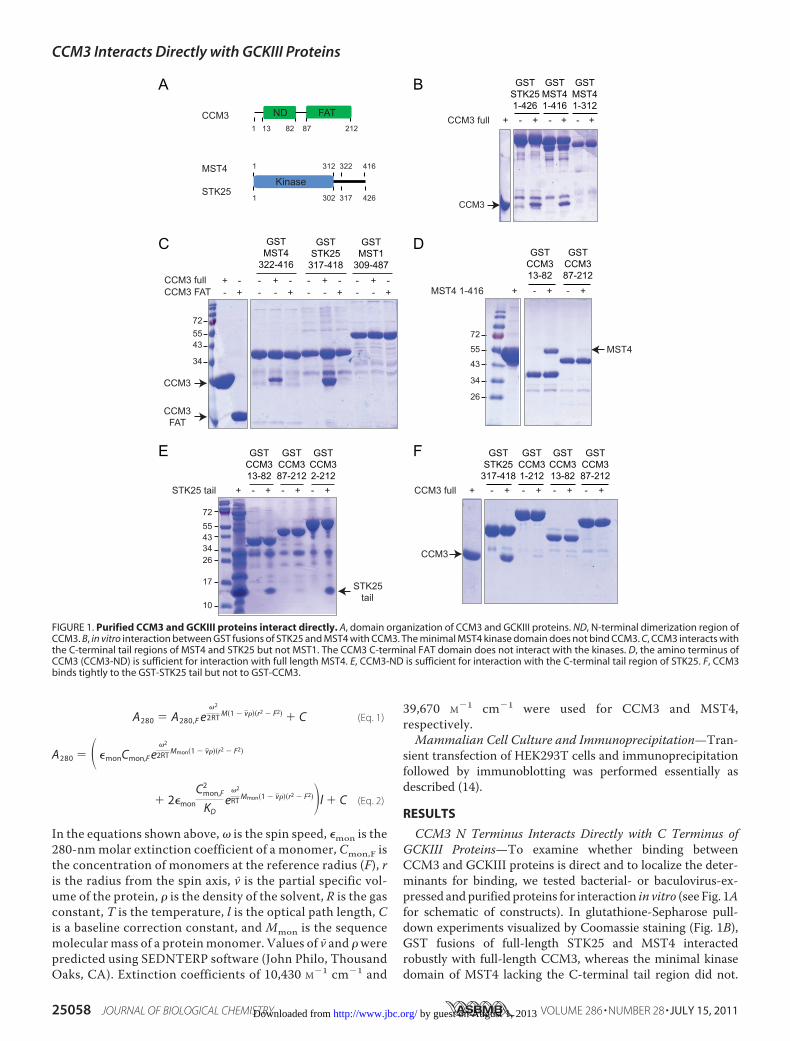
CCM3 N Terminus Interacts Directly with C Terminus ofGCKIII Proteins—To examine whether binding betweenCCM3 and GCKIII proteins is direct and to localize the deter-minants for binding, we tested bacterial- or baculovirus-ex-pressed andpurified proteins for interaction in vitro (see Fig. 1Afor schematic of constructs). In glutathione-Sepharose pull-down experiments visualized by Coomassie staining (Fig. 1B),GST fusions of full-length STK25 and MST4 interactedrobustly with full-length CCM3, whereas the minimal kinasedomain of MST4 lacking the C-terminal tail region did not.
FIGURE 1. Purified CCM3 and GCKIII proteins interact directly. A, domain organization of CCM3 and GCKIII proteins. ND, N-terminal dimerization region ofCCM3. B, in vitro interaction between GST fusions of STK25 and MST4 with CCM3. The minimal MST4 kinase domain does not bind CCM3. C, CCM3 interacts withthe C-terminal tail regions of MST4 and STK25 but not MST1. The CCM3 C-terminal FAT domain does not interact with the kinases. D, the amino terminus ofCCM3 (CCM3-ND) is sufficient for interaction with full length MST4. E, CCM3-ND is sufficient for interaction with the C-terminal tail region of STK25. F, CCM3binds tightly to the GST-STK25 tail but not to GST-CCM3.
CCM3 Interacts Directly with GCKIII Proteins
25058 JOURNAL OF BIOLOGICAL CHEMISTRY VOLUME 286 • NUMBER 28 • JULY 15, 2011 by guest on August 1, 2013http://www.jbc.org/Downloaded from
These results demonstrated that binding is direct and that theC-terminal region of the GCKIII proteins is a key determinantfor CCM3 recognition.TheC-terminal tails ofMST4 and STK25, when expressed as
GST fusion proteins, were sufficient for robust binding to fulllength CCM3 (Fig. 1C). Interestingly, neither GST kinase tailfusion was able to bind to the isolated FAT domain of CCM3,suggesting that the N-terminal dimerization domain of CCM3provides the determinant for GCKIII protein binding. We alsodemonstrated the interaction is specific to GCKIII protein tailsbecause no interactionwas detected between full-lengthCCM3and the tail region ofMST1, aGCKII protein possessing a diver-gent C-terminal tail (Fig. 1C).Wenext testedwhether theN terminus ofCCM3 is sufficient
for binding to GCKIII proteins. As shown in Fig. 1D, the Nterminus of CCM3 but not the FAT domain displayed robustbinding to full-length MST4. We reconstituted a robust inter-action between the N-terminal tail of CCM3 fused to GST andthe free C-terminal tail of STK25, demonstrating that the min-imal delineated regions in CCM3 and the GCKIII proteins arefully sufficient for the interaction (Fig. 1E). Together, theseresults define a model in which the binding of GCKIII proteinswith CCM3 is mediated entirely by their C- andN-terminal tailregions, respectively. Interestingly, although full-length CCM3interacted robustly with the GST-STK25 kinase tail, binding ofCCM3 full-length to GST-CCM3 full-length and to the GST-CCM3 N terminus was barely detectable (Fig. 1F). This resultraised the question of how precisely the respective N- and
C-terminal tail regions of CCM3 and GCKIII proteins mediatecomplex formation and how this interaction is affected bythe ability of the CCM3 N-terminal tail region to formhomodimers.The N-terminal tail region of CCM3 facilitates homo-
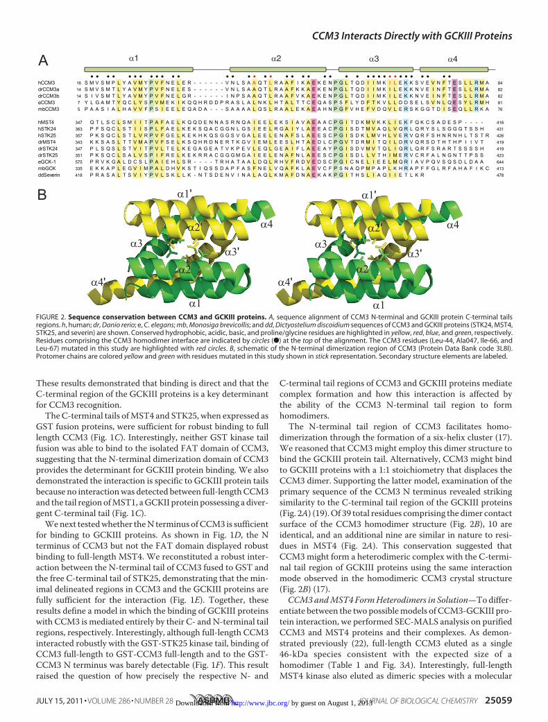
dimerization through the formation of a six-helix cluster (17).We reasoned that CCM3might employ this dimer structure tobind the GCKIII protein tail. Alternatively, CCM3 might bindto GCKIII proteins with a 1:1 stoichiometry that displaces theCCM3 dimer. Supporting the latter model, examination of theprimary sequence of the CCM3 N terminus revealed strikingsimilarity to the C-terminal tail region of the GCKIII proteins(Fig. 2A) (19).Of 39 total residues comprising the dimer contactsurface of the CCM3 homodimer structure (Fig. 2B), 10 areidentical, and an additional nine are similar in nature to resi-dues in MST4 (Fig. 2A). This conservation suggested thatCCM3might form a heterodimeric complex with the C-termi-nal tail region of GCKIII proteins using the same interactionmode observed in the homodimeric CCM3 crystal structure(Fig. 2B) (17).CCM3andMST4FormHeterodimers in Solution—Todiffer-
entiate between the two possiblemodels of CCM3-GCKIII pro-tein interaction, we performed SEC-MALS analysis on purifiedCCM3 and MST4 proteins and their complexes. As demon-strated previously (22), full-length CCM3 eluted as a single46-kDa species consistent with the expected size of ahomodimer (Table 1 and Fig. 3A). Interestingly, full-lengthMST4 kinase also eluted as dimeric species with a molecular
FIGURE 2. Sequence conservation between CCM3 and GCKIII proteins. A, sequence alignment of CCM3 N-terminal and GCKIII protein C-terminal tailsregions. h, human; dr, Danio rerio; e, C. elegans; mb, Monosiga brevicollis; and dd, Dictyostelium discoidium sequences of CCM3 and GCKIII proteins (STK24, MST4,STK25, and severin) are shown. Conserved hydrophobic, acidic, basic, and proline/glycine residues are highlighted in yellow, red, blue, and green, respectively.Residues comprising the CCM3 homodimer interface are indicated by circles (F) at the top of the alignment. The CCM3 residues (Leu-44, Ala047, Ile-66, andLeu-67) mutated in this study are highlighted with red circles. B, schematic of the N-terminal dimerization region of CCM3 (Protein Data Bank code 3L8I).Protomer chains are colored yellow and green with residues mutated in this study shown in stick representation. Secondary structure elements are labeled.
CCM3 Interacts Directly with GCKIII Proteins
JULY 15, 2011 • VOLUME 286 • NUMBER 28 JOURNAL OF BIOLOGICAL CHEMISTRY 25059 by guest on August 1, 2013http://www.jbc.org/Downloaded from
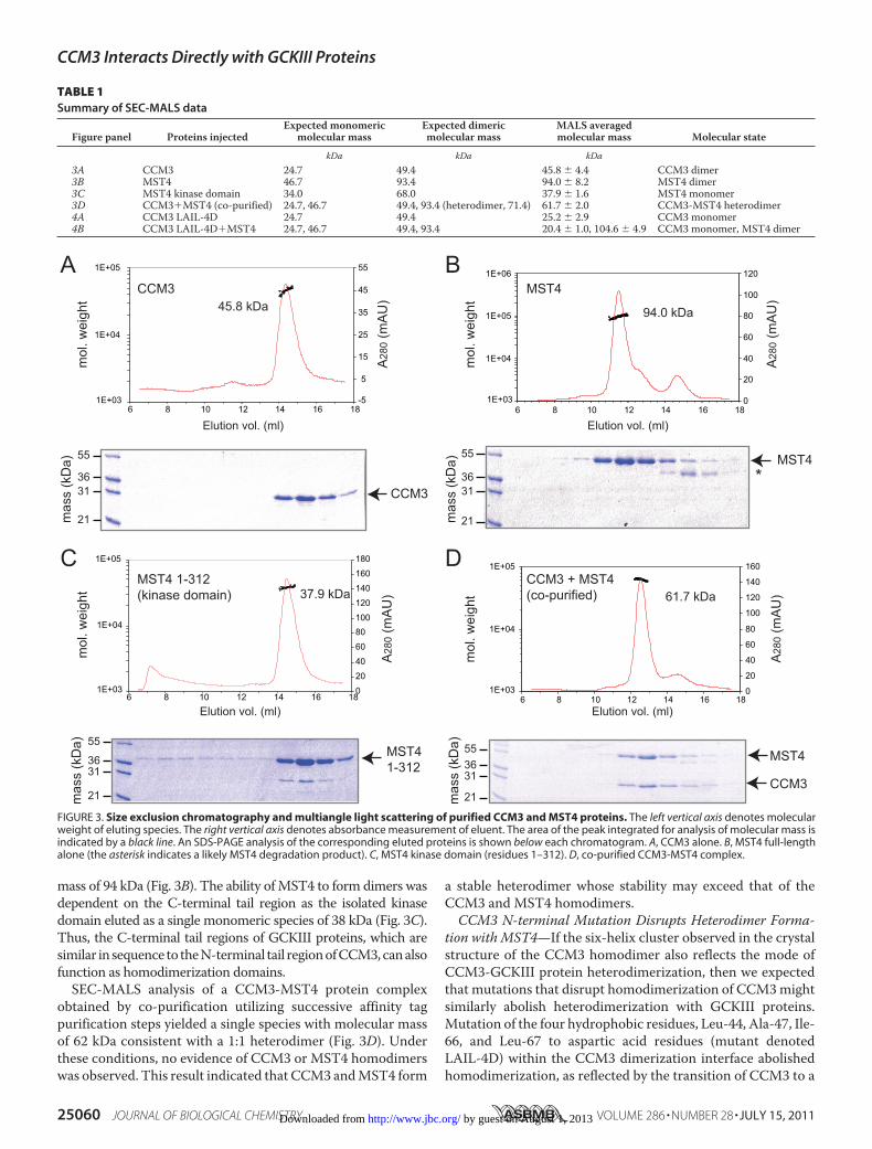
mass of 94 kDa (Fig. 3B). The ability ofMST4 to form dimers wasdependent on the C-terminal tail region as the isolated kinasedomain eluted as a single monomeric species of 38 kDa (Fig. 3C).Thus, the C-terminal tail regions of GCKIII proteins, which aresimilar in sequence to theN-terminal tail regionofCCM3,canalsofunction as homodimerization domains.SEC-MALS analysis of a CCM3-MST4 protein complex
obtained by co-purification utilizing successive affinity tagpurification steps yielded a single species with molecular massof 62 kDa consistent with a 1:1 heterodimer (Fig. 3D). Underthese conditions, no evidence of CCM3 or MST4 homodimerswas observed. This result indicated that CCM3 andMST4 form
a stable heterodimer whose stability may exceed that of theCCM3 and MST4 homodimers.CCM3 N-terminal Mutation Disrupts Heterodimer Forma-
tion with MST4—If the six-helix cluster observed in the crystalstructure of the CCM3 homodimer also reflects the mode ofCCM3-GCKIII protein heterodimerization, then we expectedthat mutations that disrupt homodimerization of CCM3mightsimilarly abolish heterodimerization with GCKIII proteins.Mutation of the four hydrophobic residues, Leu-44, Ala-47, Ile-66, and Leu-67 to aspartic acid residues (mutant denotedLAIL-4D) within the CCM3 dimerization interface abolishedhomodimerization, as reflected by the transition of CCM3 to a
FIGURE 3. Size exclusion chromatography and multiangle light scattering of purified CCM3 and MST4 proteins. The left vertical axis denotes molecularweight of eluting species. The right vertical axis denotes absorbance measurement of eluent. The area of the peak integrated for analysis of molecular mass isindicated by a black line. An SDS-PAGE analysis of the corresponding eluted proteins is shown below each chromatogram. A, CCM3 alone. B, MST4 full-lengthalone (the asterisk indicates a likely MST4 degradation product). C, MST4 kinase domain (residues 1–312). D, co-purified CCM3-MST4 complex.
TABLE 1Summary of SEC-MALS data
Figure panel Proteins injectedExpected monomeric
molecular massExpected dimericmolecular mass
MALS averagedmolecular mass Molecular state
kDa kDa kDa3A CCM3 24.7 49.4 45.8 � 4.4 CCM3 dimer3B MST4 46.7 93.4 94.0 � 8.2 MST4 dimer3C MST4 kinase domain 34.0 68.0 37.9 � 1.6 MST4 monomer3D CCM3�MST4 (co-purified) 24.7, 46.7 49.4, 93.4 (heterodimer, 71.4) 61.7 � 2.0 CCM3-MST4 heterodimer4A CCM3 LAIL-4D 24.7 49.4 25.2 � 2.9 CCM3 monomer4B CCM3 LAIL-4D�MST4 24.7, 46.7 49.4, 93.4 20.4 � 1.0, 104.6 � 4.9 CCM3 monomer, MST4 dimer
CCM3 Interacts Directly with GCKIII Proteins
25060 JOURNAL OF BIOLOGICAL CHEMISTRY VOLUME 286 • NUMBER 28 • JULY 15, 2011 by guest on August 1, 2013http://www.jbc.org/Downloaded from
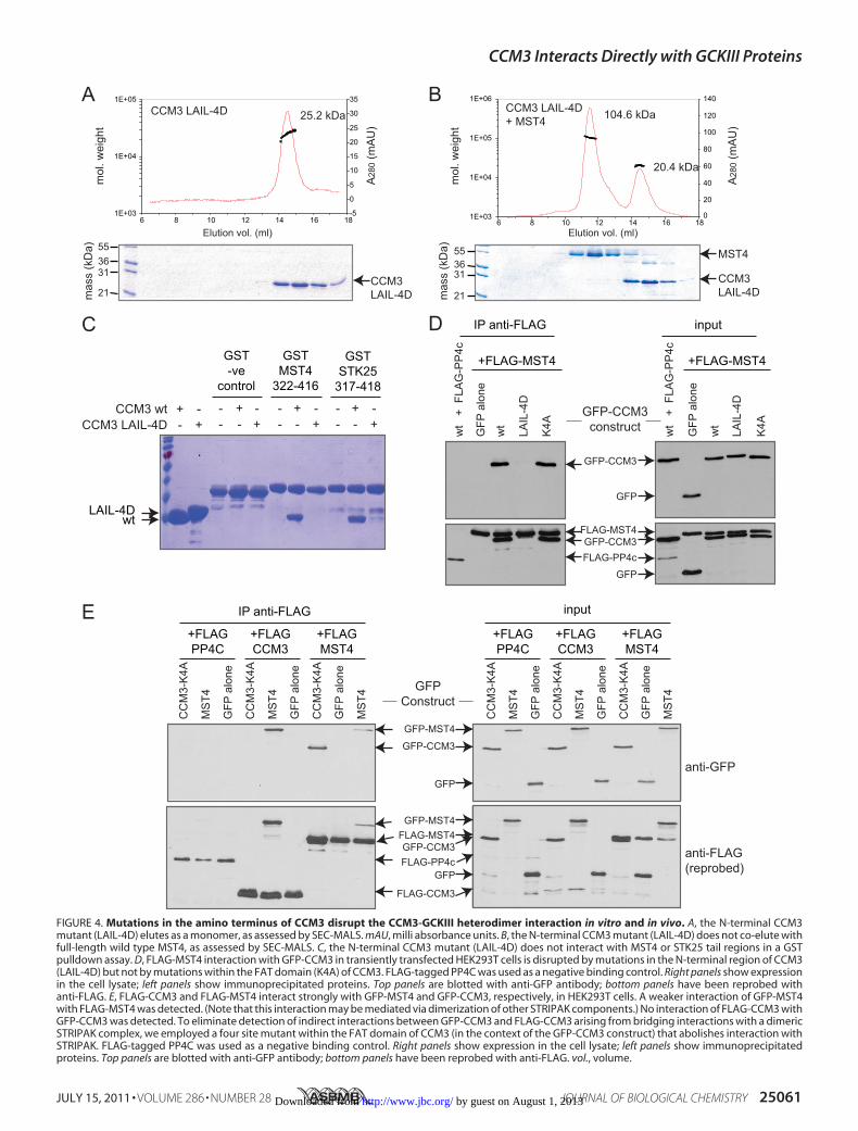
FIGURE 4. Mutations in the amino terminus of CCM3 disrupt the CCM3-GCKIII heterodimer interaction in vitro and in vivo. A, the N-terminal CCM3mutant (LAIL-4D) elutes as a monomer, as assessed by SEC-MALS. mAU, milli absorbance units. B, the N-terminal CCM3 mutant (LAIL-4D) does not co-elute withfull-length wild type MST4, as assessed by SEC-MALS. C, the N-terminal CCM3 mutant (LAIL-4D) does not interact with MST4 or STK25 tail regions in a GSTpulldown assay. D, FLAG-MST4 interaction with GFP-CCM3 in transiently transfected HEK293T cells is disrupted by mutations in the N-terminal region of CCM3(LAIL-4D) but not by mutations within the FAT domain (K4A) of CCM3. FLAG-tagged PP4C was used as a negative binding control. Right panels show expressionin the cell lysate; left panels show immunoprecipitated proteins. Top panels are blotted with anti-GFP antibody; bottom panels have been reprobed withanti-FLAG. E, FLAG-CCM3 and FLAG-MST4 interact strongly with GFP-MST4 and GFP-CCM3, respectively, in HEK293T cells. A weaker interaction of GFP-MST4with FLAG-MST4 was detected. (Note that this interaction may be mediated via dimerization of other STRIPAK components.) No interaction of FLAG-CCM3 withGFP-CCM3 was detected. To eliminate detection of indirect interactions between GFP-CCM3 and FLAG-CCM3 arising from bridging interactions with a dimericSTRIPAK complex, we employed a four site mutant within the FAT domain of CCM3 (in the context of the GFP-CCM3 construct) that abolishes interaction withSTRIPAK. FLAG-tagged PP4C was used as a negative binding control. Right panels show expression in the cell lysate; left panels show immunoprecipitatedproteins. Top panels are blotted with anti-GFP antibody; bottom panels have been reprobed with anti-FLAG. vol., volume.
CCM3 Interacts Directly with GCKIII Proteins
JULY 15, 2011 • VOLUME 286 • NUMBER 28 JOURNAL OF BIOLOGICAL CHEMISTRY 25061 by guest on August 1, 2013http://www.jbc.org/Downloaded from
25-kDa monomeric species in SEC-MALS analysis (Fig. 4A).The four-site mutant, unlike wild type CCM3, was also com-promised for its ability to interact with wild type MST4, asassessed by SEC-MALS (Fig. 4B), and to interact with thedimerization mediating tails of either MST4 or STK25 in pull-down experiments (Fig. 4C). These results were consistent withthe notion that CCM3 andGCKIII proteins form heterodimersthrough amechanism analogous to that employed in theCCM3homodimer structure (17).To investigate whether the CCM3-GCKIII protein het-
erodimers exist in vivo through the formation of a CCM3-likedimer structure, we tested wild type CCM3 and the four siteCCM3mutant for their ability to interact with GCKIII proteinsby immunoprecipitation from HEK293T cells co-transfected
with GFP-tagged CCM3 and FLAG-taggedMST4 kinase. Wildtype GFP-tagged CCM3 was readily recovered in immunopre-cipitates of FLAG-MST4, but not in immunoprecipitations ofFLAG-PP4C, used here as a negative control (Fig. 4D). Thisinteraction was abolished by the four-site mutation within theCCM3 dimerization domain (Fig. 4D; proteins expressed atsimilar levels). In contrast, a CCM3mutation within the C-ter-minal FATdomain ofCCM3 (K4A), which prevents interactionwith STRIPAK, CCM2, and paxillin (17),7 had no effect on theinteraction withMST4 (Fig. 4D). This result confirmed the roleof the N terminus of CCM3 inmediating GCKIII protein inter-actions in vivo and further supported the notion that het-erodimerization is mediated by a mechanism analogous to theCCM3 homodimerization mechanism (17).
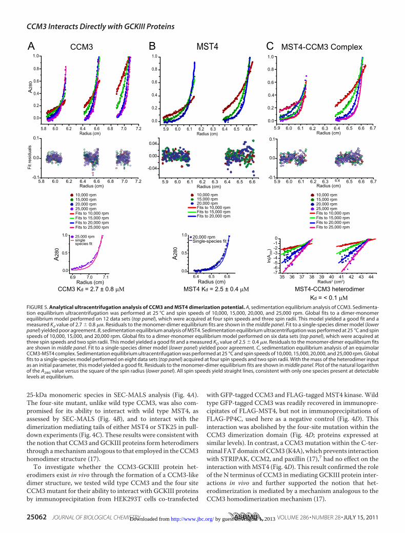
FIGURE 5. Analytical ultracentrifugation analysis of CCM3 and MST4 dimerization potential. A, sedimentation equilibrium analysis of CCM3. Sedimenta-tion equilibrium ultracentrifugation was performed at 25 °C and spin speeds of 10,000, 15,000, 20,000, and 25,000 rpm. Global fits to a dimer-monomerequilibrium model performed on 12 data sets (top panel), which were acquired at four spin speeds and three spin radii. This model yielded a good fit and ameasured Kd value of 2.7 � 0.8 �M. Residuals to the monomer-dimer equilibrium fits are shown in the middle panel. Fit to a single-species dimer model (lowerpanel) yielded poor agreement. B, sedimentation equilibrium analysis of MST4. Sedimentation equilibrium ultracentrifugation was performed at 25 °C and spinspeeds of 10,000, 15,000, and 20,000 rpm. Global fits to a dimer-monomer equilibrium model performed on six data sets (top panel), which were acquired atthree spin speeds and two spin radii. This model yielded a good fit and a measured KD value of 2.5 � 0.4 �M. Residuals to the monomer-dimer equilibrium fitsare shown in middle panel. Fit to a single-species dimer model (lower panel) yielded poor agreement. C, sedimentation equilibrium analysis of an equimolarCCM3-MST4 complex. Sedimentation equilibrium ultracentrifugation was performed at 25 °C and spin speeds of 10,000, 15,000, 20,000, and 25,000 rpm. Globalfits to a single-species model performed on eight data sets (top panel) acquired at four spin speeds and two spin radii. With the mass of the heterodimer inputas an initial parameter, this model yielded a good fit. Residuals to the monomer-dimer equilibrium fits are shown in middle panel. Plot of the natural logarithmof the A280 value versus the square of the spin radius (lower panel). All spin speeds yield straight lines, consistent with only one species present at detectablelevels at equilibrium.
CCM3 Interacts Directly with GCKIII Proteins
25062 JOURNAL OF BIOLOGICAL CHEMISTRY VOLUME 286 • NUMBER 28 • JULY 15, 2011 by guest on August 1, 2013http://www.jbc.org/Downloaded from
We also used the HEK293T cell system to further investigatethe homo- and heterodimerization properties of MST4 andCCM3. FLAG-tagged CCM3, MST4, or PP4C (negative con-trol) were co-transfected with GFP-tagged CCM3 (K4A),MST4, or GFP alone. CCM3 (K4A) was employed for this assayto prevent interactionswhichwould bemediated by interactionwith the striatin component of STRIPAK, which also homo-and heterodimerizes.7 As seen in Fig. 4D, strong het-erodimerization of FLAG-MST4 and GFP-CCM3 (K4A) wasobserved; the reciprocal interaction between GFP-MST4 andFLAG-CCM3 was also readily detected (Fig. 4E). By contrast,the recovery of GFP-MST4 with FLAG-MST4 was muchweaker (note that this could potentially be mediated by striatindimerization), and no detectable homodimerization of CCM3was observed (Fig. 4E). These results further hinted that theheterodimer state between CCM3 and MST4 might be a pre-ferred dimer conformation. To test whether this was in fact thecase, we sought to quantify the binding affinity for homo- versusheterodimerization by analytical ultracentrifugation.We performed equilibrium analytical ultracentrifugation
analysis on CCM3 and MST4 proteins in isolation and on aCCM3-MST4 complex obtained by co-purification using dualaffinity tag purification. Under the protein concentrationstested (see “Experimental Procedures”), CCM3 andMST4 pro-files were best fit using a monomer-dimer equilibrium modelwith an extracted Kd of 2.5 and 2.7 �M, respectively (Fig. 5, Aand B). In contrast, the CCM3-MST4 complex profiles werebest fit as a single species model corresponding to a tight het-erodimer (Fig. 5C). Based on the linearity of ln(A280) versusradius2 plots (Fig. 5C, bottom panel), which revealed no evi-dence of alternatemonomeric or homodimeric species, we esti-mated a binding constant for heterodimeration of 0.1 �M,which is thus minimally 25-fold tighter than the CCM3 andMST4 homotypic inteactions. From these data, we concludedthat the heterodimer state between CCM3 andMST4 is greatlyfavored over either of the two homodimer states.
DISCUSSION
In this study, we mapped the determinants of a direct inter-action between CCM3 and GCKIII proteins to the N-terminalregion of CCM3 and the C-terminal tail region of GCKIII pro-teins. These elements of both protein families are highly relatedin amino acid sequence, suggesting a common folded structureand binding function (Fig. 2A). Our data lead us to propose thatCCM3-MST4 complex formation is achieved through theadoption of a heterodimeric helical structure analogous to thatrevealed by the CCM3 homodimer crystal structure (17). Wealso confirmed the existence of CCM3 homodimers in solutionand that mutations within the N-terminal region of CCM3 dis-rupt both homodimerization and heterodimerization withpurifiedGCKIII proteins (Fig. 4). Dimerization of STE20 familykinasesmediated by conserved auxiliary domains has now beenobserved for GCKIII proteins (this study), the GCKII proteins(16), and the p21 activated kinases (23, 24). We reason thatconserved regions flanking the kinase domains of other STE20family kinases might serve analogous interaction functionsalbeit through the adoption of distinct structures.
The uncovered binding mode between CCM3 and GCKIIIproteins helps to explain the following biological observations.Depletion of CCM3 led to the destabilization of STK25/SOK1in cells (13) demonstrating an interdependence of protein func-tion. A mutant in exon 5 of CCM3 that results in deletion ofresidues 33–50 within the N-terminal region (3) failed to bindthe GCKIII proteins MST4, STK24, and STK25 (5, 19). Thisobservation is consistent with our finding that point mutationswithin the N-terminal region of CCM3 disrupt binding toGCKIII proteins both in vitro and in cells (Fig. 4). Exon 5-de-leted CCM3 also failed to rescue the cardiac phenotype ofzebrafish, further demonstrating the biological importance ofthe CCM3-GCKIII protein interaction (5, 19). Because theseregions of CCM3 and GCKIII proteins mediate both hetero-and homotypic dimerization, the relative importance of eachstate in the etiology of CCM disease needs to be exploredfurther.The interacting tail regions of GCKIII proteins and CCM3
are similar in sequence across the three -helices that mediateCCM3homodimerization (Fig. 2) (19). Althoughhighly similar,the observed differences (29 of 39 contact residues are not iden-tical) likely account for the tendency of CCM3 and MST4kinases to preferentially heterodimerize versus homodimerize.Because the dimerization-mediating tails of the two otherGCKIII proteins, STK24 and STK25, are more similar toMST4than to CCM3 (Fig. 2A), we predict that they too will preferen-tially heterodimerize with CCM3. This, however, remains to betested experimentally.The CCM3-interacting region of GCKIII proteins is unre-
lated in sequence to the C-terminal SARAH domain of GCKIIkinase MST1, an element that does not interact with CCM3(Fig. 1C). Further database searches with the dimerizationsequences of CCM3 and GCKIII proteins did not reveal otherproteins in the human genome that might interact with CCM3or GCKIII through a related structural mechanism. Interest-ingly, the dimerization regions of CCM3 and GCKIII proteinsare well conserved throughout evolution, even in more distantly-related species, such as Caenorhabditis elegans and Monosigabrevicollis, the latter being the most distantly related choano-flagellate of metazoan origin sequenced to date (Fig. 2A) (25).This conserved evolution of CCM3 (formerly called DUF1241)and GCKIII proteins further supports the functional relevanceof their observed interaction. A GCKIII-related kinase calledseverin exists in the slime mold Dictyostelium discoideum;however, a CCM3 homologue has not been identified in thisorganism suggesting that GCKIII proteinsmay retain functionsindependent of a CCM3 protein.
Acknowledgments—We thank P. Stathopulos, Le Zheng, andM. Ikurafor useful discussions regarding SEC-MALS analysis.
REFERENCES1. Labauge, P., Denier, C., Bergametti, F., and Tournier-Lasserve, E. (2007)
Lancet Neurol. 6, 237–2442. Revencu, N., and Vikkula, M. (2006) J. Med. Genet. 43, 716–7213. Bergametti, F., Denier, C., Labauge, P., Arnoult, M., Boetto, S., Clanet, M.,
Coubes, P., Echenne, B., Ibrahim, R., Irthum, B., Jacquet, G., Lonjon, M.,Moreau, J. J., Neau, J. P., Parker, F., Tremoulet,M., andTournier-Lasserve,
CCM3 Interacts Directly with GCKIII Proteins
JULY 15, 2011 • VOLUME 286 • NUMBER 28 JOURNAL OF BIOLOGICAL CHEMISTRY 25063 by guest on August 1, 2013http://www.jbc.org/Downloaded from
E. (2005) Am. J. Hum. Genet. 76, 42–514. Guclu, B., Ozturk, A. K., Pricola, K. L., Bilguvar, K., Shin, D., O’Roak, B. J.,
and Gunel, M. (2005) Neurosurgery 57, 1008–10135. Zheng, X., Xu, C., Di Lorenzo, A., Kleaveland, B., Zou, Z., Seiler, C., Chen,
M., Cheng, L., Xiao, J., He, J., Pack, M. A., Sessa, W. C., and Kahn, M. L.(2010) J. Clin. Invest. 120, 2795–2804
6. He, Y., Zhang, H., Yu, L., Gunel, M., Boggon, T. J., Chen, H., and Min, W.(2010) Sci. Signal 3, ra26
7. Louvi, A., Chen, L., Two, A.M., Zhang, H., Min,W., and Gunel, M. (2011)Proc. Natl. Acad. Sci. U.S.A. 108, 3737–3742
8. Hilder, T. L., Malone, M. H., Bencharit, S., Colicelli, J., Haystead, T. A.,Johnson, G. L., and Wu, C. C. (2007) J. Proteome Res. 6, 4343–4355
9. Voss, K., Stahl, S., Schleider, E., Ullrich, S., Nickel, J., Mueller, T. D., andFelbor, U. (2007) Neurogenetics 8, 249–256
10. Stahl, S., Gaetzner, S., Voss, K., Brackertz, B., Schleider, E., Surucu, O.,Kunze, E., Netzer, C., Korenke, C., Finckh, U., Habek, M., Poljakovic, Z.,Elbracht,M., Rudnik-Schoneborn, S., Bertalanffy, H., Sure, U., and Felbor,U. (2008) Hum. Mut. 29, 709–717
11. Rual, J. F., Venkatesan, K., Hao, T., Hirozane-Kishikawa, T., Dricot, A., Li,N., Berriz, G. F., Gibbons, F. D., Dreze, M., Ayivi-Guedehoussou, N., Klit-gord, N., Simon, C., Boxem,M.,Milstein, S., Rosenberg, J., Goldberg, D. S.,Zhang, L. V., Wong, S. L., Franklin, G., Li, S., Albala, J. S., Lim, J.,Fraughton, C., Llamosas, E., Cevik, S., Bex, C., Lamesch, P., Sikorski, R. S.,Vandenhaute, J., Zoghbi, H. Y., Smolyar, A., Bosak, S., Sequerra, R., Douc-ette-Stamm, L., Cusick, M. E., Hill, D. E., Roth, F. P., and Vidal, M. (2005)Nature 437, 1173–1178
12. Ma, X., Zhao, H., Shan, J., Long, F., Chen, Y., Chen, Y., Zhang, Y., Han, X.,and Ma, D. (2007)Mol. Biol. Cell 18, 1965–1978
13. Fidalgo,M., Fraile,M., Pires, A., Force, T., Pombo,C., andZalvide, J. (2010)J. Cell Sci. 123, 1274–1284
14. Goudreault, M., D’Ambrosio, L. M., Kean, M. J., Mullin, M. J., Larsen,B. G., Sanchez, A., Chaudhry, S., Chen, G. I., Sicheri, F., Nesvizhskii, A. I.,
Aebersold, R., Raught, B., andGingras, A. C. (2009)Mol. Cell Proteomics 8,157–171
15. Glatter, T.,Wepf, A., Aebersold, R., andGstaiger,M. (2009)Mol. Syst. Biol.5, 237
16. Hwang, E., Ryu, K. S., Paakkonen, K., Guntert, P., Cheong,H. K., Lim,D. S.,Lee, J. O., Jeon, Y. H., and Cheong, C. (2007) Proc. Natl. Acad. Sci. U.S.A.104, 9236–9241
17. Li, X., Zhang, R., Zhang,H.,He, Y., Ji,W.,Min,W., andBoggon, T. J. (2010)J. Biol. Chem. 285, 24099–24107
18. Ding, J., Wang, X., Li, D. F., Hu, Y., Zhang, Y., and Wang, D. C. (2010)Biochem. Biophys. Res. Commun. 399, 587–592
19. Voss, K., Stahl, S., Hogan, B.M., Reinders, J., Schleider, E., Schulte-Merker,S., and Felbor, U. (2009) Hum. Mut. 30, 1003–1011
20. Skarra, D. V., Goudreault, M., Choi, H., Mullin, M., Nesvizhskii, A. I.,Gingras, A. C., and Honkanen, R. E. (2011) Proteomics 11, 1508–1516
21. Stathopulos, P. B., Zheng, L., Li, G. Y., Plevin, M. J., and Ikura, M. (2008)Cell 135, 110–122
22. Dibble, C. F., Horst, J. A.,Malone,M. H., Park, K., Temple, B., Cheeseman,H., Barbaro, J. R., Johnson, G. L., and Bencharit, S. (2010) PLoS One 5,e11740
23. Lei, M., Lu, W., Meng, W., Parrini, M. C., Eck, M. J., Mayer, B. J., andHarrison, S. C. (2000) Cell 102, 387–397
24. Parrini, M. C., Lei, M., Harrison, S. C., and Mayer, B. J. (2002) Molecularcell 9, 73–83
25. King, N.,Westbrook,M. J., Young, S. L., Kuo, A., Abedin,M., Chapman, J.,Fairclough, S., Hellsten, U., Isogai, Y., Letunic, I., Marr, M., Pincus, D.,Putnam, N., Rokas, A., Wright, K. J., Zuzow, R., Dirks, W., Good, M.,Goodstein, D., Lemons, D., Li, W., Lyons, J. B., Morris, A., Nichols, S.,Richter, D. J., Salamov, A., Sequencing, J. G., Bork, P., Lim, W. A., Man-ning, G., Miller, W. T., McGinnis, W., Shapiro, H., Tjian, R., Grigoriev,I. V., and Rokhsar, D. (2008) Nature 451, 783–788
CCM3 Interacts Directly with GCKIII Proteins
25064 JOURNAL OF BIOLOGICAL CHEMISTRY VOLUME 286 • NUMBER 28 • JULY 15, 2011 by guest on August 1, 2013http://www.jbc.org/Downloaded from
Copyright © 2022 FDOKUMEN