Bright Light Therapy for Treatment of Sleep Problems ... - DTIC

Comp. by: PG1361AAruldurai Stage: Proof ChapterID: 0001253860PBR978-0-444-53817-8 Date:4/3/11 Time:20:33:12File Path:\\pchns1002z\WOMAT\Production\PRODENV\0000000001\0000025244\0000000016\0001253860.3dAcronym:PBR Volume:19007
UNCORRECTEDPROOF
CHAPTER 7
Can light make us bright? Effects of light oncognition sleep
Sarah Laxhmi Chellappa{,{, Marijke C. M. Gordijn},* and Christian Cajochen{
{ Centre for Chronobiology, Psychiatric Hospital of the University of Basel, Basel, Switzerland{ The CAPES Foundation/Ministry of Education of Brazil, Brasilia-DF, Brazil
} Department of Chronobiology, Centre for Life Sciences, University of Groningen, Groningen, The Netherlands
Abstract: Light elicits robust nonvisual effects on numerous physiological and behavioral variables,such as the human sleep–wake cycle and cognitive performance. Light effects crucially rely onproperties such as dose, duration, timing, and wavelength. Recently, the use of methods such as fMRIto assess light effects on nonvisual brain responses has revealed how light can optimize brain functionduring specific cognitive tasks, especially in tasks of sustained attention. In this chapter, we addresstwo main issues: how light impinges on cognition via consolidation of human sleep–wake cycles; andhow light directly impacts on sleep and cognition, in particular, in tasks of sustained attention.A thorough understanding of how light affects sleep and cognitive performance may help to improvelight settings at home and at the workplace in order to improve well-being.
Keywords: light; cognition; sleep; circadian clock; human.
Introduction
The 24-h reoccurrence of light and darknessrepresents the most systematic time cue on earth.Thus, it is not surprising that all living organismsintegrated the light–dark cycle in their physiologyand optimally adapted their anatomy and behav-ior to anticipate dawn and dusk. In humans, lightis intuitively linked with an alert or wakeful state,whereas in nocturnal animals, the light phase
comprises the rest phase, thus representing a dif-ferent temporal niche for sleep. The human visualsystem is designed to accommodate the needs ofa diurnal species from a visual perspective.However, also the nonvisual responses to lightpoint to the role of light as the mediator of induc-ing daytime physiology in humans. Hormonalsecretion, heart rate, body temperature, sleeppropensity, alertness, pupillary constriction, andgene expression are all immediately influencedby light—or even hours after light exposureended—in order to pursue optimal adaptation to*Corresponding author.Au2
G. Kerkhof and H. P. A. Van Dongen (Eds.)Progress in Brain Research, Vol. 190ISSN: 0079-6123Copyright � 2011 Elsevier B.V. All rights reserved.
119DOI: 10.1016/B978-0-444-53817-8.00007-4

Comp. by: PG1361AAruldurai Stage: Proof ChapterID: 0001253860PBR978-0-444-53817-8 Date:4/3/11 Time:20:33:12File Path:\\pchns1002z\WOMAT\Production\PRODENV\0000000001\0000025244\0000000016\0001253860.3dAcronym:PBR Volume:19007
UNCORRECTEDPROOF
the imposed light–dark cycle (Berson, 2003;Bromundt et al., 2010; Cajochen et al., 2005;Hatori and Panda, 2010; Lavoie et al., 2003;Muñoz et al., 2005).
Recently, these long-term and acute effects oflight on physiology have been described asbelonging to the non-image forming (NIF) sys-tem, given that these responses are not associatedwith the classical involvement of rod and conephotopigments (Guler et al., 2008). This hasopened a new area of research, which, apart fromthe role of light in regulating circadian rhythms,focuses on the physiological and anatomical under-pinnings of light in modulating sleep and cognition.
Thus, in this chapter, we address two issues: (1)how light impinges on cognition via consolidationof human sleep–wake cycles and (2) how lightdirectly impacts on sleep and cognition, inparticular, in tasks of sustained attention.
Effects of light on the circadian timing system andsleep–wake cycles
The response of the central circadian pacemakerlocated in the suprachiasmatic nuclei (SCN) tolight pulses plays a crucial role in the synchroniza-tion to the environmental light–dark cycles. Lightpulses presented during the subjective day rapidlyinduce expression of the immediate early genec-fos (Rusak et al., 1990) and the clock genePer1 within the SCN (Albrecht et al., 2001;Edelstein et al., 2003), resulting in phase shiftsof behavioral circadian rhythms. Thus, the mam-malian PerAu3 genes are not only light-responsivecomponents of the circadian oscillator but alsoare involved in resetting of the circadian clock(for a review, see Oster et al., 2002). In humans,exposure to light late in the biological day (dusk)leads to a delay in human sleep onset, while expo-sure to light early in the biological day (dawn)advances activity onset (for a review, see Czeislerand Gooley, 2007). This phase-shifting propertyof light denotes a clear NIF effect of light, whichrelies on circadian phase and substantially
impacts on the temporal organization of sleepand wakefulness.
Thus, light as the major “Zeitgeber” (i.e., timegiver) is a prerequisite for ideal synchronizationbetween the temporal organization of sleep andwakefulness and the external light–dark cycle.As a consequence, attenuated Zeitgeber strength(i.e., not enough light, or light of an inappropriatewavelength, i.e., light at longer wavelengths,>600 nm) and light at inappropriate times (i.e.,during the biological night) can lead to improperentrainment between internal (i.e., circadian)and external (i.e., 24-h earth rotation) time, whichoften occurs in people working on rotating shiftsand/or older individuals as well as visuallyimpaired people. There is ample evidence thattimed light exposure is a successful countermea-sure of circadian misalignment in shift work (Bur-gess et al., 2002). Exposure to bright light did notonly stabilize sleep–wake rhythms in dementedolder people, but significantly attenuated thedecline in mental capabilities over theinvestigated time span of 4.5 years (Riemersma-van der Lek et al., 2008). Similarly, bright lightpromoted circadian alignment and prevented thedetrimental effects of night work on sustainedattention, as measured in an increased responsespeed on the psychomotor vigilance test (Santhiet al., 2008). The beneficial effects of light on cir-cadian entrainment clearly carry on to cognitiveperformance. Thus, in this way, it is rather the sta-bilization of sleep–wake rhythms that leads tobetter brain function, than the exposure to brightlight per se. Strong evidence for this stems fromthe fact that, if sleep and wakefulness occur outof phase with internal biological time, this impairsseveral cognitive functions such as learning inhumans (Wright et al., 2006). Further, fragmenta-tion of the rest-activity rhythm correlates withage-related cognitive deficits (Oosterman et al.,2009). Thus, stable and consolidated circadiansleep–wake rhythms are an essential requirementfor proper cognitive functioning in health anddisease, and light is only a means to an end. How-ever, there is recent evidence that light per se may
120

Comp. by: PG1361AAruldurai Stage: Proof ChapterID: 0001253860PBR978-0-444-53817-8 Date:4/3/11 Time:20:33:13File Path:\\pchns1002z\WOMAT\Production\PRODENV\0000000001\0000025244\0000000016\0001253860.3dAcronym:PBR Volume:19007
UNCORRECTEDPROOF
directly impinge on sleep, alertness, cognitiveperformance, and even mood levels probablyeven without its action via the central circadianpacemaker.
Light directly impacts on sleep and cognition
In nocturnal animals, there is recent evidence thatthe acute light-induction of sleep is mediated bymelanopsin-based photoreception (Lupi et al.,2008; Tsai et al., 2009), potentially reflecting anadditional clock-independent photic input tosleep. To our knowledge, a similar wakefulness-inducing effect of light during sleep has not beensubstantiated as such in the “diurnal” humans,as it is difficult to apply light while sleeping, andtesting the effects of short-wavelength light dur-ing sleep is impossible because of the filteringproperties of the eyelids (Moseley et al., 1988).Therefore, most of the evidence of acute lighteffects on sleep in humans comes from studiesapplying light shortly before or after sleep. Brightlight in the morning has been shown to shortensleep duration (Dijk et al., 1989) and advance cir-cadian rhythms (i.e., melatonin profile), withouteffects on nonrapid eye movement (NREM)sleep homeostasis (Carrier and Dumont 1995;Dijk et al., 1989). An artificial dawn in the morn-ing during the last 30 min of sleep caused moresuperficial sleep along with a faster decline in skintemperatures and less sleepiness after waking up(Werken et al., 2010). Despite positive effects onalertness in the morning, the use of the same arti-ficial dawn during 2 weeks did not induce a signif-icant change in circadian phase (Giménez et al.,2010). Bright light in the evening may lead to anincrease in sleep latency to NREM sleep stage2 (Cajochen et al., 1992; Carrier and Dumont1995) and changes in the temporal dynamics ofelectroencephalographic (EEG) slow-wave activ-ity (SWA), such that SWA is lower during thefirst and higher during the fourth NREM–REMsleep cycle as compared to dim light condition(Cajochen et al., 1992). Exposure to bright
polychromatic light (2500 lux) in the morning(06:00 h–09:00 h) or in the evening (18:00h–21:00 h) for 3 consecutive days can result inearlier sleep termination following morning lightthan after evening light (Gordijn et al., 1999).Interestingly, the duration of the first REM sleepepisode was longer after morning light than afterevening light, most likely due to a phase advanceof the circadian influence on REM sleep produc-tion. This is supported by the observed advanceof the circadian rhythm of melatonin. Thus, bothsleep termination and REM sleep duration canbe manipulated by light exposure. All theseeffects have been interpreted as reflecting a carry-over effect of light's alerting action into sleep (i.e.,longer sleep latencies, reduced SWA in the firstcycle with an intrasleep rebound of SWA inthe last cycle), reflecting the repercussion of theimmediate induction of a circadian phase advanceor delay on the following sleep episode, andshowing the positive effects of artificial dawn onthe dissipation of sleep inertia after awakening,which could not be explained by circadianmechanisms.
Circadian and homeostatic influences on thealert state
Wakefulness is a construct associated with highlevels of environmental awareness, which can betracked down by a wide array of responses, suchas subjective perception, and behavior, subcorti-cal and cortical activity (Buysse et al., 2003).Basically, it comprises self-reported low levels offatigue or sleepiness, fast and more accurateresponses in tasks of sustained attention, lowpower densities in the EEG theta frequencyrange (4–8 Hz), and high power densities in theEEG beta frequency range (12–30 Hz; Badiaet al., 1991; Daurat et al., 2000). Subjectiveperception of wakefulness crucially relies on atime-of-day dependency, to the extent thatdiurnal fluctuations of alertness follow similardynamics of core body temperature (CBT), with
121

Comp. by: PG1361AAruldurai Stage: Proof ChapterID: 0001253860PBR978-0-444-53817-8 Date:4/3/11 Time:20:33:13File Path:\\pchns1002z\WOMAT\Production\PRODENV\0000000001\0000025244\0000000016\0001253860.3dAcronym:PBR Volume:19007
UNCORRECTEDPROOF
its maximum in the evening and nadir in the earlymorning (Kleitman, 1987).
Two important protocols have been developedin order to dissect out the relative contributionsof circadian and sleep–wake homeostatic pro-cesses in humans: the constant routine and theforced desynchrony protocols (Duffy and Dijk,2002). In the first, the amplitude and phase ofmany circadian rhythms can be elucidated with-out the masking effects of food, posture, light,and so forth. In the second protocol, subjects liveon artificially very long or very short days so thatthe circadian system is no longer entrained to theimposed sleep–wake cycle. The desynchronized/synchronized subjectsAu4 sleep at different circadianphases of the entire 24-h cycle, which enables todifferentiate the contribution of the sleep homeo-static process or the circadian system to a givenvariable (Dijk et al., 1997). Usually—but notalways—both factors contribute substantially tomeasures as alertness, mood, and neuro-bAu5 ehavioral performance (Cajochen et al., 1999a,b; Koorengevel et al., 2003). Accordingly, someforced desynchrony studies have revealed thatthe deleterious effects of prior wakefulness onalertness were strongest during the minimum ofthe endogenous CBT rhythm. This stronglyindicates that optimal levels of alertness can beachieved when the phase relationship betweenthe endogenous circadian timing system and thesleep–wake cycle is such that the former opposesthe wake-dependent deterioration of alertnessand performance, as conceptualized in the “two-process” model (Borbély 1982; Daan et al.,1984). The most effective means of obtaining thisoccurs when the waking day starts �2 h after theendogenous circadian minimum of CBT rhythm,which corresponds to �2 h after the circadianmaximum of the plasma melatonin rhythm.Taken together, this implies that the circadianprocess plays a wake-promoting role thatcounteracts the accumulating homeostatic drivefor sleep during wakefulness. The result of theinteraction between these two fundamentalproperties emanating from the central nervous
system is that humans are able to maintain alertwakefulness for �15–17 h (Czeisler et al., 1994;Dijk and Czeisler 1994). Considering the temporaldynamics of these processes on alertness, one canhypothesize that light exerts its alerting effectsmost strongly when the circadian drive for sleep isat its maximum (i.e., in the early morning at theCBT minimum), under high homeostatic sleeppressure (i.e., after more than 16 h of wakefulness).
The impact of light on sleep and wakefulness,however, does not happen in a homogenous man-ner for all types of light exposure, but intimatelydepends on intensity, timing, duration, and wave-length. Thus, in the next sections, we describehow these aspects of light exposure can play acrucial role on wakefulness.
Do light effects impact on sleep and wakefulnessirrespective of timing?
While applying light during sleep is difficult inhumans, measuring light effects during wakeful-ness is easy, but its interpretation rather multifac-eted, since factors such as the duration of priorwakefulness, endogenous circadian phase, andprior environmental light exposure all interactwith each other.
Most studies on the effects of light in humansare conducted at night (Badia et al., 1991;Campbell and Dawson 1990; Foret et al., 1996;Lockley et al., 2006). Considering how light actson the circadian and homeostatic systems (asdescribed in the previous section), one does infact expect that, even during the biological nightin extended wakefulness (when sleep pressure isrising), light can dramatically enhance subjectivealertness and reduce objective markers of sleepi-ness (e.g., slow eye movements). From a circadianperspective, there is a considerable body of evi-dence that suggests that, at night, the strong mel-atonin suppression caused by exposure to high-intensity light may be one of the underlyingmechanisms (Cajochen et al., 1998; Sack et al.,1992). Although not consistently shown, it has
122

Comp. by: PG1361AAruldurai Stage: Proof ChapterID: 0001253860PBR978-0-444-53817-8 Date:4/3/11 Time:20:33:13File Path:\\pchns1002z\WOMAT\Production\PRODENV\0000000001\0000025244\0000000016\0001253860.3dAcronym:PBR Volume:19007
UNCORRECTEDPROOF
been hypothesized that melatonin attenuatesSCN-dependent mechanisms responsible for pro-moting and maintaining cortical and behavioralarousal at particular times in the circadian cycle(Cajochen et al., 1999a,b; Dijk and Czeisler1995; Sack et al., 1997; Wright, 1997).However, it is very likely that there are addi-
tional mechanisms that mediate the alertingeffects of light. Light exposure at night on thenasal part of the retina does induce suppressionof melatonin, without effects on alertness (Rugeret al., 2006). Also during the biological day, whenmelatonin is at minimal levels, light does impacton alertness. A study in whichAu6 participants wereexposed to either bright light (5000 lux) or dimlight <10 lux (control condition) either between12:00 and 16:00 h or between 00:00 and 04:00 hshowed that bright light had a time-dependenteffect on heart rate and CBT, such that bright-light exposure at night, but not in daytime,increased heart rate and increased CBT. How-ever, the effects of bright light on the psychologi-cal variables was time independent, since bothnighttime and daytime bright light reduced sleep-iness and fatigue significantly and similarly(Fig. 1a and b; Ruger et al., 2006).Further evidence in support of daytime effects
of light effects on alertness was found in an “in-lab” study, with daytime exposure to short(21 min) white light at >7000 lux; cortical activitywas enhanced during an oddball task and subjec-tive alertness improved in a dynamic manner(Vandewalle et al., 2006). This suggests that lightmay modulate activity of subcortical structuresinvolved in alertness, thereby promoting corticalactivity in networks involved in ongoing nonvisualcognitive processes.It remains inconclusive and controversial
whether the timing of light exposure does havean impact on human sleep. For instance, in anultrashort sleep–wake schedule (Kubota et al.,2002), exposure to evening bright light(5000 lux) delayed the diurnal fluctuation of sleeppropensity, which suggests that light may have thepotential to phase-advance or -delay sleep phase
in a similar manner as to the phase-responsecurve (PRC) derived from CBT—or melato-nin—rhythm. Melatonin has been hypothesizedto act as a mediator to convey the output of thecircadian pacemaker to the sleep–wake system(Lavie, 1997). Nocturnal bouts of sleep propen-sity observed in an ultrashort sleep–wake sched-ule seem to occur in parallel with an increase inmelatonin secretion near to habitual bedtime(Kubota et al., 2002). However, it is also likelythat the magnitude of the phase change in melato-nin secretion after bright-light exposure may notcorrelate with that in sleep propensity rhythm.An alternative hypothesis suggests that an indi-rect effect of melatonin on sleep–wake cyclemediated via temperature may be essential inconsidering coupling mechanisms between thecircadian pacemaker and sleep, since melatoninacts strongly on the temperature rhythm (VanSomeren, 2000).
However, timing per se is not the only factorone should bear in mind when considering howlight impacts on alertness. Since the aforemen-tioned studies used polychromatic light above1000 lux, it may be that the intensity of the lightexposure is, in fact, responsible for such alertingeffects, which appear to be irrespective of time-related dependency. This leads to the next ques-tion: What is the threshold of light intensity thatcan keep us awake?
Dose–response effects of light: Is there asaturation point?
Light is a powerful synchronizer which resets theendogenous circadian pacemaker to the 24-h dayin an intensity-dependent manner. Although it isclearly recognized that bright light (1000 lux ormore) is an effective synchronizer in humans,one might believe that the human circadian pace-maker is insensitive to lower levels of light illumi-nation (i.e., <100 lux). However, the relationshipbetween the resetting effect of light and its inten-sity follows a compressive nonlinear function,
123

Comp. by: PG1361AAruldurai Stage: Proof ChapterID: 0001253860PBR978-0-444-53817-8 Date:4/3/11 Time:20:33:13File Path:\\pchns1002z\WOMAT\Production\PRODENV\0000000001\0000025244\0000000016\0001253860.3dAcronym:PBR Volume:19007
UNCORRECTEDPROOF
ns75A
Day
time
light
exp
osur
eN
ight
time
light
exp
osur
eB
70 65 60
Heart rate (beats/min)(mean ± sem)
Cortisol concentration (pg/ml)(mean ± sem)
Core body temperature (°C)(mean ± sem)
55 50 0 14 12 10 8 6 4 2 037
.0
36.8
36.6
36.4
36.2 0.0 08
:00
10:0
012
:00
14:0
016
:00
Ext
erna
l tim
e (h
)E
xter
nal t
ime
(h)
Dim
ligh
t con
ditio
nB
right
ligh
t con
ditio
n
18:0
020:
0022
:00
00:0
002
:00
04:0
006
:00
nsns
ns
** *
(a)
**8 6
Sleepiness scores (KSS)(mean ± sem)
Fatigue scores (VAS-Fatigue)(mean ± sem)
Energy scores (VAS-Energy)(mean ± sem)
4 2 0 10 8 6 4 2 0 10 8 6 4 2 0 08:0
010
:00
12:0
014
:00
16:0
0
Ext
erna
l tim
e (h
)
Day
time
light
exp
osur
eA
(b)
BN
ight
time
light
exp
osur
e
Ext
erna
l tim
e (h
)
Dim
ligh
t con
ditio
nB
right
ligh
t con
ditio
n
18:0
020
:00
22:0
000
:00
02:0
004
:00
06:0
0
** **
** ##
Fig.1
.(a)Tim
ecourse
ofAu1
heartrate,cortisolc
oncentration
,and
core
body
tempe
rature
forthetw
oex
perimen
ts,b
eforean
ddu
ring
bright-light
expo
sure
versus
dim
light.
Hatched
bars,p
eriodof
light
expo
sure
(day
timeex
perimen
t:no
onun
til4.00
p.m.;nigh
ttim
eex
perimen
t:midnigh
tun
til4:00
a.m.;Rug
eret
al.,20
06).(b)Tim
ecourse
ofsubjective
sleepine
ss,fatigue
,and
energy
forthetw
oexpe
rimen
ts,b
eforean
ddu
ring
bright-light
expo
sure
versus
dim
light.H
atched
bars,p
eriodof
light
expo
sure
(day
time
expe
rimen
t:no
onun
til4:00
p.m.;nigh
ttim
eex
perimen
t:midnigh
tun
til4:00
a.m.;(R
uger
etal.,20
06).

Comp. by: PG1361AAruldurai Stage: Proof ChapterID: 0001253860PBR978-0-444-53817-8 Date:4/3/11 Time:20:33:13File Path:\\pchns1002z\WOMAT\Production\PRODENV\0000000001\0000025244\0000000016\0001253860.3dAcronym:PBR Volume:19007
UNCORRECTEDPROOF
such that exposure to lower illuminances stillelicits a strong effect (Boivin et al., 1996). Indeed,the dose–response function to a single episode oflight prior to CBT nadir is such that 50% of themaximal resetting response to bright light at9100 lux can be obtained with dim room light(100 lux; Cajochen et al., 2000; Zeitzer et al.,2000; Fig. 2). Further, humans have the capacityto keep stable entrainment to a 24-h cycle evenwhen ambient light levels are around 1.5 lux,which suggests that also low-lit environmentscan induce small shifts in the circadian system(Duffy and Wright 2005).The phase-shifting dose–response function to
light is fairly similar to the dose–response func-tion for the alertness enhancing effects of light(Zeitzer et al., 2000). In other words, nighttimeexposure to typical room light can exert analerting effect in humans, as indexed by lowersubjective ratings of sleepiness, less slow eyemovements, and less theta and alpha waking
EEG activity. Taken together, this implies thatthe answer for the title of this section is yes—there is a saturation point for light impacts onalertness, and this high sensitivity may explainwhy, sometimes, a direct effect of light has notbeen observed, given that these light effects werecompared to dim light sufficient to elicit nearmaximal effects (Dollins et al., 1993; Myers andBadia 1993).
If even low-light intensities can keep us awake,could it be that the duration of light exposureand/or our prior light history also matter andnot only the intensity of light? In the next section,we will delineate some possible answers.
Duration of light and prior light exposure
A recent systematic evaluation of the durationdependence of the circadian resetting responsesof a single dose of light in mice shows that light
–4
(a)
–3
–2
–1
0
11 10 100 1000
Illuminance (lux)
Mel
aton
in p
hase
shi
ft (h
)
10,000
1.2
1.0
(b)
0.8
0.6
0.4
0.2
0.0
–0.2
1 10 100 1000Illuminance (lux)
Mel
aton
in s
uppr
essi
on
10,000
Fig. 2. Illuminance–response curve of the human circadian pacemaker. (a) illustrates the phase shift of melatonin rhythm, asassessed on the day following exposure to a 6.5 h experimental light stimulus, while (b) illustrates the magnitude of melatoninsuppression during the light exposure (Zeitzer et al., 2000).
125

Comp. by: PG1361AAruldurai Stage: Proof ChapterID: 0001253860PBR978-0-444-53817-8 Date:4/3/11 Time:20:33:13File Path:\\pchns1002z\WOMAT\Production\PRODENV\0000000001\0000025244\0000000016\0001253860.3dAcronym:PBR Volume:19007
UNCORRECTEDPROOF
pulse duration affects both amplitude and shapeof the PRC (Comas et al., 2006). Using a modelin which only phase shifts but no period or ampli-tude changes to light pulses were included, theauthors concluded that phase-shifting effects arelargest in the first hour of the light pulse, andreduce to a factor 0.22 during all hours after thefirst hour. Similar conclusions were drawn froman analysis of available data in humans (Beersmaet al., 2009). Analyses of human PRC indicatethat during exposure to 6.5 h of bright white light(�10,000 lux), phase delays occurred when lightwas centered before the critical phase at CBTminimum, while phase advances occurred whenlight fell after the critical phase (Khalsa et al.,2003). Exposure to intermittent light also seemsto be highly effective at resetting the human circa-dian system. The phase-resetting effects of 5 h ofcontinuous bright white light (�10,000 lux) arecomparable to a 5-intermittent exposure of sixcycles of 15 min of bright light (�10,000 lux;Gronfier et al., 2004). Thus, a single sequence ofintermittent bright-light pulses has a greaterresetting efficacy on a per-minute basis than doescontinuous light exposure. This has been explainedby a response saturation process, which is funda-mental to proper functioning of the circadian pace-maker in a natural environment (Beersma et al.,2009). In a subsequent study, exposure to two45-min pulses of bright light in the early subjectiveevening entrained the circadian system to anon-24-h day, again indicating that intermittentpulses are highly efficient at resetting human circa-dian rhythms (Gronfier et al., 2007).
Response saturation plays a role in phaseresetting to long light pulses, but also adaptationto light clearly affects phase-shifting responses tolight. Prior exposure to high levels of light duringperiods varying from 3 days to 1 week attenuatedsuppression of the melatonin concentration atnight in response to 200–500 lux of light, as com-pared to prior exposure to 3 days to 1 week oflow levels or dim light (Smith et al., 2004). How-ever, if preexposure to room light can desensitize
circadian phase resetting or attenuates thealerting effects of light remains an open question.
Short-wavelength effects: Conventional visualphotoreception is not the key mediator
The relationship between the wavelength of lightand its alerting response seems to indicate a pre-dominance of short-wavelength light (470 nmand lower) in comparison to other wavelengths(Lockley et al., 2006; Münch et al., 2006; Revellet al., 2006). Exposure to 460-nm (blue) mono-chromatic light for 6.5 h during the biologicalnight (maximum levels of melatonin secretion)can substantially decrease both subjective sleepi-ness, improve cognitive performance in tasks ofsustained attention (i.e., psychomotor vigilancetask), and decrease waking EEG power densityin the delta–theta frequency range (Fig. 3), whencompared to light of equal photon density of555-nm (green) monochromatic light (Lockleyet al., 2006). The magnitude of greater responsesfollowing exposure to an equal number of pho-tons of 460-nm light, as compared with 555-nmlight, strongly suggests that the photoreceptorsmediating these acute effects of light are blueshifted with respect to the visual photopic system.Similarly, a 2-h evening exposure to monochro-matic light of two different wavelengths (460and 550 nm) at very low intensities resulted inmore alertness during exposure at 460 nm, whichfurther suggests a blue-shift response to light(Revell et al., 2006). However, these responsesdo not confine only to effects on wakefulness,but have been observed on a wide array of physi-ological variables, such as melatonin suppression(Lewy et al., 1980; Zeitzer et al., 2000), circadianphase shifting (Cajochen et al., 1992), nocturnaldecline in EEG SWA (Münch et al., 2006; Fig. 4),and circadian gene expression (PER2) in oralmucosa (Cajochen et al., 2006). Nevertheless,novel evidence supports that cone photoreceptorsmay also contribute to NIF responses at the
126

Comp. by: PG1361AAruldurai Stage: Proof ChapterID: 0001253860PBR978-0-444-53817-8 Date:4/3/11 Time:20:33:13File Path:\\pchns1002z\WOMAT\Production\PRODENV\0000000001\0000025244\0000000016\0001253860.3dAcronym:PBR Volume:19007
UNCORRECTEDPROOF
beginning of a light exposure and at lowirradiances, while melanopsin can be the primarycircadian photopigment in response to long-dura-tion light exposure and at high irradiances(Gooley et al., 2010).The neurophysiology underpinnings that
account for light-induced responses are still notfully comprehended. It is known that intrinsicallyphotosensitive retinal ganglion cells (ipRGCs)project to a range of targets, including the SCN,subparaventricular zone, and the pretectal areathat are implicated in mediating NIF responses(Hattar et al., 2002). Further, these cells also proj-ect directly to the ventrolateral preoptic area, ahypothalamic nucleus lateral to the optic chiasmand rostral to the SCN that also receive second-ary afferents from the SCN, subparaventricularzone, and dorsomedial hypothalamus (Hattaret al., 2002). Ventrolateral preoptic area inner-vates all of the major nuclei of the ascending
monoaminergic and, in particular, the histaminer-gic pathways, which are thought to play a key rolein wakefulness and EEG arousal (Aston-Joneset al., 1999; Lin et al., 1996). Direct photic inputto this nucleus may therefore alter ventrolateralpreoptic area activity. The locus coeruleus (LC)located in dorsal tegmentum of the pons is alsoinvolved in the regulation of the sleep–wake cycle(Saper et al., 2005) regulating the amplitude ofthe sleep–wake circadian rhythm set by the SCNby increasing wakefulness during the activeperiod (Gonzalez and Aston-Jones 2006).
Alternatively, the acute alerting effects of lightmay happen through acute melatonin suppres-sion, given that increased melatonin suppressioncan be associated with greater arousal and/orattenuation of the endogenous circadian drivefor alertness (Lockley et al., 2006). In otherwords, if short-wavelength light can reduce cir-culating melatonin levels and/or high-frequency
*
*
**
* *
***
**
*
* **
204
5
6
7
8
3
6
9
12
15
(a) (b) (c)Lightcondition 2 lux0 lux
Sub
ject
ive
slee
pine
ss
(KS
S)
Mel
aton
in (p
g/m
l)
21 22 23Time of day (h)
24
–4R
eact
ion
time
(ms)
200
300
400
500
–2 0
0
EE
G p
ower
den
sity
(% o
f dim
ligh
t exp
osur
e)
50
100
150
200
250
5 10 15 20Frequency (Hz)
2 4 6 8 10 12Time since lights on (h)
1
Fig. 3. Effect of light exposures on melatonin secretion, subjective sleepiness, performance, and the electroencephalogram in younghealthy volunteers. (a) Effects of a 2-h darkness period (black line) and of 2-h monochromatic light exposures at 460 nm (blue line)and 550 nm (green line) in the evening on salivary melatonin levels and subjective sleepiness (Cajochen et al., 2006); (b) continuous6.5 h nighttime exposure to blue monochromatic light (significantly improved auditory reaction times to a simple vigilance taskwhen compared to green monochromatic light; Lockley et al., 2006); (c) EEG power density during continuous 6.5 h nighttimeexposure to monochromatic blue light and monochromatic green light (Lockley et al., 2006). (For interpretation of thereferences to color in this figure legend, the reader is referred to the Web version of this chapter.)
127

Comp. by: PG1361AAruldurai Stage: Proof ChapterID: 0001253860PBR978-0-444-53817-8 Date:4/3/11 Time:20:33:13File Path:\\pchns1002z\WOMAT\Production\PRODENV\0000000001\0000025244\0000000016\0001253860.3dAcronym:PBR Volume:19007
UNCORRECTEDPROOF
alpha activity (9.25–12.0 Hz; a marker of theendogenous circadian drive for alertness), it canenhance subjective and objective arousal states.This assumption seems to hold true, given a num-ber of studies which show a reduction of either orboth under blue-light exposure (Lockley et al.,2006; Münch et al., 2006). Nevertheless, it is alsoquite likely that, while melatonin could have adirect role in mediating alertness during thebiological night, there might be alternativepathways. For instance, in contrast to Lockleyet al. (2006), Phipps-Nelson et al. (2009) did notfind blue-light exposure effects on EEG alphaactivity, subjective sleepiness, or salivary melato-nin, while they observed differential response onthe delta/theta activity. These discrepancies maybe explained by the fact that in this study, theintensity of blue light was of 2.1 mW/cm2, whilethe other study used a much higher intensity(12.1 mW/cm2). Exposure to 460 nm light at3.1 mW/cm2 resulted in significant suppression ofplasma melatonin (Brainard et al., 2001), whereasexposure to the same wavelength at �2.3 mW/cm2
did not result in significant suppression. Thus, onemight speculate that if the irradiance of the blue-light (460 nm) stimulus is below the threshold ofirradiance required for melatonin suppression,other systems may, in turn, undergo light effects,such as the homeostatic rather than circadianmechanisms (Lockley and Gooley 2006).
In the next section, we now turn to what are thecerebral correlates of light that can play a pivotalrole on cognitive performance.
Cerebral correlates of light impacts’ on cognitiveperformance
The effect of light on cognitive performanceimpinges on subcortical and cortical regions in adifferential way. Light modulations on corticalactivity during auditory cognitive tasks affectalertness-related subcortical structures, such
50
2Relative clock time (h)
SWA
(% o
f dar
k)
3 4 5 6 7
100
150
200
50
100
150
200
50
100
150
200
50
100
150
200
F4
C4
P4
O2
o
D*
D*
Fig. 4. Dynamics of SWA per NREM–REM sleep cycles 1–3after sleep onset for EEG derivations F4, C4, P4, and O2.Values are expressed as percentages of the dark conditionand are plotted against relative clock time (n¼8) for bluelight (460 nm, blue circle), green light (550 nm, greentriangle, down), and dark condition (0 lux, black triangle).*P<0.05; �P<0.1, green light versus blue light; DP < 0.05,blue light versus dark condition (Münch et al., 2006). (Forinterpretation of the references to color in this figure legend,the reader is referred to the Web version of this chapter.)
128
Comp. by: PG1361AAruldurai Stage: Proof ChapterID: 0001253860PBR978-0-444-53817-8 Date:4/3/11 Time:20:33:14File Path:\\pchns1002z\WOMAT\Production\PRODENV\0000000001\0000025244\0000000016\0001253860.3dAcronym:PBR Volume:19007
UNCORRECTEDPROOF
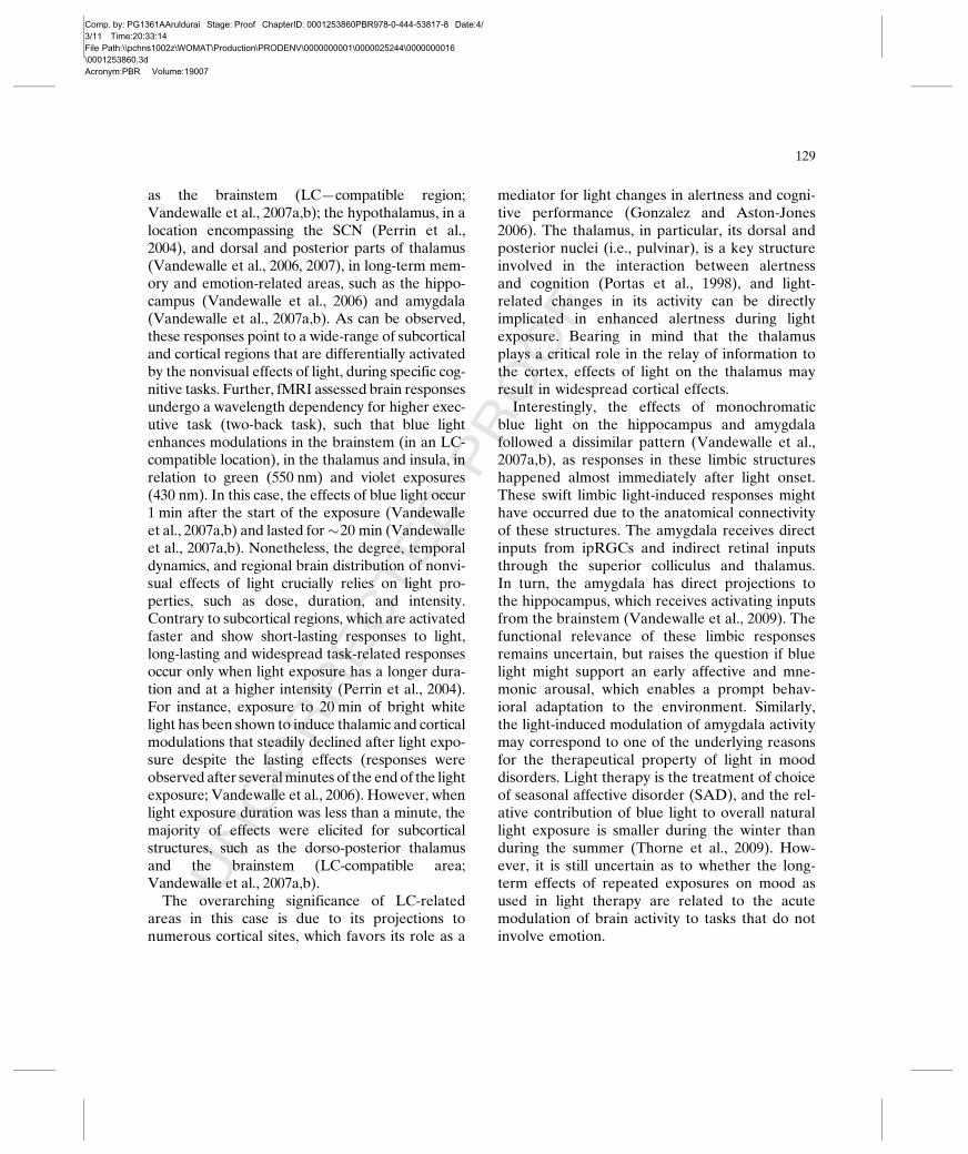
as the brainstem (LC—compatible region;Vandewalle et al., 2007a,b); the hypothalamus, in alocation encompassing the SCN (Perrin et al.,2004), and dorsal and posterior parts of thalamus(Vandewalle et al., 2006, 2007), in long-term mem-ory and emotion-related areas, such as the hippo-campus (Vandewalle et al., 2006) and amygdala(Vandewalle et al., 2007a,b). As can be observed,these responses point to a wide-range of subcorticaland cortical regions that are differentially activatedby the nonvisual effects of light, during specific cog-nitive tasks. Further, fMRI assessed brain responsesundergo a wavelength dependency for higher exec-utive task (two-back task), such that blue lightenhances modulations in the brainstem (in an LC-compatible location), in the thalamus and insula, inrelation to green (550 nm) and violet exposures(430 nm). In this case, the effects of blue light occur1 min after the start of the exposure (Vandewalleet al., 2007a,b) and lasted for�20 min (Vandewalleet al., 2007a,b). Nonetheless, the degree, temporaldynamics, and regional brain distribution of nonvi-sual effects of light crucially relies on light pro-perties, such as dose, duration, and intensity.Contrary to subcortical regions, which are activatedfaster and show short-lasting responses to light,long-lasting and widespread task-related responsesoccur only when light exposure has a longer dura-tion and at a higher intensity (Perrin et al., 2004).For instance, exposure to 20 min of bright whitelight has been shown to induce thalamic and corticalmodulations that steadily declined after light expo-sure despite the lasting effects (responses wereobserved after severalminutes of the end of the lightexposure; Vandewalle et al., 2006). However, whenlight exposure duration was less than a minute, themajority of effects were elicited for subcorticalstructures, such as the dorso-posterior thalamusand the brainstem (LC-compatible area;Vandewalle et al., 2007a,b).The overarching significance of LC-related
areas in this case is due to its projections tonumerous cortical sites, which favors its role as a
mediator for light changes in alertness and cogni-tive performance (Gonzalez and Aston-Jones2006). The thalamus, in particular, its dorsal andposterior nuclei (i.e., pulvinar), is a key structureinvolved in the interaction between alertnessand cognition (Portas et al., 1998), and light-related changes in its activity can be directlyimplicated in enhanced alertness during lightexposure. Bearing in mind that the thalamusplays a critical role in the relay of information tothe cortex, effects of light on the thalamus mayresult in widespread cortical effects.
Interestingly, the effects of monochromaticblue light on the hippocampus and amygdalafollowed a dissimilar pattern (Vandewalle et al.,2007a,b), as responses in these limbic structureshappened almost immediately after light onset.These swift limbic light-induced responses mighthave occurred due to the anatomical connectivityof these structures. The amygdala receives directinputs from ipRGCs and indirect retinal inputsthrough the superior colliculus and thalamus.In turn, the amygdala has direct projections tothe hippocampus, which receives activating inputsfrom the brainstem (Vandewalle et al., 2009). Thefunctional relevance of these limbic responsesremains uncertain, but raises the question if bluelight might support an early affective and mne-monic arousal, which enables a prompt behav-ioral adaptation to the environment. Similarly,the light-induced modulation of amygdala activitymay correspond to one of the underlying reasonsfor the therapeutical property of light in mooddisorders. Light therapy is the treatment of choiceof seasonal affective disorder (SAD), and the rel-ative contribution of blue light to overall naturallight exposure is smaller during the winter thanduring the summer (Thorne et al., 2009). How-ever, it is still uncertain as to whether the long-term effects of repeated exposures on mood asused in light therapy are related to the acutemodulation of brain activity to tasks that do notinvolve emotion.
129

Comp. by: PG1361AAruldurai Stage: Proof ChapterID: 0001253860PBR978-0-444-53817-8 Date:4/3/11 Time:20:33:14File Path:\\pchns1002z\WOMAT\Production\PRODENV\0000000001\0000025244\0000000016\0001253860.3dAcronym:PBR Volume:19007
UNCORRECTEDPROOF
Summary
Light exerts dominant nonvisual effects onnumerous physiological variables, such as thehuman sleep–wake cycle and cognitive perfor-mance, primarily through properties such as dose,duration, timing, and wavelength. The use of reli-able and sensitive methods such as fMRI to eval-uate light-induced nonvisual brain responses haveincreased our understanding on how light canoptimize brain function during specific cognitivetasks. These stimulating discoveries will certainlyhelp to unravel how retinal and suprachiasmaticnetworks modulate the complex interplay ofcircadian rhythms, sleep–wake homeostasis, andcognition.
References
Albrecht, U., Zheng, B., et al. (2001). MPer1Au7 and mPer2 areessential for normal resetting of the circadian clock. Journalof Biological Rhythms, 16(2), 100–104.
Aston-Jones, G., Rajkowski, J., et al. (1999). Role of locuscoeruleus in attention and behavioral flexibility. BiologicalPsychiatry, 46, 1309–1320.
Badia, P., Myers, B., et al. (1991). Bright light effects on bodytemperature, alertness, EEG and behavior. Physiology &Behavior, 50, 583–588.
Beersma, D. G. M., Comas, M., et al. (2009). The progressionof circadian phase during light exposure in animals andhumans. Journal of Biological Rhythms, 24(2), 153–160.
Berson, D. M. (2003). Strange vision: Ganglion cells as circa-dian photoreceptors. Trends in Neurosciences, 26, 314–320.
Boivin, D. B., Duffy, J. F., et al. (1996). Dose-responserelationships for resetting of human circadian clock by light.Nature, 379, 540–542.
Borbély, A. A. (1982). A two process model of sleep regula-tion. Human Neurobiology, 1, 195–204.
Brainard, G., Hanifin, J. P., et al. (2001). Human melatoninregulation is not mediated by the three cone photopic visualsystem. The Journal of Clinical Endocrinology andMetabolism, 86, 433–436.
Bromundt, V., Köster, M., et al. (2010). Sleep-wake cycles andcoAu8 gnitive functioning in schizophrenia. The British Journalof Psychiatry, 197, .
Burgess, H. J., Sharkey, K. M., et al. (2002). Bright light, darkand melatonin can promote circadian adaptation in nightshift workers. Sleep Medicine Reviews, 6(5), 407–420.
Buysse, D. J., Barzansky, B., et al. (2003). Sleep, fatigue, andmedical training: Setting an agenda for optimal learningand patient care. Sleep, 26, 218–225.
Cajochen, C., Dijk, D. J., et al. (1992). Dynamics of EEGslow-wave activity and core body temperature in humansleep after exposure to bright light. Sleep, 15, 337–343.
Cajochen, C., Jud, C., et al. (2006). Evening exposure to bluelight stimulates the expression of the clock gene PER2 inhumans. The European Journal of Neuroscience, 23,1082–1086.
Cajochen, C., Khalsa, S. B. S., et al. (1999a). EEG and ocularcorrelates of circadian melatonin phase and human perfor-mance decrements during sleep loss. American Journal ofPhysiology. Regulatory, Integrative and ComparativePhysiology, 277, R640–R649.
Cajochen, C., Kräuchi, K., et al. (1998). Evening administra-tion of melatonin and bright light: Interactions on theEEG during sleep and wakefulness. Journal of SleepResearch, 7, 145–157.
Cajochen, C., Münch, M., et al. (2005). High sensitivity ofhuman melatonin, alertness, thermoregulation and heartrate to short wavelength light. The Journal of Clinical Endo-crinology and Metabolism, 90, 1311–1316.
Cajochen, C., Zeitzer, J. M., et al. (1999b). Dose-responserelationship for light intensity and alertness and its ocularand EEG correlates. Sleep Research Online, 2, 517.
Cajochen, C., Zeitzer, J. M., et al. (2000). Dose- response rela-tionship for light intensity and ocular and electroencephalo-graphic correlates of human-alertness. Behavioural BrainResearch, 115, 75–83.
Campbell, S. S., & Dawson, D. (1990). Enhancement of night-time alertness and performance with bright ambient light.Physiology & Behavior, 48, 317–320.
Carrier, J., & Dumont, M. (1995). Sleep propensity and sleeparchitecture after bright light exposure at three differenttimes of day. Journal of Sleep Research, 4, 202–211.
Comas, M., Beersma, D. G. M., et al. (2006). Phase and periodresponses of the circadian system of mice (Mus musculus) tolight stimuli of different duration. Journal of BiologicalRhythms, 21(5), 362–372.
Czeisler, C. A., Dijk, D. J., et al. (1994). Entrained phase ofthe circadian pacemaker serves to stabilize alertness andperformance throughout the habitual waking day. In R. D.Ogilvie & J. R. Harsh (Eds.), Sleep onset: Normal andabnormal processes (pp. 89–110). Washington, DC Au9:American Psychological Association.
Czeisler, C. A., & Gooley, J. (2007). Sleep and circadianrhythms in humans. Cold Spring Harbor Symposia onQuantitative Biology, 72, 579–597.
Daan, S., Beersma, D. G. M., et al. (1984). Timing of humansleep: Recovery process gated by a circadian pacemaker.The American Journal of Physiology, 246, R161–R178.
130

Comp. by: PG1361AAruldurai Stage: Proof ChapterID: 0001253860PBR978-0-444-53817-8 Date:4/3/11 Time:20:33:14File Path:\\pchns1002z\WOMAT\Production\PRODENV\0000000001\0000025244\0000000016\0001253860.3dAcronym:PBR Volume:19007
UNCORRECTEDPROOF
Daurat, A., Foret, J., et al. (2000). Bright light during night-time: Effects on the circadian regulation of alertness andperformance. Biological Signals and Receptors, 9, 309–318.
Dijk, D. J., Beersma, D. G. M., et al. (1989). Bright morninglight advances the human circadian system without affectingNREM sleep homeostasis. American Journal of Physiology.Regulatory, Integrative and Comparative Physiology, 256,R106–R111.
Dijk, D. J., & Czeisler, C. A. (1994). Paradoxical timing of thecircadian rhythm of sleep propensity serves to consolidatesleep and wakefulness in humans. Neuroscience Letters,166, 63–68.
Dijk, D. J., & Czeisler, C. A. (1995). Contribution of the circa-dian pacemaker and the sleep homeostat to sleep propen-sity, sleep structure, electroencephalographic slow waves,and sleep spindle activity in humans. The Journal of Neuro-science, 15, 3526–3538.
Dijk, D. J., Shanahan, T. L., et al. (1997). Variation of electro-encephalographic activity during non-rapid eye movementand rapid eye movement sleep with phase of circadianmelatonin rhythm in humans. The Journal of Physiology,505, 851–858.
Dollins, A. B., Lynch, H. J., et al. (1993). Effects of illumina-tion on human nocturnal serum melatonin levels andperformance. Physiology & Behavior, 53, 153–160.
Duffy, J. F., & Dijk, D. J. (2002). Getting through to circadianoscillators: Why use constant routines? Journal of BiologicalRhythms, 17(1), 4–13.
Duffy, J. E., & Wright, K. P. (2005). Entrainment of thehuman circadian system by light. Journal of BiologicalRhythms, 20(4), 326–338.
Edelstein, K., de la Iglesia, H. O., et al. (2003). Behavioralarousal blocks light-induced phase advances in locomotorrhythmicity but not light-induced Per1 and Fos expressionin the hamster suprachiasmatic nucleus. Neuroscience, 118(1), 253–261.
Foret, J., Daurat, A., et al. (1996). The effect on body temper-ature and melatonin of a 39-h constant routine with two dif-ferent light levels at nighttime. Chronobiology International,13, 35–45.
Giménez, M. C., Hessels, M., et al. (2010). Effects of artificialdawn on subjective ratings of sleep inertia and dim light mel-atonin onset. Chronobiology International, 27(6), 1219–1241.
Gonzalez, M. M., & Aston-Jones, G. (2006). Circadian regula-tion of arousal: Role of the noradrenergic locus coeruleussystem and light exposure. Sleep, 29, 1327–1336.
Gooley, J. J., Rajaratnam, S. M. W., et al. (2010). Spectralresponses of the human circadian system depend on theirradiance and duration of exposure to light. ScienceTranslational Medicine, 2(31), 31ra33.
Gordijn, M. C. M., Beersma, D. G. M., et al. (1999). Effects oflight exposure and sleep displacement on dim lightmelatonin onset. Journal of Sleep Research, 8(3), 163–174.
Gronfier, C., Wright, K. P. Jr., et al. (2004). Efficacy of a singlesequence of intermittent bright light pulses for delaying cir-cadian phase in humans. American Journal of Physiology.Endocrinology and Metabolism, 287(1), E174–E181.
Gronfier, C., Wright, K. P. Jr., et al. (2007). Entrainment ofthe human circadian pacemaker to longer-than-24-h days.Proceedings of the National Academy of Sciences of theUnited States of America, 104 Au10, 9081–9086.
Guler, A. D., Ecker, J. L., et al. (2008). Melanopsin cells arethe principal conduits for rod-cone input to non-image-forming vision. Nature, 453(7191), 102–105.
Hatori, M., & Panda, S. (2010). The emerging roles ofmelanopsin in behavioral adaptation to light. Trends inMolecular Medicine, 16(10), 435–446.
Hattar, S., Liao, H. W., et al. (2002). Melanopsin-containingretinal ganglion cells: Architecture, projections, an intrinsicphotosensitivity. Science, 295, 1065–1071.
Khalsa, S. B. S., Jewett, M. E., et al. (2003). A phase responsecurve to single bright light pulses in human subjects. TheJournal of Physiology, 549(3), 945–952.
Kleitman, N. (1987). Sleep and wakefulness. Chicago andLondon: The University of Chicago Press.
Koorengevel, K. M., Beersma, D. G. M., et al. (2003). Moodregulation in seasonal affective disorder patients andhealthy controls studied in forced desynchrony. PsychiatryResearch, 117, 57–74.
Kubota, T., Uchiyama, M., et al. (2002). Effects of nocturnalbright light on saliva melatonin, core body temperatureand sleep propensity rhythms in human subjects. Neurosci-ence Research, 42, 115–122.
Lavie, P. (1997). Melatonin: Role in gating nocturnal rise insleep propensity. Journal of Biological Rhythms, 12, 657–665.
Lavoie, S., Paquet, J., et al. (2003). Vigilance levels during andafter bright light exposure in the first half of the night. Chro-nobiology International, 20, 1019–1038.
Lewy, A. J., Wehr, T. A., et al. (1980). Light suppresses mela-tonin secretion in humans. Science, 210, 1267–1269.
Lin, J. S., Hou, Y., et al. (1996). Histaminergic descendinginputs to the mesopontine tegmentum and their role in thecontrol of cortical activation and wakefulness in the cat.The Journal of Neuroscience, 16, 1523–1537.
Lockley, S. W., Evans, E. E., et al. (2006). Short-wavelengthsensitivity for the direct effects of light on alertness, vigi-lance, and the waking electroencephalogram in humans.Sleep, 29, 161–168.
Lockley, S. W., & Gooley, J. J. (2006). Circadian photorecep-tion: Spotlight on the brain. Current Biology, 16, R795–R797.
Lupi, D., Oster, H., et al. (2008). The acute light-induction ofsleep is mediated by OPN4-based photoreception. NatureNeuroscience, 11(9), 1068–1073.
Moseley, M., Bayliss, S., et al. (1988). Light transmissionthrough the human eyelid: In vivo measurement. Ophthal-mic & Physiological Optics, 8(2), 229–230.
131

Comp. by: PG1361AAruldurai Stage: Proof ChapterID: 0001253860PBR978-0-444-53817-8 Date:4/3/11 Time:20:33:15File Path:\\pchns1002z\WOMAT\Production\PRODENV\0000000001\0000025244\0000000016\0001253860.3dAcronym:PBR Volume:19007
UNCORRECTEDPROOF
Münch, M., Kobialka, S., et al. (2006). Wavelength-dependenteffects of evening light exposure on sleep architecture andsleep EEG power density in men. American Journal ofPhysiology: Regulatory, Integrative and ComparativePhysiology, 290, R1421–R1428.
Muñoz, M., Peirson, S. N., et al. (2005). Long - term constantlight induces constitutive elevated expression of mPER2protein in the murine SCN. A molecular basis for Aschoff`srule? Journal of Biological Rhythms, 20, 3–14.
Myers, B. L., & Badia, P. (1993). Immediate effects of differ-ent light intensities on body temperature and alertness.Physiology & Behavior, 54, 199–202.
Oosterman, J. M., Van Someren, E. J. W., et al. (2009). Frag-mentation of the rest-activity rhythm correlates with age-related cognitive deficits. Journal of Sleep Research, 18(1),129–135.
Oster, H., Maronde, E., et al. (2002). The circadian clock as amolecular calendar. Chronobiology International, 19(3),507–516.
Perrin, F., Peigneux, P., et al. (2004). Nonvisual responses tolight exposure in the human brain during the circadiannight. Current Biology, 14, 1842–1846.
Phipps-Nelson, J., Redman, J. R., et al. (2009). Blue lightexposure reduces objective measures of sleepiness duringprolonged nighttime performance testing. ChronobiologyInternational, 26(5), 891–912.
Portas, C. M., Rees, G., et al. (1998). A specific role for thethalamus in mediating the interaction of attention andarousal in humans. The Journal of Neuroscience, 18(21),8979–8989.
Revell, V. L., Arendt, J., et al. (2006). Alerting effects of lightare sensitive to very short wavelengths. NeuroscienceLetters, 399(1–2), 96–100.
Riemersma-van der Lek, R. F., Swaab, D. F., et al. (2008).Effect of bright light and melatonin on cognitive andnoncognitive function in elderly residents of group carefacilities: A randomized controlled trial. The Journal of theAmerican Medical Association, 299(22), 2642–2655.
Ruger, M., Gordijn, M. C. M., et al. (2006). Time-of-day-dependent effects of bright light exposure on human psycho-physiology: Comparison of daytime and nighttime exposure.American Journal of Physiology: Regulatory, Integrative andComparative Physiology, 290(5), R1413–R1420.
Rusak, B., Robertson, H., et al. (1990). Light pulses that shiftrhythms induce gene expression in the suprachiasmaticnucleus. Science, 248(4960), 1237–1240.
Sack, R. L., Blood, M. L., et al. (1992). Oral melatoninreverses the alerting effects of nocturnal bright light expo-sure in humans. Sleep Research, 21, 49.
Sack, R. L., Hughes, R. J., et al. (1997). Sleep-promotingeffects of melatonin: At what dose, in whom, under whatconditions, and by what mechanisms? Sleep, 20, 908–915.
Santhi, N., Aeschbach, D., et al. (2008). The impact of sleeptiming and bright light exposure on attentional impairmentduring night work. Journal of Biological Rhythms, 23(4),341–352.
Saper, C. B., Scammell, T. E., et al. (2005). Hypothalamic reg-ulation of sleep and circadian rhythms. Nature, 437(7063),1257–1263.
Smith, K. A., Schoen, M. W., et al. (2004). Adaptation ofhuman pineal melatonin suppression by recent photichistory. The Journal of Clinical Endocrinology andMetabolism, 89(7), 3610–3614.
Thorne, H. C., Jones, K. H., et al. (2009). Daily and seasonalvariation in the spectral composition of light exposure inhumans. Chronobiology International, 26(5), 854–866.
Tsai, J. W., Hannibal, J., et al. (2009). Melanopsin as a sleepmodulator: Circadian gating of the direct effects of light onsleep and altered sleep homeostasis in Opn4(�/�) mice.PLoS Biology, 7(6), e1000125.
Van Someren, E. (2000). More than a marker: Interactionbetween the circadian regulation of temperature and sleep,age-related changes, and treatment possibilities. Chronobi-ology International, 17(3), 313–354.
Vandewalle, G., Balteau, E., et al. (2006). Daytime lightexposure dynamically enhances brain responses. CurrentBiology, 16(16), 1616–1621.
Vandewalle, G., Gais, S., et al. (2007a). Wavelength-depen-dent modulation of brain responses to a working memorytask by daytime light exposure. Cerebral Cortex, Epubahead of print Au11.
Vandewalle, G., Maquet, P., et al. (2009). Light as a modulatorof cognitive brain function. Trends in Cognitive Sciences, 13(10), 429–438.
Vandewalle, G., Schmidt, C., et al. (2007b). Brain responses toviolet, blue, and green monochromatic light exposures inhumans: Prominent role of blue light and the brainstem.PLoS One, 2(11), e1247.
Werken, M., Giménez, M., et al. (2010). Effects of artificialdawn on sleep inertia, skin temperature, and the awakeningcortisol response. Journal of Sleep Research, 19(3), 425–435.
Wright, K. P., Jr. (1997). Combination of bright light and caf-feine as a countermeasure for impaired alertness and perfor-mance during extended sleep deprivation. Journal of SleepResearch, 6, 26–35.
Wright, K. P., Jr., Hull, J. T., et al. (2006). Sleep and wakeful-ness out of phase with internal biological time impairslearning in humans. Journal of Cognitive Neuroscience, 18,508–521.
Zeitzer, J. M., Dijk, D. J., et al. (2000). Sensitivity of thehuman circadian pacemaker to nocturnal light: Melatoninphase resetting and suppression. The Journal of Physiology,526, 695–702.
132

Comp. by: PG1361AAruldurai Stage: Proof ChapterID: 0001253860PBR978-0-444-53817-8 Date:4/3/11 Time:20:33:15File Path:\\pchns1002z\WOMAT\Production\PRODENV\0000000001\0000025244\0000000016\0001253860.3dAcronym:PBR Volume:19007
Query Form
Book: Progress in Brain Research, 190
Chapter No: 07
AU: Author Query; ED: Editor Query; TS: Query raised by Typesetter;
Query Refs. Queries Author’s Response
Au1 Note that the symbols "*, **, ns" are mentioned in the artwork of
Fig. 1 but its significance is missing in caption of Fig. 1. Please
check.
Au2 Please provide telecommunication details and E-mail address for the
corresponding author "Marijke C. M. Gordijn".
Au3 As per in-house style "PER" have been italicized when referred to asa gene. Please check.
Au4 Please check the introduction of slash in "desynchronized/
synchronized subjects."
Au5 "Cajochen et al., 1999" has been changed to "Cajochen et al.,
1999a,b" as per reference list. Please check.
Au6 Please check the edits made in the sentence “A study in which. . .”
Au7 As per style a maximum of up to six authors plus et al. should be
listed in the author group. Please list up to six authors for all the et al.
references.
Au8 Please provide page range for this reference.
Au9 Please check whether publisher name is appropriate for this
reference.
Au10 Please check whether the inserted volume number and page rangefor this reference is appropriate.
Au11 Please provide volume number and page range for this reference.
Copyright © 2022 FDOKUMEN