Antibody targeting of anaplastic lymphoma kinase induces cytotoxicity of human neuroblastoma.
Upload
independentCategory
view
4download
0
Ca2�-dependent prodynorphin transcriptional derepression inneuroblastoma cells is exerted through DREAM protein activity
in a kinase-independent manner
David Campos,b Lydia Jimenez-Dıaz,a JoseR. Naranjo,b and Angel M. Carriona,*a Laboratorio Andaluz de Biologıa (LAB), Universidad Pablo de Olavide, Ctra. de Utrera, km 1, E-41013 Sevilla, Spain
b Centro Nacional de Biotecnologia (CNB), Universidad Autonoma de Madrid, Cantoblanco, E-28049 Madrid, Spain
Received 6 December 2001; revised 23 August 2002; accepted 26 August 2002
Abstract
Prodynorphin transcription has been postulated as an important molecular mechanism involved in adaptation/repair processes. Expres-sion of prodynorphin is modulated according to the levels of the second messengers cAMP and Ca2�. In the neuroblastoma cell lines, theincrease of prodynorphin mRNA levels is coupled to an elevation of intracellular cAMP levels. Promoter analyses have revealed that theDRE site, a silencer element present in the prodynorphin promoter, is involved in PKA-dependent prodynorphin derepression. In this way,DREAM, a calcium-dependent repressor, plays an outstanding role. In this study, Ca2� release from internal stores has been found topromote an increase of prodynorphin mRNA levels in NB69 cells. Surprisingly, Ca2�-dependent prodynorphin gene transcription wasinsensitive to the broad-spectrum kinase inhibitors and sensitive to agents that alter internal Ca2� accumulation. Moreover, we demonstratethat in NB69 cells, the Ca2� signaling pathway uses DREAM as an effector to evoke prodynorphin transcription derepression in akinase-independent manner.© 2003 Elsevier Science (USA). All rights reserved.
Introduction
In living cells regulation of gene expression in responseto extracellular signals is a fundamental mechanism forproper development, homeostasis, and adaptation to theenvironment (Armstrong and Montminy, 1993; Buonannoand Fields, 1999; He and Rosenfeld, 1991). Extracellularsignals are transduced to intracellular effectors by pathwaysthat link cell surface with nuclear events (Deisseroth et al.,1996; Hill and Treisman, 1995; Karin and Smeal, 1992); insuch a way that transient inputs are processed into short aswell as relatively permanent cellular responses (Armstrongand Montminy, 1993; Goelet et al., 1986; Mayford andKandel, 1999). In neurons, the major transduction pathwaysinvolve an increase in levels of the second messengerscAMP and Ca2�, which results in activation of cAMP-
dependent protein kinase (PKA1), mitogen activated proteinkinase (MAPK), and Ca2�/calmodulin-dependent kinase(CaMK) and, eventually, in an altered pattern of gene ex-pression (Bito et al., 1997; Ghosh and Greenberg, 1995;Mellstrom and Naranjo, 2001; Montminy, 1997). In theseregulatory processes, the phosphorylation of transcriptionfactors plays a fundamental role (Hunter and Karin, 1992;Schulman, 1995; Shaywitz and Greenberg, 1999).
Learning and adaptation to chronic pain involve pro-found changes in gene expression within the central nervoussystem, involving, among many others, the prodynorphin-
* Corresponding author. Fax:�954-349375.E-mail address: [email protected] (A´ .M. Carrion).
1 Abbreviations used: AP-1, activator protein 1; bHLH, basic helix-loop-helix; CAT, chloramphenicol acetyltransferase; CaMK, Ca2�/cal-modulin-dependent protein kinase; ChIP, chromatin immunoprecipitation;CRE, cAMP-responsive element; CREB, CRE-binding protein; CBP,CREB-binding protein; DRE, downstream regulatory element; DREAM,DRE antagonist modulator; ERK, extracellular signal regulated kinase;MAPK, mitogen-activated protein kinase; ncAP-1, noncanonical AP-1; PI3-kinase, phosphoinositol 3 phosphate kinase; PKA, protein kinase A;PKC, protein kinase C.
R
Available online at www.sciencedirect.com
Molecular and Cellular Neuroscience 22 (2003) 135–145 www.elsevier.com/locate/ymcne
1044-7431/03/$ – see front matter © 2003 Elsevier Science (USA). All rights reserved.doi:10.1016/S1044-7431(03)00040-X

encoding gene (Cole et al., 1995; Impey et al., 1996;Naranjo et al., 1991; Schulman, 1995). Dynorphin peptidesmodulate the release of neurotransmitters from synapticterminals, thereby controlling synaptic efficiency (Wagneret al., 1993; Weiskopf et al., 1993). In rat, induction ofprodynorphin gene expression is associated with an increaseof both cAMP and Ca2� levels, which leads to activation ofPKA, CaMK-II, and protein kinase C (PKC) signal trans-duction pathways (Cole et al., 1995; Ha et al., 1997; Lucaset al., 1993; Ventura et al., 1995, 1997). The activity ofthese kinases, in turn, evokes changes in the expressionand/or functionality of transcriptional activators, such asc-Fos and CREB, which account for transcriptional activa-tion of the prodynorphin gene in spinal cord and striatalneurons, respectively (Cole et al., 1995; Naranjo et al.,1991).
Transcription of the rat prodynorphin gene is modulatedby at least five regulatory elements distributed along 2 kb ofits promoter region (Douglass et al., 1994). Four of theseelements, three located in the distal promoter area and onein the first exon, are recognized by CREB and have beendescribed as cAMP-response elements (CRE) (Cole et al.,1995; Collins-Hicok et al., 1994). In some cases, the bind-ing of CREB results in repression of AP-1-mediated pro-dynorphin transactivation in non-prodynorphinergic celllines (Collins-Hicok et al., 1994). In striatal neurons, CREBphosphorylation is involved in prodynorphin activation ex-erted through the three distal CRE sites (Cole et al., 1995).A fifth DNA regulatory site, located at position �257 in theprodynorphin promoter, has been described as a noncanoni-cal AP-1 site (ncAP-1). This element is recognized byc-Fos/c-Jun heterodimers and could be implicated in PKC-dependent prodynorphin transcription in the neuroblastomaNCB20 cell line (Naranjo et al., 1991). In vivo, all theseDNA response elements play a complex stimulus- and tis-sue-specific role in prodynorphin transcriptional regulation(Carrion et al., 1998a).
A downstream regulatory element (DRE) site locatedwithin the first exon of the human prodynorphin gene hasbeen characterized. Site-directed mutagenesis and deletionanalyses of the human promoter have revealed that thisDRE site is responsible for cAMP-dependent prodynorphintranscriptional derepression in neuroblastoma (Carrion etal., 1998b). The DREAM protein, a calcium-dependenttranscriptional repressor, plays a central role in gene tran-scriptional repression, being bound to the DRE site in rest-ing cells (Carrion et al., 1999). Stimuli that increase intra-cellular cAMP and Ca2� evoke release of DREAM from theDRE, which results in an increase of reporter gene tran-scription rate driven by DRE sites (Carrion et al., 1998b,1999). The molecular bases of cAMP and Ca2�-promotedderepression are completely different. Whereas Ca2� mod-ifies DREAM conformation by binding to EF-hand motifspresent in this repressor (Carrion et al., 1999), cAMP-dependent derepression is a cellular-specific event which
requires �CREM-DREAM protein interaction (Ledo et al.,2000a).
Despite evidence suggesting that DREAM is a transcrip-tion factor, the physiological function of DREAM has re-mained unclear. The discovery of several nearly identicalEF-hand proteins, calsenilin (Buxbaum et al., 1998) andKChIP (An et al., 2000), encoded by the same genomiclocus as DREAM, suggests differents biological functionfor a single gene. Calcenilin/DREAM has been shown tointeract with presenilin proteins and has been suggested tobe involved in �-amyloid plaque formation (Buxbaum et al.,1998; Choi et al., 2001; Jo et al., 2001). KChIP/DREAMswere cloned as binding partners and direct modulators forA-type potassium channels (An et al., 2000), which havebeen implicated in the pathogenesis of heart failure (Wick-enden et al., 1999). Furthermore, recent results usingDREAM knock-out mice showed that DREAM is a tran-scription factor critical for pain modulation (Cheng et al.,2002).
In this article, we show that prodynorphin transcriptionin NB69 cell line is activated by the Ca2� increase evokedby caffeine treatment. Deletion analysis and site-directedmutagenesis have demonstrated the functional importanceof the DRE site in the Ca2�-promoted transcriptional dere-pression of the prodynorphin promoter in NB69 cells (Ledoet al., 2000b). This process and Ca2�-dependent prodynor-phin gene transcription are insensitive to the protein kinaseinhibitor staurosporine in contrast with forskolin-dependentprodynorphin transcriptional derepression. Additionally theexpression of a Ca2�-insensitive DREAM mutant abolishescaffeine-dependent prodynorphin transcriptional derepres-sion. Finally, using a stable NB69 transfectant cell lineexpressing a DREAM antisense transcript, we demonstratethat the blockade of DREAM protein synthesis is sufficientto trigger a constitutive prodynorphin transcription. In thelight of these results, we propose the existence of mecha-nisms convergent on the DREAM protein for PKA- andCa2�-dependent prodynorphin gene expression.
Results
Prodynorphin transcription is modulated by caffeine inNB69 neuroblastoma cells
Prodynorphin expression is modulated in spinal cordneurons and myocytes in a Ca2�-dependent manner. Inthese cell types, it has been suggested that intracellular Ca2�
accumulation triggers an increase of CaMK and PKC ac-tivity, respectively (Ha et al., 1997; Ventura et al., 1997),but the molecular mechanisms by which Ca2� stimulatesprodynorphin transcription are actually unknown. In NB69neuroblastoma cells, prodynorphin expression increasedupon treatment with caffeine (Fig. 1), a potent releaser ofCa2� from the endoplasmic reticulum via the ryanodinereceptor (Ehrlich et al., 1994; Hernandez-Cruz et al., 1990).
136 D. Campos et al. / Molecular and Cellular Neuroscience 22 (2003) 135–145

The time course of prodynorphin mRNA induction showedan increase during 3-12 h of caffeine treatment that de-creased to basal levels within 24 h. Previous results usingdeletion analysis and site-directed mutagenesis of the hu-man prodynorphin promoter indicated that caffeine-depen-dent prodynorphin transcription derepression required thepresence of an intact DRE site in the prodynorphin promoter(Ledo et al., 2000b).
Caffeine modulates prodynorphin gene transcription in aCa2�-dependent and kinase-independent manner
In living cells caffeine could act as a calcium releaserfrom intracellular stores through ryanodine receptors and/oras a phosphodiesterase inhibitor. To distinguish betweenthese two effects, we made use of thapsigargin and highconcentrations of ryanodine in order to block intracellularCa2� release, or of BAPTA-AM in order to chelate intra-cellular Ca2� (Ehrlich et al., 1994). The pretreatment ofNB69 cells with any of these three reagents resulted in aneffective prevention of caffeine-promoted induction of pro-dynorphin gene accumulation and expression from thepHD3CAT reporter construct, the minimal inducible humanprodynorphin promoter containing the DRE site (Figs. 2Aand 2B). These results strongly suggested that caffeineworked by eliciting Ca2� release from intracellular stores.
Since known effects of intracellular Ca2� increase are theactivation of PKA and CaMK (Ghosh and Greenberg, 1995),the contribution of phosphorylation to caffeine-dependent pro-dynorphin transcription was analyzed by measuring caffeine-dependent prodynorphin mRNA accumulation and expressionfrom pHD3CAT in either the presence or the absence ofstaurosporine, a broad-spectrum kinase inhibitor (Bito et al.,1996; MacKintosh and MacKintosh, 1994). We found that thiscompound did not affect either basal or caffeine-dependentprodynorphin expression and promoter activity frompHD3CAT (Figs. 2A and 2C). Similarly, specific PKA andCaMK inhibitors (H89 and KN62) were found not to altercaffeine-dependent pHD3CAT activity (data not shown). Asinternal controls, staurosporine was observed to block forsko-lin-dependent prodynorphin gene accumulation and reporter
expression from pHD3CAT, which require PKA activation(Figs. 2A and 2C) as well as forskolin- and caffeine-dependentexpression from pENKCAT, a construct in which an enkepha-lin promoter fragment containing two CRE sites was fused tothe CAT reporter (Fig. 2D). These results suggest that thetranscription factor which recognizes the DRE site, i.e.,DREAM, was modulated directly by intracellular Ca2� levelsindependently of protein kinase activity.
Ca2� blocks DREAM activity in NB69 cells
To analyze whether the DREAM DNA-binding activitypresent in NB69 cells was sensitive to Ca2� increase evokedby caffeine treatment, we carried out DRE-binding experi-ments using nuclear extracts from NB69 cells cultured ineither the presence or the absence of caffeine. Gel-shiftassays using nuclear extracts from caffeine-treated NB69cultures showed significant reduction in DRE-binding ac-tivity with respect to the nuclear protein extract from un-treated NB69 cultures (Fig. 3A). The inhibition of DREbinding evoked by caffeine was sensitive to agents thatdisturb Ca2� homeostasis, i.e., ryanodine at high concen-tration and BAPTA-AM, and insensitive to the broad kinaseinhibitor staurosporine (Fig. 3A). In the opposite way, akinase-dependent process as the decrease of DREAM-bind-ing activity induced by forskolin was completely blocked inthe presence of staurosporine (Fig. 3A). These resultspointed out that intracellular Ca2� increase can determineunbinding of DREAM from the prodynorphin promoterDRE site independently of the activation of Ca2�-dependentkinase cascade.
This hypothesis was tested in living cells using the chro-matin immunoprecipitation (ChIP) assay with a DREAM-specific antibody, that recognized a specific 30-kDa band inthe HEK-293 cell line transfected with expression plasmidencoding for DREAM protein (Fig. 3B), and PCR withprimers designed to amplify the human prodynorphin pro-moter in NB69 control and caffeine-stimulated cultures.High levels of prodynorphin promoter sequence was de-tected in DREAM-immunoselected DNA complexes fromnonstimulated cells, while 3 h of caffeine treatment abro-gated the amplification of prodynorphin promoter fromDNA immunoprecipitated with DREAM antibody (Fig. 3C,panel 2). The inhibition prodynorphin promoter amplifica-tion evoked by caffeine was sensitive to ryanodine at highconcentration and BAPTA-AM, and insensitive to the broadkinase inhibitor staurosporine (Fig. 3C, panel 2). As internalcontrol for staurosporine, we used as a kinase-dependentmodel the decrease of DREAM binding provoked by fors-kolin treatment. In this case, the decrease of prodynorphinpromoter amplification induced by forskolin was com-pletely reverted in cultures treated which both forskolin andstaurosporine (Fig. 3C, panel 2). In all these samples West-ern blot for DREAM was performed. The polyclonalDREAM antibody recognized an approximately 30-kDaband, identical to that obtain with recombinant DREAM
Fig. 1. Caffeine activates prodynorphin transcription through DRE-medi-ated transcriptional derepression. Northern blot analysis of prodynorphinexpression in the NB69 cell line at the indicated times after treatment with10 mM caffeine. A GAPDH probe was used as a control of the amount ofRNA loaded per lane.
137D. Campos et al. / Molecular and Cellular Neuroscience 22 (2003) 135–145

protein, with the same intensity in all the immunoprecipi-tated samples (Fig. 3C, panel 1). These results confirmedthat Ca2� release by caffeine treatment produced the kinase-insensitive DNA unbinding of DREAM from the DRE siteof the human prodynorphin promoter. Further-more, theChIP assay experiment suggests that prodynorphin gene is aphysiological target of DREAM.
NB69 overexpressing a Ca2� dominant-negative DREAMmutant does not induce prodynorphin transcription
The results described above underline the possibility thatcaffeine-dependent prodynorphin transcriptional derepres-sion could be physiologically mediated by the release ofDREAM from the promoter DRE site elicited by an increaseof intracellular Ca2�. In order to test this hypothesis in vivo,experiments were carried out in which NB69 cells werecotransfected with pHD3CAT and an expression vectorcoding for a Ca2� dominant-negative DREAM mutant,
dubbed 234EFDREAM (Carrion et al., 1999), and CATactivity was measured in untreated and caffeine-treatedcells. As shown in Fig. 4A, expression of the dominant-negative DREAM mutant abolished caffeine-promoted re-porter gene induction from pHD3CAT. Thereafter, in orderto test whether the 234EFDREAM mutant was able to blockprodynorphin expression, we transiently transfected its en-coding construct into NB69 cultures that were either treatedwith caffeine or left untreated, and the cells were analyzedfor prodynorphin and DREAM mRNA expression. Figure4B shows that, whereas endogenous DREAM mRNA levelsdid not significantly increase upon caffeine treatment, theexogenous DREAM mutant mRNA was detected only incultures transfected with the 234EFDREAM expressionvector. Also, it was found that, whereas prodynorphinmRNA was induced by caffeine treatment in the controltransfected cultures, overexpression of the dominant-nega-tive DREAM mutant abolished prodynorphin mRNA accu-mulation triggered by caffeine (Fig. 4C). All these results
Fig. 2. Caffeine-dependent prodynorphin transcription depends on Ca2� increase and it is insensitive to kinase inhibitor. (A) Northern blot analysis ofprodynorphin expression in the NB69 cell line between 3 and 6 h after treatment with 10 mM caffeine or 10 �M forskolin was assayed in the absence orpresence of the intracellular Ca2�-release inhibitor ryanodine (Rya, 100 �M), or BAPTA-AM (BAPTA, 1 �M), or the kinase inhibitor staurosporine (Staur,50 nM). A cyclophilin probe was used as a control of the amount of RNA loaded per lane. (B, C, D) pHD3CAT or pENKCAT was transiently transfectedinto NB69 cells and the effect of forskolin (FSK, 25 �M) or caffeine (10 mM) was assayed in the absence or presence of the intracellular Ca2�-releaseinhibitors ryanodine (Rya, 100 �M), thapsigargin (Thap, 1 �M), or BAPTA-AM (BAPTA, 1 �M), or the kinase inhibitor staurosporine (Staur, 50 nM). (B)Effect of intracellular Ca2�-release inhibitors on caffeine-promoted induction of reporter gene expression from pHD3CAT. (C) Effect of staurosporine onforskolin- and caffeine-promoted induction of reporter expression from pHD3CAT. (D) Idem from pENKCAT.
138 D. Campos et al. / Molecular and Cellular Neuroscience 22 (2003) 135–145

illustrate that DREAM is a critical transcription factor ac-counting for Ca2�-dependent prodynorphin transcriptionevoked by caffeine in NB69 cells.
Stable DREAM translation inhibition in NB69 cellsevokes constitutive prodynorphin transcription
The results described above allowed us to hypothesizethat caffeine-dependent derepression arises as a result ofDREAM unbinding from the DRE site. In order to verifythe central role of DREAM in prodynorphin transcrip-tional regulation, we generated an NB69-derivative strainin which a DREAM antisense transcript was stably over-expressed (Fig. 5A). This cell line, termed NB69-AS,was found to be devoid of DRE-binding activity in band-shift experiments, compared with the parental NB69 cellline (Fig. 5B). Moreover, whereas in the NB69-AS cellline the CRE-dependent transcriptional response to caf-
feine was not altered, the DRE-dependent transcriptionevoked by caffeine was effectively abolished (data notshown). These results suggested that intracellularDREAM depletion resulted in a constitutive increase ofDRE-dependent prodynorphin transcription in transienttransfection experiments. To confirm this idea, we ana-lyzed endogenous prodynorphin gene expression in theNB69-AS cell line. In unstimulated cells, in contrast withthe NB69 cell line, NB69-AS cells displayed a constitu-tive prodynorphin gene expression, the magnitude of themRNA signal obtained was similar to that detected inNB69 cells after caffeine treatment (Fig. 5C). Further-more, the accumulation of prodynorphin messenger wasnot inducible by caffeine in the NB69-AS cell line (Fig.5C). All these results strongly indicate a central roleplayed by the DREAM protein on the caffeine-dependenttranscriptional derepression of the prodynorphin genethrough intracellular Ca2� oscillations.
Fig. 3. Ca2� inhibits nuclear DRE-binding activity in NB69 cells. Band-shift and ChIP assays were carried out using NB69 extracts obtained from culturestreated for 3 h with caffeine (10 mM) or forskolin (25 �M) in either absence or presence of intracellular Ca2�-release inhibitor ryanodine (100 �M), orBAPTA-AM (1 �M), or the kinase inhibitor staurosporine (50 nM). (A) Gel-shift assay using radioactivity labeled DRE oligonucleotide. Specific retardedbands are indicated with arrows and nonspecific band are indicated by asterisk. (B) Western blot showing that the 30-kDa band obtained with DREAMantibody in nuclear extract from NB69 cell line is equal to that obtained from HEK-293 cell line transfected with expression plasmid for DREAM. (C) ChIPwith DREAM antibody and PCR amplification for prodynorphin promoter (panel 2). As internal controls, 1/10 of chromatin formaldehyde cross-linked wasamplified for prodynorphin promoter (panel 3) and Western blot for DREAM protein was analyzed in immunoprecipitates (panel 1).
139D. Campos et al. / Molecular and Cellular Neuroscience 22 (2003) 135–145

Discussion
Changes in the intracellular levels of cAMP and Ca2�
constitute the major regulatory mechanisms during signaltransduction in psychostimulant adaptation processes, pain andneuronal activity. In these processes, the increase in the levelsof both second messengers has been associated with changes ingene expression (Bading et al., 1993; Impey et al., 1996;Konradi et al., 1994). In this context, prodynorphin gene tran-scription becomes activated by such physiological processes aspart of the adaptation/repair response to external stimulation(Cole et al., 1995; Hurd et al., 1992; Iadarola et al., 1988;Steiner and Gerfen, 1993). In human neuroblastoma cell lines,PKA-dependent prodynorphin transcription takes place as aconsequence of unbinding of the repressor protein DREAMfrom the DRE site of the human prodynorphin promoter (Car-rion et al., 1998b) by PKA-dependent protein-protein interac-
tion between �CREM and DREAM proteins (Ledo et al.,2000a).
The prodynorphin gene also is regulated by intracellularCa2� increase in myocytes and spinal cord neurons. In thesecases, prodynorphin transcription has been associated withan increase of PKC and CaMK activities, respectively (Haet al., 1997; Lucas et al., 1993; Ventura et al., 1995). In thehuman neuroblastoma cell line NB69, caffeine, a potentCa2�-releaser from intracellular stores, elicits an accumu-lation of prodynorphin mRNA, which we report here to bedue to an increase in transcription initiation rather than totranscript stabilization. Deletion and site-directed mutagen-esis analysis of the human prodynorphin promoter indicatethat the DRE site present in the proximal region of theprodynorphin promoter is absolutely necessary for caffeine-mediated transcription stimulation (Ledo et al., 2000a). Fur-thermore, as it occurs for cAMP-dependent prodynorphinexpression, transcriptional derepression accounts for theincrease in prodynorphin mRNA levels. This caffeine effecton prodynorphin transcription is sensitive to agents knownto alter intracellular Ca2� dynamics, such as ryanodine,thapsigargin, and BAPTA-AM, and insensitive to the broad-spectrum kinase inhibitor staurosporine. To the best of ourknowledge, the human prodynorphin gene is the first re-ported case of a gene in which Ca2� increase directly drivesits transcription, a process in which the Ca2�dependenttranscriptional repressor DREAM appears to exert an out-standing role. Intracellular Ca2� increase has been shown toabolish DREAM DNA-binding activity by promoting a con-formational change leading to its inactivation as a transcrip-
Fig. 4. The Ca2� dominant-negative DREAM protein blocks prodynorphintranscription. (A) NB69 cells were transiently cotransfected withpHD3CAT and p234EFDREAM, and reporter gene expression was mea-sured in cells treated (solid bars) or not (open bars) with caffeine. Resultsare normalized to the CAT activity obtained in untreated cells cotrans-fected with the empty expression vector pcDNA3 and pHD3CAT. (B, C,D) Northern blots showing DREAM, prodynorphin, and control GAPDHmRNA levels, respectively, in caffeine-treated and untreated NB69 cellstransiently transfected with p234EFDREAM or the empty vector pcDNA3.
Fig. 5. The prodynorphin gene is constitutively expressed in NB69 cellsoverexpressing an antisense DREAM cDNA. (A) Northern blot analysis ofNB69 and NB69-AS cells using a DREAM probe. As an internal loadingcontrol, the figure shows a 3-kb upper band corresponding to endogenousDREAM mRNA and a 1-kb lower band corresponding to the antisenseDREAM RNA. (B) Band-shift assays using NB69 and NB69-AS nuclearextracts and a radiolabeled DRE oligonucleotide. Specific retarded bandsare indicated with arrows and nonspecific band are indicated by asterisks(C) Northern blot analysis of NB69 and NB69-AS cells using a prodynor-phin probe. As an internal loading control, cyclophilin mRNA levels arealso shown.
140 D. Campos et al. / Molecular and Cellular Neuroscience 22 (2003) 135–145

tional repressor (Carrion et al., 1999; Osawa et al., 2001).For this reason, a dominant-negative DREAM mutant forCa2�, as reported in this work, was able to block caffeine-dependent prodynorphin promoter activity as well asmRNA accumulation.
Usually, Ca2�-dependent regulation of transcription is amultistep process in which the Ca2�-binding protein cal-modulin plays an outstanding role (Bito et al., 1997; Buo-nanno and Fields, 1999; Ghosh and Greenberg, 1995). TheCa2�-calmodulin complex couples intracellular Ca2� in-crease to the activation of transcription factors, such asCREB (Deisseroth et al., 1996; Impey et al., 1998). In thecentral nervous system, Ca2� increase results in activationof kinases of the ERK, PKA, and CaMK families (Impey etal., 1998; Ji and Rupp, 1997). Ca2� influx through theNMDA receptor and voltage-dependent Ca2� channels ac-tivate the Ras-MAPK signal-transduction pathway, whichleads to ERK-2 activation (Ghosh and Greenberg, 1995;Impey et al., 1998; Ji and Rupp, 1997). In this process, PKAactivation is needed to promote translocation of ERK-2 intothe nucleus, enabling its mediated phosphorylation of Elk-1and CREB transcription factors (Impey et al., 1998, Johnsonet al., 1997; Roberson et al., 1999; Sgambato et al., 1998).In the same context, the translocation of the Ca2�-calmod-ulin complex from the membrane to the nucleus is neededfor the activation of CaMK-IV, leading to intranuclear phos-phorylation of CREB (Ghosh and Greenberg, 1995; John-son et al., 1997). In this process, the duration of CREBphosphorylation depends on two important Ca2�/calmodu-lin-regulated mechanisms, involving CaMK-IV and cal-cineurin-regulated events (Bito et al., 1996, 1997; Graef etal., 1999). As well, calcineurin regulates the expression ofseveral immediate-early genes by modulating the activationand nuclear importation of transcription factors of theNF-AT family (Graef et al., 1999), for which the amplitude,duration, and oscillation of calcium signals have beenproved important (Dolmetsch et al., 1997; Dolmetsch andLewis, 1998; Li et al., 1998). In all these processes thetransmission of signals from the cell surface to the nucleusis mediated by intermediate cascades of protein kinases andphosphatases ultimately evoking changes in the activity oftranscription factor within the nucleus. Yet, an additionalprocess involving nuclear translocation of the Ca2�-cal-modulin complex is the regulation of transcription factors ofthe basic helix-loop-helix (bHLH) group. In this case, thedirect binding of Ca2�-calmodulin to the bHLH site presentin these proteins inhibits their DNA-binding activity andhence their transcriptional function (Corneliussen et al.,1994).
The present report is the first description of gene regu-lation driven directly by intracellular Ca2� increase in theabsence of protein kinase activity. This process requires theactivity of the Ca2�-sensitive DREAM transcriptional re-pressor (Carrion et al., 1999). Recently, using a fusionbetween the c-fos promoter, containing CRE and DRE sites,and a reporter gene, it has been described that the increase
in nuclear Ca2� is important for CRE-dependent gene tran-scription also (Ahn et al., 2000). In this case, the elevationin nuclear Ca2� levels together with CaMK-IV activationtriggers the activation and recruitment of CBP (Chawla etal., 1998; Hardingham et al., 1999; Hu et al., 1999) andCREB (Hardingham et al., 1997) to the promoter of c-fosgene. DREAM binds to a DRE site located within the c-fospromoter and thereby efficiently represses its transcription(Carrion et al., 1999). In the context, of the c-fos promoter,the role of DREAM could be to act as a stimulus-permissivesensor allowing and/or modulating c-fos expression depend-ing on the type of stimulus received by the cell.
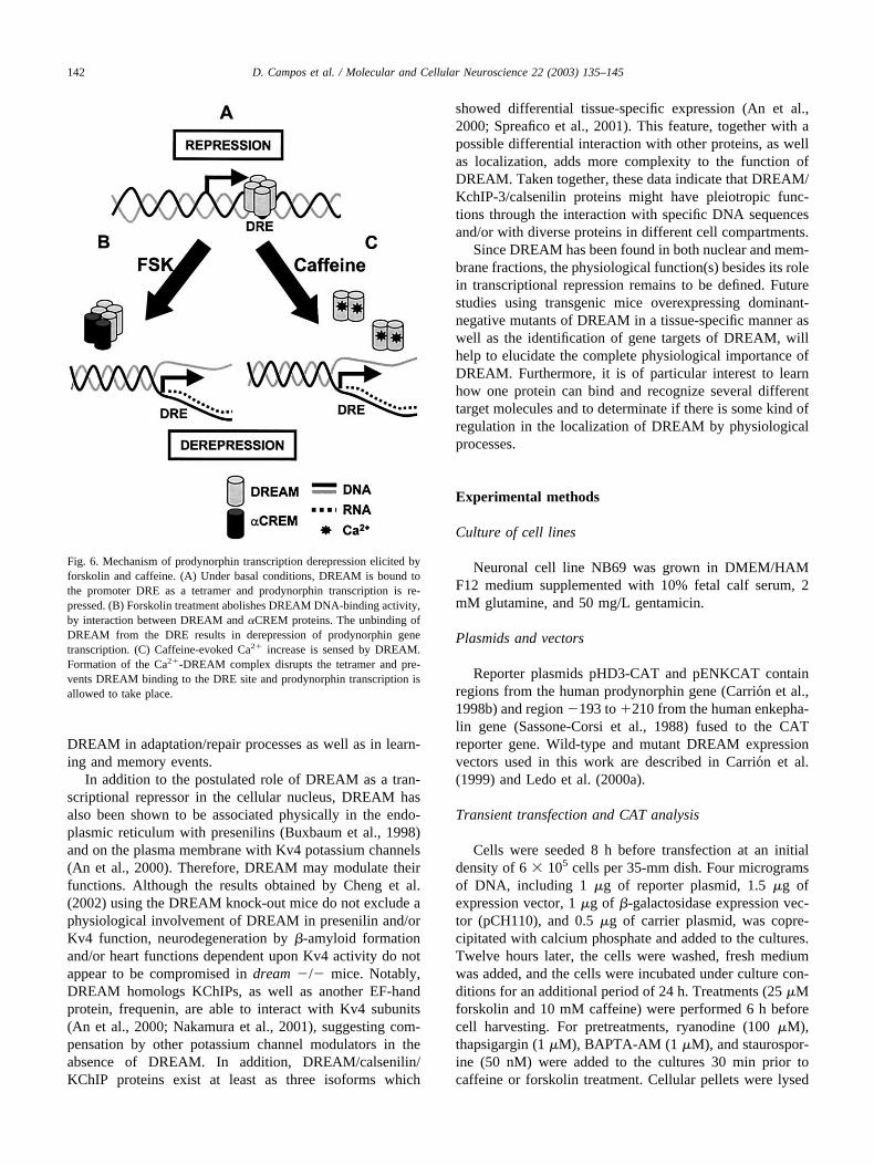
Taken together, the results exposed above allow us topropose a model according to which DREAM would act asa convergence point for two different signal transductionpathways in order to regulate prodynorphin gene transcrip-tion. First, cAMP increase would evoke cellular-specificaccumulation of prodynorphin transcripts mainly as a resultof the inhibition of the DREAM-binding activity (Carrion etal., 1998), by a mechanism involving protein-protein inter-action between �CREM and DREAM proteins (Ledo et al.,2000a) (Fig. 6B). Second, Ca2� increase would triggerprodynorphin transcriptional derepression through the for-mation of a Ca2�-DREAM complex leading to its unbind-ing from DRE sites (Fig. 6C). Recently, using physico-chemical techniques, it have been demonstrated that Ca2�-free DREAM forms a tetramer in the protein-DNAcomplex, whereas the Ca2�-bound protein forms a dimerunder physiological conditions and does not bind apprecia-bly to DNA (Osawa et al., 2001). We therefore postulatethat DREAM constitutes an important element in the cross-talk between the Ca2� and cAMP signal transduction path-ways responsible for the adequate modulation of prodynor-phin gene expression in neuronal cells.
The physiological role of DREAM-dependent transcrip-tion is not yet well established. Recently, in hematopoieticprogenitors it has been shown that DREAM DNA unbind-ing is directly related with the accumulation of Hrk protein(Sanz et al., 2001). In this process the DREAM activityinhibition is mediated by both cytoplasmatic Ca2� accumu-lation and PI 3-kinase inactivation. Both events in hemato-poietic progenitors drive to cellular apoptosis through in-duction of death genes. To confirm the possible role ofDREAM in apoptotic processes, it will be of special interestfind any other target genes of DREAM that may be involvedin proliferation and survival. In the nervous system, theresults of Cheng et al. (2002) and our results have estab-lished that the prodynorphin gene is the target of DREAMin neuronal cells. In this way, recent results obtained withDREAM knock-out mice have shown a predominant role ofDREAM in the modulation of pain response, probably bythe accumulation of the prodynorphin gene and peptide(Cheng et al., 2002). In vitro, immediate-early genes such asc-fos (Carrion et al., 1999), c-jun, and ICER (Link et al., inpreparation) have been suggested to be target genes ofDREAM. All these data suggest a physiological role for
141D. Campos et al. / Molecular and Cellular Neuroscience 22 (2003) 135–145
DREAM in adaptation/repair processes as well as in learn-ing and memory events.
In addition to the postulated role of DREAM as a tran-scriptional repressor in the cellular nucleus, DREAM hasalso been shown to be associated physically in the endo-plasmic reticulum with presenilins (Buxbaum et al., 1998)and on the plasma membrane with Kv4 potassium channels(An et al., 2000). Therefore, DREAM may modulate theirfunctions. Although the results obtained by Cheng et al.(2002) using the DREAM knock-out mice do not exclude aphysiological involvement of DREAM in presenilin and/orKv4 function, neurodegeneration by �-amyloid formationand/or heart functions dependent upon Kv4 activity do notappear to be compromised in dream �/� mice. Notably,DREAM homologs KChIPs, as well as another EF-handprotein, frequenin, are able to interact with Kv4 subunits(An et al., 2000; Nakamura et al., 2001), suggesting com-pensation by other potassium channel modulators in theabsence of DREAM. In addition, DREAM/calsenilin/KChIP proteins exist at least as three isoforms which
showed differential tissue-specific expression (An et al.,2000; Spreafico et al., 2001). This feature, together with apossible differential interaction with other proteins, as wellas localization, adds more complexity to the function ofDREAM. Taken together, these data indicate that DREAM/KchIP-3/calsenilin proteins might have pleiotropic func-tions through the interaction with specific DNA sequencesand/or with diverse proteins in different cell compartments.
Since DREAM has been found in both nuclear and mem-brane fractions, the physiological function(s) besides its rolein transcriptional repression remains to be defined. Futurestudies using transgenic mice overexpressing dominant-negative mutants of DREAM in a tissue-specific manner aswell as the identification of gene targets of DREAM, willhelp to elucidate the complete physiological importance ofDREAM. Furthermore, it is of particular interest to learnhow one protein can bind and recognize several differenttarget molecules and to determinate if there is some kind ofregulation in the localization of DREAM by physiologicalprocesses.
Experimental methods
Culture of cell lines
Neuronal cell line NB69 was grown in DMEM/HAMF12 medium supplemented with 10% fetal calf serum, 2mM glutamine, and 50 mg/L gentamicin.
Plasmids and vectors
Reporter plasmids pHD3-CAT and pENKCAT containregions from the human prodynorphin gene (Carrion et al.,1998b) and region �193 to �210 from the human enkepha-lin gene (Sassone-Corsi et al., 1988) fused to the CATreporter gene. Wild-type and mutant DREAM expressionvectors used in this work are described in Carrion et al.(1999) and Ledo et al. (2000a).
Transient transfection and CAT analysis
Cells were seeded 8 h before transfection at an initialdensity of 6 � 105 cells per 35-mm dish. Four microgramsof DNA, including 1 �g of reporter plasmid, 1.5 �g ofexpression vector, 1 �g of �-galactosidase expression vec-tor (pCH110), and 0.5 �g of carrier plasmid, was copre-cipitated with calcium phosphate and added to the cultures.Twelve hours later, the cells were washed, fresh mediumwas added, and the cells were incubated under culture con-ditions for an additional period of 24 h. Treatments (25 �Mforskolin and 10 mM caffeine) were performed 6 h beforecell harvesting. For pretreatments, ryanodine (100 �M),thapsigargin (1 �M), BAPTA-AM (1 �M), and staurospor-ine (50 nM) were added to the cultures 30 min prior tocaffeine or forskolin treatment. Cellular pellets were lysed
Fig. 6. Mechanism of prodynorphin transcription derepression elicited byforskolin and caffeine. (A) Under basal conditions, DREAM is bound tothe promoter DRE as a tetramer and prodynorphin transcription is re-pressed. (B) Forskolin treatment abolishes DREAM DNA-binding activity,by interaction between DREAM and �CREM proteins. The unbinding ofDREAM from the DRE results in derepression of prodynorphin genetranscription. (C) Caffeine-evoked Ca2� increase is sensed by DREAM.Formation of the Ca2�-DREAM complex disrupts the tetramer and pre-vents DREAM binding to the DRE site and prodynorphin transcription isallowed to take place.
142 D. Campos et al. / Molecular and Cellular Neuroscience 22 (2003) 135–145

by several freeze-thaw cycles and protein concentration wasdetermined. The CAT activity was assayed in 100 �g ofprotein extract (Gorman et al., 1982) and was normalized to�-galactosidase activity levels. To generate NB69 stablytransfected cell lines, 5 �g of DREAM expression vectorsor 5 �g of control plasmids was coprecipitated with calciumphosphate and added to the cultures. Twelve hours later, thecells were washed, and selection medium containing 600�g/mL of geneticin was added. After 3 weeks, geneticin-resistant colonies were isolated and analyzed for mRNA andprotein expression.
Transient protein overexpression in NB69 cells
Semiconfluent cells seeded in 35-mm plates were tran-siently transfected with 10 �g of each expression vector inthe presence of 50 �L of lipofectin reagent (GIBCO-BRL)added in serum-free DMEM. After 3 h of incubation, thecells were washed and left in culture medium for an addi-tional period of 48 h. The cells were then lysed by repeatedfreeze-thawing in 20 mM Hepes, pH 7.9, 25% glycerol, 420mM NaCl, 1.5 mM MgCl2, 0.2 mM EDTA, 0.5 mM DTT,and 0.2 mM PMSF, and protein concentration was deter-mined.
RNA analysis
Extraction of total RNA and Northern blotting analysiswere carried out as previously described (Iadarola et al.,1988). The nitrocellulose filters were hybridized with hu-man prodynorphin, cyclophilin, and GAPDH probes. Theprobes were labeled with [32P]dCTP by using a randomoligonucleotide primer kit (Pharmacia) to specific activity�7 � 108 cpm/�g. Optical density measurements of auto-radiograms (Molecular Dynamics) were used to estimatemRNA levels.
Electrophoretic mobility-shift analysis
Nuclear extracts were prepared as described (Andrewsand Fallers, 1991). Briefly, cells were lysed in a hypotonicbuffer on ice and the nuclei were pelleted. Then the nuclearproteins were extracted in high-salt buffer and quickly fro-zen in liquid nitrogen. All the nuclear extracts were testedfor cytoplasm protein contamination by Western blot usingpolyclonal actin (Santa Cruz, sc-1615) and monoclonal�-tubulin (Chemicom) antibodies. The nuclear extracts pos-itive for at least one of these antibodies were discarded forEMSA experiments. Double-strand oligonucleotides corre-sponding to the CRE site from the somatostatin gene (5�-CTTGGCTGACGTCAGAGA-3�) and human prodynor-phin DRE (5�-GAAGCCGGAGTCAAGGAGGCCCCT-G-3�) were radiolabeled using [�-32P]ATP and T4 polynu-cleotide kinase, and electrophoretic mobility-shift assayswere performed as described (Konradi et al., 1994).
Immunoprecipitation chromatin assays
ChIP assay were performed as described (Clayton et al.,2000). Briefly, semiconfluent cells (107) were formaldehidecross-linked for 10 min at room temperature by adding 0.1Vol of cross-linking solution (11% formaldehide, 50 mMHepes, pH 8, 1 mM EDTA, 0.5 mM EGTA) directly ontothe culture medium in the plates. Crosslinking was stoppedby the addition of glycine to a final concentration of 125mM. Cells were washed twice in ice-cold PBS, harvested inlysis buffer (10 mM Tris-HCl, pH 8.0, 0.25% Triton X-100,10 mM EDTA, 0.25 mM EGTA, 0.5 mM PMSF, 1 mMDTT), and left on ice for 10 min. Cells were centrifugatedat 3000 rpm for 4 min at 4°C. The nuclei precipitated wasresuspended in sonication buffer (10 mM Tris-HCl, pH 8.0,0.25% Triton X-100, 10 mM EDTA, 0.25 mM EGTA, 100mM NaCl, 0.5 mM PMSF, 1 mM DTT) and sonicated inorder to produce DNA fragments of an average size of 1 kb.The insoluble material was removed by centrifugation atfull speed in a microcentrifuge at room temperature for 15min. As a control, one-third of the lysate was ethanol-precipitated and the total amount of prodynorphin promoterwas analyzed by PCR. The remaining two-thirds of thelysate was diluted 10-fold with dilution buffer (0.01% SDS,1% Triton X-100, 1.2 mM EDTA, 16.7 mM Tris-HCl, pH8.1, and 150 mM NaCl), and polyclonal DREAM antibody(Santa Cruz, sc-9142) was added at 4°C overnight. Thisantibody was generated against the first 214 amino-terminalamino acids of the human recombinant DREAM protein,and it showed high specificity for DREAM protein, al-though we cannot discount the possibility that it recognizesanother member of the DREAM family of proteins. Immu-noprecipitated complexes were collected by proteinA-sepharose beads. Precipitates were sequentially washedwith dilution buffer twice, followed once with dilutionbuffer containing 500 mM NaCl. After the final wash, 250�L of elution buffer (1.5% SDS, 0.1 M NaHCO3) added,and precipitates were rotated at room temperature for 15min. The input unbound and antibody bound chromatinswere incubated at 65°C for more than 6 h to reverse form-aldehyde cross-linking. As a control, one-third of the lysatewas separated on a 10% SDS-polyacrylamide gel and thetotal amount of DREAM immunoprecipitated was analyzedby Western blot using the polyclonal DREAM antibody. Inthe remaining two-thirds of the lysate, the DNA was etha-nol-precipitated, resuspended in water, and treated withRNase A (50 �g/mL) for 1 h at 37°C followed by proteinaseK treatment (100 �g/mL) in the presence of 0.25% SDS at37°C. The DNAs were extracted with phenol-chloroformand chloroform, precipitated with ethanol, and dissolved inwater. Input DNAs were quantified by UV spectrophotom-etry. Equal amounts of DNAs were subjected to PCR am-plification using primer to the prodynorphin promoter. The5� primer was 5�-TTTCAAGTGACAAACAGCACTAC-3�, and the 3� primer was 5�-TTGGATCCCTGGGCAGCT-TCTGGCCGAGCAGGTCGGTCGGAGGTG-3�. Result-
143D. Campos et al. / Molecular and Cellular Neuroscience 22 (2003) 135–145

ing products were separated and analyzed by agarose gelelectrophoresis and Southern blot.
Acknowledgments
We thank Drs. Alejandro Munera and Jose Martın Nietofor critical reading of the manuscript and Isidro Dompablofor technical assistance. This work was supported in part bygrants from the DGICYT and CAM (to J.R.N.). L.J.-D. wassupported by a MEC predoctoral training fellowship, andA.M.C. by a postdoctoral fellowship from CAM. The majorexperimental part of this work was carried out in Dr. Naran-jo’s lab.
References
Ahn, S., Riccio, A., Ginty, D.D., 2000. Spatial considerations for stimulus-dependent transcription in neurons. Ann. Rev. Physiol. 62, 803–823.
An, W.F., Bowlby, M.R., Betty, M., Cao, J., Ling, H.P., Mendoza, G.,Hinson, J.W., Mattsson, K.I., Strassle, B.W., Trimmer, J.S., Rhodes,K.J., 2000. Modulation of A-type potassium channels by a family ofcalcium sensors. Nature 403, 553–556.
Andrews, N.C., Faller, D.V., 1991. A rapid micropreparation technique forextraction of DNA-binding proteins from limiting numbers of mam-malian cells. Nucleic Acids Res. 19, 2499.
Armstrong, R.C., Montminy, M.R., 1993. Trans-synaptic control of geneexpression. Ann. Rev. Neurosci. 16, 17–29.
Bading, H., Ginty, D.D., Greemberg, M.E., 1993. Regulation of geneexpression in hippocampal neurons by distinct calcium signaling path-ways. Science 260, 181–186.
Bito, H., Deisseroth, K., Tsien, R.W., 1996. CREB phosphorylation anddephosphorylation: a Ca2�- and stimulus duration-dependent switchfor hippocampal gene expression. Cell 87, 1203–1214.
Bito, H., Deisseroth, K., Tsien, R.W., 1997. Ca2�-dependent regulation inneuronal gene expression. Curr. Opin. Neurobiol. 7, 419–429.
Buonanno, A., Fields, R.D., 1999. Gene regulation by patterned electricalduring neural and skeletal development. Curr. Opin. Neurobiol. 9,110–120.
Buxbaum, J.D., Choi, E.K., Luo, Y., Lilliehook, C., Crowley, A.C., Mer-riam, D.E., Wasco, W., 1998. Calsenilin: a calcium-binding protein thatinteracts with the presenilins and regulates the levels of a presenilinfragment. Nat. Med. 4, 1177–1181.
Carrion, A.M., Link, W.A., Ledo, F., Mellstrom, B., Naranjo, J.R., 1999.DREAM is a Ca2�-regulated transcriptional repressor. Nature 398,80–84.
Carrion, A.M., Mellstrom, B., Luckman, S.M., Naranjo, J.R., 1998a. Stim-ulus-specific hierarchy of enhancer elements within the rat prodynor-phin-promoter. J. Neurochem. 70, 914–921.
Carrion, A.M., Mellstrom, B., Naranjo, J.R., 1998b. PKA-dependent tran-scriptional derepression of the human prodynorphin gene via differen-tial binding to an intragenic silencer element. Mol. Cell. Biol. 18,6921–6929.
Chawla, S., Hardingham, G.E., Quinn, D.R., Bading, H., 1998. CBP: asignal-regulated transcriptional coactivator controllated by nuclear cal-cium and CaM kinase IV. Science 281, 1505–1509.
Cheng, H.M., Pitcher, G.M., Laviolette, S.R., Whishaw, I.Q., Tong, K.I.,Kockeritz, L.K., Wada, T., Joza, N.A., Crackower, M., Goncalves, J.,Sarosi, I., Woodgett, J.R., Oliveira-dos-Santos, A.J., Ikura, M., van derKooy, D., Salter, M.W., Penninger, J.M., 2002. DREAM is a criticaltranscriptional repressor for pain modulation. Cell 108, 31–43.
Choi, E.K., Zaidi, N.F., Miller, J.S., Crowley, A.C., Merriam, D.M.,Lilliehook, C., Buxbaum, J.D., Wasco, W., 2001. Calsenilin is a sub-
strate for caspase-3 that preferentially interacts with the familial Alz-heimer’s disease-associated C-terminal fragment of presenilin 2.J. Biol. Chem. 276, 19197–19204.
Clayton, A.L., Rose, S., Barratt, M.J., Mahadevan, L.C., 2000. Phos-phoacetylation of histone H3 on c-fos- and c-jun-associated nucleo-somes upon gene activation. EMBO J. 19, 3714–3726.
Cole, R.L., Konradi, C., Douglass, J., Hyman, S.E., 1995. Neuronal adap-tation to amphetamine and dopamine: molecular mechanism of pro-dynorphin gene regulation in rat striatum. Neuron 14, 813–823.
Collins-Hicok, J., Lin, L., Spiro, C., Laybourn, P.J., Tschumper, R., Ra-pacz, B., McMurray, C.T., 1994. Induction of the rat prodynorphingene though Gs-protein receptors may involve phosphorylation-depen-dent derepression and activation. Mol. Cell. Biol. 14, 2837–2848.
Corneliussen, B., Holm, M., Waltersson, Y., Onions, J., Hallberg, B.,Thornell, A., Grundstrom, T., 1994. Calcium/calmodulin inhibition ofbasic helix-loop-helix transcription factor domains. Nature 368, 760–764.
Deisseroth, K., Bito, H., Tsien, R.W., 1996. Signalling from synapsis tonucleus: postsynaptic CREB phosphorylation during multiple forms ofhippocampal synaptic plasticity. Neuron 16, 89–101.
Deisseroth, K., Heist, E.K., Tsien, R.W., 1998. Translocation of calmod-ulin to the nucleus supports CREB phosphorylation in hippocampusneuron. Nature 392, 198–202.
Dolmetsch, R.E., Lewis, R.S., 1998. Calcium oscillations increase theefficiency and specificity of gene expression. Nature 392, 933–936.
Dolmetsch, R.E., Lewis, R.S., Goodnow, C.C., Healy, J.I., 1997. Differ-ential activation of transcription factors induced by Ca2�response am-plitude and duration. Nature 386, 855–858.
Douglass, J., McKinzie, A.A., Pollock, K.M., 1994. Identification of mul-tiple DNA elements regulating basal and protein kinase A-inducedtranscriptional expression of the rat prodynorphin gene. Mol. Endocri-nol. 8, 333–344.
Ehrlich, B.E., Kaftan, E., Bezprozyannaya, S., Bezprozvanny, L., 1994.The pharmacology of intracellular Ca2�-release channels. Trends Phar-macol. Sci. 15, 145–149.
Ghosh, A., Greenberg, M.E., 1995. Calcium signaling in neurons: molec-ular mechanisms and cellular consecuences. Science 268, 239–247.
Goelet, P., Castellulcci, V.P., Schacher, S., Kandel, E.R., 1986. The short-and the long-term memory—A molecular framework. Nature 322,419–422.
Gorman, C.M., Moffat, L.F., Howard, B.H., 1982. Recombinant genomeswhich express chloramphenicol acetyltransferase in mammalian cells.Mol. Cell. Biol. 2, 1044–1051.
Graef, I.A., Mermelteins, P.G., Stankunas, K., Neilson, J.R., Deisseroth,K., Tsien, R.W., Crabtree, G.R., 1999. L-type calcium channels andGSK-3 regulate the activity of NF-ATc4 in hippocampal neurons.Nature 401, 703–708.
Ha, T.S., Kim, Y.H., Song, D.K., Suh, H.W., 1997. The regulation ofprodynorphin gene expression in cultured spinal cord cells: involve-ment of second messenger. Neuropeptides 31, 125–130.
Hardingham, G.E., Chawla, S., Cruzalegui, F.H., Bading, H., 1999. Controlof recruitment and transcription-activating function of CBP determinesgene regulation by NMDA receptor and L-type calcium channel. Neu-ron 22, 789–798.
Hardingham, G.E., Chawla, S., Johnson, C.M., Bading, H., 1997. Distinctfunctions of nuclear and cytoplasmic calcium in the control of geneexpression. Nature 385, 260–265.
He, X., Rosenfeld, M.G., 1991. Mechanisms of complex transcriptionalregulation: implications for brain development. Neuron 7, 183–196.
Hernandez-Cruz, A., Salas, F., Adams, P.R., 1990. Subcellular calciumtransient visualized by confocal microscopy in a voltageclamped ver-tebrate neuron. Science 260, 858–862.
Hill, C.S., Treisman, R., 1995. Transcriptional regulation by extracellularsignals: mechanisms and specificity. Cell 80, 199–211.
Hu, S.-C., Chrivia, J., Ghosh, A., 1999. Regulation of CBP-mediatedtranscription by neuronal calcium signaling. Neuron 22, 799–808.
144 D. Campos et al. / Molecular and Cellular Neuroscience 22 (2003) 135–145

Hunter, T., Karin, M., 1992. The regulation of transcription by phosphor-ylation. Cell 70, 375–387.
Hurd, Y.L., Brown, E.E., Finlay, J.M., Fibiger, H.C., Gerfen, C.R., 1992.Cocaine self-administration differentially alters mRNA expression ofstriatal peptides. Mol. Brain Res. 13, 165–170.
Iadarola, M.J., Douglass, J., Civelli, O., Naranjo, J.R., 1988. Differentialactivation of spinal cord dynorphin and enkephalin neurons duringhyperalgesia: evidence using cDNA hybridization. Brain Res. 455,205–212.
Impey, S., Mark, M., Villacres, S.P., Poser, S., Chavkin, C., Storm, D.R.,1996. Induction of CRE-mediated gene expression by stimuli thatgenerate long-lasting LTP in area CA1 of the hippocampus. Neuron 16,973–982.
Impey, S., Obrietan, K., Wong, S.T., Posser, S., Yano, S., Wayman, G.,Deloulme, J.C., Chan, G., Storm, D.R., 1998. Cross talk between ERKand PKA is required for Ca2� stimulation of CREB-dependent tran-scription and ERK nuclear translocation. Neuron 21, 869–883.
Ji, R.-R., Rupp, F., 1997. Phosphorylation of transcription factor CREB inrat spinal cord after formalin-induced hyperalgesia: relationship toc-fos induction. J. Neurosci. 17, 1776–1785.
Jo, D.G., Kim, M.J., Hee Choi, Y., Kim, I.K., Song, Y.H., Woo, H.N.,Chung, C.W., Jung, Y.K., 2001. Pro-apoptotic function of calsenilin/DREAM/KChIP3. FASEB J. 15, 589–591.
Johnson, C.H., Hill, C.S., Clawla, S., Treisman, R., Bading, H., 1997.Calcium controls gene expression via three distinct pathways that canfunction independently of the Ras/Mitogen activated protein kinase(ERKs) signaling cascade. J. Neurosci. 17, 6189–6202.
Karin, M., Smeal, T., 1992. Control of transcription factor by signaltransduction pathways: the beginning to the end. Trends Biochem. Sci.17, 418–422.
Konradi, C., Cole, R.L., Heckers, S., Hyman, S.E., 1994. Amphetamineregulates gene expression in rat striatum via transcription factor CREB.J. Neurosci. 14, 5623–5634.
Ledo, F., Carrion, A.M., Link, W.A., Mellstrom, B., Naranjo, J.R., 2000a.DREAM-�CREM interaction via leucine-charged domains derepressesdownstream regulatory element-dependent transcription. Mol. Cell.Biol. 20, 9120–9126.
Ledo, F., Link, W.A., Carrion, A.M., Echeverria, V., Mellstrom, B.,Naranjo, J.R., 2000b. The DREAM-DRE interaction: key nucleotidesand dominant negative mutants. Biochim. Biophys. Acta 1498, 162–168.
Li, W.-H., Llopis, J., Whitney, M., Zlokarnik, G., Tsien, R.Y., 1998.Cell-permeant caged InsP3 ester shows that Ca2� spike frequency canoptimize gene expression. Nature 392, 936–941.
Liu, F.-C., Graybiel, A.M., 1996. Spatiotemporal dynamics of CREBphosphorylation: transient versus sustained phosphorylation in the de-veloping striatum. Neuron 17, 1133–1144.
Lucas, J.J., Mellstrom, B., Colado, M.I., Naranjo, J.R., 1993. Molecularmechanism of pain: serotonin 1A receptor agonists trigger transactiva-tion by c-Fos of the rat prodynorphin gene in spinal cord neurons.Neuron 10, 599–611.
MacKintosh, C., MacKintosh, R.W., 1994. Inhibitors of protein kinase andphosphatases. Trends Biochem. Sci. 19, 444–448.
Mayford, M., Kandel, E.R., 1999. Genetic approaches to memory storage.Trends Genet. 15, 463–470.
Mellstrom, B., Naranjo, J.R., 2001. Mechanisms of Ca2�-dependent tran-scription. Curr. Opin. Neuribiol. 11, 312–319.
Montminy, M., 1997. Transcriptional regulation by cyclic AMP. Annu.Rev. Biochem. 66, 807–827.
Nakamura, T.Y., Pountney, D.J., Ozaita, A., Nandi, S., Ueda, S., Rudy, B.,Coetzee, W.A., 2001. A role for frequenin, a Ca2�-binding protein, asa regulator of Kv4 K�-currents. Proc. Natl. Acad. Sci. USA 98, 12808–12813.
Naranjo, J.R., Mellstrom, B., Achaval, M., Sassone-Corsi, P., 1991. Mo-lecular pathways of pain: Fos/Jun-mediated activation of the prodynor-phin gene through a non-canonical AP-1 site. Neuron 6, 607–617.
Osawa, M., Tong, K.I., Lilliehook, C., Wasco, W., Buxbaum, J.D., Cheng,H.-Y.M., Penninger, J.M., Ikura, M., Ames, J.B., 2001. Calcium-regulated DNA-binding and oligomerization of the neuronal calciumsensing protein, Calsenilin/DREAM/KChIP3. J. Biol. Chem. 276,41005–41013.
Roberson, E.D., English, J.D., Adams, J.P., Selcher, J.C., Kondratick, C.,Sweatt, J.D., 1999. The mitogen-activated protein kinase cascade cou-ples PKA and PKC to cAMP response element binding protein phos-phorylation in area CA1 of the hippocampus. J. Neurosci. 19, 4337–4348.
Sanz, C., Mellstrom, B., Link, W.A., Naranjo, J.R., Fernandez-Luna, J.L.,2001. Interleukine 3-dependent activation of DREAM is involved intranscriptional silencing of the apoptotic hrk gene in hematopieticprogenitor cells. EMBO J. 20, 2286–2292.
Sassone-Corsi, P., Visvader, J., Ferland, L., Mellon, P.L., Verma, I.M.,1988. Induction of protoncogene fos transcription through the adenilatecyclase pathways. Characterization of cAMP-responsive element.Genes Dev. 2, 1529–1538.
Schulman, H., 1995. Protein phosphorylation in neuronal plasticity andgene expression. Curr. Opin. Neurobiol. 5, 375–381.
Sgambato, V., Pages, C., Rogard, M., Besson, M.-J., Caboche, J., 1998.Extracellular signal-regulated kinase (ERK) controls inmediate earlygene induction on corticostrial stimulation. J. Neurosci. 18, 8814–8825.
Shaywitz, A.J., Greenberg, M.E., 1999. CREB: a stimulus-induced tran-scription factor activated by a diverse array of extracellular signals.Annu. Rev. Biochem. 68, 821–861.
Spreafico, F., Barski, J.J., Farina, C., Meyer, M., 2001. Mouse DREAM/Calsenilin/KChIP3: gene structure, coding potential, and expression.Mol. Cell. Neurosci. 17, 1–16.
Steiner, H., Gerfen, C.R., 1993. Cocaine-induced c-fos messenger RNA isinversely related to dynorphin expression in striatum. J. Neurosci. 13,5066–5081.
Ventura, C., Pintus, G., Tadoni, B., 1997. Opioid peptide gene expressionin the primary hereditary cardiomyopathy of the Syrian hamster. II.Role of intracellular calcium loading. J. Biol. Chem. 272, 6693–8898.
Ventura, C., Pintus, G., Vaona, I., Bennardini, F., Pinna, G., Tadolini, B.,1995. Phorbol ester regulation of opioid peptide gene expression inmyocardial cells. J. Biol. Chem. 270, 30115–30120.
Wagner, J.J., Terman, G.W., Chavkin, C., 1993. Endogenous dynorphinsinhibit excitatory neurotransmission and block LTP induction in hip-pocampus. Nature 363, 451–454.
Weiskopf, M.G., Zalutsky, R.A., Nicoll, R.A., 1993. The opioid peptidedynorphin mediates heterosynaptic depression of hippocampal mossyfiber synapses and modulates long-term potentiation. Nature 362, 423–427.
Wickenden, A.D., Lee, P., Sah, R., Huang, Q., Fishman, G.I., Backx, P.H.,1999. Targeted expression of a dominant-negative K(v)4.2 K(�) chan-nel subunit in the mouse heart. Circ. Res. 85, 1067–1076.
145D. Campos et al. / Molecular and Cellular Neuroscience 22 (2003) 135–145
Copyright © 2022 FDOKUMEN