
“Bubble-tower” radial solutions in the slightly supercritical Brezis–Nirenberg problem
17
Transcript of “Bubble-tower” radial solutions in the slightly supercritical Brezis–Nirenberg problem


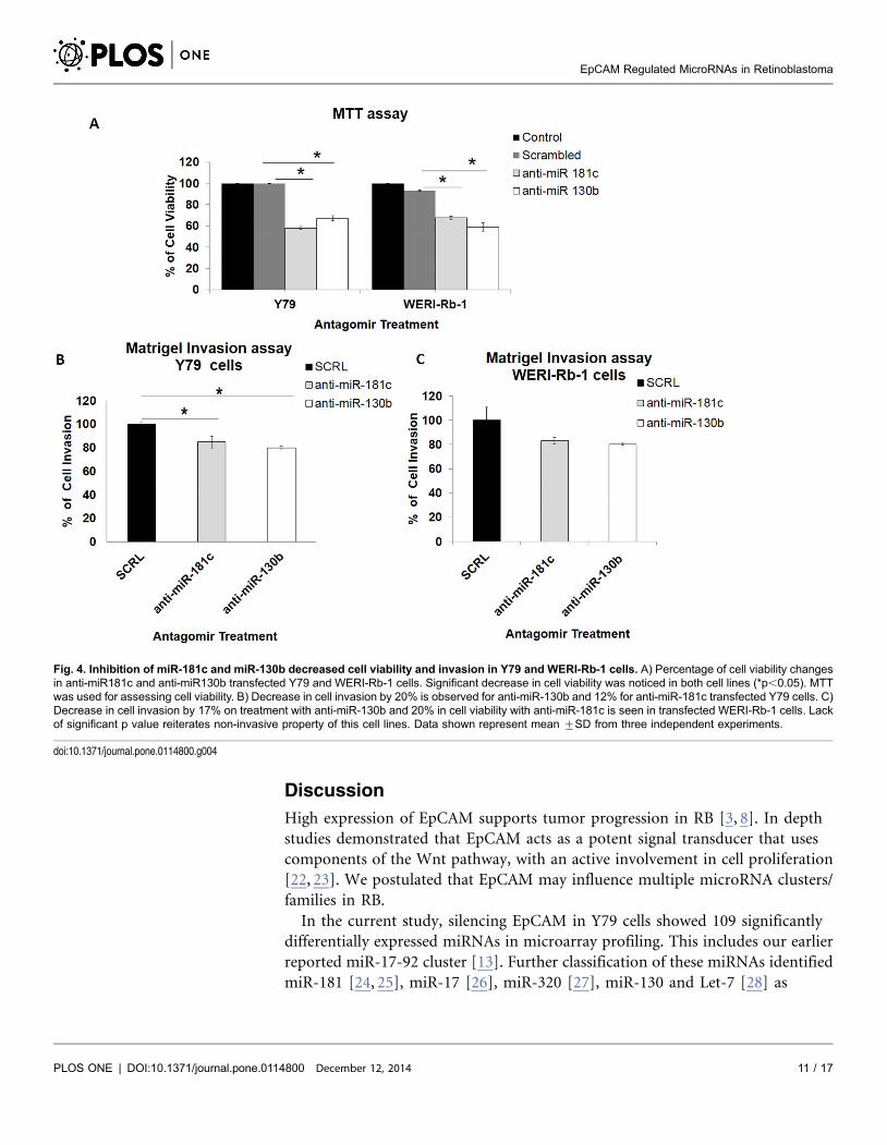
a) Antagomirs of miR-130b and miR-181c decreased cell viability in Y79 and
WERI-Rb-1
Antagomirs were used for the inhibition of miR-130b and miR-181c. Cell
proliferation decreased significantly; 41.9% (Y79 cells) for miR-181c and 32.8%
(Y79 cells) for miR-130b antagomir treated cells (p,0.05*). In treated WERI-Rb-
1, decrease in cell proliferation by 32.5% for miR-181c and 41.1% for miR-130b
(p,0.05*) were seen (Fig. 4A).
b) Cell invasion reduced in antagomir (miR-181c and miR-130b) transfected Y79
and WERI-Rb-1 cells
Cell invasion assay in Y79 cells show that on inhibiting miR-130b and miR-181c,
migration decreases by 20% and 15%, respectively (p,0.05*) (Fig. 4B, S3 File).
Percentage decrease was 17% for miR-181c and 20% for miR-130b in WERI-Rb-1
cells (p,0.05*) (Fig. 4C, S3 File).
Fig. 2. Quantitative Real Time PCR validation confirmed expression of miR-130b and miR-181c in RB tumors and microarray results in EpCAMsilenced Y79 cells. A) miR-130b and miR-181c were quantified in Y79 and WERI-Rb-1 after EpCAM siRNA. Values are represented as mean ¡SD in threeindependent experiments (*p,0.05). B) miR-130a, 130b expression ranged from 1–35 folds in the RB tumors. C) miR-181b, 181c, 181d, 130a, and 130bwere quantified in twenty RB tumors. MiR-181b, miR-181c and miR-181d expression ranged from 1–7 fold.
doi:10.1371/journal.pone.0114800.g002
EpCAM Regulated MicroRNAs in Retinoblastoma
PLOS ONE | DOI:10.1371/journal.pone.0114800 December 12, 2014 9 / 17

c) Increase in caspase-3 level in antagomir (miR-181c, miR-130b) treated Y79 and
WERI-Rb-1 cells suggest activation of cell death pathways
Apoptosis was measured by investigating level of caspase-3 protein. Increase in
fluorescence units (au) of caspase-3 in miR-181c, or miR-130b antagomir treated
Y79 and WERI-Rb-1 compared to mock miRNA treated cells suggested the
involvement of apoptotic cell death pathways (Fig. 5A, B). U937 cells treated with
camptothecin were used as positive control cells (data not shown).
Correlation of EpCAM expression and miR-130b, miR-181c in
primary RB tumors
To investigate whether a correlation in expression indeed exists between the miR
studied and EpCAM in RB, we performed correlation analysis. However, there
was no positive correlation observed between EpCAM and miR-130b, miR-181c
members.
In silico chromosomal mapping for differential microRNA of
EpCAM
Significant microRNAs mapping to different chromosomal regions, show that
among the miRNA that were down regulated distribution was limited to; 20% on
ChrX (miR-362, miR-532, miR-500*, miR-500, miR-501*, miR-532* & miR-98),
12.5% on Chr9, 10% on Chr13, and 7.5% on Chr1 and 7 (S4 File). Up regulated
miRNA had similar localised distribution; 9.3% on Chr19 (miR-150*, miR-125a*,
miR-520b, miR-371), 9.3% on Chr14 (miR-127*, miR-382, miR-485, miR-300,
miR-494, miR-134), 8% on Chr1, 6.6% on Chr16 as well as 6, 5.3% ChrX and
Chr4 (S5 File).
Fig. 3. Antagomir treatment in Y79 and WERI-Rb-1 cell line lead to suppression of miR-130b and miR-181c. Decrease in miR-130b and miR-181c levels occur in antagomir treated Y79 and WERI-Rb-1 cell lines(*p,0.05). Quantification was done by qRT-PCR.
doi:10.1371/journal.pone.0114800.g003
EpCAM Regulated MicroRNAs in Retinoblastoma
PLOS ONE | DOI:10.1371/journal.pone.0114800 December 12, 2014 10 / 17
Discussion
High expression of EpCAM supports tumor progression in RB [3, 8]. In depth
studies demonstrated that EpCAM acts as a potent signal transducer that uses
components of the Wnt pathway, with an active involvement in cell proliferation
[22, 23]. We postulated that EpCAM may influence multiple microRNA clusters/
families in RB.
In the current study, silencing EpCAM in Y79 cells showed 109 significantly
differentially expressed miRNAs in microarray profiling. This includes our earlier
reported miR-17-92 cluster [13]. Further classification of these miRNAs identified
miR-181 [24, 25], miR-17 [26], miR-320 [27], miR-130 and Let-7 [28] as
Fig. 4. Inhibition of miR-181c and miR-130b decreased cell viability and invasion in Y79 and WERI-Rb-1 cells. A) Percentage of cell viability changesin anti-miR181c and anti-miR130b transfected Y79 and WERI-Rb-1 cells. Significant decrease in cell viability was noticed in both cell lines (*p,0.05). MTTwas used for assessing cell viability. B) Decrease in cell invasion by 20% is observed for anti-miR-130b and 12% for anti-miR-181c transfected Y79 cells. C)Decrease in cell invasion by 17% on treatment with anti-miR-130b and 20% in cell viability with anti-miR-181c is seen in transfected WERI-Rb-1 cells. Lackof significant p value reiterates non-invasive property of this cell lines. Data shown represent mean ¡SD from three independent experiments.
doi:10.1371/journal.pone.0114800.g004
EpCAM Regulated MicroRNAs in Retinoblastoma
PLOS ONE | DOI:10.1371/journal.pone.0114800 December 12, 2014 11 / 17

significantly down regulated families. miR-15, miR-23, and miR-27 though not
reported in RB have some of their members associated in other cancers [29].
We selected two microRNA families, miR-181(miR-181b, miR-181c and miR-
181d) and miR-130 (miR-130a and miR-130b) families based on their previous
association with EpCAM and literature reports of cancer to find out their role in
RB tumor cell proliferation. Previous studies on miR-181 family in hepatocellular
carcinoma showed a regulatory link between miR-181 family and EpCAM positive
cancer cells [24, 25]. The oncogenic potential and over expression of miR-130b
was reported in multiple cancers; colorectal [30], gastric [31, 32], and renal
carcinoma [33]. High expression and the oncogenic role of miR-130a is also
observed in colorectal [34] and ovarian cancers [35]. In a cohort of twenty
tumors, we consistently observed high expression of miR-181 family members and
miR-130b family. Significantly expressed miR-181c and miR-130b (p,0.05) were
taken for antagomir studies to investigate their functional role associated with RB.
In vitro functional studies; cell viability, apoptosis and cell invasion study were
performed using antagomirs of miR-130b and miR-181c in Y79 and WERI-Rb-1
cells. Cell viability assay shows that viability was decreased significantly in both
Y79 and WERI-Rb-1. The decrease of cell viability for anti-miR-130b is less in Y79
compared to anti-miR-181c in Y79 cells. In contrast decrease in cell viability is
more for anti-miR-130b compared to anti-miR-181c treatment in WERI-Rb-1
cells. To support this, we analysed caspase-3 cascade in Y79 and WERI-Rb-1 cells.
Increase in fluorescence of caspase-3 in both miR-181c, and miR-130b antagomir
treated Y79 and WERI-Rb-1 cells confirmed the role of these miRNAs in cell
apoptosis. Subsequently, the inhibitory effect of these antagomirs on cell invasion
was studied using Matrigel chambers. We observed a significant decrease in cell
Fig. 5. Increase in caspase-3 level is observed in antagomir treated Y79 and WERI-Rb-1 cells. Increase in caspase-3 level occurs in A) Y79 and B)WER-Rb-1 transfected with anti-miR-181c and anti-miR-130b. Caspase-3 was measured by fluorometric assay. Control cells were untreated. Valuesrepresented in the form of mean ¡SD are from three independent experiments.
doi:10.1371/journal.pone.0114800.g005
EpCAM Regulated MicroRNAs in Retinoblastoma
PLOS ONE | DOI:10.1371/journal.pone.0114800 December 12, 2014 12 / 17

invasion in antagomir treated Y79 cells but not noticeably in WERI-Rb-1 cells. It
may be noted that WERI-Rb-1 cells are known to be less invasive [36].
Gene ontologies were predicted for miR-181c and miR-130b targeted genes. We
found that many genes were implicated in Wnt signalling and other important
pathways which play a major role in tumorigenesis. We sought to investigate with
bio-informatic tools whether differentially expressed miRNAs of EpCAM have any
association with chromosomal aberrations. In silico chromosomal mapping was
performed for differentially regulated miRNAs in EpCAM silenced Y79 data. We
addressed the following queries based on the chromosomal locations of EpCAM
regulated miRNAs; 1) The relationship between site fragility and miRNA density/
miRNA distribution on the chromosomes, 2) The locus of EpCAM gene versus
the loci of miRNAs. It was observed that many miRNA were associated with
ChrX, Chr9 and Chr13. Frequent chromosomal aberrations in RB were reported
for ChrX and Chr13 [37, 38], miR-181c which was up regulated in RB tumors is
associated with 19p13 chromosomal gain region of RB [3, 39]. Among other
significantly changing families, miR-101 and miR-30e are associated with Chr1p
gain region [37]. Several of these play important functions in cancer [40] and
immune disorders [41]. The complete set of miR-362, miR-532, miR-500*, miR-
500, miR-501*, miR-532* and miR-98 located on ChrX had been reported with
chromosomal gain region in B-cell lymphoma [42]. Unusually, miRNA which in
our experimental data show up regulation on silencing EpCAM, are theoretically
expected to be down regulated in tumors, since they are tumor suppressors. All of
these are located in chromosomal gain regions in our bioinformatics analysis. This
suggests that EpCAM mediates the control of these miRNA through multiple
target genes and other protein interactions.
In conclusion, EpCAM a potential oncogene is a master regulator of several
miRNAs and genes which are necessary for RB tumor progression. Existing
literature has implicated many of these miRNA regulated by EpCAM in various
types of cancers; it is likely that these miRNAs have a strong role in common
cancer pathways. The miRNAs regulated by EpCAM control oncogenic, tumor
suppressive and also metabolic functions. MiR-130b and miR-181c that we
studied here affected RB cell proliferation, invasion and apoptosis. MicroRNAs
can regulate multiple pathways in cancer through a complex and intricate network
of gene interactions. It has also been suggested that they can be good therapeutic
targets [43]. However, the large number of families affected as evidenced in this
study and their very interactive nature makes them difficult candidates for
therapy. It may be more worthwhile to target a potent cancer specific gene like
EpCAM that controls several miRNA for RB tumor progression.
Supporting Information
S1 Table. Clinical profile of RB tumors with fold change values of EpCAM,
miR-181c and miR-130b. Table showing clinico-pathological information of
EpCAM Regulated MicroRNAs in Retinoblastoma
PLOS ONE | DOI:10.1371/journal.pone.0114800 December 12, 2014 13 / 17

retinoblastoma tumors and fold change values obtained by qRT-PCR for EpCAM,
miR-130b and miR-181c.
doi:10.1371/journal.pone.0114800.s001 (XLSX)
S1 File. Microarray identified post EpCAM silenced miRNAs and Gene Targets.
Differential miRNAs with significant p values(,0.05) are given. Gene targets,
Gene ontologies and differential miRNA classification are given in the table.
doi:10.1371/journal.pone.0114800.s002 (XLS)
S2 File. Effect of EpCAM gene knockdown on miRNA expression profile in Y79
cells. MicroRNA expression profile in Y79 cells determined by microarray.
Silencing of EpCAM lead to differentially expressed miRNAs. Heat map shows
hierarchical arrangement based on fold change in Y79/EpCAM siRNA and Y79/
Control. Green denotes low expression level and red denotes high expression level.
doi:10.1371/journal.pone.0114800.s003 (TIF)
S3 File. Representative images of invasion assay. Cells invading into matrigel
were fixed, stained with Crystal Violet and photographed in 106 magnification
field. Invaded cells are indicated by black arrows in Y79 and WERI-Rb-1 cell
controls. Control, scrambled and treated chambers of Y79 and WERI-Rb-1 are
shown.
doi:10.1371/journal.pone.0114800.s004 (TIF)
S4 File. In silico representation of EpCAM downregulated miRNA on
chromosomal regions. Chromosomal locations of significant down regulated
miRNAs upon EpCAM silencing in Y79 cells. EpCAM is mapped to p-arm of
Chromosome-2 (blue dot). miRNAs are labelled as lines on the 24 chromosomes.
Polycistronic microRNAs-miR-17, miR-18a, miR-20a, miR-19b located on
13q31.3, miR-10, miR-30e located on chromosome-1 are associated with RB
chromosomal gain regions. miRNAs (non-polycistronic), miR-362, miR-532,
miR-500*, miR-500, miR-501*, miR-532* & miR-98 were located at
Chromosomal-Xp11.
doi:10.1371/journal.pone.0114800.s005 (TIF)
S5 File. In silico representation of significantly up regulated miRNAs on
EpCAM silencing in chromosomal regions. Details of chromosomal locations of
significant miRNAs up regulated upon EpCAM silencing in Y79 cells. EpCAM is
mapped to p-arm of Chromosome-2 (blue dot). miRNAs are labelled as lines on
the 24 chromosomes. miR-127-3p, miR-382, miR-485, miR-300, miR-494, miR-
134 map to chromosomal-14q32 region and miR-150*, miR-125a-3p, miR-520b,
miR-371 map to chromosome-19q13.4 regions.
doi:10.1371/journal.pone.0114800.s006 (TIF)
Author Contributions
Conceived and designed the experiments: SK. Performed the experiments: MB.
Analyzed the data: MB SK NC VK. Contributed reagents/materials/analysis tools:
SK NC VK SG PR JB. Wrote the paper: MB SK NC VK.
EpCAM Regulated MicroRNAs in Retinoblastoma
PLOS ONE | DOI:10.1371/journal.pone.0114800 December 12, 2014 14 / 17

References
1. Zhang J, Benavente CA, McEvoy J, Flores-Otero J, Ding L, et al. (2012) A novel retinoblastomatherapy from genomic and epigenetic analyses. Nature 481: 329–334.
2. Madhavan J, Coral K, Mallikarjuna K, Corson TW, Amit N, et al. (2007) High expression of KIF14 inretinoblastoma: association with older age at diagnosis. Invest Ophthalmol Vis Sci 48: 4901–4906.
3. Krishnakumar S, Mohan A, Mallikarjuna K, Venkatesan N, Biswas J, et al. (2004) EpCAMexpression in retinoblastoma: a novel molecular target for therapy. Invest Ophthalmol Vis Sci 45: 4247–4250.
4. Matsuda T, Takeuchi H, Matsuda S, Hiraiwa K, Miyasho T, et al. (2014) EpCAM, a PotentialTherapeutic Target for Esophageal Squamous Cell Carcinoma. Ann Surg Oncol 21 Suppl 3: 356–364.
5. Bellone S, Siegel ER, Cocco E, Cargnelutti M, Silasi DA, et al. (2009) Overexpression of epithelial celladhesion molecule in primary, metastatic, and recurrent/chemotherapy-resistant epithelial ovariancancer: implications for epithelial cell adhesion molecule-specific immunotherapy. Int J Gynecol Cancer19: 860–866.
6. Brunner A, Schaefer G, Veits L, Brunner B, Prelog M, et al. (2008) EpCAM overexpression isassociated with high-grade urothelial carcinoma in the renal pelvis. Anticancer Res 28: 125–128.
7. Sithambaram D, Palanivelu S, Subramanian K, Sahoo S, Verma RS (2011) Specific targeting of Ep-CAM in various carcinomas by novel monoclonal antibodies. Hybridoma (Larchmt) 30: 511–518.
8. Mitra M, Kandalam M, Verma RS, UmaMaheswari K, Krishnakumar S (2010) Genome-wide changesaccompanying the knockdown of Ep-CAM in retinoblastoma. Mol Vis 16: 828–842.
9. Schaefer A, O’Carroll D, Tan CL, Hillman D, Sugimori M, et al. (2007) Cerebellar neurodegenerationin the absence of microRNAs. J Exp Med 204: 1553–1558.
10. Thum T, Gross C, Fiedler J, Fischer T, Kissler S, et al. (2008) MicroRNA-21 contributes to myocardialdisease by stimulating MAP kinase signalling in fibroblasts. Nature 456: 980–984.
11. Hassan T, McKiernan PJ, McElvaney NG, Cryan SA, Greene CM (2012) Therapeutic modulation ofmiRNA for the treatment of proinflammatory lung diseases. Expert Rev Anti Infect Ther 10: 359–368.
12. Wiemer EA (2007) The role of microRNAs in cancer: no small matter. Eur J Cancer 43: 1529–1544.
13. Kandalam MM, Beta M, Maheswari UK, Swaminathan S, Krishnakumar S (2012) OncogenicmicroRNA 17–92 cluster is regulated by epithelial cell adhesion molecule and could be a potentialtherapeutic target in retinoblastoma. Mol Vis 18: 2279–2287.
14. Hayashita Y, Osada H, Tatematsu Y, Yamada H, Yanagisawa K, et al. (2005) A polycistronicmicroRNA cluster, miR-17-92, is overexpressed in human lung cancers and enhances cell proliferation.Cancer Res 65: 9628–9632.
15. Mogilyansky E, Rigoutsos I (2013) The miR-17/92 cluster: a comprehensive update on its genomics,genetics, functions and increasingly important and numerous roles in health and disease. Cell DeathDiffer 20: 1603–1614.
16. Dalgard CL, Gonzalez M, deNiro JE, O’Brien JM (2009) Differential microRNA-34a expression andtumor suppressor function in retinoblastoma cells. Invest Ophthalmol Vis Sci 50: 4542–4551.
17. Sreenivasan S, Thirumalai K, Danda R, Krishnakumar S (2012) Effect of curcumin on miRNAexpression in human Y79 retinoblastoma cells. Curr Eye Res 37: 421–428.
18. Martin A, Jones A, Bryar PJ, Mets M, Weinstein J, et al. (2013) MicroRNAs-449a and -449b exhibittumor suppressive effects in retinoblastoma. Biochem Biophys Res Commun 440: 599–603.
19. Beta M, Venkatesan N, Vasudevan M, Vetrivel U, Khetan V, et al. (2013) Identification and InsilicoAnalysis of Retinoblastoma Serum microRNA Profile and Gene Targets Towards Prediction of NovelSerum Biomarkers. Bioinform Biol Insights 7: 21–34.
20. Delfino KR, Rodriguez-Zas SL (2013) Transcription factor-microRNA-target gene networks associatedwith ovarian cancer survival and recurrence. PLoS One 8: e58608.
21. Lagatie O, Van Loy T, Tritsmans L, Stuyver LJ (2014) Circulating human microRNAs are not linked toJC polyomavirus serology or urinary viral load in healthy subjects. Virol J 11: 41.
EpCAM Regulated MicroRNAs in Retinoblastoma
PLOS ONE | DOI:10.1371/journal.pone.0114800 December 12, 2014 15 / 17

22. Yamashita T, Budhu A, Forgues M, Wang XW (2007) Activation of hepatic stem cell marker EpCAM byWnt-beta-catenin signaling in hepatocellular carcinoma. Cancer Res 67: 10831–10839.
23. Maetzel D, Denzel S, Mack B, Canis M, Went P, et al. (2009) Nuclear signalling by tumour-associatedantigen EpCAM. Nat Cell Biol 11: 162–171.
24. Ji J, Yamashita T, Budhu A, Forgues M, Jia HL, et al. (2009) Identification of microRNA-181 bygenome-wide screening as a critical player in EpCAM-positive hepatic cancer stem cells. Hepatology 50:472–480.
25. Ji J, Yamashita T, Wang XW (2011) Wnt/beta-catenin signaling activates microRNA-181 expression inhepatocellular carcinoma. Cell Biosci 1: 4.
26. Conkrite K, Sundby M, Mukai S, Thomson JM, Mu D, et al. (2011) miR-17,92 cooperates with RBpathway mutations to promote retinoblastoma. Genes Dev 25: 1734–1745.
27. Zhao JJ, Yang J, Lin J, Yao N, Zhu Y, et al. (2009) Identification of miRNAs associated withtumorigenesis of retinoblastoma by miRNA microarray analysis. Childs Nerv Syst 25: 13–20.
28. Mu G, Liu H, Zhou F, Xu X, Jiang H, et al. (2010) Correlation of overexpression of HMGA1 and HMGA2with poor tumor differentiation, invasion, and proliferation associated with let-7 down-regulation inretinoblastomas. Hum Pathol 41: 493–502.
29. Theriault BL, Dimaras H, Gallie BL, Corson TW (2014) The genomic landscape of retinoblastoma: areview. Clin Experiment Ophthalmol 42: 33–52.
30. Colangelo T, Fucci A, Votino C, Sabatino L, Pancione M, et al. (2013) MicroRNA-130b PromotesTumor Development and Is Associated with Poor Prognosis in Colorectal Cancer. Neoplasia 15: 1218–1231.
31. Lai KW, Koh KX, Loh M, Tada K, Subramaniam MM, et al. (2010) MicroRNA-130b regulates thetumour suppressor RUNX3 in gastric cancer. Eur J Cancer 46: 1456–1463.
32. Kim BH, Hong SW, Kim A, Choi SH, Yoon SO (2013) Prognostic implications for high expression ofoncogenic microRNAs in advanced gastric carcinoma. J Surg Oncol 107: 505–510.
33. Wu X, Weng L, Li X, Guo C, Pal SK, et al. (2012) Identification of a 4-microRNA signature for clear cellrenal cell carcinoma metastasis and prognosis. PLoS One 7: e35661.
34. Liu L, Nie J, Chen L, Dong G, Du X, et al. (2013) The oncogenic role of microRNA-130a/301a/454 inhuman colorectal cancer via targeting Smad4 expression. PLoS One 8: e55532.
35. Yang L, Li N, Wang H, Jia X, Wang X, et al. (2012) Altered microRNA expression in cisplatin-resistantovarian cancer cells and upregulation of miR-130a associated with MDR1/P-glycoprotein-mediated drugresistance. Oncol Rep 28: 592–600.
36. Chevez-Barrios P, Hurwitz MY, Louie K, Marcus KT, Holcombe VN, et al. (2000) Metastatic andnonmetastatic models of retinoblastoma. Am J Pathol 157: 1405–1412.
37. Van der Wal JE, Hermsen MA, Gille HJ, Schouten-Van Meeteren NY, Moll AC, et al. (2003)Comparative genomic hybridisation divides retinoblastomas into a high and a low level chromosomalinstability group. J Clin Pathol 56: 26–30.
38. Serena-Lungarotti M, Calabro A, Mariotti G, Mastroiacovo PP, Provenzano S, et al. (1979) Interstitialdeletion 13q syndromes: a report on two unrelated patients. Hum Genet 52: 269–274.
39. Rushlow DE, Mol BM, Kennett JY, Yee S, Pajovic S, et al. (2013) Characterisation of retinoblastomaswithout RB1 mutations: genomic, gene expression, and clinical studies. Lancet Oncol 14: 327–334.
40. Singhal R, Bard JE, Nowak NJ, Buck MJ, Kandel ES (2013) FOXO1 regulates expression of amicroRNA cluster on X chromosome. Aging (Albany NY) 5: 347–356.
41. Pinheiro I, Dejager L, Libert C (2011) X-chromosome-located microRNAs in immunity: might theyexplain male/female differences? The X chromosome-genomic context may affect X-located miRNAsand downstream signaling, thereby contributing to the enhanced immune response of females.Bioessays 33: 791–802.
42. Li C, Kim SW, Rai D, Bolla AR, Adhvaryu S, et al. (2009) Copy number abnormalities, MYC activity,and the genetic fingerprint of normal B cells mechanistically define the microRNA profile of diffuse largeB-cell lymphoma. Blood 113: 6681–6690.
EpCAM Regulated MicroRNAs in Retinoblastoma
PLOS ONE | DOI:10.1371/journal.pone.0114800 December 12, 2014 16 / 17

43. Bader AG, Brown D, Stoudemire J, Lammers P (2011) Developing therapeutic microRNAs for cancer.Gene Ther 18: 1121–1126.
EpCAM Regulated MicroRNAs in Retinoblastoma
PLOS ONE | DOI:10.1371/journal.pone.0114800 December 12, 2014 17 / 17