Body composition and sarcopenia: The next-generation of ...
80
HAL Id: hal-02325334 https://hal.archives-ouvertes.fr/hal-02325334 Submitted on 24 Oct 2019 HAL is a multi-disciplinary open access archive for the deposit and dissemination of sci- entific research documents, whether they are pub- lished or not. The documents may come from teaching and research institutions in France or abroad, or from public or private research centers. L’archive ouverte pluridisciplinaire HAL, est destinée au dépôt et à la diffusion de documents scientifiques de niveau recherche, publiés ou non, émanant des établissements d’enseignement et de recherche français ou étrangers, des laboratoires publics ou privés. Body composition and sarcopenia: The next-generation of personalized oncology and pharmacology? Marc Hilmi, Anne Jouinot, Robert Burns, Frédéric Pigneur, Rémi Mounier, Julien Gondin, Cindy Neuzillet, François Goldwasser To cite this version: Marc Hilmi, Anne Jouinot, Robert Burns, Frédéric Pigneur, Rémi Mounier, et al.. Body composition and sarcopenia: The next-generation of personalized oncology and pharmacology?. Pharmacology & Therapeutics Part A Chemotherapy Toxicology and Metabolic Inhibitors, Elsevier, 2019, 196, pp.135- 159. 10.1016/j.pharmthera.2018.12.003. hal-02325334
-
Upload
khangminh22 -
Category
Documents
-
view
1 -
download
0
Transcript of Body composition and sarcopenia: The next-generation of ...
HAL Id: hal-02325334https://hal.archives-ouvertes.fr/hal-02325334
Submitted on 24 Oct 2019
HAL is a multi-disciplinary open accessarchive for the deposit and dissemination of sci-entific research documents, whether they are pub-lished or not. The documents may come fromteaching and research institutions in France orabroad, or from public or private research centers.
L’archive ouverte pluridisciplinaire HAL, estdestinée au dépôt et à la diffusion de documentsscientifiques de niveau recherche, publiés ou non,émanant des établissements d’enseignement et derecherche français ou étrangers, des laboratoirespublics ou privés.
Body composition and sarcopenia: The next-generationof personalized oncology and pharmacology?
Marc Hilmi, Anne Jouinot, Robert Burns, Frédéric Pigneur, Rémi Mounier,Julien Gondin, Cindy Neuzillet, François Goldwasser
To cite this version:Marc Hilmi, Anne Jouinot, Robert Burns, Frédéric Pigneur, Rémi Mounier, et al.. Body compositionand sarcopenia: The next-generation of personalized oncology and pharmacology?. Pharmacology &Therapeutics Part A Chemotherapy Toxicology and Metabolic Inhibitors, Elsevier, 2019, 196, pp.135-159. �10.1016/j.pharmthera.2018.12.003�. �hal-02325334�
HILMI M. et al., Sarcopenia and personalized oncology
1
Body composition and sarcopenia:
the next-generation of personalized oncology and pharmacology?
Marc Hilmi1, Anne Jouinot1, Robert Burns2, Frédéric Pigneur2, Rémi Mounier4, Julien Gondin4, Cindy
Neuzillet3*, François Goldwasser1*
Affiliations :
1. Department of Medical Oncology, CAncer Research for PErsonalized Medicine (CARPEM),
Paris Centre Teaching Hospitals, Paris Descartes University, USPC, Paris, France
2. Department of Radiology, Henri Mondor University Hospital, Créteil, France
3. Department of Medical Oncology, Curie Institute, Versailles Saint-Quentin University, Saint-
Cloud, France, and GERCOR group, Paris, France
4. Institut NeuroMyoGène (INMG) CNRS 5310 – INSERM U1217 – UCBL, Lyon, France
* Contributed equally (co-last authors)
Corresponding author:
Dr. Cindy Neuzillet, M.D., Ph.Doc., Department of Medical Oncology, Curie Institute, Versailles Saint-Quentin University (UVSQ), 35 rue Dailly, 92210 Saint-Cloud, France. E-mail: [email protected] / [email protected]. Tel: +33 (0)6 82 55 04 92.
Running Title: Sarcopenia and personalized oncology.
HILMI M. et al., Sarcopenia and personalized oncology
2
ABSTRACT
Body composition has gained increasing attention in oncology in recent years due to fact that
sarcopenia has been revealed to be a strong prognostic indicator for survival across multiple stages
and cancer types and a predictive factor for toxicity and surgery complications. Accumulating
evidence over the last decade has unraveled the “pharmacology” of sarcopenia. Lean body mass
may be more relevant to define drug dosing than the “classical” body surface area or flat-fixed
dosing in patients with cancer.
Since sarcopenia has a major impact on patient survival and quality of life, therapeutic interventions
aiming at reducing muscle loss have been developed and are being prospectively evaluated in
randomized controlled trials. It is now acknowledged that this supportive care dimension of
oncological management is essential to ensure the success of any anticancer treatment.
The field of sarcopenia and body composition in cancer is developing quickly, with (i) the newly
identified concept of sarcopenic obesity defined as a specific pathophysiological entity, (ii) unsolved
issues regarding the best evaluation modalities and cut-off for definition of sarcopenia on imaging,
(iii) first results from clinical trials evaluating physical activity, and (iv) emerging body-composition-
tailored drug administration schemes.
In this context, we propose a comprehensive review providing a panoramic approach of the clinical,
pharmacological and therapeutic implications of sarcopenia and body composition in oncology.
Keywords: cachexia, chemotherapy, sarcopenic obesity, targeted therapy, toxicity.
HILMI M. et al., Sarcopenia and personalized oncology
3
ABBREVIATIONS
5-FU: 5-fluorouracil
ACF: 5-FU, cisplatin, and adriamycin
APA: adapted physical activity
AUC: area under the curve
BIA: bioelectrical impedance analysis
BMI: body mass index
BSA: body surface area
CMF: cyclophosphamide, methotrexate, 5FU
Cr: creatinine
CRP: C reactive protein
CT: computed tomography
CYP: cytochrome
CysC: cystatin C
DCF: docetaxel, cisplatin, and 5FU
DXA: dual-energy X-ray absorptiometry
DLT: dose-limiting toxicity
FM: fat mass
FOLFOX: 5FU, leucovorin and oxaliplatin
GFR: glomerular filtration rate
HRQoL: health-related quality of life
HU: Hounsfield unit
IL-1: interleukin-1
IL-6: interleukin-6
L3: third lumbar vertebra
LBM: lean body mass
mAb: monoclonal antibody
HILMI M. et al., Sarcopenia and personalized oncology
4
MKI: multikinase inhibitor
MRI: magnetic resonance imaging
NLR: neutrophil-to-lymphocyte ratio
NSCLC: non-small cell lung cancer
OS: overall survival
OR: odds ratio
PK: pharmacokinetics
PS: performance status
SCLC: small cell lung cancer
SMA: skeletal muscle area
SMI: skeletal muscle index
TGF-β: transforming growth factor beta
TMA: total muscle area
TNF: tumor necrosis factor
HILMI M. et al., Sarcopenia and personalized oncology
5
TABLE OF CONTENTS
1. Introduction
2. Sarcopenia definition, pathophysiology, and modalities of body composition analysis
2.1. Definition and pathophysiology
2.2. Modalities of body composition evaluation
2.3. Unsolved questions including cut-off issues
3. Sarcopenia prevalence and prognostic value
4. Sarcopenia and toxicity of anticancer treatments
4.1. Chemotherapy
4.2. Targeted therapy
4.3. Immunotherapy
4.4 Summary and discussion
5. The pharmacology of sarcopenia
5.1. Pharmacokinetics
5.2. Pharmacodynamics
6. Sarcopenic obesity: a new entity
7. Therapeutic implications
7.1. Muscle mass-guided dose adjustments in daily practice and clinical trials
7.2. Physical activity and nutritional interventions
8. Conclusion
HILMI M. et al., Sarcopenia and personalized oncology
6
TEXT
1. Introduction
Sarcopenia (i.e. loss of muscle mass and function) is a hallmark of cachexia, a multi-organ
syndrome characterized by negative protein and energy balance, weight loss (including muscle with
or without fat), anorexia, and decreased physical function (Fearon et al., 2011). Cachexia has been
defined in the setting of underlying inflammatory disease as: (i) unintended weight loss of > 5% over
the past 6 months; or (ii) body mass index (BMI) < 20 kg/m2 and any degree of weight loss > 2%; or
(iii) muscle atrophy (i.e. reduced muscle mass) as determinable by various modalities of body
composition analysis and any degree of weight loss > 2% (Fearon et al., 2011). Not all cachectic
patients have sarcopenia, and not all sarcopenic patients meet consensus criteria for cachexia, while
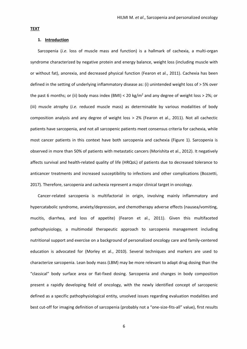
most cancer patients in this context have both sarcopenia and cachexia (Figure 1). Sarcopenia is
observed in more than 50% of patients with metastatic cancers (Morishita et al., 2012). It negatively
affects survival and health-related quality of life (HRQoL) of patients due to decreased tolerance to
anticancer treatments and increased susceptibility to infections and other complications (Bozzetti,
2017). Therefore, sarcopenia and cachexia represent a major clinical target in oncology.
Cancer-related sarcopenia is multifactorial in origin, involving mainly inflammatory and
hypercatabolic syndrome, anxiety/depression, and chemotherapy adverse effects (nausea/vomiting,
mucitis, diarrhea, and loss of appetite) (Fearon et al., 2011). Given this multifaceted
pathophysiology, a multimodal therapeutic approach to sarcopenia management including
nutritional support and exercise on a background of personalized oncology care and family-centered
education is advocated for (Morley et al., 2010). Several techniques and markers are used to
characterize sarcopenia. Lean body mass (LBM) may be more relevant to adapt drug dosing than the
“classical” body surface area or flat-fixed dosing. Sarcopenia and changes in body composition
present a rapidly developing field of oncology, with the newly identified concept of sarcopenic
defined as a specific pathophysiological entity, unsolved issues regarding evaluation modalities and
best cut-off for imaging definition of sarcopenia (probably not a “one-size-fits-all” value), first results
HILMI M. et al., Sarcopenia and personalized oncology
7
from clinical trials evaluating physical activity, and emerging body-composition-tailored drug
administration schemes.
2. Sarcopenia definition and modalities of body composition analysis
2.1 Definition and pathophysiology
Sarcopenia was first described in 1989 as an age-related loss of muscle mass (Rosenberg,
1989). Later, the definition was expanded to a syndrome combining three criteria: (i) loss of skeletal
muscle mass, (ii) loss of function, and (iii) loss of physical performance (Rosenberg, 1997; Roubenoff,
2000). Three consensus papers established a definition of sarcopenia based on these three
parameters (Cruz-Jentoft et al., 2010; Fielding et al., 2011; Muscaritoli et al., 2010). The European
Working Group on Sarcopenia in Older People (EWGSOP) proposed three stages of sarcopenia: pre-
sarcopenia (presence of one criterion), sarcopenia (two criteria), and severe sarcopenia (three
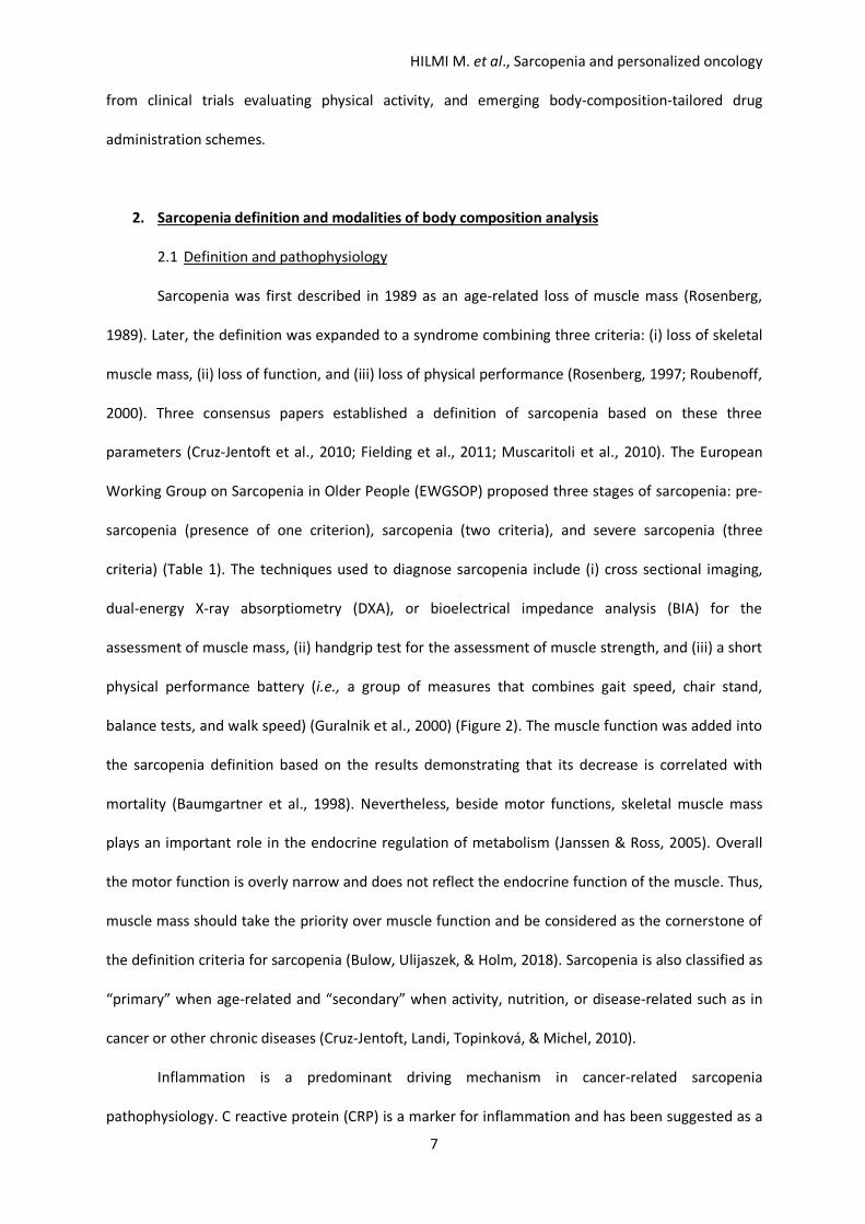
criteria) (Table 1). The techniques used to diagnose sarcopenia include (i) cross sectional imaging,
dual-energy X-ray absorptiometry (DXA), or bioelectrical impedance analysis (BIA) for the
assessment of muscle mass, (ii) handgrip test for the assessment of muscle strength, and (iii) a short
physical performance battery (i.e., a group of measures that combines gait speed, chair stand,
balance tests, and walk speed) (Guralnik et al., 2000) (Figure 2). The muscle function was added into
the sarcopenia definition based on the results demonstrating that its decrease is correlated with
mortality (Baumgartner et al., 1998). Nevertheless, beside motor functions, skeletal muscle mass
plays an important role in the endocrine regulation of metabolism (Janssen & Ross, 2005). Overall
the motor function is overly narrow and does not reflect the endocrine function of the muscle. Thus,
muscle mass should take the priority over muscle function and be considered as the cornerstone of
the definition criteria for sarcopenia (Bulow, Ulijaszek, & Holm, 2018). Sarcopenia is also classified as
“primary” when age-related and “secondary” when activity, nutrition, or disease-related such as in
cancer or other chronic diseases (Cruz-Jentoft, Landi, Topinková, & Michel, 2010).
Inflammation is a predominant driving mechanism in cancer-related sarcopenia
pathophysiology. C reactive protein (CRP) is a marker for inflammation and has been suggested as a
HILMI M. et al., Sarcopenia and personalized oncology
8
biological marker for sarcopenia (Bano et al., 2017). In a comparable way, neutrophil-to-lymphocyte
ratios (NLR) are higher in sarcopenic patients than in those without sarcopenia (Öztürk, Kul,
Türkbeyler, Sayıner, & Abiyev, 2018) as well as circulating levels of pro-inflammatory cytokines (e.g.
tumor necrosis factor [TNF], interleukin-1 [IL-1], and interleukin-6 [IL-6]) (Narsale & Carson, 2014;
Schaap, Pluijm, Deeg, & Visser, 2006; Visser et al., 2002). The cytokines of the transforming growth
factor beta (TGF-β) family, i.e., TGF-β, myostatin, GDF11, and activins are major atrophic factors
widely involved in cancer-induced cachexia (Wakefield & Hill, 2013). Their circulating levels are
increased in most cancers, including pancreatic and colorectal cancers (Wildi et al., 2001; Zhao et al.,
2016). These molecules trigger intracellular signals leading to loss of contractile proteins linked to a
decrease in protein synthesis and a significant increase in the degradation of muscle myofibrillar
proteins, inevitably impacting muscle strength production (Cohen, Nathan, & Goldberg, 2015).
Preclinical studies have shown that ZIP14, a metal-ion transporter, and Twist1, a transcription factor,
are overexpressed in muscle progenitor cells, highlighting the roles of zinc homeostasis and
activin/myostatin signaling in cancer-related muscle loss (Parajuli et al., 2018; G. Wang et al., 2018).
In addition, muscle proteolysis induces an important efflux of muscle amino acids, which constitutes
“bricks and fuel” to boost tumor progression (Mayers et al., 2016). However, the mechanisms
underlying cancer-related muscle dysfunction appear to be more complex than just a negative
balance between muscle protein synthesis and degradation. It has recently been shown that
microenvironmental alterations affecting cells located at the periphery of the muscle fiber also play
a key role in cancer-related muscle atrophy. Indeed, muscle fibers are surrounded by muscle stem
cells called "satellite cells". During muscle injury, these cells become activated and proliferate to
produce myoblasts, which differentiate and fuse to form new myofibers and thus restore muscle
tissue integrity. In cancer, inflammation induces muscle damage leading to the activation of satellite
cells that are engaged in a myogenic muscle tissue repair program, but cannot differentiate (He et
al., 2013). Other mechanisms have recently been shown to be involved in cancer-related sarcopenia,
such as epigenetic alterations involving bromodomain protein BRD4 (Segatto et al., 2017) and
calcium homeostasis involving ryanodine receptors (A. Agrawal, Suryakumar, & Rathor, 2018;
HILMI M. et al., Sarcopenia and personalized oncology
9
Waning et al., 2015). Endocrine and metabolic factors also play a pivotal role in the pathogenesis of
sarcopenia. Some publications suggests an association between insulin resistance and mitochondrial
dysfunction leading to the development of skeletal muscle lipid deposition (Corcoran, Lamon-Fava,
& Fielding, 2007) and to reduced muscle oxidative activity (Simoneau & Kelley, 1997) in sarcopenia
and cancer-induced cachexia (Abbatecola et al., 2011; van der Ende et al., 2018). Moreover, insulin
promotes amino acids transport into cells through nitric oxide synthase (Mann, Yudilevich, &
Sobrevia, 2003). Therefore, insulin resistance may lead to poorer protein synthesis due to reduced
internalization of amino acids. These hypotheses may explain why patients with type 2 diabetes
mellitus are more at risk of developing sarcopenia (Leenders et al., 2013; Morley, Malmstrom,
Rodriguez-Mañas, & Sinclair, 2014; Park et al., 2007).
Overall, systemic inflammation, metabolic changes, and secreted “atrophying” cytokines
along with the muscle microenvironmental dysfunction alter cell signaling in muscle fibers and lead
to imbalance between protein synthesis and degradation (Argilés, Campos, Lopez-Pedrosa, Rueda, &
Rodriguez-Mañas, 2016) (Figure 2). These phenomena are very early in some malignancies,
particularly in pancreatic cancer, and may precede cancer diagnosis by several years (Agustsson,
D’souza, Nowak, & Isaksson, 2011; Mayers et al., 2014). Moreover, sarcopenia itself is affected by
chemotherapy, since a variety of common chemotherapy drugs are known to experimentally induce
sarcopenia in rodent cancer models (Hojman et al., 2014; Sakai et al., 2014). Data on humans carry
some caveats because chemotherapy agents are not given to healthy adults and it seems that
muscle wasting is exacerbated by chemotherapy (Awad et al., 2012; Coletti, 2018) through reduced
food intakes (Spotten et al., 2017) and induced NF-kB expression (Damrauer et al., 2018). The
decrease in food intakes in cancer patients is multifactorial and classically thought to result from
changes to appetite, smell and taste and to behavior-regulating regions of brain occurring as a result
of inflammatory mediators (Ezeoke & Morley, 2015).
HILMI M. et al., Sarcopenia and personalized oncology
10
2.2 Modalities of body composition evaluation
In clinical practice, physical performance assessment by short physical performance battery,
usual gait speed, and get up and go test (Cruz-Jentoft, Baeyens, et al., 2010; Owusu, Margevicius,
Schluchter, Koroukian, & Berger, 2017) and muscle strength evaluation using handgrip test (Kilgour
et al., 2013; Veni et al., 2018) are quite standardized. On contrary, there is no consensus on the
optimal modality to assess muscle mass in cancer patients.
Anthropometric measurements include weight variation, BMI, waist and hip circumference,
waist-to-hip ratio, and skinfold. These are non-invasive, easy to perform, and routinely used to
estimate body surface area (BSA; derived from weight and height) for chemotherapy dosing
calculation (Griggs, Mangu, Temin, & Lyman, 2012). However, anthropometry is unreliable in some
cancer patients, particularly in case of short-term changes in body water composition (e.g. in
presence of ascites or lymphedema), or in obese patients (Di Sebastiano & Mourtzakis, 2012).
Bioelectrical impedance analysis (BIA) is an additional widely available, non-invasive
modality that uses reactance and resistance to determine total body water, fat mass (FM), and fat-
free mass. However, BIA is highly dependent on patient hydration state and is biased in case of
pathological increase in body water content such as ascites and lymphoedema. Other limitations of
BIA is the lack of specific predictive equations for cancer patients leading to frequent inaccuracies
(under or overestimations) and the lack of standardization resulting in heterogeneity of sarcopenia
prevalence rates across studies (Gonzalez, Barbosa-Silva, & Heymsfield, 2018).
Dual energy X-ray absorptiometry (DXA) uses a three-compartment model comprising FM,
fat-free mass, and bone mineral content. This method can detect early lean mass variations, is highly
accurate and reproducible (Ellis, 2001; Shiel et al., 2018). However, it cannot discriminate between
different types of fat tissues (visceral, subcutaneous, and intramuscular) as only an overall
assessment of FM at the molecular and not at compartmental tissue level can be provided by DXA
and no differentiation of specific lean tissues like skeletal muscle and the internal organs within the
thorax or abdomen can be made (Guglielmi et al., 2016; Prado & Heymsfield, 2014). Consequently,
muscle mass using DXA is estimated based on the appendicular skeleton.
HILMI M. et al., Sarcopenia and personalized oncology
11
Finally, it has been shown that cross sectional muscle surface at the third lumbar vertebrae
(L3) best reflects total skeletal muscle mass determined by computed tomography (CT) or magnetic
resonance imaging (MRI) (Heymsfield, 2008; Mitsiopoulos et al., 1998; Shen et al., 2004). In practice,
total muscle area (TMA, in cm²) is measured at L3 using a semi-automatic segmentation software on
a dedicated post-treatment station (with most packages being interchangeable) (Bonekamp et al.,
2008). TMA is then normalized to stature (using height2 in m2, similarly to BMI) (Baumgartner et al.,
1998) to obtain the skeletal muscle index (SMI) in cm²/m².
However, uncertainties remain regarding technical aspects of this method. To date, the only
technical guideline available for L3 skeletal muscle surface estimation using CT is the one described
by Mitsiopoulos et al. in 1998 and recommended by Prado et al. in 2008 (Mitsiopoulos et al., Prado
et al.). It recommends measurement of the mean skeletal muscle surface value on two consecutive
slices at the L3 level based on Hounsfield unit (HU) thresholds (–29 to +150), followed by manual
corrections where necessary. Nevertheless, several technical issues that can influence skeletal
muscle surface measurements need clarification such as the effect of intravenous contrast injection
(van Vugt et al., 2018), slice thickness (Fuchs et al., 2018), or the influence of tube potential
(Morsbach et al., 2018).
Regarding MRI, there are no clear technical recommendations on image acquisition
techniques. Most studies used standard T1 and T2-weighted imaging protocols (Heymsfield, 2008;
Mitsiopoulos et al., 1998; Yang et al., 2017) and fat-water separated imaging techniques (Dixon,
1984) with manual segmentation to determine L3 skeletal muscle surface, which is time consuming.
Very few studies have used or investigated the reproducibility of automated methods applied to MRI
(Borga, 2018). This is most likely due to the fact that MRI is not used in a quantitative way because,
unlike HU on CT imaging, the intensity levels on MRI are expressed in arbitrary units and are not
correlated to tissue composition.
A single-muscle approach to muscle mass quantification using cross-sectional analysis
(surface and/or density) of the psoas muscle on abdominal CT and of the pectoralis muscle on
thoracic CT in patients who do not undergo abdominal CT (e.g. osteosarcomas and head and neck
HILMI M. et al., Sarcopenia and personalized oncology
12
cancers) has been proposed (Go et al., 2017; Y. S. Kim, Kim, Kang, Ahn, & Kim, 2017). However, these
single-muscle, simplified approaches, which hypothesized that one muscle may be representative of
total skeletal mass have not been validated by any expert group (Bahat et al., 2016; Baracos, 2017;
Cesari et al., 2012). Automated total muscle segmentation on CT imaging has been suggested as an
alternative, more accurate method to improve body composition quantification availability and
routine application (Baracos, 2017; Popuri, Cobzas, Esfandiari, Baracos, & Jägersand, 2016).
Assessment of muscle quality may provide prognostic information beyond quantity
estimation (Sami Antoun et al., 2013; Looijaard et al., 2016; Martin et al., 2013). Skeletal muscle
lipid content has been associated with muscle quality i.e., high-lipid content being correlated with
poor muscle quality. It can be assessed by measuring muscle density on CT imaging, as the latter
decreases with lipid infiltration (Heymsfield, 2008). Goodpaster et al. showed that muscle density in
elderly patients can account for differences in muscle strength not explained by muscle quantity
(Goodpaster, Kelley, Thaete, He, & Ross, 2000). This however was not confirmed by another study of
healthy adults (Weeks, Gerrits, Horan, & Beck, 2016). Similarly to TMA estimation, there is no
consensus for important technical aspects of CT acquisition such as contrast enhancement, slice
thickness, and tube potential influence, which significantly impact HU density values of the muscle.
Further research is needed for the standardization of image acquisition and protocols of analysis.
In summary, cancer-related sarcopenia and body composition can be estimated by simple
anthropometric values (weight loss and BMI) and L3 SMI using CT or MRI, with CT being the most
commonly used modality in clinical practice and oncology research (Kazemi-Bajestani, Mazurak, &
Baracos, 2016) (Table 3). Indeed, CT is routinely performed for diagnosis, treatment evaluation, and
follow-up in oncological care and TMA assessment. In addition, it can be performed on the same
imaging exam without requiring an additional procedure. However, in the foreseeable future, with
the development of artificial intelligence, the next gold standard for body composition assessment
will most likely rely on 3D body-scanning with automated scoring systems (Cornet et al., 2015; Fang,
Berg, Cheng, & Shen, 2018).
HILMI M. et al., Sarcopenia and personalized oncology
13
2.3 Unsolved questions including cut-off issues
A major unsolved issue in sarcopenia evaluation is the question of the best cut-off value for
sarcopenia definition. Sarcopenia is more often considered in a binary fashion for research purposes
in the literature, patients being classified as either “sarcopenic” or “non-sarcopenic”. Several cut-offs
have been proposed (Cornet et al., 2015; Cruz-Jentoft, Baeyens, et al., 2010) for sarcopenia
diagnosis, yielding broad variations in sarcopenia prevalence (Bijlsma et al., 2013). The first available
cut-off values specific to patients with cancer were published by Prado et al. reporting on a
population of obese Canadian patients with respiratory and gastrointestinal tract cancers (Prado et
al., 2008). They defined cut-off value of 52.4 cm²/m² in men and 38.5 cm²/m² in women associated
with mortality. Mourtzakis et al. showed that CT-based muscle analysis at L3 was strongly related to
appendicular skeletal mass measured with DXA (Mourtzakis et al., 2008). They generated
corresponding CT cut-off values of 55.4 cm²/m² in men and 38.9 cm²/m² in women using established
DXA cut-off values (7.26 kg/m² and 5.45 kg/m², respectively). In 2011, an international panel of
cachexia experts established a new diagnostic criterion for cancer cachexia, using both
anthropometric measures and SMI based on cut-off values of 55 cm²/m² in men and 39 cm²/m² in
women by CT imaging (Fearon et al., 2011). Following, Martin et al. proposed SMI thresholds for
sarcopenia in non-obese Caucasians according to sex and BMI (Martin et al., 2013) based on optimal
stratification of SMI and survival; selected cut-off values were 43 and 53 cm²/m² in men with a BMI <
and > 25 kg/m2, respectively, and 41 cm²/m² in women. Several further studies have demonstrated
predictive value for toxicity (Sjøblom et al., 2015; Srdic et al., 2016; Stene et al., 2015; B. H. L. Tan et
al., 2015), post-surgery complications (Lieffers, Bathe, Fassbender, Winget, & Baracos, 2012; P. Peng
et al., 2012; Reisinger et al., 2015), and survival (M. H. Choi, Oh, Lee, Oh, & Won, 2018; Lee et al.,
2018) using these thresholds (Table 2).
However, normal amounts of muscle and adipose tissues depend on demographic factors
such as age (McCormick & Vasilaki, 2018) and ethnicity (Wells, 2012). It seems that sarcopenia is less
prevalent among African-American (Parsons, Baracos, Dhillon, Hong, & Kurzrock, 2012) and Asian
HILMI M. et al., Sarcopenia and personalized oncology
14
patients (Lau, Lynn, Woo, Kwok, & Melton, 2005) as compared to Caucasians. Therefore, these cut-
offs may not be optimal for all patients and may be refined depending on type and stage of the
cancer, sex, age, and ethnicity.
Nevertheless, rather than classifying a patient dichotomically as sarcopenic vs. non-
sarcopenic based on a cut-off value, imaging biomarkers like SMI may be considered as continuous
variables as suggested by Voron et al. (Voron et al., 2015). In such way, these may be used as the
next reference for chemotherapy dosing calculation. This approach is already being tested with
promising results (Baracos & Arribas, 2018; Iannessi, Beaumont, Hebert, Dittlot, & Falewee, 2018).
3. Sarcopenia prevalence and prognostic value
Sarcopenia has been reported across all cancer types. Prevalence rates according to primary
tumor location and stage are displayed in Table 3. Most studies reported sarcopenia rates above
50% using L3 CT scan, particularly at advanced stages and in pancreatic, lung, bladder, and
hematological malignancies. Noticeably, heterogeneity in prevalence rates is observed within single
cancer types due to different cut-offs used to define sarcopenia and factors (e.g., clinical stage,
ethnicity, age, cancer treatment).
Several studies investigated the prognostic value of sarcopenia in cancer patients. In a meta-
analysis of patients with solid tumors a significant association between sarcopenia and shorter
overall survival (OS; overall HR = 1.51, p<0.001) has been reported, both for advanced and localized
stages (Shachar, Williams, Muss, & Nishijima, 2016). However, a recent study suggested that
survival may not be different for combined muscle and fat loss compared to fat-only loss in patients
with advanced pancreatic cancer (Kays et al., 2018). Besides, a higher risk of post-surgical
complications has been identified in several cancers, especially infectious complications following
surgery for gastrointestinal (Ida et al., 2015; Krell et al., 2013; Lieffers et al., 2012; P. Peng et al.,
2012; Takagi et al., 2017; Zhuang et al., 2016) and lung cancers (Miller et al., 2018; R. Nakamura et
al., 2018). Finally, loss of skeletal muscle mass during chemotherapy has also been described as a
negative prognostic factor (Daly et al., 2018).
HILMI M. et al., Sarcopenia and personalized oncology
15
Overall, several studies evaluating body composition using various modalities have
consistently reported that sarcopenia is a strong prognostic indicator for localized and advanced
cancers.
4. Sarcopenia and toxicity of anticancer treatments
In addition to its prognostic value, sarcopenia is a predictive factor of anticancer drug
toxicity and may be more relevant for drug dose calculation than the “classical” body surface area or
flat-fixed dosing. Anticancer drugs display various pharmacokinetics (PK) properties but share a
narrow therapeutic index. Serious adverse events that may result in toxic death are common in this
setting. Furthermore, specific cancer patient populations may exhibit vulnerabilities due to age and
comorbidities. Identifying factors that can explain individual variations in treatment efficacy and
toxicity is a new challenge of modern oncology. Performance status (PS) has a strong prognostic
value (Atkinson et al., 2015) and is predictive of anticancer treatment-related acute toxicity (Sargent
et al., 2009). However, PS evaluation is only semi-quantitative and is subjected to inter-observer
variability. Thus, additional parameters are needed for the risk assessment of anticancer treatments.
Other studies have investigated whether wide variations in body composition could be associated
with morbidity in cancer patients. In recent years a large interest has grown in muscle mass due to
the development of muscle mass assessments by CT.
We performed systematic search on PubMed using MeSH terms “Neoplasms AND (Sarcopenia
OR Body composition OR Malnutrition OR Cachexia) AND Toxicity”. Forty-two studies that focused
on sarcopenia and toxicity of anticancer treatments were selected (Figure 3).
4.1 Chemotherapy
In total, among 32 studies that investigated the association between sarcopenia and
chemotherapy toxicity, 24 showed a significant association between sarcopenia and chemotherapy
toxicity (Table 4). Although most of the studies were conducted retrospectively and with a small
number of patients (n < 100), this correlation was found consistently across studies regardless of the
HILMI M. et al., Sarcopenia and personalized oncology
16
time of assessment (i.e., early, within the first months of chemotherapy, or later), the tumor type
and stage (i.e., from localized/curative to the metastatic/palliative setting), and the chemotherapy
regimen. For example, Prado et al. (Prado et al., 2009) evaluated prospectively the tolerance of
capecitabine in 55 patients with metastatic breast cancer after the first treatment cycle. Toxicities
occurred in 50% of sarcopenic patients vs. 20% of non-sarcopenic patients (p=0.03). Sarcopenia was
also a predictive factor of toxicity in apparently fit patients (i.e., PS 0-1) such as those treated in
phase I trials (Cousin et al., 2014). Indeed, Cousin et al. studied body composition in 93 phase I
patients and showed that low SMI was the only factor associated with severe toxicity (Cousin et al.,
2014).
Body composition in cancer patients can also be evaluated through renal function
assessments in cancer patients. In daily practice, Glomerular Filtration Rate (GFR) is evaluated using
the Cockcroft–Gault, Chronic Kidney Disease - Epidemiology Collaboration (CKD-EPI), and
Modification of diet in renal disease (MDRD) formulas, which are derived from serum creatinine (Cr)
level; this latter is influenced by the total muscle mass (Perrone, Madias, & Levey, 1992). In
sarcopenic patients, reduced muscle mass may result in apparently low Cr level and overestimation
of renal function by these formulas. Serum cystatin C (CysC) is an alternative marker of GFR,
independent from muscle mass (Baxmann et al., 2008; Rule, Bergstralh, Slezak, Bergert, & Larson,
2006). Thus, CysC appears as a better alternative for assessing renal function in patients with low
muscle mass. The Cr/CysC ratio can be used as a quantitative surrogate marker of muscle mass
(Tetsuka, Morita, Ikeguchi, & Nakano, 2013). In a study including 25 patients with non-small cell lung
cancer (NSCLC) (Suzuki et al., 2015), a significant difference was noted in the Cr/CysC ratios of
patients exhibiting moderate (grade 1-2) vs. severe (grade 3-4) toxicities (mean ratios: 0.84 vs. 0.70,
respectively; p<0.05). Similar results were found in a prospective study of ovarian cancer patients
treated with carboplatin (Bretagne et al., 2017). Moreover, renal failure from the early stages
exacerbates sarcopenia, explaining the frequent overlap between these two conditions (de Souza et
al., 2017).
HILMI M. et al., Sarcopenia and personalized oncology
17
Contrarily, eight studies showed no association between sarcopenia and chemotherapy
toxicity (Blauwhoff-Buskermolen et al., 2016; Jung et al., 2015; Miyata et al., 2017; Palmela et al.,
2017; Parsons, Tsimberidou, et al., 2012; Rollins et al., 2016; Srdic et al., 2016; Stene et al., 2015;
Versteeg et al., 2017). One explanation could be that not all forms of chemotherapy exhibit
sarcopenia-dependent toxicity, even if the studies were adequately powered to detect an
association. Finally, one study used liver intra-arterial chemotherapy (Parsons, Tsimberidou, et al.,
2012), which displays a specific PK profile.
4.2 Targeted therapy
Eight studies investigated the association between sarcopenia and toxicity of targeted
therapies with multikinase inhibitors (MKI), mainly antiangiogenics (Table 5). Overall, patients were
metastatic in all studies and had infrequent tumor types in all but one study (four studies of renal
carcinoma, two studies of hepatocellular carcinoma, and one study of thyroid carcinoma). The
association between sarcopenia and toxicity was reported in seven studies. A meta-analysis pooling
the results of four studies of metastatic renal cell carcinoma, showed that dose-limiting toxicities
(DLT) of antiangiogenic drugs were more frequent in patients with low SMI (p=0.03) (Vrieling et al.,
2016). As observed with chemotherapy, increased toxicity in sarcopenic patients was observed from
the first cycle of treatment (Huillard et al., 2013) and was associated with high serum concentration
of MKI (Massicotte et al., 2013; Mir, Coriat, Blanchet, et al., 2012). In one study of NSCLC patients
treated with afatinib (Arrieta et al., 2015), sarcopenia was not found predictive of toxicity using sex-
specific cut-offs defined by Prado et al. (Prado et al., 2008) and low LBM and malnutrition were
associated with severe gastro-intestinal toxicity and DLT.
4.3 Immunotherapy
In recent years, immune checkpoint blockade inhibitors have opened new opportunities in
cancer therapy. They have markedly improved the clinical outcomes in several cancers, including
metastatic melanoma (Hodi et al., 2010; Robert et al., 2014), renal cancer (Motzer et al., 2018), and
HILMI M. et al., Sarcopenia and personalized oncology
18
lung cancer (Reck et al., 2016). Severe immune-related adverse events occurred in 10%-20% of
patients treated with single-agent immune checkpoint inhibitor (Brahmer et al., 2015; Hodi et al.,
2010; Motzer, Escudier, et al., 2015). Predictive factors of toxicity are still poorly known. Unlike for
chemotherapy, the use of DLT for immune therapies, and more broadly for monoclonal antibodies
(mAb), is inappropriate. Indeed, no maximum tolerated dose was found in phase I trials with
bevacizumab (Gordon et al., 2001), rituximab (Ghielmini, 2005), cetuximab, pembrolizumab (Sachs,
Mayawala, Gadamsetty, Kang, & de Alwis, 2016), and nivolumab (S. Agrawal, Feng, Roy, Kollia, &
Lestini, 2016). Several dosing schemes have been developed based on weight (dose in mg/kg) or flat
doses. The Food and Drug Administration (FDA) concluded that response/toxicity of nivolumab is not
related to dose/exposure and shifted from body weight dosing (i.e. 3 mg/kg) to fixed dosing (240
mg) (www.fda.gov/Drugs/InformationOnDrugs/ApprovedDrugs/ucm520871.htm). However, the rate
of severe toxicities seems to increase with ipilimumab dose. In a retrospective study of exposure-
response analysis in advanced melanoma patients, Feng et al. (Feng et al., 2013) showed that the
occurrence of grade 3-4 immune-related adverse events was related to ipilimumab dose, with 3%,
13%, and 24% of patients developing severe toxicities when treated with 0.3 mg/kg, 3 mg/kg, and 10
mg/kg ipilimumab, respectively. Interestingly, no DLT was observed with tremelimumab despite a
similar toxicity profile to ipilimumab (Camacho et al., 2009). A study of 84 patients with metastatic
melanoma (Daly et al., 2017) treated with ipilimumab showed that sarcopenic patients developed
higher grade adverse events than non-sarcopenic patients (OR=5.34, p=0.033). Similar results were
observed in 68 metastatic melanoma patients treated with nivolumab and pembrolizumab
(Heidelberger et al., 2017). However, it is hard to discern differences between a direct PK link and a
consequence of increased frailty and susceptibility to treatment complications in sarcopenic patients
in this setting.
4.4 Summary and discussion
Overall, this literature review shows that sarcopenic patients exhibit high rates of toxicity
and highlights the role of body composition in the risk assessment of anticancer treatment in this
HILMI M. et al., Sarcopenia and personalized oncology
19
setting. However, several questions remain unsolved due to methodological limitations and
heterogeneity across reviewed studies. Reporting biases favoring positive over negative studies may
have affected the validity of evidence. In addition, most of these studies included limited numbers of
patients, with no preliminary estimation of sample size for statistical analysis. Besides, the
evaluation of toxicities varied from one study to another: some studies considered all DLT, while
other only analyzed some specific toxicities such as hematologic adverse events. The duration of
toxicity evaluation ranged from the first cycle only to the entire length of treatment. Moreover, for a
single tumor location, the thresholds and techniques used to detect sarcopenia differed between
studies. Finally, although several studies using the SMI cut-offs defined for survival by Prado et al.
showed a positive correlation between sarcopenia and toxicity, it is not certain that these are the
best suited for toxicity (Table 2). As it was achieved for prognosis, it would be relevant to conduct a
meta-analysis of studies dedicated to toxicity.
5. The pharmacology of sarcopenia
Several hypotheses can be raised to explain the association between cancer-related
sarcopenia and toxicity. Evidence has accumulated during the last decade unravelling the
“pharmacology” of sarcopenia. We will below develop the main hypotheses through PK and
pharmacodynamics.
6.1 Pharmacokinetics
The PK hypothesis assumes that toxicity may be explained by overexposure of anticancer
drugs in sarcopenic patients. From a pharmacological perspective, sarcopenia increases drug
exposure and several studies showed correlation between LBM, drug exposure, and toxicity (Ali et
al., 2016; Arrieta et al., 2015; Prado et al., 2007, 2014, 2011; Sjøblom et al., 2017, 2015;
Stanisavljevic & Marisavljevic, 2010; Sucak et al., 2012). Regarding mAb (e.g. immune therapies and
some of targeted therapies), no data was published so far on sarcopenia and PK. However, mAb are
a category of drugs known for larger therapeutic index than cytotoxic chemotherapies or MKI. For
HILMI M. et al., Sarcopenia and personalized oncology
20
instance, no DLT were observed for the highest doses of anti-programmed-death-1 (PD-1)/anti-
programmed-death-ligand-1 (PD-L1) antibodies in phase I and II trials (Motzer, Rini, et al., 2015;
Robert et al., 2014).
6.1.1 Absorption changes
It has been suggested in animal models that cancer-induced cachexia is associated with a gut
barrier dysfunction due to microbiota dysbiosis and increased intestinal permeability (Klein,
Petschow, Shaw, & Weaver, 2013), possibly leading to overexposure to drugs that are taken orally.
The same mechanisms may exist in sarcopenia, but no studies have been conducted to confirm this
hypothesis in specific models. This issue would be most pertinent when the drug in question is not
typically well-absorbed orally. Absorption of drugs that are intended to be taken with meals may be
compromised in patients with low oral intakes.
Obese sarcopenic patients have higher fat-to-lean-mass ratios than obese non-sarcopenic
patients. This may explain an overexposure to targeted subcutaneous therapies (i.e. trastuzumab)
through an increase in (highly vascularized) fat tissue and subcutaneous blood flow. Studies
supporting this hypothesis are warranted. Similarly, no studies have been conducted to assess
whether intramuscular fat deposits in obese sarcopenic patients alter absorption of intramuscular
lipophilic injection drugs (i.e., decapeptyl, fulvestrant). In contrast, drugs with intravenous
administration route are not affected by absorption changes.
6.1.2 Distribution changes
Changes in tissue relative proportion are likely to alter PK. Body composition is described as
a two compartments model consisting of FM and LBM. LBM is defined as the sum of cellular mass
and non-fatty intercellular connective tissue including tendons, ligaments, and bone (Ronenn
Roubenoff & Kehayias, 2009). Indeed, muscle mass, being part of LBM, acts as a diffusion
compartment for cancer treatments. Thus, decreased LBM in sarcopenia may lead to increased
plasma levels of anticancer drugs. Five studies showed that toxicities were increased for higher
HILMI M. et al., Sarcopenia and personalized oncology
21
doses of chemotherapies per kilogram of LBM (Ali et al., 2016; Prado et al., 2007; Sjøblom et al.,
2017, 2015; Stanisavljevic & Marisavljevic, 2010). Other chemotherapy and MKI studies showed
increased toxicity in women (Arrieta et al., 2015; Huillard et al., 2013; Prado et al., 2007; Sloan et al.,
2002). The most likely explanation is that LBM is lower in women compared to men of the same age
(Bredella, 2017), resulting in higher exposure to these drugs. Indeed, BSA, which is used for
chemotherapy prescription, does not properly reflect the relative proportion of LBM and FM that are
usually higher in men and women, respectively, in body composition (Gusella, Toso, Ferrazzi, Ferrari,
& Padrini, 2002). Distribution volumes of mAb are low (i.e only extracellular fluids and blood plasma)
due to their high hydrophilicity and size (Keizer, Huitema, Schellens, & Beijnen, 2010). As a result,
they are less affected by body composition since changes in blood volume are less than proportional
with the change in body weight (Boer, 1984). Several studies based on population PK showed that
fixed dosing of mAb is more relevant that body weight dosing since it reduces interpatient variability
(Hendrikx et al., 2017).
Besides distribution volumes, cancer-related sarcopenia could also be linked to
overexposure through its association with hypoalbuminemia (Visser et al., 2005). Plasmatic free
fraction increases for highly albumin-bound drugs (e.g. carboplatin, etoposide, cisplatin, docetaxel,
paclitaxel, irinotecan) in patients with hypoalbuminemia. In a prospective cohort of 100 patients
with NSCLC (Srdic et al., 2016), albumin concentration was established as a predictive factor for both
chemotherapy toxicity and survival. This finding was confirmed in other studies of NSCLC (Arrieta et
al., 2010; X. Wang et al., 2014).
6.1.3 Metabolic and clearance changes
Another explanation for overexposure in sarcopenic patients may be decreased activity of
liver cytochromes (CYP) that are involved in metabolism of numerous anticancer drugs. In a PK study
conducted to evaluate liver metabolism in rats affected by cancer-induced cachexia (Cvan Trobec et
al., 2015), midazolam and propranolol clearances were used as reliable markers of CYP3A4 (Rogers,
Rocci, Haughey, & Bertino, 2003) and CYP2D6 (Pirttiaho et al., 1980) activities. In the cachectic
HILMI M. et al., Sarcopenia and personalized oncology
22
setting, midazolam and propranolol clearances decreased by 80%, leading to higher drug
concentrations. Cancer-related inflammation, which is associated with sarcopenia, leads to
decreased expression of liver CYP450 (Charles et al., 2006). Beside cytochromes, other enzymes are
involved in drug metabolism and may be modulated by nutritional status and cancer-related
sarcopenia. An experimental study in rats showed that protein uptake impacted 5-fluorouracil (5-FU)
toxicity, with 85% mortality in rats fed with low-protein diet vs. 12% with high-protein diet (Davis,
Lenkinski, Shinkwin, Kressel, & Daly, 1993). The group of protein-deficient rats showed decreased
activity of dihydropyrimidine dehydrogenase, the key enzyme of 5-FU catabolism. Conversely, a
high-protein diet was associated with decreased 5-FU toxicity (Flanigen-Roat, Milholland, & Ip,
1985).
In addition, through the reduction of liver enzyme activity, sarcopenia is associated with a
lower clearance for drugs which elimination relies on liver metabolism (characterized by low
extraction ratio), including anthracyclines (Ballet, Vrignaud, Robert, Rey, & Poupon, 1987). In a study
of 24 breast cancer patients, Prado et al. (Prado et al., 2011) showed a correlation between LBM and
epirubicin clearance. Similar results were found in two other studies including adults and children
treated with doxorubicin (Thompson et al., 2009; Wong et al., 2014). Cancer-related inflammation
could be a confounding factor by downregulating CYP450 activity and thus detoxification of most of
antineoplastic agents (Christmas, 2015). Nevertheless, the epirubicin study of Prado et al. included
only localized and operated breast cancer cases for which a systemic inflammation level is low,
suggesting that sarcopenia may also have a direct, inflammation-independent effect on drug
metabolism (Prado et al., 2011). For most MKI, exposure is mainly dependent on liver clearance and
LBM was shown to be a good PK predictor. In a study of 40 hepatocellular carcinoma patients
treated with sorafenib, median area under the curve (AUC) of sorafenib was twice higher in
sarcopenic than in non-sarcopenic patients (Mir, Coriat, Blanchet, et al., 2012). Similar results were
found in medullary thyroid cancer patients treated with vandetanib (Massicotte et al., 2013).
Moreover, clearance of mAb differs from MKI and does not rely on liver elimination. Indeed, they
are primarily eliminated through intracellular degradation after binding to the target, which is fast
HILMI M. et al., Sarcopenia and personalized oncology
23
and saturable, and to a lesser extent through proteolytic catabolism, a non-specific immunoglobulin
G pathway, which is slow and linear (Keizer et al., 2010). As a result, mAb clearance is not related to
body weight, but to their affinity, tumor burden, and target expression levels. Overall, except mAb,
changes in drug metabolism in sarcopenic patients are related to liver function with an overlap with
malnutrition and inflammation.
6.2 Pharmacodynamics
Alternatively, the PD hypothesis postulates that, independently from PK, sarcopenic patients are
more sensitive to treatment and may experience toxicities even in the absence of overdose. This is
related to the concept of frailty, which has become increasingly recognized as one of the most
important issues in health outcomes and is of particular importance in cancer patients (Kumar Pal,
Katheria, & Hurria, 2010). Frailty can be defined as a state of diminished physiologic reserve that
results in increased vulnerability to stressors (cancer itself, anticancer treatments) and higher risk of
adverse events (complications, dependency, death) (Xue, 2011). Frailty, malnutrition, sarcopenia,
and cachexia are overlapping entities. Even if frailty is a phenotype that has been primarily described
in older adults (≥ 70 years), similar to sarcopenia, it is also observed in younger patients (Smart et al.,
2017) and it is associated with increased risk of postoperative complications, chemotherapy
toxicities, disease progression, and death (Ethun et al., 2017). Although the Clinical Frailty Scale
(Rockwood et al., 2005) is widely used, there is no standardized scale or cut-off to assess frailty,
contributing to variable prevalence rate. In a systematic review including 20 studies and nearly 3 000
older cancer patients, the median prevalence of frailty was 42% (range 6%-86%) (Handforth et al.,
2015). One explanation is that sarcopenic patients exhibit decreased immunity and slower cell
renewal, which results in higher risk of febrile neutropenia and severe mucositis. The association
between nutritional status and radiotherapy toxicity (Hill, Kiss, Hodgson, Crowe, & Walsh, 2011) is
another argument in favor of the frailty hypothesis since radiation therapy exposure is independent
of PK parameters.
6. Sarcopenic obesity: a new entity
HILMI M. et al., Sarcopenia and personalized oncology
24
Sarcopenic obesity is defined by the association of low muscle mass and obesity (i.e., BMI
≥30 kg/m2). The growing prevalence of obesity worldwide and the recent gain of interest in cancer-
related sarcopenia contributed to identification of a growing number of patients presenting with
both the highest ranges of fat mass and the lowest ranges of muscle mass. The mean prevalence of
sarcopenic obesity in cancer patients is around 10% (Baracos & Arribas, 2018). However, there is a
large variation in prevalence (range 1-27%; Table 3), depending on the methods used for the body
composition assessment, the inclusion of sarcopenic overweight patients (i.e. BMI ≥ 25 kg/m2), and
the tumor type and stage. Sarcopenic obesity is observed more frequently in advanced and
metastatic tumors than in localized stages (Anandavadivelan, Brismar, Nilsson, Johar, & Martin,
2016; Dalal et al., 2012; Del Fabbro et al., 2012; Heidelberger et al., 2017; Rollins et al., 2016). In
comparison to Caucasian patients, the prevalence of sarcopenic obesity seems lower in Asians,
probably owing to the low prevalence of obesity in this population (Zhuang et al., 2016).
Sarcopenic obese patients carry the burden of both obesity and sarcopenia, which result in
higher health-related risks than any of these conditions alone. Indeed, sarcopenic obesity is
associated with shorter survival, worse postoperative outcomes, and increased toxicities of
antitumor treatments. Prado et al. (Prado et al., 2008) were first to report an association between
sarcopenic obesity and survival in cancer patients. They found that sarcopenic obese patients have
poorer functional status and shorter survival compared to those with normal SMI (21.6 vs. 11.3
months respectively, p<0.001). Moreover, sarcopenic obesity was an independent predictor of
poorer survival (HR=4.2, p<0.0001) in a multivariate analysis adjusted for age, sex, tumor type,
tumor stage, and PS. These findings were confirmed by other studies in different cancer types (Table
2), especially pancreatic cancer (Dalal et al., 2012; Rollins et al., 2016; B. H. L. Tan et al., 2015). Obese
cancer patients present with usually poor outcomes, with a U-shaped association between BMI and
survival, called the “obesity paradox” (Caan et al., 2017; Valentijn et al., 2013), which may be
explained by high prevalence of sarcopenic obesity. In a cohort of 1,473 patients with lung or
gastrointestinal cancer, Martin et al. (Martin et al., 2013) showed that non-sarcopenic obese
patients had longer survival (median OS: 35.6 months, i.e., a doubling of overall median OS: 16.7
HILMI M. et al., Sarcopenia and personalized oncology
25
months) than sarcopenic and weight-losing obese patients (median OS: 8.5 months). Sarcopenic
obesity is also associated with worse short and long-term outcomes after cancer surgery. Several
studies reported lower survival rates in obese sarcopenic compared to non-obese non-sarcopenic
patients after surgery for bladder (Psutka et al., 2015), colon (Malietzis et al., 2016), hepatocellular
(Kobayashi et al., 2017), or gastric cancers (Palmela et al., 2017). Sarcopenic obesity has also been
associated with surgical complications such as infections and delayed wound healing. Sarcopenic
obese patients have 3 to 6-fold higher risk of developing major postoperative complications than
non-sarcopenic patients after gastrectomy for gastric cancer (Lou et al., 2017; Nishigori et al., 2016)
and after liver (P. D. Peng et al., 2011) or colorectal resection (Berkel et al., 2018; Malietzis et al.,
2016) for colorectal cancer. Additionally, sarcopenic obesity is associated with longer hospital stay
(Lou et al., 2017; P. D. Peng et al., 2011) and the higher 30-day readmission rates (Lou et al., 2017).
However, in another recent study, no relationship was found between sarcopenia or sarcopenic
obesity and postoperative complications (Lodewick et al., 2015).
Several hypotheses can be raised to explain the association between sarcopenic obesity and
postoperative complications in cancer patients. Obesity is commonly associated with insulin-
resistance, which could be exacerbated in case of sarcopenia, since skeletal muscle is a major target
for insulin-mediated glucose storage (Cleasby, Jamieson, & Atherton, 2016; Srikanthan, Hevener, &
Karlamangla, 2010). Moreover, obesity and sarcopenia are both strongly associated with chronic
inflammation (Cancello & Clément, 2006; Malietzis et al., 2016; Neves et al., 2016; Reisinger et al.,
2015), which could in turn negatively affect the metabolic response and impair the immune
response to surgical stress, leading to higher susceptibility to surgical site infections and impaired
wound healing (Pierpont et al., 2014). Although the association with chemotherapy toxicity is less
documented for sarcopenic obesity than for sarcopenia per se, a few recent studies reported an
association between sarcopenic obesity and the occurrence of DLT (Table 4). Anandavadivelan et al.
reported that sarcopenic obese patients have a 5-fold higher early DLT (during cycle 1) rate after
neo-adjuvant therapy for esophageal cancer, compared to non-sarcopenic obese patients
(Anandavadivelan et al., 2016). Conversely, high BMI or sarcopenia alone were not associated with a
HILMI M. et al., Sarcopenia and personalized oncology
26
significantly increased risk of DLT. Similar results were found in the neoadjuvant setting in gastric
cancer, where all sarcopenic obese patients had to discontinue chemotherapy prematurely due to
severe adverse events (Palmela et al., 2017). Heidelberger et al. showed that sarcopenic obese
women treated with anti-PD1 checkpoints inhibitors for metastatic melanoma had more frequent
early DLT (50% vs. 7.7% in non-sarcopenic obese, p<0.01) (Heidelberger et al., 2017). In contrast,
some authors did not find any association between sarcopenic obesity and the occurrence of DLT
(Cushen et al., 2016; Grotenhuis et al., 2016).
In conclusion, sarcopenic obesity has an increased prevalence in oncology and is associated
with worse functional status, shorter survival, and higher risk of developing postoperative
complications or DLT. This new entity reinforces the importance of body composition measurement
in daily oncology practice since muscle loss in sarcopenic obese patients is often hidden by the
increased BMI and stable body weight.
7. Therapeutic implications
7.1 Muscle mass-guided dose adjustments in daily practice and clinical trials
From the pharmacological point of view, body composition assessment could be helpful for
identifying patients at higher risk of complications and severe toxicity and, as a next step,
for adjusting dose administration of anticancer treatments. In recent years, many studies highlighted
the relationship between body composition and toxicities of anticancer drugs. However, in clinical
practice, dose calculation does not take into account body composition. On the one hand, the doses
of chemotherapeutic agents are calculated using BSA, which relies on the fact that blood volume –
i.e., distribution volume - is correlated to BSA. The BSA equations are based on weight and height
and were validated 100 years ago on nine subjects (Du Bois & Du Bois, 1989). Although several
formulas have been proposed for BSA calculation since then, none of them seem to fit all patients
correctly (Redlarski, Palkowski, & Krawczuk, 2016). On the other hand, oral targeted therapies are
prescribed at a flat dose, regardless of body composition. As mentioned above, sarcopenic patients
show increased drug exposure. For prescriptions, dose calculations may be adapted to the type of
HILMI M. et al., Sarcopenia and personalized oncology
27
drug and to the patient body composition. Regarding drugs prescribed for BSA, obese sarcopenic
patients are particularly at risk of overdose (La Colla et al., 2007). Indeed, an increase in the
distribution volume is expected for lipophilic molecules and thus extending their elimination half-
lives (Baker, Grochow, & Donehower, 1995) and requiring a dosage adjustment based on the total
body weight. Conversely, low lipophilic molecules have limited diffusion in adipose tissue and can be
then adjusted to LBM. On the contrary, for MKI that are prescribed in flat doses, underweight
sarcopenic patients (BMI <18.5 kg/m2) are more prone to drug overexposure since they display low
body mass in addition to low LBM. Patients treated with MKI may also benefit from therapeutic drug
monitoring (i.e., assessment of circulating levels). Therapeutic drug monitoring of drugs with strong
binding to albumin or subject to liver metabolism is also of interest. Following first cycle
administration and assessment of early toxicity, dose adaptation for subsequent cycles may be made
according to individual tolerance, as previously proposed (Gurney, 1996).
Beyond the aforementioned “frailty” hypothesis, PK changes may explain the increased
incidence of DLT in sarcopenic obese patients treated with chemotherapy. For BSA-based
prescriptions, such as cytotoxic chemotherapies, sarcopenic obesity results in high absolute doses
(due to large BSA/obesity), while drug distribution volume and metabolism are reduced. Indeed,
many cytotoxic chemotherapies are hydrophilic, with distribution and metabolism into the LBM
compartment, which is very depleted in case of sarcopenia, resulting in overexposure (La Colla et al.,
2007) and a higher incidence of DLT. The distribution volume of lipophilic drugs will also be altered
due to decreased plasma protein binding in both obesity and sarcopenia (Feldschuh & Enson, 1977;
Hunter et al., 2009). In a study of ovarian cancer patients treated with lipophilic drugs such as
liposomal doxorubicin and trabectedin toxicity was strongly associated with a higher FM/LBM
(reflecting sarcopenia) in obese and overweight patients (Prado et al., 2014). Drug metabolism may
be altered not only by muscle depletion, but also by obesity itself. Indeed, the clearance capacity of
liver and kidneys does not grow proportionally with total body weight (Young et al., 2009). For
instance, Demirovic et al. showed that incorporation of LBM into the Cockcroft-Gault equation
provides an accurate estimation of kidney function in obese patients (Demirovic, Pai, & Pai, 2009).
HILMI M. et al., Sarcopenia and personalized oncology
28
Finally, experimental studies in rats with fatty-liver showed that lipid accumulation results in
reduced CYP expression and activity (Su, Sefton, & Murray, 1999), which may partially explain excess
toxicity in obese patients.
Studies assessing DLT in obese patients should also be considered in relation to the common
practice of “dose-capping”, i.e., calculating chemotherapy doses with a maximum BSA of 2.0 m2 for
obese individuals (Griggs, Mangu, Anderson, et al., 2012; Pai, 2012). Given that dose capping is done
without considering body composition, the dose reduction cannot be optimal and may result in
under or overdosing (Hunter et al., 2009). Dose capping leads to overestimation of drug dose in
sarcopenic obese patients, and to underestimation in non-sarcopenic obese patients. Dignam et al.
showed that obese patients treated with capped doses of adjuvant chemotherapy for colon cancer,
generally tolerate more chemotherapy cycles than normal weighted individuals (Dignam et al.,
2006). However, obese patients receiving chemotherapy based on unadjusted BSA develop more
severe toxicities (Furlanetto et al., 2016). LBM-based prescriptions would then appear as a better
approach for determining chemotherapy doses, although further studies are needed for further
clarification of the potential benefit from dose modifications in obese sarcopenic patients. An
ongoing LEANOX study (NCT03255434) will compare an impact of LBM-based and BSA-based
normalization of oxaliplatin-based chemotherapy on the incidence of severe neurotoxicity in stage III
colon cancer patients.
For treatment subjected to renal clearance, we recommend calculation of GFR using CysC
since GFR formula based on creatinine may overestimate renal function in sarcopenic patients. In
addition, the calculation of carboplatin dose relies directly on GFR estimation and CysC may be
considered instead of creatinine for this agent (Schmitt et al., 2009). The relevance of CysC and LBM-
based dose adjustment for drugs with renal elimination in clinical practice needs to be confirmed by
prospective studies evaluating not only safety parameters, but also response rates, progression-free
survival, and OS to ensure that dose adjustments will not translate into decreased drug efficacy. A
randomized phase II trial of advanced lung cancer (NCT01624051) comparing cisplatin dosing based
on LBM or BSA is ongoing.
HILMI M. et al., Sarcopenia and personalized oncology
29
Finally, it is now possible to monitor immunotherapies (Puszkiel et al., 2017), even if they
display a larger therapeutic index for which the usual definition of DLT is inappropriate (Postel-Vinay,
2015). Thus, our proposal is not to include LBM-based dose adjustment, but only to closely monitor
toxicities in sarcopenic patients receiving mAb as these are more vulnerable. If a total body weight is
lower than a threshold in which the flat dose was studied, it is necessary to be careful and switch to
weight-adjusted dose (i.e., mg/kg).
Given the prognostic and predictive toxicity values of cancer-related sarcopenia, it is
preferable in phase I trials to either exclude sarcopenic patients or to perform a subgroup analysis of
those patients (i.e., specific cohort or stratification). For malignancies in which sarcopenia is
extremely prevalent, it could be argued that including a sarcopenic subanalysis in safety studies is
crucial data for determining whether the drug is a generally feasible option for that type of cancer.
7.2 Nutritional interventions, physical activity and drugs targeting muscle loss
Since sarcopenia has a major impact on patient survival and HRQoL, therapeutic
interventions aiming at reducing muscle loss (nutritional, physical activity, and pharmacological
interventions) have been developed and are prospectively evaluated in randomized controlled
clinical trials. It is now acknowledged that this supportive care dimension of oncological
management is essential to ensure the success of any anticancer treatment.
7.2.1 Nutritional interventions
A multicenter randomized trial by Cramer et al. evaluating the impact of high-protein oral
nutritional supplements during 24 weeks in sarcopenic patients with cancer (Cramer et al., 2016)
showed no benefit of nutritional intervention in the severe sarcopenia stage (like in refractory
cachexia). In patients with moderate sarcopenia, an improvement was observed only in leg muscle
strength with no benefit on muscle mass. Oral solutions enriched with specific amino acids such as
glutamine (Ishikawa et al., 2016) and leucine (Deutz et al., 2011) also appeared to improve muscle
mass loss in cancer patients. Omega-3 fatty acids such as eicosapentaenoic acid promote muscle
HILMI M. et al., Sarcopenia and personalized oncology
30
synthesis and decrease muscle degradation by downregulating IL-6 and TNF-alpha production via
ubiquitin proteasome pathway and by increasing muscle insulin sensitivity (Pappalardo, Almeida, &
Ravasco, 2015). Four randomized trials showed a positive effect of eicosapentaenoic acid on LBM
(maintenance or increase) despite the lack of survival benefit (Ryan et al., 2009; Mantovani et al.,
2008; Sánchez-Lara et al., 2014; Vasson et al., 2014).
Finally, enteral nutrition appears to provide a significantly greater benefit than parenteral
route due to the high infectious risk that is associated with this latter (Koretz, Lipman, Klein, &
American Gastroenterological Association, 2001; Zaloga, 2006). In terminally ill patients, it is now
clear that implementing artificial nutrition brings no benefit to patients in terms of OS and HRQoL
(McCann, Hall, & Groth-Juncker, 1994).
However, nutritional interventions alone are ineffective in improving survival. They may be
active and essential as a part of multimodal approaches, including combination with exercise.
7.2.2 Physical activity
In cancer patients, adapted physical activity (APA) reduces disease and/or treatment-
induced symptoms (including pain, fatigue, and anxiety/depression), improves physical fitness,
muscle function, and HRQoL (Buffart, Galvão, Brug, Chinapaw, & Newton, 2014; Cramp & Byron-
Daniel, 2012), even in advanced-stage disease (Mustian et al., 2017). On contrary, no drug has
shown any benefit in the treatment of cancer-related fatigue (Mücke et al., 2015; Mustian et al.,
2017).
A randomized controlled trial reported by Cormie et al. showed that patients with prostate
cancer benefit from supervised exercise in terms of muscle mass gain and FM loss, preventing
treatment toxicity of androgen-deprivation therapy (Cormie et al., 2015). Similarly, positive results
were found in breast cancer premenopausal women during endocrine therapy (Hojan, Milecki,
Molińska-Glura, Roszak, & Leszczyński, 2013). Two randomized trials also demonstrated a benefit of
supervised APA in breast cancer in the adjuvant setting on physical fitness, fatigue and
HILMI M. et al., Sarcopenia and personalized oncology
31
chemotherapy completion (van Waart et al., 2015), and metabolic syndrome, sarcopenic obesity and
circulating biomarkers in overweight/obese patients (Dieli-Conwright et al., 2018), respectively.
Exercise have beneficial effects on tumor outcome (Schmid & Leitzmann, 2014) by
modulating various pro-tumoral signaling pathways such as reduced insulin resistance and
inflammation (Ashcraft, Peace, Betof, Dewhirst, & Jones, 2016). Besides, combining a high protein
diet less than two hours after exercise seems to promote muscle synthesis (Atherton et al., 2010;
Pennings et al., 2011). The benefit of physical activity and nutritional interventions for treating
sarcopenia in elderly patients by promoting muscle mass, muscle strength, and physical function was
confirmed in a meta-analysis reported by Yoshimura et al. (Yoshimura et al., 2017). In cancer
patients, the extent of the benefit appears lower because chronic fatigue and a deficient oxidative
metabolism are current in advanced stage disease (Argilés, Busquets, López-Soriano, Costelli, &
Penna, 2012). In any case, as sarcopenia and fatigue frequently affect patients with metastatic
cancer, physical exercise is a promising strategy to improve HRQoL (Focht et al., 2013).
Implementation of an APA program in cancer patients implies a multidisciplinary
collaboration between physicians, nurses, dietitians, and the physical activity professionals (Wolin,
Schwartz, Matthews, Courneya, & Schmitz, 2012). The APA program should be personalized to the
patient characteristics (physical fitness, exercise type preferences, psychological functions, and
expectations) and the cancer type and settings (stage, treatments, and tolerance) in order to
improve the patient adherence. A combined aerobic exercise and resistance-training program in
groups of patients having similar physical capabilities and under the supervision of a physical activity
professional seems to be the best setting for exercise intervention efficacy (Cadore & Izquierdo,
2013; Pahor et al., 2014). Besides, cancer patients often present with an elevated basal resting
energy expenditure (Jouinot, Vazeille, & Goldwasser, 2018). Given that the APA program is expected
to increase energy expenditures, nutritional management is crucial for monitoring and adapting food
intakes to ensure that patients meet their nutritional needs. Moreover, the main difficulty is to
propose exercise interventions to all patients suffering from sarcopenia, particularly to the most
HILMI M. et al., Sarcopenia and personalized oncology
32
severely deconditioned (i.e., PS ≥ 2) for whom participation in voluntary exercise sessions in hospital
can be challenging.
Therefore, there is a need for developing alternative physical-therapy strategies in these
severely deconditioned patients. Neuromuscular electrical stimulation (NMES) consists in generating
muscle contractions using portable devices connected to surface electrodes. NMES was proven
effective to improve muscle mass and function in sarcopenic patients with chronic obstructive
pulmonary disease (Maddocks et al., 2016) and has recently been introduced in cancer patients.
NMES is safe, does not require the active cooperation of the patient and can be self-administered at
home, thereby providing an acceptable physical therapy for patients with advanced cancer and an
altered PS and/or a high-symptom burden for whom attendance to hospital-based exercise training
is difficult. However, the effectiveness of NMES in cancer patients remains equivocal (Crevenna,
Marosi, Schmidinger, & Fialka-Moser, 2006; Maddocks et al., 2013; O’Connor & Caulfield, 2018;
Windholz, Swanson, Vanderbyl, & Jagoe, 2014). Conflicting findings from previous clinical studies
may be due to methodological limitations (small sample size, timing of intervention, suboptimal
adherence) and heterogeneity of NMES protocols. Indeed, the main determinant of the NMES
effectiveness is strength produced in response to stimulation (Gondin, Cozzone, & Bendahan, 2011;
Maffiuletti, 2010), which has never been monitored in cancer patients. Overall, there is a need for
carefully designed studies in order to draw definitive conclusions on the potential benefits of NMES
interventions on cancer-related sarcopenia. It is therefore necessary to better understand the
cellular and molecular mechanisms involved in the development of sarcopenia associated with
cancer and to propose new complementary and/or alternative therapeutic interventions to APA.
7.2.2 Drugs
7.2.2.1 Hormonal therapy
Testosterone showed a benefit on muscle mass and grip strength in older men (Bakhshi,
Elliott, Gentili, Godschalk, & Mulligan, 2000; Ferrando et al., 2002). This improvement is
counterbalanced by harmful side effects (i.e., prostate cancer, vascular thrombosis, and sleep apnea)
HILMI M. et al., Sarcopenia and personalized oncology
33
leading to the development of selective androgen receptor modulators that demonstrate a safer
therapeutic profile. Enobosarm is a selective androgen receptor modulator tested vs. placebo in 159
pre-cachectic patients with hormone-naive prostate cancer. It showed a significant positive effect on
muscle mass (p=0.046) with a positive impact on HRQoL, but without difference on tumor
progression and muscle strength (Dobs et al., 2013). Similar results were observed in a randomized,
prospective, double-blinded study of 170 sarcopenic women without cancer (Papanicolaou et al.,
2013). These therapies may behave differently in young versus aged individuals.
Ghrelin is a neurohormone secreted by the stomach that stimulate appetite and muscle
anabolism in the hypothalamus (Guillory, Splenser, & Garcia, 2013). Ghrelin analogues such as
anamorelin have been developed. More than 450 cachectic advanced NSCLC patients were
randomized to receive anamoreline or placebo for 12 weeks in the ROMANA trials (Currow et al.,
2017; Temel et al., 2016). The results were in favor of a muscle mass gain in the experimental arm.
However, there was no benefit on muscle strength or on OS. In contrast, there was a significant
improvement in HRQoL and OS from 9 to 13 months only in patients with increased muscle mass
(p<0.001). Overall, there is a subgroup of good responders to anamorelin that could benefit from
this treatment in terms of survival. Another Japanese study, which enrolled 180 patients, found
similar results with an improvement in muscle mass and HRQoL, but with no effect on muscle
strength and OS (Takayama et al., 2016).
Vitamin D supplementation reduces the risk of falls in elderly patients (Bischoff-Ferrari et al.,
2004) because of its strengthening effects on muscle and bone (Arik & Ulger, 2016). A meta-analysis
that included 29 randomized clinical trials found a benefit of vitamin D supplementation on muscle
strength, without impact on muscle mass (Beaudart et al., 2014). Even if the benefit seems more
important for patients > 65 years and deficient in vitamin D, the precise therapeutic role of vitamin D
in cancer-related sarcopenia remains to be defined. Considering its favorable tolerance profile, it
would be interesting to combine it with other agents for synergistic effect.
7.2.2.2 Other therapies
HILMI M. et al., Sarcopenia and personalized oncology
34
As mentioned above, pro-inflammatory cytokines (e.g. IL-1, IL-6, TNF-alpha) are involved in
muscle wasting in cancer-related sarcopenia. Therefore, counteracting their effects is a relevant
strategy to reverse sarcopenia. Infliximab, an anti-TNF agent, has been shown to reverse sarcopenia
in inflammatory bowel disease such as Crohn’s disease (Subramaniam et al., 2015). However, in a
phase II study of 89 patients with pancreatic cancer and cachexia, infliximab failed to significantly
improve LBM when compared to placebo (Wiedenmann et al., 2008) . Besides TNF, other cytokines
may be more attractive targets. Targeting IL-6 with tocilizumab, an anti-IL-6 receptor antibody, was
demonstrated to be effective in a patient with lung cancer cachexia by reversing weight loss,
inflammation, and altered PS (Ando et al., 2013). Furthermore, blocking IL-1 pathway with MABp1
has led to a statistical increase of LBM (p=0.02) with limited adverse events in a phase I study of 30
metastatic cancer patients (Hong et al., 2014). Randomized clinical trials are warranted to
demonstrate the benefit of targeting IL-6 and IL-1 pathways in cancer-related sarcopenia. As an
alternative strategy, antagonization of myostatin pathway by targeting activin type II receptor
highlighted the potential therapeutic effects in muscle growth in preclinical models (Lach-Trifilieff et
al., 2014; Zhou et al., 2010). Clinically, the same positive results (safely increased LBM and improved
physical performance) have been found in two phase I studies in cancer patients treated by
antimyostatin mAb. (Jameson et al., 2012; Padhi et al., 2014). However, a recent randomized phase
II trial in pancreatic cancer patients showed no clinical benefit of antimyostatin mAb, with decreased
progression-free survival and OS in patients treated with LY2495655 (Golan et al., 2018). Other
cytokines from the TGFβ family may be alternative drug targets.
Other strategies to reduce muscle loss are currently being explored such as mirtazapine, a
noradrenergic and specific serotonergic antidepressant (NCT03254173, NCT03283488), and
espindolol, a non-selective beta-blocker (Stewart Coats et al., 2016).
Combining physical activity dietary counseling and drugs in a multimodal strategy is
expected to be the most effective approach and therefore is recommended (Figure 2). In addition,
administration of anticancer drugs must be considered in order to reduce tumor burden and
inflammation along with the optimal management of their adverse advents in order to limit
HILMI M. et al., Sarcopenia and personalized oncology
35
malnutrition and sarcopenia (Yip et al., 2014; Temel et al., 2010). It is therefore necessary to
question the risk benefit ratio of each prescription.
8. Conclusions
Overall, body composition has gained increasing attention in oncology practice and
research in recent years due to fact that sarcopenia has been revealed as a prognostic and predictive
factor across multiple stages and cancer types and responsible for a significant proportion of cancer-
related deaths, increased risk of surgery complications and treatment toxicities, and severe
impairment of HRQoL. About 30% of cancer deaths are related to cachexia and sarcopenia (von
Haehling & Anker, 2010). Hence, implementing therapeutic strategies to preserve or increase muscle
mass in cancer patients, in order to optimize anticancer treatment tolerance and efficacy, is now
acknowledged as an essential part of supportive care. Treatment of cancer-related sarcopenia
should be multimodal and implemented as early as possible. The assessment of body composition
may allow more accurate treatment dose calculation in sarcopenic patients. Prospective studies are
needed to evaluate the effect of dose adjustment to LBM for toxicity and for the efficacy outcomes
such as response rate and survival. The treatment efficacy may even be improved due to a more
favorable toxicity profile and less dose adjustments, leading to a better dose-intensity.
Conflicts of interest
François Goldwasser and Frédéric Pigneur participated to the Fresenius Kabi board of experts.
Other authors declare no conflicts of interest.
Acknowledgements
GERCOR for editorial assistance.
Funding
None.
HILMI M. et al., Sarcopenia and personalized oncology
36
REFERENCES Abbatecola, A. M., Paolisso, G., Fattoretti, P., Evans, W. J., Fiore, V., Dicioccio, L., & Lattanzio, F. (2011). Discovering pathways of sarcopenia in older adults: a role for insulin resistance on mitochondria dysfunction. The Journal of Nutrition, Health & Aging, 15(10), 890–895.
Agrawal, A., Suryakumar, G., & Rathor, R. (2018). Role of defective Ca2+ signaling in skeletal muscle weakness: Pharmacological implications. Journal of Cell Communication and Signaling. https://doi.org/10.1007/s12079-018-0477-z
Agrawal, S., Feng, Y., Roy, A., Kollia, G., & Lestini, B. (2016). Nivolumab dose selection: challenges, opportunities, and lessons learned for cancer immunotherapy. Journal for ImmunoTherapy of Cancer, 4(1), 72. https://doi.org/10.1186/s40425-016-0177-2
Agustsson, T., D’souza, M. A., Nowak, G., & Isaksson, B. (2011). Mechanisms for skeletal muscle insulin resistance in patients with pancreatic ductal adenocarcinoma. Nutrition (Burbank, Los Angeles County, Calif.), 27(7–8), 796–801. https://doi.org/10.1016/j.nut.2010.08.022
Ali, R., Baracos, V. E., Sawyer, M. B., Bianchi, L., Roberts, S., Assenat, E., … Senesse, P. (2016). Lean body mass as an independent determinant of dose-limiting toxicity and neuropathy in patients with colon cancer treated with FOLFOX regimens. Cancer Medicine, 5(4), 607–616. https://doi.org/10.1002/cam4.621
Amini, N., Spolverato, G., Gupta, R., Margonis, G. A., Kim, Y., Wagner, D., … Pawlik, T. M. (2015). Impact Total Psoas Volume on Short- and Long-Term Outcomes in Patients Undergoing Curative Resection for Pancreatic Adenocarcinoma: a New Tool to Assess Sarcopenia. Journal of Gastrointestinal Surgery: Official Journal of the Society for Surgery of the Alimentary Tract, 19(9), 1593–1602. https://doi.org/10.1007/s11605-015-2835-y
Anandavadivelan, P., Brismar, T. B., Nilsson, M., Johar, A. M., & Martin, L. (2016). Sarcopenic obesity: A probable risk factor for dose limiting toxicity during neo-adjuvant chemotherapy in oesophageal cancer patients. Clinical Nutrition (Edinburgh, Scotland), 35(3), 724–730. https://doi.org/10.1016/j.clnu.2015.05.011
Ando, K., Takahashi, F., Motojima, S., Nakashima, K., Kaneko, N., Hoshi, K., & Takahashi, K. (2013). Possible role for tocilizumab, an anti-interleukin-6 receptor antibody, in treating cancer cachexia. Journal of Clinical Oncology: Official Journal of the American Society of Clinical Oncology, 31(6), e69-72. https://doi.org/10.1200/JCO.2012.44.2020
Antoun, S., Baracos, V. E., Birdsell, L., Escudier, B., & Sawyer, M. B. (2010). Low body mass index and sarcopenia associated with dose-limiting toxicity of sorafenib in patients with renal cell carcinoma. Annals of Oncology: Official Journal of the European Society for Medical Oncology, 21(8), 1594–1598. https://doi.org/10.1093/annonc/mdp605
Antoun, Sami, Lanoy, E., Iacovelli, R., Albiges-Sauvin, L., Loriot, Y., Merad-Taoufik, M., … Escudier, B. (2013). Skeletal muscle density predicts prognosis in patients with metastatic renal cell carcinoma treated with targeted therapies. Cancer, 119(18), 3377–3384. https://doi.org/10.1002/cncr.28218
HILMI M. et al., Sarcopenia and personalized oncology
37
Argilés, J. M., Busquets, S., López-Soriano, F. J., Costelli, P., & Penna, F. (2012). Are there any benefits of exercise training in cancer cachexia? Journal of Cachexia, Sarcopenia and Muscle, 3(2), 73–76. https://doi.org/10.1007/s13539-012-0067-5
Argilés, J. M., Campos, N., Lopez-Pedrosa, J. M., Rueda, R., & Rodriguez-Mañas, L. (2016). Skeletal Muscle Regulates Metabolism via Interorgan Crosstalk: Roles in Health and Disease. Journal of the American Medical Directors Association, 17(9), 789–796. https://doi.org/10.1016/j.jamda.2016.04.019
Arik, G., & Ulger, Z. (2016). Vitamin D in sarcopenia: Understanding its role in pathogenesis, prevention and treatment. European Geriatric Medicine, 7(3), 207–213. https://doi.org/10.1016/j.eurger.2015.12.001
Arrieta, O., De la Torre-Vallejo, M., López-Macías, D., Orta, D., Turcott, J., Macedo-Pérez, E.-O., … Baracos, V. E. (2015). Nutritional Status, Body Surface, and Low Lean Body Mass/Body Mass Index Are Related to Dose Reduction and Severe Gastrointestinal Toxicity Induced by Afatinib in Patients With Non-Small Cell Lung Cancer. The Oncologist, 20(8), 967–974. https://doi.org/10.1634/theoncologist.2015-0058
Arrieta, O., Michel Ortega, R. M., Villanueva-Rodríguez, G., Serna-Thomé, M. G., Flores-Estrada, D., Diaz-Romero, C., … Sánchez-Lara, K. (2010). Association of nutritional status and serum albumin levels with development of toxicity in patients with advanced non-small cell lung cancer treated with paclitaxel-cisplatin chemotherapy: a prospective study. BMC Cancer, 10, 50. https://doi.org/10.1186/1471-2407-10-50
Ashcraft, K. A., Peace, R. M., Betof, A. S., Dewhirst, M. W., & Jones, L. W. (2016). Efficacy and Mechanisms of Aerobic Exercise on Cancer Initiation, Progression, and Metastasis: A Critical Systematic Review of In Vivo Preclinical Data. Cancer Research, 76(14), 4032–4050. https://doi.org/10.1158/0008-5472.CAN-16-0887
Atherton, P. J., Etheridge, T., Watt, P. W., Wilkinson, D., Selby, A., Rankin, D., … Rennie, M. J. (2010). Muscle full effect after oral protein: time-dependent concordance and discordance between human muscle protein synthesis and mTORC1 signaling. The American Journal of Clinical Nutrition, 92(5), 1080–1088. https://doi.org/10.3945/ajcn.2010.29819
Atkinson, T. M., Andreotti, C. F., Roberts, K. E., Saracino, R. M., Hernandez, M., & Basch, E. (2015). The level of association between functional performance status measures and patient-reported outcomes in cancer patients: a systematic review. Supportive Care in Cancer, 23(12), 3645–3652. https://doi.org/10.1007/s00520-015-2923-2
Awad, S., Tan, B. H., Cui, H., Bhalla, A., Fearon, K. C. H., Parsons, S. L., … Lobo, D. N. (2012). Marked changes in body composition following neoadjuvant chemotherapy for oesophagogastric cancer. Clinical Nutrition (Edinburgh, Scotland), 31(1), 74–77. https://doi.org/10.1016/j.clnu.2011.08.008
Bahat, G., Tufan, A., Tufan, F., Kilic, C., Akpinar, T. S., Kose, M., … Cruz-Jentoft, A. J. (2016). Cut-off points to identify sarcopenia according to European Working Group on Sarcopenia in Older People (EWGSOP) definition. Clinical Nutrition (Edinburgh, Scotland), 35(6), 1557–1563. https://doi.org/10.1016/j.clnu.2016.02.002
HILMI M. et al., Sarcopenia and personalized oncology
38
Baker, S. D., Grochow, L. B., & Donehower, R. C. (1995). Should anticancer drug doses be adjusted in the obese patient? Journal of the National Cancer Institute, 87(5), 333–334.
Bakhshi, V., Elliott, M., Gentili, A., Godschalk, M., & Mulligan, T. (2000). Testosterone improves rehabilitation outcomes in ill older men. Journal of the American Geriatrics Society, 48(5), 550–553.
Ballet, F., Vrignaud, P., Robert, J., Rey, C., & Poupon, R. (1987). Hepatic extraction, metabolism and biliary excretion of doxorubicin in the isolated perfused rat liver. Cancer Chemotherapy and Pharmacology, 19(3), 240–245.
Bano, G., Trevisan, C., Carraro, S., Solmi, M., Luchini, C., Stubbs, B., … Veronese, N. (2017). Inflammation and sarcopenia: A systematic review and meta-analysis. Maturitas, 96, 10–15. https://doi.org/10.1016/j.maturitas.2016.11.006
Baracos, V. E. (2017). Psoas as a sentinel muscle for sarcopenia: a flawed premise. Journal of Cachexia, Sarcopenia and Muscle, 8(4), 527–528. https://doi.org/10.1002/jcsm.12221
Baracos, V. E., & Arribas, L. (2018). Sarcopenic obesity: Hidden muscle wasting and its impact for survival and complications of cancer therapy. Annals of Oncology. https://doi.org/10.1093/annonc/mdx810
Baracos, V. E., Reiman, T., Mourtzakis, M., Gioulbasanis, I., & Antoun, S. (2010). Body composition in patients with non-small cell lung cancer: a contemporary view of cancer cachexia with the use of computed tomography image analysis. The American Journal of Clinical Nutrition, 91(4), 1133S-1137S. https://doi.org/10.3945/ajcn.2010.28608C
Barret, M., Antoun, S., Dalban, C., Malka, D., Mansourbakht, T., Zaanan, A., … Taieb, J. (2014). Sarcopenia is linked to treatment toxicity in patients with metastatic colorectal cancer. Nutrition and Cancer, 66(4), 583–589. https://doi.org/10.1080/01635581.2014.894103
Baumgartner, R. N., Koehler, K. M., Gallagher, D., Romero, L., Heymsfield, S. B., Ross, R. R., … Lindeman, R. D. (1998). Epidemiology of sarcopenia among the elderly in New Mexico. American Journal of Epidemiology, 147(8), 755–763.
Baxmann, A. C., Ahmed, M. S., Marques, N. C., Menon, V. B., Pereira, A. B., Kirsztajn, G. M., & Heilberg, I. P. (2008). Influence of muscle mass and physical activity on serum and urinary creatinine and serum cystatin C. Clinical Journal of the American Society of Nephrology: CJASN, 3(2), 348–354. https://doi.org/10.2215/CJN.02870707
Beaudart, C., Buckinx, F., Rabenda, V., Gillain, S., Cavalier, E., Slomian, J., … Bruyère, O. (2014). The effects of vitamin D on skeletal muscle strength, muscle mass, and muscle power: a systematic review and meta-analysis of randomized controlled trials. The Journal of Clinical Endocrinology and Metabolism, 99(11), 4336–4345. https://doi.org/10.1210/jc.2014-1742
Berkel, A. E. M., Klaase, J. M., de Graaff, F., Brusse-Keizer, M. G. J., Bongers, B. C., & van Meeteren, N. L. U. (2018). Patient’s Skeletal Muscle Radiation Attenuation and Sarcopenic Obesity are Associated with Postoperative Morbidity after Neoadjuvant Chemoradiation and Resection for Rectal Cancer. Digestive Surgery, 1–8. https://doi.org/10.1159/000490069
HILMI M. et al., Sarcopenia and personalized oncology
39
Bijlsma, A. Y., Meskers, C. G. M., Ling, C. H. Y., Narici, M., Kurrle, S. E., Cameron, I. D., … Maier, A. B. (2013). Defining sarcopenia: the impact of different diagnostic criteria on the prevalence of sarcopenia in a large middle aged cohort. Age (Dordrecht, Netherlands), 35(3), 871–881. https://doi.org/10.1007/s11357-012-9384-z
Bischoff-Ferrari, H. A., Dawson-Hughes, B., Willett, W. C., Staehelin, H. B., Bazemore, M. G., Zee, R. Y., & Wong, J. B. (2004). Effect of Vitamin D on falls: a meta-analysis. JAMA, 291(16), 1999–2006. https://doi.org/10.1001/jama.291.16.1999
Blauwhoff-Buskermolen, S., Versteeg, K. S., de van der Schueren, M. A. E., den Braver, N. R., Berkhof, J., Langius, J. A. E., & Verheul, H. M. W. (2016). Loss of Muscle Mass During Chemotherapy Is Predictive for Poor Survival of Patients With Metastatic Colorectal Cancer. Journal of Clinical Oncology, 34(12), 1339–1344. https://doi.org/10.1200/JCO.2015.63.6043
Boer, P. (1984). Estimated lean body mass as an index for normalization of body fluid volumes in humans. The American Journal of Physiology, 247(4 Pt 2), F632-636. https://doi.org/10.1152/ajprenal.1984.247.4.F632
Bonekamp, S., Ghosh, P., Crawford, S., Solga, S. F., Horska, A., Brancati, F. L., … Clark, J. M. (2008). Quantitative comparison and evaluation of software packages for assessment of abdominal adipose tissue distribution by magnetic resonance imaging. International Journal of Obesity, 32(1), 100–111. https://doi.org/10.1038/sj.ijo.0803696
Borga, M. (2018). MRI adipose tissue and muscle composition analysis-a review of automation techniques. The British Journal of Radiology, 91(1089), 20180252. https://doi.org/10.1259/bjr.20180252
Bozzetti, F. (2017). Forcing the vicious circle: sarcopenia increases toxicity, decreases response to chemotherapy and worsens with chemotherapy. Annals of Oncology: Official Journal of the European Society for Medical Oncology, 28(9), 2107–2118. https://doi.org/10.1093/annonc/mdx271
Brahmer, J., Reckamp, K. L., Baas, P., Crinò, L., Eberhardt, W. E. E., Poddubskaya, E., … Spigel, D. R. (2015). Nivolumab versus Docetaxel in Advanced Squamous-Cell Non–Small-Cell Lung Cancer. New England Journal of Medicine, 373(2), 123–135. https://doi.org/10.1056/NEJMoa1504627
Bredella, M. A. (2017). Sex Differences in Body Composition. Advances in Experimental Medicine and Biology, 1043, 9–27. https://doi.org/10.1007/978-3-319-70178-3_2
Bretagne, M., Jouinot, A., Durand, J. P., Huillard, O., Boudou Rouquette, P., Tlemsani, C., … Alexandre, J. (2017). Estimation of glomerular filtration rate in cancer patients with abnormal body composition and relation with carboplatin toxicity. Cancer Chemotherapy and Pharmacology. https://doi.org/10.1007/s00280-017-3326-5
Broughman, J. R., Williams, G. R., Deal, A. M., Yu, H., Nyrop, K. A., Alston, S. M., … Muss, H. B. (2015). Prevalence of sarcopenia in older patients with colorectal cancer. Journal of Geriatric Oncology, 6(6), 442–445. https://doi.org/10.1016/j.jgo.2015.08.005
Buffart, L. M., Galvão, D. A., Brug, J., Chinapaw, M. J. M., & Newton, R. U. (2014). Evidence-based physical activity guidelines for cancer survivors: current guidelines, knowledge gaps and future
HILMI M. et al., Sarcopenia and personalized oncology
40
research directions. Cancer Treatment Reviews, 40(2), 327–340. https://doi.org/10.1016/j.ctrv.2013.06.007
Bulow, J., Ulijaszek, S. J., & Holm, L. (2018). Rejuvenation of the term Sarcopenia. Journal of Applied Physiology (Bethesda, Md.: 1985). https://doi.org/10.1152/japplphysiol.00400.2018
Caan, B. J., Meyerhardt, J. A., Kroenke, C. H., Alexeeff, S., Xiao, J., Weltzien, E., … Prado, C. M. (2017). Explaining the Obesity Paradox: The Association between Body Composition and Colorectal Cancer Survival (C-SCANS Study). Cancer Epidemiology, Biomarkers & Prevention: A Publication of the American Association for Cancer Research, Cosponsored by the American Society of Preventive Oncology, 26(7), 1008–1015. https://doi.org/10.1158/1055-9965.EPI-17-0200
Cadore, E. L., & Izquierdo, M. (2013). New strategies for the concurrent strength-, power-, and endurance-training prescription in elderly individuals. Journal of the American Medical Directors Association, 14(8), 623–624. https://doi.org/10.1016/j.jamda.2013.04.008
Camacho, L. H., Antonia, S., Sosman, J., Kirkwood, J. M., Gajewski, T. F., Redman, B., … Ribas, A. (2009). Phase I/II Trial of Tremelimumab in Patients With Metastatic Melanoma. Journal of Clinical Oncology, 27(7), 1075–1081. https://doi.org/10.1200/JCO.2008.19.2435
Camus, V., Lanic, H., Kraut, J., Modzelewski, R., Clatot, F., Picquenot, J. M., … Jardin, F. (2014). Prognostic impact of fat tissue loss and cachexia assessed by computed tomography scan in elderly patients with diffuse large B-cell lymphoma treated with immunochemotherapy. European Journal of Haematology, 93(1), 9–18. https://doi.org/10.1111/ejh.12285
Cancello, R., & Clément, K. (2006). Is obesity an inflammatory illness? Role of low-grade inflammation and macrophage infiltration in human white adipose tissue. BJOG: An International Journal of Obstetrics and Gynaecology, 113(10), 1141–1147. https://doi.org/10.1111/j.1471-0528.2006.01004.x
Cesari, M., Fielding, R. A., Pahor, M., Goodpaster, B., Hellerstein, M., van Kan, G. A., … International Working Group on Sarcopenia. (2012). Biomarkers of sarcopenia in clinical trials-recommendations from the International Working Group on Sarcopenia. Journal of Cachexia, Sarcopenia and Muscle, 3(3), 181–190. https://doi.org/10.1007/s13539-012-0078-2
Cespedes Feliciano, E. M., Lee, V. S., Prado, C. M., Meyerhardt, J. A., Alexeeff, S., Kroenke, C. H., … Caan, B. J. (2017). Muscle mass at the time of diagnosis of nonmetastatic colon cancer and early discontinuation of chemotherapy, delays, and dose reductions on adjuvant FOLFOX: The C-SCANS study. Cancer, 123(24), 4868–4877. https://doi.org/10.1002/cncr.30950
Charles, K. A., Rivory, L. P., Brown, S. L., Liddle, C., Clarke, S. J., & Robertson, G. R. (2006). Transcriptional repression of hepatic cytochrome P450 3A4 gene in the presence of cancer. Clinical Cancer Research: An Official Journal of the American Association for Cancer Research, 12(24), 7492–7497. https://doi.org/10.1158/1078-0432.CCR-06-0023
Chemama, S., Bayar, M. A., Lanoy, E., Ammari, S., Stoclin, A., Goéré, D., … Antoun, S. (2016). Sarcopenia is Associated with Chemotherapy Toxicity in Patients Undergoing Cytoreductive Surgery with Hyperthermic Intraperitoneal Chemotherapy for Peritoneal Carcinomatosis from Colorectal Cancer. Annals of Surgical Oncology, 23(12), 3891–3898. https://doi.org/10.1245/s10434-016-5360-7
HILMI M. et al., Sarcopenia and personalized oncology
41
Choi, M. H., Oh, S. N., Lee, I. K., Oh, S. T., & Won, D. D. (2018). Sarcopenia is negatively associated with long-term outcomes in locally advanced rectal cancer. Journal of Cachexia, Sarcopenia and Muscle, 9(1), 53–59. https://doi.org/10.1002/jcsm.12234
Choi, Y., Oh, D.-Y., Kim, T.-Y., Lee, K.-H., Han, S.-W., Im, S.-A., … Bang, Y.-J. (2015). Skeletal Muscle Depletion Predicts the Prognosis of Patients with Advanced Pancreatic Cancer Undergoing Palliative Chemotherapy, Independent of Body Mass Index. PloS One, 10(10), e0139749. https://doi.org/10.1371/journal.pone.0139749
Christmas, P. (2015). Role of Cytochrome P450s in Inflammation. Advances in Pharmacology (San Diego, Calif.), 74, 163–192. https://doi.org/10.1016/bs.apha.2015.03.005
Cleasby, M. E., Jamieson, P. M., & Atherton, P. J. (2016). Insulin resistance and sarcopenia: mechanistic links between common co-morbidities. The Journal of Endocrinology, 229(2), R67-81. https://doi.org/10.1530/JOE-15-0533
Cohen, S., Nathan, J. A., & Goldberg, A. L. (2015). Muscle wasting in disease: molecular mechanisms and promising therapies. Nature Reviews. Drug Discovery, 14(1), 58–74. https://doi.org/10.1038/nrd4467
Coletti, D. (2018). Chemotherapy-induced muscle wasting: an update. European Journal of Translational Myology, 28(2), 7587. https://doi.org/10.4081/ejtm.2018.7587
Cooper, A. B., Slack, R., Fogelman, D., Holmes, H. M., Petzel, M., Parker, N., … Katz, M. H. G. (2015). Characterization of Anthropometric Changes that Occur During Neoadjuvant Therapy for Potentially Resectable Pancreatic Cancer. Annals of Surgical Oncology, 22(7), 2416–2423. https://doi.org/10.1245/s10434-014-4285-2
Corcoran, M. P., Lamon-Fava, S., & Fielding, R. A. (2007). Skeletal muscle lipid deposition and insulin resistance: effect of dietary fatty acids and exercise. The American Journal of Clinical Nutrition, 85(3), 662–677. https://doi.org/10.1093/ajcn/85.3.662
Cormie, P., Galvão, D. A., Spry, N., Joseph, D., Chee, R., Taaffe, D. R., … Newton, R. U. (2015). Can supervised exercise prevent treatment toxicity in patients with prostate cancer initiating androgen-deprivation therapy: a randomised controlled trial. BJU International, 115(2), 256–266. https://doi.org/10.1111/bju.12646
Cornet, M., Lim, C., Salloum, C., Lazzati, A., Compagnon, P., Pascal, G., & Azoulay, D. (2015). Prognostic value of sarcopenia in liver surgery. Journal of Visceral Surgery, 152(5), 297–304. https://doi.org/10.1016/j.jviscsurg.2015.08.001
Cousin, S., Hollebecque, A., Koscielny, S., Mir, O., Varga, A., Baracos, V. E., … Antoun, S. (2014). Low skeletal muscle is associated with toxicity in patients included in phase I trials. Investigational New Drugs, 32(2), 382–387. https://doi.org/10.1007/s10637-013-0053-6
Cramer, J. T., Cruz-Jentoft, A. J., Landi, F., Hickson, M., Zamboni, M., Pereira, S. L., … Mustad, V. A. (2016). Impacts of High-Protein Oral Nutritional Supplements Among Malnourished Men and Women with Sarcopenia: A Multicenter, Randomized, Double-Blinded, Controlled Trial. Journal of the American Medical Directors Association, 17(11), 1044–1055. https://doi.org/10.1016/j.jamda.2016.08.009
HILMI M. et al., Sarcopenia and personalized oncology
42
Cramp, F., & Byron-Daniel, J. (2012). Exercise for the management of cancer-related fatigue in adults. The Cochrane Database of Systematic Reviews, 11, CD006145. https://doi.org/10.1002/14651858.CD006145.pub3
Crevenna, R., Marosi, C., Schmidinger, M., & Fialka-Moser, V. (2006). Neuromuscular electrical stimulation for a patient with metastatic lung cancer--a case report. Supportive Care in Cancer: Official Journal of the Multinational Association of Supportive Care in Cancer, 14(9), 970–973. https://doi.org/10.1007/s00520-006-0033-x
Cruz-Jentoft, A. J., Baeyens, J. P., Bauer, J. M., Boirie, Y., Cederholm, T., Landi, F., … European Working Group on Sarcopenia in Older People. (2010). Sarcopenia: European consensus on definition and diagnosis: Report of the European Working Group on Sarcopenia in Older People. Age and Ageing, 39(4), 412–423. https://doi.org/10.1093/ageing/afq034
Cruz-Jentoft, A. J., Landi, F., Topinková, E., & Michel, J.-P. (2010). Understanding sarcopenia as a geriatric syndrome. Current Opinion in Clinical Nutrition and Metabolic Care, 13(1), 1–7. https://doi.org/10.1097/MCO.0b013e328333c1c1
Currow, D., Temel, J. S., Abernethy, A., Milanowski, J., Friend, J., & Fearon, K. C. (2017). ROMANA 3: a phase 3 safety extension study of anamorelin in advanced non-small-cell lung cancer (NSCLC) patients with cachexia. Annals of Oncology: Official Journal of the European Society for Medical Oncology, 28(8), 1949–1956. https://doi.org/10.1093/annonc/mdx192
Cushen, S. J., Power, D. G., Murphy, K. P., McDermott, R., Griffin, B. T., Lim, M., … Ryan, A. M. (2016). Impact of body composition parameters on clinical outcomes in patients with metastatic castrate-resistant prostate cancer treated with docetaxel. Clinical Nutrition ESPEN, 13, e39–e45. https://doi.org/10.1016/j.clnesp.2016.04.001
Cushen, S. J., Power, D. G., Teo, M. Y., MacEneaney, P., Maher, M. M., McDermott, R., … Ryan, A. M. (2017). Body Composition by Computed Tomography as a Predictor of Toxicity in Patients With Renal Cell Carcinoma Treated With Sunitinib. American Journal of Clinical Oncology, 40(1), 47–52. https://doi.org/10.1097/COC.0000000000000061
Cvan Trobec, K., Kerec Kos, M., Trontelj, J., Grabnar, I., Tschirner, A., Palus, S., … Lainscak, M. (2015). Influence of cancer cachexia on drug liver metabolism and renal elimination in rats: Drug liver metabolism and renal elimination in cancer cachexia. Journal of Cachexia, Sarcopenia and Muscle, 6(1), 45–52. https://doi.org/10.1002/jcsm.12012
Dalal, S., Hui, D., Bidaut, L., Lem, K., Del Fabbro, E., Crane, C., … Bruera, E. (2012). Relationships among body mass index, longitudinal body composition alterations, and survival in patients with locally advanced pancreatic cancer receiving chemoradiation: a pilot study. Journal of Pain and Symptom Management, 44(2), 181–191. https://doi.org/10.1016/j.jpainsymman.2011.09.010
Daly, L. E., Ní Bhuachalla, É. B., Power, D. G., Cushen, S. J., James, K., & Ryan, A. M. (2018). Loss of skeletal muscle during systemic chemotherapy is prognostic of poor survival in patients with foregut cancer. Journal of Cachexia, Sarcopenia and Muscle, 9(2), 315–325. https://doi.org/10.1002/jcsm.12267
Daly, L. E., Power, D. G., O’Reilly, Á., Donnellan, P., Cushen, S. J., O’Sullivan, K., … Ryan, A. M. (2017). The impact of body composition parameters on ipilimumab toxicity and survival in patients with
HILMI M. et al., Sarcopenia and personalized oncology
43
metastatic melanoma. British Journal of Cancer, 116(3), 310–317. https://doi.org/10.1038/bjc.2016.431
Damrauer, J. S., Stadler, M. E., Acharyya, S., Baldwin, A. S., Couch, M. E., & Guttridge, D. C. (2018). Chemotherapy-induced muscle wasting: association with NF-κB and cancer cachexia. European Journal of Translational Myology, 28(2), 7590. https://doi.org/10.4081/ejtm.2018.7590
Davis, L. E., Lenkinski, R. E., Shinkwin, M. A., Kressel, H. Y., & Daly, J. M. (1993). The effect of dietary protein depletion on hepatic 5-fluorouracil metabolism. Cancer, 72(12), 3715–3722.
de Souza, V. A., Oliveira, D., Barbosa, S. R., Corrêa, J. O. do A., Colugnati, F. A. B., Mansur, H. N., … Bastos, M. G. (2017). Sarcopenia in patients with chronic kidney disease not yet on dialysis: Analysis of the prevalence and associated factors. PLoS ONE, 12(4). https://doi.org/10.1371/journal.pone.0176230
Del Fabbro, E., Parsons, H., Warneke, C. L., Pulivarthi, K., Litton, J. K., Dev, R., … Bruera, E. (2012). The Relationship Between Body Composition and Response to Neoadjuvant Chemotherapy in Women with Operable Breast Cancer. The Oncologist, 17(10), 1240–1245. https://doi.org/10.1634/theoncologist.2012-0169
Demirovic, J. A., Pai, A. B., & Pai, M. P. (2009). Estimation of creatinine clearance in morbidly obese patients. American Journal of Health-System Pharmacy: AJHP: Official Journal of the American Society of Health-System Pharmacists, 66(7), 642–648. https://doi.org/10.2146/ajhp080200
Deutz, N. E. P., Safar, A., Schutzler, S., Memelink, R., Ferrando, A., Spencer, H., … Wolfe, R. R. (2011). Muscle protein synthesis in cancer patients can be stimulated with a specially formulated medical food. Clinical Nutrition (Edinburgh, Scotland), 30(6), 759–768. https://doi.org/10.1016/j.clnu.2011.05.008
Dhooge, M., Coriat, R., Mir, O., Perkins, G., Brezault, C., Boudou-Rouquette, P., … Chaussade, S. (2013). Feasibility of gemcitabine plus oxaliplatin in advanced hepatocellular carcinoma patients with Child-Pugh B cirrhosis. Oncology, 84(1), 32–38. https://doi.org/10.1159/000342763
Di Sebastiano, K. M., & Mourtzakis, M. (2012). A critical evaluation of body composition modalities used to assess adipose and skeletal muscle tissue in cancer. Applied Physiology, Nutrition, and Metabolism, 37(5), 811–821. https://doi.org/10.1139/h2012-079
Di Sebastiano, K. M., Yang, L., Zbuk, K., Wong, R. K., Chow, T., Koff, D., … Mourtzakis, M. (2013). Accelerated muscle and adipose tissue loss may predict survival in pancreatic cancer patients: the relationship with diabetes and anaemia. The British Journal of Nutrition, 109(2), 302–312. https://doi.org/10.1017/S0007114512001067
Dieli-Conwright, C. M., Courneya, K. S., Demark-Wahnefried, W., Sami, N., Lee, K., Buchanan, T. A., … Mortimer, J. E. (2018). Effects of Aerobic and Resistance Exercise on Metabolic Syndrome, Sarcopenic Obesity, and Circulating Biomarkers in Overweight or Obese Survivors of Breast Cancer: A Randomized Controlled Trial. Journal of Clinical Oncology, 36(9), 875–883. https://doi.org/10.1200/JCO.2017.75.7526
Dignam, J. J., Polite, B. N., Yothers, G., Raich, P., Colangelo, L., O’Connell, M. J., & Wolmark, N. (2006). Body mass index and outcomes in patients who receive adjuvant chemotherapy for colon
HILMI M. et al., Sarcopenia and personalized oncology
44
cancer. Journal of the National Cancer Institute, 98(22), 1647–1654. https://doi.org/10.1093/jnci/djj442
Dixon, W. T. (1984). Simple proton spectroscopic imaging. Radiology, 153(1), 189–194. https://doi.org/10.1148/radiology.153.1.6089263
Dobs, A. S., Boccia, R. V., Croot, C. C., Gabrail, N. Y., Dalton, J. T., Hancock, M. L., … Steiner, M. S. (2013). Effects of enobosarm on muscle wasting and physical function in patients with cancer: a double-blind, randomised controlled phase 2 trial. The Lancet. Oncology, 14(4), 335–345. https://doi.org/10.1016/S1470-2045(13)70055-X
Du Bois, D., & Du Bois, E. F. (1989). A formula to estimate the approximate surface area if height and weight be known. 1916. Nutrition (Burbank, Los Angeles County, Calif.), 5(5), 303–311; discussion 312-313.
Ellis, K. J. (2001). Selected Body Composition Methods Can Be Used in Field Studies. The Journal of Nutrition, 131(5), 1589S-1595S. https://doi.org/10.1093/jn/131.5.1589S
Ethun, C. G., Bilen, M. A., Jani, A. B., Maithel, S. K., Ogan, K., & Master, V. A. (2017). Frailty and cancer: Implications for oncology surgery, medical oncology, and radiation oncology. CA: A Cancer Journal for Clinicians, 67(5), 362–377. https://doi.org/10.3322/caac.21406
Ezeoke, C. C., & Morley, J. E. (2015). Pathophysiology of anorexia in the cancer cachexia syndrome. Journal of Cachexia, Sarcopenia and Muscle, 6(4), 287–302. https://doi.org/10.1002/jcsm.12059
Falk, A. T., Barrière, J., François, E., & Follana, P. (2015). Bevacizumab: A dose review. Critical Reviews in Oncology/Hematology, 94(3), 311–322. https://doi.org/10.1016/j.critrevonc.2015.01.012
Fang, H., Berg, E., Cheng, X., & Shen, W. (2018). How to best assess abdominal obesity. Current Opinion in Clinical Nutrition and Metabolic Care. https://doi.org/10.1097/MCO.0000000000000485
Fearon, K., Strasser, F., Anker, S. D., Bosaeus, I., Bruera, E., Fainsinger, R. L., … Baracos, V. E. (2011). Definition and classification of cancer cachexia: an international consensus. The Lancet Oncology, 12(5), 489–495. https://doi.org/10.1016/S1470-2045(10)70218-7
Feldschuh, J., & Enson, Y. (1977). Prediction of the normal blood volume. Relation of blood volume to body habitus. Circulation, 56(4), 605–612. https://doi.org/10.1161/01.CIR.56.4.605
Feng, Y., Roy, A., Masson, E., Chen, T.-T., Humphrey, R., & Weber, J. S. (2013). Exposure-Response Relationships of the Efficacy and Safety of Ipilimumab in Patients with Advanced Melanoma. Clinical Cancer Research, 19(14), 3977–3986. https://doi.org/10.1158/1078-0432.CCR-12-3243
Ferrando, A. A., Sheffield-Moore, M., Yeckel, C. W., Gilkison, C., Jiang, J., Achacosa, A., … Urban, R. J. (2002). Testosterone administration to older men improves muscle function: molecular and physiological mechanisms. American Journal of Physiology. Endocrinology and Metabolism, 282(3), E601-607. https://doi.org/10.1152/ajpendo.00362.2001
Fielding, R. A., Vellas, B., Evans, W. J., Bhasin, S., Morley, J. E., Newman, A. B., … Zamboni, M. (2011). Sarcopenia: an undiagnosed condition in older adults. Current consensus definition: prevalence, etiology, and consequences. International working group on sarcopenia. Journal of the American Medical Directors Association, 12(4), 249–256. https://doi.org/10.1016/j.jamda.2011.01.003
HILMI M. et al., Sarcopenia and personalized oncology
45
Flanigen-Roat, D. T., Milholland, R. J., & Ip, M. M. (1985). Effect of a high-protein-low-carbohydrate diet on toxicity and antitumor activity of 5-fluorouracil in mice. Journal of the National Cancer Institute, 74(3), 705–710.
Focht, B. C., Clinton, S. K., Devor, S. T., Garver, M. J., Lucas, A. R., Thomas-Ahner, J. M., & Grainger, E. (2013). Resistance exercise interventions during and following cancer treatment: a systematic review. The Journal of Supportive Oncology, 11(2), 45–60.
Fuchs, G., Chretien, Y. R., Mario, J., Do, S., Eikermann, M., Liu, B., … Fintelmann, F. J. (2018). Quantifying the effect of slice thickness, intravenous contrast and tube current on muscle segmentation: Implications for body composition analysis. European Radiology, 28(6), 2455–2463. https://doi.org/10.1007/s00330-017-5191-3
Fujiwara, N., Nakagawa, H., Kudo, Y., Tateishi, R., Taguri, M., Watadani, T., … Koike, K. (2015). Sarcopenia, intramuscular fat deposition, and visceral adiposity independently predict the outcomes of hepatocellular carcinoma. Journal of Hepatology, 63(1), 131–140. https://doi.org/10.1016/j.jhep.2015.02.031
Fukushima, H., Nakanishi, Y., Kataoka, M., Tobisu, K., & Koga, F. (2016). Prognostic Significance of Sarcopenia in Patients with Metastatic Renal Cell Carcinoma. The Journal of Urology, 195(1), 26–32. https://doi.org/10.1016/j.juro.2015.08.071
Fukushima, H., Yokoyama, M., Nakanishi, Y., Tobisu, K., & Koga, F. (2015). Sarcopenia as a prognostic biomarker of advanced urothelial carcinoma. PloS One, 10(1), e0115895. https://doi.org/10.1371/journal.pone.0115895
Furlanetto, J., Eiermann, W., Marmé, F., Reimer, T., Reinisch, M., Schmatloch, S., … Möbus, V. (2016). Higher rate of severe toxicities in obese patients receiving dose-dense (dd) chemotherapy according to unadjusted body surface area: results of the prospectively randomized GAIN study. Annals of Oncology: Official Journal of the European Society for Medical Oncology, 27(11), 2053–2059. https://doi.org/10.1093/annonc/mdw315
Ghielmini, M. (2005). Multimodality Therapies and Optimal Schedule of Antibodies: Rituximab in Lymphoma as an Example. ASH Education Program Book, 2005(1), 321–328. https://doi.org/10.1182/asheducation-2005.1.321
Go, S.-I., Park, M. J., Song, H.-N., Kang, M. H., Park, H. J., Jeon, K. N., … Lee, G.-W. (2016). Sarcopenia and inflammation are independent predictors of survival in male patients newly diagnosed with small cell lung cancer. Supportive Care in Cancer, 24(5), 2075–2084. https://doi.org/10.1007/s00520-015-2997-x
Go, S.-I., Park, M. J., Song, H.-N., Kim, H.-G., Kang, M. H., Kang, J. H., … Lee, G.-W. (2017). A comparison of pectoralis versus lumbar skeletal muscle indices for defining sarcopenia in diffuse large B-cell lymphoma - two are better than one. Oncotarget, 8(29), 47007–47019. https://doi.org/10.18632/oncotarget.16552
Golan, T., Geva, R., Richards, D., Madhusudan, S., Lin, B. K., Wang, H. T., … Stemmer, S. M. (2018). LY2495655, an antimyostatin antibody, in pancreatic cancer: a randomized, phase 2 trial: LY2495655 in patients with stage II-IV pancreatic cancer. Journal of Cachexia, Sarcopenia and Muscle, 9(5), 871–879. https://doi.org/10.1002/jcsm.12331
HILMI M. et al., Sarcopenia and personalized oncology
46
Gondin, J., Cozzone, P. J., & Bendahan, D. (2011). Is high-frequency neuromuscular electrical stimulation a suitable tool for muscle performance improvement in both healthy humans and athletes? European Journal of Applied Physiology, 111(10), 2473–2487. https://doi.org/10.1007/s00421-011-2101-2
Gonzalez, M. C., Barbosa-Silva, T. G., & Heymsfield, S. B. (2018). Bioelectrical impedance analysis in the assessment of sarcopenia. Current Opinion in Clinical Nutrition and Metabolic Care. https://doi.org/10.1097/MCO.0000000000000496
Goodpaster, B. H., Kelley, D. E., Thaete, F. L., He, J., & Ross, R. (2000). Skeletal muscle attenuation determined by computed tomography is associated with skeletal muscle lipid content. Journal of Applied Physiology (Bethesda, Md.: 1985), 89(1), 104–110. https://doi.org/10.1152/jappl.2000.89.1.104
Gordon, M. S., Margolin, K., Talpaz, M., Sledge, G. W., Holmgren, E., Benjamin, R., … Adelman, D. (2001). Phase I safety and pharmacokinetic study of recombinant human anti-vascular endothelial growth factor in patients with advanced cancer. Journal of Clinical Oncology: Official Journal of the American Society of Clinical Oncology, 19(3), 843–850. https://doi.org/10.1200/JCO.2001.19.3.843
Griggs, J. J., Mangu, P. B., Anderson, H., Balaban, E. P., Dignam, J. J., Hryniuk, W. M., … American Society of Clinical Oncology. (2012). Appropriate chemotherapy dosing for obese adult patients with cancer: American Society of Clinical Oncology clinical practice guideline. Journal of Clinical Oncology: Official Journal of the American Society of Clinical Oncology, 30(13), 1553–1561. https://doi.org/10.1200/JCO.2011.39.9436
Griggs, J. J., Mangu, P. B., Temin, S., & Lyman, G. H. (2012). Appropriate Chemotherapy Dosing for Obese Adult Patients With Cancer: American Society of Clinical Oncology Clinical Practice Guideline. Journal of Oncology Practice, 8(4), e59–e61. https://doi.org/10.1200/JOP.2012.000623
Grotenhuis, B. A., Shapiro, J., van Adrichem, S., de Vries, M., Koek, M., Wijnhoven, B. P. L., & van Lanschot, J. J. B. (2016). Sarcopenia/Muscle Mass is not a Prognostic Factor for Short- and Long-Term Outcome After Esophagectomy for Cancer. World Journal of Surgery, 40(11), 2698–2704. https://doi.org/10.1007/s00268-016-3603-1
Guglielmi, G., Ponti, F., Agostini, M., Amadori, M., Battista, G., & Bazzocchi, A. (2016). The role of DXA in sarcopenia. Aging Clinical and Experimental Research, 28(6), 1047–1060. https://doi.org/10.1007/s40520-016-0589-3
Guillory, B., Splenser, A., & Garcia, J. (2013). The role of ghrelin in anorexia-cachexia syndromes. Vitamins and Hormones, 92, 61–106. https://doi.org/10.1016/B978-0-12-410473-0.00003-9
Guralnik, J. M., Ferrucci, L., Leveille, S. G., Markides, K. S., Ostir, G. V., Studenski, S., … Wallace, R. B. (2000). Lower extremity function and subsequent disability: consistency across studies, predictive models, and value of gait speed alone compared with the short physical performance battery. The Journals of Gerontology. Series A, Biological Sciences and Medical Sciences, 55(4), M221-231.
Gurney, H. (1996). Dose calculation of anticancer drugs: a review of the current practice and introduction of an alternative. Journal of Clinical Oncology: Official Journal of the American Society of Clinical Oncology, 14(9), 2590–2611. https://doi.org/10.1200/JCO.1996.14.9.2590
HILMI M. et al., Sarcopenia and personalized oncology
47
Gusella, M., Toso, S., Ferrazzi, E., Ferrari, M., & Padrini, R. (2002). Relationships between body composition parameters and fluorouracil pharmacokinetics. British Journal of Clinical Pharmacology, 54(2), 131–139.
Handforth, C., Clegg, A., Young, C., Simpkins, S., Seymour, M. T., Selby, P. J., & Young, J. (2015). The prevalence and outcomes of frailty in older cancer patients: a systematic review. Annals of Oncology: Official Journal of the European Society for Medical Oncology, 26(6), 1091–1101. https://doi.org/10.1093/annonc/mdu540
Harimoto, N., Shirabe, K., Yamashita, Y.-I., Ikegami, T., Yoshizumi, T., Soejima, Y., … Yamanaka, T. (2013). Sarcopenia as a predictor of prognosis in patients following hepatectomy for hepatocellular carcinoma. The British Journal of Surgery, 100(11), 1523–1530. https://doi.org/10.1002/bjs.9258
He, W. A., Berardi, E., Cardillo, V. M., Acharyya, S., Aulino, P., Thomas-Ahner, J., … Guttridge, D. C. (2013). NF-κB-mediated Pax7 dysregulation in the muscle microenvironment promotes cancer cachexia. The Journal of Clinical Investigation, 123(11), 4821–4835. https://doi.org/10.1172/JCI68523
Heidelberger, H., François, G., Nora, K., Anne, J., Olivier, H., Pascaline, B.-R., … Sélim, A. (2017). Sarcopenic overweight is associated with early acute limiting toxicity of anti-PD1 checkpoint inhibitors in melanoma patients. Investigational New Drugs. https://doi.org/10.1007/s10637-017-0464-x
Hendrikx, J. J. M. A., Haanen, J. B. A. G., Voest, E. E., Schellens, J. H. M., Huitema, A. D. R., & Beijnen, J. H. (2017). Fixed Dosing of Monoclonal Antibodies in Oncology. The Oncologist, 22(10), 1212–1221. https://doi.org/10.1634/theoncologist.2017-0167
Heymsfield, S. B. (2008). Development of imaging methods to assess adiposity and metabolism. International Journal of Obesity (2005), 32 Suppl 7, S76-82. https://doi.org/10.1038/ijo.2008.242
Hill, A., Kiss, N., Hodgson, B., Crowe, T. C., & Walsh, A. D. (2011). Associations between nutritional status, weight loss, radiotherapy treatment toxicity and treatment outcomes in gastrointestinal cancer patients. Clinical Nutrition (Edinburgh, Scotland), 30(1), 92–98. https://doi.org/10.1016/j.clnu.2010.07.015
Hodi, F. S., O’Day, S. J., McDermott, D. F., Weber, R. W., Sosman, J. A., Haanen, J. B., … Urba, W. J. (2010). Improved Survival with Ipilimumab in Patients with Metastatic Melanoma. New England Journal of Medicine, 363(8), 711–723. https://doi.org/10.1056/NEJMoa1003466
Hojan, K., Milecki, P., Molińska-Glura, M., Roszak, A., & Leszczyński, P. (2013). Effect of physical activity on bone strength and body composition in breast cancer premenopausal women during endocrine therapy. European Journal of Physical and Rehabilitation Medicine, 49(3), 331–339.
Hojman, P., Fjelbye, J., Zerahn, B., Christensen, J. F., Dethlefsen, C., Lonkvist, C. K., … Gehl, J. (2014). Voluntary exercise prevents cisplatin-induced muscle wasting during chemotherapy in mice. PloS One, 9(9), e109030. https://doi.org/10.1371/journal.pone.0109030
Hong, D. S., Hui, D., Bruera, E., Janku, F., Naing, A., Falchook, G. S., … Kurzrock, R. (2014). MABp1, a first-in-class true human antibody targeting interleukin-1α in refractory cancers: an open-label,
HILMI M. et al., Sarcopenia and personalized oncology
48
phase 1 dose-escalation and expansion study. The Lancet. Oncology, 15(6), 656–666. https://doi.org/10.1016/S1470-2045(14)70155-X
Huang, D.-D., Wang, S.-L., Zhuang, C.-L., Zheng, B.-S., Lu, J.-X., Chen, F.-F., … Yu, Z. (2015). Sarcopenia, as defined by low muscle mass, strength and physical performance, predicts complications after surgery for colorectal cancer. Colorectal Disease: The Official Journal of the Association of Coloproctology of Great Britain and Ireland, 17(11), O256-264. https://doi.org/10.1111/codi.13067
Huillard, O., Mir, O., Peyromaure, M., Tlemsani, C., Giroux, J., Boudou-Rouquette, P., … Goldwasser, F. (2013). Sarcopenia and body mass index predict sunitinib-induced early dose-limiting toxicities in renal cancer patients. British Journal of Cancer, 108(5), 1034–1041. https://doi.org/10.1038/bjc.2013.58
Hunter, R. J., Navo, M. A., Thaker, P. H., Bodurka, D. C., Wolf, J. K., & Smith, J. A. (2009). Dosing chemotherapy in obese patients: actual versus assigned body surface area (BSA). Cancer Treatment Reviews, 35(1), 69–78. https://doi.org/10.1016/j.ctrv.2008.07.005
Iannessi, A., Beaumont, H., Hebert, C., Dittlot, C., & Falewee, M. N. (2018). Computer tomography-based body surface area evaluation for drug dosage: Quantitative radiology versus anthropomorphic evaluation. PLoS ONE, 13(2), 1–12. https://doi.org/10.1371/journal.pone.0192124
Ida, S., Watanabe, M., Yoshida, N., Baba, Y., Umezaki, N., Harada, K., … Baba, H. (2015). Sarcopenia is a Predictor of Postoperative Respiratory Complications in Patients with Esophageal Cancer. Annals of Surgical Oncology, 22(13), 4432–4437. https://doi.org/10.1245/s10434-015-4559-3
Iritani, S., Imai, K., Takai, K., Hanai, T., Ideta, T., Miyazaki, T., … Moriwaki, H. (2015). Skeletal muscle depletion is an independent prognostic factor for hepatocellular carcinoma. Journal of Gastroenterology, 50(3), 323–332. https://doi.org/10.1007/s00535-014-0964-9
Ishihara, H., Kondo, T., Omae, K., Takagi, T., Iizuka, J., Kobayashi, H., & Tanabe, K. (2016). Sarcopenia and the Modified Glasgow Prognostic Score are Significant Predictors of Survival Among Patients with Metastatic Renal Cell Carcinoma Who are Receiving First-Line Sunitinib Treatment. Targeted Oncology, 11(5), 605–617. https://doi.org/10.1007/s11523-016-0430-0
Ishikawa, T., Yasuda, T., Doi, T., Okayama, T., Sakamoto, N., Gen, Y., … Itoh, Y. (2016). The amino acid-rich elemental diet Elental® preserves lean body mass during chemo- or chemoradiotherapy for esophageal cancer. Oncology Reports, 36(2), 1093–1100. https://doi.org/10.3892/or.2016.4877
Jameson, G. S., Von Hoff, D. D., Weiss, G. J., Richards, D. A., Smith, D. A., Becerra, C., … Lin, B. K. (2012). Safety of the antimyostatin monoclonal antibody LY2495655 in healthy subjects and patients with advanced cancer. Journal of Clinical Oncology, 30(15_suppl), 2516–2516. https://doi.org/10.1200/jco.2012.30.15_suppl.2516
Janssen, I., & Ross, R. (2005). Linking age-related changes in skeletal muscle mass and composition with metabolism and disease. The Journal of Nutrition, Health & Aging, 9(6), 408–419.
Joglekar, S., Asghar, A., Mott, S. L., Johnson, B. E., Button, A. M., Clark, E., & Mezhir, J. J. (2015). Sarcopenia is an independent predictor of complications following pancreatectomy for adenocarcinoma. Journal of Surgical Oncology, 111(6), 771–775. https://doi.org/10.1002/jso.23862
HILMI M. et al., Sarcopenia and personalized oncology
49
Jouinot, A., Vazeille, C., & Goldwasser, F. (2018). Resting energy metabolism and anticancer treatments. Current Opinion in Clinical Nutrition and Metabolic Care, 21(3), 145–151. https://doi.org/10.1097/MCO.0000000000000457
Jung, H.-W., Kim, J. W., Kim, J.-Y., Kim, S.-W., Yang, H. K., Lee, J. W., … Kim, J. H. (2015). Effect of muscle mass on toxicity and survival in patients with colon cancer undergoing adjuvant chemotherapy. Supportive Care in Cancer: Official Journal of the Multinational Association of Supportive Care in Cancer, 23(3), 687–694. https://doi.org/10.1007/s00520-014-2418-6
Kamachi, S., Mizuta, T., Otsuka, T., Nakashita, S., Ide, Y., Miyoshi, A., … Anzai, K. (2016). Sarcopenia is a risk factor for the recurrence of hepatocellular carcinoma after curative treatment. Hepatology Research: The Official Journal of the Japan Society of Hepatology, 46(2), 201–208. https://doi.org/10.1111/hepr.12562
Kays, J. K., Shahda, S., Stanley, M., Bell, T. M., O’Neill, B. H., Kohli, M. D., … Zimmers, T. A. (2018). Three cachexia phenotypes and the impact of fat-only loss on survival in FOLFIRINOX therapy for pancreatic cancer. Journal of Cachexia, Sarcopenia and Muscle. https://doi.org/10.1002/jcsm.12307
Kazemi-Bajestani, S. M. R., Mazurak, V. C., & Baracos, V. (2016). Computed tomography-defined muscle and fat wasting are associated with cancer clinical outcomes. Seminars in Cell & Developmental Biology, 54, 2–10. https://doi.org/10.1016/j.semcdb.2015.09.001
Keizer, R. J., Huitema, A. D. R., Schellens, J. H. M., & Beijnen, J. H. (2010). Clinical pharmacokinetics of therapeutic monoclonal antibodies. Clinical Pharmacokinetics, 49(8), 493–507. https://doi.org/10.2165/11531280-000000000-00000
Kilgour, R. D., Vigano, A., Trutschnigg, B., Lucar, E., Borod, M., & Morais, J. A. (2013). Handgrip strength predicts survival and is associated with markers of clinical and functional outcomes in advanced cancer patients. Supportive Care in Cancer: Official Journal of the Multinational Association of Supportive Care in Cancer, 21(12), 3261–3270. https://doi.org/10.1007/s00520-013-1894-4
Kim, E. Y., Kim, Y. S., Park, I., Ahn, H. K., Cho, E. K., & Jeong, Y. M. (2015). Prognostic Significance of CT-Determined Sarcopenia in Patients with Small-Cell Lung Cancer. Journal of Thoracic Oncology: Official Publication of the International Association for the Study of Lung Cancer, 10(12), 1795–1799. https://doi.org/10.1097/JTO.0000000000000690
Kim, Y. S., Kim, E. Y., Kang, S. M., Ahn, H. K., & Kim, H. S. (2017). Single cross-sectional area of pectoralis muscle by computed tomography - correlation with bioelectrical impedance based skeletal muscle mass in healthy subjects. Clinical Physiology and Functional Imaging, 37(5), 507–511. https://doi.org/10.1111/cpf.12333
Klein, G. L., Petschow, B. W., Shaw, A. L., & Weaver, E. (2013). Gut barrier dysfunction and microbial translocation in cancer cachexia: a new therapeutic target. Current Opinion in Supportive and Palliative Care, 7(4), 361–367. https://doi.org/10.1097/SPC.0000000000000017
Kobayashi, A., Kaido, T., Hamaguchi, Y., Okumura, S., Shirai, H., Yao, S., … Uemoto, S. (2017). Impact of Sarcopenic Obesity on Outcomes in Patients Undergoing Hepatectomy for Hepatocellular Carcinoma. Annals of Surgery. https://doi.org/10.1097/SLA.0000000000002555
HILMI M. et al., Sarcopenia and personalized oncology
50
Koretz, R. L., Lipman, T. O., Klein, S., & American Gastroenterological Association. (2001). AGA technical review on parenteral nutrition. Gastroenterology, 121(4), 970–1001.
Krell, R. W., Kaul, D. R., Martin, A. R., Englesbe, M. J., Sonnenday, C. J., Cai, S., & Malani, P. N. (2013). Association between sarcopenia and the risk of serious infection among adults undergoing liver transplantation. Liver Transplantation: Official Publication of the American Association for the Study of Liver Diseases and the International Liver Transplantation Society, 19(12), 1396–1402. https://doi.org/10.1002/lt.23752
Kumar Pal, S., Katheria, V., & Hurria, A. (2010). Evaluating the Older Patient with Cancer: Understanding Frailty and the Geriatric Assessment. CA: A Cancer Journal for Clinicians, 60(2), 120–132. https://doi.org/10.3322/caac.20059
La Colla, L., La Colla, G., Albertin, A., Poli, D., Baruffaldi Preis, F. W., & Mangano, A. (2007). The use of propofol and remifentanil for the anaesthetic management of a super-obese patient. Anaesthesia, 62(8), 842–845. https://doi.org/10.1111/j.1365-2044.2007.05102.x
Lach-Trifilieff, E., Minetti, G. C., Sheppard, K., Ibebunjo, C., Feige, J. N., Hartmann, S., … Glass, D. J. (2014). An antibody blocking activin type II receptors induces strong skeletal muscle hypertrophy and protects from atrophy. Molecular and Cellular Biology, 34(4), 606–618. https://doi.org/10.1128/MCB.01307-13
Lanic, H., Kraut-Tauzia, J., Modzelewski, R., Clatot, F., Mareschal, S., Picquenot, J. M., … Jardin, F. (2014). Sarcopenia is an independent prognostic factor in elderly patients with diffuse large B-cell lymphoma treated with immunochemotherapy. Leukemia & Lymphoma, 55(4), 817–823. https://doi.org/10.3109/10428194.2013.816421
Lau, E. M. C., Lynn, H. S. H., Woo, J. W., Kwok, T. C. Y., & Melton, L. J. (2005). Prevalence of and risk factors for sarcopenia in elderly Chinese men and women. The Journals of Gerontology. Series A, Biological Sciences and Medical Sciences, 60(2), 213–216.
Lee, J., Chang, C.-L., Lin, J.-B., Wu, M.-H., Sun, F.-J., Jan, Y.-T., … Chen, Y.-J. (2018). Skeletal Muscle Loss is an Imaging Biomarker of Outcome After Definitive Chemoradiotherapy for Locally Advanced Cervical Cancer. Clinical Cancer Research: An Official Journal of the American Association for Cancer Research. https://doi.org/10.1158/1078-0432.CCR-18-0788
Leenders, M., Verdijk, L. B., van der Hoeven, L., Adam, J. J., van Kranenburg, J., Nilwik, R., & van Loon, L. J. C. (2013). Patients with type 2 diabetes show a greater decline in muscle mass, muscle strength, and functional capacity with aging. Journal of the American Medical Directors Association, 14(8), 585–592. https://doi.org/10.1016/j.jamda.2013.02.006
Levolger, S., van Vledder, M. G., Muslem, R., Koek, M., Niessen, W. J., de Man, R. A., … Ijzermans, J. N. M. (2015). Sarcopenia impairs survival in patients with potentially curable hepatocellular carcinoma. Journal of Surgical Oncology, 112(2), 208–213. https://doi.org/10.1002/jso.23976
Lieffers, J. R., Bathe, O. F., Fassbender, K., Winget, M., & Baracos, V. E. (2012). Sarcopenia is associated with postoperative infection and delayed recovery from colorectal cancer resection surgery. British Journal of Cancer, 107(6), 931–936. https://doi.org/10.1038/bjc.2012.350
HILMI M. et al., Sarcopenia and personalized oncology
51
Lodewick, T. M., van Nijnatten, T. J. A., van Dam, R. M., van Mierlo, K., Dello, S. A. W. G., Neumann, U. P., … Dejong, C. H. C. (2015). Are sarcopenia, obesity and sarcopenic obesity predictive of outcome in patients with colorectal liver metastases? HPB: The Official Journal of the International Hepato Pancreato Biliary Association, 17(5), 438–446. https://doi.org/10.1111/hpb.12373
Looijaard, W. G. P. M., Dekker, I. M., Stapel, S. N., Girbes, A. R. J., Twisk, J. W. R., Oudemans-van Straaten, H. M., & Weijs, P. J. M. (2016). Skeletal muscle quality as assessed by CT-derived skeletal muscle density is associated with 6-month mortality in mechanically ventilated critically ill patients. Critical Care (London, England), 20(1), 386. https://doi.org/10.1186/s13054-016-1563-3
Lou, N., Chi, C.-H., Chen, X.-D., Zhou, C.-J., Wang, S.-L., Zhuang, C.-L., & Shen, X. (2017). Sarcopenia in overweight and obese patients is a predictive factor for postoperative complication in gastric cancer: A prospective study. European Journal of Surgical Oncology: The Journal of the European Society of Surgical Oncology and the British Association of Surgical Oncology, 43(1), 188–195. https://doi.org/10.1016/j.ejso.2016.09.006
Maddocks, M., Halliday, V., Chauhan, A., Taylor, V., Nelson, A., Sampson, C., … Wilcock, A. (2013). Neuromuscular electrical stimulation of the quadriceps in patients with non-small cell lung cancer receiving palliative chemotherapy: a randomized phase II study. PloS One, 8(12), e86059. https://doi.org/10.1371/journal.pone.0086059
Maddocks, M., Nolan, C. M., Man, W. D.-C., Polkey, M. I., Hart, N., Gao, W., … Higginson, I. J. (2016). Neuromuscular electrical stimulation to improve exercise capacity in patients with severe COPD: a randomised double-blind, placebo-controlled trial. The Lancet. Respiratory Medicine, 4(1), 27–36. https://doi.org/10.1016/S2213-2600(15)00503-2
Maffiuletti, N. A. (2010). Physiological and methodological considerations for the use of neuromuscular electrical stimulation. European Journal of Applied Physiology, 110(2), 223–234. https://doi.org/10.1007/s00421-010-1502-y
Malietzis, G., Currie, A. C., Athanasiou, T., Johns, N., Anyamene, N., Glynne-Jones, R., … Jenkins, J. T. (2016). Influence of body composition profile on outcomes following colorectal cancer surgery. The British Journal of Surgery, 103(5), 572–580. https://doi.org/10.1002/bjs.10075
Mann, G. E., Yudilevich, D. L., & Sobrevia, L. (2003). Regulation of amino acid and glucose transporters in endothelial and smooth muscle cells. Physiological Reviews, 83(1), 183–252. https://doi.org/10.1152/physrev.00022.2002
Mantovani, G., Macciò, A., Madeddu, C., Gramignano, G., Serpe, R., Massa, E., … Floris, C. (2008). Randomized phase III clinical trial of five different arms of treatment for patients with cancer cachexia: interim results. Nutrition (Burbank, Los Angeles County, Calif.), 24(4), 305–313. https://doi.org/10.1016/j.nut.2007.12.010
Martin, L., Birdsell, L., Macdonald, N., Reiman, T., Clandinin, M. T., McCargar, L. J., … Baracos, V. E. (2013). Cancer cachexia in the age of obesity: skeletal muscle depletion is a powerful prognostic factor, independent of body mass index. Journal of Clinical Oncology: Official Journal of the American Society of Clinical Oncology, 31(12), 1539–1547. https://doi.org/10.1200/JCO.2012.45.2722
Massicotte, M.-H., Borget, I., Broutin, S., Baracos, V. E., Leboulleux, S., Baudin, E., … Antoun, S. (2013). Body Composition Variation and Impact of Low Skeletal Muscle Mass in Patients With
HILMI M. et al., Sarcopenia and personalized oncology
52
Advanced Medullary Thyroid Carcinoma Treated With Vandetanib: Results From a Placebo-Controlled Study. The Journal of Clinical Endocrinology & Metabolism, 98(6), 2401–2408. https://doi.org/10.1210/jc.2013-1115
Mayers, J. R., Torrence, M. E., Danai, L. V., Papagiannakopoulos, T., Davidson, S. M., Bauer, M. R., … Vander Heiden, M. G. (2016). Tissue of origin dictates branched-chain amino acid metabolism in mutant Kras-driven cancers. Science (New York, N.Y.), 353(6304), 1161–1165. https://doi.org/10.1126/science.aaf5171
Mayers, J. R., Wu, C., Clish, C. B., Kraft, P., Torrence, M. E., Fiske, B. P., … Wolpin, B. M. (2014). Elevation of circulating branched-chain amino acids is an early event in human pancreatic adenocarcinoma development. Nature Medicine, 20(10), 1193–1198. https://doi.org/10.1038/nm.3686
McCann, R. M., Hall, W. J., & Groth-Juncker, A. (1994). Comfort care for terminally ill patients. The appropriate use of nutrition and hydration. JAMA, 272(16), 1263–1266.
McCormick, R., & Vasilaki, A. (2018). Age-related changes in skeletal muscle: changes to life-style as a therapy. Biogerontology. https://doi.org/10.1007/s10522-018-9775-3
Meza-Junco, J., Montano-Loza, A. J., Baracos, V. E., Prado, C. M. M., Bain, V. G., Beaumont, C., … Sawyer, M. B. (2013). Sarcopenia as a prognostic index of nutritional status in concurrent cirrhosis and hepatocellular carcinoma. Journal of Clinical Gastroenterology, 47(10), 861–870. https://doi.org/10.1097/MCG.0b013e318293a825
Miller, J. A., Harris, K., Roche, C., Dhillon, S., Battoo, A., Demmy, T., … Yendamuri, S. (2018). Sarcopenia is a predictor of outcomes after lobectomy. Journal of Thoracic Disease, 10(1), 432–440. https://doi.org/10.21037/jtd.2017.12.39
Mir, O., Coriat, R., Blanchet, B., Durand, J.-P., Boudou-Rouquette, P., Michels, J., … Goldwasser, F. (2012). Sarcopenia Predicts Early Dose-Limiting Toxicities and Pharmacokinetics of Sorafenib in Patients with Hepatocellular Carcinoma. PLoS ONE, 7(5), e37563. https://doi.org/10.1371/journal.pone.0037563
Mir, O., Coriat, R., Boudou-Rouquette, P., Ropert, S., Durand, J.-P., Cessot, A., … Goldwasser, F. (2012). Gemcitabine and oxaliplatin as second-line treatment in patients with hepatocellular carcinoma pre-treated with sorafenib. Medical Oncology (Northwood, London, England), 29(4), 2793–2799. https://doi.org/10.1007/s12032-012-0208-x
Mitsiopoulos, N., Baumgartner, R. N., Heymsfield, S. B., Lyons, W., Gallagher, D., & Ross, R. (1998). Cadaver validation of skeletal muscle measurement by magnetic resonance imaging and computerized tomography. Journal of Applied Physiology (Bethesda, Md.: 1985), 85(1), 115–122. https://doi.org/10.1152/jappl.1998.85.1.115
Miyamoto, Y., Baba, Y., Sakamoto, Y., Ohuchi, M., Tokunaga, R., Kurashige, J., … Baba, H. (2015). Sarcopenia is a Negative Prognostic Factor After Curative Resection of Colorectal Cancer. Annals of Surgical Oncology, 22(8), 2663–2668. https://doi.org/10.1245/s10434-014-4281-6
Miyata, H., Sugimura, K., Motoori, M., Fujiwara, Y., Omori, T., Yanagimoto, Y., … Yano, M. (2017). Clinical Assessment of Sarcopenia and Changes in Body Composition During Neoadjuvant
HILMI M. et al., Sarcopenia and personalized oncology
53
Chemotherapy for Esophageal Cancer. Anticancer Research, 37(6), 3053–3059. https://doi.org/10.21873/anticanres.11660
Morishita, S., Kaida, K., Tanaka, T., Itani, Y., Ikegame, K., Okada, M., … Domen, K. (2012). Prevalence of sarcopenia and relevance of body composition, physiological function, fatigue, and health-related quality of life in patients before allogeneic hematopoietic stem cell transplantation. Supportive Care in Cancer: Official Journal of the Multinational Association of Supportive Care in Cancer, 20(12), 3161–3168. https://doi.org/10.1007/s00520-012-1460-5
Morley, J. E., Argiles, J. M., Evans, W. J., Bhasin, S., Cella, D., Deutz, N. E. P., … Anker, S. D. (2010). Nutritional Recommendations for the Management of Sarcopenia. Journal of the American Medical Directors Association, 11(6), 391–396. https://doi.org/10.1016/j.jamda.2010.04.014
Morley, J. E., Malmstrom, T. K., Rodriguez-Mañas, L., & Sinclair, A. J. (2014). Frailty, sarcopenia and diabetes. Journal of the American Medical Directors Association, 15(12), 853–859. https://doi.org/10.1016/j.jamda.2014.10.001
Morsbach, F., Zhang, Y.-H., Nowik, P., Martin, L., Lindqvist, C., Svensson, A., & Brismar, T. B. (2018). Influence of tube potential on CT body composition analysis. Nutrition (Burbank, Los Angeles County, Calif.), 53, 9–13. https://doi.org/10.1016/j.nut.2017.12.016
Motzer, R. J., Escudier, B., McDermott, D. F., George, S., Hammers, H. J., Srinivas, S., … CheckMate 025 Investigators. (2015). Nivolumab versus Everolimus in Advanced Renal-Cell Carcinoma. The New England Journal of Medicine, 373(19), 1803–1813. https://doi.org/10.1056/NEJMoa1510665
Motzer, R. J., Rini, B. I., McDermott, D. F., Redman, B. G., Kuzel, T. M., Harrison, M. R., … Hammers, H. J. (2015). Nivolumab for Metastatic Renal Cell Carcinoma: Results of a Randomized Phase II Trial. Journal of Clinical Oncology: Official Journal of the American Society of Clinical Oncology, 33(13), 1430–1437. https://doi.org/10.1200/JCO.2014.59.0703
Motzer, R. J., Tannir, N. M., McDermott, D. F., Arén Frontera, O., Melichar, B., Choueiri, T. K., … Escudier, B. (2018). Nivolumab plus Ipilimumab versus Sunitinib in Advanced Renal-Cell Carcinoma. New England Journal of Medicine, 378(14), 1277–1290. https://doi.org/10.1056/NEJMoa1712126
Mourtzakis, M., Prado, C. M. M., Lieffers, J. R., Reiman, T., McCargar, L. J., & Baracos, V. E. (2008). A practical and precise approach to quantification of body composition in cancer patients using computed tomography images acquired during routine care. Applied Physiology, Nutrition, and Metabolism, 33(5), 997–1006. https://doi.org/10.1139/H08-075
Mücke, M., Mochamat, Cuhls, H., Peuckmann-Post, V., Minton, O., Stone, P., & Radbruch, L. (2015). Pharmacological treatments for fatigue associated with palliative care. The Cochrane Database of Systematic Reviews, (5), CD006788. https://doi.org/10.1002/14651858.CD006788.pub3
Murimwa, G. Z., Venkat, P. S., Jin, W., Leuthold, S., Latifi, K., Almhanna, K., … Frakes, J. M. (2017). Impact of sarcopenia on outcomes of locally advanced esophageal cancer patients treated with neoadjuvant chemoradiation followed by surgery. Journal of Gastrointestinal Oncology, 8(5), 808–815. https://doi.org/10.21037/jgo.2017.06.11
Muscaritoli, M., Anker, S. D., Argilés, J., Aversa, Z., Bauer, J. M., Biolo, G., … Sieber, C. C. (2010). Consensus definition of sarcopenia, cachexia and pre-cachexia: joint document elaborated by Special
HILMI M. et al., Sarcopenia and personalized oncology
54
Interest Groups (SIG) “cachexia-anorexia in chronic wasting diseases” and “nutrition in geriatrics.” Clinical Nutrition (Edinburgh, Scotland), 29(2), 154–159. https://doi.org/10.1016/j.clnu.2009.12.004
Mustian, K. M., Alfano, C. M., Heckler, C., Kleckner, A. S., Kleckner, I. R., Leach, C. R., … Miller, S. M. (2017). Comparison of Pharmaceutical, Psychological, and Exercise Treatments for Cancer-Related Fatigue: A Meta-analysis. JAMA Oncology, 3(7), 961–968. https://doi.org/10.1001/jamaoncol.2016.6914
Nakamura, N., Hara, T., Shibata, Y., Matsumoto, T., Nakamura, H., Ninomiya, S., … Tsurumi, H. (2015). Sarcopenia is an independent prognostic factor in male patients with diffuse large B-cell lymphoma. Annals of Hematology, 94(12), 2043–2053. https://doi.org/10.1007/s00277-015-2499-4
Nakamura, R., Inage, Y., Tobita, R., Yoneyama, S., Numata, T., Ota, K., … Terashima, T. (2018). Sarcopenia in Resected NSCLC: Effect on Postoperative Outcomes. Journal of Thoracic Oncology: Official Publication of the International Association for the Study of Lung Cancer, 13(7), 895–903. https://doi.org/10.1016/j.jtho.2018.04.035
Narsale, A. A., & Carson, J. A. (2014). Role of interleukin-6 in cachexia: therapeutic implications. Current Opinion in Supportive and Palliative Care, 8(4), 321–327. https://doi.org/10.1097/SPC.0000000000000091
Nault, J.-C., Pigneur, F., Nelson, A. C., Costentin, C., Tselikas, L., Katsahian, S., … Decaens, T. (2015). Visceral fat area predicts survival in patients with advanced hepatocellular carcinoma treated with tyrosine kinase inhibitors. Digestive and Liver Disease, 47(10), 869–876. https://doi.org/10.1016/j.dld.2015.07.001
Neves, R. X., Rosa-Neto, J. C., Yamashita, A. S., Matos-Neto, E. M., Riccardi, D. M. R., Lira, F. S., … Seelaender, M. (2016). White adipose tissue cells and the progression of cachexia: inflammatory pathways. Journal of Cachexia, Sarcopenia and Muscle, 7(2), 193–203. https://doi.org/10.1002/jcsm.12041
Nishigori, T., Tsunoda, S., Okabe, H., Tanaka, E., Hisamori, S., Hosogi, H., … Sakai, Y. (2016). Impact of Sarcopenic Obesity on Surgical Site Infection after Laparoscopic Total Gastrectomy. Annals of Surgical Oncology, 23(Suppl 4), 524–531. https://doi.org/10.1245/s10434-016-5385-y
O’Connor, D., & Caulfield, B. (2018). The application of neuromuscular electrical stimulation (NMES) in cancer rehabilitation: current prescription, pitfalls, and future directions. Supportive Care in Cancer: Official Journal of the Multinational Association of Supportive Care in Cancer. https://doi.org/10.1007/s00520-018-4269-z
Owusu, C., Margevicius, S., Schluchter, M., Koroukian, S. M., & Berger, N. A. (2017). Short Physical Performance Battery, usual gait speed, grip strength and Vulnerable Elders Survey each predict functional decline among older women with breast cancer. Journal of Geriatric Oncology, 8(5), 356–362. https://doi.org/10.1016/j.jgo.2017.07.004
Öztürk, Z. A., Kul, S., Türkbeyler, İ. H., Sayıner, Z. A., & Abiyev, A. (2018). Is increased neutrophil lymphocyte ratio remarking the inflammation in sarcopenia? Experimental Gerontology, 110, 223–229. https://doi.org/10.1016/j.exger.2018.06.013
HILMI M. et al., Sarcopenia and personalized oncology
55
Padhi, D., Higano, C. S., Shore, N. D., Sieber, P., Rasmussen, E., & Smith, M. R. (2014). Pharmacological inhibition of myostatin and changes in lean body mass and lower extremity muscle size in patients receiving androgen deprivation therapy for prostate cancer. The Journal of Clinical Endocrinology and Metabolism, 99(10), E1967-1975. https://doi.org/10.1210/jc.2014-1271
Pahor, M., Guralnik, J. M., Ambrosius, W. T., Blair, S., Bonds, D. E., Church, T. S., … LIFE study investigators. (2014). Effect of structured physical activity on prevention of major mobility disability in older adults: the LIFE study randomized clinical trial. JAMA, 311(23), 2387–2396. https://doi.org/10.1001/jama.2014.5616
Pai, M. P. (2012). Drug dosing based on weight and body surface area: mathematical assumptions and limitations in obese adults. Pharmacotherapy, 32(9), 856–868. https://doi.org/10.1002/j.1875-9114.2012.01108.x
Palmela, C., Velho, S., Agostinho, L., Branco, F., Santos, M., Santos, M. P. C., … Baracos, V. E. (2017). Body Composition as a Prognostic Factor of Neoadjuvant Chemotherapy Toxicity and Outcome in Patients with Locally Advanced Gastric Cancer. Journal of Gastric Cancer, 17(1), 74–87. https://doi.org/10.5230/jgc.2017.17.e8
Papanicolaou, D. A., Ather, S. N., Zhu, H., Zhou, Y., Lutkiewicz, J., Scott, B. B., & Chandler, J. (2013). A phase IIA randomized, placebo-controlled clinical trial to study the efficacy and safety of the selective androgen receptor modulator (SARM), MK-0773 in female participants with sarcopenia. The Journal of Nutrition, Health & Aging, 17(6), 533–543. https://doi.org/10.1007/s12603-013-0335-x
Pappalardo, G., Almeida, A., & Ravasco, P. (2015). Eicosapentaenoic acid in cancer improves body composition and modulates metabolism. Nutrition (Burbank, Los Angeles County, Calif.), 31(4), 549–555. https://doi.org/10.1016/j.nut.2014.12.002
Parajuli, P., Kumar, S., Loumaye, A., Singh, P., Eragamreddy, S., Nguyen, T. L., … Atfi, A. (2018). Twist1 Activation in Muscle Progenitor Cells Causes Muscle Loss Akin to Cancer Cachexia. Developmental Cell, 45(6), 712-725.e6. https://doi.org/10.1016/j.devcel.2018.05.026
Park, S. W., Goodpaster, B. H., Strotmeyer, E. S., Kuller, L. H., Broudeau, R., Kammerer, C., … Health, Aging, and Body Composition Study. (2007). Accelerated loss of skeletal muscle strength in older adults with type 2 diabetes: the health, aging, and body composition study. Diabetes Care, 30(6), 1507–1512. https://doi.org/10.2337/dc06-2537
Parsons, H. A., Baracos, V. E., Dhillon, N., Hong, D. S., & Kurzrock, R. (2012). Body composition, symptoms, and survival in advanced cancer patients referred to a phase I service. PloS One, 7(1), e29330. https://doi.org/10.1371/journal.pone.0029330
Parsons, H. A., Tsimberidou, A. M., Pontikos, M., Fu, S., Hong, D., Wen, S., … Kurzrock, R. (2012). Evaluation of the clinical relevance of body composition parameters in patients with cancer metastatic to the liver treated with hepatic arterial infusion chemotherapy. Nutrition and Cancer, 64(2), 206–217.
Peng, P. D., van Vledder, M. G., Tsai, S., de Jong, M. C., Makary, M., Ng, J., … Pawlik, T. M. (2011). Sarcopenia negatively impacts short-term outcomes in patients undergoing hepatic resection for
HILMI M. et al., Sarcopenia and personalized oncology
56
colorectal liver metastasis. HPB: The Official Journal of the International Hepato Pancreato Biliary Association, 13(7), 439–446. https://doi.org/10.1111/j.1477-2574.2011.00301.x
Peng, P., Hyder, O., Firoozmand, A., Kneuertz, P., Schulick, R. D., Huang, D., … Pawlik, T. M. (2012). Impact of sarcopenia on outcomes following resection of pancreatic adenocarcinoma. Journal of Gastrointestinal Surgery: Official Journal of the Society for Surgery of the Alimentary Tract, 16(8), 1478–1486. https://doi.org/10.1007/s11605-012-1923-5
Pennings, B., Koopman, R., Beelen, M., Senden, J. M. G., Saris, W. H. M., & van Loon, L. J. C. (2011). Exercising before protein intake allows for greater use of dietary protein-derived amino acids for de novo muscle protein synthesis in both young and elderly men. The American Journal of Clinical Nutrition, 93(2), 322–331. https://doi.org/10.3945/ajcn.2010.29649
Perrone, R. D., Madias, N. E., & Levey, A. S. (1992). Serum creatinine as an index of renal function: new insights into old concepts. Clinical Chemistry, 38(10), 1933–1953.
Pierpont, Y. N., Dinh, T. P., Salas, R. E., Johnson, E. L., Wright, T. G., Robson, M. C., & Payne, W. G. (2014). Obesity and Surgical Wound Healing: A Current Review. ISRN Obesity, 2014. https://doi.org/10.1155/2014/638936
Pirttiaho, H. I., Sotaniemi, E. A., Pelkonen, R. O., Pitkänen, U., Anttila, M., & Sundqvist, H. (1980). Roles of hepatic blood flow and enzyme activity in the kinetics of propranolol and sotalol. British Journal of Clinical Pharmacology, 9(4), 399–405.
Popuri, K., Cobzas, D., Esfandiari, N., Baracos, V., & Jägersand, M. (2016). Body Composition Assessment in Axial CT Images Using FEM-Based Automatic Segmentation of Skeletal Muscle. IEEE Transactions on Medical Imaging, 35(2), 512–520. https://doi.org/10.1109/TMI.2015.2479252
Postel-Vinay, S. (2015). Redefining dose-limiting toxicity. Clinical Advances in Hematology & Oncology: H&O, 13(2), 87–89.
Prado, C. M. M., Baracos, V. E., McCargar, L. J., Mourtzakis, M., Mulder, K. E., Reiman, T., … Sawyer, M. B. (2007). Body composition as an independent determinant of 5-fluorouracil-based chemotherapy toxicity. Clinical Cancer Research: An Official Journal of the American Association for Cancer Research, 13(11), 3264–3268. https://doi.org/10.1158/1078-0432.CCR-06-3067
Prado, C. M. M., Baracos, V. E., McCargar, L. J., Reiman, T., Mourtzakis, M., Tonkin, K., … Sawyer, M. B. (2009). Sarcopenia as a determinant of chemotherapy toxicity and time to tumor progression in metastatic breast cancer patients receiving capecitabine treatment. Clinical Cancer Research: An Official Journal of the American Association for Cancer Research, 15(8), 2920–2926. https://doi.org/10.1158/1078-0432.CCR-08-2242
Prado, C. M. M., Baracos, V. E., Xiao, J., Birdsell, L., Stuyckens, K., Park, Y. C., … Sawyer, M. B. (2014). The association between body composition and toxicities from the combination of Doxil and trabectedin in patients with advanced relapsed ovarian cancer. Applied Physiology, Nutrition, and Metabolism = Physiologie Appliquee, Nutrition Et Metabolisme, 39(6), 693–698. https://doi.org/10.1139/apnm-2013-0403
HILMI M. et al., Sarcopenia and personalized oncology
57
Prado, C. M. M., & Heymsfield, S. B. (2014). Lean tissue imaging: a new era for nutritional assessment and intervention. JPEN. Journal of Parenteral and Enteral Nutrition, 38(8), 940–953. https://doi.org/10.1177/0148607114550189
Prado, C. M. M., Lieffers, J. R., McCargar, L. J., Reiman, T., Sawyer, M. B., Martin, L., & Baracos, V. E. (2008). Prevalence and clinical implications of sarcopenic obesity in patients with solid tumours of the respiratory and gastrointestinal tracts: a population-based study. The Lancet. Oncology, 9(7), 629–635. https://doi.org/10.1016/S1470-2045(08)70153-0
Prado, C. M. M., Lima, I. S. F., Baracos, V. E., Bies, R. R., McCargar, L. J., Reiman, T., … Sawyer, M. B. (2011). An exploratory study of body composition as a determinant of epirubicin pharmacokinetics and toxicity. Cancer Chemotherapy and Pharmacology, 67(1), 93–101. https://doi.org/10.1007/s00280-010-1288-y
Psutka, S. P., Boorjian, S. A., Moynagh, M. R., Schmit, G. D., Costello, B. A., Thompson, R. H., … Tollefson, M. K. (2016). Decreased Skeletal Muscle Mass is Associated with an Increased Risk of Mortality after Radical Nephrectomy for Localized Renal Cell Cancer. The Journal of Urology, 195(2), 270–276. https://doi.org/10.1016/j.juro.2015.08.072
Psutka, S. P., Boorjian, S. A., Moynagh, M. R., Schmit, G. D., Frank, I., Carrasco, A., … Tollefson, M. K. (2015). Mortality after radical cystectomy: impact of obesity versus adiposity after adjusting for skeletal muscle wasting. The Journal of Urology, 193(5), 1507–1513. https://doi.org/10.1016/j.juro.2014.11.088
Puszkiel, A., Noé, G., Boudou-Rouquette, P., Cossec, C. L.-, Arrondeau, J., Giraud, J.-S., … Blanchet, B. (2017). Development and validation of an ELISA method for the quantification of nivolumab in plasma from non-small-cell lung cancer patients. Journal of Pharmaceutical and Biomedical Analysis, 139, 30–36. https://doi.org/10.1016/j.jpba.2017.02.041
Reck, M., Rodríguez-Abreu, D., Robinson, A. G., Hui, R., Csőszi, T., Fülöp, A., … Brahmer, J. R. (2016). Pembrolizumab versus Chemotherapy for PD-L1–Positive Non–Small-Cell Lung Cancer. New England Journal of Medicine, 375(19), 1823–1833. https://doi.org/10.1056/NEJMoa1606774
Redlarski, G., Palkowski, A., & Krawczuk, M. (2016). Body surface area formulae: an alarming ambiguity. Scientific Reports, 6, 27966. https://doi.org/10.1038/srep27966
Reisinger, K. W., van Vugt, J. L. A., Tegels, J. J. W., Snijders, C., Hulsewé, K. W. E., Hoofwijk, A. G. M., … Poeze, M. (2015). Functional compromise reflected by sarcopenia, frailty, and nutritional depletion predicts adverse postoperative outcome after colorectal cancer surgery. Annals of Surgery, 261(2), 345–352. https://doi.org/10.1097/SLA.0000000000000628
Research, C. for D. E. and. (n.d.). Approved Drugs - Modification of the Dosage Regimen for Nivolumab [WebContent]. Retrieved September 9, 2018, from https://www.fda.gov/Drugs/InformationOnDrugs/ApprovedDrugs/ucm520871.htm
Robert, C., Ribas, A., Wolchok, J. D., Hodi, F. S., Hamid, O., Kefford, R., … Daud, A. (2014). Anti-programmed-death-receptor-1 treatment with pembrolizumab in ipilimumab-refractory advanced melanoma: a randomised dose-comparison cohort of a phase 1 trial. The Lancet, 384(9948), 1109–1117. https://doi.org/10.1016/S0140-6736(14)60958-2
HILMI M. et al., Sarcopenia and personalized oncology
58
Rockwood, K., Song, X., MacKnight, C., Bergman, H., Hogan, D. B., McDowell, I., & Mitnitski, A. (2005). A global clinical measure of fitness and frailty in elderly people. CMAJ: Canadian Medical Association Journal = Journal de l’Association Medicale Canadienne, 173(5), 489–495. https://doi.org/10.1503/cmaj.050051
Rogers, J. F., Rocci, M. L., Haughey, D. B., & Bertino, J. S. (2003). An evaluation of the suitability of intravenous midazolam as an in vivo marker for hepatic cytochrome P4503A activity. Clinical Pharmacology and Therapeutics, 73(3), 153–158. https://doi.org/10.1067/mcp.2003.23
Rollins, K. E., Tewari, N., Ackner, A., Awwad, A., Madhusudan, S., Macdonald, I. A., … Lobo, D. N. (2016). The impact of sarcopenia and myosteatosis on outcomes of unresectable pancreatic cancer or distal cholangiocarcinoma. Clinical Nutrition (Edinburgh, Scotland), 35(5), 1103–1109. https://doi.org/10.1016/j.clnu.2015.08.005
Rosenberg, I. H. (1989). Summary comments. The American Journal of Clinical Nutrition, 50(5), 1231–1233. https://doi.org/10.1093/ajcn/50.5.1231
Rosenberg, I. H. (1997). Sarcopenia: origins and clinical relevance. The Journal of Nutrition, 127(5 Suppl), 990S-991S. https://doi.org/10.1093/jn/127.5.990S
Roubenoff, R. (2000). Sarcopenia: a major modifiable cause of frailty in the elderly. The Journal of Nutrition, Health & Aging, 4(3), 140–142.
Roubenoff, Ronenn, & Kehayias, J. J. (2009). The Meaning and Measurement of Lean Body Mass. Nutrition Reviews, 49(6), 163–175. https://doi.org/10.1111/j.1753-4887.1991.tb03013.x
Rule, A. D., Bergstralh, E. J., Slezak, J. M., Bergert, J., & Larson, T. S. (2006). Glomerular filtration rate estimated by cystatin C among different clinical presentations. Kidney International, 69(2), 399–405. https://doi.org/10.1038/sj.ki.5000073
Ryan, A. M., Reynolds, J. V., Healy, L., Byrne, M., Moore, J., Brannelly, N., … Flood, P. (2009). Enteral nutrition enriched with eicosapentaenoic acid (EPA) preserves lean body mass following esophageal cancer surgery: results of a double-blinded randomized controlled trial. Annals of Surgery, 249(3), 355–363. https://doi.org/10.1097/SLA.0b013e31819a4789
Sachs, J. R., Mayawala, K., Gadamsetty, S., Kang, S. P., & de Alwis, D. P. (2016). Optimal Dosing for Targeted Therapies in Oncology: Drug Development Cases Leading by Example. Clinical Cancer Research: An Official Journal of the American Association for Cancer Research, 22(6), 1318–1324. https://doi.org/10.1158/1078-0432.CCR-15-1295
Sakai, H., Sagara, A., Arakawa, K., Sugiyama, R., Hirosaki, A., Takase, K., … Narita, M. (2014). Mechanisms of cisplatin-induced muscle atrophy. Toxicology and Applied Pharmacology, 278(2), 190–199. https://doi.org/10.1016/j.taap.2014.05.001
Sánchez-Lara, K., Turcott, J. G., Juárez-Hernández, E., Nuñez-Valencia, C., Villanueva, G., Guevara, P., … Arrieta, O. (2014). Effects of an oral nutritional supplement containing eicosapentaenoic acid on nutritional and clinical outcomes in patients with advanced non-small cell lung cancer: randomised trial. Clinical Nutrition (Edinburgh, Scotland), 33(6), 1017–1023. https://doi.org/10.1016/j.clnu.2014.03.006
HILMI M. et al., Sarcopenia and personalized oncology
59
Sargent, D. J., Köhne, C. H., Sanoff, H. K., Bot, B. M., Seymour, M. T., de Gramont, A., … Goldberg, R. M. (2009). Pooled safety and efficacy analysis examining the effect of performance status on outcomes in nine first-line treatment trials using individual data from patients with metastatic colorectal cancer. Journal of Clinical Oncology: Official Journal of the American Society of Clinical Oncology, 27(12), 1948–1955. https://doi.org/10.1200/JCO.2008.20.2879
Schaap, L. A., Pluijm, S. M. F., Deeg, D. J. H., & Visser, M. (2006). Inflammatory markers and loss of muscle mass (sarcopenia) and strength. The American Journal of Medicine, 119(6), 526.e9-17. https://doi.org/10.1016/j.amjmed.2005.10.049
Schmid, D., & Leitzmann, M. F. (2014). Association between physical activity and mortality among breast cancer and colorectal cancer survivors: a systematic review and meta-analysis. Annals of Oncology: Official Journal of the European Society for Medical Oncology, 25(7), 1293–1311. https://doi.org/10.1093/annonc/mdu012
Schmitt, A., Gladieff, L., Lansiaux, A., Bobin-Dubigeon, C., Etienne-Grimaldi, M.-C., Boisdron-Celle, M., … Chatelut, E. (2009). A universal formula based on cystatin C to perform individual dosing of carboplatin in normal weight, underweight, and obese patients. Clinical Cancer Research: An Official Journal of the American Association for Cancer Research, 15(10), 3633–3639. https://doi.org/10.1158/1078-0432.CCR-09-0017
Segatto, M., Fittipaldi, R., Pin, F., Sartori, R., Dae Ko, K., Zare, H., … Caretti, G. (2017). Epigenetic targeting of bromodomain protein BRD4 counteracts cancer cachexia and prolongs survival. Nature Communications, 8. https://doi.org/10.1038/s41467-017-01645-7
Shachar, S. S., Deal, A. M., Weinberg, M., Nyrop, K. A., Williams, G. R., Nishijima, T. F., … Muss, H. B. (2017). Skeletal Muscle Measures as Predictors of Toxicity, Hospitalization, and Survival in Patients with Metastatic Breast Cancer Receiving Taxane-Based Chemotherapy. Clinical Cancer Research: An Official Journal of the American Association for Cancer Research, 23(3), 658–665. https://doi.org/10.1158/1078-0432.CCR-16-0940
Shachar, S. S., Deal, A. M., Weinberg, M., Williams, G. R., Nyrop, K. A., Popuri, K., … Muss, H. B. (2017). Body Composition as a Predictor of Toxicity in Patients Receiving Anthracycline and Taxane-Based Chemotherapy for Early-Stage Breast Cancer. Clinical Cancer Research: An Official Journal of the American Association for Cancer Research, 23(14), 3537–3543. https://doi.org/10.1158/1078-0432.CCR-16-2266
Shachar, S. S., Williams, G. R., Muss, H. B., & Nishijima, T. F. (2016). Prognostic value of sarcopenia in adults with solid tumours: A meta-analysis and systematic review. European Journal of Cancer, 57, 58–67. https://doi.org/10.1016/j.ejca.2015.12.030
Sharma, P., Zargar-Shoshtari, K., Caracciolo, J. T., Fishman, M., Poch, M. A., Pow-Sang, J., … Spiess, P. E. (2015). Sarcopenia as a predictor of overall survival after cytoreductive nephrectomy for metastatic renal cell carcinoma. Urologic Oncology, 33(8), 339.e17-23. https://doi.org/10.1016/j.urolonc.2015.01.011
Sharma, P., Zargar-Shoshtari, K., Caracciolo, J. T., Richard, G. J., Poch, M. A., Pow-Sang, J., … Spiess, P. E. (2015). Sarcopenia as a predictor of complications in penile cancer patients undergoing inguinal
HILMI M. et al., Sarcopenia and personalized oncology
60
lymph node dissection. World Journal of Urology, 33(10), 1585–1592. https://doi.org/10.1007/s00345-014-1471-6
Shen, W., Punyanitya, M., Wang, Z., Gallagher, D., St-Onge, M.-P., Albu, J., … Heshka, S. (2004). Total body skeletal muscle and adipose tissue volumes: estimation from a single abdominal cross-sectional image. Journal of Applied Physiology (Bethesda, Md.: 1985), 97(6), 2333–2338. https://doi.org/10.1152/japplphysiol.00744.2004
Shiel, F., Persson, C., Furness, J., Simas, V., Pope, R., Climstein, M., … Schram, B. (2018). Dual energy X-ray absorptiometry positioning protocols in assessing body composition: A systematic review of the literature. Journal of Science and Medicine in Sport. https://doi.org/10.1016/j.jsams.2018.03.005
Simoneau, J. A., & Kelley, D. E. (1997). Altered glycolytic and oxidative capacities of skeletal muscle contribute to insulin resistance in NIDDM. Journal of Applied Physiology (Bethesda, Md.: 1985), 83(1), 166–171. https://doi.org/10.1152/jappl.1997.83.1.166
Sjøblom, B., Benth, J. Š., Grønberg, B. H., Baracos, V. E., Sawyer, M. B., Fløtten, Ø., … Jordhøy, M. (2017). Drug Dose Per Kilogram Lean Body Mass Predicts Hematologic Toxicity From Carboplatin-Doublet Chemotherapy in Advanced Non-Small-Cell Lung Cancer. Clinical Lung Cancer, 18(2), e129–e136. https://doi.org/10.1016/j.cllc.2016.09.008
Sjøblom, B., Grønberg, B. H., Benth, J. Š., Baracos, V. E., Fløtten, Ø., Hjermstad, M. J., … Jordhøy, M. (2015). Low muscle mass is associated with chemotherapy-induced haematological toxicity in advanced non-small cell lung cancer. Lung Cancer, 90(1), 85–91. https://doi.org/10.1016/j.lungcan.2015.07.001
Sloan, J. A., Goldberg, R. M., Sargent, D. J., Vargas-Chanes, D., Nair, S., Cha, S. S., … Loprinzi, C. L. (2002). Women experience greater toxicity with fluorouracil-based chemotherapy for colorectal cancer. Journal of Clinical Oncology: Official Journal of the American Society of Clinical Oncology, 20(6), 1491–1498. https://doi.org/10.1200/JCO.2002.20.6.1491
Smart, R., Carter, B., McGovern, J., Luckman, S., Connelly, A., Hewitt, J., … Moug, S. (2017). Frailty Exists in Younger Adults Admitted as Surgical Emergency Leading to Adverse Outcomes. The Journal of Frailty & Aging, 6(4), 219–223. https://doi.org/10.14283/jfa.2017.28
Spotten, L. E., Corish, C. A., Lorton, C. M., Ui Dhuibhir, P. M., O’Donoghue, N. C., O’Connor, B., & Walsh, T. D. (2017). Subjective and objective taste and smell changes in cancer. Annals of Oncology: Official Journal of the European Society for Medical Oncology, 28(5), 969–984. https://doi.org/10.1093/annonc/mdx018
Srdic, D., Plestina, S., Sverko-Peternac, A., Nikolac, N., Simundic, A.-M., & Samarzija, M. (2016). Cancer cachexia, sarcopenia and biochemical markers in patients with advanced non-small cell lung cancer-chemotherapy toxicity and prognostic value. Supportive Care in Cancer: Official Journal of the Multinational Association of Supportive Care in Cancer. https://doi.org/10.1007/s00520-016-3287-y
Srikanthan, P., Hevener, A. L., & Karlamangla, A. S. (2010). Sarcopenia exacerbates obesity-associated insulin resistance and dysglycemia: findings from the National Health and Nutrition Examination Survey III. PloS One, 5(5), e10805. https://doi.org/10.1371/journal.pone.0010805
HILMI M. et al., Sarcopenia and personalized oncology
61
Stanisavljevic, N. S., & Marisavljevic, D. Z. (2010). Weight and body composition changes during R-CHOP chemotherapy in patients with non-Hodgkin’s lymphoma and their impact on dose intensity and toxicity. Journal of B.U.ON.: Official Journal of the Balkan Union of Oncology, 15(2), 290–296.
Stene, G. B., Helbostad, J. L., Amundsen, T., Sørhaug, S., Hjelde, H., Kaasa, S., & Grønberg, B. H. (2015). Changes in skeletal muscle mass during palliative chemotherapy in patients with advanced lung cancer. Acta Oncologica, 54(3), 340–348. https://doi.org/10.3109/0284186X.2014.953259
Stewart Coats, A. J., Ho, G. F., Prabhash, K., von Haehling, S., Tilson, J., Brown, R., … for and on behalf of the ACT-ONE study group. (2016). Espindolol for the treatment and prevention of cachexia in patients with stage III/IV non-small cell lung cancer or colorectal cancer: a randomized, double-blind, placebo-controlled, international multicentre phase II study (the ACT-ONE trial). Journal of Cachexia, Sarcopenia and Muscle, 7(3), 355–365. https://doi.org/10.1002/jcsm.12126
Su, G. M., Sefton, R. M., & Murray, M. (1999). Down-regulation of rat hepatic microsomal cytochromes P-450 in microvesicular steatosis induced by orotic acid. The Journal of Pharmacology and Experimental Therapeutics, 291(3), 953–959.
Subramaniam, K., Fallon, K., Ruut, T., Lane, D., McKay, R., Shadbolt, B., … Taupin, D. (2015). Infliximab reverses inflammatory muscle wasting (sarcopenia) in Crohn’s disease. Alimentary Pharmacology & Therapeutics, 41(5), 419–428. https://doi.org/10.1111/apt.13058
Sucak, G. T., Suyanı, E., Baysal, N. A., Altındal, Ş., Çakar, M. K., Akı, Ş. Z., … Şanlıer, N. (2012). The role of body mass index and other body composition parameters in early post-transplant complications in patients undergoing allogeneic stem cell transplantation with busulfan-cyclophosphamide conditioning. International Journal of Hematology, 95(1), 95–101. https://doi.org/10.1007/s12185-011-0980-y
Suzuki, K., Furuse, H., Tsuda, T., Masaki, Y., Okazawa, S., Kambara, K., … Tobe, K. (2015). Utility of creatinine/cystatin C ratio as a predictive marker for adverse effects of chemotherapy in lung cancer: A retrospective study. The Journal of International Medical Research, 43(4), 573–582. https://doi.org/10.1177/0300060515579116
Takagi, K., Yoshida, R., Yagi, T., Umeda, Y., Nobuoka, D., Kuise, T., & Fujiwara, T. (2017). Radiographic sarcopenia predicts postoperative infectious complications in patients undergoing pancreaticoduodenectomy. BMC Surgery, 17(1), 64. https://doi.org/10.1186/s12893-017-0261-7
Takayama, K., Atagi, S., Imamura, F., Tanaka, H., Minato, K., Harada, T., … Watanabe, K. (2016). Quality of life and survival survey of cancer cachexia in advanced non-small cell lung cancer patients-Japan nutrition and QOL survey in patients with advanced non-small cell lung cancer study. Supportive Care in Cancer: Official Journal of the Multinational Association of Supportive Care in Cancer, 24(8), 3473–3480. https://doi.org/10.1007/s00520-016-3156-8
Tamandl, D., Paireder, M., Asari, R., Baltzer, P. A., Schoppmann, S. F., & Ba-Ssalamah, A. (2016). Markers of sarcopenia quantified by computed tomography predict adverse long-term outcome in patients with resected oesophageal or gastro-oesophageal junction cancer. European Radiology, 26(5), 1359–1367. https://doi.org/10.1007/s00330-015-3963-1
Tan, B. H. L., Brammer, K., Randhawa, N., Welch, N. T., Parsons, S. L., James, E. J., & Catton, J. A. (2015). Sarcopenia is associated with toxicity in patients undergoing neo-adjuvant chemotherapy for
HILMI M. et al., Sarcopenia and personalized oncology
62
oesophago-gastric cancer. European Journal of Surgical Oncology: The Journal of the European Society of Surgical Oncology and the British Association of Surgical Oncology, 41(3), 333–338. https://doi.org/10.1016/j.ejso.2014.11.040
Tan, Benjamin H. L., Birdsell, L. A., Martin, L., Baracos, V. E., & Fearon, K. C. H. (2009). Sarcopenia in an overweight or obese patient is an adverse prognostic factor in pancreatic cancer. Clinical Cancer Research: An Official Journal of the American Association for Cancer Research, 15(22), 6973–6979. https://doi.org/10.1158/1078-0432.CCR-09-1525
Tegels, J. J. W., van Vugt, J. L. A., Reisinger, K. W., Hulsewé, K. W. E., Hoofwijk, A. G. M., Derikx, J. P. M., & Stoot, J. H. M. B. (2015). Sarcopenia is highly prevalent in patients undergoing surgery for gastric cancer but not associated with worse outcomes. Journal of Surgical Oncology, 112(4), 403–407. https://doi.org/10.1002/jso.24015
Temel, J. S., Abernethy, A. P., Currow, D. C., Friend, J., Duus, E. M., Yan, Y., & Fearon, K. C. (2016). Anamorelin in patients with non-small-cell lung cancer and cachexia (ROMANA 1 and ROMANA 2): results from two randomised, double-blind, phase 3 trials. The Lancet. Oncology, 17(4), 519–531. https://doi.org/10.1016/S1470-2045(15)00558-6
Temel, J. S., Greer, J. A., Muzikansky, A., Gallagher, E. R., Admane, S., Jackson, V. A., … Lynch, T. J. (2010). Early Palliative Care for Patients with Metastatic Non–Small-Cell Lung Cancer. New England Journal of Medicine, 363(8), 733–742. https://doi.org/10.1056/NEJMoa1000678
Tetsuka, S., Morita, M., Ikeguchi, K., & Nakano, I. (2013). Creatinine/cystatin C ratio as a surrogate marker of residual muscle mass in amyotrophic lateral sclerosis. Neurology and Clinical Neuroscience, 1(1), 32–37. https://doi.org/10.1002/ncn3.11
Thompson, P. A., Rosner, G. L., Matthay, K. K., Moore, T. B., Bomgaars, L. R., Ellis, K. J., … Berg, S. L. (2009). Impact of body composition on pharmacokinetics of doxorubicin in children: a Glaser Pediatric Research Network study. Cancer Chemotherapy and Pharmacology, 64(2), 243–251. https://doi.org/10.1007/s00280-008-0854-z
Thoresen, L., Frykholm, G., Lydersen, S., Ulveland, H., Baracos, V., Birdsell, L., & Falkmer, U. (2012). The association of nutritional assessment criteria with health-related quality of life in patients with advanced colorectal carcinoma. European Journal of Cancer Care, 21(4), 505–516. https://doi.org/10.1111/j.1365-2354.2012.01327.x
Thoresen, Lene, Frykholm, G., Lydersen, S., Ulveland, H., Baracos, V., Prado, C. M. M., … Falkmer, U. (2013). Nutritional status, cachexia and survival in patients with advanced colorectal carcinoma. Different assessment criteria for nutritional status provide unequal results. Clinical Nutrition (Edinburgh, Scotland), 32(1), 65–72. https://doi.org/10.1016/j.clnu.2012.05.009
Valentijn, T. M., Galal, W., Tjeertes, E. K. M., Hoeks, S. E., Verhagen, H. J., & Stolker, R. J. (2013). The obesity paradox in the surgical population. The Surgeon: Journal of the Royal Colleges of Surgeons of Edinburgh and Ireland, 11(3), 169–176. https://doi.org/10.1016/j.surge.2013.02.003
van der Ende, M., Grefte, S., Plas, R., Meijerink, J., Witkamp, R., Keijer, J., & van Norren, K. (2018). Mitochondrial dynamics in cancer-induced cachexia. Biochimica Et Biophysica Acta. https://doi.org/10.1016/j.bbcan.2018.07.008
HILMI M. et al., Sarcopenia and personalized oncology
63
van Vledder, M. G., Levolger, S., Ayez, N., Verhoef, C., Tran, T. C. K., & Ijzermans, J. N. M. (2012). Body composition and outcome in patients undergoing resection of colorectal liver metastases. The British Journal of Surgery, 99(4), 550–557. https://doi.org/10.1002/bjs.7823
van Vugt, J. L. A., Braam, H. J., van Oudheusden, T. R., Vestering, A., Bollen, T. L., Wiezer, M. J., … Boerma, D. (2015). Skeletal Muscle Depletion is Associated with Severe Postoperative Complications in Patients Undergoing Cytoreductive Surgery with Hyperthermic Intraperitoneal Chemotherapy for Peritoneal Carcinomatosis of Colorectal Cancer. Annals of Surgical Oncology, 22(11), 3625–3631. https://doi.org/10.1245/s10434-015-4429-z
van Vugt, J. L. A., Coebergh van den Braak, R. R. J., Schippers, H. J. W., Veen, K. M., Levolger, S., de Bruin, R. W. F., … Willemsen, F. E. J. A. (2018). Contrast-enhancement influences skeletal muscle density, but not skeletal muscle mass, measurements on computed tomography. Clinical Nutrition (Edinburgh, Scotland), 37(5), 1707–1714. https://doi.org/10.1016/j.clnu.2017.07.007
van Waart, H., Stuiver, M. M., van Harten, W. H., Geleijn, E., Kieffer, J. M., Buffart, L. M., … Aaronson, N. K. (2015). Effect of Low-Intensity Physical Activity and Moderate- to High-Intensity Physical Exercise During Adjuvant Chemotherapy on Physical Fitness, Fatigue, and Chemotherapy Completion Rates: Results of the PACES Randomized Clinical Trial. Journal of Clinical Oncology: Official Journal of the American Society of Clinical Oncology, 33(17), 1918–1927. https://doi.org/10.1200/JCO.2014.59.1081
Vasson, M.-P., Talvas, J., Perche, O., Dillies, A.-F., Bachmann, P., Pezet, D., … Bouteloup, C. (2014). Immunonutrition improves functional capacities in head and neck and esophageal cancer patients undergoing radiochemotherapy: a randomized clinical trial. Clinical Nutrition (Edinburgh, Scotland), 33(2), 204–210. https://doi.org/10.1016/j.clnu.2013.06.008
Veni, T., Boyas, S., Beaune, B., Bourgeois, H., Rahmani, A., Landry, S., … Morel, B. (2018). Handgrip fatiguing exercise can provide objective assessment of cancer-related fatigue: a pilot study. Supportive Care in Cancer: Official Journal of the Multinational Association of Supportive Care in Cancer. https://doi.org/10.1007/s00520-018-4320-0
Versteeg, K. S., Blauwhoff-Buskermolen, S., Buffart, L. M., de van der Schueren, M. A. E., Langius, J. A. E., Verheul, H. M. W., … Konings, I. R. (2017). Higher Muscle Strength Is Associated with Prolonged Survival in Older Patients with Advanced Cancer. The Oncologist. https://doi.org/10.1634/theoncologist.2017-0193
Visser, M., Kritchevsky, S. B., Newman, A. B., Goodpaster, B. H., Tylavsky, F. A., Nevitt, M. C., & Harris, T. B. (2005). Lower serum albumin concentration and change in muscle mass: the Health, Aging and Body Composition Study. The American Journal of Clinical Nutrition, 82(3), 531–537. https://doi.org/10.1093/ajcn/82.3.531
Visser, M., Pahor, M., Taaffe, D. R., Goodpaster, B. H., Simonsick, E. M., Newman, A. B., … Harris, T. B. (2002). Relationship of interleukin-6 and tumor necrosis factor-alpha with muscle mass and muscle strength in elderly men and women: the Health ABC Study. The Journals of Gerontology. Series A, Biological Sciences and Medical Sciences, 57(5), M326-332.
HILMI M. et al., Sarcopenia and personalized oncology
64
von Haehling, S., & Anker, S. D. (2010). Cachexia as a major underestimated and unmet medical need: facts and numbers. Journal of Cachexia, Sarcopenia and Muscle, 1(1), 1–5. https://doi.org/10.1007/s13539-010-0002-6
Voron, T., Tselikas, L., Pietrasz, D., Pigneur, F., Laurent, A., Compagnon, P., … Azoulay, D. (2015). Sarcopenia Impacts on Short- and Long-term Results of Hepatectomy for Hepatocellular Carcinoma. Annals of Surgery, 261(6), 1173–1183. https://doi.org/10.1097/SLA.0000000000000743
Vrieling, A., Kampman, E., Knijnenburg, N. C., Mulders, P. F., Sedelaar, J. P. M., Baracos, V. E., & Kiemeney, L. A. (2016). Body Composition in Relation to Clinical Outcomes in Renal Cell Cancer: A Systematic Review and Meta-analysis. European Urology Focus. https://doi.org/10.1016/j.euf.2016.11.009
Wakefield, L. M., & Hill, C. S. (2013). Beyond TGFβ: roles of other TGFβ superfamily members in cancer. Nature Reviews. Cancer, 13(5), 328–341. https://doi.org/10.1038/nrc3500
Wang, G., Biswas, A. K., Ma, W., Kandpal, M., Coker, C., Grandgenett, P. M., … Acharyya, S. (2018). Metastatic cancers promote cachexia through ZIP14 upregulation in skeletal muscle. Nature Medicine, 24(6), 770–781. https://doi.org/10.1038/s41591-018-0054-2
Wang, S.-L., Zhuang, C.-L., Huang, D.-D., Pang, W.-Y., Lou, N., Chen, F.-F., … Yu, Z. (2016). Sarcopenia Adversely Impacts Postoperative Clinical Outcomes Following Gastrectomy in Patients with Gastric Cancer: A Prospective Study. Annals of Surgical Oncology, 23(2), 556–564. https://doi.org/10.1245/s10434-015-4887-3
Wang, X., Han, H., Duan, Q., Khan, U., Hu, Y., & Yao, X. (2014). Changes of serum albumin level and systemic inflammatory response in inoperable non-small cell lung cancer patients after chemotherapy. Journal of Cancer Research and Therapeutics, 10(4), 1019–1023. https://doi.org/10.4103/0973-1482.137953
Waning, D. L., Mohammad, K. S., Reiken, S., Xie, W., Andersson, D. C., John, S., … Guise, T. A. (2015). Excess TGF-β mediates muscle weakness associated with bone metastases in mice. Nature Medicine, 21(11), 1262–1271. https://doi.org/10.1038/nm.3961
Weeks, B. K., Gerrits, T. A. J., Horan, S. A., & Beck, B. R. (2016). Muscle Size Not Density Predicts Variance in Muscle Strength and Neuromuscular Performance in Healthy Adult Men and Women. Journal of Strength and Conditioning Research, 30(6), 1577–1584. https://doi.org/10.1519/JSC.0000000000001241
Wells, J. C. K. (2012). Ethnic variability in adiposity, thrifty phenotypes and cardiometabolic risk: addressing the full range of ethnicity, including those of mixed ethnicity: Ethnicity, body composition and cardiometabolic risk. Obesity Reviews, 13, 14–29. https://doi.org/10.1111/j.1467-789X.2012.01034.x
Wendrich, A. W., Swartz, J. E., Bril, S. I., Wegner, I., de Graeff, A., Smid, E. J., … Pothen, A. J. (2017). Low skeletal muscle mass is a predictive factor for chemotherapy dose-limiting toxicity in patients with locally advanced head and neck cancer. Oral Oncology, 71, 26–33. https://doi.org/10.1016/j.oraloncology.2017.05.012
HILMI M. et al., Sarcopenia and personalized oncology
65
Wiedenmann, B., Malfertheiner, P., Friess, H., Ritch, P., Arseneau, J., Mantovani, G., … Von Hoff, D. (2008). A multicenter, phase II study of infliximab plus gemcitabine in pancreatic cancer cachexia. The Journal of Supportive Oncology, 6(1), 18–25.
Wildi, S., Kleeff, J., Maruyama, H., Maurer, C. A., Büchler, M. W., & Korc, M. (2001). Overexpression of activin A in stage IV colorectal cancer. Gut, 49(3), 409–417.
Windholz, T., Swanson, T., Vanderbyl, B. L., & Jagoe, R. T. (2014). The feasibility and acceptability of neuromuscular electrical stimulation to improve exercise performance in patients with advanced cancer: a pilot study. BMC Palliative Care, 13, 23. https://doi.org/10.1186/1472-684X-13-23
Wolin, K. Y., Schwartz, A. L., Matthews, C. E., Courneya, K. S., & Schmitz, K. H. (2012). Implementing the exercise guidelines for cancer survivors. The Journal of Supportive Oncology, 10(5), 171–177. https://doi.org/10.1016/j.suponc.2012.02.001
Wong, A. L., Seng, K. Y., Ong, E. M., Wang, L. Z., Oscar, H., Cordero, M. T., … Lee, S. C. (2014). Body fat composition impacts the hematologic toxicities and pharmacokinetics of doxorubicin in Asian breast cancer patients. Breast Cancer Research and Treatment, 144(1), 143–152. https://doi.org/10.1007/s10549-014-2843-8
Xue, Q.-L. (2011). The Frailty Syndrome: Definition and Natural History. Clinics in Geriatric Medicine, 27(1), 1–15. https://doi.org/10.1016/j.cger.2010.08.009
Yang, Y. X., Chong, M. S., Lim, W. S., Tay, L., Yew, S., Yeo, A., & Tan, C. H. (2017). Validity of estimating muscle and fat volume from a single MRI section in older adults with sarcopenia and sarcopenic obesity. Clinical Radiology, 72(5), 427.e9-427.e14. https://doi.org/10.1016/j.crad.2016.12.011
Yip, C., Goh, V., Davies, A., Gossage, J., Mitchell-Hay, R., Hynes, O., … Mason, R. (2014). Assessment of sarcopenia and changes in body composition after neoadjuvant chemotherapy and associations with clinical outcomes in oesophageal cancer. European Radiology, 24(5), 998–1005. https://doi.org/10.1007/s00330-014-3110-4
Yoshimura, Y., Wakabayashi, H., Yamada, M., Kim, H., Harada, A., & Arai, H. (2017). Interventions for Treating Sarcopenia: A Systematic Review and Meta-Analysis of Randomized Controlled Studies. Journal of the American Medical Directors Association, 18(6), 553.e1-553.e16. https://doi.org/10.1016/j.jamda.2017.03.019
Young, J. F., Luecke, R. H., Pearce, B. A., Lee, T., Ahn, H., Baek, S., … Taylor, S. J. (2009). Human Organ/Tissue Growth Algorithms that Include Obese Individuals and Black/White Population Organ Weight Similarities from Autopsy Data. Journal of Toxicology and Environmental Health, Part A, 72(8), 527–540. https://doi.org/10.1080/15287390802647203
Zaloga, G. P. (2006). Parenteral nutrition in adult inpatients with functioning gastrointestinal tracts: assessment of outcomes. Lancet (London, England), 367(9516), 1101–1111. https://doi.org/10.1016/S0140-6736(06)68307-4
Zhao, J., Liang, Y., Yin, Q., Liu, S., Wang, Q., Tang, Y., & Cao, C. (2016). Clinical and prognostic significance of serum transforming growth factor-beta1 levels in patients with pancreatic ductal
HILMI M. et al., Sarcopenia and personalized oncology
66
adenocarcinoma. Brazilian Journal of Medical and Biological Research, 49(8). https://doi.org/10.1590/1414-431X20165485
Zhou, X., Wang, J. L., Lu, J., Song, Y., Kwak, K. S., Jiao, Q., … Han, H. Q. (2010). Reversal of cancer cachexia and muscle wasting by ActRIIB antagonism leads to prolonged survival. Cell, 142(4), 531–543. https://doi.org/10.1016/j.cell.2010.07.011
Zhuang, C.-L., Huang, D.-D., Pang, W.-Y., Zhou, C.-J., Wang, S.-L., Lou, N., … Shen, X. (2016). Sarcopenia is an Independent Predictor of Severe Postoperative Complications and Long-Term Survival After Radical Gastrectomy for Gastric Cancer: Analysis from a Large-Scale Cohort. Medicine, 95(13), e3164. https://doi.org/10.1097/MD.0000000000003164
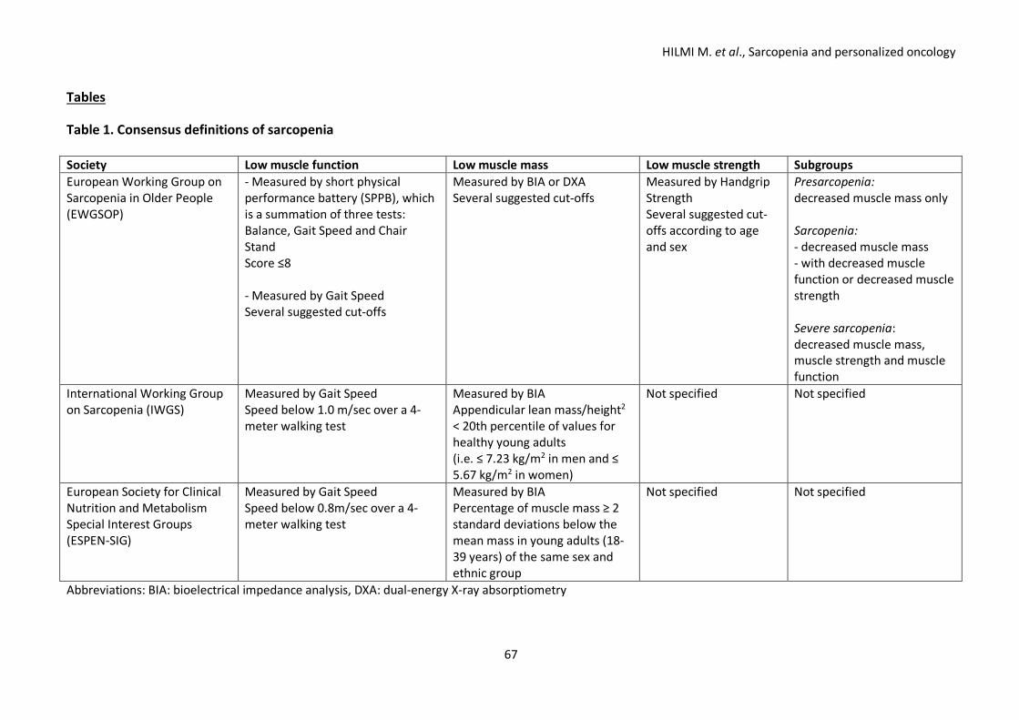
HILMI M. et al., Sarcopenia and personalized oncology
67
Tables Table 1. Consensus definitions of sarcopenia Society Low muscle function Low muscle mass Low muscle strength Subgroups European Working Group on Sarcopenia in Older People (EWGSOP)
- Measured by short physical performance battery (SPPB), which is a summation of three tests: Balance, Gait Speed and Chair Stand Score ≤8 - Measured by Gait Speed Several suggested cut-offs
Measured by BIA or DXA Several suggested cut-offs
Measured by Handgrip Strength Several suggested cut-offs according to age and sex
Presarcopenia: decreased muscle mass only Sarcopenia: - decreased muscle mass - with decreased muscle function or decreased muscle strength Severe sarcopenia: decreased muscle mass, muscle strength and muscle function
International Working Group on Sarcopenia (IWGS)
Measured by Gait Speed Speed below 1.0 m/sec over a 4-meter walking test
Measured by BIA Appendicular lean mass/height2 < 20th percentile of values for healthy young adults (i.e. ≤ 7.23 kg/m2 in men and ≤ 5.67 kg/m2 in women)
Not specified Not specified
European Society for Clinical Nutrition and Metabolism Special Interest Groups (ESPEN-SIG)
Measured by Gait Speed Speed below 0.8m/sec over a 4-meter walking test
Measured by BIA Percentage of muscle mass ≥ 2 standard deviations below the mean mass in young adults (18-39 years) of the same sex and ethnic group
Not specified Not specified
Abbreviations: BIA: bioelectrical impedance analysis, DXA: dual-energy X-ray absorptiometry
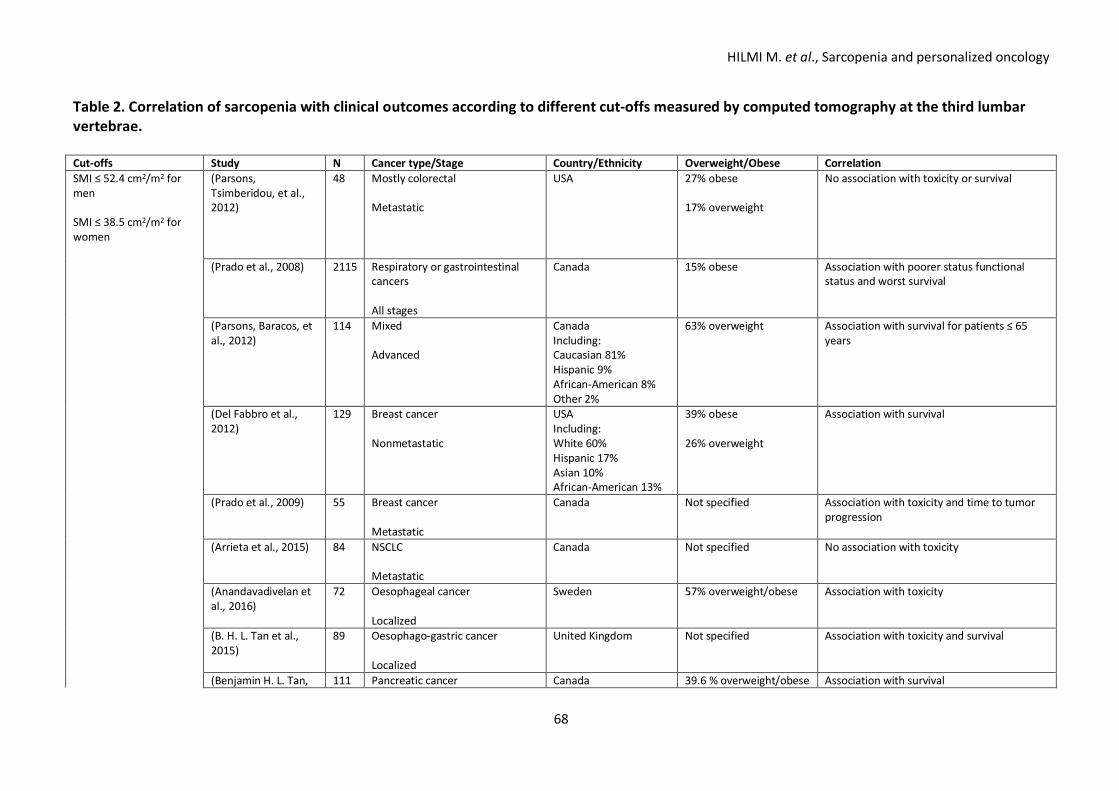
HILMI M. et al., Sarcopenia and personalized oncology
68
Table 2. Correlation of sarcopenia with clinical outcomes according to different cut-offs measured by computed tomography at the third lumbar vertebrae. Cut-offs Study N Cancer type/Stage Country/Ethnicity Overweight/Obese Correlation SMI ≤ 52.4 cm2/m2 for men SMI ≤ 38.5 cm2/m2 for women
(Parsons, Tsimberidou, et al., 2012)
48 Mostly colorectal Metastatic
USA 27% obese 17% overweight
No association with toxicity or survival
(Prado et al., 2008) 2115 Respiratory or gastrointestinal cancers All stages
Canada 15% obese Association with poorer status functional status and worst survival
(Parsons, Baracos, et al., 2012)
114 Mixed Advanced
Canada Including: Caucasian 81% Hispanic 9% African-American 8% Other 2%
63% overweight Association with survival for patients ≤ 65 years
(Del Fabbro et al., 2012)
129 Breast cancer Nonmetastatic
USA Including: White 60% Hispanic 17% Asian 10% African-American 13%
39% obese 26% overweight
Association with survival
(Prado et al., 2009) 55 Breast cancer Metastatic
Canada Not specified Association with toxicity and time to tumor progression
(Arrieta et al., 2015) 84 NSCLC Metastatic
Canada Not specified No association with toxicity
(Anandavadivelan et al., 2016)
72 Oesophageal cancer Localized
Sweden 57% overweight/obese Association with toxicity
(B. H. L. Tan et al., 2015)
89 Oesophago-gastric cancer Localized
United Kingdom Not specified Association with toxicity and survival
(Benjamin H. L. Tan, 111 Pancreatic cancer Canada 39.6 % overweight/obese Association with survival
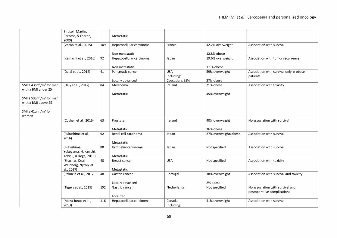
HILMI M. et al., Sarcopenia and personalized oncology
69
Birdsell, Martin, Baracos, & Fearon, 2009)
Metastatic
(Voron et al., 2015) 109 Hepatocellular carcinoma Non metastatic
France 42.2% overweight 12.8% obese
Association with survival
(Kamachi et al., 2016) 92 Hepatocellular carcinoma Non metastatic
Japan 19.6% overweight 1.1% obese
Association with tumor recurrence
(Dalal et al., 2012) 41 Pancreatic cancer Locally advanced
USA Including: Caucasians 95%
59% overweight 37% obese
Association with survival only in obese patients
SMI ≤ 43cm²/m² for men with a BMI under 25 SMI ≤ 53cm²/m² for men with a BMI above 25 SMI ≤ 41cm²/m² for women
(Daly et al., 2017) 84 Melanoma Metastatic
Ireland 21% obese 45% overweight
Association with toxicity
(Cushen et al., 2016) 63 Prostate Metastatic
Ireland 40% overweight 36% obese
No association with survival
(Fukushima et al., 2016)
92 Renal cell carcinoma Metastatic
Japan
27% overweight/obese Association with survival
(Fukushima, Yokoyama, Nakanishi, Tobisu, & Koga, 2015)
88 Urothelial carcinoma Metastatic
Japan
Not specified Association with survival
(Shachar, Deal, Weinberg, Nyrop, et al., 2017)
40 Breast cancer Metastatic
USA Not specified Association with toxicity
(Palmela et al., 2017) 48 Gastric cancer Locally advanced
Portugal 38% overweight 2% obese
Association with survival and toxicity
(Tegels et al., 2015) 152 Gastric cancer Localized
Netherlands Not specified No association with survival and postoperative complications
(Meza-Junco et al., 2013)
116 Hepatocellular carcinoma
Canada Including:
41% overweight
Association with survival
HILMI M. et al., Sarcopenia and personalized oncology
70
Localized and metastatic White 76% Oriental 10% Asian Indian 4% Arabian 3% Other 7%
30% obese
SMI ≤ 55 cm2/m2 for men SMI ≤ 39 cm2/m2 for women
(Camus et al., 2014) 80 Diffuse large B-cell lymphoma (DLBCL)
France 38% obese/overweight Association with survival
(Lanic et al., 2014) 82 Diffuse large B-cell lymphoma (DLBCL
France 39% obese/overweight Association with survival
(Srdic et al., 2016) 100 NSCLC Locally advanced and metastatic
Croatia Including: Caucasians 100%
26% overweight 14% obese
Association with survival and toxicity
(Sharma, Zargar-Shoshtari, Caracciolo, Richard, et al., 2015)
43 Penile Locally advanced
USA Not specified Association with postoperative complications
(E. Y. Kim et al., 2015) 149 Small cell lung cancer
Korea 21% overweight 20% obese
Association with survival
(Nault et al., 2015) 52 Hepatocellular carcinoma Advanced
France 38% overweight 12% obese
Association with toxicity
(Mir, Coriat, Boudou-Rouquette, et al., 2012)
18 Hepatocellular carcinoma Advanced
France 22.2% overweight 11.1% obese
Association with survival
(Barret et al., 2014) 51 Colorectal cancer Metastatic
France 41.2% overweight 3.9% obese
Association with toxicity
(Huillard et al., 2013) 61 Renal cancer Metastatic
France 26.2 % overweight 21.3% obese
Association with toxicity
SMI ≤ 52 cm2/m2 for men SMI ≤ 39.5 cm2/m2 for women
(Levolger et al., 2015) 99 Hepatocellular carcinoma Non metastatic
Netherlands 36.7 % overweight 15.6% obese
Association with survival and postoperative complications
SMI ≤ 43.7 cm2/m2 for men SMI ≤ 41.1 cm2/m2 for
(van Vledder et al., 2012)
196 Colorectal cancer Metastatic
Netherlands Not specified Association with tumor recurrence and survival
HILMI M. et al., Sarcopenia and personalized oncology
71
women SMI < 43.2 cm2/m2 (Wendrich et al.,
2017) 132 Head and neck
Locally advanced
Netherlands Not specified Association with toxicity
SMI ≤ 47.1 cm2/m2 for men SMI ≤ 34.4 cm2/m2 for women
(N. Nakamura et al., 2015)
207 Diffuse large B-cell lymphoma (DLBCL)
Japan
Not specified Association with survival
SMI ≤ 42.2 cm2/m2 for men SMI ≤ 33.9 cm2/m2 for women
(Y. Choi et al., 2015) 484 Pancreatic cancer Advanced
Korea 11.6% overweight/obese Association with survival
SMI ≤ 36 cm2/m2 for men SMI ≤ 29 cm2/m2 for women
(S.-L. Wang et al., 2016)
255 Gastric cancer Localized
China Not specified Association with postoperative complications
(Huang et al., 2015) 142 Colorectal cancer Localized
China Not specified Association with postoperative complications
HILMI M. et al., Sarcopenia and personalized oncology
72
Table 3. Prevalence of sarcopenia Tumor type Stage Study Sarcopenia evaluation Prevalence of sarcopenia Head and neck Locally advanced Wendrich et al., 2017 L3 CT scan 54 % Breast Localized Shachar, Deal, Weinberg, Williams, et al., 2017 L3 CT scan 3% obese sarcopenic
Del Fabbro et al., 2012
L3 CT scan 14% 3% obese sarcopenic
Metastatic Carla M. M. Prado et al., 2009 L3 CT scan 25%
Shachar, Deal, Weinberg, Nyrop, et al., 2017 L3 CT scan 58% Lung Metastatic Srdic et al., 2016 L3 CT scan 47% Stene et al., 2015 L3 CT scan 74% Arrieta et al., 2015 L3 CT scan 69% All stages Kim et al., 2015 L3 CT scan 79% Go et al., 2016 T4 CT scan 25% Baracos, Reiman, Mourtzakis, Gioulbasanis, & Antoun, 2010 L3 CT scan 47% Oesophagus Localized Anandavadivelan et al., 2016 L3 CT scan 43%
14% obese sarcopenic B. H. L. Tan et al., 2015 L3 CT scan 49% Tamandl et al., 2016 65% Locally advanced Murimwa et al., 2017 L4 CT scan 41% Awad et al., 2012 L3 CT scan 57% Yip et al., 2014 L3 CT scan 43% Miyata et al., 2017 BIA 47% Stomach Locally advanced Palmela et al., 2017 L3 CT scan 23%
10% obese sarcopenic Tegels et al., 2015 L3 CT scan 58% Wang et al., 2016 L3 CT scan 12% Liver Localized Meza-Junco et al., 2013 L3 CT scan 30% Levolger et al., 2015 L3 CT scan 58% Voron et al., 2015 L3 CT scan 30% Kamachi et al., 2016 L3 CT scan 66% (Harimoto et al., 2013) L3 CT scan 40% Advanced Dhooge et al., 2013 L3 CT scan 50% Mir, Coriat, Blanchet, et al., 2012 L3 CT scan 27% Nault et al., 2015 L3 CT scan 76%
HILMI M. et al., Sarcopenia and personalized oncology
73
Mir, Coriat, Boudou-Rouquette, et al., 2012 L3 CT scan 50% All stages Fujiwara et al., 2015 L3 CT scan 11% Iritani et al., 2015 L3 CT scan 11% Pancreas Localized Amini et al., 2015 L3 CT scan 25% Cooper et al., 2015 L3 CT scan 52% Joglekar et al., 2015 L3 CT scan 26% Advanced Rollins et al., 2016 L3 CT scan 60%
25% overweight/obese sarcopenic
Dalal et al., 2012 L3 CT scan 63% Benjamin H. L. Tan, Birdsell, Martin, Baracos, & Fearon, 2009 L3 CT scan 40%
16% overweight/obese sarcopenic
Choi et al., 2015 L3 CT scan 21% All stages Di Sebastiano et al., 2013 L3 CT scan 48% Colorectal Localized Broughman et al., 2015 L3 CT scan 57% Miyamoto et al., 2015 L3 CT scan 25% Reisinger et al., 2015 L3 CT scan 48% Huang et al., 2015 L3 CT scan 12% Locally advanced Chemama et al., 2016 L3 CT scan 40% Van Vugt et al., 2015 L3 CT scan 44% Metastatic Lene Thoresen et al., 2013 L3 CT scan 39% L. Thoresen et al., 2012 L3 CT scan 20% Van Vledder et al., 2012 L3 CT scan 19% Barret et al., 2014 L3 CT scan 71% Blauwhoff-Buskermolen et al., 2016 L3 CT scan 57% Parsons, Tsimberidou, et al., 2012 L3 CT scan 42% All stages Lieffers, Bathe, Fassbender, Winget, & Baracos, 2012 L3 CT scan 39% Kidney Localized Psutka et al., 2016 L3 CT scan 47% Metastatic (Fukushima et al., 2016) L3 CT scan 68% Sharma, Zargar-Shoshtari, Caracciolo, Fishman, et al., 2015 L3 CT scan 29% Huillard et al., 2013 L3 CT scan 53% Cushen et al., 2017 L3 CT scan 33%
13% overweight sarcopenic Ishihara et al., 2016 L3 CT scan 63% Antoun et al., 2010 L3 CT scan 55% Bladder Localized Psutka et al., 2015 L3 CT scan 83%
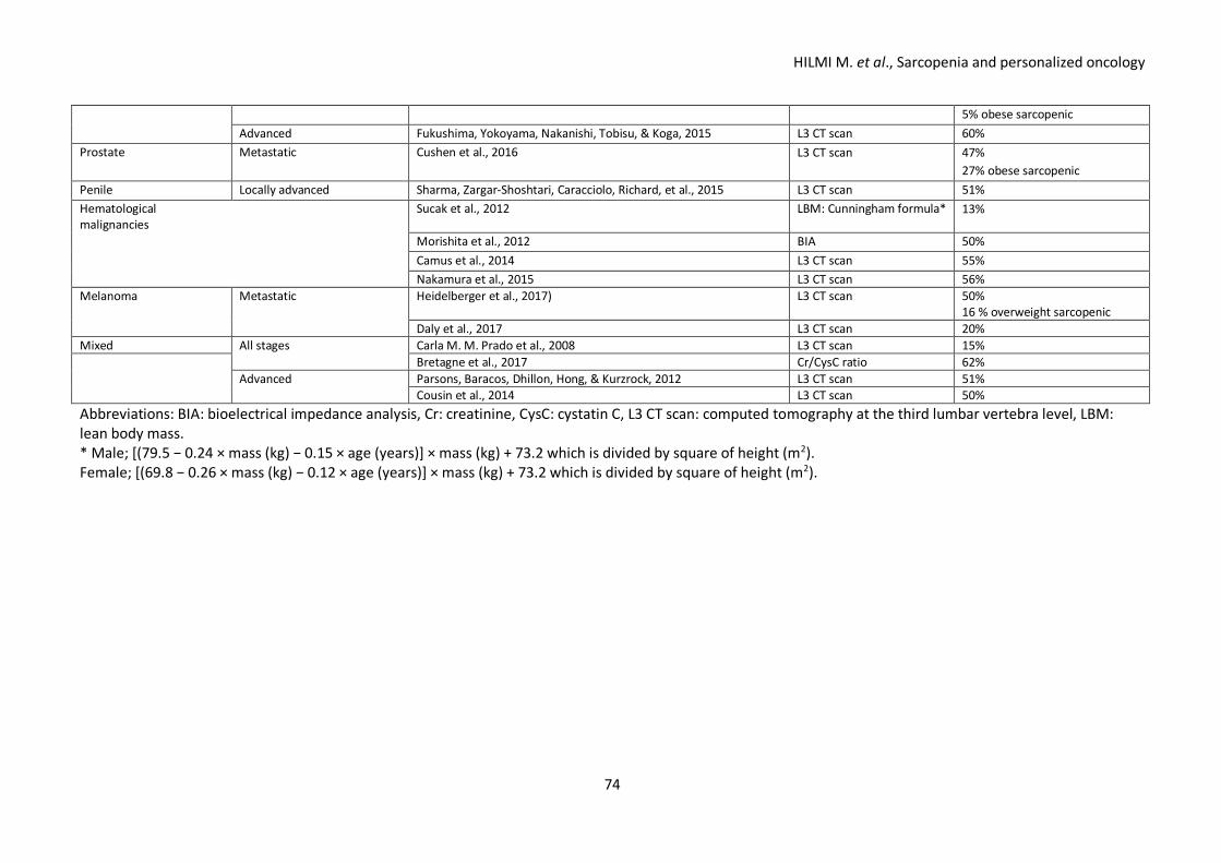
HILMI M. et al., Sarcopenia and personalized oncology
74
5% obese sarcopenic Advanced Fukushima, Yokoyama, Nakanishi, Tobisu, & Koga, 2015 L3 CT scan 60% Prostate Metastatic Cushen et al., 2016 L3 CT scan 47%
27% obese sarcopenic Penile Locally advanced Sharma, Zargar-Shoshtari, Caracciolo, Richard, et al., 2015 L3 CT scan 51% Hematological malignancies
Sucak et al., 2012 LBM: Cunningham formula* 13%
Morishita et al., 2012 BIA 50% Camus et al., 2014 L3 CT scan 55% Nakamura et al., 2015 L3 CT scan 56% Melanoma Metastatic Heidelberger et al., 2017) L3 CT scan 50%
16 % overweight sarcopenic Daly et al., 2017 L3 CT scan 20% Mixed All stages Carla M. M. Prado et al., 2008 L3 CT scan 15% Bretagne et al., 2017 Cr/CysC ratio 62% Advanced Parsons, Baracos, Dhillon, Hong, & Kurzrock, 2012 L3 CT scan 51% Cousin et al., 2014 L3 CT scan 50% Abbreviations: BIA: bioelectrical impedance analysis, Cr: creatinine, CysC: cystatin C, L3 CT scan: computed tomography at the third lumbar vertebra level, LBM: lean body mass. * Male; [(79.5 − 0.24 × mass (kg) − 0.15 × age (years)] × mass (kg) + 73.2 which is divided by square of height (m2). Female; [(69.8 − 0.26 × mass (kg) − 0.12 × age (years)] × mass (kg) + 73.2 which is divided by square of height (m2).
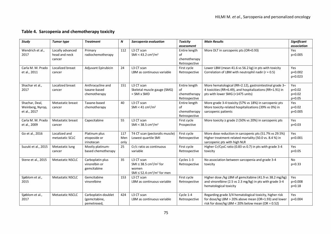
HILMI M. et al., Sarcopenia and personalized oncology
75
Table 4. Sarcopenia and chemotherapy toxicity Study Tumor type Treatment N Sarcopenia evaluation Toxicity
assessment Main Results Significant
association Wendrich et al., 2017
Locally advanced head and neck cancer
Primary radiochemotherapy
112 L3 CT scan SMI < 43.2 cm2/m2
Entire length of chemotherapy Retrospective
More DLT in sarcopenic pts (OR=0.93)
Yes p=0.005
Carla M. M. Prado et al., 2011
Localized breast cancer
Adjuvant Epirubicin 24 L3 CT scan LBM as continuous variable
First cycle Retrospective
Lower LBM (mean 41.6 vs 56.2 kg) in pts with toxicity Correlation of LBM with neutrophil nadir (r = 0.5)
Yes p=0.002 p=0.023
Shachar et al., 2017
Localized breast cancer
Anthracycline and taxane-based chemotherapy
151 L3 CT scan Skeletal muscle gauge (SMG) = SMI x SMD
Entire length of chemotherapy Retrospective
More hematological (RR=2.12), gastrointestinal grade 3–4 toxicities (RR=6.49), and hospitalizations (RR=1.91) in pts with lower SMG (<1475 units)
Yes p=0.02 p=0.02 p=0.05
Shachar, Deal, Weinberg, Nyrop, et al., 2017
Metastatic breast cancer
Taxane-based chemotherapy
40 L3 CT scan SMI < 41 cm2/m2
Entire length of chemotherapy Retrospective
More grade 3-4 toxicity (57% vs 18%) in sarcopenic pts More toxicity-related hospitalizations (39% vs 0%) in sarcopenic patients
Yes p=0.02 p=0.005
Carla M. M. Prado et al., 2009
Metastatic breast cancer
Capecitabine 55 L3 CT scan SMI < 38.5 cm2/m2
First cycle Prospective
More toxicity ≥ grade 2 (50% vs 20%) in sarcopenic pts Yes p=0.03
Go et al., 2016 Localized and metastatic SCLC
Platinum plus etoposide or irinotecan
117 Men only
T4 CT scan (pectoralis muscle) Lowest quartile SMI
First cycle Retrospective
More dose reduction in sarcopenic pts (51.7% vs 29.5%) Higher treatment-related mortality (50.0 vs. 8.4 %) in sarcopenic pts with high NLR
Yes p<0.001
Suzuki et al., 2015 Metastatic lung cancer
Mostly platinum-based chemotherapy
25 Cr/c ratio as continuous variable
First cycle Retrospective
Higher Cr/CysC ratio (0.83 vs 0.7) in pts with grade 3-4 toxicity
Yes p<0.05
Stene et al., 2015 Metastatic NSCLC
Carboplatin plus vinorelbin or gemcitabine
35 L3 CT scan SMI ≤ 38.5 cm2/m2 for women SMI ≤ 52.4 cm2/m2 for men
Cycles 1-3 Retrospective
No association between sarcopenia and grade 3-4 toxicity
No p=0.33
Sjøblom et al., 2015
Metastatic NSCLC Gemcitabine vinorelbine
153 L3 CT scan LBM as continuous variable
First cycle Retrospective
Higher dose /kg LBM of gemcitabine (41.9 vs 38.2 mg/kg) and vinorelbine (2.5 vs 2.3 mg/kg) in pts with grade 3-4 hematological toxicity
Yes p=0.008 p=0.18
Sjøblom et al., 2017
Metastatic NSCLC Carboplatin-doublet (gemcitabine, pemetrexed,
424 L3 CT scan LBM as continuous variable
Cycle 1-4 Retrospective
Regarding grade 3/4 hematological toxicity, higher risk for dose/kg LBM > 20% above mean (OR=1.93) and lower risk for dose/kg LBM < 20% below mean (OR = 0.52)
Yes p=0.004
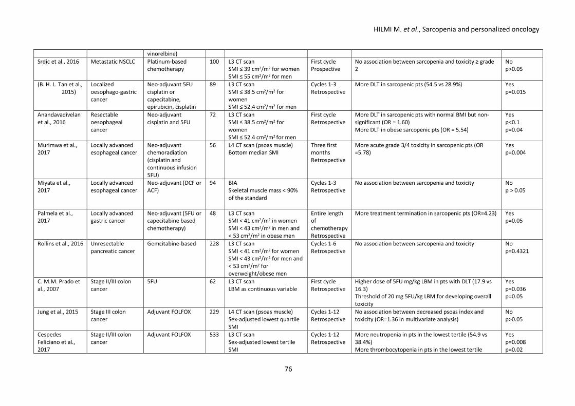
HILMI M. et al., Sarcopenia and personalized oncology
76
vinorelbine) Srdic et al., 2016 Metastatic NSCLC Platinum-based
chemotherapy 100 L3 CT scan
SMI ≤ 39 cm2/m2 for women SMI ≤ 55 cm2/m2 for men
First cycle Prospective
No association between sarcopenia and toxicity ≥ grade 2
No p>0.05
(B. H. L. Tan et al., 2015)
Localized oesophago-gastric cancer
Neo-adjuvant 5FU cisplatin or capecitabine, epirubicin, cisplatin
89 L3 CT scan SMI ≤ 38.5 cm2/m2 for women SMI ≤ 52.4 cm2/m2 for men
Cycles 1-3 Retrospective
More DLT in sarcopenic pts (54.5 vs 28.9%) Yes p=0.015
Anandavadivelan et al., 2016
Resectable oesophageal cancer
Neo-adjuvant cisplatin and 5FU
72 L3 CT scan SMI ≤ 38.5 cm2/m2 for women SMI ≤ 52.4 cm2/m2 for men
First cycle Retrospective
More DLT in sarcopenic pts with normal BMI but non-significant (OR = 1.60) More DLT in obese sarcopenic pts (OR = 5.54)
Yes p<0.1 p=0.04
Murimwa et al., 2017
Locally advanced esophageal cancer
Neo-adjuvant chemoradiation (cisplatin and continuous infusion 5FU)
56 L4 CT scan (psoas muscle) Bottom median SMI
Three first months Retrospective
More acute grade 3/4 toxicity in sarcopenic pts (OR =5.78)
Yes p=0.004
Miyata et al., 2017
Locally advanced esophageal cancer
Neo-adjuvant (DCF or ACF)
94 BIA Skeletal muscle mass < 90% of the standard
Cycles 1-3 Retrospective
No association between sarcopenia and toxicity No p > 0.05
Palmela et al., 2017
Locally advanced gastric cancer
Neo-adjuvant (5FU or capecitabine based chemotherapy)
48 L3 CT scan SMI < 41 cm2/m2 in women SMI < 43 cm2/m2 in men and < 53 cm2/m2 in obese men
Entire length of chemotherapy Retrospective
More treatment termination in sarcopenic pts (OR=4.23) Yes p=0.05
Rollins et al., 2016
Unresectable pancreatic cancer
Gemcitabine-based 228 L3 CT scan SMI < 41 cm2/m2 for women SMI < 43 cm2/m2 for men and < 53 cm2/m2 for overweight/obese men
Cycles 1-6 Retrospective
No association between sarcopenia and toxicity No p=0.4321
C. M.M. Prado et al., 2007
Stage II/III colon cancer
5FU 62 L3 CT scan LBM as continuous variable
First cycle Retrospective
Higher dose of 5FU mg/kg LBM in pts with DLT (17.9 vs 16.3) Threshold of 20 mg 5FU/kg LBM for developing overall toxicity
Yes p=0.036 p=0.05
Jung et al., 2015 Stage III colon cancer
Adjuvant FOLFOX 229 L4 CT scan (psoas muscle) Sex-adjusted lowest quartile SMI
Cycles 1-12 Retrospective
No association between decreased psoas index and toxicity (OR=1.36 in multivariate analysis)
No p>0.05
Cespedes Feliciano et al., 2017
Stage II/III colon cancer
Adjuvant FOLFOX 533 L3 CT scan Sex-adjusted lowest tertile SMI
Cycles 1-12 Retrospective
More neutropenia in pts in the lowest tertile (54.9 vs 38.4%) More thrombocytopenia in pts in the lowest tertile
Yes p=0.008 p=0.02
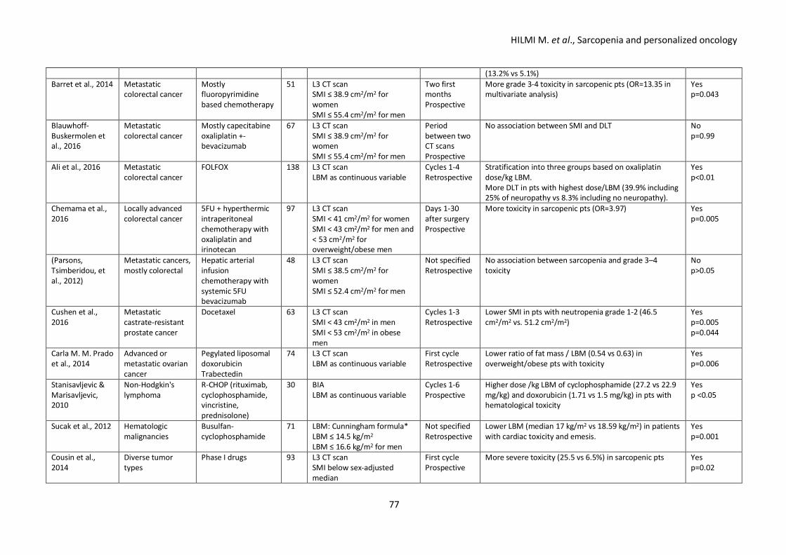
HILMI M. et al., Sarcopenia and personalized oncology
77
(13.2% vs 5.1%) Barret et al., 2014 Metastatic
colorectal cancer Mostly fluoropyrimidine based chemotherapy
51 L3 CT scan SMI ≤ 38.9 cm2/m2 for women SMI ≤ 55.4 cm2/m2 for men
Two first months Prospective
More grade 3-4 toxicity in sarcopenic pts (OR=13.35 in multivariate analysis)
Yes p=0.043
Blauwhoff-Buskermolen et al., 2016
Metastatic colorectal cancer
Mostly capecitabine oxaliplatin +- bevacizumab
67 L3 CT scan SMI ≤ 38.9 cm2/m2 for women SMI ≤ 55.4 cm2/m2 for men
Period between two CT scans Prospective
No association between SMI and DLT No p=0.99
Ali et al., 2016 Metastatic colorectal cancer
FOLFOX 138 L3 CT scan LBM as continuous variable
Cycles 1-4 Retrospective
Stratification into three groups based on oxaliplatin dose/kg LBM. More DLT in pts with highest dose/LBM (39.9% including 25% of neuropathy vs 8.3% including no neuropathy).
Yes p<0.01
Chemama et al., 2016
Locally advanced colorectal cancer
5FU + hyperthermic intraperitoneal chemotherapy with oxaliplatin and irinotecan
97 L3 CT scan SMI < 41 cm2/m2 for women SMI < 43 cm2/m2 for men and < 53 cm2/m2 for overweight/obese men
Days 1-30 after surgery Prospective
More toxicity in sarcopenic pts (OR=3.97) Yes p=0.005
(Parsons, Tsimberidou, et al., 2012)
Metastatic cancers, mostly colorectal
Hepatic arterial infusion chemotherapy with systemic 5FU bevacizumab
48 L3 CT scan SMI ≤ 38.5 cm2/m2 for women SMI ≤ 52.4 cm2/m2 for men
Not specified Retrospective
No association between sarcopenia and grade 3–4 toxicity
No p>0.05
Cushen et al., 2016
Metastatic castrate-resistant prostate cancer
Docetaxel 63 L3 CT scan SMI < 43 cm2/m2 in men SMI < 53 cm2/m2 in obese men
Cycles 1-3 Retrospective
Lower SMI in pts with neutropenia grade 1-2 (46.5 cm2/m2 vs. 51.2 cm2/m2)
Yes p=0.005 p=0.044
Carla M. M. Prado et al., 2014
Advanced or metastatic ovarian cancer
Pegylated liposomal doxorubicin Trabectedin
74 L3 CT scan LBM as continuous variable
First cycle Retrospective
Lower ratio of fat mass / LBM (0.54 vs 0.63) in overweight/obese pts with toxicity
Yes p=0.006
Stanisavljevic & Marisavljevic, 2010
Non-Hodgkin's lymphoma
R-CHOP (rituximab, cyclophosphamide, vincristine, prednisolone)
30 BIA LBM as continuous variable
Cycles 1-6 Prospective
Higher dose /kg LBM of cyclophosphamide (27.2 vs 22.9 mg/kg) and doxorubicin (1.71 vs 1.5 mg/kg) in pts with hematological toxicity
Yes p <0.05
Sucak et al., 2012 Hematologic malignancies
Busulfan-cyclophosphamide
71 LBM: Cunningham formula* LBM ≤ 14.5 kg/m2 LBM ≤ 16.6 kg/m2 for men
Not specified Retrospective
Lower LBM (median 17 kg/m2 vs 18.59 kg/m2) in patients with cardiac toxicity and emesis.
Yes p=0.001
Cousin et al., 2014
Diverse tumor types
Phase I drugs 93 L3 CT scan SMI below sex-adjusted median
First cycle Prospective
More severe toxicity (25.5 vs 6.5%) in sarcopenic pts Yes p=0.02
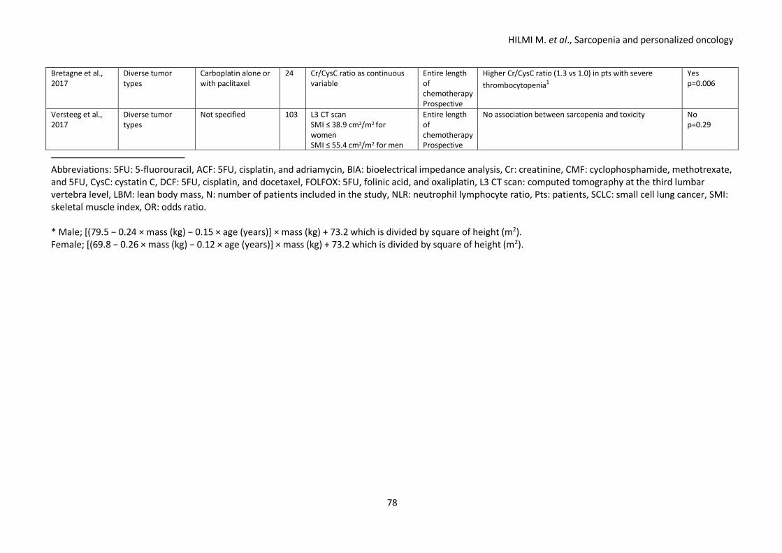
HILMI M. et al., Sarcopenia and personalized oncology
78
Bretagne et al., 2017
Diverse tumor types
Carboplatin alone or with paclitaxel
24 Cr/CysC ratio as continuous variable
Entire length of chemotherapy Prospective
Higher Cr/CysC ratio (1.3 vs 1.0) in pts with severe thrombocytopenia1
Yes p=0.006
Versteeg et al., 2017
Diverse tumor types
Not specified 103 L3 CT scan SMI ≤ 38.9 cm2/m2 for women SMI ≤ 55.4 cm2/m2 for men
Entire length of chemotherapy Prospective
No association between sarcopenia and toxicity No p=0.29
Abbreviations: 5FU: 5-fluorouracil, ACF: 5FU, cisplatin, and adriamycin, BIA: bioelectrical impedance analysis, Cr: creatinine, CMF: cyclophosphamide, methotrexate, and 5FU, CysC: cystatin C, DCF: 5FU, cisplatin, and docetaxel, FOLFOX: 5FU, folinic acid, and oxaliplatin, L3 CT scan: computed tomography at the third lumbar vertebra level, LBM: lean body mass, N: number of patients included in the study, NLR: neutrophil lymphocyte ratio, Pts: patients, SCLC: small cell lung cancer, SMI: skeletal muscle index, OR: odds ratio. * Male; [(79.5 − 0.24 × mass (kg) − 0.15 × age (years)] × mass (kg) + 73.2 which is divided by square of height (m2). Female; [(69.8 − 0.26 × mass (kg) − 0.12 × age (years)] × mass (kg) + 73.2 which is divided by square of height (m2).
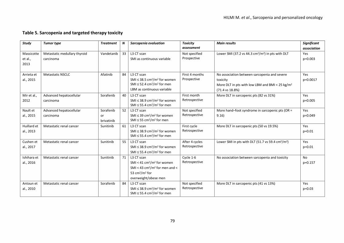
HILMI M. et al., Sarcopenia and personalized oncology
79
Table 5. Sarcopenia and targeted therapy toxicity Study Tumor type Treatment N Sarcopenia evaluation Toxicity
assessment Main results Significant
association Massicotte et al., 2013
Metastatic medullary thyroid carcinoma
Vandetanib 33 L3 CT scan SMI as continuous variable
Not specified Prospective
Lower SMI (37.2 vs 44.3 cm2/m2) in pts with DLT Yes p=0.003
Arrieta et al., 2015
Metastatic NSCLC Afatinib
84 L3 CT scan SMI ≤ 38.5 cm2/m2 for women SMI ≤ 52.4 cm2/m2 for men LBM as continuous variable
First 4 months Prospective
No association between sarcopenia and severe toxicity More DLT in pts with low LBM and BMI < 25 kg/m2 (71.4 vs 18.8%)
Yes p=0.0017
Mir et al., 2012
Advanced hepatocellular carcinoma
Sorafenib 40 L3 CT scan SMI ≤ 38.9 cm2/m2 for women SMI ≤ 55.4 cm2/m2 for men
First month Retrospective
More DLT in sarcopenic pts (82 vs 31%) Yes p=0.005
Nault et al., 2015
Advanced hepatocellular carcinoma
Sorafenib or brivatinib
52 L3 CT scan SMI ≤ 39 cm2/m2 for women SMI ≤ 55 cm2/m2 for men
Not specified Retrospective
More hand–foot syndrome in sarcopenic pts (OR = 9.16)
Yes p=0.049
Huillard et al., 2013
Metastatic renal cancer Sunitinib
61 L3 CT scan SMI ≤ 38.9 cm2/m2 for women SMI ≤ 55.4 cm2/m2 for men
First cycle Retrospective
More DLT in sarcopenic pts (50 vs 19.5%) Yes p=0.01
Cushen et al., 2017
Metastatic renal cancer Sunitinib 55 L3 CT scan SMI ≤ 38.9 cm2/m2 for women SMI ≤ 55.4 cm2/m2 for men
After 4 cycles Retrospective
Lower SMI in pts with DLT (51.7 vs 59.4 cm2/m2) Yes p=0.01
Ishihara et al., 2016
Metastatic renal cancer Sunitinib 71 L3 CT scan SMI < 41 cm2/m2 for women SMI < 43 cm2/m2 for men and < 53 cm2/m2 for overweight/obese men
Cycle 1-6 Retrospective
No association between sarcopenia and toxicity No p=0.157
Antoun et al., 2010
Metastatic renal cancer Sorafenib
84 L3 CT scan SMI ≤ 38.9 cm2/m2 for women SMI ≤ 55.4 cm2/m2 for men
Not specified Retrospective
More DLT in sarcopenic pts (41 vs 13%) Yes p=0.03