Biomineralization of endolithic microbes in rocks from the McMurdo Dry Valleys of Antarctica:...
10
Environmental Microbiology (2005) 7 (4), 566–575 doi:10.1111/j.1462-2920.2004.00725.x © 2004 Society for Applied Microbiology and Blackwell Publishing Ltd Blackwell Science, LtdOxford, UKEMIEnvironmental Microbiology 1462-2912Society for Applied Microbiology and Blackwell Publishing Ltd, 20047 4566575 Original Article Biomineralization of endolithic in Antarctica rocksJ. Wierzchos, L. García Sancho and C. Ascaso Accepted 24 August, 2004. *For correspondence. E-mail [email protected]; Tel. ( + 34) 9 7370 2417; Fax ( + 34) 9 7370 2426. Biomineralization of endolithic microbes in rocks from the McMurdo Dry Valleys of Antarctica: implications for microbial fossil formation and their detection Jacek Wierzchos, 1 * Leopoldo García Sancho 1,2 and Carmen Ascaso 1,3 1 Servei de Microscòpia Electrònica, Universitat de Lleida, c/Rovira Roure, 44, 25198 Lleida, Spain. 2 Fac. Farmacia, Universidad Complutense, 28040 Madrid, Spain. 3 Centro de Ciencias Medioambientales, CSIC, c/Serrano 115 bi, 28006 Madrid, Spain. Summary In some zones of Antarctica’s cold and dry desert, the extinction of cryptoendolithic microorganisms leaves behind inorganic traces of microbial life. In this paper, we examine the transition from live microorganisms, through their decay, to microbial fossils using in situ microscopy (transmission electron microscopy, scan- ning electron microscopy in back-scattered electron mode) and microanalytical (energy dispersive X-ray spectroscopy) techniques. Our results demonstrate that, after their death, endolithic microorganisms inhabiting Commonwealth Glacier sandstone from the Antarctica McMurdo Dry Valleys become mineralized. In some cases, epicellular deposition of minerals and/ or simply filling up of empty moulds by minerals leads to the formation of cell-shaped structures that may be considered biomarkers. The continuous deposition of allochthonous clay minerals and sulfate-rich salts fills the sandstone pores. This process can give rise to microbial fossils with distinguishable cell wall struc- tures. Often, fossilized cell interiors were of a different chemical composition to the mineralized cell walls. We propose that the microbial fossil formation observed was induced by mineral precipitation result- ing from inorganic processes occurring after the death of cryptoendolithic microorganisms. Neverthe- less, it must have been the organic template that pro- voked the diffusion of mineral elements and gave rise to their characteristic distribution pattern inside the fossilized cells. Introduction The processes microorganism biomineralization and per- mineralized microbial fossil formation have improved our knowledge of the preservation of biological remains, enabling researchers to document the persistence of ancient microorganisms over geological time. This type of study has always been of interest to palaeontologists, but recent challenges in astrobiology and the search for life traces in the form of microbial fossils require the applica- tion of unconventional methods to detect life and its traces in extreme environments (Nealson et al ., 2002). Some of these environments are the only models we have to gain insight into how life emerged and how to recognize its possible traces on other planets. One such environment is the intensely dry and cold endolithic ecosystem of the McMurdo Dry Valleys of Antarctica. Fungi, bacteria and cyanobacteria often become coated by mineral crusts through the process of biomineralization (Nealson, 1983; Fortin et al ., 1997, 1998; Fortin and Fer- ris, 1998; Warren and Ferris, 1998; Konhauser and Urru- tia, 1999; Phoenix et al ., 2000). This can lead to the formation of microbial fossils (Westall, 1999; McKinley et al ., 2000; Boyce et al ., 2001; Westall et al ., 2001). Sev- eral experimental studies performed under laboratory (Westall et al ., 1995; Ferris et al ., 1988; Toporski et al ., 2002) and field conditions, such as hot geothermal springs (Ferris et al ., 1986; Schultze-Lam et al ., 1995; Jones et al ., 1997; Konhauser et al ., 2001; Yee et al ., 2003) or salt lakes (Arp et al ., 1999; Golubic, 1991), have attempted to explain the processes involved in the biom- ineralization of organic material. However, elucidating the mechanisms giving rise to inorganic life traces in the pecu- liar environmental conditions of the Antarctic desert’s endolithic ecosystem is particularly challenging. On a macroscopic scale, microbial traces in the form of physi- cochemical bioweathering patterns in rock from the McMurdo Dry Valleys were reported by Friedmann and Weed (1987), Kappen (1993) and Sun and Friedmann (1999). More recently, it was demonstrated that some minerals in Antarctic rocks are biogenically transformed to generate inorganic biomarkers – traces left by living microorganisms resulting from their biological activity (Wierzchos and Ascaso, 2001; Ascaso and Wierzchos,
-
Upload
independent -
Category
Documents
-
view
4 -
download
0
Transcript of Biomineralization of endolithic microbes in rocks from the McMurdo Dry Valleys of Antarctica:...

Environmental Microbiology (2005)
7
(4), 566–575 doi:10.1111/j.1462-2920.2004.00725.x
© 2004 Society for Applied Microbiology and Blackwell Publishing Ltd
Blackwell Science, LtdOxford, UKEMIEnvironmental Microbiology 1462-2912Society for Applied Microbiology and Blackwell Publishing Ltd, 20047
4566575
Original Article
Biomineralization of endolithic in Antarctica rocksJ. Wierzchos, L. García
Sancho and C. Ascaso
Accepted 24 August, 2004. *For correspondence. [email protected]; Tel. (
+
34) 9 7370 2417; Fax (
+
34)9 7370 2426.
Biomineralization of endolithic microbes in rocks from the McMurdo Dry Valleys of Antarctica: implications for microbial fossil formation and their detection
Jacek Wierzchos,
1
* Leopoldo García Sancho
1,2
and Carmen Ascaso
1,3
1
Servei de Microscòpia Electrònica, Universitat de Lleida, c/Rovira Roure, 44, 25198 Lleida, Spain.
2
Fac. Farmacia, Universidad Complutense, 28040 Madrid, Spain.
3
Centro de Ciencias Medioambientales, CSIC, c/Serrano 115 bi, 28006 Madrid, Spain.
Summary
In some zones of Antarctica’s cold and dry desert, theextinction of cryptoendolithic microorganisms leavesbehind inorganic traces of microbial life. In this paper,we examine the transition from live microorganisms,through their decay, to microbial fossils using
in situ
microscopy (transmission electron microscopy, scan-ning electron microscopy in back-scattered electronmode) and microanalytical (energy dispersive X-rayspectroscopy) techniques. Our results demonstratethat, after their death, endolithic microorganismsinhabiting Commonwealth Glacier sandstone from theAntarctica McMurdo Dry Valleys become mineralized.In some cases, epicellular deposition of minerals and/or simply filling up of empty moulds by minerals leadsto the formation of cell-shaped structures that may beconsidered biomarkers. The continuous deposition ofallochthonous clay minerals and sulfate-rich salts fillsthe sandstone pores. This process can give rise tomicrobial fossils with distinguishable cell wall struc-tures. Often, fossilized cell interiors were of a differentchemical composition to the mineralized cell walls.We propose that the microbial fossil formationobserved was induced by mineral precipitation result-ing from inorganic processes occurring after thedeath of cryptoendolithic microorganisms. Neverthe-less, it must have been the organic template that pro-voked the diffusion of mineral elements and gave riseto their characteristic distribution pattern inside thefossilized cells.
Introduction
The processes microorganism biomineralization and per-mineralized microbial fossil formation have improved ourknowledge of the preservation of biological remains,enabling researchers to document the persistence ofancient microorganisms over geological time. This type ofstudy has always been of interest to palaeontologists, butrecent challenges in astrobiology and the search for lifetraces in the form of microbial fossils require the applica-tion of unconventional methods to detect life and its tracesin extreme environments (Nealson
et al
., 2002). Some ofthese environments are the only models we have to gaininsight into how life emerged and how to recognize itspossible traces on other planets. One such environmentis the intensely dry and cold endolithic ecosystem of theMcMurdo Dry Valleys of Antarctica.
Fungi, bacteria and cyanobacteria often become coatedby mineral crusts through the process of biomineralization(Nealson, 1983; Fortin
et al
., 1997, 1998; Fortin and Fer-ris, 1998; Warren and Ferris, 1998; Konhauser and Urru-tia, 1999; Phoenix
et al
., 2000). This can lead to theformation of microbial fossils (Westall, 1999; McKinley
et al
., 2000; Boyce
et al
., 2001; Westall
et al
., 2001). Sev-eral experimental studies performed under laboratory(Westall
et al
., 1995; Ferris
et al
., 1988; Toporski
et al
.,2002) and field conditions, such as hot geothermalsprings (Ferris
et al
., 1986; Schultze-Lam
et al
., 1995;Jones
et al
., 1997; Konhauser
et al
., 2001; Yee
et al
.,2003) or salt lakes (Arp
et al
., 1999; Golubic, 1991), haveattempted to explain the processes involved in the biom-ineralization of organic material. However, elucidating themechanisms giving rise to inorganic life traces in the pecu-liar environmental conditions of the Antarctic desert’sendolithic ecosystem is particularly challenging. On amacroscopic scale, microbial traces in the form of physi-cochemical bioweathering patterns in rock from theMcMurdo Dry Valleys were reported by Friedmann andWeed (1987), Kappen (1993) and Sun and Friedmann(1999). More recently, it was demonstrated that someminerals in Antarctic rocks are biogenically transformed togenerate inorganic biomarkers – traces left by livingmicroorganisms resulting from their biological activity(Wierzchos and Ascaso, 2001; Ascaso and Wierzchos,
Biomineralization of endolithic in Antarctica rocks
567
© 2005 Society for Applied Microbiology and Blackwell Publishing Ltd,
Environmental Microbiology
,
7
, 566–575
2002, 2003; Wierzchos
et al
., 2003). Furthermore, recent
in situ
microscopy studies have for the first time demon-strated the presence of microbial fossils within Antarcticsandstone rocks from the McMurdo Dry Valleys desert(Ascaso, 2000; Ascaso and Wierzchos, 2002; Wierzchosand Ascaso, 2002, 2003). Using the
in situ
procedure,microbial fossils can be systematically described andcharacterized, sometimes by identifying internal cell struc-tures within fossilized cells.
In this report, we focus on the transition from life,through decay, to fossilized microorganisms, and on thebiomineralization processes that produce their chemicaltraces. Our objectives were: (i) to micromorphologicallydescribe living and decaying Antarctic endolithic microbialcommunities and their fossils; (ii) to characterize macro-elements present in these communities and to chemicallycharacterize mineralized microbial cells (microfossils). Weaddressed the question of whether microbial fossil forma-
tion is related to intense biologically mediated mineraliza-tion or whether mineral precipitation and biomineralizationsimply occur as a result of inorganic effects after thedecay of microbes.
Results
Stereoscopic microscopy observations of sandstone sam-ple COM-120 (from the Commonwealth Glacier) revealeda green, thin, 1–2 mm layer under the rock surface con-taining the microbial endolithic communities. Spacesbetween the quartz grains of this rock were mainly inhab-ited by fungi, algae and bacteria (including cyanobacteria)colonies, as shown in the transmission electron micros-copy (TEM) images in Fig. 1. Figure 1A and B show theultrastructural details of a hyphal and algal cell respec-tively. Within the algal cell it is possible to distinguishthylakoids inside the chloroplast, pyrenoid zone, vacuole
A
C D
F
E
B
Fig. 1.
Transmission electron microscopy images of cryptoendolithic microorganisms and mineral features within COM-120 sandstone rock from Commonwealth Glacier.A. Ultrastructural details of a hyphal (L, lipid bodies; open arrow, cell wall) and bacterial cell (black arrows).B. Large algal cell with thylakoids (T), pyrenoid (P), vacuole (V), lipids (black arrows) and cell wall (open arrow).C. Small cyanobacterial cells surrounded by extracellular polymeric substances (EPS) and mineral particles (open arrows).D. Live bacterial cells (white open arrows), bac-teria-shaped mineral sheaths (black arrows) and clay particles (black open arrow).E. Cell-shaped mineral aggregate (open arrow).F. Cell-shaped clay aggregate with relicts of cell wall (open arrow), empty cell-shaped nanocrys-talline sheaths (black arrows) and plasmolysed microorganism cell (white arrow).

568
J. Wierzchos, L. G. Sancho and C. Ascaso
© 2005 Society for Applied Microbiology and Blackwell Publishing Ltd,
Environmental Microbiology
,
7
, 566–575
and large lipids in the cytoplasm. Fungal cells were oftenaccompanied by bacteria (black arrows in Fig. 1A).Figure 1C shows a colony of bacterial cells enclosed in adiscretely electro-dense matrix of polymeric substances(EPS). Outside this envelope, mineral particles of a claynature (open arrows) could be observed. Particles andsmall aggregates of minerals frequently appeared amongthe live microorganism cells. Figure 1D shows particles ofclay (open black arrow), live bacteria (open white arrows)and small bacteria-shaped empty mineral sheaths (blackarrows). In another pore, no live microbial cells couldbe identified. Several mineral deposits or precipitatesrevealed the typical shapes and dimensions of microor-ganism cells. Basically, three types of deposit wereobserved: (i) electron-dense, cell-shaped aggregatescomposed of clay-like particles (open arrow in Fig. 1E);(ii) collapsed, cell-shaped aggregates containing clayswith outer less electron-dense substances in the form ofa cell wall (open arrow in Fig. 1F); and (iii) empty cell-shaped sheaths comprised of nanocrystalline minerals(black arrows in Fig. 1F). Plasmolysed cells wereobserved in some areas of the ultrathin section (whitearrow in Fig. 1F).
Despite the high resolving power of TEM, the difficul-ties encountered in preparing ultra-thin sections pro-mpted us to apply the well established
in situ
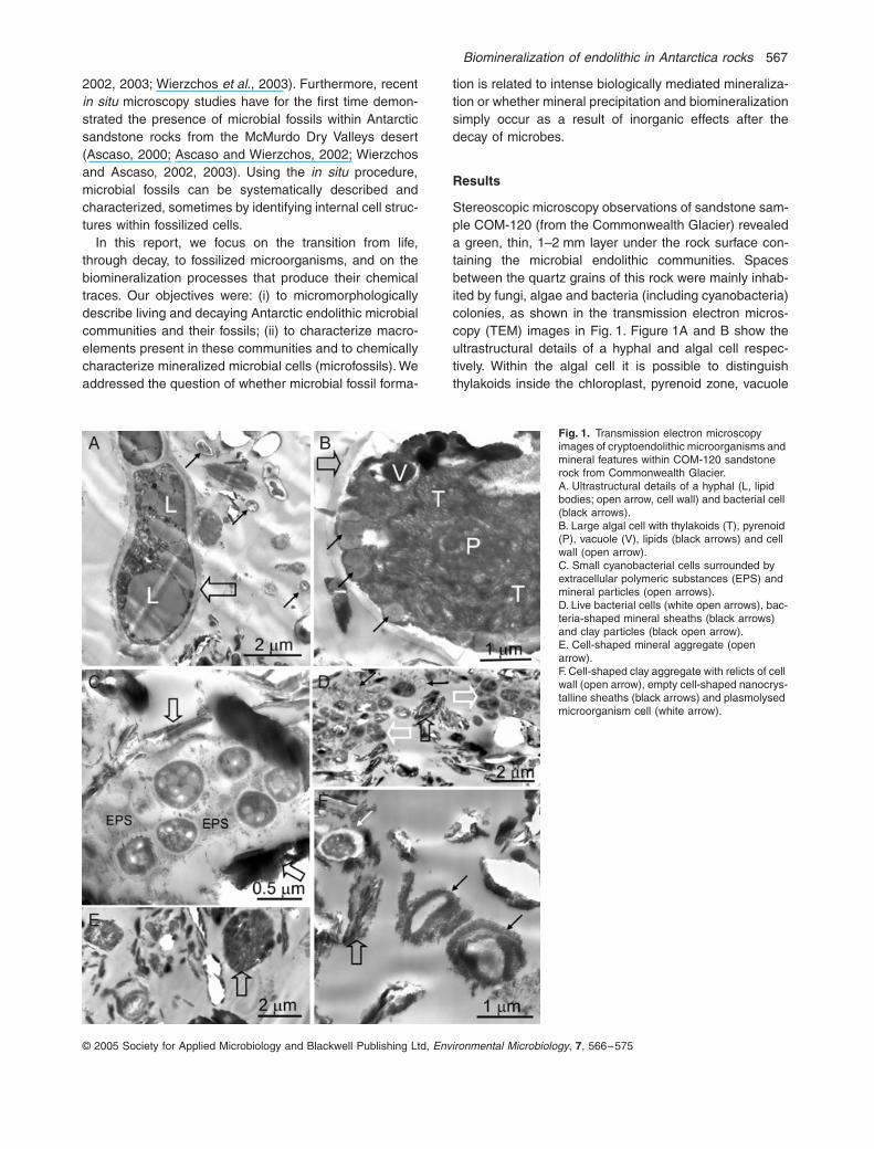
scanningelectron microscopy in back-scattered electron mode(SEM-BSE) and energy dispersive X-ray spectroscopy(EDS) microanalysis techniques (e.g. Wierzchos andAscaso, 2002). Figure 2A shows an SEM-BSE image oflive fungal and bacterial endolithic communities in sampleCOM-120, colonizing small pores between quartz grains.The simultaneous qualitative and quantitative EDSmicroanalysis of the whole area shown in Fig. 2Arevealed very low Si, P and S contents and lack of Al(Fig. 2B), indicating the absence of alumino-silicate min-erals among the live colonies. At higher magnification(Fig. 2C) lipid bodies could be detected within the hyphalcells. The high contrast of the ultrastructural elements ofthese cells provided by the back-scattered electrons indi-cates good staining (with heavy metals), reflecting theirmembrane integrity. Other pore spaces in the same sam-ple showed numerous dead cells closely surrounded byan abundant mineral phase. Figure 2D and F show theremains of decayed hyphae or cyanobacterial cells (whitearrows) and bacterial cells (black arrow in Fig. 2F). Thelow sensitivity of these cells to Os and U cation stainingindicated biochemical inactivity and that they were conse-quently dead or decaying. The open white arrows inFig. 2D point to mineral fabrics corresponding to cell wall-like sheaths. The abundance of minerals in this pore area(Fig. 2D) was confirmed by EDS, detecting high amountsof Si, Al, Mg and Na (components of alluminosilicates)(EDS spectrum in Fig. 2E). Figure 3A is a low magnifica-
tion SEM-BSE view of the pore containing decayed(small, dark cell-shaped spots) and highly biomineralizedendolithic fragments. The upper square-outlined areaindicates the zone detailed in Fig. 2D. The high BSE con-trast in Fig. 3A identifies zones of mineral deposition,confirmed by the Si and Al distribution maps provided inFig. 3B and C respectively. Note the similar distribution ofSi and Al reflecting an abundance of alluminosilicatesamong the colonies of decayed microorganisms withinthis pore. In another sample from a harsher environment,the biomineralization of dead microbial cells seemed tobe much more intense, leading to the formation of fossil-ized microbial cells. Figure 3D is an example of microbialfossils observed in a highly mineralized pore in the Lin-naeus Terrace sandstone (A834-571). Note that thesemicrobial fossils show cell-shaped features with a well-defined, thick cell wall structure. The diameter of thesefossils was 5–20
m
m, suggesting they could be fossilizedcyanobacterial and/or algal cells. Element distribution dif-fered within the fossilized microbial cells as shown by linescans (Fig. 3E) and elemental distribution maps (Fig. 3F–H) for the fossilized cells in Fig. 3D. Figure 3E showsmicroprobe profiles of Si, Al, S, K and Fe correspondingto the yellow line in Fig. 3D. The fossilized cell walls con-tained high Al, S and K levels and a low Si level (red lines‘a’ and ‘b’ in Fig. 3D and E). Fossilized cell interiors con-tained high amounts of Si (red ‘d’ line in Fig. 3D and E),and in some fossils, they showed a high Fe content(asterisks in Fig. 3D and E). The matrix observed amongthe fossilized cells revealed a relatively high Si and Fecontent (red ‘c’ line in Fig. 3D and E). These analyticaldata were confirmed by the Si, Al and S distribution maps(Fig. 3F–H) obtained from the area shown in Fig. 3D.These maps confirmed the abundance of Si in the matrixand inside the fossilized cells. Thus, the main compo-nents of the fossilized cell walls were Al and S. Figure 4presents quantitative EDS data corresponding to 14 pointanalyses of the matrix zones among fossilized cells, 11point analyses of fossilized cell walls and 15 point analy-ses of the fossilized cell interiors. Differences wererevealed in the Si, Al, S, K and Fe composition of thematrix outside the cells, cell walls and the fossilized cellinsides, reflecting a complex fossilisation mechanism inthis sandstone.
Figure 5 shows fossilized cells or only the fossilized cellwalls in the sample from Linnaeus Terrace (A856-41). Wecan discern pores just under and on the rock surface,which are extensively filled with mineral template (brightpores in upper Fig. 5A). Empty pores (black) could beseen 1 mm below the rock surface (Fig. 5A). This rocksample was subjected to intense SEM-BSE evaluation(Fig. 5F–I). Its pore contents indicated by the open arrowin Fig. 5A were also prepared as an ion-milled section forTEM (Fig. 5E). Prior to TEM, the same pore had been
Biomineralization of endolithic in Antarctica rocks
569
© 2005 Society for Applied Microbiology and Blackwell Publishing Ltd,
Environmental Microbiology
,
7
, 566–575
subjected to SEM-BSE (Fig. 5B–D). The image in Fig. 5Bshows the pore contents with an adjacent quartz grain(dark grey area in Fig. 5B). Ion milling gave rise to thehole indicated by the open arrow, but the argon ion beamalso interacts with the quartz surface. Unexpectedly, sub-jecting the quartz surface to this procedure revealed con-centric structures (left part of Fig. 5B), perhaps becauseof differences in the physical properties and different rateof ion milling of the quartz grain components. The brightarea in Fig. 5B corresponds to the pore contents withdistinct cell-shaped inorganic structures (black arrows).Figure 5C is a detailed SEM-BSE image of the content ofthis pore, showing cell wall-like structures (black arrows).In Fig. 5D, small, fossilized microbial cells were observedat the edge of the ion-milled hole. When this same frag-ment was examined by TEM (Fig. 5E), the section was toothick to transmit the electron beam. However, the shapes
of the same cell-like structures as in the SEM-BSE image(Fig. 5D) could be observed (white arrows). The next setof SEM-BSE images (Fig. 5F–I) shows several microbialfossils found in this rock. All these fossil cells contained ahigh amount of silica and some (Fig. 5H and I) alsoshowed high Fe contents.
Discussion
Friedmann and Koriem (1989) suggested that the cryp-toendolithic microbial ecosystem of the McMurdo DryValleys might closely follow the hypothetic scenario ofpossible life extinction on Mars: i.e. continuous cooling ofthe Mars surface caused stress and resulted in the extinc-tion of the microorganisms. The cryptoendolithic commu-nity of the McMurdo Dry Valleys of Antarctica is indeedremarkable for its slow rates of growth and longevity (Sun
A
D
C
B
F
E
Fig. 2.
Scanning electron microscopy in back-scattered electron mode images and EDS data of the contents of a pore in the COM-120 sand-stone sample.A and C. Scanning electron microscopy in back-scattered electron mode images of live hyphae (large cells with lipid bodies, arrows) and bacteria (small cells).B. Energy dispersive X-ray spectroscopy spec-trum of the whole area shown in A revealing low Si and P contents. The vertical units on the spectrum are counts per second (cps).D and F. Scanning electron microscopy in back-scattered electron mode images of decayed hyphal or cyanobacterial cells (white arrows), bacterial cells (black arrow in F), cell wall-like mineral sheaths (open white arrows).E. Energy dispersive X-ray spectroscopy spec-trum of the whole area shown in D revealing high amounts of Si, Al, Mg, Na and low P and S contents.

570
J. Wierzchos, L. G. Sancho and C. Ascaso
© 2005 Society for Applied Microbiology and Blackwell Publishing Ltd,
Environmental Microbiology
,
7
, 566–575
A
D
F G H
B
C
E
Fig. 3.
Scanning electron microscopy in back-scattered electron mode images and EDS data of mineral deposits and fossilized microorganisms.A. Low magnification of SEM-BSE image of a pore in the COM-120 sandstone with decayed microorganisms (black spots) and mineral deposits (bright features in A). Upper and lower square-outlined areas indicate the zones shown in Fig. 2D and F respectively.B and C. Energy dispersive X-ray spectroscopy distribution maps showing Si and Al concentra-tion images, respectively, of the area shown in A; scale bars
=
20
m
m.D. Scanning electron microscopy in back-scattered electron mode image of microbial fos-sils in a pore in the Linnaeus Terrace sandstone (A834-571). The yellow line (with red marks) indicates the position of the EDS microprobe scan-line profile.E. Energy dispersive X-ray spectroscopy scan-line profiles showing relative concentration changes (
Y
axe) in Si, Al, S, K and Fe along the yellow arrow in D; red vertical lines and asterisk indicate characteristic structures of microbial fossils in D.F, G, and H. Energy dispersive X-ray spectros-copy distribution maps showing Si, Al and S concentration images, respectively, of the area shown in D; scale bars
=
5
m
m.
and Friedmann, 1999). Considering the scarce climaticdata available for large areas of the McMurdo Dry Valleysof Antarctica (Nienow and Friedmann, 1993; Friedmann
et al
., 1994) and the limited geological, geophysical andgeochemical record, any laboratory simulation of Antarcticdesert conditions and fossilisation processes is likely tobe far from the natural situation. Hence, our insistence onthe
in situ
examination of the live and decayed microbialcontent of these rocks and their mineral traces. Bearingin mind the diversity of geophysical and geochemical pro-cesses that probably occurred in the McMurdo Dry Valleys
of Antarctica, fossilisation and/or biomineralization condi-tions of endoliths could be very different. Friedmann andWeed (1987) noted the formation of siliceous crusts andquartz crystal growth. The mechanism of crystal growthin such a low-temperature, non-aquatic, non-alkaline andnear-surface environment is unusual, and the sources ofsilica are unknown (Friedmann and Weed, 1987). Onewould expect to find some microorganism cells (or theirrelicts) encrusted in these quartz crystal rinds. However,in our study we found no traces of endolithic microorgan-isms in the quartz rinds of the highly silicified A856-41

Biomineralization of endolithic in Antarctica rocks
571
© 2005 Society for Applied Microbiology and Blackwell Publishing Ltd,
Environmental Microbiology
,
7
, 566–575
samples. Inadvertently, ion milling of transverse sectionsof this rock revealed the presence of homogenously dis-tributed concentric structures in a quartz grain. Thesestructures may indicate that pre-existing quartz grainscomprising this sandstone arose as early diagenetic silicainfills of small radiolaria and diatom cysts (e.g. Schieber
et al
., 2000). However, the homogenous distribution of
these concentric structures also in the quartz rind zoneinvalidates the hypothesis of quartz crystal growth fromsilica deposited during the time of microorganism coloni-zation. Wierzchos and Ascaso (2002) recently proposeda scenario of microbial fossil formation in McMurdo DryValley rocks. According to these authors, the first stage ofmicrobial cell biomineralization may have occurred whenmicroorganisms were still biologically active and/or aftertheir decay. The same authors also indicated that someof the pores containing the microbial fossils were infil-trated with jarosite [KFe
3
(SO
4
) 2(OH)
6
] and gypsum[Ca(SO
4
) 2(H
2
O)]. Here, we observed no mineral depositsor mineral precipitates within the pores colonized by livefungi and bacteria (Fig. 2A–C). However, in other zonesof the same sample, pores with decayed microorganismsbecame extensively filled with minerals of a clay nature(Fig. 2D–F). This could eventually lead to the formation ofmicrobial fossils and/or biomarkers. The origin of this min-eral coating observed on the rock surface and infiltratedwithin the pore spaces could be explained by the accu-mulation of airborne dust composed of quartz, clays andFe oxyhydroxides (Weed and Ackert, 1986; Friedmannand Weed, 1987; Weed and Norton, 1991). The mineral-ogical composition of microbial fossils and possible mech-anisms of biomineralization can depend on the nature of
Fig. 4.
Comparison of the Al, Si, S, K and Fe composition (EDS quantitative data in wt percentage) of the highly mineralized and fossilized microorganism features in the A834-571 sample; short upper lines – SD.
A
C
F G H I
D E
B
Fig. 5.
Microbial fossils in sandstone sample A856-41 from Linnaeus Terrace.A. Scanning electron microscopy in back-scattered electron mode (SEM-BSE) with low magnification view of a transverse section; open arrow points to the pore with microbial fossils shown in B–E.B. Scanning electron microscopy in back-scattered electron mode image showing a frag-ment of the sample after ion beam milling (hole indicated by open arrow). Black arrows indicate the microbial fossils embedded in the mineral matrix. Note that ion milling revealed the con-centric structures on the quartz grain surfaces (left).C. Detailed SEM-BSE view of the area outlined in B (lower square) showing the well preserved but totally mineralized cell walls of microorgan-isms (black arrows).D. Scanning electron microscopy in back-scattered electron mode and the same zone in (E) TEM images of the area outlined in B (upper square) of small fossilized microorganism cells (black arrows in D and white arrows in E).F–I. Scanning electron microscopy in back-scattered electron mode images showing differ-ent features of microbial fossil cells.

572
J. Wierzchos, L. G. Sancho and C. Ascaso
© 2005 Society for Applied Microbiology and Blackwell Publishing Ltd,
Environmental Microbiology
,
7
, 566–575
the minerals and/or salts deposited during some unknowngeochemical event occurring after the decay of microor-ganisms. When the main components of allochthonousminerals were clays, microbial fossils revealed high Si andAl, and lower K and Fe contents (Fig. 4). However, takinginto account detectable amounts of sulfur in the fossilizedcell walls and in the matrix among the fossils (Figs 3E, Hand 4), we suspect the sandstone rock was also infilledwith salts rich in the sulfate anion perhaps arising fromjarosite. The presence of jarosite in the matrix aroundfossilized cells may be indicated by the appreciableamounts of K, Fe and S observed. In this sample, Ca(component of gypsum) was not detectable. X-ray diffrac-tion analysis of sample A856-41 (Fig. 5) demonstrated thesmall yet detectable presence of jarosite and gypsum.After the infiltration of allochthonous minerals and/or salts,the next possible step in biomineralization and microbialfossil formation could significantly vary according to thenature of the endoliths and their decomposition stagealong with the climatic conditions. Hence, microorganismtraces in the form of fossils may also be differentiatedaccording to their chemical composition and micromor-phology. For instance, total disruption of the organic struc-ture could lead to the formation of empty moulds. Thesehollows could then become filled with clay minerals form-ing the cell-shaped spaces shown in the TEM image inFig. 1E (open arrow) and/or mineral sheaths (black arrowsin Fig. 1D), possibly corresponding to the relics of bacte-ria. Microbial fossils with distinct mineralized cell wallswere also observed in the SEM-BSE images in Figs 3Dand 5C, F, H. In several cases, the fossilized cell interiorsshowed a different chemical composition to the mineral-ized cell walls (line scan in Fig. 3E and the EDS quanti-tative data in Fig. 4). This zonation of Al, Si, S, K and Feelements in the mineral matrix among microbial fossils,their walls and the cell insides may reflect different ratesof nucleation of silica and coexisting cations to certainactive groups, such as CO, OH or PO
4
in the organictemplate (Wierzchos and Ascaso, 2002). Microanalyticalresults (Figs 3E–H and 4) revealed that Al and S werepreferentially deposited in cell walls and Si in the cellcytoplasm area. In some of the microbial fossils, cellswere filled with amorphous Si-O-rich (Fig. 3D and 5D, F,G) and Fe-O-rich (bright features in Fig. 5H, I) materialwith no preserved ultrastructural elements. In the micro-organism cell fossils observed, ultrastructural elementshad disappeared during the mineralization process. Thecomplete degradation of cellular structures by experimen-tal silicification has been reported (e.g. Francis
et al
.,1978). The authenticity of features interpreted as cryp-toendolith microfossils in this work was established by thefulfilment of the criteria for ‘biogenicity’ described by McK-inley and colleagues (2000) and Wierzchos and Ascaso(2002).
The important question is whether microorganisms arethe principal factors causing the precipitation of silica andother minerals, or whether mineral precipitation formingmicrobial fossils simply occurs as the consequence ofinorganic effects on microorganisms postmortem. It hasbeen suggested that organic matter formed after thedeath of microorganisms and their decomposition mayaccelerate the formation of new bio-minerals leading tothe formation of microbial biomarkers and/or their fossils(Leo and Barghoorn, 1976; Westall, 1999; Boyce
et al
.,2001; Wierzchos and Ascaso, 2002). However, accordingto the present findings, we consider that the key factorsinducing microbial fossil formation were inorganic pro-cesses occurring after the death of the Antarctic endolithicmicroorganisms. Therefore, decayed microorganismsmust undergo aided diffusion of mineral elements alongthe concentration gradient, resulting in fossilisation result-ing from substitution of organic substances by inorganiccomponents (Mulyukin
et al
., 2002). Indeed, this processwould lead to the characteristic distribution of elementsobserved in the fossilized cells. Thus, the life and deathof cryptoendoliths within the sandstone rocks of the Ant-arctic McMurdo Dry Valleys is an extremely lengthy pro-cess. It is very probable that this long time-scale processsubstantially increases the probability of the occurrenceof some geochemical event involving mineral and/or saltsdeposition. This would lead to the slow but distinct forma-tion of biomarkers, and in particular cases to the formationof detectable microbial fossils.
Conclusions
Considering the extremely long time-scale of the pro-cesses of colonisation, decay, death and the final extinc-tion of cryptoendolithic microorganisms in Antarctica,this ecosystem is the ideal setting for the preservation ofinorganic traces of microbial life in the form of biomark-ers and microbial fossils. It is very likely that for pre-cisely this reason, the probability and possibility ofdetecting microbial traces of Antarctic life is high, asdemonstrated here. The last stages of microbial life onMars (if ever present) and formation of their traces couldbe very similar to the extinction of life in some zones ofthe Antarctic McMurdo Dry Valleys desert. Thus, thelikelihood of discovering microbial traces of life in care-fully selected samples of Martian rocks and its regolithmay also be high. It is therefore extremely important thatwe improve our knowledge of the microbial ecology ofAntarctic desert endolithic communities, so that we areable to confidently identify live microorganisms and theirtraces. In the words of Conrad and Nealson (2001),when we can easily recognize life in Earth’s environ-ments, we will be prepared to move beyond the confinesof our planet.

Biomineralization of endolithic in Antarctica rocks
573
© 2005 Society for Applied Microbiology and Blackwell Publishing Ltd,
Environmental Microbiology
,
7
, 566–575
Experimental procedures
Materials
Fragments of sandstone rock colonized by endolithic micro-organisms were collected from the Taylor Valley, Common-wealth Glacier (McMurdo Dry Valleys, Antarctica). Mean airtemperature ranges from
+
1
∞
C (warmest month) to
-
39.3
∞
C(coldest month) and relative humidity was 18–80% duringthe austral summer season (2002/2003) when the rock sam-ple was collected. Mean annual precipitation falling as snowwas 10 mm (rainfall equivalent). Quartz was detected as themain mineral in a sample from the N/E Commonwealth Gla-cier (77
∞
35
¢
S, 163
∞
24
¢
E, 120 m) denoted COM-120. Therock fragments with subsurface layers containing cryptoen-dolithic microorganisms were shipped and stored dry at
-
20
∞
C until processing for microscopy and microanalyticalprocedures.
Pieces of sandstone rock with no observable live microor-ganisms possibly harbouring mummified microorganismsand microbial fossils were collected by E. I. Friedmann overthe years 1984 and 1985 from the Linnaeus Terrace region(77
∞
60
¢
S, 161
∞
08
¢
E, 1600 m) of the McMurdo Dry Valleys ofAntarctica. The mean annual air and rock temperature in1993 was reported as
-
23.2
∞
C for this area (Friedmann
et al
.,1994). These sandstone samples were denoted A834-571and A856-41 respectively. The rocks were air-dried andstored in an air-conditioned room until use. Mineralogicalcharacterization of the 5 mm surface layer of A856-41 wasperformed by X-ray powder diffraction using a Bragg-Bren-tano theta/2theta Siemens D-500 diffractometer (CuKa radi-ation) equipped with a secondary graphite monochromator.
Scanning electron microscopy in back-scattered electron mode (SEM-BSE) and energy dispersive X-ray spectroscopy (EDS)
The rock samples from the Commonwealth Glacier area(COM-120), which contained live cryptoendolithic micro-organisms, were processed for SEM-BSE and/or EDSmicroanalysis according to a method developed by Wierzchosand Ascaso (1994). As the intensity of the BSE signaldepends on the mean atomic number of the sample, the SEM-BSE technique enables inorganic features to be distinguishedand also identifies heavy metal-stained ultrastructural ele-ments of live material. In brief, the stone fragments, oncefixed (3.25% glutaraldehyde followed by 1% OsO
4
) and dehy-drated in an acetone series, were embedded in DurcupanACM (Fluka) epoxy resin. Uranyl acetate saturated with 70%acetone was added during the dehydration process. Afterpolymerization, the blocks were cut, finely polished at theends with diamond powder (0.1
m
m) and coated with 10 nmof carbon. Transverse sections of the polished surfaces of therocks were examined using a DSM 940 A Zeiss SEM instru-ment equipped with a four-diode, semiconductor BSE detec-tor and a Link ISIS microanalytical EDS system. Scanningelectron microscopy in back-scattered electron mode andEDS examination of the samples was simultaneously per-formed. Microscopy and/or microanalytical operating condi-tions were as follows: 0
∞
tilt angle, 35
∞
take-off angle, 15 kVacceleration potential, 6 or 25 mm working distance and 1–
5 nA specimen current. The EDS method involved qualitativeand quantitative microanalysis, including the microprobe pro-files and element spatial distribution maps. These maps indi-cate relative concentrations by a colour scale: dark-bluerepresenting a concentration of absolute zero and whitedenoting the maximum absolute concentration of the respec-tive pure-component spectrum (Kotula
et al
., 2003). Theactual numbers (counts) corresponding to a given colour areshown close to each image.
To avoid contamination, rock fragments from the hostileLinnaeus Terrace environment (A834-571 and A856-41) har-bouring putative microfossils were not subjected to any typeof chemical treatment prior to inclusion in epoxy resin. Afterembedding and polymerization, specimens were processedand subjected to SEM-BSE and EDS analysis in the sameway as the COM-120 samples.
Transmission electron microscopy (TEM)
Selected areas of sample COM-120 containing live anddecayed microorganisms were removed using a razor bladeand re-embedded in Durcupan ACM (Fluka) epoxy resin inflat embedding moulds. After polymerization, ultrathin sec-tions were obtained by diamond knife ultramicrotomy, placedin Formvar-coated grids and stained with lead citrate. Allsamples were examined using an EM910 Zeiss microscopeoperated at 80 kV. After SEM-BSE analysis, a fragment ofsample A856-41 was prepared as a standard petrographicthin section. The section was hand-ground to a final thicknessof 5–10
m
m and examined under the light microscope. In thissample, a pore containing microbial fossils was prepared forTEM followed by ion beam milling. This involved sticking thecopper TEM grid bearing a 1 mm hole to the thin section withM-Bond epoxy resin, centring the hole over the pore area.The grid plus the small portion of thin section was thenseparated and beam-thinned in an ion mill under the condi-tions: rotation and liquid-nitrogen-cooling, 4
∞
argon beamincident angle, operating at 5 kV. Before TEM examination,the grid plus sample section was carbon-coated andobserved by SEM-BSE and TEM. This attempt at observingultrastructural details of microbial fossils by TEM was onlypartially successful. The extreme fragility of the pore contentsprevented us from obtaining a good ion-milled shallow sec-tion. Notwithstanding, by correlative SEM-BSE/TEM we wereable to detect microbial fossils in a small area of the sample.
Acknowledgements
Thanks are due to Professor E. I. Friedmann for supplyingthe rock samples from Linnaeus Terrace, to A. Burton for theEnglish revision, to R. Rodriguez Ochoa for comments andto F. Pinto for technical assistance. This study was fundedby grants REN2002-03542, REN2003-07366-C02-02, andBOS2003-02418 of the
Plan Nacional I
+
D
.
References
Arp, G., Reimer, A., and Reitner, J. (1999) Calcification incyanobacterial biofilms of alkaline salt lakes.
Eur J Phycol
34:
393–403.

574
J. Wierzchos, L. G. Sancho and C. Ascaso
© 2005 Society for Applied Microbiology and Blackwell Publishing Ltd,
Environmental Microbiology
,
7
, 566–575
Ascaso, C. (2000) Lichens on rock substrates: observationof biomineralization processes. In New Aspects in Crypto-gamic Research. Contribution in Honour of Ludger Kappen.J. Cramer in der Gebrüder Borntraeger Verlagsbuchhand-lung. Schroeter, B., Schlensog, M., and Green, T.G.A.(eds). Berlin, Germany: Stuttgart, pp. 127–135.
Ascaso, C., and Wierzchos, J. (2002) New approaches to thestudy of Antarctic lithobiontic microorganisms and theirinorganic traces, and their application in detection of life inMartian rocks. Int Microbiol 5: 215–222.
Ascaso, C., and Wierzchos, J. (2003) The search for biom-arkers and microbial fossils in Antarctic rock microhabitats.Geomicrobiol J 20: 1–12.
Boyce, C.K., Hazen, R.M., and Knoll, A.H. (2001) Nonde-structive, in situ, cellular-scale mapping of elemental abun-dances including organic carbon in permineralized fossils.Proc Natl Acad Sci USA 98: 5970–5974.
Conrad, P.G., and Nealson, K.H. (2001) A non-Earthcentricapproach to life detection. Astrobiology 1: 15–24.
Ferris, F.G., Beveridge, T.J., and Fyfe, W.S. (1986) Iron-silicacrystalline nucleation by bacteria in a geothermal sedi-ment. Nature 320: 609–611.
Ferris, F.G., Fyfe, W.S., and Beveridge, T.J. (1988) Metallicion activity by Bacillus subtilis: implications for the fossil-ization of micro-organisms. Geology 16: 149–152.
Fortin, D., and Ferris, F.G. (1998) Precipitation of dissolvedsilica, sulfate and iron on bacterial surfaces. Geomicrob J15: 309–324.
Fortin, D., Ferris, F.G., and Beveridge, T.J. (1997) Surface-mediated mineral development by bacteria. Rev Mineral35: 161–180.
Fortin, D., Ferris, F.G., and Scott, S.D. (1998) Formation ofFe-silicates and Fe-oxides on bacterial surfaces in samplescollected near hydrothermal vents on the SouthernExplorer Ridge in the northeast Pacific Ocean. Am Mineral83: 1399–1408.
Francis, S., Margulis, L., and Barghoorn, E.S. (1978) On theexperimental silicification of microorganisms in the fossilrecord. Precambrian Res 6: 65–100.
Friedmann, E.I., and Koriem, A.M. (1989) Life on mars: howit disappeared (if it was ever there). Adv Space Res 9: 167–172.
Friedmann, E.I., and Weed, R. (1987) Microbial trace-fossilformation, biogenous, and abiotic weathering in the Ant-arctic cold desert. Science 236: 645–752.
Friedmann, E.I., Druk, A.Y., and McKay, C.P. (1994) Limitsof life and microbial extinction in the Antarctic desert. Ant-arct J US 29: 176–179.
Golubic, S. (1991) Modern stromatolites: a review. In Calcar-eous Algae and Stromatolites. Riding, R. (ed.). Berlin, Ger-many: Springer, pp. 541–561.
Jones, B., Renault, R.W., and Rosen, M.R. (1997) Biogene-nicity of silica precipitation around geysers and hot-springsvents, North Island, New Zeeland. J Geol Soc (London)156: 89–103.
Kappen, L. (1993) Lichens in the Antarctic region. In AntarcticMicrobiology. Friedmann, E.I. (eds). New York, USA: Wiley-Liss, pp. 433–490.
Konhauser, K.O., and Urrutia, M.M. (1999) Bacterial authi-genesis: a common biogeochemical process. Chem Geol161: 399–413.
Konhauser, K.O., Phoenix, V.R., Bottrell, S.H., Adams, D.G.,and Head, I.M. (2001) Microbial–silica interactions in Ice-landic hot spring sinter: possible analogues for some Pre-cambrian siliceous stromatolites. Sedimentology 48: 415–433.
Kotula, P.G., Keenan, M.R., and Michael, J.R. (2003) Auto-mated analysis of SEM X-ray spectral images: a powerfulnew microanalysis tool. Microsc Microanal 9: 1–17.
Leo, R.F., and Barghoorn, E.S. (1976) Silicification of wood.Bot Mus Leafl Harv Univ 25: 1–47.
McKinley, J.P., Stewens, T.O., and Westall, F. (2000) Micro-fossils and paleoenvironments in deep subsurface basaltsamples. Geomicrobiol J 17: 43–54.
Mulyukin, A.L., Sorokin, V.V., Vorobeva, E.A., Suzina, N.E.,Duda, V.I., Galchenco, V.F., and El-Registan, G.I. (2002)Detection of microorganisms in the environment and thepreliminary appraisal of their physiological state by X-raymicroanalysis. Mikrobiologiya 71: 723–734.
Nealson, K.H. (1983) The microbial iron cycle. In MicrobialGeochemistry. Krumbein, W.E. (ed.). Oxford, UK: Black-well, pp. 159–170.
Nealson, K.H., Tsapin, A., and Storrie-Lombardi, M. (2002)Searching for life in the Universe: unconventional meth-ods for an unconventional problem. Int Microbiol 5: 223–230.
Nienow, J.A., and Friedmann, E.I. (1993) Terrestrial litho-phytic (rock) communities. In Antarctic Microbiology, Fried-mann, E.I. (ed.). New York, USA: Wiley-Liss, pp. 343–412.
Phoenix, V.R., Adams, D.G., and Konhauser, K.O. (2000)Cyanobacterial viability during hydrothermal biomineraliza-tion. Chem Geol 169: 329–338.
Schieber, J., Krinsley, D., and Riciputi, L. (2000) Diageneticorigin of quartz silt in mudstones and implications for silicacycling. Nature 406: 981–985.
Schultze-Lam, S., Ferris, F.G., Konhauser, K.P., and Wiese,R.G. (1995) In-situ silicification of an Icelandic hot springmicrobial mat: implications for microfossil formation. Can JEarth Sci 32: 2021–2026.
Sun, H.J., and Friedmann, E.I. (1999) Growth on geologicaltime scales in the Antarctic cryptoendolithic microbial com-munity. Geomicrob J 16: 193–202.
Toporski, J.K.W., Steele, A., Westall, F., Thomas-Keprta,K.L., and McKay, D.S. (2002) The simulated silification ofbacteria-New clues to the modes and timing of bacteriapreservation and implications for the search for extraterres-trial microfossils. Astrobiology 2: 1–26.
Warren, L.A., and Ferris, F.G. (1998) Continuum betweensorption and precipitation of Fe(III) on bacterial cell sur-faces. Environ Sci Technol 32: 2331–2337.
Weed, R., and Ackert, R.P., Jr (1986) Chemical weatheringof Beacon supergroup sandstone and implications for Ant-arctic glacial chronology. Afr J Sci 82: 513–516.
Weed, R., and Norton, S.A. (1991) Siliceous crusts, quartzrinds and biotic weathering of sandstones in the colddesert of Antarctica. In Diversity of Environmental Bio-geochemistry (Developments in Geochemistry, Vol. 6).Berthelin, J. (ed.). Amsterdam, the Netherlands: Elsevier,pp. 327–339.
Westall, F. (1999) The nature of fossil bacteria: a guide tothe search for extraterrestrial life. J Geophys Res 104:16437–16451.

Biomineralization of endolithic in Antarctica rocks 575
© 2005 Society for Applied Microbiology and Blackwell Publishing Ltd, Environmental Microbiology, 7, 566–575
Westall, F., Boni, L., and Guerzoni, M.E. (1995) The experi-mental silicification of micro-organisms. Paleontology 38:495–528.
Westall, F., de Wit, M.J., Dann, J., van der Gaast, S., deRonde, C.E.J., and Gerneke, D. (2001) Early Archeanfossil bacteria and biofilms in hydrothermally-influencedsediments from the Barberton greenstone belt, SouthAfrica. Precambrian Res 106: 93–116.
Wierzchos, J., and Ascaso, C. (1994) Application of back-scattered electron imaging to the study of the lichen–rockinterface. J Microsc 175: 54–59.
Wierzchos, J., and Ascaso, C. (2001) Life, decay and fossil-isation of endolithic microorganisms from the Ross Desert,
Antarctica: suggestions for in situ further research. PolarBiol 24: 863–868.
Wierzchos, J., and Ascaso, C. (2002) Microbial fossil recordof rocks from the Ross Desert, Antarctica: implications inthe search for past life on Mars. Int J Astrobiol 1: 51–59.
Wierzchos, J., Ascaso, C., Sancho, L.G., and Green, A.(2003) Iron-rich diagenetic minerals are biomarkers ofmicrobial activity in Antarctic rocks. Geomicrob J 20: 15–24.
Yee, N., Phoenix, V.R., Konhauser, K.O., Benning, L.G., andFerris, F.G. (2003) The effect of cyanobacteria on silicaprecipitation at neutral pH: implications for bacterial silici-fication in geothermal hot springs. Chem Geol 199: 83–90.