Biology of Colletotrichum horii, the causal agent of persimmon anthracnose
13
PLEASE SCROLL DOWN FOR ARTICLE This article was downloaded by: On: 28 December 2010 Access details: Access Details: Free Access Publisher Taylor & Francis Informa Ltd Registered in England and Wales Registered Number: 1072954 Registered office: Mortimer House, 37- 41 Mortimer Street, London W1T 3JH, UK Mycology Publication details, including instructions for authors and subscription information: http://www.informaworld.com/smpp/title~content=t913878484 Biology of Colletotrichum horii, the causal agent of persimmon anthracnose Liu Xie a ; Jing-Ze Zhang a ; Lei Cai b ; Kevin D. Hyde c a Key Laboratory of Molecular Biology of Crop Pathogens and Insects, Ministry of Agriculture, Institute of Biotechnology, College of Agriculture and Biotechnology, Zhejiang University, Kaixuan Road 258, Hangzhou, China b Key Laboratory of Systematic Mycology and Lichenology, Institute of Microbiology, Chinese Academy of Sciences, Beijing, China c School of Science, Mae Fah Luang University, Chiang Rai, Thailand Online publication date: 10 December 2010 To cite this Article Xie, Liu , Zhang, Jing-Ze , Cai, Lei and Hyde, Kevin D.(2010) 'Biology of Colletotrichum horii, the causal agent of persimmon anthracnose', Mycology, 1: 4, 242 — 253 To link to this Article: DOI: 10.1080/21501203.2010.526644 URL: http://dx.doi.org/10.1080/21501203.2010.526644 Full terms and conditions of use: http://www.informaworld.com/terms-and-conditions-of-access.pdf This article may be used for research, teaching and private study purposes. Any substantial or systematic reproduction, re-distribution, re-selling, loan or sub-licensing, systematic supply or distribution in any form to anyone is expressly forbidden. The publisher does not give any warranty express or implied or make any representation that the contents will be complete or accurate or up to date. The accuracy of any instructions, formulae and drug doses should be independently verified with primary sources. The publisher shall not be liable for any loss, actions, claims, proceedings, demand or costs or damages whatsoever or howsoever caused arising directly or indirectly in connection with or arising out of the use of this material.
-
Upload
independent -
Category
Documents
-
view
1 -
download
0
Transcript of Biology of Colletotrichum horii, the causal agent of persimmon anthracnose

PLEASE SCROLL DOWN FOR ARTICLE
This article was downloaded by:On: 28 December 2010Access details: Access Details: Free AccessPublisher Taylor & FrancisInforma Ltd Registered in England and Wales Registered Number: 1072954 Registered office: Mortimer House, 37-41 Mortimer Street, London W1T 3JH, UK
MycologyPublication details, including instructions for authors and subscription information:http://www.informaworld.com/smpp/title~content=t913878484
Biology of Colletotrichum horii, the causal agent of persimmonanthracnoseLiu Xiea; Jing-Ze Zhanga; Lei Caib; Kevin D. Hydec
a Key Laboratory of Molecular Biology of Crop Pathogens and Insects, Ministry of Agriculture,Institute of Biotechnology, College of Agriculture and Biotechnology, Zhejiang University, KaixuanRoad 258, Hangzhou, China b Key Laboratory of Systematic Mycology and Lichenology, Institute ofMicrobiology, Chinese Academy of Sciences, Beijing, China c School of Science, Mae Fah LuangUniversity, Chiang Rai, Thailand
Online publication date: 10 December 2010
To cite this Article Xie, Liu , Zhang, Jing-Ze , Cai, Lei and Hyde, Kevin D.(2010) 'Biology of Colletotrichum horii, thecausal agent of persimmon anthracnose', Mycology, 1: 4, 242 — 253To link to this Article: DOI: 10.1080/21501203.2010.526644URL: http://dx.doi.org/10.1080/21501203.2010.526644
Full terms and conditions of use: http://www.informaworld.com/terms-and-conditions-of-access.pdf
This article may be used for research, teaching and private study purposes. Any substantial orsystematic reproduction, re-distribution, re-selling, loan or sub-licensing, systematic supply ordistribution in any form to anyone is expressly forbidden.
The publisher does not give any warranty express or implied or make any representation that the contentswill be complete or accurate or up to date. The accuracy of any instructions, formulae and drug dosesshould be independently verified with primary sources. The publisher shall not be liable for any loss,actions, claims, proceedings, demand or costs or damages whatsoever or howsoever caused arising directlyor indirectly in connection with or arising out of the use of this material.

MycologyVol. 1, No. 4, December 2010, 242–253
ISSN 2150-1203 print/ISSN 2150-1211 online© 2010 Mycological Society of ChinaDOI: 10.1080/21501203.2010.526644http://www.informaworld.com
TMYCBiology of Colletotrichum horii, the causal agent of persimmon anthracnoseMycologyLiu Xiea, Jing-Ze Zhanga*, Lei Caib and Kevin D. Hydec
aKey Laboratory of Molecular Biology of Crop Pathogens and Insects, Ministry of Agriculture, Institute of Biotechnology, College of Agriculture and Biotechnology, Zhejiang University, Kaixuan Road 258, Hangzhou 310029, China; bKey Laboratory of Systematic Mycology and Lichenology, Institute of Microbiology, Chinese Academy of Sciences, Beijing 10080, China; cSchool of Science, Mae Fah Luang University, Chiang Rai 57100, Thailand
(Received 13 April 2010; final version received 30 August 2010)
Colletotrichum horii causes serious anthracnose on persimmon (Diospyros kaki cv. Wuheshi). The taxon was previouslyidentified as C. gloeosporioides and only recently revealed to be C. horii based on molecular data and comparisons to typespecimens. This fungus provides an important new model for examining plant–fungus interactions in the perennial persim-mon crop. In this paper, we review available information on C. horii, with special focus on symptoms, morphological char-acteristics, phylogenetic analysis, host-specificity and pathogenicity testing, infection processes, and the effects ofenvironment factors on anthracnose development, including a discussion on future prospects.
Keywords: anthracnose; infection process; morphology; phylogeny; systematics; taxonomy
IntroductionDiospyros kaki, known as shizi in Chinese, is the mostwidely used species of persimmon (oriental persimmon),having been cultivated in China for over 2500 years. Morethan 2000 different cultivars exist, with 960 of them beingcultivated (Zhang 2008). Diospyros kaki cv. Wuheshi hasbeen grown for more than 600 years in the Chunan area ofZhejiang Province (Zhang et al. 2003). However, therehave been few records of persimmon anthracnose andlocal growers are unfamiliar with the disease. In 1992, thelocal government established 666 ha of persimmonorchard to stimulate the industry with the result that per-simmon anthracnose has become increasingly common. In1996, about half of the planted persimmon trees had diedfrom anthracnose (Zhang et al. 2003). The disease hascaused serious economic losses and become a major prob-lem for the persimmon industry (Zhang 2008).
The pathogen causing persimmon anthracnose waspreviously identified as C. gloeosporioides (Zhang et al.2005). Cytological research of the infection processes andintracellular infection structures have shown that Colletot-richum on persimmon is a hemibiotrophic species and,thus, is different from C. gloeosporioides sensu stricto(Cannon et al. 2008). During initial colonization of hostcells, infection vesicles and primary hyphae are sur-rounded by an interfacial matrix that separates the fungalcell wall from the invaginated host plasma membrane,closely resembling that of C. lindemuthianum on Phaseo-lus vulgaris (Zhang et al. 2003, 2005; Zhang 2008).
Weir and Johnston (2010) described the speciescausing persimmon anthracnose as C. horii Weir andJohnst., and our isolates from Chunan area were foundto be conspecific to the ex-epitypes of C. horii. Weir andJohnston (2010) considered C. horii as part of the C.gloeosporioides species complex, but recognized it as adistinct species from phylogenetic analysis based onITS, EF1a, and GPDH sequences. Although Weir andJohnston (2010) described the morphological character-istics of C. horii, other features, such as pathogenicity,host range and characteristics of appressoria and conidiain viva, were not detailed. Isolates from China (Chunan,Zhejiang) generally do not produce setae and a teleo-morph was not observed on the host in natural or in arti-ficial culture. Field and laboratory observations showedthat C. horii (as C. gloeosporioides) was specific to dif-ferent species, cultivars and organs of Diospyros (Zhangand Xu 2005), although its host range has not been fullydetermined.
Recent research has elucidated the etiology of anthrac-nose disease of persimmon (Zhang et al. 2005b), includingthe primary inoculum source (Zhang and Xu 2003), host-specificity (Zhang and Xu 2005), infection processes andthe effects of environment factors on disease and fungaldevelopment (Zhang et al. 2005; Zhang 2008). Less atten-tion has been paid to understanding the biological charac-teristics of this species. In this paper, we describe themorphological characteristics of this species in detail,determine its pathogenicity and host range, and investigate
*Corresponding author. Email: [email protected]
Downloaded At: 04:27 28 December 2010

Mycology 243
its phylogenetic relationships with closely related taxabased on multiple gene sequence analysis.
Synonyms of Colletotrichum horiiThe fungus causing anthracnose on persimmon was firstdescribed by Shotaro Hori (1910a,b) as Gloeosporiumkaki based on its morphological characteristics, whichwere similar to Glomerella rufomaculans and Gloeospo-rium fruitigenum (Hori 1910b). In China, this disease wasfirst recorded in Taiwan and noted as Gloeosporium kakiHori (Sawada 1933). Maffei (1921) had also described aleaf spot disease of persimmon from Italy and the pathogenwas described as Colletotrichum kaki Maffei. The twotaxa were distinguished by the presence of setae and path-ogenicity. G. kaki was associated with lesions on youngtwigs and shoots and as spots on unripe fruit, while Colle-totrichum kaki produced numerous setae and infected onlyleaves (Maffei 1921). However, there is no evidence thatGloeosporium kaki and Colletotrichum kaki are synonyms(Weir and Johnston 2010). Von Arx (1957) placed G. kakiin synonymy with the conidial state of Glomerella cingu-lata, i.e. Colletotrichum gloeosporioides and Sutton(1992) did not recognize G. kaki as a distinct species. Forthis reason, the pathogen-causing disease of persimmon inthe Chunan area of Zhejiang Province was known as Col-letotrichum gloeosporioides using morphological andpathogenic characters.
Phylogenetic analysis has increasingly becoming auseful tool for species delimitation in Colletotrichum(Shenoy et al. 2007a,b; Than et al. 2008; Cai et al. 2009).However, if type material is lost or in such poor conditionthat it cannot be used to extract DNA, molecular data can-not be obtained. Epitypification is a good way to resolvethis problem and has been applied to several Colletotri-chum species (Hyde and Zhang 2008; Cannon et al. 2009;Crouch et al. 2009). An isolate causing persimmon anthra-cnose was designated as the epitype of Gloeosporium kakiHori and the taxon was transferred to C. horii (Weir andJohnston 2010).
SymptomsAnthracnose is a destructive disease of persimmon nurser-ies in the field. The fungus attacks young twigs, leaves(petioles and veins) and fruits, leading to anthracnoselesions, which comprises twig blight, leaf defoliation, fruitdrop and fruit rot (Zhang and Xu 2005). If twig blightbecomes significant, the growth of a tree may decline and theentire tree can be killed within two or three years (Figure 1E).
Anthracnose symptoms on twigs, leaves and fruits firstappear in the spring as darkish, oval or elliptic spots, or aspin-pricks on newly-formed twigs. The minute spotsdevelop into dark purple or dark brown lesions (Figure 1A),with a sharp line of demarcation between diseased and
symptomless tissues. Pale orange conidial masses are fre-quently produced in the lesion centre. Under favorableconditions, adjacent lesions may coalesce, increasing insize until the entire twig is infected (Figure 1B). When atwig is girdled or completely infected, then dieback results(Figure 1C). The lesions may become dormant under unfa-vorable conditions but, in this situation, the fungus stillcontinues to extend into the xylem, resulting in collapsewith longitudinal cracking and finally forming cankers ona twig (Figure 4). Leaf defoliation occurs if lesionsdevelop at the base of petioles.
The pathogen infects petioles and leaf veins to producethe small, round or ovoid, sunken, purple to dark brownspots (Figure 1F), but they form later than those on youngtwigs. These small spots develop into the larger lesions,but they rarely coalesce on the petioles and leaf veins. If apetiole is infected, the leaf may continue to develop andremain green for an extended period, but may easily defo-liate in the wind.
The persimmon fruits can be infected throughout theentire fruit-growing season. In young fruits, the lesions areoften circular or oval, 3–8 mm in diameter, purple to darkpurple, and occasionally slightly depressed. As the diseaseprogresses, sometimes fruit lesions reach ∼20 mm indiameter (Figure 1G). The centre of the lesions becomesgrey-white over time, while the broad margins remaindark purple. Pale orange conidial masses are produced inthe lesion centre. Under dry conditions, the diseasedlesions are sunken, and a longitudinal crack often occursthrough the centre (Zhang 2008). If fruits are badlyinfected, they may drop in an unripe state (Figure 1H). Inpre-mature fruits, the diseased lesions are often darkbrown or purple dark, oval, sunken, with small cracks.Larger cracks often form and almost all deep cracks areproduced in a longitudinal direction (Figure 1I). Anthrac-nose of persimmon fruits also occurs in market shelvesand storage warehouses, resulting in fruit rot (Zhang2008).
Morphological characteristicsConidiophores (11–) 11.5–25–35 (–50)×3–3.5–4.5. μm(n = 30), congregated, produced in acervuli, fasciculate,straight or occasionally geniculate (Figure 2B), 1-3-septate,dark grey at the base, reduced to a single hyaline conidiog-enous cell on the natural host. Conidiogenous cells (7.5–)9.5–13–15 (–16) × 3–3.5–4.5. μm, produced at the apex ofconidiophores, cylindrical, rarely ampulliform, smooth,with a dark collarette (Figure 2B) or occasionally annel-lidic at the conidiogenous sites. Conidia (16.5–) 17–19.5–20.5 (–22.5) × (4.5–) 5–5.5 –6 (–6.5) μm (n = 30), formedin yellowish-orange masses, holoblastic, cylindrical,straight or slightly curved, non-septate, smooth, apex obtuse,obtuse at both ends, with a hilum-like low protuberance atthe base (Figure 2A). Under scanning electron microscopy,
Downloaded At: 04:27 28 December 2010

244 L. Xie et al.
apex of conidia obtuse, base truncate (Figure 2C), with atruncate hilum that is hollow (Figure 2D).
Colonies on PDA velvety, floccose, grey to darkgrey, with the large numbers of yellowish-orange conid-ial masses, edge regular (Figure 3A), reverse dark grey todark brown, with concentric zonation (Figure 3B). Mar-gin of colony regular and the mean daily growth rate at25°C was 12.8 ± 0.8 mm per day. Conidia producedacross the whole colony, forming slimy, pale orangeconidial masses amongst the aerial mycelium. Conidio-phores 3.5–5 μm diam., short-cylindrical. Conidiogenouscells 10–15 × 3–5 μm, cylindrical. Conidia (17–) 18.5–20–21.5 (–22.5) × (4.5–) 5–5.4–5.7 (–5.9) μm (n = 30),cylindrical (Figure 3C). Appressoria were 8–9 × 7.8–8.7
μm, smooth, globose and dark brown (Figure 3D) on pol-ystyrene Petri dishes, as described by O′Connell et al.(2004) and Sun and Zhang (2009). On coverslips, appres-soria were dark, globose to subglobose, smooth, and sim-ilar to those on plastic Petri dishes (Figure 3E), asdescribed by Cei et al. (2009).
Material examined: CHINA: Zhejiang Province,Chunan County, Weiping Town (118° 20′ E, 118º 32′ N),on Diospyros kaki cv. Wuheshi (Ebenaceae), 30 Apr.2009, J.Z. Zhang and L. Xie (HMAS 197044 ). The livingcultures (TSG001, TSG002, TSG003, TSG004 andTSG005) are deposited in the collection of BiotechnologyInstitute, Zhejiang University, Zhejiang Province, China.Sources of isolates used in this study are given in Table 1.
Figure 1. Symptoms of persimmon anthracnose on Diospyros kaki cv. Wuheshi. A-B, disease lesions on newly-formed twigs. A, largenumbers of lesions. B, infection of the whole twig. C, dieback. D, twig canker. E, persimmon tree killed by anthracnose fungus. F, lesions(arrowed) on leaf petioles and veins. G, a fruit lesion on a young fruit. H, premature fruit drop. I, lesions on premature fruits. Note that thecracks are produced longitudinally on fruits.
Downloaded At: 04:27 28 December 2010

Mycology 245
Notes: Numerous conidia aggregate on the acervuli incone-shaped masses (Zhang et al., 2005B) and the conidiaare embedded within and surrounded by mucilage (Figure2E). In the process of conidial formation, transmissionelectron microscopy clearly showed that the outer wall ofan apical conidiogenous locus breaks and forms the collar-ette, but its inner wall takes part in the formation of theouter wall of a new conidium (Figure 2E, F).
Most conidia on PDA are straight and less curved thanthat on the natural host, but they were similar in size withmean dimension of 19.5 × 5.5 μm on the natural host and20 × 5.4 μm on PDA. Similarly, conidia size of isolatesfrom Japan on PDA were (13–) 15–21 (–23) × 4–5.5 μm(mean = 17.5 × 4.8 μm) and those from New Zealand were16–29.5 (–35) × (4–) 4.5–6 (–7) μm (mean = 22 × 5 μm)(Weir and Johnston 2010). There is a large variation inconidial size in isolates from different locations. Colletot-richum horii is morphologically similar to C. gloeosporio-ides, but can be differentiated as the latter epitype byconidial size of 17–20.5 × 5–6 μm for the epitype of
C. horii compared to 12–17 × 4.5–6 μm for the epitype ofC. gloeosporioides (Cannon et al. 2008).
Host specificity and results of pathogenicity testingColletotrichum horii isolates from the Chunan area ofZhejiang Province have been tested for host specificity inthe field and laboratory (Zhang and Xu 2005). They foundthat a cultivar of Diospyros glaucifolia from the Chunanarea was completely resistant; D. kaki cv. Wuheshi, wasvery susceptible; D. kaki cv. Dongshi was susceptible andthe fruit is infected; while D. kaki var. sylvestris (wild per-simmon), whose twigs were infected, was only slightlysusceptible.
Pathogenicity testing was conducted in the laboratoryon both healthy young twigs of D. kaki cv. Wuheshi andthe fruits of several other plant species, includingMusaceae (banana, Musa acuminata), Solanaceae(tomato, Lycopersicon esculentum; green pepper, Capsi-cum annuum), Cucurbitaceae (pumpkin, Cucurbita pepo;
Figure 2. Morphological characteristics of C. horii from Diospyros kaki cv. Wuhesh. A-B, conidia on natural host. A, conidia. B,conidiophores and conidia. C-E, transmission electron micrographs showing conidia. C, straight conidium with obtuse apex and truncateend. Bar = 10 mm. D, a conidium with broken hilum at its base (arrow). E, conidia embedded within and surrounded by the mucilage inacervulus. Bar = 10 μm.
Downloaded At: 04:27 28 December 2010

246 L. Xie et al.
marrow, Cucurbita pepo), Rutaceae (orange, Citrus sinen-sis), Anacardiaceae (mango, Mangifera indica), andLeguminosae (common bean, Phaseolus vulgari; cowpea,Vigna unguiculata). An aqueous suspension 1 × 10⁵
conidia/ml was prepared from 8–10-day cultures. Theinoculated fruits were washed three times with sterilewater, and 50% were pricked with insect needles prior toinoculation. The wounded/unwound plants materials werethen inoculated with a 100 μl conidial suspension asdescribed by Zhang and Xu (2005).
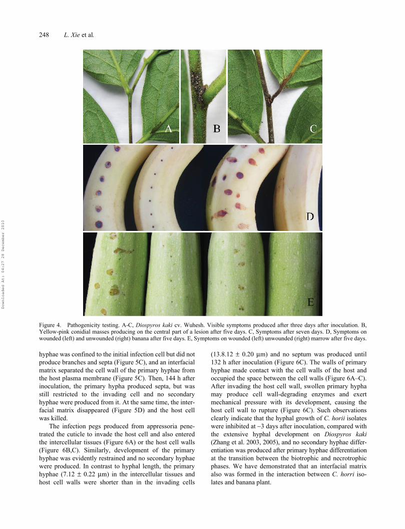
Five isolates showed strong virulence, with anthrac-nose symptoms occurring on unwounded/wounded per-simmon twigs and leaf veins within 3 days followinginoculation (Figure 4A). Serious symptoms resultedwithin 5 days. Lesion size varied greatly on inoculatedtwigs and leaf veins, depending on the age of the persim-mon twigs. Average lesion size on the unwounded/wounded persimmon twigs was 3.75 (±0.94) cm and 3.68(±1.73) cm, respectively; and 2.07 ± 0.67 (±0.67) cm and2.26 (±0.72) cm on the unwounded/wounded leaf veins.Large numbers of yellow-pink conidial masses were pro-duced on the central parts of lesions (Figure 4B). Sevendays after inoculation, the lesions extended to almostentire twigs or main leaf veins (Figure 4C). In contrast,
smaller lesions were found on unwounded/woundedbanana (0.68 ± 0.08/0.8 ± 0.07 cm) (Figure 4D) and marrow(0.76 ± 0.09/0.92 ± 0.08 cm) fruits (Figure 4E), as well ason unwounded green pepper fruits (confined to inoculationsites), 5 days after inoculation. No conidial masses wereproduced on the lesions until 7 days and no infected symp-toms were observed on the other plants inoculated.
Pathogenicity testing indicated that D. horii infectedunwounded banana and marrow fruits. Interestingly, mar-row plants are grown in the region but no infection hasbeen reported under field conditions. Banana plants do notgrow in this location. Artificial host inoculation is usuallynot reliable enough for determining host-specificity (usuallybeing determined by natural infection), but may indicatethe potential for infection (Freeman et al. 1998), cross-infection potential and cytological characteristics of theinfection process.
Infection processOn persimmonThe infection process of C. horii on susceptible Diospyroskaki cv. Wuheshi has been well documented (Zhang et al.
Figure 3. Morphological characteristics of C. horii (from Diospyros kaki cv. Wuhesh). A-C, on PDA. A, view of colony. B, reverseview of colony. C, conidia. D, appressoria formed on polystyrene Petri dish. E, appressoria formed on coverslips. Bar = 10 μm.
Downloaded At: 04:27 28 December 2010

Mycology 247
2004; Zhang 2008). C. horii exhibits an infection strategyof intracellular colonization and fungal hyphae growthwithin the cell lumen without penetrating host protoplasts.This is similar to that of C. lindemuthianum on beans andC. sublineolum on sorghum (O’Connell et al. 1985;Wharton et al. 2001). This research also revealed thatcytological and ultrastructural characters are similar in theprocess of infection of twigs and petioles, closely resem-bling that of C. lindemuthianum on bean (Zhang et al.2003, 2005). In contrast, conidia of C. horii germinate ontwigs of the resistant Zhejing persimmon (D. glaucifolia)and produce germ tubes that form dark appressoria which,in turn, produce infection pegs and penetrate the cuticlewithin 24 h of inoculation. The fungus then became quies-cent and did not develop further and no visible symptomsof disease were observed (Zhang and Xu 2005). This phe-nomenon may be similar to that of quiescent infection inunripe avocado fruits attacked by Colletotrichum gloe-osporioides (Prusky 1996).
Infection process on banana and marrowThe infection process of C. horii on banana (Musa acumi-nata) and marrow (Cucurbita pepo) was observed withfruits inoculated with a conidial suspension, as describedabove. Pieces were cut from the inoculated banana andmarrow skins and decolourized in a 0.15% (w/v) solutionof trichloroacetic acid in a 3:1 (v/v) mixture of ethanol andchloroform for 14 h. They were then stained in a 0.025%
(w/v) solution of aniline blue in lactophenol for 3–4 h, asdescribed by Sun and Zhang (2009). Light microscopicexamination was made with a Zeiss Axiophot 2 micro-scope with Axiocam CCD camera and Axiovision digitalimaging software (AxioVision Software Release 3.1, ver.3-2002; Carl Zeiss Vision Imaging Systems). The prein-fection stages of C. horii isolates on banana are very simi-lar to those on Diospyros kaki (Zhang et al. 2003, 2005), inwhich conidia adhere to and germinate on the plant sur-face, producing germ tubes that form appressoria 12 h afterinoculation, which in turn produced infection pegs 24 h afterinoculation and penetrate the cuticle directly. About 48 hafter inoculation, the inoculated tissue sites becamebrown, but no infection vesicle were seen; these appeared60 h after inoculation. Ninety six hours after inoculation,the primary hypha formed in the initial infection cell(Figure 5A) and was surrounded by an invaginated plasmamembrane. The primary hyphae did make contact with theplasma membrane and were separated by an interfacialmatrix (Figure 5B), closely resembling that on Diospyroskaki (Zhang et al. 2005; Zhang 2008). The host plasmamembrane surrounding the primary hypha became thickand was probably deposited by the opaque material, aspreviously described on Diospyros kaki (Figure 5B)(Zhang et al. 2003). However, in contrast to Diospyroskaki (Zhang 2008), the disease development was limitedand the primary hyphae developed very slowly. Even 120h after inoculation, the lesions were only slightly largerthan the inoculated sites, and dark brown. The primary
Table 1. Sources of isolates used in this study.
Colletotrichum species Isolates
GenBank Accession Number
ACT TUB-2 CAL GS ITS
C. horii TSG001 GU133374 GU133375 GU133376 GU133377 AY787483C .horii TSG002 GU133379 GU133380 GU133381 GU133382 AY791890C .horii TSG003 GU133384 GU133398 GU133385 GU133386 AY791894C. horii TSG004 GU133388 GU133389 GU133390 GU133391 AY791893C .horii TSG005 GU133393 GU133394 GU133395 GU133396 AY791892C. asianum BML-I3 FJ 903188 FJ 907434 FJ 917501 FJ 972586 FJ 972605C. asianum BPD-I4 FJ 907424 FJ 907439 FJ 917506 FJ 972595 FJ 972612C. asiaunm BML-I14 FJ 907421 FJ 907436 FJ 917503 FJ 972598 FJ 972615C. siamense BML-I6 FJ 907420 FJ 907435 FJ 917502 FJ 972599 FJ 972604C. siamense BPD-I2 FJ 907423 FJ 907438 FJ 917505 FJ 972596 FJ 972613C. siamese BML-I5 FJ 907422 FJ 907437 FJ 917504 FJ 972597 FJ 972614C. fruticola BPD-I18 FJ 907427 FJ 907442 FJ 917509 FJ 972592 FJ 972602C. fruticola BPD-I12 FJ 907425 FJ 907440 FJ 917507 FJ 972594 FJ 972611C. fruticola BPD-I16 FJ 907426 FJ 907441 FJ 917508 FJ 972593 FJ 972603C. acutatam BRIP 28519 FJ 907428 FJ 907443 FJ 917510 FJ 972591 FJ 972601C. acutatam CBS 294.67 FJ 907429 FJ 907444 FJ 917511 FJ 972590 FJ 972610C. falcatum Epitype FAL FJ 907431 GQ 289454 FJ 917513 FJ 972600 FJ 972606C. gloeosporioides CBS 953.97 FJ 907430 FJ 907445 FJ 917512 FJ 972589 FJ 972609C. kahawae IMI 319418 FJ 907432 FJ 907446 FJ 917514 FJ 972588 FJ 972608C. kahawae IMI 363578 FJ 907433 FJ 907447 FJ 917515 FJ 972587 FJ 972607
Note: ACT: actin; TUB-2: partial ß-tubulin (tub2); CAL: calmoudulin; GS: glutamine synthetase; GDPH: glyceraldehydes-3-phosphate dehydrogenase;ITS: complete rDNA-ITS region; MFU: Mae Fah Luang University, Thailand; IFRD: International Fungal Research and Development Centre, China;CBS: Centraalbureau voor Schimmelcultures, Utrecht, The Netherlands; BRIP: Plant Pathology Herbarium, Department of Primary Industries, Queensland,Australia; IMI: CABI Europe UK, Bakeham Lane, Egham, Surrey TW209TY, UK; *: holotype.
Downloaded At: 04:27 28 December 2010
248 L. Xie et al.
hyphae was confined to the initial infection cell but did notproduce branches and septa (Figure 5C), and an interfacialmatrix separated the cell wall of the primary hyphae fromthe host plasma membrane (Figure 5C). Then, 144 h afterinoculation, the primary hypha produced septa, but wasstill restricted to the invading cell and no secondaryhyphae were produced from it. At the same time, the inter-facial matrix disappeared (Figure 5D) and the host cellwas killed.
The infection pegs produced from appressoria pene-trated the cuticle to invade the host cell and also enteredthe intercellular tissues (Figure 6A) or the host cell walls(Figure 6B,C). Similarly, development of the primaryhyphae was evidently restrained and no secondary hyphaewere produced. In contrast to hyphal length, the primaryhyphae (7.12 ± 0.22 μm) in the intercellular tissues andhost cell walls were shorter than in the invading cells
(13.8.12 ± 0.20 μm) and no septum was produced until132 h after inoculation (Figure 6C). The walls of primaryhyphae made contact with the cell walls of the host andoccupied the space between the cell walls (Figure 6A–C).After invading the host cell wall, swollen primary hyphamay produce cell wall-degrading enzymes and exertmechanical pressure with its development, causing thehost cell wall to rupture (Figure 6C). Such observationsclearly indicate that the hyphal growth of C. horii isolateswere inhibited at ∼3 days after inoculation, compared withthe extensive hyphal development on Diospyros kaki(Zhang et al. 2003, 2005), and no secondary hyphae differ-entiation was produced after primary hyphae differentiationat the transition between the biotrophic and necrotrophicphases. We have demonstrated that an interfacial matrixalso was formed in the interaction between C. horri iso-lates and banana plant.
Figure 4. Pathogenicity testing. A-C, Diospyros kaki cv. Wuhesh. Visible symptoms produced after three days after inoculation. B,Yellow-pink conidial masses producing on the central part of a lesion after five days. C, Symptoms after seven days. D, Symptoms onwounded (left) and unwounded (right) banana after five days. E, Symptoms on wounded (left) unwounded (right) marrow after five days.
Downloaded At: 04:27 28 December 2010

Mycology 249
Similarly, the initial stages of C. horii isolates on mar-row are very similar to that on banana, in which theappressoria produce infection pegs 24 h after inoculation,penetrating the cuticle directly. However, the infectionpegs seemed to cease development until 144 h after inocu-lation, as described on twigs of the resistant Zhejing per-simmon (D. glaucifolia) (Zhang and Xu 2005).
Species delineation in Colletotrichum is confused, beingbased on few morphological characters and host relation-ships. Cytological studies clearly show that the infection pro-cess and intracellular infection structures of C. horii aredifferent from that of C. gloeosporioides (Sutton 1992) and
similar to hemibiotrophic species of Colletotrichum (Perfectet al. 1999), but more closely related to that of C. lindemuth-ianum on bean (O’Connell et al. 1985; Zhang et al. 2003,2005; Zhang 2008) and banana. However, morphologically,C. horii is different from C. lindemuthianum when conidialsize 9.5–11.5 × 3.5–4.5 μm is compared (Sutton 1992).
Effects of environment factors on growth and development of C. horiiColletotrichum horii overwinters mainly in lesions of livingtwigs (Zhang and Xu 2003). Although it was believed that
Figure 5. Development of fungal infection structures on host cells during interaction of banana with C. horii isolates. A, a primaryhypha in an initial infection cell 96 h after inoculation. B, enlarged portion of (A). Note that a primary hypha is separated by an interfacialmatrix from the invaginated plasma membrane (arrowhead) surrounding it. C, a primary hypha in an initial infection cell 120 h afterinoculation. Note that an enlarged portion of (C) at the right top, in which a primary hypha did not make contact with the host plasmamembrane (arrowlead). D, a primary hypha in an initial infection cell 144 h after inoculation. A, appressorium; PH, primary hyphae; IV,infection vesicle; S, septum; Bar = 10 μm.
Figure 6. Development of fungal infection structures in host intercellular space and cell walls during interaction of banana with C. horiiisolates. A, a primary hypha in intercellular space 120 h after inoculation. Note that a primary hyphae is in close contact with the host cellwalls. B, a primary hypha in cell wall 120 h after inoculation. C, a primary hypha in cell wall 132 h after inoculation. Note that swollenprimary hypha has caused the host cell wall to curve and rupture (arrowhead).
Downloaded At: 04:27 28 December 2010

250 L. Xie et al.
pathogen could overwinter on dead leaves and fruits, noexperimental data has been provided (Jia et al. 1997). Inthe field, the disease first appears in the vicinity of previ-ously diseased twigs and no symptoms are visible onnewly-formed twigs near the ground. Consequently it isspeculated the pathogen may disappear when leaves andfruits rot on the ground. Detection of pathogen from vari-ous plant parts showed that the survival rate of C. horiiwas 16.67% in lesions of living twigs, and 1.56% in thesegments of dead diseased twigs (Zhang and Xu 2003).Mycelia in diseased tissues are, therefore, considered to bean important source of inocula-producing primary conidiain field. In spring, mycelia in diseased tissues produceconidia and they are dispersed by rain splash and wind tonewly formed twigs. The pathogen can also be dispersedin symptomless seedlings over long distances (Zhang andXu 2003). Symptomless persimmon seedlings from dis-eased areas produced anthracnose lesions within 1 year onnewly formed twigs. According to the position of thelesions, it was believed that the infection source wasrelated to the bud scales that carried the pathogen. In thespring, when favorable weather conditions occur, conidiathat develop from the overwintering mycelium act as theprimary source of inocula. Successful infection involvesvarious environment factors, such as temperature, humid-ity, pH and nutrition on the surface of the host, whichdetermines disease occurrence.
The effect of temperature on growth of mycelium issignificant. The optimal temperature for growth in C. horiiis ∼25°C; higher temperature inhibits mycelium growth(Zhang and Hu 2004). While conidia germinate and formappressoria over a wide range of pH 2.0–9.0, the optimal
pH for conidial germination and appressorial formationwas between pH 5.0 and 6.0. When combining tempera-ture and pH, pathogenicity testing showed interestingresults. Visible symptoms of anthracnose occurred on newtwigs at 23°C within pH 4.0–8.0 with conidial masses onlesions after 80–90 h; at 17°C and pH 6.0 without sporemasses after 7 days, but were absent at temperatures lowerthan 17°C and pH 5.0–6.0 (Zhang and Hu 2004).
Glucose also influences conidial germination andappressoria formation (Zhang and Hu 2004). The percentageof conidial germination increases with increasingconcentrations of glucose and time (Table 2). The percent-age of conidial germination increased between 21 and 59 %with the concentration of glucose (0.1–5%) after 12 h and,subsequently, the percentage in all treatments increased withtime. After 48 h, the percentage of conidia germination wentfrom 57 to 87% with increasing glucose concentrations.
Glucose inhibited appressorial formation (Table 3),which decreased from 32 to 0% after 12 h with glucoseconcentrations (0.1–5%); similarly, with increasing time,the percentage also increased correspondingly. After 48 h,the percentage climbed from 31 to 87% with increasingconcentrations of glucose.
PCR, sequencing and phylogenetic analysisPartial actin (ACT), β-tubulin (TUB2), calmodulin (CAL),glutamine synthetase (GS), glyceraldehyde-3-phosphatedehydrogenase (GPDH) genes and the complete rDNA-ITS (ITS) region from five Colletotrichum strains wereamplified by PCR, as described by Prihatsuti et al. (2009).The amplified DNA fragments were purified by an
Table 2. Rate of conidial germination in different concentration of a glucose solution.
Time (h)
Concentration of glucose (%)
0.1 0.5 1.0 2.0 3.0 4.0 5.0
12 21.0 ± 3.6a 25.7 ± 2.1ab 33.7 ± 5.8b 39.3 ± 4.9c 41.3 ± 4.9cd 47.3 ± 4.1d 59.3 ± 5.0d18 27.3 ± 2.1a 30 ± 1.0ab 41.7 ± 3.8b 45.0 ± 2.0c 47.7 ± 2.1cd 55.7 ± 3.1d 63.0 ± 5.3d24 30.7 ± 5.1a 39 ± 4.3a 49.3 ± 4.9b 51.7 ± 4.5c 56.3 ± 2.5d 64.0 ± 1.0de 77.7 ± 4.7e36 45.7 ± 2.9a 50.7 ± 3.1a 57.0 ± 2.6b 64.3 ± 3.1c 69.0 ± 5.2d 74.7 ± 3.1d 82.7 ± 2.8d48 57.3 ± 3.8a 61 ± 3.5ab 65.0 ± 4.5b 71.3 ± 3.8c 75.7 ± 4.5d 82.7 ± 7.6de 85.7 ± 6.1e
Note: Different letters following the data in the same row show significant difference at 0.5%.
Table 3. Percentage of appressorial formation in different concentrations of a glucose solution.
Time (h)
Concentration of glucose (%)
0.1 0.5 1.0 2.0 3.0 4.0 5.0
12 31.9 ± 6.9a 27.0 ± 4.4ab 21.7 ± 4.8b 11 ± 0.2c 4.9 ± 0.6cd 2.9 ± 1.4cd 0 ± 0d18 56.0 ± 6.0a 49.9% ± 9.2a 43.4 ± 9.4a 19.43 ± 5.5b 10.4 ± 4.6b 7.7 ± 1.2b 6.1 ± 1.8b24 65.9 ± 7.9a 60.6% ± 4.7ab 50.6 ± 4.8b 35.47 ± 0.7c 21.4 ± 3.9d 17.2 ± 2.9d 12.6 ± 3.8d36 75.1 ± 8.2a 73.1% ± 8.7a 61.9 ± 2.3b 41.1 ± 6.6bc 29.3 ± 2.2a 26.3 ± 1.9c 20.2 ± 3.9c48 87 ± 3.5a 82.9% ± 4.4ab 77.4 ± 3.7b 58.33 ± 4.1c 40.4 ± 6.8d 37.1 ± 0.5de 31.3 ± 5.1e
Note: Different letters following the data in the same row show significant difference at 0.5%.
Downloaded At: 04:27 28 December 2010
Mycology 251
Axygen PCR purification kit. Purified PCR products forpartial actin, beta-tubulin, calmodulin and glutamine syn-thetase were ligated into a pGEM-T vector (TaKaRa Co.,Japan), and the ligated products were transformed intoDH5a. The positive clone was propagated, the recom-binant plasmids were extracted according to the manufac-turer’s instructions (Axygen Bioscience), and identified byPCR and restriction endomuclease enzyme digestion. Thesequence determination of PCR products and recombinantplasmid was carried out by Hangzhou Genomics Institutefor sequencing in both directions. DNA sequencings wereperformed at the SinoGenoMax Company Limited. The
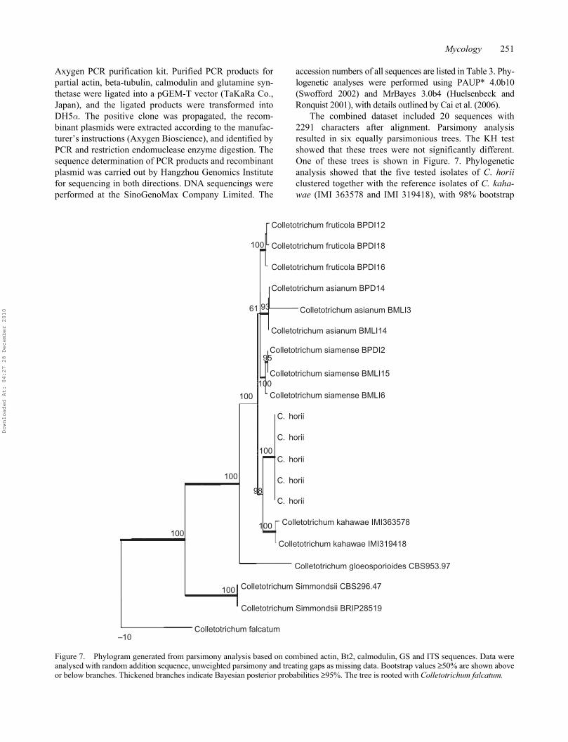
accession numbers of all sequences are listed in Table 3. Phy-logenetic analyses were performed using PAUP* 4.0b10(Swofford 2002) and MrBayes 3.0b4 (Huelsenbeck andRonquist 2001), with details outlined by Cai et al. (2006).
The combined dataset included 20 sequences with2291 characters after alignment. Parsimony analysisresulted in six equally parsimonious trees. The KH testshowed that these trees were not significantly different.One of these trees is shown in Figure. 7. Phylogeneticanalysis showed that the five tested isolates of C. horiiclustered together with the reference isolates of C. kaha-wae (IMI 363578 and IMI 319418), with 98% bootstrap
Figure 7. Phylogram generated from parsimony analysis based on combined actin, Bt2, calmodulin, GS and ITS sequences. Data wereanalysed with random addition sequence, unweighted parsimony and treating gaps as missing data. Bootstrap values ≥50% are shown aboveor below branches. Thickened branches indicate Bayesian posterior probabilities ≥95%. The tree is rooted with Colletotrichum falcatum.
Colletotrichum fruticola BPDI12
Colletotrichum fruticola BPDI18
Colletotrichum fruticola BPDI16
Colletotrichum asianum BPD14
Colletotrichum asianum BMLI3
Colletotrichum asianum BMLI14
Colletotrichum siamense BPDI2
Colletotrichum siamense BMLI15
Colletotrichum siamense BMLI6
Colletotrichum kahawae IMI363578
Colletotrichum kahawae IMI319418
Colletotrichum gloeosporioides CBS953.97
Colletotrichum Simmondsii BRIP28519
Colletotrichum falcatum
C. horii
C. horii
C. horii
C. horii
C. horii
Colletotrichum Simmondsii CBS296.47
–10
100
100
100
100
100
100
93
95
98
61
100
100
Downloaded At: 04:27 28 December 2010

252 L. Xie et al.
support. Two species could be well differentiated bythe conidia size, as C. horii has much larger conidia thanC. kahawae (12.5–19.0 × 4.0 μm) (Waller et al. 1993).
The phylogenetic relationship between C. horii and itsclosely related species in Colletotrichum is analyzed basedon combined partial actin, beta-tubulin, calmodulin,glutamine synthetase and ITS/5.8S gene sequence data.The results of phylogenetic analysis clearly indicated thatC. horri is a distinct species, closely related to C. kahawae.
Future prospectsIn this paper, we review the available information on Col-letotrichum horii, which will facilitate its correct andaccurate identification. Improved agricultural productionrequires accurate identification of pathogens to enablemore effective disease control and management (Thanet al. 2008). Previously, we had found that symptomlessseedlings of persimmon carried C. horii, which lead to itsdispersal over long distances (Zhang and Xu 2003). Asthis pathogen was previously identified as Colletotrichumgloeosporioides, which has a wide range of hosts, it hasnot been added to list of quarantine pests in China. In addi-tion, the percentage of bud scales that carried the pathogenis very low, ranging from 3.3 to 4% (Zhang and Xu 2003).Although latent infection by C. horii in persimmon seedlingsis undergoing extensive research (http://www.cab.zju.edu.cn/instswjs/people/zhang-jz/c3.htm), methods that enable itsrapid and accurate detection from a large numbers of per-simmon seedlings need to be improved.
The infection process in C. horii is well understood. C.horii provides an excellent pathosystem for studying themolecular basis for infection and fungal–plant interac-tions. To understand the molecular mechanisms of interac-tions between the pathogen and host, a genomic library ofC. horii has been constructed and cloning of pathogenesis-related genes have also been performed by Agrobacteriumtumefaciens - mediated transformation technique (Sunet al. 2008). A few mutants have been shown to be relatedto pathogenicity, and corresponding gene fragments hadhave been cloned (Sun et al. 2008). Identification andfunctional analysis of pathogenesis-related genes are amajor undertaking for future studies but will provide newinsights into the molecular mechanism of infection struc-ture differentiation and fungal–plant interactions.
AcknowledgementsThis work was supported by the National Natural Science Foun-dation of China (No. 30571208).
ReferencesArx JA von. 1957. Die Arten der Gattung Colletotrichum Cda.
Phytopathologische Zeitschrift 29:413–468.
Cai L, Hyde KD, Taylor PWJ, Weir BS, Waller JM, Abang MM,Zhang JZ, Yang YL, Phoulivong S, Liu ZY, Prihastuti H,Shivas RG, McKenzie EHC, Johnston PR. 2009. A polypha-sic approach for studying Colletotrichum. Fungal Diversity39:183–204.
Cannon PF, Buddie AG, Bridge PD. 2008. The typification ofColletotrichum gloeosporioides. Mycotaxon 104:189–204.
Crouch JA, Beirn LA, Cortese LM, Bonos SB, Clarke BB. 2009.Anthracnose disease of switchgrass caused by the novelfungal species Colletotrichum navitas. Mycol Res.113:1411–1421.
Hori S. 1910a. Kaki no Shinbyogai Tansobyo. Engei no Tomo 6(1):58–61.
Hori S. 1910b. Kaki no Shinbyogai Tansobyo. Engei no Tomo 6(2):21–24.
Huelsenbeck JP, Ronquist FR. 2001. MRBAYES: Bayesianinference of phylogenetic trees. Biometrics 17:754–755.
Jia KF, Cheng Y, Wang LH. 1997. Epidemic factors and controltechniques of Japanese sweet kaki persimmon anthracnose. JZhejiang Forestry College 14 (1):45–49.
Johnston PR, Jones D. 1997. Relationship among Colletotrichumisolates from fruit rots assessed using rDNA sequences.Mycologia 89:420–430.
Hyde KD, Zhang Y. 2008. Epitypification: should we epitypify?J Zhejiang Univ Sci. B 9:842–846.
Maffei L. 1921. Una malattia delle foglie del “Kaki” dovuta alColletotrichum kaki n. sp. Riv Patol Veg. 11:116–118.
O’Connell RJ, Bailey JA, Richmond DV. 1985. Cytology andphysiology of infection of Phaseolus vulgaris by Colletotri-chum lindemuthianum. Physiol Plant Pathol. 27:75–98.
Perfect SE, Hughes HB, O′Connell RJ, Green JR. 1999. Colle-totrichum: A model genus for studies on pathology andfungal–plant interactions, Fung Genet Biol. 27:186–198.
Prusky D. 1996. Quiescent infections by postharvest pathogens.Annu Rev Phytopathol. 34:413–434.
Sawada K. 1933. Report of survey on fungi in Taiwan. Reportof Agriculture Ministry of Taiwan Center Institute No.61:1–99.
Shenoy BD, Jeewon R, Lam WH, Bhat DJ, Than PP, TaylorPWJ, Hyde KD. 2007. Morpho-molecular characterisationand epitypification of Colletotrichum capsici (Glomeral-laceae, Sordariomycetes), the causative agent of anthracnosein chilli. Fungal Diversity 27:197–211.
Shenoy BD, Jeewon R, Hyde KA. 2007. Impact of DNAsequence-data on the taxonomy of anamorphic fungi. FungalDiversity 26(1):1–54.
Sreenivasaprasad S, Mills PR, Meehan B and Brown AE. 1996.Phylogeny and systematics of 18 Colletotrichum speciesbased on ribosomal DNA sequences. Genome 39:499–512.
Sun H, Xu T, Zhang JZ. 2008. Genome library construction ofanthracnose pathogen on persimmon and screening of patho-genesis related mutants for analysis. PhD thesis, ZhejiangUniversity, Zhejiang, China, 151 pp.
Sun H, Zhang JZ. 2009. Colletotrichum destructivum fromcowpea infecting Arabidopsis thaliana and its identity toC. higginsianum. Eur J Plant Pathol. 125:459–469.
Sutton BC. 1992. The genus Glomerella and its anamorph Colle-totrichum. In: Bailey, J. A., and Jeger, M. J., eds., Colletotri-chum: Biology, Pathology and Control. pp. Wallingford:CAB International; pp. 1–27.
Swofford DL. 2002. PAUP*: Phylogenetic Analysis Using Parsi-mony (*and other methods). Version 4b10. Sunderland(MA): Sinauer Associates.
Than PP, Prihastuti H, Phoulivong S, Taylor PWJ, Hyde KD.2008. Review: Chilli anthracnose disease caused by Colle-totrichum species. J Zhejiang Univ. 9:764–778.
Downloaded At: 04:27 28 December 2010

Mycology 253
Waller JM, Bridge PD, Black RL, Hakiza G. 1993. Characteriza-tion of the coffee berry disease pathogen, Colletotrichumkahawae Sp. Nov. Mycol Res. 97:989–994.
Weir BS, Johnston PR. 2010. Characterisation and epitypifica-tion of Gloeosporium kaki Hori as Colletotrichum horii nom.nov. Mycotaxon (in press).
Wharton PS, Julian AM, O’Connell RJ. 2001. Ultrastructure ofthe infection of Sorghum bicolor by Colletotrichum subline-olum. Phytopathology 91:149–158.
Zhang JZ. 2008. Anthracnose of persimmon caused by Colletot-richum gloeosporioides in China. Asian Australasian J PlantSci Biotechnol. 2 (2):50–54.
Zhang JZ, Hu DW. 2004. Effects of environment factors onconidia germination, appresorium formation and pathogenic-ity of the persimmon anthracnose pathogens Collectotrichumgloeosporioides. Acta Phytopathol Sinica 22(4):645–652.
Zhang JZ, Xu T. 2003. Various stages and amount of Colltotri-chum gloeosporiodes on overwintering twigs of persimmon.J Plant Prot. 30(4):437–438.
Zhang JZ, Xu T. 2005. Cytological characteristics of the infec-tion in different species, varieties and organs of persimmonby Colletotrichum gloeosporioides. Mycosystema 24(1):116–122.
Zhang JZ, Hu DW, Xu T. 2003. Studies on cytology of the infec-tion of persimmon by Colletotrichum gloeosporioides. Myc-osystema 22(4):645–652.
Zhang JZ, Hu DW, Xu T. 2005a. Ultrastructure of infection ofpersimmon petiole by Collectotrichum gloeosporioides.Acta Phytopathol Sinica 35(5):434–441.
Zhang JZ, Xu T, He LP. 2005b. Anthracnose pathogen on Diospy-ros kaki cv. Wuheshi and its nuclear behavior in process ofappressorium formation. Mycosystema 24(3):446–456.
Downloaded At: 04:27 28 December 2010