BIOLOGY - ANDHRA PRADESH OPEN SCHOOL SOCIETY
312
314 INTERMEDIATE BIOLOGY CORE MODULES-3 & LABORATORY MANUAL-4 A.P. OPEN SCHOOL SOCIETY, GUNTUR, AMARAVATI. Opp. Old Bus Stand, Govt. Urdu Boys High School, Pariksha Bhavan, GUNTUR – 522001, Phone: 0863-2239151 F 1 Website: www.apopenschool.ap.gov.in ; E-mail: [email protected]
-
Upload
khangminh22 -
Category
Documents
-
view
1 -
download
0
Transcript of BIOLOGY - ANDHRA PRADESH OPEN SCHOOL SOCIETY

314 INTERMEDIATE
BIOLOGY CORE MODULES-3
& LABORATORY MANUAL-4
A.P. OPEN SCHOOL SOCIETY, GUNTUR, AMARAVATI. Opp. Old Bus Stand, Govt. Urdu Boys High School, Pariksha Bhavan,
GUNTUR – 522001, Phone: 0863-2239151
F
1
Website: www.apopenschool.ap.gov.in ; E-mail: [email protected]

© Andhra Pradesh Open School Society
Government of Andhra Pradesh, Amaravati
Published: 2018, 2019, 2020
All Rights Reserved
No part of this publication may be reproduced, stored in a retrieval system or transmitted, in any form or by any means without the prior permission, in writing of the publisher, nor be otherwise circulated in any form of binding or cover.
M/s. V.G.S. BOOK LINKS, Vijayawada
Printed at
For the Director A.P. Govt. Text Book PressAmaravati, Andhra Pradesh

FOREWORD
The A.P. Open School Society (APOSS) is the first State Open
School in the country established in the year 1991 with an objective of
providing opportunities of alternative Schooling in Open Distance
Learning mode.
The Study Materials developed are of Self Learning in nature and
in accordance with National Curriculum Frame work, 2005. Learners are
advised to go through the Text Books/Study materials thoroughly before
attending the Personal Contact Programme. If any doubts persist get
them clarified with the subject counsellors at the Study Centres. Attend
all the Personal Contact Programme classes for better understanding of
the subjects. Do write the Tutor Marked Assignments (TMAs) and
Preparatory Examinations to check your progress in the subjects
concerned. The Public Examination Model Papers are given at the end
of Study Materials so as to enable learners to prepare well for the
examinations.
Best wishes to all the learners and happy learning experience at
APOSS.
DIRECTOR, APOSS Dr. K.V. SRINIVASULU REDDY

CURRICULUM COMMITTEEChair Person
Prof. Keshav TrehanDeptt. of Botany, Kurukshetra University, Kurukshetra
MEMBERS
Neelam GuptaExecutive Officer NIOS,New Delhi.
Dr. Meenakshi AryaRetd. Reader (Botany)
Kanodia Kanya Mahavidhyala
Jaipur
Prof. Tasneen FatimaDeptt. of BotanyJamia Millia IslamiaNew Delhi.
Dr. D.K.RaoAcademic Officer, NIOS
New Delhi.
Dr. Bharati SarkarRetd. Reader (Botany)
Maitreyi College, Delhi.
Dr. Aparna KonarRetd Reader (Botany)
Maitreyi College, Delhi.
Dr. (Mrs) Jasvant SokhiReader in Life SciencesSchool of Science, IGNOUMaidan Garhi, New Delhi.
Dr. Bharati SarkarRetd. Reader (Botany)
Maitreyi College, Delhi.
Dr. Jeepinder Jeet KaurLecturer
Khalsa College, Delhi University.
Neelam GuptaExecutive Officer NIOS,New Delhi.
Coordinated byMs. Asheema Singh
Project Officer (AEP)
National Institute of Open
Schooling, NOIDA.
Supported byMinistry of Human
Resource Development
Govt. of India, India.
Funded byUnited Nationas Population
Fund (UNFPA)
New Delhi, India.
COURSE EDITORS
LESSON WRITERS
ADOLESCENCE EDUCATION PROGRAMME
Mr. Mahesh SharmaGraphic Artist
NIOS, New Delhi.
Ms. Krishna KumariGraphic Artist.
Mr. Vinod PanditGraphic Artist.
GRAPHIC ARTISTS
Dr. Bharati SarkarRetd. Reader (Zoology)
Maitreyi College, Delhi.
Dr. Rita SinghDeptt. of BotanyGGB Singh Indraprasthu UniversityDelhi.
Mrs. Durga JodhaniRetd. Vice Principal
Central School No.3 NH-41
Faridabad.
Dr. H.S.VishnoiRetd. Reader (Zoology)
Delhi University, Delhi.
Dr. (Mrs) Jasvant SokhiReader in Life SciencesSchool of Science, IGNOUMaidan Garhi, New Delhi.
Mrs. Shivani GoswamiEx-Hod(Biology)Mother International School,New Delhi.
��

Dear Learner,
The Academic Department at the National Institute of Open Schooling tries to bring younew programmes in accordance with your needs are requirements. After making acomprehensive study, we found that our curriculum is more functional, related to lifesituations and simple. The task now was to make it more effective and useful for you. Weinvited leading educationists of the country and under their guidence, we have been ableto revise and update the curriculum in the subject of Biology in the light of NationalCurriculum framework -2005.
At the same time, we have also removed old, outdated information and added new, relevantthings and tried to make the learning material attractive and appealing for you.
I hope you will find the new material interesting and exciting with lots of activities to do.Any suggestions for further improvement are welcome.
Let me wich you all a happy and successful future.
A NOTE FROM THE DIRECTOR
v

A word with you
��

HOW TO USE STUDY MATERIAL
���

Overview of the Learning Material
9. Nutrition in plants - Mineral Nutrition
10. Nitrogen Metabolism
11. Photosynthesis
12. Respiration in Plants
13. Nutrition and Digestion
14. Respiration and Elimination of Nitrogenous
Wastes
15. Circulation of Body Fluids
16. Coordination and Control - The Nervous and
Endocrine Systems
17. Homeostasis : The Steady State
Module-1 Diversity and Evolution of Life1. Origin and Evolution of Life and Introduction to
Classification
2. The Kingdom Monera, Protoctista and Fungi
3. Kingdoms Plantae and Animalia
4. Cell Strueture and Function
5. Tissues and other Level of Organization
Module-2 Forms and Functions of Plants and Animals6. Root System
7. Shoot System
8. Absorption, Transport and Water Loss in Plants
2CORE MODULES
CORE MODULES
3CORE MODULES
1
25. Conservation and Use of Natural Resources
26. Pollution
27. Nutrition and Health
28. Some Common Human Diseases
Module-5 Emerging Areas in Biology
29. Biotechnology
30. Immunobiology : An Introduction
Module-3 Reproduction and Heredity18. Reproduction in Plants
19. Growth and Development in Plants
20. Reproduction and Population Control
21. Principles of Genetics
22. Molectular Inheritance and Gene Expression
23. Genetics and Society
Module-4 Environment and Health24. Principles of Ecology
Module-6B Economic Biology31. Agriculture, Forestry and Medicinal Plants
32. Floriculture, Mushroom Culture and Hydroponics
33. Animal Husbandry
34. Fisheries and Aquaculture
35. Apiculture, Lac Culture and Sericulture
Module-6A Tools and Techniques in Biology31. Some Common Tools and Techniques used in
Biology
32. General Laboratory Equipments
33. Some Common Preservatives, Stains and Reagents
34. Providing Organisms for Laboratory Work
35. Some Aids in Biology
����

Module Name of the Lesson Page No.
Module-6A Tools and Techniques 31. Some Common Tools and Techniques 1in Biology used in Biology
32. General Laboratory Equipments 11
33. Some Common Preservatives, Stains 22and Reagents
34. Providing Organisms for Laboratory Work 29
35. Some Aids in Biology 41
Module-6B Economic Biology 31. Agriculture, Forestry and Medicinal Plants 57
14. Animal breading 73/1
12. Plants and human welfare 73/5
32. Floriculture, Mushroom Culture 74and Hydroponics
33. Animal Husbandry 86
34. Fisheries and Aquaculture 99
35. Apiculture, Lac Culture and Sericulture 100
Appendix A : Glossary
Appendix B : Questions for Practice
Appendix C : Sample Questions Paper (With Questionpaper Design and MarkingScheme)
Appendix D : Feedback Form
CONTENTS
��


31
SOME COMMON TOOLS ANDTECHNIQUES USED IN BIOLOGY
Long back, the biologists could learn about living things, only from what they couldsee with the naked eye. New tools and techniques were invented which helped inthe study of finer structure of various kinds of organisms and their parts. Microscopenot only revealed a world of minute organisms but also minute details of internalstructure of organisms. In the course of history of biology, various new tools andtechniques have developed, like microscopy, paper chromatography, etc. In thislesson you will learn about some of these.
OBJECTIVES
After completing this lesson, you will be able to :
� trace the development of microscopes and their working;
� list the parts of a compound microscope;
� compare the working principle of compound, electron and phase contrastmicroscope;
� Differentiate between transmission electron microscope and scanning electronmicroscope;
� describe the basic aspects of some other techniques like cytochemistry,autoradiography, paper chromatography, cell fractionation , ultracentrifugationand tissue culture.
31.1 BRIEF HISTORY OF MICROSCOPESMicroscope as the name suggestes are instruments that help to enlarge minute(micro = very small) organisms or their parts. A microscope not only enlarges ormagnifies the object but also ‘resolves’ it, that is makes it possible to differentiatebetween two points present close together in the objects being viewed.
MODULE - 6ATools and Techniques in
Biology
1BIOLOGY
Notes

BIOLOGY
MODULE - 6A Some Common Tools and Techniques used in Biology
Tools and Techniques inBiology
2
Notes
The first microscope was constructed by Anton Van Leeuwenhoek (1632-1723).This microscope consisted of a single biconvex lens fitted in a small window ofa “board” and the object was viewed through it. This was a simple microscope.Next stage was that of a very primitive compound microscope in which two lenseswere used (Fig. 31.1). Improvements continued, newer and newer microscopes weredesigned and are still being improved.
Fig. 31.1 Crude microscope used by Robert Hooke.
31.2 VARIOUS TYPES OF MICROSCOPESThere are different types of microscopes which are used in studying the variousstructures and activities inside a cell. Some of these are as follows:
1. Simple microscope
2. Compound microscope
3. Phase-Contrast microscope
4. Transmission Electron microscope (TEM)
5. Scanning electron microscope (SEM)
Resolving Power : It is the ability of a microscope to show twoclosely lying points as two separate points.
Magnification : It is the ratio of the size of the image to that ofthe object.

MODULE - 6ATools and Techniques in
Biology
3
Some Common Tools and Techniques used in Biology
BIOLOGY
Notes
1. Simple Microscopes : These are oftwo types :
(i) Hand Lens : It consists of abiconvex lens, mounted on ahandle. The lens is of differentsizes and different magnifyingpowers. It is commonly usedto magnify an entire object.
(ii) Dissecting Microscope : Itconsists of a biconvex lenswhich is moved up and downby an adjustment screw, tobring the object in sharp focus. The light is focussed with the help of aconcave mirror fitted below. A magnified image of the full object can beseen through it.
Fig. 31.3 Dissecting microscope.
2. Compound Microscopes : It is commonly used in the laboratories to viewextremely minute organisms and parts and sections of larger organisms.Fig. 31.4 shows various parts of a compound microscope and table 31.1 givesthe differences between simple and compound microscope.
Fig. 31.2 A Hand Lens

BIOLOGY
MODULE - 6A Some Common Tools and Techniques used in Biology
Tools and Techniques inBiology
4
Notes
Fig. 31.4 A compound Microscope
Apart from the lenses, it also has a condenser, having a simple mirror on one sideand concave mirror on the other. The object is placed first below the objective lensover the stage. The objective lens forms an image of the object. This image is furthermagnified by the eye piece.
Table 31.1 Differences between a Simple Microscope anda Compound Microscope
Simple Microscope Compound Microscope
1. Basically one biconvex lens is used. 1. Basically two lenses are used
2. It can magnify upto 20 times. 2. It can magnify upto 1500 times3. The whole object may be seen. 3. Only a part of the object or a thin section
can be seen.
4. It uses light which is reflected by the mirror 4. It uses light which is transmitted through
and passes through the object or simply which the object.
is reflected by the object.
3. The Electron Microscope : The organelles of the cell became known after theelectron microscope was invented. As is seen in table 31.2 the magnificationand resolution of the electron microscope are much higher than that of thecompound microscope. Table 31.3 gives the comparison between transmissionelectron microscope and scanning electron microscope.

MODULE - 6ATools and Techniques in
Biology
5
Some Common Tools and Techniques used in Biology
BIOLOGY
Notes
Table 31.2 Comparison between the working of a Compound Microscopeand an Electron Microscope
Compound Microscope Electron Microscope
1. It is operated in open condition. 1. It is operated only in vacuum condition.
2. The objective lens is simply a glass lens. 2. The objective lens is electromagnetic lens.
3. The source of illumination is light. 3. The source of illumination is an electronbeam
4. The final image of an object is observed 4. The final image of an object is projectedthrough an eye-piece. on a flourescent screen.
5. It magnifies the object upto 1500 times. 5. It magnifies the object upto 200,000 times.
6. Resolution power is upto 2500Å. 6. Resolution power is upto 2.5Å.
7. It can be used to see both living and 7. It can be used to see only dead cells.dead cells.
Fig. 31.5 Showing similarities and differences between the light (compound)and electron microscope
Table 31.3 Comparison between Transmission Electron Microscope andScanning Electron Microscope
Transmission Electron Microscope Scanning Electron Microscope
1. Beam of electrons is passed through 1. Whole specimen is scanned by a beam ofsection of material to produce the image. electrons.
2. Only ultra thin sections or very small 2. Larger specimens can be viewedobjects can be examined.
3. Resolution very high. 3. Resolution inferior than that in case ofTransmission electron microscope.
BIOLOGY
MODULE - 6A Some Common Tools and Techniques used in Biology
Tools and Techniques inBiology
6
Notes
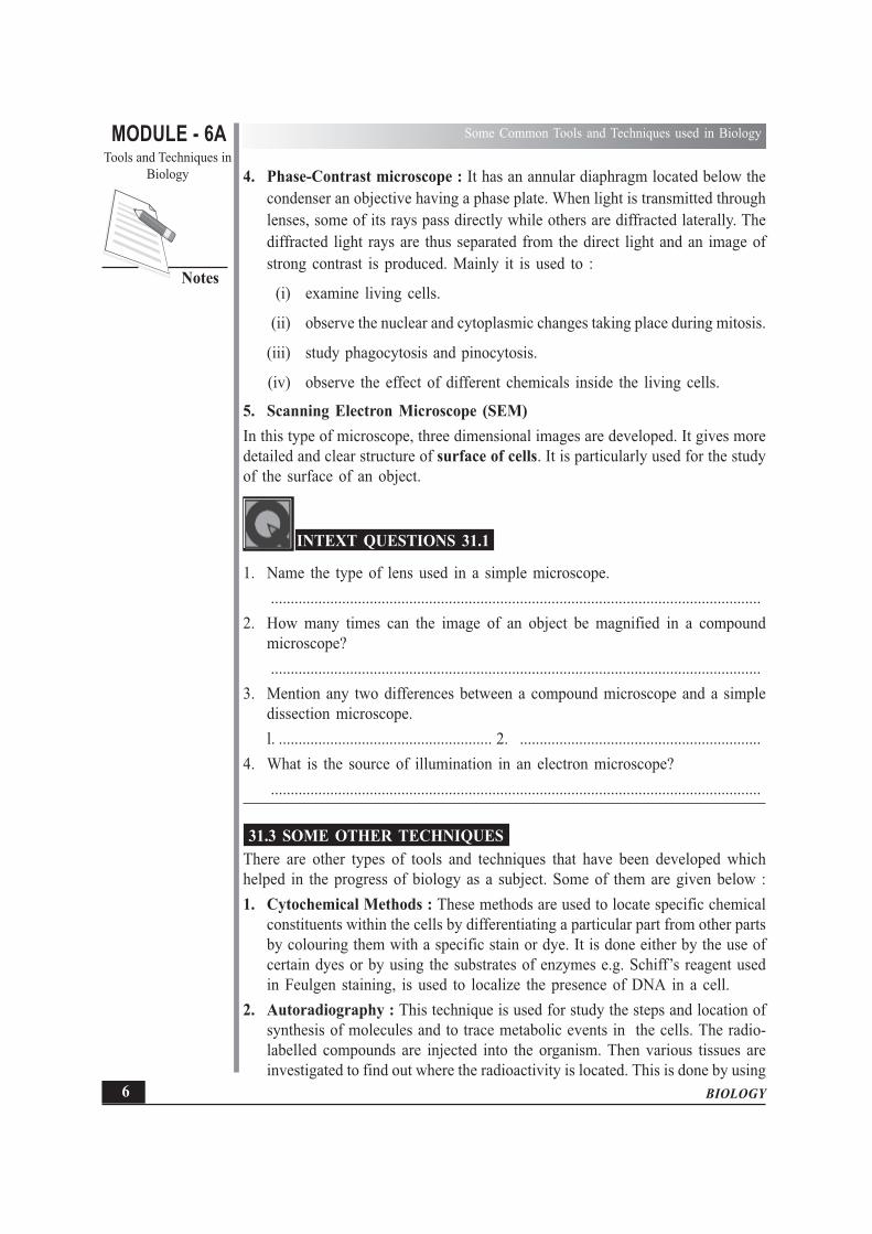
4. Phase-Contrast microscope : It has an annular diaphragm located below thecondenser an objective having a phase plate. When light is transmitted throughlenses, some of its rays pass directly while others are diffracted laterally. Thediffracted light rays are thus separated from the direct light and an image ofstrong contrast is produced. Mainly it is used to :
(i) examine living cells.
(ii) observe the nuclear and cytoplasmic changes taking place during mitosis.
(iii) study phagocytosis and pinocytosis.
(iv) observe the effect of different chemicals inside the living cells.
5. Scanning Electron Microscope (SEM)In this type of microscope, three dimensional images are developed. It gives moredetailed and clear structure of surface of cells. It is particularly used for the studyof the surface of an object.
INTEXT QUESTIONS 31.1
1. Name the type of lens used in a simple microscope.............................................................................................................................
2. How many times can the image of an object be magnified in a compoundmicroscope?............................................................................................................................
3. Mention any two differences between a compound microscope and a simpledissection microscope.l. ...................................................... 2. .............................................................
4. What is the source of illumination in an electron microscope?............................................................................................................................
31.3 SOME OTHER TECHNIQUESThere are other types of tools and techniques that have been developed whichhelped in the progress of biology as a subject. Some of them are given below :1. Cytochemical Methods : These methods are used to locate specific chemical
constituents within the cells by differentiating a particular part from other partsby colouring them with a specific stain or dye. It is done either by the use ofcertain dyes or by using the substrates of enzymes e.g. Schiff’s reagent usedin Feulgen staining, is used to localize the presence of DNA in a cell.
2. Autoradiography : This technique is used for study the steps and location ofsynthesis of molecules and to trace metabolic events in the cells. The radio-labelled compounds are injected into the organism. Then various tissues areinvestigated to find out where the radioactivity is located. This is done by using

MODULE - 6ATools and Techniques in
Biology
7
Some Common Tools and Techniques used in Biology
BIOLOGY
Notes
photosensitive film of silver bromide. Whenever in the cell or tissue or theorganism, the radio labelled substance is present, silver gets reduced byradiation and is seen as black patches in the autoradiographs.
3. Paper Chromatography : In this method the chemical substances present ina mixture can be separated. A drop of the mixture is put on one end of a longstrip of the Whatman filter paper. Thefilter paper is hung in a manner thatthe end with the drop of the mixturedips into the solvent mixture kept inthe tray/jar. As the liquid is drawn upon the paper, different substances inthe mixture begin to separate aceordingto their molecular weight, size andsolubility in the solvent and rise up todifferent heights on the paper. It isthen analysed by using certainchemicals for further investigation.
4. Cell fractionation : By this method different organelles of cells such as nucleus,mitochondria, ribosomes etc. having different particle size and weight areseparated by rotating them in a centrifuge at different speeds.
Fig. 31.7 Technique of Cell Fractionation
Fig. 31.6 Paper Chromatography

BIOLOGY
MODULE - 6A Some Common Tools and Techniques used in Biology
Tools and Techniques inBiology
8
Notes
The cells are first homogenised or broken down by a special method. Thehomogenate (crushed cells) is then put into tubes and tubes are placed in acentrifuge. The centrifuge is rotated at a high speed. By doing so under theinfluence of centrifugal force, organelles separate according to their particledensity and sizes. The lighter particles settle at the top and the heaviest particlessettle at the bottom. The layers are then studied separately and the structure indetails gets to be known.
5. Ultracentrifugation : By rotation at a high speed, particles/organelles ofdifferent sizes and shape separate, according to their density. Since the rotationis at very high speed, friction with air produces heat, so has to run underrefrigeration and vacuum. Nucleus, mitochondria etc. separate out at differentspeeds.
6. Tissue Culture : This technique involves growing living cells outside theorganism by providing all necessary conditions for their survival and growth.The cells from an organism are grown in the laboratory on a nutritive mediumat a suitable temperature. Using this technique it has been possible to developa whole organism from a single cell. Some new fully grown plants have beendeveloped in this way. (Fig. 31.8).
Fig. 31.8 Tissue culture.
The steps in tissue culture are given in Fig. 31.8. Tissue is removed from theplant body and grown in a nutrient medium. The cells divide to form anundifferentiated mass of cells called callus which then differentiates into aplant. In the diagram leaf tissue culture has been shown but tissue from any
MODULE - 6ATools and Techniques in
Biology
9
Some Common Tools and Techniques used in Biology
BIOLOGY
Notes
part of the plant has the ability to follow the similar path as shown in theFig. 31.8, and produce an entire plant. The tissue taken from the plant is calledan explants. It is now possible to culture a single cell into a whole plant.
INTEXT QUESTIONS 31.2
1. What special type of substances are injected in an organism for autoradiography?
............................................................................................................................
2. In which technique is Schiff’s reagent used?
............................................................................................................................
3. Name the technique by which the organelles from a cell can be separated.
............................................................................................................................
WHAT YOU HAVE LEARNT
� Biologists depend heavily on a number of tools and techniques for studyingorganisms.
� Microscopes, such as the simple (dissection) microscope, compound microscopeand the electron microscope are used to study organisms.
� Compound microscope uses light and can give magnification up to about 1500times whereas the electron microscope uses electron beam and magnifies theimage upto 2,00,000 times.
� Phase contrast microscope is chiefly used for observing activities inside theliving cells.
� Scanning electron microscope is used for introducing three-dimensional imageschiefly of the surfaces.
� Cytochemical methods, autoradiography, centrifugation are helpful in studyingcell chemistry, synthesis of substances inside the living organism and isolationof cell organelles respectively.
� Paper chromatography is used for separating chemical substances in a mixture.
� Tissue culture involves growing of cells and tissues outside the body of theorganism.

BIOLOGY
MODULE - 6A Some Common Tools and Techniques used in Biology
Tools and Techniques inBiology
10
Notes
TERMINAL QUESTIONS
1. Name the scientist who constructed the first microscope?
2. Mention three differences between a compound microscope and an electronmicroscope.
3. Define the term ultracentrifugation.
4. Name the microscope used in the study of a living cell and instrument usedin separating cell organelles.
5. List the main points of the technique of autoradiography.
6. Give uses of cytochemical methods and centrifugation.
7. Mention the importance of tissue culture.
ANSWER TO INTEXT QUESTIONS
31.1 1. Biconvex lens 2. upto 1500 times3. any two points given in the table 38.1 4. Electrons
31.2 1. Radiolabelled 2. Cytochemical methods3. Ultracentrifugation

MODULE - 6ATools and Techniques in
Biology
11
General Laboratory Equipments
BIOLOGY
Notes32
GENERAL LABORATORYEQUIPMENTS
A biology student has to work with various types of equipment while performingdifferent experiments. It is useful to know the principle behind the working of someof these. One main category out of these you have learnt in the previous lessoni.e. the microscopes. A few others will be explained in this lesson
OBJECTIVES
After completing this lesson you will be able to :� explain the working of an incubator and mention its uses;� explain the working of a kymograph and list its uses;� define pH and state the applications of a pH meter;� explain the working and mention the uses of autoclave;� explain the working and mention the uses of colorimeter;� describe the parts of a distillation unit and mention its applications;� Describe the working and use of spectrophotometer;� list the latest weighting balances used in laboratories and mention their need;� explain the use of centrifuge and explain the principle of centrifugation;� explain the working of microtome;� describe the working and use of sphygmomanometer.
32.1 SOME INSTRUMENTS USED IN THE LABThe following are some of the instruments :
1. Incubator (oven) regulated by a thermostatThermostat is an appliance fitted, to regulate the temperature, inside anoven or a refrigerator etc.
Incubator is an appliance shaped like a box which maintains the desired temperatureinside it (Fig. 32.1).

BIOLOGY
MODULE - 6A General Laboratory Equipments
Tools and Techniques inBiology
12
Notes
Structure : (Parts)(i) A box or container with insulated walls and a door fitted with a latch to close
the door firmly.(ii) A hole in the center of its roof for insertion of a thermometer to read the
temperature of the inside chamber.(iii) Its base contains a heating unit heated through electricity.(iv) On the front of the base on one side is a knob which can switch-on and switch-
off the instrument.(v) On the backside is fitted a thermostat to regulate the desired temperature.(vi) In the centre of the front or besides the knob, a bulb is fitted to indicate
whether the instrument is off or on(vii) The internal chamber is provided with one or more shelves.
Fig. 32.1 An incubator.
Uses : The incubator is used for the following :(i) to keep section cutting material embedded in paraffin wax (at 50-55°C)(ii) incubate eggs : for this a dish containing water is kept inside the incubator
to provide moisture to the eggs. The eggs are rotated daily to prevent stickingof the embryo to the shell membranes. The temperature is maintained at about38°- 40°C.
(iii) to study the action of chemicals, enzymes etc. at different temperatures.(iv) specimens such as pinned and stretched insects can be dried in it, so that they
are not spoiled.
MODULE - 6ATools and Techniques in
Biology
13
General Laboratory Equipments
BIOLOGY
Notes
2. Autoclave : It is an electrically operated instrument which disinfects the glass-ware before a research work is started. It works under required pressure. However,pressure cooker can be a substitute for disinfecting the small glass ware underpressure for a definite time. The autoclave works on the same principle as thepressure cooker.
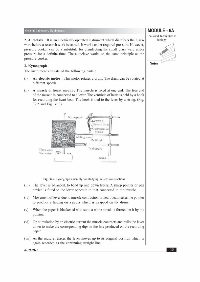
3. KymographThe instrument consists of the following parts :
(i) An electric motor : This motor rotates a drum. The drum can be rotated atdifferent speeds.
(ii) A muscle or heart mount : The muscle is fixed at one end. The free endof the muscle is connected to a lever. The ventricle of heart is held by a hookfor recording the heart beat. The hook is tied to the lever by a string. (Fig.32.2 and Fig. 32.3)
Fig. 32.2 Kymograph assembly for studying muscle constractions
(iii) The lever is balanced, to bend up and down freely. A sharp pointer or pendevice is fitted to the lever opposite to that connected to the muscle.
(iv) Movement of lever due to muscle contraction or heart beat makes the pointerto produce a tracing on a paper which is wrapped on the drum.
(v) When the paper is blackened with soot, a white streak is formed on it by thepointer.
(vi) On stimulation by an electric current the muscle contracts and pulls the leverdown to make the corresponding dips in the line produced on the recordingpaper.
(vii) As the muscle relaxes the lever moves up to its original position which isagain recorded as the continuing straight line.

BIOLOGY
MODULE - 6A General Laboratory Equipments
Tools and Techniques inBiology
14
Notes
Fig. 32.3 Kymograph assembly for studying heart contraction.
Uses :1. Used commonly for recording the reaction of a muscle when its motor nerve
is stimulated.2. Also used for recording the ventricular contraction of perfused heart.
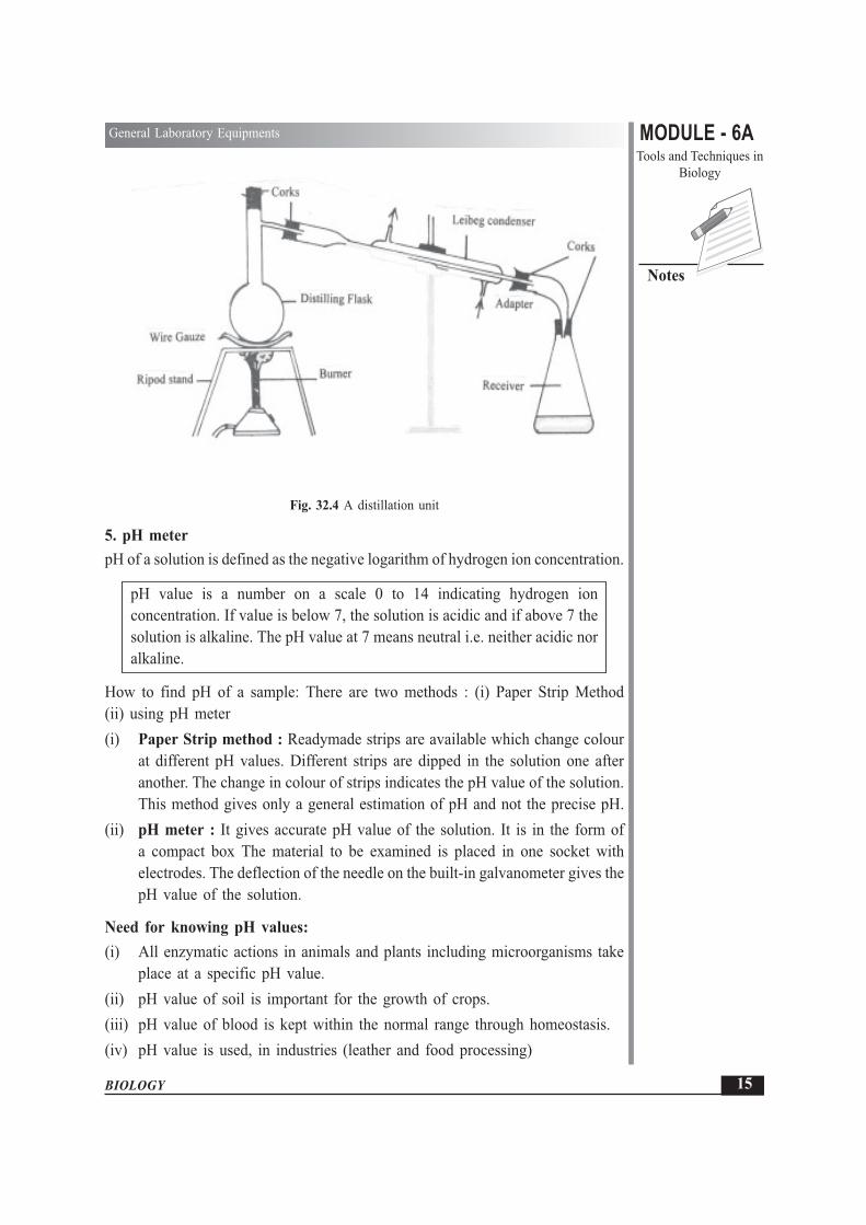
4. Distillation Unit
Distilled water is that water that has been cleared of all salts and otherimpurities which were dissolved in it.
Distilled water is an important requirement in the laboratory. Chemically, it is waterfrom which all the impurities have been removed.
A distillation unit, commonly used in Science laboratories, consists of the followingparts
(i) Distilling Flask : The size of flask varies depending on requirement. It isfilled with water and heated over flame or hot plate.
(ii) Leibig’s Condenser : It consists of an inner glass tube surrounded by a glassjacket through which water is circulated. The steam, passing through innertube condenses due to cooling effect of cold water flowing in the glass jacket.
(iii) Adapter : It is used to facilitate delivery of distillate into the receiver.
(iv) Receiver : It is a simple conical flask attached to adapter, where distillateis collected. All the connections are done through corks.
Certain precautions are taken before the start of distillation process :
1. All corks should fit tightly.
2. The apparatus should be held firmly by the help of clamp.
3. The condenser should be full of water.
4. Distillation flask should be kept over the wiregauze before heating.
Distilled water is used for preparing several reagents.
MODULE - 6ATools and Techniques in
Biology
15
General Laboratory Equipments
BIOLOGY
Notes
Fig. 32.4 A distillation unit
5. pH meterpH of a solution is defined as the negative logarithm of hydrogen ion concentration.
pH value is a number on a scale 0 to 14 indicating hydrogen ionconcentration. If value is below 7, the solution is acidic and if above 7 thesolution is alkaline. The pH value at 7 means neutral i.e. neither acidic noralkaline.
How to find pH of a sample: There are two methods : (i) Paper Strip Method(ii) using pH meter(i) Paper Strip method : Readymade strips are available which change colour
at different pH values. Different strips are dipped in the solution one afteranother. The change in colour of strips indicates the pH value of the solution.This method gives only a general estimation of pH and not the precise pH.
(ii) pH meter : It gives accurate pH value of the solution. It is in the form ofa compact box The material to be examined is placed in one socket withelectrodes. The deflection of the needle on the built-in galvanometer gives thepH value of the solution.
Need for knowing pH values:(i) All enzymatic actions in animals and plants including microorganisms take
place at a specific pH value.(ii) pH value of soil is important for the growth of crops.(iii) pH value of blood is kept within the normal range through homeostasis.(iv) pH value is used, in industries (leather and food processing)

BIOLOGY
MODULE - 6A General Laboratory Equipments
Tools and Techniques inBiology
16
Notes
6. SpectrophotometerMolecules (e.g. DNA proteins etc.) absorb and emit electromagnetic radiation ofparticular wavelength. This property of molecules is used in spectrophotometry.Spectrophotometry is a technique which is widely used to measure the absorptionof radiation in the visible and UV regions of the spectrum. Colorimeter alsofunctions on the same principles but it is a simpler instrument in which filters areused.
Colorimetry is a technique of estimating the amount or density of thecompound imparting colour to the cellular component present in a solution.
7. ColorimeterIt is an instrument to measure the absorption inthe green, blue or red regions ofthe visible spectrum. There are two type of colorimeters.(i) Visual colour colorimeter(ii) Photo electric colorimeterThe components in a colorimeter and the spectrophotometer are as follows(Fig. 32.5)
Fig 32.5 Components of a spectrophotometer.

MODULE - 6ATools and Techniques in
Biology
17
General Laboratory Equipments
BIOLOGY
Notes
1. A light source : High intensity tungsten bulb for operation in the visiblespectrum (400-700 nm) and Deuterium or Tungsten halogen lamps for UVspectrophotometry. The lamps are fitted with quartz as glass does not transmitUV rays.
2. Cuvettes made of glass or plastic are cleaned before adding the sample solution.3. Galvanometer or read out device : The reading of the standard solution is
first taken for comparison with the sample
INTEXT QUESTIONS 32.1
1. What is the pH value that indicates that a particular solution is neutral?
............................................................................................................................
2. Name the two main parts of a Kymograph?
............................................................................................................................
3. What is the final product we get from a distillation unit?
............................................................................................................................
4. Mention the use of thermostat in an incubator.
............................................................................................................................
5. What provides the power to the heating unit of incubator?
............................................................................................................................
6. Name the various components of a spectrophotometer.
............................................................................................................................
32.2 SOME OTHER TYPES OF INSTRUMENTSHere you will learn about some other types of instruments.
1. Sphygmomanometer (Blood Pressure Instrument)By this instrument, blood pressure of a person is measured.
There are three types of this instrument which are commonly used.
(i) The old or conventional type using a mercury column.
(ii) Dial type instrument without mercury column. The blood pressure (B.P.) isdirectly shown on the dial in a gadget attached to a hand pump through a tube.
(iii) The third type is the modern electronic instrument.The common part in all the above types of instruments is an inflatable rubber bagenclosed in a case called ‘cuff’. The cuff is wrapped around the arm, above theelbow and below the shoulder joint. This bag is inflated by a rubber hand pump

BIOLOGY
MODULE - 6A General Laboratory Equipments
Tools and Techniques inBiology
18
Notes
(a small balloon like structure) The hand pump is fitted with a screw to releaseair. In the conventional type of instrument another tube connects the inflatable bagwith the mercury column, movement of which, when adjusted records the systoleand diastole of heart.
Fig. 32.6 Sphygmomanometer
Stethoscope is also used to listen the throbbing in the arterywhile measuring B.P.
2. MicrotomeThe older type of microtome is called rocking microtome. It has a knife fixedvertically in front. The tissue which has to be cut is embedded in paraffin waxtrimmed into small block and is then fixed on the block holder. By moving a knob,the working handle at the rear on the base, causes the long, rocking arm to rockup and down. Due to this action a series up sections are cut from the block ofparaffin wax, fixed on the block holder. Adjustment in the microtome is done forobtaining the required thickness of the sections.The newer models of microtome are of the rotary type. In this there is a wheel whichis rotated by hand. The rest of the working is on the same general principle.
Fig. 32.7 A Rocking Microtome
Microtomes can cut thin sections easily upto the thickness of 8-10 microns(1 micron = one thousandth of a millimeter).

MODULE - 6ATools and Techniques in
Biology
19
General Laboratory Equipments
BIOLOGY
Notes
3. Centrifuge
Centrifuge is a spinning apparatus. It is used for separating objects ofdifferent densities usually from a liquid medium,
Different types of centrifuges are used for different purposes namely clinicalcentrifuge, ultracentrifuge etc. A centrifuge consists of a head (rotor) operated byhand or motor. Attached to the rotor are a number of metallic tubes. In these tubesare placed specially designed glass tubes with tapering bottom. According to therequirement the head may contain sockets in which bottles are placed. The tubesshould be properly balanced in a centrifuge to prevent wobbling.Like the mixie used in the kitchen, a centrifuge can be rotated at different speeds.The tissue is crushed or homogenised made into a solution and upon rotation ina centrifuge at the required speed, a precipitate is formed at the base of the testtube. The solution on top of the precipitate is termed supernatant. The ultracentrifugecan fractionate cells. Various organelles separate out as fractions when homogenisedtissue is centrifuged at varying speeds.
4. Weighing BalanceThere are different types of weighing balances which are used in laboratories. Aphysical balance is commonly used in the laboratory. However, more accurateweighing is done by microbalances. These balances are covered within a glasscover. Such balances are usually single pan balances and weights of the objectsare read on a scale seen from outside. The most convenient balances these daysare the digital balances which you might have seen at the jewellary shops.
INTEXT QUESTIONS 32.2
1. Mention the chief precaution one should take while using a centrifuge.
............................................................................................................................
2. Give the range of thin sections that can be cut in a microtome.
............................................................................................................................
3. Mention the use of Sphygmomanometer.
............................................................................................................................
4. Why is stethoscope used while measuring the blood pressure?
............................................................................................................................

BIOLOGY
MODULE - 6A General Laboratory Equipments
Tools and Techniques inBiology
20
Notes
WHAT YOU HAVE LEARNT
� Incubator is a chamber in which the temperature is regulated by a thermostat.It is used for incubating eggs and for keeping wax in a liquid condition usedfor section cutting.
� Autoclave is a device for sterilising glassware etc.
� Kymograph consists of an electric motor and a muscle/heart mount forrecording muscle contractions.
� Distilled water is obtained by using distillation unit.
� pH can be found by either paper strips that show change in colour or by pHmeters that give a direct reading on the built in galvanometer.
� Colorimeter enables to find out the density of colour in a solution.
� There are three kinds of blood pressure instruments, mercury instrument, dialtype with hand pump and electronic sphygmomanometer.
� Microtome is used for cutting sections for microscopic examination. There aretwo kinds of microtomes - rocking and rotor.
� Centrifuge is used for separating cell organelles.
� Microbalances give very fine measurements of weights.
TERMINAL QUESTIONS
1. How can you prepare distilled water in a laboratory?
2. Mention the different parts of a distillation unit.
3. Explain briefly the different parts of a microtome and the use of this gadget.
4. Define pH. Mention the different methods by which pH can be measured.
5. What is the range of pH value of an acidic solution?
6. Mention the uses of an incubator.
7. Why is a thermostat fixed in an incubator?
8. Which of the balance gives the most accurate weight?

MODULE - 6ATools and Techniques in
Biology
21
General Laboratory Equipments
BIOLOGY
Notes
ANSWERS TO INTEXT QUESTIONS
32.1 1. 7
2. (i) Electric motor
(ii) A muscle or heart mount
3. Distilled Water
4. To regulate the temperature at any given level.
5. Electricity.
6. light source (Tungsten halogen lamp, cuvettes with solution, read outdevice)
32.2 1. The tubes should be properly balanced on the rotor.
2. 8-10 microns.
3. To Measure blood pressure of humans.
4. To listen to the throbbing in the artery (beating of heart)
2
F

BIOLOGY
MODULE - 6A Some Common Preservatives, Stains and Reagents
Tools and Techniques inBiology
22
Notes33
SOME COMMON PRESERVATIVES,STAINS AND REAGENTS
Whenever a part of an organism is removed or an aquatic organism, as for example,the amoeba kept without water, they soon disintegrate, dry or putrefy by the actionof bacteria. In order to study these, they need to be kept in a condition as closeto normal as possible or in other words preserved and fixed. In many Biologypracticals, for example cytochemistry about which you learnt in chapter 31 andmany physiology experiments, chemicals are required. Some such chemicalspreserve and specifically dye certain parts of the cell while others are used assolutions. In this lesson you shall learn about some preservatives, stains and regents.
In the laboratory study of biological tissues and other materials, various types ofchemicals are needed for specific results. Such chemicals are mainly the preservatives,stains and some other reagents used in various experiments. Some such chemicalsare being explained in this lesson.
OBJECTIVES
After completing this lesson you will be able to :
� differentiate between reagents, stains and preservatives;
� describe methods of staining;
� list the commonly used preservatives, stains and reagents;
� state the chemical composition of Bouin’s fluid, Carnoy mixture, Leishman’sstain, Safranin, Acetocarmine, Methvlene blue, Iodine solution, Benedict’ssolution, Fehling‘s solution, Ringer’s solution, FAA (Formalin Acetic Acid-Alcohol) solution;
� describe the uses of above mentioned solutions and stains.

MODULE - 6ATools and Techniques in
Biology
23
Some Common Preservatives, Stains and Reagents
BIOLOGY
Notes
33.1 PRESERVATIVE, STAINS AND REAGENTSWhenever a tissue is outside the body, it needs to be placed in a fixative for itspreservation. It is later stained and studied in details because (a) a fixed specimenis like the original specimen and (b) it can remain in the undeformed state for along time; (c) and also it can be stained to differentiate its parts.Fixative is a chemical which maintains the equilibrium (balance) of the cellinclusions so that cell gets preserved in a condition close to normal. Fixation alsorenders the material suitable for staining.Let us first define the above mentioned three terms to know the difference betweenthem.
Preservative
Preservative is a chemical which is used to fix (to maintain) the tissuesof plants and animals for a long time so that decomposition does not takeplace.
Stain
Stain is a chemical (natural or synthetic) which imparts colour to the cellor part of it. It enables different components to differentiate more clearlythan in the unstained object. Example Safranin is a stain which coloursxylem tissues pink.
Reagents
Reagent is a substance that takes part in chemical reactions or biologicalprocesses. It is used to detect substances present in the cell. Example Iodinesolution is used for detecting starch.
33.2 PRESERVATIVES OR FIXATIVESChemicals are used to kill, preserve and fix plant/animal tissues and specimens insuch a way that they retain their original shape, form size and structure. These makethe tissues hard and prevent them from decaying. A fixative must penetrate rapidlythe tissue removed from the body. Some of the preservatives are given belowalongwith their composition :
1. Bouin’s fluid : This preservative or fixative is yellow in colour and penetratesrapidly in the tissues, for making histological preparation.
Composition :
Saturated aqueous picric acid - 75 ml
Formalin (40% Formaldehyde) - 25 ml
Glacial Acetic Acid - 5 ml

BIOLOGY
MODULE - 6A Some Common Preservatives, Stains and Reagents
Tools and Techniques inBiology
24
Notes
2. Carnoy’s fluid : It penetrates rapidly and gives excellent nuclear fixation.
Composition :
Absolute Alcohol - 60 ml
Chloroform - 30 ml
Glacial Acetic Acid - 10 ml
It is prepared fresh. Care is to be taken as it is highly poisonous andinflammable.
3. Formalin Acetic Acid Alcohol (F.A.A.) : This is or a very good fixative andtissues may be left in it for a long period without any harm.
Composition
50% Alcohol - 100 ml
40% Formaldehyde - 6.5 ml
Glacial Acetic Acid - 2.5 ml
INTEXT QUESTIONS 33.1
1. Define the terms :
(i) Preservative ...............................................................................................
..................................................................................................................
(ii) Stain ..........................................................................................................
..................................................................................................................
2. State the composition of Carnoy’s fluid.
............................................................................................................................
3. How is Bouin’s fluid more advantageous than other preservatives?
............................................................................................................................
33.3 STAINSThere are different stains for study of different biological materials. Stains are dyeswhich react with the components of the cell to give the component a particularcolour. These dyes may be synthetic chemicals or obtained from plants or fromanimals e.g. carmine is derived from the cochineal insect. Staining can be donein several ways.1. Single Staining : Where only one stain is used giving a single colour to the
tissue e.g. Acetocarmine stains both the nucleus and the cytoplasm pink.

MODULE - 6ATools and Techniques in
Biology
25
Some Common Preservatives, Stains and Reagents
BIOLOGY
Notes
2. Double Staining : Where two stains are used, each stains a specific area orthe particular cell organelle e.g. Methyl green which stains nucleus green is usedwith Pyronin which stains the cytoplasm pink.
3. Multiple Staining : More than two stains are used in the preparation of slideof tissue or organelle. Each stain will colour only the specific organelle of thecell e.g. Triple Mallory stain.
4. Vital staining : stains such as the Janus Green B which stains mitochondriais used to colour living cells. Such stains which do not kill the cell, do notrequire prior fixation and impart colour to a specific part are termed vital stains(vita = live).Some of the stains are given below :
1. Leishman’s Stain : It is a readymade double stain, used for staining bloodfilms. It gives blue colour to the nucleus and pink to the cytoplasm.CompositionLeishman stain powder - 15 gA ethyl alcohol (solvent) - 100 mlFor good results this stain is kept in dark coloured bottle.
2. Safranin : It is used as a general stain for plant tissues. The stain maybe prepared both in water as well as in 90% alcohol depending on therequirement.CompositionSafranin powder - 1 gDistilled water - 100 mlIt is a synthetic dye which gives pink or red colour to the object stained.
3. Acetocarmine : It is mainly used to stain chromosomes in the study ofcells.CompositionGlacial acetic acid - 45 mlCarmine powder - 2 gDistilled water - 55 ml
4. Methylene blue : This stain may be used both as aqueous or alcoholicstain. It is a basic stain and so mainly stains acidic parts such as DNAof the nucleus and fungal bodies. Methylene blue is a vital stainCompositionAqueous Methylene blue :Methylene blue - 100 mgDistilled water - 100 ml

BIOLOGY
MODULE - 6A Some Common Preservatives, Stains and Reagents
Tools and Techniques inBiology
26
Notes
The stain is dissolved in distilled waterAlcoholic Methylene blue :Methylene blue - 0.3 g95% Ethyl alcohol - 30 mlDistilled water - 100 mlThis stain is prepared by adding 30 ml of saturated alcoholic solution ofmethylene blue (0.3 gm of it to 30 ml of 95% ethyl alcohol) in 100 mlof distilled water.
33.4 REAGENTSThere are different reagents which are used to test the different substances presentin certain solutions. Some of them commonly used in a biology laboratory are givenbelow :1. Benedict’s Solution : It is used for the test of sugar.
CompositionCopper sulphate - 1.7 gSodium citrate - 17.3 gSodium carbonate (anhydrous) - 10.0 mlDistilled water - 1000 mlDissolve 17.3 g sodium citrate and 10 g of anhydrous sodium carbonate in 600ml of distilled water. Filter the solution. Simultaneously prepare copper sulphatesolution. Add this solution slowly to the previous filtered solution, constantlystirring it. Add enough distilled water to make a total of 1 litre.If to a solution containing glucose, Benedict’s is added and warmed a brick redprecipitate forms.
2. Fehling’s Solution A and B : It is also used for testing of sugar. It is commonlypurchased ready made from the market.CompositionFehling’s solution ACopper sulphate - 34.6 gDistilled water - 500 mlFehling’s solution BSodium hydroxide - 175 gSodium potassium tartarate - 173 gDistilled water - 500 mlWhen testing for sugar, equal amounts of Fehling’s solution A and Fehling’ssolution B are added to the solution which is to be tested. Results are the sameas that with Benedicts

MODULE - 6ATools and Techniques in
Biology
27
Some Common Preservatives, Stains and Reagents
BIOLOGY
Notes
3. Iodine Solution : It is commonly used for testing starch. As such it is brownishin colour.CompositionIodine - 0.3 gPotassium iodide - 1.5 gDistilled water - 100 mlIodine added to starch turns the starch grains or starch solution, dark blue.
4. Ringer’s Solution : This solution is isotonic to that of tissue that is when tissueis placed in Ringer’s no osmotic changes occur. It does not spoil quickly andliving material can be placed in it for observation in normal living state.CompositionPotassium chloride - 0.42 gSodium chloride - 9.0 gCalcium chloride - .24 gSodium bicarbonate - .20 g
INTEXT QUESTIONS 33.2
1. Mention the use of(i) Ringer’s solution ......................................................................................
(ii) Leishman’s stain .......................................................................................2. Write the full form of F.A.A.
............................................................................................................................3. Write the composition of
(i) Iodine solution ..........................................................................................(ii) Carnoy’s fluid ...........................................................................................
WHAT YOU HAVE LEARNT
� Preservative is a substance or method which prevents decay and decompositionof an organism or its parts.
� Stain is a chemical which colours tissue or its parts.� Different types of preservatives are used for different experimental material and
for different purposes.� Various types of stains are used for various tissues or cellular components.� Staining may be single, double or multiple. Vital staining stains living
organisms and cells.� Different types of reagents are used for different experiments.

BIOLOGY
MODULE - 6A Some Common Preservatives, Stains and Reagents
Tools and Techniques inBiology
28
Notes
TERMINAL QUESTIONS
1. Define the term reagent2. What is meant by (i) Double staining and (ii) Multiple staining?3. Mention the use and the composition of Bouin’s fluid.4. Mention the components of F.A.A.5. Which tissue is normally stained by Leishman’s stain?6. Name any one stain used generally in biology laboratories.7. Give the composition of Fehling’s Solution A and B. Mention the substance
that can be tested by Fehling’s reagent.8. Mention the use of Ringer’s solution.
ANSWERS TO INTEXT QUESTIONS
33.1 1. (i) Preservatives are chemicals used to kill and fix the tissues suchthat they retain their original form, size and structure orpreservatives maintain, plant and animal tisues withoutdecomposition for a long time.
(ii) Stain is a chemical which imparts colour to the cell or its parts.2. Abs. alcohol 60 ml, chloroform 30 ml, glacial acetic acid 10 ml3. Penetrate into tissues rapidly.
33.2 (i) cells of tissue kept in Ringer’s solution retain their normal shape as noosmotic changes take place.
(ii) It is used to stain blood cells – nucleus becomes blue and cytoplasmbecomes pink in colour.
2. Formalin Acetic Acid Alcohol.3. (i) Iodine, Potassium Iodide and Distilled water.
(ii) Absolute alcohol, Chloroform and Glacial Acetic Acid.

MODULE - 6ATools and Techniques in
Biology
29
Providing Organisms for Laboratory Work
BIOLOGY
Notes34
PROVIDING ORGANISMS FORLABORATORY WORK
Laboratory exercises are an integral part of learning science. A lot of equipmentis required for a laboratory course in physical sciences. For biological sciences, onthe other hand, living or preserved organisms have to be provided for the studyof anatomy, physiology, histology and animal behaviour. In this lesson, you willlearn about methods of culturing organisms in the lab, maintaining an animal housefor live animals used in the laboratory and using equipment such as nets and pressfor collection and preservation of plants and animals required for lab exercises.
OBJECTIVES
After reading this lesson you shall be able to :� identify and list the organisms which are usually cultured in the laboratory;� list various animals generally needed in a biological laboratory;� list the materials required and describe methods of culturing some common
protozoans such as Amoeba and Paramecium, Hydra, Rhizopus, Drosophila� describe the method of growing root tips of onion in the laboratory;� mention the facilities required in an animal house for rearing animals;� explain the measures to keep the surroundings of animal clean;� explain measures for personal hygiene of these handling the animals;� state the measures to feed animals in the animal house;� list various steps to be taken for care of sick animals;� list various equipment required for collection of flora, fauna such as nets
vasculum, plant press and mention their uses;� outline the organisation of a typical biology laboratory;� state need for proper ventilation in the lab especially as an outlet for fumes;� list measures to prevent fire hazards.

BIOLOGY
MODULE - 6A Providing Organisms for Laboratory Work
Tools and Techniques inBiology
30
Notes
34.1 CULTURING ORGANISMS IN THE LABORATORYCertain organisms can be collected from nature and then multiplied inthe laboratory.Growing large population of organisms in the laboratory by providing space andnutrition is termed Culturing. For research work, few organisms are collected fromnature or bought from dealers and then maintained and grown and multiplied ona large scale. In the school and college laboratories organisms are cultured on asmall scale specifically for laboratory use by individual students.
34.1.1 Preparation for Culturing OrganismsFour points have to be kept in mind while culturing organisms or rearing them forlaboratory work. These are :
(i) knowledge of location or habitat where a particular organism may be found;
(ii) methods of collection;
(iii) methods of culture that is kind of vessel to be used to grow them; kind offood to be given to them and ways of protecting them from enemies;
(iv) methods of preservation for future use.
34.2 COMMON ORGANISMS CULTURED IN THE LABOrganisms are cultured in the lab for morphological, taxonomic cytological, geneticand behavioral studies. Following are some organisms commonly cultured in theBiology laboratory.(i) Paramecium and Amoeba belong to the phylum Protozoa. They are obtained
from fresh water ponds and easily cultured. Being microscopic in size, stainedslides of these protozoans are prepared for observing their structure. Livingspecimens are studied under the microscope for ciliary and pseudopodialmovement.
Fig. 34.1 Paramecium Fig. 34.2 Amoeba

MODULE - 6ATools and Techniques in
Biology
31
Providing Organisms for Laboratory Work
BIOLOGY
Notes
(ii) Rhizopus, the bread mould isa fungus. Its structure and itsstages of life cycle can bestudied from a lab culture ofthe bread mould.
(iii) Hydra is a cnidarian. It isdifficult to rear it but can beobtained from the ponds whereit sticks to leaves of aquaticplants.
(iv) Drosophila is the fruitfly withwhich breeding experimentswere done by early geneticistsand many genetic principleswere discovered. In thelaboratories, all over the worldit is cultured for experimentson Behaviour, Genetics.Cytology and Evolutionbecause of its short life history,easy culture and prolificreproduction rate.
(v) Onion root tips are grownespecially for the study ofmitosis. Onion or Allium cepahas sixteen large chromosomesand slides made from onionroot tips clearly show the fourphases of mitotic cell division.
34.2.1 Culturing ParameciumMaterial required : Vegetable remains from ditches, grass, leaves, jam bottles orany other jars, cotton, glass tube which can be made into a micropipette.
Procedure : Half fill jars with grass, leaves and vegetable ramains. Add water toalmost fill the jars. Leave for a week. If kept at 70° to 80° F, results are better.This is the stock culture.
Pure Culture : Boil grass blades and seeds in water for 20 minutes. Divide thevegetable matter in different bottles and allow them to stand. Bacteria will growand appear as a scum on the surface. Take a drop on the slide and locate Paramecia(Fig. 34.1) Use micropipette to draw in Paramecia by placing it near Parameciawhich will be drawn up the micropipette by capillary action. Add them to otherjars in which Paramecia will grow and divide and a pure culture will be obtained.
Fig. 34.5 Drosophila the fruitfly
BIOLOGY
MODULE - 6A Providing Organisms for Laboratory Work
Tools and Techniques inBiology
32
Notes
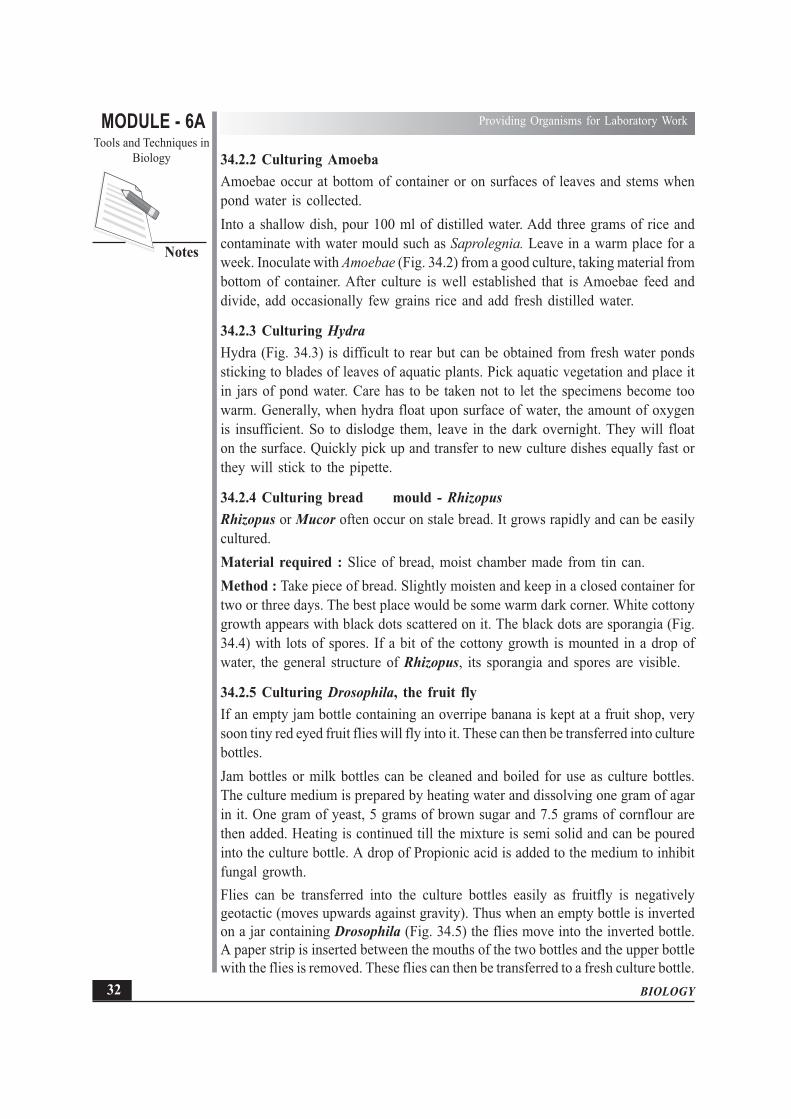
34.2.2 Culturing AmoebaAmoebae occur at bottom of container or on surfaces of leaves and stems whenpond water is collected.Into a shallow dish, pour 100 ml of distilled water. Add three grams of rice andcontaminate with water mould such as Saprolegnia. Leave in a warm place for aweek. Inoculate with Amoebae (Fig. 34.2) from a good culture, taking material frombottom of container. After culture is well established that is Amoebae feed anddivide, add occasionally few grains rice and add fresh distilled water.
34.2.3 Culturing HydraHydra (Fig. 34.3) is difficult to rear but can be obtained from fresh water pondssticking to blades of leaves of aquatic plants. Pick aquatic vegetation and place itin jars of pond water. Care has to be taken not to let the specimens become toowarm. Generally, when hydra float upon surface of water, the amount of oxygenis insufficient. So to dislodge them, leave in the dark overnight. They will floaton the surface. Quickly pick up and transfer to new culture dishes equally fast orthey will stick to the pipette.
34.2.4 Culturing bread mould - RhizopusRhizopus or Mucor often occur on stale bread. It grows rapidly and can be easilycultured.Material required : Slice of bread, moist chamber made from tin can.Method : Take piece of bread. Slightly moisten and keep in a closed container fortwo or three days. The best place would be some warm dark corner. White cottonygrowth appears with black dots scattered on it. The black dots are sporangia (Fig.34.4) with lots of spores. If a bit of the cottony growth is mounted in a drop ofwater, the general structure of Rhizopus, its sporangia and spores are visible.
34.2.5 Culturing Drosophila, the fruit flyIf an empty jam bottle containing an overripe banana is kept at a fruit shop, verysoon tiny red eyed fruit flies will fly into it. These can then be transferred into culturebottles.Jam bottles or milk bottles can be cleaned and boiled for use as culture bottles.The culture medium is prepared by heating water and dissolving one gram of agarin it. One gram of yeast, 5 grams of brown sugar and 7.5 grams of cornflour arethen added. Heating is continued till the mixture is semi solid and can be pouredinto the culture bottle. A drop of Propionic acid is added to the medium to inhibitfungal growth.Flies can be transferred into the culture bottles easily as fruitfly is negativelygeotactic (moves upwards against gravity). Thus when an empty bottle is invertedon a jar containing Drosophila (Fig. 34.5) the flies move into the inverted bottle.A paper strip is inserted between the mouths of the two bottles and the upper bottlewith the flies is removed. These flies can then be transferred to a fresh culture bottle.

MODULE - 6ATools and Techniques in
Biology
33
Providing Organisms for Laboratory Work
BIOLOGY
Notes
The optimal culture temperature for Drosophila is 25°C and Drosophila cultureis kept in a BOD maintained at this temperature. In case the culture has to bemaintained at room temperature, September to March is the best time.
34.2.6 Growing onion root tipsMaterial required : Coplin jars, or wide mouth bottles/100 m1 beakers onions,scalpel, water.Procedure : Take an onion and scrape off the dry roots from the bulb to exposethe disc. Fill a coplin jar with tap water and place the onion bulb on it such thatthe disc touches the water. Place this near the window to get enough light for threeto four days. Roots will start growing and tips can be clearly seen. (Fig. 34.6).
Fig. 34.6 Growing onion root tips
Onion root tips are used for preparing slides to observe various phases of mitosisas cells forming the root tip are rapidly dividing cells.
Preparation of slides showing stages of mitosisTo prepare cell division slides a squash preparation of onion root tips is essential.Material required : 1 : 3 Aceto Alcohol, 1 N Hydrochloric acid, 1% Acetocarminestain, slides and cover slips.
ProcedureCut off root tips and fix in 1 : 3 Aceto Alcohol (1 part of glacial acetic acid and3 parts of Absolute Alcohol) in a watch glass bottle. After five minutes, put theroot tips in 1 N HCl in a watch glass and warm them. Remove HCl by washingin water and leave in the stain, 1% Acetocarmine for five to ten minutes. Carmineis a dye obtained from the cochineal bug and the stain is prepared in Acetic Acid.Remove the stained root tip on a clean slide and tease with a needle. Place a coverslip. Put the slide on a filter paper. Fold the filter paper to cover the slide and gentlysoak the extra stain. Apply pressure with thumb on the cover slip where the teasedroot tip is. This is called squashing and root tip cells then spread out on the slideand when viewed under the microscope, stages of mitosis can be seen. Care hasto be taken not to shake the coverslip.

BIOLOGY
MODULE - 6A Providing Organisms for Laboratory Work
Tools and Techniques inBiology
34
Notes
INTEXT QUESTIONS 34.1
1. Name three organisms cultured in the laboratory.
............................................................................................................................
2. Why are root tips grown in the laboratory.
............................................................................................................................
3. How are Drosophila transferred from one cultrue bottle to another?
............................................................................................................................
4. Where is Hydra collected from?
............................................................................................................................
5. On what is Rhizopus grown?
............................................................................................................................
34.3 TIPS ON THE BIOLOGY LABORATORYIn the Biology laboratory plants and animals are handled all the time. It becomesabsolutely necessary to clean the working tables and wipe with an antiseptic beforeand after lab work starts. There should be arrangement for proper disposal of usedup animals and plants. This would prevent attack by microorganisms and smell ofrotting plants and animals. The fear of spreading infection would also not be there.
An exhaust fan is absolutely essential in a biology lab. It not only removes (i) odourof animals but also (ii) fumes of formalin used to preserve certain animals. Also(iii) chemicals are used for certain experiments - their fumes are removed whenthe air inside the lab is made to circulate with the use of an exhaust fan.
A fire fighting equipment and antiburn ointment such as Burnol should also be keptin the laboratory. Since explosive chemicals and spirit lamps or bunsen burners arerequired for experiments, it is better to take safety measures.
The biology laboratory needs to be well lit and working table should receive enoughnatural light. The chemicals should be kept on a shelf in one corner of the room.
34.4 REARING ANIMALS IN ANIMAL HOUSE AND CAGEMANAGEMENT
Animals such as frogs, rats, cockroaches, leeches etc. are used for the study of organsystems and other anatomical details. These animals have to be procured beforehand and kept in the animal house. An animal house is a room earmarked forkeeping animals required for dissection. Provision for proper ventilation, exhaustand water taps for washing animals are absolutely essential in the animal house.Insect cages can be made with cake tins or cardboard boxes (Fig. 34.7).

MODULE - 6ATools and Techniques in
Biology
35
Providing Organisms for Laboratory Work
BIOLOGY
Notes
Fig. 34.7 Insect cages that can be made at home
Animals are reared in the animal house in cages. Cockroaches are kept in cageswith wire mesh forming the floor and walls of the cage and a lid is on the top totake out the cockroaches with the help of tongs. (Fig. 34.8). Cages for rats are madeof crowbars. A door on the side is for introducing rats on taking them out. A slidingtray is kept at the bottom to remove rat droppings. (Fig. 34.9).
Fig. 34.8 Cockroach cage Fig. 34.9 Rat cage
Frogs are kept in the froggery. Froggery is a cement tank usually built in thebotanical garden so that insects coming to the garden become available as food.A mud substratum is made in the cement tank which is kept wet. The tank is coveredwith a wire mesh and there is provision for taking out and introducing frogs.Food, temperature, humidity and sanitation are important factors in the rearing ofanimals that is cage management. Proper feeding is of prime importance. Theamount of food given should be such that it is consumed completely. Overfeedingshould never be done. Also if too much food is given, it may rot and the animalsmay catch infection. Cockroaches are given wet filter paper bits or bread crumbsto eat. Rats are fed with gram, potatoes, carrots etc. They are voracious eaters andhave to be fed two or three times, otherwise hungry caged rats maul and injure eachother. Frogs trap insects that hover around.

BIOLOGY
MODULE - 6A Providing Organisms for Laboratory Work
Tools and Techniques inBiology
36
Notes
On weekends when the institution is closed somebody is put incharge of feedingthe reared animals. Providing water is as important as giving food and bowl fullof water is kept in the cage. In case an animal is sick, it should be immediatelyisolated.Workers incharge of handling animals for laboratory work have to keep in mindcertain precautions. They should wear gloves. Frogs or rats should be held fromthe waist region to avoid injury by teeth or claws in case of rats and slipping awayof frogs.
34.5 EQUIPMENT REQUIRED FOR COLLECTION OF FLORA ANDFAUNA
A few items of equipment have to be carried on collection trips.A. For carrying the collected material following vessels would be required
(i) Plastic buckets.(ii) Small vials with stoppers.
(iii) Plastic bags with rubber bands.(iv) Vasculum and plant press which are described later.
B. For picking out flora sticking on rocks or ground and aquatic animals stickingto rocks.
(i) Pocket knife for prying sessile plants and animals from rocks or hardsubstratum. The knife should be oiled before use and also used with careso that it does not injure any part of the collector.
(ii) Geology pick for turning rocks and chipping off specimens.(iii) Hammer and chisel for collecting lichens and deep rooted plants on rocks.(iv) Nails
C. Night collectionNocturnal animals (active during night) and intertidal specimens have to becollected at night. For this it is essential to carry
(i) Flash lightD. For culturing bacteria, the following is necessary
(i) Petriplates for keeping the medium(ii) Culture tubes
(iii) Wire needle and wire loopE. For trapping insects or other animals
(i) Insect trap or(ii) Berlese funnel about which you will learn in lesson 35.
(iii) Nets are required not only for trapping insects but also various otheranimals. Various kinds of nets are available about which you shall learnlater in the chapter.
F. For plant collection, sheets, gummed tape, cables, water proof pen are alsorequired.

MODULE - 6ATools and Techniques in
Biology
37
Providing Organisms for Laboratory Work
BIOLOGY
Notes
34.6 VASCULUM AND PLANT PRESS
34.6.1 VasculumVasculum is made up of a metal cylinderwith a sliding door usually worn on astrap over the collectors shoulder intowhich plant specimens are placed.(Fig. 34.10).Polythene bags and paper bags are alsoused for putting fruits, seeds and smallspecimens after collection.
34.6.2 Plant PressPlant press is an indispensable tool for pressing fresh plant specimens to subsequentlydry them and mount permanently.Plant press is made of wood and is of two types.(i) Lab press : Is shown in Fig. 34.11. It is heavier than field press.(ii) Field press : Is lighter in weightA student can make his own plant press from plywood at low cost.
Fig. 34.11 (a) Laboratory Plant Press and (b) Field Plant Press
34.7 NETSSeveral kinds of nets are used for collecting fauna. Following are some of them
(i) Biological dredge : A dredge consistsof a strong net attached to a heavyframe which is pulled along the substratein order to obtain plants and animals.It can be used in fresh water and thesea. Collector can use it from a boatalso (Fig. 34.12).
Fig. 34.10 Vasculum
Fig. 34.12 Biological dredge

BIOLOGY
MODULE - 6A Providing Organisms for Laboratory Work
Tools and Techniques inBiology
38
Notes
(ii) Plankton nets have fine or coarse mesh with a tapered tip.(iii) Insect nets are of (a) dredge type (b) aquatic dip type or sweep net. (Fig. 34.13)(iv) Fish nets are of various kinds (Fig. 34.14).
Fig. 34.13 Dip and Sweep net
Fig. 34.14 Fish net
INTEXT QUESTION 34.2
1. How are formalin fumes prevented from collecting in the lab?
............................................................................................................................
2. Mention any two factors which are important for cage management.
............................................................................................................................
MODULE - 6ATools and Techniques in
Biology
39
Providing Organisms for Laboratory Work
BIOLOGY
Notes
3. Name the equipment required to release flora or fauna from rocks.............................................................................................................................
4. What is a vasculum?............................................................................................................................
5. What is a biological dredge used for?............................................................................................................................
WHAT YOU HAVE LEARNT
� Certain organisms can be collected from nature and multiplied in the lab. Thisis called ‘culturing’.
� While preparing cultures, one should know
(i) the location or habitat from which a particular organism can be collected.
(ii) the method of collection
(iii) the method of culture and
(iv) how to preserve organisms for future use.
� Paramecium and Amoeba can be cultured after collecting them from pond.Parameciam are grown in bottles containing grass, leaves etc. on which bacteriais growing. Amoeba is cultured in shallow dish with rice and water mould init.
� Hydra can be cultured in a jar with aquatic vegetation.
� Rhizopus or bread mould is cultured on stale bread.
� Drosophila or fruit fly are cultured in empty milk bottles containing mediumhaving yeast, brown sugar, and cornflour. They are transferred by placing emptybottle on the medium and the fruit flies fly up.
� Onion root tips are grown in coplin jars with water and used for the study ofcell division by making squashes.
� Biology lab should have an exhaust fan to remove odour and fumes of formalinand other chemicals.
� A firefighting equipment should also be kept in the lab.
� Animals required for dissection are reared in cages. Cages for differentorganisms are built differently.
� Food, temperature, humidity and sanitation are important factors for cageculture. Food has to be given in adequate quantity and arrangements made forfeeding during weekends.
� Plants can also be kept in cages.

BIOLOGY
MODULE - 6A Providing Organisms for Laboratory Work
Tools and Techniques inBiology
40
Notes
� Items of equipment required for collection of fauna and flora are (a) for keepingcollected specimens or (b) for picking specimens attached to rocks. (c) forculturing microorganisms and (d) for trapping insects or other animals.
� For keeping collected specimens, plastic buckets, vials, plastic bags andvasculum are required.
� For picking specimens free of substratum, a knife or pick are required.� Flash light is required for night collection.� Insect trap or Berlese funnel is to trap insects.� Nets of different kinds are for collecting insects, fish, plankton etc.� A vasculum is a metal cylinder with a sliding door carried by the collector on
the shoulder.� A plant press is used for pressing fresh plant specimens. There are two kinds
of plant press - lab press and field press.� A dredge is a net attached to a metal frame which can scrape the ground to
collect specimens.
TERMINAL QUESTIONS
l. Describe the method of culturing any one protozoan.2. How can you culture bread mould on a piece of bread?3. How can you prepare onion root tip squash to study mitosis?4. Why should there be an exhaust fan in a biology lab?5. Write a note on animal house.6. What all should you carry if you go on an excursion to collect animals?
ANSWERS TO INTEXT QUESTIONS
34.1 1. Amoeba/Paramecium/Hydra/Rhizopus/Drosophila (any three)2. To study mitosis/cell division.3. By placing an empty bottle above culture bottle.4. Aquatic fresh water plants.5. Bread
34.2 1. By installing an exhaust fan.2. Food/temperature/humidity/ventilation3. Pocket knife/Geology prick4. A metal container for collecting plants5. Collecting aquatic flora and fauna

MODULE - 6ATools and Techniques in
Biology
41
Some Aids in Biology
BIOLOGY
Notes35
SOME AIDS IN BIOLOGY
Learning and teaching of Biology is complete only when the student is able to seeactive living and preserved variety of organisms. Learning Biology also becomeseasier by observing organisms closely rather than merely reading about them.Preserved animals and plants, duly classified are kept in museums. Charts andmodels are displayed there. Living animals are kept in animal house, frogs in aspecially constructed froggery, fish in aquarium. Plants are grown in the botanicalgarden or specially maintained in a green house. Preparation of a herbarium is anintegral part of learning Botany. Some such aids in learning and teaching Biologyare outlined in this lesson.
OBJECTIVES
After reading this lesson you will be able to :
� describe the need for a zoological museum;
� explain the ways in which specimens such as wet, dried specimens, embalmedspecimens, models, pictures, photographs and skeletons can be displayed in thezoological museum;
� explain the need for a botanical garden;
� list the categories of plants need to be grown in the botanical garden;
� describe a green house;
� explain need for a green house;
� describe herbarium and list the steps followed in making a herbarium;
� explain the steps involved in setting up an aquarium;
� list the fishes suitable for an aquarium
� explain the need for maintaining suitable temperature, proper light and properaeration in the aquarium
� list different kinds of fish food.

BIOLOGY
MODULE - 6A Some Aids in Biology
Tools and Techniques inBiology
42
Notes
35.1 ZOOLOGICAL MUSEUMA zoology museum or an animal museum houses (i) preserved animals, (ii) theirskeletons (iii) fossils (iv) models (v) charts and (vi) photographs. The personincharge of maintaining the museum is called museum curator.
1. Preservation and display of specimens
(a) Wet PreservationInvertebrates and small and medium sized vertebrates can be preserved intact inglass or transparent plastic jars of appropriate size called specimen jars (Fig. 35.1).The jars have a flat, firm base and a lid. A solution of 10% formalin fills the jar.The specimen is mounted on a glass slab of appropriate size which is then placedinside the jar and covered with the lid. The lid may then be screwed on to the jaror sealed on it. From time to time fresh formalin has to be added or replacedaccording to need. The specimen remains intact for years if handled properly. Thespecimens retain their shape and morphology. Only the natural colour is lost.
Fig. 35.1 Museum jars with preserved specimens
Wet Preservation can be done for (i) intact specimens; (ii) vertebrate embryos;(iii) organs such as heart and eye; (iv) specimens dissected out to display organsystems e.g. nervous system of an animal; and (v) in situ skeletons of smallvertebrates such as fish or frog or lizard. In situ (retained in actual position in the
Fig. 35.2 Alizarin stained skeletal preparation of a lizard

MODULE - 6ATools and Techniques in
Biology
43
Some Aids in Biology
BIOLOGY
Notes
animal) skeleton can be prepared in the following manner and mounted and keptin bottles containing glycerine. Chloroformed animal is kept in 90% alcohol in acovered jar for two days. Alcohol is then removed and animal dipped in 1% aqueoussolution of Potassium hydroxide till it becomes transparent. The animal is thenplaced in Alizarin Red Stain (0.1 gm of Alizarin Red in a litre of 1% aqueoussolution of KOH). The bones get stained (Fig. 35.2). The animal is then transferredto a specimen jar containing glycerine.
(b) Dry preservationExoskeletons (Skeletons covering the body) such as shell of molluscs, star fish,sea urchins, corals, cocoons of insect sloughed off (cast off) skin of snakes or insects(exuviae), feathers and nests of birds, honey combs and wasp or termite nests,mammalian skin with furs, dried sponges etc. can be displayed in the museum intactfor many years provided they are prevented from breaking or from attack by insectparasites or microorganisms.Apart from the above (i) vertebrate skeletons and (ii) pressed insects also form partof dry presentation.
(c) Skeleton preparationSkeletons may be prepared in the following way. Chloroformed vertebrate isdissected to remove organs and as much muscle as possible. Boil the animal sothat muscles become tender and remove them. When only the skeleton remains,dip it in Hydrogen Peroxide for bleaching (optional). Mount on a cardboard orwooden board with the help of adhesive like araldite or fevicol and display.Skull or bones of dead animals collected from the fields may be cleaned with water,disinfected with a disinfectant, dried and displayed in a museum. Stuffed animalsare also kept in the museum. Skinning, preserving, stuffing and mountingvertebrates is called TAXIDERMY.
(d) Insect collection and preservationInsects are found everywhere - cockroaches abound near the kitchen drains,butterflies roam among flowers while grasshoppers hop and crickets chirp in thegrass. Flies are where sweets are and fruit flies hover around vegetables and fruits.Insects are the most numerous and diversified group of animals.
Collection of InsectsInsect collection is an activity which combines fun and study. The equipmentrequired is (i) collecting jar, (ii) a net (iii) fine wire sieves (iv) insect trap.
The collecting jar can be of three types(a) Cyanide bottle : in which sodium cyanide is placed in small lumps in a wide
mouthed jar with a well fitting lid. Plaster of paris is made to set on the sodiumcyanide. When dry, the bottle is stoppered. Since sodium cyanide is a deadlypoison, it has to be handled with care.
(b) Carbon tetrachloride bottle is efficient and harmless. A glass tube is insertedin a small hole bored in the cork of a bottle. A cotton wad is fastened to thecork.

BIOLOGY
MODULE - 6A Some Aids in Biology
Tools and Techniques inBiology
44
Notes
(c) Chloroform bottle has rubber bands placed at thebottom of a bottle and some chloroform put in it.Rubber has the capacity to absorb chloroform.After sometime unabsorbed chloroform is thrownaway and a cardboard placed to cover the rubberbands. Fumes of chloroform absorbed by rubberfill the bottle.A net made of cotton or nylon can be stitched toa handle as shown in the Fig. 35.3.
The fine wire sieves are required to strain the mud which comes with the collectedinsects or to wash them. Flying insects or walking and hopping insects can becollected from their natural surrounding.Insects may be trapped with the use ofinsect traps. A simple trap has a largefunnel placed on the mouth of a widemouthed jar containing chloroform oralcohol.A Berlese funnel traps small insect. Afunnel is soldered at the end of a large canwith a hole at its bottom. A false bottommade of wire mesh is placed in the can,filled with leaves and grass and coveredwith cheese cloth. The stem of the funnelends in a wide mouthed bottle sealed withcotton plug. This collecting bottle maycontain alcohol or chloroform forpreserving the insects. (Fig. 35.4)Insects are caught by the net or in the trap and placed in the collecting jar. Thenthey have to be preserved.
Fig. 35.5 Where to place the insect pin through the body when mounting insects Pushthrough area indicated by the black dot
Fig. 35.3 Net for catchinginsects.
Fig. 35.4 Insect traps

MODULE - 6ATools and Techniques in
Biology
45
Some Aids in Biology
BIOLOGY
Notes
Fig. 35.6 How to spread pinnea butterflies and moths.
Insect preservationMaterial required for the preservation of collected insects are (i) Pins of varioussizes (ii) hard paper (iii) insect spreading board (iv) insect collection box (v) insectcabinet.Before collected insects dry up, pin has to be thrust through the thorax or wings(Fig. 35.5). Very small insects are mounted on a triangular piece of hard paper.Wings of butterflies, dragonflies have to be spread out as shown in (Fig. 35.6).Spreading of wings is done by first fixing the pin carrying the insect in the grooveof the spreading board, wings spread out and strips of paper pinned across wingsof either side (Fig. 35.6). Once such mounted insects are dry, they are removed tothe insect collection box (Fig. 35.7). The insects are then classified and arrangedin an insect cabinet. (Fig. 35.8).
Fig. 35.7 Standard cardboard insect box Fig. 35.8 Insect cabinet

BIOLOGY
MODULE - 6A Some Aids in Biology
Tools and Techniques inBiology
46
Notes
2. Visual aids - Charts, models, photographsCharts and models depicting morphology of organisms, phylogeny showingtaxonomic relationships, internal structure of plants and animals, life history ofsocial insects, evolution of horse and humans etc. should be available for students.The advantages of charts and models are(i) they substitute for live specimens which are not available;(ii) charts are self explanatory if well prepared and students can revise from them;(iii) the teacher can carry the chart to class while explaining the theory on black
board.Good charts prepared by students can also be put up on the wall which not onlyencourages the student who made it but also other students. Photographs of greatbiologists may also be hung up with their names and contribution written clearly.Visual aids ought to be (i) accurate (ii) relevant (iii) comprehensible (iv) realistic(v) properly labelled and (vi) title should be clearly written. The models shouldhave a three dimensional effect and the proportions should be correct. Students canmake models with wood, fibre, thermocole and any other material. Charts andpictures should be hung on the wall which is well lit. They should be within thelevel of the eyes of children using the museum.
Maintenance of the MuseumThe museum requires constant care. The museum curator has the followingduties :1. to change the charts and pictures from time to time;2. to make new visual aids:3. to be available to help students. The curator should have a Biology background;4. to change formalin of preserved specimens; and5. to be involved in overall maintenance of the museum.
INTEXT QUESTIONS 35.1
1. What is the person incharge of museum called?............................................................................................................................
2. What is the Berlese funnel used for?............................................................................................................................
3. Name the stain used for in-situ skeleton preparation.............................................................................................................................
4. Name a visual aid used in teaching Biology.............................................................................................................................

MODULE - 6ATools and Techniques in
Biology
47
Some Aids in Biology
BIOLOGY
Notes
5. Name the(i) common chemical used for preserving specimens
..................................................................................................................(ii) art and science of skinning, stuffing and preserving animals.
..................................................................................................................
35.2 BOTANICAL GARDENA well maintained garden where plants are classified and grown with care andwhere plants may even be multiplied for the purposes of observation and researchis called a BOTANICAL GARDEN. A garden full of plants, both flowering andnon flowering is indeed a treat to see. But a botanical garden is not for entertainmentalone but associated with Botany teaching and research.In our country we have the main national botanical gardens in Lucknow andKolkata. The one in Lucknow is called National Botanical Garden and has plantsof Uttar Pradesh and those from the hilly regions of Uttaranchal. Indian Botanicalgarden is in Shibpur near Kolkata. It has more than a hundred year old Banyantree.A school botanical garden is, developed on a much smaller scale. The plants grownin it are mostly the ones which form the study material for Botany. A patch of groundwithin the school premises where ample sunlight comes is the ideal place wherebotanical garden may be developed. Plants of different kinds as also those requiredfor practical study need to be grown. An ideal situation is when (i) new plants areadded from time to time and (ii) plants are labelled with labels carrying the botanicalnames as well as common names. (iii) A catalogue has to be prepared giving anumber and concise description.
35.3 GREEN HOUSEA greenhouse is a special enclosure made of glass or plastic in which plants aregrown and maintained at a specified temperature and humidity. In countries, wherefreezing temperature obtains in winter, a green house is made with roof and wallsmade of glass. Plants get adequate light and they can be watered. At the same timeheat trapped by the glass enclosure keeps the greenhouse warm. Temperature,however, is also regulated by a specialised system. Greenhouses are permanentstructures.Temperature of greenhouse is regulated through an automatically controlled heatingand ventilation system. A central coal or oil furnace supplies the heat. Morecommon is a peripheral steam heating system. Ventilation is provided at the sidesand top. In summer, when temperature goes up fan and pad cooling is used to lowertemperature. Water is circulated through pipes. Cooling pads draw cooled air acrossgreenhouse. It is more effective when humidity is low.Instead of glass, plastic films are used to make the wall and roof of glasshouse.Light absorbing qualities of plastic are similar to those of glass. Rigid plastic or

BIOLOGY
MODULE - 6A Some Aids in Biology
Tools and Techniques inBiology
48
Notes
ultraviolet resistant polythene is used. During summer a shade made of cloth is usedto cover the greenhouse.In an institute where Biology is taught, a greenhouse of smaller dimensions canbe constructed to house delicate plants. Plants would provide material for botanicalstudies and students would also be trained in growing, maintaining and propagatingplants under controlled conditions. (See Fig. 3.9).
Fig. 35.9 Green house
35.4 HERBARIUMA herbarium is defined as a collection of plants that have been dried, pressed andpreserved on sheets of hard paper. The dried plants are classified and arranged forfuture reference especially for taxonomic studies.A plant collector needs to have the followingequipment :(i) a gardener’s knife, (Fig. 35.10a)
(ii) a plant press or vasculum, (35.10b)
(iii) blotting papers to dry plants,
(iv) trowel to dig and uproot the plant,
(v) collecting and mounting sheets,
(vi) gum tape, labels, waterproof ink and pen.
1. Collecting botanical specimensFleshy plants lose their diagnostic features whendried so they are preserved in 4% formalin inglass containers. Gymnosperm cones and dryfruits are collected and preserved as such.Plants should be collected from various localitiesfor the preparation of a herbarium. The herbariumshould also have representative specimens fromvarious groups of plants.
Fig. 35.10(b) A plant press(Vasculum) with sheets
Fig. 35.10(a) Gardener’s knife –one should carry such knife
in field trips

MODULE - 6ATools and Techniques in
Biology
49
Some Aids in Biology
BIOLOGY
Notes
A complete specimen when collected, should have all the parts including the rootsystem. It is better to collect a plant at its flowering stage. A tag should give thelocation from where collected. About five or six specimens of each kind of plantshould be collected. The collected plant should either be pressed then and thereor collected in a vasculum and pressed later. Vasculum is a metal cylinder with asliding door in which plants are collected (Fig. 34.10b).
2. Pressing, Drying and PreservingThe collected plant should be pressed between sheets of blotting paper. One plantis arranged on one sheet so that its parts do not overlap. Specimens longer thanthe sheets can be folded in the form of ‘V’ or ‘N’.The plant between the sheets is put in a press for24 to 48 hours. The press is then opened, blottingsheets changed and plants rearranged again and putback in the press for another 2 or 3 days. Thepressed specimen is then dried in sunlight or heatfrom some other source.To prevent the abscission layer formation anddecay, plants are killed (poisoned) with formalin orMercuric chloride (HgCl2) or Carbon Tetra chloride(CC14). Also dipping in Mercuric chloride (HgCl2)saves them from attack by museum pests such asbeetles. Fig. 35.10(c).
3. Mounting and LabellingAfter drying, specimens are mounted on mounting papers or herbarium sheetswhich are usually of a standard size of 11.5" × 16.5" and sturdy enough to supportthe dried plants. Glue or adhesive tape or adhesive paste is used to stick thespecimens on to the sheets.Each sheet should carry a label pasted on the lower right hand corner giving the(i) the site of collection, locality and altitude (ii) name of plant (iii) family (iv) habit(v) date of collection (vi) ecological notes and (vii) name of collector.Herbarium sheets should be stored in herbarium cases or steel almirahs. Theyshould be arranged according to the system of classification. Moth balls, naphthaleneflakes or 2% of Mercuric chloride should be sprayed to keep away mould, fungiand insects.
INTEXT QUESITONS 35.2
1. Where are the national botanical gardens situated in our country?............................................................................................................................
2. What is a greenhouse?............................................................................................................................
Fig. 35.10(c) Holding of specimenfor poisoning

BIOLOGY
MODULE - 6A Some Aids in Biology
Tools and Techniques inBiology
50
Notes
3. Define herbarium.
............................................................................................................................
4. What is a vasculum?
............................................................................................................................
5. Mention the steps for preparation of a herbarium in a sequence.
............................................................................................................................
35.5 AQUARIUMAn aquarium is a glass container in which live fish are kept along with aquaticplants. A well maintained aquarium helps students to learn many biologicalprinciples. Some of these are –(i) dependance of animals on green plants for (a) food and (b) oxygen,(ii) relationship of carbon-dioxide and light to photosynthesis,(iii) food ingestion, storage, respiration, digestion, growth, reproduction and
development in plants and animals,(iv) relationship of bacteria to Nitrogen, Phosphorus and Sulphur cycle,(v) parasitism,(vi) food cycles,(vii) temperature and water relation,(viii) ecological succession.
1. How to prepare a balanced aquariumMaterials required for an aquarium are :(i) Aquarium tank approximately of five gallon capacity. Aquarium water is
static and changes its composition due to activities of fish and plants. Hence,it has to have enough surface area for adequate gas exchange. It may be madeof plastic or glass with silicon adhesive (Fig. 35.11).The tank should be placed on firm, level and smooth surface. It is best locatednear the window but direct sunlight should be avoided as it leads to excessivealgal growth in summer. The side towards window may be covered with paperor painted. If light is not enough electric light may be used.The situation of the tank should have easy access for maintenance and forpower sockets for heat and light.
(ii) Substratum in the tank can be constructed by putting one inch of sand onsoil. The substratum (a) forms the source of minerals; (b) plants can anchorin the substratum; (c) animals can burrow in it, (d) it forms the spawning siteof fish; (e) floor bed of the aquarium and (f) a biological filter.

MODULE - 6ATools and Techniques in
Biology
51
Some Aids in Biology
BIOLOGY
Notes
Fig. 35.11 An aquarium
(iii) Water can be procured from tap, well, spring or pond to fill the tank. Thelid of the aquarium prevents excessive evaporation.
(iv) Aquarium plants and fish
2. Temperature of the aquariumThe temperature of the aquarium needs to be maintained at an optimum of 24°C(75°F). To maintain the temperature, a thermostat controlled heating device maybe used. In case of power failure, tank may be covered by a blanket in winter orsuitably hot water added. A floating or adhesive type of thermometer can recordthe temperature. These days a combined heater and thermostat enclosed in a watertight glass tube is used and kept in place by special clips made of non toxic plastic.Thermostat with microchip (computerised) circuit is fixed for accurate temperaturecontrol. All electric connections, however, should be outside the tank.
3. Lighting of the aquariumLighting arrangement not only makes the aquarium attractive but it also forms anessential stimulus to plants for photosynthesis. In nature fish are lit by sunlight.Light in the aquarium may be provided by lamps (tube or bulb of 40 Watts) mountedin aquarium cover called hood or reflector. Tungsten lamps and fluorescent tubesare used for every 30 centimeters (12 inches) length of the tank. Aquarium has tobe lit at least for ten hours a day.

BIOLOGY
MODULE - 6A Some Aids in Biology
Tools and Techniques inBiology
52
Notes
4. Biological filtrationThe gravel in the substratum acts as a filter bed. Aquarium water passes throughthe gravel and bacterial colonies develop on the gravel and convert the waste ofthe fish into ammonia and nitrates by Nitrosomonas bacteria and nitrites into nitratesby Nitrobacter. Nitrates are taken up by plants.
5. Aquarium PlantsThere are many species of aquatic plants which provide shade, refuge, spawningsites, food, water and source of oxygen for the aquaria fish. Plants may be of thefloating type such as Hydrilla, Elodea or rooted such as Vallisneria which growrapidly (Fig. 35.11). To many plants may be avoided.
6. Aquarium FishA number of small fish with unique shapes and beautiful designs are appropriatefor keeping in the aquariums. They are variously coloured. However, care has tobe taken to assure the absence of predatory varieties in the aquaria. The commonaquaria fish are (i) angel fish (2) molie (3) guppy. (Fig. 35.11)
7. Fish FeedFood of aquaria fish are either live feed such as waterborne insects, worms,crustaceans such as water flea. Daphnia, cyclops earthworms. However, if any ofthese are not eaten by the fish, they should be removed and not permitted to increasein number. Lettuce, spinach, peas, wheat grain may be given for herbivorous fish.With progress in technology, a balanced diet with special formula is commerciallyprepared in the form of flakes, granules, powder or liquid food. Freeze dried fishfood such as Daphnia, Tubifex and other worms are packed. It is convenient to justopen the packet and sprinkle in the aquarium for fish to eat. But never should toomuch food be put as the left over food may decompose and pollute the aquarium.For fish to develop best colours, size and a healthy constitution, the diet shouldbe varied. The best food, however, is live food.
INTEXT QUESTION 35.3
1. What is an aquarium?............................................................................................................................
2. Name two plants which are grown in an aquarium.............................................................................................................................
3. What do plants provide the fish in the aquaria?............................................................................................................................
4. What is biological filtration?............................................................................................................................

MODULE - 6ATools and Techniques in
Biology
53
Some Aids in Biology
BIOLOGY
Notes
WHAT YOU HAVE LEARNT
� In a Zoology museum preserved animals, skeletons, fossils, charts, models dryspecimens are kept.
� 10% formalin is used to preserve small animals in glass jars.� Mollusc shells, corals, dried sponges, nests, feathers are dry preservations. They
are also kept in the museum.� Skeletons can be prepared and kept in the museum.� Insect collection requires pinning properly and drying insects after catching
them and killing them. They can be trapped in Berlese funnel or simple traps.� Charts, models can be made by students or bought from the market and kept
in the museum. They should be properly displayed.� Museum should be looked after by a museum curator.� A botanical garden should be maintained in the school and plants for study
grown there. Plants should be labelled.� A greenhouse is a special enclosure made of glass or plastic where plants are
maintained at a constant temperature and humidity.� Herbarium is a collection of dried and pressed plants preserved on paper sheets.
Plants are collected undamaged and then pressed in a press and dried. They arethen mounted on herbarium sheets and labelled and classified.
� An aquarium is a glass or plastic container in which fish are grown andmaintained. It also has aquatic plants which provide food and Oxygen for thefish.
� An aquarium has to be well lit and temperature has to be maintained. Elodea,Hydrilla, Vallisneria are some aquatic plants kept in the aquarium.
� Substratum of the aquarium is a filter bed in which bacteria can grow andconvert waste into nitrates for the use of plants.
� Aquarium fish are of many colours. Some of these are angel fish, black mollie,guppy etc.
� Aquarium fish can be given live food such as worms and crustaceans or driedfood.
TERMINAL EXERCISES
1. How is wet preservation done for museum specimens?
2. What are the ways in which skeletons can be prepared for displaying in themuseum?
3
F

BIOLOGY
MODULE - 6A Some Aids in Biology
Tools and Techniques inBiology
54
Notes
3. Name the equipment items required for insect collection and mention theirutility.
4. What is a Berlese trap?
5. Write notes on (a) Botanical garden (b) green house
6. How is a herbarium prepared?
7. Mention three biological principles which can be learnt by maintaining anaquarium. How can temperature, light and fish food be arranged for anaquarium?
ANSWERS TO INTEXT QUESTIONS
35.1 1. museum curator2. trapping small insects3. Alizarine4. chart/model/photograph/living museum specimens/dry specimens in the
museum5. (i) Formalin (ii) Taxidermy
35.2 1. Lucknow and Kolkata2. A special enclosure made of glass or plastic in which plants are grown and
maintained at a specific temperature and humidity3. A collection of plants which are dried, pressed and preserved on sheets4. A metal cylinder with a sliding door in which plants are collected5. Collection, pressing, preserving, mounting, labelling.1. a glass tank in which live fish are grown and maintained.
35.3 2. Elodea, Vallisneria, Hydrilla (any two)3. Oxygen and food4. Conversion of waste of the fish into nitrates to be taken up by the aquarium
plants


Evolution of human civilization has depended mainly on agriculture. It has not onlygiven man the freedom from the risks of hunting and collecting food from the forestbut has also given assurance of continuous availability of food.Agriculture plays an important role in providing basic necessities of mankind inthree ways – food, clothes and shelter. In addition to agriculture, we draw manyessentials for our living from nature. Forests play a very important role in providinguseful products for man e.g. rubber, supporting wildlife, e.g., such as tiger andimproving the environment. Medicinal plants many of them obtained from forests,provide important drugs, which are used for treating a large number of diseases.In this lesson, you will learn about agriculture, forestry and medicinal plants.
OBJECTIVES
After completing this lesson, you will be able to:� state the major role of agriculture;� classify the crops giving examples under each category and also mention their
importance;� briefly describe the cultivation of cereals crops viz wheat and rice along with
cotton and sugarcane;� define green revolution and list the factors that made it possible;� explain the principles of farm management and the proper ways of storage of
food grains;� mention the utility and the method of establishing a kitchen garden;� define the term forestry;� name the different types of forests, appreciate their effects on ecosystem and
enumerate the commercial products provided by it;� list the common names of some medicinal plants giving their uses in treating
various diseases.
31
AGRICULTURE, FORESTRY ANDMEDICINAL PLANTS
MODULE - 6BEconomic Biology
57BIOLOGY
Notes

BIOLOGY
MODULE - 6B Agriculture, Forestry and Medicinal Plants
Economic Biology
58
Notes
31.1 ROLE OF AGRICULTUREFood and clothing are the basic needs of man. Whatever we eat in our daily life,most of them we get from plants. The clothes we wear are also made from plantse.g. cotton is a plant product. Plants provide most of the basic necessities of lifeto human beings. These include (1) Food (cereals, fruits, oil seeds etc.) (2) Fibresfor making clothes (cotton, jute, etc.) (3) Beverages (tea, coffee, etc.) (4) Forests(timber, rubber etc.). Herbivores consume plants as their food. Plants can be of twotypes i.e. cultivated and wild plants.
Cultivated and Wild plantsPlants which are specially grown by humans are called cultivated plants e.g. wheat,rice. The plants that grow by themselves in nature are called wild plants e.g bathua,cholai etc.
31.2 CLASSIFICATION OF CROPSCrops are grown regularly in large numbers in fields for food or for obtaining otherproducts. Crops are classified in a number of ways as follows:(i) According to use, crops may be classified into food crops and cash crops.
Table 31.1 Difference between food and cash crops
Food Crops Cash Crops
1. They are grown to provide foodand may be stored for a long periode.g. wheat, rice, vegetables.
(ii) According to the season of growing, crops are classified into three types-1. Rabi crops (grown in winter) : Wheat, gram, pea etc.2. Kharif (grown in monsoon : Maize, jowar, bajra etc.
rainy season)3. Ziad crops (grown during : Melon, cucumber, water melon etc.
summer)(iii) According to the edible parts of the plant, the crops are termed as
follows :1. Cereal crops (seed grains) - Wheat, rice, maize etc.
2. Fruit crops - Mango, apple, grape, banana etc.
3. Vegetable crops - Ladiesfinger, tomato, cauliflower etc.
4. Stem crops - Potato, sugarcane, colocasia etc.
5. Root crops - Radish, carrot, sweet potato etc.
6. Leaf crops - Spinach, mustard (sarson), maithi etc.
1. Plants or plant products whichcan be sold and money can beearned out of it. e.g. tea, jutecashew nut, rubber, sugarcane.

MODULE - 6BEconomic Biology
59
Agriculture, Forestry and Medicinal Plants
BIOLOGY
Notes
(iv) The most widely used general classification of crops are-
1. Cereal CropsCereals are used as food not only in India but all over the world. They are richin carbohydrates and grown for food. It includes wheat, rice, maize, bajra, barley,rye, oat, sorgham (jowar), ragi etc. These are consumed by animals as well as byhuman.
2. Pulse CropsIt includes all the types of ‘dals’ (pulses) we use as a food, these are also calledas leguminous plants. They are rich sources of protein e.g. sweet peas, gram, pigeonpea (arhar), soyabean, beans, ground nut, lentil etc.
Agricultural importance of pulse cropsAll leguminous plants have special nodules (swellings) in their root systemand these contain bacteria. (Fig 31.1) These bacteria fix atmosphericnitrogen directly and change it into nitrogen compounds. This process isknown as nitrogen-fixation. These nitrogen compounds are used by theplants as a natural nitrogen fertilizer. For this reason farmers practise croprotation i.e. they grow a pulse crops in between two cereal crops to enrichthe soil to get a better yield of the crop.
Fig. 31.1 Leguminous plants contain nodulated bacteria.
3. Fruit crops(i) Fruits are rich in vitamins, mineral and carbohydrates. Fruits provide vitamins,
minerals and variety to our diet. Some fruits are also a rich source of sugare.g. grape, banana and mango.
(ii) Fruits are consumed fresh or in the form of juice, jam etc. examples – mango,apple, banana, oranges, pomgranate etc.
(iii) Fruit trees are usually perennial trees, which bear fruits once a year. Many fruitcrops are grown in India examples – chickoo, mango, apple, orange etc.
4. Vegetables CropsVegetables provide minerals, proteins and carbohydrates. A large number ofvegetables are grown in our country. Some important ones are e.g. potato, tomato,pea, cauliflower, brinjals, gourds etc.

BIOLOGY
MODULE - 6B Agriculture, Forestry and Medicinal Plants
Economic Biology
60
Notes
5. Stem CropsGinger, potato, onion, garlic, colocasia are the examples of stem crops. Sugarcaneis the main source of sugar in our country. Sugarcane is an annual cash crop.
6. Root CropsCarrots, radish, sweet potato and turnip are the examples of root crops.
7. Leaf CropsSpinach, Methi, Corainder, Mint and mustard are the examples of leaf crops.
8. Oil Seed CropsOils are an important part of our food. They are a rich source of energy. Seeds ofsome crops yield oil on being pressed in machines. The remainder becomes theseed cake to be used as cattle feed. Some of the oil yielding crops are groundnut,cottonseed, coconut, mustard and sunflower.
9. Fibre CropsThe two most important fibre crops of our country are cotton and jute. They supplythe raw materials for our textile and jute industries.
RettingThe process by which the fibres are separated from the jute plant is calledretting.In this process we make small bundles of plants. These bundles are keptin side pond or canal 15 days. During this period degradation process occursin the presence of certain bacteria, and the fibres are separated easily, theyare then washed and dried.
INTEXT QUESTIONS 31.1
1. Give the difference between cultivated plants and wild plants.............................................................................................................................
2. Give two examples each of Rabi crop and Kharif crop.............................................................................................................................
3. Match the items in Column I with those in Column IIColumn I Column II
1. Pulse crops A. a nutritious fruit2. Oil seed crop B. ground nut3. Soya bean C. rich source of protein.4. Banana D. nitrogen fixing bacteria
4. Name four oil seed crops.............................................................................................................................

MODULE - 6BEconomic Biology
61
Agriculture, Forestry and Medicinal Plants
BIOLOGY
Notes
31.3 CULTIVATION OF WHEAT, RICE, COTTON AND SUGARCANE
(a) Cultivation of wheatWheat belongs to the family Poaceae (Graminae). The grains are rich in carbohydrates.This is the winter season crop. Method of cultivation are as follows :1. Area of production UP, Punjab, Haryana, Rajasthan, Madhya Pradesh, Bihar,
Maharastra etc.2. Soil grow in loamy soil3. Climate Cool weather with moderate rainfall4. Temperature 10-15°C (during winter season)5. Irrigation 4-5 times its cultivation6. Harvesting April to May in northern India7. Botanical Name Triticum vulgare
(b) Cultivation of rice cropRice is one of the major crop of India. About 23 percent of the total cultivated areaof the country cultivates this crop. Method of rice cultivation is as follows -1. Area of production UP, Bihar, West Bengal, Assam,Andhra Pradesh, Punjab,
Tamil Nadu, Orissa2. Soil deep fertile loamy, clayey loam, loamy3. Climate Tropical and moist with 125 cm rainfall4. Temperature 20-25°C5. Irrigation every weeek, soil should not be dry6. Harvesting September -October in northern India7. Botanical Name Oriza sativa
(c) Cultivation of cottonCotton is one of the major crops of India. The fibre of cotton is used as raw materialfor the textile industry. Cotton seeds after extraction of the oil are also used as acattle feed. Methods of cotton cultivation is as follows -1. Area of production Punjab, Haryana, Maharastra, Gujarat, Andhra Pradesh,
Rajasthan, Karnataka, Madhya Pradesh, Tamil Nadu2. Soil Black cotton soil of deccan plateau and alluvial soil of
the northern plain.3. Climate cloud free weather for about 150 days with 75 cm rainfall
at the time of flowering and boll opening4. Rainfall moderate rainfall of about 75 cm5. Specific characters 1. Indian cotton is used for coarse cloth
2. Fruit is capsule3. Oil is used for cooking and oil cake for feeding cattle.
6. Botanical Name Gossypium sp.

BIOLOGY
MODULE - 6B Agriculture, Forestry and Medicinal Plants
Economic Biology
62
Notes
(d) Cultivation of sugarcaneSugarcane is the main source of sugar in our country. This is an annual and cashcrop. India is the original land of this crop. Sugarcane is sown by “setts” (piecesof stem containing buds). Method of sugarcane cultivation is as follows -1. Area of production Punjab, Haryana, UP, Bihar, Gujarat, Maharastra,
Karnataka, Andhra Pradesh, Tamil Nadu2. Soil Heavy loamy soil3. Climate hot and humid4. Harvesting from mid February to mid March in North India. From
20th September to 20th October in West India andslightly earlier in south India.
5. Irrigation 1st after one month of sowing in summer on every 10thday. In winter on every 15th day
6. Botanical Name Saccharum officinarum
31.4 GREEN REVOLUTIONIn 1952, the yield of Wheat and Rice was 654 kg and 800 kg per hectare,respectively. This yield was insufficient to fulfil the demand. Therefore ourGovernment and Agriculture Scientists paid a special attention towards theimprovement of agriculture. By doing so, the production of cereal crops increasedtremendously and we became self-sufficient. The various steps taken to achieve thisgoal have been collectively termed as green revolution.
Green Revolution: Spectacular increase in the yield of crops, particularlycereals, through the application of modern techniques in agriculture iscalled Green Revolution
The factors which help to bring about the green revolution are:1. Introduction of high yielding varieties of crops.2. Multiple cropping, better irrigation and sufficient supply of fertilizers.3. Use of crop protection measures against diseases and pest.4. Transfer of the technology of scientific farming from research farms to villager
farmers.5. Organised transport arrangements to reach the produce from the fields to
market.
31.5 FARM MANAGEMENTEffective farm management requires detailed planning for growing highyielding crops and their storage.
There are various steps involved in farm management are as follows:
1. Correct preparation of the soil.
2. The right variety and quality of seeds or saplings for planting.

MODULE - 6BEconomic Biology
63
Agriculture, Forestry and Medicinal Plants
BIOLOGY
Notes
3. The right amount of water and time at which it has to be supplied.
4. The amount and time of application of fertilizers.
5. Timely application of pesticides.
After harvesting, proper storage of the crop is very important to avoid loss andwastage due to lack of transit from field to the consumer.
1. Proper storage of FoodFood is spoilt, by variations in temperature, humidity, insects, microorganisms,rodents, birds and animals. Food grains are therefore stored in waterproof andairtight containers. Small farmers, store the grain in jute bags in godowns withpucca floor, plain roof and single entrance. Special large sized godowns like silosare used these days at the government level.2. Storage and Preservation of Fruits and VegetablesFresh fruit and vegetables are kept in cold storage at low temperatures to help retaintheir nutritive value and reduce losses from spoilage.These are preserved by other methods also as given below-
(i) Dehydration and Sun drying to remove water and thus prevent growth of micro-organism.
(ii) Pickling by using large amounts of salt, vinegar and spices.(iii) Canning (storing in plastic or metallic cans)
31.6 KITCHEN GARDENKitchen garden is a small garden next to a house. It is grown mainly as a hobbybut is becoming very popular these days. The persons living in house growvegetables in earthen posts. Wooden boxes as well as in the soil beds.The common vegetables grown in kitchen garden are cauliflower, brinjal, tomato,radish, potatoes, lady’s finger, mint, onion etc. Some common fruit trees like lemon,papaya, custard apple, guava may also be grown.The three main advantages of a kitchen garden are:(i) It provides a light exercise to the gardener.(ii) It adds beauty to the house.(iii) Fresh vegetables are available at any time.
INTEXT QUESTIONS 31.2
1. Give botanical names of wheat and cotton and climatic conditions in which thyare grown.............................................................................................................................
2. Give two advantages of green revolution.............................................................................................................................

BIOLOGY
MODULE - 6B Agriculture, Forestry and Medicinal Plants
Economic Biology
64
Notes
3. What is meant by farm management?............................................................................................................................
4. What is a kitchen garden? Give any one advantage.............................................................................................................................
5. Mention two ways of the storage of food.(i) ..................................................................................................................
(ii) ..................................................................................................................
31.7 WHAT IS A FORESTJust any kind of vegetation is not a forest. The rows of trees along the roadsideis not a forest. A piece of land with hundred or more mango tree is again not aforest. The reeds and tall grasses growing along the bank of a river is also not aforest. Then what is a forest.A forest is a large area of land with a lot of vegetation but dominated by varioustypes of trees. These trees are usually of different species and are of differentage. Wild animals may or may not be present.
Forestry is the science of study of forest
31.7.1 Types of forestsThe different kinds of forests are:-1. Coniferous forestTrees with needle-like leaves with persistent foliage and cone bearing reproductiveorgans. Found in cold or temperate cold climates e.g. Pine, Cedrus (Deodar), Firetc.2. Deciduous forestMost of the trees in such forests drop their leaves in winter and new leaves growin springs. There are found in temperate warm and temperate cold climates e.g.Oak, beach, heath, hickory, bassword, chestnuts, bitch bine, cypress.3. Tropical rain forestTrees with broad-leaves, deciduous or evergreen. Such forest occur in warmclimate. Zones of the world with high rainfall. Trees are covered with epiphytesand the soil in which humus large number of animals inhabit such forests e.g. vines,creepers, lianas and orchids.
31.7.2 Importance of forestForest plays an important role in various ways.1. Forest as an EcosystemIf you walk through a forest you may find various insects, birds, animals and wildbeasts. There are different kinds of organisms living in close association with eachother, feeding on others and being themselves eaten by some others. The ants,

MODULE - 6BEconomic Biology
65
Agriculture, Forestry and Medicinal Plants
BIOLOGY
Notes
termites and bacteria break down the dead wood, the fallen leaves and the excretaof animals, into simpler compounds, which become a part of soil. The ultimatesource of energy is the solar energy, which is trapped by the green leaves. This chainof getting food and energy goes on and on. Such a community of organisms alongwith its environment is called an ecosystem. Thus forest is an ecosystem.2. Forests Improve the Environment in many ways(a) Forests release oxygen
Plants in forests take in large quantities of carbon-dioxide and improve thequality of air by releasing oxygen into the atmosphere during the process ofphotosynthesis.
(b) Forests prevent soil erosion
Plants in forests check soil erosion in four ways :
(i) Roots hold the soil particles together preventing them from being washedaway
(ii) Leaves and branches take much of the force of the falling rain thuschecking the loosening of top soil.
(iii) Trunks reduce the force of flowing waters, thus checking soil erosion aswell as preventing floods.
(iv) The tall trees reduce the wind velocity during storms and thus check thetop soil from being blown away.
Thus the forests help in retaining the fertile topsoil not only in the forestbut also in the neighbouring fields.
(c) The forest trees hold sufficient moisture in their root system.(d) The forest trees release large quantities of water vapour through their leaves
(by the process of transpiration) thus adding to the moisture in the atmosphereand brings rains.
Through these and many other similar ways the forest help in maintaining thequality of physical environment (climate, etc.)3. Forests Support Wild LifeThe wild animals are nature’s great beautfies and even more than that they maintainproper balance in their ecosystem through food chains. Many forests in our countryhave been specially reserved for preserving wild life. These also are a source ofknowledge and provide recreation to people all over the world.4. Forests provide Commercial ProductsForests are full of commercially important products. They provide us manymaterials e.g. wood, resin, lac etc. These basic products are used in makingfurniture, tyres, paints, varnishes, fuel etc. by different methods.

BIOLOGY
MODULE - 6B Agriculture, Forestry and Medicinal Plants
Economic Biology
66
Notes
5. Forests supports Sericulture and Lac CultureSericulture is the breeding and management of insects for the production of silk.You will know more about it in the lesson no.35. and lac is used in several ways.Its commercial use is in the polishing wooden furniture. The other common useis in sealing parcels, packets and envelopes. You will also learn more about theculture of lac in the lesson 35.Forest maintains the proper balance of atmospheric gases and stabilizes the climaticconditions. They provide hundred kinds of usable materials. They are indeed anation’s wealth. Forestry is a science in itself. Overall the forests are not only vitalfor the sustenance of life on earth but also provide a lot of usable materials.
INTEXT QUESTIONS 31.3
1. Mention four ways in which forest are useful(i) ..................................................................................................................
(ii) ..................................................................................................................(iii) ..................................................................................................................(iv) ..................................................................................................................
2. How can forests help us in improving the environment?............................................................................................................................
3. List different types of forests ?............................................................................................................................
31.8 MEDICINAL PLANTSEach one of us must have suffer from some disease or other in our life. Fortreatment, we depended upon medicines. Most medicines are obtained directly orindirectly from plants. All the major system of medicines such as Allopathy,Homeopathy, Unani and Ayurvedic use most of the drugs obtained from plants.
31.8.1 HistoryThe history of the medicinal plants is as old as the history of human being. Theprimitive man attempted to cure his ailments with all sorts of materials around himincluding plants as well as animals. Discovery of their medicinal values was mainlyby trial and error. For example, quinine is the drug for curing malaria and amlais effective in curing many diseases of the digestive system, the heart, the respiratorysystem and skin diseases.All the early civilizations like the Egyptian, the Babylonians, the Sumerians, theGreeks and the Romans have mentioned a large number of modern medicines thatwere used in their time. In China, plants has been utilized as a source of drugs formore than 6000 years ago. Curative nature of various plants have been mentionedin the Bible and the Vedas. In India, the earliest reference of medicinal plants is

MODULE - 6BEconomic Biology
67
Agriculture, Forestry and Medicinal Plants
BIOLOGY
Notes
written in the Rigveda (3500-1800BC). A vast varieties of medicinal plants aredescribed in the Atharavaveda (200-100 BC). After Vedas, two Indians namedCharaka and Sushruta has worked on Indian system of medicine (Ayurveda) in theform of Charaka Samhita (1000BC-100AD) and Sushruta Samhita respectively.Sushruta Samhita deals with about more than 700 drugs.There are many plants, which provide medicines. These medicines are usedfor treating many diseases. These plants are described in tabular form as below(Table 31.2 on page 68-70)
31.8.2 Drugs obtained from other plantsIn addition to the above mentioned higher plants, some very important drugs likethe antibiotics are obtained from the lower plants like fungi e.g. Pencillin,Streptomycin, tetracycline etc. Similarly, Ergot, which is derived from anotherfungus Claviceps purpurea, is used in obstetrics for accelerating childbirth. It is alsoused extensively to increase blood pressure and for controlling uterine haemorrhageafter child birth.
31.8.3 Central Drug Research Institute of India (CDRI)This is located in Lucknow. Here extensive research is done on the drugs theirsources, extraction and effects. Regular attempts are made to fine out new medicinesfrom herbs for treating various diseases.
INTEXT QUESTIONS 31.4
1. Match correctly the name of the drug with the disease against which it iseffective, by writing the corresponding number within brackets.
Drug Disease
(a) Reserpine ( ) (i) Malaria Fever(b) Alloin ( ) (ii) Jaundice, Diabetes(c) Quinine ( ) (iii) Treatment of leukaemia(d) Taral ( ) (iv) heal wounds(e) Tylophoine ( ) (v) Blood pressure(f) Morphine ( ) (vi) inhibits cell division(g) Vincristine ( ) (vii) remedy for cancer(h) Flavonoids ( ) (viii) Relieve pain and relax muscular
Contraction
2. Name four plants which provide medicines.
............................................................................................................................
3. Where is CDRI located?
............................................................................................................................

BIOLOGY
MODULE - 6B Agriculture, Forestry and Medicinal Plants
Economic Biology
68
Notes
Tabl
e 31
.2 S
ome
med
icin
es o
btai
ned
from
pla
nts
S.N
oN
ame
of P
lant
sN
ame
of t
he D
rug
Part
from
whi
chD
isea
ses
for
whi
chSp
ecia
l Fe
atur
es a
ndO
btai
ned
Obt
aine
dm
ay b
e us
edD
istri
butio
n
1.Sa
rpag
andh
a (R
auol
fia“R
eser
pine
”D
ried
root
s w
ith i
ntac
tTo
red
uce
bloo
d pr
essu
re;
as a
Erec
t sh
rub
with
who
rled
serp
entin
a)ba
rk f
rom
3-4
yea
rs o
ldtra
nqui
llise
r fo
r m
ild a
nxie
tyle
aves
fou
nd i
n al
mos
t al
lpl
ants
patie
nts
parts
of
Indi
a.
2.K
unai
n “
Qui
nine
”Q
uini
neB
ark
of t
he t
ree
Mal
aria
, Pne
umon
ia, A
moe
bic
In I
ndia
, Cin
chon
a is
cul
tivat
ed(C
inch
ona
offic
inal
is)
dyse
nter
y an
d fo
r ey
e lo
tions
in N
ilgiri
s, S
ikki
m, A
ssam
and
Ben
gal.
Tree
s ar
e pr
opag
ated
fro
mcu
tting
, bar
k of
the
trunk
, bra
nche
san
d ro
ots
are
rem
oved
fro
m 6
-8ye
ar o
ld t
rees
.
3.Pi
ne ,
“ C
hir”
Sco
t pi
neVo
latil
e oi
l co
nsis
tsPu
rific
atio
n of
res
inC
hron
ic b
ronc
hitis
and
fla
tule
ntLa
rge
trees
with
nee
dle
like
(Pin
e sy
lves
tris
)of
pin
ere
extra
cted
fro
m t
he s
tem
colic
; as
an
oint
men
t in
leav
es i
n cl
uste
rs o
f tre
es f
ound
rheu
mat
ic p
ains
in t
he l
ower
Him
alay
as a
ndot
her
hills
of
Indi
a
4.Ty
loph
ora
asm
atic
aTy
loph
erin
eLe
aves
Ant
i-inf
lam
mat
ory
and
anti-
tum
our
Pere
nnia
l tw
inin
g, cl
imbe
r with
lanc
epr
oper
ties.
Spe
cific
rem
edy
for
shap
ed l
eave
s, g
reen
ish
flow
ers.
asth
ma
Nat
ive
to I
ndia
5.A
loe
vera
Alo
in,A
loin
em
olin
Leav
esG
et f
rom
the
lea
ves
heal
s w
ound
s,N
ativ
e to
Am
eric
a, c
ultiv
ated
as
aA
loec
tion-
Bem
ollie
nt l
axot
ive
and
usef
ul f
orpo
t pl
ant.
any
skin
con
ditio
n th
at n
eeds
soot
hing

MODULE - 6BEconomic Biology
69
Agriculture, Forestry and Medicinal Plants
BIOLOGY
Notes
6.“D
hatu
ra”
(Dat
ura
Alk
aloi
dLe
aves
Bro
nchi
tis a
nd a
sthm
a; g
ives
Bus
hy p
lant
with
larg
e le
aves
stra
mon
ium
)hy
oscy
amin
ere
lief
from
pai
n in
sud
den
and
whi
te f
low
ers
grow
n in
mus
cula
r co
ntra
ctio
ns o
f th
e u
pper
Him
alay
as a
nd i
n hi
llybo
dyre
gion
s of
cen
tral
and
sout
hern
Ind
ia.
7.B
ella
dona
“ A
ngur
shaf
a”A
lkal
oid
Leav
esR
elie
ves
pain
due
to
mau
scul
arEr
ect
pere
nnia
l he
rb w
ithD
eadl
y N
ight
shad
ehy
oscy
amin
e an
dco
ntra
ctio
ns s
peci
ally
tha
t of
brow
nish
-gre
en le
aves
and
(Atr
opa
bella
dona
)at
ropi
nein
test
ines
als
o e
ffec
tive
inye
llow
ish
brow
n flo
wer
s.w
hoop
ing
coug
h &
ast
hma
Foun
d in
Ind
ia o
nly
unde
rA
tropi
ne i
s us
ed t
o di
late
culti
vatio
nth
e pu
pil
of t
he e
ye
8.Tu
lsi
(Oci
mum
san
ctum
)Es
sent
ial o
ilLe
aves
Bro
nchi
tis, C
atar
rh, c
old
and
Muc
h br
anch
ed, h
airy
, ere
ctdi
gest
ive
com
plai
nts.
Its
oil
isan
d sc
ente
d he
rb.
appl
ied
loca
lly o
n rin
g w
orm
and
othe
r sk
in d
isea
ses
9.“N
eem
” M
argo
sa t
ree.
Aza
dira
chtin
Leav
es, b
ark
frui
t/see
dsFo
r fe
ver
to b
reak
its
per
iodi
cTr
ee w
ith p
inna
te l
eave
s an
d(A
zadi
rach
ta i
ndic
a)se
quen
ce;
also
use
ful
in t
reat
ing
gree
n fr
uits
skin
dis
ease
s.
10.
Euca
lypt
us “
Safe
da”
Esse
ntia
l oil
Leav
es f
rom
whi
ch a
pal
eU
sed
in t
he t
reat
men
t of
nos
eN
ativ
e to
Aus
tralia
com
mon
ly(E
ucal
yptu
s gl
obul
us)
cont
ains
euc
alyp
tol
yello
w, s
picy
pun
gent
oil
and
thro
at d
isor
ders
and
fev
ers
grow
n in
all
parts
of
Indi
a. T
all
and
pine
neis
obt
aine
dtre
es w
ith w
hitis
h ba
rk a
nd l
ong,
blue
gre
en l
eave
s.11
.“M
ahua
” (M
adhu
caA
lkal
oid
and
Flow
ers,
see
d oi
lU
sed
in t
he t
reat
men
t of
dis
ease
sLa
rge
deci
duou
s tre
e w
ith l
arge
indi
ca)
sepo
nin
of t
he r
espi
rato
ry s
yste
m l
ike
and
thic
k le
aves
. Flo
wer
e sm
all
coug
h &
bro
nchi
tisfle
shy
with
bro
wni
sh h
airs
. Gro
wn
in th
e pl
ains
and
foo
t hill
s of
Ind
iasp
ecia
lly in
Him
alay
as.

BIOLOGY
MODULE - 6B Agriculture, Forestry and Medicinal Plants
Economic Biology
70
Notes
12.
Phyl
lant
hus
embl
ica
alka
loid
sLe
af, f
low
er a
nd y
oung
Jaun
dice
, Gon
orrh
ea, D
iabe
tes
All
tropi
cal a
nd s
ubtro
pica
l reg
ions
(Ind
ian
Goo
sebe
rry)
and
flavo
noid
ssh
oot
and
skin
pro
blem
son
ear
th s
mal
l he
rb w
ith p
inna
tele
aves
, flo
wer
aris
ing
belo
w t
hem
id r
ib
13.
Cat
hara
nthu
s ro
seus
Vin
blas
tin,V
incr
istin
eLe
aves
and
ste
mTr
eatm
ent
of l
euka
emia
- a
for
mA
nnua
l an
d pe
renn
ial
herb
s w
ith“S
adab
ahar
”of
can
cer
sim
ple
oppo
site
lea
ves
and
funn
elsh
aped
flo
wer
s th
at b
ear
5 st
amen
sfu
sed
to t
he i
nsid
e th
e tu
be.
Mad
agas
ker,
wid
ely
natu
raliz
edth
roug
h ou
t th
e tro
pics
.
14.
“A
phim
” op
ium
Drie
d, b
row
nish
Frui
tU
sed
to r
elie
ve p
ain,
ind
uce
Ann
ual h
erb
with
larg
e sh
owy
(Pap
aver
som
nife
rum
)ju
ice
obta
ined
fro
msl
eep
and
rela
x m
uscu
lar
whi
te f
low
ers,
cul
tivat
edun
ripe
frui
ts,
cont
ract
ions
.ex
tens
ivel
y in
Ind
ia.
cont
ains
mor
phin
e
15.
Am
la ‘
Indi
anU
sed
in d
rugs
lik
eFr
uit
To t
reat
con
stip
atio
n, d
iarr
hoea
,Tr
ee w
ith p
inna
te l
eave
s; f
ruits
Goo
sebe
rry”
(Em
blic
a“C
hyav
an P
raas
h”dy
sent
ery,
ent
eric
fev
er,
flesh
y, r
ound
ish
indi
stin
ctly
offic
inal
is)
and
“Trip
hala
”in
dige
stio
n an
d en
larg
ed l
iver
,m
arke
d in
to 6
lob
es, r
ich
inan
aem
ia, j
aund
ice,
cou
gh, f
unga
lvi
tam
ins
C. p
lant
s oc
curs
infe
ctio
ns, s
curv
y.th
roug
hout
pla
ins
and
foot
hills
.In
Ind
ia a
bund
ant
in M
adhy
aPr
ades
h
16.
Texu
s ba
ccat
a “y
ew”
Taxo
lLe
aves
Taxo
l in
hibi
ts c
ell
divi
sion
and
is
Gro
ws
in n
orth
ern
tem
pera
te z
ones
.a
very
im
porta
nt a
ntic
ance
r dr
ug.
Tree
s ar
e 25
m h
eigh
t. H
as r
ust r
edba
rk a
nd d
ark
gree
n ne
edle
lik
ele
aves
.

MODULE - 6BEconomic Biology
71
Agriculture, Forestry and Medicinal Plants
BIOLOGY
Notes
WHAT YOU HAVE LEARNT
� Agriculture plays a major role in providing food, clothing and other humanneeds.
� The important food crops are cereals, millets, fruit crops, vegetable crops, pulsesand oil seeds.
� Spectacular increase in the yield of crops, particularly cereals, through theapplication of modern techniques in agriculture is called Green Revolution.
� Modern agriculture requires special inputs of fertilizers, proper irrigation, cropprotection against diseases and pests, breeding of disease resistant and high-yielding varieties.
� Proper storage and preservation of food is very important in order to avoidlosses.
� A kitchen garden provides fresh vegetables all the year round.
� A forest is a large area of land with a lot of vegetation dominated by trees.
� There are four major kinds of forests – Rain forests, Tropical forests, Deciduousforests and Coniferous forests.
� Forest is an ecosystem with its community of organisms and the physicalenvironment, in which the ultimate source of energy is the Sun.
� Forests improve the environment by removing large quantities of carbon dioxidefrom the atmosphere and by adding oxygen to it. They bring rains, preventfloods and cut down soil erosion.
� Forests support wildlife. Forests provide several commercial products such aswood and medicines.
� Rubber is an important yield of forest trees which are now being widelycultivated and further improved.
� Common drugs used to treat a variety of diseases are obtained from differentparts of the common plants.
TERMINAL QUESTIONS
1. What do you mean by the term “ Green Revolution”? List the factors that helpedto bring it about.
2. Explain the principle of farm management.3. Which are the different methods of preserving fruits and vegetables?

BIOLOGY
MODULE - 6B Agriculture, Forestry and Medicinal Plants
Economic Biology
72
Notes
4. How does food get spoilt?
5. Why is agriculture said to paly a major role in providing important humanneeds?
6. Define a forest. What are the main types of forests?
7. How are forests helpful in maintaining proper environment?
8. List and classify the commercial products obtained from forests.
9. Which important drugs are obtained from lower plants?
10. Name the source of the following drugs:
(a) Reserpine
(b) Quinine
(c) Seponin
(d) Morphine
ANSWER TO INTEXT QUESTIONS
31.1 1. Cultivated plants grown by man, wild plants grow themselves.2. Rabi crop
e.g. 1. Wheat2. Gram
Kharif crope.g. 1. Maize
2. Jowar3. 1. D 2. B 3. C 4. A4. 1. Cottonseed 2. Ground nut
3. Mustard 4. Coconut31.2 1. (i) Triticum valgare, cool weather with moderate rainfall, temperature
10-15°C(ii) Gossipium Sps. cloud free weather with 75 cm rainfall.
2. Spectacular increase in the food/crop production specially cereals, usingmodern technique in called Green Revolution.
3. details planning for growing high yielding crops and their storage
4. (i) Food can be stored in airtight, water proof containers
(ii) Special large sized godowns like silos.
5. is a small garden next to house. advantage - adds beauty to the house,fresh vegetables are available at any time.

MODULE - 6BEconomic Biology
73
Agriculture, Forestry and Medicinal Plants
BIOLOGY
Notes
31.3 1. (i) Forest as an ecosystem.
(ii) Forest improves environment by cleaning air, and preventing soilerosion.
(iii) Forest support wild life
(iv) Forests provide commercial products
2. release oxygen prevent soil erosion holding moisture through root systemand releases large quantities of water vapour through their leaves by theprocess of transpiration adding moisture for bringing rains.
3. Coniferous, Deciduous and Tropical rain forest.
31.4 1. (a) (v) (b) (iv) (c) (i)
(d) (vi) (e) (vii) (f) (viii)
(g) (iii) (h) (ii)
2. (i) Sarpagandha (Rauvolfia serpentina)
(ii) Quinine (Cinchona officinalis)
(iii) Pine “Chir” (Pine sylvestris)
(iv) Taxol (Taxas baccata)
3. Lucknow

73/1
14. ANIMAL BREEDINGImproving the genetic merit of livestock populations is the concern of animal breeding. Human
being used the simple process called selection in the early days. With the improvement in the knowledgeof Genetics, he adopted modern techniques for the improvement of available breeds or to produce thebetter breeds.
OBJECTIVES :
After studying this lesson you know the :
l Systems of breeding - Inbreeding, outbreeding, species hybridisation etc.,
l Heterosis
l Progeny testing
l Basic method of cloning
l Transgenic animals
I. Systems of breeding :
Various methods of breeding techniques for improvement of available breeds or to produce thebetter breeds, scientists adapt various breeding techniques. Some of them are as follows :
1. Inbreeding :
Mating of the closely related individuals within 4 - 6 generations is known as inbreeding. It is oftwo types -
a. Close breeding : Mating of male and female offspring of same pair of parents is known asclose breding (Mating of male parent to his female offspring or mating of female parent to hermale offspring)
b. Linebreeding : Mating of related animals, but they are not as close as the animal selected forclose breeding, is the line breeding (Cousin mating).
In breeding increases homozygosity and hence it favours the genetic uniformity. But it causesinbreeding depression.
2. Outbreeding :
Mating of unrelated individuals is known as out breeding. It is of the following types -
a. Outcrossing : Mating of unrelated pure breeding animals of different traits within same breed.e.g., Mating of Rhode Island Red fowl with single comb and Rhode Island Red fowl with rosecomb.

73/2
b. Crossbreeding : Mating of animals of different breeds is known as outcrossing. e.g., Matingof White Leg Horn fowl and Rhode Island Red fowl.
c. Species hybridisation : Mating of individuals of different species of same genus is known asspecies hybridisation. e.g., Mating of horse and ass.
When a male ass (Jack) is crossed to female horse (Mare) a mule is produced. When a femaleass (Jennet) and male horse (Stallion) are crossed, a hinny is produced.
Mule and hinny are sterile and are used as work animals (Hinny in inferior to mule in work).
II. Heterosis :
Superiority of characters of offspring over its parents is known as heterosis or hybrid vigour.Generally, it is observed in the offspring produced by crossing of unrelated individuals.
III. Progeny Testing :
Assessment of characters of parent by studying the characters of its progeny is the Progenytesting. In this process, parents are choosen on the basis of phenotype performance of theirprogency.
IV. Cloning :
Production of genes, cells or organisms with identical genetic constitution is known as Cloning.Cloning is of different types, such as gene cloning, cell cloning, organism cloning etc., In higheranimals, cloning depends on totipotency. Meristematic cells of plants and zyote of animals aretotipotent. Ion wilmut cloned a lamb of sheep for the first time and was named as Dolly. Theprocedure of cloning is as follows :
a. Isolation of a cell (diploid) from the udder of a sheep - A. It was cultured in the laboratory toform several cells.
b. An egg (Zygote) is collected from a sheep - B and its nucleus is removed.
c. The denucleated Zygote is fused with the udder cell by electrical stimuli. Now the resultant cellis similar to Zygote (diploid). Now it is allowed to divide.
d. This blastocyst is implanted in the uterus of sorrogated mother (sheep - C), where it developsfurther.
e. The new born lamb is identical to the sheep - A, that is, from which udder cell was collected.
V. Transgenic animals :
Animals in which its own genome contains a part of the genome of the other animal is known asTransgenic animal. R.D. Palmiter and R.L. Brinter isolated the gene for growth hormone of humanbeing. It was tagged to the promotor region of a mouse gene. It was ligated to the plasmid vector toproduce recombinant DNA (or transgene).

73/3
Fig. Transgenic Animals - Fish, Goat and Sheep
It was introduced into the zygote of a mouse. Then it was implanted in the uterus of a sorrogatedmother. The new born mouse is now a transgenic animal. Transgenic animals are used to produceimportant and rare proteins for use in Pharmacy.
Intext Questions :
1. Define inbreeding
.............................................................................................................................................
2. Define outbreeding
.............................................................................................................................................
3. What is meant by species hybridisation ? Give an example.
.............................................................................................................................................
4. Define Heterosis
.............................................................................................................................................
5. What is progeny testing ?
.............................................................................................................................................
6. Who performed successful cloning for the first time ? Name the clone he produced.
.............................................................................................................................................
What you have learned ?
l Animal breeders adopt various systems of breeding, such as, inbreeding, out - breeding, specieshybridisation etc.,
l Crossing of unrelated organisms leads to heterosis.
l Assessment of characters of parent by studying the characters of offspring is known as Progenytesting.

73/4
l Production of genes, cells or organisms with identical genetic costitution is known as cloning.
l Ian Wilmut developed the first clone, namely Dolly.
Terminal Questions :
1. Describe in breeding.
2. Write about out breeding.
3. What is heterosis ?
4. Define Progeny testing.
5. Describe the process of cloning.
Answers for intext Questions :
1. Mating of closely related animals within 4 - 6 generations is known as inbreeding.
2. Mating of distantly releated animals is known as outbreeding.
3. Mating of animals of different species of same genus is known as species hybridisation.
4. Superiority of characters of offspring over its parents is known as heterosis.
5. Assessment of characters of parent by studying the characters of offspring is called Progenytesting.
6. Ian Wilmut ; Dolly.

73/5
Introduction :
Man is dependent on plants for his food, clothing, fuel, medicines etc. with the beginning of thecivilization man started selecting and cultivating some varieties of plants. Due to 'domestication' of wildspecies, crop plants became available. with the increase in population, our food requirements are alsoconstantly increasing, with this the need for breeding high yielding crop plants has arisen.
Plant breeding began when man first choose certain plants for cultivation. Migration of humanpopulation from one place to another brought about the movement of cultivated plant species. Plantbreeding as a science developed only after these discovery of Mendel's laws of hereoity in 1900.
Objectives :
After completing this lesson, you will be able to :
� Describe the plant breeding which helps in producing better varieties of crop plants.
� Explain that the heredity characters can be changed as per need.
� Justify plant brecding which ensures food safety
� Explain that speical characters like high yield, longterm storage, quality, large size can be ob-tained.
� Explain that crops having resistance to diseases, insects, drought, frost, floods, alkaline andsaline conditions can be produced.
� Explain that varities which can adopt to various regions can be obtained.
Plant breeding
Plant breeding or crop improvement is 'An applied branch of Botany thatdeals with the improvement of crops and production of new crop varitieswhich are superior to the existing types in all characters.
Methods of plant breeding
The methods of plant breeding are dependent on the type of reproduction (vegetative, asexualand sexual methods) and pollination mechanisums in plants.
There are mainly five methods of plant breeding
12. PLANTS AND HUMAN WELFARE
CROP IMPROVEMENT

73/6
A. Plant introduction
B. Selection
C. Hybridization
D. Mutation breeding
E. Poly ploidy breeding
A. Plant Introduction
Plant introduction is a process of introducing plants into a new locality with different climate fromtheir natural growing places. Introduction takes place between two districts of a state or two states ofa country or two countries of a continent or two continents. Tourists, scientists introduced plants intotheir respective countries.
Advantages
� No scientific knowledge is necessary and only some amount of skill is required
� The new varieties can be directly used in agriculture and horticulture.
� They serve as germ plasm banks for crop improvement.
Germplasm
The sum total of genes in a plant species is referred to as germplasm. Thegerm plasm is stored in the form of seeds, pollen etc.
Achievements
Some of the varieties in our country are given below
a. Sonara b3, Sonara b4 varieties of wheat from Mexico
b. 1R8 variety of rice from philippines.
B. Selection
Selection is the oldest breeding method. It is the process of cultivating plants with desirablecharacters. Selection is an age old process in nature. Nature selects plants and animals whichare fit to survive in their respective enviroments is called 'Natural Selection'. Man selected certaintypes of plants with desirable characters from the mixed population and used them next year forobtaining better yield and this is called 'Artificial Selection'
Selection is the basis for crop improvement Many of the existing crops are obtained by selection.Mainly there are three methods of selection.
i. Mass selection
ii. Pure-line selection
iii. Clonal selection

73/7
i. Mass Selection
This is the oldest known method of selection and useful in cross pollinated crops. Farmer selectthe best appearing and most vigorous plants from the mixed population of crops. The selectedplants are thrashed and mixture of seeds is obtained. These are stored and used as seed mateialto raise the next generation, by repeating the same technique every year. Mass selection is uti-lized to improve the yield and quality of the crop. Best results are obtained based on the heterozygosity within the crop varieties. About 8 years time is taken for production of a variety by thismethod.
Advantages
� It is the easiest method of selection. crops improvement takes lesser time. No scientific knowl-edge is required. Mass selection is more of an art than a science.
� This is the only method of selection in wild or local crop varieites of cross pollinated plants.
Achievements
Many of the existing crops are products of mass selection only. They are
a) Cotton : Commercial Varieties like Dharwar American, Dodahattilocal, Cambodiasb) Bajra : Pusa Moti
ii. Pure - Line Selection
The progeny of a single, self - pollinated, homozygous plant is known as 'pure line'. The methodof production of a vaiety from pureline is known as 'pureline selection'. In 1913, W.L. Johannson,proposed pure line selection method for crop improvement. He conducted selection experi-ments on 'princess variety' of bean (phaseolus vulgaris)
This method is employed for self - pollinating crops, 50-1000 plants are selected and grown inseparate rows. Seeds are collected and next year also the same method is followed. By repeat-ing the process for about 10 years, a new variety is prouced.
Advantages
� It is the only method to improve the local varieties of self pollinated crops.
� Pureline selection increses homozygosity and consequently all the progeny developed by thismethod are phenotypically and genotypically uniform.
Achievements
a. Groundnut : TMV-3 and RSB-17b. Rice : CO 4, 6, 10, 14 varieties
iii. Clonal Selection
A group of plants obtained vegetatively from a single plant is known as 'clone' and the method ofdeveloping varieties from the clones is known as 'clonal selection'. All the plants in a clone are

73/8
phenotypically and genotypically similar. Like pureline selection; their characters remain con-stant. Selection is effective when it is between clones but not within a clone. The time taken inproducing a variety by this method depends upon the method of vegetative preparation appli-cable in particular crop. Generally it takes nine years to produce superior variety.
Advantages
� The progeny of clonal selection remain stable for any number of generations.
� If they possess hybrid vigour the character can be exploited for many generations.
A chievemnets
Many crops have been developed through clonal selection.
a. Potato : Kufrired and Kufri safed varieties
b. Mango : Mundapa pedda neelam
C. Hybridization
New desirable characters canbe incorporated through hybridization Hybridization can be de-fined as the method of producing new crop varieties by crossing two genetically different par-ents. The plant breeder always aims to incorporate as many desirable qualities from variousvarities into a single variety. In hybridization genetic recombination occurs. Hence great amountof genetic variability results in the off spring, which are utilized for crop improvement.
Monohybrid Cross
Parents – TT X tt
Tall Dwarf
F1 – Tt – Hybrid
Tall
Advantages
New genetic recombinations can be created by hybridization Many hybrids exhibit hybridvigour. A large number of desirable characters can be incorporated into a single variery.
Hybrid vigour or Heterosis
Hybrids, as a rule in majority of the cases are more vigorous (taller, sturdier and more produc-tive)
Hybrid Vigoure or Heterosis
The superiority of the hybrids over the parents in terms of size and vigour isknown as Hybrid vigour or Heteroris.
Kolerenter (1760-66) identified hybrid vigour. G.H. shull (1914) introduced the term 'Heterosis'

73/9
D. Mutation breeding
Mutations are sudden heritable changes in the genotype of an organism. Hugo de vries (1900)for the first time used the term 'mutation'. Induction of desirable mutations in plants and theirutilization for the production of new superior varieties is called. Mutation breeding'.
It is an efficient method of plant breeding. Mutations are classified into two types
i. Spontaneous mutations
ii. Induced mutations
i. Spontaneous mutations are caused due to the action of naturally occuring aspects like elec-tric currents, atomic rays and particles, temperature variations. They act as basis for tradi-tional methods of plant breeding.
Oenothera gigas (Large sized plants) and O. nanella (dwarf plants) are few examples ofspoutaneous mutations.
ii. Substances which induce mutations are called 'mutagens'. These are of two types
1) Physical mutagens : X-rays, Y- rays
2) Chemical mutagens : Colchicine, Formaldehyde
Achievements
a) 1 R 8 is high yieding variety of rice
b) The sweedish variety of barley with hardiness
c) Aruna variety of castor
E. Polyploidy breeding
In nature, sexually reproducing organisms are in diploid condition. In them two sets ofchromosomes are present. However, many plants, especially grasses are either triploids (3X), or tetraploids (4X). Such plants having more than two sets of chromosomes are termed 'polyploids'.The utilization of polyploidy for the improvement of crops is called 'polyploidy breeding'.
Hexaploids : The commercial bread wheat (Triticum aestivum) is a classic example for anallohexaploid.
By cold treatment an application of colchicine on subjecting to X-ray treatment polyploids canbe produced.
Intext Questions
1. How many methods of plant breedings are there?
–––––––––––––––––––––––––––––––––––––––––––––––––––––––––––––––
2. What is meant by plant introduction?
–––––––––––––––––––––––––––––––––––––––––––––––––––––––––––––––

73/10
3. What is germplasm?
–––––––––––––––––––––––––––––––––––––––––––––––––––––––––––––––
4. Natural Selection means?
–––––––––––––––––––––––––––––––––––––––––––––––––––––––––––––––
5. What are the three varities of commerical cotton obtained by mass selection?
1)––––––––––––––––––––2)––––––––––––––––––––3)––––––––––––––––––––––––––
6. What is a cloue?
–––––––––––––––––––––––––––––––––––––––––––––––––––––––––––––––
7. What is 'Heterosis?
–––––––––––––––––––––––––––––––––––––––––––––––––––––––––––––––
8. Who identified hybrid vigour
–––––––––––––––––––––––––––––––––––––––––––––––––––––––––––––––
9. Define mutation
–––––––––––––––––––––––––––––––––––––––––––––––––––––––––––––––
10. Name three naturally occuring aspects which cause spontaneous mutations?
1)––––––––––––––––––––2)––––––––––––––––––––3)––––––––––––––––––––––––––
11. High yielding variety of rice obtained through mutations breeding
–––––––––––––––––––––––––––––––––––––––––––––––––––––––––––––––
12. State of having more than three sets of chromosomes
–––––––––––––––––––––––––––––––––––––––––––––––––––––––––––––––
13. Using polyploidy in crop improvement is known as
–––––––––––––––––––––––––––––––––––––––––––––––––––––––––––––––
What you have learnt
� Crop plants are obtained through domestication of wild species
� Plant breeding began when man first choose certain plants for cultivation.
� Plant breeding is improvement of crops and production of new crop varieties which are superiorto the existing types in all characters

73/11
� There are mainly five methods of Plant breeding Plant introduction, selection Hybridization,mutation brecding and polyploidy breeding
� Plant introduction is a process of introducing plants into a new locality with different climateforms their natural growing places. some of the varieties introduced in our country are sonora 63,sonora b4 varieties of wheat from Mexic, 1R8 variety of rice from philippines.
� Selection is the process of cultivating plants with desirable characters Nature selects plants fit tosurvive in their respective envirouments is called 'Natural Selection'. Selection through humaninterference is called 'Artificial Selection'
� Mainly there are three methods of selection, Mass selection (Cambodias cotton) pureline selec-tion (TMV-3ground nut) and clonal selection (Mundapa pedda neelam)
� The progeny of a single, self-pollinated, homozygous plant is known as 'pure line'
� A group of plants obtained vegetatively from a single plant is known as 'clone'.
� Hybridization can be defined as the method of producing new crop varieties by crossing twogenetically different parents.
� Mutations are hidden heritable changes in the genotype of an organism
� Spontaneous mutation arise due to the action of naturally occuring aspects like electric current,atomic rays and particles, temparature variations
� Mutations can be induced by the use of physical and chemical mutagens.
� The utilization of polyploidy for the improvement of crops is called 'polyploidy breeding.
Terminal Exercises
1. Enumerate the main objectives of plant breeding.
2. What is meant by plant breeding
3. Write about plant introduction
4. Selection is of how many types? Explain them
5. What is a clone? Write about clonal selection and its advantages.
6. Define hybridization? Write about he.........
7. What is meant by mutation? Write about types of mutations?
8. Write about polyploidy breeding
Answers to Intext Questions
1. Five methods.

73/12
2. It is a process of introducing plants into a new locality with different climate from their naturalgrowing places.
3. The sum total of genes in a species.
4. Natural selection of plants and animals which are fit to survive in their respective envirouments.
5. Dharwar American, Dodahallilocal, Combodias
6. A group of plants obtained vegetatively from a single plant.
7. The supriority of the hybids over the parents in terms of size and vigour
8. Koelrenter
9. Sudden heritable changes in the genotype of an organism
10. Electric currents, atomic rays, particles and temparature vaiations (any 3)
11. 1. R. 8
12. Triploids
13. Polyploidy breeding
Glossary
Phenotype : The external observable charactristics of an organism
Geno type : The entire genome or internal genetic constitution of an organism
Homozygous : Individuals with like alleles at the corresponding loci or identical alleles present onthe homozygous chromorones
Heterozygous : Individuals with unlike alleles at the corresponding loci
Heterosis : The superiority of the hybrid over the parents in terms of size and vigour
4
F

BIOLOGY
MODULE - 6B Floriculture, Mushroom Culture and HydroponicsEconomic Biology
74
Notes
Advances in agricultural practices have led to the introduction of many newmethods in raising different kinds of plants. Floriculture, mushroom culture andhydroponics are few of these techniques which increase the commercial value offlowers, mushrooms and other plant products. In this lesson you will learn aboutthese three methods.
OBJECTIVES
After completing this lessons, you will be able to :
� identify different types of ornamental plants;
� classify ornamental plants into different categories;
� explain the difference between flowering and foliage shrubs and the methodsof raising them;
� classify different types of trees, climbers and bulbous plants with few examplesof each;
� mention the ways by which indoor plants can help to decorate our homes andthe techniques of growing them;
� explain the importance of indoor decorative plants with examples;
� list some facts about designing a home garden;
� list the steps of mushroom culture;
� define hydroponics, explain its methods and its limitations.
32.1 FLORICULTUREFloriculture is the study of different types of ornamental plants and theircultivation. It also includes propagating, growing and marketing of all kinds of
32
FLORICULTURE, MUSHROOM CULTUREAND HYDROPONICS

MODULE - 6BEconomic Biology
75
Floriculture, Mushroom Culture and Hydroponics
BIOLOGY
Notes
flowers, flower seeds and seedlings, growing bulbs, nursery operations, chemicalprotection of plants, post harvest storage and handling and use of preservatives.
32.2. ORNAMENTAL PLANTSWhat are ornamental plants?Ornamental plants are the cultivated plants.Ornamental plants, both flowering and non-flowering may be classified as below:
Ornamental Plants
Seasonal annuals Perennials(Flowering plants)
Shrubs Climbers Bulbous Trees Indoorplants plants
Foliage FloweringMost seeds are sown in suitably prepared seed pans or seed beds and the seedlingstransplanted to the main beds at the proper stage. However, the seeds of some plantslike Larkspur, Lupins and Californian poppy have delicate roots and are sowndirectly into small pots.
Some flowering plants are given below in Table 32.1
Table : 32.1 The time and type of soil for growing certain flowering plantsSoil Climate/Season Name
Porous, fairly deep Seeds may be sown in Portulacawell drained, rich February-Marchand loamy soilsPorous, fairly deep, Seeds may be sown in Zinnia, Lady’s facewell drained, rich February- Marchand loamy soilsPorous, fairly deep, Seeds may be sown in Larkspur Sunflowerwell drained, rich and September-October Marigoldloamy soilsPorous, fairly deep, Seeds may be sown in Petunia, Phlox,well drained, rich and September- October Salvialoamy soils
Uses of flowers(i) They impart colour and beauty to the garden within a short time.(ii) They are grown in pots and soil beds to decorate our homes.(iii) Flowers are used for making bouquets, flower arrangements, on festive
occasions and for interior decoration.(iv) Flowers are also used for personal adornment by ladies and for religious and
ceremonial offerings.

BIOLOGY
MODULE - 6B Floriculture, Mushroom Culture and HydroponicsEconomic Biology
76
Notes
32.2.1 Types of Ornamental PlantsOrnamental Plants
Ornamental Ornamental Ornamental Ornamentalshrubs climbers bulbous plants tree
1. Ornamental ShrubsShrubs may bear either flowers or beautiful foliage. They are perennial and theylive for many years. Foliage shrubs have coloured or attractive leaves and may beevergreen or deciduous. Flowering shrubs are grown for their beautiful flowers.Some examples of foliage shrubs are mehndi, croton, Acalypha and jasmine.Soil and climate requirements for cultivated shrubsThe soil should be rich, well drained and fairly deep. The sub-soil should be freefrom hard substratum and be able to retain moisture. Shrubs are tropical, subtropicaland temperate plants.
Uses of shrubs(i) Shrubs are mainly used for garden decoration and various types of landscape
work.
(ii) They can be used for developing hedges around the garden.
(iii) They are sometimes used for covering ugly sites like manure pits, potting shedsetc.
2. Ornamental climbersClimbers are plants which have special structures (tendrils, thorns etc.) to climbon a support. These add beauty to the garden due to attractive flowers or foliage.
3. Ornamental bulbous plantsThe word bulb includes underground modified stems e.g. corms, tubers. Ongrowing they give attractive coloured flowers e.g. Lilies, Dahlia, Canna, Gladiolus.Some examples of bulbous ornamentals grown for their attractive leaves are moneyplant, Colocasia, Monstera and Ferns.
4. Ornamental treesOrnamental trees are the most important perennial hardy plants with a single straightstem. They are grown in gardens, parks and on the roadside to provide shade andbeauty and to create a good landscape.
Trees are classified as flowering trees, foliage trees (evergreen and deciduous) shadytrees with a dense foliage and medicinal trees which are used to provide medicinesfrom their various parts.
Some examples of ornamental flowering trees are Gulmohar, Amaltas, Silk cottontree and Acacia.

MODULE - 6BEconomic Biology
77
Floriculture, Mushroom Culture and Hydroponics
BIOLOGY
Notes
INTEXT QUESTIONS 32.1
1. Differentiate between foliage and flowering shrubs.
............................................................................................................................
2. Classify the following shrubs into foliage and flowering types:
Mehndi, Crotons, Jasmine, Acalypha,
(i) ..................................................................................................................
(ii) ..................................................................................................................
3. The names of some bulbous plants bearing pretty flowers and those bearingattractive leaves are given below. Separate them into their respective groups.
Iris, Caladium, Gladiolus, Monstera, Ferns.
............................................................................................................................
4. Mention the main method by which bulbous plants can be propagated.
............................................................................................................................
32.3. INDOOR PLANTSIndoor plants are those plants, which thrive in the living room of a house undernormal conditions of light and humidity.Plants are grown indoors because:(i) Indoor plants are never out of season.(ii) They are especially useful in places where people live such as in condominiums
or flats that may not have space for gardens.(iii) They provide charm and beauty to the house and add to the interior setting.
32.3.1 Different types of indoor plants are:(i) Foliage plants with green or variegated leaves in different shapes e.g.
Asparagus, Tradescantia.
(ii) Ferns which are attractive, non-flowering and shade loving e.g. Maiden hairfern, Silver fern, etc.
(iii) Palms with a single stem and large leaves suitable for growing in large roomsor halls e.g. Dwarf palm, Pygmy date palm.
(iv) Cacti and succulents are plants with thick fleshy leaves or stem, which storewater. They are of various forms, shapes and sizes and bear attractive flowerse.g. Cacti like Opuntia, Epiphyllum and succulents like, Bryophyllum andEuphorbia.

BIOLOGY
MODULE - 6B Floriculture, Mushroom Culture and HydroponicsEconomic Biology
78
Notes
(v) Bonsai are dwarf adult plants grown in shallow pans which add to the decorof drawing rooms and are grown by a special technique.
Fig. 32.1 Indoor plants
32.3.2 TerrariumsTerrariums are set up in glass cases like that of an aquarium, and using differenttypes of plants. They add beauty and variety to the internal decor. Commontechniques for propagating indoor plants are cuttage, layerage and budding.When there is no space for growing plants in the ground, plants are grown in pots.Also ornamental foliage plants and some other indoor plants are normally grownin pots of different types e.g. earthen, metallic, plastic, wooden or cement.
INTEXT QUESTIONS 32.2
1. What are the different types of indoor plants?
............................................................................................................................
2. Why are plants grown indoors?
............................................................................................................................
3. Name different types of pots which are used for cultivating various ornamentalplants?
............................................................................................................................
4. What is a terrarium?
............................................................................................................................
32.4 DESIGNING A HOME GARDENA garden in a home not only adds to its beauty but also increases its real estatevalue. The following points may help in designing a home garden. It can be changedaccording to the plant and size available.
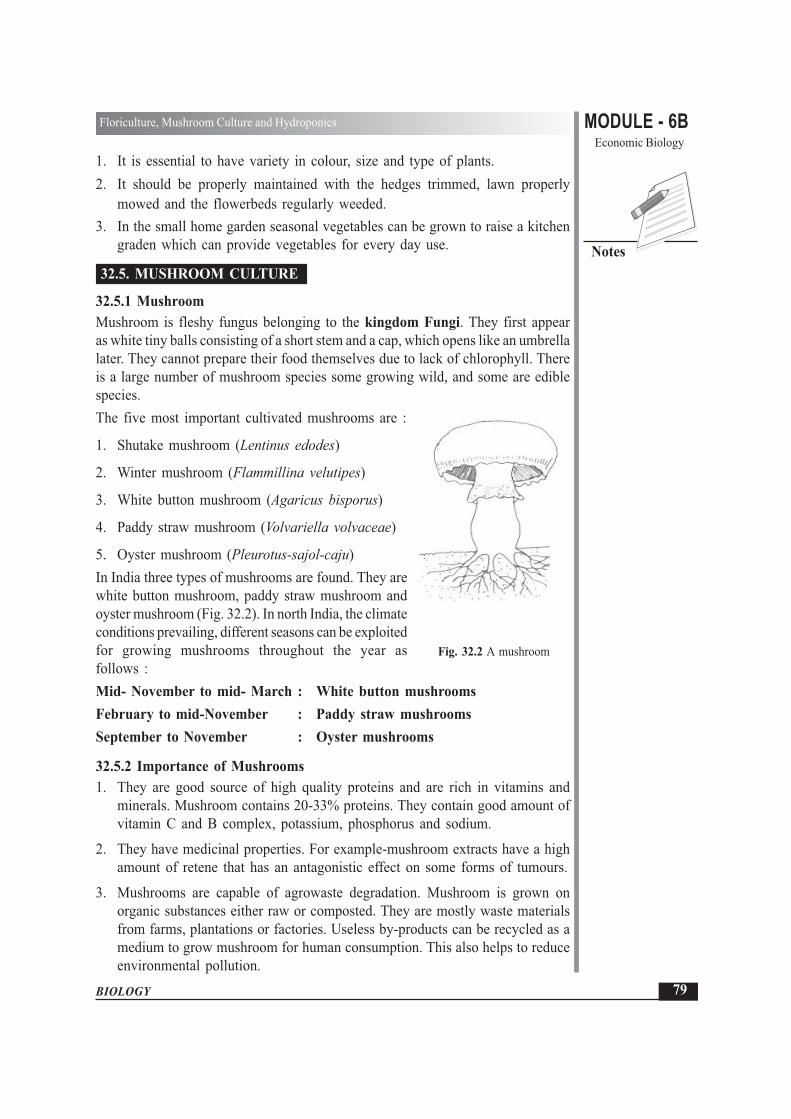
MODULE - 6BEconomic Biology
79
Floriculture, Mushroom Culture and Hydroponics
BIOLOGY
Notes
1. It is essential to have variety in colour, size and type of plants.2. It should be properly maintained with the hedges trimmed, lawn properly
mowed and the flowerbeds regularly weeded.3. In the small home garden seasonal vegetables can be grown to raise a kitchen
graden which can provide vegetables for every day use.
32.5. MUSHROOM CULTURE
32.5.1 MushroomMushroom is fleshy fungus belonging to the kingdom Fungi. They first appearas white tiny balls consisting of a short stem and a cap, which opens like an umbrellalater. They cannot prepare their food themselves due to lack of chlorophyll. Thereis a large number of mushroom species some growing wild, and some are ediblespecies.The five most important cultivated mushrooms are :
1. Shutake mushroom (Lentinus edodes)
2. Winter mushroom (Flammillina velutipes)
3. White button mushroom (Agaricus bisporus)
4. Paddy straw mushroom (Volvariella volvaceae)
5. Oyster mushroom (Pleurotus-sajol-caju)In India three types of mushrooms are found. They arewhite button mushroom, paddy straw mushroom andoyster mushroom (Fig. 32.2). In north India, the climateconditions prevailing, different seasons can be exploitedfor growing mushrooms throughout the year asfollows :Mid- November to mid- March : White button mushroomsFebruary to mid-November : Paddy straw mushroomsSeptember to November : Oyster mushrooms
32.5.2 Importance of Mushrooms1. They are good source of high quality proteins and are rich in vitamins and
minerals. Mushroom contains 20-33% proteins. They contain good amount ofvitamin C and B complex, potassium, phosphorus and sodium.
2. They have medicinal properties. For example-mushroom extracts have a highamount of retene that has an antagonistic effect on some forms of tumours.
3. Mushrooms are capable of agrowaste degradation. Mushroom is grown onorganic substances either raw or composted. They are mostly waste materialsfrom farms, plantations or factories. Useless by-products can be recycled as amedium to grow mushroom for human consumption. This also helps to reduceenvironmental pollution.
Fig. 32.2 A mushroom

BIOLOGY
MODULE - 6B Floriculture, Mushroom Culture and HydroponicsEconomic Biology
80
Notes
4. Mushrooms have a huge export potential.
5. Mushroom grows independent of sunlight without fertile land.
6. Mushroom-cultivation is a woman-friendly operation. Women in farmingsystem contribute a high percentage of the labour in the field especially indeveloped countries. It is one agricultural activity in which women can playan important role without sacrificing their household responsibilities.
32.5.3 Steps to be followed in the Cultivation of MushroomsThe use of mushrooms as human food dates back to time immemorial. Today withthe development of better technologies and greater realisation of their nutritivevalue, mushrooms have come to occupy an important place in the diet of peoplein several parts of the world including India. It is therefore necessary to haveknowledge of growing mushrooms.Since the white-button mushroom (Agaricus bisporous) is popular mushroom,its method of cultivation is discussed here.
Requirement for its cultivationThe optimum temperature for vegetative growth i.e. spread of the mycelium is 22°to 25°C and for the reproductive stage is 14° to 18°C. The following steps arerequired to be followed:
(a) Composting (b) Spawning (c) Casing
(d) Cropping and harvesting (e) Preservation
(a) CompostingCompost is prepared by mixing wheat or paddy straw, chicken manure with anumber of organic and inorganic fertilizers in fixed proportions. The compost iskept at a high temperature (Approximately 50°C) for one week and then mushroomsare cultivated on it.
(b) SpawningThis is the process of introducing the spawn, which is the “mushroom seed”, intothe compost. Spawn is merely the vegetative mycelium from a selected mushroomspecies grown in a convenient medium. The success of mushroom cultivation andits yield depends to a large extent on the purity and quality of the spawn used. Theamount of spawn used should be sufficient to help rapid and vigorous coverageof the beds with the organism.
(c) CasingCasing means covering the compost with a thin layer of soil or soil-like materialafter the spawn has spread in the compost. It gives support to the mushroom,

MODULE - 6BEconomic Biology
81
Floriculture, Mushroom Culture and Hydroponics
BIOLOGY
Notes
provides humidity, prevents quick drying of the spawned compost and thus helpin better growth of the spawn. Casing also helps to regulate the temperature.
(d) Cropping and harvestingThe arrangement is made for maintaining circulation of fresh air around the beds.Temperatures have to be kept low to prevent growth of pests and diseases andregular spraying of insecticides is also required to be done for the same purpose.It generally takes 7 to 8 days to come to the button stage from the first appearanceof the formation of a pinhead.
(e) PreservationMushrooms, like fruits and vegetables are perishable and require a great deal ofattention during storage, marketing and processing at the post harvest stage.Discolouration, weight and flavour loss are some of the problems.The following methods are used to increase their shelf life:(i) Vaccuum cooling(ii) keeping in a controlled atmosphere consisting of 9 percent oxygen and 25
percent carbon dioxide.(iii) giving gamma radiation and storing at 15°C(iv) freeze drying in a solution of brine, citric acid and ascorbic acid and
dehydration.(v) Canning
INTEXT QUESTIONS 32.3
1. Mention three advantages of mushroom cultivation.............................................................................................................................
2. Name the species found in India.............................................................................................................................
3. How can be preserve cultivated mashrooms for a long time?............................................................................................................................
4. List the main steps in mushroom cultivation............................................................................................................................
32.6 HYDROPONICSThe development of hydroponics has not been rapid although this technology isalmost 1500 years old.Hydroponics is a techniques for growing plants in nutrient solution (water withfertilizers) with or without the use of an artificial medium (sand, gravel,peatmoss, coir or saw dust ) to provide mechanical support.

BIOLOGY
MODULE - 6B Floriculture, Mushroom Culture and HydroponicsEconomic Biology
82
Notes
Hydroponics involves growing plants in containers filled with water or with coarsesand, gravel or other materials, to which nutrients have been added. The containersare made of glass, metal or plastic. They range in size from small pots for individualplants to huge tanks for large scale growing.Plants growing without soil require the same amount of light and warmth that theywould if they were growing in soil. Growers who use hydroponics indoors providea source of light and heat.
32.6.1 Methods of growing plants through hydroponicsThere are two main methods of growing plants without soil. They are water cultureand aggregate culture.(i) Water Culture (Fig. 32.3)In water culture, plants are suspended with theirroots submerged in water that contains plantnutrients. The roots absorb water and nutrients,but do not perform the anchoring function.Therefore, the plants must be mechanicallysupported from above. Air must be regularlypumped or mixed into the nutrient solution.
Culture SolutionThere are different kinds of nutrient solutions.A basic solution might contain:
3.4 kg - Potassium nitrate
0.65 kg - Ammonium Sulphate
2.65 kg - Magnesium Sulphate
1.05 kg - Monocalcium Phosphate Mixed together in 5000 litres of water
3.0 kg - Calcium Sulphate
Once the solution is in the tank, 5 litres of water containing 37 grams of manganoussulphate and 4 to 6 drops of concentrated sulphuric acid should be added to each5000 litres of solution once a month. In addition, 150 grams of ferrous sulphatein 5 litres of water should be added once a week.
(ii) Aggregate CultureIn aggregate culture although plants are grown without soil, the roots not onlyabsorb water and nutrients, but also anchor the plant. Instead of being placeddirectly in a water and nutrient solution, the roots are placed in a substrate material,such as coarse sand, gravel, peat etc. A nutrient solution is then repeatedly appliedto the substrate material. The solution is either pumped up from below the rootsor trickled down from above.
Fig. 32.3 Hydroponics

MODULE - 6BEconomic Biology
83
Floriculture, Mushroom Culture and Hydroponics
BIOLOGY
Notes
32.6.2 Importance of hydroponicsGrowing plants without soil is an effective way to study the needs of plants. Byvarying the amounts of nutrients, we can find the best ratio of nutrients forsuccessful growth of the plant.It is suggested that hydroponics can be used for commercial crop production. Inplaces where soil is not available, such as on ships at sea, deserts and in coveredArctic areas, hydroponics is an effective alternative.Hydroponics is used in large-scale cultivation of flowers and vegetables. The yieldis the same as for soil-grown plants and the technique saves time by automaticallywatering and fertilizing the crop.
INTEXT QUESTIONS 32.4
1. Define Hydroponics
............................................................................................................................
2. State the two methods of growing plants without soil.
............................................................................................................................
3. Why does air have to be constantly pumped into the nutrient solution? Givereasons for your answer.
............................................................................................................................
4. What will happen, when plants grown in a nutrient solution have to bemechanically supported from above?
............................................................................................................................
WHAT YOU HAVE LEARNT
� Floriculture is the study of different types of ornamental plants and theircultivation.
� There are different types of ornamental plants like seasonal annuals, floweringand foliage shrubs, ornamental trees, climbers, bulbous and indoor plants.
� Seasonal annuals are grouped according to the season of their growth andflowering into summer, winter and rainy season annuals.
� Ornamental shrubs are both of the flowering and foliage types.
� Indoor plants are used to decorate our homes.
� A home garden (kitchen garden) adds beauty to the house. It has to be carefullydesigned.

BIOLOGY
MODULE - 6B Floriculture, Mushroom Culture and HydroponicsEconomic Biology
84
Notes
� The five species of mushrooms cultivated in India are white button mushroom,paddy straw mushroom,oyster mushroom, shutake mushroom, and watermushroom.
� The main steps in mushroom cultivation are composting, spawning, casing,cropping and harvesting.
� Hydroponics is the method of growing plants without soil.� Two methods of growing plants without soil are water culture and aggregate
culture.� The nutrient solution contains nitrates, sulphates, phosphates of potassium,
magnesium, calcium, manganese and iron dissolved in water.� Air has to be constantly pumped into the solution.� Hydroponics is used for commercial production of crops, vegetables and
flowers in places where soil is not available.
TERMINAL EXERCISES
1. What is meant by Floriculture?2. Define the technique of Hydropnics.3. What is the importance of flowers in our daily life?4. List the different techniques used to propagate indoor plants.5. Which important points would you bear in mind while designing a home
garden?6. What are the advantages of Mushroom Cultivation?7. Which are the methods used to increase the shelf life of mushrooms?8. Explain the terms composting and spawning.9. Give the composition of a basic nutrient solution used in hydroponics.10. Distinguish between:
(i) Water culture and aggregate culture
(ii) Seasonal plants and perennial plants
(iii) Ferns and palms
(iv) Casing and spawning
11. List three varieties of mushroom found in India.
12. Give two examples of different types of ornamental plants
13. Describe the required various steps for mushroom culture.
14. Explain in brief about the importance of hydroponics.

MODULE - 6BEconomic Biology
85
Floriculture, Mushroom Culture and Hydroponics
BIOLOGY
Notes
ANSWER TO INTEXT QUESTIONS
32.1 1. Foliage shrubs are grown for their decorative leaves while floweringshrubs are grown for their beautiful flowers.
2. Foliage Shrubs – Mehndi, croton, acalypha
Flowering Shrubs – Jasmine3. Bulbous foliage plants – Colocasia, Caladium, Monstera, Ferns.
Bulbous flowering plants – Iris, Gladiolus, Tulip, Narcissus
4. Bulbous plants can be propagated through offsets, corms, fragments andbulbils.
32.2 1. Different types of indoor plants are foliage plants, ferns palms, cacti,flowering plants and bonsai.
2. Plants are grown indoors to decorate our homes specially the flats wherethere is no space for an outdoor garden.
3. Earthworm, metallic, plastic, wooden or cement.4. Set up of glass case for growing plants.
32.3 1. (i) Mushrooms are richer in proteins, vitamins and minerals than mostother vegetables.
(ii) Mushrooms help to reduce weight.
(iii) Mushroom cultivation opens new avenues for employment.
2. (i) White button, mushroom.
(ii) Paddy straw mushrooms
(iii) Oyster mushroom
3. see text
4. Composting, spawning, casing, cropping and harvesting.
32.4 1. Hydroponics is the science of growing plants without soil.
2. Water culture and aggregate culture.
3. Air has to be constantly pumped into the nutrient solution because rootsfor respiration require oxygen.
4. Plants grown in nutrient solution have to be mechanically supportedfrom above because their roots are unable to provide anchor to themas the medium is a liquid.

BIOLOGY
MODULE - 6B Animal HusbandryEconomic Biology
86
Notes
Ever since the beginning of civilisation, humans have depended on animals formany requirements, such as that of food (milk, meat and egg), clothing (hide orwool), labour (pulling, carrying load) and security etc. The development of desirablequalities in all such animal species, through creating better breeds, has been animportant human achievement. For this, humans have consistently tried to improvethe breeds of domesticated animals to make them more useful for them. In thislesson, you will learn about the common breeds of such animals, their uses andsome methods of improving their breeds.
OBJECTIVES
After completing this lesson, you will be able to:
� define the term Animal Husbandry;
� defferentiate between wild, tamed and domesticated animals;
� classify the domesticated animals into various categories;
� list the high yielding breeds of cattle in India and abroad;
� describe in brief the raising and caring of cattle;
� enumerate principal dairy products;
� describe modern techniques of genetic improvement such as artificialinsemination and embryo transplant;
� list the important breeds of cattle, pig, sheep, goat, horse, poultry and theirspecific uses;
� list the common diseases of animals, their symptoms and their causal organisms.
33.1 ANIMAL HUSBANDRYThe branch of science, which deals with the study of various breeds of domesticatedanimals and their management for obtaining better products and services from them
33
Animal Husbandry

MODULE - 6BEconomic Biology
87
Animal Husbandry
BIOLOGY
Notes
is known as Animal Husbandry. The term husbandry derives from the word“husband” which means ‘one who takes care’. When it incorporates the study ofproper utilisation of economically important domestic animals, it is called LivestockManagement.
Different Categories of AnimalsWild –Those that breed better where they are free than they do when they arecaptivated. They have no common use for humans. Example Lion, Tiger, Rhinoceres,Deer etc.
Tamed – Those, which are caught from the wild and trained to be useful to humansin some way. Elephant, Chimpanzee, Gorilla, Yak etc.
Domesticated – Those that are of use at home and are easily bred and looked afterby humans. Common domesticated animals are dog, horse, cow, sheep, buffalo,fowl etc.
Importance of domestic animalsOn the basis of utility, domestic animals are categorised into the followingfunctional groups
1. Milk giving animals Cattle, buffalo, goat, sheep etc.
2. Draught (used for load Bullock, horse, donkey, mule,bearing) animals camel, elephant, yak etc.
3. Fibre, hide and skin yielding Sheep, goat, cattle, buffalo, camel etc.
4. Meat and egg yielding animals Fowl (hen) and duck, goat, buffalo, pig etc.
33.2 MILK AND MEAT YIELDING ANIMALSDepending upon the availability and regional considerations different animals arereared for the purposes of yielding milk and meat in India. India is the world’slargest producer of milk. The majority of the milk consumed is also in liquid formin India. Over 53% of milk produced in India is from the water buffalo and amajority of milk processing plants in the country depend upon buffalo milk.
The National Dairy Development Board (NDDB) is the main agency behind thecooperative movement in India. India is now seeking joint ventures and financialparticipation from the private sector including foreign investment for productionof milk and milk products in India.
CattleCattle mainly include cow, bull, oxen, goat, sheep etc. The females of the speciesprovide milk, which in turn contribute animals protein to the diet of people. Whilethe female species of these cattle are used for milk, the male species play an

BIOLOGY
MODULE - 6B Animal HusbandryEconomic Biology
88
Notes
important role in the agricultural economy by providing labour, meat and hide. Milkitself is taken in many forms like ghee, curd, butter and cheese etc. The excretaof these animals (dung) is used as manure, in biogas and as fuel. There are severalimportant breeds of cattle in India and abroad.
33.2.1 Milk yielding animals
What is a breed ?A breed is a group of one species of animals, which have the same descent andare similar in body shape, size and structure.
Categories of Important breeds:There is following three categories
1. Indian breeds 2. Exotic Breeds 3. Improved breeds
(a) Indian BreedsGir, Sahiwal, Red Sindhi, Thararkar, Kankrej etc. are some high yieldingvarieties of Indian cattle (Fig. 33.1)
Fig. 33.1 Indian breeds of high milk yielding
(b) Exotic Breeds (Imported breeds)Hilstein, Friesian, Jersey, Swiss etc. are some of the high yielding varieties thathave been imported from abroad and reared widely in India.
(c) Improved breeds of Indian cattleCertain improved breeds have been developed by making a cross between twodesired breeds. A cross between Sahiwal and Friesian varieties has been namedas Friewal, Karan Swiss is another improved breed for milk production in largequantities. Table 33.1 shows some indian breeds, their milk yield and distribution.

MODULE - 6BEconomic Biology
89
Animal Husbandry
BIOLOGY
Notes
Table 33.1 Some Indian Breeds and their Milk Yield
Breed Milk yield (litres) DistributionPer lactation period
Gir 1200-2200 Gujarat, Rajasthan,Maharastra
Red Sindhi 700- 2200 Andhra Pradesh, all part ofworld including India and
PakistanSahiwal 1100- 3100 Haryana, Punjab. Uttar PradeshKankrej 1400 GujaratTharparkar 700-2200 RajasthanMewati 1100 RajasthanOngole 700 Andhra PradeshHariana 500 Gujarat,RajasthanHallikar 227-1134 litres South IndiaKangayam 665 litres Tamil NaduMurrah 20-22 litre/day Punajb, Haryana, Uttar Pradesh
Lactation Period is the period of milk production between birth of a young oneand the next pregnancy and it usually lasts about 300 days.
Breeds of Buffaloes
Breed Distribution
Murrah Haryana and PunjabBhadawari Uttar Pradesh and Madhya PradeshJaffarabadi GujaratSurti GujaratMehsana Gujarat (cross breed between Surti and Murrah)Nagpuri MaharastraNill Ravi PunjabPorlakmedi Orissa
33.2.2 Cattle feedThe main feed of cows and buffaloes are grass but this does not provide them allthe nourishment. They require balanced diet in the form of roughage which isfibrous food containing large amount of fibres such as hay fodder, leguminousplants-soyabeans, peas and cereals like maize, jowar etc.

BIOLOGY
MODULE - 6B Animal HusbandryEconomic Biology
90
Notes
The diet of cattle mainly consists of roughage (dry or green fodder or fibrous food)and concentrates like grains, oil cakes and seeds, mineral salts and vitamins.
33.2.3 Dairy ProductsMilk as drawn from the animals is known as full cream milk. When the cream isseparated and the remaining milk is called toned milk. This milk contains no fatand is known as skimmed milk. On the basis of fat contents the various milkproduct are as follows:
Cream : It is prepared by churning milk, the fat comes on the top which is separatedby draining out the liquid. It is known as cream with 10-70% fat contents.
Curd : Milk is converted to curd due to bacterial activities.
Butter Milk : It is the left over liquid after removal of butter.
Ghee : After heating butter, the water evaporates and fat contents are almost 100%.
Condensed milk : Milk is concentrated by removing water contents with or withoutadding sugar. It has 31% milk solids with 9% fats.
Powdered milk : It is the powdered form of milk.
Cheese : It is coagulated milk protein-casein with fat and water.
Khoya : A desicated milk product prepared by evaporating water contents andreducing the bulk to about 70-75%.
Cattle Dung : Cattle dung is mainly used to make dung cakes for burning as fuels.It is used mainly in villages of India. The farmers also use cattle dung to producebio gas and the leftover residue as manure.
Biogas plant (Gobar gas plant)Bio gas plant is a chamber where animal excreta (Cow dung, buffalo dung etc) andsome anaerobic bacteria are fed into airtight biogas chamber. Decomposition ofexcreta produces methane gas used as a smoke free gas for cooking. This gas canalso be utilized for lighting. The left over solid residue serves as a good manure.
33.2.4 Meat yielding animals(i) SheepSheep is the second largest species reared by mankind and it provides wool, meat,milk and hide. Their droppings form good manure. Important breeds of sheep inIndia are as follows:

MODULE - 6BEconomic Biology
91
Animal Husbandry
BIOLOGY
Notes
Breeds of Sheep
Breed Distribution
Chokla RajasthanNial Rajasthan and HaryanaMarwari Rajasthan and GujaratMagra RajasthanJaisalmeri RajasthanPugul Bikaner (Rajasthan)Malpura RajasthanPotanwadi Uttar Pradesh and DelhiMuzaffararanagari HaryanaHissardale Himachal Pradesh and HaryanaNellore Andhra PradeshBellary, Hassan, Mandya KarnatakaMecheri, Kalikarsal, Vembur Tamil Nadu
Exotic BreedsThe main exotic breeds of sheep are Toggenberg, Saanen, French, Alpine andNuibian and Angora.
Feeding of sheepThey feed on green grasses and other wild plants. When sheep are reared for aparticular purpose, they are given protein, minerals and vitamin rich food. The mainconstituents of their food are as follows :Leguminous fodder : Urad, mung, berseem etc.Oil cakes : Groundnut, seasame cake, (rich in proteins)Grains : Maize, barley, oats and jowar.Lime, common salt : Sterilised bone meal (rich in mineral salts)
(ii) GoatImportant breeds of goats used for milk, meat and hide. There are about 19 wellknown Indian breeds, apart from a number of local non-descript breeds that arescattered throughout the country. The breeds are mentioned below on the basis oftheir location.
Himalayan Region (hilly track)Cham, Gadd : Kashmir, Himachal Pradesh , Jammu and KashmirPashmina : Himachal Pradesh, Ladakh, Lahul and Spiti valleyChegu : Kashmir

BIOLOGY
MODULE - 6B Animal HusbandryEconomic Biology
92
Notes
Northern RegionJamunaparu – Uttar Pradesh, Madhya PradeshBeetal – PunjabBarhari – Delhi, Uttar Pradesh, Haryana
Central RegionMarwari, Mehsana and Zelwadi – Rajasthan, Gujarat and Madhya Pradesh
Kathiawar – Gujarat and Rajasthan
Southern RegionSurti – GujaratDeccani, Osmanabadi – Andhra Pradesh, TamilnaduMalabari – Kerala
Eastern RegionBengali – West Bengal, Assam and Tripura
Feeding of goatThe goats are fed on open fields with enough green. They can be only given cerealand grain products. Sometimes however, a milk goat requires a balanced feed with4-5 kg of fodder and a mixture of crushed grains such as yellow maize, jowar andother cereals and ground nut or linseed oil meal or steamed bone meal.
(iii) PigPig farming is gaining importance in India. Pigs provide only 8% of total meatproduction in our country. Pig skin, fat and hair are required for leather, soap, oil,hair-brush industry respectively. Pig manure is rich in nitrogen, phosporus andpotassium.
Pigs contribute about 5% of total meat production in India, and constitute a richsource of animal protein available at low cost. The calorific value of Pork (pig’smeat) is much more than the other edible meats. Pigs can feed on farm waste,garbage and spoiled grains.
Breeds of pigsBreed DistributionLarge white Yorkshire England, IndiaMiddle white Yorkshire England, IndiaLandrace DenmarkEssex saddleback HempshireTamworth EnglandBershile England

MODULE - 6BEconomic Biology
93
Animal Husbandry
BIOLOGY
Notes
INTEXT QUESTIONS 33.1
1. What is practice of feeding, caring and breeding of domestic animals is called?............................................................................................................................
2. Give difference between wild, tamed and domesticated animals.............................................................................................................................
3. Name two high milk yielding varieties of Indian cattle.............................................................................................................................
4. What does cattle feed mainly consists of ?............................................................................................................................
33.3 DRAUGHT ANIMALSDraught animals are animals need for carrying load. From time immemorial anumber of animal species have been used for special purposes by humans, utilisingtheir mechanical strength, endurance and speed. These include horse for riding andswift running; elephant for riding, strength and heavy load lifting, camel for ridingin sandy desert and ability to survive without water for long duration, donkey andmule (a hybrid of male donkey and female horse) for carrying load. Most of thedraught animals are herbivorous and survive on leaves of trees, shrubs and bushes.While raising them, they are also fed on grains, beans, cottonseeds, maize and branbesides dry/ green fodder. In Rajasthan, camel is used for yielding milk also.
HorseThe horse has fast movement, great stamina and endurance. Its body is suited forride, load pulling, mountain climbing and forest travelling. So the horse is animportant draught or work animal. They learn fast and can be maintained easilyin various climatic conditions. Due to their ability to move swiftly in rough areas,they are still useful in hills and in the deserts.Common Indian breeds and their distribution are as follows-
Breed Region
Kathiawari or kaunchi Rajasthan and GujaratMarwari or malvi RajasthanBhutia Tarai belt of Himalayan region (Punjab-Bhutan)Manipuri Pony Eastern hill regionSipti Pony Himachal Pradesh
33.4 FIBRE, HIDE AND SKIN YIELDING ANIMALSBesides providing meat, milk and transport, livestock provide many commerciallyuseful products such as fibre, skin and hide. Generally sheep and goat provide fibresfor making of products like wollen strings, ropes, carpets, clothing and brushes etc.

BIOLOGY
MODULE - 6B Animal HusbandryEconomic Biology
94
Notes
33.5 EGG YIELDING ANIMALSThis category consists of egg producing animals whose eggs are used as food bymankind to provide proteins. Poultry farming is defined as a term for rearingand keeping of birds such as fowl, duck and hen for egg and meat. Poultryfarming has become popular because it is comparatively easy to start and maintain.It gives quick return within one to six month of investments,, is easily manageableand requires less space and labour. Poultry birds and their eggs are a rich sourceof nutrients. Figure 33.2 gives a comparative account of the composition of fowlmeat and that of eggs.
Fig. 33.2 Composition of chicken and egg.
Common breeds of Poultry birdsIndian poultry breeds provide good quality meat but produces small sized eggs.They have natural immunity against common diseases as compared to exoticvarieties bred abroad which require greater protection and immunisation.The chicken is commonly classified on the basis of its origin.
(a) American, (b) Asiatic,(c) Mediterranean and (d) English
1. Plymouth Rock, Rhode Island Red, New Hampshire - American2. Brahma, Cochin, Langshan - Asiatic3. Leg horn, Minoxa - Mediterranean4. Cornish, Australorp - English
Indigenous BreedsAseel – Rajasthan, Andhra Pradesh, Uttar Pradesh
Busra – Gujarat and Maharastra
Chittagong – Eastern India
Karaknath – Madhya Pradesh

MODULE - 6BEconomic Biology
95
Animal Husbandry
BIOLOGY
Notes
(i) Indian BreedsThe Indian breeds of hen include Aseel, Chittagaog, Ghagus and Basra. Their egglaying capacity is around 200 eggs per year.
(ii) Exotic BreedsThese breeds are important from other countries and include White leghorn,Minorca, Rhode Island red. These birds have high egg laying capacity but carryless flesh as compared to Indian birds.
(iii) Upgraded varietySome improved varieties have been developed in India by hybridisation such asB 77, ILS 82 etc. They grow fast and also have as high an egg laying capacity asthe exotic varieties and are better suited to the Indian climate.Poultry FeedDepending upon the requirement of meat or egg production, poultry feed mainlyconsists of maize, rice, wheat bran, ground nut cake, fish meal, lime stones, bonemeal, common salt, vitamins and minerals.
33.6 GENETIC IMPROVEMENT IN ANIMALSThe application of laws of animal health and reproduction genetics has contributedtowards increase in milk, egg and meat productivity. The increase in egg productionbrought about the silver revolution in the area of animal husbandry. The methodsbeing widely used are artificial insemination and embryo transplant.
(i) Artificial inseminationArtificial insemination involves collection of semen from a healthy bull of thedesired breed, its storage at low temperatures and introduction into the females ofcattle of other breeds for bringing about fertilisation using sterilised (germ free)equipment. Advantages of this method are:
(a) Up to 3000 females can be fertilised from semen collected from one bull.(b) The semen can be stored for a long period and transported over long
distances.(c) Economical and high success rates of fertilisation.
(ii) Embryo transplantThis method of breed improvement has been quite successful in sheep and goat.In this method, embryos (depending on their period of development) from superiorbreeds are removed during the early stages of pregnancy and are transferred to theother female with inferior characters, in whose body the gestation period iscompleted. By this technique, quality and productivity in the livestock can beimproved. Unlike artificial insemination, this method has low success rate due togreater chances of contamination.

BIOLOGY
MODULE - 6B Animal HusbandryEconomic Biology
96
Notes
33.7 COMMON DISEASES OF ANIMALSDomestic animals often suffer from various diseases caused due to infection bybacteria, viruses, fungi, protozoa and worms.
(a) CattleCommon diseases in cattle are as follows:
Category of Pathogen Disease Symptoms
Bacteria Anthrax Swelling on the body reduced yield of milk
Tuberculosis Dry husky cough, Lungs are affected.
Virus Foot and Mouth disease Excessive salivation, Lameness and fever
Rinder pest Blood stained high fever diarrhoea
Protozoa Trypanosomiasis Intermittent fever and death
Fungus Ringwork Rounded scabs on head and neck.
(b) Poultry DiseaseRearing of poultry birds requires properly ventilated place and vaccination of newborn chicks. Poultry diseases can be classified as infectious or non-infectious. Non-infectious diseases are caused by faulty management, faulty feed preparation andinadequate diet or nutritionally deficient disease. Infectious diseases are classifiedaccording to the type of disease causing organisms and are as follows.Parasitic (external) – Lice, mites, tick and fleas
(internal) – Round worms, tapeworm and hexamitiasisProtozoan – Coccidiosis in chicken, leucocytozoonosisBacterial – Pullorum, Typhoid, Paratyphoid, fowl choleraViral – Ranikhet disease, fowl pox, infectious bronchitis, infection
bursitis, avian encephalomycytis, Marek disease, leukosis,chronic respiratory disease, Hepatitis etc.
Fungal – Aspergillosis, Moniliasis
INTEXT QUESTIONS 33.2
1. Name common bacterial diseases and their symptoms in cattle.............................................................................................................................
2. Name any two improved varieties of poultry birds.............................................................................................................................
3. Name any two draught animals
(i) ..................................................................................................................
(ii) ..................................................................................................................

MODULE - 6BEconomic Biology
97
Animal Husbandry
BIOLOGY
Notes
4. State two methods which we can make genetic improvement in the animals?
(i) ..................................................................................................................
(ii) ..................................................................................................................
5. A certain cattle is suffering from blood stained and high fever diarrhoea. Namethe disease and its pathogen.
............................................................................................................................
6. Mentioned common diseases in the birds.
............................................................................................................................
WHAT YOU HAVE LEARNT
� Animal husbandry deals with breeding, feeding and caring of domestic animals.
� Gir, Red, Sindhi, Sahiwal and Tharparker are some high-yielding varieties ofcows.
� Friesian, Jersey, Swiss are high-yielding exotic varieties of cows.
� Cattle dung provides manure, fuel, and bio-gas for cooking and lighting.
� Sheep provide milk, wool, hide and manure.
� Bikaneri, Kashmiri, Gaddi are some important breed of sheep.
� Indian Poultry birds provide more meat.
� Aseel, Ghagus, Chittagong and Basra are some good varieties of Indian Poultry.
� ILS 82 and B 77 are improved varieties of Indian poultry.
� Artificial insemination and embryo transplant are methods of geneticimprovement.
� Cattle may be affected by bacterial, viral , fungal or protozoan diseases.
� Ranikhet and Fowl pox are diseases of poultry birds.
TERMINAL QUESTIONS
1. Define the term animal husbandry.
2. Discuss the importance of artificial insemination.
BIOLOGY
MODULE - 6B Animal HusbandryEconomic Biology
98
Notes
3. Classify animals according to their importance. Also give two examples of eachcategory.
4. Name any three diseases that affect cattle, also name the pathogen involved andgive one symptom of each.
ANSWER TO INTEXT QUESTIONS
33.1 1. Animal Husbandry2. See section 33.13. Gir, Red Sindhi, Sahiwal4. Roughage and concentrate
33.2 1. Anthrax - swelling on the body tuberculosis by husky cough, lungs areaffected
2. ILS 82, B 773. Horse, mule, camel4. (i) Artificial insemination (ii) embryo transplant5. Render pest, virus6. Ranikhet, cowl pox

MODULE - 6BEconomic Biology
99
Fisheries and Aquaculture
BIOLOGY
Notes
In the world, India is amongst the top three producers of rice, wheat, liquid milk,poultry products, fruits, vegetables, coconut, tea, spices, marine and fresh waterproducts including fish and shrimp. Fishes are rich in protein, vitamins and mineralsalts and are also known as valuable protective food. Fish forms an important itemof the diet in many areas of the world. The development of fisheries is therefore,one of the most promising industry. In this lesson, you will learn about manyvarieties of fish, their collection, rearing, breeding and their economic importance.
OBJECTIVES
After completing this lesson, you will be able to:
� define the term fisheries;
� list some important fresh and marine fish found in India;
� list the economic importance of fish, some molluscs and sea weeds;
� mention the effect of environmental pollution on fish;
� describe fishing technology and equipments used for fishing;
� define fish migration and fish diseases;
� define aquaculture.
34.1 FISHERIESIndia has a coastline of about 8,129 km, 5 million km of continental shelf and 2.02million of exclusive economic zone. India is a major marine fish producer and ranksseventh in the world.Areas where fish are reared commercially, are known as artificial fisheries.The fishes are bred, reared and later harvested. The fishery may be a natural waterbody or an artificial one. A variety of fish may be reared together.In addition to fish, aquatic crustaceans and molluscs are included in fisheries. InIndia the economically most important crustacean fisheries are those of prawns,
34
FISHERIES AND AQUACULTURE

BIOLOGY
MODULE - 6B Fisheries and AquacultureEconomic Biology
100
Notes
shrimps and crabs. Among molluscs, edible oysters and pearl oysters are also ofeconomic importance.
34.2 COMMON EDIBLE FISH FOUND IN INDIADepending on the nature of water in which fish is reared, fisheries are divided intothree categories:1. Marine Fisheries: These deal with fishing operations along seacoasts. The
Indian subcontinent approximately has a 5600 kms long coastline. About 80%of India’s marine fish are supplied by the west coast and the remaining 20%by the east coast. The premier varieties are mackerels, sardines, sharks, andcatfish.
2. Fresh Water Fisheries or inland Fisheries : They include fish found in rivers,irrigation canals, reservoirs, lakes, tanks and ponds. Rohu, Catla, Mystus,Gourami,and Gambusia are some of the best varieties of fresh water fish.
3. Estuarine or Brackish Water Fisheries: They operate in estuaries (where riverwater and sea water get mixed), delta channels, backwaters, lagoons and coastallakes. Estuarine fish are more common in Bengal and Kerala. The tidal watercollects the fish in the enclosures. The main varieties are Pearl spot, Milkfishand Mullet.
Table 34.1 Common Indian edible fishFish Occurrence
A. Fresh water fish (inland fish)Carp (Herbivorous Fishes) Throughout India in its Northern,
Eastern and Southern parts,(i) Catla
(ii) Rohu (Fig. 34.1)Cat fishes (Carnivorous) Throughout India
(iii) Mystus Entire Indian coast(iv) Hilsa Maharashtra coast(v) Bombay duck Maharashtra coast
B. Marine Fish(i) Pomfrets Indo-pacific coast
(ii) Salmon Eastern and Western coast(iii) Sardines (Fig. 34.2) Southern and Western coast
C. Estuarine or Brackish water fish(i) Mullet Lagoons and coastal areas
(ii) Pearl Spot (Fig. 34.3) Coastal lakes of Bengal and Kerala
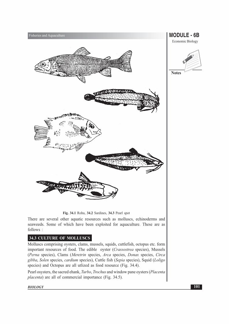
MODULE - 6BEconomic Biology
101
Fisheries and Aquaculture
BIOLOGY
Notes
Fig. 34.1 Rohu, 34.2 Sardines, 34.3 Pearl spot
There are several other aquatic resources such as molluscs, echinoderms andseaweeds. Some of which have been exploited for aquaculture. These are asfollows :
34.3 CULTURE OF MOLLUSCSMolluscs comprising oysters, clams, mussels, squids, cuttlefish, octopus etc. formimportant resources of food. The edible oyster (Crassostrea species), Mussels(Perna species), Clams (Meretrin species, Arca species, Donax species, Circagibba, Solen species, cardium species), Cuttle fish (Sepia species), Squid (Loligospecies) and Octopus are all utlized as food resource (Fig. 34.4).Pearl osysters, the sacred ehank, Turbo, Trochus and window pane oysters (Placentaplacenta) are all of commercial importance (Fig. 34.5).
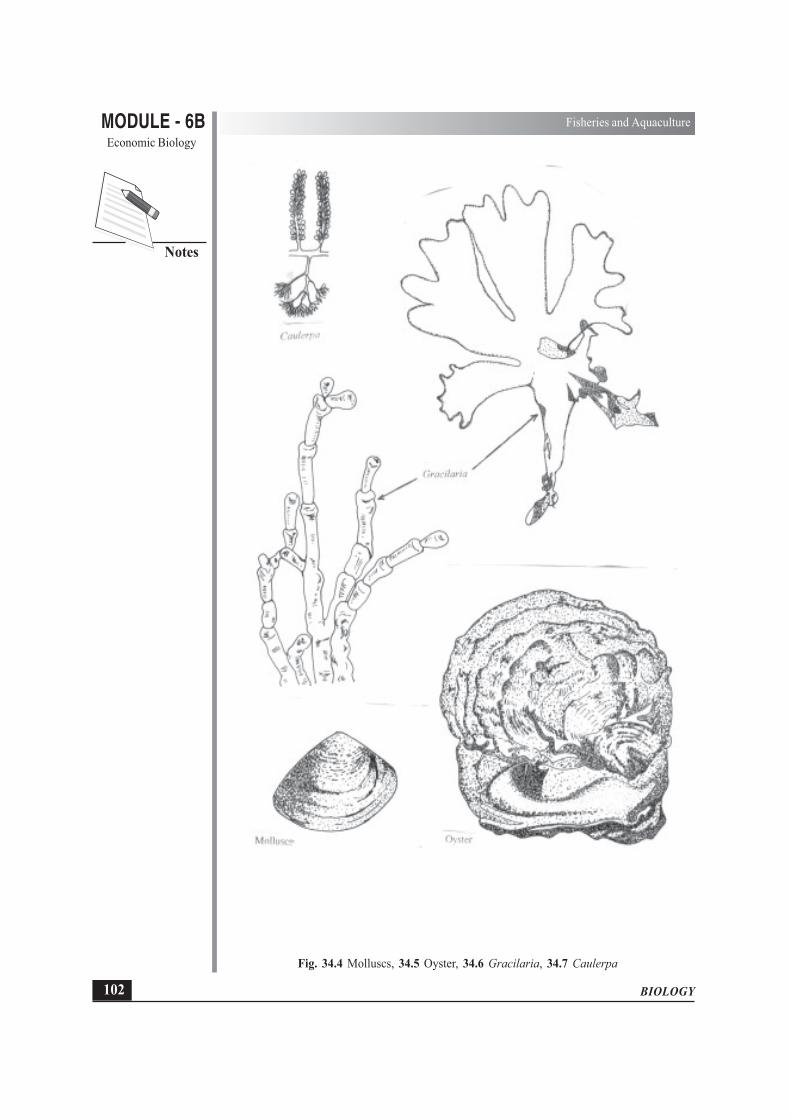
BIOLOGY
MODULE - 6B Fisheries and AquacultureEconomic Biology
102
Notes
Fig. 34.4 Molluscs, 34.5 Oyster, 34.6 Gracilaria, 34.7 Caulerpa

MODULE - 6BEconomic Biology
103
Fisheries and Aquaculture
BIOLOGY
Notes
34.4 SEA WEEDSSea weeds are also an importance marine resource and are found along the rockyintertidal and sub-tidal regions of the coast of India. The Sunderbans, the Chilkalake, the deltas of Godavari and Krishna, the rocky shore of Vishakhapatnam,Mahabalipuram, Gulf of Mannar, Gujarat coasts and Lakshadeep, Andaman andNicobar islands are rich in sea weeds. Seeweeds are used for human consumptionas cattle and poultryfeed, as manure and for industrial purpose as a source of agar-agar and algin. Species of Gelidiella and Gracilaria (the red sea weeds) are sourcefor manufacture of agar-agar (Fig. 34.6).The brown sea weeds like Sargassum, Turbinaria Dictyota contains alginic acid.Ulva, Entromorphas, Caulerpa, Porphyra are varieties used in human food(Fig. 34.7).
INTEXT QUESTIONS 34.1
1. Name two fresh water fishes.
............................................................................................................................2. In which part of India is pearl spot found
............................................................................................................................3. Define the term fisheries.
............................................................................................................................4. Give two examples of marine fish.
............................................................................................................................5. Name two common edible fish commonly consumed in India.
............................................................................................................................
34.5 ECONOMIC IMPORTANCE OF FISHFish is a valuable source of food that is rich in proteins. Fish proteins are easilydigestible. Apart from being a good source of food, Fish also have the followinguses:1. Medicinal use – fish liver oil is a natural source of vitamin A and Vitamin D.2. Industrial use – Body oils of sardines, herrings and salmons are used for the
manufacture of edible oils and margarine. This oil is used in soap, paint, andvarnish industries.
3. Feed for farm animals – Fish meal (dried fish) provides proteins to farmanimals.
4. Agricultural use – As organic manure in the fields.5. Adhesive – Skins and bones are also used in making high quality glues and
adhesives.

BIOLOGY
MODULE - 6B Fisheries and AquacultureEconomic Biology
104
Notes
6. Shark skin- Shark skin is also used in the manufacture of handbags, wallets,shoes etc. after tanning. Hide (leather) is also used to make parchment sheets.
34.6 AQUACULTURE AS AN OCCUPATIONAquaculture is an occupation of many living near rivers and the sea and mostaquaculture jobs are located in coastal community.Aquaculture involves the rearing and management of useful aquatic plant andanimal resources such as fish and shell fish, (prawn, molluscs, crabs etc.) It is alsoknown as fish farming and accounts for about ten percent of the world’s commercialfish harvest. China leads all countries in aquaculture production. Fish farms rangefrom simple ponds or flooded rice fields to highly engineered hatcheries in whichthe environment is monitored and kept under control. Environment controleliminates harmful environmental conditions and helps fish flourish and grow fast.Fish are provided with proper nutrients as per a regulated plan and are protectedfrom the harmful animals that prey on them. Aquaculture is utilised for culturingpearls on a commercial scale as well as in:
(i) rebuilding of salmon and trout stocks that have been severely reduced,and
(ii) raising fishes for consumption as food such as carp, cat fish, gourami,milk fish, salmon, tilapia etc.
Practice of aquaculture also includes Pisciculture and Pond culture (fish cultivationin large water bodies).
(i) Pisciculture (Fish Farming)Is concerned with the production of fish in lakes, rivers, large ponds,canals and is called fresh water or inland fisheries. In Pisciculture youngfishes are reared in nursery ponds, transferred to lakes or rivers and finallyharvested as fish for table food.
(ii) Pond culture (Kitchen Fisheries)This involves culturing fish in small ponds. This practice is quite commonin Bengal. Proper management is carried on till fish attains full size. Theyare also protected from diseases.
INTEXT QUESTONS 34.2
1. At the present times, which country leads in fish production.
............................................................................................................................2. What does aquacultrue include other than culturing fish?
............................................................................................................................3. What is meant by pisciculture?
............................................................................................................................

MODULE - 6BEconomic Biology
105
Fisheries and Aquaculture
BIOLOGY
Notes
4. Give one difference between pisciculture and pond culture.
............................................................................................................................5. Why is regulation of the environment in which fish are nearest important?
............................................................................................................................
34.7 EFFECT OF CHANGING ENVIRONMENT ON AQUACULTUREFish population is adversely affected by a number of environmental factors. Theseare as follows.� Water PollutionThe problem of water pollution mainly affects marine water fish. A variety ofinsecticides, pesticides, industrial effluents and domestic sewage find their way intomany rivers and pose a serious problem for fisheries. The magnitude of pollutionvaries with the size of the river, the flow of water etc. Let us take few examples.(i) The paper mills located on the bank of a river in Orissa consume nearly 270 million
litres of water per day. This heavy withdrawal of water together with the dischargeof high toxic effluents causes considerable harm to the fisheries for a stretch of nearly24 km downstream.
(ii) The effluents from Sindhri fertilizer factory have been found to have adverse effectson fish and prawn. Vast investigations in respect of effluents of paper pulp-textileindustries, tannery manufacturing units, sugar distillery, coal, etc. have shown adverseeffects on fisheries while sewage used as a fertilizer for fish farm has been found tocause extreme damage to fish culture.
(iii) Thermal pollution caused by the discharge of hot water used for cooling reactorsand generators can be a serious problem in tropical waters where the normaltemperature itself is high and further increase would be lethal to the fish whichare already living in the higher ranges of temperature tolerance.At present, apotential source of pollution is the atomic reactor wastes. Consequences ofwater pollution are given below.
� Many of favourite fishes like the Bombay Duck have almost disappearedfrom the Kalu river near Mumbai. This has been due to the release ofa number of toxic wastes from the chemical industries into the river.
� Excessive use of chemical fertilisers may lead to the phenomenon ofEutrophication (enrichment of the water body with nutrients). Thisresults in algal bloom, (excessive growth of algae which use up availableoxygen) followed by oxygen depletion in water and ultimately the deathof fish.
� Thermal (heat) pollution from various heavy industries causes fishmortality.
5
F

BIOLOGY
MODULE - 6B Fisheries and AquacultureEconomic Biology
106
Notes
� Utra violet radiations affect fish eggs that become non-viable, that is, they failto develop.
� Leakage of petroleum from ships and off-shore oil wells forms an oil slick onthe surface of the water and thus fishes are unable to breathe due to non-availability of dissolved Oxygen in the water.
34.8 FISHING TECHNOLOGYFish constitutes the most important part of the diet of many people. Fishing has beencarried out since ages and human beings have developed various gadgets and technologiesfor catching all varieties of fish from all regions and masses of water, at all depths aswell as in large quantities to take care of the ever increasing population and demands.Today it constitutes one of the largest traded food items.Organised fishing is practised with the help of equipment specially designed for easycapture of fish. Equipment differs according to the nature of water bodies, and thecharacteristics of the fish species to be captured.
Principal equipment for fishing includes
1. Sea fishing gearsThe various equipment used in fishing are called fishing gears. Most of the large scalefishing is done at sea. Sea fishing gears are of the following types.Nets: The main gears are made of cotton yarn hemp or other special man-made fibres.Nets are fixed in the tidal region during the low tide. High tide brings the fish along withwater flow into the net. (Fig. 34.8).
Fig. 34.8 Various types of nets for fishing.

MODULE - 6BEconomic Biology
107
Fisheries and Aquaculture
BIOLOGY
Notes
2. SeinesThese are very large nets for active fishing. The net encircles a large part of water believedto contain a lot of fish. A Seine is used in running water. Depending upon the nature ofwater body, different types of Seines (Pure Seine, Shore Seine, etc) are used.
3. Hooks and LinesMetallic hooks of various sizes and designs are provided with a bait to catch differenttypes of fish. The line is a wire, which carries the hook to various depths and distancesfrom the fishing raft or boat.
34.9 FISH MIGRATIONA number of fish show a periodic movement form one dwelling place to another. Thisperiodic movement is called migration. Fish migrate to other places for spawning (egglaying) or to avoid unfavourable climatic conditions. Many fish migrate from one part ofthe ocean to another at times. Marine fish like Hilsa swim up from sea to fresh waterfor spawning e.g. cel. Some fresh water fish migrate from lakes to sea for spawning.During winters in the cold part of the globe, water at the surface freezes. The fish migrateto the bottom and remain there till the end of the cold season.
INTEXT QUESTIONS 34.3
1. Mention two way by which gets polluted water.............................................................................................................................
2. What do you mean by thermal pollution?............................................................................................................................
3. What causes “algal bloom”?............................................................................................................................
4. How does UV radiation cause loss of fish?............................................................................................................................
5. What is “seine”?............................................................................................................................
34.10 FISH DISEASESLike other animals, fishes are also affected by various diseases. These diseases can bebroadly classified under bacterial, viral, fungal, protozoan and those caused by largeparasites like tapeworms and roundworms. Diseases like tuberculosis, eye infection,kidney diseases and various types of tumours – benign and malignant are reported fromboth freshwater as well as marine fish.
BIOLOGY
MODULE - 6B Fisheries and AquacultureEconomic Biology
108
Notes
Furunculosis, a common disease of fresh water fish is caused by bacteria and can betreated with tetracyclines and the vibrio infections respond to sulphonamide therapy.Tail rot, another common disease caused by bacteria can be controlled by administeringa mixture of penicillin and streptomycin.
Food poisoning caused by fish and fishery products may be due to a variety of causes.Some fish and shellfish (prawns, shrinps etc.) possess poisons or biotoxins. Thesebiotoxins are not destroyed by cooking and cause illness when fish containing them areeaten. Some people are allergic to fish, molluscs or crustaceans.
Some poisons affect the central nervous system, while others cause gastrointestinal andskin disorders. Food poisoning can also be caused by eating spoiled or rotting fish. Toxicprinciples in the flesh of such fish cause gastroenteritis. Bites and strings caused byvarieties of fishes and invertebrates like jelly fish result in serious inflammatory conditions.Dermatitis can be caused by irritants found in the skin of fishes belonging to the tunafish group.
WHAT YOU HAVE LEARNT
� The areas used for rearing, breeding and catching of fish constitute fisheries� Fish and shell fish, that is, molluscs and crustaceans such as crab, prawn and shrimp
are reared in fisheries.� Mackerels, sardines, sharks and catfish are some marine edible fish.
� Fresh water edible fish include rohu, catla, Mystus, gourami, and Gambusia.
� Estuarine fish such as pearl spol, milk fish and mullet are found in back water lagoons(areas where sea water comes and cover a part of land).
� Fish are a good and cheap source of animals proteins, and Vitamins A and D.
� Aquaculture involves rearing and management of useful aquatic plants and animals.
� Aquaculture or fish farming provides controlled environment, protection and nutrientsto the fish.
� Aquaculture includes Pisciculture and Pond culture.
� Fish are used for animal feed, agricultural manure, making of adhesives, soaps, paintsand varnishes.
� Environment changes adversely affect the fish population.
� Water pollution due to chemicals released from industry and agriculture, fertilisers,release of hot water, UV rays and oil spills cause fish mortality and affect fishproduction.

MODULE - 6BEconomic Biology
109
Fisheries and Aquaculture
BIOLOGY
Notes
TERMINAL QUESTIONS
1. Classify different fish on the basis of their natural habitat. Also mention two examplesof each.
2. List any four uses of fish for mankind.3. Define aquaculture mentioning its importance.4. Discuss briefly the effect of changing environment on fish population.5. What is meant by migration? Why do fish migrate?6. What can happen if rotton fish is consumed?
ANSWER TO INTEXT QUESTIONS
34.1 1. Rohu, Catla, Mystus (any two).2. Estuarine areas3. Areas where fish are reared for commercial purposes.4. Pomfret, sardine, salmon (any two)5. Rohu, Catla, Pomfret
34.2 1. China
2. Culturing ediable oysters, pearl oysters, crabs, shrimps, sea weeds.
3. Produciton of fish in ponds, lakes, rivers and canals.
4. In fisciculture, fish are reared in nursery ponds, then transferred to lakes orrivers and finally harvested while in pond culture, fish are reared and grownin small ponds and harvested.
5. It eliminates harmful conditions and helps the fish grow fast.
34.3 1. Addition of pesticides/effluents from factories/domestic sewage/hot water (anytwo)
2. By discharge of hot water used for cooling reactors and generators.3. Chemicals from factories that find their way into water bodies result in
excessive growth of algae or algal bloom.
4. Make fish eggs inviable
5. large fishing nets.

BIOLOGY
MODULE - 6B Apiculture, Lac Culture and SericultureEconomic Biology
110
Notes
Ever since the beginning of civilization, man has been trying to make use animalsaround him for various purposes and to rear them for increasing their number. Inthis context, you have learnt in the two previous lessons about animal husbandryand fisheries. In this lesson you will get acquainted with the major aspects ofapiculture (Bee- keeping), lac culture (rearing lac insects) and sericulture (rearingsilk moths).
OBJECTIVES
After completing this lesson, you will be able to :
� define apiculture (bee-keeping), list the species of honey bee and emphasizetheir economic importance;
� describe the structure of the bee-hive and various castes in a normal bee colony;
� explain the different biological features of the castes of honey-bee, castedetermination, development of the brood in comb (nest) and swarming;
� explain the methods of catching and hiving of swarms of honey bees and thecommon methods of bee keeping;
� describe the extraction of honey and beeswax and enumerate their uses;
� name the insect that produces lac and mention its uses;
� describe the main aspects in the life history of a lac producing insect;
� explain the terms stick lac, seed lac and shellac;
� define sericulture, name the insects (moths) that produce silk and theirrespective host plants;
� give a general account of the life history of the silk moth; and
� list the non-mulberry hosts for silk production, their sources and the parts ofIndia where they are produced.
35
APICULTURE, LAC CULTURE ANDSERICULTURE

MODULE - 6BEconomic Biology
111
Apiculture, Lac Culture and Sericulture
BIOLOGY
Notes
35.1 APICULTURE35.1.1 Bee-keeping – Its meaning and importanceApiculture is also known as bee-keeping. Why this name?‘Apis’ means bee. The scientific names of different species of honeybees begin withthe generic name Apis. Apiculture or bee-keeping is the art of caring for, andmanipulating colonies of honeybee in large quantity, over and above their ownrequirement.
35.1.2 Brief HistoryThe first evidence of this association came to light from the rock paintings madeby primitive human. Thousands of years ago, Egyptian were well acquainted withbee keeping before the Christian Era. In Rigveda, there are many references to beeand honey. Bee-keeping became a commercial proposition during the 19th centuryas a result of scientific research. Apiculture is a flourishing industry in manyadvanced countries like USA, Canada, Germany and Australia.
35.1.3 Importance of bee keepingThere are three main advantages of bee-keeping:
(i) Provides honey - a valuable nutritional food
(ii) Provides bees wax - which has many uses in industry
(iii) Honey bees are excellent pollinating agents, thus increasing agricultural yields.In terms of actual value this advantage exceeds the other two.
35.1.4 Species of honey beeThere are four common species of honey bee under a single genus Apis (apis =bee):
1. Apis dorsata (The rock- bee)
This is the largest honeybee.Builds single large open comb on high branches of trees and rocks.Produces large quantity of honey, but this bee is difficult to domesticate.This bee is ferocious, stings severely causing fever and sometimes even death.
2. Apis indica (The Indian bee)Medium - sizedHive consists of several parallel combs in dark places such as cavities of treetrunks, mud walls, earthen posts, etc.This bee is not so ferocious and can be domesticated
3. Apis florea (The little bee)small - sizedBuilds single small combs in bushes, hedges, etc.Honey yield is poor.

BIOLOGY
MODULE - 6B Apiculture, Lac Culture and SericultureEconomic Biology
112
Notes
4. Apis mellifera (The European bee)Somewhat like the Indian bee (Apis indica).This has been introducted in many parts of the world including India.It is easily domesticated.
INTEXT QUESTIONS 35.1
1. What does “Apis”, a latin word mean literally?............................................................................................................................
2. Write three advantages of bee-keeping.............................................................................................................................
3. Name the two species of honeybee that can be domesticated.............................................................................................................................
35.1.5 The bee colony – various castes and their activitiesA honey bee colony has three castes (Fig. 35.1a)(i) Queen – only one; functional female(ii) Workers – 20,000-30,000, sterile females(iii) Drones – a few only, functional males available prior to swarming.(i) Queen Bee
Queen bee is the only perfectly developed female, that is has well developedovaries and other organs of female reproductive system.She is largest in size.Its wings are smaller and are shrivelled.Mouth parts for sucking food is shorter than that of workers.No wax glands.Live for about 3 - 4 years.May lay eggs at the rate of 800 - 1500 per day.
35.1.6 Events in the life of queen beeUsually at the age of 7-10 days in her parent hive, after the old mother queen alongwith some workers had left for starting another hive, this new virgin queen goesout for marriage (nuptial) flights. The drones from the same hive chase her. Thisswarm may also be joined by drones (male bees) from other hives. Mating takesplace, while flying, on an average, the queen mates with about six drones and thenreturns to the hive. The sperms she has received are enough for her whole life, andshe never mates again.The queen has a control mechanism on the release of the sperms from thespermatheca (sperm store). She can lay two types of eggs:

MODULE - 6BEconomic Biology
113
Apiculture, Lac Culture and Sericulture
BIOLOGY
Notes
Fig. 35.1(a) Various castes of honey bee.
1. Fertilized – eggs that produce females (either sterile workers or fertile females(new queens).
2. Unfertilised – eggs which produce drones.
(ii) Worker beesWorker bees are imperfectly developed females.
These are smaller than the queen.
These have strong wings to fly.
These have a large and efficient proboscis (mouth parts packed together like a thintube) for sucking nectar.
A well-developed sting is present.

BIOLOGY
MODULE - 6B Apiculture, Lac Culture and SericultureEconomic Biology
114
Notes
Hind legs have “pollen basket” for collecting pollen.
The workers have a life span of about 35 days. The different duties which theyperform age-wise are as follows:
Day 1-14 Activity inside the hive such as cleaning the hive, feeding the larvae,etc.
Day 14-20 Guard duties at entrance to the hive
Day 21- 35 Foraging, i.e. collecting the food (nectar and pollen from thesurroundings)
For foraging, some scout bees set out in the morning. On locating good sourcesof nectar (i.e. flowers) they return to their hive and perform characteristicmovements (bee dances) at the comb. These dances communicate to the otherworker bees the distance and the direction of the food source. This is how moreand more worker bees are deployed in food gathering. The workers visit flowerto flower, collect nectar and pollen and return to their own nest against taking cluefrom the position of Sun as well as by certain amount of memory and finally thesmell of their own particular hive (Fig. 35.1b).
Fig. 35.1b A portion of the nest (hive) of the honey bee
The bee danceIn this dance the middle course of the dance communicates to the other bees theangle from the hive with reference to the sun. Taking a hint from this angle theyhave to fly to reach the food source (Fig. 35.1c).

MODULE - 6BEconomic Biology
115
Apiculture, Lac Culture and Sericulture
BIOLOGY
Notes
Fig. 35.1C The bee dance
(iii) DronesDrones are the male bees produced from unfertilised eggs. Their production in thehive synchronises with the production of the new (virgin) queens. At the age of14-18 days the drones perform mating flight chasing the virgin queen in the air.Drones can live up to about 60 days, although they are stung and killed after themating.The schematic representation of formation of different castes of bees is shown inFig. 35.2.
Queen lays
Unifertilized Eggs Fertilized Eggs↓ ↓
Hatch into Larvae Hatch into Larvae↓ ↓
For first few days fed on “Royal jelly” Fed on “royal jelly”(Saliva of workers) then on honey and for first few days.
pre-digested pollen↓
Pupa↓
Drones Royal jelly replaced Royal jelly feedingby honey and pre-digested continued
pollen ↓↓ Pupae
Pupae ↓↓ Queen
Workers
Fig. 35.2 Schematic representation of the formation of different castes in honeybee.

BIOLOGY
MODULE - 6B Apiculture, Lac Culture and SericultureEconomic Biology
116
Notes
Emergence of new Queen, and Swarming of the old oneWhen the queen gets older (usually in the third year) her body gives out a chemicalstimulus to the workers to construct a few rearing cells for queens. She places onefertilized egg in each of such brood cells. The larvae are fed on royal jelly (salivaof workers). They turn into pupae and then into queens. The first queen to emergefrom the brood cells, kills the remaining ones.Now the old queen takes to swarming along with a mixture of workers of all ages,leaves the old hive to develop a colony at some new site.The new queen in the old hive takes to mating flight with the drones and returnsto the same hive, as described earlier.
INTEXT QUESTIONS 35.2
1. Name the three castes in a honey bee colony
............................................................................................................................
2. Name the following
(i) The caste largest in size
..................................................................................................................
(ii) The kind of flight which the new virgin queen takes along with the dronesout of the hive
..................................................................................................................
(iii) The caste produced from unfertilised eggs
..................................................................................................................
3. What are the main duties of a worker bee in its following day wise age phases
(i) Days 1-14 .................................................................................................
(ii) Days 14-20 ...............................................................................................
(iii) Days 21-35 ...............................................................................................
4. What happens to the drones after mating flight?
............................................................................................................................
35.1.7 Apiculture and commercial production of honeyBees produce honey and wax both of which are valuable and marketable commodities.
(a) Indigenous methods of bee keepingMany villagers make (i) wall or fixed types of hives in rectangular spaces in thewalls with a small hole or (ii) movable types of hives in wooden boxes or earthenpitchers. The traditional beekeepers catch clustered swarms from trees, bushes, etcand transfer them to the above-mentioned spaces. After sometime when the honey

MODULE - 6BEconomic Biology
117
Apiculture, Lac Culture and Sericulture
BIOLOGY
Notes
is ready, the bees are driven away from the comb usually by smoking the hive. Thenthe comb is cut away and the honey is squeezed out through a piece of large -meshed cloth.
(b) Modern hivesThe modern beehive is made up of a series of square or oblong boxes without topsor bottoms, set one above the other. This hive has the floor at the bottom, and acrown board at the top, and a roof over all. Inside these boxes, wooden frames arevertically hung paralled to each other. The wooden frames are filled with sheetsof wax foundation on which the combs are built by the bees. The only entranceto the hive is below the large bottom box (brood chamber). The queen is usuallyconfined to the brood chamber. The boxes termed “supers” are used for storageof honey. The queen is prevented from going to the “supers” by the “queenexcluder” that allows only the workers to move (Fig. 35.3).
Fig. 35.3 A modern bed hive.
Catching a swarmYou have already read what a swarm is. It is an old queen accompanied by hugepopulation of workers flying to start a new hive. Swarms are collected from wherethey are settled. Some kind of a container is needed to collect the bees. The containeris usually a straw basket (skep) with a lid.
Hiving a SwarmIt is the process in which the collected swarm is transferred to the hive to buildup the colony and produce honey. It is operated in two ways:

BIOLOGY
MODULE - 6B Apiculture, Lac Culture and SericultureEconomic Biology
118
Notes
(i) Traditional methodThe hive is set up with brood chamber filled with its full number of frames. Eachframe has a full sheet of foundation and there is a crown board with roof at thetop.
A sloping board with white sheet is set against the entrance of the hive.
Bees in the skep (basket) are knocked out of it on to the slope.
The instinct of the bees to move upwards onto the dark, drives them onto the hivethrough the entrance.
(ii) Quick methodIn this method the crown board of the hive is taken off, frames are also taken offand the entrance is closed.
The skep is intimately united with the hive and the bees are poured into the broodchamber from the top.
The frames containing the wax foundation are placed in the hive.
The crown board is put back in its position and the entrance is opened.
It must be seen that the queen enters the hive. Now, sugar syrup must be fed tothe swarm, as this feeding will help the bees to settle down to work in their newhome.
Bee PasturageThe plants that yield nectar and pollen are collectively termed “bee pasturage”. Thefruit trees, ornamental plants and forest trees comprise important bee pasturage.
Nectar is the sweet secretion of the flowers. It is raw material for honey.
Pollen provides the raw material necessary for the major food of the brood.
INTEXT QUESTIONS 35.3
1. Name the two types of indigenously made hives.
............................................................................................................................
2. Rearrange the following parts of a modern hive for bee keeping, in their propersequence from bottom to the top:
Brood chambers, crown board, queen excluder, floor, super, roof.
............................................................................................................................
3. Name the two methods for hiving a swarm.
............................................................................................................................
4. What is the meaning of the term “bee pasturage” ?
............................................................................................................................

MODULE - 6BEconomic Biology
119
Apiculture, Lac Culture and Sericulture
BIOLOGY
Notes
35.1.8 Products from a bee hive
A. HoneyHoney is a food material for the bees and their larvae. Large quantities of honeyare stored in the hive to meet the demands in scarcity. Chemically, honey is a viscouswater solution of sugar. Its approximate composition in percentage is as follows:
Water 13-20
Fructose 40-50
Glucose 2-3
Minerals Traces
Vitamins (minute quantities)
(B1, B2, C)
Composition of honey and its different flavours depend on the kinds of flowersfrom which the nectar is collected.Nectar is sucked from flowers and mixed with saliva. It is swallowed into a specialregion of the gut called honey stomach. Nectar is a disaccharide (sucrose) it ishydrolysed by the salivary amylase to produce monosaccharides (fructose andglucose).Inside the hive the workers regurgitate the processed nectar. The honey thusproduced is still very dilute. After placing this honey onto the storage cells of thehive the bees “fan” with their wings to evaporate the excess water and bring thehoney to its required concentration.Extraction of honey from the combs is done by centrifugation.
Uses of HoneySome uses of honey are as follows– Food : Honey is a nutritious food, rich in energy and vitamins.– Medicines: It is used as a carrier in ayurvedic and unani medicines. It acts as
a laxative and prevents cold, cough and fever.
– It is used in religious ceremonies.
– It goes in the making of alcoholic drinks and beauty lotions.
– Another important use is in scientific research for making bacterial cultures.
– It is also utilised for making poison baits for certain insect pests.
Purity StandardsThere is no ready method to test the purity of honey by the customers. Homogenousgranulation is a probable sign of its purity. Otherwise there are laboratory methodsfor testing (test for monosaccharides).

BIOLOGY
MODULE - 6B Apiculture, Lac Culture and SericultureEconomic Biology
120
Notes
B. BeeswaxBeeswax is secreted by the wax glands located on the underside of the last fourabdominal segments (4th to 7th) of the worker bee. This wax is used in constructingbee combs in which the colony of the bees develops.
Uses of beeswaxSome uses are as follows:– making of candles (the modern candles are made of paraffin wax, a petroleum
product);– making pharmaceutical preparations;– preparation of varnishes and paints;– Water proofing and waxing of threads; and– formation of comb foundation (wax foundation in apiaries).
INTEXT QUESTIONS 35.4
1. Name the two major kinds of sugar found in honey............................................................................................................................
2. Mention any one non-food use of honey............................................................................................................................
3. How is honey chemically different from nectar?............................................................................................................................
4. Mention any two uses of beeswax.............................................................................................................................
35.2 LAC CULTURE
LAC and SHELLACLac is a resinous substance secreted by a tiny insect called Lacciferlacca (popular name “lac insect”)Shellac is the purified lac usually prepared in the orange or yellowflakes
35.2.1 Lac or shellac is used in many ways– Commonest use is in polishing wooden furniture. The granules are dissolved
in spirit and then are applied in very thin layers on the wooden surfaces– In sealing parcels, packets and envelopes– As insulating material in electrical work

MODULE - 6BEconomic Biology
121
Apiculture, Lac Culture and Sericulture
BIOLOGY
Notes
– In making phonograph records (now replaced by synthetic material)– In shoe polishes– In toys and jewelleryUtilization of lac for various purposes has been very ancient in India. A “lac palace”is described in Mahabharata, which was intended to be used for burning thePandavas alive.
35.2.2 Lac insectThe lac insect lives on native trees in India, Burma (now called Myanmar) andMalaysia. In India it is chiefly grown on trees like “ Kusum”, “ Palas”, and “Ber”.– The minute young lac insect (also called crawler) finds a suitable branch.– The insect inserts its beak into the plant tissue to obtain nourishment.– It grows in size and secretes a resinous material around itself.– The resinous material hardens on exposure to air.– Thousands of crawlers settle side by side and the resinous secretion builds up
around them and completely encases the twig.– Most crawlers develop in about 3 months into female which occupy small
cavities in the resinous mass. The females can never come out of these masses.– Eggs develop inside the body of the female and she assumes a sac like
appearance.– The female dies, the eggs hatch, the crawlers escape and move to a nearby-
uninfected part of the twig, and the process is repeated. (Fig. 35.4).
Fig. 35.4 A piece of twig encrusted with lac.
35.2.3 Extraction of LacThe encrusted twigs are known as stick lac. Such twigs are harvested.– The stick lac is ground largely in crude mortars, and the resulting granular lac
is called seed lac.– The fine particles or the dust separated from the granular lac is used in making
toys, bangles etc.– The wood portion is used as fuel.– The seed lac is washed, melted, spread out in a thin layer and dried. This is
the shellac of commerce.

BIOLOGY
MODULE - 6B Apiculture, Lac Culture and SericultureEconomic Biology
122
Notes
The Hindi name “Lakh” or “Laksha” in SanskritIt requires about 4,00,000 (4 lacs) insects to yield one kilogram oflac. The Hindi word “Lakh” for shellac possibly derives from suchlarge number of insects required to produce lac.
In India the lac insect is found in great abundance and millions of people directlyor indirectly find livelihood in this industry.
Lac Research Institute in Ranchi (Now in Jharkhand) conducts research on thevarious aspects of the lac insect, its life history, protection against enemies, etc.
Synthetic lacquers have been produced by the modern industry, which is replacingtrue shellac for many purposes.
INTEXT QUESTIONS 35.5
1. What is the scientific name of the lac insect?............................................................................................................................
2. List any three common uses of shellac.............................................................................................................................
3. Name any two trees on which lac insect grows.............................................................................................................................
4. What is seed lac?............................................................................................................................
35.3 SERICULTURE
Sericulture or silk production is the breeding and management of silk worms forthe commercial production of silk.
Sericulture is an important industry in Japan, China, India , Italy, France and Spain.
35.3.1 Brief HistorySericulture or silk production from the moth, Bombyx mori has a long and colourfulhistory unknown to most people. This insect is the only living species of familyBombycidae and has been domesticated for so long that it is possible that thereare no survivors in the wild any longer.
According to the Chinese records, the discovery of silk production from B. morioccurred about 2700 BC. It is believed that empress Si-lung-Chi was asked byemperor Huang-ti to find the cause of damaged mulberry leaves on trees in theirgarden. The empress found white worms eating the leaves. She noticed that theywere also shiny cocoons around themselves. A cocoon dropped in her cup of tea

MODULE - 6BEconomic Biology
123
Apiculture, Lac Culture and Sericulture
BIOLOGY
Notes
and silky threads separated from the cocoon. Silk industry began in China wherethe source of silk was kept a secret for more than 2000 years. After some time,China lost their monopoly in silk production, sericulture reached Japan throughKorea and then to other countries.Sericulture has been growing in India as an agro-based industry playing a vital role inthe improvement of rural economy.
35.3.2 Source of silk – The silkwormThe silkworm is the larva or the caterpillar of the moth Bombyx mori (popularly calledthe silk moth) the total life history of the moth (from egg to adult take 50 days. The differentstages are as follows:(i) Egg 10 days(ii) Larva (4 Stages) 30 days(iii) Pupa (Cocoon) 10 days
Fig. 35.4a Stages of life history of silk worm moth.
(i) AdultThe adult silk moth is a creamy white moth that has a flat body and a wing expanseof about 5 cms. It takes no food and seldom attempts to fly. It lives for only 2 to3 days. After mating, the female moth lays 300-500 eggs on leaves of the mulberrytree.
(ii) EggsThe eggs are round and yellowish-white, and they become grey as hatching timeapproaches.

BIOLOGY
MODULE - 6B Apiculture, Lac Culture and SericultureEconomic Biology
124
Notes
(iii) LarvaeThe newly hatched larva is about 3 mm long and somewhat black in colour.The larvae grow in size and shed their skin (moult) four times. Each growing stage ofthe caterpillar consumes lot of mulberry leaves.The last stage full grown larva is about 7 cm long. It has a hump behind the head anda spine-like horn at the tail end.When full grown, the mature larva stops feeding, climbs on a twig and spins a cocoon.
(iv) PupaThe full grown larva pupates inside the cocoon– In about 10 days time it transforms into a winged adult. The adult moth makes an
opening in the cocoon and escapes through it.
The cocoonThe cocoon is formed from a secretion from two large silk glands (actually the salivaryglands), which extend along the inside of the body and open through a common ducton the lower lip of the mouthparts. The larva moves the head from side to side veryrapidly (about 65 times per minute) throwing out the secretion of the silk glands in theform of a thread. The secretion is a clear viscous fluid, which on exposure to the airgets hardened into the fine silk fibre.The filament forming a cocoon is continuos and ranges in length from 700-1100 metres.The cocoons from which moths have emerged are called pierced cocoons. These areof low value because continuous thread cannot be obtained. Pieces are removed byinstruments and spun into a thread.
35.3.3 Rearing of silkwormsSelected healthy silk moths are allowed to mate for 4 hours. Female moth is then keptin a dark plastic bed. She lays about 400 eggs in 24 hours, the female is taken out andis crushed and examined for any disease, only the certified disease- free eggs are rearedfor industrial purpose. The eggs are hatched in an incubator.The hatched larvae are kept in trays inside a rearing house at a temperature of about20°C-25°C . These are first fed on chopped mulberry leaves. After 4-5 days freshleaves are provided. As the larvae grow, they are transferred to fresh leaves on cleantrays, when fully grown they spin cocoons.
35.3.4 Reeling silkThe cocoons are cooked in hot water and the silk fibre is unwound from the cocoons.This process is called reeling. The silk consists of two proteins the inner core isfibroin and an outer cover of sericin. There are four following steps for thecompletion of the process of reeling:
For reeling silk the cocoons are gathered about 8 days after spinning had begun.– The cocoons are first treated by steam or dry heat to kill the insect inside. This

MODULE - 6BEconomic Biology
125
Apiculture, Lac Culture and Sericulture
BIOLOGY
Notes
is necessary to prevent the destruction of the continuous fibre by the emergence ofthe moth.
– Next, the cocoons are soaked in hot water (95° -97°C) for 10-15 minutes to softenthe gum that binds the silk threads together. This process is called cooking.
– The “cooked” cocoons are kept in hot water and the loose ends of the thread arecaught by hand.
– Threads from several cocoons are wound together on wheels (“charakhas”) to formthe reels of raw silk (Fig. 35.5).
Only about one-half of the silk of each cocoon is reelable, the remainder is used as asilk waste and formed into spun silk.Raw silk thus obtained is processed through several treatments to give it the final shape.
Fig. 35.5 Reeling of silk cocoons.
35.3.5 Main properties of silk1. It is lustrous, soft and strong.2. It is made of two proteins : the inner core is fibroin and an outer cover is sericin3. It is hard wearing.4. It can be dyed into several colours
Silk moth Bombyx mori is at present fully domesticated. It no longer exists in awild state and it cannot survive without the human care.
35.3.6 Silk Producing States of IndiaMajor Indian States producing mulberry silk are:
KarnatakaWest BengalJammu and Kashmir
35.3.7 Non-mulberry “silks”1. Tasar silk is produced by certain species of another moth Antherea royeli. Their

BIOLOGY
MODULE - 6B Apiculture, Lac Culture and SericultureEconomic Biology
126
Notes
larvae are reared on Arjun trees, chiefly in Bihar, Madhya Pradesh and West Bengal.
2. Muga silk is obtained from Antherea assama whose larvae are reared on “ Som”trees in Brahmaputra Valley.
3. Eri silk is produced by the moth Philosamia ricini whose larvae feed on castorleaves. It is produced in Assam.
INTEXT QUESTIONS 35.6
1. Match the items of the columns A, B, and CA B C
Kind of silk Insect source Trees1. Mulberry silk (i) Antherea royeli (a) Arjun tree2. Tasar silk (ii) Antherea assama (b) Mulberry tree3. Muga silk (iii) Philosamia ricini (c) Som tree4. Eri silk (iv) Bombyx mori (d) Castor
2. Where did the silk industry first start in the world?............................................................................................................................
3. State the following :
(i) The number of days for which the adult silk moth survives...................................................................................................................
(ii) The number of times the silkworm sheds its skin before turning into a pupa...................................................................................................................
(iii) The temperature at which the silk worms are kept in the rearing house...................................................................................................................
(iv) The purpose of cooking the cocoons in hot water...................................................................................................................
4. Name the three largest silk growing states of India.............................................................................................................................
5. Which body part of the silkworm produces the silk forming substance?............................................................................................................................

MODULE - 6BEconomic Biology
127
Apiculture, Lac Culture and Sericulture
BIOLOGY
Notes
WHAT YOU HAVE LEARNT
� Bee- keeping helps in three ways – provides honey, provides wax and bring aboutpollination of agricultural crops.
� There are four common species of honey bee - the wild Apis dorsata the twodomestic ones Apis indica and Apis mellifera and the little bee Apis florea.
� A bee colony has three castes - a queen, large number of workers and the maledrones (produced only for mating in the nuptial flight).
� Queen is the largest, has no wax glands, lives up to 3-4 years, and lays eggsat the rate of 800- 1500 per day.
� Queen lays two types of eggs - fertilized eggs produce females and unfertilisedeggs produce male bees.
� Workers are sterile females and possess an efficient sucking proboscis, waxglands, and a sting.
� Workers live for about 35 days, and they perform different duties in differentlife periods.
� Workers communicate to the fellow workers of the hive about the direction anddistance of the food source by means of “bee dances”.
� When the hive is overpopulated the old queen with a large number of workersleaves the parent hive (swarming) and settles at some other site, and one newqueen takes charge of the previous hive.
� Beekeeping is an ancient industry but the modern way is very technical. Modernhive consists of several boxes one above the other.
� A swarm is caught and is hived by either traditional method or the quickermethods.
� The plants visited by the bees are called “ bee pasturage”.� Honey is a nutritious food rich in simple sugars and vitamins.� Honey has numerous uses besides as a direct food.
Beeswax is secreted by the wax glands of the workers. It has wide uses incosmetics, varnishes, paints, candle-making.
� Lac is produced by a tiny insect Laccifer lacca.� Lac has numerous uses in industry- largest being as a polishing material and
in making phonograph records.� Lac is the secretion of the lac insect, which hardens and covers the insect,
making an encrustation on the twig.� The lac on the twigs is called stick lac and after removal from the wood, and
is ground into grains is called the seed lac.� Lac is grown in the largest quantity in India in the state of Bihar.
BIOLOGY
MODULE - 6B Apiculture, Lac Culture and SericultureEconomic Biology
128
Notes
� Silk is the product of silk glands of the silkworm or in the larva (caterpillar) of themoth Bombyx mori.
� Besides mulberry silk there are tasar silk, muga silk and eri silk produced by threeother moths.
� For obtaining silk the cocoons are killed by steam, then they are kept hot water (cooking) and the silk is reeled.
� Silk is lustrous, and made of the proteins Fibroin and sericin.
TERMINAL QUESTIONS
1. Describe the composition of a flourishing bee colony.
2. Describe the composition of a modern hive used in beekeeping. How is a swarmhived in it?
3. What is lac? Differentiate between stick lac, seed lac and shellac.
4. What is silk? How does the silkworm produce it?
5. Describe the various steps in obtaining raw silk form the cocoons.
ANSWER TO INTEXT QUESTIONS
35.1 1. bee
2. Provide (i) honey, (ii) Wax and (3) bees are pollinating agents
3. Apis indica, Apis florea, Apis mellifera
35.2 1. Queen, worker, drone
2. (i) Queen, (ii) nuptial flight (iii) drones
3. day 1 to 14 – cleaning the hive, feeding larvae
day 14 to 20 – guard entrance of the hive
day 21 to 35 – foraging/collecting food/collecting nector
4. They get killed
35.3 1. Fixed on wall, movable type in wooden box or earthen pitchers
2. Floor, brood chamber, queen excluder, supers, crown board
3. Traditional method, quick method
4. A collective term for plants that yield nector and pollen
MODULE - 6BEconomic Biology
129
Apiculture, Lac Culture and Sericulture
BIOLOGY
Notes
35.4 1. Glucose, fructose
2. Medicine/cosmetics/bacterial culture (any one)
3. Nector is sucrose a disaccharide and honey has monosaccharides fructose andglucose
4. Making candles or varnish, water proofing waxing threads (any two)35.5 1. Leccifer lacca
2. Sealing, insulation, gramophone records, shoe polish, toys, jewellery (anythree)
3. Kusum, palas, Ber (any two)4. Granular lac
35.6 1. 1-iv-b; 2-(i)-a; 3-(ii)-c 4-(iii)-d2. China3. (i) 2 to 3 day
(ii) 4 times(iii) 20°C to 25°C(iv) to soften the gum that binds the silk fibres.
4. Karnataka, West Bengal, Jammu Kashmir5. Silk glands

APOSS- INTERMEDIATE
BIOLOGY (THEORY)
MODEL QUESTION PAPER
(ENGLISH VERSION)
Time: 3 hrs
Max. Marks: 80
Instructions to Candidates:-
1) Answer all Questions in Section-A
2) Answer any Six questions in Section B
3) Answer any three questions in Section C
4) Answer all these questions in same order (1 to 16).
SECTION A
Answer all the Questions. 2 x 16 = 32
Each question carries 2 marks.
1) What is a tissue? Name the tissue which helps in absorption?
2) Name any two biochemical reactions involved in the biosynthesis of Amino acids
3) What is meant by haemodialysis.
4) Draw a neat labelled diagram of Stomata
5) Define enetic Code ?
6) Give two examples for turgor movements
7) Explain any two methods of cross pollination
8) Write the steps involved in the recombinant DNA technology
9) Define cology
10) Differentiate the macro from micronutrients
130

11) Write the differences between broad spectrum and narrow spectrum antibiotics.
12) State the two approaches of human gene therapy
13) What is a Herbarium ?
14) Mention any two differences between the compound microscope and simple microscope
15) How are the Drosophila transferred from one culture bottle to another ?
16) Mention any two medicinal plants with their scientific names.
SECTION B
Answer any Six questions. Each question carries 4 marks. 4 x 6 = 24
17) Explain the differences between plant cell and animal cell
18) Name the suicidal bag of the cell? Explain its structure and function
19) What are the reactions involved in glycolysis.
20) How does thermoregulation take place in man and animals.
21) Draw a neat and well labelled diagram of the L.S. of ovule.
22) What are the effects of global warming.
23) Mention any four bacterial diseases in man and their causative organisms.
24) Describe the steps involved in the production of Bio-gas and mention the precautions to be taken.
SECTION- C
Answer any three questions.
Each question carries 8 marks. 8 x 3 = 24
25) Explain the process of cellular respiration in man.
26) Draw a neat and well labelled diagram of the T.S. of monocot leaf.
27) How does the population growth influence the urbanization and environmental degradation in our country ?
28) griculture is said to play a major role in providing the important human .
131

INTERMEDIATE (APOSS) (Practicals) and Teaching Learning Material (TLM)
Samples
1) Hemitoxilin
2) Saframin
3)
4) Soils (Clay Soil & Red Soil)
5) Iodin Solution
Slides
1) T.S. of Monocot Stem
2) T.S. of Dicot Stem
3) T.S of Monot root
4) T.S. of Dicot root
5) Different stages of Mitosis slides
6) Different stages of Meosis slides
7) T.S of bone
8) T.S. of Costilege
9) T.S. of Mammalian ovary
10) Human blood cells
11) T.S of Mammalian Testis
12) Stranted muscle fibres
132
Specimen
1) Sponze
2) Earth worm
3) Star fish
4) Dog fish
5) Toad
6) Bat
7) Rohu
8) Pirtia
9) Opentia
10) Ginger
11) Lemon
12) Pitcher plant
Experiments in Biology (Practicals)
1. Demonstration of Photosynthesis process in an aquatic plants
2. Preparation of slides of Onion-root tip for observation of stages of mitosis.
3. Demonstration of osmosis by Potato- Osmometer
4. Study about the salivary amylase on starch
5. Preparation of temporary mount of onion peel to observe and study epidermal cell.
133


A.P. STATE OPEN SCHOOL, HYDERABAD
Senior Secondary Course
BIOLOGY
3(CORE MODULES)
Designed and Developedby
Neelam Gupta
S.C.E.R.T. Campus, Opp. L.B.Stadium, Basheerbagh, Hyderabad - 500 001.Ph:040-23299568. Website : www.apopenschool.org; E-mail : [email protected]
LABORATORY MANUAL
135

136
CONTENTSS.No. Name of the Exercise Page No.
1. Introduction 143
2. Exercise 1 147
Some Common Instruments
3. Exercise 2
2.1 Preparation of temporary mount of onion peel to observe 153
and study epidermal cells
2.2 Preparation of temporary stained mount of human cheek cells 158
2.3 Preparation of temporary mount of leaf epidermis to 161study the structure of stomata
2.4 Preparation and study of xylem and phloem from cucurbita stem. 164
2.5 Temporary stained preparation and study of striated muscle fibres 166 in cockroach.
4. Exercise 3 169
Study of morphological modifications of plant parts like root, stem and leaf.
5. Exercise 4 189
To study the T.S of dicot and monocot stems and roots from permanent slide.
6. Exercise 5 198
Study of the microscopic: anatomy (histology) of mammalian tissues and organs.
7. Exercise 6 205
To study the structure and function of different parts of flowers(China rose and Petunia)
8. Exercise 7 213
Study of animal specimens and their classification- to identify the characteristicfeatures of sponge, earthworm, butterfly, apple snail, starfish, dogfish(Scoliodon) bonyfish (Rohu), toad, house lizard, pigeon and bat.

137
9. Exercise 8 243
Preparation of a slide of onion root tip for observation of stages of mitosis.
10. Exercise 9 250
To study the special adapting features in some plants and animals
11. Exercise 10
(a) To study the physical properties of different soil samples 255
(b) To study the water holding capacity of different soil samples 258
12. Exercise 11 261
Demonstration of osmosis by potato osmometer
13. Exercise 12 263
Determine the rate of photosynthesis in an aquatic plant(Hydrilla or Elodea)
14. Exercise 13 267
Study the structure and germination in gram and bean seeds.
15. Exercise 14 273
To demonstrate the release of CO2 during germination of seeds.
16. Exercise 15 277
To study about the action of salivary amylase on starch
17. Exercise 16 (Optional Module)
(a) To study the developmental stages in the life cycle of 282
Drosophila by preparing a culture
(b) A project to study the growth pattern of money plant 285
(c) To make a herbarium288
6
F

138

139
����������
As in any other science subject, practicals have an important role in Biology too. The purpose ofteaching biology is not only to acquaint the learner with biological terms, facts, concepts and principlesbut also to prepare him/her to understand these concepts by doing exercises relating to them. Selfexperience not only eliminates doubts and misbeliefs in one’s mind but also generates an interest in thesubject. The present practical course thus considers practical work as an integral part of the biologycurriculum at Senior Secondary stage.
1. THE OBJECTIVES OF BIOLOGY PRACTICALS
The objectives biology practicals are to:
● develop practical skill for better understanding through first hand experience;
● demonstrate the principles covered in the theory;
● develop observational skill in the form of identifying and locating desired parts in specimen;
● develop manipulative skills in arranging and handling the apparatus and instruments and takingreadings on them;
● collect material and to mount it and to develop skill in preserving biological material and specimens;
● draw, label and record experimental results and interpret them;
Through practical work, not only the theoretical concepts are tested but also it trains you in thescientific method.
2. THE FORMAT OF THIS MANUAL
The exercises presented in this manual are in the form of self-instructional material. Each exercisein the manual has the following format:
1. Aim: It defines the scope of the exercise.

140
2. Introduction : It describes the purpose of the experiment.
3. Objectives : The objective of an experiment gives you an idea about the skills and knowledge tobe developed after performing that experiment.
4. What you should know : It highlights the concepts and background knowledge relating to theexperiment, which should be known to you in order to perform the experiment in a meaningfulmanner.
5. Materials required : Listed various materials, apparatus etc. required to carry out the exercise.
6. How to proceed : It includes the steps to perform an experiment in a sequential manner.
7. Precautions : The precautions to be taken in carrying out the exercise are listed here. Anyspecific precaution wherever necessary is listed at the relevant step of the exercise.
8. Observation and Documentation : A detailed format of observations, step by step and theirrecording is given in observation and documentation. An effort has been made to adopt a self -interactive method of recording these observations.
9. Diagrams, wherever necessary, are given in each exercise and it is advisable that the studentsshould compare the diagrams with the actual one as seen in the slide / specimen etc.
10. For the teacher : The teacher will help you to perform an experiment.
3. HOW TO USE THIS MANUAL
This manual consist, of the following parts:
- Illustrative step-by- step instructions for doing the practical.
- Worksheets for recording observations and answering related questions.
Use the manual in the following way for performing the practicals.
1. Read the aim of the experiment carefully. Try to understand what is required to be done
2. Get ready by collecting all materials required for the exercise.
3. Read the instructions given in the procedure step by step and keep following the instructions.
4. Wherever "observe" comes, carry out the observation and fill up the observations in the spaceprovided for observations and documentation or in your notebook. The sequence of differentobservations is indicated by numbers 1,2.3 etc. Record observations in the correct sequence.Try noting down the observations then and there instead of doing it later. Draw the diagrams asyou actually see them. Only the part of the specimen should be drawn which is asked for.
5. Apart from the general precautions to be taken while working in a laboratory also follow theprecautions given either at the end or in between the instruction steps for each practical withinbox. Do not avoid these precautions if you want better results they arc specific for the particularexperiment.
6. Complete the worksheet for each experiment. You will find that the worksheet is based on yourobservations and also on theoretical knowledge which you have studied in the study material.

141
7. Reference of the books has been given wherever necessary.After doing the practicals you maygo back and study the book once again for better understanding.
8. Keep your record book neat and clean as it is an important material for practical examination.Three marks are allocated for keeping proper records of practicals.
9. Do not forget to carry your manual with you when you go for the practical work.
Once again the steps involved in performing a practical are listed below in the chart to help youdo the practicals.
Read instructions Follow each Make
carefully step Observations
Get it evaluated Complete Note down all
worksheet observations
Prepare a Record Book
4. SAFETY IN THE LABORATORY (DO’S AND DON’TS)
The following precautions and care should be taken while working in the biology laboratory :
(i) The students should be well aware of the exercise they are going to perform in the laboratory.
(ii) The instruments, glassware and any other equipment should be kept clean at its proper placebefore and after its use.
(iii) The microscope and other delicate instruments should be atleast 5 inches from the edge of thetable to avoid its knocking off accidently.
(iv) Do not throw any broken glassware in the sink. It should be thrown in the dust bin.
(v) Whenever working with the sharp instrument as blade/ scalpel etc, be careful not to cut orpuncture your skin.
(vi) Do not inhale, never taste or apply stain or any chemical as it may harm.
(vii) Never eat in the laboratory to avoid infection.
5. MAINTENANCE OF RECORD BOOK
We hope you will follow the instructions listed in each experiment while performing it and recordyour observations in your notebook. You may use following style for writing the exercise in your recordbook.
● Aim of the exercise.
● Materials and method used for performing the exercise.
● Procedure followed.

142
● Observations which you made during performing the exercise and diagram wherever asked.
● Precautions taken during experimentation.
6. SCHEME OF PRACTICAL EXAMINATION
There will be a practical examination of three hours duration carrying 20 marks. The distributionof marks is as follows:
(i) Performing an experiment 4 marks
(ii) Submitting a project report 3 marks
(ill) Identification of given samples 4 marks
(4 samples)
(iv) Preparing mounts 3 marks
(v) Maintenance of Record Book 3 marks
(vi) Viva Voce 3 marks
Total 20 marks
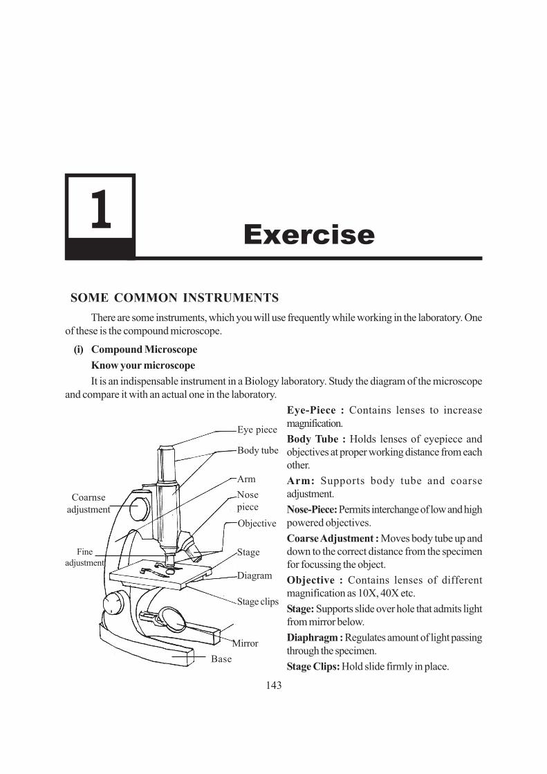
143
�� �� �SOME COMMON INSTRUMENTS
There are some instruments, which you will use frequently while working in the laboratory. Oneof these is the compound microscope.
(i) Compound MicroscopeKnow your microscopeIt is an indispensable instrument in a Biology laboratory. Study the diagram of the microscope
and compare it with an actual one in the laboratory.Eye-Piece : Contains lenses to increasemagnification.Body Tube : Holds lenses of eyepiece andobjectives at proper working distance from eachother.Arm: Supports body tube and coarseadjustment.Nose-Piece: Permits interchange of low and highpowered objectives.CoarseAdjustment : Moves body tube up anddown to the correct distance from the specimenfor focussing the object.Objective : Contains lenses of differentmagnification as 10X, 40X etc.Stage: Supports slide over hole that admits lightfrom mirror below.Diaphragm : Regulates amount of light passingthrough the specimen.Stage Clips: Hold slide firmly in place.
Eye piece
Body tube
ArmNosepieceObjective
Stage
Diagram
Stage clips
MirrorBase
Coarnseadjustment
Fineadjustment

144
Base: Firm support bearing weight of microscope.
Mirror: Reflects light upward through diaphragm and hole in stage.
FineAdjustment : Permits exact focussing by moving stage or body’ tube up or down very slightly.
Inclination Joint: Permits tilting to adjust the eye level.
Using the microscope
● Always use both hands when carrying the microscope, one hand beneath the base and the otherholding the arm of the microscope in an upright position to be check. Walk, holding the microscopeclose to your body.
● Set the microscope at least 5 inches from the edge or the table to avoid its knocking off accidently.
● Always clean the lenses and mirror of the microscope with the lens paper/ cloth. Otherwise theremight be scratches on them.
● Adjust the mirror by slightly tilting it and by seeing through the eye piece so that sufficient lightenters the microscope when you view under low magnification objectives.
● Place the prepared slide directly over the hole in the stage.
● Secure the slide on the stage with the stage clips to prevent accidental movement of the slide.
● Look through the eye piece and slowly bring the low magnification objective towards the materialby using the coarse adjustment until the specimen comes into view.
● To change to high power, rotate the nose-piece to bring the high power objective in position(taking precaution that the body tube does not move up or down).
● Look through the eye piece, if the light is insufficient, open out the diaphragm slightly.
● Gently raise the objective by using fine adjustment. If the image worsens without improving, startlowering the objective by the same fine adjustment. (Do not use coarse adjustment while viewingunder high power). By gently moving up and down you will be able to get a clear focus.
● While removing the slide from the stage release the spring clips. Do not allow the stage clips toextend out of the stage.
● When work gets over, rotate nose piece such that the objective lens diagram of simple hand lensnot over the hole in the stage.
● When not in use keep it covered by a polythene cover and/or lock it in its box.
(ii) A simple hand lens
● Contains a single double convex lensmounted on a handle.
● Can magnify things four to five times.
● Used for smaller magnification.

145
(iii) Scalpel
● Works like a knife, used to cut out thin slicesand peel.
(iv) Fine scissors
● Used for cutting.
(v) A pair of forceps
● Used for picking up very thin slices or material.
(vi) Fine needles
● Used for (i) adjusting sample/ tearing anybiological material on a glass slide withouttouching it. (ii) placing the cover slip on the slide.
(vii) Fine hair brush
● Mainly used for transferring material for mountingon the slides.
(vii) Spatula
● Used to pick up solid chemicals.
GLASSWARE
(i) A dropper
● Used for (i) putting a drop of liquid on the slide.
(ii) Plain glass slides
● Used for preparing temporary or permanentmounts.

146
(iii) Cover slips (Very thin glass cover)
● Used for covering the material placed on glassslide to be observed under the microscope. Thisprotects the objective lens.
(iv) Petridish
● Is a shallow dish often with a cover.
● Used for soaking specimen for the purpose ofpreservation, staining etc. Also used to keep amedium on which bacteria or small organismsmay be cultured.
(v) Beaker
● Available in various sizes like 100 ml and 250 mletc.
● Used for preparing and storing chemicals andperforming experiments.
(vi) Flask
● A bottle with a narrow neck used in thelaboratory for performing experiments (keepingsolution, for heating solution etc).
(vii) Funnel
● Available in various sizes i.e. in different diameterof the mouth of the funnel.
● Used during filtration of solutions.

147
(viii) Pipette
● A slender graduated glass tube for measuring andtransferring known volume of liquid.
(ix) Spirit lamp or Bunsen burner
● Used for heating. It should be extinguishedimmediately after use. .

148

149
Exercise 2
�� ���������� ����� �� �������2.1 Epidermal peel of onion
2.2 Squamous epithelium from human cheek cells.
2.3 Epidermal peel of leaf to observe stomata
2.4 Xylem and phloem from cucurbita stem
2.5 Striated muscle fibres (cockroach)

150

151
�� �� �2.1 PREPARATION OF TEMPORARY MOUNT OF ONION PEEL
TO OBSERVE AND STUDY EPIDERMAL CELLS
An onion peel is a very suitable material for observing a cell and its parts. The components such as cellwall, cytoplasm, nucleus and vacuoles can be easily observed through this exercise.
OBJECTIVES
After performing this exercise, you should be able to:
● acquire the skill of removing thin outer layers from plant material;
● prepare a temporary stained mount without trapping air bubbles;
● learn to handle and use the microscope such that its light is adjusted and material focussed toclarity;
● observe a typical plant cell and tally with your theoretical knowledge about the cell and itscomponents;
● distinguish between ·some components of a plant cell such as the cell wall, cytoplasm; nucleusand vacuole.
2.1.1 WHAT YOU SHOULD KNOW
1. A tissue such as that of the peel is made of many cells.
2. A cell has many components, some of which can be seen under the compound microscope.

152
Materials Required
(i) Onion bulbs (ii) Paper towelling / Blotting paper
(iii) Dropper (iv) Glycerine
(v) Saffranine solution (for staining)
2.1.2 HOW TO PROCEED
(i) Select an onion bulb, discard the brown dryouter scales.
(ii) Cut the onion into four pieces (quarters)vertically.
(See Fig. 2.1.1). Remove one fleshy scale.
Fig. 2.1.1
(iii) Bend the outer (convex) surface of the fleshyscale towards you with your right hand tobreak it. (Fig. 2.1.2)
Fig. 2.1.2
(iv) It forms a neat break yet it remains attachedto the other end of the scale that you areholding with your left hand (See Fig. 2.1.3).
Fig. 2.1.3
(v) Gently pull the broken end. You will find thatfrom other half of the scale held in your lefthand, a thin transparent layer of epidermis ispeeling off easily (See Fig. 2.1.4).
Fig. 2.1.4

153
(vi) If the peel is large, use a fine pair of scissor ora blade to cut a small piece of about 2 mm.To do this place the peel in a drop of wateron a clean slide and trim it.
(vii) If there are any wrinkles in the peel, stretch itwith the help of dissecting needle.
(viii) Place this neatly cut peel in the centre of aclean slide in a fresh drop of water (Fig. 2.1.5)and blot out the excess water.
Fig. 2.1.5
(ix) Examine the slide under low power of themicroscope (fill up observation 1).
Staining
(i) When you are able to see the epidermal cells clearly in your peel, remove the slide from themicroscope.
(ii) Drain off water and then add a very small drop of Saffranine to the peel on the slide and leave thematerial in the stain for about two minutes.
(iii) See the stained material under the microscope to check staining. It should neither be too dark norto light. If it is light, leave in the stain for some more time.
(iv) Pick up the stained material from the slide, wash it and place it in a drop of glycerine on a freshslide.
(v) Hold the coverslip with your left hand at 45° (as shown in the diagram) on the slide in such a waythat the lower edge of the coverslip touches the glycerine. Now using the needle, gradually lowerthe coverslip so that no air bubble gets trapped in the material. Excess glycerine should beremoved with the help of a blotting paper.
Fig. 2.1.6 Putting coverslip Fig. 2.1.7 Epidermal cells in onion

154
The slide is now ready for further observation (fill up observation 2).
(vi) Observe under the microscope and compare the diagram provided (Fig. 2.1.7) with the slide asseen under the microscope.
2.1.3 PRECAUTIONS
1. Do not leave the peel too long in air, otherwise it will dry and show air bubbles in it
2. The peel should be mounted in the centre of the slide.
3. Always use a brush (not a needle) to transfer the peel from petridish to the slide or from one slideto another. Otherwise, the peel will tear off.
4. Avoid the entry of any air-bubbles in the mount.
5. Use clean slides and cover slips for mounting.
2.1.4 RECORDING OF OBSERVATION
Observation 1
Under low power of the microscope
(i) What can you see? (long rows of rectangular cells in the unstained onion peel)
.............................................................................................................................................
(ii) Which structures of the cell can you see? Do you see the cell wall, the nucleus and a large vacuolecontained in the cytoplasm?
.............................................................................................................................................
Observation 2
After staining the onion peel
(i) Do you see large number of cells in the peel or only one? What is the general shape of these cells(rectangular, circular, triangular, polygonal etc)?
.............................................................................................................................................
(ii) What is the darkly stained body in each cell?
.............................................................................................................................................
(iii) Can you see any vacuole in the cell cytoplasm?
.............................................................................................................................................
(iv) Does the nucleus become more conspicuous after staining?
.............................................................................................................................................
(v) What is the position of the cytoplasm in the cell? (central or peripheral)

155
.............................................................................................................................................
(vi) What is the shape of the nucleus? (spherical, oval, irregular etc.)
.............................................................................................................................................
(vii) Sketch the onion peel cell as seen under the microscope. Label the parts such as the cell wall,cytoplasm, vacuole and the nucleus.
.............................................................................................................................................
(viii) Record all the observations in your record book.
.............................................................................................................................................
2.1.5 FOR THE TEACHER
Please ensure that
1. slides and coverslips are cleaned before use.
2. microscope is handled properly.
3. staining is properly done as staining helps to highlight certain cell components.
4. the students are told that cell has other components also but they can not be seen under compoundmicroscope.

156
2.2 PREPARATION OF TEMPORARY STAINED MOUNT OFHUMAN CHEEK CELLS
The slide of human cheek cells is easy to prepare and gives a view of an animal cell and also howthe cells of squamous epithelium are arranged.
OBJECTIVES
After performing this exercise, you should be able to:
● acquire the skill of taking out human cheek cells;
● learn to prepare a uniform smear;
● observe the special features of squamous epithelium.
2.2.1 WHAT YOU SHOULD KNOW
1. Animal cell lacks the cell wall and large vacuoles.
2. Epithelial tissue forms covering of organs and is of various types.
3. Inner lining of the cheek is made of squamous epithelium where cells are (a) flat (b) closelypacked and (c) have central nucleus.
Materials Required
(i) Slides (ii) Coverslips (iii) Filter - papers
(iv) Needles (v) Methylene blue (vi) Brush
(vii)Tooth pick.
�� ��

157
2.2.2 HOW TO PROCEED
(i) Take a washed tooth pick and gently slide its tip over the inner lining of your cheek. Its tip wouldcollect some viscous transparent substance. Smear this substance on a slide. (Instead of toothpick, you can use the uncoated end of a matchstick).
(ii) Add a drop of water to the smear and also a drop of Methylene blue stain.
(iii) Leave for about one minute.
(iv) Tilt the slide to let the extra stain drain off wash gently with water.
(v) Put a coverslip gently over the material with the help of a needle avoiding entry of any air bubbles.
(vi) Press it gently with a needle to make the cellsunder the coverslip uniform.
(vii) Soak away extra stain by placing the slidewithin a folded filter paper, taking care not tomove the coverslip.
(viii) Observe under a microscope and find out thestructural details of cheek cells and answerthe questions given in observation 1.
Fig. 2.2.1 Cheek cells
2.2.3 PRECAUTIONS
1. Scrape the inner surface of the cheek gently to avoid any damage or bleeding.
2. See that you, do not break the coverslip.
3. While removing the extra stain, make sure you do not move the coverslip and the material underit.
2.2.4 RECORDING OF OBSERVATION
Observation 1
Cheek cells under the microscope:
(i) Draw a few cells in your record book as you see them.
.............................................................................................................................................
(ii) What is the shape of cheek cells?
.............................................................................................................................................
(iii) What is the location of nucleus in a cheek cell?
.............................................................................................................................................
(iv) List the differences between the cells you see in this exercise (cheek cells) and the cells you sawin onion peel with respect to the following:

158
(a) Presence or absence of cell wall :
........................................................................................................................................
(b) Presence or absence of large vacuole:
........................................................................................................................................
(c) Difference in shape:
........................................................................................................................................
(v) Is there any cell wall in the cheek cells?
.............................................................................................................................................
(vi) Cheek cells are epithelial cells. What is the name of this kind of epithelium?
.............................................................................................................................................
2.2.5 FOR THE TEACHER
Please ensure that the student
1. does not get hurt while removing the cheek cells.
2. is able to identify the differences between a plant cell (onion peel) and an animal cell (humancheek cells).

159
2.3 PREPARATION OF TEMPORARY MOUNT OF LEAF EPIDERMISTO STUDY THE STRUCTURE OF STOMATA
The slide gives a view of (i) leaf epidermal cells and (ii) stomata made of two guard cells. The guardcells contain prominent nucleus and chloroplasts. In contrast, the epidermal cells lack chloroplasts.
OBJECTIVES
After performing this exercise, you should be able to :
● acquire the skill of taking out the epidermal peel from a leaf;
● prepare a stained mount of leaf peel without trapping air bubbles;
● observe the special features of the leaf epidermis and compare it with that of onion peel
2.3.1 WHAT YOU SHOULD KNOW
(i) Leaf epidermis is made up of tightly fitted cells. These cells show cell wall nucleus and cytoplasm.
(ii) In between the epidermal cells are present small pores called stomata (singular-stoma). Each ofthese pores is enclosed by two large bean shaped cells called guard cells. The guard cells areresponsible for opening and closing of stomata. They contain chloroplasts in addition to cell wall,nucleus and cytoplasm.
(iii) The inner walls of guard cells are thicker than the outer walls.
Materials Required
(i) Slide (ii) Filter paper (iii) Brush
(iv) Coverslip (v) Needles (vi) Water
(vii) Lily leaf/any other leaf from which a peel can be obtained easily
2.3.2 HOW TO PROCEED
(i) Take a lily leaf. Cut it into smaller pieces of about 6 cm2.
�� �� ����
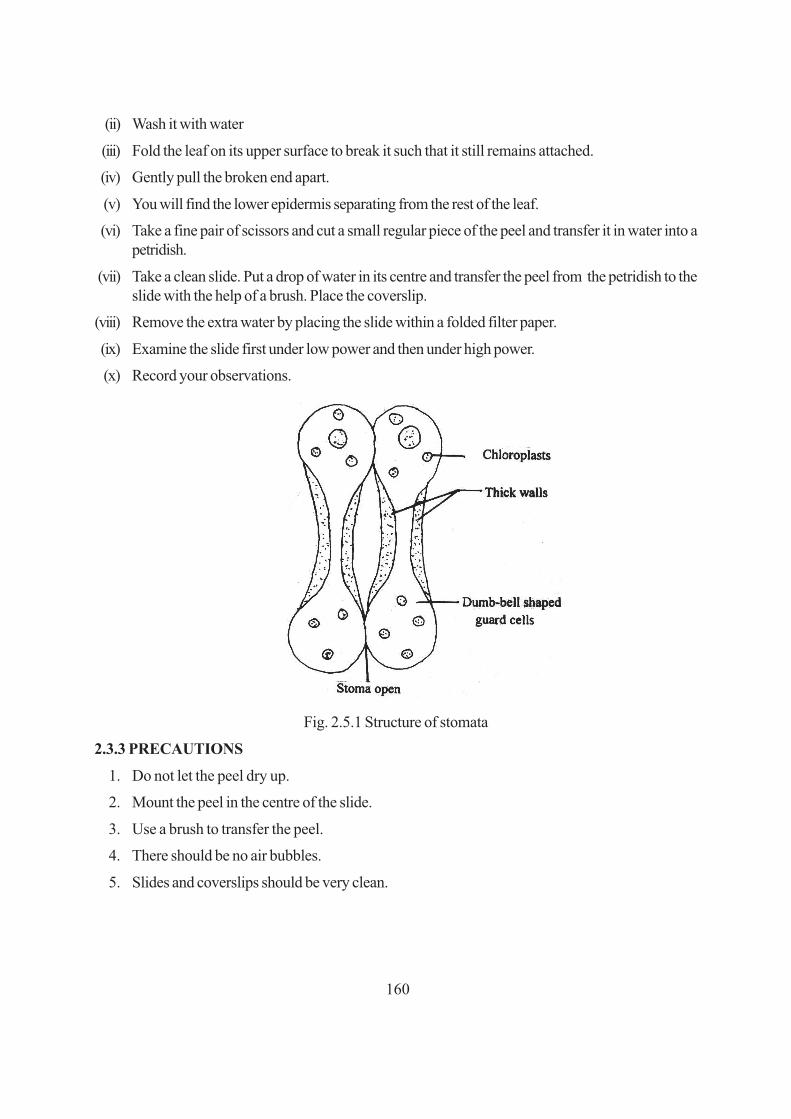
160
(ii) Wash it with water
(iii) Fold the leaf on its upper surface to break it such that it still remains attached.
(iv) Gently pull the broken end apart.
(v) You will find the lower epidermis separating from the rest of the leaf.
(vi) Take a fine pair of scissors and cut a small regular piece of the peel and transfer it in water into apetridish.
(vii) Take a clean slide. Put a drop of water in its centre and transfer the peel from the petridish to theslide with the help of a brush. Place the coverslip.
(viii) Remove the extra water by placing the slide within a folded filter paper.
(ix) Examine the slide first under low power and then under high power.
(x) Record your observations.
Fig. 2.5.1 Structure of stomata
2.3.3 PRECAUTIONS
1. Do not let the peel dry up.
2. Mount the peel in the centre of the slide.
3. Use a brush to transfer the peel.
4. There should be no air bubbles.
5. Slides and coverslips should be very clean.

161
2.3.4 RECORDING OF OBSERVATIONS
Observe under low power of the microscope:
(i) How many different types of cells can you see in the leaf epidermis?
.............................................................................................................................................
(ii) How do the guard cells differ from the other epidermal cells. Mention three differences.
(a) .....................................................................................................................................
(b) .....................................................................................................................................
(c) .....................................................................................................................................
(iii) Is the cell wall of guard cells uniformly thick? If not describe the cell wall.
(iv) How will you differentiate between the epidermal cells and guard cells on the basis of their shape.
(v) Draw a labelled diagram of the leaf peel showing stomata.
2.3.5 FOR THE TEACHER
Please emphasize that
1. the lower surface has more stomata than the upper surface in most types of leaves.
2. the same technique is applicable for viewing stomata in the leaf of any other plant.
3. the guard cells are epidermal cells specialised for a particular function.

162
2.4 PREPARATION AND STUDY OF XYLEM AND PHLOEM FROM CUCURBITA STEM
Xylem and phloem are complex tissues present in plants. They constitute the vascular bundles in leaf,stem and root. Xylem consists of vessels, tracheids, parenchyma and fibres. Phloem consists of phloemtubes (sieve-tubes), companion cells, parenchyma and fibres.
OBJECTIVES
After completing this exercise, you should be able to:
● identify xylem and phloem under a microscope;
● locate and differentiate between xylem and phloem.
2.4.1 WHAT YOU SHOULD KNOW
1. Xylem and Phloem are the constituents of a vascular bundle.
2. These are present in roots, stem and leaves.
Fig. 2.4.1 Xylem and Phloem
�� ��

163
Materials Required
(i) Cucurbita stem (ii) Sharp blade/razor
(iii) Slides (iv) Thin brush
(v) Water (vi) Cover slip
(vii) Glycerine (viii) Saffranine stain
(ix) Compound microscope
2.4.2 HOW TO PROCEED
(i) Cut a T.S. of cucurbita stem.
(ii) Select a thin section and stain in Saffranin.
(iii) Wash the section with fresh water to remove the extra stain.
(iv) Put the stained section in a drop of glycerine on the centre of a slide.
(v) Put a cover slip over it and see the vascular bundle under the microscope.
2.4.3 PRECAUTIONS
l. Thin uniform section should be cut.
2. A good section is cut in a straight, transverse or longitudinal plane and should not be oblique.
3. Observe under the microscope before it dries up.
2.4.4 RECORDING OF OBSERVATIONS
(i) Do you see thick walled more or less circular cells stained red?
If yes .................. These cells constitute xylem.
(ii) Do all the vessels appear of same diameter in cross section?
Yes/No ..........................................................................................................................
(iii) Do you find some thin walled cells unstained, just above the xylem.
Yes/No .....................................................................................If yes, These are phloem.
(iv) Draw a few cells of xylem and phloem in your record book .
2.4.5 FOR THE TEACHER
The teacher to help the students to :
(i) locate vascular bundle in the section and
(ii) identify xylem and phloem.

164
2.5 TEMPORARY STAINED PREPARATION AND STUDY OFSTRIATED MUSCLE FIBRES IN COCKROACH
Muscle fibres are cells which are responsible for motility of an animal or that of the parts of its body.Limb muscles have muscle cells which are called striped or striated muscles and these are under voluntarycontrol.You will study their structure by making a slide from the leg of a cockroach. Unstriated musclecells are involuntary and found in muscles of various internal organs such as those of the digestivesystem.
OBJECTIVES
After performing this exercise, you should be able to:
● acquire the skill to handle live cockroach and remove its legs;
● acquire the skill of making a stained preparation of striated muscle fibres;
● identify, draw and label striated muscle fibres;
2.5.1 WHAT YOU SHOULD KNOW
1. Muscle fibre is a muscle cell.
2. Contractility is its special property.
3. Muscle fibres form the muscle tissue.
4. Muscles are of three types - striated, unstriated and cardiac, which differ from each other in theirstructural details and mode of functioning. Revise these differences from the theory text book.
�� ��

165
Materials Required
(i) Cockroach (live) (Try to collect one yourself).
(ii) Glass slides (iii) Cover slips (iv) Forceps
(v) Needles (vi) Brush (vii)Watch glass
(viii) Methylene blue
(ix) Glycerine
(x) Compound Microscope
2.5.2 HOW TO PROCEED
(i) Remove one of the legs of a cockroach.
(ii) Locate its coxa (the broadest, first segment of theleg). See Fig. 2.5.1
(iii) Slit open the leg (longitudinally) with the help of finescissors.
(iv) Whitish fibrous tissue is the striated muscle.
(v) Add 2-3 drops of methylene blue to stain it.
(vi) Place the muscle in a watch glass in water.
Fig. 2.5.1 Leg of a Cockroach
(vii) Using a forceps pull a few fibres from the stained muscle and place these fibres in another watchglass.
(viii) Put the stained muscle fibres on a clean slide.
(ix) Blot out excess stain surrounding the tissue with the help of a filter paper.
(x) Tease the muscle with a needle.
(xi) Add a drop of glycerine on the slide and gently put the coverslip. Avoid air bubbles. Mount thematerial in the centre of the slide.
(xii) After putting the coverslip press it gently with the back of a needle or pencil to spread out theglycerine and the muscle fibres under the coverslip.
(xiii) Examine the slide under the microscope and note the following points. (Fill up observation 1)
- The plasma membrane of a muscle fibre is called Sarcolemma.
- The muscle fibres (muscle cells) show alternate light and dark bands or striations and hencethe name striated muscles.
- Each muscle fibre is long and cylindrical.
- Many nuclei can be seen in the muscle fibre at the periphery.

166
Sometimes in your slide you may come across striated (striped) silvery shining cylindrical structure.They are not striated muscle fibres. They are tracheal tubes and can be distinguished from muscle fibresby (a) their broader diameter and (b) absence of nucleus.
Fig. 2.5.2 Striated muscle fibres.
2.5.3 PRECAUTIONS
1. Use clean slides and coverslips.
2. Use adequate amount of stain.
3. Do not let the slide dry.
4. Manipulate such that the material is neither too darkly stained nor very lightly stained.
2.5.4 RECORDING OF OBSERVATIONS
Observation
(i) In which kind of muscle fibre do you see light and dark bands? Striated or unstriated.
.............................................................................................................................................
(ii) Are these fibres uninucleate or multinucleate?
.............................................................................................................................................
(iii) What is the shape of the muscle fibres?
.............................................................................................................................................
2.5.5 FOR THE TEACHER
Please ensure that:
1. the microscope is adjusted and the slide properly focussed.
2 the student identifies the striated muscle fibre and observes its model and does not mistake thetrachea for striated muscle fibre as trachea also shows striations.

167
PLANT TAXONOMY
EXERCISE - 1
TO STUDYTHE MAIN CHARACTERISTIC FEATURES OFTHE FAMILIES
23a. MALVACEAEAND SOLANACEAE
Bentham and Hookr classified flowering plants on the basis of the arrangement of floral partspositionof thalamus, and the number of cotyledons in the seed etc.
Objectives :After performing this exercise, you shoold be able to
● Identify the characters of the families.
● Differentiate between the two families.
● Recognise main features of stem and leaf.
● Idintify the non-essential parts of the flowers.
Materials Required :(i) Twigs with flowers of malvaceae and solanaceae families.(ii) Needles(iii) Brushes (o size )(iv) Lens(v) Watch glass(vi) Glass slide(vii) Dissecting microscope(viii) Blade
�� �� �

168
1.1. What you should know1. The stems of malvaceae and solanaceae are different.
2. Venation of the leaf and parts of leaf, shape.
3. The arrangement of floral parts on the thalamus.
4. Type of inflorescence.
5. Position of stamens, number unitid or frec.
6. Position of ovary, number of carpels.
A. Vegetative Parts :
(i) Stem : Aerial or underground ot subaerial, errect or not, branched or unbranched, herboceousor woody.
(ii) Leaf : Venation, structure, stipulate or exstipulate, simple or compound.
B. Floral Parts :i) Inflorescence : Type of inflorescence.
ii) Flower in general : Complete or incomplete, position of the flower, stalked or sessile, bracteateor ebracteate, bracteolate or ebracteolate, symmetry, bisexual or unisexual.
iii) Flower in detail :
a) Calyx : Observe and record the number of sepals, fused or free, aestivation.
b) Corolla : Observe and record the number of petals, united or free, aestivation, whether unitidwith stamens or not.
c) Androecium : The number of stamens, fused or united, whether with corolla or not, theheight of the filaments, number of lobes in the anther, nature of pollen.
d) Gynoecium : The position of the ovary, number of carpels, height of the style, nature ofstigma, number of locules, placentation.
How to proceed :i) Take the twig and observe the stem, leaves and inflorescence by using needle.
ii) Note down the main features as described.
iii) Remove the sepals and petals in the flower
iv) Draw the diagrams of twig with inflorescence, L.S. of flower, T.S. of ovary, floral digrams.
v) Take L.S. of flower and draw the diagram.
vi) Take the T.S. of ovary.
vii) Observe the fruit
viii) Observe the seeds.

169
I) FAMILY : MALVACEAEEx : Hibiscus rosa-sinensis (china rose)
observe the vegetative characters and floral characters and note the observations.
1.3. Observation and Documentation :Vegetative Characters
1. Stem :
a) aerial or underground ? ......................................................
b) any out growths? ...............................................................
c) branched or unbranched ....................................................
d) herbceous or woody .........................................................
2. Leaf :
a) Position .............................................................................
b) Stipules present or not .......................................................
c) Petiole ...............................................................................
d) Simple or compound .........................................................
e) Venation ............................................................................
Draw the diagram of the twig with inflorescence. floral characters draw the diagram of flowerand lable them.
3. Inflorescence :
a) Type .................................................................................
b) Position ............................................................................
4. Flower :
a) Number of whorls ....................................................................
b) Monochlamydous or dichlamydous .....................................
c) Complete or incomplete ......................................................
d) Bracts ...............................................................................
e) Bracteolates ......................................................................
f) Symmery ...........................................................................
g) Number of floral parts ...............................................................
Draw the diagram of flower. take L.S. of flower and draw the digram.
7
F

170
Fig. Hibiscus Rosa - Sinensis

171
5. Calyx :
a) Number of sepals ..............................................................
b) Free or fused ....................................................................
c) Colour ..............................................................................
d) Aestivation ........................................................................
6. Corolla :
a) Number of petals ...............................................................
b) Free of fused......................................................................
c) Aestivation .........................................................................
d) Whether united with the staminal tube .................................
7. Androecium :
Observe under dissecting microscope.
a) Number .............................................................................
b) Whether united, completely united or not .............................
c) Whether united with the corolla ..........................................
d) Shape of the pollen grains ..................................................
8. Gynoecium :
Take T.S. of ovary observe under dissectintg microscope.
a) Position of the ovary ..........................................................
b) Number of carpels ...................................................................
c) Carpels united or free ........................................................
d) Number of locules ...................................................................
e) Placentation ........................................................................
9. write the floral formula.
10. draw the floral diagram.

172
Identify the family
Identification :
i) ReticulateVenation, Pentamerous flowers.
Class : Dicotyledons.
ii) Dichlamydeous, free corolla.
Subclass : Polypetalae.
iii) Hypogynous flowers, stamens numerous
Series : Thalamiflorae.
iv) Bisexual, actinomorphic. stamens monadelphous, carpels five, axile placentation.
Order : Malvales.
v) Stellate hairs on vegetative parts.
Presence of epicalyx
Twisted acstivation of corolla
Monadelphous, staminal tube
Monothecous, reniform anthers
Pollongrains spinous.
Family-Malvaceae.
RESULT :
Hence the observed twig belongs to the family malvuceae.
Observation 23b. Family Solanaceae
Ex : Daura metel
Draw the diagram of the twig, observe the vegetative and floral characters and record yourobservation.
Observation and documentation
a. Vegetative Characters
(i) stem :
a) Aerial or other type ...........................................................
b) Herbaceous or woody .......................................................
c) Branched or unbranched ...................................................

173
1.5.2. FLORAL CHARACTERS

174
ii) Leaf :
a) Position .............................................................................
b) Stipules .............................................................................
c) Petiole ...............................................................................
d) Leaf base adnation ............................................................
e) Simple or compound ..........................................................
f) Venation .............................................................................
B. FLORAL CHARACTERSDraw the diagram of the flowers, and lable the parts.
3. Inflorescence :a) Type .................................................................................b) Position ............................................................................
4. Flower :a) Number of whorls ....................................................................
b) Monochlamydeous or dichlamydeous .................................c) Complete or incomplete .....................................................d) Bracts ...............................................................................
e) Bracteoles .........................................................................f) Symmetry ..........................................................................g) Number of floral parts ..............................................................h) Position .............................................................................
Take L.S. of flower, observe, draw the diagram.Take T.S. of ovary and observe under dessecting microscope.
5. Calyx :a) Number of sepals ..............................................................b) Free or fused .....................................................................c) Aestivation ........................................................................d) Is the calyx persistent ? ......................................................
6. Corolla :
a) Number of petals ...............................................................
b) Free or fused .....................................................................
c) Aestivation .........................................................................

175
7. Androecium :
a) Number of stamens ............................................................
b) Fused with petals or not .....................................................
8. Gynoecium :
a) Position of the ovary ..........................................................
b) Ovary position straight or oblique ......................................
c) Number of carpels .............................................................
d) Carpels united or free ........................................................
e) Number of locules .............................................................
f) Number of ovules ...............................................................
g) Placentation .........................................................................
h) Style ..................................................................................
i) Stigma ................................................................................
9. Floral formula
10. Draw the floral diagram.
IDENTIFICATION1. Reticulate venation, pentamerous flowers.
Class : Dicotyledonae
2. Dichlamydeous, fused corolla, epipetalous stamens.
Subclass : Gamopetalae
3. Bicarpellary, hypogynous ovary, number of stamens equal to the number of petals.
Series : Bicarpellatae.
4. Exstipulate and alternate leaves, bicarpellary, bilocular ovary with many ovules on axile placentation.
Order : Polymoniales.
5. Exstipulate leaves, leaf base adnates with the stem, solitary cyme inflorescence, persistent calyx,epipetalous stamens, obliquely arranged carpels, axile placentation.
Family : Solanaceac.
Result :
Hence the observed twing belongs to the family solanaceae.

176
Appendix – Practical
1. Adventious roots show dichotomous branching
2. Grow apogeotropically
3. Appear blue green and are coral like
4. The surface show some lenticels
Cycas corrolloid roots or Corallorhiza

177
1. Adventious roots show dichotomous branching
2. Grow apogeotropically
3. Appear blue green and are coral like
4. The surface show some lenticels
T.S. of Cycas corolloid root :
1. Shows three regions epiermis, cortex and stele
2. Exidermis is single layered with thin walled cells
3. The cortex is massive and is differentiated into three distinct zones such as outer covtex, middlecortex, and inner covtex.
4. In the middle cortex, the cells gets disorganised and are inhabited by symbiotic nitrogen fixingblue-green alge like Anbaena and Nostoc
5. Radial vascular bundles. Xylem is exarch and triarch.

178
Cycas Male coue :
1. The male cone is shortly stalked, long, compact and fusiform or oval.
2. Mature cones appear woody and about 20 to 60cm. long
3. The male strobilus (=male flower) cousists of number of microsporophylls (=stamens) arrangedclosely on the centralaxis
4. Some of the microsporophylls situated at the extreme top and at the base maybe sterile
Cycas megasporophyll :
1. Looks like foliage leaf. Borne on the female plant. There is no female cone.
2. Having three well defined parts i) stalk ii) middle fertile portion bearing ovules iii) upper sterileportion.
3. Lanceolate or rhomboidal. Both sides of stalk bears naked ovules
4. Ovules are of the size of hen’s egg & browuish in colour
5. The upper portion of the megasporophyll is broad and serrated.

179
STUDY OF MORPHOLOGICAL MODIFICATIONS OF PLANT PARTSLIKE ROOT, STEM AND LEAF
The practical exercise has been planned to give an idea that plant parts like root, stem and leaf incertain plants can get modified structurally to perform functions which are very different from theirnormal functions.
OBJECTIVES
After performing this exercise, you should be able to :
● identify the root, stem and leaf in their modified form in plants other than what you will be observingin this exercise.
● differentiate or identify these modified structures on the basis of their primary characters.
3.1 WHAT YOU SHOULD KNOW
1. Recapitulate what you have learnt about modification of various plant parts like root, stem andleaf.
2. The modified structure or parts may look very different from the normal structure, that is a stemmay look like a root or a leaf and the leaf may take the shape of a thorn or a tendril.
3. In their modified form, they perform very different functions from what they normally do. Themodified roots do the job of storage and support, the stem may take up the job of photosynthesisand multiplication; the leaf may do the function of protection and support.
Material Required
(i) Fresh or museum specimens
(ii) Models of specimens
(iii) Photographs or pictures of specimens of carrot, radish, beet, ginger, potato, zamikand,onion, grass, Eichhoria, strawberry, lemon and grape twigs, pea leaf, Opuntia, pitcherplant,Australian acacia
3.2 HOW TO PROCEED
(i) Observe the specimens from different sides.
(ii) In most cases, you will know what you are looking at, in your first glance only.
(iii) You can use a hand lens, if need be.
(iv) Draw labelled diagram of the specimens provided, write their salient features of identification .

180
(v) A short guideline of diagram with points of identification has been given for each specimen. Youobserve the specimens carefully and record your observation on the basis of what you actuallyobserve.
A. Modifications of Roots
a. Radish
1. The tap root is swollen in the middle andtapers towards apex and base
2. It is known as fusiform root and it storesexcess food.
b. Beet
1. It is swollen at the upper part almost becomingspherical and abruptly tapering at the lowerpoint.
2. It is known as napiform root.
3. It is a storage root and a source of sugar.
c. Carrot
1. It is broad at the base and tapers graduallytoward the apex.
2. This is known as conical root.
3. Function is storage of food.
d. Banyan Tree
1. Roots are produced from main stem branchesfor mechanical support.
2. These roots grow downwards and penetratethe soil and act as supporting pillars.
3. These roots are known as prop root.

181
e. Sugarcane
1. From the lower portions of the main stem largenumber of strong roots are produced toprovide support.
2. These roots are known as stilt roots.
f. Rhizophora
1. These plants grow in marshy places.
2. Large number of conical structures, which areroots, grow vertically upwards.
3. These roots being aerial perform the function of respiration and are known as pneumatophoresor breathing roots.
3.3(a) RECORDING OF OBSERVATION
1. Do you find hair like structures coming out from carrot and radish? What are these?
.............................................................................................................................................
2. What kind of function these roots perform?
.............................................................................................................................................
3. Have you been able to locate the stem in these plants?
.............................................................................................................................................
4. Where do you find the leaves in these plants?
.............................................................................................................................................

182
B. Modification of Stems :
● Stems get modified in various ways
● These modified structures help the plant to survive during unfavourable seasons by storing food,help in vegetative multiplication of the plant and provide mechanical support and protection.
● They can be studied by grouping them into underground, subaerial and aerial.
(i) Underground modifications
a. Ginger
1. It has an irregularly branched prostratestructure.
2. There are nodes, internodes, buds and scaleleaves.
3. It is known as rhizome.
b. Zamikand
1. It is a condensed form of rhizome growingmore or less in vertical direction and knownas corm.
2. Axillary buds and scale leaves are present.
c. Potato
1. The smooth brown, swollen structure isknown as tuber.
2. There are a number of axillary buds knownas eye: located on one side of the tuber.
3. The axillary buds give rise to new plant.
Onion
1. The bases of the bulb as it is termed has aconvex, compressed stem which producescluster of firbrous roots at its base.
2. There are many scale leaves which are fleshyand store food.
3. Buds are present in the axil of scale leave.
4. The complete shoot is modified.

183
(ii) Subaerial modifications
In some plants the stem is partly aerial and partly underground. The underground part is not very deepand lies horizontally underground. It has nodes and internodes. The nodes give out leaves which growabove the soil surface and roots below:
● The delicate branch arising from an axillary bud grows horizontally below the surface of the soil.
● It creeps on the ground with roots at the nodes and is called a runner.
● It may break off from the mother plant and can grow independently.
a. Strawberry
1. Branches originates from the base of the stemwhich grows obliquely known as stolon.
2. You have studied potato which is actually astolon.
b. Eichhornia and Pistia
1. Short, thick, horizontal branch originates inthe axil of a leaf.
2. It elongates to produce a tuft of leaves aboveand clusters of small roots below.
3. This is known as offset.
(ii) Aerial modifications
a. Grape-wine
1. From the axil of leaves arise tendrils whichare wiry, coiled structures.
2. Tendrils help the climber in clinging to support.
b. Lemon and Karonda
1. The axillary or terminal buds of the stem aremodified into thorns, which are hard pointedstructures.
2. Thorns provide protection to the plant.
stolon
offset
Tendrils
thorns

184
c.Opuntia
1. Green, flat, thick branches have unlimitedgrowth.
2. Leaves are modified into spines.
3. The modified structure is known asphylloclade.
d.Asparagus
1. There are branches of limited growth ,whichbecome green and that like a leaf.
2. These are called cladodes.
3.3(b) RECORDING OF OBSERVATION
1. Can you name the three structures you observe on the rhizome of ginger?
.............................................................................................................................................
2. Which structure of the potato tuber can give to new plants?
.............................................................................................................................................
3. Can you locate the axillary bud on the runner of grass?
.............................................................................................................................................
4. Can you see the stem of grass in your lawns?
.............................................................................................................................................
5. What causes fast rate of growth in Eichhomia?
.............................................................................................................................................
6. Why are the tendrils in grape-wine stem and thorns of karonda or lemon called modified stems?
.............................................................................................................................................
7. Give reason why phylloclade and cladode are modified xerophytes.
.............................................................................................................................................

185
C. Modification of Leaf
Although the main function of leaf is to synthesize food for the plant, in some plants they get modified toperform functions of support and protection for the plant.
a. Pea
1. Leaves (a portion) are modified into slender,wiry. Closely coiled structures called tendrils.
2. These are climbing organs for the plant.
b. Opuntia
1. Leaves are modified into sharp, pointed,spines for defensive purpose.
2. These spines also help for reducingtrans-piration.
c. Australian acacia
1. The petiole becomes flat, green leaflike calledphyllode.
2. It helps in photosynthesis.
d. Pitcher plant
1. Leaf is modified into a pitcher and the leaf tipinto a lid to trap insects.
2. It is an insectivorous plant.

186
3.3(c) RECORDING OF OBSERVATIONS
1. Observe the tendril of pea carefully. Why do you call them modified leaves?
.............................................................................................................................................
2. Which portion of the leaf is modified in pea?
.............................................................................................................................................
3. Which part of opuntia plant prepares food?
.............................................................................................................................................
4. Can you observe the modified leaves, where are they located?
.............................................................................................................................................
3.4 FOR THE TEACHER
Teacher may help the students to identify or locate the modified structures like axillary buds,scale leaves, root hairs, spines and thorns on various specimens.

187
�� �� �Exercise
TO STUDY THE T.S. OF DICOT AND MONOCOT STEMS ANDROOTS FROM PERMANENT SLIDES
Stem and root are made up of different types of tissues. These tissues form different layers in thecomposition of stem and root. This exercise is intended to study the structural details (anatomicaldetails) of these tissues.
OBJECTIVES
After performing this exercise, you should be able to:
● identify the sections of dicot and monocot stem;
● identify the sections of dicot and monocot root;
● identify location of various layers in the above, formed by different tissues;
● differentiate anatomically between the various sections of stem and root.
4.1 WHAT YOU SHOULD KNOW
1. Different layers are made up of different types of tissues.
2. The layers are present in a definite sequence.
3. Anatomically the monocot and dicot stems differ significantly in the arrangement various tissues.
4. Anatomical differences between monocot and dicot roots exist in the vascular zone.

188
Materials Required
(i) Compound microscope (iii) Dissecting microscope
(ii) Permanent slides of dicot and monocot stems
(iv) Permanent slides of dicot and monocot roots.
4.2 HOW TO PROCEED
(i) Take permanent slides of T.S. of the dicot and monocot stem and root.
(ii) Adjust the slides under the microscope.
(iii) Note the outline of the sections, and the main structures and their arrangement inside.
(iv) Select a part of the slide as viewed under the microscope and draw a labelled diagram.
1. Stem
(A) T.S. of Dicot Stem
Observation
From the permanent slide of the T.S. of dicot stem (sunflower plant), try to locate the followingtissues (Fig. 4.1)
Fig. 4.1 T.S. of Dicot stem

189
● Outermost layer of single row of cells-epidermis.
It bears some multicellular hairs.
● Immediately below the epidermis is two-three layers of collenchymatous hypodermis.
● Inner to the hypodermis are few layers of thin walled cells-cortex.
● Innermost layer of cortex forms a distinct layer-endodermis
● Inner to endodermis lies a layer of cells-pericycle
● The pericycle encloses vascular bundle and pith in the centre
● Each vascular bundle consists of phloem towards outside and xylem towards inside.
Thus the vascular bundles are conjoint and collateral.
● Xylem and phloem are separated by cambium thus these vascular bundles are open.
Thus the vascular bundles are conjoint, collateral and open.
● Parenchyma tissue separating the vascular bundles is termed medullary rays. Main points ofidentification of T.S. dicot stem are :
1. Cortex is differentiated into hypodermis (collenchymatous), parenchymatous cortex and innermostlayer of endodermis.
2. Note the conjoint, collateral, open, endarch vascular bundles.
(B) T.S. of Monocot Stem
(i) Keep the slide containing T.S. of monocot stem (Maize stem) under a dissecting microscope(Fig. 4.2). Do you observe scattered vascular bundles?
(ii) Now place the slide under low power of the microscope and focus only a portion of the sectionin a view for greater details.
(iii) Start observing from the periphery.
(a)

190
(b)
Fig. 4.2 T.S. of monocot stem
Observations
Do you notice a large difference between the section of maize (monocot) stem and that of dicot stem?
Note these differences.
Important distinguishing charactors of monocot stem are:
1. Single layer of epidermis covered with thick cuticle.
2. Narrow zone of sclerenchymatous hypodermis.
3. A mass of thin walled parenchyma tissue known as ground tissue below the hypodermis.
4. Scattered vascular bundles.
5. Have you observed four distinct vessels stained red and arranged in the form of letter ‘Y’ . Twolarge ones are metaxylem and two smaller inner ones are protoxylem.
6. Observe the thin walled small cells towards outside which form the phloem.
4.3 RECORDING OF OBSERVATIONS
Observation 1
T.S. of dicot stem
(i) How many layers are there in the epidermis? Draw few cells of epidermis as seen under themicroscope.
.............................................................................................................................................
(ii) Is there any outgrowth or structure visible on the epidermis. If yes what are these called?
.............................................................................................................................................
(iii) What is pericycle? See in your slide and draw it.
.............................................................................................................................................

191
(iv) Can you locate the vascular bundle. Draw a few xylem and phloem cells as you see them in avascular bundle.
.............................................................................................................................................
Observation 2
T.S. of Monocot stem
(i) Observe the outermost epidermis. Can you see some ‘holes’ scattered all around in the cortex?
(ii) Can you make out any difference in the position of vascular bundle in monocot and dicot stem?
(iii) Are the vascular bundles separated from one another by the intervening ground tissue?Yes/No
(iv) What is the difference between the hypodermis that you have seen in sunflower stem (dicot stem)and the hypodermis that you are seeing here in the maize stem (monocot stem)?
Sunflower (dicot) Maize (monocot)
.......................... ..........................
.......................... ..........................
.......................... ..........................
Observation 3
Differences between Dicot and Monocot Stem
Structures Dicot stem Monocot Stem
1. Epidermis .......................... ..........................2. Hypodermis .......................... ..........................3. Cortex .......................... ..........................4. Endodermis .......................... ..........................5. Pericycle .......................... ..........................6. General cortex .......................... ..........................7. Medullary rays .......................... ..........................8. Vascular bundles .......................... ..........................9. Pith .......................... ..........................
2. Root
(A) T.S. of Dicot Root
(i) Place the slide under the dissecting microscope and observe its structure.
(ii) Observe the single outermost layer -epiblema which gives out single celled hairs. Inner to this,there is a compact mass of rounded cells with intercellular spaces forming cortex.
(iii) The central cylinder constitutes, the vascular bundle.
(iv) Do you find that inner cylinder is also surrounded by two definite layers of cells?

192
Name the two layers from the diagram.
(v) Semi-circular patch of thin walled cells with blue stain constitute phloem .
Fig. 4.3 T.S. of Dicot root
(vi) This alternates with group of thick walled cells which have taken up red stain.
(vii) Both these structures constitute vascular bundle.
Note: In Root, the xylem and phloem are in separate bundles and are at different radii.
(viii) Do you observe that protoxylem is placed towards pericycle and the metaxylem towards centre.It is one of the characteristic points to identify root. It is known as exarch condition.
(ix) Do you find any projections coming out from the epiblema? These are called root hairs.
(xi) Count the number of vascular bundles present. You will note that they are in the numbers of 2 to6.
(B) T.S. Of Monocot Root
(i) Place the permanent slide of T.S. of monocot root under low power of the microscope.
The outline of monocot root is much bigger in T.S., so you will not be able to see it as an entiresection under the microscopic field as in case of dicot root. So to find out the general outline view theslide under dissecting microscope (Fill up observation) (Fig. 4.4)

193
Fig. 4.4 T.S. of Monocot Root
(ii) Do you observe the difference in the number of vascular bundles? If yes, what is their approximatenumber?
(iii) Do you see the large pith? Yes/No
(iv) Tabulate the difference between dicot root and monocot root.
Dicot Root Monocot Root
1. ................................... ...................................
2 ................................... ...................................

194
4.4 RECORDING OF OBSERVATIONS
Observation 1
T.S. of Dicot Root
(i) Is it circular in outline? Can you view the entire section in the microscopic field?
.............................................................................................................................................
(ii) Can you distinguish two distinct zones in the section?
.............................................................................................................................................
(iii) Is the peripheral outermost layer epiblema, single layered or multilayered?
.............................................................................................................................................
(iv) Is there any outgrowth or projections or any structures on this epiblema? If so, what are they?
.............................................................................................................................................
(v) Look at the epiblema.
Can you see multilayered loosely packed thin walled parenchymatous cells with intercellularspaces?
Yes/No .................................................................................................................................
Is this ground tissue or cortex?
(vi) Can you differentiate the central cylinder from the cortex by any complete layer of cells?
Yes/No. .................................................................................................................................
If yes, how many layers of cells are there.
(vii) Look at between xylem and phloem bundles. Can you see thin walled parenchyma cells? Yes/No.......................................................................................................................................
What are these called? .................................................................................................................
(viii) Draw a simple sketch of T.S. of dicot root in your record book.
Observation 2
T.S. of Monocot Root
(i) Is it circular in outline? Yes/No .............................................................................................
(ii) Are there root hairs? Yes/No .............................................................................................
(iii) Does the endodermis have its radial walls thickened? Yes/No .....................................................
(iv) Is the pericycle thin walled or thick walled? . .............................................................................
(v) Are xylem and phloem in separate bundles? Yes/No .
(vi) How many bundles are there?

195
(vii) Is there a pith? Is it large and well developed or small in size?
(viii) Write the difference between a dicot and monocot root?
Dicot root Monocot root
........................... ...........................
........................... ...........................
........................... ...........................
........................... ...........................
(ix) Draw a simple sketch of T.S. of monocot root in your record book.
4.5 FOR THE TEACHER
Please enusure that
1. the microscope in adjusted and the slide properly focused.
2. the student identifies the various tissues in both dicot and monocot roots and stems.
3. the students get a clear concept regarding the structure and conditions of the vascular bundles.
The teacher may kindly inform the students that the red and blue colours of cells maintainedinside are not their natural colours, but colours taken up during staining of the slide.

196
�� �� �Exercise
STUDY OF THE MICROSCOPIC ANATOMY (HISTOLOGY) OF MAMMALIANTISSUES AND ORGANS
Every tissue has a special structure suited to its function. In this exercise you will study thehistological features of some of the major tissues and organs of mammals.
OBJECTIVES
After performing this exercise, you should be able to :
● identify and differentiate between various kinds of mammalian tissues and organs based on theirshape, size and structural details;
● differentiate between different types of blood cells.
5.1 WHAT YOU SHOULD KNOW
1. Animals have different types of tissues and organs which perform specific functions.
2. Each organ is different histologically.
• Cartilage and bone represent supportive connective tissue where matrix is solid.
3. Blood is another type of connective tissue composed of plasma and cells. Matrix is fluid.
4. Testis and ovary produce male and female gametes respectively.They also secrete sex hormones.
Aim: To study the histology of mammalian tissues and organs from permanent slides. (cartilage, bone,blood, testis and ovary)
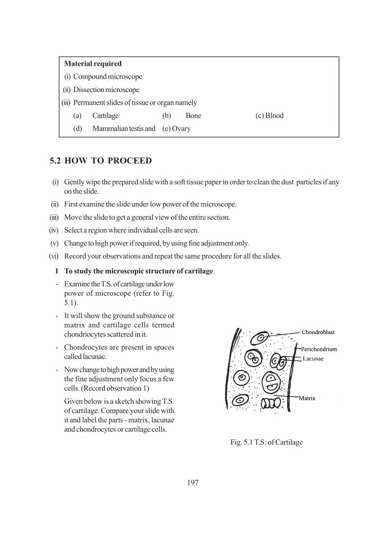
197
Material required
(i) Compound microscope
(ii) Dissection microscope
(iii) Permanent slides of tissue or organ namely
(a) Cartilage (b) Bone (c) Blood
(d) Mammalian testis and (e) Ovary
5.2 HOW TO PROCEED
(i) Gently wipe the prepared slide with a soft tissue paper in order to clean the dust particles if anyon the slide.
(ii) First examine the slide under low power of the microscope.
(iii) Move the slide to get a general view of the entire section.
(iv) Select a region where individual cells are seen.
(v) Change to high power if required, by using fine adjustment only.
(vi) Record your observations and repeat the same procedure for all the slides.
1 To study the microscopic structure of cartilage
- Examine the T.S. of cartilage under lowpower of microscope (refer to Fig.5.1).
- It will show the ground substance ormatrix and cartilage cells termedchondriocytes scattered in it.
- Chondrocytes are present in spacescalled lacunae.
- Now change to high power and by usingthe fine adjustment only focus a fewcells. (Record observation 1)
Given below is a sketch showing T.S.of cartilage. Compare your slide withit and label the parts - matrix, lacunaeand chondrocytes or cartilage cells.
Fig. 5.1 T.S: of Cartilage

198
2. To study the microscopic structure of bone
T.S. of Bone (long bone such as femur)
- Examine the slide under the low power ofmicroscope.
- Observe some areas showing concentric ringsor lamellae, and each such area having anarrow central canal. The lamellae with theirlacunae and central canal form the Haversiansystem.
- Compare the section in the slide with thefigure (Fig. 5.2) provided.
Fig. 5.2 T.S : of bone
- Try to locate the central canal, bone lamellae and lacunae (spaces that contained bone cells)arranged in concentric rings.
- Lying in the bone lamellae are empty lacunae (spaces) which in natural condition contain bonecells (osteocytes). Some fine canals (canaliculi) radiate out from these lacunae. (Fill up observation2)
You may not see the osteocytes within the lacunae as they get removed while processing thebone for slide preparation.
(If the section passes obliquely or longitudinally, you will not find the Haversian systems soperfectly and the central canals may become oblong or even longitudinal).
3. To study the microscopic structure of mammalian testis (T.S.)
Place the slide under the microscope under low magnification and observe. (Fig. 5.3)
Fig. 5.3 T.S: of Testis

199
- Do you find any circular, oval compartments? These are seminiferous tubules.
- Can you see some material filling the space between the tubules? This is connective tissue matrix.
- Record the shape of the semini-ferous tubule.
- Locate the germinal epithelium which is first layer of cells lining each seminiferous tubule. It isinterrupted in between by vertical row of cells which proceed from the surface towards theinterior of the tubule.
- Inner to the germinal epithelium lie, spermatogonia, spermatocytes, spermatids and spermatozoa.
- Can you also see in the centre of the tubules the cluster of spermatozoa in seminiferous fluid.Observe their tail ends which are clustered together towards the centre.
- Between the seminiferous tubules are interlobular spaces containing Leydig cells. Can you locatethem?
- Draw a labelled diagram of T.S. of testis.
4. To study the microscopic structure of mammalian ovary
Examine the slide under low magnification moving it in all directions. First of all, observe the generaloutline of the ovary. Is it plain or uneven with slight bulges here and there?
Then study part by part all the structures contained in it. Compare the slide with the diagram provided(Fig. 5.4).
(i) Observe cells contained in the outermost lining of the ovary.They constitute the germinal epithelium.
(ii) Observe the developing primary follicles.
(iii) Observe the multilayered (graffian follicle) and the ruptured follicle which forms corpus luteum.(Fill up observation 4).
Fig. 5.4 T.S. of Mammalian Ovary

200
5. To study human blood smear and identify the different types of blood cells.
Examine the slide of human bloodsmear under the microscope, first underlow power and then under high power.Look for various types of blood cells.Record your observations and drawRBCs and WBCs.
You will see a large number of circularconcave disc like structure, which haveno nuclei. These are red blood cells,Red Blood cells (RBCs).
Fig. 5.5 Different type of human blood cells.
You should be able to see a fewer number of stained larger cells (larger than RBC) irregular in shape,with a nucleus of various shapes. (Fig. 5.5). These are white blood cells (WBCs).
(Fill up observation 5)
How many WBCs are you able to see in a single focal field?
5.4 RECORDING OF OBSERVATIONS
Observation 1
Cartilage under high power of the microscope:
(i) What is the shape of the cartilage cells?
(square, hexagonal, spherical, or hemispherical)
.............................................................................................................................................
(ii) Does each cell lie in a cavity? If so, what is the name of this cavity?
.............................................................................................................................................
(iii) Is the nucleus of the cells oblong, oval or round?
.............................................................................................................................................
(iv) Note that most cells are in groups. Are they mostly in groups of two’s, three’s or four’s?
.............................................................................................................................................
(v) What is the term for the ground substance in which the cell clusters lie?.............................................................................................................................................

201
(vi) Draw a labelled diagram of T.s. of cartilage.
.............................................................................................................................................
Observation 2
Bone under high power of the microscope:
(i) Draw at least three adjacent Haversian systems as you see them and label the parts.
.............................................................................................................................................
(ii) Mention any two differences between bone and cartilage.
.............................................................................................................................................
(a) Arrangement of cells: in lamellae single or in clusters?
.........................................................................................................................................
(b) Central canal : present or absent
.........................................................................................................................................
(iii) Draw a labelled diagram of T.S. of bone.
.............................................................................................................................................
Observation 3
Mammalian testis under low power of microscope:
(i) Are there prominently large tubules of uniform size and shape?
.............................................................................................................................................
(ii) What are these tubules called?
.............................................................................................................................................
(iii) Is there any space between tubules or are they tightly arranged? What is this space called? Whatcan you see in the space?
.............................................................................................................................................
(iv) Name the structures present in the interior of each seminiferous tubule?
.............................................................................................................................................
(v) How does a spermatozoon look, as you see it in the slide? Draw it as you see it
.............................................................................................................................................
8
F

202
(vi) Draw a labelled diagram of T.S. of testis.
.............................................................................................................................................
Observation 4
Mammalian ovary under low power of microscope:
(i) Can you see several follicles? Are they similar with respect to size and structure?
.............................................................................................................................................
(ii) Count the number of follicle.
.............................................................................................................................................
(iii) Draw a diagram of a portion of your slide showing a primary follicle and the Graafian follicle.
.............................................................................................................................................
(iv) Can you see the corpus luteum? Label it in the diagram.
.............................................................................................................................................
(v) Draw a labelled diagram of section of ovary.
.............................................................................................................................................
Observation 5
Human blood cells under low power of Microscope
(i) (a) Draw the RBCs as you see them in the slide.
.............................................................................................................................................
(b) Draw two or three different types of white blood cells and label them..............................................................................................................................................
(ii) What are the different types of WBCs present in human blood? Name them.
.............................................................................................................................................
(iii) List any two differences between RBC and WBC.
.............................................................................................................................................
5.6 FOR THE TEACHER
Please ensure that the student
1. handles the permanent slides with much care while focussing it, especially under high power.
2. can identify the various types of cells (eg. chondrocytes, WBC, RBC etc) in the different tissuesand
3. also identify the various parts seen in the sections of mammalian testis and ovary which they,need to label in their drawings.
203
�� �� �Exercise
TO STUDYTHE STRUCTURE AND FUNCTION OF DIFFERENT PARTS OF FLOWERS(PETUNIA AND CHINA ROSE)
Flowering plants are classified on the basis of the structure and arrangement of floral parts on andaround the receptacle or thalamus (the swollen end part of the flower stalk) in concentric whorls.
OBJECTIVES
After performing this exercise, you should be able to:
● identify different parts of the flower;
● recognise main features of the flowers of petunia and china rose;
● explain the structure of any type of flower.
6.1 WHAT YOU SHOULD KNOW
1. The flowering plants are classified on the basis of the structure of flowers and arrangement offloral parts around the receptacle or thalamus.
2. This arrangement is specific for a specific family.
3. Flowers have parts such as sepals, petals, androecium, gynoecium etc.
Materials Required
(i) Flowers of china rose/hollyhock and petunia
(ii) Dissecting microscope

204
A. Floral Parts
Main points to be noted in these two (or in any other) flowers as follows:
(a) The size and nature of flower whether the flowers are large and showy or inconspicuous.
(b) The origin of flower whether they are borne on the flowering twig singly/in clusters or seriallyalong the twig etc. (i.e. the kind of inflorescence).
Inflorescence
(i) Main axis does not terminate in a flower-Recemose
(ii) Main axis terminates in a flower-cymose
Size of the stalk whether the flowers have a long stalk (pedicellate) or they have no stalk (sessile).
Floral parts
Each flower has to be observed starting from outermost whorl (calyx/sepals) or epicalyx and toproceed to the inward whorls (corolla, stamens, pistils, etc.)
(a) Calyx (Sepals)
Observe and record the number of sepals, their colour and whether they are free or united.Consult your Biology text book-l, lesson 7 and find out the function of the calyx.
(b) Corolla (Petals)
- The number of petals, their colour and shape, whether they are free or fused, their relationwith each other (overlapping, twisted, or free etc.)
- Whether the flower has both male (Androecium) and female (Gynoecium) parts or only oneof them. Thus whether the flower is bisexual or unisexual.
- Find out the function of the corolla from your text book.
(c) Androecium:
- The number of stamens, whether fused or free.
- Each stamen has an anther attaches to a long filament.
- Whether the filaments are free or attached to the corolla.
- It is the male part of the flower and has pollen grains in the anther.
(d) Gynoecium (Carpels)
The gynoecium consists of carpels and each carpel has three parts-ovary, style and stigma.
- The position of the ovary on the thalamus with respect to the position of other parts- above,at the same level or below i.e. inferior ovary or superior ovary.
- Number of Carpels.

205
- Whether the style is short or protruding out.
- The stigma, whether simple or divided into lobes or branches.
In order to find out the number of ovary chambers (locules) and the number of ovules in eachchamber cut T.S. of ovary. In such sections you can also observe the attachment of ovules to theovary wall (i.e. placentation).
Fig. 6.1 Flowering twig, parts of flower of Hibiscus rosa-sinensis (China rose)
B. Symmetry
Actinomorphic
symmetrical, can be cut along more than one plane into two similar halves.
Zygomorphic
Bilaterally symmetrical can be cut into two similar halves along one plane.
C. Aestivation
The arrangement of sepals and petals in a floral bud with respect to the members of the same whorl.

206
6.2 HOW TO PROCEED
(i) Take the flower and observe the different floral parts by using hand lens/ dissecting microscope,needles and forceps.
(ii) Note down the main features as described.
(iii) Remove the sepals one by one. Draw one of them, or the entire calyx if fused, in your notebook.
(iv) Remove the petals. If all are similar, draw one of them otherwise each one of them separately.
(v) Observe the stamens and ovary. Locate their position/attachment/inter-relationship amongthemselves and with other floral members.
(vi) Cut transverse sections of ovary to observe placentation and draw it in your record book.
(i) China rose
Observe the different parts of the flower carefully (Fill up observation 1)
(ii) Petunia
Observe the different parts of flower carefully (Fill up observation 2)
6.3 OBSERVATION AND DOCUMENTATION
Observation 1
(A) China-rose (Hibiscus rosasinensis)1. Inflorescence ........................................................................................................................
Draw the inflorescence2. Pedicellate/sessile ........................................................................................................................3. Sepals (Calyx)
(i) Shape ............................................................................................................................(ii) Number ...........................................................................................................................(iii) Free/fused ........................................................................................................................(iv) Colour. ............................................................................................................................(v) Do the sepal-lobes face each other (valvate) or do they overlap (twisted)?
.........................................................................................................................................(vi) Draw one sepal as you see it in your flower.
.........................................................................................................................................4. Petals (Corolla)
(i) Size..................................................................................................................................

207
(ii) Colour ..............................................................................................................................(iii) Number ...........................................................................................................................(iv) Free/fused ........................................................................................................................(v) Do the petals face each other (valvate) or do they overlap (twisted) one above the other by
their edges?.........................................................................................................................................
(vi) Draw a figure to show aestivation in corolla.
.........................................................................................................................................
5. Stamens (Androecium)
(i) Position (whether attached to corolla or not)
.........................................................................................................................................
(ii) Number ..........................................................................................................................
(iii) Free Fused ..........................................................................................................................
(iv) Are the anthers free/united .....................................................................................................
(v) Does the staminal tube protrude out of the flower?
.........................................................................................................................................
(vi) Is the anther one-lobed or four lobed?
.........................................................................................................................................
6. Carpels (Gynoecium)
(i) Position of ovary on the thalamus (superior/Inferior)
.........................................................................................................................................
(ii) Style: Is it exposed or enclosed in a tube?
.........................................................................................................................................
(iii) Stigma: Is it branched?
.........................................................................................................................................
(iv) If so, how many branches?
.........................................................................................................................................
(v) Take T.S. of ovary and examine and draw the diagram as you see in the section under adissecting microscope.
.........................................................................................................................................
208
(vi) How many chambers are there in the ovary?
.........................................................................................................................................
(vii) How many ovules are there inside each chamber?
.........................................................................................................................................
(B) Petunia
1. Draw the flower of Petunia.
2. Pedicellate/sessile
.........................................................................................................................................
3. Sepal (Calyx)
(i) Number: ...........................................................................................................................(ii) Free / Fused: ..........................................................................................................................(iii) Colour: ............................................................................................................................
(iv) Do the sepals face each other (valvate) or do they overlap (twisted)?.........................................................................................................................................
(v) Draw one sepal.4. Petals (Corolla)
(i) Number ............................................................................................................................(ii) Colour ..............................................................................................................................(iii) Free/fused ..........................................................................................................................(iv) Valvate or twisted ?...............................................................................................................(v) Draw one corolla.
5. Stamens (Androecium)(i) Number ..........................................................................................................................(ii) Position (whether attached to corolla or not).............................................................................(iii) Free/united ..........................................................................................................................(iv) How many lobes in each anther : ...............................................................................................(v) Draw a stamen indicating the filament, connective and anther lobe.
7. Carpels (Gynoecium)(i) Position of ovary on the thalamus (Superior/inferior)
.........................................................................................................................................(ii) Is the style protruding out?
.........................................................................................................................................

209
(iii) Is the style longer than the stamens?.........................................................................................................................................
(iv) What is the type of placentation?....................... (Observe T.S. of ovary under a dissection microscope)
(v) How many chambers are there in the ovary?.........................................................................................................................................
(vi) How many ovules are there in each chamber?.........................................................................................................................................
(vii) Draw T.S. of ovary.
6.4 PRECAUTIONS
1. Use the needle carefully so that the floral parts are not damaged.
2. The flowers must be kept fresh by dipping the stalks in water.
6.5 FOR THE TEACHER
1. Teacher may help the students to separate out various floral parts
2. The function of flowers may be emphasised as a reproductive organ of a plant.
210
����������������� � ������
�� ������������Invertebrates Vertebrates
Sponge Dogfish
Earthworm Rohu
Butterfly Toad
Apple Snail Wall lizard
Starfish Pigeon
Bat

211
�� �� �Exercise
AMOEBA PROTEUS
1. It is an unicellular organism. It lives in fresh water.
2. It has no definite shape. From the body surface, finger like structures are formed called pseudopodiawhich helps in locomotion and food collection.
3. Body is covered by plasmalemma.
4. Cytoplasm is divided into outer, clear ectoplasm and inner, granular endoplasm.
5. In the centre of the body there is a single and conspicuous nucleus.
6. Contractile vacuoles and food vacuoles are present in the cytoplasm.

212
PARAMOECIUM
1. It is a free swimming protozoan found in fresh water.
2. Body of Paramoecium is sole of a slipper shaped and hence it is called as slipper animal cule.
3. Its anterior end is round and posterior end is pointed.
4. Its body covered by pellicle. Entire body is coverd by cilia which help in locomotion and foodcollection.
5. It possess oral groove and cytopharynx.
6. There are two unequal nuclei - a large marconucleus and small micro nucleus.
7. Two contractile vacuoles are present - one in the anterior part and the other in the posterior.
HYDRA

213
1. It is a solitory and sedentary coelenterate found in fresh waters. Its body is cylindrical.
2. It is attached to the substratum with the help of a flat basal disc. At the free end of the body thereis a conical hypostome.
3. Mouth is present at the tip of the hypostome. Anus is absent.
4. Hypostome is surrounded by a circle of tentacles.
5. It is a diploblastic animal and the body contains coelenteron.
MOUTH PARTS OF HOUSE FLY
1. Mouth parts are sponging type. They help in taking liquid food only.
2. Labium is developed into proboscis. It contains rostrum, haustellum and labellum
3. Food canal is present in the groove of haustellum. Food canal is formed by labrum epipharynxand hypopharynx.
4. A pair of compound eyes and a pair of antennae are present on the head.
5. Mandibles are absent. Maxillae are represented as a pair of maxillary palps attached to therostrum.
6. Labium possess two labella at the tip. They contain pseudotracheal tubes which open into mouth.
MOUTH PARTS OF FEMALE ANOPHELES

214
1. Female Anopheles has piercing and sucking type of mouth parts.
2. Head contains one pair of compound eyes and one pair of antennae.
3. Labium is long and forms the proboscis. Two labellae are present at the tip of the labium.
4. Labrum epipharynx and hypopharynx will form food canal.
5. Salivary duct is present in the hypopharynx.
6. Maxillae and mandibles are long with pointed ends. They help in piercing.
7. Maxillary palp simple and nearly equal to proboscis.
TAENIA SOLIUM (TAPE WORM)
1. Taenia solium is commonly called pork tape worm and is parasitic in the intestine of man.
2. Body is elongated, flat and ribbon like.
3. Body is divided into three parts, namely, scolex, neck and strobila.
4. Body is enclosed by a thick tegument.
5. Scolex posseses hooks and suckers which helps in attachment.
6. Alimentary canal is absent.
7. Strobila is divided into about 800 - 900 segments called proglottids.
8. It is a bisexual animal.

215
ASCARIS LUMBRICOIDES
Female Male1. Ascaris lumbricoides is commonly called round worm and is parasitic in the intestine of man.2. Body is elongated, cylindrical, yellowish brown in colour.3. Mouth is guarded by three lips.4. Ascaris is unisexual and exhibits sexual dimorphism.5. Males are short and their posterior end is curved. Two pineal setae are present in the cloaca.6. Female are long and their posterior end is straight. Gonopore is present on the ventral surface.
STUDY OF ANIMAL SPECIMENS CLASSIFICATIONTo identify the Characteristic features of Sponge, Earthworm, Butterfly,Apple snail, Starfish,Cartilegenous fish (Dogfish, Scoliodon), Bony fish (Rohu), Toad, House lizard, Pigeon andBat.
The animal world is a group of large variety of animals which can be sub grouped on the basis ofdifferences in their specific body forms and morphological features. The study of the animal specimenhelps us in understanding relationship with other animals belonging to its own subgroup and to theothers.
OBJECTIVESAfter performing this exercise, you should be able to:identify the given animal specimens;
● identify even those animals which are closely similar to the ones prescribed;● point out the important features of the specimens, especially those that form the basis of their
classification;

216
● assign the organisms to their systematic position, i.e. Phylum, Sub-phylum (if any), and Class;
● list the general distinguishing features of the specimens;
● mention any specific features (if present) of the specimen as different from others of the sameclass.
7.1 WHAT YOU SHOULD KNOW1. The name of all the Phyla, Subphyla (if any), and the Classes under each phylum.
2. The distinctive features of the above mentioned categories.
3. The common names of the specimens recommended.
4. One or more special features (if any) of the given specimens.
5. The manner in which scientific names are written, i.e. genus name to start with capital, speciesname to start with small letter, and the entire name to be underlined when written or to be in italicswhen printed.
6. Revise the lesson on classification of animals in Biology text book-I.
Materials Required
(i) Museum specimens maintained for study.
(ii) Dry or stuffed specimens for study.
If specimens are not available then the study may be conducted with.
(iii) Models of specimens, photographs/pictures.
7.2 HOW TO PROCEED
A. Specimens in Museum jars/stuffed specimens(i) Observe the specimens from different sides.(ii) In most cases you will know what you are looking at in your first glance.(iii) You can use handlens in some cases, if need be.(iv) Dry and stuffed specimens and models of specimens-proceed in the same way as in A (Museum
jars).(v) Models and pictures of specimens provide only limited scope of observation, and can be
used only when actual specimens are either not available or are somewhat broken.
7.3 OBSERVATIONS(i) Observe the specimens. Locate the characteristics required for classifying them, for example,
the kind of body covering (hairs. feathers, scales, etc), the appendages - their number, arrangementand other structural characteristics.
(ii) Note down these observations in your record book.(iii) Make labelled diagrams of the specimens provided.
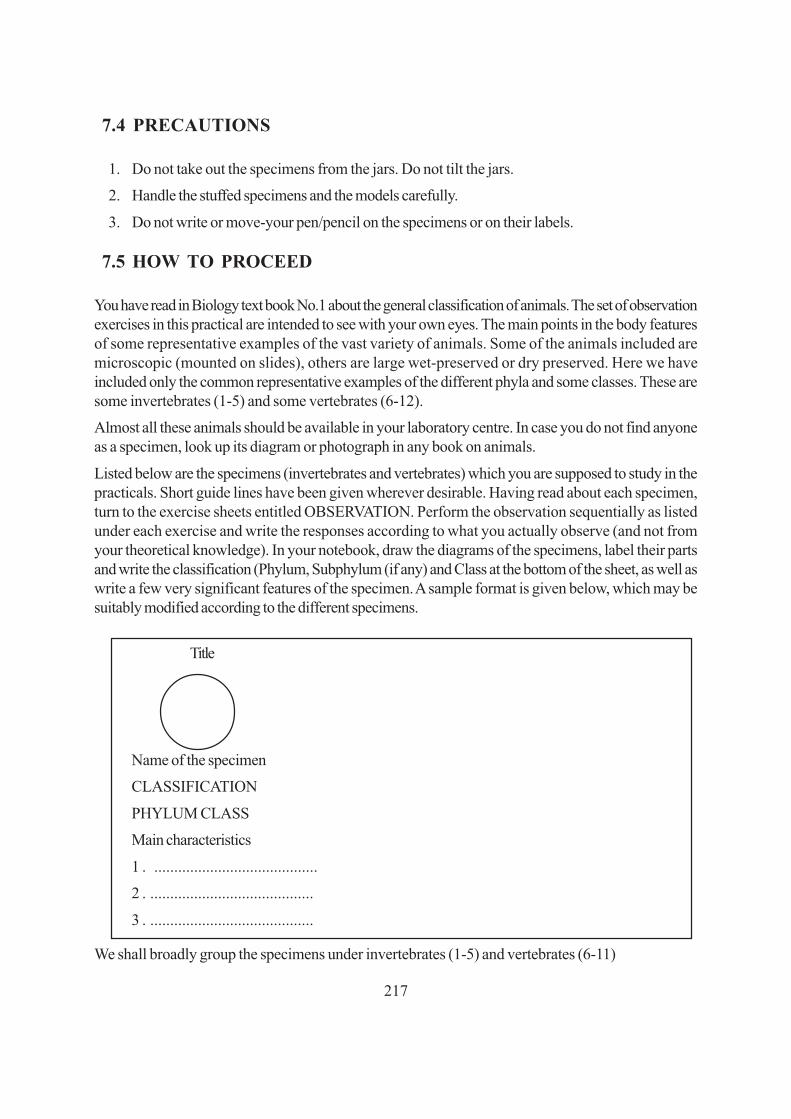
217
7.4 PRECAUTIONS
1. Do not take out the specimens from the jars. Do not tilt the jars.
2. Handle the stuffed specimens and the models carefully.
3. Do not write or move-your pen/pencil on the specimens or on their labels.
7.5 HOW TO PROCEED
You have read in Biology text book No.1 about the general classification of animals. The set of observationexercises in this practical are intended to see with your own eyes. The main points in the body featuresof some representative examples of the vast variety of animals. Some of the animals included aremicroscopic (mounted on slides), others are large wet-preserved or dry preserved. Here we haveincluded only the common representative examples of the different phyla and some classes. These aresome invertebrates (1-5) and some vertebrates (6-12).
Almost all these animals should be available in your laboratory centre. In case you do not find anyoneas a specimen, look up its diagram or photograph in any book on animals.
Listed below are the specimens (invertebrates and vertebrates) which you are supposed to study in thepracticals. Short guide lines have been given wherever desirable. Having read about each specimen,turn to the exercise sheets entitled OBSERVATION. Perform the observation sequentially as listedunder each exercise and write the responses according to what you actually observe (and not fromyour theoretical knowledge). In your notebook, draw the diagrams of the specimens, label their partsand write the classification (Phylum, Subphylum (if any) and Class at the bottom of the sheet, as well aswrite a few very significant features of the specimen. A sample format is given below, which may besuitably modified according to the different specimens.
Title
Name of the specimen
CLASSIFICATION
PHYLUM CLASS
Main characteristics
1 . .........................................
2 . .........................................
3 . .........................................
We shall broadly group the specimens under invertebrates (1-5) and vertebrates (6-11)
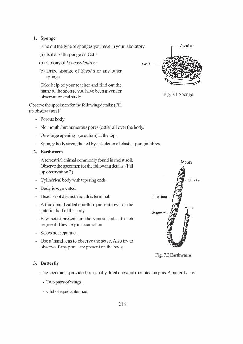
218
Fig. 7.1 Sponge
1. Sponge
Find out the type of sponges you have in your laboratory.
(a) Is it a Bath sponge or Ostia
(b) Colony of Leucosolenia or
(c) Dried sponge of Scypha or any othersponge.
Take help of your teacher and find out thename of the sponge you have been given forobservation and study.
Observe the specimen for the following details: (Fillup observation 1)
- Porous body.
- No mouth, but numerous pores (ostia) all over the body.
- One large opening - (osculum) at the top.
- Spongy body strengthened by a skeleton of elastic spongin fibres.
2. Earthworm
A terrestrial animal commonly found in moist soil.Observe the specimen for the following details: (Fillup observation 2)
- Cylindrical body with tapering ends.
- Body is segmented.
- Head is not distinct, mouth is terminal.
- A thick band called clitellum present towards theanterior half of the body.
- Few setae present on the ventral side of eachsegment. They help in locomotion.
- Sexes not separate.
- Use a’ hand lens to observe the setae. Also try toobserve if any pores are present on the body.
Fig. 7.2 Earthwarm
3. Butterfly
The specimens provided are usually dried ones and mounted on pins. A butterfly has:
- Two pairs of wings.
- Club shaped antennae.
Chactae

219
- Powdery scales on wings.
- Observe the butterfly carefully and answer
the questions given in observation 3.
Fig. 7.3 Butterfly
4. Apple snail (pila)
Observe the specimen (fill up observation 4).
It is a mollusc.
Observe the “mouth” of the shell. In preservedspecimens it is firmly closed by a “door” the lid.
If ever you get a spare specimen break open theshell and look for the animal contained inside. (Youmay sometimes find only the empty shells)
- unsegmented body.
- Body is soft and encosed in a calcareous shell. Fig. 7.4 Apple snail
- Head bears eyes and tentacles.
Observe carefully and fill up observation 4.
5. Starfish
Starfish is an Echinoderm. It is an unsegmentedmarine animal, showing radial symmetry. It has aspiny body surface. It moves by tube feet. Head isabsent.
Observe the animal carefully and fill up observation 5. Fig. 7.5 Starfish
7.6 RECORDING OF OBSERVATION
Observation 1
A. Outward appearance of sponge
(i) Looking at it what do you think-is it unicellular or multicellular?
.........................................................................................................................................
●

220
(ii) Is it a single lump or branched?
.........................................................................................................................................
(iii) What is its shape?
Vase-shaped/cup-shaped/bottle-shaped or irregular mass.
.........................................................................................................................................
B. Finer structure
Look at the surface of the main body.
(v) Do you find pores? ...................................................................................................................
(These pores are called ostia, and serve as the entrances for water into the body).
You may also find a large hole osculum. It is for exit of water from the body. If the specimen is adried one, you may not see the pores. The dried material is the skeleton made of spongin fibres.
C. Classification
Why are sponges placed in phylum Porifera? Give one reason.
.........................................................................................................................................
Can you assign the particular specimen given to you to its class - Calcarea, Hexactinellida, orDemospongiae?
.............................................................................................................................................
Observation 2
Earthworm (Pheretima) .
A. External features
(i) Is the animal (a) long and dorso-ventrally flattened or (b) cylindrical and contracted?
...........................................................................................................................................
(ii) What is the colour of the animal?
...........................................................................................................................................
(iii) What is the shape of the animal?
...........................................................................................................................................
(iv) Is the body even or segmented?
...........................................................................................................................................
(v) How does the clitellum differ from the skin over the rest of the body?
...........................................................................................................................................
(vi) What is the shape of the part of organ present at the upper (anterior) end?
...........................................................................................................................................

221
(vii) Where is the mouth present?
...........................................................................................................................................
(viii) Look for the anal aperture. Where is it present?
...........................................................................................................................................
(ix) Do you observe any setae? On which surface are they present, dorsal or ventral or on both?
...........................................................................................................................................
B. Classification
(i) Why does the earthworm belong to the phylum Annelida? Give one reason?
...........................................................................................................................................
(ii) In which class will you place Pheretima in Polychaeta, Oligochaeta or Hirudinea?
...........................................................................................................................................
Observation 3
Butterfly (Danaus)
A. External Features
(i) Is the animal bilaterally symmetrical or radially symmetrical?
...........................................................................................................................................
(ii) Into how many parts or regions is the body divisible?
...........................................................................................................................................
Name the regions ........................................................................................................................
(iii) How many pairs of eyes do you see? (Use a hand lens)
...........................................................................................................................................
(iv) Are these simple eyes or compound eyes?
...........................................................................................................................................
(v) How many pairs of antennae do you see?
...........................................................................................................................................
(vi) What is the shape of the antennae?
...........................................................................................................................................
(vii) Are they long and swollen at tip?
...........................................................................................................................................
The scientific term for this type of antennae is clavate (club-shaped).

222
(viii) Mention the type of mouth parts. (sucking/piercing/biting).
.............................................................................................................................................
(ix) Do you see a coiled (spring-like) part located on the lower side of the head? ...........................It is the proboscis.
(x) What does the butterfly feed upon?
...........................................................................................................................................
(xi) How many pairs of wings are there?
...........................................................................................................................................
(xii) Describe the pattern of colouration of the body?
...........................................................................................................................................
In case you ever catch a butterfly then only try the following:
● Gently move your finger on the wings. Do you find something powdery coming on to yourfinger?
● Spread this powdery material on a clean slide and examine under microscope. (These are thescales).
(xiii) How many pairs of legs do you see?
...........................................................................................................................................
(xv) Are the legs jointed?
...........................................................................................................................................
(xv) From which region of body do they originate?
...........................................................................................................................................
(xvi) How many joints are there in each leg?
...........................................................................................................................................
(xvii) Are the legs hairy or smooth?
...........................................................................................................................................
(xviii) What are the legs used for (resting/walking)?
...........................................................................................................................................
B. Classification
(i) Why is butterfly classified as an Arthropod? Give one character.
...........................................................................................................................................
(ii) Give two characters that justify that butterfly is an insect.
(1) ...........................................................................................................................................
(2) ...........................................................................................................................................

223
Observation 4
4. Apple snail (Pila)
A. External Features
(i) Is the body of the animal enclosed in a shell?
...........................................................................................................................................
(ii) Is the shell made up of one piece (univalve) or of two pieces (bivalve)?
...........................................................................................................................................
(iii) Is the shell plain or spirally coiled?
...........................................................................................................................................
(iv) Locate the position of the opening of the shell. Is it open or closed?
...........................................................................................................................................
B. Classification
Let us try to classify pila:
(i) Why is Pila placed in phylum Mollusca?
Give one reason
...........................................................................................................................................
(ii) Mention the class of Pila. Give one reason.(Bivalvia, Gastropoda or Cephalopoda)
...........................................................................................................................................
Observation 5
Starfish
A. External features
(i) Is the animal radially symmetrical or bilaterally symmetrical?
...........................................................................................................................................
(ii) How many aims does it have?
...........................................................................................................................................
(iii) Can you make out any central region i.e. the central disc?(Yes/No)
...........................................................................................................................................
(iv) Locate the mouth on the lower (lighter coloured) side of the body. Is it located in the centre?
...........................................................................................................................................
(v) Do you observe any plates and spines on the surface of the body?
...........................................................................................................................................

224
Examine the lower surface of the arm
(vi) Do you find a groove running through the middle?
...........................................................................................................................................
(vii) Do you observe any fleshy structures along the sides of the groove?
These are tube feet ....................................................................................................................
(viii) What is the natural habitat of starfish?
...........................................................................................................................................
(ix) Why is it called starfish?
...........................................................................................................................................
Classification
(Echinodermata: echinos = spiny, derma = skin).
(i) Why is the starfish placed in the phylum Echinodermata? Give one reason?
...........................................................................................................................................
(ii) In which class will you place starfish - Asteroidea, Ophiuroidea, Echinoidea Holothuroidea orCrinoidea? Give reason.
...........................................................................................................................................
B. Vertebrata
Animals observed and studied upto this point were all invertebrates (without backbone). We willnow take up Vertebrates.
6. Dogfish
- Scales’ embedded in skin.
- Paired pectoral and pelvic fins.
- Unpaired dorsal, caudal and ventral fins. Five Gill slits.
- Dog fish has a cartilagenous skeleton. Refresh your memory about cartilage. Observe the animalcarefully and fill up observation 6.
Fig. 7.6 Dogfish

225
7. Rohu
- Large scales cover the body.
- Gills covered by operculum.
- Rohu is a bony fish. i.e. it has a bony skeleton.
Observe the animal carefully and fill up observation 7.
Fig. 7.7 Rohu
8. Toad (Bufo)- Dry skin.
- Parotid glands.
- Toad has much in common with frog,but it has some of its owncharacteristics.
- Count the number of toes in fore andhind limbs.
Observe the specimen carefully and fill upobservation 8.
Fig. 7.8 Toad
9. Wall-lizard (Hemidactyles)
- Dry scaly skin,
- Hands and feet with flat expanded digits forclasping.
- Wall lizard is the most familiar repitle.
Observe the specimen carefully and fill up theobservation 9.
Fig. 7.9 Wall lizard
10. Pigeon (Columba)
- Has feather.
- Wings (modified forelimbs).
- Beak: but no teeth.
Pigeon or any other bird have the same general features of class Aves. Observe the specimencarefully. Fill up observation 10.

226
Fig. 7.10 Pigeon
11. Bat (pteropus)
- Hair on the body.
- Projecting external ears.
- Forelimbs modified as wings.
Observe the animal carefully and fill upobservation 11.
Bat flies like a bird but it is not a bird. What isit then? Exercise under the observation no.11 to find out.
7.7 RECORDING OF OBSERVATIONS
Observation 6
Dogfish
A. External features
(i) What is the shape of the body? boat shaped/spindle shaped or any other shape.
.............................................................................................................................................
(ii) How many distinct regions are there in the body?
.............................................................................................................................................
(iii) Is the surface of the body smooth or covered by any distinct scales?
.............................................................................................................................................
Fig. 7.11 Bat

227
(iv) How many fins do you find in the trunk?
.............................................................................................................................................
(v) Tick (✓) mark those fins which you observe in the specimen.
(l) Paired pectoral fins ( ) (3) Paired pelvic fins ( ) (5) Anal fin ( )
(2) Dorsal fins ( ) (4) Ventral fins ( )
(vi) Where is the mouth located- on the front tip or on the dorsal or on the ventral side of the head?
.............................................................................................................................................
(vii) Does the head bear a pair of nostrils?
.............................................................................................................................................
(viii) What are the nostrils used for in a fish?
.............................................................................................................................................
(ix) Where are the eyes located?
.............................................................................................................................................
Observe the gill-slits behind and slightly below the eyes.
(x) How many gill-slits do you find on each side?
(xi) Are the gill slits covered by any flap?
.............................................................................................................................................
(xii) Do you see a “line” extending all along from head to tail on each side?
.............................................................................................................................................
(xiii) What is this line called?
.............................................................................................................................................
(xiv) Does the fish posses an internal skeleton or an external skeleton?
.............................................................................................................................................
B. Classification
(i) Why the dogfish has been placed in Phylum Chordata? Give one suitable reason for it.
.............................................................................................................................................
(ii) Why is dogfish also a vertebrate?
.............................................................................................................................................
(iii) Mention three features in this animal which shows that it lives in water?
.............................................................................................................................................

228
Observation 7
Rohu (Bony fish)
A. External features
(i) Find out the shape of the body-fusiform/spindle shaped/boat shaped (mark ✓ on the rightanswer)
.............................................................................................................................................
(ii) How many distinct regions are there in the animal?
.............................................................................................................................................
(iii) Does the head possess any scales? Yes-or No.
.............................................................................................................................................
(iv) Where is the mouth located? (terminal/sub-terminal/dorsal/ventral)
.............................................................................................................................................
(v) What other parts do you observe on the head?
.............................................................................................................................................
(vi) Are the gill-slits naked or covered by a flap?
.............................................................................................................................................
The flap is called operculum.
(vii) Does the dogfish possess Operculum?
.............................................................................................................................................
(viii) Is the trunk of Rohu covered by hair or by scales?
.............................................................................................................................................
(ix) Observe the fins and fill in the blanks below:
Name the unpaired fins Name the paired fins.
1 . .................................. 1. ..................................
2. .................................. 2 . ..................................
3 . ..................................
(x) Is the lateral line distinct or indistinct?
.............................................................................................................................................
(xi) List any two features by which you can differentiate a cartilaginous fish from bony fish.
.............................................................................................................................................
(i) . .................................. (ii) ..................................

229
B. Classification
(i) Why is Rohu placed in the Sub phylum Vertebrata?
Give one reason
(ii) Mention three features of this animal which show that it lives in water.
1. .......................................................................................................................................
2 . .......................................................................................................................................
3 . .......................................................................................................................................
Observation 8
Toad (Bufo)
A. External features
(i) Name the regions into which the body is divided.
(Head, neck and trunk/head and trunk) (mark ✓)
(ii) Is there a neck?
.............................................................................................................................................
(iii) What type of skin do you observe? Tick (✓) mark the appropriate one.
Smooth/dry/warty/rough/scaly.
(iv) Is the snout pointed or semicircular?
.............................................................................................................................................
(v) Name the structures present on the head.
.............................................................................................................................................
(vi) Are the eyes large and bulging?
Yes/No ......................................................................................................................................
(vii) Is there a dark patch seen behind the eye?
Yes/No ........................................................................................................................................
Behind each tympanum, you observe an oval shaped area which is different from the rest of theskin. It is the parotid gland.
(viii) What is its function?
.............................................................................................................................................
(ix) How many appendages (limbs) do you observe?
.............................................................................................................................................

230
(x) Which limb is longer? (fore limb/hind limb)
.............................................................................................................................................
(xi) How many digits do you observe in the fore-limb?
.............................................................................................................................................
(xii) How many digits do you observe in the hind-limb?
.............................................................................................................................................
(xiii) How do these differ from the digits of the fore limbs? Give two differences.
1 . .............................................................................................................................................
2 . .............................................................................................................................................
(xiv) Does the skin of the toad serve as an organ for respiration or for protection or for both?
.............................................................................................................................................
(xv) From the external features that you have observed, point out whether this animal is (a) aquatic (b)terrestrial or (c) both. Tick (✓) mark.
B. Classification
(i) Why has toad been included in the phylum Chordata?
Give one reason ............................................................................................................................
(ii) Why is it grouped in the class Amphibia?
Mention a suitable explanation. (Amphi=both; bios=life).
Observation 9
Wall lizard
A. External features
(i) In how many regions can you divide the body of a lizard: Name them.
.............................................................................................................................................
(ii) Is the surface of the body covered by hairs/warts/horny scales?
.............................................................................................................................................
(iii) What is the shape of the head?
.............................................................................................................................................
(iv) How does the tympanum (ear-drum) of lizard differ from that of a toad?
.............................................................................................................................................
(v) How many limbs are there?
.............................................................................................................................................

231
(vi) How many digits does each limb have?
.............................................................................................................................................
These limbs are called pentadactyl limbs.
(vii) Do the digits have claws? Yes or No ...........................................................................................
(viii) What do you observe on the ventral side of the digit?
.............................................................................................................................................
(ix) Why does the wall lizard not fall when it creeps on the ceiling or the wall? (Co-relate it with thestructure of the digit).
(x) Why is it known as wall-lizard?
.............................................................................................................................................
(xi) Does this animal walk/creep/run?
.............................................................................................................................................
Reptiles are creeping animals.
B. Classification
(i) Why is it placed in phylum Chordata?
Give one reason .........................................................................................................................
(ii) Why is it grouped in class Reptelia?
Give one reason.............................................................................................................................
(iii) Give the scientific name of the wall lizard.
Observation 10
Pigeon
A. External features
(i) What is the shape of the body?
.............................................................................................................................................
(ii) What is the body covered with?
.............................................................................................................................................
(iii) Do you find the same type of structures covering on the head?
Yes/No . .......................................................................................................................................
(iv) If they are different, in what way do they?
.............................................................................................................................................
(v) How is its beak? (Short and pointed short and curved).
.............................................................................................................................................

232
(vi) From the shape of the beak, suggest the - type of food it feeds on.
.............................................................................................................................................
(vii) Can you locate the nostrils? Yes/No.
.............................................................................................................................................
(viii) Are the nostrils slit -like or oval?
.............................................................................................................................................
(ix) How many eyes are there?
.............................................................................................................................................
(x) How many wings does it have?
.............................................................................................................................................
Fore-1imbs are modified into wings.
(xi) Do you see the digits at the tip of the wings?
Yes/No ....................................................................................................................................
Pigeon has a pair of hind limbs.
(xii) Are they covered with hairs/scales or are smooth?
.............................................................................................................................................
(xiii) How many digits do you observe in each hind limb?
.............................................................................................................................................
(xiv) Are they clawed?
.............................................................................................................................................
(xv) How do they make use of their feet?
.............................................................................................................................................
(xvi) What is the tail made up of ?
.............................................................................................................................................
(xvii) How is it useful ?
.............................................................................................................................................

233
B. Classification
(i) Enumerate any three external features of pigeon that suggests aerial mode of life.
(1) ........................................................................................................................................
(2) ........................................................................................................................................
(3) ........................................................................................................................................
(ii) Why is the pigeon placed in phylum chordata?
Give one reason ..........................................................................................................................
Birds are grouped under the class Aves. List any two characteristic features.
.............................................................................................................................................
Observation 11
Bat
A. External features
(i) Body is divisible into ...................., .......................... and tail. (Mention the regions)
1. ............................................................. 2..............................................................
(ii) What structures are located on the head? Tick (✓) mark the ones you observe.eyes/nostrils/tympanum/external ear (pinna).
(iii) Is the snout long or short?
.............................................................................................................................................
(iv) How many limbs do you observe?.............................................................................................................................................
(v) Fore-limbs are in the form of wings..............................................................................................................................................
Does the fore limb possess the skin/feathers?
Examine the folds of skin
The wing skin called patagium begins from shoulder, extends along the upper margin of the armto the base of the thumb, between the finger (digits) and along the sides of the body to the hindlegs. It helps in flying.
(vi) Which digits support the wings? (Second to fifth/first to fifth)
.............................................................................................................................................
9
F

234
(vii) Which digits are clawed?
.............................................................................................................................................
(viii) Which digit is free?
.............................................................................................................................................
(ix) Are the hind limbs small/large?.............................................................................................................................................
(x) How many digits are there in each hind limb?.............................................................................................................................................
(xi) Are the digits clawed?
.............................................................................................................................................
B. Classification
(i) Why is bat included in chordata?
Give one reason . ................................................................................................................
(ii) It is grouped in class Mammalia why?
Tick (✓) mark those external features which show mammalian characters.
1. Presence of hair
2. Presence of external ear (pinna)
3. Mammary glands in female.
4. Presence of limbs.
(ii) Is it justified to call bat as a flying mammal?
Yes/No ........................................................................................................................................
7.8 PRECAUTION
Handle the specimens carefully so that they are not damaged.
7.9 FOR THE TEACHERPlease ensure that the student
(i) learns the scientific name of the animals studied in the laboratory and spells and writes.
(ii) relate the features with the classification.

235
DISSECTIONS
a. EARTHWORM (Pheretima)
Fig : Earthworm :
A. ventral view (entire specimen), B. Genital area, ventral view
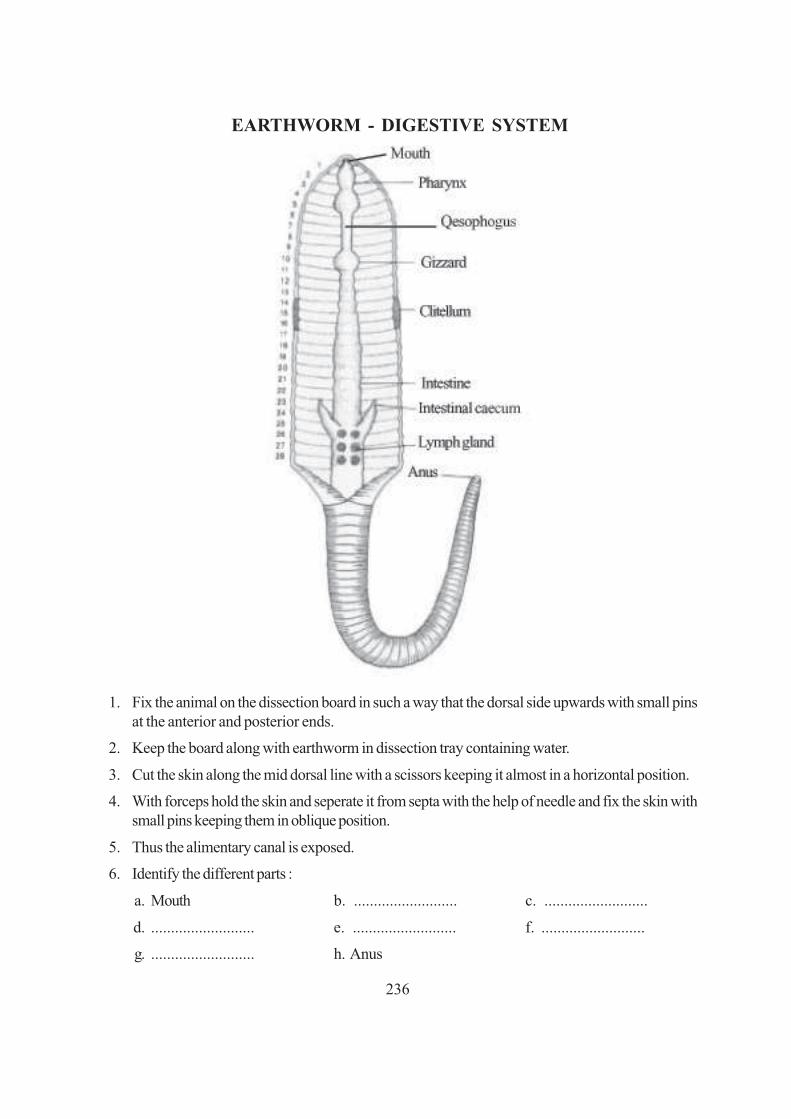
236
EARTHWORM - DIGESTIVE SYSTEM
1. Fix the animal on the dissection board in such a way that the dorsal side upwards with small pinsat the anterior and posterior ends.
2. Keep the board along with earthworm in dissection tray containing water.
3. Cut the skin along the mid dorsal line with a scissors keeping it almost in a horizontal position.
4. With forceps hold the skin and seperate it from septa with the help of needle and fix the skin withsmall pins keeping them in oblique position.
5. Thus the alimentary canal is exposed.
6. Identify the different parts :
a. Mouth b. .......................... c. ..........................
d. .......................... e. .......................... f. ..........................
g. .......................... h. Anus

237
EARTHWORM - NERVOUS SYSTEM
1. Fix the animal on the dissection board in such a way that the dorsal side upwards with small pinsat the anterior and posterior ends.
2. Keep the board along with earthworm in dissection tray containing water.
3. Cut the skin along the mid dorsal line with a scissors keeping it almost in a horizontal position.
4. With forceps hold the skin and seperate it from septa with the help of needle and fix the skin withsmall pins keeping them in oblique position.
5. Thus the alimentary canal is exposed.
6. Cut the oesophagus and carefully pull out the anterior end of the gut from behind. Nerve ring isexposed. Remove the rest of the gut. Thus the vental nerve cord is exposed.
7. Identify the componets of nerve ring :
a. .....................................................................................
b. .....................................................................................
c. .....................................................................................

238
22b. COCKROACH (Periplaneta)
COCKROACH - MOUTH PARTS

239
COCKROACH - DIGESTIVE SYSTEM
1. Hold the cockroach with your left hand and remove the wings with scissors.
2. Hold the animal between your thumb and first finger and cut along the sides.
3. Fix the cockroach on the wax of the tray with its dorsal side upwards with pins on head, posteriorend and coxae of legs.
4. Carefully remove the dorsal plates of abdomen and thorax.
5. Carefully uncoil the alimentary canal and keep on one side on a black paper.
6. Identify different parts of the alimentary system.
a. Mouth b. .......................... c. ..........................
d. .......................... e. .......................... f. ..........................
g. .......................... h. .......................... i. Anus
7. Identify the glands and other structures.
a. .......................... b. .......................... c. ..........................

240
COCKROACH - NERVOUS SYSTEM
1. Hold the cockroach with your left hand and remove the wings with scissors.
2. Hold the animal between your thumb and first finger and cut along the sides.
3. Fix the cockroach on the wax of the tray with its dorsal side upwards with pins on head, posteriorend and coxae of legs.
4. Carefully remove the dorsal plates of abdomen and thorax.
5. Remove the alimentary canal and clean the fat bodies and blood. Hence nerve cord is exposed.
6. Carefully remove the epicranial plate on the head and expose the cerebral ganglia.
7. Pull out the oesophagus through the nerve ring.
8. Arrange black papers.
9. Identify different parts of the nerve ring.
a. .......................... b. .......................... c. ..........................
10. Name the two thread like structures along the mid ventral line
............................................................................................................................................

241
�� �� Exercise
PREPARATION OF A SLIDE OF ONION ROOT TIP FOROBSERVATION OF STAGES OF MITOSIS
Growth and repair of any part of an organism takes place through mitotic division of cells of that part.The growing tip of onion roots form an excellent material to study various stages of mitosis.
OBJECTIVES
After performing this exercise, you should be able to:
● acquire the skill of making a root tip squash preparation;
● distinguish between dividing and non-dividing cells;
● identify different stages of mitotic cell division;
● differentiate between different stages of mitosis.
8.1 WHAT YOU SHOULD KNOW
1. Cells follow a cell cycle in which there is a phase termed interphase in which cells do not divideand another phase termed mitosis in which one cell divides to give two identical cells.
2. In non-dividing cells, nucleus is seen to contain a chromatin network.
3. Mitosis can be divided into four phases (stages) Prophase, Metaphase, Anaphase and Telophase(Fig. 8.1)
10
F

242
4. At Prophase(a) Nuclear membrane remains intact.(b) Chromatin is resolved into thread like chromosomes.
At Metaphase(a) Nuclear membrane disappears.(b) Spindle forms (may not be seen in the slides).(c) Chromosomes arrange at the equator.(d) Each chromosome has two chromatids joined by a centromere.
At Anaphase(a) Centromere splits.(b) Each chromatid now has its own centromere and so becomes a chromosome.(c) Equal number of chromosomes move to opposite poles.
Fig. 8.1 Different stages of cell division.
1 2
3 4
5 6

243
At Telophase
(a) Two groups of chromosomes lie at two poles and nuclear membranes form around them.
(b) Chromosomes clump together and lose their identity and once again form chromatin network.
(c) Thus two nuclei are formed in the cell containing the same number and types of chromosome.
(d) A partition wall (cell plate) begins to form in the centre of the cell.
4. Cytokinesis
(a) Cell plate formed in the middle extends and divides the cell into two daughter cells.
(b) Each daughter cell now contains a single nucleus.
Materials Required
(i) Onion bulb (vi) Microscope (xi) Match-stick
(ii) Needles (vii) Acetocarmine (xii) Scalpel
(iii) Brush (viii) Dilute HCl (xiii) Scissors
(iv) Slides (ix) Wide-mouthed bottle/ (xiv) 70% Alcohol. container/vial
(v) Coverslips (x) Beaker (xv) Blotting paper.
8.1 HOW TO PROCEED
This exercise has to be done in three phases:
1. Growing an onion for 3 to 5 days till roots emerge.
2. Fixing the root-tips.
3. Preparing a microscopic slide.
Phase 1. Growing onion for root-tips
(i) Take a wide-mouthed bottle 3-4 days prior to the day you have fixed for this experiment and fillit with water very close upto the mouth.
(ii) Take one medium sized onion bulb and remove its dry roots if any.
(iii) Place the onion at the mouth of the bottle so that only the base (disc) of the onion touches thewater (Fig. 8.2a).

244
Fig. 8.2(a) Placing onion at the mouth of bottle Fig. 8.2(b) Growing root tips
(iv) In 3 - 4 days new roots will appear (Keep watching everyday).
(v) When the roots are about 2-3cms long (Fig. 8.2b) you can start with the next phase (2) of theexercise.
Phase 2. Fixing the root-tips
(i) Remember you have to cut the root-tips only in early morning (around 9 A.M.) (Generallymitotic activity occurs at this time).
(ii) Remove the onion bulb from water. Using scissors, cut only the root tips (about 0.5 cm longfrom their ends from the cluster of white slender thread - like roots).
(iii) Put them in 1 : 3 Acetic Alcohol for 10 minutes. Remove from fixative and put the cut root tips in70% Alcohol. (This is permanent preservation for any length of time).
(iv) The root-tips are ready for use after 24 hours.
Phase 3. Preparing a microscopic slide
(i) Take one root tip on a clean slide.
(ii) Add a few drops of dilute HCl just for 1 or 2 seconds. (This will soften the root tips).
(iii) Immediately add a few drops of ordinary water to this material to wash off the acid,
(iv) Decant the water by holding the slide tilted over a watch glass with one hand and holding thematerial on the slide with a brush by the other hand,
(v) Shift the material to a cavity block/watch glass. Add a few drops of acetocarnine stain, cover itwith a lid. Wait for 5-8 minutes. The root tips become deep red.
(vi) Now take a clean slide. Place 3-4 drops of acetocarmine stain on the slide and transfer thematerial from the watch glass to the stain on the slide.

245
(vii) Gently warm the slide and then place on a square piece of paper towel/blotting paper. Take carethat the slide is not overheated.
(viii) Squash the stained root tip and then place a coverslip over the material.
(ix) Place the slide between folded filter paper or blotting paper and blot gently without moving thecoverslip, to remove excess stain.
(x) Take a pencil and using its blunt end gently tap over the cover-slip (cells of the root tip will spreadout.
(This will crush the root-tips and the cells lying deepest which may not have picked up the stainearlier would now do so, as they are again submerged in the stain).
(xi) If the material is soft, a few tappings will be enough for the material to be squashed (squashmeans crushing to release contents).
Note: Do not crush the coverslip while tapping the material below it.
Always use glass-rod, brush, or needles, forceps etc.
for handling the material in this exercise.
Metallic contact with the stained material causes a
dark-brown precipitate in the material.
(xii) Observe the slide under the microscope first under the low power.
(xiii) Locate a specific good area on the slide and then observed under the high power.
(xiv) Move your slide gradually to observe different areas for various stages of mitosis.
8.3 YOU WILL OBSERVE
(i) Cells in onion are rectangular. Do you see any circular or oval cells? Check.
(ii) Acetocarmine stains the chromosomes, that is why you do not observe spindle fibres.
(iii) Look for a cell with distinct nucleus (no separate chromosomes). Such cells are in Interphasestage.
(iv) Look for cells where the chromosomes are thick, deeply stained and easily visible.
They are arranged in the middle (equator) of the cell, arranged in a circle or in a row etc. Thesecells are in Metaphase stage.
(v) Look for some cells in your slide in which the chromosomes are away from the middle and in twogroups, each such group lying at the opposite ends. These cells are in the Anaphase stage.
(vii) You may see cells where cell plate formation is completed and cell is divided into two daughtercells. These cells are in the Cytokinesis stage.
(vi) You may also see cells where the chromosomes form a cluster at extreme opposite ends. Thesecells are in the Telophase stage. You may also see beginning of cell plate formation.

246
(viii) In case you are unable to see all the stages of mitosis in your slide try to see them, in the preparationof other students.
Why should you cut only the tips for this exercise and not any other region of the root?
8.4 RECORDING OF OBSERVATIONS
Observation 1
(i) Name the stage at which a cell is in the non-dividing phase. Interphase/Telophase/ orAmitosis.
.............................................................................................................................................
(ii) (a) Look for a stage where the chromosomes are arranged in the middle of a cell. What is thisstage known as?
.............................................................................................................................................
(b) Is there a nuclear membrane at this stage?
.........................................................................................................................................
(iii) Can you count the number of chromosomes in the above mentioned stage?
.............................................................................................................................................
(iv) Can you see two chromatids of each chromosome?
...........................................................................................................................................
(v) Look for a stage where you see the chromosomes in two groups moving towards the two oppositeends.
...........................................................................................................................................
(a) Are they slightly away from the middle? At this stage the cell is said to be at early Anaphase.
........................................................................................................................................
(b) Have they reached the opposite ends of a cell? Then it would be late Anaphase.
........................................................................................................................................
(vii) Look for a cell with two nuclei which have a partition between them.
...........................................................................................................................................
(viii) What is this stage of cell division called?
...........................................................................................................................................
(ix) In some cells the partition will be incomplete, what is this partition called?
...........................................................................................................................................
(x) Draw the following in your record book.
1. An interphase cell
2. A cell at prophase

247
3. A cell at metaphase
4. A cell at anaphase
5. A cell at telophase
6. A cell showing cell plate formation (cytokinesis)
8.5 PRECAUTIONS
1. Use clean slides and coverslips.
2. While fixing the tissues do not leave it in acid for more than the required time.
3. Do not heat the slide. It is only to be warmed.
4. After putting coverslip over stained material, blot off excess stain.
5. Do not allow the slide to dry up.
8.6 FOR THE TEACHER
1. This is a comparatively difficult exercise as students have to be trained to locate particular stagesof cell division in the microscopic field under observation.
2. It will be of immense help if a squash preparation is first demonstrated by the teacher.
3. An interphase cell may then be shown under the microscope. The student may then be asked tofocus an interphase cell in the slide prepared by the student. Dividing cells can then be distinguishedfrom the non dividing cells lying in the same field.
4. In this way, demonstration of a particular mitotic stage followed by its identification, in the studentsown slide, may be carried out for each of the different stages of mitosis.

248
�� �� Exercise
TO STUDY THE SPECIAL ADAPTIVE FEATURES IN SOME PLANTSAND ANIMALS
Animals and plants have evolved special features in order to live successfully in a particular habitat.These features known as adaptive features, help the organisms to adjust to their habitats.
You will study the adaptive features in a hydrophyte (Water hyacinth), xerophyte (Opuntia) and aparasitic animal (Tapeworm).
OBJECTIVES
After completing this exercise, you should be able to :
● identify the specimen and know its habitat;
● list the general features as well as the special adaptive features of these organisms;
● mention the role played by the adaptive features;
● identify and relate the habitat of other organisms showing similar adaptive features.
9.1 WHAT YOU SHOULD KNOW
1. Diverse habitats in which plants and animals live are (i) terrestrial (ii) aquatic (iii) aerial.
2. Depending upon the availability of water, the habitat can be xeric, mesic or aquatic.
3. Name some plants and animals belonging to the above categories.
4. The term adoption can be defined as the modifications of characteristics which have evolvedover a period of time in the living organisms. These modifications help the organisms to adjust ina particular environment.

249
5. Some of the adaptive features in aquatic plants are presence of air cavities in stem or leaf forbuoyancy; presence of waxy coating on leaves to protect them from damage due to continuousflow of water on them; roots are partly developed as water is present in abundance.
6. Some of the xeric plants show adaptation which help them to conserve water.
7. Some parasitic worms have thick cuticle which protects them from the action of digestive enzymesof the host.
Material Required
1. Fresh or preserved specimens of (a) water Hyacinth (b) Opuntia (c) preserved specimen oftapeworm and a slide of head of tapeworm (scolex)
2. Handlens
How to proceed
1. Water Hyacinth Free floating aquatic plant :
Take a fresh or preserved specimen and observe itsparts carefully. Take special note of the following :
(a) Roots - Its type, growth pattern and any specialfeature that comes to your notice.
(b) Stem - Its nature, length etc.
(c) Leaves - Observe the petiole, the protectivecoating on the leaves and the texture of the leaves
Note down the special features that help it tosurvive in aquatic habitat. Record yourobservations.
2. Opuntia are xeric plants so observe a fresh orpreserved specimen with special attention to
(a) Root - Its type, length etc.
(b) Stem - If it is modified then the type ofmodifications it shows. Observe its colour. Doesit suggest any special function that it perform?
(c) Leaves - are present? If not, then what are theymodified into. What is the significance of thismodification?
Fig. 9.1
Fig. 9.2

250
Fill up observation
3. Tapeworm (Taenia) =A human intestinal parasite
(a) Observe the entire specimen from the head up to the last segment or the widest end andidentify the following parts:
(i) Scolex or the head
(ii) Neck
(iii) Proglottides forming the strobila
(b) Observe the slide of the scolex under a dissection microscope or the low power of a compoundmicroscope and identify
(i) the hooks in the form of a circlet onthe top of the head.
(ii) Four suckers present on fourdifferent sides of the scolex.
The parasite attaches itself to thewall of the human instestine with thehelp of its scolex.
( c) Observe that there is no mouth andanus in the parasite because it absorbsdigested food surrounding it
(d) How do you think it respires inside thehuman intestine?
Fig. 9.3
RECORDING OF OBSERVATIONS
Observation 1
(i) Are the plants rooted or freely floating?
.............................................................................................................................................
(ii) Is the root system poorly developed or well developed?
.............................................................................................................................................
(iii) Are root hairs present or absent?
.............................................................................................................................................
(iv) Is the fibrous root system or tap root system?
.............................................................................................................................................

251
(v) What is the significance of these types of roots?
.............................................................................................................................................
(vi) What is the nature of the petiole of the leaf?
.............................................................................................................................................
(vii) What role do the air cavities in the stem or petiole play in life of plant in aquatic habitat?
.............................................................................................................................................
(viii) What is the nature of special coating on the leaves? How is it useful?
.............................................................................................................................................
(ix) List the special adaptive features in water hyacinth for aquatic life
(i) ............................................. (ii) ............................................. (iii) ......................................
(x) Draw a labelled diagram of the specimen.
.............................................................................................................................................
Observation 2
(i) What is the nature of the stem? Is it fleshy or dry?
.............................................................................................................................................
(ii) Are there any leaves present? What are they reduced to? What is the significance?
.............................................................................................................................................
(iii) What is the term used to describe such a stem?
.............................................................................................................................................
(iv) How have such plants reduced the loss of water?
.............................................................................................................................................
(v) Draw a labelled diagram of specimen.
Observation 3
(i) Draw a labelled diagram showing
(a) Scolex with hooks and suckers
(b) Neck
(c) Proglottides
(ii) What is the use of the hooks and suckers in the scolex?
(a) .............................................................................................................................................
(b) .............................................................................................................................................

252
(iii) Comment upon the digestive system of Taenia.
(a) Does it have a well developed digestive system?
......................................................................................................................................
(b) How does its obtain food?
.........................................................................................................................................
(iv) List at least three parasitic adaptations in Taenia.
(a) .........................................................................................................................................
(b) .........................................................................................................................................
(c) .........................................................................................................................................
(v) How does it protect itself from the action of the digestive enzymes present in the intestine?
PRECAUTIONS
1. Carefully handle the opuntia plant with fine spines, it is fresh specimen.
FOR THE TEACHER
Please ensure that student is able to
1. correlate special adaptive features with the habitat
2. identify the spines and suckers on the scolex of Taenia.

253
�� �� �!��Exercise
TO STUDY THE PHYSICAL PROPERTIES OF DIFFERENT SOILSAMPLES
Soil is the uppermost layer of the earth. It is formed by disintegration and decomposition of rocks. Soilis a mixture of mineral particles of varying sizes and decaying organic matter called humus. Numerousorganisms live in soil and soil sustains plant life. On the nature of soil depends the type of plants or cropsthat can be grown on it.
OBJECTIVES
After performing the exercise, you should be able to :
● acquire the skill of setting up the experiment;
● identify different layers or components of the soil;
● compare the physical properties of different soil samples. Fig. 10.1 Soil sample
10.I(a) WHAT YOU SHOULD KNOW
Soil is a mixture of mineral particles of different sizes and decaying organic matter. The different sizedsoil particles are classified as follows:
S.No Diameter of particles Name of the soil particles
1. more than 2.00 mm gravel
2. 2.00 mm to 0.2 mm Coarse sand
3. 0.2 mm to 0.02 mm Fine sand
4. 0.02 mm to 0.002 mm Silt
5. below 0.002 mm Clay

254
The varying percentage of different particles in the soil are responsible for the difference in soil texture.According to the texture, characteristics of soil it may be named as :
1. Sandy soil - When soil consists of 60% of sand, 10% clay and 10% silt.
2. Loamy soil - When soil has 30-50% silt, 5-20% clay and rest sand.
3. Clay soil- When soil contains 50% of clay particles or more, !est as silt and sand.
Material Required
(i) Paper bags for collecting soil samples (ii) Hand lens
(iii) Measuring cylinder (iv) Water
(v) Glass rod
10.2(a) HOW TO PROCEED
1. Collect soil samples from different places in different paper bags and label the place and date ofcollection. Bring them to the laboratory.
2. Examine the soil samples by a hand lens and feel its texture and note down in the observationtable given below.
3. Take about 50 gm of soil from a sample in a 250 ml measuring cylinder.
4. Add 150 ml of water and stir it well with a glass rod.
5. Allow it to settle down.
6. Record the thickness of the layers formed by different types of particles Calculate their relativepercentage and note down your observations in the table given below.
7. Similarly record the relative percentage of different types of particles in different soil samples.
Fig. 10(a) Different layers formed by different types of soil particles in water.

255
10.3(a) RECORDING OF OBSERVATIONS
1. Name the layer that settles at the bottom and the layer that floats on water.
(a) ................................................................. (b) ..........................................................
2. How many layers are formed by the soil samples
(a) ........................................................................................................................................
(b) ........................................................................................................................................
(c) ........................................................................................................................................
3. What is the colour and texture of the soil sample A, Band C in the table given below.
S.No. Soil sample Colour Texture Relative percentage
Sand Silt Clay
1. Soil from a crop land
2. Soil from road side
3. Soil from river or
any other source
10.4(a) PRECAUTIONS
1. Soil samples should be collected in separate bag labelled and brought to the laboratory.
2. The thickness ofthe layers formed in water should be carefully measured.
10.5(a) FOR THE TEACHER The teacher may help the students to
1. identify the different layers formed by the soil in water.
2. identify the sample/type of soil smaple A, B and C.

256
TO STUDY THE WATER HOLDING CAPACITY OF DIFFERENT SOILSAMPLE
Soil water is one of the most important ecological factors. Soil water is derived either from rain or fromirrigation. All the water falling on soil in an area is not retaind by it. Most of it is lost as gravitationalwater, the rest of it is retained as capillary water and hygroscopic water. The amount of waterretained by the soil depends upon its particle size.
OBJECTIVES
After performing the exercise, you should be able to :
● acquire the skill of weighing soil samples by using physical balance;
● develop the skill to set up an apparatus to perform this exercise;
● explain that water rises up in soil by capillarily;
● explain why different soil,samples have different water holding capacities.
l0.l (b) WHAT YOU SHOULD KNOW
1. The maximum amount of water retained by a unit mass of dry soil after the loss due to gravitationalflow is called its water holding capacity.
2. It varies in different types of soils.
3. Soil is a complex mixture of mineral particles, humus, water and air.
4. The texture of the soil depends upon its particle size.
5. The soil particles may be classified into (a) coarse sand (b) fine sand (c) silt and (d) clay dependingupon the particle size (0.2 mm-0.002 mm)
�� �� "!��Exercise
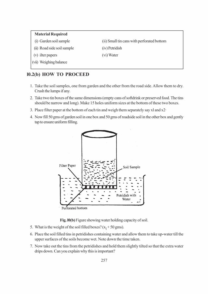
257
Material Required
(i) Garden soil sample (ii) Small tin cans with perforated bottom
(ii) Road side soil sample (iv) Petridish
(v) ilter papers (vi) Water
(vii) Weighing balance
l0.2(b) HOW TO PROCEED
1. Take the soil samples, one from garden and the other from the road side. Allow them to dry.Crush the lumps if any.
2. Take two tin boxes of the same dimensions (empty cans of softdrink or preserved food. The tinsshould be narrow and long). Make 15 holes uniform sizes at the bottom of these two boxes.
3. Place filter paper at the bottom of each tin and weigh them separately say xl and x2·
4. Now fill 50 gms of garden soil in one box and 50 gms of roadside soil in the other box and gentlytap to ensure uniform filling.
Fig. l0(b) Figure showing water holding capacity of soil.
5. What is the weight of the soil filled boxes? (xl + 50 gms).
6. Place the soil filled tins in petridishes containing water and allow them to take up-water till theupper surfaces of the soils become wet. Note down the time taken.
7. Now take out the tins from the petridishes and hold them slightly tilted so that the extra waterdrips down. Can you explain why this is important?

258
8. Now weigh the tins again say it weighs Y1 and Y2 gms respectively.
9. Now complete the following table and record it in your recod book.
1 2 3 4 5
Soil sample Wt of the Wt of the can Wt of the can Wt of water
empty can (x) after filling after removing retained by
soil it from petri dish soil
x +50 gm y gm y − (x + 50) = z
Garden soil ................... ................... ................... ...................
Road side soil ................... ................... . .................. . ..................
Water holding capacity of soil Z/50 x 100
l0.3(b) RECORDING OF OBSERVATION1. In which of the soil samples did the water reach the top layer earlier?
2. How much time did the water take to rise to the top in the two samples?
3. What was the final weight of the soils after the completion of the experiment?
SampleA................. gm
SampleB ................. gm
4. How much of water was gained bySampleA .................. gmSampleB .................. gm
5. Which of the two samples has a higher water holding capacity, sample A or sample B?
6. What is the nature of soil in the samples? Coarse/fine?SampleA.................. gm.Sample B .................. gm .
7. How do you corelate the water holding capacity with the texture of the soil?
Did the sample with finer texture of soil show higher water holding capacity?
l0.4(b) PRECAUTIONS1. The tins should be weighed accurately.
2. Weighing of the cans after taking out of the petridishes should be done only after the dripning ofwater has stopped.
10.5(b) FOR THE TEACHER1. Collection of soil sample be done tinder the guidance of the teacher.
2. Significance of water holding capacity may be emphasized by the teacher.

259
�� �� ��Exercise
DEMONSTRATION OF OSMOSIS BY POTATO OSMOMETER
Materials move in and out of the cells by different cell processes. Water moves in and out of the cells byosmosis through the cell membrane. This exercise aims at studying the osmosis in detail
OBJECTIVES
After performing this exercise, you should be able to:
● develop a skill to make an osmometer with some plant material such as carrot, potato;
● reason out that cell membrane of the potato cells acts as semipermeable membrane.
11.1 WHAT YOU SHOULD KNOW
An osmometer is used to see the movement of water molecules from the region of higher waterconcentration to the region of lower water concentration through the semipermeable membrane of thecells.
Materials Required
(i) Potato (ii) Sugar Solution (iii) Stand
(iv) Petridish (v) Water (vi) Scalpel
11.2 HOW TO PROCEED
(i) Select a medium-sized potato.
(ii) Peel a potato and cut one end of the potato so that it can stand on its base.
Make a cavity (2 cm broad x 3 cm long) with the help of a scalpel on the upper portion of thepotato.

260
(iv) A measured amount of 10% sugar solution is placed in the cavity of the tuber.
(v) Make the initial level of solution in the cavity with the help of a common pin.
(vi) Place the potato tuber containing sugar solutio~ in a petri dish containing water.
(vii) You can keep the set up for 2-3 hours or even over night.
(viii) Observe the set up after 2 hours and measure and record the level of solution.
(ix) Measure the volume of solution after the experiment is over:
Fig. 11.1 Potato osmometer.
11.3 RECORDING OF OBSERVATIONS
(i) Has the level in the potato cavity (a) remained same, (b) risen, or (c) fallen?
(ii) Explain the reason for your observation made in observation no. (i)
(iii) Name the processes involved in this experiment. (diffusion, osmosis, imbibition, absorption)
(iv) Define or explain the phenomenon concerned with the change of the level of sugar solution in thecavity of potato stated above.
(v) What will happen if sugar solution is kept in the petridish and water in the potato cavity?
11.4 PRECAUTIONS
1. Make a cavity carefully in the potato so that it should deep enough.
2. Initial level of water should be carefully marked.
11.5 FOR THE TEACHER
1. The teacher may demonstrate to students, how to make cavity in the potato tuber.
2. Make sure that the sugar is properly dissolved while preparing its solution before pouring it in thepotato cavity.
3. Mark the initial level of solution in the cavity.

261
�� �� ��Exercise
DETERMINING THE RATE OF PHOTOSYNTHESIS IN AN AQUATICPLANT (HYDRILLA OR ELODEA)
Plants take CO2 and water to produce food in the presence of sunlight. The process is referred to asphotosynthesis. O2 is one of the end products during photosynthesis.
In the present exercise you will study the rate of photosynthesis in an aquatic plant Hydrilla. Rate ofphotosynthesis will be measured by counting the number of bubbles evolved per minute from the cutend of the plant.
OBJECTIVES
After performing this exercise, you should be able to:
● explain that different wave lengths of light affect the rate of photosynthesis;
● explain that release of O2 during the day indicates that photosynthesis is taking place,
● argue that it is therefore one of the reasons to suggest that during night one should not sleep underthe trees because at night there is photosynthesis and therefore, no O2 is evolved but only CO2is released in respiration;
● explain giving one reason why during the day, one feels fresh under the tree; (that is because ofoxygen given out by the trees)
● give reason why aquatic plants are best suited for such experiments.
12.1 WHAT YOU SHOULD
1. In the presence of light, green plants take in CO2 and water and synthesize sugar and liberate O2in the process of photosynthesis.

262
2. Light and carbon dioxide are two important factors which control the rate of photosynthesis.
Materials Required
(i) Water (ii) Sodium bicarbonate (iii) Glass rod (v) Hydrilla plants,
(vi) Glass jar (12" by 5") or xuide-mouth bottle
(vii) Stop watch with seconds hand
12.3 HOW TO PROCEED
(i) Collect some Hydrilla plants from a nearby pond. May be your school centre has an aquariumcontaining Hydrilla. It is a free floating green plant with several leaves arising in whorls at thenodes ..
(ii) Take a big bucket full of water and leave the Hydrilla plants in it.
(iii) Select a healthy twig and tie it to a glass rod in such a way that the cut end of the stem is facingupwards as shown in the Fig. 12.1. It must remain inside the water to prevent any air getting intothe xylem at the cut ends of the twig.
Fig. 12.1 Experimental set up for rate of photosynthesis.
(v) Now introduce the Hydrilla plant which is tied to a rod, inside the jar filled with water.

263
IMPORTANT
The Hydrilla plant must always remain submerged in water.
(vii) Add a pinch of sodium bicarbonate (NaHCO3) to the water which will provide CO2 to the plant.
(viii) What do you observe at the cut end of stem of Hydrilla twig? You will find that air bubbles arecoming out.
(ix) Keep the set up in full sunlight and take five readings by counting the number of bubbles perminute using a stop watch.
(x) Remove the set up in the shade and count the number of bubbles per minute using a stop watch.
12.4 OBSERVATION AND DOCUMENTATION
Observation
No. of observation No of bubbles given out/minute
Full Under the shade Meansunlight of a tree
1 .
2
3
4
5
(i) In which light the number of bubbles evolved is more?
.............................................................................................................................................
(ii) Why is it that under the tree shade there is less number of bubbles per minute?
.............................................................................................................................................
(iii) Name the gas which is evolved in the form of bubbles.
.............................................................................................................................................
(iv) What is the source of the gas mentioned in item no. (iii) above
.............................................................................................................................................

264
(v) Name the process involved in this experiment.
.............................................................................................................................................
(vi) What does the rate of bubbles given out indicate?
.............................................................................................................................................
(vii) Can we replace Hydrilla by any grass or any common plant like balsam or rose
Yes/No ............................... and why?
12.5 PRECAUTIONS
1. Set up the apparatus very carefully.
2. Never permit air bubbles to enter the xylem vessels of Hydrilla. Therefore always keep the twigssubmerged in water.
3. Do not damage the plant while tying to the grass rod.
12.6 FOR THE TEACHER
1. Teacher may help the student to select a healthy twig of Hydrilla.
2. Student be helped in recording the observation and counting of bubbles.

265
�� �� ��Exercise
STUDY THE STRUCTURE AND GERMINATION IN GRAM ANDBEAN SEEDS
13(A) STRUCTURE
All seeds have the same function i.e. to produce a new plant. For this they have an embryo, but theyalso have some other parts. This exercise is intended to make you study by yourself the detailedstructure of the two common seeds gram and bean. In the dry condition they are available throughoutthe year.
OBJECTIVES
After performing this exercise you should be able to :
● identify the different parts of the seed;
● highlight the characteristics of each component of the seed;
● justify the classification of the two prescribed seeds as dicotyledonous;
● make a temporary mount of the embryonal axis;
● identify the embryonal axis and its parts such as epicotyl and hypocotyl regions;
● identify two basic patterns of germination like epigeal and hypogeal.

266
13.1 WHAT YOU SHOULD KNOW
1. Seed is a reproductive part.
2. Seed contains an embryo consisting of plumule and radicle.
3. Cotyledons usually store food, and are the first leaves in same seeds.
4. Seeds are classified as monocotyledonous (single cotyledon) and dicotyledonous (two cotyledons).
Material Required
(i) Seeds of gram/bean/castor (iii) Watch glass/Petridish
(ii) Dissecting microscope/hand lens (iv) Needles
(v) Ice cream cups (vi) Soil
13.2 HOW TO PROCEED
Place the seeds in water in a petridish and leave them as such for about 24 hours. You will find that the
seeds are somewhat swollen and that their coverings have become soft.
A. Gram and Bean
(i) Pick up one large sized soaked seed and place it in a watch glass or on a slide
(Fill up observation NO.1)
(ii) Keep another watch glass ready with some water in it to keep the embryonal axis.
(iii) Remove the outermost covering of the seed, i.e the seed coat, with the help of fine” needlestaking care that the underlying parts are not damaged, and the cotyledons are intact (Fill upObservation No.2).
(iv) Gently open out the two cotyledons from their most convex side taking care. That they do notseparate totally (Fill up Observation NO.3).
(v) Observe the point of attachment of the two cotyledons with the embryonal axis.
(vi) With the help of fine needles separate the embryonal axis by breaking the point of attachmentwith the cotyledons.
(vii) Place the embryonal axis in the other watch glass containing some water (Fill up ObservationNo.4).

267
Fig. 13.1 Structure of gram and bean seeds
13.3 RECORDING OF OBSERVATION
Observation No. 1
(i) What is the shape of the seed (somewhat conical, rounded or kidney shaped)
.............................................................................................................................................
(ii) Do you find any markings on the surface? Yes/No : ................................................................
(iii) If yes, describe the markings.
.............................................................................................................................................
(iv) Can you make out the point by which the seed was attached to the parent fruit?
Yes/No : ..............................................................................................................................
If yes, what is its name
.............................................................................................................................................
Observation No.2
(i) How many cotyledons are there?
.............................................................................................................................................

268
(ii) What is the colour of the cotyledons?
.............................................................................................................................................
Observation No.3
(i) What is nature of the outer surface of the cotyledons? (Convex, concave or flat)
.............................................................................................................................................
(ii) What is the shape of the inner surface of the cotyledons? (Convex, concave or flat)
.............................................................................................................................................
Observation No.4
(i) How many distinct parts do you see in the embryonal axis? (One, two, three or more)
.............................................................................................................................................
(ii) Which parts of the embryonal axis will form
(a) Shoot ..............................................................................................................................
(b) Root ..............................................................................................................................
13B GERMINATION
Embryo lies dormant in the seed but when supplied with moisture and optimum temperature, the embryobecomes active and grows and develops into a small seedling. The process by which the dormantembryo becomes active and grows out of the seed coat and establishes itself as a seedling is calledgermination.
OBJECTIVES
After performing this exercise, you should be able to :
● develop the skill of germinating the seeds under optimum conditions;
● identify the two basic patterns of germination: epigeal and hypogeal;
● identify the embryonal axis and its parts such as epicotyl and hypocotyl regions.
13.4 HOW TO PROCEED
(i) Take two clean and empty ice cream cups of about 6 cm diameter and fill them with soil.
(ii) Take six dry seeds each of gram and bean and sow them in the soil in the cups.
(iii) Make sure to keep the soil moist althrough the experiment.
(iv) Note down the time when the first pair of leaves emerge.

269
(v) In case the cotyledonary leaves do not come out of the soil, dig the seeds out to see the conditionof the cotyldedons.
(vi) Observe and study the structure of the first pair of leaves very carefully.
(a)
(b)
Fig. 13.2 Seed germination of bean and gram seeds
13.5 RECORDING OF OBSERVATIONS
1. Bean as well as in gram seeds which part emerged first from the soil, radical/plumule/ cotyledons?
.............................................................................................................................................

270
2 What was the nature of the first leaf formed in (a) gram and (b) bean?
.............................................................................................................................................
3. What happen to the cotyledons in the (a) gram needs and (b) bean seeds. You may dig out thegerminating seeds to see the condition of the cotyledons.
.............................................................................................................................................
4. Which portion of the embryonal axis grow faster epicotyl/hypocotyl in (a) and (b)?
.............................................................................................................................................
5. What type of germination is found in (a) and in (b)?
.............................................................................................................................................
6. What is the major difference you find in the first pair of leave in the two types of seedlings andwhy?
.............................................................................................................................................
13.6 PRECAUTION
1. Make sure to keep the soil wet through the experiment.
2. While digging the soil to examine the condition of the cotyledons, do not damage the seeds .
13.7 FOR THE TEACHER
1. Student should take help of the teacher to identify different parts of a seed.
2. Students may take help from the teacher to select out right kind of seeds to study epigeal andhypogeal germination.
3. Teacher may kindly emphasize the significance of seed as a reproductive unit or a propagativebody.

271
�� �� ��Exercise
TO DEMONSTRATE THE RELEASE OF CO2 DURINGGERMINATION OF SEEDS
All living beings respire whether a developing baby plant (germinating seeds) or a developing humanfoetus, or a single cell. During respiration oxygen is taken in while carbon dioxide is liberated which canbe demonstrated by the present exercise.
OBJECTIVES
After performing this exercise you should be able to:
● develop a skill to set up an apparatus to perform this exercise;
● reason out why germinating seeds and not dry seeds are selected;
● explain that the rate of respiration is higher in germinating seeds than in non-germinating ones, asthe rate of growth is faster.
14.1 WHAT YOU SHOULD KNOW
1. All living being respire take in of O2 from the inspired air and give out CO2 in the expired air.
2. Inspiration and expiration together constitute breathing.
3. 02 taken in is used for oxidation of food to release energy-Cellular respiration.

272
Materials Required
(i) Conical flask-250 ml, capacity (v) Small bottle (4 cm x 3/4 cm)
(ii) One holed rubber cork (vi) Thread
(iii) Glass-tube bent twice at right angles
(vii) KOH-pellets (caustic or potassium hydroxide)
(iv) Beaker
(viii) Gram seeds/Moong seeds/Wheat grains
14.2 HOW TO PROCEED
(i) Take about 25 gms of gram seeds and soak them overnight in 1/2 a beaker of water.
(ii) Next day decant the water and wrap the seeds in a wet cloth.
(iii) After one or two days, open the cloth and look at the seeds.
(iv) The needs have sprouted or germinated (The radicle and plumule have appeared)
(v) You may use the same method to germinate moong seeds and wheat grains and use them inplace of gram seeds.
Material is now ready to proceed further
(vi) Take a dry conical flask and put sufficient number of germinated seeds into it, so as to cover thebase of the flask. (Two to three layers of germinated seeds).
(vii) Insert a one-holed rubber cork in the mouth of the conical flask.
(viii) Take a small test-tube and put 5 to 6 pellets of KOH (Potassium hydroxide).
(ix) Tie the test-tube with a piece of thread and hang it as shown in the diagram.
Fig. 14.1 Experimental set-up

273
(x) Introduce one end of the bent glass tube into the conical flask through the cork.
(xi) The end of the tube must be slightly away from the seeds.
(xii) Dip the other end into a beaker of water coloured with a drop of saffranin.
(xiii) Mark the initial level of water inside the tube.
Your experimental set-up is now ready for observation
(xiv) Leave your set -up and observe the level of the water after every half an hour. Now turn to yourwork-sheet and fill up observation 1.
14.3 RECORDING OF OBSERVATION
Observation 1
(i) Why do we take germinating seeds for this experiment and not the dry seeds?
.............................................................................................................................................
(ii) Can we take young, floral buds instead of germinating seeds? Yes/No
.............................................................................................................................................
(iii) If your answer is ‘Yes’ what precaution to be taken?
.............................................................................................................................................
(iv) Why do we take KOH pellets inside the conical flask?
.............................................................................................................................................
(v) Why is the other end of the tube dipped in beaker of water?
.............................................................................................................................................
(vi) Does the level of water in the tube (a) remain same, (b) rise or (c) fall?
.............................................................................................................................................
(vii) State a suitable reason for your answer in Q, No. (vi)
.............................................................................................................................................
(viii) Name the process that your experiment has demonstrated.
.............................................................................................................................................
(ix) Define or explain the process, mentioned in answer no. (viii)
.............................................................................................................................................
(x) Can you perfom the experiment with boiled seeds? If not why not?
.............................................................................................................................................

274
14.4 PRECAUTIONS
(i) The cork of the flask should remain air tight.
(ii) The KOH pellets should not come in contact with the germinating seeds.
14.5 FOR THE TEACHER
1. Students may be explained the significance of the process of respiration.
2. Students should be explained the metabolites the seeds and grains have
3. Teacher may explain the significance of oxygen in the process of respiration.

275
�� �� ��Exercise
TO STUDY ABOUT THE ACTION OF SALIVARY AMYLASE ONSTARCH
Enzymes are involved in major physiological processes and biochemical reactions in the living bodysystems such as digestion, cellular respiration, biosynthesis etc. Salivary amylase is present in oursaliva and is an important enzyme for digestion in the mouth.
OBJECTIVES
After performing this exercise, you should be able to:
● reason out that
(i) enzymes are specific for specific biochemical reactions; (ii) act best at optimum temperature andPH;
● develop skill to prepare different solutions of specific concentration;
● show that salivary amylase acts best on cooked starch.
15.1 WHAT YOU SHOULD KNOW
1. Saliva is the secretion of three pairs of salivary glands opening into the buccal cavity of humans.
2. Saliva is a mixture of salivary amylase, mucin, minerals and water.
3. Salivary amylase is the first digestive enzyme acting on starch.

276
Materials Required
(i) Test-tubes (vii) Starch powder
(ii) Test-tube stand (viii) Iodine
(iii) Beakers (ix) Benedicts reagent
(iv) Burner (x) Pipette
(v) Measuring cylinder (xi) Water bath
(vi) Physical balance (xii) Thermometer
Note: Starch solution and iodine solution to be prepared one day before the experiment.
15.2 HOW TO PROCEED
A. Preparation of Starch Solution Note: Starch is soluble only in hot water.
(i) Weigh one gram of starch and dissolve it in 10 ml hot (boiling) distilled water.
(ii) Keep it aside.
(iii) Heat 90 ml of distilled water in a conical flask (85°C-95°C).
(iv) When the air bubbles form, remove the flask from the source of heat.
(v) Gradually transfer the prepared starch to this hot water.
(vi) Shake it thoroughly and leave it overnight.
(vii) Cork the conical flask containing starch solution.
This is 1 % starch solution
B. Preparation of Iodine solution
(i) Dissolve 1 gm. of iodine and 2 gms. of potassium iodide in 100 ml of water in a beaker.
(ii) Pour it in a bottle and cork it.
Action of salivary amylase on starch
(i) Rinse your mouth with warm water. Make sure that no particle is sticking in your teeth.
(ii) Chew a piece of paraffix wax, to get saliva collected in your mouth. Chewing enhances secretionof saliva.
(iii) Collect your saliva in a test-tube (spit it into a test-tube).
(iv) Filter the saliva through a thin layer of moistened cotton to collect frothless, clear saliva in anothertest-tube (Fig. 15.1)
(v) Take two test -tubes A and B. Pour 1 ml. of 1 % starch solution in both A and B. Add a dropof iodine in A.
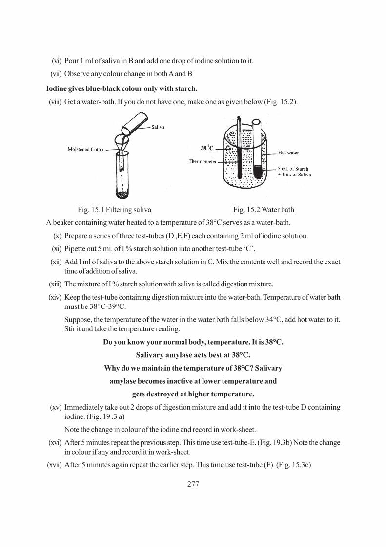
277
(vi) Pour 1 ml of saliva in B and add one drop of iodine solution to it.
(vii) Observe any colour change in both A and B
Iodine gives blue-black colour only with starch.
(viii) Get a water-bath. If you do not have one, make one as given below (Fig. 15.2).
Fig. 15.1 Filtering saliva Fig. 15.2 Water bath
A beaker containing water heated to a temperature of 38°C serves as a water-bath.
(x) Prepare a series of three test-tubes (D ,E,F) each containing 2 ml of iodine solution.
(xi) Pipette out 5 mi. of I % starch solution into another test-tube ‘C’.
(xii) Add I ml of saliva to the above starch solution in C. Mix the contents well and record the exacttime of addition of saliva.
(xiii) The mixture of I % starch solution with saliva is called digestion mixture.
(xiv) Keep the test-tube containing digestion mixture into the water-bath. Temperature of water bathmust be 38°C-39°C.
Suppose, the temperature of the water in the water bath falls below 34°C, add hot water to it.Stir it and take the temperature reading.
Do you know your normal body, temperature. It is 38°C.
Salivary amylase acts best at 38°C.
Why do we maintain the temperature of 38°C? Salivary
amylase becomes inactive at lower temperature and
gets destroyed at higher temperature.
(xv) Immediately take out 2 drops of digestion mixture and add it into the test-tube D containingiodine. (Fig. 19 .3 a)
Note the change in colour of the iodine and record in work-sheet.
(xvi) After 5 minutes repeat the previous step. This time use test-tube-E. (Fig. 19.3b) Note the changein colour if any and record it in work-sheet.
(xvii) After 5 minutes again repeat the earlier step. This time use test-tube (F). (Fig. 15.3c)

278
Note the change in colour if any and record it in work-sheet.
(a) (b) (c)
Addition of digestion mixture Addition of digestion mixture Addition of digestion mixtureimmediately after keeping in after 5 minutes after 10 minutes.
water bath
Fig. 15.3
Clue: Some chemical reaction must have taken place during this time.
(xviii) Keep all the three test-tubes (D,E and F) and compare the colours and fill up Observation 1.
15.3 CONCLUSION
Salivary amylase of saliva acts on starch and converts it into sugar. During this chemical action someintermediate substances like dextrins are formed. Dextrins give reddish brown colour with iodine.
15.4 RECORDING OF OBSERVATION
Observation 1
(i) Why do you rinse your mouth before collecting saliva in a test tube?
.............................................................................................................................................
(ii) Did you find any change in colour in test -tube A?
If yes, what was the colour?
.............................................................................................................................................
(iii) Did you find any change in colour in test- Tube B? Yes/No
.............................................................................................................................................
(iv) What was the significance of keeping the digestion mixture in water-bath at a temperature of38°C.
.............................................................................................................................................

279
(v) Mention the change in colour of the contents in test-tubes - D,E,F,
.............................................................................................................................................
Change in colour
(i) Test-tube D...................... Why/ ...................................
(ii) Test-tube E...................... Why/ ...................................
(iii) Test-tube F Why/ ...................................
(Hint :- Starch may be undigested, partially digested, completely digested)
(vi) How much time has your saliva taken to convert starch into sugar?
.............................................................................................................................................
15.5 PRECAUTIONS
1. Rinse your mouth before collecting saliva.
2. Prepare the solutions carefully.
3. Make sure the required temperature is maintained in the water bath.
15.6 FOR THE TEACHER
Please ensure that the student observes closely and times changtes in the various test tubes.

280
���������� � � � � �� � � � � #������������ #������������ #������������ #������������ #������������ !!!!!��Exercise
TO STUDY THE DEVELOPMENTAL STAGES IN THE LIFE CYCLEOF DROSOPHILA BY PREPARING A CULTURE
The progress in genetics has been largely due to experiments with common red-eyed fruit fly (Drosophilla)hovering over fruits. It is easily available and easily cultured. The generation time (time to completedevelopment is also short). It has conspicuous stages carvae and pupa observing the eggs hatch intolarvae of then into adults through pupal stage is a delight to watch.
OBJECTIVES
After preparing a culture of Drosophila, you should be able to :
● prepare the culture medium;
● crop this from the fruit shop;
● transfer flies from one bottle to another;
● identify the various stages of life history.
Materials Required
(i) Empty jam bottle or milk bottle (ii) agar (iii) yeast
(iv) sugar, (v) corn flour, (vi) propionic acid,
(vii) banana (viii) water (ix) brush
16.1 WHAT YOU SHOULD KNOW
● Culturing of an organism in laboratory is required to study the behaviour, genetics cytology andevolution purpose.

281
● Growing large population of organisms in the laboratory by providing space and nutrition istermed culturing.
● For research work, few organism are collected from nature or brought from dealer and maintainedand grown and multiplied on a large scale.
● In school laboratories. Drosophila is cultured on a small scale for laboratory use by the student.
16.2 HOW TO PROCEED
Drosophila, the fruit fly can be cultured by the following method:
1. Clean the empty jam bottle or milk bottle and keep in boiling water for 4-5 minutes.
2. Dissolve one gram of agar in water.
Fig. 16.1a Drosophila culture
3. Add one gram of yeast, 5 grams of sugar and 7.5 grams of conflour to the above solution.
4. Heat the mixture till it is semi solid.
5. Transfer it into the empty and clean jam bottle.
6. Add a drop of propionic acid to it. The culture bottle is ready.
7. Put on overripe banana in an empty and clean bottle. Place it at a fruit shop. Soon red eyed fruitflies will come into the bottle.
8. Bring the bottle containing fruit flies to your place and transfer the fruit flies into the culture bottle.Note the date and time.
9. Observe the tiny, red eyed fruit flies daily and record your observations.
10. Note the changes they undergo from egg to larva, larva to pupa and pupa to adult.
11. Draw diagrams of each stage.
12. Do not forget to write the date and time of each observation.
16.3 RECORDING OBSERVATIONS
Write the procedure of making the culture and obtaining this to lay eggs.

282
Draw labelled diagrams of the stages of life history that is adult male and female Drosophila, eggs,larvae of any in bottle or pupa.
Note: Drosophila flies move up into the empty bottle if you place it upside down above the culturebottle
16.4 PRECAUTIONS
1. The nutrient medium should not become hard
2. Care should be taken while transferring flies
3. Close observation is needed to see the various larval stages or larval instars as they grow in size.
16.5 FOR THE TEACHER
The students might need help in transferring the flies. If an inverted jam bottle of same size is kept overculture bottle. This easily fly into the upper bottle.

283
���������� � � � � �� � $% "!� � $% "!� � $% "!� � $% "!� � $% "!A PROJECT TO STUDYTHE GROWTH PATTERN OF MONEY PLANT
Growth is an essential character of life or living organisms. Growth may be defined as all permanentchange in sizes. When growth occurs in plants, it organs increase in number and size. Thus growthplants, its organs increase in number and size. Thus growth is a vital process which brings about apermanent change in any plant or its part in respect to size, form, weight and volume.
OBJECTIVES
After completing this project, you should be able to
· know and differentiate between temporary increase due to water absorption and permanentincrease in size and number of plant organs.
· develop the skill of using methods to measure length and size of roots, stems and leaves.
· learn the technique of measuring number and size of leaves.
· learn to draw a graph to show the growth pattern of various organs of the plant.
Material Required
(i) Discarded bulb or jam bottle (ii) money plant (iii) water
(iv) thread (v) scale (vi) graph paper, pencil
16.1 WHAT YOU SHOULD KNOW
1. Growth is a permanent change in size and weight of any organism.
2. In growth of a whole organism or a part of an organism, like the twig of a money plant can bemeasured by vaious methods.
3. Measurement of length of the internodes and the size and number leaves can be recorded everyday at the same time to determine the growth pattern of money plant.
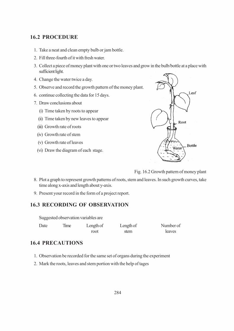
284
16.2 PROCEDURE
1. Take a neat and clean empty bulb or jam bottle.
2. Fill three-fourth of it with fresh water.
3. Collect a piece of money plant with one or two leaves and grow in the bulb/bottle at a place withsufficient light.
4. Change the water twice a day.
5. Observe and record the growth pattern of the money plant.
6. continue collecting the data for 15 days.
7. Draw conclusions about
(i) Time taken by roots to appear
(ii) Time taken by new leaves to appear
(iii) Growth rate of roots
(iv) Growth rate of stem
(v) Growth rate of leaves
(vi) Draw the diagram of each stage.
Fig. 16.2 Growth pattern of money plant
8. Plot a graph to represent growth patterns of roots, stem and leaves. In such growth curves, taketime along x-axis and length about y-axis.
9. Present your record in the form of a project report.
16.3 RECORDING OF OBSERVATION
Suggested observation variables are
Date Time Length of Length of Number ofroot stem leaves
16.4 PRECAUTIONS
1. Observation be recorded for the same set of organs during the experiment
2. Mark the roots, leaves and stem portion with the help of tages

285
16.5 FOR THE TEACHER
1. Teacher may explain to the students that temporary increase in size or volume of certain organdue absorption of water should not be confused with real growth.
2. Students should be helped while recording the length with the help of thread.
286
���������� � � � � �� � $% !� � $% !� � $% !� � $% !� � $% !The books are kept in the libraries in a classified manner, so that it becomes easier for us to find aspecific book when we need it. The same idea applies to systems guiding us about living world. Plantsare kept in dry conditions mounted on hard sheets of paper, in a classified manner in a herbarium.Preparation of a plant to be kept in a herbarium is an important technique.
OBJECTIVES
After performing the exercise, you should be able to :
● develop the skill of collecting plants for their study;
● prepare a plant for mounting on herbarium sheets;
● learn the technique of classifying plants.
Material Required
(i) A gardener’s knife (ii) a plant press blotting papers or news papers
(iii) trowel (iv) herbarium sheets
(v) tape (vi) pen
(vii) plastic bags (ix) tags
(viii) water (x) labels.
16.1 WHAT YOU SHOULD KNOW
1. A herbarium is defined as a collection of plants that have been dried pressed and preserved onsheets.
2. Dried plants are classified and arranged for future reference in taxonomic studies.
3. Plants should be collected from various localities for preparation of herbarium.

287
16.2 PROCEDURE
1. Collect 10 to 15 plants of different types from various localities with the help of knife and trowel.
2. The plants should be from at least five different groups.
3. The plants should be moistened with water and kept in the plastic bags during collection.
4. At the time of collection, the plant specimen should have all the parts such as stem, root and leaf.
5. Name of location, from where the specimen has been collected, should be taggd with it.
6. The collected plant should be spread evenly between the sheets of blotting paper or newspapers.
7. Then the plant should be pressed with the help of a plant press. If the plant press is not available,then some other heavy objects having plane surface can be used for the purpose.
8. While pressing, care must be taken that the parts of the plant do not overlap and the pressure isapplied uniformly on the entire plant
9. The plant should be kept under some heavy weight for about three days.
10. The plant is taken out of the sheets, that is the sheets should be blotting paper or newspaperschanged for about three days. The same procedure is followed with other plant specimenssimultaneously.
11. Now, the dried specimens are mounted on the herbarium sheets/big drawing sheets with the helpof tape.
12. Only one specimen should be mounted on one herbarium sheet.
13. On each sheet the following detail should be given on the lower right hand corner.
(i) The site of collection
(ii) Date of collection
(iii) Name of the plant
(iv) Family
(v) Ecological and morphological note (vi) Habitat
(vii) Name of the collector
14. Herbarium sheets should be preserved safely with moth balls/naphthalene balls etc.
15. These sheets should be presented in the form of a file.

288
1. The site of collection................................................
2. Date ‘of collection ................................................
3. Name of the plant ................................................
4. Family ................................................
5. Ecological and morphological note ........................
6. Habitat................................................
7. Name of the collector ........................
16.3 PRECAUTIONS
1. If a plant is too large then a 12 inch long flowering twig can be collected and identified
2. The leaves must be pressed in a manner that they do not overlap and get wrinkled.
3. make sure that the plants contain flowers
16.4 FOR THE TEACHER
1. Learners should be helped in identifying plants
2. Significance of taxonomic classification be explained.