Biology and comparative anatomy of Divariscintilla yoyo and D. troglodytes, two new species of...
21
MALACOLOGIA, 1989, 31(1): 175-195 BIOLOGY AND COMPARATIVE ANATOMY OF DIVARISC/NTILLA YOYO AND D. TROGLODYTES, TWO NEW SPECIES OF GALEOMMATIDAE (BIVALVIA) FROM STOMATOPOD BURROWS IN EASTERN FLORIDA Paula M. Mikkelsen 1 & Rudiger Bieler2 ABSTRACT Two new galeommatid bivalves, Divariscintilla yoyo and D. troglodytes, are described as commensals in burrows of the stomatopod Lysiosquilla scabricauda from central eastern Flor- ida. They are remarkable in their snail-like appearance and behavior, due to elaborately orna- mented pallial layers enclosing the shell, and their ability to actively crawl on a highly mobile foot. Both are simultaneous hermaphrodites, brooding their larvae in the suprabranchial chamber prior to release of straight-hinged veligers. The two new species differ from one another in shell morphology, the number of secretory "flower-like organs," and the nature and ornamentation of the mantle. They differ from the type and only other described species in this genus, D. maoria, primarily in shell characters, namely in anterior (rather than posterior) prolongation, and in the absence of a ventral cleft. The genus Divariscintil/a, previously known only from New Zealand, is redefined with the following diagnostic characters: incompletely internalized shell with anterior or posterior prolongation, species-specific numbers of pallial tentacles and papillae, a two-part foot used in active crawling and "hanging" utilizing both byssus- and byssus adhesive glands, secretory "flower-like organs" on the anterior surface of the visceral mass, eulamellibranch ctenidia with interlamellar and interfilamentary junctions, and simultaneous hermaphroditism with larval brooding. Key words: Divariscintilla, Galeommatidae, Galeommatoidea, systematics, anatomy, Sto- matopoda, commensalism, Florida. INTRODUCTION A wide variety of mollusks are known to associate with other invertebrates in symbi- otic relationships. Galeommatoidean [ = ga- leommatacean] bivalves are among the best known symbionts (Boss, 1965, as Erycina- cea), and are interesting in the anatomical and behavioral modifications associated with their specialized mode of life. These include (1) internalization of the shell by the middle pallial fold, (2) elaboration of this pallial layer by tentacles and papillae, (3) snail-like loco- motion on a highly extensible foot, and (4) the occurrence of hermaphrodites or dwarf males. Anatomical data are available for spe- cies in less than 30 of the approximately 11 O Recent, presumably valid genera (Vokes, 1980; Chavan, 1969). Within the family Galeommatidae Gray, 1840, the monospecific genus Divariscintilla Powell, 1932, was originally based on empty shells of the New Zealand species D. maoria Powell, 1932. Distinguishing shell characters include a deep ventral notch, a strongly ob- lique posterior prolongation, and dentition lim- ited to a small conical tooth in each valve. The anatomy and biology of D. maoria were sub- sequently described by Judd (1971) from specimens found living in the burrows of the stomatopod crustacean Heterosquilla tricari- nata (Claus). A study of organisms associated with the sand-burrowing stomatopod Lysiosquilla sca- bricauda (Lamarck) in shallow waters in eastern Florida has yielded a number of undescribed or poorly known molluscan species. Data on the two species of vitrinellid gastropods in the burrows have appeared elsewhere (Bieler & Mikkelsen, 1988). Five previously undescribed species of galeom- matid bivalves were also encountered, and two, assignable to Divariscintilla, are here described. The data presented here identify anatomical characters of value at the generic level and represent a step toward clarification of the taxonomic disorder in this super- family. Coastal Zone Museum, Harbor Branch Oceanographic Institution, 5600 Old Dixie Highway, Ft. Pierce, Florida 34946, and Dept. of Biological Sciences, Florida Institute of Technology, Melbourne, Florida 32901 U.S.A. 2 Delaware Museum of Natural History, P.O. Box 3937, Wilmington, Delaware 19807 U.S.A. 175
-
Upload
fieldmuseum -
Category
Documents
-
view
2 -
download
0
Transcript of Biology and comparative anatomy of Divariscintilla yoyo and D. troglodytes, two new species of...

MALACOLOGIA, 1989, 31(1): 175-195
BIOLOGY AND COMPARATIVE ANATOMY OF DIVARISC/NTILLA YOYO AND D. TROGLODYTES, TWO NEW SPECIES OF GALEOMMATIDAE (BIVALVIA)
FROM STOMATOPOD BURROWS IN EASTERN FLORIDA
Paula M. Mikkelsen1 & Rudiger Bieler2
ABSTRACT
Two new galeommatid bivalves, Divariscintilla yoyo and D. troglodytes, are described as commensals in burrows of the stomatopod Lysiosquilla scabricauda from central eastern Florida. They are remarkable in their snail-like appearance and behavior, due to elaborately ornamented pallial layers enclosing the shell, and their ability to actively crawl on a highly mobile foot. Both are simultaneous hermaphrodites, brooding their larvae in the suprabranchial chamber prior to release of straight-hinged veligers. The two new species differ from one another in shell morphology, the number of secretory "flower-like organs," and the nature and ornamentation of the mantle. They differ from the type and only other described species in this genus, D. maoria, primarily in shell characters, namely in anterior (rather than posterior) prolongation, and in the absence of a ventral cleft. The genus Divariscintil/a, previously known only from New Zealand, is redefined with the following diagnostic characters: incompletely internalized shell with anterior or posterior prolongation, species-specific numbers of pallial tentacles and papillae, a two-part foot used in active crawling and "hanging" utilizing both byssus- and byssus adhesive glands, secretory "flower-like organs" on the anterior surface of the visceral mass, eulamellibranch ctenidia with interlamellar and interfilamentary junctions, and simultaneous hermaphroditism with larval brooding.
Key words: Divariscintilla, Galeommatidae, Galeommatoidea, systematics, anatomy, Stomatopoda, commensalism, Florida.
INTRODUCTION
A wide variety of mollusks are known to associate with other invertebrates in symbiotic relationships. Galeommatoidean [ = galeommatacean] bivalves are among the best known symbionts (Boss, 1965, as Erycinacea), and are interesting in the anatomical and behavioral modifications associated with their specialized mode of life. These include (1) internalization of the shell by the middle pallial fold, (2) elaboration of this pallial layer by tentacles and papillae, (3) snail-like locomotion on a highly extensible foot, and (4) the occurrence of hermaphrodites or dwarf males. Anatomical data are available for species in less than 30 of the approximately 11 O Recent, presumably valid genera (Vokes, 1980; Chavan, 1969).
Within the family Galeommatidae Gray, 1840, the monospecific genus Divariscintilla Powell, 1932, was originally based on empty shells of the New Zealand species D. maoria Powell, 1932. Distinguishing shell characters
include a deep ventral notch, a strongly oblique posterior prolongation, and dentition limited to a small conical tooth in each valve. The anatomy and biology of D. maoria were subsequently described by Judd (1971) from specimens found living in the burrows of the stomatopod crustacean Heterosquilla tricarinata (Claus).
A study of organisms associated with the sand-burrowing stomatopod Lysiosquilla scabricauda (Lamarck) in shallow waters in eastern Florida has yielded a number of undescribed or poorly known molluscan species. Data on the two species of vitrinellid gastropods in the burrows have appeared elsewhere (Bieler & Mikkelsen, 1988). Five previously undescribed species of galeommatid bivalves were also encountered, and two, assignable to Divariscintilla, are here described. The data presented here identify anatomical characters of value at the generic level and represent a step toward clarification of the taxonomic disorder in this superfamily.
1 ~00\anRl'lef Coastal Zone Museum, Harbor Branch Oceanographic Institution, 5600 Old Dixie Highway, Ft. Pierce, Florida 34946, and Dept. of Biological Sciences, Florida Institute of Technology, Melbourne, Florida 32901 U.S.A. 2Delaware Museum of Natural History, P.O. Box 3937, Wilmington, Delaware 19807 U.S.A.
175

176 MIKKELSEN & BIELER
MATERIAL AND METHODS
Stomatopod burrows in shallow-water sand flats in the Indian River lagoon just inside the Ft. Pierce Inlet, St. Lucie County, eastern Florida (27°28.3'N, 80°17.9'W), were sampled using a stainless steel bait pump ("yabby pump") and sieves of 1-2 mm mesh. Depths during extreme low water ranged from less than 0.5 m to supratidal, wherein the water level lay several centimeters below the level of the sand.
Living clams were maintained in finger bowls of seawater at room temperature (24°C). Behavioral studies were aided by video recordings taken of the living animals in aquaria using a standard commercial 1 /2-inch-format video camera equipped with a macro lens.
Carmine and fluorescein sodium particles aided observation of ciliary action and currents produced by the animals. Relaxation prior to dissection or preservation was most effectively accomplished with menthol crystals, floated on the seawater surface, or with crystalline magnesium sulfate, added directly in small, gradual amounts. Methylene-blue/ basic-fuchsin and neutral red were used to delineate tissues and organs in gross dissections.
For histological serial sections, animals were fixed in either a glutaraldehyde-formalin solution (4% formalin, 2.5% glutaraldehyde in 0.1 M phosphate buffer, pH 7.2) or in 5% buffered formalin (Humason, 1962: 14). Shells were decalcified using either dilute hydrochloric acid (complete decalcification within minutes, however, with bubble production presenting technical histological difficulties) or a 1 % solution of ethylene diamene tetraacetic acid (EDTA, adjusted to pH 7.2; decalcification complete over a period of 5-6 days). Specimens were embedded in paraffin, sectioned at 5-7 µ.m and stained with alcian blue/ periodic-acid-Schiff (PAS), counterstained with Harris' hematoxylin/eosin (Humason, 1962: 125, 269, 298), hereafter referred to as APH. Staining reactions described in the text refer to this method unless otherwise noted. Colors referred to in the text are supplied for future use, e.g., to infer homologies of the various glands. Other similarly prepared specimens were stained with hematoxylin/eosin. The section in Fig. 23 was fixed in Karnovsky's fixative (Karnovsky, 1965), postfixed in 1% osmium tetroxide in a phosphate buffer, dehydrated through an ethanol propy-
lene oxide series, embedded in Epon-812, sectioned at 1 µ.m and stained with Richardson's stain (Richardson, et al., 1960). Photomicrographs of sections were taken either with a Zeiss Photomicroscope-3 or an Olympus BH-2 stereomicroscope fitted with an Olympus OM-2 camera.
For scanning electron microscopy (SEM), partially dissected preserved specimens were passed through an ethanol-to-acetone series and critical-point dried. These and air-dried shells were coated with gold/palladium and scanned using a Zeiss Novascan-30.
All cited anatomical measurements were taken from specimens of average size (approximately 10-15 mm mantle length). Because of the extreme expansivity and contractility of the mantle, it is difficult to accurately measure "length" of a living animal of this type. Approximate mantle length was measured along an anteroposterior axis from an animal in normal crawling or hanging posture; throughout the text, this is expressed as "relaxed" and does not refer to any chemical treatment of the animals. Measurements of type specimens refer to preserved mantle lengths. Shell length is expressed as the maximum dimension, i.e. an oblique anteroposterior length.
Cited institutions are (* indicates location of type and other voucher material):
AMNH- American Museum of Natural History, New York
*DMNH- Delaware Museum of Natural History, Wilmington
HBOI- Harbor Branch Oceanographic Institution, Ft. Pierce, Florida
*IRCZM-lndian River Coastal Zone Museum, HBOI
*MCZ- Museum of Comparative Zoology, Harvard University, Cambridge, Massachusetts
SMSLP- Smithsonian Marine Station at Link Port, Ft. Pierce, Florida
*USNM- National Museum of Natural History, Smithsonian Institution, Washington, D.C.
TAXONOMIC DESCRIPTIONS
Family GALEOMMATIDAE GRAY, 1840
Gray (1840: 154) introduced the family name Galeommidae for Galeomma Turton, 1825 (erroneously spelled "Galeomidae" by

i. BIOLOGY OF DIVARISC/NTILLA 177
Gray, 1842: 78). It has been used in that form by various authors (e.g. H. & A. Adams, 1857; Tyron, 1872; Kisch, 1958). Dall (1899: 875) emended the spelling without explanation to Galeommatidae, and it is this form that is now in common use (e.g. Thiele, 1934; Popham, 1939; Vokes, 1980; Chavan, 1969; B. Morton, 1973; Abbott, 1974; Boss, 1982). Dall's action was a justified emendation of an incorrect original spelling [ICZN, 1985: Art. 29(b)(i), 32(c)(iii)] because the Greek noun oµ,µ,a provides the stem ommat- for the formation of the family name. As a justified emendation, Galeommatidae bears Gray, 1840, as authority and date [ICZN, 1985: Arts. 11 (f)(ii), 19(a)(i)]; the name of the superfamily is accordingly Galeommatoidea [ = Galeommatacea] Gray, 1840. The nominal superfamilies Leptonoidea Gray, 1847; Erycinoidea Deshayes, 1850; and Chlamydoconchoidea Dall, 1889, are here considered junior synonyms.
DIVARISCINTILLA POWELL, 1932: 66.
Type species: Divariscintilla maoria Powell, 1932 (by original designation). Recent, New Zealand.
DIVAR/SC/NTILLA YOYO, SP. NOV. (FIGS. 1, 3, 5, 6, 8-11, 26)
Material examined: Holotype: 5.3 mm [preserved mantle length], USNM 860036. Paratypes (8): 5.8, 4.7 mm, USNM 860037; 8.3, 7.5 mm, MCZ 297406; 6.0, 5.2 mm, IRCZM 064:01721; 5.7, 4.7 mm, DMNH 175516. Total material: 83 specimens: FLORIDA: Ft. Pierce Inlet: March 1987, 4; 2-3 May 1987, 19 (including MCZ paratypes); 24 June 1987, 4; 03 August 1987, 6; 14 August 1987, 7; 31 August 1987, 17 (including USNM, IRCZM, and DMNH type specimens); 28 December 1987, 1; 11 March 1988, 2; 12 April 1988, 13. -Sebastian Inlet: 30 December 1987, 10.
Type locality: Ft. Pierce Inlet, St. Lucie County, Florida, 27°28.3'N, 80°17.9'W, in Lysiosquilla scabricauda burrows on intertidal sand flats with patches of the seagrass Halodu/e wrightii Ascherson.
Diagnosis
Animal translucent white. Mantle thick, surface granular, falling into large creases. Two long "cephalic" pallial tentacles; one very short mid-dorsal tentacle just anterior to ex-
current siphon. Shell wedge-shaped, elongate-pointed anteriorly, with weak internal ribs, length approximately 40% of extended mantle length. "Flower-like organs" on anterior surface of visceral mass ventral to labial palps, numbering 3-7 (usually 5).
Description
External features and mantle: Living extended animal (Fig. 1) 10-15 mm in length, globular in general shape, entirely translucent white, except for dark upper portion of digestive gland. Shell nearly completely enclosed by thick mantle with granular external surface (Fig. 6; formed by middle pallial fold) falling into large creases, and with sparse, minute papillae; mantle thinner, more translucent, and with scattered, relatively larger papillae in smaller (approximately 7 mm) specimens. Anteropedal pallial opening wide, forming extensive anterior cowl (Fig. 1, c). Two long, retractable, "cephalic" tentacles (Fig. 1, cpht) anterodorsally just behind cowl. Very short ( < 1 mm) median pallial tentacle (Fig. 1, mpt) on dorsal midline anterior to excurrent siphon (Fig. 1, exs). Each tentacle with central core of longitudinal muscle and nerve fibers, visible as an inner thread under low magnification. Mantle fused dorsally from edge of cowl to excurrent siphon located posterodorsally and often on conspicuous rounded protuberance (dependent on degree of pallial expansion); fusion interrupted only by small circular (approximately 2 mm diameter) foramen (Fig. 1 , uf) just over umbo of shell. Mantle fused posteroventrally from excurrent siphon to mid-ventral point (Fig. 1, mf) at posterior end of anteropedal opening. Inner pallial fold highly muscular; fibers continuous with muscles of central "core" of tentacles.
Preserved animals characterized by general globular appearance, with retracted cephalic tentacles, mantle-covered shell (with small foramen over umbo), contracted cowl and excurrent siphon. Foot usually completely withdrawn into pallial cavity.
Shell (Figs. 8-11 ). Thin, transparent to translucent white except for yellow prodissoconch and network of opaque white coloration on early portion; equivalve, showing fine growth lines; oval initially, changing to anteriorly elongate-pointed with angulate corners anteriorly and posteriorly near attachment points of adductor muscles; weak internal radial ribs corresponding in placement to weak

178 MIKKELSEN & BIELER
1 2 cpht mpt
ct
37\ ppr ppm
apr~ .. ::. @}-pam
aam~ :i
4 ppm)~ ( ap~·~ ~ppr
aam .,"· .... ,, ~~ ~~pam
FIGS. 1-5. External appearance and internal shell morphology. 1. Divariscintilla yoyo in crawling position, from left side. 2. D. troglodytes, same as Fig. 1. 3. D. yoyo, internal surface of right valve, showing approximate location of muscle insertions. 4. D. troglodytes, same as Fig. 3. 5. D. yoyo, in hanging position, from right side. Scale bars: 1, 2, 5 = 2.0 mm; 3, 4 = 1.0 mm. (aam, anterior adductor muscle; apr, anterior pedal retractor muscle; apt, anterior pallial tentacle; bag, byssus adhesive gland; by, byssus; byg, byssusgland; c, cowl; cpht, cephalic tentacles; ct, ctenidium; exs, excurrent siphon; fl, flower-like organ; ft, foot; mf, point of ventral mantle fusion; mpt, median pallial tentacle; pam, posterior adductor muscle; pef, posterior extension of foot; ppm, pedal protractor muscle; ppr, posterior pedal retractor muscle; ppt, posterior pallial tentacles; sh, shell; uf, umbonal foramen; vgr, ventral groove).
external grooves. Muscle scars indistinct. Hinge (Fig. 10) primarily internal, with weak external ligament, stronger internal resilium and two rudimentary, non-interlocking cardinal teeth; lateral teeth absent. Shell nearly completely enclosed in chamber formed by pallial layers, communicating with exterior via small umbonal foramen (Fig. 1, uf ). Periostracal groove lying between inner and outer pallial folds. Size small in relation to body, extending only over dorsal portion of visceral mass; length approximately 40% of relaxed mantle length. Permanently gaping at 110-1200 angle while relaxed, incapable of closure more than 50°. Periostracum colorless, most evident as periostracal webbing extending between valves, anterior and posterior to hinge, and at periphery of shell. Shell microstructure (Fig. 11) cross-lamellar, with thin homogeneous layer on either side.
Prodissoconch (Fig. 16) approximately 350 µm in length, having distinct prodissoconch I and prodissoconch II stages; prodissoconch I approximately 140 µm in length, with "granulated" surface sculpture and marginal radial ridges; prodissoconch II stage relatively large, sculptured with distinct concentric ridges. Abrupt demarcation between prodissoconch and dissoconch.
Organs of the pallial cavity: Foot (Figs. 1 , 5, 18-19, ft, pef) highly extensible, with hatchet-shaped anterior crawling portion and elongated tubular posterior extension. Anterior portion internally with dorsal haemocoel, sparse longitudinal musculature, and with extensive lateral and ventral external ciliation and accompanying mucous glands (staining turquoise to dark blue in APH). Ventral groove (Figs. 1, 18, 21, vgr) extending from approx-
..
....

·~
.. '
BIOLOGY OF DIVARISCINTILLA 179
FIGS. 6-7. Exterior pallial surfaces (SEM). 6. Divariscintilla yoyo. 7. D. troglodytes; arrows indicate enlarged pallial papillae. Scale bars = 50 µm.
imate midpoint of anterior tip to terminus of posterior extension, heavily ciliated interiorly along entire length. Byssus-gland (Figs. 1, 21, byg) restricted to very small area on either side of ventral groove, a short distance behind anterior end of groove in vicinity of pedal ganglia (see below); closely associated with numerous blood spaces; faintly whitish in living animal, staining dark blue in methylene blue, purple in APH. Posterior extension terminally pointed (Fig. 19), consisting internally largely of longitudinal muscle fibers and connective tissue; proximal half with free edges of ventral groove heavily ciliated externally; external ciliation and accompanying mucous glands disappearing abruptly to leave distal half of posterior extension with ciliation restricted to interior of groove. Byssus adhesive gland [Figs. 1, 22, bag; see Ecology and Behavior (below) for explanation of term] just short of terminus of posterior extension, with internal lamellae surrounding a common lumen; opaque white in living animal, staining dark blue in methylene blue, light purple in APH.
Anterior and posterior adductor muscles subequal, long, of moderate diameter. Attachment ends oval, subequal. Anterior pedal retractor slightly smaller in diameter than, and attaching to shell anterior to, anterior adductor; posterior pedal retractor smaller than, and attaching to shell posterodorsal to, posterior adductor. Very small pedal protractor attaching to shell dorsal to anterior adductor. Integument of visceral mass with numerous
longitudinal muscle fibers, most highly concentrated posteriorly; these continuous with anterior and posterior pairs of pedal retractor muscles, only slightly smaller in diameter than adductor muscles. Small anterior pedal protractor originating ventral to anterior adductor muscle within anterior tissues of digestive gland, passing posterodorsally to insert on shell, dorsal to anterior adductor. Muscles leaving no visible attachment scars on shell; approximate insertion locations shown in Fig. 3.
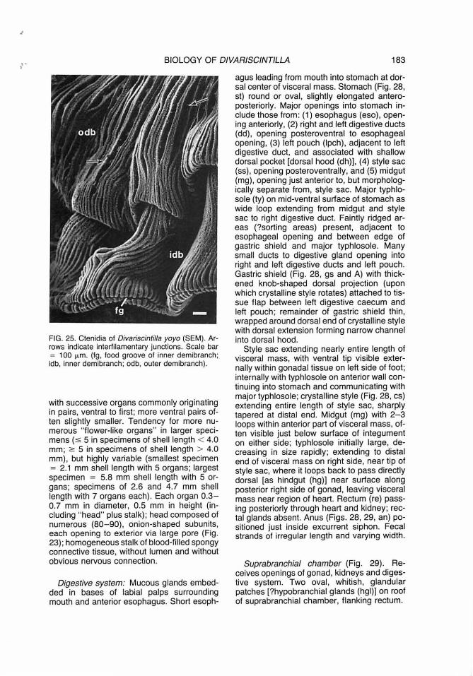
Labial palps (Fig. 28, Ip) large, oval, each with 10- 14 lamellae each side; each pair fused at midline near mouth. Outer palps attached laterally by elongated strip of tissue to inner surface of mantle lining shell; inner palps similarly attached to surface of visceral mass. Cilial currents moving particles oralward on inner palp surfaces, laterally toward ctenidia on outer palp surfaces. Ctenidia (Fig. 25) eulamellibranch, homorhabdic, hanging in loosely pleated folds, with numerous filaments; inner and outer demibranchs on each side with both ascending and descending lamellae. Both demibranchs with well-developed, numerous interfilamentary junctions (Fig. 24, ifj; Fig. 25, arrow) , and evenly distributed, albeit few, interlamellar junctions (Fig. 24, iii). Inner demibranch approximately 50% larger, extending farther ventrally and anteriorly than outer, with food groove (Fig. 25, fg) at free edge; anterior end (with terminus of food groove) extending between labial palps. Ventral tips of anterior filaments of in-
180 MIKKELSEN & BIELER
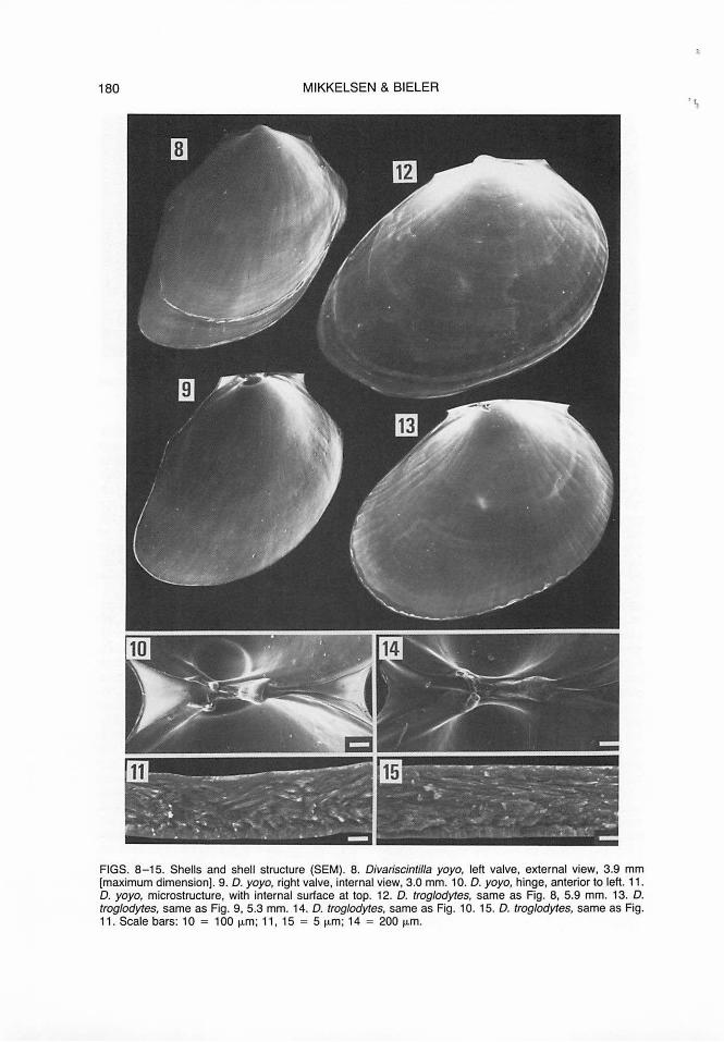
FIGS. 8- 15. Shells and shell structure (SEM). 8. Divariscintilla yoyo, left valve, external view, 3.9 mm [maximum dimension]. 9. D. yoyo, right valve, internal view, 3.0 mm. 10. D. yoyo, hinge, anterior to left. 11 . D. yoyo, microstructure, with internal surface at top. 12. D. troglodytes, same as Fig. 8, 5.9 mm. 13. D. troglodytes, same as Fig. 9, 5.3 mm. 14. D. troglodytes, same as Fig. 10. 15. D. troglodytes, same as Fig. 11. Scale bars: 10 = 100 µm; 11 , 15 = 5 µm; 14 = 200 µm.
...

..
BIOLOGY OF DIVAR/SC/NTILLA 181
FIGS. 16-17. Prodissoconch and larval shell morphologies (SEM). 16. Prodissoconch (Oivariscintilla yoyo); arrow indicates boundary between prodissoconch I and II. 17. Shell of newly hatched larva (0. troglodytes); arrow indicates zone of initial shell formation. Scale bars: 16 = 50 µm; 17 = 1 O µm.
FIGS. 18-19. Foot (SEM). 18. Ventral view of entire foot, showing ventral groove (Oivariscintilla troglodytes). 19. Terminus of posterior foot extension, showing byssus-threads (0 . yoyo). Scale bars: 18 = 200 µm; 19 = 50 µm. (by, byssus; pef, posterior extension; vgr, ventral groove).
ner demibranch "not inserted into a distal oral groove" {Stasek, 1963, Category Ill}; anteroventral margin of inner demibranch fused to inner palp lamella. Outer demibranch shorter, without food groove; margins inserting between inner and outer labial palps, along upper portion of visceral mass, and on inner surface of mantle to posterior end of pallial cavity. Cilial currents as in Fig. 30, moving food particles in food groove and in groove between demibranchs oralward. Tract on inner surface of cowl anterior to palps as exit point for removal of waste particles. No cilial
currents evident on inner pallial surface or surface of visceral mass.
Visceral mass of brownish digestive gland anterodorsally, whitish granular-appearing gonad posteriorly, distally, and, in ripe specimens, overlapping digestive tissue laterally and dorsally; red pedal ganglia visible at distal end of gonad in base of foot.
Cluster of 3-7 {most often 5; x = 4.7, n = 31) nonretractable "flower-like organs" (Figs. 23, 26} originating on anterior surface of visceral mass just ventral to labial palps. Usually arranged as single organ closest to palps,

182 MIKKELSEN & BIELER
vgr
FIGS. 20-24. Internal structure, histological sections. 20. Cross-section at level of esophagus and anterior edge of ctenidia (Divariscintilla yoyo). 21 . Cross-section through foot at region of byssus-gland (D. yoyo) . 22. Longitudinal section through byssus adhesive gland at terminus of posterior foot extension (D. yoyo). 23. Longitudinal thin section through two "flower-like organs" (D. yoyo) . 24. Cross-section of ctenidium showing interlamellar and interfilamentary junctions (D. troglodytes). Scale bars: 20 = 300 µm; 21, 24 = 100 µm; 22, 23 = 50 µm. (bag, byssus adhesive gland; byg, byssus-gland; ct, ctenidium; dg, digestive gland; es, esophagus; gon, gonad; idb, inner demibranch; ifj, interfilamentary junctions; ilj, interlamellar junctions; lam, lamellae of byssus adhesive gland; Ip, labial palp; man, mantle; mg, midgut; mug, mucous glands; mus, muscle fibers; odb, outer demibranch; pef, posterior extension of foot; pg, pedal ganglia; pn, pedal nerve; sh, shell; ss, style sac; vgr, ventral groove).
.. '
BIOLOGY OF DIVARJSCINTILLA 183
FIG. 25. Ctenidia of Divariscintilla yoyo (SEM). Arrows indicate interfilamentary junctions. Scale bar = 100 µm. (lg, food groove of inner demibranch; idb, inner demibranch; odb, outer demibranch).
with successive organs commonly originating in pairs, ventral to first ; more ventral pairs often slightly smaller. Tendency for more numerous " flower-like organs" in larger specimens (s 5 in specimens of shell length < 4 .0 mm; 2: 5 in specimens of shell length > 4.0 mm), but highly variable (smallest specimen = 2.1 mm shell length with 5 organs; largest specimen = 5.8 mm shell length with 5 organs; specimens of 2.6 and 4.7 mm shell length with 7 organs each) . Each organ 0.3-0.7 mm in diameter, 0.5 mm in height (including "head" plus stalk); head composed of numerous (80-90), onion-shaped subunits, each opening to exterior via large pore (Fig. 23); homogeneous stalk of blood-filled spongy connective tissue, without lumen and without obvious nervous connection.
Digestive system: Mucous glands embedded in bases of labial palps surrounding mouth and anterior esophagus. Short esoph-
agus leading from mouth into stomach at dorsal center of visceral mass. Stomach (Fig. 28, st) round or oval, slightly elongated anteroposteriorly. Major openings into stomach include those from: (1) esophagus (eso), opening anteriorly, (2) right and left digestive ducts (dd), opening posteroventral to esophageal opening, (3) left pouch (lpch), adjacent to left digestive duct, and associated with shallow dorsal pocket [dorsal hood (dh)], (4) style sac (ss), opening posteroventrally, and (5) midgut (mg), opening just anterior to, but morphologically separate from, style sac. Major typhlosole (ty) on mid-ventral surface of stomach as wide loop extending from midgut and style sac to right digestive duct. Faintly ridged areas (?sorting areas) present, adjacent to esophageal opening and between edge of gastric shield and major typhlosole. Many small ducts to digestive gland opening into right and left digestive ducts and left pouch. Gastric shield (Fig. 28, gs and A) with thickened knob-shaped dorsal projection (upon which crystalline style rotates) attached to tissue flap between left digestive caecum and left pouch; remainder of gastric shield thin, wrapped around dorsal end of crystalline style with dorsal extension forming narrow channel into dorsal hood.
Style sac extending nearly entire length of visceral mass, with ventral tip visible externally within gonadal tissue on left side of foot; internally with typhlosole on anterior wall continuing into stomach and communicating with major typhlosole; crystalline style (Fig . 28, cs) extending entire length of style sac, sharply tapered at distal end. Midgut (mg) with 2-3 loops within anterior part of visceral mass, often visible just below surface of integument on either side; typhlosole initially large, decreasing in size rapidly; extending to distal end of visceral mass on right side, near tip of style sac, where it loops back to pass directly dorsal [as hindgut (hg)] near surface along posterior right side of gonad, leaving visceral mass near region of heart. Rectum (re) passing posteriorly through heart and kidney; rectal glands absent. Anus (Figs. 28, 29, an) positioned just inside excurrent siphon. Fecal strands of irregular length and varying width.
Suprabranchial chamber (Fig. 29). Receives openings of gonad, kidneys and digestive system. Two oval, whitish, glandular patches [?hypobranchial glands (hgl)] on roof of suprabranchial chamber, flanking rectum.

184 MIKKELSEN & BIELER
FIGS. 26-27. Flower-like organs (SEM). 26. of Divariscintilla yoyo, showing typical number of five, of subequal size. 27. of D. troglodytes, in lateral view. Scale bars: 26 = 100 µm; 27 = 50 µm.
Nervous system (Fig. 31 ). Nervous system of typical bivalve organization with three pairs of ganglia, red in living animal, connected by long commissures. Ganglia relatively large, subequal (length approximately 0.4 mm), cerebropleural ganglia more elongate than others. Cerebropleural ganglia (cplg) lying just anteroventral to anterior adductor muscle, dorsal to esophagus; cerebral commissure (cc) short; each ganglion giving rise to three additional major branches: (1 ) dorsally, common trunk giving rise to cephalic tentacular nerve (ctn) leading to cephalic tentacle, and pallial nerve (pain) leading to ventral shell edge as a continuous cord (to visceral ganglion) with numerous smaller nerves extending into mantle tissue; (2) ventrally, cerebropleural-pedal commissure (cpc) passing between visceral mass and integument along anterior face of foot, penetrating gonadal tissue distally to join with pedal ganglion; and (3) laterally, cerebrovisceral commissure (eve) passing through upper portion of visceral mass to visceral ganglion. Pedal ganglia (Figs. 21, 31, pg) closely fused at midline; showing through integument of foot in living animal as red organ at extreme distal end of gonad; each dorsally receiving cerebropedal commissure from cerebropleural ganglion;
each ventrally giving rise to two anterior and one posterior pedal nerves. Statocyst (Fig. 31 , stc) on posterodorsal face of each pedal ganglion; each with one spherical statolith (stl) 50 µm in diameter. Visceral ganglia (vg) joined together at midline (but not as closely fused as pedal ganglia), lying ventral to heart, posteroventral to posterior adductor muscle; each receiving cerebrovisceral commissure (anterolaterally) and pallial nerve (dorsolaterally) from cerebropleural ganglion; each giving rise ventrolaterally to branchial nerve (with somewhat swollen base), which extends along common axis of inner and outer demibranchs of ctenidium.
Reproductive system: Simultaneous hermaphrodite. Ovotestis white, encompassing most of volume of visceral mass, extending from small portion in umbonal area, down posterior surface of digestive gland, and expanding to surround pedal ganglia and ventral extensions of style sac and intestinal loops. "Spent" appearance sparse, with pedal ganglia fully exposed at terminus, and with silvery ducts clearly visible, packed with mature spermatozoa. Common genital ducts with large ciliated openings, emptying into supra-

.~. BIOLOGY OF DIVARISC/NTILLA 185
29
30
odb'/;;/ ·v •db .
FIGS. 28-31. Anatomical structures in Divariscintilla species. 28. Visceral mass, from right side, showing stomach, opened laterally. Midgut (mg) with four cross-sections, showing reduction of typhlosole. (A) = gastric shield, excised. 29. Roof of suprabranchial chamber, with anterior end up and inner lamellae of right and left inner demibranchs removed. 30. Diagrammatic representation of ctenidial demibranchs in crosssection, showing cilia! currents. 31. Nervous system (semi-diagrammatic). Tentacles with nerves drawn as broken lines present only in D. troglodytes: remainder identical for both species. Sizes of ganglia and lengths of nerves not drawn to scale. Scale bars = 1.0 mm. (an, anus; apn, anterior pedal nerves; aptn, anterior pallial tentacular nerve; bn, branchial nerve; cc, cerebropleural commissure; cpc, cerebropleural-pedal commissure; cs, crystalline style; cplg, cerebropleural ganglion; ct, ctenidium; ctn, cephalic tentacular nerve; eve, cerebropleural-visceral commissure; dd, right and left digestive ducts; dg, digestive gland; dh, dorsal hood; eso, esophageal opening; exs, excurrent siphon; fl, flower-like organs; ft, foot; go, gonadal opening; gon, gonad; gs, gastric shield; hg, hindgut; hgl, ?hypobranchial gland; idb, inner ctenidial demibranch; ki, kidney; kio, kidney opening; Ip, labial palps; lpch, left pouch; mg, midgut; mptn, median pallial tentacular nerve; odb, outer ctenidial demibranch; pain, pallial nerve; pam, posterior adductor muscle; pg, pedal ganglion; ppn, posterior pedal nerve; ppr, posterior pedal retractor muscle; pptn, pallial tentacular nerve; re, rectum; sh, shell; ss, style sac; st, stomach; stc, statocyst; stl, statolith; ty, major typhlosole; vg, visceral ganglion; vm, visceral mass).
186 MIKKELSEN & BIELER
branchial chamber just anterior to visceral ganglia (Fig. 29, go).
Ova small, approximately 20 µm in diameter (maturity not determined, measured in paraffin sections, in posterodorsal region of gonad). Spermatozoa approximately 7 µm in head length (acrosome + nucleus + middle piece); nucleus cylindrical, asymetrical; acrosome subterminal, dish-shaped with central "papilla," tilted approximately 45° from long axis of nucleus; middle piece short; tail long. Gametes and gametogenesis to be described in detail in a paper currently in preparation.
Brooding large number of small larvae for an undetermined period (longest time observed 14 days). Larvae held primarily within outer demibranch, and in suprabranchial chamber where they are circulated via pallial expansions and contractions. During brooding, excurrent siphonal opening constricted by sphincter-like muscles around plug formed by free end of rectum (often expanded into bulb by haemocoelic pressure), allowing digestive processes to continue during brooding and preventing loss of larvae through excurrent siphon. Larvae initially white, turning pink with shell development; released as straight-hinged veligers with apical flagella, 122-138 µm in shell length (x = 131.8 µm, n = 20; Fig. 17). Larval shell with distinct zone of initial shell formation (Fig. 17, arrow). Larvae expelled through excurrent siphon via strong contractions of shell and pallial muscles. Adults brooding larvae collected in May and June 1987, and April 1988.
Circulatory system: Heart just posterior to umbones, within pericardium lined by brownish pericardia! gland. Doughnut-shaped ventricle traversed by intestine; auricles lateral to ventricle, inconspicuous. Blood vessels not evident; major haemocoelic spaces present within foot, tentacles and main axes of demibranchs.
Excretory system: Kidney yellow, ventral to heart, dorsal to visceral ganglia, surrounding pedal retractor muscles on roof of suprabranchial chamber. Paired ciliated renopericardial apertures opening anteriorly into ventral wall of pericardium. Paired renal openings into the suprabranchial chamber large, funnelshaped, adjacent to visceral ganglia.
Distribution: Known only from the type locality, Ft. Pierce Inlet, St. Lucie County, and
one other location approximately 45 km north, Sebastian Inlet, Brevard County, Florida.
Etymology: A noun in apposition from the English vernacular "yo-yo," a child's toy originating in China about 1000 B.C., referring here to the periodic up-and-down motion of the bivalve as it hangs from its byssus-thread The word "yo-yo" is in Tagalog, an Indonesian language, for a similarly constructed, sixteenth-century hunter's weapon made of large wooden disks and twine.
DIVARISCINTILLA TROGLODYTES, SP. NOV. (FIGS. 2, 4, 7, 12-15, 27).
Material examined: Holotype: 7.7 mm [preserved mantle length], USNM 860038. Paratypes (9): 6.7, 4.5 mm, USNM 860039; 6.8, 6.6 mm, MCZ 297407; 7.0, 5.0 mm, IRCZM 064:01722; 4.9, 4.8, 4.5 mm, DMNH 175517. Total material: 87 specimens: FLORIDA: Ft. Pierce Inlet: 10 March 1987, 1: 2-3 May 1987, 12 (including MCZ paratypes); 24 June 1987, 12; 03 August 1987, 4; 14 August 1987, 7; 31 August 1987, 13 (including USNM and IRCZM type specimens); 28December1987, 6; 11 March 1988, 6; 12 April 1988, 13. -Sebastian Inlet: 30 December 1987, 13 (including DMNH paratypes).
Type locality. Ft. Pierce Inlet, St. Lucie County, Florida, 27°28.3'N, 80°17.9'W, in Lysiosquilla scabricauda burrows on intertidal sand flats with patches of the seagrass Halodule wrightii Ascherson.
Diagnosis
Animal translucent yellowish-white. Mantle thin, surface granular with numerous evenly distributed short papillae. Two long "cephalic" tentacles; two short anterior pallial tentacles; three long posterior pallial tentacles surrounding posterodorsal excurrent siphon. Shell oval, elongate-rounded anteriorly, with weak internal ribs, length approximately 50% of extended mantle length. A single "flower-like organ" on anterior surface of visceral mass ventral to labial palps.
Description
External features and mantle: Living extended animal (Fig. 2) 10-15 mm in length, globular to oval in general shape, translucent white to yellowish, except for dark upper portion of digestive gland showing through tis-
BIOLOGY OF DIVAR/SCINTILLA 187
sues. Shell nearly completely enclosed by relatively thin mantle, clearly revealing outlines of shell and ctenidia. Extended mantle somewhat posteriorly elongated, with granular surface and numerous, evenly distributed, short papillae (Fig. 7, arrows). Anteropedal pallial opening and cowl as in Divariscintilla yoyo. Two long, retractable, "cephalic" tentacles anterodorsally just behind cowl; two shorter, retractable anterior pallial tentacles originating laterally to cephalic pair. Posterodorsal excurrent siphon on prominent rounded protuberance; pallial fusion as in Divariscintilla yoyo. Three long posterior pallial tentacles surrounding excurrent siphon: one anterior and mid-dorsal, two lateral. Umbonal foramen (Fig. 2, uf) a transverse slit-like opening, capable of distention during periods of stress to expose nearly entire shell.
Preserved animals with shell nearly completely exposed by retraction of umbonal foramen, retracted pallial tentacles, contracted cowl and excurrent siphon. Foot often anteriorly protruding from pallial cavity.
Shell (Figs. 12-15). Nearly completely enclosed in chamber formed by pallial layers, communicating with exterior via slit-like umbonal foramen (Fig. 2, uf). Shell length approximately 50% of relaxed mantle length, extending over dorsal half of visceral mass, and anterior portion of ctenidia. Permanently gaping at 80-120° angle while relaxed, incapable of closure more than 50°. Shell (Figs. 12-13) thin, transparent to translucent white except for yellow prodissoconch and network of opaque white coloration on early portion; equivalve, oval, roundly elongated anteriorly, showing fine growth lines; weak internal radial ribs strongest at periphery, corresponding in placement to weak external grooves. Slightly scalloped edge formed by ends of radial ribs, also evident on former heavier growth lines. Periostracum, hinge (Fig. 14), internal muscle scars (Fig. 4), prodissoconch, and microstructure (Fig. 15) as in Divariscintilla yoyo.
Organs of the pal/ial cavity: Foot, visceral mass, and ctenidia as in Divariscintilla yoyo. Labial palps of same general structure as those of D. yoyo, but each with more numerous (14-20) lamellae each side. Adductor, pedal, and internal pallial musculature similar to that in D. yoyo. Core muscle bundles of all tentacles continuous with pallial muscle layer; single posterior tentacle receiving muscles
from both sides of midline. "Flower-like organ" (Fig. 27) always single, similar in general size and form to those of D. yoyo, head with fewer (approximately 25) subunits.
Nervous system: As described for Divariscintilla yoyo. Anterolateral and paired posterior tentacles, as well as single posterior tentacle receiving innervation from branches of the pallial nerve (Fig. 31, aptn, mptn, pptn).
Reproductive system: Simultaneous hermaphrodite. Ovotestis, ova and spermatozoa as in Divariscintilla yoyo. Gametes and gametogenesis to be described in detail in a paper currently in preparation.
Divariscintilla troglodytes broods its larvae in the suprabranchial chamber and outer demibranch as in D. yoyo for an undetermined period (longest time observed 29 days). Adults brooding larvae were collected in June and December 1987. White larvae with initial stages of shell measured at 68 µm diameter. Newly released straight-hinged veligers 120-130 µm in length (x = 126.1 µm, n = 20).
Digestive, circulatory and excretory systems: As described for Divariscintilla yoyo.
Distribution: Same as that of Divariscintilla yo yo.
Etymology: A noun in apposition from the Greek -rpwyA.o8V1'71l = troglodytes, a hole- or cave-dweller.
ECOLOGY AND BEHAVIOR
Neither Divariscintilla yoyo nor D. troglodytes was ever found actually attached to a stomatopod, either in the field or in museum specimens (IRCZM). They are assumed to be free-living within the burrow near the opening(s), although specimens were never visible at the opening prior to pumping. However, in spite of other burrowing invertebrates in the area (callianassid shrimps, polychaetes, sipunculans), neither Divariscintilla species has ever been collected in any habitat other than a Lysiosquilla burrow. The two species were often collected together [and often also with two species of vitrinellid gastropods (Bieler & Mikkelsen, 1988) and two of another galeommatid genus] from a single Lysiosquil/a burrow (of 21 burrows with Divariscintilla, 12 con-
188 MIKKELSEN & BIELER
D OE
\;
~
J )(r G
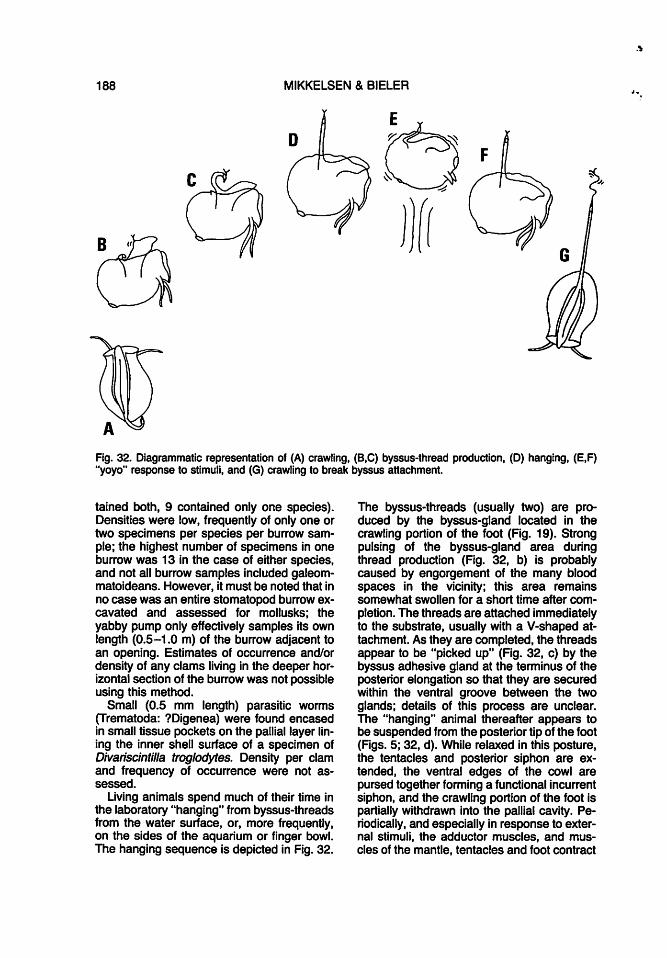
Fig. 32. Diagrammatic representation of (A) crawling, (B,C) byssus-thread production, (D) hanging, (E,F) "yoyo" response to stimuli, and (G) crawling to break byssus attachment.
tained both, 9 contained only one species). Densities were low, frequently of only one or two specimens per species per burrow sample; the highest number of specimens in one burrow was 13 in the case of either species, and not all burrow samples included galeommatoideans. However, it must be noted that in no case was an entire stomatopod burrow excavated and assessed for mollusks; the yabby pump only effectively samples its own length (0.5-1.0 m) of the burrow adjacent to an opening. Estimates of occurrence and/or density of any clams living in the deeper horizontal section of the burrow was not possible using this method.
Small (0.5 mm length) parasitic worms (Trematoda: ?Digenea) were found encased in small tissue pockets on the pallial layer lining the inner shell surface of a specimen of Divariscintilla troglodytes. Density per clam and frequency of occurrence were not assessed.
Living animals spend much of their time in the laboratory "hanging" from byssus-threads from the water surface, or, more frequently, on the sides of the aquarium or finger bowl. The hanging sequence is depicted in Fig. 32.
The byssus-threads (usually two) are produced by the byssus-gland located in the crawling portion of the foot (Fig. 19). Strong pulsing of the byssus-gland area during thread production (Fig. 32, b) is probably caused by engorgement of the many blood spaces in the vicinity; this area remains somewhat swollen for a short time after completion. The threads are attached immediately to the substrate, usually with a V-shaped attachment. As they are completed, the threads appear to be "picked up" (Fig. 32, c) by the byssus adhesive gland at the terminus of the posterior elongation so that they are secured within the ventral groove between the two glands; details of this process are unclear. The "hanging" animal thereafter appears to be suspended from the posterior tip of the foot (Figs. 5; 32, d). While relaxed in this posture, the tentacles and posterior siphon are extended, the ventral edges of the cowl are pursed together forming a functional incurrent siphon, and the crawling portion of the foot is partially withdrawn into the pallial cavity. Periodically, and especially in response to external stimuli, the adductor muscles, and muscles of the mantle, tentacles and foot contract
.\

BIOLOGY OF DIVARISCINTILLA 189
simultaneously producing rapid movement upward toward the byssus attachment point. This is followed by gradual relaxation and return to the resting/feeding posture. This action (Fig. 32, e,f) resembles the up-and-down motion produced with a child's yo-yo toy, and suggested the name for one of the species described here.
During normal "hanging," the posterior foot extension is capable of stretching to a length approximately 1-2 times the mantle length. One specimen of Divariscintilla troglodytes, whose byssus adhesive gland was accidently severed during examination, was able to produce a byssus-thread and hang, although the threads did not lie within the full extent of the ventral groove; the distal half of the posterior extension remained curled at the side of the animal, unextended and unused. These observations lead to the conclusion that the glandular structure at the terminus of the posterior foot extension serves to secure the byssus-threads within the full length of the ventral groove. It is likely that this gland secretes an adhesive substance for this purpose, therefore we refer to it here as the "byssus adhesive gland." The fact that the injured animal could still use part of the ventral groove for hanging suggests that the extensive ciliation and mucous secretion of the proximal part of the groove also serve to secure the threads. This specimen was maintained and observed in the laboratory; two weeks after severing the tip of the posterior extension, the animal was observed to be hanging normally and the tip of the foot with its whitish gland had regenerated and regained function.
When dislodged, the clams actively crawl about, using an even, gliding motion produced by ciliary action on the ventral surface of the foot. Sudden contractions of the shell and pallial muscles frequently occur during crawling, and, although not providing any significant forward propulsion, probably assist the animal in moving its not-so-streamlined body, as well as in cleansing the pallial cavity. The anterior unslit tip of the foot is continually actively "seeking" appropriate substrate. The terminal half of the posterior extension, which is unciliated externally, is not active in crawling, being carried behind either in a trailing curl or with the tip bearly touching the substrate behind. The clams were observed to dislodge themselves voluntarily from laboratory substrate via initiation of crawling activity, thus stretching the byssus-threads until breaking occurred (Fig. 32, g). It is assumed
from laboratory observations that the animals spend most of their time in the burrows attached to the smoothly packed walls, and that crawling is utilized only when relocation is necessary or when dislodged by external forces.
DISCUSSION
The two new species described here are remarkably similar to each other in morphology and behavior. Significant differences are found in shell morphology, the number of "flower-like organs," and the nature of the mantle, including color, thickness, papillation, and number of pallial tentacles (Table 1). Thus far, both species are known only from the specimens studied and cited here; shells are unknown from dry collections (AMNH, DMNH, IRCZM, USNM), probably because of their fragile nature.
The two new species agree closely in anatomy, habitat, and behavior with those described for Divariscintilla maoria Powell, 1932, type and sole described species of the genus, by Judd (1971). Some of the features reported here (e.g. stomach, nervous system, reproductive anatomy, shell musculature, circulatory and excretory systems) were not discussed for D. maoria, and others (e.g. ctenidia, "flower-like organ," byssus apparatus) were described in less detail (Judd, 1971 ). Differences between our two species and D. maoria in shell, pallial, and perhaps ctenidial characters (see below; Table 1) are weak when weighed against the many similarities, and, we accordingly place our new species in Divariscintilla.
Shell and musculature: The most distinct difference between the two species described here and the type species of Divariscintilla is in the shape of the shell. Divariscintilla maoria has a ventral notch in each valve, a character used at the generic level (Chavan, 1969) to treat Divariscintilla as a subgenus under Vasconiella Dall, 1899, whose members possess a likewise-notched shell. The shell of Vasconiella jeffreysiana (P. Fischer, 1873), the type and sole described species of the genus, however, is greatly inequivalve and notched in only the right valve (Kisch, 1958). As noted by Judd (1971: 344), the ventral notch of Divariscintilla maoria "does not appear to be functionally important," as it is not associated with a cleft in the mantle nor with the passage

190 MIKKELSEN & BIELER
TABLE 1. Distinguishing characteristics of the three described species of Divariscintilla (for additional information, see text).
D. maoria (from Judd, 1971)
Shell: General shape oval
ventrally notched Prolongation posterior Sculpture unribbed Length relative to
mantle length 68% Mantle:
Color, thickness (not given) Extent covering shell margins only Papillae very small
Anterior tentacles 41ong Posterior tentacles 1 long Defensive appendages 6-8 present
Flower-like organs: number 1
Ctenidia: smooth Labial palps:
Lamellae per palp (not given) Geographical range: New Zealand
of byssus-threads. Therefore, we do not consider the presence of this notch a prerequisite to inclusion in Divariscintilla and furthermore do not advocate treatment of Divariscintilla as a subgenus of Vasconiel/a on this basis. Claims of a "tendency" within the family for the development of a concave or indented ventral shell margin (Powell, 1932; J.E. Morton, 1957) appear overstated; a cursory survey of the galeommatid shells illustrated by Chavan (1969) show that most are evenly rounded at the ventral margin.
A second conchological difference between Divariscintilla maoria and the two new species is the direction of prolongation of the shell. All are skewed, but in opposite directions; D. maoria is prolonged posteriorly while D. yoyo and D. troglodytes are prolonged anteriorly.
Divariscintilla maoria also has a relatively larger shell in relation to its body (maximum shell length approximately 68% of the relaxed mantle length in fig. 1 of Judd, 1971 ), which also differs by being covered by pallial folds only along its margins. The degree of shell reduction and internalization in members of this superfamily varies widely and is a character which deserves further attention at supraspecific levels.
Divariscintilla yoyo and D. troglodytes both possess the full complement of five major
D. yoyo D. troglodytes
elongate-pointed oval unnotched unnotched anterior anterior unribbed internal radial ribs
40% 50%
whitish, thick yellowish, thin entire entire sparse, very small numerous, small,
evenly-distributed 21ong 2 short, 2 long 1 very short 31ong absent absent
3-7 (usually 5) 1 pleated pleated
10-14 14-20 eastern Florida eastern Florida
muscles (e.g. two adductors, two pedal retractors, and one protractor). As in D. maoria (see Powell, 1932), these leave no muscle scars on the shell, even to the extent, realized during this study, that they do not show under scanning electron microscopy.
Mantle ornamentation: The complement of pallial tentacles and papillae is quite different in Divariscintilla maoria and the two new species described here. The two pairs of anterior tentacles and the single posterior tentacle of D. maoria, described by Judd (1971 ), seem homologous to tentacles described in this study. However, D. yoyo and D. troglodytes do not possess anything resembling the "posterior appendages" described by Judd (1971 ). They do, however, possess numerous papillae on the exterior portion of the mantle, beyond the shell margins; this area is without papillae in D. maoria.
Ctenidia: As shown in Judd (1971: fig. 3) and confirmed here (Fig. 30), the ciliary currents of the ctenidia in Divariscintilla can be ascribed to type C(1) as defined by Atkins (1937). This type, in which only the inner demibranch bears a ventral marginal food groove, is known from a great number of genera (e.g., Galeomma: B. Morton, 1973; Ceratobornia Dall, 1899: Narchi, 1966). Also like

BIOLOGY OF DIVARISCINTILLA 191
most other galeommatoideans, the outer demibranch is shorter in Divariscintilla, and interfilamentary junctions are well-developed and numerous. The presence of interlamellar junctions in Divariscintilla (this study; no data available for the type species), contradicting a statement by B. Morton (1973: 142) that the presence of interfilamentary and the lack of interlamellar junctions "is typical of the Leptonacea [ = Galeommatoidea] in general and can be correlated with the habit of incubating their larvae within the ctenidia ... " (see also B. Morton, 1981: 97-99). Divariscintilla does show, however, a negative correlation between the number of interlamellar junctions and the incubatory habit (see also Oldfield, 1961: 290).
In gross morphology, the gills of Divariscintilla yoyo and D. troglodytes contrast with those of D. maoria and nearly all other galeommatoideans in being loosely pleated rather than smooth. This may be an adaptation for increasing surface area of the foodgathering structures of animals in habitats (e.g. burrows) that may present reduceE'.I food density. Pleating was also observed by Popham (1939) in Phlyctaenachlamys lysiosquillina Popham, 1939, another burrowdwelling commensal, although it was interpreted as an artifact due to preservation.
Flower-like organs: The function of the "flower-like organs" of Divariscintilla species is not evident, although the presence of glandular tissues in the flower head plus the lack of nervous connections point to a secretory rather than sensory role. Judd (1971: 352) described a single "small median papilla on the dorsal edge of the anterior part" of the visceral mass of D. maoria which is clearly the same structure. No function was suggested by Judd, however, reference to a "mucoid secretion" from subepithelial gland cells agrees with this study that the organs are generally secretory. The "flower-like organs" could possibly emit a pheromone for attracting reproductive partners in conditions of low population densities. If so, their placement at the incurrent opening, requiring flow of the attractant through the animal prior to release, is indeed peculiar.
"Flower-like organs" have not been reported in any other galeommatoidean genus. However, the pheromone organ and defensive papillae of Chlamydoconcha orcutti Dall, 1884 (see B. Morton, 1981), bear at least superficial resemblance. They cannot be con-
sidered homologous, because the structures described for C. orcutti arise from the middle pallial fold, while Divariscintilla's "flower-like organs" are from the surface of the visceral mass. Also unlike the organs of C. orcutti, the "flower-like organs" of Divariscintilla are not retractable.
Stomach and feeding: The structure of the stomach was not discussed by Judd (1971) in the redescription of Divariscintilla maoria. Stomach structure in the two species described here agrees well with those of others in this superfamily (e.g. Phlyctaenachlamys Popham, 1939; Galeomma: B. Morton, 1973), defined as type IV by Purchon (1958). Major common features include complete separation of the midgut from the style sac (exceptions are Pseudopythina P. Fischer, 1884: B. Morton; 1972, and Montacutona Yamamoto & Habe, 1959: B. Morton, 1980), and an arcshaped major typhlosole leading toward the openings to the digestive diverticula. Variation occurs in the degree of concentration of these latter openings into caeca or ducts, and in the extent of the typhlosole in the midgut.
Reproduction: Galeommatoideans are frequently cited as having "the most cor:nplex reproductive patterns in the Bivalvia" (0 Foighil & Gibson, 1984: 72). Hermaphroditism, the occurrence of dwarf or "complemental" males, and ctenidial brooding of larvae are common features of this superfamily and have been claimed to be trends associated with a commensal mode of life (B. Morton, 1976; 6 Foighil, 1985). No data were presented on the reproductive biology of Divariscintilla maoria by Judd (1971 : 349) other than noting the incubation of larvae in the "exhalant chamber." The two Divariscintilla species described here were both found to brood their young within the folds of the outer demibranch as well as in the suprabranchial chamber. Both species are simultaneous hermaphrodites. The most unusual reproductive feature encountered was the morphology of the spermatozoa which exhibit rotational asymmetry: the dish-shaped acrosome is tilted approximately 45° from the long axis of the cylindrical nucleus.
Foot and locomotion: All galeommatoideans are capable of active, snail-like locomotion, and most (e.g. Galeomma and Kellia Turton, 1822: Popham, 1940) possess a blunt, heel-like posterior foot with a ventral
192 MIKKELSEN & BIELER
groove and a more-or-less postero-terminal byssus-gland. Divariscintil/a differs from the typical, but is not unique in having an elongated posterior foot; this feature is also present in Ph/yctaenachlamys, Rhamphidonta Bernard, 1975, and Ceratobornia. The foot of Phlyctaenachlamys {Popham, 1939) is nearly identical to that of Divariscintilla, with an elongated posterior portion and a whitish organ at the terminus. In Ph/yctaenachlamys, the byssus-gland {although likewise concentrated in the central portion) continues throughout the posterior extension {Popham, 1939); the "opaque white area immediately short of the tip" of the posterior extension {Popham, 1939: 64) may be similar to the byssus adhesive gland of Divariscintilla, although "hanging" behavior has not been described for Phlyctaenachlamys.
The byssus-gland in the elongated posterior foot of Rhamphidonta retifera {Dall, 1899) was vaguely described by Bernard (1975) as centrally located; this posterior extension is very different from those discussed above in being dorsoventrally flattened and wider than the anterior portion. "Hanging" behavior was not mentioned by Bernard (1975) for Rhamphidonta.
Members of both described species of Ceratobornia, C. longipes (Stimpson, 1855) and C. cema Narchi, 1966, are capable of hanging from a highly extensible posterior foot; the byssus-gland has been described at the extreme posterior tip in both [for C. longipes, see Dall, 1899: 889, pl.88, figs. 10, 11, 13 {previously unpublished figures of Stimpson); for C. cema, see Narchi, 1966: 515, 517-518, text-figs. 2, 5]. However, the byssus apparatus in Ceratobornia may deserve reinvestigation in light of the structures found in Divariscintilla. Narchi {1966: 518, fig. 5) provided confusing statements about the midventral "main mucous gland" and byssus-gland of Ceratobornia cema, with the latter both at the extreme posterior in a text-figure and "extend[ing] along the groove throughout the posterior portion, excepting for the tip" in the text. The "byssogenus [sic] lamellae" described for the byssusgland of C. cema bear greater resemblance to Divariscintilla's byssus adhesive gland than to the byssus-gland proper. Stimpson (1855: 112) reported that, in C. longipes, "there is no true byssus" although a "glutinous substance" secreted by the "opaque byssal gland" at the posterior terminus "may be slightly drawn out"; this material was re-interpreted by Dall (1899: 889) as a "single byssal thread."
In Divariscintilla maoria, the white organ at the end of the posterior extension was interpreted as the byssus-gland, secreting a single thread by which the animal "hangs." In view of the present interpretation of the foot of the two new species, this seems in need of reinvestigation.
The members of Divariscintilla, Phlyctaenachlamys, and Ceratobornia have all been described as inhabiting the holes of burrowing invertebrates (Stimpson, 1855; Popham, 1939; Narchi, 1966); although the location of the byssus-gland apparently differs, the ability to hang from an extensible, thread-like foot may be an advantage in attachment to vertical walls. [However, other galeommatoideans that lack this feature also inhabit holes, e.g. three species referrable to Scintilla Deshayes, 1856, from these same Lysiosquilla burrows (Mikkelsen & Bieler, pers. obs.).] Ceratobornia cema was also reported to attach "temporarily to the body of the shrimp [Callianassa major Say]" (Narchi, 1966: 522), although details of this attachment were not given.
The byssus apparatus of Kellia suborbicularis (Montague, 1803) and of Montacuta substriata (Montagu, 1808) described by Oldfield (1961 : 269-270, figs. 5, 7), each consist of a "subsidiary" byssus-gland dorsal to the ventral groove of the foot, plus a "main" byssusgland with "byssogenous lamellae" equipped with a duct to the posterior "heel." These bear superficial resemblance to the foot of Divariscintilla, but need behavioral observations and more detailed histological examination for proper comparison.
The supportive "chondroid wedge" described for Ceratobornia cema, Rhamphidonta retifera, and several other galeommatoideans (Narchi, 1966; Bernard, 1975) is not present in Divariscintilla.
Although extremely similar in gross morphology, the anterior foot of Divariscintilla is not the "compact mass of muscle" described for Phlyctaenachlamys by Popham (1939). Longitudinal muscle fibers are sparse in the anterior foot of Divariscintilla, being more concentrated in the posterior extension. Extension of the anterior tip is apparently accomplished by haemocoelic pressure.
The crawling activity of many galeommatoideans (e.g. Montacuta Turton, 1822: Gage, 1968a; Mysella Angas, 1877: Gage, 1968b) has been described roughly as an extend-attach-pull forward sequence. Divariscintilla, in contrast, relies primarily on ciliary action for
-·.
BIOLOGY OF DIVARISC/NTILLA 193
forward crawling movement. Propulsive shell and pallial muscle contractions assist locomotion in Phlyctaenach/amys (Popham, 1939) and Ga/eomma (B. Morton, 1973), and at least to some extent in Divariscintilla. During these contractions in Divariscintilla, the relatively large adductor muscles partially close the shell, reducing the shell angle by about 50%. This is in contrast to the "poorly developed" adductor muscles of Phlyctaenachlamys, contraction of which causes "little movement of the shell valves" (Popham, 1939: 67). Retention of well-developed adductor muscles in Divariscintilla may have been favored by providing additional waterpropelling force for locomotion and cleansing of the pallial cavity.
Commensa/ism: The nature of the association between these bivalves and their stomatopod "host" is unclear. Although apparently more dependent upon the burrow habitat than on the resident stomatopod, collection records suggest a strongly obligatory association with Lysiosquil/a for both new Divariscintilla species. The large-diameter burrows of this particular stomatopod, with their smooth, hard-packed walls, are well suited for byssus attachment by the clams and provide a well-maintained, protective habitat. Furthermore, strong respiratory currents produced in the burrow by the stomatopod are undoubtedly beneficial to these more-or-less confined, filter-feeding clams. Ockelmann & Muus (1978) have suggested that this type of association is dependent upon the bivalves' responses to chemical/host, rather than physical, stimuli. Indeed, neither Divariscintilla species described here has ever been found in any other habitat, protective or otherwise.
Redescription of the genus: This work redefines Divariscintilla and identifies the following generic characters: Shell thin, prolonged anteriorly or posteriorly, incompletely internalized within pallial tissues; hinge teeth reduced, possessing only small cone-shaped cardinals. Mantle ornamented with speciesspecific numbers of tentacles and papillae. Byssus-gland communicating by ventral groove with posterior byssus adhesive gland. Two-part foot, consisting of extensible anterior crawling portion and tubular posterior extension, used in active crawling and "hanging." Secretory "flower-like organs" on anterior surface of visceral mass, situated ventral to labial palps. Eulamellibranch
ctenidia with interlamellar and interfilamentary junctions. Simultaneous hermaphroditic reproduction with ctendial incubation of larvae.
The known range of the genus is extended from New Zealand alone (type species, Divariscintilla maoria) into the western Atlantic (D. yoyo and D. troglodytes, described here).
ACKNOWLEDGMENTS
The following are gratefully acknowledged: Dr. Raymond B. Manning (USNM), for bringing this interesting fauna to our attention; William D. ("Woody") Lee (SMSLP), for invaluable field help; Patricia A. Linley (HBOI) for SEM assistance and preparation of the sections in Fig. 23; Julianne Piraino (SMSLP) for SEM assistance; Tom Smoyer (HBOI) for photographic work; Dr. Kerry B. Clark (Florida Institute of Technology, Melbourne) and John E. Miller (HBOI) for the use of and help with video recording equipment; Dr. A. Tucker Abbott for literature; Selene Morris [British Museum (Natural History)] for the loan of pertinent museum specimens; Ors. Richard S. Haubrick (USNM), Kenneth J. Boss (MCZ), and one anonymous reviewer for valuable comments on the manuscript.
This research was supported in part by the Smithsonian Marine Station at Link Port; the cooperation of Dr. Mary E. Rice and her team is gratefully acknowledged. Funding for this project was derived in part from a National Capital Shell Club scholarship to P.M., and a NATO Postdoctoral Fellowship at SMSLP to R.B. (administered by the German Academic Exchange Service [Deutscher Akademischer Austauschdienst (DAAD)], Bonn). This is Harbor Branch Oceanographic Institution Contribution no. 704 and Smithsonian Marine Station Contribution no. 237.
LITERATURE CITED
ABBOTT, A. T., 197 4, American Seashells: the Marine Mollusca of the Atlantic and Pacific coasts of North America, 2nd ed. Van Nostrand Reinhold, New York, 663 pp., 24 color pis.
ADAMS, H., & A. ADAMS, 1857, The genera of Recent Mollusca; arranged according to their organization. Vol. II. John Van Voorst, London, 661 pp.
ATKINS, D., 1937, On the ciliary mechanisms and interrelationships of lamellibranchs. Part 111. Types of lamellibranch gills and their food cur-

194 MIKKELSEN & BIELER
rents. Quarterly Journal of Microscopical Science, 79: 375-421.
BERNARD, F. R., 1975, Rhamphidonta gen. n. from the northeastern Pacific (Bivalvia, Leptonacea). Journal de Conchyliologie, 112(3-4 ): 105-115.
BIELER, R., & P. M. MIKKELSEN, 1988, Anatomy and reproductive biology of two western Atlantic species of Vitrinellidae, with a case of protandrous hermaphroditism in the Rissoacea. The Nautilus, 102(1):1-29.
BOSS, K. J., 1965, Symbiotic erycinacean bivalves. Malacologia, 3(2): 183-195.
BOSS, K. J., 1982, Mollusca. Pp. 945-1166, in: PARKER, S. P. (ed.-in-chief), Synopsis and Classification of Living Organisms, vol. 1. McGraw-Hill, New York, xviii + 1166 pp.
CHAVAN, A., 1969, Superfamily Leptonacea Gray, 1847. Pp. 518-537, in: MOORE, R.C. (ed.), Treatise on Invertebrate Paleontology. Pan N. Mollusca 6. Bivalvia 2. Geological Society of America (Boulder, Colorado) & University of Kansas (Lawrence), ii + N491-N952 pp.
DALL, W. H., 1899, Synposis of the Recent and Teritary Leptonacea of North America and the West Indies. Proceedings of the United States National Museum, 21: 873-893.
GAGE, J., 1968a, The mode of life of Montacuta elevata, a bivalve 'commensal' with Clymenel/a torquata (Polychaeta). Canadian Journal of Zoology, 46(5): 8n-892.
GAGE, J., 1968b, The mode of life of Mysella cuneata, a bivalve 'commensal' with Phascolion strombi (Sipunculoidea). Canadian Journal of Zoology, 46(5): 919-934.
GRAY, J. E., 1840, [Mollusca]. Pp. 86-89, 106-156, in: Synopsis of the Contents of the British Museum, 42nd ed. British Museum (Natural History), London.
GRAY, J.E., 1842, [Mollusca]. Pp. 48-92, in: Synopsis of the Contents of the British Museum, 44th ed. British Museum (Natural History), London.
HUMASON, G. L., 1962, Animal tissue techniques. W.H. Freeman & Co., San Francisco & London, xv + 468 pp.
INTERNATIONAL COMMISSION ON ZOOLOGICAL NOMENCLATURE, 1985, International Code of Zoological Nomenclature [ICZN}, 3rd ed. International Trust for Zoological Nomenclature, London, xx + 338 pp.
JUDD, W., 1971, The structure and habits of Divariscintilla maoria Powell (Bivalvia: Galeommatidae). Proceedings of the Malacological Society ofLondon,39:343-354.
KARNOVSKY, M. J., 1965, A formaldehyde-glutaraldehyde fixative of high osmolarity for use in electron microscopy. Journal of Cell Biology, 27: 137A.
KISCH, B. S., 1958, Vasconiella jeffreysiana (P. Fischer). Proceedings of the Malacological Society of London, 33(1): 21-24, pl. 3.
MORTON, B., 1972, Some aspects of the functional morphology and biology of Pseudopythina
subsinuata (Bivalvia: Leptonacea) commensal on stomatopod crustaceans. Journal of Zoology (London), 166: 79-96.
MORTON, B., 1973, The biology and functional morphology of Galeomma (Paralepida) takii (Bivalvia: Leptonacea). Journal of Zoology (London), 169(2); 133-150.
MORTON, B., 1976, Secondary brooding of temporary dwarf males in Ephippodonta (Ephippodontina) oedipus sp. nov. (Bivalvia: Leptonacea). Journal of Conchology, 29: 31-39.
MORTON, B., 1980, Some aspects of the biology and functional morphology (including the presence of a ligamental lithodesma) of Montacutona compacts and M. olivacea (Bivalvia: Leptonacea) associated with coelenterates in Hong Kong. Journal of Zoology (London), 192: 431-455.
MORTON, B., 1981, The biology and functional morphology of Chlamydoconcha orcutti with a discussion on the taxonomic status of Chlamydoconchacea (Mollusca: Bivalvia). Journal of Zoology (London), 195: 81-121.
MORTON, J.E., 1957, The habits of Scintillona zelandica (Odhner) 1924 (Lamellibranchia: Galeommatidae). Proceedings of the Malacological Society of London, 32: 185-188.
NARCHI, w .. 1966, The functional morphology of Ceratobornia cema, new species of the Erycinacea (Mollusca, Eulamellibranchiata). Anais da Academia Brasileira de Ci~ncias, 38(3-4): 513-524.
OCKELMANN, K. W., & K. MUUS, 1978, The biology, ecology and behaviour of the bivalve Mysella bidentata (Montagu}. Ophelia, 17(1): 1-93.
6 FOIGHIL, D., 1985, Form, function, and origin of temporary dwarf males in Pseudopythina rugifera (Carpenter, 1864) (Bivalvia: Galeommatacea). The Veliger, 27(3): 245-252.
6 FOIGHIL, D., & A. GIBSON, 1984, The morphology, reproduction and ecology of the commensal bivalve Scintillona bellerophon spec. nov. (Galeommatacea}. The Veliger, 27(1 }: 72-70.
OLDFIELD, E., 1961, The functional morphology of Kellia suborbicularis (Montagu}, Montacuta ferruginosa (Montagu) and M. substriata (Montagu}, (Mollusca, Lamellibranchiata). Proceedings of the Malacological Society of London, 34: 255-295.
POPHAM, M. L., 1939, On Phlyctaenachlamys lysiosquillina gen. and sp. nov., a lamellibranch commensal in the burrows of Lysiosquilla maculata. British Museum (Natural History), Great Barrier Reef Expedition 1928-29, Scientific Reports, 6(2): 61-84.
POPHAM, M. L., 1940, The mantle cavity of some of the Erycinidae, Montacutidae and Galeommatidae with special reference to the ciliary mechanisms. Journal of the Marine Biological Association of the United Kingdom, 24(2): 549-587.
POWELL, A. W. B., 1932, On some New Zealand pelecypods. Proceedings of the Malacological Society of London, 20(pt. 1 ): 65-72, pl. 6.
PURCHON, R. D., 1958, The stomach in the Eu-

BIOLOGY OF DIVARISC/NTILLA 195
lamellibranchia: stomach Type IV. Journal of Zoology (London), 131: 487-525.
RICHARDSON, K. C., L. JARRETT, & E. H. FINKE, 1960, Embedding in epoxy resins for ultra-thin sectioning in electron microscopy. Stain Technol-ogy, 35: 313-323. •
STASEK, C.R., 1964, Synopsis and discussion of the association of ctenidia and labial palps in the bivalved Mollusca. The Veliger, 6: 91-97.
STIMPSON, W., 1855, On some remarkable marine lnvertebrata inhabiting the shores of South Carolina. Proceedings of the Boston Society of Natural History, 5: 110-117.
THIELE, J., 1934, Handbuch der Systematischen Weichtierkunde, Teil 3. G. Fischer, Jena, 779-1022 pp.
TRYON, G. W., JR., 1872, Catalogue and synonymy of the family Galeommidae. Proceedings of the Academy of Natural Sciences of Philadelphia, 1872: 222-226.
VOKES, H. E., 1980, Genera of the Bivalvia: a systematic and bibliographic catalogue (revised and updated). Paleontological Research Institution, Ithaca, New York, xxvii + 307 pp.
Revised Ms. accepted 18 April 1989