Biallelic expression of IGFBP1 andIGFBP3, two candidate genes for the Silver-Russell syndrome
19
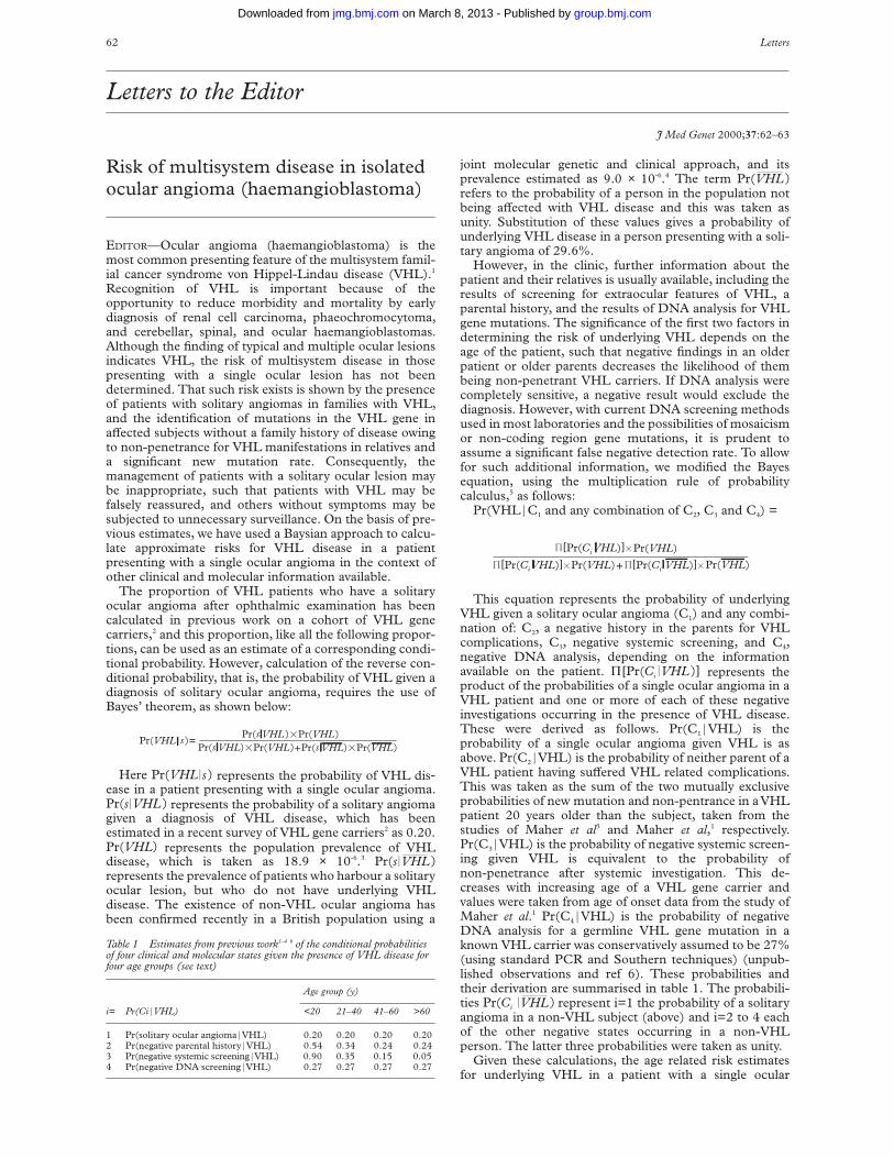
Letters to the Editor J Med Genet 2000;37:62–63 Risk of multisystem disease in isolated ocular angioma (haemangioblastoma) EDITOR—Ocular angioma (haemangioblastoma) is the most common presenting feature of the multisystem famil- ial cancer syndrome von Hippel-Lindau disease (VHL). 1 Recognition of VHL is important because of the opportunity to reduce morbidity and mortality by early diagnosis of renal cell carcinoma, phaeochromocytoma, and cerebellar, spinal, and ocular haemangioblastomas. Although the finding of typical and multiple ocular lesions indicates VHL, the risk of multisystem disease in those presenting with a single ocular lesion has not been determined. That such risk exists is shown by the presence of patients with solitary angiomas in families with VHL, and the identification of mutations in the VHL gene in aVected subjects without a family history of disease owing to non-penetrance for VHL manifestations in relatives and a significant new mutation rate. Consequently, the management of patients with a solitary ocular lesion may be inappropriate, such that patients with VHL may be falsely reassured, and others without symptoms may be subjected to unnecessary surveillance. On the basis of pre- vious estimates, we have used a Baysian approach to calcu- late approximate risks for VHL disease in a patient presenting with a single ocular angioma in the context of other clinical and molecular information available. The proportion of VHL patients who have a solitary ocular angioma after ophthalmic examination has been calculated in previous work on a cohort of VHL gene carriers, 2 and this proportion, like all the following propor- tions, can be used as an estimate of a corresponding condi- tional probability. However, calculation of the reverse con- ditional probability, that is, the probability of VHL given a diagnosis of solitary ocular angioma, requires the use of Bayes’ theorem, as shown below: Here Pr(VHL ) s ) represents the probability of VHL dis- ease in a patient presenting with a single ocular angioma. Pr(s * VHL ) represents the probability of a solitary angioma given a diagnosis of VHL disease, which has been estimated in a recent survey of VHL gene carriers 2 as 0.20. Pr(VHL) represents the population prevalence of VHL disease, which is taken as 18.9 × 10 -6 . 3 Pr(s * VHL ) represents the prevalence of patients who harbour a solitary ocular lesion, but who do not have underlying VHL disease. The existence of non-VHL ocular angioma has been confirmed recently in a British population using a joint molecular genetic and clinical approach, and its prevalence estimated as 9.0 × 10 -6 . 4 The term Pr(VHL ) refers to the probability of a person in the population not being aVected with VHL disease and this was taken as unity. Substitution of these values gives a probability of underlying VHL disease in a person presenting with a soli- tary angioma of 29.6%. However, in the clinic, further information about the patient and their relatives is usually available, including the results of screening for extraocular features of VHL, a parental history, and the results of DNA analysis for VHL gene mutations. The significance of the first two factors in determining the risk of underlying VHL depends on the age of the patient, such that negative findings in an older patient or older parents decreases the likelihood of them being non-penetrant VHL carriers. If DNA analysis were completely sensitive, a negative result would exclude the diagnosis. However, with current DNA screening methods used in most laboratories and the possibilities of mosaicism or non-coding region gene mutations, it is prudent to assume a significant false negative detection rate. To allow for such additional information, we modified the Bayes equation, using the multiplication rule of probability calculus, 5 as follows: Pr(VHL|C 1 and any combination of C 2 ,C 3 and C 4 )= This equation represents the probability of underlying VHL given a solitary ocular angioma (C 1 ) and any combi- nation of: C 2 , a negative history in the parents for VHL complications, C 3 , negative systemic screening, and C 4 , negative DNA analysis, depending on the information available on the patient. —[Pr(C i * VHL )] represents the product of the probabilities of a single ocular angioma in a VHL patient and one or more of each of these negative investigations occurring in the presence of VHL disease. These were derived as follows. Pr(C 1 |VHL) is the probability of a single ocular angioma given VHL is as above. Pr(C 2 |VHL) is the probability of neither parent of a VHL patient having suVered VHL related complications. This was taken as the sum of the two mutually exclusive probabilities of new mutation and non-pentrance in a VHL patient 20 years older than the subject, taken from the studies of Maher et al 3 and Maher et al, 1 respectively. Pr(C 3 |VHL) is the probability of negative systemic screen- ing given VHL is equivalent to the probability of non-penetrance after systemic investigation. This de- creases with increasing age of a VHL gene carrier and values were taken from age of onset data from the study of Maher et al. 1 Pr(C 4 |VHL) is the probability of negative DNA analysis for a germline VHL gene mutation in a known VHL carrier was conservatively assumed to be 27% (using standard PCR and Southern techniques) (unpub- lished observations and ref 6). These probabilities and their derivation are summarised in table 1. The probabili- ties Pr(C i * VHL ) represent i=1 the probability of a solitary angioma in a non-VHL subject (above) and i=2 to 4 each of the other negative states occurring in a non-VHL person. The latter three probabilities were taken as unity. Given these calculations, the age related risk estimates for underlying VHL in a patient with a single ocular Table 1 Estimates from previous work 1–4 6 of the conditional probabilities of four clinical and molecular states given the presence of VHL disease for four age groups (see text) i= Pr(Ci|VHL) Age group (y) <20 21–40 41–60 >60 1 Pr(solitary ocular angioma|VHL) 0.20 0.20 0.20 0.20 2 Pr(negative parental history|VHL) 0.54 0.34 0.24 0.24 3 Pr(negative systemic screening|VHL) 0.90 0.35 0.15 0.05 4 Pr(negative DNA screening|VHL) 0.27 0.27 0.27 0.27 62 Letters group.bmj.com on March 8, 2013 - Published by jmg.bmj.com Downloaded from
Transcript of Biallelic expression of IGFBP1 andIGFBP3, two candidate genes for the Silver-Russell syndrome
Letters to the Editor
J Med Genet 2000;37:62–63
Risk of multisystem disease in isolatedocular angioma (haemangioblastoma)
EDITOR—Ocular angioma (haemangioblastoma) is themost common presenting feature of the multisystem famil-ial cancer syndrome von Hippel-Lindau disease (VHL).1
Recognition of VHL is important because of theopportunity to reduce morbidity and mortality by earlydiagnosis of renal cell carcinoma, phaeochromocytoma,and cerebellar, spinal, and ocular haemangioblastomas.Although the finding of typical and multiple ocular lesionsindicates VHL, the risk of multisystem disease in thosepresenting with a single ocular lesion has not beendetermined. That such risk exists is shown by the presenceof patients with solitary angiomas in families with VHL,and the identification of mutations in the VHL gene inaVected subjects without a family history of disease owingto non-penetrance for VHL manifestations in relatives anda significant new mutation rate. Consequently, themanagement of patients with a solitary ocular lesion maybe inappropriate, such that patients with VHL may befalsely reassured, and others without symptoms may besubjected to unnecessary surveillance. On the basis of pre-vious estimates, we have used a Baysian approach to calcu-late approximate risks for VHL disease in a patientpresenting with a single ocular angioma in the context ofother clinical and molecular information available.
The proportion of VHL patients who have a solitaryocular angioma after ophthalmic examination has beencalculated in previous work on a cohort of VHL genecarriers,2 and this proportion, like all the following propor-tions, can be used as an estimate of a corresponding condi-tional probability. However, calculation of the reverse con-ditional probability, that is, the probability of VHL given adiagnosis of solitary ocular angioma, requires the use ofBayes’ theorem, as shown below:
Here Pr(VHL ) s) represents the probability of VHL dis-ease in a patient presenting with a single ocular angioma.Pr(s *VHL) represents the probability of a solitary angiomagiven a diagnosis of VHL disease, which has beenestimated in a recent survey of VHL gene carriers2 as 0.20.Pr(VHL) represents the population prevalence of VHLdisease, which is taken as 18.9 × 10-6.3 Pr(s *VHL)represents the prevalence of patients who harbour a solitaryocular lesion, but who do not have underlying VHLdisease. The existence of non-VHL ocular angioma hasbeen confirmed recently in a British population using a
joint molecular genetic and clinical approach, and itsprevalence estimated as 9.0 × 10-6.4 The term Pr(VHL)refers to the probability of a person in the population notbeing aVected with VHL disease and this was taken asunity. Substitution of these values gives a probability ofunderlying VHL disease in a person presenting with a soli-tary angioma of 29.6%.
However, in the clinic, further information about thepatient and their relatives is usually available, including theresults of screening for extraocular features of VHL, aparental history, and the results of DNA analysis for VHLgene mutations. The significance of the first two factors indetermining the risk of underlying VHL depends on theage of the patient, such that negative findings in an olderpatient or older parents decreases the likelihood of thembeing non-penetrant VHL carriers. If DNA analysis werecompletely sensitive, a negative result would exclude thediagnosis. However, with current DNA screening methodsused in most laboratories and the possibilities of mosaicismor non-coding region gene mutations, it is prudent toassume a significant false negative detection rate. To allowfor such additional information, we modified the Bayesequation, using the multiplication rule of probabilitycalculus,5 as follows:
Pr(VHL|C1 and any combination of C2, C3 and C4) =
This equation represents the probability of underlyingVHL given a solitary ocular angioma (C1) and any combi-nation of: C2, a negative history in the parents for VHLcomplications, C3, negative systemic screening, and C4,negative DNA analysis, depending on the informationavailable on the patient. Ð[Pr(Ci
*VHL)] represents theproduct of the probabilities of a single ocular angioma in aVHL patient and one or more of each of these negativeinvestigations occurring in the presence of VHL disease.These were derived as follows. Pr(C1|VHL) is theprobability of a single ocular angioma given VHL is asabove. Pr(C2|VHL) is the probability of neither parent of aVHL patient having suVered VHL related complications.This was taken as the sum of the two mutually exclusiveprobabilities of new mutation and non-pentrance in aVHLpatient 20 years older than the subject, taken from thestudies of Maher et al3 and Maher et al,1 respectively.Pr(C3|VHL) is the probability of negative systemic screen-ing given VHL is equivalent to the probability ofnon-penetrance after systemic investigation. This de-creases with increasing age of a VHL gene carrier andvalues were taken from age of onset data from the study ofMaher et al.1 Pr(C4|VHL) is the probability of negativeDNA analysis for a germline VHL gene mutation in aknown VHL carrier was conservatively assumed to be 27%(using standard PCR and Southern techniques) (unpub-lished observations and ref 6). These probabilities andtheir derivation are summarised in table 1. The probabili-ties Pr(Ci
*VHL) represent i=1 the probability of a solitaryangioma in a non-VHL subject (above) and i=2 to 4 eachof the other negative states occurring in a non-VHLperson. The latter three probabilities were taken as unity.
Given these calculations, the age related risk estimatesfor underlying VHL in a patient with a single ocular
Table 1 Estimates from previous work1–4 6 of the conditional probabilitiesof four clinical and molecular states given the presence of VHL disease forfour age groups (see text)
i= Pr(Ci|VHL)
Age group (y)
<20 21–40 41–60 >60
1 Pr(solitary ocular angioma|VHL) 0.20 0.20 0.20 0.202 Pr(negative parental history|VHL) 0.54 0.34 0.24 0.243 Pr(negative systemic screening|VHL) 0.90 0.35 0.15 0.054 Pr(negative DNA screening|VHL) 0.27 0.27 0.27 0.27
62 Letters
group.bmj.com on March 8, 2013 - Published by jmg.bmj.comDownloaded from
angioma after careful ophthalmic examination, and acombination of other negative information, are summa-rised in table 2.
Although some caution should be exerted whenextrapolating these results to other populations (for exam-ple, the mutation detection sensitivity will depend on theprecise investigations performed and the prevalence ofsporadic ocular angioma might vary), this analysis does, forthe first time, provide clinicians with risk estimates for thelikelihood of underlying systemic disease in patients with asolitary ocular angioma. This information will help deter-
mine the most appropriate investigation and managementof such patients.
We gratefully acknowledge the Guide Dogs for the Blind Association and theTFC Frost Trust for support and Dr C Bunce for advice.
ANDREW R WEBSTER*EAMONN R MAHER†
ALAN C BIRD*ANTHONY T MOORE*‡
*Moorfields Eye Hospital, London, UK
†Section of Medical and Molecular Genetics, Department of Paediatricsand Child Health, University of Birmingham, UK
‡Ophthalmology Department, Addenbrooke’s Hospital, Cambridge, UK
1 Maher ER, Yates JRW, Harries R, Benjamin C, Harris R, Ferguson-SmithMA. Clinical features and natural history of von Hippel-Lindau disease. QJ Med 1990;77:1151-63.
2 Webster AR, Moore AT, Maher ER. The clinical characteristics of ocularangiomatosis in von Hippel Lindau disease and correlation with germlinemutation. Arch Ophthalmol 1999;117:371-8.
3 Maher ER, Iselius L, Yates JR, et al. Von Hippel-Lindau disease: a geneticstudy. J Med Genet 1991;28:443-7.
4 Webster AR, Maher ER, Bird AC, Gregor ZJ, Moore AT. A clinical andmolecular genetic analysis of solitary ocular angioma. Ophthalmology 1999;106:623-9.
5 Rosner B. Fundamentals of biostatistics. 4th ed. Chapter 5. London:Wadsworth, 1995.
6 Maher ER, Webster AR, Richards et al. Phenotypic expression in vonHippel-Lindau disease: correlations with germline VHL gene mutations. JMed Genet 1996;33:328-32.
J Med Genet 2000;37:63–64
Confirmation of the assignment of theSanjad-Sakati (congenitalhypoparathyroidism) syndrome (OMIM241410) locus to chromosome lq42-43
EDITOR—Over the past 12 years, 26 patients with an unu-sual syndrome of congenital hypoparathyroidism associ-ated with severe prenatal and postnatal growth retardationand a pattern of facial anomalies have been seen at theKing Faisal Specialist Hospital and Research Centre, SaudiArabia.1 2 The disorder has been listed by McKusick inOMIM as “hypoparathyroidism-retardation-dysmorphismsyndrome; HRD” as entry 241410. Recently, Parvari et al3
reported the assignment of the gene for this disorder tochromosome 1 at 1q42-43. Their report was based on astudy of consanguineous Bedouin families from Israel andtheir linkage analysis was based on homozygosity bydescent.4 This reports describes a study of three consan-guineous Saudi families, which yielded results consistentwith the 1q42-43 location of the responsible gene.
Blood samples were collected and DNA extracted fromthree Saudi families consisting of first cousin parents andtheir 14 children, five of whom manifested the Sanjad-Sakati syndrome. DNA samples were pooled from the fiveaVected children and a separate pooled sample preparedfrom the DNA of their nine unaVected sibs. The initialanalysis included PCR amplified DNA markers linked togenes involved either in parathyroid structure or function.5
As no evidence of linkage was found, the analysis wasexpanded to the human genome screening set fromResearch Genetics (Huntsville, Alabama). The analysisproceeded from chromosome 22 to chromosome 1. Apositive result was based on finding a single band in thepooled sample from the aVected children indicatinghomozygosity, while the pooled sample from the unaf-fected sibs showed two or more bands. A positive result
with marker DlS235 prompted analysis of all 20 samplesseparately with the additional markers D1S1656, D1S163,D1S179, D1S2712, D1S1540, D1S1680, D1S2678,D1S2680, D1S2850, D1S373, and D1S2670, all of whichcluster around 1q42-43.
Multipoint lod scores were generated usingMAPMAKER/HOMOZ.6 Analysis of the data assumedequal frequencies of the alleles at each marker. The orderof the markers was taken from the maps published by Bro-man et al.7 The data showed that the aVected sibs in thethree families were homozygous for markers that clusteredaround the marker D1S235. A maximum lod score of 4.12around D1S235 at 1q42-43 was obtained. Flanking mark-ers D1S1656 and D1S2678 were consistent with thosefound by Parvari et al3 and suggest a candidate regionmaximally at 1 cM.
The initial report of Sanjad et al1 in 1988 and theirdefinitive report in 19912 clearly established this as adistinct disorder with autosomal recessive inheritance. Theconsistency with which hypocalcaemic tetany or seizures orboth occur in intrauterine growth retarded infants suggeststhat this is not a diagnosis likely to be missed. That thisdisorder has only been reported in consanguineous Arabicfamilies suggests that a founder eVect of a long standingmutation is responsible for this disorder.
Kenny-CaVey syndrome type 1 is clinically manifest asgrowth retardation, craniofacial anomalies, small handsand feet, hypocalcaemia, hypoparathyroidism, and radio-logical evidence of cortical thickening in the long boneswith medullary stenosis and absent diploic space in theskull. The original reports of CaVey8 and Kenny andLinarelli9 suggested autosomal dominant inheritance andthe condition is now referred to as Kenny-CaVey syndrometype 2. In 1997 Khan et al10 reported on 16 aVectedchildren with Kenny-CaVey syndrome type 1 in sixunrelated sibships born to healthy, consanguineous,Bedouin parents from Kuwait. From this group of patients,Diaz et al11 in 1998 mapped the locus for this disorder to1q42-43. All of this information taken together suggests
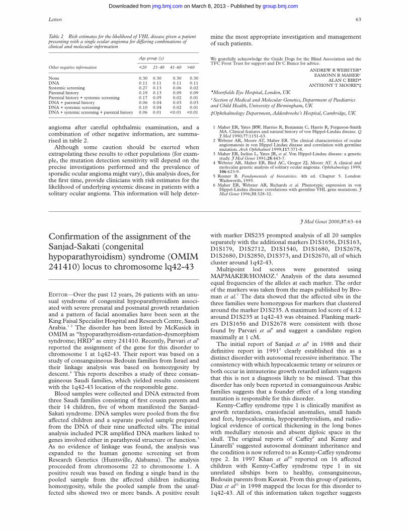
Table 2 Risk estimates for the likelihood of VHL disease given a patientpresenting with a single ocular angioma for diVering combinations ofclinical and molecular information
Other negative information
Age group (y)
<20 21–40 41–60 >60
None 0.30 0.30 0.30 0.30DNA 0.11 0.11 0.11 0.11Systemic screening 0.27 0.13 0.06 0.02Parental history 0.19 0.13 0.09 0.09Parental history + systemic screening 0.17 0.05 0.02 0.01DNA + parental history 0.06 0.04 0.03 0.03DNA + systemic screening 0.10 0.04 0.02 0.01DNA + systemic screening + parental history 0.06 0.01 <0.01 <0.01
Letters 63
group.bmj.com on March 8, 2013 - Published by jmg.bmj.comDownloaded from
that the Sanjad-Sakati syndrome and type 1 Kenny-CaVeysyndrome are at least allelic disorders if not the same con-dition. Despite the multiplicity of abnormalities, includingintrauterine growth retardation, mental retardation, andfacial dysmorphism with congenital hypoparathyroidism,there is currently no information about the nature of theunderlying molecular defect in either disorder. Mapping ofthe locus responsible now oVers promise for analysis ofcandidate genes or positional cloning as likely methods todelineate the molecular basis.
THADDEUS E KELLYSUSAN BLANTON
Division of Medical Genetics, University of Virginia School of Medicine,Charlottesville, Virginia 22908, USA
RAMLA SAIFSAMI A SANJAD
NADIA A SAKATIDepartment of Pediatrics, King Faisal Specialist Hospital and ResearchCentre, Riyadh, Saudi Arabia
1 Sanjad SA, Sakati NA, Abu-Osba YK. Congenital hypoparathyroidism withdysmorphic features: a new syndrome. Pediatr Res 1988:22A.
2 Sanjad SA, Sakati NA, Abu-Osba YK, Kaddoura R, Milner RDG. A newsyndrome of congenital hypoparathyroidism, severe growth failure and dys-morphic features. Arch Dis Child 1991;66:193-6.
3 Parvari R, Hershkovitz E, Kanis A, et al. Homozygosity and linkage disequi-librium mapping of the syndrome of congenital hypoparathyroidism,growth and mental retardation, and dysmorphism to a 1-cM interval ofchromosome 1. Am J Hum Genet 1998;63:163-9.
4 Lander ES, Botstein D. Homozygosity mapping: a way to map human reces-sive traits with the DNA of inbred children. Science 1987;236:1567-70.
5 Shashi V, Golden WL, Allinson PS, Kelly TE. Molecular analysis of recom-bination in a family with a large pericentric X-chromosome inversion. Am JHum Genet 1998;58:1231-8.
6 Kruglyak L, Daly MJ, Lander ES. Rapid multipoint linkage analysis ofrecessive traits in nuclear families, including homozygosity mapping. Am JHum Genet 1995;56:519-27.
7 Broman KW, Murray JC, SheYeld VC, White RL, Weber JL. Comprehen-sive human genetic maps: individual and sex-specific variation in recombi-nation. Am J Hum Genet 1998;63:861-9.
8 CaVey J. Congenital stenosis of medullary spaces in tubular bones and cal-varia in two proportional dwarfs - mother and son coupled with transitoryhypocalcemic tetany. AJR 1967;100:1-11.
9 Kenny FM, Lineralli L. Dwarfism and cortical thickening of tubularbones: transient hypocalcemia in mother and son. Am J Dis Child1985;111:201-7.
10 Khan KTS, Uma R, Usha R, Al Ghanem MM, Al Awadi SA, Farag TI.Kenny-CaVey syndrome in six Bedouin sibships: autosomal recessiveinheritance is confirmed. Am J Med Genet 1997;69:126-32.
11 Diaz GA, Khan KTS, Gelb BD. The autosomal recessive Kenny-CaVeysyndrome locus maps to chromosome 1q42-43. Genomics 1998;54:13-18.
J Med Genet 2000;37:64–65
Molecular diagnosis is important toconfirm suspectedpseudoachondroplasia
EDITOR—Pseudoachondroplasia (PSACH) is an auto-somal dominant chondrodysplasia. In the majority of clini-cally defined cases, mutations have been identified in thegene encoding cartilage oligomeric matrix protein(COMP).1 Mutations in the COMP gene have also beenidentified in some forms of multiple epiphyseal dysplasia(MED), a related skeletal dysplasia.1 All of the mutationsassociated with PSACH and MED have been found inexons encoding the type III repeat region or C-terminaldomain of COMP.
Clinically, PSACH is characterised by short limbeddwarfism, which first becomes apparent in infancy, shortfingers, ligamentous laxity, scoliosis, and early onsetosteoarthritis (OA).2 Radiographic features include smallirregular epiphyses with delayed ossification, flared metaphy-ses, anterior beaking of the vertebral bodies, and delayedmaturation of the triradiate cartilage and acetabulum.3
We report three patients who had previously been givenerroneous diagnoses, in whom mutations in exon 13 of theCOMP gene have been identified. This emphasises theutility of molecular diagnosis, particularly in adult patientswhere radiological diagnosis can be diYcult.
All three aVected subjects were born to unaVectedparents. Each was of normal intelligence and normal facialappearance.
Case 1 presented at 5 years because of pain in both hips.Numerous diagnoses, including spondyloepiphyseal dys-plasia congenita with coxa vara and Morquio’s syndrome,were considered following x ray examination. Extensivesurgery over the following years included a left femoralosteotomy and bilateral Girdlestones operations to treather osteoarthritis. She has had two unaVected children.Examination at 65 years showed her height at 136 cm(<3rd centile), reduced extension at the elbows, short,stubby fingers, and severe kyphosis. Radiological examina-tion showed rhizomelic limb shortening, a prominent del-toid insertion, brachydactyly, metaphyseal broadening,
extensive degenerative changes of the knee and elbow,femoral head destruction with formation of pseudo-acetabula superiorly bilaterally, a thoracolumbar kyphosiswith anterior wedging of the lower thoracic vertebral bod-ies, and a horizontal sacrum.
Case 2 first presented at 3 years with short stature (87.5cm, <3rd centile) and bowed legs. Clinical and radiologicalexamination suggested a diagnosis of spondylometaphysealdysplasia type Kozlowski. Eight operations had beenperformed to eVect tibial lengthening and straightening.Examination at 16 years showed a height of 124 cm (<3rdcentile), genu varum, a waddling gait, and short stubbyfingers. X ray appearances showed ovoid vertebral bodies,epiphyseal involvement, hypoplasia of the iliac bone,splayed irregular metaphyses, and evidence of the multipleoperations, with pins and a plate in situ.
Case 3, a 36 year old woman initially presented at 18months with an intermittent limp. Radiological assessmentat this time was normal. Referral at 9 years for investigationof bilateral hip and left knee pain confirmed short stature,107 cm (<3rd centile), with rhizomelic limb shortening,short fingers, and a waddling gait. Radiological assessmentshowed abnormal epiphyses and metaphyses, but normalvertebral bodies. At this time diagnoses of multiple epiphy-seal dysplasia, PSACH, and Morquio’s syndrome were allconsidered. At 21 years she had surgery to correct asubluxated left patella. She was recently referred to ourdepartment with a diagnosis of achondroplasia. She isawaiting bilateral total hip replacements for treatment ofosteoarthritis. Examination showed a height of 125.5 cm(<3rd centile), short fingers, mild ligamentous laxity, and awaddling gait. X ray appearances showed marked epiphy-seal involvement of the knees, hips, and wrists bilaterally,anterior beaking of the vertebral bodies, and metaphysealchanges in the metacarpals.
All three cases presented during infancy, had heightbelow the 3rd centile, and rhizomelic limb shortening,normal skulls, and short, stubby fingers. Cases 1 and 3both had severe osteoarthritis aVecting their hips bilaterallyand necessitating surgery. Case 1 also had a severe dorso-lumbar kyphosis and case 2 genu varum. The features areall within the recognised spectrum associated with PSACHand although radiological investigations were compatiblewith this diagnosis they were not definitive. Previous x rays
64 Letters
group.bmj.com on March 8, 2013 - Published by jmg.bmj.comDownloaded from
taken in childhood were not available from the threepatients. In view of this, the clinical presentation, and theprevious diYculty in diagnosis, molecular confirmationwas sought.
Mutation screening in exons of the COMP gene usingSSCP and sequence analysis has been described previously.1
In addition to mutation screening in genomic DNA from allthree patients, we also screened for a COMP mutation inRNA isolated from a skin fibroblast cell line established oncase 3. In this instance, cDNA was PCR amplified usingprimers flanking the type III repeat region and C-terminaldomain of COMP. The oligonucleotide primers used wereCaMF (5'-ggt cgc gac act gac cta gac-3') and CaMR (5'-ggtgag cgt gac ttc cag cgt t-3'), which amplified a 789 bp frag-ment containing the entire type III repeat region, and CtF(5'-gaa gtc acg ctc acc gac-3') and CtR (5'-cta ggc ttg ccg cagctg atg g-3'), which amplified a 702 bp fragment containingthe entire C-terminal domain.
Each patient was found to have a mutation in exon 13 ofCOMP. Case 1 was heterozygous for an in frame 3 bpdeletion of GAG between nucleotides 1394-1397, which ispredicted to result in the deletion of glutamic acid at resi-due 457. Case 2 was found to be heterozygous for an inframe 3 bp deletion of GAC between nucleotides1430-1444, which is predicted to result in the deletion ofan aspartic acid residue from between residues 469-473.Case 3 was found to be heterozygous for a G to T transver-sion at nucleotide 1418, which is predicted to result in thesubstitution of glycine by serine at residue 465. All of themutations aVect residues within the seventh repeat of thetype III repeat region and are predicted to result in a quali-tative defect in the COMP protein. The in frame deletionof one or more residues from the type III repeats of COMPhave been described previously.1 The G465S substitutionaVects a highly conserved glycine residue and the substitu-tion of equivalent glycine residues in the second, fourth,and sixth type III repeats have previously been identified inpatients with PSACH. Previous work has determined thatin approximately 40% of cases of PSACH, deletion muta-tions in exon 13 of the COMP gene are responsible.1 Ourwork (present study and M D Briggs, unpublished data)suggests that approximately 50% of all PSACH resultsfrom various mutations, either deletions/duplications orpoint mutations, in exon 13 of the COMP gene and wepropose that analysis of this exon would be an appropriate
initial step in the evaluation of a patient with suspectedPSACH. We have also shown that it is possible to screen forCOMP gene mutations in mRNA isolated from skin fibro-blast cell lines using RT-PCR. This approach will allow forthe rapid screening of mutations and used in conjunctionwith genomic analysis of the COMP gene will help providemolecular diagnosis to confirm a clinically suspected diag-nosis of PSACH.
Diagnosis of PSACH has previously been based on theclinical assessment and the characteristic radiologicalfeatures of people with short stature. These three cases showthe diYculty in making a definitive diagnosis of PSACH inthe absence of molecular confirmation. Accurate diagnosis isimportant, not only so that reproductive options may beconsidered in an informed context, but also so that accurateinformation about the prognosis of the condition may beprovided.4 In case 1 the erroneous diVerential diagnoses hadbeen made 50 years ago and had not been re-evaluated in theinterim, despite the improved and expanded classification ofthe skeletal dysplasias. Molecular diagnosis can be especiallyhelpful in adulthood when radiological diagnosis is morediYcult owing to concomitant changes resulting from osteo-arthritis.
This work was supported by grants from the Arthritis Research Campaign(MDB is an ARC Post-Doctoral Research Fellow) and Wellcome Trust (BN isa Wellcome Trust Clinical Training Fellow).
B NEWMAN*†D DONNAI†
M D BRIGGS*
*Wellcome Trust Centre for Cell-Matrix Research, School of BiologicalSciences, 2.205 Stopford Building, University of Manchester, OxfordRoad, Manchester M13 9PT, UK†University Department of Medical Genetics and Regional GeneticService, St Mary’s Hospital, Hathersage Road, Manchester, M13 0JH,UK
1 Briggs MD, Mortier GR, Cole WG, et al. Diverse mutations in the gene forcartilage oligomeric matrix protein in the pseudoachondroplasia-multipleepiphyseal dysplasia disease spectrum. Am J Hum Genet 1998;62:311-19.
2 Wynne-Davies R, Hall CM, Young ID. Pseudoachondroplasia: clinical diag-nosis at diVerent ages and comparison of autosomal dominant andrecessive types. A review of 32 cases (26 kindreds). J Med Genet1986;23:425-34.
3 Crossan JF, Wynne-Davies R, Fulford GE. Bilateral failure of the capitalfemoral epiphysis: bilateral Perthes disease, multiple epiphyseal dysplasia,pseudoachondroplasia and spondyloepiphyseal dysplasia congenita andtarda. J Pediatr Orthop 1983;3:297-301.
4 McKeand J, Rotta J, Hecht JT. Natural history study of pseudoachondro-plasia. Am J Med Genet 1996;63:406-10.
J Med Genet 2000;37:65–67
Biallelic expression of IGFBP1 andIGFBP3, two candidate genes for theSilver-Russell syndrome
EDITOR—Silver-Russell syndrome (SRS) is a conditioncharacterised by intrauterine and postnatal growth retarda-tion with relative sparing of cranial growth, triangularfacies, fifth finger clinodactyly, and facial, limb, or truncalasymmetry.1 2 The molecular basis of SRS remains elusiveand seems likely to be heterogeneous. However, maternaluniparental disomy of chromosome 7 (mUPD7) has beenfound in approximately 10% of SRS patients, suggestingthat at least one gene on chromosome 7 is imprinted andinvolved in the pathogenesis of this condition.3 4 Interesthas surrounded the human chromosomal region 7p12-13,which is homologous to mouse proximal chromosome 11,
since mUPD for this region in mice leads to prenatalgrowth failure.5 Within this region lie the genes for insulin-like growth factor binding proteins 1 and 3 (IGFBP1 andIGFBP3). Both are involved in regulation of fetal growthvia the insulin-like growth factor axis.6 IGFBP1 andIGFBP3 are therefore obvious candidates for a role in theSRS phenotype associated with mUPD7.3 4
In order to determine whether IGFBP1 and/or IGFBP3are likely to be implicated in SRS, we have investigatedtheir imprinting status in normal fetal tissues collectedfrom termination of pregnancies. Samples were obtainedfrom Queen Charlotte’s and Chelsea Hospital with theapproval of the Research Ethics Committee of the RoyalPostgraduate Medical School (96/4955) and from theMRC Tissue Bank at Hammersmith Hospital (L Wong).Genomic DNA and total RNA was extracted from thesetissues and RT-PCR performed as previously described.7
The primers used in this study are listed in table 1. PCRwas carried out for 35 cycles for all reactions. Products
Letters 65
group.bmj.com on March 8, 2013 - Published by jmg.bmj.comDownloaded from
were sequenced on an ABI PRISM 377 automated DNAsequencer using the dRhodamine Ampli-Taq dye-terminator cycle sequencing kit, according to the manufac-turer’s instructions.
Since fetal IGFBP1 expression is highly tissue specific,8
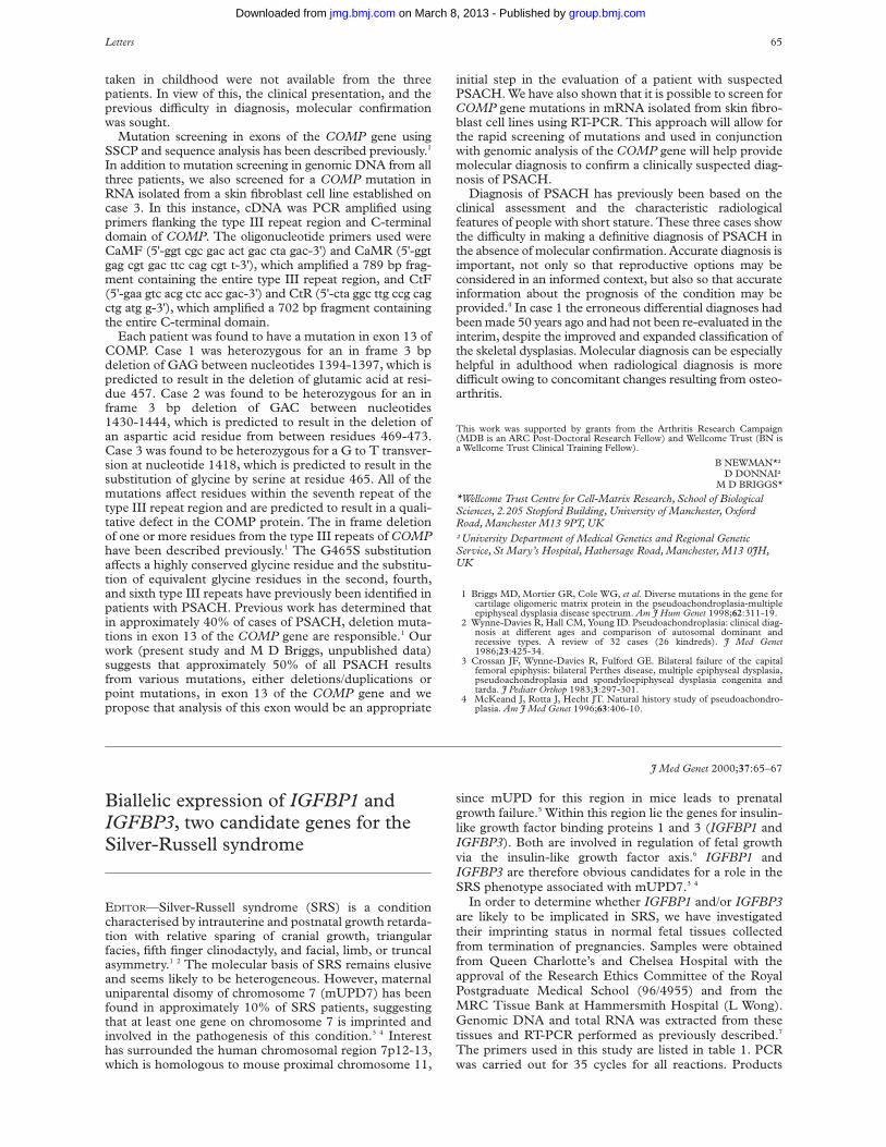
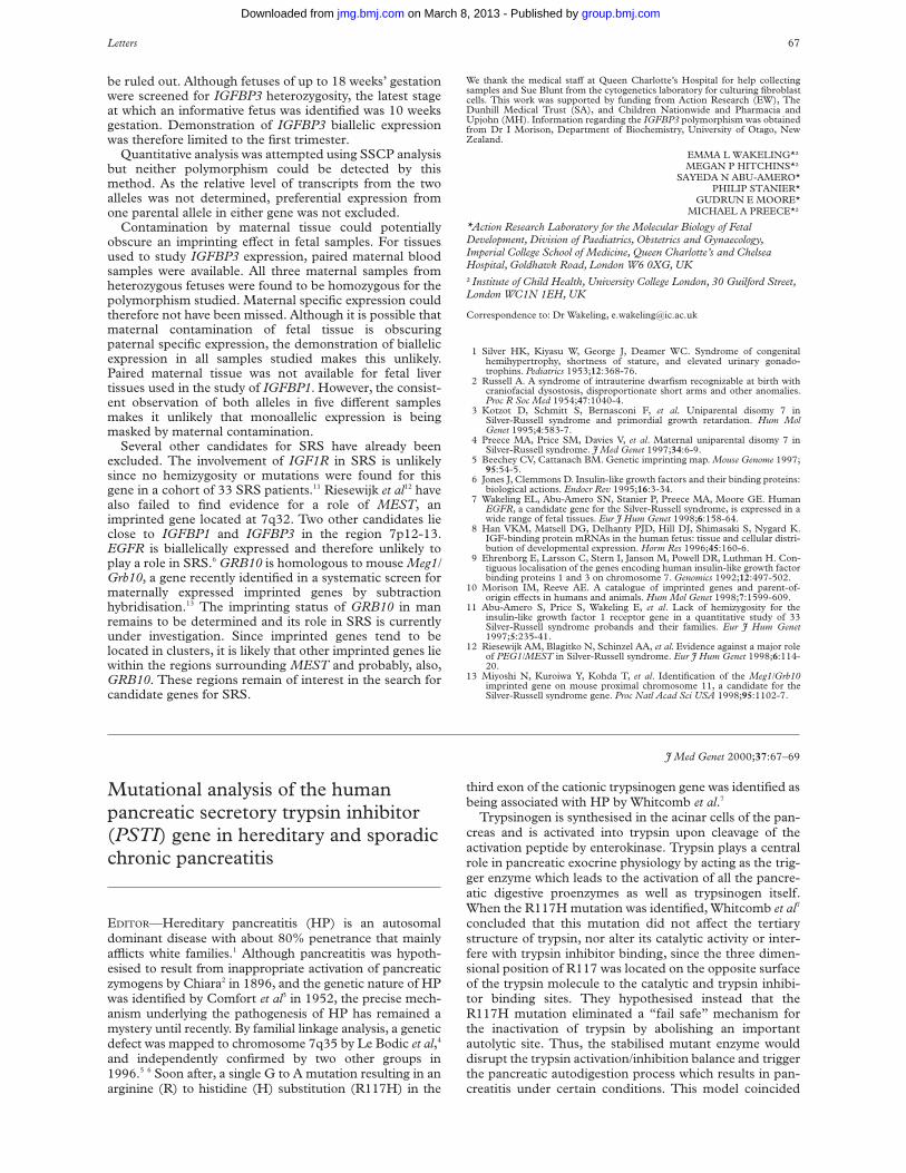
its imprinting status was only investigated in fetal liver.Primers IGFBP1-BF and -ER were designed to span anexpressed polymorphism at nucleotide 5772 within exon 4(Genbank accession: M59316).9 These primers were usedto screen genomic DNA from 16 first and second trimes-ter fetuses. Five were identified as being heterozygous forthe polymorphism and thus informative. Their gestationalages ranged from 5 to 17 weeks. Primers IGFBP1-EF and-ER, which span introns 2 and 3, were used to amplify livercDNA derived from these informative fetuses. Controlsamples prepared from liver RNA without the addition ofreverse transcriptase (RT) were also amplified. Nogenomic contamination was observed in any of thesamples. RT-PCR products were sequenced using thereverse primer IGFBP1-ER and biallelic expression wasseen in all five cases (fig 1).
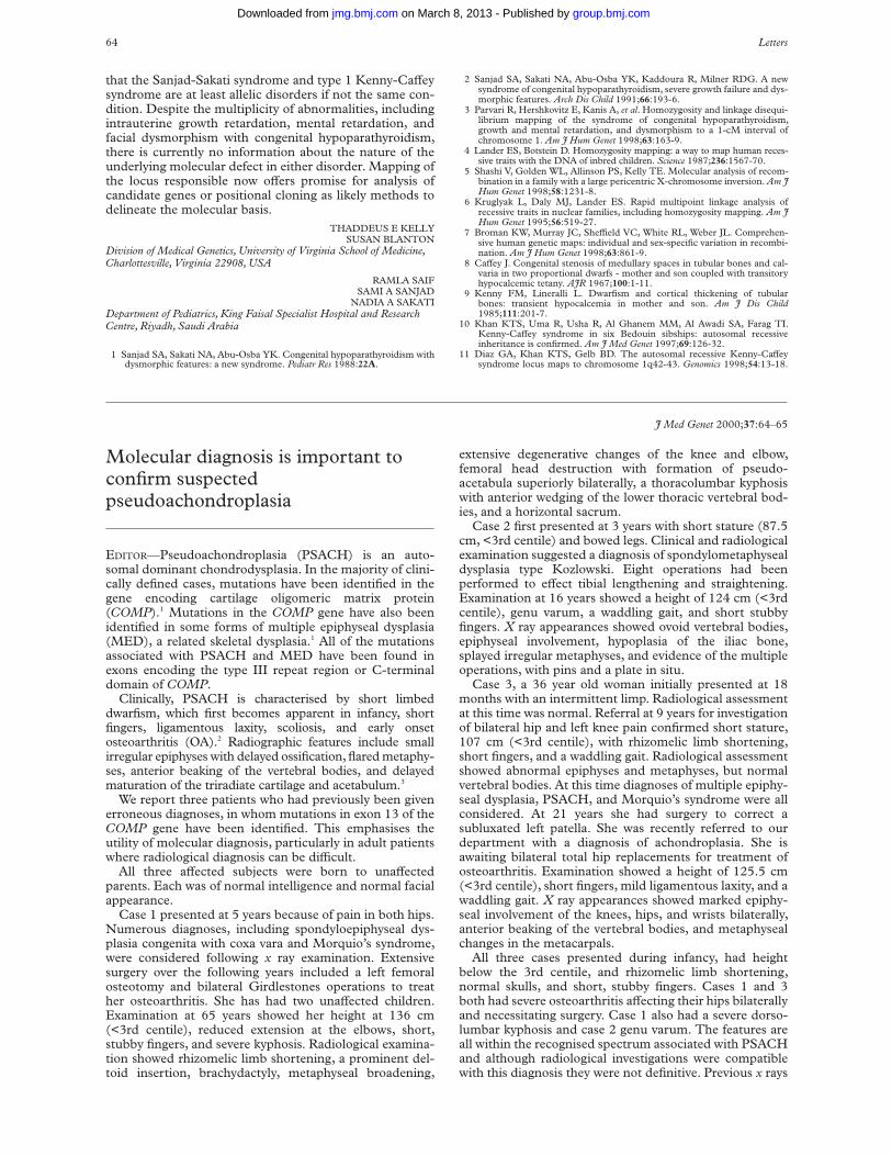
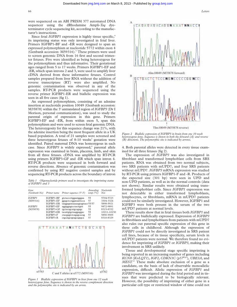
An expressed polymorphism, consisting of an adenineinsertion at nucleotide position 10049 (Genbank accession:M35878) within the 3' untranslated region of IGFBP3 (Dr IMorison, personal communication), was used to study theparental origin of expression in this gene. PrimersIGFBP3-EF and -ER, from within exon 5, span thispolymorphism and were used to screen fetal genomic DNA.The heterozygosity for this sequence change was 21%, withthe adenine insertion being the most frequent allele in a UKbased population. A total of 21 samples were screened andthree heterozygous fetuses of 8-10 weeks’ gestation wereidentified. Paired maternal DNA was homozygous in eachcase. Since IGFBP3 is widely expressed,8 parental alleleexpression was examined in brain, placenta, limb, and skinfrom all three fetuses. cDNA was amplified by RT-PCRusing primers IGFBP3-GF and -ER which span intron 4.RT-PCR products were sequenced in both forward andreverse directions. Absence of genomic contamination wasconfirmed by using RT negative control samples and bysequencing RT-PCR products across the boundary of intron
4. Both parental alleles were detected in every tissue exam-ined for all three fetuses (fig 2).
The expression of IGFBP3 was also investigated infibroblast and transformed lymphoblast cells from SRSpatients. RNA was obtained from two normal subjects,two SRS patients with mUPD7, and four SRS patientswithout mUPD7. IGFBP3 mRNA expression was studiedby RT-PCR using primers IGFBP1-F and -R. Products ofthe expected size (301 bp) were seen in UPD andnon-UPD patients, as well as in the normal controls (datanot shown). Similar results were obtained using trans-formed lymphoblast cells. Since IGFBP1 expression wasnot detectable in either transformed lymphoblasts,lymphocytes, or fibroblasts, mRNA in mUPD7 patientscould not be similarly investigated. However, IGFBP1 andIGFBP3 were both present in the serum of the twomUPD7 patients at normal levels.
These results show that in fetal tissues both IGFBP1 andIGFBP3 are biallelically expressed. Expression of IGFBP3in fibroblasts and lymphoblasts from patients with mUPD7also rules out paternal specific expression of this gene inthese cells in childhood. Although the expression ofIGFBP1 could not be directly investigated in SRS patientcell lines, because of its tissue specificity, serum levels inmUPD7 patients were normal. We therefore found no evi-dence for imprinting of IGFBP1 or IGFBP3, making theirinvolvement in SRS unlikely.
Tissue and developmental stage specific imprinting isbeing reported in an increasing number of genes includingKCN8 (KvLQT1), IGF2, CDKN1C (p57Kip2), UBE3A, andMEST.10 These make absolute exclusion of a gene as acandidate, on the basis of lack of observable monoallelicexpression, diYcult. Allelic expression of IGFBP1 andIGFBP3 was investigated during the fetal period and in tis-sues that were predicted to be biologically relevant.However, the possibility of imprinting of either gene in aparticular cell type or restricted window of time could not
Table 1 Oligonucleotide primers used to investigate the imprinting statusof IGFBP1 and 3
Gene(Genbank No) Primer name Primer sequence (5'-3')
Annealingtemp (°C)
NucleotideNos
IGFBP1 IGFBP1-BF gtctacccttggaatgggaa 55 5704-5723(M59316) IGFBP1-EF ggagctcctggataatttcca 57 3304-3324
IGFBP1-ER tatggagtataaaaagtagttact 55/57 5898-5921IGFBP3 IGFBP3-EF aggtggagtcctacttgttt 55 9872-9891(M35878) IGFBP3-GF tgctacagcatgcagagc 55 8315-8332
IGFBP3-ER tacattgtgctaaggaggac 55 10211-10230IGFBP3-F ctacgagtctcagagcacag 57 5890-5909IGFBP3-R ctgcatgctgtagcagtgca 57 8310-8329
Figure 1 Biallelic expression of IGFBP1 in liver from one 12 weekheterozygous fetus. Sequence is shown in the reverse complement directionand the polymorphic site is indicated by an arrow.
Figure 2 Biallelic expression of IGFBP3 in brain from one 10 weekheterozygous fetus. Sequence is shown in both the forward (A) and reverse(B) directions. The polymorphic site is indicated by arrows.
66 Letters
group.bmj.com on March 8, 2013 - Published by jmg.bmj.comDownloaded from
be ruled out. Although fetuses of up to 18 weeks’ gestationwere screened for IGFBP3 heterozygosity, the latest stageat which an informative fetus was identified was 10 weeksgestation. Demonstration of IGFBP3 biallelic expressionwas therefore limited to the first trimester.
Quantitative analysis was attempted using SSCP analysisbut neither polymorphism could be detected by thismethod. As the relative level of transcripts from the twoalleles was not determined, preferential expression fromone parental allele in either gene was not excluded.
Contamination by maternal tissue could potentiallyobscure an imprinting eVect in fetal samples. For tissuesused to study IGFBP3 expression, paired maternal bloodsamples were available. All three maternal samples fromheterozygous fetuses were found to be homozygous for thepolymorphism studied. Maternal specific expression couldtherefore not have been missed. Although it is possible thatmaternal contamination of fetal tissue is obscuringpaternal specific expression, the demonstration of biallelicexpression in all samples studied makes this unlikely.Paired maternal tissue was not available for fetal livertissues used in the study of IGFBP1. However, the consist-ent observation of both alleles in five diVerent samplesmakes it unlikely that monoallelic expression is beingmasked by maternal contamination.
Several other candidates for SRS have already beenexcluded. The involvement of IGF1R in SRS is unlikelysince no hemizygosity or mutations were found for thisgene in a cohort of 33 SRS patients.11 Riesewijk et al12 havealso failed to find evidence for a role of MEST, animprinted gene located at 7q32. Two other candidates lieclose to IGFBP1 and IGFBP3 in the region 7p12-13.EGFR is biallelically expressed and therefore unlikely toplay a role in SRS.6 GRB10 is homologous to mouse Meg1/Grb10, a gene recently identified in a systematic screen formaternally expressed imprinted genes by subtractionhybridisation.13 The imprinting status of GRB10 in manremains to be determined and its role in SRS is currentlyunder investigation. Since imprinted genes tend to belocated in clusters, it is likely that other imprinted genes liewithin the regions surrounding MEST and probably, also,GRB10. These regions remain of interest in the search forcandidate genes for SRS.
We thank the medical staV at Queen Charlotte’s Hospital for help collectingsamples and Sue Blunt from the cytogenetics laboratory for culturing fibroblastcells. This work was supported by funding from Action Research (EW), TheDunhill Medical Trust (SA), and Children Nationwide and Pharmacia andUpjohn (MH). Information regarding the IGFBP3 polymorphism was obtainedfrom Dr I Morison, Department of Biochemistry, University of Otago, NewZealand.
EMMA L WAKELING*†MEGAN P HITCHINS*†
SAYEDA N ABU-AMERO*PHILIP STANIER*
GUDRUN E MOORE*MICHAEL A PREECE*†
*Action Research Laboratory for the Molecular Biology of FetalDevelopment, Division of Paediatrics, Obstetrics and Gynaecology,Imperial College School of Medicine, Queen Charlotte’s and ChelseaHospital, Goldhawk Road, London W6 0XG, UK
†Institute of Child Health, University College London, 30 Guilford Street,London WC1N 1EH, UK
Correspondence to: Dr Wakeling, [email protected]
1 Silver HK, Kiyasu W, George J, Deamer WC. Syndrome of congenitalhemihypertrophy, shortness of stature, and elevated urinary gonado-trophins. Pediatrics 1953;12:368-76.
2 Russell A. A syndrome of intrauterine dwarfism recognizable at birth withcraniofacial dysostosis, disproportionate short arms and other anomalies.Proc R Soc Med 1954;47:1040-4.
3 Kotzot D, Schmitt S, Bernasconi F, et al. Uniparental disomy 7 inSilver-Russell syndrome and primordial growth retardation. Hum MolGenet 1995;4:583-7.
4 Preece MA, Price SM, Davies V, et al. Maternal uniparental disomy 7 inSilver-Russell syndrome. J Med Genet 1997;34:6-9.
5 Beechey CV, Cattanach BM. Genetic imprinting map. Mouse Genome 1997;95:54-5.
6 Jones J, Clemmons D. Insulin-like growth factors and their binding proteins:biological actions. Endocr Rev 1995;16:3-34.
7 Wakeling EL, Abu-Amero SN, Stanier P, Preece MA, Moore GE. HumanEGFR, a candidate gene for the Silver-Russell syndrome, is expressed in awide range of fetal tissues. Eur J Hum Genet 1998;6:158-64.
8 Han VKM, Matsell DG, Delhanty PJD, Hill DJ, Shimasaki S, Nygard K.IGF-binding protein mRNAs in the human fetus: tissue and cellular distri-bution of developmental expression. Horm Res 1996;45:160-6.
9 Ehrenborg E, Larsson C, Stern I, Janson M, Powell DR, Luthman H. Con-tiguous localisation of the genes encoding human insulin-like growth factorbinding proteins 1 and 3 on chromosome 7. Genomics 1992;12:497-502.
10 Morison IM, Reeve AE. A catalogue of imprinted genes and parent-of-origin eVects in humans and animals. Hum Mol Genet 1998;7:1599-609.
11 Abu-Amero S, Price S, Wakeling E, et al. Lack of hemizygosity for theinsulin-like growth factor 1 receptor gene in a quantitative study of 33Silver-Russell syndrome probands and their families. Eur J Hum Genet1997;5:235-41.
12 Riesewijk AM, Blagitko N, Schinzel AA, et al. Evidence against a major roleof PEG1/MEST in Silver-Russell syndrome. Eur J Hum Genet 1998;6:114-20.
13 Miyoshi N, Kuroiwa Y, Kohda T, et al. Identification of the Meg1/Grb10imprinted gene on mouse proximal chromosome 11, a candidate for theSilver-Russell syndrome gene. Proc Natl Acad Sci USA 1998;95:1102-7.
J Med Genet 2000;37:67–69
Mutational analysis of the humanpancreatic secretory trypsin inhibitor(PSTI) gene in hereditary and sporadicchronic pancreatitis
EDITOR—Hereditary pancreatitis (HP) is an autosomaldominant disease with about 80% penetrance that mainlyaZicts white families.1 Although pancreatitis was hypoth-esised to result from inappropriate activation of pancreaticzymogens by Chiara2 in 1896, and the genetic nature of HPwas identified by Comfort et al3 in 1952, the precise mech-anism underlying the pathogenesis of HP has remained amystery until recently. By familial linkage analysis, a geneticdefect was mapped to chromosome 7q35 by Le Bodic et al,4
and independently confirmed by two other groups in1996.5 6 Soon after, a single G to A mutation resulting in anarginine (R) to histidine (H) substitution (R117H) in the
third exon of the cationic trypsinogen gene was identified asbeing associated with HP by Whitcomb et al.7
Trypsinogen is synthesised in the acinar cells of the pan-creas and is activated into trypsin upon cleavage of theactivation peptide by enterokinase. Trypsin plays a centralrole in pancreatic exocrine physiology by acting as the trig-ger enzyme which leads to the activation of all the pancre-atic digestive proenzymes as well as trypsinogen itself.When the R117H mutation was identified, Whitcomb et al7
concluded that this mutation did not aVect the tertiarystructure of trypsin, nor alter its catalytic activity or inter-fere with trypsin inhibitor binding, since the three dimen-sional position of R117 was located on the opposite surfaceof the trypsin molecule to the catalytic and trypsin inhibi-tor binding sites. They hypothesised instead that theR117H mutation eliminated a “fail safe” mechanism forthe inactivation of trypsin by abolishing an importantautolytic site. Thus, the stabilised mutant enzyme woulddisrupt the trypsin activation/inhibition balance and triggerthe pancreatic autodigestion process which results in pan-creatitis under certain conditions. This model coincided
Letters 67
group.bmj.com on March 8, 2013 - Published by jmg.bmj.comDownloaded from
with Chiara’s pancreatitis hypothesis2 and has beensupported by in vitro mutagenesis data. When the R117 ofrat trypsin was replaced by other amino acids, the rate ofautolysis of certain mutant enzymes was significantlyslower than that of the wild type protein.8 9 While theR117H mutation has been shown to be a commonmutation in HP by several laboratories world wide,10–13 fur-ther mutations in the cationic trypsinogen gene have beenreported recently.13–15 These mutations are also presumedto facilitate the trypsin autodigestion process by alteringeither the tertiary structure of the protein or the binding ofthe pancreatic secretory trypsin inhibitor (PSTI).
However, mutations in the cationic trypsinogen gene donot appear to be the whole story. When 14 HP familiesfrom diVerent regions of France were scanned formutations in the cationic trypsinogen gene by denaturinggradient gel electrophoresis (DGGE) analysis, no muta-tions were detected in either part of the promoter region, inthe intron/exon junctions, or in the gene coding sequenceof six families. Furthermore, segregation analysis of onefamily with microsatellite markers (D7S640, D7S495,D7S684, D7S661, D7S676, D7S688) showed that theaVected subjects had inherited two diVerent haplotypes.13
Locus heterogeneity in HP was also suggested by the nega-tive linkage and absence of the R117H mutation in two outof eight families studied by Dasouki et al.12 These findings,along with the incomplete penetrance of HP, indicated thatanother gene, or maybe even more than one, is involved inthe pathogenesis of HP.
Human PSTI, a single chain polypeptide consisting of56 amino acids, is also synthesised in the acinar cells of thepancreas. Its main physiological function is believed to bethe prevention of the trypsin driven digestive enzyme acti-vation cascade and of pancreatic autodigestion.16 17 Be-cause of this central role of PSTI as a negative regulator oftrypsin activity, it has been speculated that mutations inthis gene may contribute to the development ofpancreatitis.13 To date, no mutations have been reported inthe human PSTI gene, which is located on chromosome5.17 We therefore sought to investigate the possibility ofmutations in the PSTI gene in a cohort of hereditary andsporadic chronic pancreatitis patients, as part of a continu-ing eVort to gain further insight into the molecular basis ofthis disorder.
The human PSTI gene is approximately 7.5 kb long andis separated into 4 exons.17 By designing three exonicprimer pairs (sequence not shown), we first successfullyamplified the three introns of the PSTI gene from genomicDNA samples. The sizes of them were ∼1.7 kb, 1.5 kb, and3.5 kb respectively, with a total length of ∼6.7 kb, which iswithin the range of 7.5 kb. The three PCR fragments werethen cloned into the pGEM®-T vector (Promega) and the
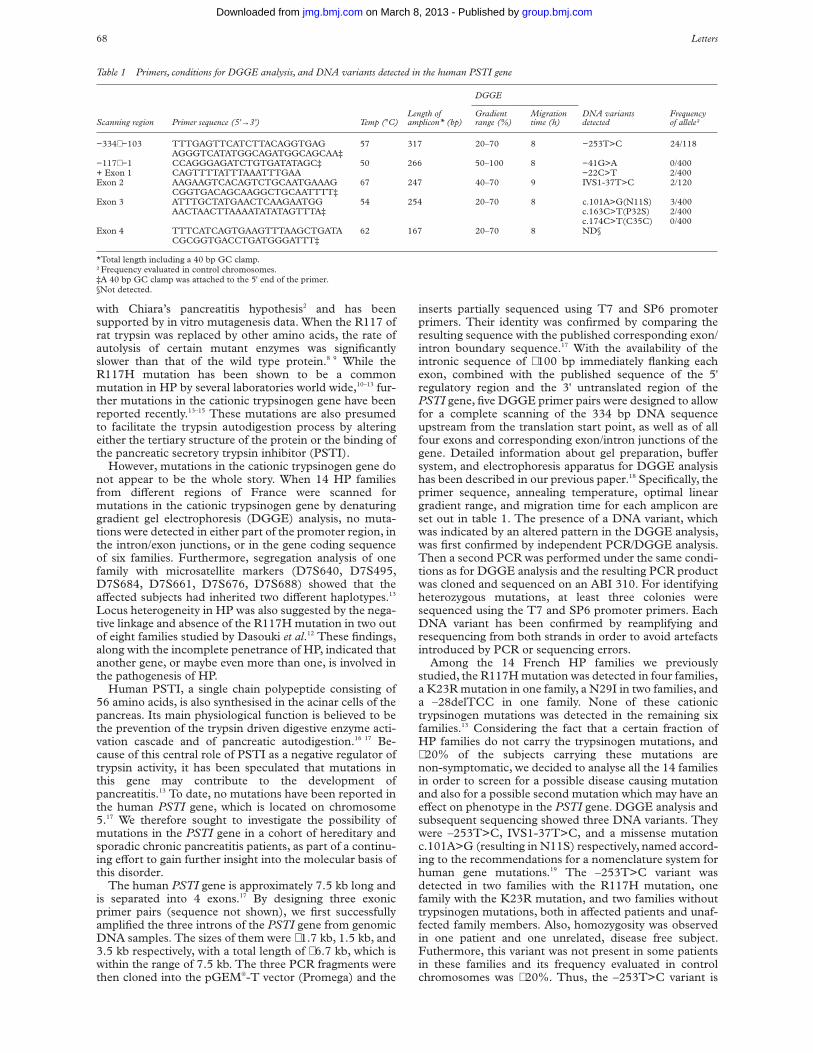
inserts partially sequenced using T7 and SP6 promoterprimers. Their identity was confirmed by comparing theresulting sequence with the published corresponding exon/intron boundary sequence.17 With the availability of theintronic sequence of ∼100 bp immediately flanking eachexon, combined with the published sequence of the 5'regulatory region and the 3' untranslated region of thePSTI gene, five DGGE primer pairs were designed to allowfor a complete scanning of the 334 bp DNA sequenceupstream from the translation start point, as well as of allfour exons and corresponding exon/intron junctions of thegene. Detailed information about gel preparation, buVersystem, and electrophoresis apparatus for DGGE analysishas been described in our previous paper.18 Specifically, theprimer sequence, annealing temperature, optimal lineargradient range, and migration time for each amplicon areset out in table 1. The presence of a DNA variant, whichwas indicated by an altered pattern in the DGGE analysis,was first confirmed by independent PCR/DGGE analysis.Then a second PCR was performed under the same condi-tions as for DGGE analysis and the resulting PCR productwas cloned and sequenced on an ABI 310. For identifyingheterozygous mutations, at least three colonies weresequenced using the T7 and SP6 promoter primers. EachDNA variant has been confirmed by reamplifying andresequencing from both strands in order to avoid artefactsintroduced by PCR or sequencing errors.
Among the 14 French HP families we previouslystudied, the R117H mutation was detected in four families,a K23R mutation in one family, a N29I in two families, anda –28delTCC in one family. None of these cationictrypsinogen mutations was detected in the remaining sixfamilies.13 Considering the fact that a certain fraction ofHP families do not carry the trypsinogen mutations, and∼20% of the subjects carrying these mutations arenon-symptomatic, we decided to analyse all the 14 familiesin order to screen for a possible disease causing mutationand also for a possible second mutation which may have aneVect on phenotype in the PSTI gene. DGGE analysis andsubsequent sequencing showed three DNA variants. Theywere –253T>C, IVS1-37T>C, and a missense mutationc.101A>G (resulting in N11S) respectively, named accord-ing to the recommendations for a nomenclature system forhuman gene mutations.19 The –253T>C variant wasdetected in two families with the R117H mutation, onefamily with the K23R mutation, and two families withouttrypsinogen mutations, both in aVected patients and unaf-fected family members. Also, homozygosity was observedin one patient and one unrelated, disease free subject.Futhermore, this variant was not present in some patientsin these families and its frequency evaluated in controlchromosomes was ∼20%. Thus, the –253T>C variant is
Table 1 Primers, conditions for DGGE analysis, and DNA variants detected in the human PSTI gene
Scanning region Primer sequence (5'→3') Temp (°C)Length ofamplicon* (bp)
DGGE
DNA variantsdetected
Frequencyof allele†
Gradientrange (%)
Migrationtime (h)
−334∼−103 TTTGAGTTCATCTTACAGGTGAG 57 317 20–70 8 −253T>C 24/118AGGGTCATATGGCAGATGGCAGCAA‡
−117∼−1 CCAGGGAGATCTGTGATATAGC‡ 50 266 50–100 8 −41G>A 0/400+ Exon 1 CAGTTTTATTTAAATTTGAA −22C>T 2/400Exon 2 AAGAAGTCACAGTCTGCAATGAAAG 67 247 40–70 9 IVS1-37T>C 2/120
CGGTGACAGCAAGGCTGCAATTTT‡Exon 3 ATTTGCTATGAACTCAAGAATGG 54 254 20–70 8 c.101A>G(N11S) 3/400
AACTAACTTAAAATATATAGTTTA‡ c.163C>T(P32S) 2/400c.174C>T(C35C) 0/400
Exon 4 TTTCATCAGTGAAGTTTAAGCTGATA 62 167 20–70 8 ND§CGCGGTGACCTGATGGGATTT‡
*Total length including a 40 bp GC clamp.†Frequency evaluated in control chromosomes.‡A 40 bp GC clamp was attached to the 5' end of the primer.§Not detected.
68 Letters
group.bmj.com on March 8, 2013 - Published by jmg.bmj.comDownloaded from
clearly a natural polymorphism. The IVS1-37T>C andc.101A>G (N11S) variants occurred together in one fam-ily without trypsinogen mutations and were present in thesame haplotype. They have been classified as neutral poly-morphisms based primarily on the fact that they did notsegregate with the disease and that they were present incontrol chromosomes. Moreover, the IVS1-37T>C variantdid not appear to aVect the splice recognition sites and thec.101A>G variant did not replace the asparagine (N) atposition 11 of the protein with an amino acid of diVerentphysical characteristics, although N11 is conserved in thehuman and two types of rat PSTI proteins.20
Owing to the similar clinical, laboratory, and pathologi-cal features of hereditary and sporadic chronic pancreatitis,we also undertook DGGE analysis of sporadic chronicpancreatitis. An additional three heterozygous DNAvariants were separately identified in one out of 30 patientswith sporadic chronic pancreatitis. The first was a C to Ttransition at position 163 of the PSTI cDNA, resulting in aproline (P) to serine (S) change at position 32 of the pro-tein (c.163C>T (P32S)). This variant did not change aconserved amino acid and it was present in controlchromosomes, indicating a harmless eVect on phenotype.The second was a G to A substitution at position 41upstream from the translation start site (−41G>A). This–41G is not conserved in the human and the two types ofrat PSTI genes and is located ∼20 bp downstream from themain transcription start site. This suggests that the−41G>A substitution could not have a significant eVect onthe transcriptional or translational activity of the PSTIgene. We believe it to be a rare polymorphism as it was notdetected in 400 control chromosomes. The third variantwas a C to T transition at position 174 of the cDNAresulting in a silent mutation at position 35 of the protein(c.174C>T (C35C)). In addition, a heterozygous C to Tsubstitution at position 22 upstream from the translationstart site (−22C>T) was detected in two out of 200 controlsubjects (representing 400 chromosomes). All of the DNAvariants detected in this study as well as their frequencyevaluated in control chromosomes are described in table 1.
DGGE analysis is one of the most sensitive and eYcientestablished mutation scanning techniques to date. It canallow for the discrimination of DNA molecules diVering byas little as only one base change. Using this technique, weidentified nearly 100% of mutations in the cystic fibrosistransmembrane conductance regulator (CFTR) gene in acertain population.21 In this study, we detected up to sevendiVerent DNA variants. Given this high sensitivity ofDGGE analysis, although we cannot exclude the possibilityof mutations in the more upstream 5' regulatory region orin the remaining intronic sequences, our results stronglysuggested that the PSTI gene could be neither a cause nora cofactor in the development of HP. Future research intothis disease may be directed towards other pancreaticdigestive proenzyme genes such as the anionic trypsinogenand mesotrypsinogen genes.22
When mutations in the cationic trypsinogen gene wereidentified as the molecular basis of HP, it was questionedwhether they could predispose patients to develop sporadicpancreatitis. Until now, these cationic trypsinogen muta-tions have not been detected in sporadic chronicpancreatitis.15 23 In this study, although seven diVerentDNA variants in the human PSTI gene were identified insporadic chronic pancreatitis, none of them seems to have
a functional eVect on phenotype. Recently, mutations inthe CFTR gene have been reported to be closely associatedwith this disorder24 25 and it would be interesting to look atwhether CFTR also plays a role in the hereditary form ofpancreatitis.
In conclusion, this study is the first comprehensive searchfor possible mutations in the human PSTI gene that may belinked to pancreatitis, and represents the first identificationof seven DNA variants of the gene. Furthermore, PSTI hasbeen excluded from involvement in the pathogenesis ofhereditary and sporadic chronic pancreatitis.
We thank Isabelle Quere and Caroline Jacques for help with sequencing, Clau-dine Verlingue for help with DGGE analysis, and Marjorie Corso for reading themanuscript. This work was supported by the INSERM (CRI No 96-07). JMCis a postdoctoral scientist receiving a grant from the INSERM.
JIAN-MIN CHENBERNARD MERCIER
MARIE-PIERRE AUDREZETCLAUDE FEREC
Centre de Biogenetique, University, Hospital, ETSBO, 46 rue Felix LeDantec, BP454, 29275 Brest Cedex, France
Correspondence to: Dr Ferec, [email protected]
1 Perrault J. Hereditary pancreatitis. Gastroenterol Clin North Am 1994;23:743-52.
2 Chiara H. Ueber selbstverdauung des menschlichen pankreas. ZeitschrHeilkd 1896;17:69-96.
3 Comfort MW, Steinberg AG. Pedigree of a family with hereditary chronicrelapsing pancreatitis. Gastroenterology 1952;21:54-63.
4 Le Bodic L, Bignon JD, Raguenes O, et al. The hereditary pancreatitis genemaps to long arm of chromosome 7. Hum Mol Genet 1996;5:549-54.
5 Whitcomb DC, Preston RA, Aston CE, et al. A gene for hereditary pancrea-titis maps to chromosome 7q35. Gastroenterology 1996;110:1975-80.
6 Pandya A, Blanton SH, Landa B, et al. Linkage studies in a large kindredwith hereditary pancreatitis confirms mapping of the gene to a 18-cMregion on 7q. Genomics 1996;38:227-30.
7 Whitcomb DC, Gorry MC, Preston RA, et al. Hereditary pancreatitis iscaused by a mutation in the cationic trypsinogen gene. Nat Genet 1996;14:141-5.
8 Varallyay E, Pal G, Patthy A, Szilagyi L, Graf L. Two mutations in rat trypsinconfer resistance against autolysis. Biochem Biophys Res Commun 1998;243:56-60.
9 Li XF, Nie X, Tang JG. Anti-autolysis of trypsin by modification of autolyticsite Arg117. Biochem Biophys Res Commun 1998;250:235-9.
10 Gress TM, Micha AE, Lacher U, Alder G. Diagnosis of a “hereditary pan-creatitis” by the detection of a mutation in the cationic trypsinogen gene.Dtsch Med Wochenschr 1998;123:453-6.
11 Bell SM, Bennett C, Markham AF, Lench NJ. Evidence for a commonmutation in hereditary pancreatitis. Mol Pathol 1998;51:115-17.
12 Dasouki MJ, Cogan J, Summar ML, et al. Heterogeneity in hereditary pan-creatitis. Am J Med Genet 1998;77:47-53.
13 Ferec C, Raguenes O, Salomon R, et al. Mutations in the cationic trypsino-gen gene and evidence for genetic heterogeneity in hereditary pancreatitis.J Med Genet 1999;36:228-32.
14 Gorry MC, Gabbaizedeh D, Furey W, et al. Mutations in the cationictrypsinogen gene are associated with recurrent acute and chronic pancrea-titis. Gastroenterology 1997;113:1063-8.
15 Teich N, Mossner J, Keim V. Mutations of the cationic trypsinogen inhereditary pancreatitis. Hum Mutat 1998;12:39-43.
16 Laskowski MJ, Ikunoshin K. Protein inhibitors of proteinases. Annu RevBiochem 1980;49:593-626.
17 Horii A, Kobayashi T, Tomita N, et al. Primary structure of human pancre-atic secretory trypsin inhibitor (PSTI) gene. Biochem Biophys Res Commun1987;149:635-41.
18 Macek M, Mercier B, Mackova A, et al. Sensitivity of the denaturinggradient gel electrophoresis technique in detection of known mutations andnovel Asian mutations in the CFTR gene. Hum Mutat 1997;9:136-47.
19 Antonarakis SE, the Nomenclature Working Group. Recommendations for anomenclature system for human gene mutations. Hum Mutat 1998;11:1-3.
20 Horii A, Tomita N, Yokouchi H, et al. On the cDNAs for two types of ratpancreatic secretory trypsin inhibitor. Biochem Biophys Res Commun 1989;162:151-9.
21 Ferec C, Audrezet MP, Mercier B, et al. Detection of over 98% cystic fibro-sis mutations in a Celtic population. Nat Genet 1992;1:188-91.
22 Nyaruhucha CNM, Kito M, Fukuoka SI. Identification and expression ofthe cDNA-encoding human mesotrypsin(ogen), an isoform of trypsin withinhibitor resistance. J Biol Chem 1997;272:10573-8.
23 Rossi L, Whitcomb DC, Ehrlich GD, et al. Lack of R117H mutation in thecationic trypsinogen gene in patients with tropical pancreatitis from Bang-ladesh. Pancreas 1998;17:278-80.
24 Cohn JA, Friedman KJ, Noone PG, Knowles MR, Silverman LM, JowellPS. Relation between mutations of the cystic fibrosis gene and idiopathicpancreatitis. N Engl J Med 1998;339:653-8.
25 Sharer N, Schwarz M, Malone G, et al. Mutations of the cystic fibrosis genein patients with chronic pancreatitis. N Engl J Med 1998;339:645-52.
Letters 69
group.bmj.com on March 8, 2013 - Published by jmg.bmj.comDownloaded from
J Med Genet 2000;37:70–71
A case of inv dup(8p) with early onsetbreast cancer
EDITOR—More than 50 cases have been described with invdup(8p) which can be either di- or monocentric.1–4 A roughestimate of the prevalence of both is 1/22 000-30 000 of thewhite population. Concurrently with the 8p duplication,markers at the tip of chromosome 8 are consistently deleted.All the cases described are associated with mental retarda-tion, facial dysmorphism, brain defects and/or developmen-tal delay. Allele loss and amplifications of regions ofchromosome 8p are commonly reported in sporadic breastcancer,5–7 and two recent papers have suggested linkage to8p11-12 in familial breast cancer.5–8 We report a case of 8pduplication and inversion in a woman who developed breastcancer at the age of 36, with a personal history ofdevelopmental abnormality and a family history of breastand other cancers. Because of the possible link between thischromosomal abnormality and a breast cancer predisposinggene on chromosome 8p, we analysed the chromosome inmore detail. Our analysis suggests, however, that the cancersand the chromosomal abnormality are unrelated.
The patient (DD003-1EW) was born in 1951. She wasconsidered to have had a birth injury resulting in hypoxic
encephalopathy and cerebral palsy. At the age of 25 she hada left breast biopsy which was diagnosed as benign, and atthe age of 36 (in 1987) she had an infiltrating ductal carci-noma of the right breast. In 1988 chromosome analysis wasundertaken by G banding because of a suspected“developmental disorder of the brain”. This showed aninversion and duplication of chromosome 8p. In her family,her mother is well, her maternal grandfather was reportedto have had colon cancer, his sister (the patient’s greataunt) to have had breast and colon cancers and his motherbreast and pancreatic cancer, a son and grandson of thegreat aunt to have had leukaemia of unspecified type, anda granddaughter to have had breast cancer in her 30s. Noneof these family members could, however, be contacted.
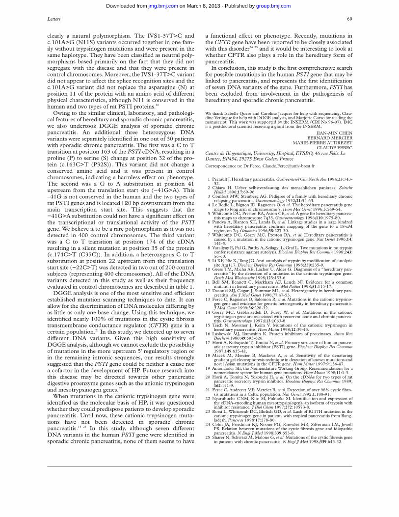
An 8p+ karyotype was reported suggesting an invdup(8p)(p11.2→p23.1) after routine G banding chromo-some analysis. Metaphase chromosomes from EBVimmortalised lymphoblasts from the patient were preparedafter synchronisation with thymidine and incubation withcolcemid by standard techniques. Fluorescence in situhybridisation (FISH) studies performed with a wholechromosome 8 paint (Cambio) showed that the additionalmaterial present in the short arm of the rearrangedchromosome 8 is derived from chromosome 8 (data notshown). To define the breakpoints of the rearrangementmore accurately, dual colour FISH experiments were per-formed using total yeast DNA from YAC clones fromchromosome 8 (HGMP Resource Centre, UK) togetherwith a chromosome 8 specific centromeric probe (Boeh-ringer Mannheim). By analysis of 20 metaphases each itcould be shown that the short arm of the rearranged chro-mosome 8p is dicentric with most of the short arm dupli-cated (cen→p23.1) and inverted (fig 1). The telomericregion distal to p23.1 is deleted. The proximal breakpointseems to be the centromere as the dicentric chromosome 8shows a second centromere at the very end of the tip of theshort arm.
Neither the breakpoints nor the telomeric deletion lay inregions associated with breast carcinomas, which fre-quently show allelic deletions in regions 8p11-p12 and8p21-p22 in sporadic cases.5 6 8p12 is also found to beamplified in 10-15% of breast tumours.7 For the NEFLmarker (8p11-p12), a lod score of 2.5 was obtained bylinkage analysis using families unlinked to BRCA1 orBRCA2 indicating the presence of a putative BRCA3 gene.5
Because samples from other family members of the indexcase were not available for linkage analysis, mutationalanalysis of BRCA1 and BRCA2 was performed in theproband but did not detect any mutation (data not shown).
In conclusion, the presented case of inv dup(8p) showsthe genotype of other reported cases associated with devel-opmental delay and/or mental retardation. The occurrenceof breast cancer is probably coincidental and unrelated tothe chromosome 8p rearrangement.
We thank Ian Roberts for providing help with the image analysis system.This work was supported by grant D/96/03248 to MS from the Deutsche Kreb-shilfe.
M SELTMANNP HARRINGTON
B A J PONDERCRC Human Cancer Genetics Research Group, Box 139, CambridgeInstitute for Medical Research, Cambridge CB2 2XY, UK
L R WILLATTCytogenetics Laboratory, Box 108, Level 2, Addenbrooke’s Hospital, HillsRoad, Cambridge, CB2 2QQ, UK
A C HEPPELL-PARTONMedical Research Council, Hills Road, Cambridge, CB2 2QQ, UK
Figure 1 FISH analysis of diVerent 8p YAC probes (green) incombination with a human chromosome 8 specific centromeric probe (red).(Yellow colour indicates overlap of the signals). (Left) Ideogram of 8pwhich shows the localisation of the probes. The duplicated and invertedportion of the rearranged chromosome 8p is indicated by arrowheads.(Right) Images obtained for the normal and rearranged chromosome 8showing inv dup(8p). The following YAC probes were used from top tobottom: Y787_c_11,Y700_d_3,Y812_g_7,Y936_g_4.
70 Letters
group.bmj.com on March 8, 2013 - Published by jmg.bmj.comDownloaded from
H ANTON-CULVERUniversity of California, Irvine, USA
Correspondence to: Dr Seltmann, Essen University Medical School, VirchowStrasse 173, Institute of Cell Biology, D-45147 Essen, Germany
1 Florida G, Piantanida M, Minelli A, et al. The same molecular mechanismat the maternal meiosis I produces mono- and dicentric 8p duplications.Am J Hum Genet 1996;58:785-96.
2 Guo WJ, Faith CD, Zapata MC, Miller ME. Clinical and cytogenetic find-ings in seven cases of inverted duplication of 8p with evidence of a telo-meric deletion using fluorescence in situ hybridization. Am J Med Genet1995;58:230-6.
3 Feldman GL, Weiss L, Phelan MC, Schroer RJ, Van Dyke DL. Invertedduplication of 8p: ten new patients and review of the literature. Am J MedGenet 1993;47:482-6.
4 Minelli A, Floridia G, Rossi E, et al. D8S7 is consistently deleted in invertedduplications of the short arm of chromosome 8 (inv dup 8p). Hum Genet1993;92:391-6.
5 Kerangueven F, Essioux L, Dib A, et al. Loss of heterozygosity andlinkage analysis in breast carcinoma: indication for a putative thirdsusceptibility gene on the short arm of chromosome 8. Oncogene 1995;10:1023-6.
6 Chuaqui RF, Sanz-Ortega J, Vocke C, et al. Loss of heterozygosity on theshort arm of chromosome 8 in male breast carcinomas. Cancer Res1995;55:4995-8.
7 Dib A, Adelaide J, ChaVanet M, et al. Characterization of the region of theshort arm of chromosome 8 amplified in breast carcinoma. Oncogene 1995;10:995-1001.
8 ChaVanet M, Popovici C. t(6;8) and t(8;13) translocations associated withstem cell myeloproliferative disorders have close or identical breakpoints inchromosome region 8p11-12. Oncogene 1998;16:945-9.
J Med Genet 2000;37:71–75
Appendiceal carcinoma complicatingadenomatous polyposis in a youngwoman with a de novo constitutionalreciprocal translocationt(5;8)(q22;p23.1)
EDITOR—Familial adenomatous polyposis (FAP) is anautosomal dominant condition characterised by thepresence of more than 100 adenomatous polyps in thecolon and rectum. Polyps generally first appear in the sec-ond or third decade of life and are usually most numerousdistally. Left untreated, colorectal cancer is virtually inevi-table and generally arises in the fourth or fifth decade.1
Adenocarcinoma of the appendix is an uncommonneoplasm and has only rarely been reported in associationwith FAP.2
The gene responsible for FAP, APC, was initiallylocalised to the long arm of chromosome five (5q) bylinkage.3 4 This followed a case report describing carcino-mas of the rectum and ascending colon, adenomatouspolyposis, mental retardation, and various dysmorphic fea-
tures in a 42 year old man with a constitutional deletion of5q.5 Most patients with FAP have normal karyotypes.6
Mental retardation and dysmorphic features are unusual insuch people but characterise those rare patients withcytogenetically visible 5q deletions and FAP.5–12 The fewreports detailing the clinical findings in patients with sub-microscopic deletions of APC suggest that such people maybe mentally normal.13 14
In this report we describe a patient with adenomatouspolyposis, mental retardation, and an apparently balancedtranslocation t(5;8)(q22;p23.1) causing submicroscopicdeletion of APC and MCC.
Clinical data were obtained by review of medicalrecords. In addition, the patient was interviewed andexamined by two of the authors (JF and AS) before herdeath. Cytogenetic studies were performed using standardtechniques on a 72 hour peripheral blood culture withGTG banding, as previously reported.6
Slides for fluorescence in situ hybridisation (FISH) wereobtained using the cell suspension retained after routinecytogenetic harvest. RNAse treatment, probe and chromo-somal denaturation, and hybridisation conditions were aspreviously described15 with the stringencies adjusted afterassessment of the optimal conditions for each probe com-bination. The biotinylated probes were detected with
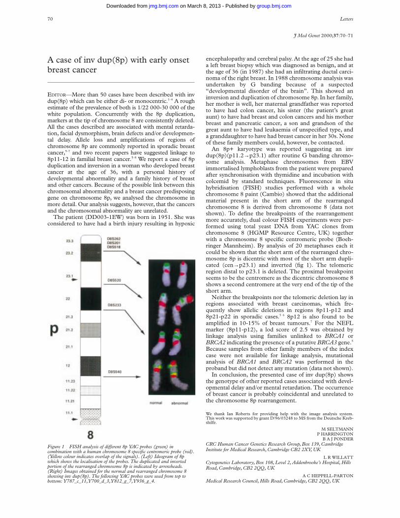
Figure 1 (A, B) The patient aged 26 years.
Letters 71
group.bmj.com on March 8, 2013 - Published by jmg.bmj.comDownloaded from
avidin-FITC (Calbiochem) followed by biotinylated anti-avidin (Vector) and finally avidin-FITC. The digoxigeninlabelled probes were detected with mouse anti-digoxigeninfollowed by sheep anti-mouse Ig-rhodamine conjugatedantibody (Boehringer Mannheim). Chromosomes, coun-terstained with DAPI (Sigma) and visualised by fluores-cence microscopy (Zeiss), were captured using a computerimage analysis system (Cytovision).
The following probes were used in FISH studies: MCC40CI (partial MCC cDNA nucleotides 1634-3969),FB70B (partial APC cDNA nucleotides 2877-6452), andAPC (full length APC cDNA) each kindly provided by DrBert Vogelstein (Johns Hopkins Oncology Center, Balti-more); D5S23 (chromosome 5p probe, Vysis); EGR-1(chromosome 5q31 probe, Vysis); CHR8B/wcp8 (chromo-some 8 library probe, Cambio); D8Z1 (chromosome 8centromere probe, Oncor).
Genomic DNA was extracted from blood samples usingthe Instagene purification matrix (Biorad) according to themanufacturer’s instructions. Each polymerase chain reac-tion (PCR) used 50-100 ng genomic DNA, 50 pmol/l ofeach oligonucleotide primer, 0.2 mmol/l of each dNTP(Pharmacia), 1.25 U Amplitaq DNA polymerase (PerkinElmer) in Amplitaq reaction buVer (10 mmol/l Tris-HCl,50 mmol/l KCl, MgCl optimised for each primer pair, pH8.3) to a final volume of 25 µl. Thirty five amplificationcycles were performed using an FTS-320 ThermalSequencer (Corbett Research).
An RsaI polymorphism in exon 11 of APC16 was analysedby amplification of exon 11 and digestion of the PCR
product with RsaI (Boehringer). Reaction products wereseparated by electrophoresis in a 10% non-denaturingpolyacrylamide minigel (Biorad) and visualised by ethid-ium bromide staining. The 255 bp amplification productyielded digestion fragments of 155 bp and 100 bp. A CArepeat polymorphism within MCC (CAMBC)17 and a CArepeat polymorphism between APC and MCC(LNS-CA)13 18 were labelled by [áS35]dATP incorporationduring PCR and characterised by electrophoresis in a 6%denaturing polyacrylamide gel and autoradiography aspreviously reported.19
The patient (fig 1) was the second child of a 32 year oldmother and a 33 year old father. Her older brother was welland mentally normal. She was born prematurely at 29weeks’ gestation after an uncomplicated pregnancy. Therewere no major problems in the neonatal period but she wasslow to speak and did not walk until the age of 2 years. Atthat time she began to have generalised convulsions. Thesewere only partially controlled by medication and continueduntil the age of 18. Her performance at school was poor.Her hospital file notes an estimated IQ between 70 and 80at the age of 10 although the method of assessment was notrecorded. Physical examination at this time showedcrowded dentition and she later required extensive dentalwork. The posterior hairline was noted to be low. The headcircumference was normal (75th centile) while height wason the 10th centile. The third and fourth toes were shortwith the fifth toe longer than the fourth. Bilateral genurecurvatum was evident and the patient was mildly ataxic.There were no skin lesions and the fundi were normal.
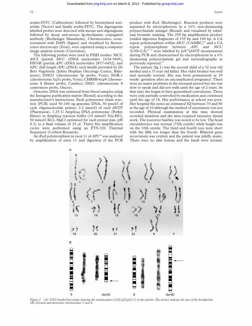
Figure 2 (A) GTG banded karyotype showing the translocation t(5;8)(q22;p23.1) in the patient. The arrows indicate the sites of the breakpoints.(B) Normal and derivative chromosomes 5 and 8.
1 2 3 4 5
1211109876
13 14 15 16 17
X Y22212019
18
A
B
5 der(5) 8 der(8)
72 Letters
group.bmj.com on March 8, 2013 - Published by jmg.bmj.comDownloaded from

Visual acuity and hearing were unimpaired. An EEG at theage of 4 was abnormal with excessive generalised slow waveactivity. A cerebral CT scan at 9 years showed mildventricular dilatation and localised atrophy in the frontalareas and bordering the interhemispheric commissure. Sheattended a special school until the age of 17 and wasactively involved in sports. Subsequently she workedsuccessfully in a sheltered workshop while living independ-ently with a group of mildly handicapped people.
She presented acutely at 26 years of age with a 24 hourhistory of right iliac fossa pain. She was febrile and hadguarding and rebound tenderness at the site of her pain. Adiagnosis of acute appendicitis was made and an appendi-cectomy performed. The appendix contained a mucinsecreting carcinoma arising in a dysplastic villous adenomaand invading through the full thickness of the muscle wall.A right hemicolectomy was performed. The appendicealstump had foci of adenomatous change but there was noresidual carcinoma. Numerous small adenomatous polypswere noted throughout the right colon but the exactnumber was not recorded. Sigmoidoscopy subsequentlyshowed left sided polyps but these were less numerous thanhad been found proximally. Eight months after hemicolec-tomy she presented again with a painful right iliac fossamass. Laparotomy confirmed local tumour recurrence
which could be only partially excised. She was treated with5-fluorouracil and folinic acid but died approximately 12months later. A post mortem examination was notperformed.
Cytogenetic analysis showed a female karyotype with atranslocation involving chromosomes 5 and 8 at break-points q22 and p23.1 respectively (fig 2). The translocationwas cytogenetically balanced. Both parents had normalkaryotypes. The patient’s brother refused testing.
FISH studies were performed using several probe com-binations: APC, D5S23, and EGR-1; FB70B and D5S23;FB70B and D8Z1; MCC 40CI and CHR8B/wcp8 (fig 3);and MCC 40CI and D8Z1. These studies confirmed thetranslocation and showed submicroscopic deletion of bothAPC and MCC. The APC and MCC probes hybridisedonly to the normal chromosome 5 and not to the der(5) order(8) chromosomes.
To confirm that the translocation identified had resultedin deletion of the APC and MCC genes, intragenicpolymorphisms were examined in DNA from the patientand her parents. The RsaI polymorphism in exon 11 ofAPC was informative in the family. The patient’s motherwas homozygous for absence of the restriction site whereasher father was homozygous for presence of the restrictionsite. The patient had only a maternally derived allele, sug-
Figure 3 FISH study using the chromosome 8 library probe, CHR8B/wcp8, labelled green and the MCC gene probe, MCC 40CI, labelled red. An MCCsignal is seen only on the normal chromosome 5 (large arrow) and not on either the der(8) (asterisk) or the der(5) (star) chromosomes. The normalchromosome 8 is indicated by a small arrow.
Figure 4 Exon 11 of APC was amplified from the patient (P), her father (F), and her mother (M). The PCR product from each source waselectrophoresed in a 10% polyacrylamide gel either with (+) or without (−) preceding digestion with RsaI. The first lane shows a molecular weight marker(MspI digest of pBR322).
F
–
F
+
P
–
P
+
M
–
M
+
Letters 73
group.bmj.com on March 8, 2013 - Published by jmg.bmj.comDownloaded from
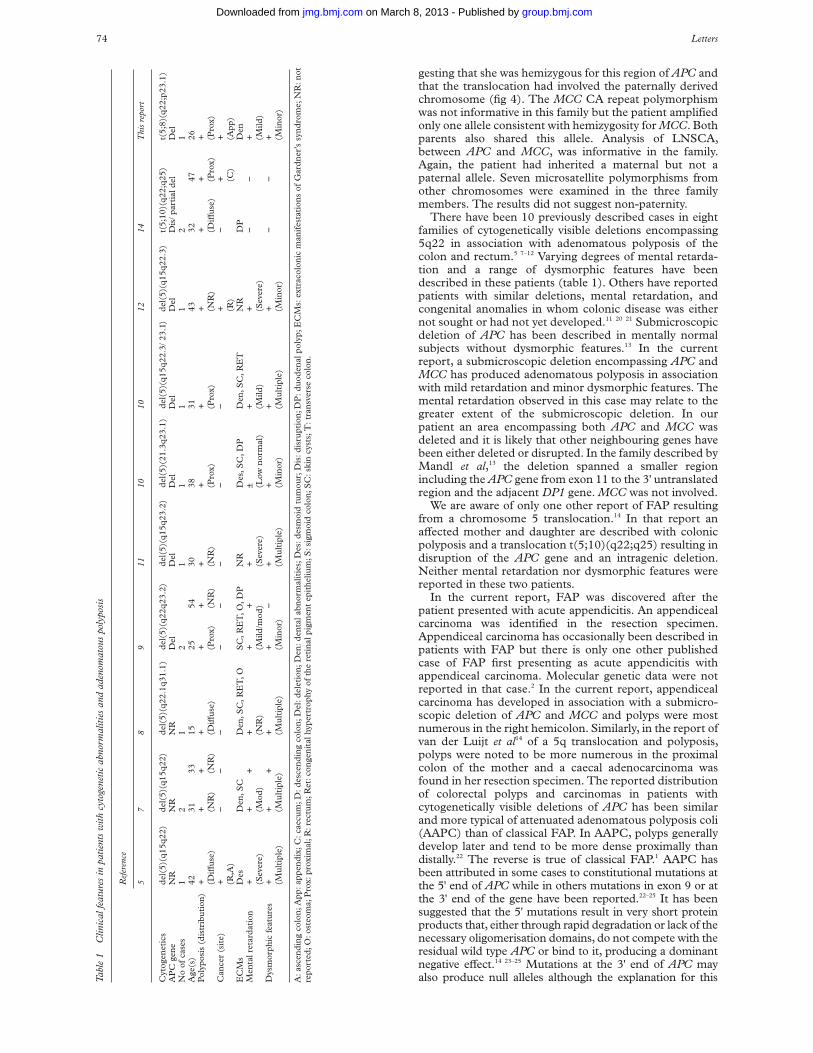
gesting that she was hemizygous for this region of APC andthat the translocation had involved the paternally derivedchromosome (fig 4). The MCC CA repeat polymorphismwas not informative in this family but the patient amplifiedonly one allele consistent with hemizygosity for MCC. Bothparents also shared this allele. Analysis of LNSCA,between APC and MCC, was informative in the family.Again, the patient had inherited a maternal but not apaternal allele. Seven microsatellite polymorphisms fromother chromosomes were examined in the three familymembers. The results did not suggest non-paternity.
There have been 10 previously described cases in eightfamilies of cytogenetically visible deletions encompassing5q22 in association with adenomatous polyposis of thecolon and rectum.5 7–12 Varying degrees of mental retarda-tion and a range of dysmorphic features have beendescribed in these patients (table 1). Others have reportedpatients with similar deletions, mental retardation, andcongenital anomalies in whom colonic disease was eithernot sought or had not yet developed.11 20 21 Submicroscopicdeletion of APC has been described in mentally normalsubjects without dysmorphic features.13 In the currentreport, a submicroscopic deletion encompassing APC andMCC has produced adenomatous polyposis in associationwith mild retardation and minor dysmorphic features. Themental retardation observed in this case may relate to thegreater extent of the submicroscopic deletion. In ourpatient an area encompassing both APC and MCC wasdeleted and it is likely that other neighbouring genes havebeen either deleted or disrupted. In the family described byMandl et al,13 the deletion spanned a smaller regionincluding the APC gene from exon 11 to the 3' untranslatedregion and the adjacent DP1 gene. MCC was not involved.
We are aware of only one other report of FAP resultingfrom a chromosome 5 translocation.14 In that report anaVected mother and daughter are described with colonicpolyposis and a translocation t(5;10)(q22;q25) resulting indisruption of the APC gene and an intragenic deletion.Neither mental retardation nor dysmorphic features werereported in these two patients.
In the current report, FAP was discovered after thepatient presented with acute appendicitis. An appendicealcarcinoma was identified in the resection specimen.Appendiceal carcinoma has occasionally been described inpatients with FAP but there is only one other publishedcase of FAP first presenting as acute appendicitis withappendiceal carcinoma. Molecular genetic data were notreported in that case.2 In the current report, appendicealcarcinoma has developed in association with a submicro-scopic deletion of APC and MCC and polyps were mostnumerous in the right hemicolon. Similarly, in the report ofvan der Luijt et al14 of a 5q translocation and polyposis,polyps were noted to be more numerous in the proximalcolon of the mother and a caecal adenocarcinoma wasfound in her resection specimen. The reported distributionof colorectal polyps and carcinomas in patients withcytogenetically visible deletions of APC has been similarand more typical of attenuated adenomatous polyposis coli(AAPC) than of classical FAP. In AAPC, polyps generallydevelop later and tend to be more dense proximally thandistally.22 The reverse is true of classical FAP.1 AAPC hasbeen attributed in some cases to constitutional mutations atthe 5' end of APC while in others mutations in exon 9 or atthe 3' end of the gene have been reported.22–25 It has beensuggested that the 5' mutations result in very short proteinproducts that, either through rapid degradation or lack of thenecessary oligomerisation domains, do not compete with theresidual wild type APC or bind to it, producing a dominantnegative eVect.14 23–25 Mutations at the 3' end of APC mayalso produce null alleles although the explanation for thisTa
ble
1C
linic
alfe
atur
esin
patie
nts
with
cyto
gene
ticab
norm
aliti
esan
dad
enom
atou
spo
lypo
sis
Ref
eren
ce
57
89
1110
1012
14T
his
repo
rt
Cyt
ogen
etic
sd
el(5
)(q
15q
22)
del
(5)(
q15
q22
)d
el(5
)(q
22.1
q31
.1)
del
(5)(
q22
q23
.2)
del
(5)(
q15
q23
.2)
del
(5)(
21.3
q23
.1)
del
(5)(
q15
q22
.3/2
3.1)
del
(5)(
q15
q22
.3)
t(5;
10)(
q22
;q25
)t(
5;8)
(q22
;p23
.1)
AP
Cge
ne
NR
NR
NR
Del
Del
Del
Del
Del
Dis
/par
tial
del
Del
No
ofca
ses
12
12
11
11
21
Age
(s)
4231
3315
2554
3038
3143
3247
26P
olyp
osis
(dis
trib
uti
on)
++
++
++
++
++
++
+(D
iVu
se)
(NR
)(N
R)
(DiV
use
)(P
rox)
(NR
)(N
R)
(Pro
x)(P
rox)
(NR
)(D
iVu
se)
(Pro
x)(P
rox)
Can
cer
(sit
e)+
−−
−−
−−
−−
+−
++
(R,A
)(R
)(C
)(A
pp
)E
CM
sD
esD
en,S
CD
en,S
C,R
ET
,OS
C,R
ET
,O,D
PN
RD
es,S
C,D
PD
en,S
C,R
ET
NR
DP
Den
Men
talr
etar
dat
ion
++
++
++
+±
++
−−
+(S
ever
e)(M
od)
(NR
)(M
ild/m
od)
(Sev
ere)
(Low
nor
mal
)(M
ild)
(Sev
ere)
(Mild
)D
ysm
orph
icfe
atu
res
++
++
+−
++
++
−−
+(M
ult
iple
)(M
ult
iple
)(M
ult
iple
)(M
inor
)(M
ult
iple
)(M
inor
)(M
ult
iple
)(M
inor
)(M
inor
)
A:a
scen
din
gco
lon
;Ap
p:a
pp
end
ix;C
:cae
cum
;D:d
esce
nd
ing
colo
n;D
el:d
elet
ion
;Den
:den
tal
abn
orm
alit
ies;
Des
:des
moi
dtu
mou
r;D
is:d
isru
pti
on;D
P:d
uod
enal
pol
yp;E
CM
s:ex
trac
olon
icm
anif
esta
tion
sof
Gar
dn
er’s
syn
dro
me;
NR
:not
repo
rted
;O:o
steo
ma;
Pro
x:pr
oxim
al;R
:rec
tum
;Ret
:con
gen
ital
hype
rtro
phy
ofth
ere
tin
alpi
gmen
tep
ithe
lium
;S:s
igm
oid
colo
n;S
C:s
kin
cyst
s;T
:tra
nsv
erse
colo
n.
74 Letters
group.bmj.com on March 8, 2013 - Published by jmg.bmj.comDownloaded from
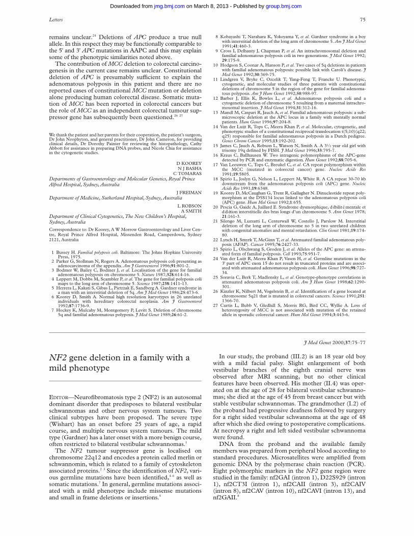
remains unclear.24 Deletions of APC produce a true nullallele. In this respect they may be functionally comparable tothe 5' and 3' APC mutations in AAPC and this may explainsome of the phenotypic similarities noted above.
The contribution of MCC deletion to colorectal carcino-genesis in the current case remains unclear. Constitutionaldeletion of APC is presumably suYcient to explain theadenomatous polyposis in this patient and there are noreported cases of constitutional MCC mutation or deletionalone producing human colorectal disease. Somatic muta-tion of MCC has been reported in colorectal cancers butthe role of MCC as an independent colorectal tumour sup-pressor gene has subsequently been questioned.26 27
We thank the patient and her parents for their cooperation, the patient’s surgeon,Dr John Neophyton, and general practitioner, Dr John Cameron, for providingclinical details, Dr Dorothy Painter for reviewing the histopathology, CathyAbbott for assistance in preparing DNA probes, and Nicole Chia for assistancein the cytogenetic studies.
D KOOREYN J BASHA
C TOMARASDepartments of Gastroenterology and Molecular Genetics, Royal PrinceAlfred Hospital, Sydney, Australia
J FREIMANDepartment of Medicine, Sutherland Hospital, Sydney, Australia
L ROBSONA SMITH
Department of Clinical Cytogenetics, The New Children’s Hospital,Sydney, Australia
Correspondence to: Dr Koorey, A W Morrow Gastroenterology and Liver Cen-tre, Royal Prince Alfred Hospital, Missenden Road, Camperdown, Sydney2121, Australia
1 Bussey H. Familial polyposis coli. Baltimore: The Johns Hopkins UniversityPress, 1975.
2 Parker G, Stollman N, Rogers A. Adenomatous polyposis coli presenting asadenocarcinoma of the appendix. Am J Gastroenterol 1996;91:801-2.
3 Bodmer W, Bailey C, Bodmer J, et al. Localisation of the gene for familialadenomatous polyposis on chromosome 5. Nature 1987;328:614-16.
4 Leppert M, Dobbs M, Scambler P, et al. The gene for familial polyposis colimaps to the long arm of chromosome 5. Science 1987;238:1411-13.
5 Herrera L, Kakati S, Gibas L, Pietrzak E, Sandberg A. Gardner syndrome ina man with an interstitial deletion of 5q. Am J Med Genet 1986;25:473-6.
6 Koorey D, Smith A. Normal high resolution karyotypes in 26 unrelatedindividuals with hereditary colorectal neoplasia. Am J Gastroenterol1992;87:1736-9.
7 Hockey K, Mulcahy M, Montgomery P, Levitt S. Deletion of chromosome5q and familial adenomatous polyposis. J Med Genet 1989;26:61-2.
8 Kobayashi T, Narahara K, Yokoyama Y, et al. Gardner syndrome in a boywith interstitial deletion of the long arm of chromosome 5. Am J Med Genet1991;41:460-3.
9 Cross I, Delhanty J, Chapman P, et al. An intrachromosomal deletion andfamilial adenomatous polyposis coli in two generations. J Med Genet 1992;29:175-9.
10 Hodgson S, Coonar A, Hanson P, et al. Two cases of 5q deletions in patientswith familial adenomatous polyposis: possible link with Caroli’s disease. JMed Genet 1992;30:369-75.
11 Lindgren V, Bryke C, Ozcelik T, Yang-Feng T, Francke U. Phenotypic,cytogenetic, and molecular studies of three patients with constitutionaldeletions of chromosome 5 in the region of the gene for familial adenoma-tous polyposis. Am J Hum Genet 1992;50:988-97.
12 Barber J, Ellis K, Bowles L, et al. Adenomatous polyposis coli and acytogenetic deletion of chromosome 5 resulting from a maternal intrachro-mosomal insertion. J Med Genet 1994;31:312-16.
13 Mandl M, Caspari R, Jauch A, et al. Familial adenomatous polyposis: a sub-microscopic deletion at the APC locus in a family with mentally normalpatients. Hum Genet 1996;97:204-8.
14 Van der Luijt R, Tops C, Meera Khan P, et al. Molecular, cytogenetic, andphenotypic studies of a constitutional reciprocal translocation t(5;10)(q22;q25) responsible for familial adenomatous polyposis in a Dutch pedigree.Genes Chrom Cancer 1995;13:192-202.
15 James C, Jauch A, Robson L, Watson N, Smith A. A 31⁄2 year old girl withtrisomy 19q defined by FISH. J Med Genet 1996;33:795-7.
16 Kraus C, Ballhausen W. Two intragenic polymorphisms of the APC-genedetected by PCR and enzymatic digestion. Hum Genet 1992;88:705-6.
17 Van Leeuwen C, Tops C, Breukel C, et al. CA repeat polymorphism withinthe MCC (mutated in colorectal cancer) gene. Nucleic Acids Res1991;19:5805.
18 Spirio L, Joslyn G, Nelson L, Leppert M, White R. A CA repeat 30-70 kbdownstream from the adenomatous polyposis coli (APC) gene. NucleicAcids Res 1991;19:6348.
19 Koorey D, McCaughan G, Trent R, Gallagher N. Dinucleotide repeat poly-morphism at the D5S134 locus linked to the adenomatous polyposis coli(APC) gene. Hum Mol Genet 1992;1:655.
20 Pescia G, Gaide A, Juillard E. Syndrome dysmorphique, débilité mentale etdélétion interstitielle des bras longs d’un chromosome 5. Ann Genet 1978;21:161-3.
21 Silengo M, Luzzatti L, Centerwall W, Costello J, Parslow M. Interstitialdeletion of the long arm of chromosome no 5 in two unrelated childrenwith congenital anomalies and mental retardation. Clin Genet 1981;19:174-80.
22 Lynch H, Smyrk T, McGinn T, et al. Attenuated familial adenomatous poly-posis (AFAP). Cancer 1995;76:2427-33.
23 Spirio L, Olschwang S, Groden J, et al. Alleles of the APC gene: an attenu-ated form of familial polyposis. Cell 1993;75:951-7.
24 Van der Luijt R, Meera Khan P, Vasen H, et al. Germline mutations in the3' part of APC exon 15 do not result in truncated proteins and are associ-ated with attenuated adenomatous polyposis coli. Hum Genet 1996;98:727-34.
25 Soravia C, Berk T, Madlensky L, et al. Genotype-phenotype correlations inattenuated adenomatous polyposis coli. Am J Hum Genet 1998;62:1290-301.
26 Kinzler K, Nilbert M, Vogelstein B, et al. Identification of a gene located atchromosome 5q21 that is mutated in colorectal cancers. Science 1991;251:1366-70.
27 Curtis L, Bubb V, Gledhill S, Morris RG, Bird CC, Wyllie A. Loss ofheterozygosity of MCC is not associated with mutation of the retainedallele in sporadic colorectal cancer. Hum Mol Genet 1994;3:443-6.
J Med Genet 2000;37:75–77
NF2 gene deletion in a family with amild phenotype
EDITOR—Neurofibromatosis type 2 (NF2) is an autosomaldominant disorder that predisposes to bilateral vestibularschwannomas and other nervous system tumours. Twoclinical subtypes have been proposed. The severe type(Wishart) has an onset before 25 years of age, a rapidcourse, and multiple nervous system tumours. The mildtype (Gardner) has a later onset with a more benign course,often restricted to bilateral vestibular schwannomas.1
The NF2 tumour suppressor gene is localised onchromosome 22q12 and encodes a protein called merlin orschwannomin, which is related to a family of cytoskeletonassociated proteins.2 3 Since the identification of NF2, vari-ous germline mutations have been identified,4–6 as well assomatic mutations.7 In general, germline mutations associ-ated with a mild phenotype include missense mutationsand small in frame deletions or insertions.5
In our study, the proband (III.2) is an 18 year old boywith a mild facial palsy. Slight enlargement of bothvestibular branches of the eighth cranial nerve wasobserved after MRI scanning, but no other clinicalfeatures have been observed. His mother (II.4) was oper-ated on at the age of 28 for bilateral vestibular schwanno-mas; she died at the age of 45 from breast cancer but withstable vestibular schwannomas. The grandmother (I.2) ofthe proband had progressive deafness followed by surgeryfor a right sided vestibular schwannoma at the age of 48after which she died owing to postoperative complications.At necropsy a right and left sided vestibular schwannomawere found.
DNA from the proband and the available familymembers was prepared from peripheral blood according tostandard procedures. Microsatellites were amplified fromgenomic DNA by the polymerase chain reaction (PCR).Eight polymorphic markers in the NF2 gene region werestudied in the family: nf2GAI (intron 1), D22S929 (intron1), nf2CT3I (intron 1), nf2CAII (intron 3), nf2CAIV(intron 8), nf2CAV (intron 10), nf2CAVI (intron 13), andnf2GAII.8
Letters 75
group.bmj.com on March 8, 2013 - Published by jmg.bmj.comDownloaded from
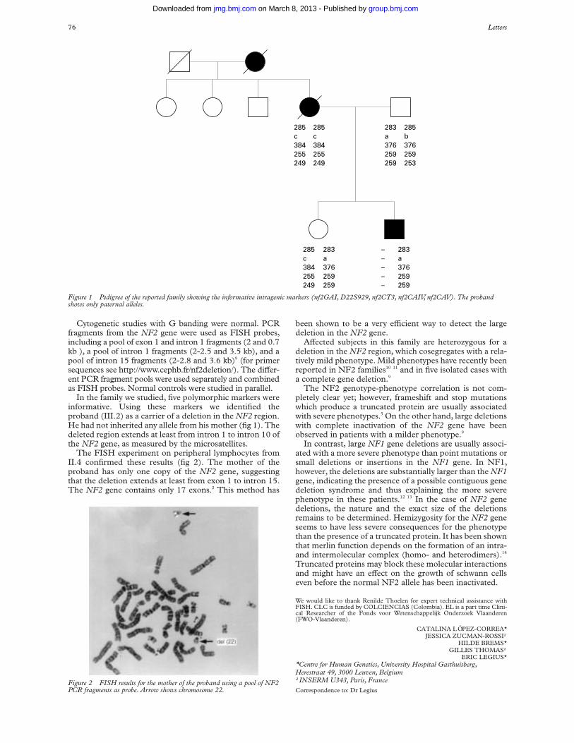
Cytogenetic studies with G banding were normal. PCRfragments from the NF2 gene were used as FISH probes,including a pool of exon 1 and intron 1 fragments (2 and 0.7kb ), a pool of intron 1 fragments (2-2.5 and 3.5 kb), and apool of intron 15 fragments (2-2.8 and 3.6 kb)9 (for primersequences see http://www.cephb.fr/nf2deletion/). The diVer-ent PCR fragment pools were used separately and combinedas FISH probes. Normal controls were studied in parallel.
In the family we studied, five polymorphic markers wereinformative. Using these markers we identified theproband (III.2) as a carrier of a deletion in the NF2 region.He had not inherited any allele from his mother (fig 1). Thedeleted region extends at least from intron 1 to intron 10 ofthe NF2 gene, as measured by the microsatellites.
The FISH experiment on peripheral lymphocytes fromII.4 confirmed these results (fig 2). The mother of theproband has only one copy of the NF2 gene, suggestingthat the deletion extends at least from exon 1 to intron 15.The NF2 gene contains only 17 exons.2 This method has
been shown to be a very eYcient way to detect the largedeletion in the NF2 gene.
AVected subjects in this family are heterozygous for adeletion in the NF2 region, which cosegregates with a rela-tively mild phenotype. Mild phenotypes have recently beenreported in NF2 families10 11 and in five isolated cases witha complete gene deletion.9
The NF2 genotype-phenotype correlation is not com-pletely clear yet; however, frameshift and stop mutationswhich produce a truncated protein are usually associatedwith severe phenotypes.5 On the other hand, large deletionswith complete inactivation of the NF2 gene have beenobserved in patients with a milder phenotype.9
In contrast, large NF1 gene deletions are usually associ-ated with a more severe phenotype than point mutations orsmall deletions or insertions in the NF1 gene. In NF1,however, the deletions are substantially larger than the NF1gene, indicating the presence of a possible contiguous genedeletion syndrome and thus explaining the more severephenotype in these patients.12 13 In the case of NF2 genedeletions, the nature and the exact size of the deletionsremains to be determined. Hemizygosity for the NF2 geneseems to have less severe consequences for the phenotypethan the presence of a truncated protein. It has been shownthat merlin function depends on the formation of an intra-and intermolecular complex (homo- and heterodimers).14
Truncated proteins may block these molecular interactionsand might have an eVect on the growth of schwann cellseven before the normal NF2 allele has been inactivated.
We would like to thank Renilde Thoelen for expert technical assistance withFISH. CLC is funded by COLCIENCIAS (Colombia). EL is a part time Clini-cal Researcher of the Fonds voor Wetenschappelijk Onderzoek Vlaanderen(FWO-Vlaanderen).
CATALINA LÓPEZ-CORREA*JESSICA ZUCMAN-ROSSI†
HILDE BREMS*GILLES THOMAS†
ERIC LEGIUS**Centre for Human Genetics, University Hospital Gasthuisberg,Herestraat 49, 3000 Leuven, Belgium†INSERM U343, Paris, FranceCorrespondence to: Dr Legius
Figure 1 Pedigree of the reported family showing the informative intragenic markers (nf2GAI, D22S929, nf2CT3, nf2CAIV, nf2CAV). The probandshows only paternal alleles.
285
384255249
c285
384255249
c283
376259259
a285
376259253
b
285
384255249
c283
376259259
a–
–––
–283
376259259
a
Figure 2 FISH results for the mother of the proband using a pool of NF2PCR fragments as probe. Arrow shows chromosome 22.
76 Letters
group.bmj.com on March 8, 2013 - Published by jmg.bmj.comDownloaded from
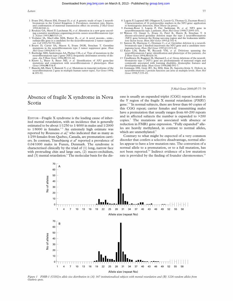
1 Evans DG, Huson SM, Donnai D, et al. A genetic study of type 2 neurofi-bromatosis in the United Kingdom. I. Prevalence, mutation rate, fitness,and confirmation of maternal transmission eVect on severity. J Med Genet1992;12:841-6.
2 Rouleau GA, Merel P, Lutchman M, et al. Alteration in a new gene encod-ing a putative membrane-organizing protein causes neurofibromatosis type2. Nature 1993;363:515-21.
3 Trofatter JA, MacCollin MM, Rutter JL, et al. A novel moesin-, erzin-,radixin-like gene is a candidate for the neurofibromatosis 2 tumor suppres-sor. Cell 1993;72:791-800.
4 Bourn D, Carter SA, Mason S, Evans DGR, Strachan T. Germlinemutations in the neurofibromatosis type 2 tumor suppressor gene. HumMol Genet 1994;3:813-16.
5 Ruttledge MH, Andermann AA, Phelan CM, et al. Type of mutations in theneurofibromatosis type 2 gene (NF2) frequently determines severity of dis-ease. Am J Hum Genet 1996;59:331-42.
6 Kluwe L, Bayer S, Baser ME, et al. Identification of NF2 germ-linemutations and comparison with neurofibromatosis 2 phenotypes. HumGenet 1996;98:534-8.
7 Bianchi AB, Hara T, Ramesh V, et al. Mutations in trancript isoforms of theneurofibromatosis 2 gene in multiple human tumor types. Nat Genet 1994;6:185-92.
8 Legoix P, Legrand MF, Ollagnon E, Lenoir G, Thomas G, Zucman-Rossi J.Characterisation of 16 polymorphic markers in the NF2 gene: applicationto hemizygosity detection. Hum Mutat (in press).
9 Zucman-Rossi J, Legoix P, Der Sarkissian H, et al. NF2 gene inneurofibromatosis type 2 patients. Hum Mol Genet 1998;7:2095-101.
10 Watson CJ, Gaunt L, Evans G, Patel K, Harris R, Strachan T. Adisease-associated germline deletion maps the type 2 neurofibromatosis(NF2) gene between the Ewing sarcoma region and the leukaemia inhibi-tory factor locus. Hum Mol Genet 1993;2:701-4.
11 Sanson M, Marineau C, Desmaze C, et al. Germline deletion in a neurofi-bromatosis type 2 kindred inactivates the NF2 gene and a candidate men-ingioma locus. Hum Mol Genet 1993;2:1215-20.
12 Kayes LM, Burke W, Riccardi VM, et al. Deletions spanning theneurofibromatosis I gene: identification and phenotype of five patients. AmJ Hum Genet 1994;54:424-36.
13 Upadhyaya M, Ruggieri M, Maynard J, et al. Gross deletions of the neurofi-bromatosis type 1 (NF1) gene are predominantly of maternal origin andcommonly associated with learning disability, dysmorphic features anddevelopmental delay. Hum Genet 1998;102:591-7.
14 Gutmann DH, Geist RT, Xu HM, Kim JS, Saporito-Irwin S. Defects inneurofibromatosis 2 protein function can arise at multiple levels. Hum MolGenet 1998;7:335-45.
J Med Genet 2000;37:77–79
Absence of fragile X syndrome in NovaScotia
EDITOR—Fragile X syndrome is the leading cause of inher-ited mental retardation, with an incidence that is generallyestimated to be about 1/1250 to 1/4000 in males and 1/2000to 1/8000 in females.1–3 An extremely high estimate wasreported by Rousseau et al,4 who indicated that as many as1/259 females from Quebec, Canada, are premutation carri-ers. In contrast, Tranebjaerg et al5 reported a prevalence of0.04/1000 males in Funen, Denmark. The syndrome ischaracterised clinically by the triad of (1) long, narrow facewith protruding chin and large ears, (2) macro-orchidism,and (3) mental retardation.6 The molecular basis for the dis-
ease is usually an expanded triplet (CGG) repeat located inthe 5' region of the fragile X mental retardation (FMR1)gene.7 8 In normal subjects, there are fewer than 60 copies ofthis CGG repeat; carrier females and transmitting maleshave a premutation that usually ranges from 60-200 repeatsand in aVected subjects the number is expanded to >200copies.7 The mutations are associated with absence orreduction in FMR1 gene expression. “Fully expanded” alle-les are heavily methylated, in contrast to normal alleles,which are unmethylated.9
Contrary to what might be expected of a very commondisorder that confers a selective disadvantage, normal alle-les appear to have a low mutation rate. The conversion of anormal allele to a premutation, or to a full mutation, hasnot been reported.10 Indirect evidence of a low mutationrate is provided by the finding of founder chromosomes.11
Figure 1 FMR-1 (CGG)n allele size distribution in (A) 167 institutionalised subjects with mental retardation and (B) 1226 random alleles fromGuthrie spots.
70A
B
60
50
40
30
20
10
0
Allele size (repeat No)
No
of
alle
les
5855524946434037343128252219161310741
70
60
50
40
30
20
10
0
Allele size (repeat No)
No
of
alle
les
5855524946434037343128252219161310741
Letters 77
group.bmj.com on March 8, 2013 - Published by jmg.bmj.comDownloaded from
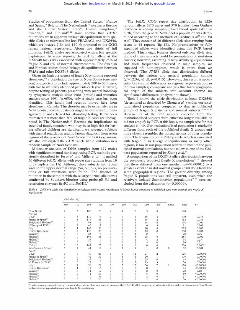
Studies of populations from the United States,12 Franceand Spain,13 Belgium/The Netherlands,14 northern Europeand the United States,15 Italy16, United Kingdom,17
Sweden,18 and Finland19–21 have shown that FMR1mutations are in apparent linkage disequilibrium with spe-cific alleles at microsatellite loci FRAXAC1 and DXS548,which are located 7 kb and 150 kb proximal to the CGGrepeat region, respectively. About two thirds of fullmutation FMR1 alleles are associated with a few specifichaplotypes. In some reports, the 204 bp allele at theDXS548 locus was associated with approximately 25% offragile X and 8% of normal chromosomes. The Swedishand Finnish studies found linkage disequilibrium betweenFMR1 and other DXS548 alleles.
Given the high prevalence of fragile X syndrome reportedelsewhere,1–3 a population the size of Nova Scotia (one mil-lion) is expected to include approximately 200 to 550 cases,with two to six newly identified patients each year. However,despite testing of patients presenting with mental handicapby cytogenetic analysis since 1980 (n=423) and mutationanalysis since 1991 (n>650), only a single case has beenidentified. This family had recently moved here fromelsewhere in Canada. This disorder may be extremely rare inNova Scotia; however, patients might be overlooked, misdi-agnosed, or not referred for laboratory testing. It has beenestimated that more than 50% of fragile X cases are undiag-nosed in The Netherlands.22 Because the implications toextended family members who may be at high risk for hav-ing aVected children are significant, we screened subjectswith mental retardation and no known diagnosis from sevenregions of the province of Nova Scotia for CGG expansion.We also investigated the FMR1 allele size distribution in arandom sample of Nova Scotians.
Molecular analyses of DNA samples from 177 maleswith significant mental handicap, using PCR methods pre-viously described by Fu et al7 and Miller et al,23 identified30 diVerent FMR1 alleles with repeat sizes ranging from 19to 59 triplets (fig 1A). Although three subjects had repeatsizes in the upper normal range (54, 57, 59), no premuta-tions or full mutations were found. The absence ofmutation in the samples with these large normal alleles wasconfirmed by Southern blotting using probe pE 5.1 andrestriction enzymes EcoRI and BssHII.8
The FMR1 CGG repeat size distribution in 1226random alleles (470 males and 378 females) from Guthrienewborn screening samples (dried blood spots taken atbirth) from the general Nova Scotia population was deter-mined according to the methods of Carduci et al24 and Fuet al.7 They contained 36 diVerent allele sizes ranging fromseven to 55 repeats (fig 1B). No premutations or fullyexpanded alleles were identified using this PCR basedmethod. Thirty eight females showed only one allele size.Some of these subjects could be premutation or mutationcarriers; however, assuming Hardy-Weinberg equilibriumand allele frequencies observed in male samples, weexpected 45 homozygotes, which is more than weobserved. The FMR1 allele size distribution diVeredbetween the patient and general population sample(÷2=12.34, 42 df, p=0.015). However, this result is appar-ently because of diVerences in regional representation inthe two samples; chi-square analysis that takes geographi-cal origin of the subjects into account showed nosignificance diVerences (analysis not shown).
Table 1 shows the allele distribution at locus DXS548(determined as described by Zhong et al12) within our insti-tutionalised population compared to that in publishedgroups of fragile X patients and normal subjects.12–21 25 26
Because 37 of the 177 samples collected from theinstitutionalised subjects were either no longer available ordid not amplify by PCR at this locus, the sample size for thisanalysis is 140. Our institutionalised population is markedlydiVerent from each of the published fragile X groups andmore closely resembles the normal groups of other popula-tions. The frequency of the 204 bp allele, which is associatedwith fragile X in linkage disequilibrium in many otherregions, is low in our population relative to most of the pub-lished normal populations, but not as low as one of the Chi-nese populations reported by Zhong et al.25
A comparison of the DXS548 allele distribution betweenthe previously reported fragile X populations12 21 showedthat these diVered from one another (p=<0.00001) to agreater extent than did normal groups (p=0.053) from thesame geographical regions. The greater diversity amongfragile X populations was still apparent, even when therelatively isolated Scandinavian populations18–21 were ex-cluded from the calculation (p=0.00006).
Table 1 DXS548 allele size distribution in subjects with mental retardation in Nova Scotia compared to published data from normal and fragile Xpopulations
Allele size (bp)
Other Total p*190 192 194 196 198 200 202 204 206
Nova Scotia 1 3 100 20 0 1 10 5 0 140NormalUSA9 0 6 139 18 2 0 6 15 4 190 0.076France & Spain10 0 3 117 23 0 1 1 15 2 162 0.031Beligium & Holland11 1 0 98 14 0 2 1 14 4 134 0.006N Europe & USA12 1 2 60 17 1 0 1 6 3 91 0.109Italy13 2 4 162 30 3 0 0 10 4 215 0.005United Kingdom14 0 2 138 28 0 0 0 12 8 188 0.001Sweden15 0 0 23 4 0 0 1 0 28 0.76Finland16 0 0 196 45 0 0 8 28 6 283 0.002Finland17 0 0 37 9 0 0 2 4 2 54 0.23Finland18 0 0 26 6 0 0 1 3 0 36 0.72China23 0 10 167 25 3 1 0 0 0 206 0.0002Sub-Saharan Africa24 0 1 44 12 4 3 9 1 0 8 82 0.004Fra(X)USA9 0 9 49 26 1 6 11 21 2 125 0.00001France & Spain10 0 0 42 32 0 1 1 29 1 106 0.00001Beligium & Holland11 0 1 27 14 0 1 0 25 0 68 0.00001N. Europe & USA12 0 0 22 6 1 2 3 13 3 50 0.00001Italy13 0 7 36 24 0 2 12 36 7 1 120 <0.00001United Kingdom14 0 4 15 16 0 0 0 8 1 44 <0.00001Sweden15 0 0 14 12 0 0 0 2 0 28 0.18Finland16 0 0 1 54 0 0 0 5 0 60 <0.00001Finland17 0 0 0 35 0 0 0 2 0 37 <0.00001Finland18 0 0 0 24 0 0 0 2 0 26 <0.00001
*p values were generated from ÷2 tests of independence that were used to compare the DXS548 allele frequency in subjects with mental retardation from Nova Scotiato that of other reported normal and fragile X populations.
78 Letters
group.bmj.com on March 8, 2013 - Published by jmg.bmj.comDownloaded from

Studies from elsewhere have shown that approximately2-10% of developmentally delayed patients are positive forthe fragile X mutation by cytogenetic or molecular analy-sis.3 27–29 For example, 16 of 219 male and 13 of 128 femalementally retarded children studied from Birmingham, UK,had mutations in the FMR1 gene.27 Similarly, a study ofchildren with special educational needs from Salisbury, UK,found an expanded allele in four out of 180 male and 0 of 74female cases.28 We have analysed a similar number of maleswith mental handicaps and found none. This patient popu-lation and our screen of 1226 random alleles showed bimo-dal size distributions similar to those reported elsewhere,19 28
but identified no allele expansions into the premutation orfull mutation ranges. Fragile X syndrome, as suspected fromour previous lack of detection through cytogenetic andmolecular testing, is therefore rare in Nova Scotia.
The conclusion that fragile X syndrome is rare in NovaScotia is also supported by molecular analysis of themicrosatellite locus DXS548 in the same “high risk”patient population with mental retardation. Alleles at thislocus have been shown to be in linkage disequilibrium withFMR1 expansions in many populations.11–20 We have shownthat the allele distribution in our high risk group resemblesthat of other normal populations, with a dearth of thosealleles commonly associated with fragile X.
It is noteworthy that the DXS548 allele distribution inpreviously studied fragile X populations12–21 is moreheterogeneous that those in the corresponding normalpopulations.30 This observation suggests the possibility ofmultiple origins of the fragile X mutations from a limitednumber of pre-premutation alleles,30 31 with additionaldiversity generated by recombination and mutation oftightly linked microsatellite marker loci. Nova Scotia haslong been noted for having a high prevalence of specificrare genetic disorders in various regions, for example,Niemann-Pick type D disease,32 Huntington disease,33
Charcot-Marie-Tooth disease,34 acute intermittentporphyria,35 Fabry disease,36 and nephrogenic diabetesinsipidus.37 Common ancestry of aVected subjects in eachof these cases has been documented, and molecular analy-sis supports the conclusion of a founder eVect. Given thelow rate of mutation, the absence of fragile X syndrome inNova Scotia can be seen as an example of an “absence offounder eVect”. Considering that our population (approxi-mately one million) has tens of thousands of founders,from multiple founding groups (chiefly English, Scottish,Irish, French, and German immigrants in the 18th and19th centuries), this phenomenon appears remarkable.
We would like to thank the institutions, families, and patients who participatedin this study, Dr David Nelson for providing the pE5.1 probe, and Karen Cleve-land for secretarial support in preparation of this manuscript. We would also liketo thank Dalhousie Medical Research Foundation and University AvenueLaboratory Medicine Associates for financial support. PEN is a MedicalResearch Council of Canada Scholar.
R G BERESFORD*C TATLIDIL*
D C RIDDELL*J P WELCH†
M D LUDMAN†P E NEUMANN*‡
W L GREER**Division of Molecular Pathology and Molecular Genetics, Department ofPathology, Faculty of Medicine, Dalhousie University, Halifax, NovaScotia, Canada†Department of Paediatrics, Faculty of Medicine, Dalhousie University,Halifax, Nova Scotia, Canada‡Department of Anatomy and Neuroscience, Faculty of Medicine,Dalhousie University, Halifax, Nova Scotia, CanadaCorrespondence to: Dr Greer, Queen Elizabeth II Health Sciences Centre, VGSite, DNA Laboratory, Haematology, Room 223B, Mackenzie Building, 5788University Avenue, Halifax, Nova Scotia B3H 1V8, Canada
1 Sherman S. Epidemiology. In: Hagerman RJ, Silverman AC, eds. Fragile Xsyndrome. Diagnosis, treatment and research. Baltimore: The Johns HopkinsUniversity Press, 1991:69-97.
2 Turner G, Robinson H, Laing S, Purvis-Smith S. Preventive screening forthe fragile X syndrome. N Engl J Med 1986;315:607-9.
3 Turner G, Webb T, Wake S, Robinson H. Prevalence of fragile X syndrome.Am J Med Genet 1996;64:196-7.
4 Rousseau F, Rouillard P, Morel ML, Khandjian EW, Morgan K. Prevalenceof carriers of premutation-size alleles of the FMR1 gene and implicationsfor the population genetics of the fragile X syndrome. Am J Hum Genet1995;57:1006-18.
5 Tranebjaerg L, Hilling S, Jessen J, Lind D, Hansen MS. Prevalence of fra(X)in the county of Funen in Denmark is lower than expected. Am J Med Genet1994;51:423-7
6 Fryns JP. X-linked mental retardation and the fragile X syndrome: a clinicalapproach. In: Davies KE, ed. The fragile X syndrome. Oxford: Oxford Uni-versity Press, 1989:1-21.
7 Fu YH, Kuhl DPA, Pizzutti A, et al. Variation of the CGG repeat at the frag-ile X site results in genetic instability: resolution of the Sherman paradox.Cell 1991;67:1047-58.
8 Verkerk AJMH, Pieretti M, SutcliVe JS, et al. Identification of a gene(FMR-1) containing a CGG repeat coincident with a fragile X breakpointcluster region exhibiting length variation in fragile X syndrome. Cell 1991;65:905-14.
9 Oberle I, Rousseau F, Heitz D, et al. Instability of a 550-base pair DNA seg-ment and abnormal methylation in fragile X syndrome. Science 1991;252:1097-102.
10 Smits APT, Dreesen JCFM, Post JG, et al. The fragile X syndrome: no evi-dence for any recent mutations. J Med Genet 1993;30:94-6.
11 Richards RL, Holman K, Friend K, et al. Evidence of founder chromosomesin fragile X syndrome. Nat Genet 1992;1:257-60.
12 Zhong N, Lingling Y, Dobkin C, Brown TW. Fragile X founder chromosomeeVects: linkage disequilibrium or microsatellite heterogenetity. Am J MedGenet 1994;51:405-11.
13 Oudet C, Mornet E, Serre JL, et al. Linkage disequilibrium between thefragile X mutation and two closely linked CA repeats suggests that fragileX chromosome are derived from a small number of founder chromosomes.Am J Hum Genet 1993;52:297-304.
14 Buyle S, Reyniers E, Vits L, et al. Founder eVect in a Belgian-Dutch fragileX population. Hum Genet 1993;92:269-72.
15 Riggins GJ, Sherman SL, Oostra BA, et al. Characterization of a highlypolymorphic dinucleotide repeat 150 kb proximal to the fragile X site. AmJ Med Genet 1992;43:237-43.
16 Chiurazzi p, Genuardi M, Kozak L, et al. Fragile X founder chromosomes inItaly: few initial events and possible explanation for their heterogeneity. AmJ Med Genet 1996;64:209-15.
17 Macpherson JN, Bullman H, Youings SA, Jacobs PA. Insert size and flank-ing haplotype in fragile X and normal populations: possible multiple originsfor the fragile X mutation. Hum Mol Genet 1994;3:399-405.
18 Malmgren H, Gustavson KH, Oudet C, Holmgren G, Pettersson U, DahlN. Strong founder eVect for fragile X syndrome in Sweden. Eur J HumGenet 1994;21:103-9.
19 Haataja R, Vaisanen ML, Li M, Ryyranen M, Leisti J. The fragile Xsyndrome in Finland: demonstration of a founder eVect by analysis of mic-rosatellite haplotypes. Hum Genet 1994;94:479-83.
20 Oudet C, Mornet E, Serre JL, et al. Linkage disequilibrium between thefragile X mutation and two closely linked CA repeats suggests that fragileX chromosomes are derived from a small number of founder chromo-somes. Am J Hum Genet 1993;52:297-304.
21 Zhong N, Kajanoja E, Smits B, et al. Fragile X founder eVects and newmutations in Finland. Am J Med Genet 1996;64:226-33.
22 de Vries BBA, van den Ouweland AMW, Mohkamsing S, et al. Screeningand diagnosis for the fragile X syndrome among the mentally retarded: anepidemiological and psychological survey. Am J Hum Genet 1997;61:660-7.
23 Miller SA, Dykes DD, Polesky HF. A simple salting out procedure forextracting DNA from nucleated cells. Nucleic Acids Res 1988;16:1215.
24 Carducci C, Ellul L, Antonizzi I, Pontecorvi A. DNA elution and amplifica-tion by polymerase chain reaction from dried blood spots. Biotechniques1992;13:735-7.
25 Zhong N, Liu X, Gou S, Houck GE, Li S, Dobkin C, Brown WT. Distribu-tion of FMR-1 and associated microsatellite alleles in a normal chinesepopulation. Am J Med Genet 1994;51:417-22.
26 Chiurazzi P, Destro-Bisol G, Genuardi M, Oostra BA, Spedini G, Neri G.Extended gene diversity at the FMR 1 locus and neighouring CA repeats ina sub-Saharan population. Am J Med Genet 1996;64:216-19.
27 Webb TP, Bundey S, Thake A, Todd J. The frequency of the fragile X chro-mosome among school children in Coventry. J Med Genet 1986;23:396-9.
28 Jacobs PA, Bullman H, MacPherson J, et al. Population studies of the fragileX: a molecular approach. J Med Genet 1993;30:454-9.
29 Van den Ouweland AMW, de Vries BBA, Bakker PLG, et al. DNA diagno-sis of the fragile X syndrome in a series of 236 mentally retarded subjectsand evidence for a reversal of mutation in the FMR-1 gene. Am J MedGenet 1994;51:482-5.
30 Chiurazzi P, Macpherson J, Sherman S, Neri G. Editorial comment:significance of linkage disequilibrium between the fragile X locus and itsflanking markers. Am J Med Genet 1996;64:204-8.
31 Kolehmainen K. Population genetics of fragile X: a multiple allele model withvariable risk of CGG repeat expansion. Am J Med Genet 1994;51:428-35.
32 Winsor EJT, Welch JP. Genetic and demographic aspects of Nova ScotiaNiemann-Pick disease (type D). Am J Hum Genet 1978;30:530-8.
33 Winsor EJT, Welch JP. Huntington’s chorea in Nova Scotia. Nova ScotiaMed Bull June 1997:108-9.
34 Neumann PE, Benstead TJ, Gupta S, Guernsey DL. Sex influencedphenotype in a large Nova Scotia kindred with autosomal dominantCharcot-Marie tooth disease (CMTIA). Can J Neurol Sci 1996; suppl 1:12.
35 Green-Davis S, Neumann PE, Mann OE, et al. Detection of a R173W muta-tion in the porphobilinogen deaminase gene in a Nova Scotia “ForeignProtestant” population with acute intermitant porphyria: a founder eVect.Clin Biochem 1997;30:607-12.
36 Kirkilionis AJ, Riddell DC, Spence MW, Fenwick RG. Fabry disease in alarge Nova Scotia kindred: carrier detection using leucocyteá-galactisudase activity and an NcoI polymorphism detected by aná-galactosidase cDNA clone. J Med Genet 1991;28:232-40.
37 Bode HH, Crawford JD. Nephrogenic diabetes insipidus in North America:the Hopewell hypothesis. N Engl J Med 1969;280:750-4.
Letters 79
group.bmj.com on March 8, 2013 - Published by jmg.bmj.comDownloaded from

doi: 10.1136/jmg.37.1.65 2000 37: 65-67J Med Genet
al.EMMA L WAKELING, MEGAN P HITCHINS, SAYEDA N ABU-AMERO, et syndromecandidate genes for the Silver-Russell
, twoIGFBP3 andIGFBP1Biallelic expression of
http://jmg.bmj.com/content/37/1/65.full.htmlUpdated information and services can be found at:
These include:
References
http://jmg.bmj.com/content/37/1/65.full.html#related-urlsArticle cited in:
http://jmg.bmj.com/content/37/1/65.full.html#ref-list-1This article cites 13 articles, 6 of which can be accessed free at:
serviceEmail alerting
box at the top right corner of the online article.Receive free email alerts when new articles cite this article. Sign up in the
Notes
http://group.bmj.com/group/rights-licensing/permissionsTo request permissions go to:
http://journals.bmj.com/cgi/reprintformTo order reprints go to:
http://group.bmj.com/subscribe/To subscribe to BMJ go to:
group.bmj.com on March 8, 2013 - Published by jmg.bmj.comDownloaded from