Bern Biomedical Engineering Network - The TOM Lab
129
Bern Biomedical Engineering Network Annual Report 2016/17
-
Upload
khangminh22 -
Category
Documents
-
view
0 -
download
0
Transcript of Bern Biomedical Engineering Network - The TOM Lab
Bern Biomedical Engineering NetworkAnnual Report 2016/17
CONTENTS
Editorial ....................................................................................................................5
Institutional Overview ...............................................................................................7
Overview of Clinical Specialities .................................................................................8
Swiss Institute for Translational and Entrepreneurial Medicine, sitem-insel
sitem-insel ..............................................................................................................12
MAS, DAS, CAS in Translation and Entrepreneurship in Medicine ............................14
ARTORG Center for Biomedical Engineering Research
Hearing Research Laboratory ...................................................................................18
Cardiovascular Engineering .....................................................................................20
Diabetes Technology Research .................................................................................22
Gerontechnology and Rehabilitation Group .............................................................24
Chair for Image-guided Therapy ..............................................................................26
Organs-on-Chip Technologies .................................................................................28
Ophthalmic Technology Laboratory .........................................................................30
Institute for Surgical Technology and Biomechanics
Computational Bioengineering ................................................................................34
Information Processing in Medical Interventions ......................................................36
Medical Image Analysis ...........................................................................................38
Musculoskeletal Biomechanics ................................................................................40
Tissue and Organ Mechanobiology .........................................................................42
Mechanical Design and Production .........................................................................44
Inselspital, Bern University Hospital
Bone Biology and Orthopaedic Research .................................................................48
Orthopedic Surgery .................................................................................................50
Inner Ear Research Laboratory .................................................................................52
Magnetic Resonance Spectroscopy and Methodology DKF-DIPR ..............................54
Division of Medical Radiation Physics within Department of Radiation Oncology .....56
Department of Neurosurgery ..................................................................................58
Support Center for Advanced Neuroimaging SCAN .................................................62
Interventional Neurovascular Research Group ..........................................................65
Visceral Surgery ......................................................................................................68
Invasive Cardiology Research ...................................................................................70
Radiology Research Group ......................................................................................74
Biomedical Engineering Research at other Institutes of the University of Bern
Clinical Trials Unit CTU Bern ....................................................................................78
Department of Biomedical Photonics of the Institute of Applied Physics ..................80
Translational Biomaterials Research in Implant Dentistry and Periodontology ...........82
Bern University of Applied Sciences
BFH Centre for Technologies in Sports and Medicine ...............................................86
Institute for Rehabilitation and Performance Technology IRPT ..................................88
Applied Research and Development Physiotherapy ..................................................90
Institute for Medical Informatics I4MI ......................................................................92
Institute for Human Centered Engineering HuCE .....................................................94
Continuing Medical Education in Medical Technology and Medical Informatics .......99
Networking
Switzerland Innovation Park Biel/Bienne ................................................................102
Biomedical Engineering Day ..................................................................................104
The Biomedical Engineering Club ..........................................................................106
Academic Education and Teaching
Master of Science in Biomedical Engineering .........................................................110
Graduate School for Cellular and Biomedical Sciences ...........................................121
Acknowledgments ................................................................................................127
5
Prof. Dr. med. em. Felix FreyCEO sitem-insel AG
Felix KunzCEOSwitzerland Innovation ParkBiel/Bienne AG
Prof. Dr. med. Matthias GuggerDirector Teaching and ResearchInselspital, Bern University Hospital
Prof. Dr. Lukas RohrHead of DepartmentBern University of Applied SciencesEngineering and Information Technology
Prof. Dr. Lutz-P. NolteDirector ISTBUniversity of Bern
Prof. Dr.-Ing. Stefan WeberDirector ARTORG CenterUniversity of Bern
The publication of the Bern Biomedical Engineering Network (BBN) Report 2016/17 sees the international and national clinical research landscape continuing to change. Realignment of transnational unifying bodies (EU, NATO) and global healthcare funders (e.g. The Wellcome Trust) could mean that Swiss eligibility and remit refocus - infectious diseases over surgical technology for example, could become barriers to entry. Bioengineering projects may have to be highly out-come-driven and less exploratory, making supportive networks more important than ever.
In fact, this potential shift in perspective must be viewed as an opportunity for clinical and translational research in Switzerland to assume leadership and forge a distinctive profile of an open and multidisciplinary research culture.National funding programmes for Swiss research, translation, and commercial co-development are instrumental towards this goal by putting resources behind endeavours that generate impact from research and development. Successful impact can act as a springboard for novel ideas and innovations that can reach a global audience to attract collaborators, com-mercial partners, and investment from home and abroad.
This BBN Report is the second edition to showcase the collaborative activities of the BBN, and all groups have updated their profile contributions to reflect the significant progress in their basic, applied, and translational research efforts.This is also a clear demonstration of the sustainability of the BBN and how it contributes to building a resilient and vibrant research culture for biomedical engineering excellence in the Canton Bern.
The major, recent development that the BBN is part of is the foundation of the Swiss Institute for Translational and Entrepreneurial Medicine (SITEM), a joint effort of the Inselspital, Bern University Hospital, the University of Bern, and a number of committed commercial partners.
SITEM will be at the forefront of capturing transformational ideas at the threshold of clinical introduction and accelerating them in a systematic manner through the development, regulatory, and clinical adoption pathway.
SITEM rests on three pillars: SITEM Swiss School, SITEM Enabling Facilities, and SITEM Promoting Services. The SITEM education, infrastructure, and process will support individuals and projects from the wider BBN network. The incubation pathway within SITEM is designed to take late-stage research projects and translate them into the clinical arena. In par-allel, the individuals who have originated the innovations will receive coaching and mentoring to furnish them with the training and know-how to lead a clinical commercialisation project. Regulatory, pilot studies and prototyping are some of the elements that contribute to SITEM venture projects.
The next phase of the BBN will continue to build on the excellent efforts of the members to shape a distinctive and visible medical technology hub in the Bern region, by integrating new members such as SITEM and reaching out to new national and international partners.
At the same time, the founding principles of the BBN still hold:• Translation of medical technology for patient benefit• World-class research and discovery through multi-disciplinary collaboration• Globally leading biomedical engineering graduate and post-graduate education and training.
We hope you find this BBN Report an exciting and insightful read, and that it inspires you to continue the outstanding work of the BBN going forward.
EDITORIAL

6
7
INSTITUTIONAL OVERVIEW
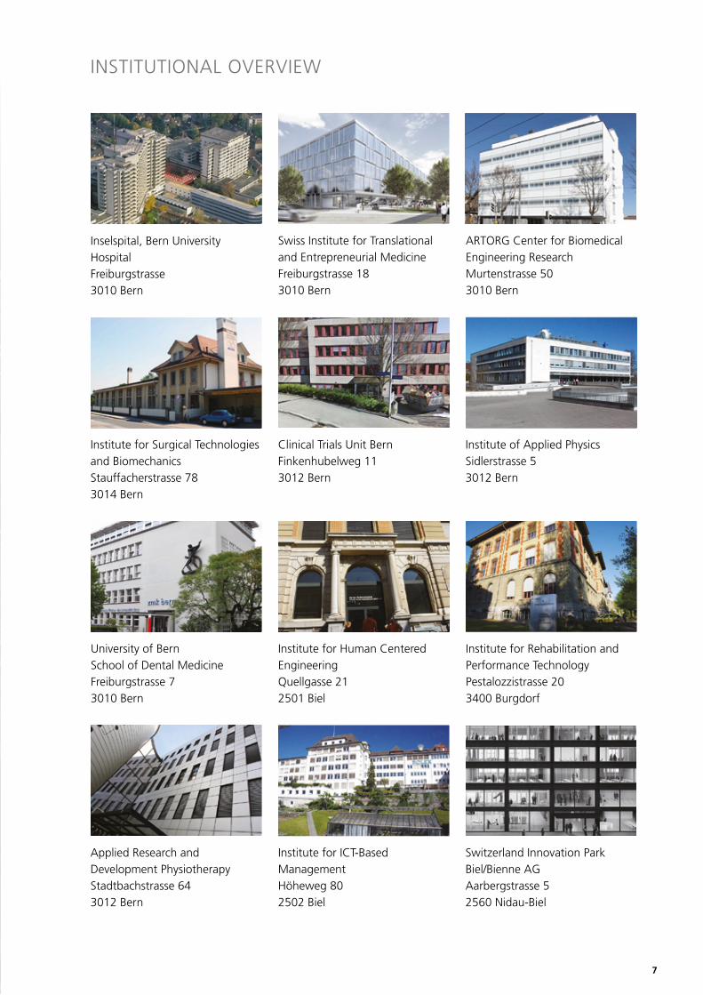
ARTORG Center for Biomedical Engineering ResearchMurtenstrasse 503010 Bern
Institute for Surgical Technologiesand BiomechanicsStauffacherstrasse 783014 Bern
Clinical Trials Unit BernFinkenhubelweg 113012 Bern
Institute of Applied PhysicsSidlerstrasse 53012 Bern
University of BernSchool of Dental MedicineFreiburgstrasse 73010 Bern
Institute for Human Centered EngineeringQuellgasse 212501 Biel
Institute for Rehabilitation andPerformance TechnologyPestalozzistrasse 203400 Burgdorf
Applied Research and Development PhysiotherapyStadtbachstrasse 643012 Bern
Institute for ICT-Based ManagementHöheweg 802502 Biel
Inselspital, Bern University HospitalFreiburgstrasse3010 Bern
Swiss Institute for Translational and Entrepreneurial MedicineFreiburgstrasse 183010 Bern
Switzerland Innovation ParkBiel/Bienne AGAarbergstrasse 52560 Nidau-Biel
8
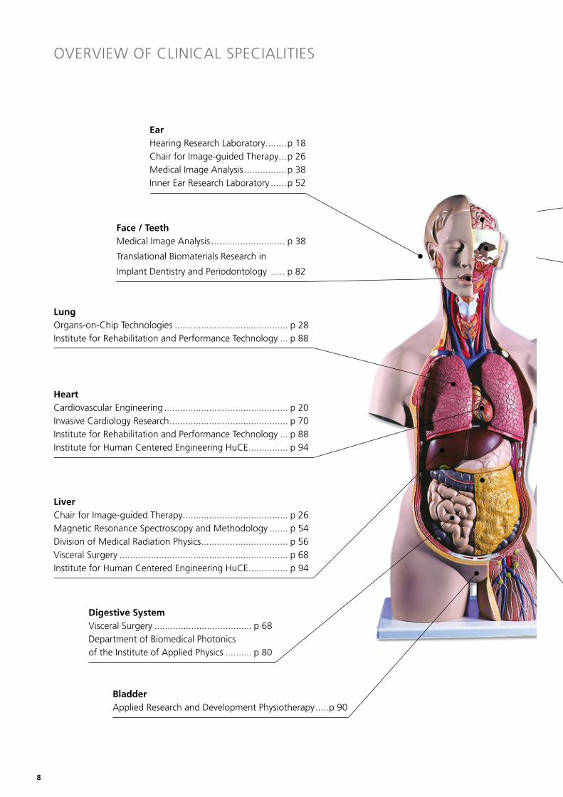
LungOrgans-on-Chip Technologies ........................................... p 28Institute for Rehabilitation and Performance Technology ... p 88
EarHearing Research Laboratory ........p 18Chair for Image-guided Therapy ...p 26Medical Image Analysis ................p 38Inner Ear Research Laboratory ......p 52
•
HeartCardiovascular Engineering ............................................... p 20Invasive Cardiology Research ............................................. p 70Institute for Rehabilitation and Performance Technology ... p 88Institute for Human Centered Engineering HuCE ............... p 94
LiverChair for Image-guided Therapy ........................................ p 26Magnetic Resonance Spectroscopy and Methodology ....... p 54Division of Medical Radiation Physics ................................. p 56Visceral Surgery ................................................................ p 68Institute for Human Centered Engineering HuCE ............... p 94
Digestive SystemVisceral Surgery ..................................... p 68Department of Biomedical Photonicsof the Institute of Applied Physics .......... p 80
BladderApplied Research and Development Physiotherapy .....p 90
•
•
•
•
•
•
•
•
•
OVERVIEW OF CLINICAL SPECIALITIES
Face / TeethMedical Image Analysis ............................ p 38
Translational Biomaterials Research in
Implant Dentistry and Periodontology ..... p 82
9
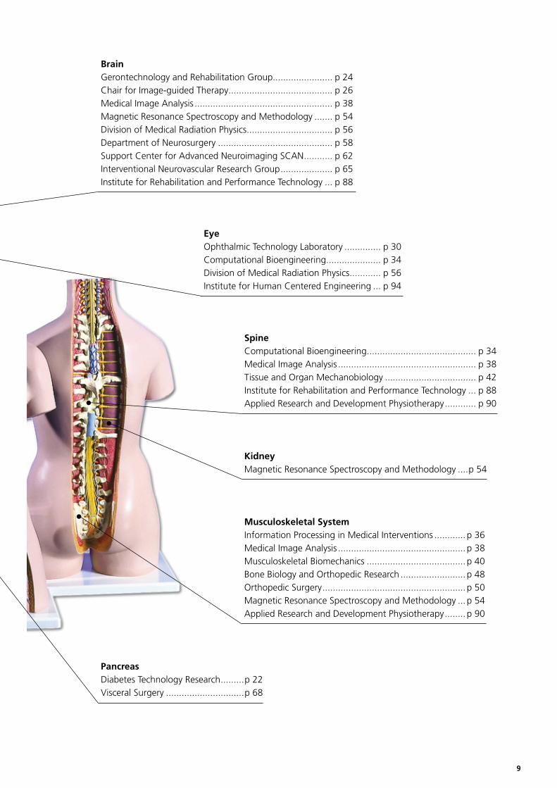
SpineComputational Bioengineering .......................................... p 34Medical Image Analysis ..................................................... p 38Tissue and Organ Mechanobiology ................................... p 42Institute for Rehabilitation and Performance Technology ... p 88Applied Research and Development Physiotherapy ............ p 90
BrainGerontechnology and Rehabilitation Group....................... p 24Chair for Image-guided Therapy ........................................ p 26Medical Image Analysis ..................................................... p 38Magnetic Resonance Spectroscopy and Methodology ....... p 54Division of Medical Radiation Physics ................................. p 56Department of Neurosurgery ............................................ p 58Support Center for Advanced Neuroimaging SCAN ........... p 62Interventional Neurovascular Research Group .................... p 65Institute for Rehabilitation and Performance Technology ... p 88
EyeOphthalmic Technology Laboratory .............. p 30Computational Bioengineering ..................... p 34Division of Medical Radiation Physics ............ p 56Institute for Human Centered Engineering ... p 94
KidneyMagnetic Resonance Spectroscopy and Methodology ....p 54
PancreasDiabetes Technology Research .........p 22Visceral Surgery ..............................p 68
Musculoskeletal SystemInformation Processing in Medical Interventions ............p 36Medical Image Analysis .................................................p 38Musculoskeletal Biomechanics ......................................p 40Bone Biology and Orthopedic Research .........................p 48Orthopedic Surgery .......................................................p 50Magnetic Resonance Spectroscopy and Methodology ...p 54Applied Research and Development Physiotherapy ........p 90
•
•
•
12
Swiss Institute for Translational and Entrepreneurial Medicinesitem-insel
Head: Felix FreyEmail: [email protected]; phone +41 31 632 58 41; www.sitem-insel.ch
Research Profilesitem-insel AG – The Swiss Institute for Translational and Entrepreneurial Medicine in Bern – was created to estab-lish, operate, and develop a National Center of Excellence for Translational Medicine. Translational medicine is a new, process-oriented discipline that aims to translate new find-ings and products emerging from private-sector devel-opment and basic research into clinical applications. The discipline seeks to professionalize the essential interaction between scientists conducting basic research in the private sector and universities, clinicians, regulatory bodies, and investors.The mission of sitem-insel is to create and foster an enhanced environment for translational medicine in Switzerland. The sitem-insel strategy rests on three pillars:
• The sitem-insel School organises university-level Continuing Professional Development (CPD) courses taught by university and private-sector lecturers.
• The sitem-insel Enabling Facilities provide infrastructure and personnel at the interface between the private sector and university hospitals for R&D and clinical trials of innovative products.
• The sitem-insel Promoting Services aim to optimize the administrative-regulatory effort along the route from laboratory bench to commercial products.
Organizationsitem-insel is an independent, non-profit public private partnership. Government funding has been approved for the start-up phase. After that time, sitem-insel should be financially independent.
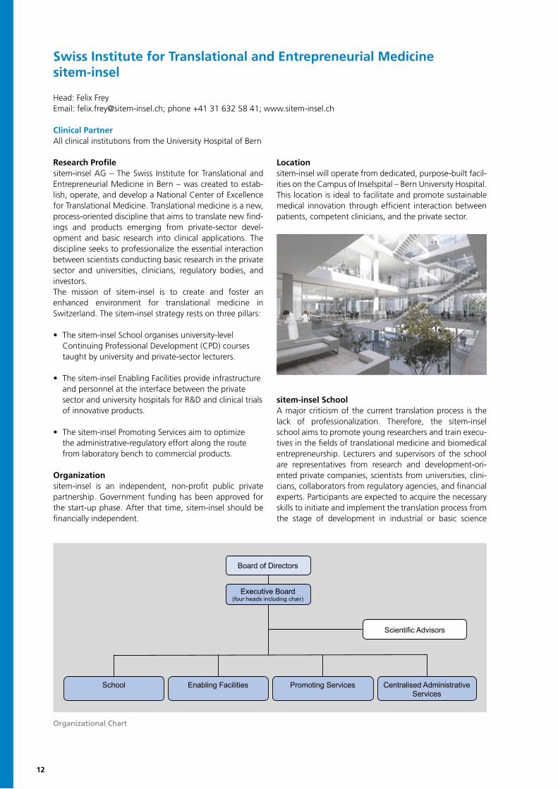
Locationsitem-insel will operate from dedicated, purpose-built facil-ities on the Campus of Inselspital – Bern University Hospital. This location is ideal to facilitate and promote sustainable medical innovation through efficient interaction between patients, competent clinicians, and the private sector.
sitem-insel SchoolA major criticism of the current translation process is the lack of professionalization. Therefore, the sitem-insel school aims to promote young researchers and train execu-tives in the fields of translational medicine and biomedical entrepreneurship. Lecturers and supervisors of the school are representatives from research and development-ori-ented private companies, scientists from universities, clini-cians, collaborators from regulatory agencies, and financial experts. Participants are expected to acquire the necessary skills to initiate and implement the translation process from the stage of development in industrial or basic science
Clinical PartnerAll clinical institutions from the University Hospital of Bern
Organizational Chart
Board of Directors
Executive Board(four heads including chair)
Scientific Advisors
School Enabling Facilities Promoting Services Centralised Administrative Services
13
institutions into clinical applications with the ultimate pur-pose of the latter’s commercialization. The focus will be on both diagnostic and therapeutic products. The acquired theoretical knowledge will be directly applied to the partici-pants’ own projects to provide specific solutions and strate-gies to each individual project. The program is approved by the University of Bern, and participants may obtain the de-grees of “Master of Advanced Studies” (MAS), “Diploma of Advanced Studies” (DAS), or “Certificate of Advanced Studies” (CAS). For further information, please visit www.sitem.insel.ch and http://sitem.bio-med.ch.
sitem-insel Promoting ServicesComplicated and slow regulation processes are relevant obstacles for translational projects. The promoting services, therefore, aim to optimize these processes with the ulti-mate goal of reducing the administrative-regulatory effort. Based on the promoting services’ analyses, strategies will be developed to determine how regulation requirements may be addressed to facilitate translational efforts of med-tech and pharmaceutical companies. The services provided will include undergraduate and postgraduate teaching at the University of Bern and in private institutions. The direc-tor of the Promoting Services, Prof. Dr. Rudolf Blankart, was recruited by the Faculty of Business, Economics and Social Sciences of the University of Bern and the board of direc-tors of sitem-insel. The focus of the Promoting Services for the years 2017-2020 is as follows:
• Consulting service in regulatory affairs
• Design of a basic and advanced university training program in regulatory affairs in biomedicine and medical technology
• Analysis of the economic importance of approval procedures in Switzerland
• Analysis and strategy development for the medium-term coordination of administrative-regulatory procedures
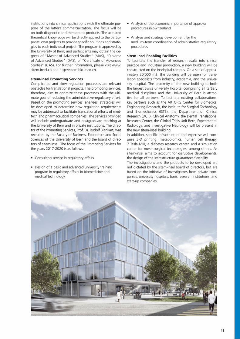
sitem-insel Enabling FacilitiesTo facilitate the transfer of research results into clinical practice and industrial production, a new building will be constructed on the Inselspital campus. On a site of approx-imately 20’000 m2, the building will be open for trans-lation specialists from industry, academia, and the univer-sity hospital. The proximity of the new building to both the largest Swiss university hospital comprising all tertiary medical disciplines and the University of Bern is attrac-tive for all partners. To facilitate existing collaborations, key partners such as the ARTORG Center for Biomedical Engineering Research, the Institute for Surgical Technology and Biomechanics (ISTB), the Department of Clinical Research (DCR), Clinical Anatomy, the Dental Translational Research Center, the Clinical Trials Unit Bern, Experimental Radiology, and Investigative Neurology will be present in the new sitem-insel building.In addition, specific infrastructure and expertise will com-prise 3-D printing, metabolomics, human cell therapy, 7 Tesla MRI, a diabetes research center, and a simulation center for novel surgical technologies, among others. As sitem-insel aims to account for disruptive developments, the design of the infrastructure guarantees flexibility.The investigations and the products to be developed are not dictated by the sitem-insel board of directors, but are based on the initiative of investigators from private com-panies, university hospitals, basic research institutions, and start-up companies.
14
MAS, DAS, and CAS in Translation and Entrepreneurship in Medicine
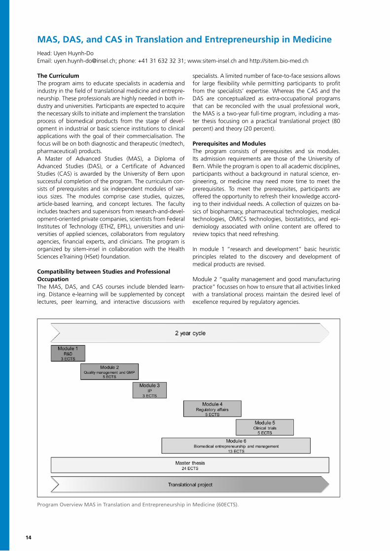
The CurriculumThe program aims to educate specialists in academia and industry in the field of translational medicine and entrepre-neurship. These professionals are highly needed in both in-dustry and universities. Participants are expected to acquire the necessary skills to initiate and implement the translation process of biomedical products from the stage of devel-opment in industrial or basic science institutions to clinical applications with the goal of their commercialisation. The focus will be on both diagnostic and therapeutic (medtech, pharmaceutical) products.A Master of Advanced Studies (MAS), a Diploma of Advanced Studies (DAS), or a Certificate of Advanced Studies (CAS) is awarded by the University of Bern upon successful completion of the program. The curriculum con-sists of prerequisites and six independent modules of var-ious sizes. The modules comprise case studies, quizzes, article-based learning, and concept lectures. The faculty includes teachers and supervisors from research-and-devel-opment-oriented private companies, scientists from Federal Institutes of Technology (ETHZ, EPFL), universities and uni-versities of applied sciences, collaborators from regulatory agencies, financial experts, and clinicians. The program is organized by sitem-insel in collaboration with the Health Sciences eTraining (HSet) foundation.
Compatibility between Studies and Professional OccupationThe MAS, DAS, and CAS courses include blended learn-ing. Distance e-learning will be supplemented by concept lectures, peer learning, and interactive discussions with
specialists. A limited number of face-to-face sessions allows for large flexibility while permitting participants to profit from the specialists’ expertise. Whereas the CAS and the DAS are conceptualized as extra-occupational programs that can be reconciled with the usual professional work, the MAS is a two-year full-time program, including a mas-ter thesis focusing on a practical translational project (80 percent) and theory (20 percent).
Prerequisites and ModulesThe program consists of prerequisites and six modules. Its admission requirements are those of the University of Bern. While the program is open to all academic disciplines, participants without a background in natural science, en-gineering, or medicine may need more time to meet the prerequisites. To meet the prerequisites, participants are offered the opportunity to refresh their knowledge accord-ing to their individual needs. A collection of quizzes on ba-sics of biopharmacy, pharmaceutical technologies, medical technologies, OMICS technologies, biostatistics, and epi-demiology associated with online content are offered to review topics that need refreshing.
In module 1 “research and development” basic heuristic principles related to the discovery and development of medical products are revised.
Module 2 “quality management and good manufacturing practice” focusses on how to ensure that all activities linked with a translational process maintain the desired level of excellence required by regulatory agencies.
Head: Uyen Huynh-DoEmail: [email protected]; phone: +41 31 632 32 31; www.sitem-insel.ch and http://sitem.bio-med.ch
Program Overview MAS in Translation and Entrepreneurship in Medicine (60ECTS).
6
to their individual needs. A collection of quizzes on basics of biopharmacy, pharmaceutical technologies,
medical technologies, OMICS technologies, biostatistics, and epidemiology associated with online
content are offered to review topics that need refreshing.
In module 1 “research and development” basic heuristic principles related to the discovery and
development of medical products are revised. Module 2 “quality management and good manufacturing
practice” focusses on how to ensure that all activities linked with a translational process maintain the
desired level of excellence required by regulatory agencies. Module 3 “intellectual property” is dedicated
to different types of intellectual property and legal aspects for biomedical products that are crucial for the
successful commercialization of biomedical products. Module 4 “regulatory affairs” sheds light on the
role different regulatory authorities play along the translational pathway. In module 5 “clinical trials” key
characteristics of clinical trial design and conduct will be considered. Clinical trials are designed to test
how well new medical approaches work in humans. Participants will learn about the prerequisites for
such scientific studies, the understanding of the pathophysiology of the underlying diseases, the
definition of quantifiable endpoints by clinicians, and the analyses of statistical data. Finally, module 6
“biomedical entrepreneurship and leadership” concentrates on various aspects of entrepreneurship,
such as leadership in large multidisciplinary teams, product management, business administration, and
the successful commercialisation of biomedical products. In contrast to the other five modules,
interactive face-to-face sessions with the teachers and various guest speakers from the industrial and
financial fields will be important.
Depending on the certificate selected, different numbers of ECTS points are required:
Figure 1: Program Overview MAS in Translation and Entrepreneurship in Medicine (60ECTS)
15
Module 3 “intellectual property” is dedicated to different types of intellectual property and legal aspects for biomed-ical products that are crucial for the successful commercial-ization of biomedical products.
Module 4 “regulatory affairs” sheds light on the role dif-ferent regulatory authorities play along the translational pathway.
In module 5 “clinical trials” key characteristics of clinical trial design and conduct will be considered. Clinical trials are designed to test how well new medical approaches work in humans. Participants will learn about the prereq-uisites for such scientific studies, the understanding of the pathophysiology of the underlying diseases, the definition of quantifiable endpoints by clinicians, and the analyses of statistical data.
Finally, module 6 “biomedical entrepreneurship and leader-ship” concentrates on various aspects of entrepreneurship, such as leadership in large multidisciplinary teams, product management, business administration, and the successful commercialisation of biomedical products. In contrast to the other five modules, interactive face-to-face sessions with the teachers and various guest speakers from the in-dustrial and financial fields will be important.
Depending on the certificate selected, different numbers of ECTS points are required:
• CAS “Translation and Entrepreneurship in Medicine” with focus on translational medicine: Three modules of choice from modules 1 to 5 (min. 13 ECTS) and certificate work (2 ECTS)
• CAS “Translation and Entrepreneurship in Medicine” with focus on biomedical entrepreneurship: Module 6 and certificate work (2 ECTS)
• DAS: Both CAS programs
• MAS: All six modules, master thesis, and elective courses (min. 2 ECTS)
Additional InformationThe course language is English. E-learning courses may be performed anywhere. In-class courses are held at the University of Bern. The participants of the CAS, DAS, and MAS programs will be registered at the University of Bern.
The fee for the MAS is CHF 31’500.-, for the DAS CHF 23’100.- and for each CAS CHF 12’600.-. The fee for single modules is between CHF 3’150.- and CHF 6300.-, depend-ing on the size.
For more information please contact Uyen Huynh-Do ([email protected]) and visit www.sitem-insel.ch.
17
ARTORG CENTER FOR BIOMEDICAL ENGINEERING RESEARCH
Now in its seventh year, the ARTORG Center continues the successful translation of innovative biomedical engineering discoveries into clinical application that bring disruptive clinical interventions and commercial medtech venturing, and with these patient benefits into areas of unaddressed clinical need. The working practice of the technical leadership of the biomedical engineering groups with its clinical peers has deepened and constructed a pipeline of projects across the full spectrum of technol-ogy-readiness levels (TRLs) from 1 = basic discovery to 8/9 = pre-production prototype with pilot clinical data. The first iteration of the BBME collaboration has extended the network with capabilities in allied health and applied research in materials and technology and has enabled the multidisciplinary-teams to add care pathways and manufacturing models for novel interventions and technologies. As intended by the principles of clinical translation, findings from the patient bedside have made their way back to the laboratory for further discovery. The ARTORG Center has sought out partnerships with research groups in basic biomedical research, such as lung-on-a-chip technology and specialist centers (proton beam therapy center) to inform new lines of clinical and technical enquiry. The intention is to build on the network gaining momentum to access funding, attract talent, and extend the network to international partners. The ARTORG Center maintains its focus areas and in 2017 saw the addition of Laura Marchal-Crespo as a new SNSF professorship focusing on motor learning after brain injury, broadening the research activities to cover:
• Hearing Research Laboratory (W. Wimmer)• Cardiovascular Engineering (D. Obrist)• Image-guided Therapy (S. Weber)• Diabetes Technology (S. Mougiakakou)• Gerontechnology and Rehabilitation (L. Marchal-Crespo & T. Nef)• Organs-on-Chip Technoligies (O. Guenat)• Ophthalmic Technology (R. Sznitman)
The infrastructure and resources from rapid protyping, in vitro and in vivo pre-clinical models, research strategy development support and technology transfer skills housed within the University of Bern continue to benefit all projects at the ARTORG Center and the network partners. The next stage of activities will involve ongoing emphasis on excellence initiatives, not only to build the reach and reputation of the ARTORG Center, but also the BBME network as whole. We hope to encourage everyone in the network to collaborate with us in this endeavour.
Stefan WeberDirector
18
Hearing Research Laboratory
Wilhelm Wimmer, Group HeadEmail: [email protected]: +41 31 632 87 89
Lukas Tom Nicolas Sidharta Suyi Markus Georgios Fabio Christoph StefanAnschütz Gawliczek Gerber Gupta Hu Huth Mantokoudis Munzinger Rathgeb Weder
Wilhelm Marco MartinWimmer Caversaccio Kompis
Research PartnersDeborah Hall, Nottingham Hearing Biomedical Research Unit, University of Nottingham, UKTobias Kleinjung, Department of Otorhinolaryngology, University Hospital of Zurich, SwitzerlandCAScination AG, Bern, SwitzerlandCochlear AG, Basel, SwitzerlandMED-EL GmbH, Innsbruck, AustriaOticon A/S, Smørum, DenmarkSonova AG, Stäfa, Switzerland
Research ProfileThe Hearing Research Laboratory is a clinically directed research collaboration between the ARTORG Center and the Department of Otolaryngology at the Bern University Hospital (Inselspital). Our research activities aim to provide innovative technology to help hearing-impaired patients and to assist clinicians in the diagnosis and treatment of hearing pathologies. The multidisciplinary group gathers experts from the fields of audiology, medicine, and engi-neering sciences. The range of the group’s activity includes basic psychoacoustic research, anatomical studies, the conception and implementation of novel clinically appli-cable technology, and the conduction of clinical trials. To promote a sustainable research progress, the members of the Hearing Research Laboratory actively collaborate with leading medical, academic, and industrial partners.
Experimental AudiologySound field audiometry, in which acoustic test stimuli are delivered through loudspeakers instead of earphones, is an integral component in the evaluation of the clinical hearing-rehabilitation progress. The assessment of hear-ing thresholds, speech understanding in quiet and noise,
and sound localization abilities provides essential outcome measures that can be directly linked to the quality of life of patients who were treated with hearing implants. In the area of experimental audiology, the Hearing Research Laboratory focuses on clinical studies aiming to contribute to the scientific communty and clinical practitioners alike. To enable a more realistic but reproducible assessment, our group develops methods to reproduce complex sound en-vironments and dynamic test situations that are required to capture the benefit of modern hearing implant tech-nology. In addition, the group aims to better understand the electrical current spread during stimulation in cochlear implants and to improve speech recognition in noisy condi-tions by optimization of parameters associated with front-end technology.
Software-Aided Cochlear ImplantationCochlear implantation is a microsurgical procedure that de-mands a great level of skill and experience due to the com-plex and highly variable anatomy of the human temporal bone. A major research focus of the Hearing Research Laboratory is to aid clinicians during several processes involved in cochlear implantation. To this end, software tools that empower surgeons to treat patients under con-sideration of their individual anatomy and physiology are currently developed and clinically evaluated. For example, preoperatively taken computed tomography images can be utilized to extract and reconstruct anatomical structures as three-dimensional models. The surgeon can use the virtu-al models to plan the surgical access and to optimize the electrode-array-insertion vector. Suitable lengths for the implanted electrode array can be selected depending on the cochlear size and the patient’s residual hearing. After implantation, the software tools can be applied to repro-ducibly assess the surgical outcome in postoperative image data sets. Moreover, suggestions for stimulus parameters for the first fitting of the implant can be derived.
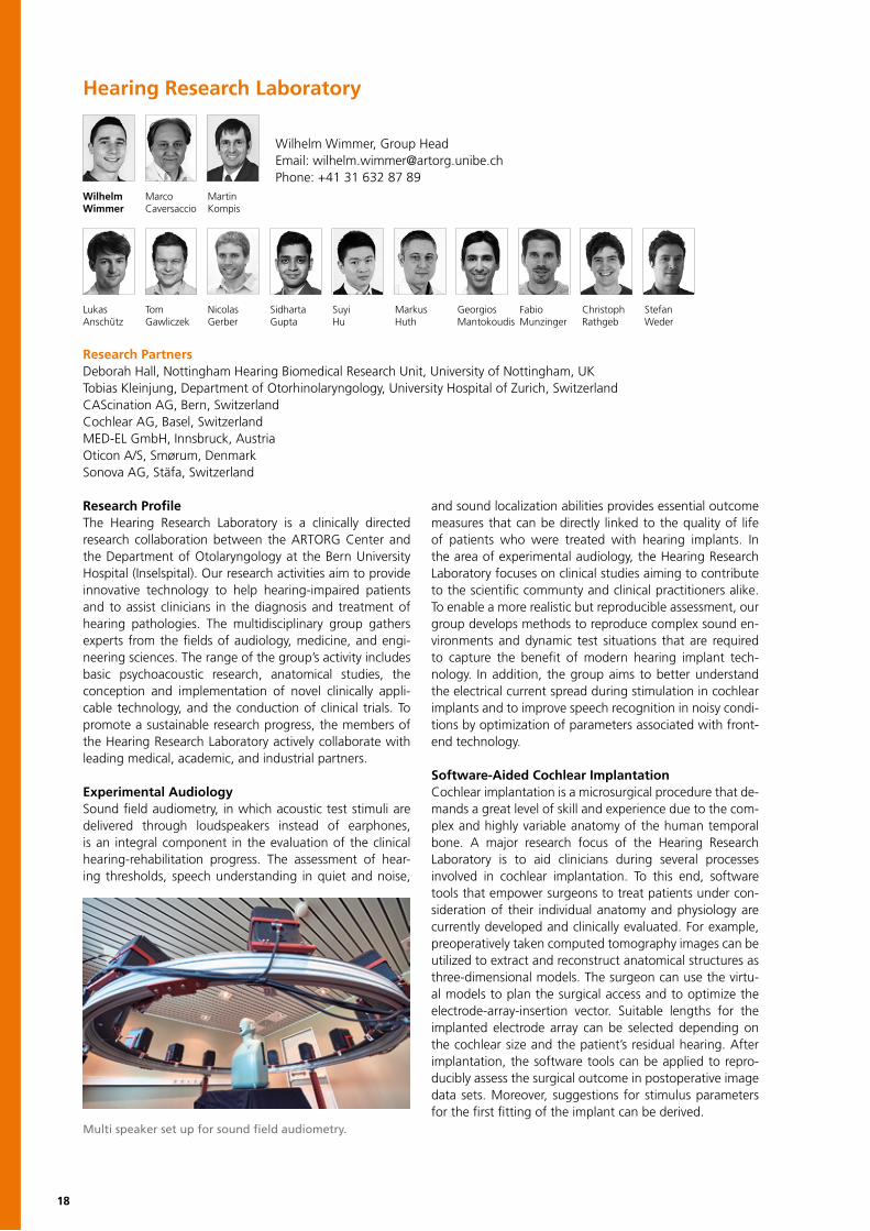
Multi speaker set up for sound field audiometry.

19
Tinnitus Assessment Tinnitus is the perception of sound in the absence of an ex-ternal acoustic stimulus. Severe forms of tinnitus can sub-stantially impair quality of life. Although often originating from inner ear damage, most types of tinnitus are main-tained in their chronic form by abnormal neuronal activity. Objective tinnitus assessment could be enabled by identi-fication of neuronal correlates in Electroencephalography (EEG). In a collaborative effort, the Hearing Research Laboratory and the Ophthalmic Technology Laboratory are investigating statistical approaches and computational
modelling to extract direct signatures of tinnitus in EEG data. The project aims to gain new insights into the be-havior of tinnitus and to potentially improve clinical tinni-tus assessment and classification. In addition, the group is developing mobile tools for tinnitus self-assessment. The patient-centered approach aims to deepen clinical assess-ment datasets from snapshot measurement under quiet conditions, to continuous long-term self-monitoring of the symptoms under more “life-like” conditions.
Temporal Bone LabThe activity spectrum of the Hearing Research Laboratory encompasses projects that require research on human spec-imens, such as the evaluation of novel implantation tech-nologies and surgical training. For these purposes, a fully equipped facility with several work spaces for anatomical dissections and otologic surgery is hosted in collaboration with the Institute of Anatomy of the University of Bern. The Temporal Bone Lab offers the opportunity for experi-mental and translational research and has a key function in the transfer of novel technologies prior to implementation into clinical routine. The proximity to the Bern University Hospital (Inselspital) permits concomitant radiological ex-amination of the specimens. Currently investigated topics include cochlear implantation procedures, endoscopic ap-proaches to the middle ear and the lateral skull base, as well as the development of suitable surgical instrumenta-tion. In addition, the Hearing Research Laboratory supports surgical training and anatomical studies to enable the re-finement of personal surgical skills and one-on-one teach-ing by our experienced faculty members.
Computation of optimal insertion trajectories forpatient-specific cochlear implantation.
Selected PublicationsWimmer W, Kompis M, Stieger S, Caversaccio M, Weder S (2017) Directional Microphone Contralateral Routing of Signals in Cochlear Implant Users: A Within-Subjects Comparison. Ear Hearing
Kompis M, Wimmer W, Caversaccio M (2017) Long Term Benefit of Bone Anchored Hearing Systems in Single Sided Deafness. Acta Otolaryngol 137(4):398-402
Caversaccio M, Gavaghan K, Wimmer W, et al (2017) Robotic Cochlear Implantation: Surgical Procedure and First Clinical Experience. Acta Otolaryngol 137(4):447-454
Kompis M, Kurz A, Flynn M, et al (2016) Estimating the benefit of a second bone anchored hearing implant in unilaterally implanted users with a testband. Acta Otolaryngol 136(4):379-84
Wagner F, Wimmer W, Leidolt L, et al (2015) Significant Artifact Reduction at 1.5T and 3T MRI by the Use of a Cochlear Implant with Removable Magnet: An Experimental Human Cadaver Study. PLoS One 10(7): e0132483
Wimmer W, Caversaccio M, Kompis M (2015) Speech Intelligibility in Noise With a Single-Unit Cochlear Implant Audio Processor. Otol Neurotol 36(7):1197-202
Wimmer W, Venail F, Williamson T, et al (2014) Semiautomatic cochleostomy target and insertion trajectory planning for minimally invasive cochlear implantation. Biomed Res Int 2014: 596498
20
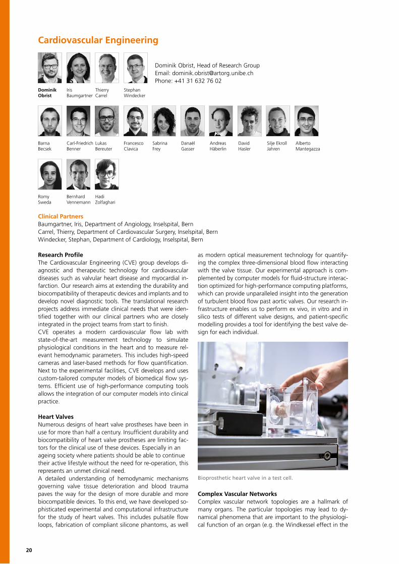
Cardiovascular Engineering
Dominik Obrist, Head of Research GroupEmail: [email protected]: +41 31 632 76 02
Barna Carl-Friedrich Lukas Francesco Sabrina Danaël Andreas David Silje Ekroll AlbertoBecsek Benner Bereuter Clavica Frey Gasser Häberlin Hasler Jahren Mantegazza
Romy Bernhard HadiSweda Vennemann Zolfaghari
Dominik Iris Thierry StephanObrist Baumgartner Carrel Windecker
Research ProfileThe Cardiovascular Engineering (CVE) group develops di-agnostic and therapeutic technology for cardiovascular diseases such as valvular heart disease and myocardial in-farction. Our research aims at extending the durability and biocompatibility of therapeutic devices and implants and to develop novel diagnostic tools. The translational research projects address immediate clinical needs that were iden-tified together with our clinical partners who are closely integrated in the project teams from start to finish.CVE operates a modern cardiovascular flow lab with state-of-the-art measurement technology to simulate physiological conditions in the heart and to measure rel-evant hemodynamic parameters. This includes high-speed cameras and laser-based methods for flow quantification. Next to the experimental facilities, CVE develops and uses custom-tailored computer models of biomedical flow sys-tems. Efficient use of high-performance computing tools allows the integration of our computer models into clinical practice.
Heart ValvesNumerous designs of heart valve prostheses have been in use for more than half a century. Insufficient durability and biocompatibility of heart valve prostheses are limiting fac-tors for the clinical use of these devices. Especially in anageing society where patients should be able to continuetheir active lifestyle without the need for re-operation, this represents an unmet clinical need.A detailed understanding of hemodynamic mechanisms governing valve tissue deterioration and blood trauma paves the way for the design of more durable and more biocompatible devices. To this end, we have developed so-phisticated experimental and computational infrastructure for the study of heart valves. This includes pulsatile flow loops, fabrication of compliant silicone phantoms, as well
as modern optical measurement technology for quantify-ing the complex three-dimensional blood flow interacting with the valve tissue. Our experimental approach is com-plemented by computer models for fluid-structure interac-tion optimized for high-performance computing platforms, which can provide unparalleled insight into the generation of turbulent blood flow past aortic valves. Our research in-frastructure enables us to perform ex vivo, in vitro and in silico tests of different valve designs, and patient-specific modelling provides a tool for identifying the best valve de-sign for each individual.
Complex Vascular NetworksComplex vascular network topologies are a hallmark of many organs. The particular topologies may lead to dy-namical phenomena that are important to the physiologi-cal function of an organ (e.g. the Windkessel effect in the
Clinical PartnersBaumgartner, Iris, Department of Angiology, Inselspital, Bern Carrel, Thierry, Department of Cardiovascular Surgery, Inselspital, BernWindecker, Stephan, Department of Cardiology, Inselspital, Bern
Bioprosthetic heart valve in a test cell.
21
arterial tree) or that are central functional elements of a pathologies (e.g. congenital vascular malformations). Custom-tailored computer models allow us to study of the transport of substances (e.g. pharmaceutical agents) through the network by advective and diffusive process-es. In many cases, this transport leads to heterogeneous distributions of substances prompting specific physiological reactions. A detailed understanding of such phenomena is the basis for novel diagnostic tools for vascular lesions (e.g. in neuro-radiological imaging) or for the planning of percu-taneous interventions in the cardiovascular system.
Translational ElectrophysiologyHeart rhythm disorders are common and may have dev-astating consequences. The group for Translational Electrophysiology - a collaboration of the Department of Cardiology, Bern University Hospital, and CVE - aims at de-veloping tools and devices for cardiac rhythm management.
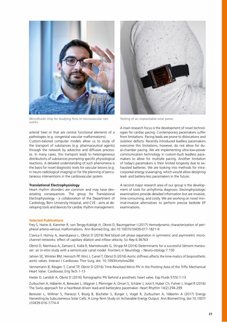
A main research focus is the development of novel technol-ogies for cardiac pacing. Contemporary pacemakers suffer from limitations. Pacing leads are prone to dislocations and isolation defects. Recently introduced leadless pacemakers overcome this limitations, however, do not allow for du-al-chamber pacing. We are implementing ultra-low-power communication technology in custom-built leadless pace-makers to allow for multisite pacing. Another limitation of today’s pacemakers is their limited longevity due to ex-hausted batteries. We are looking into methods for intra-corporeal energy scavenging, which would allow designing lead- and battery-less pacemakers in the future.
A second major research area of our group is the develop-ment of tools for arrhythmia diagnosis. Electrophysiologic examinations provide detailed information but are invasive, time-consuming, and costly. We are working on novel min-imal-invasive alternatives to perform precise bedside EP examinations.
Selected PublicationsFrey S, Haine A, Kammer R, von Tengg-Kobligk H, Obrist D, Baumgartner I (2017) Hemodynamic characterization of peri-pheral arterio-venous malformations. Ann Biomed Eng, doi:10.1007/s10439-017-1821-9
Clavica F, Homsy A, Jeandupeux L, Obrist D (2016) Red blood cell phase separation in symmetric and asymmetric micro-channel networks: effect of capillary dilation and inflow velocity. Sci Rep 6:36763
Obrist D, Nienhaus A, Zamaro E, Kalla R, Mantokoudis G, Strupp M (2016) Determinants for a successful Sémont maneu-ver: an in-vitro study with a semicircular canal model. Frontiers in Neurology – Neuro-otology 7:150
Jahren SE, Winkler BM, Heinisch PP, Wirz J, Carrel T, Obrist D (2016) Aortic stiffness affects the kine-matics of bioprosthetic aortic valves. Interact J Cardiovasc Thor Surg, doi: 10.1093/icvts/ivw284
Vennemann B, Rösgen T, Carrel TP, Obrist D (2016) Time-Resolved Micro PIV in the Pivoting Area of the Triflo Mechanical Heart Valve. Cardiovasc Eng Tech 1-13
Hasler D, Landolt A, Obrist D (2016) Tomographic PIV behind a prosthetic heart valve. Exp Fluids 57(5):1-13
Zurbuchen A, Häberlin A, Bereuter L, Wagner J, Pfenniger A, Omari S, Schärer J, Jutzi F, Huber Ch, Fuhrer J, Vogel R (2016) The Swiss approach for a heartbeat-driven lead-and batteryless pacemaker. Heart Rhythm 14(2):294-299
Bereuter L, Williner S, Pianezzi F, Bissig B, Bücheler S, Burger J, Vogel R, Zurbuchen A, Häberlin A (2017) Energy Harvesting by Subcutaneous Solar Cells: A Long-Term Study on Achievable Energy Output. Ann Biomed Eng, doi:10.1007/s10439-016-1774-4
Microfluidic chip for studying flow in microvascular net-works.
Testing of an implantable solar panel.
22
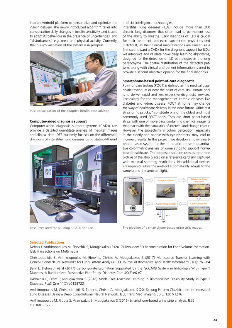
Research ProfileThe interface between machine learning, artificial intel-ligence, and their applications in diabetes, obesity, and other non-communicable diseases is the primary research focus of the Diabetes Technology Research (DTR) group. DTR creates innovation to translate “data into knowledge” and “research into clinical praxis.” Our ongoing research activities include:• innovative systems for dietary monitoring and assessment based on computer vision• reinforcement learning for insulin treatment optimization• artificial intelligence systems for computer-aided diagnosis • smartphone-based point-of-care diagnostics Nutrient intake monitoring and diet assessmentThe prevention of onset and progression of diet-related acute and chronic diseases (e.g. diabetes, obesity, kid-ney disease) requires reliable and intuitive systems able to translate food intake into nutrient intake. To this end, systems based on innovative technologies are introduced exploiting the recent advances in the areas of computer vision, machine learning, wearable sensors, and smart-phone technologies. Toward this direction, DTR has intro-duced the first fully operative system for calculating the carbohydrate content of meals for individuals with Type 1 Diabetes (T1D). The user takes two images of a meal from different viewing angles, with a reference card placed next to it. A series of computer vision processing steps follow: First the dish is detected, then the different food items on it are segmented and recognized, and finally the volume for each food item is estimated by reconstructing its 3D shape. Having the information about the types and the portion sizes for the existing food, the nutritional content of the meal can be estimated by using established nutrient data-bases. The system was developed within the framework of the GoCARB project (www.gocarb.eu). GoCARB was vali-dated in a clinical trial involving individuals with T1D under sensor-augmented pump therapy. The aim of the trial was
to assess the effects of the system in the glucose control. Indeed, the results indicated that such systems may im-prove diabetes management. Currently, the system is being optimized and extended to calories and other macro-nutri-ent estimation to cover the needs of people with obesity and kidney diseases for dietary and nutrient intake.
Insulin treatment optimizationTreating T1D requires the infusion of exogenous insulin. Insulin, as a medicine, has side-effects mainly related to improper dose, which may lead to sudden life-threatening events due to severe hypoglycaemia or cause long-term complications due to hyperglycaemia. The aim of the re-search is to personalize the insulin treatment so that in-dividuals with T1D stay at normal glycaemia. To this end, reinforcement learning (RL) algorithms, along with control theory and mobile phone technologies are used in a syner-getic manner. The RL-based algorithmic part is integrated
Diabetes Technology Research
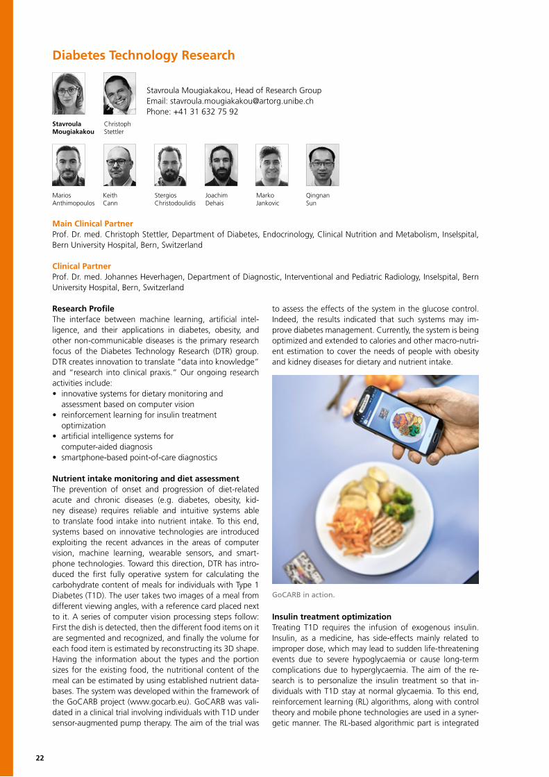
Stavroula ChristophMougiakakou Stettler
Stavroula Mougiakakou, Head of Research GroupEmail: [email protected]: +41 31 632 75 92
Keith Stergios Joachim Marko QingnanCann Christodoulidis Dehais Jankovic Sun
MariosAnthimopoulos
Main Clinical PartnerProf. Dr. med. Christoph Stettler, Department of Diabetes, Endocrinology, Clinical Nutrition and Metabolism, Inselspital, Bern University Hospital, Bern, Switzerland
Clinical PartnerProf. Dr. med. Johannes Heverhagen, Department of Diagnostic, Interventional and Pediatric Radiology, Inselspital, Bern University Hospital, Bern, Switzerland
GoCARB in action.
23
into an Android platform to personalize and optimize the insulin delivery. The newly introduced algorithm takes into consideration daily changes in insulin sensitivity, and is able to adapt its behaviour in the presence of uncertainties, and “disturbances” e.g. meal and physical activity. Currently, the in silico validation of the system is in progress.
Computer-aided diagnosis supportComputer-aided diagnosis support systems (CADx) can provide a detailed quantitate analysis of medical images and clinical data. DTR currently focuses on the differential diagnosis of interstitial lung diseases using state-of-the-art
artificial intelligence technologies.Interstitial lung diseases (ILDs) include more than 200 chronic lung disorders that often lead to permanent loss of the ability to breathe. Early diagnosis of ILDs is crucial for their treatment, but even experienced physicians find it difficult, as their clinical manifestations are similar. As a first step toward a CADx for the diagnosis support for ILDs, we introduce and validate novel deep learning algorithms, designed for the detection of ILD pathologies in the lung parenchyma. The spatial distribution of the detected pat-tern, along with clinical and patient information is used to provide a second objective opinion for the final diagnosis.
Smartphone-based point-of-care diagnosticPoint-of-care testing (POCT) is defined as the medical diag-nostic testing, at or near the point of care. Its ultimate goal is to deliver rapid and less expensive diagnostic services. Particularly for the management of chronic diseases like diabetes and kidney disease, POCT at home may change the way of healthcare delivery in the near future. Urine test strips or “dipsticks,” constitute one of the oldest and most commonly used POCT tools. They are short paper-based strips with one or more pads containing chemical reagents that react with their analytics of interest, and change colour. However, the subjectivity in colour perception, especially in the elderly and people with eye disorders, may lead to incorrect results. In this project, we develop a novel smart-phone-based system for the automatic and semi-quantita-tive colorimetric analysis of urine strips to support home-based healthcare. The proposed solution uses as input one picture of the strip placed on a reference card and captured with minimal shooting restrictions. No additional devices are required, while the method automatically adapts to the camera and the ambient light.
Selected PublicationsDehais J, Anthimopoulos M, Shevchik S, Mougiakakou S (2017) Two-view 3D Reconstruction for Food Volume Estimation. IEEE Transactions on Multimedia
Christodoulidis S, Anthimopoulos M, Ebner L, Christe A, Mougiakakou S (2017) Multisource Transfer Learning with Convolutional Neural Networks for Lung Pattern Analysis. IEEE Journal of Biomedical and Health Informatics 21(1): 76 – 84
Bally L, Dehais J, et al (2017) Carbohydrate Estimation Supported by the GoCARB System in Individuals With Type 1 Diabetes: A Randomized Prospective Pilot Study. Diabetes Care 40(2):e6-e7
Daskalaki E, Diem P, Mougiakakou S (2016) Model-Free Machine Learning in Biomedicine: Feasibility Study in Type 1 Diabetes. PLoS One 11(7):e0158722
Anthimopoulos M, Christodoulidis S, Ebner L, Christe A, Mougiakakou S (2016) Lung Pattern Classification for Interstitial Lung Diseases Using a Deep Convolutional Neural Network. IEEE Trans Med Imaging 35(5):1207-1216
Anthimopoulos M, Gupta S, Arampatzis S, Mougiakakou S (2016) Smartphone-based urine strip analysis. IEEEIST 368 – 372
In silico validation of the adaptive insulin dose advisor.
Resources used for building a CADx for ILDs. The pipeline of a smartphone-based urine strip reader.

24
Gerontechnology and Rehabilitation GroupLaura Marchal-Crespo, Technical Group HeadTobias Nef, Technical Group Head Urs Mosimann, Clinical Group HeadRené Müri, Clinical Group HeadEmail: [email protected], phone: +41 31 632 75 79
Dario Alvin Farouk Lorenzo Stephan Jurka Rebecca Hugo Nadine Narayan PrabithaCazzoli Chesham Chrif Diana Gerber Meichtry Paladini Saner Schmid Schütz Urwyler
Laura Tobias Urs RenéMarchal-Crespo Nef Mosimann Müri
Research ProfileThe interdisciplinary Gerontechnology and Rehabilitation Research Group is a collaborative research effort of the ARTORG Center for Biomedical Engineering Research, the Department of Old Age Psychiatry, and the Division of Cognitive and Restorative Neurology within the medical faculty at the University of Bern.Gerontechnology is the study of technology and aging to promote good health, social participation, and indepen- dent living. Rehabilitation embraces the coordinated use of medical, social, professional, and technical means to improve function to allow independent participation in all areas of life with acceptable risks and good quality of life.The relevance of these fields increases with the aging of our society. In this context, the group develops and eval- uates assistive and rehabilitative technologies to support elderly and disabled people and enhance autonomy and promote independent living while reducing the risks asso- ciated with daily living. Current projects aim to promote independence in patients with cognitive impairments by enhancing in-home mobility as well as new training to strengthen cognitive performance.
Optimize Motor Learning to ImproveNeurorehabilitationThere is increasing interest in using robotic devices to deliver rehabilitation therapy following stroke. Robotic guidance is generally used in motor training to reduce performance errors during practice. However, to date, the functional gains obtained after robotic rehabilitation are limited. A possible explanation for this limited benefit is the inability of the controllers to adapt to the subjects’ special needs. Research on motor learning has emphasized that movement errors are fundamental signals that drive motor
adaptation. Thereby, robotic algorithms that augment errors rather than decrease them have great potential to provoke better motor learning and neurorehabilitation out-comes, especially in initially more skilled subjects. The aim of this project is to improve robotic neurorehabilitation, de-veloping novel robotic training strategies that augment or reduce movement errors based on subjects’ skill (disability) level, age, and characteristics of the trained motor task.
Puzzling the Mind: Tablet-Computer Intervention for Cognitively Impaired PatientsThe main objective of this project is to investigate long- term training benefits of a casual puzzle video game inter-vention on cognitive and emotional functioning in healthy older adults and patients with cognitive impairment. Casual video games (CVG) are highly popular with easy-to-use interfaces, simple rules, and goals that can be quickly mastered by players of different skill levels.
Research PartnersStefan Klöppel, University Hospital of Old Age Psychiatry, BernStephan Jakob, Department of Intensive Care Medicine, Inselspital, BernMartin Schimmel, Division of Gerodontology, School of Dental Medicine, University of BernThomas Nyffeler & Stephan Bohlhalter, Department of Internal Medicine, Luzerner Kantonsspital, LuzernKenneth Hunt, Department of Engineering and Information Technology, Bern University of Applied Sciences
Vanessa Tim PatricVallejo Vanbellingen Wyss
Rehabilitation Robot ARMin for optimized motor learning (R. Riener & T. Nef).
25
CVG are primarily designed to be enjoyable and foster sus-tained player engagement. Of note, CVG were shown to engage cognitive abilities that are particularly subject to age-related cognitive decline and affected by brain injury. To ensure that patients will adhere to a CVG intervention, games should challenge players at an optimal difficulty lev-el (task difficulty aspect) that matches their level of skill. This dynamic difficulty adjustment can further increase motivation and promote learning and transfer to cogni-tive functions. We registered a clinical trial in which we will conduct a 16-week randomized crossover design and compare the cognitive and emotional benefits of the tablet computer-based puzzle game intervention with an active control intervention.
Virtual Reality Stimulation to Enhance CognitiveFunctions of Intensive Care Unit PatientsPatients in the intensive care unit (ICU) often have long- term functional deficits. Up to 70 percent suffer from long-term cognitive impairment for the rest of their life, resulting in a reduction of quality of life compared to their state before admission to the ICU. Typically, the constant exposure to meaningless and arbitrary stimuli such as light, noise, and lack of daily living routine leads to a loss of differentiated perception and orientation. Virtual reali-ty (VR) can stimulate the patient in a safe and controlled environment. The underlying concept suggests that cog- nitive stimulation can have a beneficial effect on cognitive function, preventing the emergence of neuro-cognitive impairments or improving cognitive and functional out- comes after discharge. In this project in collaboration with the department of Intensive Care Medicine at University Hospital Bern, we investigate the cognitive and functional outcomes of VR stimulation of ICU patients compared to healthy volunteers.The VR stimulation consists of immersive nature scener-ies, five minutes in length. A pilot study showed that VR
stimulation in healthy volunteers had a relaxing effect and did not evoke any side effects. Furthermore during stimu-lation, the visual search activity was reduced when given attention to a target. In the next step, a clinical trial will be conducted in the ICU to find out how critically ill patients will interact with the VR.
Bern Aphasia App for Tele-rehabilitationAphasia is the loss or impairment of language functions that occurs following brain damage. This disorder af-fects the four linguistic modalities in different combina-tions and levels of severity. A key factor for a successful speech and language therapy (SLT) is dose frequency. Tablet-based aphasia tele-rehabilitation increases access to high-frequency SLT while reducing cost. Together with the speech and language therapists of the University Hospital Inselspital, we have implemented a novel tablet application called Bern Aphasia App. This tele-rehabilitation applica-tion enables patients to train language-related tasks inde-pendently at home (patient interface) where the therapists can access the performance of the patients and can adjust the exercise type and difficulty level (therapist interface). A dedicated website for the creation of novel exercises was implemented, and exercises were developed together with speech therapists. The application allows therapists to generate the following exercise types: relating images to a word, words to images, images to images, or words to words; inserting letters; completing sentences; sorting let-ters; sorting words; writing words (with keyboard or hand-writing); answering questions (on audio, picture, video, or text) and pronunciation training.The developed aphasia application is currently in clinical use at the University Hospital Inselspital. To date, 10 ambulant patients have exercised 88.9 hours and stationary patients 16.3 hours (in total 10386 solved tasks). The usability was tested in preclinical studies with patients, therapists, and healthy participants, showing that the Bern Aphasia App was well accepted. Currently we are conducting a clinical trial to evaluate the effects of high-frequency tele-rehabili-tation SLT using the Bern Aphasia App.
Tele-rehabilitation with the Bern Aphasia App.
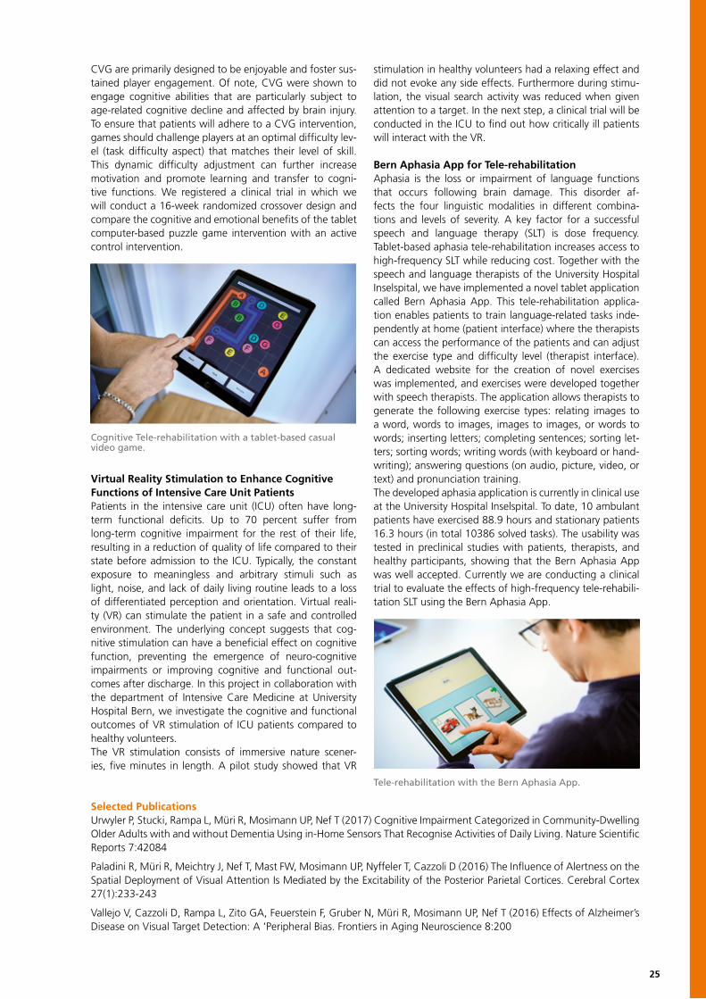
Cognitive Tele-rehabilitation with a tablet-based casual video game.
Selected PublicationsUrwyler P, Stucki, Rampa L, Müri R, Mosimann UP, Nef T (2017) Cognitive Impairment Categorized in Community-Dwelling Older Adults with and without Dementia Using in-Home Sensors That Recognise Activities of Daily Living. Nature Scientific Reports 7:42084
Paladini R, Müri R, Meichtry J, Nef T, Mast FW, Mosimann UP, Nyffeler T, Cazzoli D (2016) The Influence of Alertness on the Spatial Deployment of Visual Attention Is Mediated by the Excitability of the Posterior Parietal Cortices. Cerebral Cortex 27(1):233-243
Vallejo V, Cazzoli D, Rampa L, Zito GA, Feuerstein F, Gruber N, Müri R, Mosimann UP, Nef T (2016) Effects of Alzheimer’s Disease on Visual Target Detection: A ‘Peripheral Bias. Frontiers in Aging Neuroscience 8:200

26
Stefan Weber, Head of Research GroupEmail: [email protected]: +41 31 632 75 74
Chair for Image-guided Therapy
Juan Lukas Toni Johan David Cilgia Teo Kate Sidharta David JanAnsó Anschütz Antunovic Baijot Bervini Dür Eterovic Gerber Gupta Hänggi Hermann
Rafael Anja Georgios Martin Iwan Daniel Marius Manuel Pascale Hendrik ThomasKammer Lachenmayer Mantokoudis Maurer Paolucci Schneider Schwalbe Stebinger Tinguely von Tengg Winklehner
Stefan Daniel Iris Daniel Marco Jan Johannes AndreasWeber Aebersold Baumgartner Candinas Caversaccio Gralla Heverhagen Raabe
Research ProfileResearch led by the Chair for Image-guided Therapy (IGT) spans the arc from basic research to translation-ready tech-nology of all facets of stereotactic surgery and intervention. Novel diagnostic, therapeutic, interventional and surgical technologies that augment the clinician’s abilities and fa-cilitate increased surgical accuracy, reduced invasiveness, and improved clinical outcomes drive the discovery process of the multidisciplinary (MD) work of the IGT Group. The aim of work in the IGT Group is to combine simulation and modelling, imaging and sensing, computing, surgical robotics as well as visualisation. This will improve the local-isation and targeting of pathological tissue with surgical instruments and focused energy (Hepatobiliary-Oncology) or enable precision image-guided surgical approaches to microsurgical applications (Cochlear Implantation). Technology development in stereotactic instrument guid-ance, surgical robotics, and rapid prototyping permits the embodiment and testing of novel approaches to IGT
applications. Strong partnerships with clinical collaborators at the Bern University Hospital and clinical centres of ex-cellence throughout Europe, ensure the development of clinically validated tools for near-term use in the OR and treatment rooms. Projects in the MD IGT Group emphasise critical assessment of clinical “technology-pull” and utility to address unmet clinical needs with disruptive treatment models that can deliver better patient outcomes.
Minimally Invasive Cochlear Implantation (CI)Surgical robot systems can work beyond the limits of hu-man perception, dexterity and scale making them inherent-ly suitable for use in microsurgical procedures. Our concept of robotic CI aims to increase consistency of surgical out-comes, such as preservation of residual hearing, and re-duce invasiveness of the procedure. In 2016, we reported the first successful, stereotactically-guided, robotic CI (RCI) man. The underlying robotic treatment model developed by us encompasses: computer-assisted surgery planning, precision stereotactic image-guidance, in-situ assessment of tissue properties and multipolar neuromonitoring, based on in vitro, in vivo and pilot data. The clinical study design we derived from the model for RCI sets-out a completely novel surgical workflow: planning, robotic access-drilling, computer-assisted electrode selection and keyhole elec-trode-placement. Looking ahead, the robotic treatment model is expandable to integrate additional robotic func-tionalities such as cochlear access and electrode insertion. Our results demonstrate the feasibility and possibilities of using robotic technology for microsurgery on the lateral skull base. It has the potential for significant benefit in oth-er microsurgical domains for which there is no task-orient-ed, robotic technology available at present.
Research PartnersIntervention Centre, Oslo University Hospital, Oslo, NorwayKarolinska Institutet, Stockholm, SwedenIRCAD France, Research Institute against Digestive Cancer, Strasbourg, FranceINSERM Gui de Chauliac et Institut des Neurosciences, University Montpellier, FranceCAScination AG, Bern, SwitzerlandMED-EL GmbH, Innsbruck, Austria
The robotic drilling process: The robotic drill accesses the situs through a 20 mm incision.
27
High-Performance Soft-tissue NavigationUse of state-of-the art nagivation capabilities to extend the clinician’s faculties further than human limitations have been well established in a number of surgical and inter-ventional domains. Nonetheless, despite wide acceptance by clinical key opinion leaders, that image-guidance and virtual reality enhancement of the operating field-of view are central to impact performance significantly, no consen-sus exists on standardised solutions. In a first iteration, the hepatobiliary MD IGT Team have been awarded HORIZON 2020 funding for HiPerNav to address challenges faced in primary liver cancer, aka hepatocellular carcinoma (HCC) treatment. HCC is fifth most common cancer worldwide and the third most common cause of cancer mortality. At present, a proportion of HCC patients will undergo sur-gery for complete removal of the tumor including a safety margin while sparing as much healthy tissue as possible. However, due to the limitations of current visualisation and surgical technologies and lack of agile navigation solutions, only a relatively low percentage of patients are eligible for liver surgery, and the recurrence rate is considerable. The MD IGT Team seeks to deliver the prospect of many more patients benefitting from curative liver surgical procedures and ablation.
Navigated, Ultrasound-based, StereotacticLaparoscopic Ablation TreatmentsWhen considering patient benefit, replacing all open sur-gical procedures with minimally-invasive interventions is the universal aim of clinical and biomedical-engineering researchers working in MD Teams. Using focused energy through an ablation needle into a solid liver tumor, is a tissue-sparing treatment method with known advantages
such as reduced complication rates and shorter hospital stays. Evidence is increasing that ablation is set to super-sede open surgery for the treatment of solid tumors of the liver. The major challenge for laparoscopic tumor ablation is the skill required to place an ablation needle precisley into the tumor. Standard operating field visualisation based on imaging alone is not reliable enough to allow the clinician to “aim and shoot” with optimal accuracy. Therefore, an integrated software workflow for efficient intra-operative ablation planning, needle placement, and validation based on navigated ultrasound has been developed. A first step underway at present is the evaluation of accuracy and ef-ficiency of the system in a phantom study, followed by the next stages along the translation pathway into clinical care.
Image Guidance for the Treatment of VascularMalformationsPerformance enhancement through surgical and interven-tional navigation technology that seamlessly becomes part of the clinician’s own telemetry can permit manual surgical intervention at millimetric scales. This is critical in the treat-ment of vascular malformations, in which needles have to be placed within vessels as small as 1 mm. Stereotactic im-age-guidance enhancement of existing targeting methods could result in faster and more reproducible needle place-ments. In the context of the interventional workflow and clinical care a speedier procedure would mean: reduced radiation dose for the patient, and wider access to proce-dures safely executed by clinicians earlier in their training. Pilot work has generated data that suggest that use of nav-igation could mean such benefits for patients. The team is actively engaged in developing the approach for clinical application.
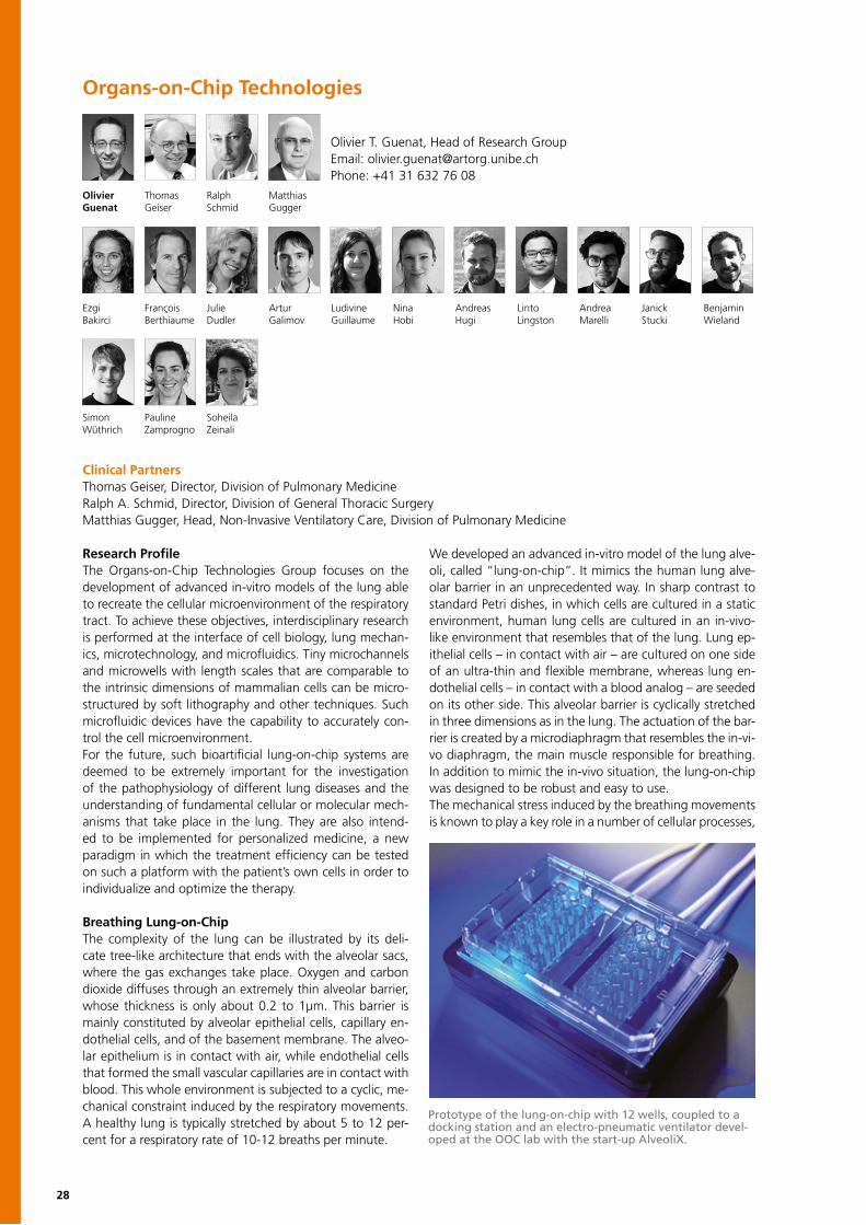
Instrument calibration for navigated needle placement during the treatment of a venous malformation in the angiography suite.
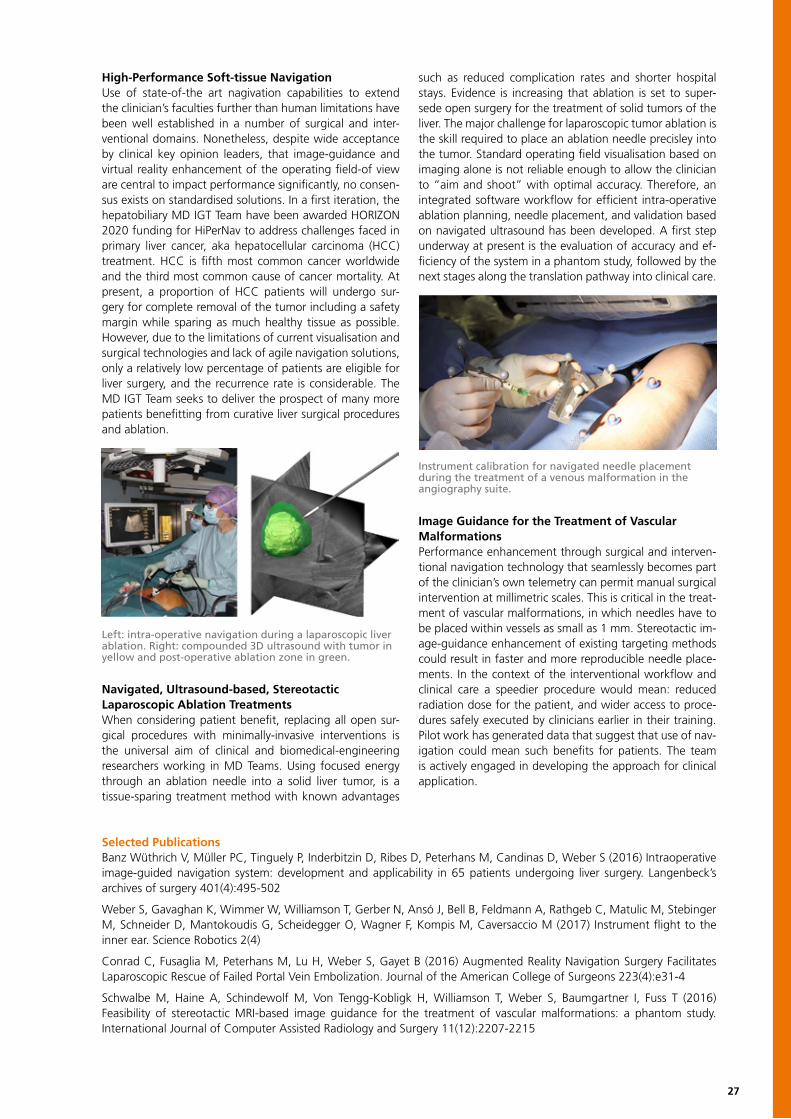
Left: intra-operative navigation during a laparoscopic liverablation. Right: compounded 3D ultrasound with tumor in yellow and post-operative ablation zone in green.
Selected PublicationsBanz Wüthrich V, Müller PC, Tinguely P, Inderbitzin D, Ribes D, Peterhans M, Candinas D, Weber S (2016) Intraoperative image-guided navigation system: development and applicability in 65 patients undergoing liver surgery. Langenbeck’s archives of surgery 401(4):495-502
Weber S, Gavaghan K, Wimmer W, Williamson T, Gerber N, Ansó J, Bell B, Feldmann A, Rathgeb C, Matulic M, Stebinger M, Schneider D, Mantokoudis G, Scheidegger O, Wagner F, Kompis M, Caversaccio M (2017) Instrument flight to the inner ear. Science Robotics 2(4)
Conrad C, Fusaglia M, Peterhans M, Lu H, Weber S, Gayet B (2016) Augmented Reality Navigation Surgery Facilitates Laparoscopic Rescue of Failed Portal Vein Embolization. Journal of the American College of Surgeons 223(4):e31-4
Schwalbe M, Haine A, Schindewolf M, Von Tengg-Kobligk H, Williamson T, Weber S, Baumgartner I, Fuss T (2016) Feasibility of stereotactic MRI-based image guidance for the treatment of vascular malformations: a phantom study. International Journal of Computer Assisted Radiology and Surgery 11(12):2207-2215
28
Research Profile The Organs-on-Chip Technologies Group focuses on the development of advanced in-vitro models of the lung able to recreate the cellular microenvironment of the respiratory tract. To achieve these objectives, interdisciplinary research is performed at the interface of cell biology, lung mechan-ics, microtechnology, and microfluidics. Tiny microchannels and microwells with length scales that are comparable to the intrinsic dimensions of mammalian cells can be micro-structured by soft lithography and other techniques. Such microfluidic devices have the capability to accurately con-trol the cell microenvironment. For the future, such bioartificial lung-on-chip systems are deemed to be extremely important for the investigation of the pathophysiology of different lung diseases and the understanding of fundamental cellular or molecular mech-anisms that take place in the lung. They are also intend-ed to be implemented for personalized medicine, a new paradigm in which the treatment efficiency can be tested on such a platform with the patient’s own cells in order to individualize and optimize the therapy.
Breathing Lung-on-ChipThe complexity of the lung can be illustrated by its deli-cate tree-like architecture that ends with the alveolar sacs, where the gas exchanges take place. Oxygen and carbon dioxide diffuses through an extremely thin alveolar barrier, whose thickness is only about 0.2 to 1μm. This barrier is mainly constituted by alveolar epithelial cells, capillary en-dothelial cells, and of the basement membrane. The alveo-lar epithelium is in contact with air, while endothelial cells that formed the small vascular capillaries are in contact with blood. This whole environment is subjected to a cyclic, me-chanical constraint induced by the respiratory movements. A healthy lung is typically stretched by about 5 to 12 per-cent for a respiratory rate of 10-12 breaths per minute.
We developed an advanced in-vitro model of the lung alve-oli, called “lung-on-chip”. It mimics the human lung alve-olar barrier in an unprecedented way. In sharp contrast to standard Petri dishes, in which cells are cultured in a static environment, human lung cells are cultured in an in-vivo-like environment that resembles that of the lung. Lung ep-ithelial cells – in contact with air – are cultured on one side of an ultra-thin and flexible membrane, whereas lung en-dothelial cells – in contact with a blood analog – are seeded on its other side. This alveolar barrier is cyclically stretched in three dimensions as in the lung. The actuation of the bar-rier is created by a microdiaphragm that resembles the in-vi-vo diaphragm, the main muscle responsible for breathing. In addition to mimic the in-vivo situation, the lung-on-chip was designed to be robust and easy to use.The mechanical stress induced by the breathing movements is known to play a key role in a number of cellular processes,
Organs-on-Chip Technologies
Olivier T. Guenat, Head of Research GroupEmail: [email protected]: +41 31 632 76 08
Ezgi François Julie Artur Ludivine Nina Andreas Linto Andrea Janick BenjaminBakirci Berthiaume Dudler Galimov Guillaume Hobi Hugi Lingston Marelli Stucki Wieland
Olivier Thomas Ralph MatthiasGuenat Geiser Schmid Gugger
Simon Pauline Soheila Wüthrich Zamprogno Zeinali
Clinical PartnersThomas Geiser, Director, Division of Pulmonary MedicineRalph A. Schmid, Director, Division of General Thoracic SurgeryMatthias Gugger, Head, Non-Invasive Ventilatory Care, Division of Pulmonary Medicine
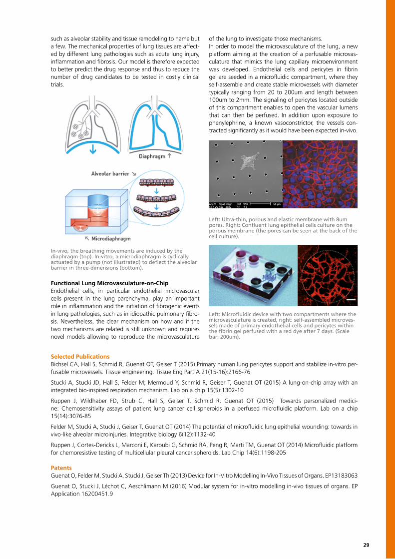
Prototype of the lung-on-chip with 12 wells, coupled to a docking station and an electro-pneumatic ventilator devel-oped at the OOC lab with the start-up AlveoliX.
29
such as alveolar stability and tissue remodeling to name but a few. The mechanical properties of lung tissues are affect-ed by different lung pathologies such as acute lung injury, inflammation and fibrosis. Our model is therefore expected to better predict the drug response and thus to reduce the number of drug candidates to be tested in costly clinical trials.
Functional Lung Microvasculature-on-ChipEndothelial cells, in particular endothelial microvascular cells present in the lung parenchyma, play an important role in inflammation and the initiation of fibrogenic events in lung pathologies, such as in idiopathic pulmonary fibro-sis. Nevertheless, the clear mechanism on how and if the two mechanisms are related is still unknown and requires novel models allowing to reproduce the microvasculature
of the lung to investigate those mechanisms. In order to model the microvasculature of the lung, a new platform aiming at the creation of a perfusable microvas-culature that mimics the lung capillary microenvironment was developed. Endothelial cells and pericytes in fibrin gel are seeded in a microfluidic compartment, where they self-assemble and create stable microvessels with diameter typically ranging from 20 to 200um and length between 100um to 2mm. The signaling of pericytes located outside of this compartment enables to open the vascular lumens that can then be perfused. In addition upon exposure to phenylephrine, a known vasoconstrictor, the vessels con-tracted significantly as it would have been expected in-vivo.
In-vivo, the breathing movements are induced by the diaphragm (top). In-vitro, a microdiaphragm is cyclically actuated by a pump (not illustrated) to deflect the alveolar barrier in three-dimensions (bottom).
Left: Ultra-thin, porous and elastic membrane with 8um pores. Right: Confluent lung epithelial cells culture on the porous membrane (the pores can be seen at the back of the cell culture).
Left: Microfluidic device with two compartments where the microvasculature is created, right: self-assembled microves-sels made of primary endothelial cells and pericytes within the fibrin gel perfused with a red dye after 7 days. (Scale bar: 200um).
Selected PublicationsBichsel CA, Hall S, Schmid R, Guenat OT, Geiser T (2015) Primary human lung pericytes support and stabilize in-vitro per-fusable microvessels. Tissue engineering. Tissue Eng Part A 21(15-16):2166-76
Stucki A, Stucki JD, Hall S, Felder M; Mermoud Y, Schmid R, Geiser T, Guenat OT (2015) A lung-on-chip array with an integrated bio-inspired respiration mechanism. Lab on a chip 15(5):1302-10
Ruppen J, Wildhaber FD, Strub C, Hall S, Geiser T, Schmid R, Guenat OT (2015) Towards personalized medici-ne: Chemosensitivity assays of patient lung cancer cell spheroids in a perfused microfluidic platform. Lab on a chip 15(14):3076-85
Felder M, Stucki A, Stucki J, Geiser T, Guenat OT (2014) The potential of microfluidic lung epithelial wounding: towards in vivo-like alveolar microinjuries. Integrative biology 6(12):1132-40
Ruppen J, Cortes-Dericks L, Marconi E, Karoubi G, Schmid RA, Peng R, Marti TM, Guenat OT (2014) Microfluidic platform for chemoresistive testing of multicellular pleural cancer spheroids. Lab Chip 14(6):1198-205
PatentsGuenat O, Felder M, Stucki A, Stucki J, Geiser Th (2013) Device for In-Vitro Modelling In-Vivo Tissues of Organs. EP13183063
Guenat O, Stucki J, Léchot C, Aeschlimann M (2016) Modular system for in-vitro modelling in-vivo tissues of organs. EP Application 16200451.9
30
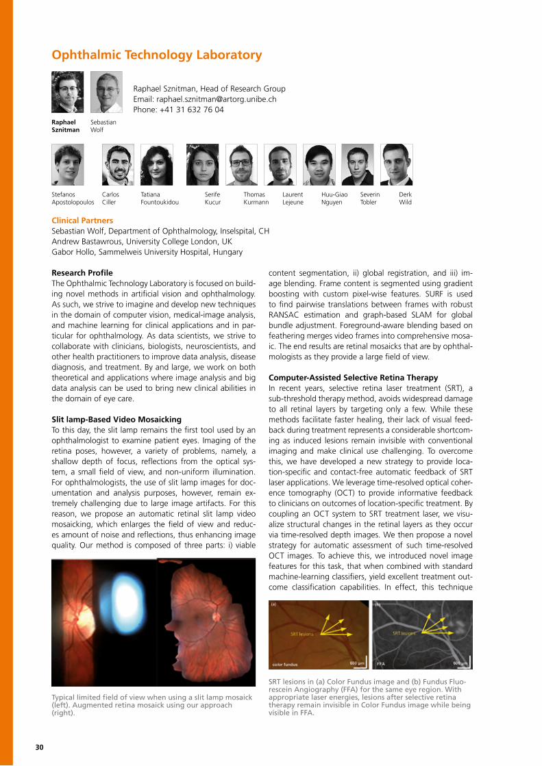
Ophthalmic Technology Laboratory
Raphael Sznitman, Head of Research GroupEmail: [email protected]: +41 31 632 76 04
StefanosApostolopoulos
Raphael Sebastian Sznitman Wolf
Research ProfileThe Ophthalmic Technology Laboratory is focused on build-ing novel methods in artificial vision and ophthalmology. As such, we strive to imagine and develop new techniques in the domain of computer vision, medical-image analysis, and machine learning for clinical applications and in par-ticular for ophthalmology. As data scientists, we strive to collaborate with clinicians, biologists, neuroscientists, and other health practitioners to improve data analysis, disease diagnosis, and treatment. By and large, we work on both theoretical and applications where image analysis and big data analysis can be used to bring new clinical abilities in the domain of eye care.
Slit lamp-Based Video MosaickingTo this day, the slit lamp remains the first tool used by an ophthalmologist to examine patient eyes. Imaging of the retina poses, however, a variety of problems, namely, a shallow depth of focus, reflections from the optical sys-tem, a small field of view, and non-uniform illumination. For ophthalmologists, the use of slit lamp images for doc-umentation and analysis purposes, however, remain ex-tremely challenging due to large image artifacts. For this reason, we propose an automatic retinal slit lamp video mosaicking, which enlarges the field of view and reduc-es amount of noise and reflections, thus enhancing image quality. Our method is composed of three parts: i) viable
content segmentation, ii) global registration, and iii) im-age blending. Frame content is segmented using gradient boosting with custom pixel-wise features. SURF is used to find pairwise translations between frames with robust RANSAC estimation and graph-based SLAM for global bundle adjustment. Foreground-aware blending based on feathering merges video frames into comprehensive mosa-ic. The end results are retinal mosaicks that are by ophthal-mologists as they provide a large field of view.
Computer-Assisted Selective Retina Therapy In recent years, selective retina laser treatment (SRT), a sub-threshold therapy method, avoids widespread damage to all retinal layers by targeting only a few. While these methods facilitate faster healing, their lack of visual feed-back during treatment represents a considerable shortcom-ing as induced lesions remain invisible with conventional imaging and make clinical use challenging. To overcome this, we have developed a new strategy to provide loca-tion-specific and contact-free automatic feedback of SRT laser applications. We leverage time-resolved optical coher-ence tomography (OCT) to provide informative feedback to clinicians on outcomes of location-specific treatment. By coupling an OCT system to SRT treatment laser, we visu-alize structural changes in the retinal layers as they occur via time-resolved depth images. We then propose a novel strategy for automatic assessment of such time-resolved OCT images. To achieve this, we introduced novel image features for this task, that when combined with standard machine-learning classifiers, yield excellent treatment out-come classification capabilities. In effect, this technique
Carlos Tatiana Ciller Fountoukidou
Serife Thomas Laurent Huu-Giao Severin DerkKucur Kurmann Lejeune Nguyen Tobler Wild
Clinical PartnersSebastian Wolf, Department of Ophthalmology, Inselspital, CHAndrew Bastawrous, University College London, UKGabor Hollo, Sammelweis University Hospital, Hungary
Typical limited field of view when using a slit lamp mosaick (left). Augmented retina mosaick using our approach (right).
SRT lesions in (a) Color Fundus image and (b) Fundus Fluo-rescein Angiography (FFA) for the same eye region. With appropriate laser energies, lesions after selective retina therapy remain invisible in Color Fundus image while being visible in FFA.
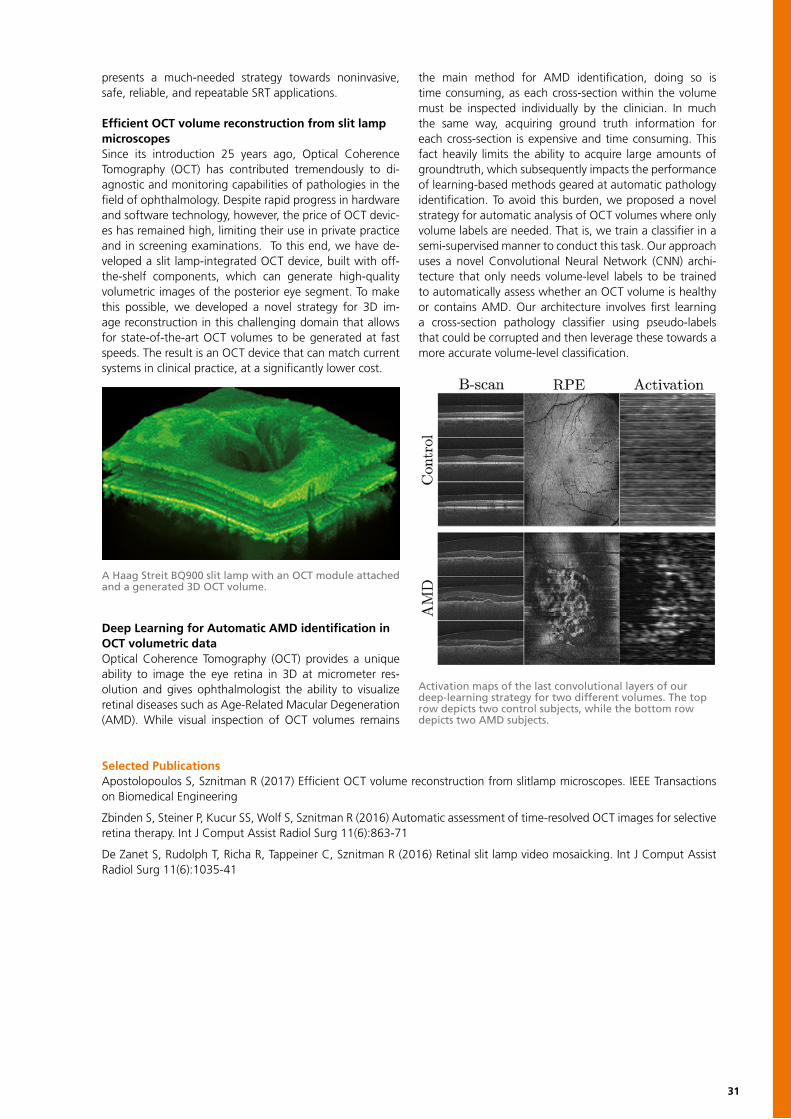
31
presents a much-needed strategy towards noninvasive, safe, reliable, and repeatable SRT applications.
Efficient OCT volume reconstruction from slit lamp microscopesSince its introduction 25 years ago, Optical Coherence Tomography (OCT) has contributed tremendously to di-agnostic and monitoring capabilities of pathologies in the field of ophthalmology. Despite rapid progress in hardware and software technology, however, the price of OCT devic-es has remained high, limiting their use in private practice and in screening examinations. To this end, we have de-veloped a slit lamp-integrated OCT device, built with off-the-shelf components, which can generate high-quality volumetric images of the posterior eye segment. To make this possible, we developed a novel strategy for 3D im-age reconstruction in this challenging domain that allows for state-of-the-art OCT volumes to be generated at fast speeds. The result is an OCT device that can match current systems in clinical practice, at a significantly lower cost.
Deep Learning for Automatic AMD identification in OCT volumetric dataOptical Coherence Tomography (OCT) provides a unique ability to image the eye retina in 3D at micrometer res-olution and gives ophthalmologist the ability to visualize retinal diseases such as Age-Related Macular Degeneration (AMD). While visual inspection of OCT volumes remains
the main method for AMD identification, doing so is time consuming, as each cross-section within the volume must be inspected individually by the clinician. In much the same way, acquiring ground truth information for each cross-section is expensive and time consuming. This fact heavily limits the ability to acquire large amounts of groundtruth, which subsequently impacts the performance of learning-based methods geared at automatic pathology identification. To avoid this burden, we proposed a novel strategy for automatic analysis of OCT volumes where only volume labels are needed. That is, we train a classifier in a semi-supervised manner to conduct this task. Our approach uses a novel Convolutional Neural Network (CNN) archi-tecture that only needs volume-level labels to be trained to automatically assess whether an OCT volume is healthy or contains AMD. Our architecture involves first learning a cross-section pathology classifier using pseudo-labels that could be corrupted and then leverage these towards a more accurate volume-level classification.
Selected PublicationsApostolopoulos S, Sznitman R (2017) Efficient OCT volume reconstruction from slitlamp microscopes. IEEE Transactions on Biomedical Engineering
Zbinden S, Steiner P, Kucur SS, Wolf S, Sznitman R (2016) Automatic assessment of time-resolved OCT images for selective retina therapy. Int J Comput Assist Radiol Surg 11(6):863-71
De Zanet S, Rudolph T, Richa R, Tappeiner C, Sznitman R (2016) Retinal slit lamp video mosaicking. Int J Comput Assist Radiol Surg 11(6):1035-41
A Haag Streit BQ900 slit lamp with an OCT module attached and a generated 3D OCT volume.
Activation maps of the last convolutional layers of our deep-learning strategy for two different volumes. The top row depicts two control subjects, while the bottom row depicts two AMD subjects.
Serife Thomas Laurent Huu-Giao Severin DerkKucur Kurmann Lejeune Nguyen Tobler Wild
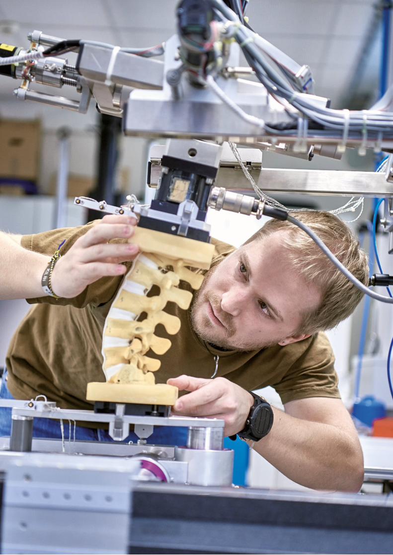
33
INSTITUTE FOR SURGICAL TECHNOLOGY AND BIOMECHANICS
The Institute for Surgical Technology and Biomechanics (ISTB) comprises a multidis-ciplinary team of about 50 co-workers from more than 10 countries. It stands in the tradition of the late Professor Maurice E. Müller, the former chairman of the Department of Orthopaedics at the Bern University Hospital, not only known for his many pioneering innovations in orthopaedics and traumatology, but also for his vision that only a close collaboration among surgeons, scientists, engineers, and in-dustrialists will allow sustainable progress in the field.
To date, the ISTB hosts five research groups in various fields of basic and applied re-search for the prevention, diagnosis, and treatment of disease, working from the cell level to organ systems: • Musculoskeletal Biomechanics• Computational Bioengineering• Tissue and Organ Mechanobiology• Medical Image Analysis• Information Processing in Medical Interventions The mission of the multidisciplinary team of the ISTB is to advance human under-standing, health, and quality of life with a focus on developing solutions that address particular clinical problems or unmet clinical needs. It supports this effort through internationally recognized research, discovery, and invention in the area of biomed-ical engineering, translation of research results from the lab to the clinic to improve patient care, transfer of scientific discoveries, and biomedical technology through national and international industrial collaborations and a world- class post-graduate biomedical engineering education program.
Lutz-P. NolteDirector
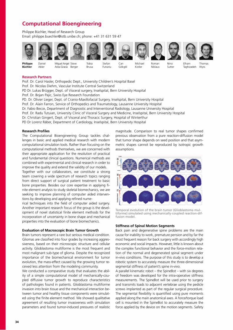
34
Computational BioengineeringPhilippe Büchler, Head of Research GroupEmail: [email protected]; phone: +41 31 631 59 47
Philippe Daniel Miguel Angel Steve Tobia Stefan Can Michael Roman Nino Elham ThomasBüchler Abler Ariza Gracia Berger Brusa Funariu Gökgöl Kistler Niklaus Sutter Taghizadeh Wyss
Research ProfilesThe Computational Bioengineering Group tackles chal-lenges in basic and applied medical research with modern computational simulation tools. Rather than focusing on the computational methods themselves, we are concerned with their appropriate application for the resolution of practical and fundamental clinical questions. Numerical methods are combined with experimental and clinical research in order to improve the quality and extend the validity of our models.Together with our collaborators, we constitute a strong team covering a wide spectrum of research topics ranging from direct support of surgical patient treatment to basic bone properties. Besides our core expertise in applying fi-nite element analysis to study skeletal biomechanics, we are seeking to improve planning of computer aided interven-tions by developing and applying refined nume-rical techniques into the field of computer aided surgery. Another important research focus of the group is the devel-opment of novel statistical finite element methods for the incorporation of uncertainty in bone shape and mechanical properties into the evaluation of bone biomechanics.
Evaluation of Macroscopic Brain Tumor GrowthBrain tumors represent a rare but serious medical condition. Gliomas are classified into four grades by increasing aggres-siveness, based on their microscopic structure and cellular activity. Glioblastoma multiforme is the most frequent and most malignant sub-type of glioma. Despite the recognized importance of the biomechanical environment for tumor evolution, the mass-effect caused by the growing tumor re-ceived less attention from the modeling community.We conducted a comparative study that evaluates the abil-ity of a simple computational model of mechanically-cou-pled diffusive tumor growth to reproduce characteristics of pathologies found in patients. Glioblastoma multiforme invasion into brain tissue and the mechanical interaction be-tween tumor and healthy tissue components were simulat-ed using the finite element method. We showed qualitative agreement of resulting tumor invasiveness with simulation parameters and found tumor-induced pressures of realistic
magnitude. Comparison to real tumor shapes confirmed previous observation from a pure reaction-diffusion model that tumor shape depends on seed position and that asym-metric shapes cannot be reproduced by isotropic growth assumptions.
Stiffness of Spinal Motion SegmentsBack pain and degenerative spine problems are the main cause for inability to work, premature pension and by far the most frequent reason for back surgery with accordingly high economic and social impacts. However, little is known about the complex functional behavior and the force-motion rela-tion of the normal and degenerated spinal segment under in-vivo conditions. The purpose of this study is to develop a robotic system to accurately measure the three-dimensional segmental stiffness of patient’s spine in-vivo.A parallel kinematic robot – the SpineBot – with six degrees of freedom was developed for the intra-operative stiffness measurements. The SpineBot will be used prior to surgery and transmits loads to adjacent vertebrae using the pedicle screws implanted as part of the regular surgical procedure. The segmental flexibility is quantified using pure moments applied along the main anatomical axes. A force/torque load cell is mounted in the SpineBot to accurately measure the force applied by the device on the motion segments. Safety
Temporal evolution of the brain tumor (Glioblastoma mul-tiforme) simulated using mechanically-coupled reaction-dif-fusion model.
Research PartnersProf. Dr. Carol Hasler, Orthopedic Dept., University Children’s Hospital BaselProf. Dr. Nicolas Diehm, Vascular Institute Central SwitzerlandPD Dr. Lukas Brügger, Dept. of Visceral surgery, Inselspital, Bern University HospitalProf. Dr. Bojan Pajic, Swiss Eye Research FoundationPD. Dr. Olivier Lieger, Dept. of Cranio-Maxillofacial Surgery, Inselspital, Bern University HospitalProf. Dr. Alain Farron, Service of Orthopedics and Traumatology, Lausanne University HospitalDr. Fabio Becce, Department of Diagnostic and Interventional Radiology, Lausanne University HospitalProf. Dr. Radu Tutuian, University Clinic of Visceral Surgery and Medicine, Inselspital, Bern University HospitalDr. Christian Gingert, Dept. of Visceral and Thoracic Surgery, Hospital of WinterthurPD Dr Lorenz Räber, Department of Cardiology, Inselspital, Bern University Hospital
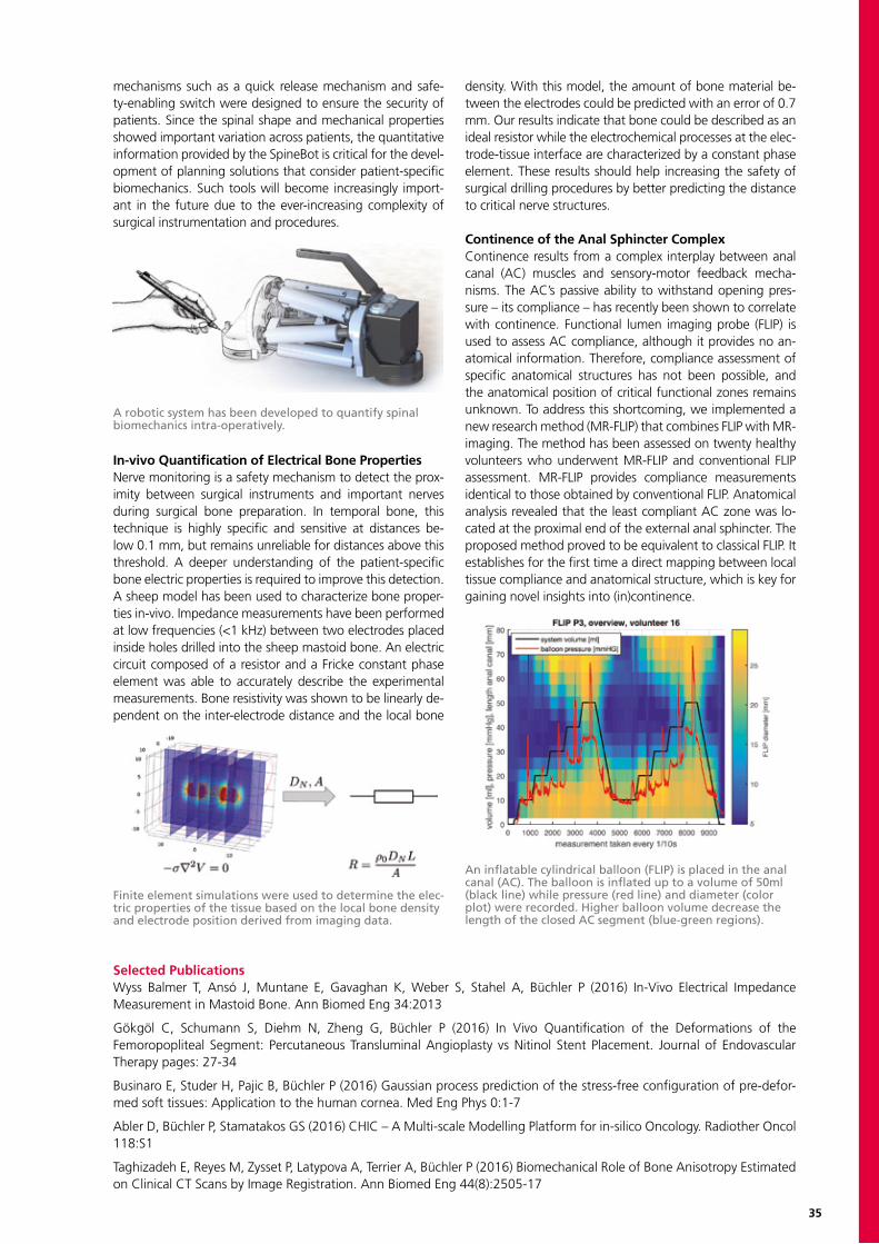
35
mechanisms such as a quick release mechanism and safe-ty-enabling switch were designed to ensure the security of patients. Since the spinal shape and mechanical properties showed important variation across patients, the quantitative information provided by the SpineBot is critical for the devel-opment of planning solutions that consider patient-specific biomechanics. Such tools will become increasingly import-ant in the future due to the ever-increasing complexity of surgical instrumentation and procedures.
In-vivo Quantification of Electrical Bone PropertiesNerve monitoring is a safety mechanism to detect the prox-imity between surgical instruments and important nerves during surgical bone preparation. In temporal bone, this technique is highly specific and sensitive at distances be-low 0.1 mm, but remains unreliable for distances above this threshold. A deeper understanding of the patient-specific bone electric properties is required to improve this detection. A sheep model has been used to characterize bone proper-ties in-vivo. Impedance measurements have been performed at low frequencies (<1 kHz) between two electrodes placed inside holes drilled into the sheep mastoid bone. An electric circuit composed of a resistor and a Fricke constant phase element was able to accurately describe the experimental measurements. Bone resistivity was shown to be linearly de-pendent on the inter-electrode distance and the local bone
density. With this model, the amount of bone material be-tween the electrodes could be predicted with an error of 0.7 mm. Our results indicate that bone could be described as an ideal resistor while the electrochemical processes at the elec-trode-tissue interface are characterized by a constant phase element. These results should help increasing the safety of surgical drilling procedures by better predicting the distance to critical nerve structures.
Continence of the Anal Sphincter ComplexContinence results from a complex interplay between anal canal (AC) muscles and sensory-motor feedback mecha-nisms. The AC’s passive ability to withstand opening pres-sure – its compliance – has recently been shown to correlate with continence. Functional lumen imaging probe (FLIP) is used to assess AC compliance, although it provides no an-atomical information. Therefore, compliance assessment of specific anatomical structures has not been possible, and the anatomical position of critical functional zones remains unknown. To address this shortcoming, we implemented a new research method (MR-FLIP) that combines FLIP with MR-imaging. The method has been assessed on twenty healthy volunteers who underwent MR-FLIP and conventional FLIP assessment. MR-FLIP provides compliance measurements identical to those obtained by conventional FLIP. Anatomical analysis revealed that the least compliant AC zone was lo-cated at the proximal end of the external anal sphincter. The proposed method proved to be equivalent to classical FLIP. It establishes for the first time a direct mapping between local tissue compliance and anatomical structure, which is key for gaining novel insights into (in)continence.
A robotic system has been developed to quantify spinal biomechanics intra-operatively.
Finite element simulations were used to determine the elec-tric properties of the tissue based on the local bone density and electrode position derived from imaging data.
An inflatable cylindrical balloon (FLIP) is placed in the anal canal (AC). The balloon is inflated up to a volume of 50ml (black line) while pressure (red line) and diameter (color plot) were recorded. Higher balloon volume decrease the length of the closed AC segment (blue-green regions).
Selected PublicationsWyss Balmer T, Ansó J, Muntane E, Gavaghan K, Weber S, Stahel A, Büchler P (2016) In-Vivo Electrical Impedance Measurement in Mastoid Bone. Ann Biomed Eng 34:2013
Gökgöl C, Schumann S, Diehm N, Zheng G, Büchler P (2016) In Vivo Quantification of the Deformations of the Femoropopliteal Segment: Percutaneous Transluminal Angioplasty vs Nitinol Stent Placement. Journal of Endovascular Therapy pages: 27-34
Businaro E, Studer H, Pajic B, Büchler P (2016) Gaussian process prediction of the stress-free configuration of pre-defor-med soft tissues: Application to the human cornea. Med Eng Phys 0:1-7
Abler D, Büchler P, Stamatakos GS (2016) CHIC – A Multi-scale Modelling Platform for in-silico Oncology. Radiother Oncol 118:S1
Taghizadeh E, Reyes M, Zysset P, Latypova A, Terrier A, Büchler P (2016) Biomechanical Role of Bone Anisotropy Estimated on Clinical CT Scans by Image Registration. Ann Biomed Eng 44(8):2505-17
Philippe Daniel Miguel Angel Steve Tobia Stefan Can Michael Roman Nino Elham ThomasBüchler Abler Ariza Gracia Berger Brusa Funariu Gökgöl Kistler Niklaus Sutter Taghizadeh Wyss
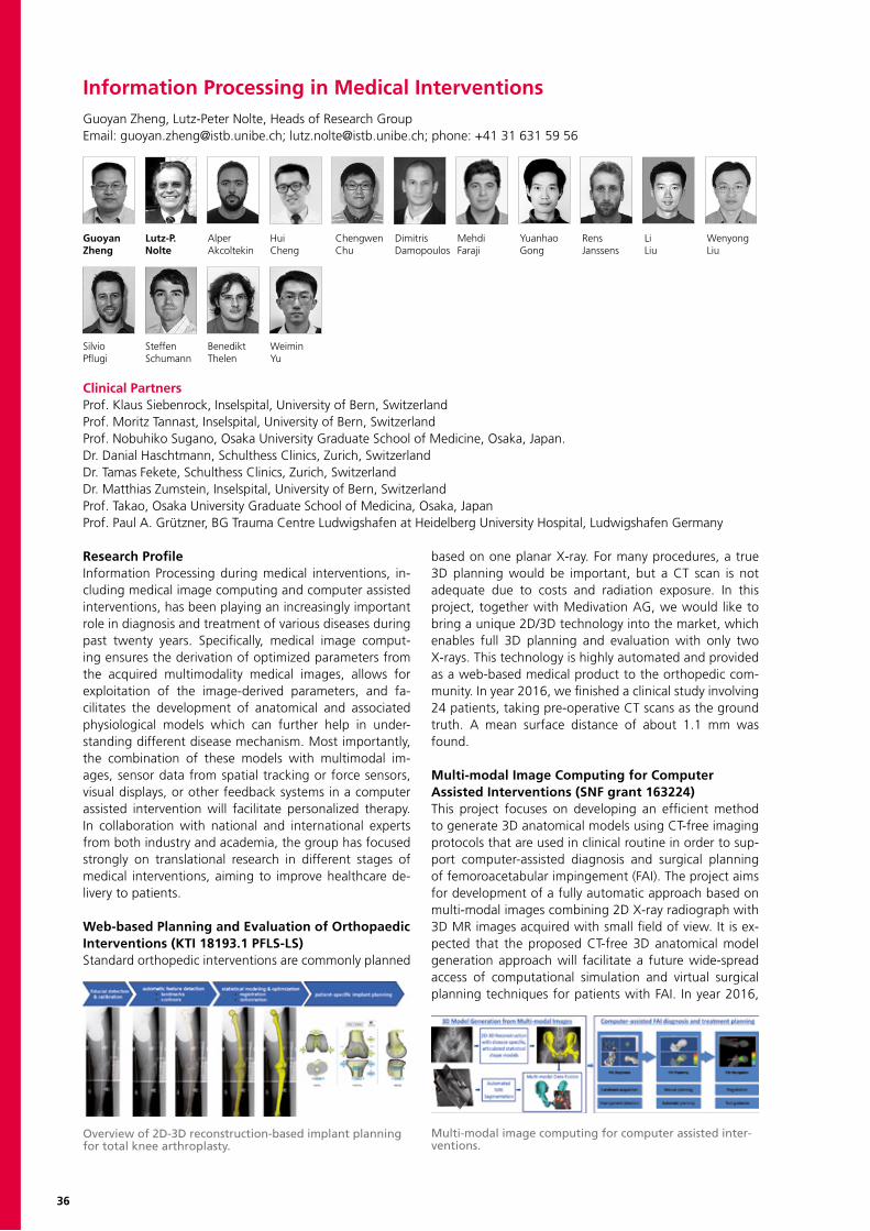
36
Information Processing in Medical InterventionsGuoyan Zheng, Lutz-Peter Nolte, Heads of Research GroupEmail: [email protected]; [email protected]; phone: +41 31 631 59 56
Guoyan Lutz-P. Alper Hui Chengwen Dimitris Mehdi Yuanhao Rens Li WenyongZheng Nolte Akcoltekin Cheng Chu Damopoulos Faraji Gong Janssens Liu Liu
Clinical PartnersProf. Klaus Siebenrock, Inselspital, University of Bern, SwitzerlandProf. Moritz Tannast, Inselspital, University of Bern, SwitzerlandProf. Nobuhiko Sugano, Osaka University Graduate School of Medicine, Osaka, Japan.Dr. Danial Haschtmann, Schulthess Clinics, Zurich, SwitzerlandDr. Tamas Fekete, Schulthess Clinics, Zurich, SwitzerlandDr. Matthias Zumstein, Inselspital, University of Bern, SwitzerlandProf. Takao, Osaka University Graduate School of Medicina, Osaka, JapanProf. Paul A. Grützner, BG Trauma Centre Ludwigshafen at Heidelberg University Hospital, Ludwigshafen Germany
Silvio Steffen Benedikt WeiminPflugi Schumann Thelen Yu
Research ProfileInformation Processing during medical interventions, in-cluding medical image computing and computer assisted interventions, has been playing an increasingly important role in diagnosis and treatment of various diseases during past twenty years. Specifically, medical image comput-ing ensures the derivation of optimized parameters from the acquired multimodality medical images, allows for exploitation of the image-derived parameters, and fa-cilitates the development of anatomical and associated physiological models which can further help in under-standing different disease mechanism. Most importantly, the combination of these models with multimodal im-ages, sensor data from spatial tracking or force sensors, visual displays, or other feedback systems in a computer assisted intervention will facilitate personalized therapy. In collaboration with national and international experts from both industry and academia, the group has focused strongly on translational research in different stages of medical interventions, aiming to improve healthcare de-livery to patients.
Web-based Planning and Evaluation of Orthopaedic Interventions (KTI 18193.1 PFLS-LS)Standard orthopedic interventions are commonly planned
based on one planar X-ray. For many procedures, a true 3D planning would be important, but a CT scan is not adequate due to costs and radiation exposure. In this project, together with Medivation AG, we would like to bring a unique 2D/3D technology into the market, which enables full 3D planning and evaluation with only two X-rays. This technology is highly automated and provided as a web-based medical product to the orthopedic com-munity. In year 2016, we finished a clinical study involving 24 patients, taking pre-operative CT scans as the ground truth. A mean surface distance of about 1.1 mm was found.
Multi-modal Image Computing for ComputerAssisted Interventions (SNF grant 163224)This project focuses on developing an efficient method to generate 3D anatomical models using CT-free imaging protocols that are used in clinical routine in order to sup-port computer-assisted diagnosis and surgical planning of femoroacetabular impingement (FAI). The project aims for development of a fully automatic approach based on multi-modal images combining 2D X-ray radiograph with 3D MR images acquired with small field of view. It is ex-pected that the proposed CT-free 3D anatomical model generation approach will facilitate a future wide-spread access of computational simulation and virtual surgical planning techniques for patients with FAI. In year 2016,
Overview of 2D-3D reconstruction-based implant planning for total knee arthroplasty.
Multi-modal image computing for computer assisted inter-ventions.
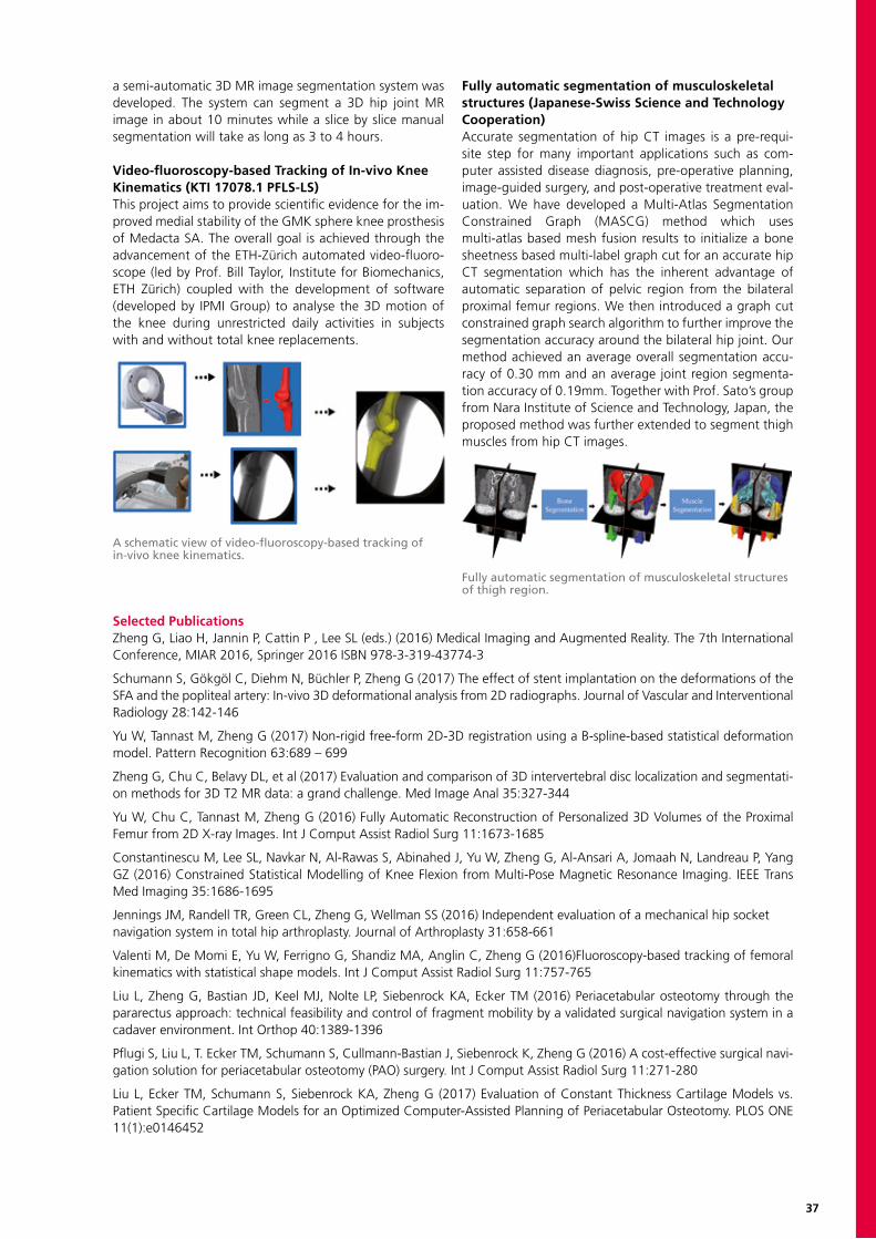
37
a semi-automatic 3D MR image segmentation system was developed. The system can segment a 3D hip joint MR image in about 10 minutes while a slice by slice manual segmentation will take as long as 3 to 4 hours.
Video-fluoroscopy-based Tracking of In-vivo Knee Kinematics (KTI 17078.1 PFLS-LS)This project aims to provide scientific evidence for the im-proved medial stability of the GMK sphere knee prosthesis of Medacta SA. The overall goal is achieved through the advancement of the ETH-Zürich automated video-fluoro-scope (led by Prof. Bill Taylor, Institute for Biomechanics, ETH Zürich) coupled with the development of software (developed by IPMI Group) to analyse the 3D motion of the knee during unrestricted daily activities in subjects with and without total knee replacements.
Fully automatic segmentation of musculoskeletal structures (Japanese-Swiss Science and Technology Cooperation)Accurate segmentation of hip CT images is a pre-requi-site step for many important applications such as com-puter assisted disease diagnosis, pre-operative planning, image-guided surgery, and post-operative treatment eval-uation. We have developed a Multi-Atlas Segmentation Constrained Graph (MASCG) method which uses multi-atlas based mesh fusion results to initialize a bone sheetness based multi-label graph cut for an accurate hip CT segmentation which has the inherent advantage of automatic separation of pelvic region from the bilateral proximal femur regions. We then introduced a graph cut constrained graph search algorithm to further improve the segmentation accuracy around the bilateral hip joint. Our method achieved an average overall segmentation accu-racy of 0.30 mm and an average joint region segmenta-tion accuracy of 0.19mm. Together with Prof. Sato’s group from Nara Institute of Science and Technology, Japan, the proposed method was further extended to segment thigh muscles from hip CT images.
Fully automatic segmentation of musculoskeletal structures of thigh region.
A schematic view of video-fluoroscopy-based tracking of in-vivo knee kinematics.
Selected PublicationsZheng G, Liao H, Jannin P, Cattin P , Lee SL (eds.) (2016) Medical Imaging and Augmented Reality. The 7th International Conference, MIAR 2016, Springer 2016 ISBN 978-3-319-43774-3
Schumann S, Gökgöl C, Diehm N, Büchler P, Zheng G (2017) The effect of stent implantation on the deformations of the SFA and the popliteal artery: In-vivo 3D deformational analysis from 2D radiographs. Journal of Vascular and Interventional Radiology 28:142-146
Yu W, Tannast M, Zheng G (2017) Non-rigid free-form 2D-3D registration using a B-spline-based statistical deformation model. Pattern Recognition 63:689 – 699
Zheng G, Chu C, Belavy DL, et al (2017) Evaluation and comparison of 3D intervertebral disc localization and segmentati-on methods for 3D T2 MR data: a grand challenge. Med Image Anal 35:327-344
Yu W, Chu C, Tannast M, Zheng G (2016) Fully Automatic Reconstruction of Personalized 3D Volumes of the Proximal Femur from 2D X-ray Images. Int J Comput Assist Radiol Surg 11:1673-1685
Constantinescu M, Lee SL, Navkar N, Al-Rawas S, Abinahed J, Yu W, Zheng G, Al-Ansari A, Jomaah N, Landreau P, Yang GZ (2016) Constrained Statistical Modelling of Knee Flexion from Multi-Pose Magnetic Resonance Imaging. IEEE Trans Med Imaging 35:1686-1695
Jennings JM, Randell TR, Green CL, Zheng G, Wellman SS (2016) Independent evaluation of a mechanical hip socket navigation system in total hip arthroplasty. Journal of Arthroplasty 31:658-661
Valenti M, De Momi E, Yu W, Ferrigno G, Shandiz MA, Anglin C, Zheng G (2016)Fluoroscopy-based tracking of femoral kinematics with statistical shape models. Int J Comput Assist Radiol Surg 11:757-765
Liu L, Zheng G, Bastian JD, Keel MJ, Nolte LP, Siebenrock KA, Ecker TM (2016) Periacetabular osteotomy through the pararectus approach: technical feasibility and control of fragment mobility by a validated surgical navigation system in a cadaver environment. Int Orthop 40:1389-1396
Pflugi S, Liu L, T. Ecker TM, Schumann S, Cullmann-Bastian J, Siebenrock K, Zheng G (2016) A cost-effective surgical navi-gation solution for periacetabular osteotomy (PAO) surgery. Int J Comput Assist Radiol Surg 11:271-280
Liu L, Ecker TM, Schumann S, Siebenrock KA, Zheng G (2017) Evaluation of Constant Thickness Cartilage Models vs. Patient Specific Cartilage Models for an Optimized Computer-Assisted Planning of Periacetabular Osteotomy. PLOS ONE 11(1):e0146452

38
Medical Image AnalysisMauricio Reyes, Head of Research GroupEmail: [email protected]; phone: +41 31 631 59 50
Mauricio Fabian Livia Vimal Simon Alain Ping Raphael Martin AlainReyes Balsiger Barazzetti Chandran Habegger Jungo Lu Meier Pletscher Reimann
Kamal Carlos Correa Yannick Elham WaldoShahim Shokiche Suter Taghizadeh Valenzuela
Research PartnersR. Wiest, Institute of Diagnostic and Interventional Neuroradiology, Inselspital, Bern Univ. HospitalP. Schucht, University Clinic for Neurosurgery, Bern Univ. HospitalC. Boesch, Magnetic Resonance Spectroscopy and Methodology - Department of Clinical Research – Bern Univ.V. Djonov, Department of Anatomy, Bern Univ.
Research ProfileThe Medical Image Analysis group conducts theoretical and applied research in image processing, computer vision, and artificial intelligence for the analysis of medical image datasets. The focus of our research relies on the paradigm of evidence-based image modeling and personalized med-icine, with main applications to machine learning based analysis of brain lesions from multi-sequence MRI, multi-res-olution computational anatomy from CT imaging, and ad-vanced techniques for clinically-relevant human-machine interfacing.
Brain Lesion Image AnalysisMagnetic Resonance Imaging (MRI) and its variants are a powerful imaging modality that encompasses rich anatom-ical and physiological information at a high resolution. In neurosciences these modalities have become a standard in clinical practice. However, the interpretation of the images requires the combined use of different modalities, which leads to the need of computer-assisted technologies. The group has developed several methodologies to analyze MRI
images with focus on multimodal image segmentation for brain image lesion analysis studies. These developments are driven by clinical requirements such as computation speed, robustness, and use of standard clinical imaging protocols.During 2016 we performed clinical evaluations of our soft-ware BraTumIA (Brain Tumor Image Analysis), and tested it on longitudinal set-ups, for neurosurgical procedures, and compared against other FDA-approved tools for neurosurgi-cal planning. During 2016 we also started a CE marking pro-cess and licensing of this technology to a leader in the medi-cal imaging field. In addition, our group was awarded Young Scientist Publication Impact Award 2016 for our seminal 2011 Miccai work. In collaboration with our partners at the Institute of Diagnostic and Interventional Neuroradiology, we further extended initial developments for the prediction of lesion outcome in ischemic stroke patients, leading to a second place in the ISLES2016 segmentation challenge. Similarly, the seminal work on multiple sclerosis allowed us to obtain the first prize in the multiple sclerosis segmenta-tion challenge at Miccai 2016.
Brain image lesion analysis (In clockwise order): Improving the assessment of response to therapy through automated brain tumor quantification. Radiomics, and the role of tumor volumetry for patient survival analysis. Advanced brain tumor quan-tification for neurosurgery and radiotherapy. Robust and clinically-validated longitudinal brain tumor quantification.

39
Modeling anatomical variability and its application to applications such as cochlear electrode assessment, sub-voxel facial nerve segmentation, direct bone modeling for FE analysis, and modern image-based stereology.
Selected PublicationsMeier R, Knecht U, Loosli T, Bauer S, Slotboom J, Wiest R, Reyes M (2016) Clinical Evaluation of a Fully-automatic Segmentation Method for Longitudinal Brain Tumor Volumetry. Nature Scientific Reports 6
Maier O, Menze B, Wiest R, Handels H, Reyes M (2016) ISLES 2015 - A public evaluation benchmark for ischemic stroke lesion segmentation from multispectral MRI. Medical Image Analysis 35:250—269
Porz N, Habegger S, Meier R, Verma RK, Jilch A, Fichtner J, Knecht U, Radina C, Schucht P, Jürgen B, Raabe A, Slotboom J, Reyes M, Wiest R (2016) Fully Automated Enhanced Tumor Compartmentalization: Man vs. Machine Reloaded. PLoS ONE 11(11):e0165302
McKinley R, Haeni L, Gralla J, El-Koussy M, Bauer S, Arnold M, Fischer U, Jung S, Mattmann K, Reyes M, Wiest R (2016) Fully automated stroke tissue estimation using random forest classifiers (FASTER). Journal of Cerebral Blood Flow & Metabolism
Shokiche C, Baumann P, Ruslan H, Djonov V, Reyes M (2016) High-Throughput Glomeruli Analysis of micro-CT Kidney Images Using Tree Priors and Scalable Sparse Computation. Medical Image Computing and Computer-Assisted Intervention -- MICCAI 2016: 19th International Conference, Athens, Greece, Proceedings, Part II, 370—378
Valenzuela W, Ferguson S, Ignasiak D, Diserens G, Haeni L, Wiest R, Vermathen P, Boesch C, Reyes M (2016) FISICO: Fast Image SegmentatIon COrrection. PLoS ONE 11:1-17
Seif M, Mani L, Lu H, Boesch C, Reyes M, Vogt B, Vermathen P (2016) Diffusion tensor imaging of the human kidney: Does image registration permit scanning without respiratory triggering? J Magn Reson Imaging 44:327-334
Crimi A, Menze B, Maier O, Reyes M, Handels H (2016) Brainlesion: Glioma, Multiple Sclerosis, Stroke and Traumatic Brain Injuries. First International Workshop, Brainles 2015, Held in Conjunction with MICCAI 2015, Munich, Germany, Revised Selected Papers
Reyes M, Shahim K, Jürgens P (2016) Computational Image-guided Technologies in Cranio-Maxillofacial Soft Tissue Planning and Simulation. In Computer-Assisted Musculoskeletal Surgery pp. 43-56
Computational Anatomy Computational anatomy enables analysis of biological vari-abilComputational anatomy enables analysis of biological variability throughout a population. Using statistical math-ematical techniques, models can be built to represent the typical shape of an anatomical structure and its predominant patterns of variability across a given population. During 2016 we used these techniques to evaluate the performance of cochlear electrodes, used to reestablish hearing on patient suffering from moderate to severe hearing loss, as well as super-resolution techniques able to enhance the quality and the accuracy of segmentations of the facial nerve, needed
for cochlear implantation planning procedures. In combina-tion with Machine Learning techniques, we have developed modeling engines able to yield, directly from the clinical images, patient-specific models compliant with FE analysis. During 2016 we continued the development of algorithms and tools to perform fast corrections of segmentations. The results of this research were released to the community in the form of a software tool, FISICO (Fast Image Segmentation Correction). Similarly, we developed fast and accurate seg-mentation and quantification algorithms for high-resolution micro-CT kidney scans employed for the design of modern image-based stereology techniques.
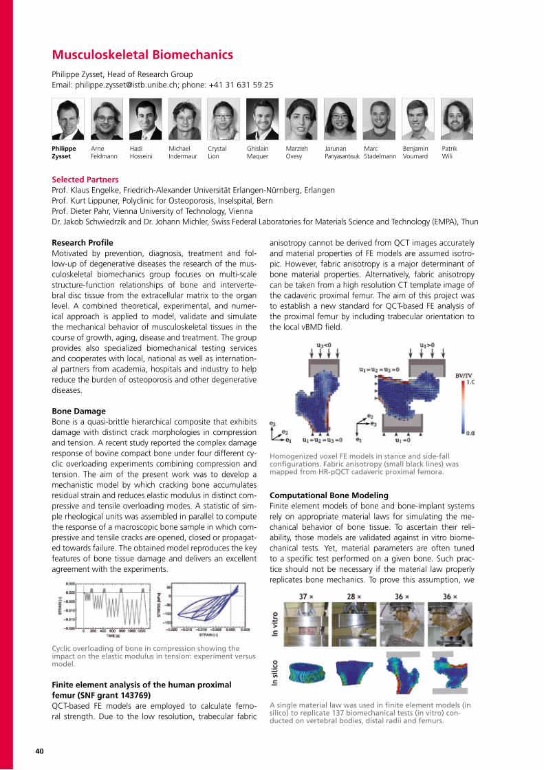
40
Musculoskeletal BiomechanicsPhilippe Zysset, Head of Research GroupEmail: [email protected]; phone: +41 31 631 59 25
Philippe Arne Hadi Michael Crystal Ghislain Marzieh Jarunan Marc Benjamin PatrikZysset Feldmann Hosseini Indermaur Lion Maquer Ovesy Panyasantisuk Stadelmann Voumard Wili
Selected PartnersProf. Klaus Engelke, Friedrich-Alexander Universität Erlangen-Nürnberg, ErlangenProf. Kurt Lippuner, Polyclinic for Osteoporosis, Inselspital, BernProf. Dieter Pahr, Vienna University of Technology, ViennaDr. Jakob Schwiedrzik and Dr. Johann Michler, Swiss Federal Laboratories for Materials Science and Technology (EMPA), Thun
Research ProfileMotivated by prevention, diagnosis, treatment and fol-low-up of degenerative diseases the research of the mus-culoskeletal biomechanics group focuses on multi-scale structure-function relationships of bone and interverte-bral disc tissue from the extracellular matrix to the organ level. A combined theoretical, experimental, and numer-ical approach is applied to model, validate and simulate the mechanical behavior of musculoskeletal tissues in the course of growth, aging, disease and treatment. The group provides also specialized biomechanical testing services and cooperates with local, national as well as internation-al partners from academia, hospitals and industry to help reduce the burden of osteoporosis and other degenerative diseases.
Bone DamageBone is a quasi-brittle hierarchical composite that exhibits damage with distinct crack morphologies in compression and tension. A recent study reported the complex damage response of bovine compact bone under four different cy-clic overloading experiments combining compression and tension. The aim of the present work was to develop a mechanistic model by which cracking bone accumulates residual strain and reduces elastic modulus in distinct com-pressive and tensile overloading modes. A statistic of sim-ple rheological units was assembled in parallel to compute the response of a macroscopic bone sample in which com-pressive and tensile cracks are opened, closed or propagat-ed towards failure. The obtained model reproduces the key features of bone tissue damage and delivers an excellent agreement with the experiments.
Finite element analysis of the human proximalfemur (SNF grant 143769)QCT-based FE models are employed to calculate femo-ral strength. Due to the low resolution, trabecular fabric
anisotropy cannot be derived from QCT images accurately and material properties of FE models are assumed isotro-pic. However, fabric anisotropy is a major determinant of bone material properties. Alternatively, fabric anisotropy can be taken from a high resolution CT template image of the cadaveric proximal femur. The aim of this project was to establish a new standard for QCT-based FE analysis of the proximal femur by including trabecular orientation to the local vBMD field.
Computational Bone ModelingFinite element models of bone and bone-implant systems rely on appropriate material laws for simulating the me-chanical behavior of bone tissue. To ascertain their reli-ability, those models are validated against in vitro biome-chanical tests. Yet, material parameters are often tuned to a specific test performed on a given bone. Such prac-tice should not be necessary if the material law properly replicates bone mechanics. To prove this assumption, we
Cyclic overloading of bone in compression showing the impact on the elastic modulus in tension: experiment versus model.
Homogenized voxel FE models in stance and side-fall configurations. Fabric anisotropy (small black lines) was mapped from HR-pQCT cadaveric proximal femora.
A single material law was used in finite element models (in silico) to replicate 137 biomechanical tests (in vitro) con-ducted on vertebral bodies, distal radii and femurs.
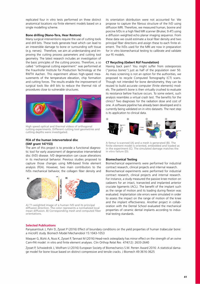
41
replicated four in vitro tests performed on three distinct anatomical locations via finite element models based on a single modelling scheme.
Bone drilling (Nano-Tera, Hear Restore)Many surgical interventions require the use of cutting tools and drill bits. These tools generate heat which can lead to an irreversible damage to bone or surrounding soft tissue (e.g. nerves). Therefore, we aim at understanding and im-proving the cutting process parameters and cutting tool geometry. The latest research includes an investigation of the basic principles of the cutting process. Therefore, a so called “orthogonal cutting experiment” was performed at the Fraunhofer Institute for Production Technology at the RWTH Aachen. This experiment allows high-speed mea-surements of the temperature elevation, chip formation and cutting forces. The results enable the improvement of surgical tools like drill bits to reduce the thermal risk of procedures close to vulnerable structures.
FEA of the human intervertebral disc(SNF grant 147153)The aim of this project is to provide a functional diagnos-tic tool for early assessment of degenerative intervertebral disc (IVD) disease. IVD degeneration can cause alterations in its mechanical behavior. Previous studies proposed to capture those changes using MRI-based finite element analysis (FEA). However, two main contributors to the IVDs mechanical behavior, the collagen fiber density and
its orientation distribution were not accounted for. We propose to capture the fibrous structure of the IVD using diffusion MRI. Therefore, we measured human, bovine and porcine IVDs in a high filed MR scanner (Bruker, 9.4T) using a diffusion weighted echo planar imaging sequence. From these data we could estimate a local fiber density and two principal fiber directions and assign these to each finite el-ement. The IVDs used for the MRI are now in preparation for in vitro biomechanical testing to calibrate and validate our FE models.
CT Recycling (Gebert Rüf Foundation)Having back pain? You might suffer from osteoporosis (“porous bones”) just as half of the population over 50. As mass screening is not an option for the authorities, we proposed to recycle Computed Tomography (CT) scans. Though not intended for bone densitometry, they can be reused to build accurate computer (finite elements) mod-els. The patient’s bone is then virtually crushed to evaluate its resistance before fracture occurs. To some extent, such analysis resembles a virtual crash test. The benefits for the clinics? Two diagnoses for the radiation dose and cost of one. A software pipeline has already been developed and is currently being validated on in vitro datasets. The next step is its application to clinical data.
Biomechanical TestingBiomechanical experiments were performed for industrial contract research, clinical projects and internal research.Biomechanical experiments were performed for industrial contract research, clinical projects and internal research. For instance, a study measured the passive knee motion on cadavers for an intact, transected and implanted anterior cruciate ligaments (ACL). The benefit of the implant such as the range of motion and its loading during flexion was evaluated. Implantation site errors were simulated in order to assess the impact on the range of motion of the knee and the implant effectiveness. Another project in collab-oration with the Dental School evaluated the mechanical properties of ceramic dental implants according to indus-trial testing standards.
A) T1-weighted image of a human IVD and its principal diffusion direction. The color represents a normalized local mean diffusion. B) Corresponding mesh and computed fiber orientations.
High speed optical and thermal videos of orthogonal cutting experiments. Different cutting tool geometries and cutting depths were investigated.
A femur is scanned (A) and a mesh is generated (B). The finite element model is oriented, embedded and loaded as in the experiment (C). The simulated damage matches the in vitro failure (D).
Selected PublicationsPanyasantisuk J, Pahr D, Zysset P (2016) Effect of boundary conditions on the yield properties of human trabecular bone: a microFE study. Biomech Model Mechanobiol 15:1043-1053
Maquer G, Bürki A, Nuss K, Zysset P, Tannast M (2016) Head-neck osteoplasty has minor effect on the strength of an ovine Cam-FAI model: in vitro and finite element analyses. Clin Orthop Relat Res 474(12): 2633-2640
Zysset P, Schwiedrzik J, Wolfram U (2016) European Society of Biomechanics S.M. Perren Award 2016: A statistical dama-ge model for bone tissue based on distinct compressive and tensile cracks. J Biomech 49:3616-3625
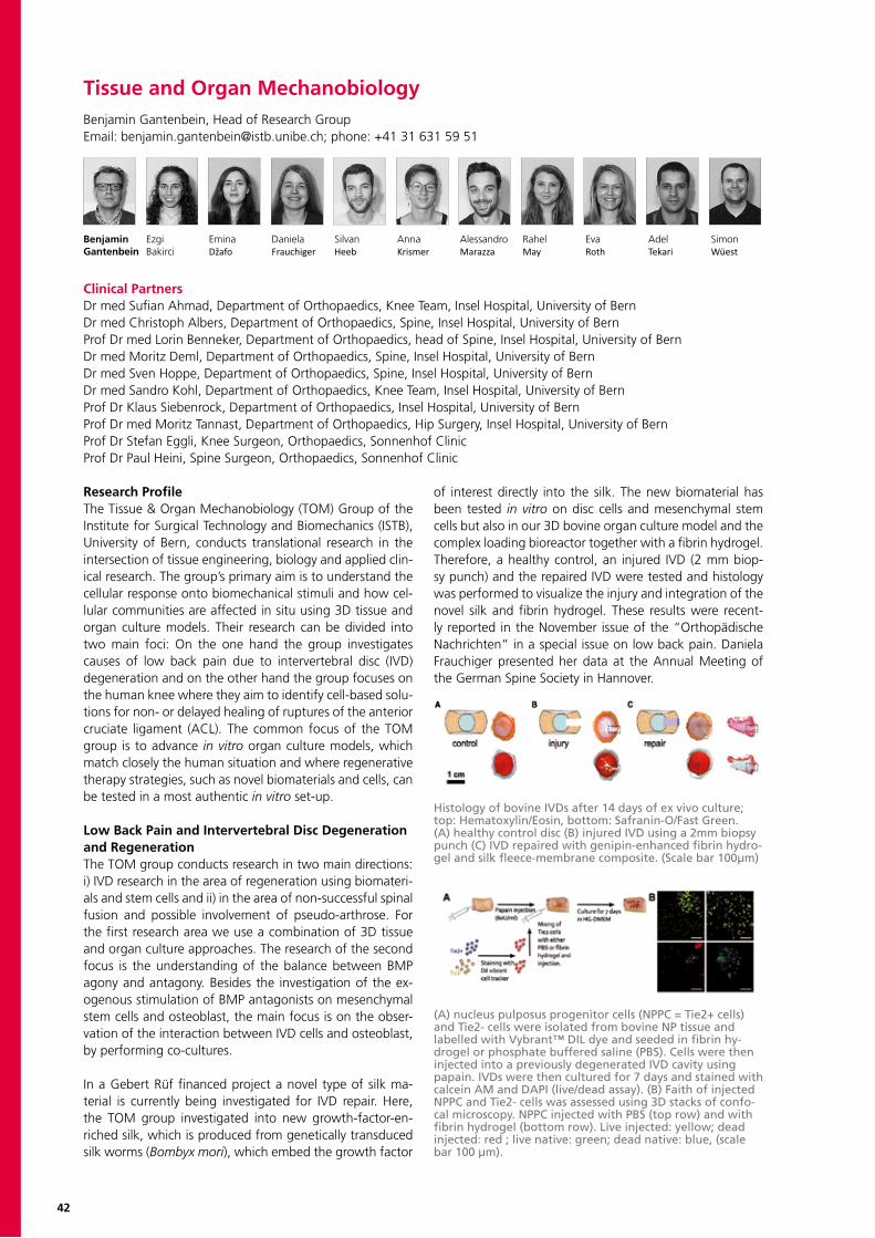
42
Research ProfileThe Tissue & Organ Mechanobiology (TOM) Group of the Institute for Surgical Technology and Biomechanics (ISTB), University of Bern, conducts translational research in the intersection of tissue engineering, biology and applied clin-ical research. The group’s primary aim is to understand the cellular response onto biomechanical stimuli and how cel-lular communities are affected in situ using 3D tissue and organ culture models. Their research can be divided into two main foci: On the one hand the group investigates causes of low back pain due to intervertebral disc (IVD) degeneration and on the other hand the group focuses on the human knee where they aim to identify cell-based solu-tions for non- or delayed healing of ruptures of the anterior cruciate ligament (ACL). The common focus of the TOM group is to advance in vitro organ culture models, which match closely the human situation and where regenerative therapy strategies, such as novel biomaterials and cells, can be tested in a most authentic in vitro set-up.
Low Back Pain and Intervertebral Disc Degeneration and RegenerationThe TOM group conducts research in two main directions: i) IVD research in the area of regeneration using biomateri-als and stem cells and ii) in the area of non-successful spinal fusion and possible involvement of pseudo-arthrose. For the first research area we use a combination of 3D tissue and organ culture approaches. The research of the second focus is the understanding of the balance between BMP agony and antagony. Besides the investigation of the ex-ogenous stimulation of BMP antagonists on mesenchymal stem cells and osteoblast, the main focus is on the obser-vation of the interaction between IVD cells and osteoblast, by performing co-cultures.
In a Gebert Rüf financed project a novel type of silk ma-terial is currently being investigated for IVD repair. Here, the TOM group investigated into new growth-factor-en-riched silk, which is produced from genetically transduced silk worms (Bombyx mori), which embed the growth factor
of interest directly into the silk. The new biomaterial has been tested in vitro on disc cells and mesenchymal stem cells but also in our 3D bovine organ culture model and the complex loading bioreactor together with a fibrin hydrogel. Therefore, a healthy control, an injured IVD (2 mm biop-sy punch) and the repaired IVD were tested and histology was performed to visualize the injury and integration of the novel silk and fibrin hydrogel. These results were recent-ly reported in the November issue of the “Orthopädische Nachrichten” in a special issue on low back pain. Daniela Frauchiger presented her data at the Annual Meeting of the German Spine Society in Hannover.
Tissue and Organ MechanobiologyBenjamin Gantenbein, Head of Research GroupEmail: [email protected]; phone: +41 31 631 59 51
Benjamin Ezgi Emina Daniela Silvan Anna Alessandro Rahel Eva Adel SimonGantenbein Bakirci Džafo Frauchiger Heeb Krismer Marazza May Roth Tekari Wüest
Clinical PartnersDr med Sufian Ahmad, Department of Orthopaedics, Knee Team, Insel Hospital, University of BernDr med Christoph Albers, Department of Orthopaedics, Spine, Insel Hospital, University of BernProf Dr med Lorin Benneker, Department of Orthopaedics, head of Spine, Insel Hospital, University of BernDr med Moritz Deml, Department of Orthopaedics, Spine, Insel Hospital, University of BernDr med Sven Hoppe, Department of Orthopaedics, Spine, Insel Hospital, University of BernDr med Sandro Kohl, Department of Orthopaedics, Knee Team, Insel Hospital, University of BernProf Dr Klaus Siebenrock, Department of Orthopaedics, Insel Hospital, University of BernProf Dr med Moritz Tannast, Department of Orthopaedics, Hip Surgery, Insel Hospital, University of BernProf Dr Stefan Eggli, Knee Surgeon, Orthopaedics, Sonnenhof ClinicProf Dr Paul Heini, Spine Surgeon, Orthopaedics, Sonnenhof Clinic
(A) nucleus pulposus progenitor cells (NPPC = Tie2+ cells) and Tie2- cells were isolated from bovine NP tissue and labelled with Vybrant™ DIL dye and seeded in fibrin hy-drogel or phosphate buffered saline (PBS). Cells were then injected into a previously degenerated IVD cavity using papain. IVDs were then cultured for 7 days and stained with calcein AM and DAPI (live/dead assay). (B) Faith of injected NPPC and Tie2- cells was assessed using 3D stacks of confo-cal microscopy. NPPC injected with PBS (top row) and with fibrin hydrogel (bottom row). Live injected: yellow; dead injected: red ; live native: green; dead native: blue, (scale bar 100 µm).
Histology of bovine IVDs after 14 days of ex vivo culture; top: Hematoxylin/Eosin, bottom: Safranin-O/Fast Green. (A) healthy control disc (B) injured IVD using a 2mm biopsy punch (C) IVD repaired with genipin-enhanced fibrin hydro-gel and silk fleece-membrane composite. (Scale bar 100µm)
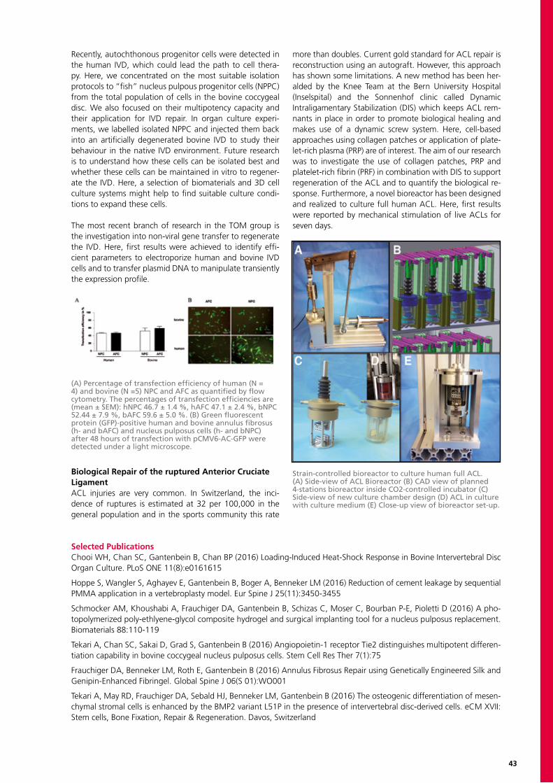
43
Recently, autochthonous progenitor cells were detected in the human IVD, which could lead the path to cell thera-py. Here, we concentrated on the most suitable isolation protocols to “fish” nucleus pulpous progenitor cells (NPPC) from the total population of cells in the bovine coccygeal disc. We also focused on their multipotency capacity and their application for IVD repair. In organ culture experi-ments, we labelled isolated NPPC and injected them back into an artificially degenerated bovine IVD to study their behaviour in the native IVD environment. Future research is to understand how these cells can be isolated best and whether these cells can be maintained in vitro to regener-ate the IVD. Here, a selection of biomaterials and 3D cell culture systems might help to find suitable culture condi-tions to expand these cells.
The most recent branch of research in the TOM group is the investigation into non-viral gene transfer to regenerate the IVD. Here, first results were achieved to identify effi-cient parameters to electroporize human and bovine IVD cells and to transfer plasmid DNA to manipulate transiently the expression profile.
Biological Repair of the ruptured Anterior Cruciate LigamentACL injuries are very common. In Switzerland, the inci-dence of ruptures is estimated at 32 per 100,000 in the general population and in the sports community this rate
more than doubles. Current gold standard for ACL repair is reconstruction using an autograft. However, this approach has shown some limitations. A new method has been her-alded by the Knee Team at the Bern University Hospital (Inselspital) and the Sonnenhof clinic called Dynamic Intraligamentary Stabilization (DIS) which keeps ACL rem-nants in place in order to promote biological healing and makes use of a dynamic screw system. Here, cell-based approaches using collagen patches or application of plate-let-rich plasma (PRP) are of interest. The aim of our research was to investigate the use of collagen patches, PRP and platelet-rich fibrin (PRF) in combination with DIS to support regeneration of the ACL and to quantify the biological re-sponse. Furthermore, a novel bioreactor has been designed and realized to culture full human ACL. Here, first results were reported by mechanical stimulation of live ACLs for seven days.
(A) Percentage of transfection efficiency of human (N = 4) and bovine (N =5) NPC and AFC as quantified by flow cytometry. The percentages of transfection efficiencies are (mean ± SEM): hNPC 46.7 ± 1.4 %, hAFC 47.1 ± 2.4 %, bNPC 52.44 ± 7.9 %, bAFC 59.6 ± 5.0 %. (B) Green fluorescent protein (GFP)-positive human and bovine annulus fibrosus (h- and bAFC) and nucleus pulposus cells (h- and bNPC) after 48 hours of transfection with pCMV6-AC-GFP were detected under a light microscope.
Strain-controlled bioreactor to culture human full ACL. (A) Side-view of ACL Bioreactor (B) CAD view of planned 4-stations bioreactor inside CO2-controlled incubator (C) Side-view of new culture chamber design (D) ACL in culture with culture medium (E) Close-up view of bioreactor set-up.
Selected PublicationsChooi WH, Chan SC, Gantenbein B, Chan BP (2016) Loading-Induced Heat-Shock Response in Bovine Intervertebral Disc Organ Culture. PLoS ONE 11(8):e0161615
Hoppe S, Wangler S, Aghayev E, Gantenbein B, Boger A, Benneker LM (2016) Reduction of cement leakage by sequential PMMA application in a vertebroplasty model. Eur Spine J 25(11):3450-3455
Schmocker AM, Khoushabi A, Frauchiger DA, Gantenbein B, Schizas C, Moser C, Bourban P-E, Pioletti D (2016) A pho-topolymerized poly-ethlyene-glycol composite hydrogel and surgical implanting tool for a nucleus pulposus replacement. Biomaterials 88:110-119
Tekari A, Chan SC, Sakai D, Grad S, Gantenbein B (2016) Angiopoietin-1 receptor Tie2 distinguishes multipotent differen-tiation capability in bovine coccygeal nucleus pulposus cells. Stem Cell Res Ther 7(1):75
Frauchiger DA, Benneker LM, Roth E, Gantenbein B (2016) Annulus Fibrosus Repair using Genetically Engineered Silk and Genipin-Enhanced Fibringel. Global Spine J 06(S 01):WO001
Tekari A, May RD, Frauchiger DA, Sebald HJ, Benneker LM, Gantenbein B (2016) The osteogenic differentiation of mesen-chymal stromal cells is enhanced by the BMP2 variant L51P in the presence of intervertebral disc-derived cells. eCM XVII: Stem cells, Bone Fixation, Repair & Regeneration. Davos, Switzerland

44
Mechanical Design and ProductionUrs Rohrer, Head of Mechanical Design and ProductionEmail: [email protected]; phone: +41 31 631 59 35
Urs Andreas Danaël Ronald Lukas FabioRohrer Beck Gasser Ramseier Rufener Spena
Group ProfileThe primary function of the Mechanical Design and Production (MDP) group is the co-development and man-ufacturing of mechanical and electro-mechanical com-ponents related to the research pursuits of the ISTB and ARTORG-Center. The MDP group supports all levels of the design and manufacturing process from concept to produc-tion. This includes computer assisted design (CAD) model-ling, prototyping and production with technical drawings, standard tooling, computer assisted manufacturing (CAM), a CNC-milling-machine and a CNC-lathe. We also support industrial and academic external research collaborators with their mechanical design and production needs.
Training & EducationThe MDP group has a secondary role in training. This train-ing encompasses the skills required to safely and profi-ciently operate machine shop tooling and equipment, the knowledge required to achieve the best results with a vari-ety of materials and the skills needed to efficiently manage the design and production workflow. Trial apprenticeships are used as a means to evaluate candidates for a full ap-prenticeship in the MDP group. This year we performed three courses and at we selected Simon Lüthi as our next apprentice. He will begin his training in August of 2017 as a polymechanic EFZ apprentice.
From April to August, Andreas Beck reinforced our team by working part-time as design engineer. He worked mainly on a forearm-rotating device. Since September, he works in industry. We are very grateful for the work he perfomed.Due to a high demand, we recruited our former polyme-chanic apprentice Ronald Ramseier as cilvil-service employ-ee. He performed administrative tasks and increased the productivity of our team from mid-August to October. We thank him also for the work he performed in our workshop.This year we could welcome for the first time a student from ETH for a construction internship. David Flückiger spent five weeks in our workshop and worked actively on the construction of mechanical pieces. During this short period, he performed practical work with machines.In July Lukas Rufener completed his apprenticeship with a very good result and we congratulate him. He will be employed until end of February 2017 part-time as a polytechnician.
Research Equipment Design & Manufacturing ISTBAs expected, the requirements of a machine shop sup-porting research in the biomedical engineering field are as diverse as the research field itself. The variety of subjects researched in the ISTB yield a number of diverse design and
production requests from prototype clinical and surgical tooling to fixtures for mechanical, biological and kinematic testing, as well as imaging system accessories and calibra-tion equipment. The following illustrations highlight a few of this year’s projects:
Project: Prosup Arm Bed Twist DeviceThis device was developed and produced in collaboration with Dr. Philipp Fürnstahl from Balgrist University Hospital. The radiolucent pieces were made of acrylic glass or by 3D-printing. With this device, the medical doctors are able to analyse the motion of the forearm. The device is either powered directly by muscle force or additional motors can guide the motion.
Project: Endoscope CalibrationWe built a device that allows the calibration of endoscopes with oblique viewing. The challenge of the project was to define the angles of the plates and its assembly. With this calibration system, the endoscopes can now be calibrated for angles from 30° to 70°.
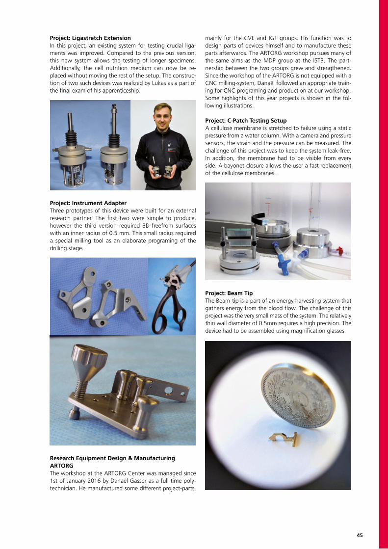
45
Project: Ligastretch ExtensionIn this project, an existing system for testing crucial liga-ments was improved. Compared to the previous version, this new system allows the testing of longer specimens. Additionally, the cell nutrition medium can now be re-placed without moving the rest of the setup. The construc-tion of two such devices was realized by Lukas as a part of the final exam of his apprenticeship.
Project: Instrument AdapterThree prototypes of this device were built for an external research partner. The first two were simple to produce, however the third version required 3D-freefrom surfaces with an inner radius of 0.5 mm. This small radius required a special milling tool as an elaborate programing of the drilling stage.
Research Equipment Design & Manufacturing ARTORGThe workshop at the ARTORG Center was managed since 1st of January 2016 by Danaël Gasser as a full time poly-technician. He manufactured some different project-parts,
mainly for the CVE and IGT groups. His function was to design parts of devices himself and to manufacture these parts afterwards. The ARTORG workshop pursues many of the same aims as the MDP group at the ISTB. The part-nership between the two groups grew and strengthened. Since the workshop of the ARTORG is not equipped with a CNC milling-system, Danaël followed an appropriate train-ing for CNC programing and production at our workshop.Some highlights of this year projects is shown in the fol-lowing illustrations. Project: C-Patch Testing SetupA cellulose membrane is stretched to failure using a static pressure from a water column. With a camera and pressure sensors, the strain and the pressure can be measured. The challenge of this project was to keep the system leak-free. In addition, the membrane had to be visible from every side. A bayonet-closure allows the user a fast replacement of the cellulose membranes.
Project: Beam TipThe Beam-tip is a part of an energy harvesting system that gathers energy from the blood flow. The challenge of this project was the very small mass of the system. The relatively thin wall diameter of 0.5mm requires a high precision. The device had to be assembled using magnification glasses.
47
INSELSPITAL, BERN UNIVERSITY HOSPITAL
Inselspital, Bern University Hospital is one of the largest, tertiary clinical center in Switzerland, covering a full complement of clinical disciplines. Inselspital’s teaching and research is integral to the medical faculty of the University of Bern. Biomedical engineering research, in particular, plays a key role, and the institutional links be-tween Inselspital and the biomedical engineering research institutes at the University of Bern foster strong multidisciplinary research and translation activities. Clinicians, engineers, computer scientists, and life and physical scientists develop new approaches for diagnosis and treatment, and the resources of Inselspital allow clinical introduction from pilot work to full-scale clinical trials. The in-house capabilities and resources are globally competitive and enable Inselpital to support transformation of clinical care to meet the future challenges of healthcare. Inselspital aims to lead re-search that will deliver the highest standards of diagnosis, treatment, and care based on excellent research and translation.
The research teams at Inselspital are part of a growing network of leading national and international research institutions and stakeholder groups. The wider network in the Canton of Bern extends to commercial partners and industry collaborations that benefit from access to infrastructure, know-how, and significant clinical resources. Together, these partnerships make Inselspital a singular institution in the Swiss clinical research landscape and beyond, capable of addressing unmet clinical needs through interdisciplinary innovation.
Matthias GuggerDirector Teaching and Research

48
Research ProfileThe research of the Bone Biology & Orthopaedic Research Group of the Department Clinical Research focuses on the topics of (i) osteoimmunology, (ii) fracture repair, (iii) molec-ular transport in bone cells, and (iv) cartilage development. (i) Osteoimmunology, the investigation of the crosstalk between inflammatory processes and bone homeosta-sis, is studied in close collaboration with the Department of Rheumatology, Clinical Immunology and Allergology, University Hospital, Inselspital. Based on a clinical study, we use in vitro methodology to elucidate aspects of the mechanisms employed by inflammatory factors and cyto-kines to modulate the development and activation of bone cell lineages. (ii) Fracture repair in the elderly undergoing treatment against osteoporosis is both a scientifically and clinically relevant process. We have developed experimen-tal models allowing the investigation of the repair of model defects in osteoporotic animals treated with anti-resorptive bisphosphonates. (iii) Within the NCCR Platform TransCure, which aspires to identify molecular transporters as putative therapeutic targets, we focus on bone cell lineages and the selective expression of transport systems during cell de-velopment. (iv) Cartilage repair is and remains a pressing issue. We lack the understanding, however, of the condi-tions required for regeneration of the tissue. Using 3D cell cultures and applying a transcriptomics approach, we aim to elucidate some aspects of healing.
Osteoclastogenesis in inflammatory processesWith the description of RANKL (receptor activator of NF-κB ligand) as a growth factor for bone resorbing osteoclast lineage cells, investigations of the crosstalk between the immune system and bone took center stage. Inflammatory diseases of the skeleton, i.e. Rheumatoid Arthritis, cause lo-cal subchondral and systemic bone loss. Inflammatory cyto-kines such as tumor necrosis factor- α (TNFα), Interleukin-1 (IL1), and IL17 stimulate bone resorption. The effects of cytokines on resorption, however, differ with the respective target cells, TNFα stimulating osteoclastogenesis through a direct effect on osteoclast progenitors, while inhibiting this process by inducing the release of the haematopoietic growth factor GM-CSF (granulocyte – macrophage colony stimulating factor) by osteoblast lineage cells. On a similar note, IL17, in synergy with TNFα, induces the release of
GM-CSF by osteoblast lineage cells. This data suggests that osteoclast progenitor cells are distributed in distinct pools, where precursor populations can be expanded. After hom-ing to bone with its osteoclastogenic environment, these cells develop into osteoclasts. Knowledge on the roles of inflammatory mediators in modulating bone metabolism will help to identify therapeutic targets and eventually im-pact the development of new and efficient, therapies.
Bone Repair and OsseointegrationThe repair of large bone defects (trauma or tumor resec-tion) requires the use of bone substitutes. To support the healing process, growth factors like Bone Morphogenetic Proteins (BMP) are employed. These factors, however, have proven to be biologically inefficient, supraphysiological quantities being necessary to achieve the desired results. Furthermore, the rigidity of fixation determines the path of repair, rigid fixation leading to repair through direct, membranous bone formation, non-rigid fixation leading to formation of a cartilaginous callous to achieve primary stability, which is subsequently replaced by woven bone.Using a fixation system developed in the ARI (Davos, Switzerland) for small rodents, healing of a femoral defect after rigid and non-rigid fixation, respectively, was com-pared. The data suggested a transient induction of endog-enous BMPs, counteracted by the simultaneous induction of inactivating BMP-antagonists, nullifying the effects of the growth factors. From this data we hypothesized that inhibition of BMP antagonists might improve the bioeffica-cy of endo- and exogenous BMP during repair. To address
Bone Biology and Orthopaedic ResearchWilly Hofstetter, Head of Research GroupEmail: [email protected]; www.bonebiology.dkf.unibe.ch; phone: +41 31 632 87 86
Willy Romina Silvia Michel Silvio Dobrila Nina MarkHofstetter Cabra Dolder Hauser Hoppen Nesic Ruef Siegrist
Clinical PartnersKlaus Siebenrock/ Frank Klenke, Department of Orthopaedic Surgery, Inselspital, Bern University HospitalMichael Seitz/ Daniel Aeberli, Department of Rheumatology, Clinical Immunology and Allergology (RIA), Inselspital, Bern Tateyuki Iizuka/ Nikola Saulacic, Department of Cranio-Maxillofacial Surgery, Inselspital, Bern University HospitalRainer Egli, Department of Diagnostic & Interventional Neuroradiology, Inselspital, Bern University HospitalBeat Gasser/ Marc Bohner, Dr.h.c. Robert Mathys Foundation, BettlachOlivier Bonny, CHUV, Lausanne
Development of osteoclast lineage cells. The development of haematopoietic stem cells to osteoclasts is controlled by numerous growth factors and cytokines. M-CSF and RANKL are essential for osteoclastogenesis, while GM-CSF induces the development of dendritic cells.
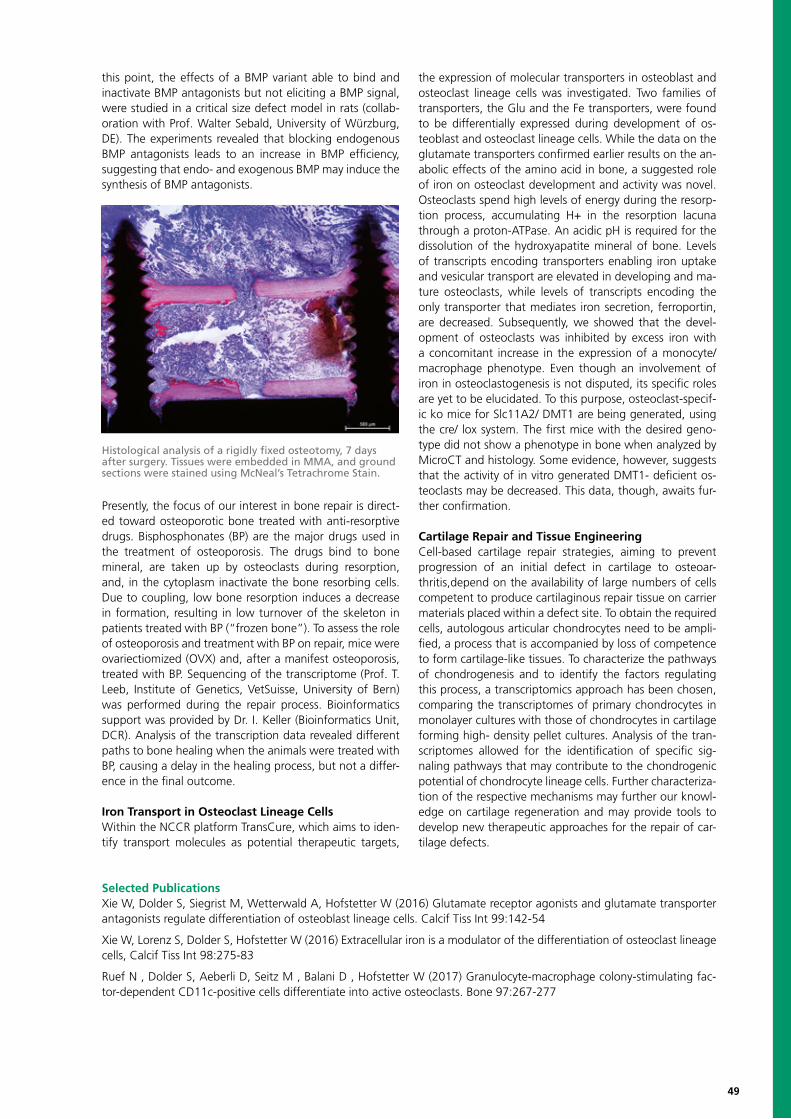
49
this point, the effects of a BMP variant able to bind and inactivate BMP antagonists but not eliciting a BMP signal, were studied in a critical size defect model in rats (collab-oration with Prof. Walter Sebald, University of Würzburg, DE). The experiments revealed that blocking endogenous BMP antagonists leads to an increase in BMP efficiency, suggesting that endo- and exogenous BMP may induce the synthesis of BMP antagonists.
Presently, the focus of our interest in bone repair is direct-ed toward osteoporotic bone treated with anti-resorptive drugs. Bisphosphonates (BP) are the major drugs used in the treatment of osteoporosis. The drugs bind to bone mineral, are taken up by osteoclasts during resorption, and, in the cytoplasm inactivate the bone resorbing cells. Due to coupling, low bone resorption induces a decrease in formation, resulting in low turnover of the skeleton in patients treated with BP (“frozen bone”). To assess the role of osteoporosis and treatment with BP on repair, mice were ovariectiomized (OVX) and, after a manifest osteoporosis, treated with BP. Sequencing of the transcriptome (Prof. T. Leeb, Institute of Genetics, VetSuisse, University of Bern) was performed during the repair process. Bioinformatics support was provided by Dr. I. Keller (Bioinformatics Unit, DCR). Analysis of the transcription data revealed different paths to bone healing when the animals were treated with BP, causing a delay in the healing process, but not a differ-ence in the final outcome.
Iron Transport in Osteoclast Lineage CellsWithin the NCCR platform TransCure, which aims to iden-tify transport molecules as potential therapeutic targets,
the expression of molecular transporters in osteoblast and osteoclast lineage cells was investigated. Two families of transporters, the Glu and the Fe transporters, were found to be differentially expressed during development of os-teoblast and osteoclast lineage cells. While the data on the glutamate transporters confirmed earlier results on the an-abolic effects of the amino acid in bone, a suggested role of iron on osteoclast development and activity was novel. Osteoclasts spend high levels of energy during the resorp-tion process, accumulating H+ in the resorption lacuna through a proton-ATPase. An acidic pH is required for the dissolution of the hydroxyapatite mineral of bone. Levels of transcripts encoding transporters enabling iron uptake and vesicular transport are elevated in developing and ma-ture osteoclasts, while levels of transcripts encoding the only transporter that mediates iron secretion, ferroportin, are decreased. Subsequently, we showed that the devel-opment of osteoclasts was inhibited by excess iron with a concomitant increase in the expression of a monocyte/ macrophage phenotype. Even though an involvement of iron in osteoclastogenesis is not disputed, its specific roles are yet to be elucidated. To this purpose, osteoclast-specif-ic ko mice for Slc11A2/ DMT1 are being generated, using the cre/ lox system. The first mice with the desired geno-type did not show a phenotype in bone when analyzed by MicroCT and histology. Some evidence, however, suggests that the activity of in vitro generated DMT1- deficient os-teoclasts may be decreased. This data, though, awaits fur-ther confirmation.
Cartilage Repair and Tissue EngineeringCell-based cartilage repair strategies, aiming to prevent progression of an initial defect in cartilage to osteoar-thritis,depend on the availability of large numbers of cells competent to produce cartilaginous repair tissue on carrier materials placed within a defect site. To obtain the required cells, autologous articular chondrocytes need to be ampli-fied, a process that is accompanied by loss of competence to form cartilage-like tissues. To characterize the pathways of chondrogenesis and to identify the factors regulating this process, a transcriptomics approach has been chosen, comparing the transcriptomes of primary chondrocytes in monolayer cultures with those of chondrocytes in cartilage forming high- density pellet cultures. Analysis of the tran-scriptomes allowed for the identification of specific sig-naling pathways that may contribute to the chondrogenic potential of chondrocyte lineage cells. Further characteriza-tion of the respective mechanisms may further our knowl-edge on cartilage regeneration and may provide tools to develop new therapeutic approaches for the repair of car-tilage defects.
Histological analysis of a rigidly fixed osteotomy, 7 days after surgery. Tissues were embedded in MMA, and ground sections were stained using McNeal’s Tetrachrome Stain.
Selected PublicationsXie W, Dolder S, Siegrist M, Wetterwald A, Hofstetter W (2016) Glutamate receptor agonists and glutamate transporter antagonists regulate differentiation of osteoblast lineage cells. Calcif Tiss Int 99:142-54
Xie W, Lorenz S, Dolder S, Hofstetter W (2016) Extracellular iron is a modulator of the differentiation of osteoclast lineage cells, Calcif Tiss Int 98:275-83
Ruef N , Dolder S, Aeberli D, Seitz M , Balani D , Hofstetter W (2017) Granulocyte-macrophage colony-stimulating fac-tor-dependent CD11c-positive cells differentiate into active osteoclasts. Bone 97:267-277

50
Orthopedic SurgeryKlaus A. Siebenrock, Head of Research GroupEmail: [email protected]; phone: +41 31 632 22 22
Klaus A. Christoph E. Lorin M Moritz Timo Eliza Sven Frank M.Siebenrock Albers Benneker Deml Ecker Hartmann Hoppe Klenke
Research PartnersMauro Alini, AO Research Institute, DavosBenjamin Gantenbein, Institute for Surgical Technologies and Biomachanics Bern Willy Hofstetter, Department of Clinical Research, University of BernErnst Hunziker, Department of Orthopedic Surgery, Inselspital, Bern University HospitalHans-Jörg Sebald, thespinecenter, ThunWaldo Vanlenzuela, Institute for Surgical Technologies and Biomachanics, University of Bern
Research ProfileThe Department of Orthopedic Surgery and Traumatology at the Inselspital Bern conducts research projects pertaining to (i) the elucidation mechanisms involved in attenuated bone healing with special emphasis on the regulation of bone morphogenetic protein (BMP) signalling, (ii) the de-velopment of bone substitute materials delivering ostoin-ductive growth factors for bone repair, (iii) imaging, and (iv) regenerative medicine for disc degeneration. Our de-partment works in close collaboration with the Group for Bone Biology and Orthopedic Research at the Department of Clinical Research, University of Bern (Prof. W. Hofstetter), the Institute for Surgical Technologies and Biomachanics Bern (Prof. B. Gantenbein), and the AO Research Institute Davos (Prof. M. Alini).
Regulation of BMP SignallingBMP-2 and BMP-7 have been approved for clinical use for bone regeneration. However, the growth factors are re-quired at supraphysiological concentrations to stimulate bone healing effectively. Endogenous antagonist induced by exogenous BMP therapy may be responsible for the limited clinical efficacy of BMPs. We, along with other researchers, have shown that BMPs induce the expression of their antagonists such as Noggin, Gremlin, and Chordin in vitro. To enhance the osteogenic activity of BMPs, L51P, a molecularly engineered BMP-2 variant with deficient BMP receptor binding but unchanged binding to modulator proteins, such as Noggin, Gremlin, and Chordin has been developed. The modification made L51P a BMP receptor-inactive inhibitor of BMP antagonists. In vitro, L51P reversed the Noggin-mediated inhibition of BMP-2-induced alkaline phosphatase expression in C2C12 promyoblasts and primary murine osteoblasts. L51P itself did not induce osteoblast differentiation directly and did not activate BMP receptor-dependent intracellular signalling due to deficient BMP type I receptor-binding. Recent data suggest that the BMP-antagonist Noggin stim-ulates osteoclast development of osteoclast progenitor cells (OPC) in the presence of trace levels of the osteoclast growth factor Receptor Activator of NF-κB Ligand (RANKL). The finding that the Noggin effect on osteoclast develop-ment could not be blocked by L51P, led to the hypothesis that Noggin action is not restricted to an anti-osteogenic
effect by blocking BMP actions, but that Noggin supports osteoclastogenesis through a direct and eventually recep-tor-mediated mechanism. We have collected evidence that Noggin, by an as-yet-unknown pathway, activates TGFβ signaling. Furthermore, recently evidence was published that linked RANKL signalling to the family of lgr orphan re-ceptors, which in turn may be activated by members of the R-spondin family and by Noggin. While lrg5 has been used as markers for adult stem cells, we have generated evidence that the members of this family of proteins are expressed in differentiating haematopoietic cells as well. Further eluci-dation of the effects of BMP-2 treatment on the induction of endogenous antagonists and the regulatory function of these antagonists is essential and will help to minimize BMP-therapy-associated adverse effects and improve the clinical efficacy of BMP-2 treatment.
Bone Substitute Materials Autologous bone is widely accepted as the standard bio-logic material to augment bone healing and to recon-struct bone defects. There are, however, critical limitations
BMP-antagonist Noggin enhances RANKL mediated OC formation. TRAP staining of OPC cultures on day 5 after addition of RANKL 2.5 ng/ml (A) and RANKL 2.5 ng/ml with Noggin 33 nM (B).
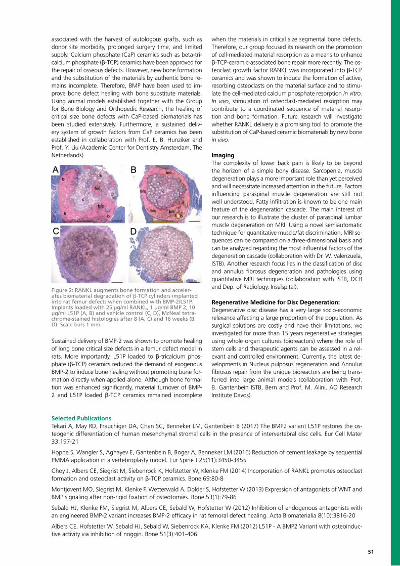
51
associated with the harvest of autologous grafts, such as donor site morbidity, prolonged surgery time, and limited supply. Calcium phosphate (CaP) ceramics such as beta-tri-calcium phosphate (β-TCP) ceramics have been approved for the repair of osseous defects. However, new bone formation and the substitution of the materials by authentic bone re-mains incomplete. Therefore, BMP have been used to im-prove bone defect healing with bone substitute materials. Using animal models established together with the Group for Bone Biology and Orthopedic Research, the healing of critical size bone defects with CaP-based biomaterials has been studied extensively. Furthermore, a sustained deliv-ery system of growth factors from CaP ceramics has been established in collaboration with Prof. E. B. Hunziker and Prof. Y. Liu (Academic Center for Dentistry Amsterdam, The Netherlands).
Sustained delivery of BMP-2 was shown to promote healing of long bone critical size defects in a femur defect model in rats. More importantly, L51P loaded to β-tricalcium phos-phate (β-TCP) ceramics reduced the demand of exogenous BMP-2 to induce bone healing without promoting bone for-mation directly when applied alone. Although bone forma-tion was enhanced significantly, material turnover of BMP-2 and L51P loaded β-TCP ceramics remained incomplete
when the materials in critical size segmental bone defects. Therefore, our group focused its research on the promotion of cell-mediated material resorption as a means to enhance β-TCP-ceramic-associated bone repair more recently. The os-teoclast growth factor RANKL was incorporated into β-TCP ceramics and was shown to induce the formation of active, resorbing osteoclasts on the material surface and to stimu-late the cell-mediated calcium phosphate resorption in vitro. In vivo, stimulation of osteoclast-mediated resorption may contribute to a coordinated sequence of material resorp-tion and bone formation. Future research will investigate whether RANKL delivery is a promising tool to promote the substitution of CaP-based ceramic biomaterials by new bone in vivo.
ImagingThe complexity of lower back pain is likely to be beyond the horizon of a simple bony disease. Sarcopenia, muscle degeneration plays a more important role than yet perceived and will necessitate increased attention in the future. Factors influencing paraspinal muscle degeneration are still not well understood. Fatty infiltration is known to be one main feature of the degeneration cascade. The main interest of our research is to illustrate the cluster of paraspinal lumbar muscle degeneration on MRI. Using a novel semiautomatic technique for quantitative muscle/fat discrimination, MRI se-quences can be compared on a three-dimensional basis and can be analyzed regarding the most influential factors of the degeneration cascade (collaboration with Dr. W. Valenzuela, ISTB). Another research focus lies in the classification of disc and annulus fibrosus degeneration and pathologies using quantitative MRI techniques (collaboration with ISTB, DCR and Dep. of Radiology, Inselspital).
Regenerative Medicine for Disc Degeneration:Degenerative disc disease has a very large socio-economic relevance affecting a large proportion of the population. As surgical solutions are costly and have their limitations, we investigated for more than 15 years regenerative strategies using whole organ cultures (bioreactors) where the role of stem cells and therapeutic agents can be assessed in a rel-evant and controlled environment. Currently, the latest de-velopments in Nucleus pulposus regeneration and Annulus fibrosus repair from the unique bioreactors are being trans-ferred into large animal models (collaboration with Prof. B. Gantenbein ISTB, Bern and Prof. M. Alini, AO Research Institute Davos).
Selected PublicationsTekari A, May RD, Frauchiger DA, Chan SC, Benneker LM, Gantenbein B (2017) The BMP2 variant L51P restores the os-teogenic differentiation of human mesenchymal stromal cells in the presence of intervertebral disc cells. Eur Cell Mater 33:197-21
Hoppe S, Wangler S, Aghayev E, Gantenbein B, Boger A, Benneker LM (2016) Reduction of cement leakage by sequential PMMA application in a vertebroplasty model. Eur Spine J 25(11):3450-3455
Choy J, Albers CE, Siegrist M, Siebenrock K, Hofstetter W, Klenke FM (2014) Incorporation of RANKL promotes osteoclast formation and osteoclast activity on β-TCP ceramics. Bone 69:80-8
Montjovent MO, Siegrist M, Klenke F, Wetterwald A, Dolder S, Hofstetter W (2013) Expression of antagonists of WNT and BMP signaling after non-rigid fixation of osteotomies. Bone 53(1):79-86
Sebald HJ, Klenke FM, Siegrist M, Albers CE, Sebald W, Hofstetter W (2012) Inhibition of endogenous antagonists with an engineered BMP-2 variant increases BMP-2 efficacy in rat femoral defect healing. Acta Biomaterialia 8(10):3816-20
Albers CE, Hofstetter W, Sebald HJ, Sebald W, Siebenrock KA, Klenke FM (2012) L51P - A BMP2 Variant with osteoinduc-tive activity via inhibition of noggin. Bone 51(3):401-406
Figure 2: RANKL augments bone formation and acceler-ates biomaterial degradation of β-TCP cylinders implanted into rat femur defects when combined with BMP-2/L51P. Implants loaded with 25 µg/ml RANKL, 1 µg/ml BMP 2, 10 µg/ml L51P (A, B) and vehicle control (C, D), McNeal tetra-chrome-stained histologies after 8 (A, C) and 16 weeks (B, D). Scale bars 1 mm.
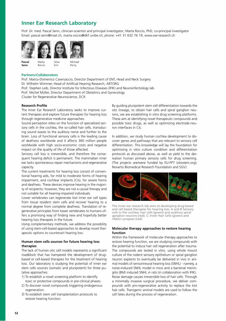
52
Inner Ear Research LaboratoryProf. Dr. med. Pascal Senn, clinician-scientist and principal investigator; Marta Roccio, PhD, co-principal investigatorEmail: [email protected], [email protected]; phone: +41 31 632 76 19; www.ear-research.ch
Pascal Marta Silvia MichaelSenn Roccio Erni Perny
Research ProfileThe Inner Ear Research Laboratory seeks to improve cur-rent therapies and explore future therapies for hearing loss through regenerative medicine approaches:Sound perception relies on the function of specialized sen-sory cells in the cochlea, the so-called hair cells, transduc-ing sound waves to the auditory nerve and further to the brain. Loss of functional sensory cells is the leading cause of deafness worldwide and it affects 360 million people worldwide with high socio-economic costs and negative impact on the quality of life of those affected. Sensory cell loss is irreversible, and therefore the conse-quent hearing deficit is permanent. The mammalian inner ear lacks spontaneous repair mechanisms and regenerative capacity. The current treatments for hearing loss consist of conven-tional hearing aids, for mild to moderate forms of hearing impairment, and cochlear implants (CIs), for severe forms and deafness. These devices improve hearing in the majori-ty of recipients; however, they are not a causal therapy and not suitable for all hearing-impaired individuals. Lower vertebrates can regenerate lost inner ear cell types from tissue resident stem cells and recover hearing to a normal degree from complete deafness. Translation of re-generative principles from lower vertebrates to humans of-fers a promising way of finding new and hopefully better hearing loss therapies in the future. Using complementary methods, we address the possibility of using stem-cell-based approaches to develop novel ther-apeutic options to counteract hearing loss.
Human stem cells sources for future hearing loss therapiesThe lack of human otic cell models represents a significant roadblock that has hampered the development of drug-based or cell-based therapies for the treatment of hearing loss. Our laboratory is studying the potential of inner ear stem cells sources (somatic and pluripotent) for three pu-tative approaches: 1) To establish a novel screening platform to identify toxic or protective compounds in pre-clinical phases. 2) To discover novel compounds triggering endogenous regeneration.3) To establish stem cell transplantation protocols to restore hearing function.
By guiding pluripotent stem cell differentiation towards the otic lineage, to obtain hair cells and spiral ganglion neu-rons, we are establishing in vitro drug screening platforms. These aim at identifying novel therapeutic compounds and possible toxic drugs, as well as optimizing electrode-neu-ron interfaces in CIs.
In addition, we study human cochlea development to dis-cover genes and pathways that are relevant to sensory cell differentiation. This knowledge will lay the foundation for optimizing in vitro culture condition and differentiation protocols as discussed above, as well as yield to the der-ivation human primary sensory cells for drug screening. (The projects are/were funded by EU-FP7 (otostem.org); Novartis Biomedical Research Foundation and SGV).
Molecular therapy approaches to restore hearing functionWithin the framework of molecular therapy approaches to restore hearing function, we are studying compounds with the potential to induce hair cell regeneration after trauma.The compounds are tested in vitro, using whole mount culture of the rodent sensory epithelium or spiral ganglion neuron explants to eventually be delivered in vivo in ani-mal models of sensorineural hearing loss (SNHL) - namely, a noise-induced SNHL model in mice and a bacterial menin-gitis (BM) induced SNHL in rats (in collaboration with IFIK).Noise damage causes irreversible loss of hair cells. Through a minimally invasive surgical procedure, we deliver com-pounds with pro-regenerative activity to replace the lost hair cells. Transgenic animal models are used to follow the cell fates during the process of regeneration.
Partners/CollaboratorsProf. Marco-Domenico Caversaccio, Director Department of ENT, Head and Neck SurgeryDr. Wilhelm Wimmer, Head of Artificial Hearing Research, ARTORGProf. Stephen Leib, Director Institute for Infectious Diseases (IFIK) and Neuroinfectiology lab.Prof. Michel Müller, Director Department of Obstetrics and GynecologyCluster for Regenerative Neuroscience, DCR
The inner ear research lab aims at developing drug-based and cell-based therapies for hearing loss. A and B Sensory cells in the cochlea: hair cells (green) and auditory spiral ganglion neurons (red). C: Inner Hair Cells (green) and ribbon synapses (red).
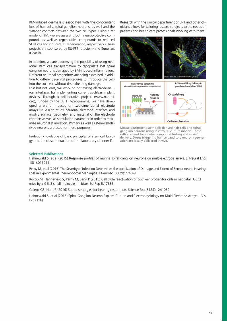
53
BM-induced deafness is associated with the concomitant loss of hair cells, spiral ganglion neurons, as well and the synaptic contacts between the two cell types. Using a rat model of BM, we are assessing both neuroprotective com-pounds as well as regenerative compounds to reduced SGN loss and induced HC regeneration, respectively. (These projects are sponsored by EU-FP7 (otostem) and Eurostars (Hear-it).
In addition, we are addressing the possibility of using neu-ronal stem cell transplantation to repopulate lost spiral ganglion neurons damaged by BM-induced inflammation.Different neuronal progenitors are being examined in addi-tion to different surgical procedures to introduce the cells into the cochlea, without tissue/hearing damage.Last but not least, we work on optimizing electrode-neu-ron interfaces for implementing current cochlear implant devices. Through a collaborative project. (www.nanoci.org), funded by the EU FP7-programme, we have devel-oped a platform based on two-dimensional electrode arrays (MEAs) to study neuronal-electrode interface and modify surface, geometry, and material of the electrode contacts as well as stimulation parameter in order to maxi-mize neuronal stimulation. Primary as well as stem-cell-de-rived neurons are used for these purposes.
In-depth knowledge of basic principles of stem cell biolo-gy and the close interaction of the laboratory of Inner Ear
Research with the clinical department of ENT and other cli-nicians allows for tailoring research projects to the needs of patients and health care professionals working with them.
Selected PublicationsHahnewald S, et al (2015) Response profiles of murine spiral ganglion neurons on multi-electrode arrays. J. Neural Eng 13(1):016011
Perny M, et al (2016) The Severity of Infection Determines the Localization of Damage and Extent of Sensorineural Hearing Loss in Experimental Pneumococcal Meningitis. J Neurosci 36(29):7740-9
Roccio M, Hahnewald S, Perny M, Senn P (2015) Cell cycle reactivation of cochlear progenitor cells in neonatal FUCCImice by a GSK3 small molecule inhibitor. Sci Rep 5:17886
Geleoc GS, Holt JR (2016) Sound strategies for hearing restoration. Science 344(6184):1241062
Hahnewald S, et al (2016) Spiral Ganglion Neuron Explant Culture and Electrophysiology on Multi Electrode Arrays. J Vis Exp (116)
Mouse pluripotent stem cells derived hair cells and spiral ganglion neurons using in vitro 3D culture models. These cells are used for in vitro compound testing and in vivo delivery. Drugs triggering hair cell/auditory neuron regener-ation are locally delivered in vivo.
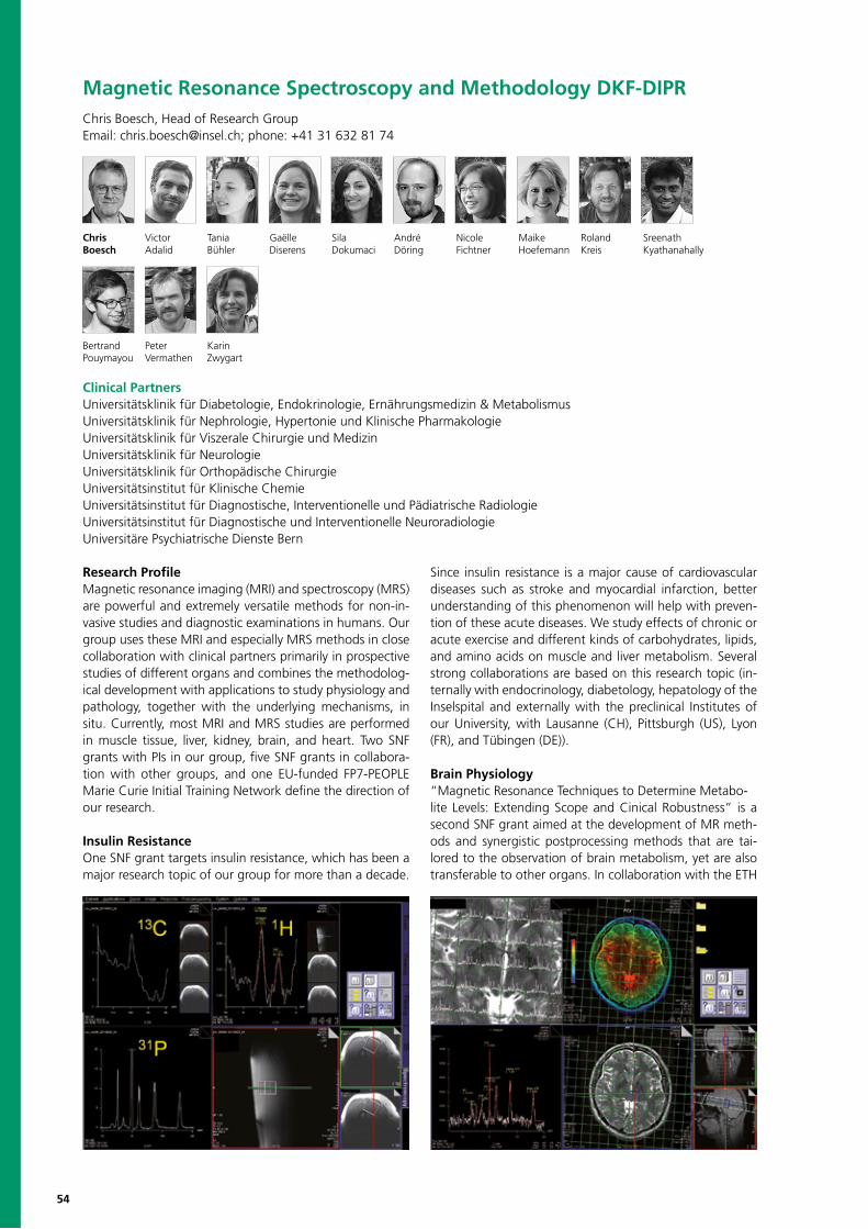
54
Magnetic Resonance Spectroscopy and Methodology DKF-DIPRChris Boesch, Head of Research GroupEmail: [email protected]; phone: +41 31 632 81 74
Chris Victor Tania Gaëlle Sila André Nicole Maike Roland SreenathBoesch Adalid Bühler Diserens Dokumaci Döring Fichtner Hoefemann Kreis Kyathanahally
Bertrand Peter KarinPouymayou Vermathen Zwygart
Research ProfileMagnetic resonance imaging (MRI) and spectroscopy (MRS) are powerful and extremely versatile methods for non-in-vasive studies and diagnostic examinations in humans. Our group uses these MRI and especially MRS methods in close collaboration with clinical partners primarily in prospective studies of different organs and combines the methodolog-ical development with applications to study physiology and pathology, together with the underlying mechanisms, in situ. Currently, most MRI and MRS studies are performed in muscle tissue, liver, kidney, brain, and heart. Two SNF grants with PIs in our group, five SNF grants in collabora-tion with other groups, and one EU-funded FP7-PEOPLE Marie Curie Initial Training Network define the direction of our research.
Insulin ResistanceOne SNF grant targets insulin resistance, which has been a major research topic of our group for more than a decade.
Since insulin resistance is a major cause of cardiovascular diseases such as stroke and myocardial infarction, better understanding of this phenomenon will help with preven-tion of these acute diseases. We study effects of chronic or acute exercise and different kinds of carbohydrates, lipids, and amino acids on muscle and liver metabolism. Several strong collaborations are based on this research topic (in-ternally with endocrinology, diabetology, hepatology of the Inselspital and externally with the preclinical Institutes of our University, with Lausanne (CH), Pittsburgh (US), Lyon (FR), and Tübingen (DE)).
Brain Physiology“Magnetic Resonance Techniques to Determine Metabo-lite Levels: Extending Scope and Cinical Robustness” is a second SNF grant aimed at the development of MR meth-ods and synergistic postprocessing methods that are tai-lored to the observation of brain metabolism, yet are also transferable to other organs. In collaboration with the ETH
Clinical PartnersUniversitätsklinik für Diabetologie, Endokrinologie, Ernährungsmedizin & MetabolismusUniversitätsklinik für Nephrologie, Hypertonie und Klinische PharmakologieUniversitätsklinik für Viszerale Chirurgie und MedizinUniversitätsklinik für NeurologieUniversitätsklinik für Orthopädische ChirurgieUniversitätsinstitut für Klinische ChemieUniversitätsinstitut für Diagnostische, Interventionelle und Pädiatrische RadiologieUniversitätsinstitut für Diagnostische und Interventionelle NeuroradiologieUniversitäre Psychiatrische Dienste Bern
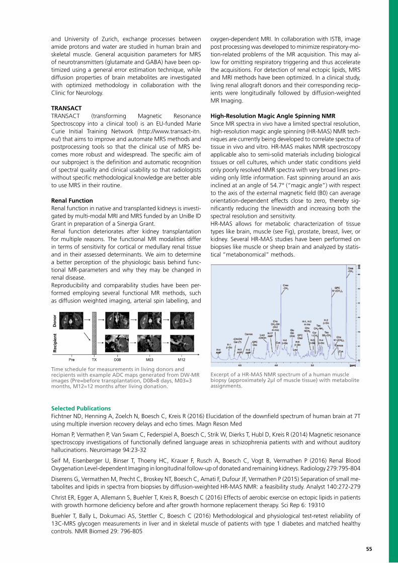
55
and University of Zurich, exchange processes between amide protons and water are studied in human brain and skeletal muscle. General acquisition parameters for MRS of neurotransmitters (glutamate and GABA) have been op-timized using a general error estimation technique, while diffusion properties of brain metabolites are investigated with optimized methodology in collaboration with the Clinic for Neurology.
TRANSACTTRANSACT (transforming Magnetic Resonance Spectroscopy into a clinical tool) is an EU-funded Marie Curie Initial Training Network (http://www.transact-itn.eu/) that aims to improve and automate MRS methods and postprocessing tools so that the clinical use of MRS be-comes more robust and widespread. The specific aim of our subproject is the definition and automatic recognition of spectral quality and clinical usability so that radiologists without specific methodological knowledge are better able to use MRS in their routine.
Renal Function Renal function in native and transplanted kidneys is investi-gated by multi-modal MRI and MRS funded by an UniBe ID Grant in preparation of a Sinergia Grant. Renal function deteriorates after kidney transplantation for multiple reasons. The functional MR modalities differ in terms of sensitivity for cortical or medullary renal tissue and in their assessed determinants. We aim to determine a better perception of the physiologic basis behind func-tional MR-parameters and why they may be changed in renal disease.Reproducibility and comparability studies have been per-formed employing several functional MR methods, such as diffusion weighted imaging, arterial spin labelling, and
oxygen-dependent MRI. In collaboration with ISTB, image post processing was developed to minimize respiratory-mo-tion-related problems of the MR acquisition. This may al-low for omitting respiratory triggering and thus accelerate the acquisitions. For detection of renal ectopic lipids, MRS and MRI methods have been optimized. In a clinical study, living renal allograft donors and their corresponding recip-ients were longitudinally followed by diffusion-weighted MR Imaging.
High-Resolution Magic Angle Spinning NMR Since MR spectra in vivo have a limited spectral resolution, high-resolution magic angle spinning (HR-MAS) NMR tech-niques are currently being developed to correlate spectra of tissue in vivo and vitro. HR-MAS makes NMR spectroscopy applicable also to semi-solid materials including biological tissues or cell cultures, which under static conditions yield only poorly resolved NMR spectra with very broad lines pro-viding only little information. Fast spinning around an axis inclined at an angle of 54.7° (“magic angle”) with respect to the axis of the external magnetic field (B0) can average orientation-dependent effects close to zero, thereby sig-nificantly reducing the linewidth and increasing both the spectral resolution and sensitivity.HR-MAS allows for metabolic characterization of tissue types like brain, muscle (see Fig), prostate, breast, liver, or kidney. Several HR-MAS studies have been performed on biopsies like muscle or sheep brain and analyzed by statis-tical “metabonomical” methods.
Selected PublicationsFichtner ND, Henning A, Zoelch N, Boesch C, Kreis R (2016) Elucidation of the downfield spectrum of human brain at 7T using multiple inversion recovery delays and echo times. Magn Reson Med
Homan P, Vermathen P, Van Swam C, Federspiel A, Boesch C, Strik W, Dierks T, Hubl D, Kreis R (2014) Magnetic resonance spectroscopy investigations of functionally defined language areas in schizophrenia patients with and without auditory hallucinations. Neuroimage 94:23-32
Seif M, Eisenberger U, Binser T, Thoeny HC, Krauer F, Rusch A, Boesch C, Vogt B, Vermathen P (2016) Renal Blood Oxygenation Level-dependent Imaging in longitudinal follow-up of donated and remaining kidneys. Radiology 279:795-804
Diserens G, Vermathen M, Precht C, Broskey NT, Boesch C, Amati F, Dufour JF, Vermathen P (2015) Separation of small me-tabolites and lipids in spectra from biopsies by diffusion-weighted HR-MAS NMR: a feasibility study. Analyst 140:272-279
Christ ER, Egger A, Allemann S, Buehler T, Kreis R, Boesch C (2016) Effects of aerobic exercise on ectopic lipids in patients with growth hormone deficiency before and after growth hormone replacement therapy. Sci Rep 6: 19310
Buehler T, Bally L, Dokumaci AS, Stettler C, Boesch C (2016) Methodological and physiological test-retest reliability of 13C-MRS glycogen measurements in liver and in skeletal muscle of patients with type 1 diabetes and matched healthy controls. NMR Biomed 29: 796-805
Time schedule for measurements in living donors and recipients with example ADC maps generated from DW-MR images (Pre=before transplantation, D08=8 days, M03=3 months, M12=12 months after living donation.
Excerpt of a HR-MAS NMR spectrum of a human muscle biopsy (approximately 2µl of muscle tissue) with metabolite assignments.
56
Division of Medical Radiation Physics within Department of Radiation OncologyPeter Manser, Head of DivisionEmail: [email protected]; phone: +41 31 632 37 71Daniel M. Aebersold, Chairman of the Department of Radiation OncologyEmail: [email protected]; phone: +41 31 632 24 31
Peter Daniel Cécile Pascal Michael Daniel Daniel Dominik Bernhard Andreas NorbertManser Aebersold Chatelain Favre Fix Frauchiger Frei Henzen Isaak Joosten Klippel
Reto Kristina Paul-Henry Marco Silvan Manfred Daniel Dario Daniel Veronika WernerKüng Lössl Mackeprang Malthaner Müller Sassowsky Schmidhalter Terribilini Vetterli Vitzthum Volken
Research ProfileThe Division of Medical Radiation Physics is part of the Department of Radiation Oncology and is active in clini-cal services as well as in research projects and education. Medical physics is an interdisciplinary field and combines physics with medicine. Traditionally, medical physics is re-lated to medical radiation physics and addresses aspects like dosimetry, treatment planning, quality assurance, and radiation protection. In addition to the implementation of new methods in radiation therapy, the research activities cover more fundamental research in medical radiation physics. To accomplish the high accuracy needed in radia-tion therapy, sophisticated methods have to be established and validated before being used in clinical routine. The re-search performed at our Division has not only proven to be of interest on a national and international level, but has also been transferred to commercial products and clinical applications.
Image-guided Radiation Therapy in Stereotactic Radiosurgery or Radiation TherapyOver the last decade, image-guided radiation thera-py (IGRT) has been used more and more for stereotactic
radiosurgery and radiation therapy. By this means, the ac-curacy of patient setup is improved and a reduction of safe-ty margins is possible. As a consequence, better normal tissue sparing can be realized. To perform IGRT on a routine basis, six degrees of freedom (6DoF) couches are used in combination with dedicated imaging systems. When using a robotic stereotactic system, tumors, which are affected by motion such as breathing, can be tracked in real time.
Monte Carlo Simulations for Dose Calculations and Medical Physics ModellingAccurate and efficient dose calculation is essential for treat-ment planning purposes in radiation therapy, and Monte Carlo (MC) simulations are commonly known as the most accurate method to perform this task. However, the major limitation of MC simulations is the enormous computation time needed. Our research activities deal with increasing the efficiency of MC-based dose calculations, such as the
Clinical PartnersDaniel Candinas, Director and Head of Department of Visceral Surgery and Medicine, Inselspital, Bern University HospitalJohannes Heverhagen, Director and Head of Department of Diagnostic, Interventional and Pediatric Radiology, Inselspital, Bern University HospitalThomas Krause, Director and Head of Department of Nuclear Medicine, Inselspital, Bern University Hospital Andreas Raabe, Director and Chairman of Department of Neurosurgery, Inselspital, Bern University HospitalStefan Weber, Chair Image-guided Therapy and Director, ARTORG Center, University of Bern
Histograms of initial setup errors with respect to rotations.Dose distribution of a proton beam calculated by Macro Monte Carlo.
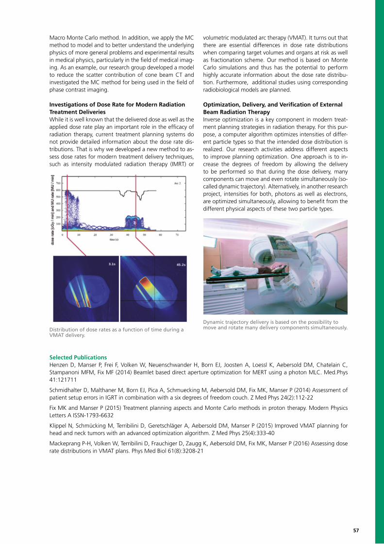
57
Macro Monte Carlo method. In addition, we apply the MC method to model and to better understand the underlying physics of more general problems and experimental results in medical physics, particularly in the field of medical imag-ing. As an example, our research group developed a model to reduce the scatter contribution of cone beam CT and investigated the MC method for being used in the field of phase contrast imaging.
Investigations of Dose Rate for Modern Radiation Treatment DeliveriesWhile it is well known that the delivered dose as well as the applied dose rate play an important role in the efficacy of radiation therapy, current treatment planning systems do not provide detailed information about the dose rate dis-tributions. That is why we developed a new method to as-sess dose rates for modern treatment delivery techniques, such as intensity modulated radiation therapy (IMRT) or
volumetric modulated arc therapy (VMAT). It turns out that there are essential differences in dose rate distributions when comparing target volumes and organs at risk as well as fractionation scheme. Our method is based on Monte Carlo simulations and thus has the potential to perform highly accurate information about the dose rate distribu-tion. Furthermore, additional studies using corresponding radiobiological models are planned.
Optimization, Delivery, and Verification of External Beam Radiation TherapyInverse optimization is a key component in modern treat-ment planning strategies in radiation therapy. For this pur-pose, a computer algorithm optimizes intensities of differ-ent particle types so that the intended dose distribution is realized. Our research activities address different aspects to improve planning optimization. One approach is to in-crease the degrees of freedom by allowing the delivery to be performed so that during the dose delivery, many components can move and even rotate simultaneously (so-called dynamic trajectory). Alternatively, in another research project, intensities for both, photons as well as electrons, are optimized simultaneously, allowing to benefit from the different physical aspects of these two particle types.
Selected PublicationsHenzen D, Manser P, Frei F, Volken W, Neuenschwander H, Born EJ, Joosten A, Loessl K, Aebersold DM, Chatelain C, Stampanoni MFM, Fix MF (2014) Beamlet based direct aperture optimization for MERT using a photon MLC. Med.Phys 41:121711
Schmidhalter D, Malthaner M, Born EJ, Pica A, Schmuecking M, Aebersold DM, Fix MK, Manser P (2014) Assessment of patient setup errors in IGRT in combination with a six degrees of freedom couch. Z Med Phys 24(2):112-22
Fix MK and Manser P (2015) Treatment planning aspects and Monte Carlo methods in proton therapy. Modern Physics Letters A ISSN-1793-6632
Klippel N, Schmücking M, Terribilini D, Geretschläger A, Aebersold DM, Manser P (2015) Improved VMAT planning for head and neck tumors with an advanced optimization algorithm. Z Med Phys 25(4):333-40
Mackeprang P-H, Volken W, Terribilini D, Frauchiger D, Zaugg K, Aebersold DM, Fix MK, Manser P (2016) Assessing dose rate distributions in VMAT plans. Phys Med Biol 61(8):3208-21
Distribution of dose rates as a function of time during a VMAT delivery.
Dynamic trajectory delivery is based on the possibility to move and rotate many delivery components simultaneously.
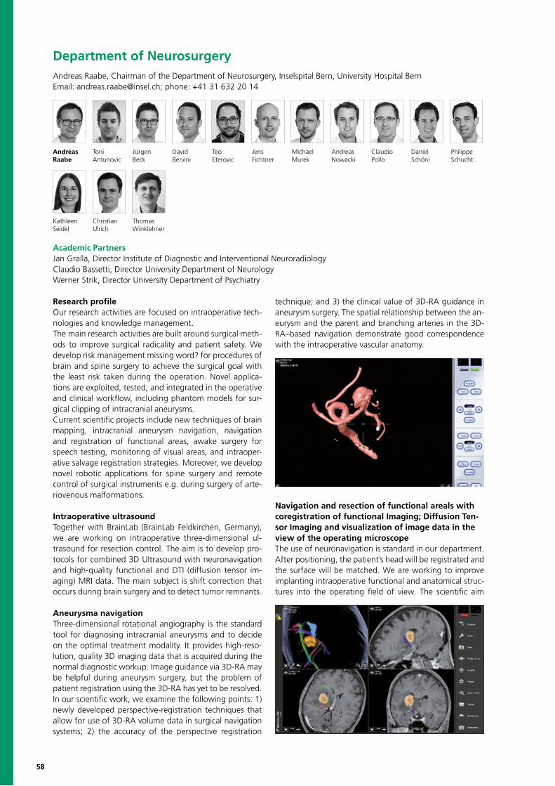
58
Department of NeurosurgeryAndreas Raabe, Chairman of the Department of Neurosurgery, Inselspital Bern, University Hospital BernEmail: [email protected]; phone: +41 31 632 20 14
Andreas Toni Jürgen David Teo Jens Michael Andreas Claudio Daniel PhilippeRaabe Antunovic Beck Bervini Eterovic Fichtner Murek Nowacki Pollo Schöni Schucht
Research profileOur research activities are focused on intraoperative tech-nologies and knowledge management. The main research activities are built around surgical meth-ods to improve surgical radicality and patient safety. We develop risk management missing word? for procedures of brain and spine surgery to achieve the surgical goal with the least risk taken during the operation. Novel applica-tions are exploited, tested, and integrated in the operative and clinical workflow, including phantom models for sur-gical clipping of intracranial aneurysms.Current scientific projects include new techniques of brain mapping, intracranial aneurysm navigation, navigation and registration of functional areas, awake surgery for speech testing, monitoring of visual areas, and intraoper-ative salvage registration strategies. Moreover, we develop novel robotic applications for spine surgery and remote control of surgical instruments e.g. during surgery of arte-riovenous malformations.
Intraoperative ultrasoundTogether with BrainLab (BrainLab Feldkirchen, Germany), we are working on intraoperative three-dimensional ul-trasound for resection control. The aim is to develop pro-tocols for combined 3D Ultrasound with neuronavigation and high-quality functional and DTI (diffusion tensor im-aging) MRI data. The main subject is shift correction that occurs during brain surgery and to detect tumor remnants.
Aneurysma navigationThree-dimensional rotational angiography is the standard tool for diagnosing intracranial aneurysms and to decide on the optimal treatment modality. It provides high-reso-lution, quality 3D imaging data that is acquired during the normal diagnostic workup. Image guidance via 3D-RA may be helpful during aneurysm surgery, but the problem of patient registration using the 3D-RA has yet to be resolved. In our scientific work, we examine the following points: 1) newly developed perspective-registration techniques that allow for use of 3D-RA volume data in surgical navigation systems; 2) the accuracy of the perspective registration
technique; and 3) the clinical value of 3D-RA guidance in aneurysm surgery. The spatial relationship between the an-eurysm and the parent and branching arteries in the 3D-RA–based navigation demonstrate good correspondence with the intraoperative vascular anatomy.
Navigation and resection of functional areals with coregistration of functional Imaging; Diffusion Ten-sor Imaging and visualization of image data in the view of the operating microscopeThe use of neuronavigation is standard in our department. After positioning, the patient’s head will be registrated and the surface will be matched. We are working to improve implanting intraoperative functional and anatomical struc-tures into the operating field of view. The scientific aim
Kathleen Christian Thomas Seidel Ulrich Winklehner
Academic PartnersJan Gralla, Director Institute of Diagnostic and Interventional NeuroradiologyClaudio Bassetti, Director University Department of NeurologyWerner Strik, Director University Department of Psychiatry
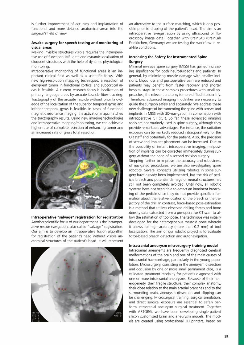
59
is further improvement of accuracy and implantation of functional and more detailed anatomical areas into the surgeon’s field of view.
Awake surgery for speech testing and monitoring of visual areasMaking invisible structures visible requires the intraopera-tive use of functional MRI data and dynamic localisation of eloquent structures with the help of dynamic physiological monitoring. Intraoperative monitoring of functional areas is an im-portant clinical field as well as a scientific focus. With new high-resolution mapping techniques, a resection of eleoquent tumor in functional cortical and subcortical ar-eas is feasible. A current research focus is localization of primary language areas by arcuate fascicle fiber tracking. Tractography of the arcuate fascicle without prior knowl-edge of the localization of the superior temporal gyrus and inferior temporal gyrus is feasible. In cases of functional magnetic resonance imaging, the activation maps matched the tractography results. Using new imaging technologies and intraoperative mapping/monitoring, we can achieve a higher rate of complete resection of enhancing tumor and an increased rate of gross total resection.
Intraoperative “salvage” registration for registrationAnother scientific focus of our department is the intraoper-ative rescue navigation, also called “salvage” registration. Our aim is to develop an intraoperative fusion algorithm for registration of the patient’s head without visible an-atomical structures of the patient’s head. It will represent
an alternative to the surface matching, which is only pos-sible prior to draping of the patient’s head. The aim is an intraoperative re-registration by using ultrasound or flu-oroscopy image data. Together with BrainLAB (BrainLab Feldkirchen, Germany) we are testing the workflow in re-al-life conditions.
Increasing the Safety for Instrumented SpineSurgery Minimal invasive spine surgery (MISS) has gained increas-ing significance for both neurosurgeons and patients. In general, by minimizing muscle damage with smaller inci-sions, blood loss and postoperative pain are reduced and patients may benefit from faster recovery and shorter hospital stays. In these complex procedures with small ap-proaches, the relevant anatomy is more difficult to identify. Therefore, advanced imaging modalities are necessary to guide the surgeon safely and accurately. We address these new challenges of instrumenting the spine with screws and implants in MISS with 3D-navigation in combination with intraoperative CT (iCT). So far, these advanced imaging tools are not routinely used in spine surgery, although they provide remarkable advantages. For instance, the radiation exposure can be markedly reduced intraoperatively for the OR staff and potentially for the patient. Also, the precision of screw and implant placement can be increased. Due to the possibility of instant intraoperative imaging, malposi-tion of implants can be corrected immediately during sur-gery without the need of a second revision surgery. Stepping further to improve the accuracy and robustness of navigated procedures, we are also investigating spine robotics. Several concepts utilizing robotics in spine sur-gery have already been implemented, but the risk of ped-icle breach and potential damage of neural structures has still not been completely avoided. Until now, all robotic systems have not been able to detect an imminent breach-ing of the pedicle since they do not provide specific infor-mation about the relative location of the breach or the tra-jectory of the drill. In contrast, force-based pose estimation is a method that utilizes observed drilling forces and bone density data extracted from a pre-operative CT scan to al-low the estimation of tool pose. The technique was initially developed for the heterogeneous mastoid bone wherein it allows for high accuracy (more than 0.2 mm) of tool localization. The aim of our robotic project is to evaluate force-based breach detection and autonavigation.
Intracranial aneurysm microsurgery training modelIntracranial aneurysms are frequently diagnosed cerebral malformations of the brain and one of the main causes of intracranial haemorrhage, particularly in the young popu-lation. Microsurgery, consisting in the aneurysm dissection and occlusion by one or more small permanent clips, is a validated treatment modality for patients diagnosed with one or more intracranial aneurysms. Because of their het-erogeneity, their fragile structure, their complex anatomy, their close relation to the main arterial branches and to the surrounding brain, aneurysm dissection and clipping can be challenging. Microsurgical training, surgical simulation, and direct surgical exposure are essential to safely per-form intracranial aneurysm surgical treatment. Together with ARTORG, we have been developing single-patient silicon customized brain and aneurysm models. The mod-els are created using professional 3D printers, based on
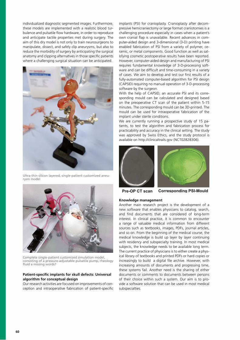
60
individualized diagnostic segmented images. Furthermore, these models are implemented with a realistic blood tur-bulence and pulsatile flow hardware, in order to reproduce and anticipate tactile properties met during surgery. The aim of this dry model is not only to train neurosurgeons to manipulate, dissect, and safely clip aneurysms, but also to reduce the morbidity of surgery by anticipating the surgical anatomy and clipping alternatives in those specific patients where a challenging surgical situation can be anticipated.
Patient-specific implants for skull defects: Universal algorithm for conceptual designOur research activities are focused on improvements of con-ception and intraoperative fabrication of patient-specific
implants (PSI) for cranioplasty. Cranioplasty after decom-pressive hemicraniectomy or large format craniotomies is a challenging procedure especially in cases when a patient’s own cranial flap is unavailable. Recent advances in com-puter-aided design and 3-dimensional (3-D) printing have enabled fabrication of PSI from a variety of polymer, ce-ramic, or metal components. Good function as well as sat-isfying cosmetic postoperative results have been reported. However, computer-aided design and manufacturing of PSI requires fundamental knowledge of 3-D-processing soft-ware and can be difficult and time-consuming in a variety of cases. We aim to develop and test our first results of a fully-automated computer-based algorithm for PSI design (CAPSID) requiring no manual operation of 3-D-processing software by the surgeon.With the help of CAPSID, an accurate PSI and its corre-sponding mould can be calculated and designed based on the preoperative CT scan of the patient within 5-15 minutes. The corresponding mould can be 3D-printed. The mould can be used for intraoperative fabrication of the implant under sterile conditions.We are currently running a prospective study of 15 pa-tients, to test the algorithm and fabrication process for practicability and accuracy in the clinical setting. The study was approved by Swiss Ethics, and the study protocol is available on http://clinicaltrails.gov (NCT02828306).
Knowledge managementAnother main research project is the development of a new software that enables physicians to catalog, search, and find documents that are considered of long-term interest. In clinical practice, it is common to encounter a range of valuable medical information from different sources such as textbooks, images, PDFs, journal articles, and so on. From the beginning of the medical course, the medical knowledge is build up layer by layer continuing with residency and subspecialty training. In most medical subjects, the knowledge needs to be available long term. The current practice of physicians is to either create a phys-ical library of textbooks and printed PDFs or hard copies or increasingly to build a digital file archive. However, with increasing amounts of documents and progressing time, these systems fail. Another need is the sharing of either documents or comments to documents between persons of their choice within such a system. Our aim is to pro-vide a software solution that can be used in most medical subspecialties.
Ultra-thin silicon layered, single-patient customized aneu-rysm model
Complete single-patient customized simulation model, consisting of a pressure-adjustable pulsatile pump, rheology fluid a missing words?

61
Selected PublicationsRaabe A, Beck J, Rohde S, Berkefeld J, Seifert V (2006) Three-dimensional rotational angiography guidance for aneurysm surgery. Journal of neurosurgery 105:406-11
Raabe A, Krishnan R, Wolff R, Hermann E, Zimmermann M, Seifert V (2002) Laser surface scanning for patient registration in intracranial image-guided surgery. Neurosurgery 50:797-801
Stieglitz LH, Fichtner J, Andres R, et al (2013) The silent loss of neuronavigation accuracy: a systematic retrospective analy-sis of factors influencing the mismatch of frameless stereotactic systems in cranial neurosurgery. Neurosurgery 72:796-807
Henning Stieglitz L, Seidel K, Wiest R, Beck J, Raabe A (2012) Localization of primary language areas by arcuate fascicle fiber tracking. Neurosurgery 70:56-64
Schucht P, Beck J, Abu-Isa J, et al (2012) Gross total resection rates in contemporary glioblastoma surgery: results of an ins-titutional protocol combining 5-aminolevulinic acid intraoperative fluorescence imaging and brain mapping. Neurosurgery 71:927-35
Seidel K, Beck J, Stieglitz L, Schucht P, Raabe A (2012) Low-threshold monopolar motor mapping for resection of primary motor cortex tumors. Neurosurgery 71:104-14
62
Support Center for Advanced Neuroimaging SCANRoland Wiest, Head of Research Group Email: [email protected]; phone: +41 31 632 26 55
Roland Nuno Marwan Simon Frauke Claus Urspeter Werner Richard Manuela ChristianWiest Barros El-Koussy Habegger Kellner-Weldon Kiefer Knecht Krammer McKinley Pastore-Wapp Rummel
Olivier Johannes Waldo Franca BrunoScheidegger Slotboom Valenzuela Wagner Weder
Research ProfileThe Support Center for Advanced Neuroimaging (SCAN) is a multidisciplinary imaging laboratory, where physi-cists, mathematicians, computer engineers, neuroradiol-ogists, neurologists, and psychologists investigate new applications for advanced neuroimaging. The main fo-cus of research is the development of computer-assisted post-processing techniques for clinical applications in neu-romedicine and neuroscience (in close collaboration with the neurocenter at the Inselspital, the ISTB, the UPD trans-lational research center, our international research, and industrial partners). We aim to improve quantification of lesion load, monitoring of regional brain plasticity, and tis-sue-at-risk estimation in frequent neurological disorders, e.g. stroke, multiple sclerosis, brain tumors, and epilep-sy. In addition, we translate novel sequence technologies as neuronal current imaging, advanced MR spectroscopy, model-based magnetization, and chemical exchange sat-uration transfer and multiband functional MRI into clinical practice.
Quantitative NeuroimagingQuantification of image information becomes more and more important in clinical routine, since it may comple-ment visual analysis by objective, independent and repro-ducible information. MRT applied to radiology uses the signal intensity variation as a function of the time and of intrinsic physical mechanisms such as relaxation (T1, T2), spin coupling (J), model-based magnetization, and chem-ical exchange transfer (MT, CEST) or diffusion (D). The examination of those intensity variations allows a quali-tative analysis by comparing intensity distributions on T1- or T2-weighted images with a radiological database and thus allows the clinician to make a diagnosis. In order to detect more subtle changes in biological tissues, typically during the early stages of a disease process, more quan-titative analyses (qMRT) are required to guarantee a truly
patient-specific – hardware (scanner electronics, coils) in-dependent –evaluation. This means that not only T1- or T2-weighted images are acquired but merely that the T1 - and T2-relaxation times themselves must be measured on a voxel-by-voxel basis. A multi-parametric analysis, therefore, has the potential to increase biological speci-ficity. Moreover, discriminant or cluster analysis of several parameters (e.g. MT, T1, T2) can give improvements over single parameters. Quantitative Susceptibility Mapping (QSM) in combination with T2*-mapping is another tech-nique used in the SCAN for the correct delineation of bleedings and calcifications. In order to integrate qMRT in the clinical routine, it is, however, necessary to shorten the acquisition times (see “Sequence Development”) as well as the post-processing computational time whereas the latter e.g. includes parallelization by means of GPUs or multi-core systems.The SCAN has developed quantitative neuroimaging methodology in the following fields:
• MR spectroscopy for quantification of the concentra-tions of metabolites in brain tissue to support standard MR, e.g. to diagnose brain tumors and metabolic disorders.
• Novel neural-current-imaging techniques (pc-SIRS) to measure neuronal discharges directly with MRI via the in-duced distortions of the magnetic field. This technology is expected to become relevant in the evaluation of epilepsy patients.
• Statistical comparison of morphometric parameters (grey matter volume, cortical thickness and surface area, and contrast at the grey-white boundary) between pa-tients and healthy controls to detect pathologies related to brain atrophy and plasticity. This supports diagnostics in dementia, movement disorder, multiple sclerosis, and epilepsy.
Clinical PartnersClaudio Bassetti, Department of Neurology, Inselspital, Bern University HospitalAndreas Raabe, Department of Neurosurgery, Inselspital, Bern University HospitalMaja Steinlin, Division of Neuropediatrics, University of BernGeorge Thalmann, Department of Urology, Inselspital, Bern University HospitalMarco Caversaccio, Chairman and Head, ENT Department, Inselspital, Bern University HospitalWerner Strik, Stefan Klöppel, Thomas Dierks, University Hospital of Psychiatry BernMauricio Reyes, Institute for Surgical Technology and Biomechanics, University of Bern

63
Metabolic ImagingMagnetic Resonance Spectroscopy (MRS) allows physicians to retrieve localized chemical information in a non-invasive manner. Because of this, MRS is often referred to as “vir-tual biopsy.” In the brain, MRS is often applied to the di-agnosis and follow-up of brain tumors, metabolic diseases, bacterial infections, etc.Besides the added value of MRS, the complexity associat-ed with the processing and interpretation of the data rep-resents a considerable limiting factor for its use in clinical practice. At the SCAN, we have been working on methods that facilitate the use of MRS in clinical routine:
• Automatic quality control of MR Spectra – The quality of MR Spectra is often corrupted, and spectra containing signal artifacts can potentially lead to wrong interpreta-tions. Moreover, visually identifying which spectra must be excluded from the analysis is an extremely time-consuming task that requires a certain level of experience. Therefore, we have worked on machine learning methods that can assess the quality of the spectra with accuracy identical to experts but that can analyze thousands of spectra in a few seconds.
• Metabolite levels correction – Due to acquisition-related limitations, the sensitivity to the several detectable metab-olites is spatially dependent. At the SCAN, we have been working on methods that help minimizing these effects on the computed metabolic maps.
• Nosologic maps – The combination and interpretation of the information provided by the different metabolite maps is often challenging, namely when there is a lack of a solid experience with MRS. Consequently, we have been working on methods for the automatic creation of easi-ly interpretable maps where different metabolic regions are identified – nosologic maps. A single nosologic map summarizes the information provided by all the different metabolic maps and allows incorporating the know-how of an experienced spectroscopic in the interpretation of the different types of information provided by MRS.
• Absolute quantification – Introduction of absolute quantification of single-voxel-spectroscopy (SVS) in clinical routine.
MR Sequence DevelopmentThe SCAN has established a research collaboration with Siemens, which gives us the unique opportunity to develop new control software (sequences) for the MR-scanner. This is an important step towards a fast customization of the clinical claims by the physicists of the SCAN team. It also enables us to produce qMR parameter maps, which could enhance the sensitivity and specificity of machine- learn-ing-based computer-aided diagnosis. One of the newest developments (pc-SIRS) tries to measure neuronal current distributions in the brain, especially pathologically phe-nomena related to epilepsy (Figure 3, 4). Other trends will be to accelerate sequences in order to either statistically improve resting-state-based analysis of the default mode networks or to integrate the quantitative MRT, typically as-sociated with long acquisition times, in a day-to-day clinical service work. Other SCAN sequences are used for mod-el-based MT/CEST imaging and elastography in the context of neurodegeneration and aneurysms, respectively.
Computer-Assisted Image AnalysisIn diseases of the nervous system, it is relevant to identify pathologies and quantify them, both in terms of number of abnormalities and volume of affected tissue. Accelerated processing of neuroimaging data becomes most relevant in emergency settings, where every minute gained in assess-ing a patient’s situation can improve the prognosis as well as in elective patient monitoring.For this reason, we have developed a number of auto-mated approaches to the quantification of brain lesions in close cooperation with the ISTB (Prof. Reyes). These meth-ods harness the increasing power of machine-learning, in
Example of longitudinal morphometric changes in patient with MS scanned repetitively over five years. The volume of the corpus callosum (left) and the cortical thickness in the right temporal lobe (middle) indicate progressive volume loss. Patient data are displayed as filled symbols, and healthy control data as open symbols. The grey-white contrast in temporal, parietal, and occipital brain regions is significantly lower than expected from age and gender-matched healthy controls (right).
SpectrIm –developed by J. Slotboom and N. Pedrosa de Barros for the combined processing of MR spectroscopy and imaging. The tool includes several processing meth-ods developed at the SCAN. In the figure, a Cho/Cr map is displayed as an overlay.
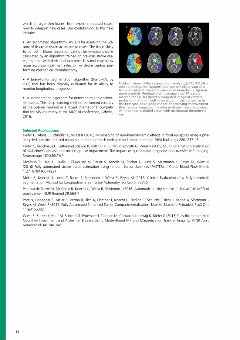
64
which an algorithm learns, from expert-annotated cases, how to interpret new cases. Our contributions to this field include:
• An automated algorithm (FASTER) for assessing the vol-ume of tissue-at-risk in acute stroke cases. The tissue likely to be lost if blood circulation cannot be re-established is calculated by an algorithm trained on previous stroke cas-es, together with their final outcome. This tool may allow more accurate treatment selection in stroke centres per-forming mechanical thrombectomy.
• A brain-tumor segmentation algorithm (BraTuMIA, by ISTB) that has been clinically evaluated for its ability to monitor longitudinal progression.
• A segmentation algorithm for detecting multiple sclero-sis lesions. This deep-learning method performed recently as the optimal method in a recent international competi-tion for MS volumetry at the MICCAI conference, Athens, 2016.
Selected PublicationsKiefer C, Abela E, Schindler K, Wiest R (2016) MR-imaging of non-hemodynamic effects in focal epilepsies using a pha-se-cycled stimulus-induced rotary saturation approach with spin-lock preparation (pc-SIRS).Radiology 280: 237-43
Kiefer C, Brockhaus L, Cattapan-Ludewig K, Ballinari P, Burren Y, Schroth G, Wiest R (2009) Multi-parametric classification of Alzheimer’s disease and mild cognitive impairment: The impact of quantitative magnetization transfer MR imaging. Neuroimage 48(4):657-67
McKinley R, Häni L, Gralla J, El-Koussy M, Bauer S, Arnold M, Fischer U, Jung S, Mattmann K, Reyes M, Wiest R (2016) Fully automated stroke tissue estimation using random forest classifiers (FASTER). J Cereb Blood Flow Metab 1:271678X16674221
Meier R, Knecht U, Loosli T, Bauer S, Slotboom J, Wiest R, Reyes M (2016) Clinical Evaluation of a Fully-automatic Segmentation Method for Longitudinal Brain Tumor Volumetry. Sci Rep 6: 23376
Pedrosa de Barros N, McKinley R, Knecht U, Wiest R, Slotboom J (2016) Automatic quality control in clinical (1)H MRSI of brain cancer. NMR Biomed 29:563-7
Porz N, Habegger S, Meier R, Verma R, Jilch A, Fichtner J, Knecht U, Radina C, Schucht P, Beck J, Raabe A, Slotboom J, Reyes M, Wiest R (2016) Fully Automated Enhanced Tumor Compartmentalization: Man vs. Machine Reloaded. PLoS One 11:e0165302
Wiest R, Burren Y, Hauf M, Schroth G, Pruessner J, Zbinden M, Cattapan-Ludewig K, Kiefer C (2013) Classification of Mild Cognitive Impairment and Alzheimer Disease Using Model-Based MR and Magnetization Transfer Imaging. AJNR Am J Neuroradiol 34: 740-746
Unlike in linear diffusion/perfusion analyis (c), FASTER (d) is able to distinguish hypoperfused, potentially salvageable tissue (blue) and irreversibly damaged brain tissue (green) more precisely. Residual brain damage after 90 days is marked red (e). (e) shows a computed image of cerebral perfusion that is difficult to interpret. If the patient, as in the first case, has a good chance of achieving improvement (low residual damage), the interventional neuroradiologist will treat the occluded vessel with mechanical thrombecto-my.
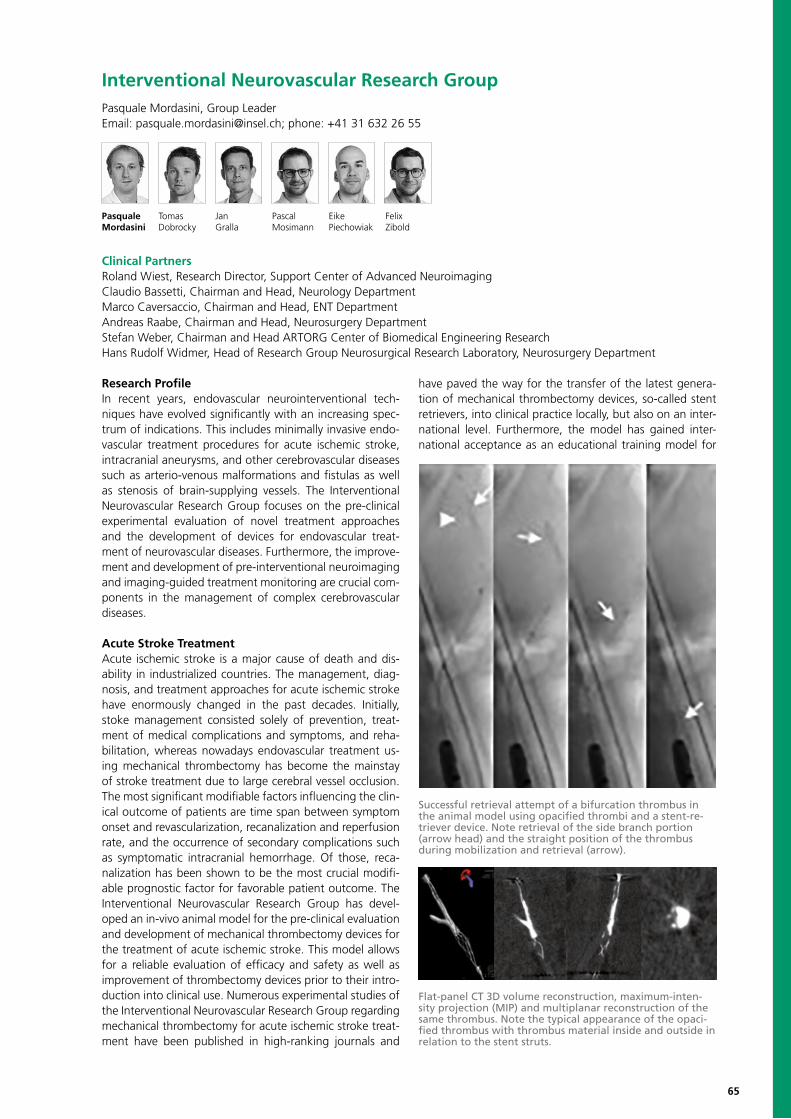
65
Research Profile In recent years, endovascular neurointerventional tech-niques have evolved significantly with an increasing spec-trum of indications. This includes minimally invasive endo-vascular treatment procedures for acute ischemic stroke, intracranial aneurysms, and other cerebrovascular diseases such as arterio-venous malformations and fistulas as well as stenosis of brain-supplying vessels. The Interventional Neurovascular Research Group focuses on the pre-clinical experimental evaluation of novel treatment approaches and the development of devices for endovascular treat-ment of neurovascular diseases. Furthermore, the improve-ment and development of pre-interventional neuroimaging and imaging-guided treatment monitoring are crucial com-ponents in the management of complex cerebrovascular diseases.
Acute Stroke TreatmentAcute ischemic stroke is a major cause of death and dis-ability in industrialized countries. The management, diag-nosis, and treatment approaches for acute ischemic stroke have enormously changed in the past decades. Initially, stoke management consisted solely of prevention, treat-ment of medical complications and symptoms, and reha-bilitation, whereas nowadays endovascular treatment us-ing mechanical thrombectomy has become the mainstay of stroke treatment due to large cerebral vessel occlusion. The most significant modifiable factors influencing the clin-ical outcome of patients are time span between symptom onset and revascularization, recanalization and reperfusion rate, and the occurrence of secondary complications such as symptomatic intracranial hemorrhage. Of those, reca-nalization has been shown to be the most crucial modifi-able prognostic factor for favorable patient outcome. The Interventional Neurovascular Research Group has devel-oped an in-vivo animal model for the pre-clinical evaluation and development of mechanical thrombectomy devices for the treatment of acute ischemic stroke. This model allows for a reliable evaluation of efficacy and safety as well as improvement of thrombectomy devices prior to their intro-duction into clinical use. Numerous experimental studies of the Interventional Neurovascular Research Group regarding mechanical thrombectomy for acute ischemic stroke treat-ment have been published in high-ranking journals and
have paved the way for the transfer of the latest genera-tion of mechanical thrombectomy devices, so-called stent retrievers, into clinical practice locally, but also on an inter-national level. Furthermore, the model has gained inter-national acceptance as an educational training model for
Interventional Neurovascular Research GroupPasquale Mordasini, Group LeaderEmail: [email protected]; phone: +41 31 632 26 55
Pasquale Tomas Jan Pascal Eike Felix Mordasini Dobrocky Gralla Mosimann Piechowiak Zibold
Successful retrieval attempt of a bifurcation thrombus in the animal model using opacified thrombi and a stent-re-triever device. Note retrieval of the side branch portion (arrow head) and the straight position of the thrombus during mobilization and retrieval (arrow).
Flat-panel CT 3D volume reconstruction, maximum-inten-sity projection (MIP) and multiplanar reconstruction of the same thrombus. Note the typical appearance of the opaci-fied thrombus with thrombus material inside and outside in relation to the stent struts.
Clinical PartnersRoland Wiest, Research Director, Support Center of Advanced NeuroimagingClaudio Bassetti, Chairman and Head, Neurology DepartmentMarco Caversaccio, Chairman and Head, ENT DepartmentAndreas Raabe, Chairman and Head, Neurosurgery DepartmentStefan Weber, Chairman and Head ARTORG Center of Biomedical Engineering ResearchHans Rudolf Widmer, Head of Research Group Neurosurgical Research Laboratory, Neurosurgery Department
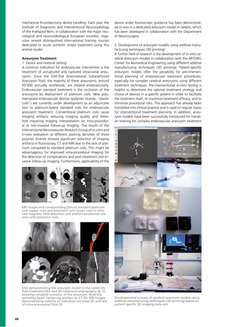
66
mechanical thrombectomy device handling. Each year, the Institute of Diagnostic and Interventional Neuroradiology of the Inselspital Bern, in collaboration with the major neu-rological and neuroradiological European societies, orga-nizes several distinguished international training courses dedicated to acute ischemic stroke treatment using this animal model.
Aneurysm Treatment1. Device and material testingA common indication for endovascular intervention is the treatment of unruptured and ruptured intracranial aneu-rysms. Since the ISAT-Trial (International Subarachnoid Aneurysm Trial), the majority of these aneurysms, around 50’000 annually worldwide, are treated endovascularly. Endovascular standard treatment is the occlusion of the aneurysms by deployment of platinum coils. New poly-mer-based endovascular devices (polymer strands, “plastic coils”) are currently under development as an adjunctive tool to platinum-based standard coils for endovascular aneurysm treatment. Conventional platinum coils cause imaging artifacts reducing imaging quality and there-fore impairing imaging interpretation on intra-procedur-al or non-invasive follow-up imaging. The results of the Interventional Neurovascular Research Group of in-vitro and in-vivo evaluation at different packing densities of these polymer strands showed significant reduction of imaging artifacts in fluoroscopy, CT and MRI due to the lack of plat-inum compared to standard platinum coils. This might be advantageous for improved intra-procedural imaging for the detection of complications and post-treatment non-in-vasive follow-up imaging. Furthermore, applicability of the
device under fluoroscopic guidance has been demonstrat-ed in-vivo in a dedicated aneurysm model in rabbits, which has been developed in collaboration with the Department of Neurosurgery.
2. Development of aneurysm models using additive manu-facturing techniques (3D printing)A further field of research is the development of in-vitro ce-rebral aneurysm models in collaboration with the ARTORG Center for Biomedical Engineering using different additive manufacturing techniques (3D printing). Patient-specific aneurysm models offer the possibility for pre-interven-tional planning of endovascular treatment procedures, especially for complex cerebral aneurysms using different treatment techniques. Pre-interventional in-vitro testing is helpful to determine the optimal treatment strategy and choice of devices in a specific patient in order to facilitate the treatment itself, to maximize treatment efficacy, and to minimize procedural risks. This approach has already been translated into clinical practice and is used on regular bases for interventional treatment planning. In addition, aneu-rysm models have been successfully introduced for hands-on training for complex endovascular aneurysm treatment
MR images and corresponding DSA of standard platinum coils (upper row) and polymeric coils (lower row) in-vitro. Less magnetic field distortion and artefact production are seen with polymeric coils.
DSA demonstrating the aneurysm model in the rabbit (A). Post-treatment DSA and 3D rotational angiography (B, C) showing complete occlusion of the aneurysm. Note the persisting beam hardening artefact on CT (D). MR images demonstrating visibility of individual coil loops (E) and lack of intra-aneurysmal flow (F).
Developmental process of cerebral aneurysm models using additive manufacturing techniques (3D printing) based on patient specific 3D imaging data sets.
67
Selected PublicationsGralla J, Schroth G, Remonda L, Fleischmann A, Fandino J, Slotboom J, Brekenfeld C (2005) A dedicated animal model for mechanical thrombectomy in acute stroke. AJNR Am J Neuroradiol 27(6):1357-1361
Brekenfeld C, Schroth G, El-Koussy M, Nedeltchev K, Reinert M, Slotboom J, Gralla J (2008) Mechanical thromboem-bolectomy for acute ischemic stroke: comparison of the catch thrombectomy device and Merci Retriever in vivo. Stroke 39(4):1213-1219
Mordasini P, Hiller M, Brekenfeld C, Schroth G, Fischer U, Slotboom J, Arnold M, Gralla J (2010) In-vivo evaluation of the Phenox CRC mechanical thrombectomy device in a swine model of acute vessel occlusion. AJNR Am J Neuroradiol 31(5):972-978
Mordasini P, Frabetti N, Gralla J, Schroth G, Fischer U, Arnold M, Brekenfeld C (2011) In-vivo evaluation of a dedicated combined flow restoration and mechanical thrombectomy device in a swine model of acute vessel occlusion. AJNR Am J Neuroradiol 32:294-300
Mordasini P, Brekenfeld C, Byrne JV, Fischer U, Arnold M, Jung S, Schroth G, Gralla J (2013) Experimental evaluation of immediate recanalization effect and recanalization efficacy of a new thrombus retriever for acute stroke treatment in-vivo. AJNR Am J Neuroradiol 34:153-158
Mordasini P, Kraehenbuehl AK, Byrne JV, Vandenberghe S, Reinert M, Hoppe H, Gralla J (2013) In vitro and in vivo ima-ging characteristics assessment of polymeric coils compared with standard platinum coils for the treatment of intracranial aneurysms. AJNR Am J Neuroradiol 34(11):2177-2183
Jung S, Gilgen M, Slotboom J, El-Koussy M, Zubler C, Kiefer C, Luedi R, Mono ML, Heldner MR, Weck A, Mordasini P, Schroth G, Mattle HP, Arnold M, Gralla J, Fischer U (2013) Factors that determine penumbral tissue loss in acute ischaemic stroke. Brain 136:3554-60
and educational purposes. Furthermore, aneurysm models are used for the development, testing, and evaluation of novel endovascular devices and treatment approaches, as
well as a model for measurements of different aspects of flow dynamics and their role in aneurysm formation and growth.

68
Visceral Surgery Daniel Candinas, Head of Division of Visceral SurgeryEmail: [email protected]; phone: +41 31 632 24 04
Daniel Vanessa Guido Lukas Sabrina Agata Tobias Manuel Fadi Adrian AndreasCandinas Banz Beldi Brügger Frey Gôrecka Haltmeier Jakob Jebbawi Keogh Kohler
Anja Dominik Deborah Pascale Stefan JoelLachenmayer Obrist Stroka Tinguely Weber Zindel
Research ProfileThe research interests of Visceral Surgery include regen-erative medicine, liver cancers, and computer-assisted liv-er surgery. Specific topics of interest within regenerative medicine include the molecular basis of liver regeneration and sphincter muscle augmentation and regeneration. For both aspects, elements of adult-derived stem cells are explored to enhance the regrowth of hepatic parenchy-mal cells, and of the various compartments of the sphinc-ter muscle. In our study of liver cancers, preclinical models and translational studies are used to better understand the pathophysiology of hepatocellular carcinoma and intrahepatic cholangiocarcinoma and to develop novel treatment approaches.Furthermore, we explore novel developments in im-age-guided technology into the context of computer-as-sisted liver surgery, to improve intraoperative naviga-tion and augmenting accuracy in targeting intrahepatic lesions. New approaches for augmented reality are also currently being developed for minimally invasive surgery. In partic-ular, we are developing tools that enhance the visible field that is displayed on monitors during laparoscopic surgery.Using variable approaches, our research activities aim to improve standards to establish well-founded clinical treat-ment strategies within the broad field of visceral surgery. A dedicated team of over 50 members of the Department of Visceral Surgery perform a broad range of clinical, translational, and basic studies, some of which are shown in further detail below.
Regenerative Medicine in the LiverOur group studies regenerative medicine in the liver fol-lowing two approaches. In the first approach, we rely on the ability of the liver to sense and respond to damage to regenerate itself by its major parenchymal cell type, the hepatocyte, to re-enter cell cycle, and proliferate to restore the damaged or lost tissue mass. We investigate this unique ability by focusing on the role of the innate immune system and the influence of extra-cellular signals on immune function during liver regenera-tion. In addition, we investigate novel pathways activated in the regenerating liver, with the goal of defining new targets to promote liver function and hepatocyte prolifer-ation during regeneration of diseased livers. In the second approach, we isolate and expand liver-derived progenitor
cells in vitro. Liver progenitor cells have the potential to assist the regenerative capacity of diseased livers and may also offer a cell source for cell transplantation.
Hemodynamic changes in the liver after resectionSurgical resection of liver tissue is used for the treatment of hepatic neoplasms, both benign and malignant. Partial removal of the liver initiates regeneration of the remnant tissue, but also reduces the cross-sectional area of the liv-er vascular bed. This reduction leads to changes in blood flow velocity, portal pressure, shear stress in the sinusoidal vessels, oxygen supply to the liver, and many other phys-iological parameters. Our research explores the changes in blood flow through the liver vascular system after liver
Photomicrograph taken at 200x magnification of mouse liver tissue 48 hours after 67% hepatectomy. Brown reac-tions are the nuclei of hepatocytes stained positive for the proliferation marker Ki67 indicating a robust proliferative response throughout the liver tissue.
Photomicrograph taken at 50x magnification of liver progenitor cells isolated from a human gall bladder and cultured in vitro for 8 passages.
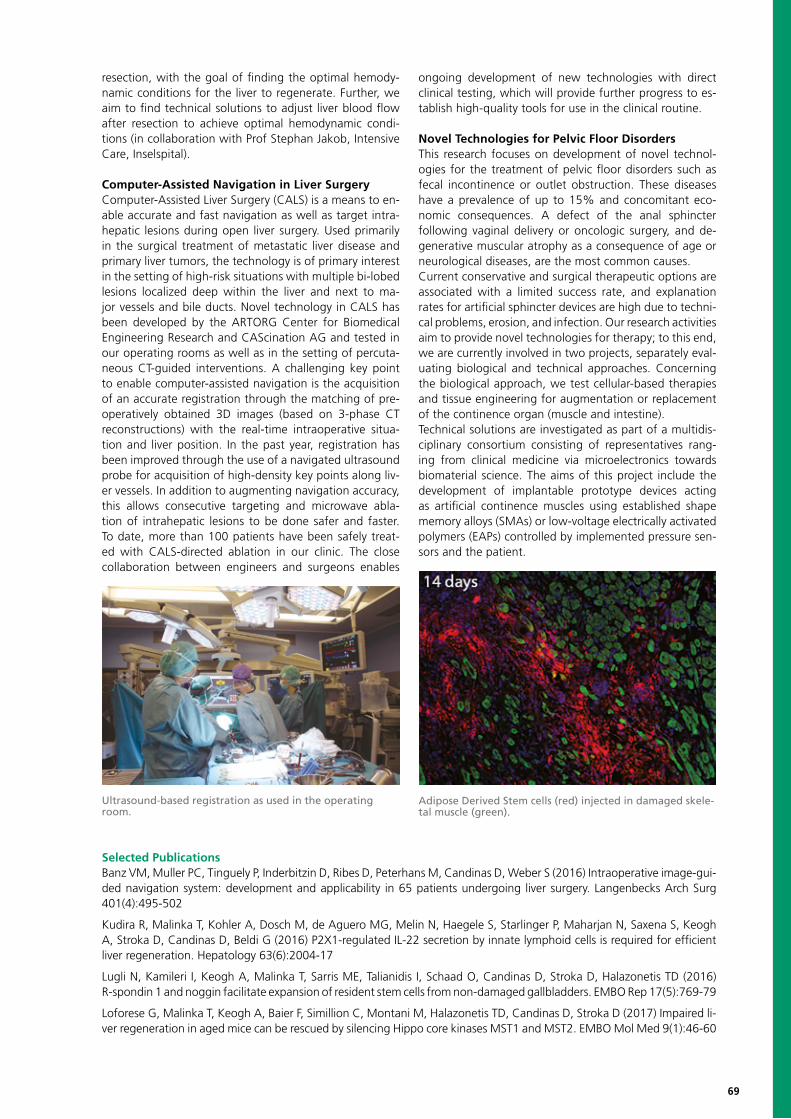
69
resection, with the goal of finding the optimal hemody-namic conditions for the liver to regenerate. Further, we aim to find technical solutions to adjust liver blood flow after resection to achieve optimal hemodynamic condi-tions (in collaboration with Prof Stephan Jakob, Intensive Care, Inselspital).
Computer-Assisted Navigation in Liver SurgeryComputer-Assisted Liver Surgery (CALS) is a means to en-able accurate and fast navigation as well as target intra-hepatic lesions during open liver surgery. Used primarily in the surgical treatment of metastatic liver disease and primary liver tumors, the technology is of primary interest in the setting of high-risk situations with multiple bi-lobed lesions localized deep within the liver and next to ma-jor vessels and bile ducts. Novel technology in CALS has been developed by the ARTORG Center for Biomedical Engineering Research and CAScination AG and tested in our operating rooms as well as in the setting of percuta-neous CT-guided interventions. A challenging key point to enable computer-assisted navigation is the acquisition of an accurate registration through the matching of pre-operatively obtained 3D images (based on 3-phase CT reconstructions) with the real-time intraoperative situa-tion and liver position. In the past year, registration has been improved through the use of a navigated ultrasound probe for acquisition of high-density key points along liv-er vessels. In addition to augmenting navigation accuracy, this allows consecutive targeting and microwave abla-tion of intrahepatic lesions to be done safer and faster. To date, more than 100 patients have been safely treat-ed with CALS-directed ablation in our clinic. The close collaboration between engineers and surgeons enables
ongoing development of new technologies with direct clinical testing, which will provide further progress to es-tablish high-quality tools for use in the clinical routine.
Novel Technologies for Pelvic Floor DisordersThis research focuses on development of novel technol-ogies for the treatment of pelvic floor disorders such as fecal incontinence or outlet obstruction. These diseases have a prevalence of up to 15% and concomitant eco-nomic consequences. A defect of the anal sphincter following vaginal delivery or oncologic surgery, and de-generative muscular atrophy as a consequence of age or neurological diseases, are the most common causes. Current conservative and surgical therapeutic options are associated with a limited success rate, and explanation rates for artificial sphincter devices are high due to techni-cal problems, erosion, and infection. Our research activities aim to provide novel technologies for therapy; to this end, we are currently involved in two projects, separately eval-uating biological and technical approaches. Concerning the biological approach, we test cellular-based therapies and tissue engineering for augmentation or replacement of the continence organ (muscle and intestine). Technical solutions are investigated as part of a multidis-ciplinary consortium consisting of representatives rang-ing from clinical medicine via microelectronics towards biomaterial science. The aims of this project include the development of implantable prototype devices acting as artificial continence muscles using established shape memory alloys (SMAs) or low-voltage electrically activated polymers (EAPs) controlled by implemented pressure sen-sors and the patient.
Ultrasound-based registration as used in the operating room.
Adipose Derived Stem cells (red) injected in damaged skele-tal muscle (green).
Selected PublicationsBanz VM, Muller PC, Tinguely P, Inderbitzin D, Ribes D, Peterhans M, Candinas D, Weber S (2016) Intraoperative image-gui-ded navigation system: development and applicability in 65 patients undergoing liver surgery. Langenbecks Arch Surg 401(4):495-502
Kudira R, Malinka T, Kohler A, Dosch M, de Aguero MG, Melin N, Haegele S, Starlinger P, Maharjan N, Saxena S, Keogh A, Stroka D, Candinas D, Beldi G (2016) P2X1-regulated IL-22 secretion by innate lymphoid cells is required for efficient liver regeneration. Hepatology 63(6):2004-17
Lugli N, Kamileri I, Keogh A, Malinka T, Sarris ME, Talianidis I, Schaad O, Candinas D, Stroka D, Halazonetis TD (2016) R-spondin 1 and noggin facilitate expansion of resident stem cells from non-damaged gallbladders. EMBO Rep 17(5):769-79
Loforese G, Malinka T, Keogh A, Baier F, Simillion C, Montani M, Halazonetis TD, Candinas D, Stroka D (2017) Impaired li-ver regeneration in aged mice can be rescued by silencing Hippo core kinases MST1 and MST2. EMBO Mol Med 9(1):46-60
70
Invasive Cardiology ResearchStephan Windecker, Head of Research GroupEmail: [email protected]; phone: + 41 31 632 96 53
Stephan Anna Andreas Kostantinos Jonas Aris Raffaele Thomas Lorenz Stefan HildegardWindecker Franzone Häberlin Koskinas Lanz Moschovitis Piccolo Pilgrim Räber Stortecky Tanner
Research ProfileThe Department of Cardiology at Bern University Hospital has a broad range of research activities that encompass clinical research with devices for the trans-catheter treat-ment of coronary artery disease and valvular heart dis-ease, pharmacology studies on antithrombotic drugs to prevent thromboembolism and on lipid-lowering drugs to prevent progression of atherosclerosis, as well as in the field of electrophysiology. The teams are involved in large-scale, multicenter, international, randomized clinical tri-als as well as in first-in-man studies with novel innovative products. The research group has well-established nation-al and international partnerships.
Invasive cardiology: coronary artery diseasesProf. Dr. med. L. Räber, PhDThe coronary artery disease research group conducts a prospective registry to assess procedural and mid-term clinical outcomes in all consecutive patients undergoing percutaneous coronary interventions (PCI) at Inselspital.Currently, more than 12,000 patients have been included. In 2016, two articles were published focusing on patients undergoing PCI with atrial fibrillation requiring triple anticoagulation and on the prognostic impact of stable coronary artery disease patients with increased level of high-sensitivity troponin. Side effects, adherence patterns, and clinical efficacy of novel antiplatelet agents and an- ticoagulant medications are specifically addressed within an additional prospective registry. Based on concerns re-garding the long-term performance of drug-eluting stents (DES), we extended the follow-up of a 1000-patient co-hort treated with DES throughout 10 years.Intracoronary imaging represents a key focus of our re-search group. In 2016, mechanisms underlying metallic DES thrombosis were investigated by means of high-res-olution optical coherence tomography in a large interna-tional registry and successfully published in Circulation. New projects in the field of intracoronary imaging en-compass an international registry on thrombosis occur-ring in novel fully bioabsorbable stents (INVEST registry), a randomized controlled trial investigating the potential of optical coherence tomography to improve outcome
for coronary revascularization using fully absorbable stent implantation at more than 10 international sites (OPTICO BVS), and a multimodality imaging trial investigating the effects of PSCK-9 inhibition on coronary atherosclerosis in patients with acute myocardial infarction (PACMAN AMI). The research team led by Prof. Räber currently includes one post-doc research fellow and an M.D.-Ph.D. candi-date who was granted a three-year SNF fellowship in the field of optical coherence tomography and shear stress analysis.
Invasive cardiology: valvular heart diseasesProf. Dr. med. T. PilgrimTranscatheter Aortic Valve Replacement (TAVR) represents an alternative treatment strategy for patients with severe aortic valve stenosis at increased risk for surgical aortic valve replacement. Refinements in device technology, improved imaging, and streamlining of the procedure resulted in a decline in peri-procedural complications and prepared the ground for the expansion of TAVR to intermediate and low-risk patients. Our group compares different strategies for the treatment of valvular heart dis-ease, investigates the importance of cardiac comorbidities on clinical outcomes, and studies newer generation devic-es for the treatment of aortic stenosis.In a meta-analysis of randomized trials, our group showed a survival benefit of TAVR as compared to surgical aor-tic valve replacement throughout two years of follow-up. The findings were consistent across the risk spectrum of high and intermediate-risk patients and were particularly pronounced among female patients and patients under-going TAVR by transfemoral approach. The findings antic-ipate a paradigm shift in the treatment of aortic stenosis and need confirmation in dedicated clinical trials with ex-tended follow-up.Newer generation TAVR devices demonstrated improved device success compared to early generation devices primarily by a reduction of moderate or severe para-prosthetic valvular regurgitation. Residual regurgitation across the bioprosthetic valve has been associated with increased mortality during extended clinical follow-up. Different strategies have been implemented to decrease
Marco Kyohei ThomasValgimigli Yamaji Zanchin
Project PartnersDr. Adrian Zurbuchen, University of Michigan, Ann Arbor, United StatesProf. Dominik Obrist, ARTORG Center, University of Bern, Switzerland
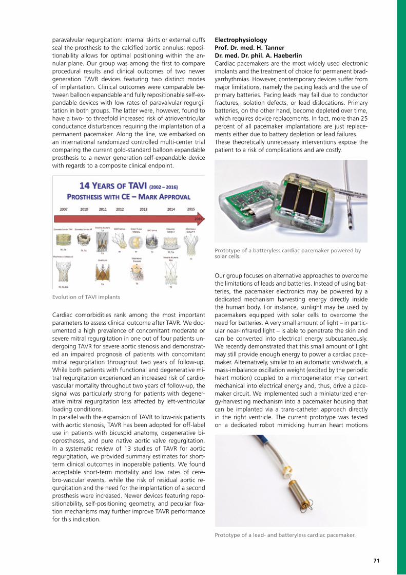
71
paravalvular regurgitation: internal skirts or external cuffs seal the prosthesis to the calcified aortic annulus; reposi-tionability allows for optimal positioning within the an-nular plane. Our group was among the first to compare procedural results and clinical outcomes of two newer generation TAVR devices featuring two distinct modes of implantation. Clinical outcomes were comparable be-tween balloon expandable and fully repositionable self-ex-pandable devices with low rates of paravalvular regurgi-tation in both groups. The latter were, however, found to have a two- to threefold increased risk of atrioventricular conductance disturbances requiring the implantation of a permanent pacemaker. Along the line, we embarked on an international randomized controlled multi-center trial comparing the current gold-standard balloon expandable prosthesis to a newer generation self-expandable device with regards to a composite clinical endpoint.
Cardiac comorbidities rank among the most important parameters to assess clinical outcome after TAVR. We doc-umented a high prevalence of concomitant moderate or severe mitral regurgitation in one out of four patients un-dergoing TAVR for severe aortic stenosis and demonstrat-ed an impaired prognosis of patients with concomitant mitral regurgitation throughout two years of follow-up. While both patients with functional and degenerative mi-tral regurgitation experienced an increased risk of cardio-vascular mortality throughout two years of follow-up, the signal was particularly strong for patients with degener-ative mitral regurgitation less affected by left-ventricular loading conditions.In parallel with the expansion of TAVR to low-risk patients with aortic stenosis, TAVR has been adopted for off-label use in patients with bicuspid anatomy, degenerative bi-oprostheses, and pure native aortic valve regurgitation. In a systematic review of 13 studies of TAVR for aortic regurgitation, we provided summary estimates for short-term clinical outcomes in inoperable patients. We found acceptable short-term mortality and low rates of cere-bro-vascular events, while the risk of residual aortic re-gurgitation and the need for the implantation of a second prosthesis were increased. Newer devices featuring repo-sitionability, self-positioning geometry, and peculiar fixa-tion mechanisms may further improve TAVR performance for this indication.
ElectrophysiologyProf. Dr. med. H. TannerDr. med. Dr. phil. A. HaeberlinCardiac pacemakers are the most widely used electronic implants and the treatment of choice for permanent brad- yarrhythmias. However, contemporary devices suffer from major limitations, namely the pacing leads and the use of primary batteries. Pacing leads may fail due to conductor fractures, isolation defects, or lead dislocations. Primary batteries, on the other hand, become depleted over time, which requires device replacements. In fact, more than 25 percent of all pacemaker implantations are just replace- ments either due to battery depletion or lead failures.These theoretically unnecessary interventions expose the patient to a risk of complications and are costly.
Our group focuses on alternative approaches to overcome the limitations of leads and batteries. Instead of using bat-teries, the pacemaker electronics may be powered by a dedicated mechanism harvesting energy directly inside the human body. For instance, sunlight may be used by pacemakers equipped with solar cells to overcome the need for batteries. A very small amount of light – in partic-ular near-infrared light – is able to penetrate the skin and can be converted into electrical energy subcutaneously. We recently demonstrated that this small amount of light may still provide enough energy to power a cardiac pace-maker. Alternatively, similar to an automatic wristwatch, a mass-imbalance oscillation weight (excited by the periodic heart motion) coupled to a microgenerator may convert mechanical into electrical energy and, thus, drive a pace- maker circuit. We implemented such a miniaturized ener- gy-harvesting mechanism into a pacemaker housing that can be implanted via a trans-catheter approach directly in the right ventricle. The current prototype was tested on a dedicated robot mimicking human heart motions
Evolution of TAVI implants
Prototype of a batteryless cardiac pacemaker powered by solar cells.
Prototype of a lead- and batteryless cardiac pacemaker.

72
previously acquired using cardiac magnetic resonance im-aging. We showed that even if the contractile function of the heart is impaired, it is still possible to harvest a sufficient amount of energy to drive a pacemaker. This approach also overcomes the need for pacemaker leads since the device is directly implanted inside the heart.Ongoing research investigates the performance of lead- and batteryless pacemaker systems in worst-case scenari-os and targets further refinement of the underlying engi-neering concepts.
Antithrombotic therapiesProf. Dr. M. Valgimigli / R. Piccolo, MDCardiovascular and cerebrovascular manifestations com- monly arise from atherosclerotic plaque rupture that pro-duces platelet activation, thrombus formation, and reduc- tion of blood flow to the heart or brain. Antithrombotic therapy is central to prevent blood clot formation and constitutes an essential component of therapy in the field of cardiology. Our group has been engaged in feverish research activity for many years now by investigating several aspects of antithrombotic therapy in the domain of cardiovascular intervention. Mainly in the setting of randomized clinical trials, we compared different anti-thrombotic regimens for short-term as well as long-term management of patients requiring percutaneous coronary intervention.Recently, we developed a simple five-item risk score, which provides a standardized tool for the prediction of out-of-hospital bleeding while on dual antiplatelet thera-py (www.precisedaptscore.com). As an important aspect for any treatment strategy is the long-term potential for harm, ongoing trials are focusing on different duration and composition of antithrombotic therapies among pa-tients at high risk of bleeding, in whom an ultrashort du-ration of dual antiplatelet therapy of only one month will be tested, among patients with atrial fibrillation requiring
concomitant antiplatelet and anticoagulant therapy, and among patients undergoing transcatheter aortic valve re-placement for aortic stenosis.The results of ongoing studies are expected to shed new light on novel approaches to the antithrombotic thera-py by refining current knowledge and challenging older paradigms.
Selected PublicationsKoskinas KC, Räber L, Zanchin T, Pilgrim T, Stortecky S, Hunziker L, Blöchlinger S, Billinger M, Gartwyl F, Moro C, Moschovitis A, Jüni P, Heg D, Windecker S (2016) Duration of Triple Antithrombotic Therapy and Outcomes Among Patients Undergoing Percutaneous Coronary Intervention. JACC Cardiovasc Interv 9(14):1473-83
Zanchin T, Räber L, Koskinas KC, Piccolo R, Jüni P, Pilgrim T, Stortecky S, Khattab AA, Wenaweser P, Bloechlinger S, Moschovitis A, Frenk A, Moro C, Meier B, Fiedler GM, Heg D, Windecker S (2016) Preprocedural High-Sensitivity Cardiac Troponin T and Clinical Outcomes in Patients With Stable Coronary Artery Disease Undergoing Elective Percutaneous Coronary Intervention. Circ Cardiovasc Interv 9(6)
Yamaji K, Räber L, Zanchin T, Spitzer E, Zanchin C, Pilgrim T, Stortecky S, Moschovitis A, Billinger M, Schönenberger C, Eberli F, Jüni P, Lüscher TF, Heg D, Windecker S (2016): Ten-year clinical outcomes of first-generation drug-eluting stents: the Sirolimus-Eluting vs. Paclitaxel-Eluting Stents for Coronary Revascularization (SIRTAX) VERY LATE trial. Eur Heart J 37(45):3386-3395
Taniwaki M, Radu MD, Zaugg S, Amabile N, Garcia-Garcia HM, Yamaji K, Jørgensen E, Kelbæk H, Pilgrim T, Caussin C, Zanchin T, Veugeois A, Abildgaard U, Jüni P, Cook S, Koskinas KC, Windecker S, Räber L (2016) Mechanisms of Very Late Drug-Eluting Stent Thrombosis Assessed by Optical Coherence Tomography. Circulation 133(7):650-60
Siontis GC, Praz F, Pilgrim T, Mavridis D, Verma S, Salanti G, Søndergaard L, Jüni P, Windecker S (2016) Transcatheter aortic valve implantation vs. surgical aortic valve replacement for treatment of severe aortic stenosis: a meta-analysis of rando- mized trials. Eur Heart J37(47):3503-3512
Pilgrim T, Stortecky S, Nietlispach F, Heg D, Tueller D, Toggweiler S, Ferrari E, Noble S, Maisano F, Jeger R, Roffi M, Grünenfelder J, Huber C, Wenaweser P, Windecker S (2016) Repositionable Versus Balloon-Expandable Devices for Transcatheter Aortic Valve Implantation in Patients With Aortic Stenosis. J Am Heart Assoc 5(11)
Prediction of out-of-hospital bleeding while on dual anti-platelet therapy.
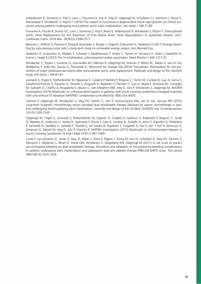
73
Vollenbroich R, Stortecky S, Praz F, Lanz J, Franzone A, Zuk K, Heg D, Valgimigli M, O’Sullivan CJ, Heinisch C, Roost E, Wenaweser P, Windecker S, Pilgrim T (2016) The impact of functional vs degenerative mitral regurgitation on clinical out-comes among patients undergoing transcatheter aortic valve implantation. Am Heart J 184:71-80
Franzone A, Piccolo R, Siontis GC, Lanz J, Stortecky S, Praz F, Roost E, Vollenbroich R, Windecker S, Pilgrim T. Transcatheter Aortic Valve Replacement for the Treatment of Pure Native Aortic Valve Regurgitation: A Systematic Review. JACC Cardiovasc Interv. 2016 Nov 28;9(22):2308-2317
Bereuter L, Williner S, Pianezzi F, Bissig B, Buecheler S, Burger J, Vogel R, Zurbuchen A, Haeberlin A (2017) Energy harves-ting by subcutaneous solar cells: a long-term study on achievable energy output. Ann Biomed Eng
Haeberlin A, Zurbuchen A, Walpen S, Schaerer J, Niederhauser T, Huber C, Tanner H, Servatius H, Seiler J, Haeberlin H, Fuhrer J, Vogel R (2015) The first batteryless, solar-powered cardiac pacemaker. Heart Rhythm 12(6):1317-23
Windecker S, Tijssen J, Giustino G, Guimarães AH, Mehran R, Valgimigli M, Vranckx P, Welsh RC, Baber U, van Es GA, Wildgoose P, Volkl AA, Zazula A, Thomitzek K, Hemmrich M, Dangas GD (2016) Trial design: Rivaroxaban for the pre-vention of major cardiovascular events after transcatheter aortic valve replacement: Rationale and design of the GALILEO study. Am Heart J 184:81-87
Leonardi S, Frigoli E, Rothenbühler M, Navarese E, Calabró P, Bellotti P, Briguori C, Ferlini M, Cortese B, Lupi A, Lerna S, Zavallonito-Parenti D, Esposito G, Tresoldi S, Zingarelli A, Rigattieri S, Palmieri C, Liso A, Abate F, Zimarino M, Comeglio M, Gabrielli G, Chieffo A, Brugaletta S, Mauro C, Van Mieghem NM, Heg D, Jüni P, Windecker S, Valgimigli M; MATRIX Investigators (2016) Bivalirudin or unfractionated heparin in patients with acute coronary syndromes managed invasively with and without ST elevation (MATRIX): randomised controlled trial. BMJ 354:i4935
Vranckx P, Valgimigli M, Windecker S, Steg PG, Hamm C, Jüni P, Garcia-Garcia HM, van Es GA, Serruys PW (2016) Long-term ticagrelor monotherapy versus standard dual antiplatelet therapy followed by aspirin monotherapy in pati-ents undergoing biolimuseluting stent implantation: rationale and design of the GLOBAL LEADERS trial. EuroIntervention 12(10):1239-1245
Valgimigli M, Frigoli E, Leonardi S, Rothenbühler M, Gagnor A, Calabrò P, Garducci S, Rubartelli P, Briguori C, Andò G, Repetto A, Limbruno U, Garbo R, Sganzerla P, Russo F, Lupi A, Cortese B, Ausiello A, Ierna S, Esposito G, Presbitero P, Santarelli A, Sardella G, Varbella F, Tresoldi S, de Cesare N, Rigattieri S, Zingarelli A, Tosi P, van ‘t Hof A, Boccuzzi G, Omerovic E, Sabaté M, Heg D, Jüni P, Vranckx P; MATRIX Investigators (2015) Bivalirudin or Unfractionated Heparin in Acute Coronary Syndromes. N Engl J Med 373(11):997-1009
Costa F, van Klaveren D, James S, Heg, D, Räber L, Feres F, Pilgrim T, Hong M, Kim H, Colombo A, Steg PG, Zanchin T, Palmerini T, Wallentin L, Bhatt D, Stone GW, Windecker S, Steyerberg EW, Valgimigli M (2017) A risk score to predict out-of-hospital bleeding on dual antiplatelet therapy. Derivation and validation of the predicting bleeding complications In patients undergoing stent implantation and subsequent dual anti platelet therapy (PRECISE-DAPT) score. The Lancet 389(10073):1025-1034

74
Radiology Research GroupHendrik von Tengg-Kobligk, Head of Research GroupEmail: [email protected]; phone: +41 31 632 73 24
Hendrik Johannes T. Verena Ingrid Andreas Jennifer Florian Kathrin Lukas Martin Michael Berndvon Tengg- Heverhagen Beutler Böhm Christe Cullmann Dammann Dopke Ebner Hungerbühler Ith JungKobligk
Miltiadis Martina Martin Verena Daniel Alexander Stephan Val Anna-Christina Zoran Carlo HarrietKrokidis C.-Londoño Maurer Obmann Ott Pöllinger Raible Runge Stamm Stankovic Tappero Thöny
Research Profile Biomedical imaging plays an essential role in leading re-search and rapid development in healthcare. Using state-of-the-art infrastructure, non-invasive imaging biomarkers are available to perform quantitative image analysis, enhancing, therefore, translational research. Artificial intelligence and machine learning permit the analysis of a massive amount of imaging data for knowledge extraction, which may be enhanced by considering the entire spectrum of available clinical, biological, and genetic information. Novel deep “al-gorithmic approaches” open the road toward big data anal-ysis. Up-to-date technology is being introduced and tested to co-develop prototypes together with our interdisciplinary partnerships. Collaborations, like the one with ARTORG, strengthen the role of biomedical imaging and intervention within a multi-disciplinary team, to nourish our healthcare system and the hospital’s future joint vision. Personalized medicine requires advanced tissue diagnosis involving diag-nostic radiology, as well as image-based Intervention. MR-guided intervention avoids ionizing radiation while providing real-time imaging during the procedure. Many patients may benefit from image-guided therapy by reducing intervention time and increasing minimal invasive procedures’ precision.
Artificial Intelligence and Machine Learning INTACTInterstitial pneumonia pattern analysis for computer-aided diagnosis (INTACT)Interstitial Lung Diseases (ILD) are a heterogeneous group of more than 200 chronic, overlapping lung disorders, char-acterized by fibrosis and/or inflammation of the lung tis-sue that ultimately lead to respiratory failure. By achieving a reliable diagnosis on chest computed tomography (CT), patients could avoid potential complications, as well as the
high costs associated with a surgical biopsy. The main goal of the INTACT research division is the development of auto-mated, diagnostic support tools enhancing clinical decision making in ILD patients. We are currently investigating au-tomated tissue characterization in CT imaging by applying a convolutional neural network paired with multi-source transfer learning. In the preclinical program, our group is working together with a translational team of researchers to explore computer vision and machine-learning tech-niques. The clinical program aims to improve the diagnostic accuracy for ILDs and further optimize the corresponding treatment to increase patient care. Furthermore, research for novel treatments is supported by standardization of the diagnostic procedure.Our group currently receives funding from the Swiss National Science Foundation (SNF; grant number 32003B_156511)
Research Partners Chris Bösch and Team, Department of Clinical Research, Magnetic Resonance Spectroscopy and Methodology, University of BernDaniel Candinas and Team, University Clinic for Visceral Surgery and Medicine, Inselspital, Bern University HospitalThierry Carrrel and Team, Department of Cardiac Surgery, Inselspital, Bern University HospitalMartin Fey and Team, Medical Oncology, Inselspital, Bern University Hospital Thomas Geiser and Team, Pulmonary Unit at the University Hospital of BernStavroula Mougiakakou and Team, ARTORG, Diabetes Technology Research Group, University of BernDominik Obrist and Team, Cardiovascular Engineering, ARTORG, University of BernAurel Perren and Team, Institute of Pathology, University of BernJürg Schmidli and Team, Department of Vascular Surgery, Inselspital, Bern University HospitalKlaus Siebenrock and Team, Clinic of Orthopedic Surgery, Inselspital, Bern University HospitalStefan Weber and Team, ARTORG, University of Bern
INTACT project outline for differential diagnosis of ILDs.
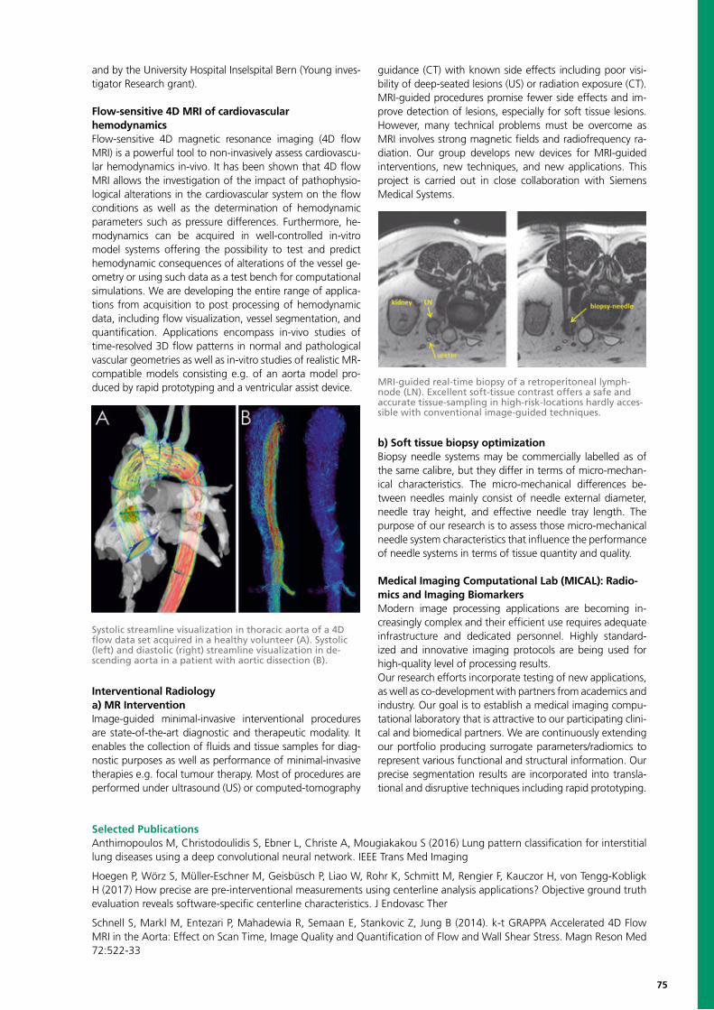
75
and by the University Hospital Inselspital Bern (Young inves-tigator Research grant).
Flow-sensitive 4D MRI of cardiovascularhemodynamics Flow-sensitive 4D magnetic resonance imaging (4D flow MRI) is a powerful tool to non-invasively assess cardiovascu-lar hemodynamics in-vivo. It has been shown that 4D flow MRI allows the investigation of the impact of pathophysio-logical alterations in the cardiovascular system on the flow conditions as well as the determination of hemodynamic parameters such as pressure differences. Furthermore, he-modynamics can be acquired in well-controlled in-vitro model systems offering the possibility to test and predict hemodynamic consequences of alterations of the vessel ge-ometry or using such data as a test bench for computational simulations. We are developing the entire range of applica-tions from acquisition to post processing of hemodynamic data, including flow visualization, vessel segmentation, and quantification. Applications encompass in-vivo studies of time-resolved 3D flow patterns in normal and pathological vascular geometries as well as in-vitro studies of realistic MR-compatible models consisting e.g. of an aorta model pro-duced by rapid prototyping and a ventricular assist device.
Interventional Radiology a) MR Intervention Image-guided minimal-invasive interventional procedures are state-of-the-art diagnostic and therapeutic modality. It enables the collection of fluids and tissue samples for diag-nostic purposes as well as performance of minimal-invasive therapies e.g. focal tumour therapy. Most of procedures are performed under ultrasound (US) or computed-tomography
guidance (CT) with known side effects including poor visi-bility of deep-seated lesions (US) or radiation exposure (CT). MRI-guided procedures promise fewer side effects and im-prove detection of lesions, especially for soft tissue lesions. However, many technical problems must be overcome as MRI involves strong magnetic fields and radiofrequency ra-diation. Our group develops new devices for MRI-guided interventions, new techniques, and new applications. This project is carried out in close collaboration with Siemens Medical Systems.
b) Soft tissue biopsy optimization Biopsy needle systems may be commercially labelled as of the same calibre, but they differ in terms of micro-mechan-ical characteristics. The micro-mechanical differences be-tween needles mainly consist of needle external diameter, needle tray height, and effective needle tray length. The purpose of our research is to assess those micro-mechanical needle system characteristics that influence the performance of needle systems in terms of tissue quantity and quality.
Medical Imaging Computational Lab (MICAL): Radio-mics and Imaging BiomarkersModern image processing applications are becoming in-creasingly complex and their efficient use requires adequate infrastructure and dedicated personnel. Highly standard-ized and innovative imaging protocols are being used for high-quality level of processing results. Our research efforts incorporate testing of new applications, as well as co-development with partners from academics and industry. Our goal is to establish a medical imaging compu-tational laboratory that is attractive to our participating clini-cal and biomedical partners. We are continuously extending our portfolio producing surrogate parameters/radiomics to represent various functional and structural information. Our precise segmentation results are incorporated into transla-tional and disruptive techniques including rapid prototyping.
Systolic streamline visualization in thoracic aorta of a 4D flow data set acquired in a healthy volunteer (A). Systolic (left) and diastolic (right) streamline visualization in de-scending aorta in a patient with aortic dissection (B).
MRI-guided real-time biopsy of a retroperitoneal lymph-node (LN). Excellent soft-tissue contrast offers a safe and accurate tissue-sampling in high-risk-locations hardly acces-sible with conventional image-guided techniques.
Selected PublicationsAnthimopoulos M, Christodoulidis S, Ebner L, Christe A, Mougiakakou S (2016) Lung pattern classification for interstitial lung diseases using a deep convolutional neural network. IEEE Trans Med Imaging
Hoegen P, Wörz S, Müller-Eschner M, Geisbüsch P, Liao W, Rohr K, Schmitt M, Rengier F, Kauczor H, von Tengg-Kobligk H (2017) How precise are pre-interventional measurements using centerline analysis applications? Objective ground truth evaluation reveals software-specific centerline characteristics. J Endovasc Ther
Schnell S, Markl M, Entezari P, Mahadewia R, Semaan E, Stankovic Z, Jung B (2014). k-t GRAPPA Accelerated 4D Flow MRI in the Aorta: Effect on Scan Time, Image Quality and Quantification of Flow and Wall Shear Stress. Magn Reson Med 72:522-33
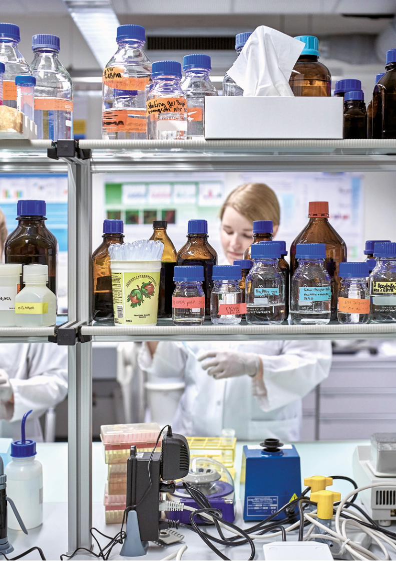

77
BIOMEDICAL ENGINEERING RESEARCHAT OTHER INSTITUTES OF THE UNIVERSITY OF BERN
Supplementary to the previously mentioned institutions concerned with biomedical engineering research, a number of additional research institutions of the University of Bern, active in vastly different domains of science, also conduct specific research activities in biomedical engineering.
While the research interests of the institutes lie in differing domains, biomedical en-gineering research, with its interdisciplinary nature, benefits greatly from the specific expertise that these technological and scientific institutes provide.
78
Clinical Trials Unit CTU BernSven Trelle & Urs Fischer, Co-DirectorsEmail: [email protected]; phone: +41 31 631 33 72
Sven UrsTrelle Fischer
Research ProfileCTU Bern (Clinical Trials Unit) was formally founded at the end of 2007 with initial funding provided by the Swiss National Science Foundation (SNF). It is the clinical trials unit of the Faculty of Medicine of the University of Bern and the Inselspital, Bern University Hospital.The aims of the CTU are to support and collaborate in clin-ical trials, epidemiological studies and meta-analyses in any clinical field to strengthen and expand the evidence base for healthcare nationally and internationally. The CTU of-fers the necessary scientific, technical, and computing ex-pertise to support patient-oriented clinical research at all stages, from conception to completion and dissemination. Relevant expertise is provided within the unit itself or by facilitating and coordinating contact with outside experts. Services are provided in a modular fashion and range from advice and general support to full development of the de-sign and conduct of clinical studies. CTU Bern works ac-cording to established scientific standards, and the services offered by the unit enable clinical researchers to comply with legal and regulatory requirements.CTU Bern is organized into five workflows: Statistics and Methodology, Data Management, Project Management, Quality Assurance and Monitoring, and Clinical Investigation. These workflows provide a range of services from methodological consulting to GCP-compliant da-ta-management solutions, central/on-site monitoring, and statistical analysis. Although the primary focus of CTU Bern lies on late- phase clinical studies, it has some expertise on early-phase clinical trials, including exploratory clinical trials.
Statistics and MethodologyInitiating and conducting a clinical study according to scien-tific and international standards is becoming more complex and time consuming. Several issues have to be considered before initiating a study, including the relevance and poten-tial impact, methodology, feasibility, and costs. CTU Bern provides an open-access consultancy service covering all relevant aspects of study design, management, and analy-sis. This includes power analysis and sample size estimation depending on objective, study design, and other relevant aspects. Our expertise is primarily in randomized-controlled trials. However, we also support first-in-human proof-of-concept trials, dose-finding trials, and single-arm efficacy trials as well as observational studies, including diagnostic test accuracy studies.
Systematic Reviews and Meta-AnalysisDecisions in healthcare should be informed by as high a proportion as possible of all the reliable evidence relevant to particular questions. The results of new studies should therefore be presented and interpreted within the context of previous research summarized in systematic reviews and meta-analyses.
Statistical AnalysisData of a clinical study can be analyzed in many ways, some of which may be inappropriate in a particular situation. It is essential to pre-specify which statistical procedure will be used for each analysis, including a distinction between primary and secondary analyses and an explicit description of methods and software used for the analysis. An analysis plan should ensure the coherent and appropriate use of study design and analysis to address the objectives of the study. The analysis should provide coherent and relevant in-formation while realistically acknowledging its uncertainty.
Write-Up of Scientific ReportsParticularly for inexperienced clinical researchers, it may be difficult to determine a successful strategy for write-up and publication of clinical study reports. Support may be required to understand the implicit rules and dynamics of scientific publishing.
Data ManagementCTU Bern provides and maintains a secure and up-to-date IT infrastructure. The servers hosting the study databases are stored in a dedicated server facility. We have a 24x7 maintenance agreement with its hardware supplier to reduce downtime in case of hardware breakdowns. CTU Bern Data Management ensures that all software re-quired to run the servers is regularly updated. Backups of all study- and meta-data are made regularly according to a detailed back-up plan. The plan defines internal back-ups several times per day and daily back-ups on external disks. Our security measures have been checked by the “Datenschutzaufsichtsstelle des Kantons Bern.”The work-flow Data Management (DM) at CTU Bern offers two dif-ferent models of services and support during the setup of a clinical study database or register:
Entire setup of a study database done by CTU BernBased on paper case report forms (CRFs) or on study
Clinical PartnersAll clinical departments of Inselspital Bern as well as other Swiss hospitals.
79
specifications (list of CRFs, variables etc.), CTU Bern will perform the complete setup of the database (eCRFs, ed-it-checks, visit structure) using one of its two web-based Electronic Data Capturing (EDC) solutions.
Collaborative SetupIf an investigator wants to setup the study database mostly by her-/himself, we can introduce her/him to our EDC sys-tem REDCap, where studies can be set-up independently (eCRFs, edit-checks, visit structure). CTU Bern will give as much support as requested. At the end, CTU Bern will re-view the implementation and put it in production so that data-entry can start.
Electronic Data Capturing (EDC) SolutionsAll EDC systems used at CTU Bern are web-based i.e. autho-rized users can access the study database via any comput-er with internet connection (and web-browser installed). Secure Sockets Layer (SSL) encryption is used to ensure a secure internet connection. Depending on the complexity of the study design and the needs of the investigator, CTU Bern offers different GCPcompliant EDC solutions.
Quality Assurance and MonitoringCTU Bern offers quality control services (monitoring) to sponsors, including sponsor-investigators working at Inselspital Bern or other interested parties for single-center or multi-center clinical studies. Our approach considers the risk of the individual study, and the monitoring strategy is risk adapted. We offer two interrelated services to support you in ensuring a high-quality study conduct:
On-site monitoringThe on-site monitor provides support for clinical studies in terms of quality control and quality assurance. He or she visits study sites at regular intervals during the study to ensure that the study is conducted in accordance with the protocol, applicable Standard Operating Procedures (SOPs), International Conference on Harmonization Good Clinical Practice (ICH-GCP) guidelines, and regulatory re-quirements. For multi-center studies, the monitor is also an important point of contact between the sponsor and the coordination center.
Central and statistical data monitoringCentral data monitoring is concerned with centralized checks of the accumulating study data. These checks are usually done on a regular basis during study conduct and include range, plausibility, and consistency checks. Because some of these measures are based on statistical tech-niques, there is a close collaboration between the Quality Assurance and Monitoring workflow and the Statistics and Methodology workflow. There is also a close link to on-site monitoring because findings identified during central data monitoring might trigger on-site visits and vice versa.
Clinical InvestigationThe Clinical Investigation workflow is a service unit within the CTU Bern that offers professional services to investi-gators working at Inselspital Bern or to other interested parties engaged in single-center or multi-center clinical studies. These services include logistic support, planning, coordination, and execution of clinical studies from phases I to IV as well as observational studies.
Selected PublicationsVilliger PM, Adler S, Kuchen S, Wermelinger F, Dan D, Fiege V, Bütikofer L, Seitz M, Reichenbach S (2016) Tocilizumab for induction and maintenance of remission in giant cell arteritis: a phase 2, randomised, double-blind, placebo-controlled trial. Lancet 387(10031):1921-7
Da Costa BR, Reichenbach S, Keller N, Nartey L, Wandel S, Jüni P, Trelle S (2016) Effectiveness of non-steroidal anti-inflammat-ory drugs for the treatment of pain in knee and hip osteoarthritis: a network meta-analysis. Lancet 387(10033):2093-105
Henao-Restrepo AM, Camacho A, Longini IM, Watson, Conall H, Edmunds WJ, Egger M, Carroll MW, Dean NE, Diatta I, Doumbia M, Draguez B, Duraffour S, Enwere G, Grais R, Gunther S, Gsell PS, Hossmann S, Watle SV, Kondé MK, Kéïta S (2016) Efficacy and effectiveness of an rVSV-vectored vaccine in preventing Ebola virus disease: final results from the Guinea ring vaccination, open-label, cluster-randomised trial (Ebola Ça Suffit!). Lancet 389(10068):505-518 (In Press)
80
Department of Biomedical Photonics of the Institute of Applied PhysicsMartin Frenz, Head of Research GroupEmail: [email protected]; phone: +41 31 631 89 43
Martin Günhan Manes Michael Arushi Maju René Tigran Chen Tobias FlorentinFrenz Akarcay Hornung Jaeger Jain Kuriakose Nyffenegger Petrosyan Qiu Schweizer Spadin
Research ProfileThe research of the department is focused upon the in-vestigation of various possibilities to employ pulsed infra-red lasers in new medical disciplines and the optimization of the clinical outcome in fields the laser is already being used. Emphasis is placed upon four interdependent fields: (1) the study of the physical processes underlying the light propagation in tissue and the interaction of laser radiation with soft and hard tissues, (2) laser-induced reversible and irreversible changes in optical properties of tissue response and the consequence of these changes on thermal tissue damage and ablation, (3) the development of laser and fiber delivery systems optimized for specific medical ap-plications; and (4) the development of novel biomedical optical and ultrasound imaging techniques. In particular, we concentrate on the development of optoacoustic imag-ing techniques, an emerging molecular imaging modality able to image tissue structures and function, which has the potential of patient-tailored tumor nano-theranostics. Questions to be addressed are of applied and fundamental character: (i) What limits the image depth and resolution and how can both be increased? (ii) How does laser light interact with nano-sized contrast agents and how to de-termine their biodistribution in the body? and (iii) How to improve multimodal optoacoustic devices for clinical use simultaneously measuring echo and Doppler ultrasound, tissue elastography, speed of sound, and optoacoustics? Further main topics include two-photon imaging and
interactions of ultrashort near-infrared laser pulses with biological soft matter as well as laser-induced tissue solder-ing using nanoparticle scaffolds.
Optoacoustic imaging Optoacoustic (OA) imaging allows the display of optical contrast inside tissue based on detection of thermoelas-tically generated ultrasound after tissue irradiation using short nanosecond pulsed lasers. In combination with pulse-echo ultrasound, OA is promising in the improvement of diagnostic accuracy via the display of small blood vessels and the local blood oxygenation level within the anatom-ical context. An important requirement for such a combi-nation is a clinically useful OA imaging depth of several centimeters. This has been difficult to achieve in the past because of clutter signals originating from the site of tissue irradiation.Sound speed as a diagnostic marker has been of interest for many years due to the fact that sound speed can reveal structural changes of tissue that come with diseases such as cancer, cirrhosis, fibrosis, and fatty liver disease. In addition, knowledge of the spatial distribution of sound speed benefits ultrasound imaging in general: Image reconstruction con-ventionally assumes a homogeneous sound speed, which leads to blurring and inaccurate display of tissue anatomy in the presence of acoustic aberrations. We are developing a technique where spatially resolved quantitative detection of sound speed is achieved using conventional pulse-echo
Research PartnersMihai Constantinescu, Clinic of Plastic Surgery, Bern University HospitalMeike Mevissen, Division of Veterinary Pharmacology & Toxicology, University of BernReto Luginbühl, RMS Foundation, BettlachAnnalisa Berzigotti, Swiss Liver Center, Hepatology, University Clinic for Visceral Surgery and Medicine, Inselspital, University of Bern Philippe Schucht, University Clinic for Neurosurgery, University of BernJeff Bamber, Institute of Cancer Research and Royal Marsden NHS Foundation Trust, LondonWalter Inäbnit, Haag-Streit, LiebefeldBernhard Nussbaumer, Pantec Engineering AG, Ruggell, Liechtenstein SwissTransMed – Project partners “Non-invasive multimodal high resolution optical acoustic imager for early hypoxia de-tection in neonatal brain ONIRIUS”, www.swisstransmed.chEU-Horizon 2020 Project partners “Risk assessment of plaque rupture and future cardiovascular events (CVENT) by multi-spectral photoacoustic imaging”EU-Horizon 2020 Project partners “Photoacoustic/Ultrasound Mammoscopy for evaluating screening-detectedabnormalities in the breast (PAMMOTH)”
Patrick LeonieStähli Ulrich
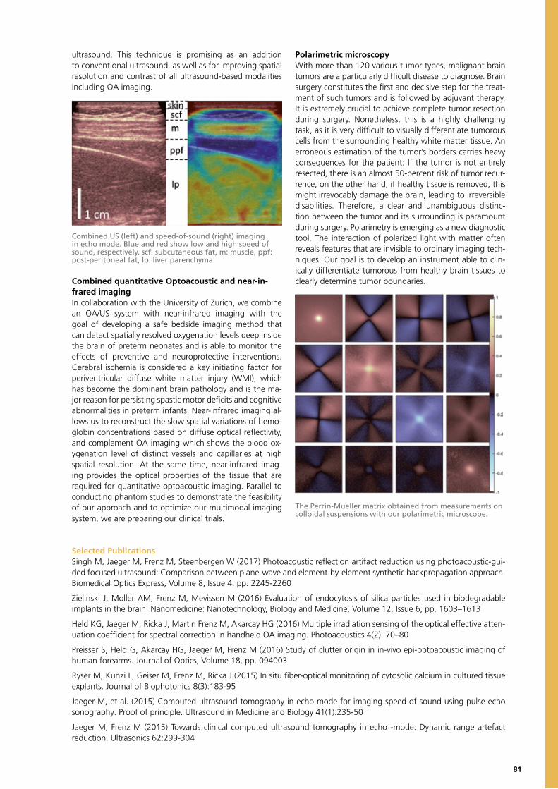
81
ultrasound. This technique is promising as an addition to conventional ultrasound, as well as for improving spatial resolution and contrast of all ultrasound-based modalities including OA imaging.
Combined quantitative Optoacoustic and near-in-frared imaging In collaboration with the University of Zurich, we combine an OA/US system with near-infrared imaging with the goal of developing a safe bedside imaging method that can detect spatially resolved oxygenation levels deep inside the brain of preterm neonates and is able to monitor the effects of preventive and neuroprotective interventions. Cerebral ischemia is considered a key initiating factor for periventricular diffuse white matter injury (WMI), which has become the dominant brain pathology and is the ma-jor reason for persisting spastic motor deficits and cognitive abnormalities in preterm infants. Near-infrared imaging al-lows us to reconstruct the slow spatial variations of hemo-globin concentrations based on diffuse optical reflectivity, and complement OA imaging which shows the blood ox-ygenation level of distinct vessels and capillaries at high spatial resolution. At the same time, near-infrared imag-ing provides the optical properties of the tissue that are required for quantitative optoacoustic imaging. Parallel to conducting phantom studies to demonstrate the feasibility of our approach and to optimize our multimodal imaging system, we are preparing our clinical trials.
Polarimetric microscopyWith more than 120 various tumor types, malignant brain tumors are a particularly difficult disease to diagnose. Brain surgery constitutes the first and decisive step for the treat-ment of such tumors and is followed by adjuvant therapy. It is extremely crucial to achieve complete tumor resection during surgery. Nonetheless, this is a highly challenging task, as it is very difficult to visually differentiate tumorous cells from the surrounding healthy white matter tissue. An erroneous estimation of the tumor’s borders carries heavy consequences for the patient: If the tumor is not entirely resected, there is an almost 50-percent risk of tumor recur-rence; on the other hand, if healthy tissue is removed, this might irrevocably damage the brain, leading to irreversible disabilities. Therefore, a clear and unambiguous distinc-tion between the tumor and its surrounding is paramount during surgery. Polarimetry is emerging as a new diagnostic tool. The interaction of polarized light with matter often reveals features that are invisible to ordinary imaging tech-niques. Our goal is to develop an instrument able to clin-ically differentiate tumorous from healthy brain tissues to clearly determine tumor boundaries.
Selected PublicationsSingh M, Jaeger M, Frenz M, Steenbergen W (2017) Photoacoustic reflection artifact reduction using photoacoustic-gui-ded focused ultrasound: Comparison between plane-wave and element-by-element synthetic backpropagation approach. Biomedical Optics Express, Volume 8, Issue 4, pp. 2245-2260
Zielinski J, Moller AM, Frenz M, Mevissen M (2016) Evaluation of endocytosis of silica particles used in biodegradable implants in the brain. Nanomedicine: Nanotechnology, Biology and Medicine, Volume 12, Issue 6, pp. 1603–1613
Held KG, Jaeger M, Ricka J, Martin Frenz M, Akarcay HG (2016) Multiple irradiation sensing of the optical effective atten-uation coefficient for spectral correction in handheld OA imaging. Photoacoustics 4(2): 70–80
Preisser S, Held G, Akarcay HG, Jaeger M, Frenz M (2016) Study of clutter origin in in-vivo epi-optoacoustic imaging of human forearms. Journal of Optics, Volume 18, pp. 094003
Ryser M, Kunzi L, Geiser M, Frenz M, Ricka J (2015) In situ fiber-optical monitoring of cytosolic calcium in cultured tissue explants. Journal of Biophotonics 8(3):183-95
Jaeger M, et al. (2015) Computed ultrasound tomography in echo-mode for imaging speed of sound using pulse-echo sonography: Proof of principle. Ultrasound in Medicine and Biology 41(1):235-50
Jaeger M, Frenz M (2015) Towards clinical computed ultrasound tomography in echo -mode: Dynamic range artefact reduction. Ultrasonics 62:299-304
Combined US (left) and speed-of-sound (right) imaging in echo mode. Blue and red show low and high speed of sound, respectively. scf: subcutaneous fat, m: muscle, ppf: post-peritoneal fat, lp: liver parenchyma.
The Perrin-Mueller matrix obtained from measurements on colloidal suspensions with our polarimetric microscope.
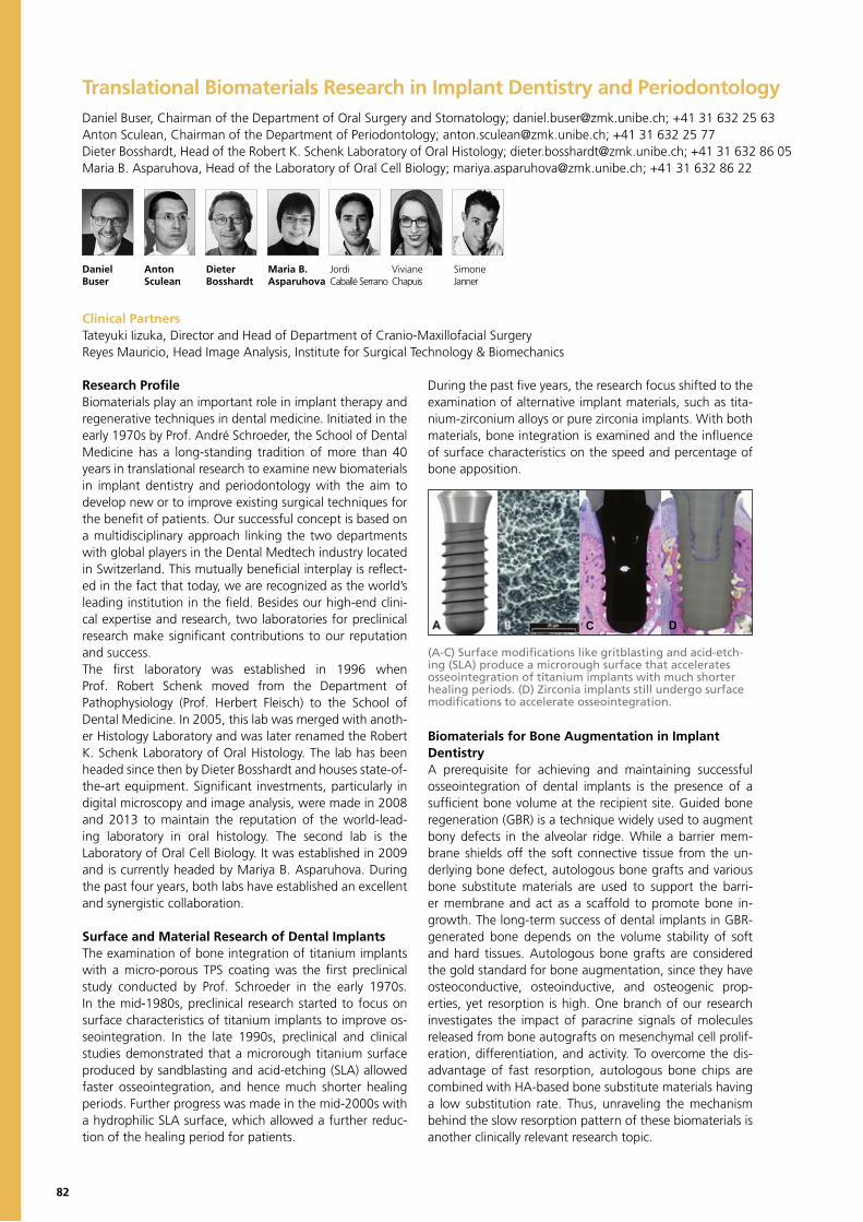
82
Translational Biomaterials Research in Implant Dentistry and Periodontology Daniel Buser, Chairman of the Department of Oral Surgery and Stomatology; [email protected]; +41 31 632 25 63Anton Sculean, Chairman of the Department of Periodontology; [email protected]; +41 31 632 25 77Dieter Bosshardt, Head of the Robert K. Schenk Laboratory of Oral Histology; [email protected]; +41 31 632 86 05Maria B. Asparuhova, Head of the Laboratory of Oral Cell Biology; [email protected]; +41 31 632 86 22
Daniel Anton Dieter Maria B. Jordi Viviane SimoneBuser Sculean Bosshardt Asparuhova Caballé Serrano Chapuis Janner
Research ProfileBiomaterials play an important role in implant therapy and regenerative techniques in dental medicine. Initiated in the early 1970s by Prof. André Schroeder, the School of Dental Medicine has a long-standing tradition of more than 40 years in translational research to examine new biomaterials in implant dentistry and periodontology with the aim to develop new or to improve existing surgical techniques for the benefit of patients. Our successful concept is based on a multidisciplinary approach linking the two departments with global players in the Dental Medtech industry located in Switzerland. This mutually beneficial interplay is reflect-ed in the fact that today, we are recognized as the world’s leading institution in the field. Besides our high-end clini-cal expertise and research, two laboratories for preclinical research make significant contributions to our reputation and success. The first laboratory was established in 1996 when Prof. Robert Schenk moved from the Department of Pathophysiology (Prof. Herbert Fleisch) to the School of Dental Medicine. In 2005, this lab was merged with anoth-er Histology Laboratory and was later renamed the Robert K. Schenk Laboratory of Oral Histology. The lab has been headed since then by Dieter Bosshardt and houses state-of-the-art equipment. Significant investments, particularly in digital microscopy and image analysis, were made in 2008 and 2013 to maintain the reputation of the world-lead-ing laboratory in oral histology. The second lab is the Laboratory of Oral Cell Biology. It was established in 2009 and is currently headed by Mariya B. Asparuhova. During the past four years, both labs have established an excellent and synergistic collaboration.
Surface and Material Research of Dental ImplantsThe examination of bone integration of titanium implants with a micro-porous TPS coating was the first preclinical study conducted by Prof. Schroeder in the early 1970s. In the mid-1980s, preclinical research started to focus on surface characteristics of titanium implants to improve os-seointegration. In the late 1990s, preclinical and clinical studies demonstrated that a microrough titanium surface produced by sandblasting and acid-etching (SLA) allowed faster osseointegration, and hence much shorter healing periods. Further progress was made in the mid-2000s with a hydrophilic SLA surface, which allowed a further reduc-tion of the healing period for patients.
During the past five years, the research focus shifted to the examination of alternative implant materials, such as tita-nium-zirconium alloys or pure zirconia implants. With both materials, bone integration is examined and the influence of surface characteristics on the speed and percentage of bone apposition.
Biomaterials for Bone Augmentation in Implant DentistryA prerequisite for achieving and maintaining successful osseointegration of dental implants is the presence of a sufficient bone volume at the recipient site. Guided bone regeneration (GBR) is a technique widely used to augment bony defects in the alveolar ridge. While a barrier mem-brane shields off the soft connective tissue from the un-derlying bone defect, autologous bone grafts and various bone substitute materials are used to support the barri-er membrane and act as a scaffold to promote bone in-growth. The long-term success of dental implants in GBR-generated bone depends on the volume stability of soft and hard tissues. Autologous bone grafts are considered the gold standard for bone augmentation, since they have osteoconductive, osteoinductive, and osteogenic prop-erties, yet resorption is high. One branch of our research investigates the impact of paracrine signals of molecules released from bone autografts on mesenchymal cell prolif-eration, differentiation, and activity. To overcome the dis-advantage of fast resorption, autologous bone chips are combined with HA-based bone substitute materials having a low substitution rate. Thus, unraveling the mechanism behind the slow resorption pattern of these biomaterials is another clinically relevant research topic.
Clinical PartnersTateyuki Iizuka, Director and Head of Department of Cranio-Maxillofacial SurgeryReyes Mauricio, Head Image Analysis, Institute for Surgical Technology & Biomechanics
C D
(A-C) Surface modifications like gritblasting and acid-etch-ing (SLA) produce a microrough surface that accelerates osseointegration of titanium implants with much shorter healing periods. (D) Zirconia implants still undergo surface modifications to accelerate osseointegration.
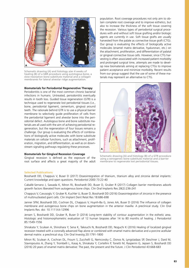
83
Biomaterials for Periodontal Regenerative TherapyPeriodontitis is one of the most common chronic bacterial infections in humans. Untreated, periodontitis eventually results in tooth loss. Guided tissue regeneration (GTR) is a technique used to regenerate lost periodontal tissues (i.e., bone, periodontal ligament, cementum, gingiva) around teeth. The rationale behind GTR is to use a physical barrier membrane to selectively guide proliferation of cells from the periodontal ligament and alveolar bone into the peri-odontal defect. Autologous bone and bone substitute ma-terials are all used with the aim of achieving periodontal re-generation, but the regeneration of four tissues remains a challenge. Our group is evaluating the effects of combina-tions of biologically active molecules with bone substitute materials on cellular functions, such as attachment, prolif-eration, migration, and differentiation, as well as on down-stream signaling pathways regulating these processes.
Biomaterials for Gingival Recession CoverageGingival recession is defined as the exposure of the root surface and affects a great majority of the adult
population. Root coverage procedures not only aim to ob-tain complete root coverage and to improve esthetics, but also to increase the thickness of the soft tissue covering the recession. Various types of periodontal surgical proce-dures with and without soft tissue grafting and/or biologic agents are currently in use. Soft tissue grafts are usually harvested from the palate as connective tissue graft (CTG). Our group is evaluating the effects of biologically active molecules (enamel matrix derivative, hyaluronan, etc.) on the attachment, proliferation, and differentiation of palatal or gingival connective tissue cells. However, since CTG har-vesting is often associated with increased patient morbidity and prolonged surgical time, attempts are made to devel-op new biomaterials aiming at replacing CTGs to improve patient acceptance and minimize morbidity. Recent results from our group suggest that the use of some of these ma-terials may represent an alternative to CTG.
Schematic drawing (A) and histology aJer 4 weeks of healing (B) of a GBR procedure using autologous bone, a slow-resorption bone substitute material and a collagen membranes for lateral alveolar ridge augmentation.
A B
Schematic drawing (A) and histology (B) of a GTR procedure using a xenogeneic bone substitute material and a barrier membrane to regenerate lost periodontal tissues.
Selected PublicationsBosshardt DD, Chappuis V, Buser D (2017) Osseointegration of titanium, titanium alloy and zirconia dental implants: Current knowledge and open questions. Periodontol 2000 73:22-40
Caballé-Serrano J, Sawada K, Miron RJ, Bosshardt DD, Buser D, Gruber R (2017) Collagen barrier membranes adsorb growth factors liberated from autogenous bone chips. Clin Oral Implants Res 28(2):236-241
Chappuis V, Cavusoglu Y, Gruber R, Kuchler U, Buser D, Bosshardt DD (2016) Osseointegration of zirconia in the presence of multinucleated giant cells. Clin Implant Dent Relat Res 18:686-698
Janner SFM, Bosshardt DD, Cochran DL, Chappuis V, Huynh-Ba G, Jones AA, Buser D (2016) The influence of collagen membrane and autogenous bone chips on bone augmentation in the anterior maxilla: A preclinical study. Clin Oral Implants Res. doi: 10.1111/clr.12996
Jensen S, Bosshardt DD, Gruber R, Buser D (2014) Long-term stability of contour augmentation in the esthetic area. Histologic and histomorphometric evaluation of 12 human biopsies after 14 to 80 months of healing. J Periodontol 85:1549-1556
Shirakata Y, Sculean A, Shinohara Y, Sena K, Takeuchi N, Bosshardt DD, Noguchi K (2016) Healing of localized gingival recession treated with a coronally advanced flap alone or combined with enamel matrix derivative and a porcine acellular dermal matrix: a preclinical stuy. Clin Oral Investig 20:1791-1800
Miron RJ, Sculean A, Cochran DL, Froum S, Zucchelli G, Nemcovsky C, Donos N, Lyngstadaas SP, Deschner J, Dard M, Stavropoulos A, Zhang Y, Trombelli L, Kasaj A, Shirakata Y, Cortellini P, Tonetti M, Rasperini G, Jepsen S, Bosshardt DD (2016) 20 years of enamel matrix derivative: The past, the present and the future. J Clin Periodontol 43:668-683
A B
85
BERN UNIVERSITY OF APPLIED SCIENCES
Bern University of Applied Sciences offers bachelor’s and master’s degrees in Medical Technology / Medical Informatics, and also conducts applied research and develop-ment. The BFH disposes a broad knowledge that is efficiently networked and applied interdisciplinarily. The aim is to share our know-how with industrial partners as well as promoting innovation.
The BFH is the first university in all of continental Europe to be awarded the ISO-13485 certificate, allowing monitoring of medical products, regardless of their class from the idea up to technology transfer into industry. The acquired certification per-mits BFH to be a valuable research and development partner for SMEs in medical technology, thus enabling industry ease of access to the European market (CE label-ling). BFH researchers are involved in a network of national and international research institutions, industry, hospitals, and practice sharings, and further act as active links between basic research and practice.
Many new products are developed by means of cooperation with universities. Owing to the close link between research and teaching, the latest discoveries and methods are smartly incorporated in the classroom and thus provide university graduates with a practice-oriented education at the bachelor’s or master‘s level. These graduates become outstandingly-qualified specialists and executives in industry.
The new degree of B.Sc. in Industrial Engineering and Management Science, for ex-ample, guides undergraduates to master complex challenges, as well as successfully leading their enterprises through times of digital transformation. Further, digitalization will massively change medical technology. Value chains are be-coming more and more application-oriented, so customers and patients will sooner become more intensively involved in future developments. The chances are promis-ing. Faster, more flexible and more intelligent medical innovations help companies strengthen their positions in the market.
Innovation Parc Biel/Bienne and the SITEMThe BFH as an educational and research partner occupies a strong position. Backed by professional know-how and impulses from applied sciences, it is establishing the foundation for innovation, which is also the basis for cooperation with Switzerland Innovation Park Biel/Bienne and SITEM.
Lukas RohrHead of DepartmentBern University of Applied SciencesEngineering and Information Technology
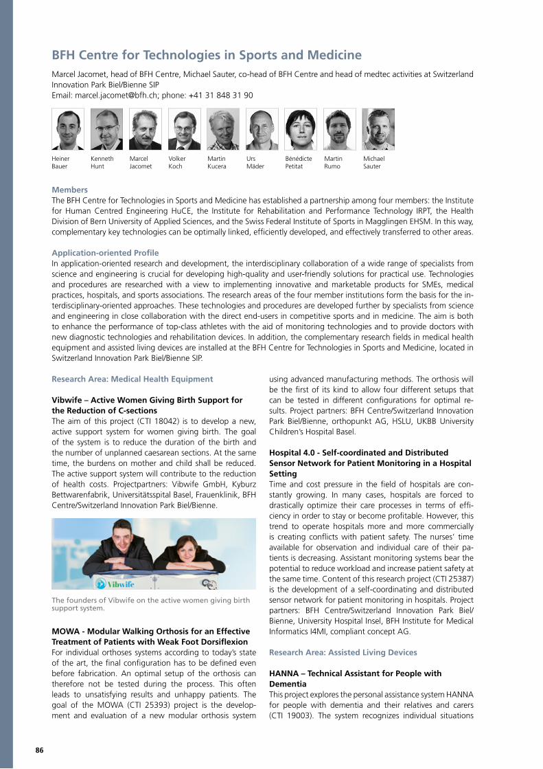
86
BFH Centre for Technologies in Sports and MedicineMarcel Jacomet, head of BFH Centre, Michael Sauter, co-head of BFH Centre and head of medtec activities at Switzerland Innovation Park Biel/Bienne SIPEmail: [email protected]; phone: +41 31 848 31 90
Heiner Kenneth Marcel Volker Martin Urs Bénédicte Martin MichaelBauer Hunt Jacomet Koch Kucera Mäder Petitat Rumo Sauter
MembersThe BFH Centre for Technologies in Sports and Medicine has established a partnership among four members: the Institute for Human Centred Engineering HuCE, the Institute for Rehabilitation and Performance Technology IRPT, the Health Division of Bern University of Applied Sciences, and the Swiss Federal Institute of Sports in Magglingen EHSM. In this way, complementary key technologies can be optimally linked, efficiently developed, and effectively transferred to other areas.
Application-oriented Profile In application-oriented research and development, the interdisciplinary collaboration of a wide range of specialists from science and engineering is crucial for developing high-quality and user-friendly solutions for practical use. Technologies and procedures are researched with a view to implementing innovative and marketable products for SMEs, medical practices, hospitals, and sports associations. The research areas of the four member institutions form the basis for the in-terdisciplinary-oriented approaches. These technologies and procedures are developed further by specialists from science and engineering in close collaboration with the direct end-users in competitive sports and in medicine. The aim is both to enhance the performance of top-class athletes with the aid of monitoring technologies and to provide doctors with new diagnostic technologies and rehabilitation devices. In addition, the complementary research fields in medical health equipment and assisted living devices are installed at the BFH Centre for Technologies in Sports and Medicine, located in Switzerland Innovation Park Biel/Bienne SIP.
Research Area: Medical Health Equipment
Vibwife – Active Women Giving Birth Support for the Reduction of C-sectionsThe aim of this project (CTI 18042) is to develop a new, active support system for women giving birth. The goal of the system is to reduce the duration of the birth and the number of unplanned caesarean sections. At the same time, the burdens on mother and child shall be reduced. The active support system will contribute to the reduction of health costs. Projectpartners: Vibwife GmbH, Kyburz Bettwarenfabrik, Universitätsspital Basel, Frauenklinik, BFH Centre/Switzerland Innovation Park Biel/Bienne.
MOWA - Modular Walking Orthosis for an Effective Treatment of Patients with Weak Foot Dorsiflexion For individual orthoses systems according to today’s state of the art, the final configuration has to be defined even before fabrication. An optimal setup of the orthosis can therefore not be tested during the process. This often leads to unsatisfying results and unhappy patients. The goal of the MOWA (CTI 25393) project is the develop-ment and evaluation of a new modular orthosis system
using advanced manufacturing methods. The orthosis will be the first of its kind to allow four different setups that can be tested in different configurations for optimal re-sults. Project partners: BFH Centre/Switzerland Innovation Park Biel/Bienne, orthopunkt AG, HSLU, UKBB University Children’s Hospital Basel.
Hospital 4.0 - Self-coordinated and Distributed Sensor Network for Patient Monitoring in a Hospital SettingTime and cost pressure in the field of hospitals are con-stantly growing. In many cases, hospitals are forced to drastically optimize their care processes in terms of effi-ciency in order to stay or become profitable. However, this trend to operate hospitals more and more commercially is creating conflicts with patient safety. The nurses’ time available for observation and individual care of their pa-tients is decreasing. Assistant monitoring systems bear the potential to reduce workload and increase patient safety at the same time. Content of this research project (CTI 25387) is the development of a self-coordinating and distributed sensor network for patient monitoring in hospitals. Project partners: BFH Centre/Switzerland Innovation Park Biel/Bienne, University Hospital Insel, BFH Institute for Medical Informatics I4MI, compliant concept AG.
Research Area: Assisted Living Devices
HANNA – Technical Assistant for People withDementiaThis project explores the personal assistance system HANNA for people with dementia and their relatives and carers (CTI 19003). The system recognizes individual situations
The founders of Vibwife on the active women giving birth support system.

87
independently, offers simple communication functions for the relatives / caregivers, interacts actively with the demen-tia patient, adapts its user interface and the patient behav-ior accordingly. HANNA is developed in cooperation with its users and integrates these into qualitative tests in re-al-time environments. Projectpartners: iHomeLab – HSLU, BFH Centre/Switzerland Innovation Park Biel/Bienne, BFH Health Division (Nursing).
Sysmo - Feasibility Study of Sensor-basedTherapeutic Back Trainer Back pain creates great suffering for those concerned, and it also causes huge costs for society. In Switzerland, ap-proximately 15 million days per year are lost due to persons unable to work because of back pain. The goal of the re-ported project (CTI 25175) is to check the feasibility of an intelligent Sensor-based Therapeutic Back Trainer, which is fitted on an ergonomic articulated stool and that supervises and monitors the correct use of the stool as a training de-vice, motivating and guiding the trainer by visual feedback via a smart phone or tablet, which analyses the progress of the trainer, thus preventing the user from overexertion. Projectpartners: BFH Centre/Switzerland Innovation Park Biel/Bienne, BFH Health Division (Physiotherapy).
Sam & Me – Contact-aware Interaction Engine for the Digital Butler SAMSAM & Me explores the self-learning, interactive core of the Digital Butler SAM (CTI 25351). This is intended to create a low-threshold access to meaningful services for people who are excluded from the digital world or are tired of their growing complexity. SAM reverses the interaction paradigm - it serves the user, not vice versa. It recogniz-es the current usage context and makes targeted sugges-tions. The demonstrator is created together with its users. Projectpartners: iHomeLab – HSLU, BFH Centre/Switzerland Innovation Park Biel/Bienne.
Bodygee – 3D Body Scan Analysis in Fitness and Nutritional ConsultingHuCE-cpvrLab: In a CTI project (CTI 19154.1) we developed a 3D body scan data processing application for the start-up company BODYGEE AG in Berne. The goal is to process the raw scan data from low-cost consumer scan devices and deliver the relevant information and visualization to the BODYGEE web platform. To visualize the progress of a training or treatment, the platform must be able to com-pare multiple scans taken at different times. Because per-sons have different poses in multiple scans, all body scans must be registered onto a template body before they can be aligned and morphed in between. After registration, all scans have the same resolution and structure, which means that the same body features are at the same data index. In addition to these advantages, we can also fix ar-tefacts in the scan, such as wholes or other scan errors.
Another advantage of registered body scans is that they can be transformed into a statistical body shape space with the principal component analysis. For body scans in the shape space we can generate statistically and anatomically correct predictions – e.g. for weight gain or weight loss. Projectpartners: BFH Centre/Switzerland Innovation Park Biel/Bienne, BodyGee AG
Aide-Moi – Fall Detector for Active and Independent Seniors There is strong statistical and scientific evidence that se-niors prefer to live at home as long as possible. According to statistics, there are over 250’000 persons older than 65 in Switzerland who fall at least once a year. These falls have often severe consequences and require in many cases ur-gent hospitalisation. It is therefore important to provide se-niors with electronic devices which that them to keep their high level of independence and let them cope confidently and as long as possible in a self-determined manner with their daily life. For these purposes, an automatic fall detec-tor and alarming device has been developed (supported by Inventus and Age Foundation). The fall detector uses an accelerometer and a sophisticated algorithm to detect falls. The device is completely waterproof and is attached to the thorax, close to the center of gravity. The device has a wire-less connection to a smartphone, or to a proprietary base station. With the user friendly app, specifically designed for seniors, Aide-Moi allows seniors to decide when an alarm will be sent (manually) and who will be alarmed (automat-ically) in case of a fall. The Aide-Moi fall detector has only to be put on the wireless charging station every 14 days to recharge the built-in battery. Trials with seniors have suc-cessfully been carried out, and it is planned that a start-up will bring the device to the market. Further information can be found on www.aidemoi.ch.
The sensor-based therapeutic back trainer used in various positions during standard office work.
Body scan comparison with cross-section analysis.
The Aide-Moi Fall detector is attached to the thorax with a skin-friendly band-aid and is connected to a smartphone for upto 14 days without requiring charging. The fall sensor is therefore very comfortable, not visible to anybody and hardly noticed after a very short while.
88
Institute for Rehabilitation and Performance Technology IRPTMember of the BFH Centre for Technologies in Sports and Medicine.Kenneth J. Hunt, Head of InstituteEmail: [email protected]; phone: +41 34 426 43 69
Kenneth Anil Farouk Stephan Beat Marco Jan Corina Sarah Hunt Aksöz Chrif Debrunner Kessler Laubacher Riedo Schuster Degallier Rochat
Research ProfileThe Institute for Rehabilitation and Performance Technology IRPT uses methods and technologies from sports and exer-cise physiology to improve the rehabilitation process after accidents or illnesses. Core competencies are cardiopulmo-nary and neurological rehabilitation after stroke or spinal injury, feedback systems, as well as automation and control of training equipment. The IRPT develops its procedures and systems in collaboration with Swiss companies and re-habilitation clinics, and can thus ensure that they can be quickly and directly used by patients. A growing number of CTI- and SNF-funded projects supports the transfer of knowledge into industry and application to concrete prod-ucts. The IRPT team consists of research associates, Ph.D. students, postdocs, and a group of bachelor’s and master’s students, with backgrounds in electrical, mechanical and bio-mechanical engineering, human movement sciences, physiotherapy, and rehabilitation medicine. The IRPT is lo-cated in Burgdorf (Canton of Bern) on the campus of the Engineering and Information Technology department of Bern University of Applied Sciences.
Rehabilitation EngineeringThe interdisciplinary research of the Rehabilitation Engineering group focuses on neural control of move-ment in clinical populations with neurological deficits re-sulting from spinal cord injury, stroke, and other causes. Combining rehabilitation technology and cognitive perfor-mance feedback, the group’s goal is to reinforce the pa-tient’s volitional drive and to exploit the central nervous
system’s lifelong capacity for plasticity, regeneration, and repair. This approach promotes cardiopulmonary and mus-culoskeletal health and supports an environment in which positive neurological adaptations can occur. The work har-nesses multidisciplinary expertise in engineering, neurosci-ences, sports and exercise science, and medicine. This al-lows professionals to address prevention and management of the progressive secondary complications of spinal cord injury, stroke, and a wide range of further neurological conditions. The promotion of neurological recovery for im-proved motor control, sensation, and autonomic function is also facilitated.
The Rehabilitation Engineering group develops new tech-nical devices and extends the functionality of existing prod-ucts. New developments include novel rehabilitation tricy-cles for adults and children with neurological impairment. This new generation of tricycles combines novel drivetrain technology using electric drives with functional electrical stimulation (FES) of paralysed muscle groups. The func-tionality of existing robotics-assisted rehabilitation devic-es, including treadmills, end-effectors, and tilt tables, has been extended to cover application for cardiopulmonary rehabilitation. This involves biofeedback of patient effort, volitional control of mechanical work rate, together with automatic feedback control of physiological outcome vari-ables including heart rate, oxygen uptake, and metabolic work rate. A key feature of the group’s work is the employ-ment of methods and protocols from sports and exercise physiology and the adaptation of these to the rehabilita-tion setting. These approaches are applied in the clinic for rehabilitation of people with various neurological problems including stroke and spinal cord injury.
The following selection of research and clinical projects gives an overview of the spectrum of research activities of the Rehabilitation Engineering group:• Cardiopulmonary rehabilitation of patients following stroke using robotics-assisted treadmill exercise (RATE)• Cardiopulmonary rehabilitation of patients following incomplete spinal cord injury or stroke using a robotics-assisted tilt table• Rehabilitation tricycle incorporating FES and motorized assistance
Clinical PartnersDr Corina Schuster-Amft, Reha Rheinfelden, RheinfeldenDr Angela Frotzler, Swiss Paraplegic Centre (SPZ), Nottwil
G-EO end-effector gait rehabilitation robot augmented at IRPT with biofeedback for cardiopulmonary testing and training.
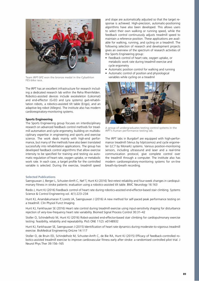
89
The IRPT has an excellent infrastructure for research includ-ing a dedicated research lab within the Reha Rheinfelden. Robotics-assisted devices include exoskeleton (Lokomat) and end-effector (G-EO and Lyra systems) gait-rehabili-tation robots, a robotics-assisted tilt table (Erigo), and an adaptive-leg robot (Allegro). The institute also has modern cardiorespiratory-monitoring systems.
Sports EngineeringThe Sports Engineering group focuses on interdisciplinary research on advanced feedback control methods for tread-mill automation and cycle ergometry, building on multidis-ciplinary expertise in engineering and sports and exercise science. The work deals mainly with high-end perfor-mance, but many of the methods have also been translated successfully into rehabilitation applications. The group has developed feedback control algorithms that allow exercise intensity to be specified for training and testing via auto-matic regulation of heart rate, oxygen uptake, or metabolic work rate. In each case, a target profile for the controlled variable is selected. During the exercise, treadmill speed
and slope are automatically adjusted so that the target re-sponse is achieved. High-precision, automatic-positioning algorithms have also been developed. This allows users to select their own walking or running speed, while the feedback control continuously adjusts treadmill speed to maintain a reference position. These applications are avail-able for walking, running, and cycling on a treadmill. The following selection of research and development projects gives an overview of the spectrum of research activities of the Sports Engineering group:• Feedback control of heart rate, oxygen uptake, or metabolic work rate during treadmill exercise and cycle ergometry• Automatic position control for walking and running• Automatic control of position and physiological variables while cycling on a treadmil
The IRPT labs in Burgdorf are equipped with high-perfor-mance treadmill (Venus by h/p/cosmos) and cycle ergome-ter (LC7 by Monark) systems. Various position-monitoring sensors, including ultrasound and laser and a real-time communication protocol, give complete control over the treadmill through a computer. The institute also has modern cardiorespiratory-monitoring systems for on-line breath-by-breath recording.
Selected PublicationsSaengsuwan J, Berger L, Schuster-Amft C, Nef T, Hunt KJ (2016) Test-retest reliability and four-week changes in cardiopul-monary fitness in stroke patients: evaluation using a robotics-assisted tilt table. BMC Neurology 16:163
Riedo J, Hunt KJ (2016) Feedback control of heart rate during robotics-assisted end-effector-based stair climbing. Systems Science & Control Engineering vol. 4(1):223–234
Hunt KJ, Anandakumaran P, Loretz JA, Saengsuwan J (2016) A new method for self-paced peak performance testing on a treadmill. Clin Physiol Funct Imaging
Hunt KJ, Fankhauser SE (2016) Heart rate control during treadmill exercise using input-sensitivity shaping for disturbance rejection of very-low-frequency heart rate variability. Biomed Signal Process Control 30:31–42
Stoller O, Schindelholz M, Hunt KJ (2016) Robot-assisted end-effector-based stair climbing for cardiopulmonary exercise testing: feasibility, reliability and repeatability. PloS ONE 11(2): e0148932
Hunt KJ, Fankhauser SE, Saengsuwan J (2015) Identification of heart rate dynamics during moderate-to-vigorous treadmill exercise. BioMedical Engineering OnLine 14:117
Stoller O, de Bruin ED, Schindelholz M, Schuster-Amft C, de Bie RA, Hunt KJ (2015) Efficacy of feedback-controlled ro-botics-assisted treadmill exercise to improve cardiovascular fitness early after stroke: a randomised controlled pilot trial. J Neurol Phys Ther 39:156–165
Team IRPT-SPZ won the bronze medal in the CybathlonFES-bike race.
A group of undergraduates testing control systems in the IRPT’s human performance testing lab.

90
Applied Research and Development PhysiotherapyMember of the BFH Centre for Technologies in Sports and Medicine.Lorenz Radlinger, Head of Research Group; Heiner Baur, Head of Movement LaboratoryEmail: [email protected]; [email protected]; phone: +41 31 848 35 87; +41 31 848 45 15
Lorenz Heiner Angela Patric Irene Monika Helena Eugen Helene Slavko StefanRadlinger Bauer Blasimann Eichelberger König Leitner Luginbühl Mischler Moser Rogan Schmid
Matthias Jan Amir DörteStettler Taeymans Tal Watzek
Research PartnersJean-Pierre Baeyens, Head Laboratory Biomechanics, Vrjie Universiteit BrusselsAchim Elfering, Department of Work and Organisational Psychology, University of BernPhilipp Henle, Knee Surgery and Sports Orthopaedics, Sonnenhofspital BernFabian Krause, Clinic of Orthopaedic Surgery and Traumatology, University Hospital BernAnnette Kuhn, Department of Gynaecology and Obstetrics, University Hospital BernAndreas Reinhard, ORTHO-TEAM AG, BernMartin Verra, Department of Physiotherapy, University Hospital Bern
Research ProfileThe Applied Research and Development Physiotherapy Unit of the Health Department at the Bern University of Applied Sciences focuses on the analyses of functional human movements in relevant daily life activities. Our laboratory is equipped with kinematic, kinetic, and neuromuscular analysis methods to provide an excellent environment for movement science research. In addition to general facil-ities necessary for teaching physiotherapy for bachelor’s, master’s, or Ph.D. students, the profile of the lab has a number of foci. One research group investigates pelvic floor muscle activity and displacement in order to devel-op diagnostic tools and therapeutic concepts to enhance incontinence therapy. The investigation of postural control and movement patterns in elderly people, as well as other musculoskeletal complaints, through the use of stochastic resonance whole-body vibration training interventions is a second focus area. A third focus is on neuromuscular and biomechanical adaptations of the lower extremity to train-ing and therapy interventions.
Stress Urinary Incontinence (SUI)Pelvic floor muscles (PFM) have to counter impact loads during running or jumping to secure continence. We in-vestigated whether these impact activities could be an adequate stimulus for involuntary and reflexive PFM contractions.Results indicated that PFM activities increased significant-ly from rest to running speeds: 7 km/h (46 – 92 %MVC; %MVC: normalised to peak activity during maximum vol-untary contraction), 11 km/h (56 –108 %MVC), and 15 km/h (81 –152 %MVC). Drop jumps generated an even higher PFM activity.Impact exercises generate higher involuntary PFM activ-ities than during MVC. Although these results seem to be self-evident, fast, submaximal, and maximal voluntary
contractions are the current state of the art in PFM training. Training stimuli as impact activities may serve as a benefi-cial complement to a PFM training rationale, when used in sense of power training methodology.
Innovative Exercise Programs for No-Go ElderlyDue to different physical and mental functions in the el-derly, a classification into independent person (Go-Go), needy person with slight handicap (Slow-Go), and person in need of care with severe functional limitation (No-Go) should be performed. Slow-Go and Go-Go can participate in traditional trainings. Feasible training for No-Go should be carried out within a short time frame.In our study an intervention group performed five sets of stochastic resonance whole-body vibration (frequency 3 to 6 Hz) lasting five minutes with a one-minute rest be-tween sets during four weeks. From week five to eight video gaming was added. A sham group used the same
PFM activity during drop jumps. Activities before and after initial foot ground contact (T0) are significantly higher than during rest or MVC.
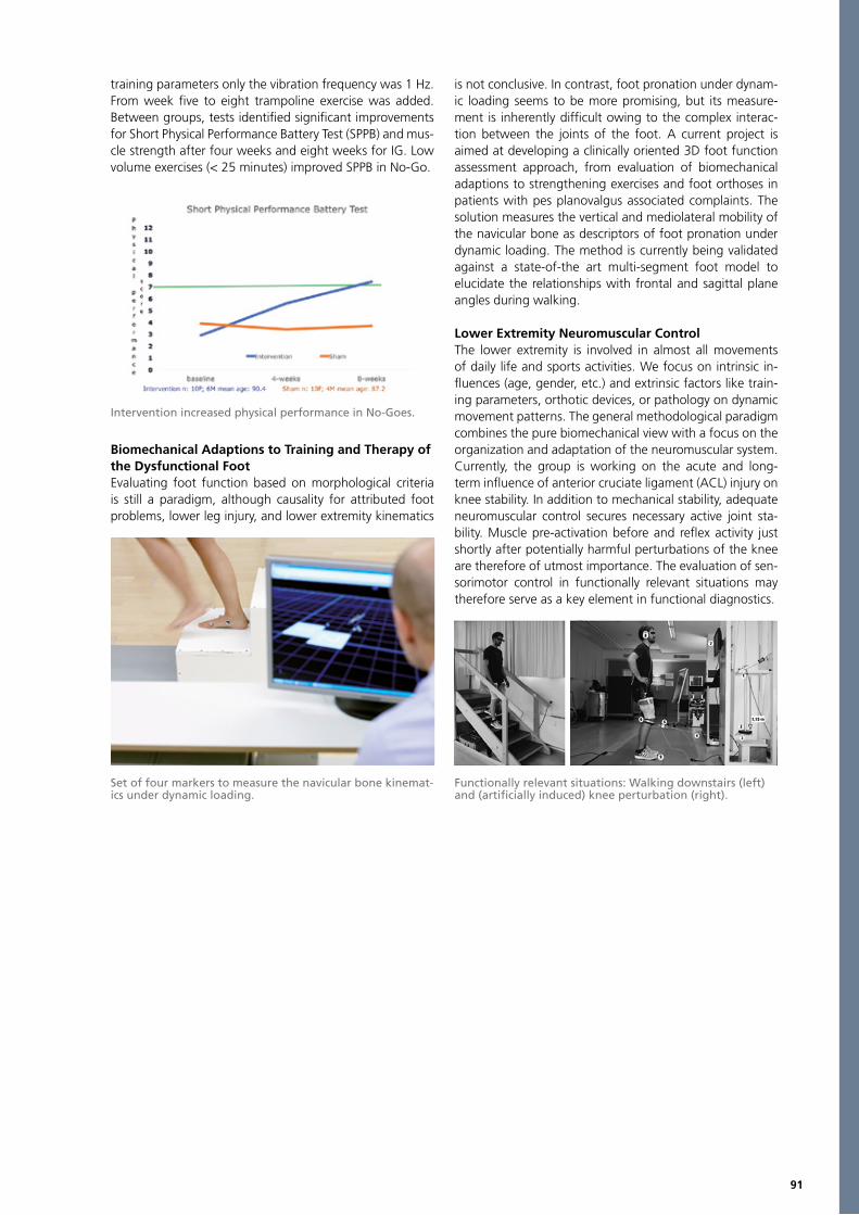
91
training parameters only the vibration frequency was 1 Hz. From week five to eight trampoline exercise was added. Between groups, tests identified significant improvements for Short Physical Performance Battery Test (SPPB) and mus-cle strength after four weeks and eight weeks for IG. Low volume exercises (< 25 minutes) improved SPPB in No-Go.
Biomechanical Adaptions to Training and Therapy of the Dysfunctional FootEvaluating foot function based on morphological criteria is still a paradigm, although causality for attributed foot problems, lower leg injury, and lower extremity kinematics
is not conclusive. In contrast, foot pronation under dynam-ic loading seems to be more promising, but its measure-ment is inherently difficult owing to the complex interac-tion between the joints of the foot. A current project is aimed at developing a clinically oriented 3D foot function assessment approach, from evaluation of biomechanical adaptions to strengthening exercises and foot orthoses in patients with pes planovalgus associated complaints. The solution measures the vertical and mediolateral mobility of the navicular bone as descriptors of foot pronation under dynamic loading. The method is currently being validated against a state-of-the art multi-segment foot model to elucidate the relationships with frontal and sagittal plane angles during walking.
Lower Extremity Neuromuscular ControlThe lower extremity is involved in almost all movements of daily life and sports activities. We focus on intrinsic in-fluences (age, gender, etc.) and extrinsic factors like train-ing parameters, orthotic devices, or pathology on dynamic movement patterns. The general methodological paradigm combines the pure biomechanical view with a focus on the organization and adaptation of the neuromuscular system.Currently, the group is working on the acute and long-term influence of anterior cruciate ligament (ACL) injury on knee stability. In addition to mechanical stability, adequate neuromuscular control secures necessary active joint sta-bility. Muscle pre-activation before and reflex activity just shortly after potentially harmful perturbations of the knee are therefore of utmost importance. The evaluation of sen-sorimotor control in functionally relevant situations may therefore serve as a key element in functional diagnostics.
Intervention increased physical performance in No-Goes.
Set of four markers to measure the navicular bone kinemat-ics under dynamic loading.
Functionally relevant situations: Walking downstairs (left) and (artificially induced) knee perturbation (right).
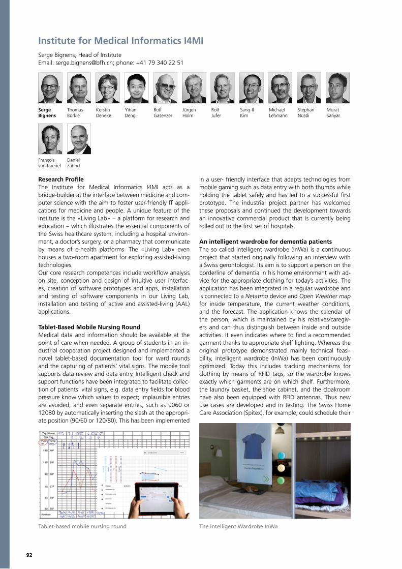
92
Institute for Medical Informatics I4MISerge Bignens, Head of InstituteEmail: [email protected]; phone: +41 79 340 22 51
Serge Thomas Kerstin Yihan Rolf Jürgen Rolf Sang-Il Michael Stephan MuratBignens Bürkle Deneke Deng Gasenzer Holm Jufer Kim Lehmann Nüssli Sariyar
Research ProfileThe Institute for Medical Informatics I4MI acts as a bridge-builder at the interface between medicine and com-puter science with the aim to foster user-friendly IT appli-cations for medicine and people. A unique feature of the institute is the «Living Lab» – a platform for research and education – which illustrates the essential components of the Swiss healthcare system, including a hospital environ-ment, a doctor’s surgery, or a pharmacy that communicate by means of e-health platforms. The «Living Lab» even houses a two-room apartment for exploring assisted-living technologies. Our core research competences include workflow analysis on site, conception and design of intuitive user interfac-es, creation of software prototypes and apps, installation and testing of software components in our Living Lab, installation and testing of active and assisted-living (AAL) applications.
Tablet-Based Mobile Nursing RoundMedical data and information should be available at the point of care when needed. A group of students in an in-dustrial cooperation project designed and implemented a novel tablet-based documentation tool for ward rounds and the capturing of patients’ vital signs. The mobile tool supports data review and data entry. Intelligent check and support functions have been integrated to facilitate collec-tion of patients’ vital signs, e.g. data entry fields for blood pressure know which values to expect; implausible entries are avoided, and even separate entries, such as 9060 or 12080 by automatically inserting the slash at the appropri-ate position (90/60 or 120/80). This has been implemented
in a user- friendly interface that adapts technologies from mobile gaming such as data entry with both thumbs while holding the tablet safely and has led to a successful first prototype. The industrial project partner has welcomed these proposals and continued the development towards an innovative commercial product that is currently being rolled out to the first set of hospitals.
An intelligent wardrobe for dementia patientsThe so called intelligent wardrobe (InWa) is a continuous project that started originally following an interview with a Swiss gerontologist. Its aim is to support a person on the borderline of dementia in his home environment with ad-vice for the appropriate clothing for today’s activities. The application has been integrated in a regular wardrobe and is connected to a Netatmo device and Open Weather map for inside temperature, the current weather conditions, and the forecast. The application knows the calendar of the person, which is maintained by his relatives/caregiv-ers and can thus distinguish between inside and outside activities. It even indicates where to find a recommended garment thanks to appropriate shelf lighting. Whereas the original prototype demonstrated mainly technical feasi-bility, intelligent wardrobe (InWa) has been continuously optimized. Today this includes tracking mechanisms for clothing by means of RFID tags, so the wardrobe knows exactly which garments are on which shelf. Furthermore, the laundry basket, the shoe cabinet, and the cloakroom have also been equipped with RFID antennas. Thus new use cases are developed and in testing. The Swiss Home Care Association (Spitex), for example, could schedule their
François Danielvon Kaenel Zahnd
Tablet-based mobile nursing round The intelligent Wardrobe InWa
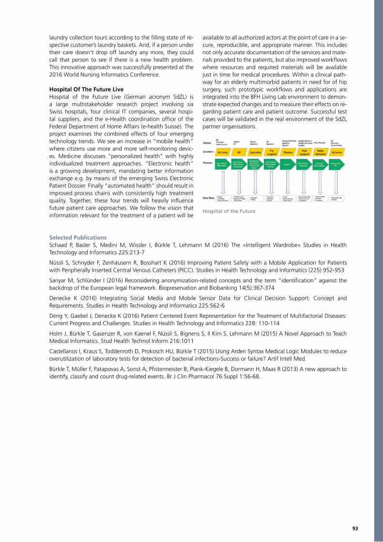
93
laundry collection tours according to the filling state of re-spective customer’s laundry baskets. And, if a person under their care doesn’t drop off laundry any more, they could call that person to see if there is a new health problem. This innovative approach was successfully presented at the 2016 World Nursing Informatics Conference.
Hospital Of The Future Live Hospital of the Future Live (German acronym SdZL) is a large multistakeholder research project involving six Swiss hospitals, four clinical IT companies, several hospi-tal suppliers, and the e-Health coordination office of the Federal Department of Home Affairs (e-health Suisse). The project examines the combined effects of four emerging technology trends. We see an increase in “mobile health” where citizens use more and more self-monitoring devic-es. Medicine discusses “personalized health” with highly individualized treatment approaches. “Electronic health” is a growing development, mandating better information exchange e.g. by means of the emerging Swiss Electronic Patient Dossier. Finally “automated health” should result in improved process chains with consistently high treatment quality. Together, these four trends will heavily influence future patient care approaches. We follow the vision that information relevant for the treatment of a patient will be
available to all authorized actors at the point of care in a se-cure, reproducible, and appropriate manner. This includes not only accurate documentation of the services and mate-rials provided to the patients, but also improved workflows where resources and required materials will be available just in time for medical procedures. Within a clinical path-way for an elderly multimorbid patients in need for of hip surgery, such prototypic workflows and applications are integrated into the BFH Living Lab environment to demon-strate expected changes and to measure their effects on re-garding patient care and patient outcome. Successful test cases will be validated in the real environment of the SdZL partner organisations.
Selected PublicationsSchaad P, Basler S, Medini M, Wissler I, Bürkle T, Lehmann M (2016) The «Intelligent Wardrobe» Studies in Health Technology and Informatics 225:213-7
Nüssli S, Schnyder F, Zenhäusern R, Bosshart K (2016) Improving Patient Safety with a Mobile Application for Patients with Peripherally Inserted Central Venous Catheters (PICC). Studies in Health Technology and Informatics (225) 952-953
Sariyar M, Schlünder I (2016) Reconsidering anonymization-related concepts and the term “identification” against the backdrop of the European legal framework. Biopreservation and Biobanking 14(5):367-374
Denecke K (2016) Integrating Social Media and Mobile Sensor Data for Clinical Decision Support: Concept and Requirements. Studies in Health Technology and Informatics 225:562-6
Deng Y, Gaebel J, Denecke K (2016) Patient Centered Event Representation for the Treatment of Multifactorial Diseases: Current Progress and Challenges. Studies in Health Technology and Informatics 228: 110-114
Holm J, Bürkle T, Gasenzer R, von Kaenel F, Nüssli S, Bignens S, Il Kim S, Lehmann M (2015) A Novel Approach to Teach Medical Informatics. Stud Health Technol Inform 216:1011
Castellanos I, Kraus S, Toddenroth D, Prokosch HU, Bürkle T (2015) Using Arden Syntax Medical Logic Modules to reduce overutilization of laboratory tests for detection of bacterial infections-Success or failure? Artif Intell Med.
Bürkle T, Müller F, Patapovas A, Sonst A, Pfistermeister B, Plank-Kiegele B, Dormann H, Maas R (2013) A new approach to identify, classify and count drug-related events. Br J Clin Pharmacol 76 Suppl 1:56-68.
Hospital of the Future

94
Institute for Human Centered Engineering HuCEMember of the BFH Centre for Technologies in Sports and Medicine.Marcel Jacomet, Head of Institute Email: [email protected]; phone: +41 32 321 62 41
Industrial PartnersAlfred Bruno, Advanced Osteotomy Tools AG, Basel; Michael Gasser, Axiamo GmbH, BielJuan Elices, Stéve Gigandet, Bien-Air Surgery, Le Noirmont; Thomas Bauer, CoreMedic GmbH, BielJoerg Breitenstein, Haag-Streit AG, Bern; Thio Krüger, Oliver Weihberger, inomed Medizintechnik GmbH, GermanyAndré Reber, Reber Engineering und Informatik GmbH, MünsingenChristian Rathjen, Harmut Kanngiesser, Ziemer Opthalmic Systems AG, Port
Research or Clinical PartnersSven Schulzke, Kerstin Jost, Department of Neonatology, University of Basel Children's Hospital (UKBB), Basel; John Farserotu, CSEM, Neuchâtel; Christian Enz, Integrated Circuits Laboratory, EPFL, Neuchâtel; Hans-Andrea Loeliger, Signal and Information Processing Laboratory, ETHZ, Zürich; Hubert Kaeslin, Microelectronics Design Center, ETHZ, Zürich; Christina Spengler, Institute of Movement Sciences and Sport, ETH, Zurich; Hildegard Tanner, Andreas Häberlin, Department of Cardiology, Inselspital, Bern University Hospital; Thilo Weizel, University Clinic for Nuclear Medicine, Inselspital, Bern University Hospital; Rolf Vogel, Head of Cardiology, Solothurn Hospital, Solothurn; Adrian Lussi, School of Dental Medicine, University of Bern
Research ProfileThe Institute for Human Centered Engineering HuCE com-bines its know-how acquired from research projects in various fields with engineering technologies in an inter-disciplinary way to develop new products for industry and clinics. The focus is on strong engineering technology core competences. Our practical problem-solving approach to-gether with our clinical partnerships provides a basis for
innovative products. The engineering core competences in our six research laboratories are Medical Instrumentation, Electronic Implants, Optical Instruments for Diagnosis, Imaging in Medical Technology, Optical Coherence Tomography, Haptic Feedback Systems, Sensors and Sensor Networks, Signal Processing and Control, Low-power Microelectronics, and High-speed Hardware Algorithms in Combination with Biomedical Engineering Applications.
Mar
cel
Jaco
met
Patr
ick
Arn
old
Dan
iel
Deb
runn
er
Bert
rand
Dut
oit
Jürg
enEc
kerle
Jose
fG
oett
e
Hor
stH
eck
Mar
cus
Hud
ritsc
h
Jörn
Just
iz
Theo
Klu
ter
Volk
erK
och
Mar
tinK
ucer
a
Urs
Kue
nzle
r
Lore
nzM
ülle
r
Chr
isto
phM
eier
Yves
Mus
sard
Thom
asN
iede
rhau
ser
And
rea
Rido
lfi
And
reas
Stah
el
Tobi
asA
esch
liman
n
Thib
aud
Alla
man
Luka
sBö
sch
Chr
isto
fBa
eris
wyl
Mar
cel
Baer
tsch
i
Dom
inik
Brüg
ger
Tim
Bues
ch
Chr
istia
nBu
rri
Luca
Flei
schm
ann
Mic
hael
Göt
tlich
er
Lore
nzG
rimm
Fabr
ice
Hän
ni
Benj
amin
Hab
egge
r
Han
spet
erH
ess
Hua
iqi
Hua
ng
Barb
ara
Jesa
cher
Ger
hard
Fré
déric
Kue
rt
Tao
Li Jan
Loch
mat
ter
Dav
idLu
ggen
Adr
ian
Maa
g
Mat
thia
s M
oose
r
And
reas
Mos
er
Youy
ouM
oser
Mic
hael
Nyd
egge
r
Boris
Pova
jay
Dom
inik
Robe
llaz
Tizi
ano
Ronc
hett
i
Kla
usSc
hürc
h
Tobi
asSc
haue
ble
Sim
onSc
heur
er
Dom
inik
Taes
chle
r
Emily
Thom
pson
Phuo
ng-A
nh
Laur
ence
Tra
n
Dam
ian
Web
er
Reto
Wild
habe
r
Thom
asW
yss
Cam
ille
Zann
i
Rom
anA
mre
in
Adr
ian
Salla
z
Dan
iel
Kau
fman
n
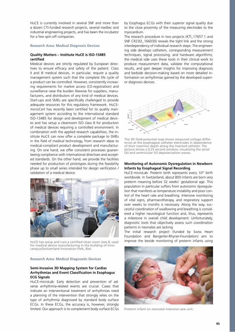
95
HuCE is currently involved in several SNF and more than a dozen CTI-funded research projects, several medtec and industrial engineering projects, and has been the incubator for a few spin-off companies.
Research Area: Medical Diagnosis Devices
Quality Matters – Institute HuCE is ISO-13485certifiedMedical devices are strictly regulated by European direc-tives to ensure efficacy and safety of the patient. Class II and III medical devices, in particular, require a quality management system such that the complete life cycle of a product can be controlled. However, consistently increas-ing requirements for market access (CE-registration) and surveillance raise the burden likewise for suppliers, manu-facturers, and distributors of any kind of medical devices. Start-ups and SMEs are specifically challenged to provide adequate resources for this regulatory framework. HuCE-microCert has recently been certified for its quality man-agement system according to the international standard ISO-13485 for design and development of medical devic-es and has setup a cleanroom ISO class 8 for production of medical devices requiring a controlled environment. In combination with the applied research capabilities, the in-stitute HuCE can now offer a complete package to SMEs in the field of medical technology, from research ideas to medical-compliant product development and manufactur-ing. On one hand, we offer consistent processes guaran-teeing compliance with international directives and accept-ed standards. On the other hand, we provide the facilities needed for production of prototypes during the feasibility phase up to small series intended for design verification / validation of a medical device.
Research Area: Medical Diagnostic Devices
Semi-Invasive 3D Mapping System for CardiacArrhythmias and Event Classification in Esophagus ECG SignalsHuCE-microLab: Early detection and prevention of ad-verse arrhythmia-related events are crucial. Cases that indicate an interventional treatment of arrhythmias need a planning of the intervention that strongly relies on the type of arrhythmia diagnosed by standard body surface ECGs. In these ECGs, the accuracy is, however, strongly limited. Our approach is to complement body surface ECGs
by Esophagus ECGs with their superior signal quality due to the close proximity of the measuring electrodes to the myocardium. The research procedure in two projects (KTI_17657.1 and SNF CR23I2_166030) reveals the tight link and the strong interdependency of individual research steps: The engineer-ing side develops catheters, corresponding measurement techniques, signal processing, and hardware algorithms; the medical side uses these tools in their clinical work to produce measurement data, validate the computational results, and gain deeper insights for improving diagnosis and bedside decision-making based on more detailed in-formation on arrhythmias gained by the developed superi-or diagnosis devices.
Monitoring of Autonomic Dysregulation in Newborn Infants by Esophageal Signal RecordingHuCE-microLab: Preterm birth represents every 10th birth worldwide. In Switzerland, about 800 infants are born very preterm meaning before 32 weeks’ gestational age. This population in particular suffers from autonomic dysregula-tion that manifests as temperature instability and poor con-trol of the heart rate and breathing. Intensive monitoring of vital signs, pharmacotherapy, and respiratory support over weeks to months is necessary. Along the way, suc-cessful coordination of swallowing and breathing is consid-ered a higher neurological function and, thus, represents a milestone in overall child development. Unfortunately, diagnostic tools that objectively assess such coordination patterns in neonates are lacking.The initial research project (funded by Swiss Heart Foundation and Bangerter-Rhyner-Foundation) aim to improve the beside monitoring of preterm infants using HuCE has setup and runs a certified clean room class 8, used
for medical device manufacturing in the building of Inno-campus/Switzerland Innovation Park, Biel.
The 3D field-potential map shows measured voltage differ-ences at the esophageal catheter electrodes in dependence of their insertion depth along the inserted catheter. The picture shows a 0.25 second window, visualizing the atrial (A) and ventriclular (V) depolarization waves.
Preterm infant on neonatal intensive care unit.
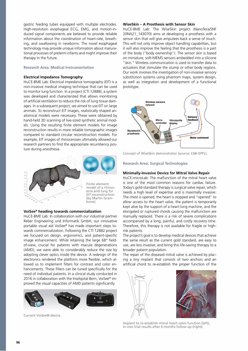
96
gastric feeding tubes equipped with multiple electrodes. High-resolution esophageal ECG, EMG, and motion-in-duced signal components are believed to provide reliable information about the coordination of heart-rate, breath-ing, and swallowing in newborns. The novel esophageal technology may provide unique information about matura-tional processes of preterm infants and might improve their therapy in the future.
Research Area: Medical Instrumentation
Electrical Impedance Tomography HuCE-BME Lab: Electrical impedance tomography (EIT) is a non-invasive medical imaging technique that can be used to monitor lung function. In a project (CTI 12888), a system was developed and characterized that allows monitoring of artificial ventilation to reduce the risk of lung tissue dam-ages. In a subsequent project, we aimed to use EIT on large animals. To reconstruct EIT images, realistically shaped an-atomical models were necessary. These were obtained by hand-held 3D scanning of live-sized synthetic animal mod-els. Using the resulting finite element models for image reconstruction results in more reliable tomographic images compared to standard circular reconstruction models. For example, EIT images of rhinoceroses ultimately allowed our research partners to find the appropriate recumbency pos-ture during anesthesia.
VoiSee® heading towards commercializationHuCE-BME Lab: In collaboration with our industrial partner Reber Engineering und Informatik GmbH, our innovative portable visual aid VoiSee® has made important steps to-wards commercialization. Following the CTI 12882 project we focused on design, ergonomics, and patient-specific image enhancement. While retaining the large 68° field-of-view, crucial for patients with macula degenerations (AMD), we were able to considerably reduce the size by adopting clever optics inside the device. A redesign of the electronics rendered the platform more flexible, which al-lowed us to implement filters for contrast and color en-hancements. These filters can be tuned specifically for the need of individual patients. In a clinical study conducted in 2016 in collaboration with the Inselspital Bern, VoiSee® im-proved the visual capacities of AMD patients significantly.
WiseSkin – A Prosthesis with Sensor Skin HuCE-BME Lab: The WiseSkin project (NanoTera/SNF 20NA21_143070) aims at developing a prosthesis with a sensor skin that will give amputees back a sense of touch. This will not only improve object handling capabilities, but it will also improve the feeling that the prosthesis is a part of the body (“body ownership”). The sensor skin is based on miniature, soft-MEMS sensors embedded into a silicone “skin.” Wireless communication is used to transfer data to actuators that stimulate the stump or other body regions. Our work involves the investigation of non-invasive sensory substitution systems using phantom maps, system design, as well as integration and development of a functional prototype.
Research Area: Surgical Technologies
Minimally-Invasive Device for Mitral Valve RepairHuCE-microLab: The malfunction of the mitral heart valve is one of the most common reasons for cardiac failure. Today’s gold-standard therapy is surgical valve repair, which needs a high level of expertise and is maximally invasive: The chest is opened, the heart is stopped and “opened” to allow access to the heart valve, the patient is temporarily kept alive by the support of a heart-lung machine, and the elongated or ruptured chords causing the malfunction are manually replaced. There is a risk of severe complications accompanied by a long, painful, and costly recovery time. Therefore, this therapy is not available for fragile or high-risk patients. The project’s goal is to develop medical devices that achieve the same result as the current gold standard, are easy to use, are less invasive, and bring this life-saving therapy to a broader patient population. The repair of the diseased mitral valve is achieved by plac-ing a tiny implant that consists of two anchors and an artificial chord to re-establish the proper function of the
Finite element model of a rhinoc-eros and lung for EIT reconstruction (by Martin Gram-bone).
Current VoiSee® device.
Concept of WiseSkin demonstrator (source: LSBI-EPFL).
Implant to re-establish mitral heart valve function (left), in-vivo trial results after 6 months follow-up (right).
97
heart valve (CTI 16922). The implant is delivered using min-imally-invasive delivery systems that allow the repair of the mitral valve through a small incision at the groin and at the beating heart.
DrillMon – A high-speed surgical drilling system with integrated nerve monitoringHuCE-BME Lab: Misplaced surgical drilling, e.g., for co-chlear implants, risks irreparable damage to nerves. This might lead to a permanent or temporal paralysis in the fa-cial area and thus also to strong physical impairment and social problems. The goal of this project (CTI 19076) is to research, develop, optimize, and characterize a system that combines a surgical drilling device with an electrical nerve-stimulation probe. This allows surgeons to continu-ously monitor nerves while drilling with up to 80’000 rpm – without changing tools. The result is improved safety for patients, more confidence for surgeons, and savings of time and costs.
Research Area: Ophthamology
Cornea Power MeasurementsHuCE-microLab: An essential step in the planning of a large number of ophthalmologic interventions is the determina-tion of the corneal refractive power. Current biometric de-vices determine the power solely on the basis of measure-ments of the cornea front surface. The goal of the present research project (CTI 16077) is to measure both, the cor-neal power of the front as well as that of the back surfaces by 3D optical imaging. Uncontrollable eye movements, like
saccades, happen several times per second, and ask thus, for a very high-speed image-capturing system to prevent image blurring by eye movement. In our approach, we re-search for near redundancy-free 3D imaging and, at the same time, increased the speed of the image capturing op-tics and the post-processing by implementing the needed signal-processing tasks directly in hardware algorithms.
Surgical Laser with OCT for OphthalmologyHuCE-optoLab: In the field of ophthalmology, OCT (Optical Coherence Tomography) is an essential imaging technique due to its ability to acquire fast cross-sectional images of the whole anterior eye segment with a high level of detail. The novel application of Femtosecond-Lasers in refractive and cataract surgery requires a detailed visualization of the whole operating field. The goal of this project (CTI 17300) is to extend an existing Femtosecond-Laser system by such a visualization based on a dedicated OCT System.Measurements on human patients with a high- resolution system for anterior eye segment measurements integrat-ed into a Femto LDV-System have been conducted with a prototype system. Today, the OCT-System is industrialized and sold in the device of the industrial partner. OCT mea-surements are performed in clinics worldwide on a routine basis.
Real Time Dosimetry ControlHuCE-optoLab: Selective retina therapy and optical co-herence tomography have been combined to monitor laser-tissue interaction in real-time. An ex-vivo study of porcine eyes unveils mechanisms that enable automated accurate dose-control during laser-therapy.Precise control of pulse energy is crucial to initiate suffi-cient RPE damage but to prevent collateral cellular dam-age caused by excess energy deposition. Since absorption
Rapido micromotor by Bienne-Air Surgery SA.
Various OCT scan patterns for near redundancy-free 3D imaging used to capture the corneal power at the front and back of cornea.
Segmented full-range OCT-B-Scan image of the anterior segment from a human eye )left). Final OCT-Box for the Ziemer Femto LDV- Setup (right).
Left: Eight OCT M-scans with treatment energy increasing from bottom to top at different location. Signal losses appear only during treatments at higher energies. Right: top view onto the RPE under a microscope. Lesion 1 corresponds to very strong signal losses (top left), lesion 2 correspond to moderate signal loss (middle left) and the spot where lesion 3 is implied, low energy treatment was applied and no signal loss occurred in the M-scan (bottom left).
98
properties strongly vary between different eyes, but also locally within the same eye, it is impossible to set static default values like energy-thresholds for which a success-ful treatment of the RPE can be guaranteed. Furthermore, pure RPE lesion is ophthalmoscopically invisible during treatment; hence, detection of treatment success (i.e., the formation of a well-confined lesion) remains a challenge. Thus, to establish SRT as the future treatment method of choice, a real-time monitoring and automated dose-con-trol is essential. Our method of choice for the monitoring is optical coherence tomography (OCT), a depth-resolving imaging technique CTI 25030 and SNF 323523_166306). OCT is a suitable method because it is non-invasive and non-contact, and it allows seeing the deeper layers of the retina with high axial resolution and data acquisition is fast.
Choroidal ChangesHuCE-optoLab: Optical coherence tomography systems, operating at 850 and 1060 nm, were collinearly integrated with an eye-tracked confocal-scanning-laser-ophthalmo-scope for reliable myopia progression and therapy moni-toring in children. The Hydra-OCT presented here (SNF 320030_146021) builds a base not only to investigate the similarities and differences of the two (OCT-channels, but also provides basis for evaluating scattering, absorption, and dispersion
phenomena in clinical settings, as two OCT devices, cov-ering both wavelengths were built with commercial level integration and have been tested for myopia-research in children in Hong Kong.
Selected PublicationsZalmai N, Wildhaber RA, Clausen D, Loeliger HA (2016) Interferring Depolarization of Cells From 3D-Electrode Measurements Using a Bank of Linear State Space Models. 41st IEEE International Conference on Acoustics, Speech and Signal Processing, Shanghai, China
Marisa T, Niederhauser T, Haeberlin A, Wildhaber RA, Vogel R, Jacomet M, Goette J (2015) Bufferless Compression of Asynchronously Sampled ECG Signals in Cubic Hermitian Vector Space. IEEE Trans Biomed Eng 62(12):2878-87
Huang H, Li T, Antfolk C, Bruschini C, Enz C, Justiz J, Koch VM (2015) Automatic Hand Phantom Map Detection Methods. presented at the bioCAS conference Atlanta, Georgia, USA,
Martin L, Gräub S, Meier C (2015) Optimal integration time in OCT imaging. Proc. of SPIE-OSA Biomedical Optics
Gaggero PO, Adler A, Waldmann AD, Mamatjan Y, Justiz J, Koch VM (2015) Automated robust test framework for electri-cal impedance tomography. Physiol Meas 36(6):1227-44
Huang H, Li T., Antfolk C, Enz C, Justiz J, Koch VM (2016) Experiment and Investigation of Two Types of Vibrotactile Devices. 6th IEEE RAS/EMBS International Conference on Biomedical Robotics and Biomechatronics (BioRob), Singapore
Huang H, Li T, Bruschini C, Enz C, Koch VM, Justiz J, Antfolk C (2016) EMG Pattern Recognition Using Decomposition Techniques for Constructing Multiclass Classifiers. 6th IEEE RAS/EMBS International Conference on Biomedical Robotics and Biomechatronics (BioRob), Singapore
Steiner P, Kowal JH, Povazay B, Meier C, Sznitman R (2015) Automatic estimation of noise parameters in Fourierdomain optical coherence tomography. Applied Optics
Steiner P, Enzmann V, Wolf S, Bossen A, Meier C, Sznitman R (2015) Assessment of ultra-high resolution optical coherence tomography for monitoring tissue effects caused by laser photocoagulation of ex-vivo porcine retina. Proc SPIE 9321
Croke R, Mueller JL, Stahel A (2015) Transverse instability of plane wave soliton solutions of the Novikov-Veselov equation. American Mathematical Society, Contemporary Mathematics volume 635, pages 71-89
Croke R, Mueller JL, Music M, Perry P, Siltanen S, Stahel A (2015) The Novikov-Veselov equation: Theory and computation. American Mathematical Society, Contemporary Mathematics volume 635, pages 25-70
Left: (a) 3D optical arrangement, (b) added compact mount-ing elements, (c) additive-manufactured uni-body. Right: (d) Integrated measurement head (without cover) and (e) completed user interface during study of myopia progress in children.
Left: graph-cut segmentation of the ILM, RPE and CSI - sta-bility and verification of HDR-tomogram (a) vs. single frame (b). Right: Automated thickness map extraction and volume calculation based on 3D-measurement (c).
99
Continuing Medical Education (CME) in Medical Technology and Medical Informatics
Daniel ZahndEmail: [email protected]; phone +41 31 848 32 41
DanielZahnd
DAS, MAS, and EMBA Studies for Practitioners and InnovatorsIn its department for continuing education, Bern University of Applied Sciences offers a modular, part-time university degree in Medical Technology Management and Medical Informatics Management, which is unique in Switzerland. The curriculum is part of the Continuing Education in the Department of Engineering and Information Technology, where EMBA courses in Management, Innovation, Business Creation, and Information Technology can also be completed.
The coordinated modules in Medical Technology and Medical Informatics can be scheduled to fit with the par-ticipants’ professional demands and can be completed at various levels up to the degree of a Master of Advanced Studies (MAS). The practice-oriented study courses are structured in reality-based scenarios around which the indi-vidual courses are grouped.
The courses in Medical Technology Management are pri-marily geared towards engineers, technical profession-als, and natural scientists. Students are gradually pre-pared to carry out demanding projects in the domain of
Development, Procurement, Marketing, and Maintenance of Medical Technology Products, and for assuming man-agerial responsibilities in the field of Medical Technology or in Research and Development of medical devices and products. In our Regulatory Affairs courses, we teach stu-dents to be fully aware of and to comply with the growing demands of the governmental authorities in the field of Medical Technology.
The courses in Medical Informatics Management are pri-marily geared towards information technology profession-als and medical specialists. Students are specifically pre-pared for interdisciplinary tasks in the field of eHealth. The content focuses primarily on the introduction and support of information and communication systems in the health-care sector, the development of strategies and concepts, as well as preparation for management tasks in the field of Medical Informatics.
Selected Master ThesesJanser C, Reichlin H, Ritter A (2017) Einführung eHealth – eine Guideline für Spitäler und Heime.
Mühlemann D, Isler A (2015) Unser Spital - Bauen mit Zukunft. Handbuch für interdisziplinäre Umsetzung.
Parkel T (2015) IMU based Gonyometry Monitoring on Patients. quo vadis?
Pfyl M (2015) Systematische Evaluation und anwenderzentrierte Modifikation der Gebrauchstauglichkeit von Mutter-milchpumpen.
Fuhrer M (2015) In-vitro Untersuchung der mechanischen Eigenschaften von Primärkonstruktionen aus Hochleistungs-polymer.


101
NETWORKING
Networking activities are at the heart of delivering successful projects by the BBN. The Biomedical Engineering Day (BME Day) is a great example of facilitating interac-tion and exchange in the three main areas the BBN has set-out to support:
• Encouragement of present and potential BME students to further their careers through the alumni Biomedical Engineering Club• Potential partnering and commercial networking of medtech and life science companies at the BME Day• Exploration of academic – commercial projects to benefit industry-driven project proposal that the skills and knowledge of the research community and industry partners.
The BBN welcomes a new cornerstone partner – Switzerland Innovation Park Biel, a fitting addition to the BBN to support the projects transitioning through devel-opment. The provision of infrastructure, technical facilities, networking events and specialist support services is the perfect accelerator for projects from the BBN that are ready to take steps into commercial new ventures. The members of the BBN look forward to strengthening ties with Switzerland Innovation Park Biel and advance promising teams and projects to join the Switzerland Innovation Park Biel community.

102
Vibwife - a Medtech start-up at the Switzerland Innovation Park Biel/BienneSince May 2016, another Medtech start-up has been tak-ing advantage of the electronics and mechanical laborato-ry environment at Switzerland Innovation Park Biel/Bienne (Class 5 and Class 7 clean rooms are under construction). The start-up, founded in 2015 by Anna Peters and Tobias von Siebenthal, is developing a system that aims to make childbirth faster and safer.
Tobias von Siebenthal: VibwifeThat SIP Biel/Bienne is run by entrepreneurs is reflected in the projects that move mountains. The collaboration is very close and responsive. New findings are incorporated during the course of the project.
Anna Peters: VibwifeThanks to Switzerland Innovation / Bern UAS Centre we have been able to realise our vision. Since day one they have shared our vision and supported us quickly and proficiently.
Vibwife is revolutionising midwifery. The start-up has de-veloped a first prototype that helps a woman giving birth to be placed in the optimal birth position for the child. The
mattress topper actively mobilises the mother-to-be during the birthing process to reduce the risk of a Caesarian sec-tion and also when one-to-one support by a midwife is not continuously guaranteed as it often the case. According to WHO statistics, Caesarean section rates higher than 10% are not associated with reductions in maternal and new-born mortality rates.
www.vibwife.com
Switzerland Innovation Park Biel/BienneCEO: Felix KunzEmail: [email protected]; phone: +41 32 530 88 88; www.switzerland-innovation.com/biel-bienneResearch Director Medtech: Dr. Michael SauterEmail: [email protected]; phone : +41 32 530 88 88; www.switzerland-innovation.com/biel-bienne
Felix MichaelKunz Sauter
Switzerland Innovation Park Biel/Bienne helps innovative start-ups and SMEs develop ideas and transform research results more quickly into marketable solutions. We transfer our specific Medtech knowledge to our customers through project work and workshops.
Our StrengthsWe promote innovation and help to develop ideas.We identify the right research partners.We help in the successful acquisition of research funding.We have know-how and experience in the project management of Medtech developments.
Our Research PartnersBern University of Applied Sciences, Centre for Technologies in Sports and MedicineBern University of Applied Sciences, Business Health Social Work - Physiotherapy DepartmentBern University of Applied Sciences, Business Health Social Work - Nursing DepartmentBern University of Applied Sciences, Institute for Medical InformaticsiHomeLab, University of LucerneTechnical University of RapperswilUniversity Hospital of BaselUniversity Children’s Hospital BaselInselspital, Bern University Hospital
Anna Peters and Tobias von Siebenthal: Vibwife
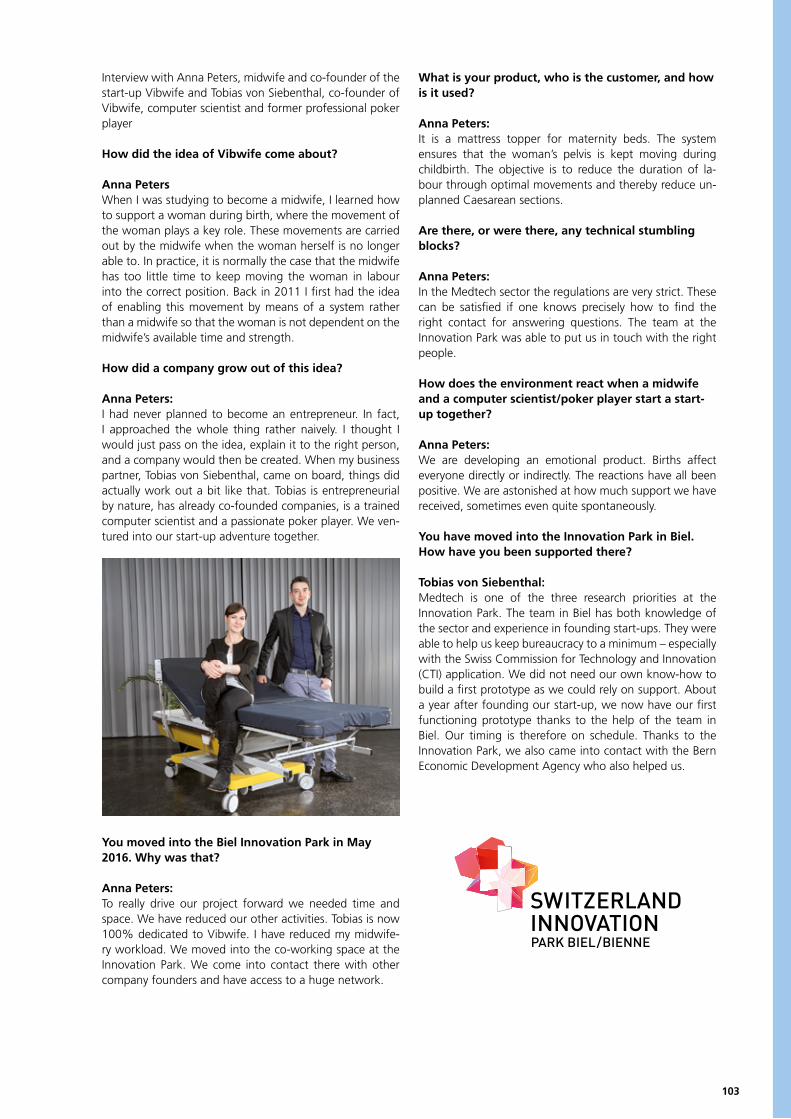
103
Interview with Anna Peters, midwife and co-founder of the start-up Vibwife and Tobias von Siebenthal, co-founder of Vibwife, computer scientist and former professional poker player
How did the idea of Vibwife come about?
Anna PetersWhen I was studying to become a midwife, I learned how to support a woman during birth, where the movement of the woman plays a key role. These movements are carried out by the midwife when the woman herself is no longer able to. In practice, it is normally the case that the midwife has too little time to keep moving the woman in labour into the correct position. Back in 2011 I first had the idea of enabling this movement by means of a system rather than a midwife so that the woman is not dependent on the midwife’s available time and strength.
How did a company grow out of this idea?
Anna Peters: I had never planned to become an entrepreneur. In fact, I approached the whole thing rather naively. I thought I would just pass on the idea, explain it to the right person, and a company would then be created. When my business partner, Tobias von Siebenthal, came on board, things did actually work out a bit like that. Tobias is entrepreneurial by nature, has already co-founded companies, is a trained computer scientist and a passionate poker player. We ven-tured into our start-up adventure together.
You moved into the Biel Innovation Park in May 2016. Why was that?
Anna Peters:To really drive our project forward we needed time and space. We have reduced our other activities. Tobias is now 100% dedicated to Vibwife. I have reduced my midwife-ry workload. We moved into the co-working space at the Innovation Park. We come into contact there with other company founders and have access to a huge network.
What is your product, who is the customer, and how is it used?
Anna Peters:It is a mattress topper for maternity beds. The system ensures that the woman’s pelvis is kept moving during childbirth. The objective is to reduce the duration of la-bour through optimal movements and thereby reduce un-planned Caesarean sections.
Are there, or were there, any technical stumbling blocks?
Anna Peters:In the Medtech sector the regulations are very strict. These can be satisfied if one knows precisely how to find the right contact for answering questions. The team at the Innovation Park was able to put us in touch with the right people.
How does the environment react when a midwife and a computer scientist/poker player start a start-up together?
Anna Peters:We are developing an emotional product. Births affect everyone directly or indirectly. The reactions have all been positive. We are astonished at how much support we have received, sometimes even quite spontaneously.
You have moved into the Innovation Park in Biel. How have you been supported there?
Tobias von Siebenthal:Medtech is one of the three research priorities at the Innovation Park. The team in Biel has both knowledge of the sector and experience in founding start-ups. They were able to help us keep bureaucracy to a minimum – especially with the Swiss Commission for Technology and Innovation (CTI) application. We did not need our own know-how to build a first prototype as we could rely on support. About a year after founding our start-up, we now have our first functioning prototype thanks to the help of the team in Biel. Our timing is therefore on schedule. Thanks to the Innovation Park, we also came into contact with the Bern Economic Development Agency who also helped us.
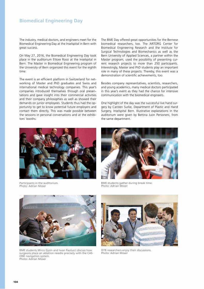
104
Biomedical Engineering Day
The industry, medical doctors, and engineers meet for the Biomedical Engineering Day at the Inselspital in Bern with great success.
On May 27, 2016, the Biomedical Engineering Day took place in the auditorium Ettore Rossi at the Inselspital in Bern. The Master in Biomedical Engineering program of the University of Bern organized this event for the eighth time.
The event is an efficient platform in Switzerland for net-working of Master and PhD graduates and Swiss and international medical technology companies. This year’s companies introduced themselves through oral presen-tations and gave insight into their commercial activities and their company philosophies as well as showed their demands on junior employees. Students thus had the op-portunity to get to know potential future employers and contact them directly. This was made possible between the sessions in personal conversations and at the exhibi-tors’ booths.
The BME Day offered great opportunities for the Bernese biomedical researchers, too. The ARTORG Center for Biomedical Engineering Research and the Institute for Surgical Technologies and Biomechanics as well as the Bern University of Applied Sciences, a partner within the Master program, used the possibility of presenting cur-rent research projects to more than 250 participants. Interestingly, Master and PhD students play an important role in many of these projects. Thereby, this event was a demonstration of scientific achievements, too.
Besides company representatives, scientists, researchers, and young academics, many medical doctors participated in this year’s event as they had the chance for intensive communication with the biomedical engineers.
One highlight of the day was the successful live hand sur-gery by Carsten Surke, Department of Plastic and Hand Surgery, Inselspital Bern. Illustrative explanations in the auditorium were given by Bettina Juon Personeni, from the same department.
ISTB researchers enjoy their discussions.Photo: Adrian Moser
BME students Mirco Gysin and Iwan Paolucci discuss how surgeons place an ablation needle precisely with the CAS-ONE navigation system.Photo: Adrian Moser
Participants in the auditorium.Photo: Adrian Moser
BME students gather during break time.Photo: Adrian Moser
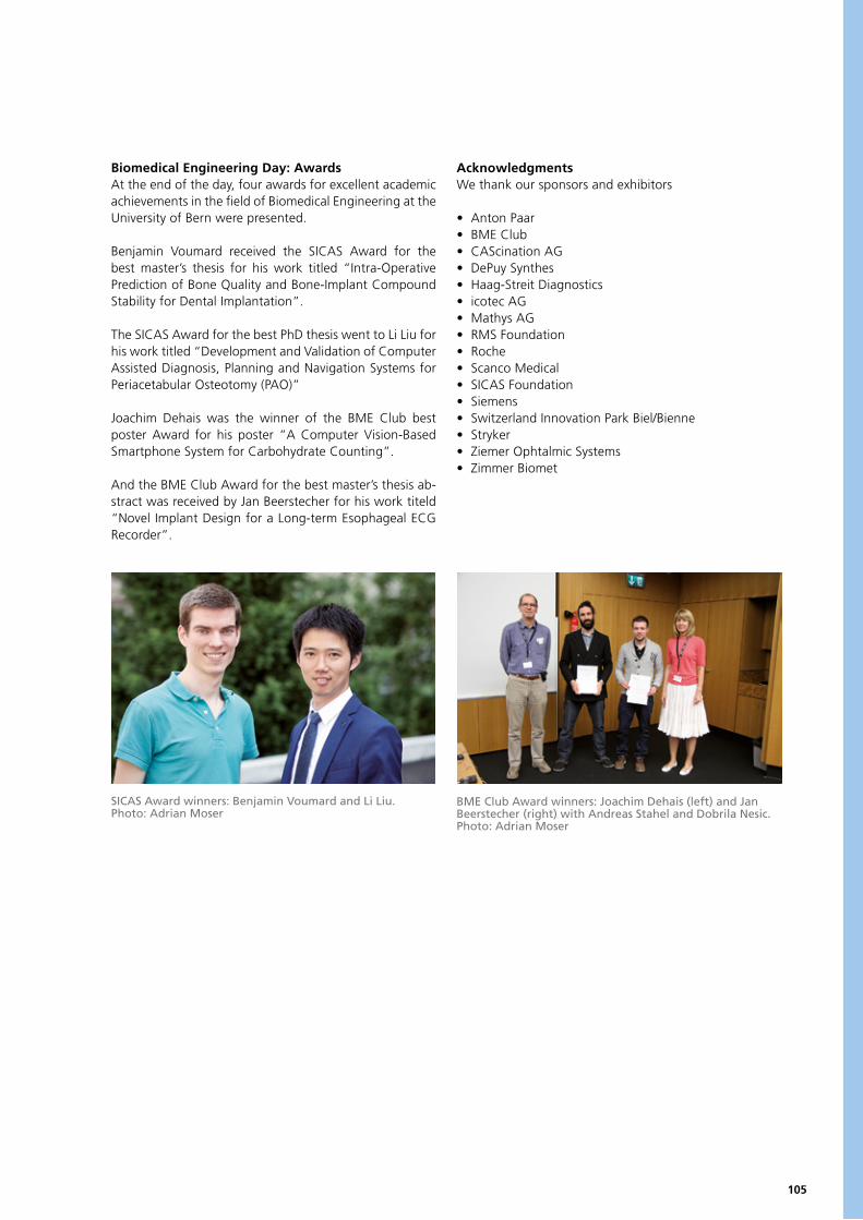
105
Biomedical Engineering Day: AwardsAt the end of the day, four awards for excellent academic achievements in the field of Biomedical Engineering at the University of Bern were presented.
Benjamin Voumard received the SICAS Award for the best master’s thesis for his work titled “Intra-Operative Prediction of Bone Quality and Bone-Implant Compound Stability for Dental Implantation”.
The SICAS Award for the best PhD thesis went to Li Liu for his work titled “Development and Validation of Computer Assisted Diagnosis, Planning and Navigation Systems for Periacetabular Osteotomy (PAO)”
Joachim Dehais was the winner of the BME Club best poster Award for his poster “A Computer Vision-Based Smartphone System for Carbohydrate Counting”.
And the BME Club Award for the best master’s thesis ab-stract was received by Jan Beerstecher for his work titeld “Novel Implant Design for a Long-term Esophageal ECG Recorder”.
AcknowledgmentsWe thank our sponsors and exhibitors
• Anton Paar• BME Club• CAScination AG• DePuy Synthes• Haag-Streit Diagnostics• icotec AG• Mathys AG• RMS Foundation• Roche• Scanco Medical• SICAS Foundation• Siemens• Switzerland Innovation Park Biel/Bienne• Stryker• Ziemer Ophtalmic Systems• Zimmer Biomet
SICAS Award winners: Benjamin Voumard and Li Liu.Photo: Adrian Moser
BME Club Award winners: Joachim Dehais (left) and Jan Beerstecher (right) with Andreas Stahel and Dobrila Nesic.Photo: Adrian Moser
106
The Biomedical Engineering Club
The BME Club and Its MissionThe BME Club is an alumni club whose mission is to pro-vide and promote networking among its interdisciplinary members. We are a constantly growing group of biomed-ical engineers, scientists, past and present students, and medical technology corporates; eager to bring together the principles of engineering, biology, and clinical medi-cine. The club accomplishes these goals by hosting events such as information sessions on the latest cutting-edge research in different fields of biomedical engineering, attendance at international conferences, and organizing visits of various industrial plants and laboratories. The BME Club is recognized as an official alumni association of the University of Bern under the umbrella organization – Alumni UniBE. A dedicated executive committee follows the principles of our constitution.
We are an enthusiastic and versatile group with diverse activities:• bi-monthly “Stammtisch” in a local restaurant as an amiable platform to exchange, discuss, brainstorm, or simply chat• visits to Swiss medical and engineering companies• organization of the annual MEDICA trip• information on career opportunities (incl. job offers)• organization of the annual welcome event for new students of the BME Master program• organization of an annual alumni gathering
• sponsorship of the poster and abstract awards at the annual BME day• sponsorship of conference travel grants• provide access to the Medical Cluster events• automatic joint membership with Alumni UniBE• offer joint membership with SSBE (Swiss Society for Biomedical Engineering)
In short, the BME club represents a unique platform for professional, lifelong communication and networking. For further details, look up our website athttp://www.bmeclub.ch.
How to JoinBecoming a member is easy! Simply sign up at any BME Club event or visit us at http://www.bmeclub.ch. We look forward to seeing you!
The BME Club Board in 2016
Prabitha UrwylerPresident
Tom de BruyneVice President
Andrea NienhausSecretary
Carlos CillerTreasurer
Juan AnsoPhD Students
Tobias ImfeldWebmaster
Christian GüderPR / Newsletter
Dobrila NesicFaculty
Hector AlvarezMaster Students
Fatih ToyAlumni
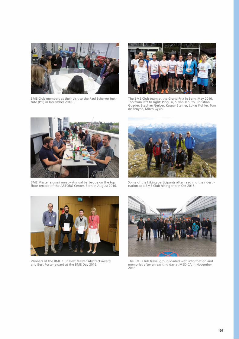
107
BME Club members at their visit to the Paul Scherrer Insti-tute (PSI) in December 2016.
The BME Club team at the Grand Prix in Bern, May 2016. Top from left to right: Ping Lu, Silvan Januth, Christian Gueder, Stephan Gerber, Kaspar Steiner, Lukas Kohler, Tom de Bruyne, Mirco Gysin.
BME Master alumni meet – Annual barbeque on the top floor terrace of the ARTORG Center, Bern in August 2016.
Some of the hiking participants after reaching their desti-nation at a BME Club hiking trip in Oct 2015.
Winners of the BME Club Best Master Abstract award and Best Poster award at the BME Day 2016.
The BME Club travel group loaded with information and memories after an exciting day at MEDICA in November 2016.

109
ACADEMIC EDUCATION AND TEACHING
Academic education is a cornerstone of the Bern Biomedical Engineering Network. At the master’s level, a joint program in Biomedical Engineering (BME) was established by the Medical Faculty of the University of Bern and the Department of Engineering and Information Technology of the University of Applied Sciences of Bern with the aim of training multidisciplinary engineers to deliver scientifically founded and cost-effective solutions for biomedical problems in research and industry.
For graduates of the MSc in Biomedical Engineering and other candidates from re-lated areas who are interested in an in-depth training in experimental and compu-tational research and/or who aim for an academic career, a corresponding doctoral degree (PhD in Biomedical Engineering) can be earned at the inter-facultary Graduate School for Cellular and Biomedical Sciences (GCB) of the University of Bern. The GCB is jointly administered by the Faculties of Medicine, Science, and Vetsuisse, and offers tailored, structured training programs at an international level.
At Bern University of Applied Sciences, the Medical Information Laboratory, for ex-ample, enables the processes of the Swiss health care system to be visualised on a one-to-one basis. Students of Medical Informatics learn and work with the new lab-oratory infrastructure on realistic questions and projects that have been developed in conjunction with partners from hospitals, industry, and public authorities.
Philippe ZyssetProgram Director Master of Biomedical EngineeringUniversity of Bern
Marlene WolfCoordinator Graduate School for Cellular and Biomedical SciencesUniversity of Bern
Lukas RohrHead of DepartmentBern University of Applied SciencesEngineering and Information Technology
110
Master of Science in Biomedical Engineering
Since the start of the Master’s Program Biomedical Engineering in March 2006, the constant effort to improve the quality of our curriculum has resulted in substantial changes of the course structure over the past years. The first curriculum consisted of a number of individual courses that were either mandatory or elective, but their coherence with regards to contents was in most cases not expressed by a defined structure. However, two major modules (for-merly called “focus areas”) already existed.
As of Fall Semester 2009, all courses were grouped in a strictly modular way in order to enhance both the clari-ty and the complexity of the curricular structure. A main idea was to guide the students through their studies in a better way by adding an elective part to the major mod-ules, which formerly had consisted exclusively of manda-tory courses. Besides, the curriculum was expanded by a number of new specialized courses as well as an additional major module called “Image-guided Therapy”.
Adaptations in the legal framework of the master’s pro-gram are now offering more flexibility in the design of courses and modules, thus providing the basis for a sec-ond fundamental restructuration of the curriculum as of Fall Semester 2013. In particular, a new module called “Complementary Skills” is replacing the former module “Unrestricted Electives”. In addition, the list of mandatory courses in both basic and major modules was revised.
The Curriculum
Duration of Studies and Part-Time Professional OccupationThe full-time study program takes 4 semesters, which cor-responds to 120 ECTS points, one ECTS point being defined as 25-30 hours of student workload. It can be extended to a maximum of 6 semesters. When a student decides to complete the studies in parallel to a part-time profession-al occupation, further extension is possible on request. To support regular part-time work, mandatory courses take place (with rare exceptions) on only 3 days per week.
Basic ModulesThe basic modules provide the students with the necessary background to be able to fully understand the highly com-plex subject matter in the specialized courses. All students with an engineering background (for all other students, individual study plans are set up which may contain cer-tain variations) have to complete all courses in the Basic Modules Human Medicine, Applied Mathematics, and Biomedical Engineering. In the first semester, all courses belong to this group, whereas in the second and third se-mester, the courses from the basic modules make up for approximately 30%.Major ModulesThe choice of one of three major modules Biomechanical
Systems, Electronic Implants, or Image-guided Therapy af-ter the first semester constitutes the first opportunity for specialization.
Approximately one third of the major modules consist of mandatory courses. In the elective part of the major mod-ule, the student is allowed to select every course from the list of courses in the master’s program, giving rise to a high degree of diversity and flexibility and allowing for numer-ous course combinations. However, this freedom makes it somewhat difficult for the student to make reasonable choices regarding professional prospects.
This is why the responsible lecturers developed a recom-mended study plan to guide the students through the course selection process and to avoid organizational prob-lems such as overlapping courses. If a student follows the recommended path, he or she can be sure to establish a sound professional profile.
Module “Complementary Skills”Apart from the rapid development of technology itself, today’s biomedical engineers are increasingly challenged by complementary issues like ethical aspects, project plan-ning, quality assurance and product safety, legal regula-tions and intellectual property rights, as well as marketing aspects. Language competence in English is of paramount importance both in an industrial and academic environ-ment. This situation has been accounted for by the intro-duction of a new module called “Complementary Skills” where students are required to complete two mandatory courses (Innovation Management; Regulatory Affairs and Patents) as well as 2 ECTS from the electives courses (Ethics in Biomedical Engineering; Scientific Writing in Biomedical Engineering; Introduction to Epidemiology and Health Technology Assessment). If a student selects more than 2 ECTS from the elective part, the additional points can be credited in the student’s major module.
Master’s ThesisThe last semester is dedicated to a master’s thesis project on an individually suited topic in an academic research group at the University of Bern or the Bern University of Applied Sciences or, for particular cases, in an industrial research and development environment. As a rule, all 90 ECTS points from the course program have to be com-pleted, thus ensuring that the student is able to fully con-centrate on the challenges imposed by exciting research activities. The master’s thesis includes the thesis paper, a thesis presentation and defense as well as a one-page ab-stract for publication in the Annual Report of the master’s program.

111
List of CoursesApplied Biomaterials Basics of Applied Molecular BiologyBasics in Physiology for Biomedical EngineeringBiological Principles of Human MedicineBiomaterialsBiomedical SensorsBiomedical AcousticsBiomedical InstrumentationBiomedical Laser ApplicationsBiomedical Signal Processing and AnalysisBioMicrofluidicsC++ Programming IC++ Programming IICardiovascular TechnologyClinical Applications of Image-guided TherapyComputer Assisted SurgeryComputer GraphicsComputer VisionContinuum MechanicsCutting Edge MicroscopyDesign of Biomechanical SystemsEngineering MechanicsEthics in Biomedical EngineeringFinite Element Analysis IFinite Element Analysis IIFluid MechanicsFunctional Anatomy of the Locomotor ApparatusImage-guided Therapy LabInnovation ManagementIntelligent Implants and Surgical Instruments
Introduction to Clinical Epidemiology and Health Technology AssessmentIntroduction to Digital LogicIntroduction to Medical StatisticsIntroduction to Signal and Image Processing Introductory Anatomy and Histology for Biomedical EngineersLow Power MicroelectronicsMachine LearningMeasurement Technologies in BiomechanicsMedical Image AnalysisMedical Image Analysis LabMedical Robotics Microsystems EngineeringModeling and SimulationMolecular and Cellular Biology PracticalNumerical MethodsOphthalmic TechnologiesOsteologyPrinciples of Medical ImagingProgramming of MicrocontrollersRegenerative Dentistry for Biomedical EngineeringRegulatory Affairs and PatentsRehabilitation TechnologyScientific Writing in Biomedical EngineeringTechnology and Diabetes ManagementTissue BiomechanicsTissue Biomechanics LabTissue EngineeringTissue Engineering - Practical CourseWireless Communication for Medical Devices
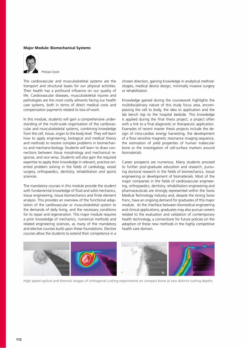
112
Major Module: Biomechanical Systems
Philippe Zysset
The cardiovascular and musculoskeletal systems are the transport and structural bases for our physical activities. Their health has a profound influence on our quality of life. Cardiovascular diseases, musculoskeletal injuries and pathologies are the most costly ailments facing our health care systems, both in terms of direct medical costs and compensation payments related to loss-of-work. In this module, students will gain a comprehensive under-standing of the multi-scale organisation of the cardiovas-cular and musculoskeletal systems, combining knowledge from the cell, tissue, organ to the body level. They will learn how to apply engineering, biological and medical theory and methods to resolve complex problems in biomechan-ics and mechano-biology. Students will learn to draw con-nections between tissue morphology and mechanical re-sponse, and vice versa. Students will also gain the required expertise to apply their knowledge in relevant, practice-ori-ented problem solving in the fields of cardiology, vessel surgery, orthopaedics, dentistry, rehabilitation and sports sciences.
The mandatory courses in this module provide the student with fundamental knowledge of fluid and solid mechanics, tissue engineering, tissue biomechanics and finite element analysis. This provides an overview of the functional adap-tation of the cardiovascular or musculoskeletal system to the demands of daily living, and the necessary conditions for its repair and regeneration. This major module requires a prior knowledge of mechanics, numerical methods and related engineering sciences, as many of the mandatory and elective courses build upon these foundations. Elective courses allow the students to extend their competence in a
chosen direction, gaining knowledge in analytical method-ologies, medical device design, minimally invasive surgery or rehabilitation.
Knowledge gained during the coursework highlights the multidisciplinary nature of this study focus area, encom-passing the cell to body, the idea to application and the lab bench top to the hospital bedside. This knowledge is applied during the final thesis project, a project often with a link to a final diagnostic or therapeutic application. Examples of recent master thesis projects include the de-sign of intra-cardiac energy harvesting, the development of a flow sensitive magnetic resonance imaging sequence, the estimation of yield properties of human trabecular bone or the investigation of cell-surface markers around biomaterials.
Career prospects are numerous. Many students proceed to further post-graduate education and research, pursu-ing doctoral research in the fields of biomechanics, tissue engineering or development of biomaterials. Most of the major companies in the fields of cardiovascular engineer-ing, orthopaedics, dentistry, rehabilitation engineering and pharmaceuticals are strongly represented within the Swiss Medical Technology industry and, despite the strong Swiss franc, have an ongoing demand for graduates of this major module. At the interface between biomedical engineering and clinical applications, graduates may also pursue careers related to the evaluation and validation of contemporary health technology, a cornerstone for future policies on the adoption of these new methods in the highly competitive health care domain.
High speed optical and thermal images of orthogonal cutting experiments on compact bone at two distinct cutting depths.

113
Major Module: Electronic Implants
Volker M. Koch
Electronic implants are devices like cardiac pacemakers and cochlear implants. Due to miniaturization and other deve-lopments, many new applications become feasible and this exciting area is growing rapidly. For example, cochlear im-plants provide already approximately more than 320’000 people worldwide a sense of sound. These people were previously profoundly deaf or severely hard of hearing. Recently, researchers demonstrated that electronic retinal implants allow the blind to read large words.
There are many more applications for electronic implants beyond treating heart problems, hearing loss or blindness. For example, there are electronic implants that treat obesi-ty, depression, incontinence, hydrocephalus, pain, paraple-gia, and joint diseases.
In this module, students will learn about the basics of electronic implants. This includes: signal processing and analysis, low-power microelectronics, wireless communica-tions, and MEMS technology. Application-oriented elective courses are also taught, e.g., diabetes management, bio-medical acoustics, and biomedical sensors.
Since the development and manufacturing of electronic implants is highly complex and since it involves many diffe-rent disciplines, it is not the goal of this major that students are able to develop an electronic implant on their own but rather to be able to work successfully in a project team that develops electronic implants.
Students may already apply their knowledge as a part- time assistant in a laboratory and/or during their master’s pro-jects. After finishing the degree program, a wide variety of career paths are available, ranging from research and development to project and product management. Many companies in Switzerland work in this field and “traditi-onal” implants manufacturers have recently become in-terested in electronic implants, e.g., to measure forces in knee implants.
Prototype of a lead- and batteryless cardiac pacemaker (courtesy of the Group for Translational Electrophysiology. Deptartment of Cardiology, Bern University Hospital & AR-TORG Center for Biomedical Engineering Research).
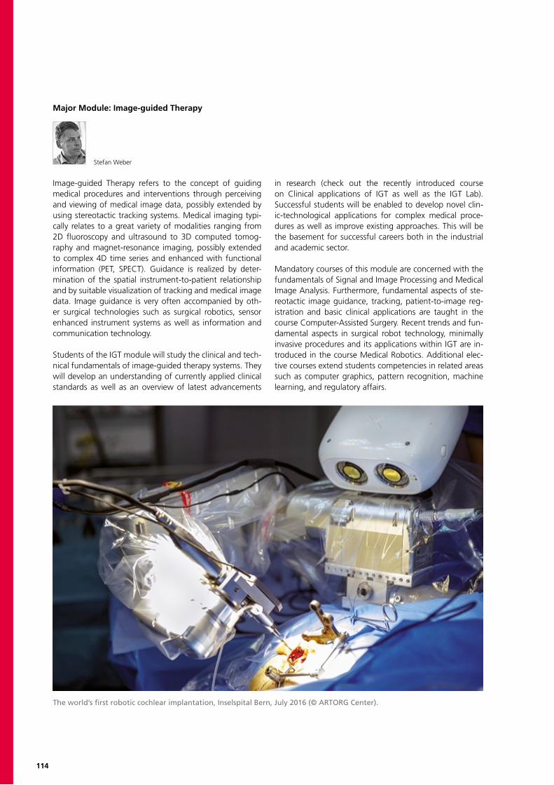
114
Major Module: Image-guided Therapy
Stefan Weber
Image-guided Therapy refers to the concept of guiding medical procedures and interventions through perceiving and viewing of medical image data, possibly extended by using stereotactic tracking systems. Medical imaging typi-cally relates to a great variety of modalities ranging from 2D fluoroscopy and ultrasound to 3D computed tomog-raphy and magnet-resonance imaging, possibly extended to complex 4D time series and enhanced with functional information (PET, SPECT). Guidance is realized by deter-mination of the spatial instrument-to-patient relationship and by suitable visualization of tracking and medical image data. Image guidance is very often accompanied by oth-er surgical technologies such as surgical robotics, sensor enhanced instrument systems as well as information and communication technology.
Students of the IGT module will study the clinical and tech-nical fundamentals of image-guided therapy systems. They will develop an understanding of currently applied clinical standards as well as an overview of latest advancements
in research (check out the recently introduced course on Clinical applications of IGT as well as the IGT Lab). Successful students will be enabled to develop novel clin-ic-technological applications for complex medical proce-dures as well as improve existing approaches. This will be the basement for successful careers both in the industrial and academic sector.
Mandatory courses of this module are concerned with the fundamentals of Signal and Image Processing and Medical Image Analysis. Furthermore, fundamental aspects of ste-reotactic image guidance, tracking, patient-to-image reg-istration and basic clinical applications are taught in the course Computer-Assisted Surgery. Recent trends and fun-damental aspects in surgical robot technology, minimally invasive procedures and its applications within IGT are in-troduced in the course Medical Robotics. Additional elec-tive courses extend students competencies in related areas such as computer graphics, pattern recognition, machine learning, and regulatory affairs.
The world’s first robotic cochlear implantation, Inselspital Bern, July 2016 (© ARTORG Center).
115
Faculty
University of BernChristiane Albrecht, Prof. Dr.Philippe Büchler, Prof. Dr.Dario Cazzoli, Dr.Roch-Philippe Charles, Prof. Dr.Bruno da Costa, Dr.Marcel Egger, Prof. Dr.Matthias Egger, Prof. Dr.Tobias Erlanger, Dr.Favaro Prof. Dr., PaoloChristian Fernandez Palomo, Dr.Martin Frenz, Prof. Dr.Benjamin Gantenbein, Prof. Dr.Amiq Gazdhar, Dr.Kate Gerber, Dr.Nicolas Gerber, Dr.Olivier Guenat, Prof. Dr.Wilhelm Hofstetter, Prof. Dr.Doris KoppJan Kucera, Prof. Dr.Glenn Lurman, Dr.Ange Maguy, Dr.Ghislain Maquer, Dr. Ines Marques, Dr.Beatrice MinderStavroula Mougiakakou, PD Dr.Tobias Nef, Prof. Dr.Thomas Nevian, Prof. Dr.Lutz Nolte, Prof. Dr.Dominik Obrist, Prof. Dr.Mauricio Reyes, Prof. Dr.Steffen Schumann, Dr.Walter Martin Senn, Prof. Dr.Marc StadelmannJürg Streit, Prof. Dr.Raphael Sznitman, Prof. Dr.Stefan Andreas Tschanz, Dr.Prabitha Urwyler, Dr.Benjamin VoumardStefan Weber, Prof. Dr.Tom Williamson, Dr.Wilhelm Wimmer, Dr.Guoyan Zheng, Prof. Dr.Matthias Zwicker, Prof. Dr.Philippe Zysset, Prof. Dr.
Inselspital, Bern University Hospi-tal and School of Dental MedicineDaniel Aeberli, PD Dr.Sufian Ahmad, Dr.Tommy Baumann, Dr.Dieter Bosshardt, Prof. Dr.Vivianne Chappuis, Dr.Sigrun Eick, Prof. Dr.Jens Fichtner, Dr.Michael Fix, PD Dr.Simon Flury, Dr.Pjotr Fudalej, PD Dr.Andreas Häberlin, Dr.Tim Joda, Dr.Joannis Katsoulis, PD Dr.Martin Kompis, Prof. Dr.Kurt Laederach, Prof. Dr.Kurt Lippuner, Prof. Dr.Dobrila Nesic, PD Dr.Thomas Pilgrim, Prof. Dr.Lorenz Räber, PD Dr.Christoph Andreas Ramseier, Dr.Thiago Saads Carvalho, Dr.Christophe Von Garnier, Prof. Dr.
Bern University of Applied SciencesNorman Urs Baier, Prof. Dr.Heiner Baur, Prof. Dr.Daniel Debrunner, Prof.Bertrand Dutoit, Prof. Dr.Josef Götte, Prof. Dr.Kenneth James Hunt, Prof. Dr.Marcel Jacomet, Prof. Dr.Björn Jensen, Prof. Dr.Jörn Justiz, Prof. Dr.Theo Kluter, Prof Dr.Volker M. Koch, Prof. Dr.Martin Kucera, Prof.Alexander Mack, Dr.Christoph Meier, Prof.Thomas Niederhauser, Dr.Heinrich Schwarzenbach, Prof.Andreas Stahel, Prof. Dr.Jasmin Wandel, Prof. Dr.
Partner Institutions and IndustryDaniel Baumgartner, Dr.Marc Bohner, Dr.Mathias Bonmarin, Dr.Jürgen Burger, Prof. Dr.Philippe Cattin, Prof. Dr.Alessandro Cianfoni, Dr.Barbara Cvikl, PD Dr.Emmanuel de Haller, Dr.Nicolas Alexander Diehm, Prof. Dr.Nicola Döbelin, Dr.Alex Dommann, Prof. Dr.Patrick Dubach, Dr.David Eglin, Dr.Lukas Eschbach, Dr.Marie-Noëlle Giraud, PD Dr.Reinhard Gruber, Prof. Dr.Janosch Häberli, Dr.Daniel Haschtmann, Dr.Bernd Heinlein, Prof. Dr.Philippe Henle, Dr.Roman Heuberger, Dr.Ulrich Hofer, Dr.Thomas Imwinkelried, Dr.Herbert Keppner, Prof. Dr.Jens Kowal, PD Dr.Beat LechmannReto Lerf, Dr.Lukas Lichtensteiger, Dr.Reto Luginbühl, Dr.Simon Milligan, Dr.Walter Moser, Dr.Richard Nyffeler, PD Dr.Yves Pauchard, Dr.Matthias Peterhans, Dr.Jorge Sague, Dr.Birgit Schäfer, PD Dr.Matthias Schwenkglenks, PD Dr.Jivko Stoyanov, PD Dr.Tim Vanbellingen, Dr.Jürgen Vogt, Dr.André Weber, Dr.

116
Statistics
BME Alumni: Career Directions
Number of New Students and Graduates per Year
Profession after Graduation
Profession 5 Years after Graduation

117
Completed Master Theses in 2016
Emir ArtikDevelopment of a Leadless Cardiac Dual-Chamber PacemakerSupervisors: Lukas Bereuter, MSc and Dr. Thomas NiederhauserInstitutions: ARTORG Center for Biomedical Engineering Research, University of BernExaminers: Lukas Bereuter, MSc and Dr. med. Dr. phil. Andreas Häberlin
Fabian BalsigerDevelopment of Algorithms for Peripheral Nerve Segmentation using Machine Learning ApproachesSupervisors: Dr. Waldo Valenzuela and Dr. Olivier ScheideggerInstitutions: Institute for Surgical Technology & Biomechanics, University of Bern; Institute of Diagnostic and Interventional Neuroradiology, Inselspital, Bern University HospitalExaminers: Prof. Dr. Mauricio Reyes and Prof. Dr. Roland Wiest
Sarah BlankenbergerDevelopment and Evaluation of a Tele-Rehabilitation Application for Aphasic patientsSupervisors: Dr. Prabitha Urwyler and Silvan JanuthInstitutions: ARTORG Center for Biomedical Engineering Research, University Bern; Division of Cognitive and Restorative Neurology, Department of Neurology, Inselspital Bern Examiners: Prof. Dr. sc. Tobias Nef and Prof. Dr. med. René M. Müri
Dominik M. Brügger3D Reconstruction and Simulation of Heart Potentials in the EsophagusSupervisors: Dr. med. Reto Wildhaber and Dr. Thomas NiederhauserInstitutions: Institute for Human Centered Engineering, Bern University of Applied SciencesExaminers: Prof. Dr. Marcel Jacomet and Prof. Dr. Josef Götte
Sébastien BuchwalderReliability Study of Neuro-Stimulation Electrodes SurfacesSupervisors: Prof. Dr. Herbert Keppner Institutions: Aleva Neurotherapeutics SA, EPFL Innovation Park; Institut des Microtechnologies Appliquées Arc, HE-Arc NeuchâtelExaminers: Prof. Dr. Herbert Keppner and Dr. Jason Jinyu Ruan
Silvan DunerRadial Flow Sensitive Magnetic Resonance ImagingSupervisors: PD Dr. Bernd JungInstitution: Department of Diagnostic, Interventional and Paediatric Radiology, Inselspital BernExaminers: Prof. Dr. Dominik Obrist and PD Dr. Bernd Jung
Thomas FalkIntegration of Wireless Functional Electrical Stimulation in a Recumbent Therapy Trike Supervisor: Prof. Dr. Kenneth James HuntInstitution: Institute for Rehabilitation and Performance Technology, Department of Engineering and Information Technology, Bern University of Applied SciencesExaminers: Prof. Dr. Kenneth James Hunt and MSc Manuel Bracher
Stefan M. FunariuReal-time Tool Localization in Endovascular TreatmentSupervisor: Prof. Dr.-Ing. Björn JensenInstitutions: Robotics Lab, Institute for Human Centered Engineering, Bern University of Applied Sciences, Engineering and Information Technology; ARTORG Center for Biomedical Engineering Research, University of BernExaminers: Prof. Dr.-Ing. Björn Jensen and Prof. Dr.-Ing. Stefan Weber
118
Completed Master Theses in 2016
Stephan GerberExploring Virtual Reality in MedicineSupervisors: Prof. Dr. sc. T. Nef and Prof. Dr. med. R. MüriInstitutions: ARTORG Center for Biomedical Engineering Research, University Bern; Department of Intensive Care Medicine, Bern University HospitalExaminers: Prof. Dr. sc. T. Nef and Prof. Dr. med. R. Müri
Martin GramboneAdaptation and Optimization of an EIT System to Reduce Motion Artefacts in Applications for Animals and ChildrenSupervisors: Prof. Dr. Volker M. Koch, Prof. Dr. Jörn Justiz, Andreas WaldmannInstitutions: Institute for Human Centered Engineering, Bern University of Applied Sciences; Veterinary Clinics Zürich, Swisstom AG Landquart Examiners: Prof. Dr. Volker M. Koch and PD Dr. Martina Mosing DECVAA
Tess GroeneveldDevelopment and Evaluation of an iPad-based Eye Gaze TrackerApplication for Real Time Visual Exploration Data ExtractionSupervisor: Prof. Dr. sc. Tobias NefInstitutions: Gerontechnology and Rehabilitation Research Group, ARTORG Center for Biomedical Engineering Research, University of BernExaminers: Prof. Dr. sc. Tobias Nef and MSc Alvin Chesham
Alain JungoA Deep Learning-based Approach for Skull-StrippingSupervisor: Raphael Meier, MScInstitution: Institute for Surgical Technology & Biomechanics, University of BernExaminers: Prof. Dr. Mauricio Reyes and Dr. Richard McKinley
Dr. med. Dent. Ioannis Kapagiannidis Tooth Scanning and Multilayer Modeling for 3D Printing of Aesthetic Dental RestorationsSupervisors: Prof. Dr.-Ing. Stefan Weber and Prof. Dr. Adrian LussiInstitutions: ARTORG Center for Biomedical Engineering Research, University of Bern; School of Dental Medicine, University of BernExaminers: Prof. Dr.-Ing. Stefan Weber and Prof. Dr. Adrian Lussi
Daniel KaufmannCombining SRT with OCT for Automated Dose-Control Supervisors: Prof. Dr. Jörn Justiz and Prof. Dr. Volker M. KochInstitutions: Institute for Human Centered Engineering, Bern University of Applied SciencesExaminers: Prof. Christoph Meier and Dr. Boris Považay
Christian Hannes KummerDevelopment of Tools for Investigation of ULF to VLF HRV during Treadmill ExerciseSupervisors: Prof. Dr. Kenneth J. HuntInstitutions: Institute for Rehabilitation and Performance Technology, Bern University of Applied Sciences, Department of Engineering and Information TechnologyExaminers: Prof. Dr. Kenneth J. Hunt and Prof. Dr. Josef Götte
Benjamin MeierIntracardial Energy Harvesting by Aspring-Actuated Electromagnetic Conversion MechanismSupervisors: Dr. Adrian Zurbuchen and Lukas BereuterInstitutions: Department of Cardiology, Inselspital, Bern University Hospital; ARTORG Center for Biomedical Engineering Research, Cardiovascular Engineering, University of BernExaminers: Dr. med. Dr. phil. Andreas Häberlin and Prof. Dr. Dominik Obrist
119
Manuel I. MosimannOptimization of Images for AMD Patients Using Digital Image Processing in a Portable Visual AidSupervisors: Prof. Dr. Jörn Justiz and Prof. Dr. Volker M. KochInstitutions:Institute for Human Centered Engineering – BME Lab, Bern University of Applied SciencesExaminers: Prof. Dr. Jörn Justiz and André Reber
Martin PletscherSegmentation-Guided MRS Analysis of Brain Tumor PatientsSupervisors: MSc Nuno Pedrosa de Barros, MSc Raphael MeierInstitutions: Institute for Surgical Technology & Biomechanics, University of Bern; Institute of Diagnostic and Interventional Neuroradiology, Inselspital, Bern University HospitalExaminers: Prof. Dr. Mauricio Reyes and PD Dr. Johannes Slotboom
Yannick RöschThe Torpedo-Pacemaker – a Batteryless Cardiac Pacemaker Driven by Blood flow in the right Ventricular Outflow TractSupervisors: Dr. med. Dr. phil. Andreas HäberlinInstitutions: ARTORG Center for Biomedical Research, University of Bern; Department of Cardiology, University Hospital BernExaminers: Prof. Dr. Dominik Obrist and Dr. Adrian Zurbuchen
Marc RueschPrediction analysis in paediatric obsessive-compulsive disorder (OCD) and attention-deficit hyperactivity disorder (ADHD) combining multimodal neuroimaging-genetic dataSupervisors: Prof. Dr. Mauricio Reyes and PD Dr. sc. nat.Silvia BremInstitutions: Institute for Surgical Technology & Biomechanics, University of Bern; Department of Child and Adolescent Psychiatry, University of Zurich Examiners: Prof. Dr. Mauricio Reyes and PD Dr. sc. nat. Silvia Brem
Adrian SallazFast Spinning Drill with Integrated Nerve DetectionSupervisors: Prof. Dr. Volker M. Koch and Anton SchärerInstitutions: Institute for Human Centered Engineering, Biomedical Engineering Lab, Bern University of Applied SciencesExaminers: Prof. Dr. Jörn Justiz and Stève Gigandet
Dominic Sebastian SchmidInfluence of the Sinus of Valsalva on the three-dimensional Flow Field behind a Prosthetic ValveSupervisor: David HaslerInstitution: ARTORG Center for Biomedical Engineering Research, University of BernExaminers: Prof. Dr. Dominik Obrist and PD Dr. med Stefan Stortecky
Sandro Michael SchultzImage-Based Localization Method for Automatic Cell Sheet Layering in Robotized Heart Regeneration Therapy SystemSupervisors: Ryoichi Nakamura, PhDInstitutions: Laboratory of Innovative Therapeutic Engineering (LITE), University of Chiba (Japan); University of BernExaminers: Prof. Dr.-Ing. Stefan Weber and Ryoichi Nakamura, PhD
Apollonius SchwarzEvaluation and Improvement of Force-Based Tool Pose Estimation Confidence, Accuracy and RobustnessSupervisors: Dr. Tom Williamson and Dr. Kate GerberInstitution: Image-guided Therapy, ARTORG Center for Biomedical Engineering Research, University of BernExaminers: Dr. Kate Gerber and Dr. Tom Williamson
Samuel ShaheenIntracardial Piezoelectric Energy HarvestingSupervisors: Dr. Thomas Niederhauser and Dr. Adrian Zurbuchen Institutions: ARTORG Center for Biomedical Engineering Research, University of Bern; Institute for Human Centered Engineering, Bern University of Applied SciencesExaminers: Dr. med. Dr. phil. Andreas Häberlin and Prof. Dr. Josef Goette
120
Nino SutterHuman-in-the-loop Training Interface for a new active Balance BoardSupervisor: Patric EichelbergerInstitutions: Bern University of Applied Sciences Health, R&D Physiotherapy, Institute Robotics Lab, Institute for Human Centered Engineering, Bern University of Applied Sciences, Engineering and Information TechnologyExaminer: Prof. Dr.-Ing. Björn Jensen
Maximilien Victor ThollDevelopment of an Intracardiac Flow-based Energy Harvesting Mechanism for Cardiac Pacing Supervisors: Dr. Adrian Zurbuchen and Barna Becsek, M.Sc.Institutions: ARTORG Center for Biomedical Engineering Research, Cardiovascular Engineering, University of Bern; Department of Cardiology, University of BernExaminers: Prof. Dr. Dominik Obrist and Dr. med. Dr. phil. Andreas Häberlin
Jariyaporn Thongbudda An Evaluation of Image Motion Blur and its Effect on the Accuracy of Image Guided SurgerySupervisors: Dr. Kate Gavaghan and Dr. Nicolas GerberInstitutions: ARTORG Center for Biomedical Engineering Research, University of BernExaminers: Dr. Nicolas Gerber and Christoph Rathgeb
Severin ToblerInstrument Detection in 4D OCTSupervisor: Prof. Dr. Raphael SznitmanInstitution: ARTORG Center for Biomedical Engineering Research, University of BernExaminers: Prof. Dr. Raphael Sznitman and PD Dr. Stavroula Mougiakakou
Patrik WiliEstimation of Effective Yield Properties of Human Trabecular Bone Using Nonlinear Micro Finite Element AnalysisSupervisors: Jarunan Panyasantisuk and Dr. Ghislain MaquerInstitutions: Institute for Surgical Technology & Biomechanics, University of BernExaminers: Prof. Dr. Philippe Zysset and Jarunan Panyasantisuk
Hamoon ZohdiCell-Surface Markers around Bone Biomaterials Supervisors: Dr. Richard Miron and Prof. Dr. Dieter BosshardtInstitutions: Laboratory of Oral Histology, School of Dental Medicine, University of Bern, Laboratory of Oral Cell Biology, School of Dental Medicine, University of BernExaminers: Prof. Dr. Dieter Bosshardt and Dr. Richard Miron
Andreas Walter Zumbrunnen A Lead- and Battery-less Cardiac Pacemaker Driven by a Massimbalance Oscillation GeneratorSupervisors: Dr. med. Dr. phil. Andreas Häberlin and Dr. Adrian ZurbuchenInstitutions: ARTORG Center for Biomedical Engineering Research, University of Bern; Department of Cardiology, Bern University HospitalExaminers: Dr. med. Dr. phil. Andreas Häberlin, Prof. Dr. Dominik Obrist
Completed Master Theses in 2016
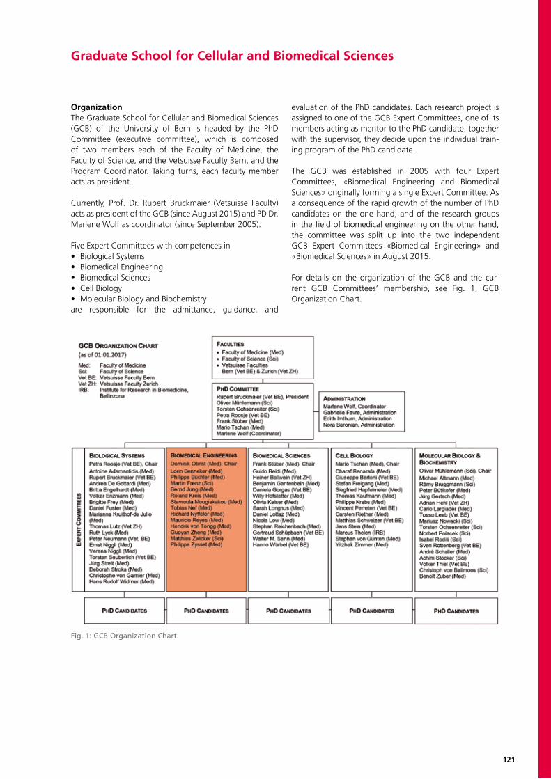
121
Graduate School for Cellular and Biomedical Sciences
OrganizationThe Graduate School for Cellular and Biomedical Sciences (GCB) of the University of Bern is headed by the PhD Committee (executive committee), which is composed of two members each of the Faculty of Medicine, the Faculty of Science, and the Vetsuisse Faculty Bern, and the Program Coordinator. Taking turns, each faculty member acts as president.
Currently, Prof. Dr. Rupert Bruckmaier (Vetsuisse Faculty) acts as president of the GCB (since August 2015) and PD Dr. Marlene Wolf as coordinator (since September 2005).
Five Expert Committees with competences in• Biological Systems• Biomedical Engineering• Biomedical Sciences• Cell Biology• Molecular Biology and Biochemistryare responsible for the admittance, guidance, and
evaluation of the PhD candidates. Each research project is assigned to one of the GCB Expert Committees, one of its members acting as mentor to the PhD candidate; together with the supervisor, they decide upon the individual train-ing program of the PhD candidate.
The GCB was established in 2005 with four Expert Committees, «Biomedical Engineering and Biomedical Sciences» originally forming a single Expert Committee. As a consequence of the rapid growth of the number of PhD candidates on the one hand, and of the research groups in the field of biomedical engineering on the other hand, the committee was split up into the two independent GCB Expert Committees «Biomedical Engineering» and «Biomedical Sciences» in August 2015.
For details on the organization of the GCB and the cur-rent GCB Committees’ membership, see Fig. 1, GCB Organization Chart.
Fig. 1: GCB Organization Chart.
122
PhD ProgramThe GCB offers structured training in experimental research in the fields of biochemistry, cell and molecular biology, immunology, biomedical sciences, epidemiology, neuro-science, and biomedical engineering, leading to a PhD, MD,PhD, or DVM,PhD degree.
The thesis projects are carried out at laboratories of the three participating faculties or at affiliated institutions, which include, among others, the Institute of Virology and Immunology (IVI) in Mittelhäusern (now part of the Vetsuisse Faculty Bern), the Institute for Research in Biomedicine (IRB) in Bellinzona, the EMPA, and the Swiss Paraplegic Research in Nottwil.
Each PhD candidate is supervised by a thesis committee consisting of the supervisor, a co-advisor, and a member of the appropriate GCB Expert Committee (mentor).The supervisor is responsible for the research project, ad-equate supervision, the laboratory infrastructure, and the salary of the candidate.
The co-advisor must not be affiliated with the same in-stitute as the supervisor, but should be an expert in the research area of the thesis project. He meets with the can-didate at least twice a year to discuss and assess progress of the thesis work, as well as advise and support him/her.
The mentor decides, together with the candidate and the supervisor, on the individual training program, taking into account the candidate’s previous education.Moreover, to promote independent evaluation, an external co-referee assesses the candidate’s PhD thesis at the end of the study course.
The training program requires at least 6.0 ECTS of learning credits, which can be obtained by participating in approved, project-related and interdisciplinary courses, workshops,
seminars, and lectures. In the course of the second year, candidates present their work in a scientific seminar in the presence of the thesis committee, to document in-depth knowledge of the research field («Mid-term Evaluation»).
By granting a financial contribution, the GCB actively sup-ports the participation of candidates in national and inter-national conferences and in special training courses offered by recognized institutions in Switzerland and abroad.
A further aim of the structured doctoral program of the GCB lies in the promotion of a lively scientific exchange between PhD candidates and senior scientists, and the stimulation of networking activities, such as regular dis-cussions with the thesis committee and exchange among peers during the annual GCB Symposium. Moreover, PhD candidates have the opportunity to invite internationally renowned scientists for a seminar, or to organize a small workshop or meeting on a specific topic.
MD-PhD Program and MD-PhD FellowshipsThe MD-PhD program is intended for medical graduates interested in experimental research and aiming at an ac-ademic career. A structured training program within the framework of the GCB enables them to acquire a high standard of knowledge in natural sciences and physiology.
The National MD-PhD Program, which is supported by the Swiss National Science Foundation (SNSF), the Swiss Academy of Medical Sciences (SAMW), and several oth-er foundations, awards 9 to 13 fellowships every year to outstanding candidates studying at Swiss universities. For more information, see http://www.samw.ch/en/Funding/MD-PhD-Programme.html.

123
Doctoral CandidatesCandidates can apply three times per year for admission to the GCB: on 15 April, 15 August, and 15 December. In the course of 2016, 120 PhD candidates were newly admitted.The GCB is currently represented by doctoral candidates with degrees from 43 different countries. At the end of 2016, 414 candidates were enrolled in the GCB. 213 can-didates (51.4%) held a degree from a foreign university, and 53.6% of the doctoral candidates were women. Since
the start of the PhD program in 2005, the GCB has ex-perienced a 9.2-fold increase in participants (Fig. 2, PhD Candidates 2005-2016).
The number of candidates aiming for a PhD in Biomedical Engineering has increased accordingly as well (Fig. 3, PhD Candidates Aiming for a PhD in Biomedical Engineering 2011-2016).
GCB 2016:PhD Candidates 2005-2016
2/23/2017/gf GCB_BME_Statistics_2016 Page 1/1
Women Men Total Women in %2005 24 21 45 53.32006 67 57 124 54.02007 107 84 191 56.02008 135 100 235 57.42009 139 124 263 52.92010 135 136 271 49.82011 155 153 308 50.32012 173 156 329 52.62013 190 168 358 53.12014 207 182 389 53.2 % auf eine Kommastelle gerundet2015 216 183 399 54.12016 222 192 414 53.6
2467
107 135 139 135 155 173 190
21
57
84
100124 136
153156
168
0
50
100
150
200
250
300
350
400
2005 2006 2007 2008 2009 2010 2011 2012 2013
PhD Students 2005-2013
Men
Women
45 124 191 235 263 271 308 329 358 Total
2467
107 135 139 135 155 173 190 207 216 222
21
57
84100
124 136153
156168
182 183 192
0
50
100
150
200
250
300
350
400
450
2005 2006 2007 2008 2009 2010 2011 2012 2013 2014 2015 2016
PhD Candidates 2005-2016
Men
Women
45 124 191 235 263 271 308 329 358 389 399 414 Total
Fig. 2: Number of PhD Candidates 2005-2016.
GCB 2016:PhD Candidates aiming for a
PhD in Biomedical Engineering, 2011-2016
2/23/2017/gf GCB_BME_Statistics_2016 Page 1/1
Women Men Total Women in %2011 4 23 27 14.82012 5 26 31 16.12013 6 31 37 16.22014 9 44 53 17.02015 14 44 58 24.12016 12 42 54 22.2
% auf eine Kommastelle gerundet
* surveyed separately since 2011
4 5 6 914 12
2326
31
4444
42
0
10
20
30
40
50
60
70
2011 2012 2013 2014 2015 2016
PhD Candidates aiming for a PhD in Biomedical Engineering, 2011-2016*
Men
Women
27 31 37 53 58 54 Total
Fig. 3: Number of PhD Candidates Aiming for a PhD in Biomedical Engineering 2011-2016 (surveyed separately since 2011).
124
Courses and SeminarsThe curriculum comprises courses organized and support-ed by the GCB, courses scheduled by the teaching units of the faculties, as well as courses offered by other Swiss uni-versities, in particular by the ETHZ or the EPFL for the field of biomedical engineering, or by inter-university programs like the NCCRs «TransCure» and «RNA and Disease», the Doctoral Program in Biology and StarOmics (both CUSO), the Doctoral Program in Population Genomics (Universities of Basel, Bern, Fribourg & Lausanne), the Forum for Genetic Research (sc | nat, Swiss Academy of Sciences), the BENEFRI Neuroscience Program (Universities of Bern, Neuchâtel & Fribourg), the Swiss Institute of Bioinformatics (SIB), or the Swiss School of Public Health PhD program (SSPH+). Some candidates also take part in internationally organized sum-mer schools, which provide high-quality training in specific fields.
GCB Seminars GCB Seminars give PhD candidates the opportunity to in-vite internationally renowned specialists from their field of research for an interactive teaching lecture and a research seminar intended for a broad audience.
GCB Students’ MeetingsPhD candidates also have the opportunity to optain finan-cial support for the organization of workshops or small meetings among their peers on a specific topic. Submitted projects are jointly evaluated and selected by the GCB and the MVUB (Mittelbauvereinigung der Universität Bern), within their framework of the «Fund for the Promotion of Young Researchers» (Nachwuchsförderungs-Projektpool).
GCB Symposium Since 2007, the GCB organizes an annual symposium for all PhD candidates and their thesis committees as part of the doctoral training. From the second year onwards, doctoral candidates are called for to present their research projects in the form of posters or short talks, and since 2016, ini-tiated in context with the 10th Anniversary program, also in the form of a science slam («GCB Slam»). The presenta-tions illustrate the wide range of topics covered by the GCB and demonstrate the candidates’ high level of competence
in the fields of cellular and biomedical sciences, epidemiol-ogy, and biomedical engineering. The GCB Symposia offer an excellent opportunity for both GCB candidates and their supervisors and mentors to engage in reciprocally reward-ing and highly stimulating discussions on the research work conducted at the GCB, and for active networking among peers. As a regular highlight, an invited keynote speaker delivers a lecture on a broad topic covering the wide inter-ests of the GCB’s young researchers.In 2016, the distinguished keynote speaker was Prof. Dr. Piet Borst, MD,PhD, Division of Molecular Oncology, The Netherlands Cancer Institute, Amsterdam. In his highly acclaimed lecture, Prof. Piet Borst discussed the topic «Mammalian ABC-Transporters: From Multi-Drug Resistance to Inborn Errors of Metabolism».
GCB Award for Best PhD ThesisAt the start of each year, the PhD Committee selects one of the most promising graduates of the past year and confers the «GCB Award for Best PhD Thesis» (CHF 3.000.–) at the annual GCB Symposium.On 2 February 2017, on occasion of the GCB Symposium 2017, the «GCB Award for Best PhD Thesis 2016» was conferred on Mariana De Niz, PhD of Science in Cell Biology, for her work entitled «Imaging of malaria: con-served mechanisms of Plasmodium sequestration and viru-lence factor export in rodents and humans». Mariana per-formed her research work under the supervision of Prof. Dr. Volker Heussler at the Institute of Cell Biology, University of Bern, and defended her PhD thesis successfully on 15 January 2016. She is currently pursuing her career as SNF postdoctoral fellow in Prof. Matthias Marti’s lab, at the Harvard School of Public Health in Boston, USA, and the Wellcome Centre for Molecular Parasitology in Glasgow, Scotland.
GraduationsIn the course of 2016, 97 candidates successfully com-pleted the PhD program of the GCB and obtained their doctoral degree, jointly issued by the Faculty of Medicine, the Faculty of Science, and the Vetsuisse Faculty. Of these, 18 PhD theses focus on a biomedical engineering topic.
125
Colette A. Bichsel, PhD in Biomedical SciencesIn Vitro Human Lung MicrovasculatureSupervisor: Thomas Geiser
Chengwen Chu, PhD in Biomedical EngineeringMusculoskeletal Image Analysis: Detection, Segmentation, and ModellingSupervisor: Guoyan Zheng
Carlos Correa Shokiche, PhD in Biomedical EngineeringMicroCT based kidney morphometry: A machine learning approachSupervisor: Mauricio Reyes
Gaëlle Diserens, PhD in Biomedical SciencesIn Vivo and Ex Vivo Investigations of Ectopic Lipids in Renal and Other Tissues by Magnetic Resonance Imaging and Spectroscopy: Method Establishment and First Applications for Determining Disease BiomarkersSupervisor: Peter Vermathen
Ayşe Sıla Dokumacı, PhD in Biomedical EngineeringDevelopment and application of non-invasive MR spectroscopy methods for studies of insulin resistance in humansSupervisor: Chris Boesch
Matteo Fusaglia, PhD in Biomedical EngineeringDesign, integration and clinical investigations of image-guided laparoscopic liver surgerySupervisor: Stefan Weber
Can Gökgöl, PhD in Biomedical EngineeringCharacterization of the Deformation Behavior and Mechanical Response of the Femoro-popliteal Arterial Tract after Stent PlacementSupervisor: Philippe Büchler
Michael Kistler, PhD in Biomedical EngineeringA database framework to incorporate statistical variability in biomechanical simulationsSupervisor: Philippe Büchler
Ramesh Kudira, PhD in Biomedical SciencesPurinergic modulation of innate lymphoid cells during liver regenerationSupervisor: Guido Beldi
Li Liu, PhD in Biomedical EngineeringDevelopment and Validation of Computer Assisted Diagnosis, Planning and Navigation System of Periacetabular Osteotomy (PAO)Supervisor: Guoyan Zheng
Thanks Marisa, PhD in Biomedical EngineeringSub Nyquist-Rate Sampling for Space-Restricted, Long-Term Oesophageal ECG Signal RecordingSupervisor: Rolf Vogel
Jarunan Panyasantisuk, PhD in Biomedical EngineeringInclusion of Fabric in QCT-based Patient-specific Finite Element Analysis of the Proximal FemurSupervisor: Philippe Zysset
Andreas Oliver Stucki, PhD in Biomedical SciencesA Breathing Lung-on-a-ChipSupervisor: Olivier T. Guenat
Completed PhD Theses in 2016, Focusing on a Biomedical Engineering Topic
126
Janick Daniel Stucki, PhD in Biomedical EngineeringDevelopment of a breathing lung-on-a-chipSupervisor: Olivier T. Guenat
Reto Stucki, PhD in Biomedical EngineeringDevelopment and Evaluation of a Novel Sensor System to Quantify Behaviour and Performance in Activities of Daily Living in the Older AdultsSupervisor: Tobias Nef
Elham Taghizadeh, PhD in Biomedical EngineeringStatistical Shape Model of the Leg to Improve the Treatment of Patella Pathology in Total Knee ArthroplastySupervisors: Philippe Büchler, Mauricio Reyes
Waldo Enrique Valenzuela Pinilla, PhD in Biomedical EngineeringEffective Human Machine Interfaces for Medical Image AnalysisSupervisor: Mauricio Reyes
Giuseppe Angelo Zito, PhD in Biomedical EngineeringDevelopment and evaluation of a new instrument to measure higher visual functionsSupervisor: Tobias Nef
Completed PhD Theses in 2016, Focusing on a Biomedical Engineering Topic

127
ACKNOWLEDGMENTS
The BBN team is indebted to a significant number of partners, stakeholders, and funders for their visionary endorsement and ongoing support. Infrastructure resources and core funding from the University of Bern and the Bern University of Applied Sciences continues to provide baseline backing for existing collaborations and new projects. Project funding through the Swiss National Science Foundation and the Federal CTI Innovation Promotion Agency, in particular for translational and matched R&D funding, has been instrumental in moving a number of projects forward. Our academic and clinical partners at the Swiss Institutes of Technology in Zurich and Lausanne and at the Basel University Hospital deserve a special mention. Through their collaboration, the BBN is able to transition ideas and
innovations to a national and international level. The most striking development that is worth noting is the significant increase in the number, variety, and generosity of our sponsors and commercial collaborators including private entities and Swiss, and international medical technology industry partners. We have seen remarkable growth and consolidation of financial, in-kind, infrastructure, and research resources from our industry partners, and we are most grateful for this. Open and collaborative teams that develop solutions to meet needs in the clinical and humanitarian arenas should be the measure of success for the BBN. Our outlook for the BBN is enthusiastic and energised, and we look forward to the next stage.
Alfred & Anneliese Sutter Stöttner Stiftung
Fondation Placide Nicod
Mach Gänsslen Stiftung
Inselspital-Stiftung

128
VCCI
Zur Rose Group AG
LifeCell
SNSF

Imprint
Bern Biomedical Engineering NetworkEditor: Competence Center for Medical Technology, 3008 BernLayout: thinkNEO GmbH, 3400 BurgdorfPrint: SAXOPRINT AG, 8008 Zürich

Universität BernHochschulstrasse 4 CH-3012 Berntel +41 31 631 81 11 www.unibe.ch
InselspitalFreiburgstrasseCH-3010 Berntel +41 31 632 21 11www.insel.ch
Berner FachhochschuleFalkenplatz 24CH-3012 Berntel +41 31 848 33 00www.bfh.ch
sitem-insel AGFreiburgstrasse 18CH-3010 Berntel +41 31 632 58 41www.sitem-insel.ch
Competence Center for Medical TechnologyMurtenstrasse 50CH-3008 Berntel +41 31 632 74 78www.ccmedtech.ch
Bern Biomedical Engineering N
etwork A
nnual Report 2016/17