Behavioral, Biochemical, and Molecular Indices of Stress are Enhanced in Female Versus Male Rats...
12
PSYCHIATRY ORIGINAL RESEARCH ARTICLE published: 20 May 2013 doi: 10.3389/fpsyt.2013.00038 Behavioral, biochemical, and molecular indices of stress are enhanced in female versus male rats experiencing nicotine withdrawal Oscar V. Torres 1 , Luciana G. Gentil 2 , Luis A. Natividad 1 , Luis M. Carcoba 1 and Laura E. O’Dell 1 * 1 Department of Psychology,The University ofTexas at El Paso, El Paso,TX, USA 2 Department of Biological Sciences, The University of Texas at El Paso, El Paso, TX, USA Edited by: Nicholas W. Gilpin, LSUHSC-New Orleans, USA Reviewed by: A. D. Lê, Centre for Addiction and Mental Health, Canada Cynthia Kuhn, Duke University Medical Center, USA *Correspondence: Laura E. O’Dell, Department of Psychology,The University ofTexas at El Paso, 500 West University Avenue, El Paso, TX 79968, USA. e-mail: [email protected] Stress is a major factor that promotes tobacco use and relapse during withdrawal. Although women are more vulnerable to tobacco use than men, the manner in which stress con- tributes to tobacco use in women versus men is unclear.Thus, the goal of this study was to compare behavioral and biological indices of stress in male and female rats during nicotine withdrawal. Since the effects of nicotine withdrawal are age-dependent, this study also included adolescent rats. An initial study was conducted to provide comparable nicotine doses across age and sex during nicotine exposure and withdrawal. Rats received sham surgery or an osmotic pump that delivered nicotine. After 14days of nicotine, the pumps were removed and controls received a sham surgery.Twenty-four hours later, anxiety-like behavior and plasma corticosterone were assessed.The nucleus accumbens (NAcc), amyg- dala, and hypothalamus were examined for changes in corticotropin-releasing factor (CRF) gene expression. In order to differentiate the effects of nicotine withdrawal from expo- sure to nicotine, a cohort of rats did not have their pumps removed. The major finding is that during nicotine withdrawal, adult females display higher levels of anxiety-like behavior, plasma corticosterone, and CRF mRNA expression in the NAcc relative to adult males. However, during nicotine exposure, adult males exhibited higher levels of corticosterone and CRF mRNA in the amygdala relative to females. Adolescents displayed less nicotine withdrawal than adults. Moreover, adolescent males displayed an increase in anxiety-like behavior and an up-regulation of CRF mRNA in the amygdala during nicotine exposure and withdrawal. These findings are likely related to stress produced by the high doses of nicotine that were administered to adolescents to produce equivalent levels of cotinine as adults. In conclusion, these findings suggest that intense stress produced by nicotine withdrawal may contribute to tobacco use in women. Keywords: sex difference, adolescent, adolescence, CRF, nucleus accumbens, tobacco INTRODUCTION Epidemiological reports have indicated that women are more susceptible to tobacco use as compared to men (Perkins, 2009; Lombardi et al., 2011; Rahmanian et al., 2011). For example, women consume more tobacco products relative to men (Ham- mond, 2009; Oh et al., 2010). Women also exhibit higher relapse rates and are less likely to benefit from nicotine replacement ther- apy (NRT) than men (Perkins, 2001; Cepeda-Benito et al., 2004; Schnoll et al., 2007; Perkins and Scott, 2008; Piper et al., 2010). During abstinence from tobacco, women also report more intense symptoms of withdrawal than men (Heishman et al., 2010; Naka- jima and al’Absi, 2012; Perkins et al., 2013). There is also evidence to suggest that the enhanced susceptibility to tobacco use in women begins at a young age. For example, a recent survey revealed that the daily consumption of tobacco is higher in adolescent females than males [Centers for Disease Control and Prevention (CDC), 2012]. During abstinence from tobacco, adolescent females also report higher levels of stress and relapse rates as compared to adolescent males (Anderson and Burns, 2000; Colby et al., 2000; Dickmann et al., 2009). Regardless of age, females are at a higher risk of devel- oping tobacco-related diseases than males (Langhammer et al., 2000, 2003; Kiyohara and Ohno, 2010). Despite the magnitude of this problem, there is a critical knowledge gap regarding the factors that contribute to enhanced vulnerability to tobacco use among women. Stress has emerged as a major factor that contributes to tobacco use in women. For example, women report more often than men that the anxiety-reducing effects of cigarettes are the main reason for smoking (Perkins and Scott, 2008; Piper et al., 2010; Perkins et al., 2012). Although tobacco is used to cope with anxiety, long- term tobacco use is also motivated by avoiding negative affective states, such as stress, that emerge during withdrawal (Aronson et al., 2008; Hughes and Callas, 2010; Parrott and Murphy, 2012; Perkins et al., 2012). Accordingly, women also report higher levels of stress during abstinence from tobacco than men (Schnoll et al., 2007; Perkins and Scott, 2008; Xu et al., 2008; Perkins et al., 2012; Saladin et al., 2012). In addition, women display higher levels of cortisol (a biological marker of stress in humans) during tobacco www.frontiersin.org May 2013 |Volume 4 | Article 38 | 1
Transcript of Behavioral, Biochemical, and Molecular Indices of Stress are Enhanced in Female Versus Male Rats...
PSYCHIATRYORIGINAL RESEARCH ARTICLE
published: 20 May 2013doi: 10.3389/fpsyt.2013.00038
Behavioral, biochemical, and molecular indices of stressare enhanced in female versus male rats experiencingnicotine withdrawalOscar V.Torres1, Luciana G. Gentil 2, Luis A. Natividad 1, Luis M. Carcoba1 and Laura E. O’Dell 1*1 Department of Psychology, The University of Texas at El Paso, El Paso, TX, USA2 Department of Biological Sciences, The University of Texas at El Paso, El Paso, TX, USA
Edited by:Nicholas W. Gilpin, LSUHSC-NewOrleans, USA
Reviewed by:A. D. Lê, Centre for Addiction andMental Health, CanadaCynthia Kuhn, Duke UniversityMedical Center, USA
*Correspondence:Laura E. O’Dell , Department ofPsychology, The University of Texas atEl Paso, 500 West University Avenue,El Paso, TX 79968, USA.e-mail: [email protected]
Stress is a major factor that promotes tobacco use and relapse during withdrawal. Althoughwomen are more vulnerable to tobacco use than men, the manner in which stress con-tributes to tobacco use in women versus men is unclear.Thus, the goal of this study was tocompare behavioral and biological indices of stress in male and female rats during nicotinewithdrawal. Since the effects of nicotine withdrawal are age-dependent, this study alsoincluded adolescent rats. An initial study was conducted to provide comparable nicotinedoses across age and sex during nicotine exposure and withdrawal. Rats received shamsurgery or an osmotic pump that delivered nicotine. After 14 days of nicotine, the pumpswere removed and controls received a sham surgery. Twenty-four hours later, anxiety-likebehavior and plasma corticosterone were assessed.The nucleus accumbens (NAcc), amyg-dala, and hypothalamus were examined for changes in corticotropin-releasing factor (CRF)gene expression. In order to differentiate the effects of nicotine withdrawal from expo-sure to nicotine, a cohort of rats did not have their pumps removed. The major finding isthat during nicotine withdrawal, adult females display higher levels of anxiety-like behavior,plasma corticosterone, and CRF mRNA expression in the NAcc relative to adult males.However, during nicotine exposure, adult males exhibited higher levels of corticosteroneand CRF mRNA in the amygdala relative to females. Adolescents displayed less nicotinewithdrawal than adults. Moreover, adolescent males displayed an increase in anxiety-likebehavior and an up-regulation of CRF mRNA in the amygdala during nicotine exposureand withdrawal. These findings are likely related to stress produced by the high doses ofnicotine that were administered to adolescents to produce equivalent levels of cotinineas adults. In conclusion, these findings suggest that intense stress produced by nicotinewithdrawal may contribute to tobacco use in women.
Keywords: sex difference, adolescent, adolescence, CRF, nucleus accumbens, tobacco
INTRODUCTIONEpidemiological reports have indicated that women are moresusceptible to tobacco use as compared to men (Perkins, 2009;Lombardi et al., 2011; Rahmanian et al., 2011). For example,women consume more tobacco products relative to men (Ham-mond, 2009; Oh et al., 2010). Women also exhibit higher relapserates and are less likely to benefit from nicotine replacement ther-apy (NRT) than men (Perkins, 2001; Cepeda-Benito et al., 2004;Schnoll et al., 2007; Perkins and Scott, 2008; Piper et al., 2010).During abstinence from tobacco, women also report more intensesymptoms of withdrawal than men (Heishman et al., 2010; Naka-jima and al’Absi, 2012; Perkins et al., 2013). There is also evidenceto suggest that the enhanced susceptibility to tobacco use in womenbegins at a young age. For example,a recent survey revealed that thedaily consumption of tobacco is higher in adolescent females thanmales [Centers for Disease Control and Prevention (CDC), 2012].During abstinence from tobacco, adolescent females also reporthigher levels of stress and relapse rates as compared to adolescentmales (Anderson and Burns, 2000; Colby et al., 2000; Dickmann
et al., 2009). Regardless of age, females are at a higher risk of devel-oping tobacco-related diseases than males (Langhammer et al.,2000, 2003; Kiyohara and Ohno, 2010). Despite the magnitude ofthis problem, there is a critical knowledge gap regarding the factorsthat contribute to enhanced vulnerability to tobacco use amongwomen.
Stress has emerged as a major factor that contributes to tobaccouse in women. For example, women report more often than menthat the anxiety-reducing effects of cigarettes are the main reasonfor smoking (Perkins and Scott, 2008; Piper et al., 2010; Perkinset al., 2012). Although tobacco is used to cope with anxiety, long-term tobacco use is also motivated by avoiding negative affectivestates, such as stress, that emerge during withdrawal (Aronsonet al., 2008; Hughes and Callas, 2010; Parrott and Murphy, 2012;Perkins et al., 2012). Accordingly, women also report higher levelsof stress during abstinence from tobacco than men (Schnoll et al.,2007; Perkins and Scott, 2008; Xu et al., 2008; Perkins et al., 2012;Saladin et al., 2012). In addition, women display higher levels ofcortisol (a biological marker of stress in humans) during tobacco
www.frontiersin.org May 2013 | Volume 4 | Article 38 | 1

Torres et al. Females display stress during withdrawal
abstinence as compared to men (Hogle and Curtin, 2006). Thesestudies suggest that stress is an important factor that contributesto tobacco use in women.
Pre-clinical evidence has established that the motivationalproperties of tobacco are due, in large part, to the presence of nico-tine. A study comparing sex differences during withdrawal fromnicotine demonstrated that female adult rats display more physi-cal signs of nicotine withdrawal relative to males (Hamilton et al.,2009). Also, female adult mice display more anxiety-like behavioron the elevated plus maze during nicotine withdrawal as comparedto males (Caldarone et al., 2008). Taken together, there is evidenceat the clinical and pre-clinical levels to suggest that females experi-ence higher levels of stress during nicotine withdrawal. However,there are several remaining questions with regard to the under-lying neurobiology that modulates the contribution of stress totobacco use in females.
The main neuroendocrine substrate of the stress response isthe hypothalamic-pituitary-adrenal (HPA) axis (see Smith andVale, 2006; Gallagher et al., 2008). When a stressor is experi-enced, corticotropin-releasing factor (CRF) is secreted from thehypothalamus that then stimulates adrenocorticotropic hormone(ACTH) release from the pituitary gland. ACTH then simulatesthe release of corticosterone and other glucocorticoids from theadrenal cortex. Corticosterone serves as a major negative feed-back that terminates HPA axis activity. Within the hypothalamus,corticosterone binds to nuclear glucocorticoid receptor II sub-units causing an inhibition of CRF mRNA synthesis. Studiescomparing biological indices of stress produced by nicotine with-drawal have demonstrated that plasma levels of corticosteroneand ACTH are increased in rats experiencing withdrawal fromthis drug (Rhodes et al., 2004; Semba et al., 2004; Lutfy et al.,2006). With regard to sex differences, female adult rats display ele-vated plasma levels of corticosterone and ACTH during nicotinewithdrawal relative to males (Gentile et al., 2011; Skwara et al.,2012).
Recent theories of drug abuse have suggested that CRF playsa central role in the development of negative affective states thatemerge during withdrawal (Koob and Volkow, 2010). Changesin CRF systems are hypothesized to occur within brain struc-tures of the extended amygdala, including the central nucleus ofthe amygdala, and the nucleus accumbens (NAcc) (Koob, 2010;Bruijnzeel, 2012). Pre-clinical work with nicotine has supportedthis hypothesis, as CRF-like immunoreactivity is increased inthe amygdala during nicotine withdrawal (George et al., 2007).Consistent with this, CRF mRNA levels are over-expressed inthe central nucleus of the amygdala during nicotine withdrawal(Aydin et al., 2011). Also, administration of non-specific CRF-R1/R2 receptor antagonists into the amygdala or NAcc have beenshown to reverse the deficits in brain reward function produced bynicotine withdrawal (Marcinkiewcz et al., 2009; Bruijnzeel et al.,2012). Collectively, these studies suggest that CRF systems withinthe NAcc and amygdala play an important role in mediating nico-tine withdrawal. To our knowledge; however, no one has examinedwhether the influence of CRF systems on nicotine withdrawal issex-dependent.
Thus, the goal of this study was to compare various biologi-cal and behavioral indices of stress during nicotine withdrawal in
female and male rats. Anxiety-like behavior was examined on theelevated plus maze and open-field tests. Plasma corticosterone lev-els, and changes in CRF gene expression in the amygdala and NAccwere also explored. CRF gene expression was also examined in thehypothalamus given the primary role of this structure in initiatingstress responses. A sub-goal of this study was to examine whethersex differences in adult rats occur during the adolescent periodof development. Thus, the biological and behavioral indices ofstress produced nicotine withdrawal were also compared in ado-lescent male and female rats. In order to differentiate the effects ofwithdrawal from those produced by nicotine exposure, a separatecohort of rats from both age and sex groups did not experiencewithdrawal and were assessed with nicotine circulating in theirsystem. Another important factor to consider when comparingthe effects of nicotine across age and sex is differences in metab-olism of this drug. Given this potential confound, an initial studywas conducted to determine equivalent plasma levels of nicotinein female and male rats of both ages.
MATERIALS AND METHODSANIMALSMale and female adult (n= 92) and adolescent (n= 98) Wistar ratswere used. Rats were bred in the Psychology Department from astock of out bred Wistar rats from Harlan, Inc. (Indianapolis, IN,USA). All rats were housed in groups of two to three per cagein a humidity- and temperature-controlled (20–22˚C) vivariumusing a 12-/12-hour light/dark cycle with lights off at 8:00 a.m.The home cages consisted of a rectangular Plexiglas® hangingcage (41.5 cm long× 17 cm wide× 21 cm high) with pine bed-ding. Rats had ad libitum access to standard rodent chow and waterat all times except during testing. Adults were postnatal day (PND)60 and adolescents were PND 28 at the start of the experiment. Allrats were handled for approximately 5 min/day for 3 days prior tothe start of experimentation. All procedures were approved by theUTEP Animal Care and Use Committee and followed the guide-lines of the National Institutes of Health Guide for the Care andUse of Laboratory Animals.
NICOTINE EXPOSURE AND WITHDRAWALRats were anesthetized with an isoflurane/oxygen mixture (1–3% isoflurane) and received a sham surgery or were surgicallyprepared with subcutaneous pumps that delivered nicotine con-tinuously for 14 days. After 14 days of nicotine exposure, thepumps were surgically removed and control rats received anothersham surgery. After pump removal, rats were returned to theirhome cages. Twenty-four hours later, rats were tested for variousbehavioral and biological measures of anxiety.
STUDY 1: ASSESSING NICOTINE METABOLISM ACROSSEXPERIMENTAL CONDITIONSNicotine metabolism was assessed indirectly by comparing coti-nine levels in plasma from adolescent and adult male and femalerats during exposure and withdrawal from nicotine. Adult ratsreceived pumps that were appropriately sized for larger ani-mals (4.5 mm in length; Alzet model 2ml2), whereas adolescentsreceived either one or two pumps that were approximately halfas small (2.5 mm in length; Alzet model 2002). Different doses
Frontiers in Psychiatry | Addictive Disorders and Behavioral Dyscontrol May 2013 | Volume 4 | Article 38 | 2
Torres et al. Females display stress during withdrawal
of nicotine were delivered for 14 days, as described below. Plasmasamples were collected from tail blood on days 7, 10, and 14 ofnicotine exposure. After 14 days of nicotine exposure, the pumpswere surgically removed and plasma samples were collected 6, 12,and 24 h later.
Separate groups of rats were used to determine equivalentdoses in adolescent and adult male and female rats. One group ofmale and female adults was prepared with pumps (model 2ml2)that delivered a nicotine dose of 3.2 mg/kg/day (expressed as baseform) that produces robust physical and affective signs of with-drawal in adult rats (O’Dell et al., 2004). Given the fast growthrates and drug metabolism during adolescence, three groups ofadolescent rats received pumps with different nicotine doses andexperimental procedures. First, a group of male and female ado-lescents was prepared with one small pump (model 2002) thatdelivered 4.7 mg/kg/day of nicotine for 14 days. This dose wasselected from previous work showing that adolescents implantedwith a large pump (model 2ml2) require 1.5-fold higher doses ofnicotine to produce equivalent levels in adult rats (O’Dell et al.,2006). Second, a group of male and female adolescents was pre-pared with one small osmotic pump containing 4.7 mg/kg/dayof nicotine. Seven days later, the pump was replaced with a newpump that was re-adjusted for the rats’ rapid weight gain. Last, athird group of male and female adolescents was prepared with twosmall pumps that each delivered 4.7 mg/kg/day each of nicotine for14 days. This group received a total of 9.4 mg/kg/day of nicotine.Plasma cotinine levels were analyzed using commercially available96-well plate ELISA kits (OraSure Technologies, Inc., Bethlehem,PA, USA). Standard curves were used to estimate plasma coti-nine levels using a Spectra Maxplus spectrophotometer (MolecularDevices Inc., Sunnyvale, CA, USA).
STUDY 2: ASSESSING BEHAVIORAL AND BIOLOGICAL INDICES OFSTRESS DURING NICOTINE EXPOSURE AND WITHDRAWALAdolescent and adult male and female rats received a sham surgeryor were implanted with nicotine pumps (Alzet model 2ml2 foradults and two Alzet models 2002 for adolescents). Adult ratsreceived a nicotine dose of 3.2 mg/kg/day (expressed as base form)for 14 days and adolescent rats received a total nicotine dose of9.4 mg/kg/day (expressed as base form) for 14 days. To minimizestress produced by repeated tail vein blood sampling from study1, separate groups of rats were used in this study.
After 14 days of nicotine exposure, the pumps were removed toinduce spontaneous withdrawal. Twenty-four hours after pumpremoval, behavioral tests were conducted to compare physicalsigns of withdrawal and anxiety-like behavior, using the elevatedplus maze and open-field tests. After behavioral testing, the brainswere removed and analyzed for CRF mRNA levels using qRT-PCR.Blood samples were also collected and analyzed for corticosteronelevels. To examine anxiety-like behavior and biological markers ofstress during nicotine exposure, separate cohorts of rats did nothave their pumps removed and were tested with nicotine circu-lating in their system on the 14th day of nicotine exposure. Atthe time of testing, adult rats were PND 75 and adolescent ratswere PND 43.
Rats were tested for anxiety-like behavior using the elevatedplus maze procedure. The animals were first acclimated to the
testing room in a rectangular Plexiglas® cage for 20 min. After20 min, the rats were placed onto the elevated plus maze, whichwas in the middle of the testing room beneath a red light. Theplus maze apparatus consisted of four arms (10 cm× 50 cm) thatwere elevated to a height of 50 cm above the ground. The closedarms had 40 cm high walls around them, and the open arms didnot have walls that enclosed the open platforms. At the beginningof the test, the rats were placed into the maze facing the openarm and time spent in each arm was recorded for 5 min. The mazewas thoroughly cleaned with 70% ethanol and then water betweeneach individual test. Rats that fell off the maze were excluded fromthe study.
After elevated plus maze testing, the rats were returned to theisolation cage for 10 min. The open-field apparatus consisted of aclear Plexiglas® box (60 cm× 60 cm× 15 cm) that was positionedin the middle of an adjacent room beneath a red light. The wallsof the maze were clear and the floor was divided into 25 equalsquares (12 cm× 12 cm; 16 peripheral and 9 center squares). Atthe start of the test, rats were placed in the center of the open field,and time spent in the center versus corner areas was recordedfor 5 min.
After the open-field test, the rats were returned to the isolationcage for somatic signs of withdrawal testing. Ten minutes later, therats were moved to another testing room and placed in a clear Plex-iglas® cylindrical container (30 cm× 29 cm) cage for 10 min. Ratswere then monitored for physical signs of nicotine withdrawal for10 min. The observed signs include blinks, writhes, body shakes,teeth chatters, gasps, and ptosis. If present, ptosis was counted onlyonce. The total number of somatic signs was defined as the sumof individual occurrences of the aforementioned signs during theentire observation period. The duration of the entire test batterywas approximately 70 min.
After behavioral testing, rats were sacrificed by rapid decapi-tation to ensure preservation of the neurochemical environmentand minimize degradation during tissue dissection. The amygdala,hypothalamus, and NAcc from both hemispheres were collectedand flash frozen at −80˚C within an estimated time of 30 s fromsacrifice. Total RNA was isolated from neuronal tissue samplesusing the All Prep DNA/RNA Mini kit (QIAGEN, Inc.) for smalltissue sections. After isolation, RNA was quantified using a UV/Vspectrophotometer (Beckman Coulter Inc.). The target ratio of1.8–2.0 for A260/280 was used as an inclusion criterion for allRNA samples. The quality of the RNA was then visualized byMOPS 1% agarose gel (37% formaldehyde) using the ThermoScientific easy cast electrophoresis system. The gels were verifiedfor characteristic 18S and 28S ribosomal RNA bands using ethid-ium bromide and the Bio-Rad ChemiDoc XRS+ imaging system.Samples that had insufficient amounts of RNA were excluded fromfurther analyses. One microgram of total RNA was then digestedwith DNaseI, Amp Grade (Invitrogen) prior to cDNA synthesisin order to remove any DNA contamination. The RNA was thenreverse transcribed into cDNA with the Advantage® RT-for-PCRkit (Clontech) using Oligo(dT) primers, following the manufac-turer’s instructions. Once the cDNA was synthesized, the cDNAsamples were diluted 1:10 in nuclease-free water, separated intoaliquots and stored at−20˚C. Specific primers for CRF and refer-ence gene ribosomal protein L13A (RPL13A) were obtained from
www.frontiersin.org May 2013 | Volume 4 | Article 38 | 3
Torres et al. Females display stress during withdrawal
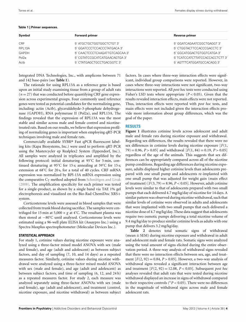
Table 1 | Primer sequences.
Symbol Forward primer Reverse primer
CRF 5′ ATGCTGCTGGTGGCTCTGT 3′ 5′ GGATCAGAATCGGCTGAGGT 3′
RPL13A 5′ GGATCCCTCCACCCTATGACA 3′ 5′ CTGGTACTTCCACCCGACCTC 3′
GAPDH 5′ CAACTCCCTCAAGATTGTCAGCAA 3′ 5′ GGCATGGACTGTGGTCATGA 3′
Pol2a 5′ CGTATCCGCATCATGAACAGTGA 3′ 5′ TCATCCATCTTATCCACCACCTCTT 3′
Actb 5′ CTATGAGCTGCCTGACGGTC 3′ 5′ AGTTTCATGGATGCCACAGG 3′
Integrated DNA Technologies, Inc., with amplicons between 71and 142 base-pairs (see Table 1).
The rationale for using RPL13A as a reference gene is basedupon an initial study examining tissue from a group of adult rats(n= 27) that was conducted before quantifying CRF gene expres-sion across experimental groups. Four commonly used referencegenes were tested as potential candidates for the normalizing gene,including: actin (Actb), glyceraldehyde-3-phosphate dehydroge-nase (GAPDH), RNA polymerase II (Pol2a), and RPL13A. Thefindings revealed that the expression of RPL13A was the moststable and similar across male and female control and nicotine-treated rats. Based on our results, we believe that expression profil-ing of normalizing genes is important when employing qRT-PCRtechniques involving male and female rats.
Commercially available SYBR® Fast qPCR fluorescent label-ing kits (Kapa Biosystems, Inc.) were used to perform qRT-PCRusing the Mastercycler ep Realplex2 System (Eppendorf, Inc.).All samples were analyzed in triplicates and amplified by thefollowing protocol: initial denaturing at 95˚C for 5 min, con-tinued denaturing at 95˚C for 15 s; annealing at 59˚C for 15 s;extension at 68˚C for 20 s, for a total of 40 cycles. CRF mRNAexpression was normalized by RPl-13A mRNA expression usingthe comparative CT method adopted from Schmittgen and Livak(2008). The amplification specificity for each primer was testedfor a single-product, as shown by a single band via TAE 1% gelelectrophoresis and visualized on the Bio-Rad ChemiDoc XRS+system.
Corticosterone levels were assessed in blood samples that werecollected from trunk blood during sacrifice. The samples were cen-trifuged for 15 min at 5,000× g at 4˚C. The resultant plasma wasthen stored at −80˚C until analyzed. Corticosterone levels wereestimated using a 96-well plate ELISA kit (Assaypro Inc.) using aSpectra Maxplus spectrophotometer (Molecular Devices Inc.).
STATISTICAL APPROACHFor study 1, cotinine values during nicotine exposure were ana-lyzed using a three-factor mixed model ANOVA with sex (maleand female), and age (adult and adolescent) as between subjectfactors, and day of sampling (7, 10, and 14 days) as a repeatedmeasures factor. Similarly, cotinine values during nicotine with-drawal were analyzed using a three-factor mixed model ANOVAwith sex (male and female), and age (adult and adolescent) asbetween subject factors, and time of sampling (6, 12, and 24 h)as a repeated measures factor. For study 2, each measure wasanalyzed separately using three-factor ANOVAs with sex (maleand female), age (adult and adolescent), and treatment (control,nicotine exposure, and nicotine withdrawal) as between subject
factors. In cases where three-way interaction effects were signif-icant, individual group comparisons were reported. However, incases where three-way interactions were not significant, two-wayinteractions were reported. All post hoc tests were conducted usingFisher’s LSD tests where appropriate (P < 0.05). Given that theresults revealed interaction effects, main effects were not reported.Thus, interaction effects were reported with post hoc tests, andmain effects were not included given the interaction effects pro-vide more information about group differences, which was thegoal of the paper.
RESULTSFigure 1 illustrates cotinine levels across adolescent and adultmale and female rats during nicotine exposure and withdrawal.Regarding sex differences, the results revealed that there were nosex differences in cotinine levels during nicotine exposure [F(1,79)= 0.96, P > 0.05] and withdrawal [F(1, 84)= 0.19, P > 0.05]regardless of the age of the animals. This suggests that sex dif-ferences can be appropriately compared across all of the nicotinepump conditions. Regarding age differences during nicotine expo-sure, adults displayed higher cotinine levels than adolescents pre-pared with one small pump and adolescents re-implanted withone small pump that was adjusted for weight gain (main effectof treatment) [F(3, 79)= 8.96, P < 0.05]. However, adult cotininelevels were similar to that of adolescents prepared with two smallpumps that each delivered 4.7 mg/kg/day of nicotine for 14 days. Asimilar pattern was observed during nicotine withdrawal, such thatsimilar levels of cotinine were observed in adults and adolescentsthat were implanted with two small pumps that each delivered anicotine dose of 4.7 mg/kg/day. These data suggest that adolescentsrequire two osmotic pumps delivering a total nicotine volume of9.4 mg/kg/day to produce similar cotinine levels as adults with onepump that delivers 3.2 mg/kg/day.
Table 2 denotes total somatic signs of withdrawal(mean± SEM) during nicotine exposure and withdrawal in adultand adolescent male and female rats. Somatic signs were analyzedusing the total amount of signs elicited during the entire obser-vation period. A three-way analysis of withdrawal signs revealedthat there were no interaction effects between sex, age, and treat-ment [F(2, 92)= 0.84, P > 0.05]. However, a two-way analysis ofwithdrawal signs revealed a significant interaction between ageand treatment [F(2, 92)= 12.08, P < 0.05]. Subsequent post hocanalyses revealed that adult rats that were tested during nicotinewithdrawal displayed an increase in signs of withdrawal comparedto their respective controls (∗P < 0.05). There were no differencesin the magnitude of withdrawal signs across male and femaleadolescent rats.
Frontiers in Psychiatry | Addictive Disorders and Behavioral Dyscontrol May 2013 | Volume 4 | Article 38 | 4

Torres et al. Females display stress during withdrawal
FIGURE 1 | Blood plasma cotinine levels (ng/ml±SEM) 7, 10, and14 days during nicotine exposure (top row) and then 6, 12, and 24 hafter pump removal (bottom row) in adult and adolescent male andfemale rats. Adult rats (n=26) received a large pump (model 2ml2) thatdelivered nicotine 3.2 mg/kg for 14 days. Three separate groups ofadolescent rats received a smaller model of pump (model 2002) that
delivered: (1) a dose of 4.7 mg/kg/day for 14 days (n=17), (2) a dose of4.7 mg/kg/day that was replaced after 7 days with a new pump that alsodelivered 4.7 mg/kg/day (n=17), and (3) a dose of 9.4 mg/kg/day that wasdivided in two small pumps (n=32). The dagger (†) denotes a significantdifference across all time points between nicotine-treated adolescents andadults (P < 0.05).
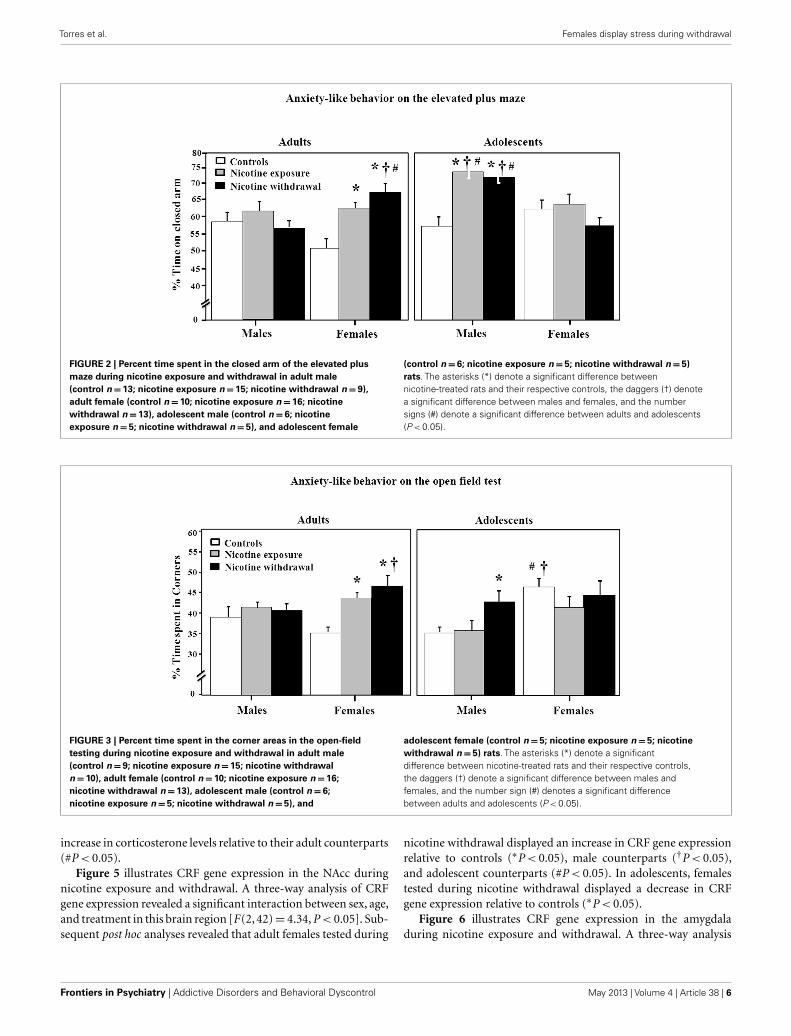
Figure 2 illustrates anxiety-like behavior as assessed by theelevated plus maze during nicotine exposure and withdrawal.Anxiety-like behavior was operationally defined as an increase intime spent in the closed arm as compared to controls. A three-wayanalysis of percent time spent in the closed arm revealed a signifi-cant interaction between sex, age, and treatment [F(2, 96)= 8.85,P < 0.05]. Subsequent post hoc analyses revealed that adult femalesthat were tested during nicotine exposure displayed an increasein anxiety-like behavior relative to controls (∗P < 0.05). How-ever, adult females tested during nicotine withdrawal displayedan increase in anxiety-like behavior that was significantly higherthan their respective controls (∗P < 0.05), male counterparts(†P < 0.05), and adolescent counterparts (#P < 0.05). In adoles-cents, the males displayed the largest effects of nicotine exposureand withdrawal on anxiety-like behavior as compared to respec-tive controls (∗P < 0.05), female counterparts (†P < 0.05), andadolescent counterparts (#P < 0.05).
Figure 3 illustrates anxiety-like behavior as assessed by theopen-field test during nicotine exposure and withdrawal. Anxiety-like behavior was operationally defined as an increase in time spentin the corners of the open field as compared to controls. A three-way analysis of percent corner time revealed a significant interac-tion between sex, age, and treatment [F(2, 92)= 3.85, P < 0.05].Subsequent post hoc analyses revealed that adult females testedduring nicotine exposure displayed an increase in anxiety-likebehavior relative to controls (∗P < 0.05). However, adult femalestested during nicotine withdrawal displayed an increase in anxiety-like behavior that was higher than respective controls (∗P < 0.05)
Table 2 | Physical signs of withdrawal.
Experimental
group
Adult
male
Adult
female
Adolescent
male
Adolescent
female
Controls 7.6±0.8 7.8±0.9 5±0.7 3.8±0.3
Nicotine exposure 5.2±0.4 4.3±0.3 3.2±0.3 4.3±0.5
Nicotine withdrawal *15.5±1.8 *11.5±1.4 3.8±0.3 3.2±0.6
The asterisks (*) denote a significant difference from respective controls
(P < 0.05).
and their male counterparts (†P < 0.05). In adolescents, malestested during nicotine withdrawal displayed an increase in anxiety-like behavior relative to controls (∗P < 0.05). Adolescent femalecontrols displayed an increase in anxiety-like behavior relative tomales (†P < 0.05) and their adult counterparts (#P < 0.05).
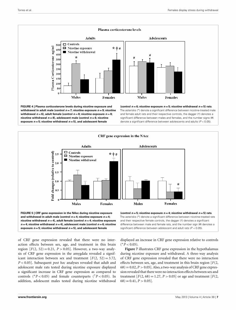
Figure 4 illustrates plasma corticosterone levels during nico-tine exposure and withdrawal. A three-way analysis of corticos-terone levels revealed a significant interaction between sex, age,and treatment [F(2, 66)= 3.2, P < 0.05]. Subsequent post hocanalyses revealed that adult males tested during nicotine expo-sure displayed an increase in corticosterone levels relative tocontrols (∗P < 0.05). Adult females tested during nicotine with-drawal displayed an increase in corticosterone levels relative tocontrols (∗P < 0.05), male counterparts (†P < 0.05), and ado-lescent counterparts (#P < 0.05). In adolescents, the male con-trols and males tested during nicotine withdrawal displayed an
www.frontiersin.org May 2013 | Volume 4 | Article 38 | 5
Torres et al. Females display stress during withdrawal
FIGURE 2 | Percent time spent in the closed arm of the elevated plusmaze during nicotine exposure and withdrawal in adult male(control n=13; nicotine exposure n=15; nicotine withdrawal n=9),adult female (control n=10; nicotine exposure n=16; nicotinewithdrawal n=13), adolescent male (control n=6; nicotineexposure n=5; nicotine withdrawal n=5), and adolescent female
(control n= 6; nicotine exposure n=5; nicotine withdrawal n=5)rats. The asterisks (*) denote a significant difference betweennicotine-treated rats and their respective controls, the daggers (†) denotea significant difference between males and females, and the numbersigns (#) denote a significant difference between adults and adolescents(P < 0.05).
FIGURE 3 | Percent time spent in the corner areas in the open-fieldtesting during nicotine exposure and withdrawal in adult male(control n=9; nicotine exposure n=15; nicotine withdrawaln=10), adult female (control n=10; nicotine exposure n=16;nicotine withdrawal n=13), adolescent male (control n=6;nicotine exposure n=5; nicotine withdrawal n= 5), and
adolescent female (control n= 5; nicotine exposure n=5; nicotinewithdrawal n=5) rats. The asterisks (*) denote a significantdifference between nicotine-treated rats and their respective controls,the daggers (†) denote a significant difference between males andfemales, and the number sign (#) denotes a significant differencebetween adults and adolescents (P < 0.05).
increase in corticosterone levels relative to their adult counterparts(#P < 0.05).
Figure 5 illustrates CRF gene expression in the NAcc duringnicotine exposure and withdrawal. A three-way analysis of CRFgene expression revealed a significant interaction between sex, age,and treatment in this brain region [F(2, 42)= 4.34, P < 0.05]. Sub-sequent post hoc analyses revealed that adult females tested during
nicotine withdrawal displayed an increase in CRF gene expressionrelative to controls (∗P < 0.05), male counterparts (†P < 0.05),and adolescent counterparts (#P < 0.05). In adolescents, femalestested during nicotine withdrawal displayed a decrease in CRFgene expression relative to controls (∗P < 0.05).
Figure 6 illustrates CRF gene expression in the amygdaladuring nicotine exposure and withdrawal. A three-way analysis
Frontiers in Psychiatry | Addictive Disorders and Behavioral Dyscontrol May 2013 | Volume 4 | Article 38 | 6
Torres et al. Females display stress during withdrawal
FIGURE 4 | Plasma corticosterone levels during nicotine exposure andwithdrawal in adult male (control n=7; nicotine exposure n=9; nicotinewithdrawal n=6), adult female (control n=8; nicotine exposure n=8;nicotine withdrawal n= 8), adolescent male (control n=6; nicotineexposure n=5; nicotine withdrawal n= 5), and adolescent female
(control n=6; nicotine exposure n=5; nicotine withdrawal n=5) rats.The asterisks (*) denote a significant difference between nicotine-treated maleand female adult rats and their respective controls, the dagger (†) denotes asignificant difference between males and females, and the number signs (#)denote a significant difference between adolescents and adults (P < 0.05).
FIGURE 5 | CRF gene expression in the NAcc during nicotine exposureand withdrawal in adult male (control n=4; nicotine exposure n=4;nicotine withdrawal n=4), adult female (control n=4; nicotine exposuren=4; nicotine withdrawal n=4), adolescent male (control n=6; nicotineexposure n=5; nicotine withdrawal n= 5), and adolescent female
(control n=5; nicotine exposure n=4; nicotine withdrawal n=5) rats.The asterisks (*) denote a significant difference between nicotine-treated ratsand their respective female controls, the dagger (†) denotes a significantdifference between male and female rats, and the number sign (#) denotes asignificant difference between adolescent and adult rats (P < 0.05).
of CRF gene expression revealed that there were no inter-action effects between sex, age, and treatment in this brainregion [F(2, 52)= 0.21, P > 0.05]. However, a two-way analy-sis of CRF gene expression in the amygdala revealed a signif-icant interaction between sex and treatment [F(2, 52)= 3.72,P < 0.05]. Subsequent post hoc analyses revealed that adult andadolescent male rats tested during nicotine exposure displayeda significant increase in CRF gene expression as compared tocontrols (∗P < 0.05) and female counterparts (†P < 0.05). Inaddition, adolescent males tested during nicotine withdrawal
displayed an increase in CRF gene expression relative to controls(∗P < 0.05).
Figure 7 illustrates CRF gene expression in the hypothalamusduring nicotine exposure and withdrawal. A three-way analysisof CRF gene expression revealed that there were no interactioneffects between sex, age, and treatment in this brain region [F(2,68)= 0.02, P > 0.05]. Also, a two-way analysis of CRF gene expres-sion revealed that there were no interaction effects between sex andtreatment [F(2, 68)= 1.27, P > 0.05] or age and treatment [F(2,68)= 0.41, P > 0.05].
www.frontiersin.org May 2013 | Volume 4 | Article 38 | 7

Torres et al. Females display stress during withdrawal
FIGURE 6 | CRF gene expression in the amygdala during nicotineexposure and withdrawal in adult male (control n=10; nicotineexposure n=5; nicotine withdrawal n=5), adult female (control n=7;nicotine exposure n=7; nicotine withdrawal n= 4), adolescent male
(control n= 4; nicotine exposure n=6; nicotine withdrawal n=4), andadolescent female (control n= 4; nicotine exposure n=4; nicotinewithdrawal n=4) rats. The asterisks (*) denote a significant differencebetween nicotine-treated rats and their respective male controls (P < 0.05).
FIGURE 7 | CRF gene expression in the hypothalamus duringnicotine exposure and withdrawal in adult male (control n=13;nicotine exposure n=13; nicotine withdrawal n=6), adultfemale (control n=6; nicotine exposure n=6; nicotine
withdrawal n=4), adolescent male (control n=8; nicotineexposure n=5; nicotine withdrawal n=5), and adolescentfemale (control n=5; nicotine exposure n=5; nicotinewithdrawal n=6) rats.
DISCUSSIONTo summarize, during nicotine withdrawal, adult females dis-played increases in anxiety-like behavior, increases in plasma cor-ticosterone levels, and changes in CRF gene expression in the NAccthat were higher as compared to males. Control studies comparingsex differences during nicotine exposure, revealed that adult malesdisplayed an increase in plasma corticosterone levels and increasesin CRF gene expression in the amygdala. The sex differences in
adults did not appear to be confounded by nicotine metabo-lism, since cotinine values were the same in male and female ratsthroughout our experimental procedures. Regarding age differ-ences, adolescent males displayed some indices of stress duringnicotine exposure that persisted into the withdrawal period. Thismay have been related to the high doses of nicotine that theadolescents required to produce comparable cotinine values asadults.
Frontiers in Psychiatry | Addictive Disorders and Behavioral Dyscontrol May 2013 | Volume 4 | Article 38 | 8
Torres et al. Females display stress during withdrawal
The major finding of this study is that adult females expe-rience greater behavioral and biological indices of stress duringnicotine withdrawal as compared to males. Adult females spentmore time on the closed arm of the elevated plus maze duringnicotine withdrawal as compared to males. Consistent with this,adult females also spent more time in the corner areas of the openfield during nicotine withdrawal relative to males. Our behav-ioral results corroborate with our biological assessment of stress,as adult females also displayed increases in plasma corticosteronelevels during nicotine withdrawal that were higher than males.Our results are consistent with previous reports demonstratingthat female adult mice display more anxiety-like behavior on theelevated plus maze during nicotine withdrawal as compared tomales (Kota et al., 2007, 2008; Caldarone et al., 2008). Two recentreports also showed that adult female rats display higher plasmacorticosterone levels during nicotine withdrawal as compared tomales (Gentile et al., 2011; Skwara et al., 2012).
The present study also revealed that adult females displayed anincrease in CRF mRNA expression in the NAcc during nicotinewithdrawal that was higher than males. Previous reports supportthe role of the NAcc in modulating stress. For example, intra-NAccadministration of CRF has been shown to produce anxiety-likebehavior on the elevated plus maze (Chen et al., 2012). The NAccis also strongly activated following presentation of a stressful stim-ulus (Noh et al., 2012). The latter report showed that the NAcc wasactivated to a greater extent following restraint stress as comparedto cold-water submersion. Thus, the NAcc may be differentiallyresponsive to various types of stressors. Our findings suggest thatthe NAcc is also involved in stress produced by nicotine with-drawal. Consistent with this hypothesis, the deficits in brain rewardfunction produced by nicotine withdrawal are alleviated by block-ade of CRF receptors in the NAcc (Marcinkiewcz et al., 2009). Ourfinding that the hypothalamus was not altered during withdrawal,is consistent with the finding that CRF mRNA was not altered inthe hypothalamus of male rats experiencing spontaneous nicotinewithdrawal (Semba et al., 2004). Thus, the hypothalamus may notplay a central role in modulating negative affective states involvingstress produced by nicotine withdrawal.
Our findings also suggest that the NAcc is a structure involvedin sex-dependent differences to drug withdrawal. This is con-sistent with previous studies examining withdrawal from otherdrugs of abuse. For example, morphine withdrawal produced adecrease in µ-opioid receptors in the NAcc of female but not malemice (Diaz et al., 2006). Also, multiple withdrawal periods fromethanol produced an increase in proteins involved in vesicularpackaging and exocytosis in the NAcc of female but not malerats (Bell et al., 2006, 2009). Following abstinence from cocaineself-administration, delta opioid receptors and dopamine phos-phoproteins are increased to a greater extent in the NAcc of femaleversus male rats (Lynch et al., 2007; Ambrose-Lanci et al., 2008).Taken together with the present findings, there is strong evidenceto suggest that the NAcc modulates sex differences produced bywithdrawal from drugs of abuse.
There are several ways in which females may be more suscep-tible to stress produced by nicotine withdrawal. There is muchevidence to suggest that CRF systems are enhanced in females ver-sus males (Bangasser, 2013; Valentino et al., 2013). Females display
hypersecretion of CRF and more CRF-1 receptors in the locuscoeruleus, a brain region that coordinates arousal components ofthe stress response (Curtis et al., 2006; Bangasser et al., 2013).Females also display a higher ratio of CRF-1 receptors to couplingof G-proteins versus male rats, suggesting that the female CRF sys-tem has greater intracellular signaling capacity (Bangasser et al.,2010). The beta-arrestin2 protein is an intracellular protein thatinternalizes the CRF-1 receptor into the cell cytoplasm and pre-vents it from being activated by CRF (Aguilera et al., 2004; Holmeset al., 2006). Female rats display lower levels of beta-arrestin2 thanmale rats, suggesting that females are more responsive to CRFstimulation due to reduced internalization of the CRF-1 receptoras compared to males (Bangasser and Valentino, 2012). Femalesmay also be more susceptible to stress produced by withdrawal viaovarian hormones. For example, direct activation of estrogen-betareceptors (ERβ) increase CRF mRNA expression in vitro (Chenet al., 2008; Lalmansingh and Uht, 2008; Zhu and Zhou, 2008).Furthermore, the estrogen gene sequence serves as a promoter ofCRF gene transcription (Vamvakopoulos and Chrousos, 1993).Collectively, these studies suggest that females have a hypersensi-tive CRF system, and this may contribute to the enhanced stressproduced by nicotine withdrawal in females versus males.
The present study also revealed a robust increase in CRF geneexpression in the amygdala of male rats during nicotine exposure.A recent report showed that CRF levels were increased in the amyg-dala of adult male rats experiencing nicotine withdrawal (Georgeet al., 2007). The rats in the latter study received a nicotine antag-onist to precipitate withdrawal while nicotine was being deliveredvia an osmotic pump. The findings from the present study are con-sistent with those of George et al. (2007), given that the rats fromboth studies had circulating levels of nicotine in their system at thetime of analysis. Thus, the possibility exists that nicotine directlyactivates CRF systems in the amygdala, especially given that thechanges in CRF were not observed in the absence of nicotine inour study. Future studies are needed to fully understand the roleof CRF systems in the amygdala in modulating the direct effectsof nicotine and the long-term consequences of withdrawal fromthis drug. A unique challenge for this work is that nicotine expo-sure is an inherent part of studies that assess withdrawal, either byspontaneous or precipitated methods.
The present study also compared sex differences in the somaticsigns of nicotine withdrawal. Our findings suggest that there wereno differences in somatic signs of withdrawal between adult maleand female rats. A report by Hamilton et al. (2009) showed thatfemale rats display more somatic signs of withdrawal relative tomales. The discrepancy between these reports may be related todifferences in lighting conditions given that Hamilton et al. onlyreported sex differences in rats that were tested in dim, but notwell-lit conditions. In the present study, the somatic signs data werecollected in well-lit conditions whereas the anxiety-like behav-ior was collected in the dark under a red light. Perhaps differentlighting conditions may be considered in future studies examininganxiety-like behavior produced by nicotine withdrawal, especiallygiven the reported effects of lighting conditions on the somaticsigns of withdrawal.
Another finding of this study is that male and female ado-lescents generally displayed fewer somatic signs of withdrawal
www.frontiersin.org May 2013 | Volume 4 | Article 38 | 9

Torres et al. Females display stress during withdrawal
as compared to adults. These findings are consistent with pre-vious work in our laboratory and others demonstrating that thebehavioral and neurochemical effects of withdrawal are dimin-ished in adolescent versus adult rats (Smith et al., 2006; Wilmouthand Spear, 2006; Shram et al., 2008; O’Dell, 2009). This studyextends this work by showing that adolescent females are also lesssensitive to nicotine withdrawal as compared to adult females.An important caveat; however, is that adolescent males displayedanxiety-like behavior and biological markers of stress during nico-tine exposure that persisted 24-h later into withdrawal. There aretwo possible explanations for this effect. First, adolescent malesmay not be impervious to all aspects of withdrawal, which mayinduce a stress response that contributes to tobacco use in ado-lescent males. Second, it is possible that nicotine elicited a stressresponse in adolescent males. This explanation is consistent withthe finding that CRF gene expression was increased in the amyg-dala of adolescent males during nicotine exposure. We suggestthat the ability of nicotine to induce a stress response was likelyrelated to the three-fold higher doses of this drug that were usedto produce equivalent plasma levels of cotinine as adults. Thelack of stress effects in female adolescents was likely related tohigh tolerance to the aversive effects of nicotine, an effect that hasbeen previously demonstrated (Torres et al., 2009). Future stud-ies are needed to examine sex differences to stress produced bynicotine withdrawal, perhaps with a model such as nicotine vaporinhalation that circumvents the dosing issues that arose in thepresent study with osmotic pumps. Despite this, the present studyprovided important parametric information regarding equivalentdoses of nicotine in adolescent and adult rats using different pumpsizes. Our results raise an important issue for future studies com-paring developmental differences to nicotine since high doses ofnicotine may produce stress in adolescent males.
There are some limitations in the present study. In some cases,our behavioral and biochemical measures appear to contradicteach other. For example, in adolescent males, we observed anincrease in anxiety-like behavior in the plus maze but not theopen field. This discrepancy is likely related to the sensitivity ofthese measures in assessing anxiety-like behavior. In adult females,during withdrawal, the pattern of changes was consistent (highanxiety-like behavior and corticosterone). However, during nico-tine exposure the pattern of changes was not consistent (highanxiety-like behavior but no changes in corticosterone). The lack
of changes in corticosterone was likely due to a higher baselinevalue in adult females. In adult males, during withdrawal, the pat-tern of changes was consistent (no anxiety-like behavior and nochanges in corticosterone). However,during nicotine exposure, thepattern of changes was not consistent (no changes in anxiety-likebehavior and an increase in corticosterone). One might argue thatthe changes in corticosterone were aberrant; however, this groupalso showed an increase in CRF gene expression in the amyg-dala. Thus, it may be the case the nicotine exposure is more stressinducing in adult males as compared to withdrawal from this drug.
In conclusion, our results suggest that during nicotine with-drawal female rats display behavioral and biological markers ofstress that are enhanced compared to males. These findings con-tribute to a body of literature showing that female rats displaygreater rewarding effects of nicotine as compared to males (Donnyet al., 2000; Klein et al., 2004; Chaudhri et al., 2005; Torres et al.,2009). Taken together, there is pre-clinical evidence to suggestthat enhanced rewarding effects of nicotine and intense stressproduced by withdrawal both contribute to the greater vulnera-bility to tobacco use observed in women. In addition, our findingssuggest that the most effective cessation treatments for womenshould also alleviate intense stress produced by nicotine with-drawal. For example, one approach might include CRF antagonistsin combination with other tobacco cessation treatments, suchas NRT or partial nicotinic agonists. Future studies are neededto understand the complex interactions in the brain that mod-ulate sex differences to nicotine use. This work is importanttoward reducing health disparities related to tobacco use amongwomen.
ACKNOWLEDGMENTSThe authors appreciate the insightful comments during the prepa-ration of this manuscript from Drs. Eddie Castañeda, ChristinaSobin, Kristin Gosselink, and Theodore Cooper. The authorswould also like to thank Dr. James Orfila, Arturo Orona, VanessaValenzuela, Lorena De los Santos, and Adrian Muñiz for their tech-nical assistance. This research was supported by The NationalInstitute on Drug Abuse (R01-DA021274, R24-DA029989, andR25-DA033613) and The National Institute of Minority HealthDisparities (G12MD007592). Student funding was also providedby the UTEP Dodson Doctoral Fellowship Program (Dr. OscarTorres).
REFERENCESAguilera, G., Nikodemova, M., Wynn,
P. C., and Catt, K. J. (2004). Corti-cotropin releasing hormone recep-tors: two decades later. Peptides 25,319–329.
Ambrose-Lanci, L. M., Peiris, N.B., Unterwald, E. M., and VanBockstaele, E. J. (2008). Cocainewithdrawal-induced trafficking ofdelta-opioid receptors in rat nucleusaccumbens. Brain Res. 1210, 92–102.
Anderson, C., and Burns, D. M. (2000).Patterns of adolescent smoking initi-ation rates by ethnicity and sex. Tob.Control. 9, 4–8.
Aronson, K. R., Almeida, D. M., Stawski,R. S., Klein, L. C., and Kozlowski,L. T. (2008). Smoking is associatedwith worse mood on stressful days:results from a national diary study.Ann. Behav. Med. 36, 259–269.
Aydin, C., Oztan, O., and Isgor, C.(2011). Vulnerability to nico-tine abstinence-related socialanxiety-like behavior: molecularcorrelates in neuropeptide Y, Y2receptor and corticotropin releas-ing factor. Neurosci. Lett. 490,220–225.
Bangasser, D. A. (2013). Sex differ-ences in stress-related receptors:
“micro” differences with “macro”implications for mood and anxietydisorders. Biol. Sex Differ. 4, 2.
Bangasser, D. A., Reyes, B. A., Piel, D.,Garachh, V., Zhang, X. Y., Plona, Z.M., et al. (2013). Increased vulner-ability of the brain norepinephrinesystem of females to corticotropin-releasing factor overexpression. Mol.Psychiatry 18, 166–173.
Bangasser, D. A., and Valentino, R. J.(2012). Sex differences in molecularand cellular substrates of stress. Cell.Mol. Neurobiol. 32, 709–723.
Bangasser, D. A., Curtis, A., Reyes, B.A., Bethea, T. T., Parastatidis, I.,
Ischiropoulos, H., et al. (2010).Sex differences in corticotropin-releasing factor receptor signalingand trafficking: potential rolein female vulnerability tostress-related psychopathol-ogy. Mol. Psychiatry 15, 877,896–904.
Bell, R. L., Kimpel, M. W., McClintick,J. N., Strother, W. N., Carr, L. G.,Liang, T., et al. (2009). Gene expres-sion changes in the nucleus accum-bens of alcohol-preferring rats fol-lowing chronic ethanol consump-tion. Pharmacol. Biochem. Behav. 94,131–147.
Frontiers in Psychiatry | Addictive Disorders and Behavioral Dyscontrol May 2013 | Volume 4 | Article 38 | 10

Torres et al. Females display stress during withdrawal
Bell, R. L., Kimpel, M. W., Rodd, Z.A., Strother, W. N., Bai, F., Peper, C.L., et al. (2006). Protein expressionchanges in the nucleus accumbensand amygdala of inbred alcohol-preferring rats given either contin-uous or scheduled access to ethanol.Alcohol 40, 3–17.
Bruijnzeel,A. W. (2012). Tobacco addic-tion and the dysregulation of brainstress systems. Neurosci. Biobehav.Rev. 36, 1418–1441.
Bruijnzeel, A. W., Ford, J., Rogers, J.A., Scheick, S., Ji, Y., Bishnoi, M., etal. (2012). Blockade of CRF1 recep-tors in the central nucleus of theamygdala attenuates the dysphoriaassociated with nicotine withdrawalin rats. Pharmacol. Biochem. Behav.101, 62–68.
Caldarone, B. J., King, S. L., and Pic-ciotto, M. R. (2008). Sex differ-ences in anxiety-like behavior andlocomotor activity following chronicnicotine exposure in mice. Neurosci.Lett. 439, 187–191.
Centers for Disease Control and Pre-vention (CDC). (2012). Currenttobacco use among middle and highschool students – United States,2011. MMWR Morb. Mortal. Wkly.Rep. 61, 581–585.
Cepeda-Benito, A., Reynoso, J. T., andErath, S. (2004). Meta-analysis ofthe efficacy of nicotine replace-ment therapy for smoking cessa-tion: differences between men andwomen. J. Consult. Clin. Psychol. 72,712–722.
Chaudhri, N., Caggiula, A. R., Donny,E. C., Booth, S., Gharib, M. A.,Craven, L. A., et al. (2005). Sex dif-ferences in the contribution of nico-tine and nonpharmacological stim-uli to nicotine self-administrationin rats. Psychopharmacology (Berl.)180, 258–266.
Chen, X. N., Zhu, H., Meng, Q. Y.,and Zhou, J. N. (2008). Estrogenreceptor-alpha and -beta regulatethe human corticotropin-releasinghormone gene through similar path-ways. Brain Res. 1223, 1–10.
Chen, Y. W., Rada, P. V., Butzler, B. P.,Leibowitz, S. F., and Hoebel, B. G.(2012). Corticotropin-releasing fac-tor in the nucleus accumbens shellinduces swim depression, anxiety,and anhedonia along with changesin local dopamine/acetylcholine bal-ance. Neuroscience 206, 155–166.
Colby, S. M., Tiffany, S. T., Shiffman, S.,and Niaura, R. S. (2000). Are ado-lescent smokers dependent on nico-tine? A review of the evidence. DrugAlcohol Depend. 59, 83–95.
Curtis, A. L., Bethea, T., and Valentino,R. J. (2006). Sexually dimorphic
responses of the brain norep-inephrine system to stress andcorticotropin-releasing factor. Neu-ropsychopharmacology 31, 544–554.
Diaz, S. L., Barros, V. G., Antonelli,M. C., Rubio, M. C., and Balerio,G. N. (2006). Morphine withdrawalsyndrome and its prevention withbaclofen: autoradiographic study ofmu-opioid receptors in prepubertalmale and female mice. Synapse 60,132–140.
Dickmann, P. J., Mooney, M. E., Allen,S. S., Hanson, K., and Hatsukami,D. K. (2009). Nicotine withdrawaland craving in adolescents: effects ofsex and hormonal contraceptive use.Addict. Behav. 34, 620–623.
Donny, E. C., Caggiula, A. R., Row-ell, P., Gharib, M. A., Maldovan, V.,Booth, S., et al. (2000). Nicotine self-administration in rats: estrous cycleeffects, sex differences and nicotinicreceptor binding. Psychopharmacol-ogy (Berl.) 151, 392–405.
Gallagher, J. P., Orozco-Cabal, L. F.,Liu, J., and Shinnick-Gallagher, P.(2008). Synaptic physiology of cen-tral CRF system. Eur. J. Pharmacol.583, 215–225.
Gentile, N. E., Andrekanic, J. D., Kar-woski, T. E., Czambel, R. K., Rubin,R. T., and Rhodes, M. E. (2011).Sexually diergic hypothalamic-pituitary-adrenal (HPA) responsesto single-dose nicotine, continuousnicotine infusion, and nicotinewithdrawal by mecamylamine inrats. Brain Res. Bull. 85, 145–152.
George, O., Ghozland, S., Azar, M.R., Cottone, P., Zorrilla, E. P., Par-sons, L. H., et al. (2007). CRF–CRF 1 system activation medi-ates withdrawal-induced increasesin nicotine self-administration innicotine-dependent rats. Proc. Natl.Acad. Sci. U.S.A. 104, 17198–17203.
Hamilton, K. R., Berger, S. S., Perry,M. E., and Grunberg, N. E. (2009).Behavioral effects of nicotine with-drawal in adult male and femalerats. Pharmacol. Biochem. Behav. 92,51–59.
Hammond, S. K. (2009). Global pat-terns of nicotine and tobacco con-sumption. Handb. Exp. Pharmacol.192, 3–28.
Heishman, S. J., Lee, D. C., Taylor, R.C., and Singleton, E. G. (2010). Pro-longed duration of craving, mood,and autonomic responses elicitedby cues and imagery in smokers:effects of tobacco deprivation andsex. Exp. Clin. Psychopharmacol. 18,245–256.
Hogle, J. M., and Curtin, J. J.(2006). Sex differences in negativeaffective response during nicotine
withdrawal. Psychophysiology 43,344–356.
Holmes, K. D., Babwah, A. V., Dale, L.B., Poulter, M. O., and Ferguson, S.S. (2006). Differential regulation ofcorticotropin releasing factor 1alphareceptor endocytosis and traffickingby beta-arrestins and Rab GTPases.J. Neurochem. 96, 934–949.
Hughes, J. R., and Callas, P. W. (2010).Definition of a quit attempt: a repli-cation test. Nicotine Tob. Res. 12,1176–1179.
Kiyohara, C., and Ohno, Y. (2010).Sex differences in lung cancer sus-ceptibility: a review. Gend. Med. 7,381–401.
Klein, L. C., Stine, M. M., Vanden-bergh, D. J., Whetzel, C. A., andKamens, H. M. (2004). Sex dif-ferences in voluntary oral nicotineconsumption by adolescent mice: adose-response experiment. Pharma-col. Biochem. Behav. 78, 13–25.
Koob, G. F. (2010). The role of CRF andCRF-related peptides in the dark sideof addiction. Brain Res. 1314, 3–14.
Koob, G. F., and Volkow, N. D. (2010).Neurocircuitry of addiction. Neu-ropsychopharmacology 35, 217–238.
Kota, D., Martin, B. R., and Damaj, M.(2008). Age-dependent differencesin nicotine reward and withdrawalin female mice. Psychopharmacology(Berl.) 198, 201–210.
Kota, D., Martin, B. R., Robinson, S.E., and Damaj, M. I. (2007). Nico-tine dependence and reward differbetween adolescent and adult malemice. J. Pharmacol. Exp. Ther. 322,399–407.
Lalmansingh, A. S., and Uht, R.M. (2008). Estradiol regulatescorticotropin-releasing hormonegene (CRF) expression in a rapidand phasic manner that paral-lels estrogen receptor-alpha and-beta recruitment to a 3′,5′-cyclicadenosine 5′-monophosphateregulatory region of the proximalCRF promoter. Endocrinology 149,346–357.
Langhammer, A., Johnsen, R., Gulsvik,A., Holmen, T. L., and Bjermer, L.(2003). Sex differences in lung vul-nerability to tobacco smoking. Eur.Respir. J. 21, 1017–1023.
Langhammer, A., Johnsen, R., Hol-men, J., Gulsvik, A., and Bjermer,L. (2000). Cigarette smoking givesmore respiratory symptoms amongwomen than among men. The Nord-Trondelag Health Study (HUNT).J. Epidemiol. Community Health 54,917–922.
Lombardi, E. M., Prado, G. F., SantosUde, P., and Fernandes, F. L. (2011).Women and smoking: risks, impacts,
and challenges. J. Bras. Pneumol. 37,118–128.
Lutfy, K., Brown, M. C., Nerio, N.,Aimiuwu, O., Tran, B., Anghel, A.,et al. (2006). Repeated stress altersthe ability of nicotine to activatethe hypothalamic-pituitary-adrenalaxis. J. Neurochem. 99, 1321–1327.
Lynch, W. J., Kiraly, D. D., Cal-darone, B. J., Picciotto, M. R.,and Taylor, J. R. (2007). Effect ofcocaine self-administration on stri-atal PKA-regulated signaling in maleand female rats. Psychopharmacology(Berl.) 191, 263–271.
Marcinkiewcz, C. A., Prado, M. M.,Isaac, S. K., Marshall, A., Rylkova,D., and Bruijnzeel, A. W. (2009).Corticotropin-releasing factorwithin the central nucleus of theamygdala and the nucleus accum-bens shell mediates the negativeaffective state of nicotine withdrawalin rats. Neuropsychopharmacology34, 1743–1752.
Nakajima, M., and al’Absi, M. (2012).Predictors of risk for smokingrelapse in men and women: aprospective examination. Psychol.Addict. Behav. 26, 633–637.
Noh, S. J., Kang, D. W., Yoo, S. B., Lee, J.Y., Kim, J. Y., Kim, B. T., et al. (2012).Stress-responsive hypothalamic-nucleus accumbens regulation mayvary depending on stressors. IndianJ. Exp. Biol. 50, 447–454.
O’Dell, L. E. (2009). A psychobiolog-ical framework of the substratesthat mediate nicotine use duringadolescence. Neuropharmacology 56,263–278.
O’Dell, L. E., Bruijnzeel, A. W., Ghoz-land, S., Markou, A., and Koob, G. F.(2004). Nicotine withdrawal in ado-lescent and adult rats. Ann. N. Y.Acad. Sci. 1021, 167–174.
O’Dell, L. E., Bruijnzeel, A. W., Smith,R. T., Parsons, L. H., Merves, M.L., Goldberger, B. A., et al. (2006).Diminished nicotine withdrawal inadolescent rats: implications for vul-nerability to addiction. Psychophar-macology (Berl.) 186, 612–619.
Oh, D. L., Heck, J. E., Dresler, C., All-wright, S., Haglund, M., Del Mazo,S. S., et al. (2010). Determinants ofsmoking initiation among womenin five European countries: a cross-sectional survey. BMC Public Health10:74. doi:10.1186/1471-2458-10-74
Parrott, A. C., and Murphy, R. S. (2012).Explaining the stress-induced effectsof nicotine to cigarette smok-ers. Hum. Psychopharmacol. 27,150–155.
Perkins,K. A. (2001). Smoking cessationin women. Special considerations.CNS Drugs 15, 391–411.
www.frontiersin.org May 2013 | Volume 4 | Article 38 | 11
Torres et al. Females display stress during withdrawal
Perkins, K. A. (2009). Sex differences innicotine reinforcement and reward:influences on the persistence oftobacco smoking. Nebr. Symp. Motiv.55, 143–169.
Perkins, K. A., Giedgowd, G. E., Kare-litz, J. L., Conklin, C. A., and Ler-man,C. (2012). Smoking in responseto negative mood in men versuswomen as a function of distresstolerance. Nicotine Tob. Res. 14,1418–1425.
Perkins, K. A., Karelitz, J. L., Giedgowd,G. E., and Conklin, C. A. (2013).Negative mood effects on craving tosmoke in women versus men. Addict.Behav. 38, 1527–1531.
Perkins, K. A., and Scott, J. (2008). Sexdifferences in long-term smokingcessation rates due to nicotine patch.Nicotine Tob. Res. 10, 1245–1250.
Piper, M. E., Cook, J. W., Schlam, T.R., Jorenby, D. E., Smith, S. S., Bolt,D. M., et al. (2010). Gender, race,and education differences in absti-nence rates among participants intwo randomized smoking cessationtrials. Nicotine Tob. Res. 12, 647–657.
Rahmanian, S. D., Diaz, P. T., andWewers, M. E. (2011). Tobaccouse and cessation among women:research and treatment-relatedissues. J. Womens Health (Larchmt)20, 349–357.
Rhodes, M. E., Kennell, J. S., Belz, E.E., Czambel, R. K., and Rubin, R.T. (2004). Rat estrous cycle influ-ences the sexual diergism of HPAaxis stimulation by nicotine. BrainRes. Bull. 64, 205–213.
Saladin, M. E., Gray, K. M., Carpenter,M. J., LaRowe, S. D., DeSantis, S. M.,
and Upadhyaya, H. P. (2012). Gen-der differences in craving and cuereactivity to smoking and negativeaffect/stress cues. Am. J. Addict. 21,210–220.
Schmittgen, T. D., and Livak, K. J.(2008). Analyzing real-time PCRdata by the comparative C(T)method. Nat. Protoc. 3, 1101–1108.
Schnoll, R. A., Patterson, F., and Lerman,C. (2007). Treating tobacco depen-dence in women. Womens Health 16,1211–1218.
Semba, J., Wakuta, M., Maeda, J., andSuhara, T. (2004). Nicotine with-drawal induces subsensitivity ofhypothalamic-pituitary-adrenal axisto stress in rats: implications forprecipitation of depression duringsmoking cessation. Psychoneuroen-docrinology 29, 215–226.
Shram, M. J., Siu, E. C., Li, Z., Tyndale,R. F., and Lê, A. D. (2008). Inter-actions between age and the aver-sive effects of nicotine withdrawalunder mecamylamine-precipitatedand spontaneous conditions inmale Wistar rats. Psychopharmacol-ogy (Berl.) 198, 181–190.
Skwara, A. J., Karwosk, T. E., Czambel,R. K., Rubin, R. T., and Rhodes, M.E. (2012). Influence of environmen-tal enrichment on hypothalamic-pituitary-adrenal (HPA) responsesto single-dose nicotine, continu-ous nicotine by osmotic mini-pumps, and nicotine withdrawal bymecamylamine in male and femalerats. Behav. Brain Res. 234, 1–10.
Smith, L. N., McDonald, C. G.,Bergstrom, H. C., Brielmaier, J. M.,Eppolito, A. K., Wheeler, T. L., et
al. (2006). Long-term changes infear conditioning and anxiety-likebehavior following nicotine expo-sure in adult versus adolescentrats. Pharmacol. Biochem. Behav. 85,91–97.
Smith, S. M., and Vale, W. W. (2006).The role of the hypothalamic-pituitary-adrenal axis in neuroen-docrine responses to stress. Dia-logues Clin. Neurosci. 8, 383–395.
Torres, O. V., Natividad, L. A., Tejeda,H. A., Van Weelden, S. A., andO’Dell, L. E. (2009). Female ratsdisplay dose-dependent differencesto the rewarding and aversiveeffects of nicotine in an age-,hormone-, and sex-dependent man-ner. Psychopharmacology (Berl.) 206,303–312.
Valentino, R. J., Bangasser, D., and VanBockstaele, E. J. (2013). Sex-biasedstress signaling: the corticotropin-releasing factor receptor as a model.Mol. Pharmacol. 83, 737–745.
Vamvakopoulos, N. C., and Chrousos,G. P. (1993). Evidence of directestrogenic regulation of humancorticotropin-releasing hor-mone gene expression. Potentialimplications for the sexual dimor-phism of the stress response andimmune/inflammatory reaction. J.Clin. Invest. 92, 1896–1902.
Wilmouth, C. E., and Spear, L. P.(2006). Withdrawal from chronicnicotine in adolescent and adultrats. Pharmacol. Biochem. Behav. 85,648–657.
Xu, J., Azizian, A., Monterosso, J.,Domier, C. P., Brody, A. L., Fong,T. W., et al. (2008). Gender effects
on mood and cigarette craving dur-ing early abstinence and resumptionof smoking. Nicotine Tob. Res. 10,1653–1661.
Zhu, H., and Zhou, J. N. (2008).SUMO1 enhances 17-beta estra-diol’s effect on CRF promoteractivation through estrogen recep-tors. Neuro Endocrinol. Lett. 29,230–234.
Conflict of Interest Statement: Theauthors declare that the research wasconducted in the absence of any com-mercial or financial relationships thatcould be construed as a potential con-flict of interest.
Received: 31 January 2013; accepted: 05May 2013; published online: 20 May2013.Citation: Torres OV, Gentil LG, Nativi-dad LA, Carcoba LM and O’Dell LE(2013) Behavioral, biochemical, and mol-ecular indices of stress are enhancedin female versus male rats experiencingnicotine withdrawal. Front. Psychiatry4:38. doi: 10.3389/fpsyt.2013.00038This article was submitted to Frontiersin Addictive Disorders and BehavioralDyscontrol, a specialty of Frontiers inPsychiatry.Copyright © 2013 Torres, Gentil, Nativi-dad, Carcoba and O’Dell. This is anopen-access article distributed under theterms of the Creative Commons Attribu-tion License, which permits use, distrib-ution and reproduction in other forums,provided the original authors and sourceare credited and subject to any copy-right notices concerning any third-partygraphics etc.
Frontiers in Psychiatry | Addictive Disorders and Behavioral Dyscontrol May 2013 | Volume 4 | Article 38 | 12