Band 3 mutations, renal tubular acidosis and South-East Asian ovalocytosis in Malaysia and Papua New...
11
Biochem. J. (2000) 350, 41–51 (Printed in Great Britain) 41 Band 3 mutations, renal tubular acidosis and South-East Asian ovalocytosis in Malaysia and Papua New Guinea: loss of up to 95% band 3 transport in red cells Lesley J. BRUCE*, Oliver WRONG†, Ashley M. TOYE*, Mark T. YOUNG*, Graham OGLE‡§, Zulkifli ISMAILs, Ashim K. SINHA§, Paddy MCMASTER¶, Ilomo HWAIHWANJE**, Gerard B. NASH††, Sarah HART*, Evelyn LAVU§, Ronald PALMER‡‡, Ainoon OTHMAN§§, Robert J. UNWIN† and Michael J. A. TANNER* 1 *Department of Biochemistry, University of Bristol, School of Medical Sciences, University Walk, Bristol BS8 1TD, U.K., †Centre for Nephrology, Royal Free and University College Medical School, London W1N 8AA, U.K., ‡HOPE worldwide (PNG), Boroko, National Capital District, Papua New Guinea, §Department of Clinical Sciences, University of Papua New Guinea, Boroko, Papua New Guinea, sDepartment of Paediatrics, Universiti Kebangsaan Malaysia, Kuala Lumpur, Malaysia, ¶9 McCardle St, Ermington, NSW 2115, Australia, **Wewak Hospital, East Sepik Province, Papua New Guinea, ††Department of Physiology, University of Birmingham, B15 2TT, U.K., ‡‡Department of Haematology, Bristol Royal Infirmary, Bristol BS2 8HW, U.K., and §§Haematology Unit, Department of Pathology, Universiti Kebangsaan, Malaysia We describe three mutations of the red-cell anion exchanger band 3 (AE1, SLC4A1) gene associated with distal renal tubular acidosis (dRTA) in families from Malaysia and Papua New Guinea : Gly(!" ! Asp (G701D), Ala)&) ! Asp (A858D) and deletion of Val)&! (ΔV850). The mutations A858D and ΔV850 are novel ; all three mutations seem to be restricted to South-East Asian populations. South-East Asian ovalocytosis (SAO), resulting from the band 3 deletion of residues 400–408, occurred in many of the families but did not itself result in dRTA. Compound heterozygotes of each of the dRTA mutations with SAO all had dRTA, evidence of haemolytic anaemia and abnormal red-cell properties. The A858D mutation showed dominant inheritance and the recessive ΔV850 and G701D mutations showed a pseudo-dominant phenotype when the transport-inactive SAO allele was also present. Red-cell and Xenopus oocyte expression studies showed that the ΔV850 INTRODUCTION Distal renal tubular acidosis (dRTA) results from the impaired secretion of hydrogen ions from the distal nephron, causing metabolic acidosis, often with nephrocalcinosis, hypokalaemia and metabolic bone disease [1]. dRTA is usually characterized by a patient’s inability to acidify the urine to pH ! 5.5. Patients of European extraction with autosomal dominant dRTA have recently been shown to have mutations in the red-cell anion exchanger band 3 (AE1, SLC4A1) gene [2–5]. A form of red-cell band 3, truncated at the N-terminus, described here as kidney band 3 (kB3), is present on the basolateral membrane of the α- intercalated cells of the renal collecting duct, and is involved in renal tubular acid secretion [6–8]. Although initial studies suggested that recessive dRTA did not result from band 3 mutations [5], recessive dRTA associated with mutant band 3 has recently been reported [9,10]. Mutations in the B1 subunit of the H + -ATPase also give rise to recessive dRTA associated with nerve deafness [11]. Abbreviations used : DIDS, 4,4«-di-isothiocyanostilbene-2,2«-disulphonate ; dRTA, distal renal tubular acidosis ; GPA, glycophorin A ; kB3, kidney band 3 ; PNG, Papua New Guinea ; SSCP, single-strand conformation polymorphism ; SAO, South-East Asian ovalocytosis. 1 To whom correspondence should be addressed (e-mail m.tanner!bristol.ac.uk). and A858D mutant proteins have greatly decreased anion trans- port when present as compound heterozygotes (ΔV850}A858D, ΔV850}SAO or A858D}SAO). Red cells with A858D}SAO had only 3 % of the SO % #- efflux of normal cells, the lowest anion transport activity so far reported for human red cells. The results suggest dRTA might arise by a different mechanism for each mutation. We confirm that the G701D mutant protein has an absolute requirement for glycophorin A for movement to the cell surface. We suggest that the dominant A858D mutant protein is possibly mis-targeted to an inappropriate plasma membrane domain in the renal tubular cell, and that the recessive ΔV850 mutation might give dRTA because of its decreased anion transport activity. Key words : erythrocyte, membrane, anion exchanger. The red-cell abnormality South-East Asian ovalocytosis (SAO) is common in the Malay Archipelago and Papua New Guinea (PNG), where it affects up to 40 % of the population in some coastal areas [12,13]. The condition affords some protection against cerebral malaria in children [14,15] and causes increased red-cell rigidity [16,17]. SAO results from the heterozygous presence of a 9-amino-acid deletion (residues 400–408) in band 3 (B3 SAO) and is linked to the relatively common B3 Memphis mutation (K56E) [18–20]. B3 SAO is inactive in anion transport and does not bind some anion transport inhibitors [21–24]. There have been several reports of an association between dRTA and SAO [10,25–27]. To explore the relationships between band 3 gene mutations, dRTA and SAO, we studied 13 Malaysian and PNG dRTA patients from 9 families with dRTA or both dRTA and SAO, and 8 other subjects with SAO alone. We describe here three different band 3 mutations associated with dRTA in families from Malaysia and PNG : Ala)&) ! Asp (A858D), Gly(!" ! Asp (G701D) and deletion of Val)&! (ΔV850). The A858D and ΔV850 changes have not previously been # 2000 Biochemical Society
Transcript of Band 3 mutations, renal tubular acidosis and South-East Asian ovalocytosis in Malaysia and Papua New...
Biochem. J. (2000) 350, 41–51 (Printed in Great Britain) 41
Band 3 mutations, renal tubular acidosis and South-East Asian ovalocytosisin Malaysia and Papua New Guinea : loss of up to 95% band 3 transportin red cellsLesley J. BRUCE*, Oliver WRONG†, Ashley M. TOYE*, Mark T. YOUNG*, Graham OGLE‡§, Zulkifli ISMAILs, Ashim K. SINHA§,Paddy MCMASTER¶, Ilomo HWAIHWANJE**, Gerard B. NASH††, Sarah HART*, Evelyn LAVU§, Ronald PALMER‡‡,Ainoon OTHMAN§§, Robert J. UNWIN† and Michael J. A. TANNER*1
*Department of Biochemistry, University of Bristol, School of Medical Sciences, University Walk, Bristol BS8 1TD, U.K., †Centre for Nephrology,Royal Free and University College Medical School, London W1N 8AA, U.K., ‡HOPE worldwide (PNG), Boroko, National Capital District, Papua New Guinea,§Department of Clinical Sciences, University of Papua New Guinea, Boroko, Papua New Guinea, sDepartment of Paediatrics, Universiti Kebangsaan Malaysia,Kuala Lumpur, Malaysia, ¶9 McCardle St, Ermington, NSW 2115, Australia, **Wewak Hospital, East Sepik Province, Papua New Guinea,††Department of Physiology, University of Birmingham, B15 2TT, U.K., ‡‡Department of Haematology, Bristol Royal Infirmary, Bristol BS2 8HW, U.K., and§§Haematology Unit, Department of Pathology, Universiti Kebangsaan, Malaysia
We describe three mutations of the red-cell anion exchanger
band 3 (AE1, SLC4A1) gene associated with distal renal tubular
acidosis (dRTA) in families from Malaysia and Papua New
Guinea: Gly(!"!Asp (G701D), Ala)&)!Asp (A858D) and
deletion of Val)&! (∆V850). The mutations A858D and ∆V850
are novel ; all three mutations seem to be restricted to South-East
Asian populations. South-East Asian ovalocytosis (SAO),
resulting from the band 3 deletion of residues 400–408, occurred
in many of the families but did not itself result in dRTA.
Compound heterozygotes of each of the dRTA mutations with
SAO all had dRTA, evidence of haemolytic anaemia and
abnormal red-cell properties. The A858D mutation showed
dominant inheritance and the recessive ∆V850 and G701D
mutations showed a pseudo-dominant phenotype when the
transport-inactive SAO allele was also present. Red-cell and
Xenopus oocyte expression studies showed that the ∆V850
INTRODUCTION
Distal renal tubular acidosis (dRTA) results from the impaired
secretion of hydrogen ions from the distal nephron, causing
metabolic acidosis, often with nephrocalcinosis, hypokalaemia
and metabolic bone disease [1]. dRTA is usually characterized by
a patient’s inability to acidify the urine to pH! 5.5. Patients of
European extraction with autosomal dominant dRTA have
recently been shown to have mutations in the red-cell anion
exchanger band 3 (AE1, SLC4A1) gene [2–5]. A form of red-cell
band 3, truncated at the N-terminus, described here as kidney
band 3 (kB3), is present on the basolateral membrane of the α-
intercalated cells of the renal collecting duct, and is involved in
renal tubular acid secretion [6–8]. Although initial studies
suggested that recessive dRTA did not result from band 3
mutations [5], recessive dRTA associated with mutant band
3 has recently been reported [9,10]. Mutations in the B1 subunit
of the H+-ATPase also give rise to recessive dRTA associated
with nerve deafness [11].
Abbreviations used: DIDS, 4,4«-di-isothiocyanostilbene-2,2«-disulphonate ; dRTA, distal renal tubular acidosis ; GPA, glycophorin A; kB3, kidney band3; PNG, Papua New Guinea; SSCP, single-strand conformation polymorphism; SAO, South-East Asian ovalocytosis.
1 To whom correspondence should be addressed (e-mail m.tanner!bristol.ac.uk).
and A858D mutant proteins have greatly decreased anion trans-
port when present as compound heterozygotes (∆V850}A858D,
∆V850}SAO or A858D}SAO). Red cells with A858D}SAO had
only 3% of the SO%
#− efflux of normal cells, the lowest anion
transport activity so far reported for human red cells. The results
suggest dRTA might arise by a different mechanism for each
mutation. We confirm that the G701D mutant protein has an
absolute requirement for glycophorin A for movement to the cell
surface. We suggest that the dominant A858D mutant protein is
possibly mis-targeted to an inappropriate plasma membrane
domain in the renal tubular cell, and that the recessive ∆V850
mutation might give dRTA because of its decreased anion
transport activity.
Key words: erythrocyte, membrane, anion exchanger.
The red-cell abnormality South-East Asian ovalocytosis
(SAO) is common in the Malay Archipelago and Papua New
Guinea (PNG), where it affects up to 40% of the population in
some coastal areas [12,13]. The condition affords some protection
against cerebral malaria in children [14,15] and causes increased
red-cell rigidity [16,17]. SAO results from the heterozygous
presence of a 9-amino-acid deletion (residues 400–408) in band 3
(B3 SAO) and is linked to the relatively common B3 Memphis
mutation (K56E) [18–20]. B3 SAO is inactive in anion transport
and does not bind some anion transport inhibitors [21–24].
There have been several reports of an association between
dRTA and SAO [10,25–27]. To explore the relationships
between band 3 gene mutations, dRTA and SAO, we studied 13
Malaysian and PNG dRTA patients from 9 families with dRTA
or both dRTA and SAO, and 8 other subjects with SAO alone.
We describe here three different band 3 mutations associated
with dRTA in families from Malaysia and PNG: Ala)&)!Asp
(A858D), Gly(!"!Asp (G701D) and deletion of Val)&! (∆V850).
The A858D and ∆V850 changes have not previously been
# 2000 Biochemical Society

42 L. J. Bruce and others
reported. G701D and ∆V850 have autosomal recessive inherit
ance but segregate as though dominant when the B3 SAO allele
is present, whereas A858D has autosomal dominant inheritance.
Studies of the expression and function of the abnormal proteins
in red cells and Xenopus oocytes suggest that the mutations exert
different effects on the biosynthesis, structure and function of
band 3 that lead to defective acid secretion in the kidney.
MATERALS AND METHODS
Patients
Three families with dRTA and SAO from Malaysia (Table
1, families A, B and H) and six families from PNG (Table 1,
families C–G and J) were studied. The family trees are shown in
Figure 1. The Malaysian families were racial Malays from
separate states in central peninsular Malaysia ; PNG families
were all from the malarious northern coastal region of PNG.
Family histories gave no evidence of consanguinity, nor were any
of the families known to be related.
The diagnosis of dRTA was usually made in children under
5 years of age, who presented with failure to thrive and commonly
with rickets. Growth milestones were delayed and most children
had body weights and heights under the third percentile. Thirst
and tachypnoea were common features. dRTA was not diagnosed
until adult life in three patients : E:II :1 (∆V850}SAO) aged 19
Table 1 Clinical details of dRTA and band 3 mutations present in selected family members
Origins : Mal, Malaysia, PNG, Papua New Guinea. Band 3 mutations : N indicates a normal allele ; SAO indicates the presence of both the K56E and ∆SAO (∆1198–1224) mutations. An asterisk
indicates that all exons of the band 3 genes in these individuals were examined by DNA sequencing or SSCP ; for all other subjects the presence of the mutations was confirmed but not all exons
of the band 3 gene were examined. The DNA sequence alterations corresponding to each amino acid sequence change were : M31T, t92c ; D38A, a123c ; K56E, g166a ; G701D, g2102a ; ∆V850,
∆2548–2550 ; A858D, c2573a. Blood films : SAO indicates the presence of stomatocytic ovalocytes characteristic of SAO. See the text for a description of films described as abnormal. Ages shown
are the current age or the age at presentation for affected subjects (fractions represent the ages of children who are 12 months old or younger). Normal plasma K+ values are 3.5–5 mmol/l.
Abbreviations : n.t., not tested ; M, methionine ; T, threonine ; K, lysine ; E, glutamic acid ; G, glycine ; D, aspartic acid ; A, alanine ; c, cytosine ; a, adenine ; t, thymine ; g, guanine.
dRTA mutation Subject no. Origin Sex/age (years) Band 3 mutations present Blood film Hb (g/dl) dRTA Rickets Nephrocalcinosis Initial plasma K+ (mmol/l)
G701D/SAO A : I : 1 Mal M/42 M31T,K56E,G701D/N N 14.0 Absent ® n.t. n.t.
A : I : 2 Mal F/43 SAO/D38A SAO 12.6 Absent ® n.t. n.t.
A : II : 3 Mal F/4 M31T,K56E,G701D/SAO* SAO 5.2 Complete Present 2.2
B : I : 1 Mal M/30 M31T,K56E,G701D/N N 14.2 Absent ® n.t. n.t.
B : I : 2 Mal F/30 SAO/D38A SAO 12.2 Absent ® n.t. n.t.
B : II : 2 Mal F/110/12 M31T,K56E,G701D/SAO* SAO 9.8 Complete Present 3.3
B : II : 3 Mal M/7/12 M31T,K56E,G701D/SAO SAO 10.4 Complete ® Present 2.9
∆V850 C : I : 1 PNG M/43 ∆V850/N N n.t. n.t. n.t. n.t. n.t.
C : I : 2 PNG F/35 ∆V850/K56E N n.t. n.t. n.t. n.t. n.t.
C : II : 1 PNG M/2 ∆V850/∆V850* N 15.0 Complete Absent 2.3
C : II : 2 PNG M/14 ∆V850/K56E N n.t. n.t. n.t. n.t. n.t.
C : II : 3 PNG F/10 K56E/N N 9.4 n.t. n.t. n.t. n.t.
∆V850/SAO D : I : 1 PNG M/29 SAO/N SAO 14.3 n.t. ® Absent n.t.
D : I : 2 PNG F/27 ∆V850/D38A N 12.8 Absent ® n.t. n.t.
D : II : 1 PNG M/12/12 ∆V850/SAO* SAO 11.1 Complete ® Absent 2.5
D : II : 2 PNG M SAO/D38A SAO 9.5 n.t. ® n.t. n.t.
E : II : 1 PNG M/19 ∆V850/SAO* SAO 11.5 Complete ® Present 2.3
F : I : 1 PNG M SAO/N SAO 11.9 n.t. ® n.t. n.t.
F : I : 2 PNG F ∆V850/N N 11.7 n.t. ® n.t. n.t.
F : II : 1 PNG F ∆V850/N n.t. n.t. n.t. n.t. n.t. n.t.
F : II : 2 PNG M/12/12 ∆V850/SAO SAO 8.1 Complete n.t. 1.6
G : II : 1 PNG M/2 ∆V850/SAO SAO 9.2 Complete n.t. 3.2
A858D/SAO H : I : 1 Mal M/30 SAO/N SAO 13.7 Absent ® n.t. n.t.
H : I : 2 Mal F/29 A858D/N Abnormal 12.7 Incomplete ® Absent 3.9
H : II : 2 Mal F/7/12 A858D/SAO* Abnormal 8.5 Complete Present 2.9
∆V850/A858D J : I : 1 PNG M/36 A858D/N N 13.8 Incomplete ® Absent 4.0
J : I : 2 PNG F/35 ∆V850/N N 11.5 Absent ® Absent 3.8
J : II : 1 PNG F/4 ∆V850/A858D* Abnormal 10.7 Complete Present 2.4
J : II : 2 PNG F/4 ∆V850/N N 10.9 n.t. ® n.t. n.t.
J : II : 3 PNG F/5/12 ∆V850/A858D Abnormal 7.7 Complete ® n.t. 3.6
with hypokalaemic paresis, systemic acidosis and symptoms of
renal stone; and H:I:2 aged 29 and J:I :1 aged 36 (both
A858D}N), each of whom was the symptomless parent of a
severely affected dRTA infant (see below). Blood studies of
acidotic patients showed hyperchloraemia and hypokalaemia;
two patients (C:II :1 and E:II :1) had episodes of hypokalaemic
paralysis. Plasma bicarbonate, when this could be measured, was
decreased to 5–15 mmol}l. Nephrocalcinosis was shown in six
patients, by renal ultrasound or radiography (Table 1).
The complete dRTA syndrome was diagnosed in subjects with
a urinary pH over 6.0 (usually more than 6.5) despite systemic
acidosis. Normal urinary acidification was diagnosed in subjects
whose early morning random urine pH was under 6.0 [28] and in
subjects who lowered their urine pH to less than 5.5 after
challenge with frusemide}fludrocortisone or NH%Cl [29–31].
Subjects H:I :2 and J:I :1, symptomless and non-acidotic parents
of children with the complete syndrome, did not lower their
urinary pH below 6.5 on frusemide}fludrocortisone challenge,
and were diagnosed as incomplete dRTA [29,32].
Most dRTA children were anaemic, with haemoglobin values
below 10.8 g}dl (Table 1). Two of the three Malaysian children
(A:II :3, B:II :2 ; G701D}SAO) had more profound anaemia,
with haemoglobin values down to 5 g}dl despite repeated trans-
fusions; both had splenomegaly, reticulocyte counts up to 5%
and nucleated red cells in their blood films, suggesting that
# 2000 Biochemical Society
43Band 3 mutations associated with distal renal tubular acidosis
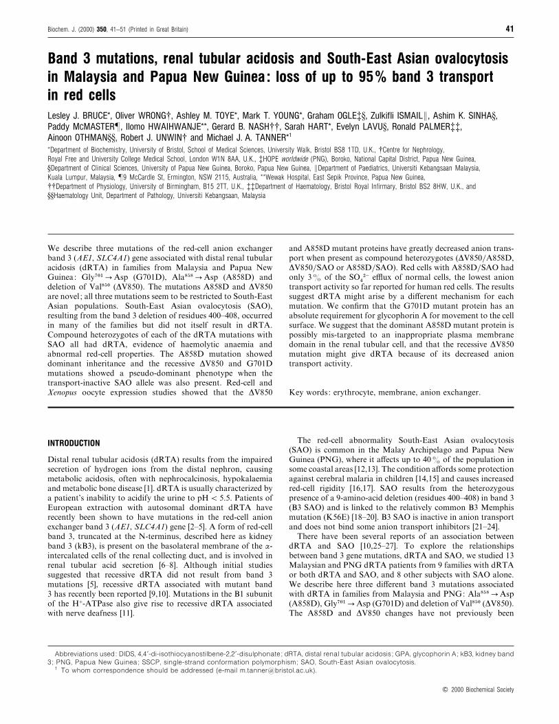
Figure 1 Family trees of dRTA patients examined in this study
The inheritance of the SAO deletion and the dRTA-associated band 3 mutations (G701D, ∆V850 and A858D) are shown. Where only one allele is shown, the other allele is normal (K56E [18–20],
D38A [40] or the most common form of band 3 [36]). Families A, B and H were from Malaysia ; families C–G and J were from PNG. Malaysian families were from separate states in peninsular
Malaysia ; PNG families were all derived from the malarious northern coastal region of PNG. Family histories gave no evidence of consanguinity, nor were any of the families known to be related.
Filled symbols (+,E) denote individuals affected by dRTA. *E : II : 2 is almost certainly affected by dRTA but this individual was not assessed for an acidification defect and is no longer traceable.
Abbreviation : n.t., not tested.
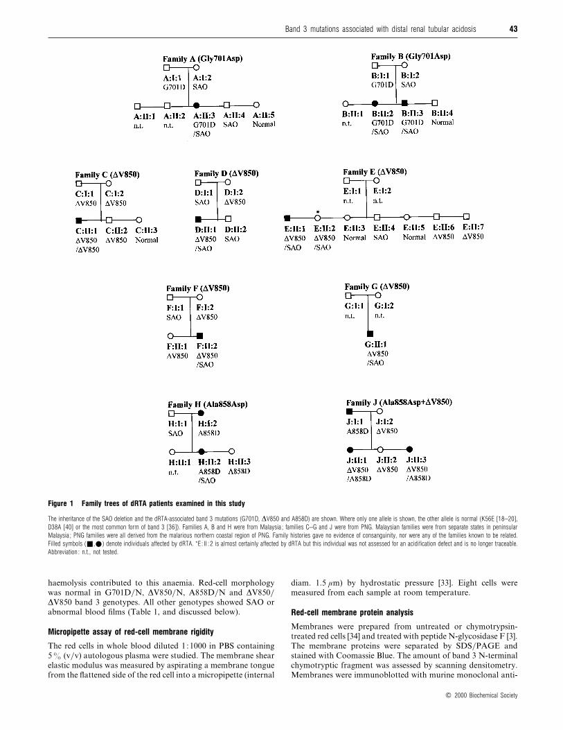
haemolysis contributed to this anaemia. Red-cell morphology
was normal in G701D}N, ∆V850}N, A858D}N and ∆V850}∆V850 band 3 genotypes. All other genotypes showed SAO or
abnormal blood films (Table 1, and discussed below).
Micropipette assay of red-cell membrane rigidity
The red cells in whole blood diluted 1:1000 in PBS containing
5% (v}v) autologous plasma were studied. The membrane shear
elastic modulus was measured by aspirating a membrane tongue
from the flattened side of the red cell into a micropipette (internal
diam. 1.5 µm) by hydrostatic pressure [33]. Eight cells were
measured from each sample at room temperature.
Red-cell membrane protein analysis
Membranes were prepared from untreated or chymotrypsin-
treated red cells [34] and treated with peptide N-glycosidase F [3].
The membrane proteins were separated by SDS}PAGE and
stained with Coomassie Blue. The amount of band 3 N-terminal
chymotryptic fragment was assessed by scanning densitometry.
Membranes were immunoblotted with murine monoclonal anti-
# 2000 Biochemical Society
44 L. J. Bruce and others
bodies : BRIC 163, directed against glycophorin A (GPA), or
BRIC 170 and BRIC155, directed against band 3 [35].
Analysis of the band 3 gene by single-strand conformationpolymorphisms (SSCPs) and DNA sequencing
Genomic DNA was isolated from blood samples by using Isocode
Stix (Schleicher and Schuell, Dassel, Germany) or buffy coats.
The coding regions of exons 2–20 [36], the putative kidney
promoter (in intron 3), and kidney translation initiator codons of
the human band 3 gene [8], were analysed for SSCPs and by
DNA sequencing as described previously [3].
Anion transport studies
Sulphate transport studies
The number of 4,4«-di-isothiocyanostilbene-2,2«-disulphonate
(DIDS)-binding sites was determined by titration of $&SO%
#−
influx into red cells, using equal numbers of dRTA and control
cells (determined with a cell counter) at 10% haematocrit [3,23].$&SO
%
#− efflux was measured from red cells that had been pre-
equilibrated with 4 mM SO%
#− ; the flux (JA) was calculated [37].
Chloride efflux assay
$'Cl− efflux was measured [38] from red cells that had been pre-
equilibrated with 165 mM Cl−.
Preparation of mutant constructs and expression in Xenopusoocytes
The cDNA clones encoding human band 3 (BSXG1.B3), the
kidney isoform, kB3 (BSXG1.KB3) and glycophorin A
(BSXG.GPA) have been described previously [3,39]. The
BSXG1.B3 clone was used to construct the G701D, A858D and
∆V850 mutants by using the Seamless Cloning Kit (Stratagene,
La Jolla, CA, U.S.A.). The kB3 mutants were prepared by
substituting the XbaI–SalI fragments of the BSXG1.B3 mutants
into BSXG1.KB3. The kB3-SAO construct was derived from
BSXG1.B3 SAO [24]. The methods used for the preparation of
band 3 cRNA, expression in oocytes, assay of $'Cl− uptake, and
estimation of cell-surface expression of band 3 in oocytes have
been described previously [3,39].
RESULTS
SAO alone does not cause dRTA
Eight individuals with simple SAO (SAO}N) were examined:
three from Malaysia, four from PNG and one from South
Africa. The SAO status of all subjects was ascertained from the
characteristic red-cell morphology in blood films. The presence
of the SAO deletion was confirmed by PCR, SSCP or DNA
sequencing in six of the subjects.
Urine pH was tested with different methods on different
occasions. Four individuals produced early morning random
urine pH values of 5.87, 5.71, 5.49 and 5.94. (Normal urinary pH
! 6.0 in individuals tested in this way [28].) Three individuals
produced a minimum urine pH of 5.12, 5.10 and 4.82 after
frusemide}fludrocortisone treatment and one individual pro-
duced a minimum urine pH of 4.63 after NH%Cl treatment.
(Normal urinary pH! 5.5 in individuals tested by frusemide}fludrocortisone or NH
%Cl [29–31]). All these subjects with simple
SAO therefore had normal ability to acidify their urine and did
not have dRTA. These findings confirm previous observations
Figure 2 Morphology of red cells
(a) Normal discoid red cells ∆V850/∆V850 (C : II : 1). (b) SAO/N red cells (D : I : 1). (c)Abnormal red cells ∆V850/A858D showing microcytes, elliptocytes and poikilocytes (J : II : 3).
(d) Small, elliptocytic red cells A858D/SAO (H : II : 2).
Table 2 Shear elastic modulus of red cells from families A, B and H
The normal control value was 6.5¬10−3 dyn/cm [20,33] (1 dyn¯ 10−5 N). Results are
means³S.E.M. for measurements on eight red cells from each donor. **P ¯ 0.02, *P ¯ 0.04,
†P ¯ 0.06 for comparisons between the compound heterozygotes and SAO/N subjects in each
family by Student’s t test.
Subject Mutation
103¬Shear elastic
modulus (dyn/cm)
Control 6.5³0.8
A : I : 1 G701D/N 8.4³1.7
A : I : 2 SAO/N 18.1³5
A : II : 3 G701D/SAO 29.6³10.1*
B : I : 1 G701D/N 6.2³0.6
B : I : 2 SAO/N 14.8³3.7
B : II : 3 G701D/SAO 22.5³9.45†H: I : 2 A858D/N 5.4³0.65
H : I : 1 SAO/N 16.3³7.5
H : II : 2 A858D/SAO 25.5³6.8**
by ourselves [3] and others [10,15] that the heterozygous presence
of B3 SAO is not sufficient to cause dRTA.
Mutations in the band 3 gene associated with dRTA
To explore the relationships between mutations in the band 3
gene, dRTA and SAO we studied 13 Malaysian and PNG dRTA
patients from 9 families with dRTA, or both dRTA and SAO
(Figure 1). Three different band 3 mutant alleles (G701D, ∆V850
and A858D) were found in the families with dRTA, either in
association with each other or with the SAO allele (Table 1 and
Figure 1). The D38A (Darmstadt) band 3 mutation [40] was
present in families A, B and D but did not segregate with the
# 2000 Biochemical Society
45Band 3 mutations associated with distal renal tubular acidosis
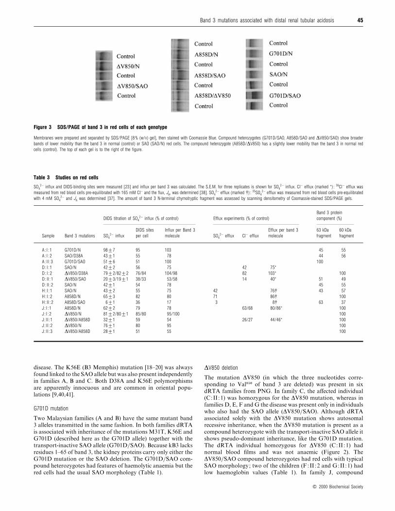
Figure 3 SDS/PAGE of band 3 in red cells of each genotype
Membranes were prepared and separated by SDS/PAGE [8% (w/v) gel], then stained with Coomassie Blue. Compound heterozygotes (G701D/SAO, A858D/SAO and ∆V850/SAO) show broader
bands of lower mobility than the band 3 in normal (control) or SAO (SAO/N) red cells. The compound heterozygote (A858D/∆V850) has a slightly lower mobility than the band 3 in normal red
cells (control). The top of each gel is to the right of the figure.
Table 3 Studies on red cells
SO42− influx and DIDS-binding sites were measured [23] and influx per band 3 was calculated. The S.E.M. for three replicates is shown for SO4
2− influx. Cl− efflux (marked *) : 36Cl− efflux was
measured from red blood cells pre-equilibrated with 165 mM Cl− and the flux, JA, was determined [38]. SO42− efflux (marked †) : 35SO4
2− efflux was measured from red blood cells pre-equilibrated
with 4 mM SO42− and JA was determined [37]. The amount of band 3 N-terminal chymotryptic fragment was assessed by scanning densitometry of Coomassie-stained SDS/PAGE gels.
DIDS titration of SO42− influx (% of control) Efflux experiments (% of control)
Band 3 protein
component (%)
Sample Band 3 mutations SO42− influx
DIDS sites
per cell
Influx per Band 3
molecule SO42− efflux Cl− efflux
Efflux per band 3
molecule
63 kDa
fragment
60 kDa
fragment
A : I : 1 G701D/N 98³7 95 103 45 55
A : I : 2 SAO/D38A 43³1 55 78 44 56
A : II : 3 G701D/SAO 51³6 51 100 100
D : I : 1 SAO/N 42³2 56 75 42 75*
D : I : 2 ∆V850/D38A 79³2/82³2 76/84 104/98 82 103* 100
D : II : 1 ∆V850/SAO 20³3/19³1 38/33 53/58 14 40* 51 49
D : II : 2 SAO/N 42³1 54 78 45 55
H : I : 1 SAO/N 43³2 55 75 42 76† 43 57
H : I : 2 A858D/N 65³3 82 80 71 86† 100
H : II : 2 A858D/SAO 6³1 36 17 3 8† 63 37
J : I : 1 A858D/N 62³2 79 78 63/68 80/86* 100
J : I : 2 ∆V850/N 81³2/80³1 85/80 95/100 100
J : II : 1 ∆V850/A858D 32³1 59 54 26/27 44/46* 100
J : II : 2 ∆V850/N 76³1 80 95 100
J : II : 3 ∆V850/A858D 28³1 51 55 100
disease. The K56E (B3 Memphis) mutation [18–20] was always
found linked to the SAO allele but was also present independently
in families A, B and C. Both D38A and K56E polymorphisms
are apparently innocuous and are common in oriental popu-
lations [9,40,41].
G701D mutation
Two Malaysian families (A and B) have the same mutant band
3 alleles transmitted in the same fashion. In both families dRTA
is associated with inheritance of the mutations M31T, K56E and
G701D (described here as the G701D allele) together with the
transport-inactive SAO allele (G701D}SAO). Because kB3 lacks
residues 1–65 of band 3, the kidney proteins carry only either the
G701D mutation or the SAO deletion. The G701D}SAO com-
pound heterozygotes had features of haemolytic anaemia but the
red cells had the usual SAO morphology (Table 1).
∆V850 deletion
The mutation ∆V850 (in which the three nucleotides corre-
sponding to Val)&! of band 3 are deleted) was present in six
dRTA families from PNG. In family C, the affected individual
(C:II :1) was homozygous for the ∆V850 mutation, whereas in
families D, E, F and G the disease was present only in individuals
who also had the SAO allele (∆V850}SAO). Although dRTA
associated solely with the ∆V850 mutation shows autosomal
recessive inheritance, when the ∆V850 mutation is present as a
compound heterozygote with the transport-inactive SAO allele it
shows pseudo-dominant inheritance, like the G701D mutation.
The dRTA individual homozygous for ∆V850 (C:II :1) had
normal blood films and was not anaemic (Figure 2). The
∆V850}SAO compound heterozygotes had red cells with typical
SAO morphology; two of the children (F:II :2 and G:II :1) had
low haemoglobin values (Table 1). In family J, compound
# 2000 Biochemical Society

46 L. J. Bruce and others
Figure 4 Anion transport studies
Representative examples of the SO42− influx (a) and efflux (b) experiments showing the data
from the Malaysian family H : +, father of family H (SAO/N) (H : I : 1) ; E, mother of family
H (A858D/N) (H : I : 2) ; U, child of family H (A858D/SAO) (H : II : 2) ; ^, normal control red
cells. Each data point was derived from measurements in triplicate. The lines show the results
of linear regression analysis of the data. (a) DIDS titration of SO42− influx. The influx of 35SO4
2−
into the cells was measured at 10% haematocrit in isotonic citrate buffer [84 mM sodium
citrate/1 mM EGTA (pH 6.5)] containing 4 mM Na2SO4. Influx was determined after 5 min at
30 °C in the presence of different concentrations of DIDS as described previously [23]. (b)SO4
2− efflux. Red cells were equilibrated in citrate buffer containing 4 mM Na2SO4 and tracer35SO4
2− ; the efflux of SO42− was determined over 24 min at 30 °C as described previously
[37].
heterozygotes of the ∆V850 and A858D mutations had the
complete form of dRTA (see below).
A858D mutation
The Malaysian family H carried the A858D mutation and the
SAO allele, whereas the PNG family J carried both the A858D
and ∆V850 mutations. The simple heterozygotes (A858D}N;
H:I:2 and J:I :1) had incomplete dRTA (they were unable to
acidify their urine when tested with frusemide}fludrocortisone
but had no other feature of dRTA). This demonstrates the
autosomal dominant nature of the A858D mutation. Complete
dRTA was present in the compound heterozygote A858D}SAO
(H:II :2) and in the two children who were compound hetero-
zygotes for A858D}∆V850 (J :II :1 and J:II :3). The compound
heterozygote A858D}SAO did not have the usual SAO red-cell
morphology; instead, the cells were small and elliptocytic (Figure
2). However, this might not simply be due to the band 3
mutations present because the mother (H:I :2, A858D}N) had
acanthocytic red cells. It is possible that these acanthocytes were
an artifact of the blood film, relating to the condition of the
blood sample when the film was made; equally, the mother
(H:I :2, A858D}N) might have a further red-cell abnormality
because an unrelated A858D}N subject (J :I :1) had normal red
cells. The compound heterozygote A858D}∆V850 patients both
showed bizarre red-cell morphology with microcytes, elliptocytes
and poikilocytes (Figure 2). All three A858D}SAO and A858D}∆V850 patients (H:II :2, J :II :1 and J:II :3) had low haemo-
globin values, suggesting increased haemolysis.
DNA sequences of the region of the kB3 promoter
The DNA sequence of the kidney promoter [8] (from the 5« end
of erythroid intron 3 to the 3« end of kidney exon 3A [36]) was
also examined. Polymorphisms c87t, c242t, g259a and a580g
(numbered from the 5« end of intron 3) were found but were not
associated with dRTA. However, the c242t and g259a changes
were correlated with the K56E Memphis polymorphism (and
SAO).
Studies on red cells
Red-cell studies were performed where suitable fresh blood
samples were available.
Red-cell membrane rigidity
The shear elastic modulus of red-cell samples from families A, B
and H was measured with a micropipette method (Table 2). The
simple heterozygotes G701D}N (A:I:1, B:I :1) and A858D}N
(H:I:2) gave normal values, similar to the value of 6.5¬10−$
dyn}cm (1 dyn¯ 10−& N) obtained for a normal control and
reported previously [20,33]. The SAO}N samples from the
families gave significantly higher values [(14.8–18.1)¬10−$ dyn}cm], indicating increased membrane rigidity, as reported pre-
viously [20]. Interestingly, the red cells of A:II :3 and B:II :3
(G701D}SAO) and H:II :2 (A858D}SAO) had an even higher
shear elastic modulus [(22.5–29.6)¬10−$ dyn}cm], and thus even
greater membrane rigidity, than their SAO parents.
Analysis of red-cell membrane band 3 in the families
SDS}PAGE of the band 3 in the red cells of all the individuals
with complete dRTA gave bands of slightly lower mobility than
the band 3 of normal individuals or simple SAO}N heterozygotes
(Figure 3). This shift in mobility was more evident in the 35 kDa
C-terminal fragment of band 3 that was obtained after the
treatment of red cells with chymotrypsin (results not shown).
Treatment of the membranes with peptide N-glycosidase F
resulted in deglycosylated 35 kDa fragments with the same
mobility, suggesting that the altered mobility was due to altered
N-glycosylation (results not shown). Similar N-glycan-dependent
changes in mobility have been observed in band 3 with mutations
at Arg&)* that result in dRTA [3]. Immunoblotting with an anti-
GPA antibody showed that GPA was present in all the red-cell
samples (results not shown).
Band 3 proteins carrying the K56E mutation (B3 Memphis)
can be distinguished from other band 3 proteins by SDS}PAGE
of chymotrypsin-treated red cells [42]. B3 Memphis yields a
63 kDa fragment, whereas normal band 3 yields a 60 kDa
fragment. In appropriate heterozygotes the relative expression of
B3 Memphis (in K65E, SAO and G701D alleles) was measured
and compared with the expression of band 3 with normal
mobility (in normal, D38A, ∆V850 and A858D alleles) (Table 3).
As noted previously [20] in SAO heterozygotes, the SAO protein
# 2000 Biochemical Society
47Band 3 mutations associated with distal renal tubular acidosis
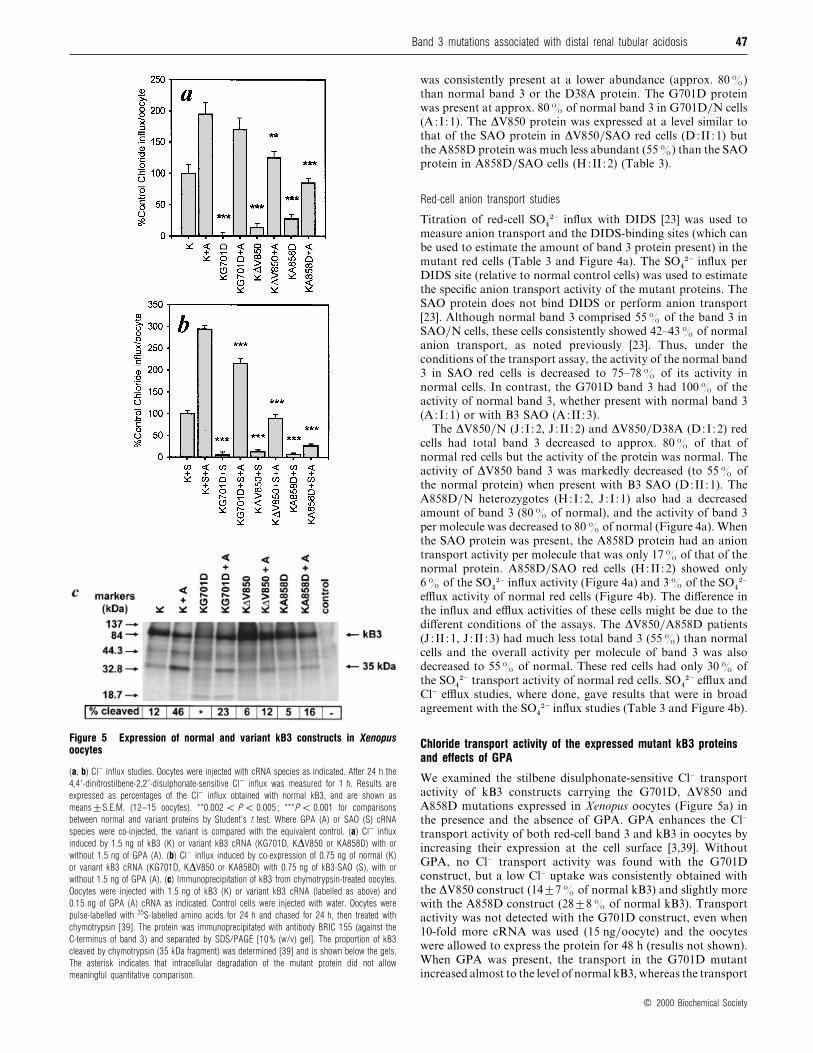
Figure 5 Expression of normal and variant kB3 constructs in Xenopusoocytes
(a, b) Cl− influx studies. Oocytes were injected with cRNA species as indicated. After 24 h the
4,4«-dinitrostilbene-2,2«-disulphonate-sensitive Cl− influx was measured for 1 h. Results are
expressed as percentages of the Cl− influx obtained with normal kB3, and are shown as
means³S.E.M. (12–15 oocytes). **0.002! P ! 0.005 ; ***P ! 0.001 for comparisons
between normal and variant proteins by Student’s t test. Where GPA (A) or SAO (S) cRNA
species were co-injected, the variant is compared with the equivalent control. (a) Cl− influx
induced by 1.5 ng of kB3 (K) or variant kB3 cRNA (KG701D, K∆V850 or KA858D) with or
without 1.5 ng of GPA (A). (b) Cl− influx induced by co-expression of 0.75 ng of normal (K)
or variant kB3 cRNA (KG701D, K∆V850 or KA858D) with 0.75 ng of kB3-SAO (S), with or
without 1.5 ng of GPA (A). (c) Immunoprecipitation of kB3 from chymotrypsin-treated oocytes.
Oocytes were injected with 1.5 ng of kB3 (K) or variant kB3 cRNA (labelled as above) and
0.15 ng of GPA (A) cRNA as indicated. Control cells were injected with water. Oocytes were
pulse-labelled with 35S-labelled amino acids for 24 h and chased for 24 h, then treated with
chymotrypsin [39]. The protein was immunoprecipitated with antibody BRIC 155 (against the
C-terminus of band 3) and separated by SDS/PAGE [10% (w/v) gel]. The proportion of kB3
cleaved by chymotrypsin (35 kDa fragment) was determined [39] and is shown below the gels.
The asterisk indicates that intracellular degradation of the mutant protein did not allow
meaningful quantitative comparison.
was consistently present at a lower abundance (approx. 80%)
than normal band 3 or the D38A protein. The G701D protein
was present at approx. 80% of normal band 3 in G701D}N cells
(A:I :1). The ∆V850 protein was expressed at a level similar to
that of the SAO protein in ∆V850}SAO red cells (D:II :1) but
the A858D protein was much less abundant (55%) than the SAO
protein in A858D}SAO cells (H:II :2) (Table 3).
Red-cell anion transport studies
Titration of red-cell SO%
#− influx with DIDS [23] was used to
measure anion transport and the DIDS-binding sites (which can
be used to estimate the amount of band 3 protein present) in the
mutant red cells (Table 3 and Figure 4a). The SO%
#− influx per
DIDS site (relative to normal control cells) was used to estimate
the specific anion transport activity of the mutant proteins. The
SAO protein does not bind DIDS or perform anion transport
[23]. Although normal band 3 comprised 55% of the band 3 in
SAO}N cells, these cells consistently showed 42–43% of normal
anion transport, as noted previously [23]. Thus, under the
conditions of the transport assay, the activity of the normal band
3 in SAO red cells is decreased to 75–78% of its activity in
normal cells. In contrast, the G701D band 3 had 100% of the
activity of normal band 3, whether present with normal band 3
(A:I :1) or with B3 SAO (A:II :3).
The ∆V850}N (J:I :2, J :II :2) and ∆V850}D38A (D:I:2) red
cells had total band 3 decreased to approx. 80% of that of
normal red cells but the activity of the protein was normal. The
activity of ∆V850 band 3 was markedly decreased (to 55% of
the normal protein) when present with B3 SAO (D:II :1). The
A858D}N heterozygotes (H:I :2, J :I :1) also had a decreased
amount of band 3 (80% of normal), and the activity of band 3
per molecule was decreased to 80% of normal (Figure 4a). When
the SAO protein was present, the A858D protein had an anion
transport activity per molecule that was only 17% of that of the
normal protein. A858D}SAO red cells (H:II :2) showed only
6% of the SO%
#− influx activity (Figure 4a) and 3% of the SO%
#−
efflux activity of normal red cells (Figure 4b). The difference in
the influx and efflux activities of these cells might be due to the
different conditions of the assays. The ∆V850}A858D patients
(J :II :1, J :II :3) had much less total band 3 (55%) than normal
cells and the overall activity per molecule of band 3 was also
decreased to 55% of normal. These red cells had only 30% of
the SO%
#− transport activity of normal red cells. SO%
#− efflux and
Cl− efflux studies, where done, gave results that were in broad
agreement with the SO%
#− influx studies (Table 3 and Figure 4b).
Chloride transport activity of the expressed mutant kB3 proteinsand effects of GPA
We examined the stilbene disulphonate-sensitive Cl− transport
activity of kB3 constructs carrying the G701D, ∆V850 and
A858D mutations expressed in Xenopus oocytes (Figure 5a) in
the presence and the absence of GPA. GPA enhances the Cl−
transport activity of both red-cell band 3 and kB3 in oocytes by
increasing their expression at the cell surface [3,39]. Without
GPA, no Cl− transport activity was found with the G701D
construct, but a low Cl− uptake was consistently obtained with
the ∆V850 construct (14³7% of normal kB3) and slightly more
with the A858D construct (28³8% of normal kB3). Transport
activity was not detected with the G701D construct, even when
10-fold more cRNA was used (15 ng}oocyte) and the oocytes
were allowed to express the protein for 48 h (results not shown).
When GPA was present, the transport in the G701D mutant
increased almost to the level of normal kB3, whereas the transport
# 2000 Biochemical Society
48 L. J. Bruce and others
in the ∆V850 and A858D mutants was increased by the presence
of GPA to 64³5% and 43³3% of that obtained with the
normal protein.
To mimic the situation in the red cells of patients who were
compound heterozygotes with the SAO allele, we co-expressed
kB3 containing the SAO deletion with an equal amount of each
of the other kB3 mutants and GPA (Figure 5b). Expressed with
GPA, the G701DSAO mixture was almost as active as
the normal kB3SAO (73³4% of the normal kB3SAO); the
∆V850SAO combination also gave some activity (31³3% of
the normal kB3SAO). In contrast, the A858DSAO mixture
had very little activity even in the presence of GPA. This result
suggests that the enhancement of the activity of the A858D
mutant by GPA (and to a smaller extent the ∆V850 mutant) is
suppressed by kB3 SAO. When equal amounts of the ∆V850 and
A858D mutants were co-expressed, the results were not signifi-
cantly different from the average of the measured transport
activity when an equal amount of each mutant was expressed
alone, whether GPA was present or not (results not shown).
Cell-surface movement of the mutant proteins in oocytes
We examined whether the reduced transport observed with the
mutant proteins in oocytes resulted from their impaired move-
ment to the cell surface (Figure 5c), with the use of a proteolysis-
based assay [39]. Extracellular band 3 cleavage by chymotrypsin
yields a C-terminal 35 kDa fragment that can be used to assess
the proportion of kB3 at the oocyte cell surface [39].Metabolically
radiolabelled oocytes expressing kB3 were digested with chymo-
trypsin and the intact kB3 and 35 kDa fragment were immuno-
precipitated with an antibody that binds to the band 3 C-
terminus [39]. In the absence of GPA, much less ∆V850 or
A858D than normal kB3 was present at the cell surface. There
was also little or no G701D expressed at the cell surface but
quantitative comparison was not possible because there was a
substantial decrease in the total amount of G701D protein
expressed in the cells (found in two independent experiments).
GPA substantially enhanced the expression of normal kB3 and
G701D at the cell surface but gave less enhancement of the
∆V850 and A858D expressed at the cell surface. Co-expression of
GPA also resulted in an increase in the total amount of G701D
protein expressed in the cells.
DISCUSSION
Band 3 mutations in oriental populations associated with dRTA
The oriental dRTA families studied here contained three different
band 3 mutations, the properties of which are summarized in
Tables 4 and 5. The ∆V850 and A858D mutations have not been
described previously but two dRTA families carrying the G701D
mutation have been found in Thailand [9,10]. The G701D and
∆V850 mutations show recessive inheritance of dRTA, which
presents as pseudo-dominant inheritance in the presence of the
SAO allele. The A858D mutation is dominant and the simple
heterozygotes studied here have incomplete dRTA, an inability
to acidify their urine but no accompanying features of dRTA
[29,32]. None of our patients was homozygous for the A858D
mutation, but the compound heterozygotes of ∆V850 and
A858D showed full-blown dRTA.
Role of the SAO allele in dRTA in oriental populations
The association between SAO and dRTA has been noted several
times [10,25–27]. It is clear from our own [3] and other studies
[10,14] that the SAO allele does not itself give dRTA. It is evident
that the association between SAO and dRTA exists because the
transport-inactive SAO allele gives recessive dRTA mutations a
pseudo-dominant phenotype when they are present as compound
heterozygotes with the SAO allele. All three dRTA mutations
were found as compound heterozygotes with SAO where they
presented with complete dRTA.
Haematology of the dRTA mutants
Anaemia is not a recognized feature of dRTA. The∆V850}∆V850
phenotype showed normal red-cell morphology and haema-
tology. In contrast, all the other phenotypes associated with
complete dRTA showed anaemia, probably haemolytic in origin,
especially in the children. These phenotypes also showed changes
in the rigidity and morphology of the mutant red cells (see
below). Although these changes could result in anaemia, other
possible causes of anaemia in these populations, such as chronic
infection, nutritional iron deficiency and hookworm infestation,
cannot be ruled out.
The G701D}SAO red cells had the usual SAO morphology
but were much more rigid than SAO cells ; the condition was
associated with anaemia. The A858D}SAO red cells were also
more rigid than SAO red cells and were elliptocytic, although it
is possible that there was another red-cell abnormality in this
family. The reason for the increased rigidity is unclear but in
both cell types the increased rigidity might contribute to the
anaemia. We were unable to test the rigidity of the ∆V850}SAO
red cells but this condition was also associated with anaemia.
The ∆V850}A858D compound heterozygotes had red cells of
bizarre shape (microcytes, elliptocytes and poikilocytes) and
anaemia. The much-reduced band 3 content of these cells might
decrease red-cell stability and cause haemolytic anaemia in a
manner similar to band 3-deficient hereditary spherocytosis [43].
Homozygous G701D}G701D red cells have been reported to
have abnormal shapes and associated haemolytic anaemia [9].
Anion transport properties of mutant red cells
The presence of the B3 SAO protein decreased the specific SO%
#−
influx activity of normal band 3 compared with its activity in
normal red cells. A similar but more marked effect was observed
when B3 SAO was present with the ∆V850 and A858D proteins.
In contrast, the specific SO%
#− influx activity of the G701D
protein was the same in normal and in G701D}SAO red cells.
This suggests that the normal, ∆V850 and A858D proteins
interact with B3 SAO but the G701D protein might not.
The G701D protein was expressed in red cells at a level close
to that of the normal protein. However, there were substantially
smaller amounts of the ∆V850 and A858D proteins when B3
SAO was present. This, in combination with the lower activity of
the proteins, resulted in very low anion transport activity in the
∆V850}SAO and A858D}SAO red cells. Indeed, the 6% of
normal SO%
#− influx activity and 3% of normal SO%
#− efflux
activity in the A858D}SAO red cells is the lowest anion transport
activity so far reported for human red cells. The red cells of the
∆V850}A858D individual also had very low anion transport.
Activity of the expressed mutant proteins
Although red cells express GPA, there is no evidence for the
presence of GPA in the kidney. GPA facilitates the movement of
band 3 and kB3 to the oocyte cell surface [3,39]. The mutant kB3
proteins were expressed in Xenopus oocytes to assess their
transport activity in the absence and the presence of GPA. The
G701D protein did not induce any anion transport in the absence
# 2000 Biochemical Society
49Band 3 mutations associated with distal renal tubular acidosis
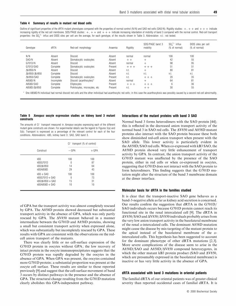
Table 4 Summary of results in mutant red blood cells
Outline of significant properties of the dRTA mutant phenotypes compared with the properties of normal control (N/N) and SAO red cells (SAO/N). Rigidity studies : , and indicate
increasing rigidity of the red cell membrane. SDS/PAGE studies : , and indicate increasing retardation of mobility of band 3 compared with the normal control. Red-cell transport
properties : the SO42− influx and DIDS sites per cell are the average, for each genotype, of the results shown in Table 3. Abbreviation : n.t. : not tested.
Genotype dRTA Red-cell morphology Anaemia Rigidity
SDS/PAGE band 3
mobility
SO42− influx
(% of normal)
DIDS sites per cell
(% of normal)
N/N Absent Discoid Absent normal normal 100 100
SAO/N Absent Stomatocytic ovalocytes Absent 42 55
G701D/N Absent Discoid Absent normal 98 95
G701D/SAO Complete Stomatocytic ovalocytes Present 51 51
∆V850/N Absent Discoid Absent n.t. 80 81
∆V850/∆V850 Complete Discoid Absent n.t. n.t. n.t. n.t.
∆V850/SAO Complete Stomatocytic ovalocytes Present n.t. 20 35
A858D/N Incomplete Discoid (acanthocytes)* Absent normal 64 81
A858D/SAO Complete Elliptocytes Present 6 36
A858D/∆V850 Complete Poikilocytes, microcytes, etc Present n.t. 30 55
* One A858D/N individual had normal discoid red cells and the other individual had acanthocytic red cells ; in this case the acanthocytosis was possibly caused by a second red-cell abnormality.
Table 5 Xenopus oocyte expression studies on kidney band 3 mutantconstructs
The amounts of Cl− transport measured in Xenopus oocytes expressing each of the different
mutant gene constructs are shown. For experimental details see the legend to Figures 5(a) and
5(b). Transport is expressed as a percentage of the relevant control for each of the four
conditions. Abbreviations : kB3, kidney band 3 ; SAO, SAO band 3.
Cl− transport (% of control)
Construct ®GPA GPA
kB3 100 100
kB3G701D 0 87
kB3∆V850 14 64
kB3A858D 29 43
kB3SAO 100 100
kB3G701DSAO 0 73
kB3∆V850SAO 15 31
kB3A858DSAO 10 10
of GPA but the transport activity was almost completely rescued
by GPA. The A858D protein showed decreased but substantial
transport activity in the absence of GPA, which was only partly
rescued by GPA. The ∆V850 mutant behaved in a manner
intermediate between the G701D and A858D proteins, showing
a small but consistent transport activity when expressed alone,
which was substantially but incompletely rescued by GPA. These
results with GPA are consistent with the observations on the red-
cell anion transport of the mutants.
There was clearly little or no cell-surface expression of the
G701D protein in oocytes without GPA; the low recovery of
intact protein in the oocyte immunoprecipitates suggests that the
G701D protein was rapidly degraded by the oocytes in the
absence of GPA. When GPA was present, the oocytes contained
more G701D protein; a substantial proportion was present at the
oocyte cell surface. These results are similar to those reported
previously [9] and suggest that the cell-surface movement of band
3 occurs by distinct pathways in the presence and the absence of
GPA. The structural change resulting from the G701D mutation
clearly abolishes this GPA-independent pathway.
Interactions of the mutant proteins with band 3 SAO
Normal band 3 forms heterodimers with the SAO protein [44] ;
this is reflected in the decreased anion transport activity of the
normal band 3 in SAO red cells. The ∆V850 and A858D mutant
proteins also interact with the SAO protein because these both
show diminished red-cell anion transport when present with the
SAO allele. This lower activity is particularly evident in
the A858D}SAO red cells. When co-expressed with kB3 SAO, the
A858D protein showed very little enhancement of transport
activity by GPA. In contrast, the anion transport activity of the
G701D mutant was unaffected by the presence of the SAO
protein, either in red cells or when co-expressed in oocytes,
suggesting that G701D does not interact with the SAO protein to
form heterodimers. This finding suggests that the G701D mu-
tation might alter the structure of the band 3 membrane domain
at the dimer interface.
Molecular basis for dRTA in the families studied
It is clear that the transport-inactive SAO gene behaves as a
band-3-negative allele as far as kidney acid secretion is concerned.
Our results confirm the suggestion that dRTA in the G701D}SAO individuals occurs because G701D protein cannot reach its
functional site in the renal intercalated cell [9]. The dRTA in
∆V850}SAOand∆V850}∆V850 individuals probably arises from
the very low anion transport activity in the basolateral membrane
of the renal α-intercalated cells. The dominant A858D mutation
might cause the disease by mis-targeting of the mutant protein to
the apical instead of the basolateral membrane of the α-
intercalated cells. This hypothesis has been suggested to account
for the dominant phenotype of other dRTA mutations [2,3].
More severe complications of the disease seem to arise in the
A858D}SAO and A858D}∆V850 compound heterozygotes, in
which the other mutant kB3 protein product (SAO and ∆V850,
which are presumably expressed in the basolateral membrane) is
inactive or has very little activity in the absence of GPA.
dRTA associated with band 3 mutations in oriental patients
The familial dRTA of our oriental patients was of greater clinical
severity than reported occidental cases of familial dRTA. It is
# 2000 Biochemical Society
50 L. J. Bruce and others
not clear whether this is the result of the different mutations
causing the disease, case selection, or other environmental or
genetic factors. The incidences of nephrocalcinosis in the two
groups are similar (approx. 70%) but metabolic bone disease
and severe hypokalaemia were more a feature of the oriental
patients ; thus 73% of the oriental patients with complete dRTA
had rickets, and their mean plasma [K+] at presentation was
2.7³0.6 mmol}l, whereas a recent survey of occidental patients
(most subsequently shown to have band 3 mutations) found that
only 25% had bone disease; mean plasma [K+] at presentation
was 3.6³0.6 mmol}l [45]. Two of our oriental patients were
sufficiently hypokalaemic to have general paralysis, which has
not been reported in occidental familial dRTA [45].
The three different band 3 mutations reported here from
oriental dRTA patients, affecting band 3 residues 701, 850 and
858, have not so far been reported in occidental cases of the
disease, in whom most mutations have affected residue 589,
usually with histidine substituting for arginine. No band 3
mutation, causally related to dRTA, has yet been reported in
either occidental or oriental subjects. Our findings suggest that
there might be a clustering of these newly discovered band 3
mutations in the orient : the 701 mutation has now been described
in four families with dRTA from Malaysia and the immediately
adjacent area of Thailand [10] and in a fifth family in the
northeast part of Thailand [9], which is 700 miles distant ;
the ∆V850 mutation was found by us in six families in PNG; and
we found the A858D mutation in two families in Malaysia and
PNG, sites 3000 miles apart. All these areas have until very
recent times been affected by endemic Plasmodium falciparum
malaria, against which the band 3 mutation that gives rise to
SAO affords some clinical protection [14,15]. There is also a high
frequency of SAO in conjunction with the dRTA mutations
(seven of the nine families that we studied). Our results raise the
intriguing possibility that these newly discovered band 3
mutations might have evolved locally because they provide some
protection against the clinical effects of P. falciparum malaria, as
occurs with the band 3 mutation that gives rise to SAO [15].
We thank the Papua New Guinea Paediatric Surveillance Unit for its assistance inthe collection of PNG cases ; D. J. Anstee for monoclonal antibodies (BRIC 163, anti-GPA ; BRIC 170 and BRIC 155, anti-band 3) ; R. Beckmann for the kB3 cDNA ;R. C. Morris and Church & Dwight Co. (Princeton, NJ, U.S.A.) for travel support ;C. S. Keng for help with photographic facilities and supplies ; and Tan Sri Abubakr,R. Taufa and I. Kevau for their interest and support. This work was supported in partby grants from the National Kidney Research Fund and the Wellcome Trust. M.T.Y.was the recipient of a Wellcome Prize Studentship.
REFERENCES
1 Morris, R. C. and Ives, H. E. (1996) Inherited disorders of the renal tubule. in The
Kidney, 5th edn (Brenner, B. M., ed.), pp. 1764–1827, W. B. Saunders, Philadelphia
2 Wrong, O., Unwin, R. J., Cohen, E., Tanner, M. and Thakker, R. (1996) Unravelling of
the molecular mechanism of kidney stones. Lancet 348, 1561–1565
3 Bruce, L. J., Cope, D. L., Jones, G. K., Schofield, A. E., Burley, M., Povey, S., Unwin,
R. J., Wrong, O. and Tanner, M. J. A. (1997) Familial renal tubular acidosis is
associated with mutations in the red cell anion exchanger (band 3 ; AE1) gene.
J. Clin. Invest. 100, 1693–1707
4 Jarolim, P., Shayakul, C., Prabakaran, D., Jiang, L., Stuart-Tilley, A., Rubin, H. L.,
Simova, S., Zavadil, J., Herrin, J. T., Brouillette, J. et al. (1998) Autosomal dominant
distal renal tubular acidosis is associated in three families with heterozygosity for the
R589H mutation in the AE1 (band 3) Cl−/HCO3− exchanger. J. Biol. Chem. 273,
6380–6388
5 Karet, F. E., Gainza, F. J., Gyory, A. Z., Unwin, R. J., Wrong, O., Tanner, M. J. A.,
Nayir, A., Alpay, H., Santos, F., Hulton, S. A. et al. (1998) Mutations in the
chloride–bicarbonate exchanger gene AE1 cause autosomal dominant but not
autosomal recessive distal renal tubular acidosis. Proc. Natl. Acad. Sci. U.S.A. 95,6337–6342
6 Wagner, S., Vogel, R., Lietzke, R., Koob, R. and Drenckhahn, D. (1987)
Immunochemical characterisation of a band 3-like anion exchanger in collecting duct
of human kidney. Am. J. Physiol. 253, 213–221
7 Alper, S. L., Natale, J., Gluck, S., Lodish, H. F. and Brown, D. (1989) Subtypes of
intercalated cells in rat kidney collecting duct defined by antibodies against erythroid
band 3 and renal vacuolar H+-ATPase. Proc. Natl. Acad. Sci. U.S.A. 86, 5429–5433
8 Kollert-Jons, A., Wagner, S., Hubner, S., Appelhans, H. and Drenckhahn, D. (1993)
Anion exchanger 1 in human kidney and oncocytoma differs from erythroid AE1 in
its NH2 terminus. Am. J. Physiol. 265, F813–F821
9 Tanphaichitr, V. S., Sumboonnanonda, A., Ideguchi, H., Shayakul, C., Brugnara, C.,
Takao, M., Veerakul, G. and Alper, S. L. (1998) Novel AE1 mutations in recessive
distal renal tubular acidosis. J. Clin. Invest. 102, 2173–2179
10 Vasuvattakul, S., Yenchitsomanus, P.-T., Vachuanichsanong, P., Thuwajit, P.,
Kaitwatcharachai, C., Laosombat, V., Malasit, P., Wilairat, P. and Nimmannit, S.
(1999) Autosomal recessive distal renal tubular acidosis associated with Southeast
Asian ovalocytosis. Kidney Int. 56, 1674–1682
11 Karet, F. E., Finberg, K. E., Nelson, R. D., Nayir, A., Mocan, H., Sanjad, S. A.,
Rodriguez-Soriano, J., Santos, F., Cremers, C. W. R. J., Di Pietro, A. et al. (1999)
Mutations in the gene encoding B1 subunit of H+-ATPase cause renal tubular
acidosis with sensorineural deafness. Nat. Genet. 21, 84–90
12 Serjeantson, S., Bryson, K., Amato, D. and Babona, D. (1977) Malaria and hereditary
ovalocytosis. Hum. Genet. 37, 161–168
13 Lie-Injo, L. E. (1965) Hereditary ovalocytosis and haemoglobin E-ovalocytosis in
Malay Aborigines. Nature (London) 208, 1329–1330
14 Genton, B., Al-Yaman, F., Mgone, C. S., Alexander, N., Paniu, M. M., Alpers, M. P.
and Mokela, D. (1995) Ovalocytosis and cerebral malaria. Nature (London) 378,564–565
15 Allen, S. J., O’Donnell, A., Alexander, N. D., Mgone, C. S., Peto, T. E., Clegg, J. B.,
Alpers, M. P. and Weatherall, D. J. (1999) Prevention of cerebral malaria in children
in Papua New Guinea by southeast Asian ovalocytosis band 3. Am. J. Trop. Med.
Hyg. 60, 1056–1060
16 Mohandas, N., Lie-Injo, L. E., Friedman, M. and Mak, J. W. (1984) Rigid membranes
of Malayan ovalocytes : a likely genetic barrier against malaria. Blood 63, 1385–1392
17 Saul, A., Lamont, G., Sawyer, W. H. and Kidson, C. (1984) Decreased membrane
deformability in Melanesian ovalocytes from Papua New Guinea. J. Cell Biol. 98,1348–1354
18 Jarolim, P., Palek, J., Amato, D., Hassan, K., Sapak, P., Nurse, G. T., Rubin, H. L.,
Zhai, S., Sahr, K. E. and Liu, S.-C. (1991) Deletion in erythrocyte band 3 gene in
malaria-resistant Southeast Asian ovalocytosis. Proc. Natl. Acad. Sci. U.S.A. 88,11022–11026
19 Mohandas, N., Winardi, R., Knowles, D., Leung, A., Parra, M., George, E., Conboy, J.
and Chasis, J. (1992) Molecular basis of membrane rigidity of hereditary
ovalocytosis. A novel mechanism involving the cytoplasmic domain of band 3. J. Clin.
Invest. 89, 686–692
20 Schofield, A. E., Tanner, M. J. A., Pinder, J. C., Clough, B. C., Bayley, P. M., Nash,
G. B., Dluzewski, A. R., Reardon, D. M., Cox, T. M., Wilson, R. J. et al. (1992) Basis
of unique red cell membrane properties in hereditary ovalocytosis. J. Mol. Biol. 223,949–958
21 Moriyama, R., Ideguchi, H., Lombardo, C. R., Van Dort, H. M. and Low, P. S. (1992)
Structural and functional characterization of band 3 from Southeast Asian ovalocytes.
J. Biol. Chem. 267, 25792–25797
22 Sarabia, V. E., Casey, J. R. and Reithmeier, R. A. F. (1993) Molecular characterization
of the band 3 protein from Southeast Asian ovalocytes. J. Biol. Chem. 268,10676–10680
23 Schofield, A. E., Reardon, D. M. and Tanner, M. J. A. (1992) Defective anion transport
activity of the abnormal band 3 in hereditary ovalocytic red blood cells. Nature
(London) 355, 836–838
24 Groves, J. D., Ring, S. M., Schofield, A. E. and Tanner, M. J. A. (1993) The
expression of the abnormal human red cell anion transporter from South-East Asian
ovalocytes (band 3 SAO) in Xenopus oocytes. FEBS Lett. 330, 180–190
25 Baehner, R. L., Gilchrist, G. S. and Anderson, E. J. (1968) Hereditary elliptocytosis
and primary renal tubular acidosis in a single family. Am. J. Dis. Child. 115,414–419
26 Thong, M. K., Tan, A. A. L. and Lin, H. P. (1997) Distal renal tubular acidosis and
hereditary elliptocytosis in a single family. Singapore Med. J. 38, 388–390
27 Kaitwatcharachai, C., Vasuvattakul, S., Yenchitsomanus, P., Thuwajit, P., Malasit, P.,
Chuawatana, D., Mingkum, S., Halperin, M. L., Wilairat, P. and Nimmannit, S. (1999)
Distal renal tubular acidosis and high urine carbon dioxide tension in a patient with
southeast Asian ovalocytosis. Am. J. Kidney Dis. 33, 1147–1152
28 Chafe, L. and Gault, M. H. (1994) First morning urine pH in the diagnosis of renal
tubular acidosis with nephrolithiasis. Clin. Nephrol. 41, 159–162
29 Wrong, O. and Davies, H. E. F. (1959) The excretion of acid in renal disease.
Quart. J. Med. 28, 259–314
# 2000 Biochemical Society
51Band 3 mutations associated with distal renal tubular acidosis
30 Battle, D. C. (1986) Segmental characterization of defects in collecting tubule
acidification. Kidney Int. 30, 546–554
31 Smulders, Y. M., Frisson, P. H. J., Slaats, E. H. and Silverbusch, J. (1996) Renal
tubular acidosis. Pathophysiology and diagnosis. Arch. Int. Med. 156, 1629–1636
32 Buckalew, V. M., McCurdy, D. K., Ludwig, G. D., Chaykin, L. B. and Elkington, J. R.
(1968) Incomplete renal tubular acidosis. Physiologic studies in three patients with a
defect in lowering urine pH. Am. J. Med. 45, 322–342
33 Nash, G. B. and Wyard, S. J. (1981) Erythrocyte membrane elasticity during in vivo
ageing. Biochim. Biophys. Acta 643, 269–275
34 Spring, F. A., Bruce, L. J., Anstee, D. J. and Tanner, M. J. A. (1992) A red cell
variant with altered stilbene-disulphonate binding is associated with the Diego (Dia)
blood group antigen. Biochem J. 288, 713–716
35 Wainwright, S. D., Tanner, M. J. A., Martin, G. E. M., Yendle, J. E. and Holmes, C.
(1989) Monoclonal antibodies to the membrane domain of the human erythrocyte
anion transport protein. Biochem. J. 258, 211–220
36 Schofield, A. E., Martin, P. G., Spillett, D. and Tanner, M. J. A. (1994) The structure
of the human red blood cell anion exchanger (EPB3, AE1, Band 3) gene. Blood 84,2000–2012
37 Funder, J. and Wieth, J. O. (1976) Chloride transport in human erythrocytes and
ghosts : a quantitative comparison. J. Physiol. (London) 262, 679–698
38 Gunn, R. B. and Fro$ hlich, O. (1989) Methods and analysis of erythrocyte anion
fluxes. Methods Enzymol. 173, 54–80
Received 18 January 2000/14 April 2000 ; accepted 23 May 2000
39 Groves, J. D. and Tanner, M. J. A. (1992) Glycophorin A facilitates the expression of
human band 3-mediated anion transport in Xenopus oocytes. J. Biol. Chem. 267,22163–22170
40 Miraglia del Giudice, E., Vallier, A., Maillet, P., Perrotta, S., Cutillo, S., Iolascon, A.,
Tanner, M. J. A., Delaunay, J. and Alloisio, N. (1997) Novel band 3 variants (band
3 Foggia, Napoli I and Napoli II) associated with hereditary spherocytosis and band 3
deficiency : status of the D38A polymorphism within the EPB3 locus. Br. J. Haematol.
96, 70–76
41 Ranney, H. M., Rosenberg, G. H. and Morrison, M. (1990) Frequencies of band 3
variants of human red cell membranes in some different populations. Br. J. Haematol.
76, 262–267
42 Mueller, T. J. and Morrison, M. (1977) Detection of a variant of protein 3, the major
transmembrane protein of the human erythrocyte. J. Biol. Chem. 252, 6573–6576
43 Lux, S. E. and Palek, J. (1995) Disorders of the red cell membrane. in Blood :
Principles and practice (Handin, R. I., Lux, S. E. and Stossel, T. P., eds.),
pp. 1701–1808, J. B. Lippincott, Philadelphia
44 Jennings, M. L. and Gosselink, P. G. (1995) Anion exchange protein in Southeast
Asian ovalocytes : Heterodimer formation between normal and variant subunits.
Biochemistry 34, 3588–3595
45 Wrong, O. M., Feest, T. G. and McIver, A. G. (1993) Immune-related potassium-losing
interstitial nephritis : a comparison with distal renal tubular acidosis. Quart. J. Med.
86, 513–534
# 2000 Biochemical Society