Balance Sheet and Accounts - Lister Institute
382
THE LISTER INSTITUTE OF PREVENTIVE MEDICINE Balance Sheet and Accounts 31 DECEMBER 1971 CHELSEA BRIDGE ROAD . LONDON, S.W.I. . 23 MAY, 1971
-
Upload
khangminh22 -
Category
Documents
-
view
0 -
download
0
Transcript of Balance Sheet and Accounts - Lister Institute

THE LISTER INSTITUTE OF PREVENTIVE MEDICINE
Balance Sheet and Accounts31 DECEMBER
1971CHELSEA BRIDGE ROAD . LONDON, S.W.I. . 23 MAY, 1971


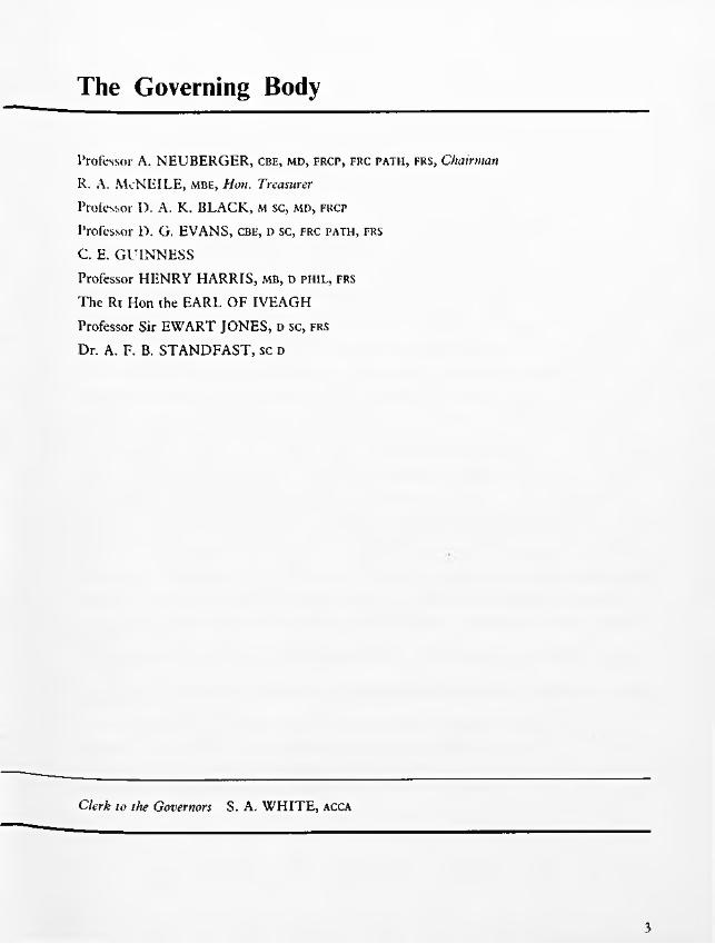
The Governing Body
Professor A. NEUBERGER, CBE, md, frcp, frc path, frs, ChairmanR. A. McNEILE, mbe, Hon. TreasurerProfessor D. A. K. BLACK, M sc, md, frcp
Professor D. G. EVANS, cbe, d sc, frc path, frs
C. E. GUINNESSProfessor HENRY HARRIS, mb, d phil , frs
The Rt Hon the EARL OF IVEAGH Professor Sir EWART JONES, D sc, frs
Dr. A. F. B. STANDFAST, sc d
Clerk to the Governors ... S. A. WHITE, ACCA

Financial Report of the Governing Body
The Governing Body presents the accounts of the Institute for the year ended 31st December 1971.1. Results
The General Fund Income and Expenditure Account shows the Income for the year as £405,796 compared with £344,136 in 1970. Expenditure amounted to £545,583 against £495,405 last year. The deficit for the year is £139,787 compared with a deficit of £151,269 in 1970. The capital fund has been reduced by the year’s deficit of £139,787. The Governors are well aware that deficits on the scale of those of recent years cannot be allowed to continue. Active steps are being taken to remedy the situation and an announcement concerning the future of the Institute is likely to be made to members at the Annual General Meeting.
The third of four annual instalments of £75,000 from the Wolfson Foundation and of five annual instalments of £1,000 from the Grocers’ Company have been added to the capital fund and in addition £300,000 has been transferred from Investment Reserve.2. Principal Activities
The Institute continued to carry out research work in connection with the prevention of diseases. It produces for sale sera, and bacterial and virus vaccines, the profits from which are utilised for its research and experimental work.3. Exports
Sera and vaccines to the value of £207,896 were exported from the United Kingdom during the year.4. Fixed Assets
The movements in fixed assets during the year are set out in the table in note 1 on the
accounts. These include further payments on account of the new wing at Chelsea, the cost of which is expected to be about £400,000. The Governors have transferred to Capital Fund a further £25,000 from the Sinking Fund, first set up in 1901 for the replacement and repair of buildings, towards the cost of this new wing.5. Interests in Land
The market value of the Institute’s properties is now in excess of the amount at which they are included in the Balance Sheet. In the opinion of the Governing Body such difference is of no significance as the properties are occupied for the purposes of the Institute’s activities.6. Governing Body
Professor R. A. Kekwick and Sir Ashley Miles retired from the Governing Body in June and September 1971 respectively. Dr. A. F. B. Standfast joined the Governing Body in June and Sir Ewart Jones in October. Other members of the Governing Body shown on page 1 held office during the whole of the year ended 31st December 1971.7. Employees
The average number of persons employed by the Institute in each week during the year ended 31st December 1971 was 319. The aggregate remuneration paid or payable in respect of that year to these employees amounted to £474,562.8. Auditors
The auditors, Cooper Brothers & Co., will continue in office in accordance with Section 159 (2) of the Companies Act 1948.
A. NEUBERGER Chairman
2

Report of the Auditors to the MembersIn our opinion the accounts set out on pages 4 to 9 give a true and fair view of the state of the company’s affairs at 31st December, 1971 and of its deficit for the year ended on that date and comply with the Companies Acts 1948 and 1967.
London, 25th May 1972COOPER BROTHERS & CO.
Chartered Accountants.

The Lister Institute of Preventive Medicine BALANCE SHEET * 31 December 1971
1970 £ £ £
£
546,238 FIXED ASSETS (note 1 ) ..................................................... 633,492INVESTMENTS AND UNINVESTED CASH (note 2)
406,406 General .......................................................................... 321,029164,587 Specific funds ............................................................................... 126,67424,471 Bequest funds ................................................................................. 25,834
595,464 473,537
1,107,029
CURRENT ASSETS— Stock (note 3) .............................................................. 184,824
171,545 Debtors .......................................................................... 127,80886,918 Cash and Bank Balances............................................. 45,226
258,463 357,858
Less:CURRENT LIABILITIES
84,512 Creditors ....................................................................... 69,727185,666 Bank Overdraft (note 14) .......................................... 62,630
270,178 132,357
(11,715) 225,501
£1,129,987 £1,332,530
Represented by494,323 CAPITAL FUND (note 4) ...................................................... 914,691148,477 SPECIFIC FUNDS (notes 5 and 8) 116,46424,471 BEQUEST FUNDS (note 6) 25,834
6,396 SPECIFIC GRANTS AND LEGACIES UNEXPENDED(note 7) ............................................................................. 1,479
456,320 INVESTMENT RESERVE (note 8) ....................................... 274,062
£1,129,987 £1,332,530
A. NEUBERGER
C. E. GUINNESSMembers ot the Governing Body
4
The notes on pages 6 to 9 form part ot these accounts. Audit report on page 3.

The Lister Institute of Preventive Medicine INCOME AND EXPENDITURE ACCOUNTfor the year ended 31 December 1971
1970£
INCOME£ £ £
303,664 Sales of sera and bacterial and virus vaccines (note 9) 340,764— Stock adjustment (note 3) ....
Investment Income:25,669
General fund29,971 Quoted ..................... ........................................... 22,351
2,444 Unquoted ................ ........................................... 2,46624,817
7,99364
Rent ......................................... 10,117Other income ......................... 4,429
344,136 405,796
Totalexpenditure
Externalcontributions
EXPENDITURE277,288 Salaries and wages ............................................................. 464,817 156,720 308,097
18,477 Superannuation premiums ................................................ 32,856 8,991 23,86512,261 Rates and insurances ........................................................ 12,022 — 12,02220,525 Gas, water, fuel and electricity ........................................ 34,228 7,558 26,67011,628 Office expenses, stationery and printing ........................ 16,119 350 15,769
950 Audit fee ............................................................................. 950 — 9505,689 Interest on overdraft............................................................. 9,485 — 9,4859,195 Chelsea research ................................................................. 24,856 16,991 7,865
72,583 Elstree research and production .................................... 68,618 349 68,26934,687 Animals and forage ............................................................. 35,128 5,024 30,10415,224 Alterations, repairs and renewals .................................... 18,791 326 18,46510,287 General expenses.................................................................
Depreciation17,281 3,355 13,926
3,239 Buildings ..................................................................... 3,296 — 3,2963,372 Furniture, fittings, scientific apparatus and books 6,461 — 6,461
495,405 £744,908 £199,664 545,244
151,269 Excess of expenditure over income ................................ 139,448— Exceptional items — net (note 10) ................................ 339
£151,269 Deficit transferred to capital fund .................................... £139,787
I1
The notes on pages 6 to 9 form part of these accounts. Audit report on page 3.

NOTES ON THE ACCOUNTS • 31 December 1971
1. FIXED ASSETSFreehold
Land and buildings Chelsea
propertyQueensbury
LodgeEstate,Elstree
Furniture, fittings,
scientific apparatus and books
Total
£ £ £ £Cost
At 1st January 1971 ................................................. 413,643 120,960 40,618 575,221Additions at cost .....................................................Amount written off (note 10) ................................
58,708 13,287(6,048)
31,064 103,059(6,048)
At 31st December 1971 ............................................ £472,351 £128,199 £71,682 £672,232
DepreciationAt 1st January 1971 ................................................ 4,050 12,288 12,645 28,983Charged to income and expenditure accoun t....... 1,350 1,946 6,461 9,757
At 31st December 1971 ............................................ £5,400 £14,234 £19,106 £38,740Net book value at 31st December 1971 ............... £466,951 £113,965 £52,576 £633,492
DepreciationFreehold property additions and replacements since 1912 at Elstree and since 1935 at Chelsea until
31st December 1964 have been charged to revenue. Additions since that date until 31st December 1967 have been depreciated at the rate of 10%. Since 1st January 1968 buildings shown in the balance sheet have been depreciated at the rate of 2% on a straight line basis from the date of completion.
Additions and replacements to furniture, fittings, scientific apparatus and books between 31st December 1920 and 31st December 1963 have been charged to revenue. The additions since 1st January 1964 have been depreciated on a straight line basis by reference to the anticipated useful lives of the assets.
INVESTMENTS AND UNINVESTED CASH £ £ £ £ £Q u o te d a t c o s t
in G re a t E ls e w h e re B r it a in
U n q u o te d a t c o s t
U n in v e s te dc a s h
T o ta l
General .........................................................
Specific
136,310 146,012 38,707 321,029
Sinking fund for freehold buildings .... 65,181 — — 398 65,579Pension fund ........................................ 15,871 — — 2,218 18,089Re-endowment fund ............................ 37,666 — — 5,340 43,006
BequestJenner Memorial studentship fund .... 11,766 — 940 5,130 17,836Morna Macleod scholarship fu n d ....... 5,653 — — 2,345 7,998
£272,447 £146,012 £39,647 £15,431 £473,537
1970 (£358,463) (£160,749) (£40,197) (£36,055) (£595,464)
Market value of quoted investments ....... 1971 £772,965 (1970 £801,468)Unquoted investments valued by Institute’s
investment advisers ................................ 1971 £37,022 (1970 £34,397)

3. STOCKSAs from 1st January 1971 stocks of bacterial and virus vaccines and sera have been valued in the
accounts.The amount of the 1970 valuation £159,155 has been credited to the General Fund Income and
Expenditure Account and the 1971 valuation of £184,824 is for the first time shown as a current asset in the balance sheet.
The effect of the change in the basis of accounting is to reduce what would otherwise have been shown as the deficit of the year by the difference between the two stock valuations namely £25,669 and to increase the Capital Fund by the amount of the closing stock namely £184,824.
The following bases have been used for stock valuations:—(i) At 31st December 1971
(a) Bacterial Vaccines and Sera £134,824At 1971 costs of direct materials and labour with additions for overheads appropriate to the stages of production reached.
(b) Virus Vaccines £50,000At current minimum' realisable value, less an appropriate deduction to cover selling and administrative expenses.
' (ii) At 31st December 1970At the same unit values as those used for 31st December 1971 stocks.
4. CAPITAL FUNDDonations etc. have been received to date from the following:—
Dr. Ludwig Mond (1893) .............................................................Berridge Trustees (1893-1898) ....................................................Worshipful Company of Grocers (1894 and 1969/71) ...........Lord Iveagh (1900) .........................................................................Lord Lister’s Bequest (1913-1923) ............................................Willian Henry Clarke Bequest (1923-1926) ................................Rockefeller Foundation (1935-1936) ............................................Wolfson Foundation (1969-71) ....................................................Other donations and legacies (1891-1954) ................................
Amount transferred from Sinking Fund (note 5) ................................Amount transferred from Investment Reserve (note 8) ...................
General Fund Income and Expenditure Account ................................Accumulated deficit at 31st December 1970 ............................Add: Stock at 1st January 1971 (note 3) ....................................
(CR)Less: Deficit 1971 .............................................................................
Accumulated d e fic it.................................................................................
5. SPECIFIC FUNDSSinking Fund for Freehold Buildings
As at 1st January 1971 ...................................Interest on investments ...................................
Less: Expenditure on reablement of buildings Amount transferred to Capital Fund ...
Pension FundAs at 1st January 1971 Interest on investments
Less: Pensions ............
Re-endowment FundAs at 1st January 1971 ......Donations ...............................Profit on sales of investments
1971 1970£ £ £
2,000 2,00046,380 46,38013.000 12,000
250.000 250,00018,904 18,9047,114 7,1143.400 3,400
225.000 150,00022,669 22,669
588.467 512,46785.000 60,000
300.000 —973.467 572,467
78,144159,15581,011
139,78758,776 78,144
£914,691 £494,323
1971 ,
96,0313,419
99,45019,08125,000
44,08155,369
19,4541,372
20,8262,737
18,089
32,992663
9,35143,006
£116,464
1

6. BEQUEST FUNDSJenner Memorial Studentship Fund
As at 1st January 1971 .............Interest on investments .........
Morna Macleod Scholarship FundAs at 1st January 1971 .........Interest on investments .........
7. SPECIFIC GRANTS AND LEGACIES
Nuffield Foundation GrantsAs at 1st January 1971 ...........Less: Laboratory expenses .......
Guinness-Lister Research GrantAs at 1st January 1971 ...........Amounts received ...................
Less: Salaries and wages .......Laboratory expenses .......
8. GENERAL AND SINKING FUNDS INVESTMENT RESERVE General
As at 1st January 1971 ...................................................Add: Profits on sales of investments..............................
Less: Amount transferred to Capital Fund .................
Sinking FundAs at 1st January 1971 ...................................................Less: Losses on sales of investments ..........................
9. TURNOVERTurnover has been arrived at after deducting commissions due to
agents from the invoice value of sales of sera, vaccines and virus vaccines.
10. EXCEPTIONAL ITEMSProfessional fees were incurred during 1970-71 in respect of
buildings to be erected at Elstree. This project was abandoned and the total expenditure written off during the year, including £6,048 in respect of the previous year, amounted to ...................
A successful appeal against the assessment on the Chelsea buildings resulted in the repayment of rates overpaid during the years 1963-71. The amount repaid was ............................................
£
£
15,8573,687
£
440,210123,642563,852300,000
16,1105,900
£16,975
861
7,496502
£
1,583373
4,81315,00019,813
19,544
£
263,852
£
17,836
7,998£25,834
£
1,210
269 £1,479
£
10,210 _______£274,062
£20,616
£20,277£339

11. EMOLUMENTS OF MEMBERS OF THE GOVERNING BODY1971 1970
Emoluments in an executive capacity ..................................................................... £16,814 £16,556
Particulars of emoluments of the Governing Body in accordance with Section 6 of the Companies Act 1967
1971 1970Emoluments of the Chairman of the Governing B o d y .................................... Nil NilEmoluments of the highest paid member of the Governing B o d y .................. £6,297 £7,256
Numbers of members of the Governing Body whose emoluments were within the range
No emoluments ...................................................................................................... 7 10£1 — £2,500 — —
£2,501 — £5,000 3 —£5,001 — £7,500 1 2
12. CAPITAL EXPENDITURE SCHEMES1971 1970
The position at 31st December 1971 was as follows:—Commitments in respect of contracts................................................................. 32,075 86,332Approved by the Governing Body in addition to commitments, for the new
laboratories at Elstree ..................................................................................... — 136,000
£32,075 £222,332
13. CONTINGENT LIABILITIESAt 31st December 1971 there were contingent liabilities amounting to £12,810 in respect of indemnities issued to third parties.
14. BANK OVERDRAFT
The overdraft is secured by the Institute's investments.
9

.


I
!«i
!
Ii
i

THE LISTER INSTITUTE OF PREVENTIVE MEDICINE
Report1972

I !
! 1
lì

THE LISTER INSTITUTE OF PREVENTIVE MEDICINE
Reportof theGOVERNING BODY
1972
CHELSEA BRIDGE ROAD : LONDON : SW1

!
¡
i
i
i
!I\
i1
; ’C
The Governing Body
Professor A. NEUBERGER, Cbe, md, frcp, frc path, frs, ChairmanR. A. McNEILE, mbe, Hon. TreasurerProfessor D. A. K. BLACK, M sc, md, frcp
Professor D. G. EVANS, cbe, d sc, frc path, frs
C. E. GUINNESSProfessor HENRY HARRIS, mb, d phil , frs
The Rt Hon the EARL OF IVEAGH Professor Sir EWART JONES, D sc, frs
Dr. A. F. B. STANDFAST, sc d
Clerk to the Governors S. A. WHITE, ACCA
3

T
(<
'it't
:jj■ l!.i*H||'
!|!‘:
'll
il!I!
1

The Council
A. LAWRENCE ABEL, MS, frcs Representing the British Medical Association
Professor D. A. K. BLACK, M sc, md, frcp Representing the Members of the Institute
The Rt. Hon. Lord BROCK, MS, frcs Representing the Members o f the Institute
H. P. G. CHANNON, MP Representing the Members of the Institute
Dame HARRIETTE CHICK, dbe, d sc Representing the Members of the Institute
Professor P. J. COLLARD, md, mrcp Representing the University of Manchester
M. L. CONALTY, MD, MRC path, dph , mria Representing the Royal Irish Academy
Major L. M. E. DENT, dso Representing the Worshipful Company of Grocers
Sir ALAN N. DRURY, cbe, ma, md, FRCP, FRS Representing the Members o f the Institute
Professor D. G. EVANS CBE, d SC, FRC PATH, frs Representing the Royal Society
Professor R. I. N. GREAVES, ba, md, frcp Representing the University of Cambridge
C. E. GUINNESS Representing the Members of the Institute
Professor HENRY HARRIS, MB, d ph il , frs Representing the University of Oxford
The Rt Hon the EARL OF IVEAGH Representing the Members o f the Institute
R. A. McNEILE, MBE Representing the Members o f the Institute
Professor B. P. MARMION, md, D SC, frc PATH Representing the University o f Edinburgh
Professor Sir ASHLEY M ILES, CBE, md, frc PATH, frcp, frs Representing the Members o f the Institute
Professor J. S. M ITCHELL, CBE, MA, md, FRS Representing the Members o f the Institute
Professor W. T. J. MORGAN, cbe, d sc, ph d, md (he) fric, frs Representing the Members o f the Institute
Professor A. NEUBERGER, cbe, md, frcp, frc path, frs Representing the Members o f the Institute
The President of the ROYAL COLLEGE OF PHYSICIANS Representing the Royal College of Physicians, London
The President of the ROYAL COLLEGE OF SURGEONS Representing the Royal College o f Surgeons o f England
The President of the ROYAL COLLEGE OF VETERINARY SURGEONS Representing the Royal College o f Veterinary Surgeons
A STEELE-BODGER, ma, b sc, mrcvs Representing the Royal Agricultural Society
Professor F. S. STEWART, md Representing the University o f Dublin
W ILLIAM J. THOMPSON Representing the Worshipful Company of Grocers
Sir GRAHAM WILSON, md, frcp Representing the University o f London
5

The StaffDirector:
fProfessor D. G. Evans, CBE, D SC, FRC path, frs (Professor of Bacteriology and Immunology)Deputy Director:
fProfessor L. H. Collier, md, d sc, mrcp
Superintendent o f Elstree Laboratories:*W. d’A. Maycock, cbe, mvo, md, frcp frc path
MICROBIOLOGY, EXPERIMENTAL PATHOLOGY AND IMMUNOLOGY Experimental Pathology and ImmunologyF. R. Wells, BM, B CH, MA Brenda Mason, b sc, mi biol
*D. G. Godfrey, o b e , b sc, PH D (M .R.C . External Scientific S ta ff)Angela E. R. Taylor, b sc, p h d Sheila M. Lanham, B SC
Trypanosomiasis Research Unit
MicrobiologyfG . G. Meynell, MD, D SC (Guinness Professor of
Microbiology)K. G. Hardy, b sc Valerie M. Harden, b sc
Guinness-Lister Research Unit
*Elinor W. Meynell, ba, md, dip bact *Ruth M. Lemcke, B se, ph D J. E. Dowman, MA, PH D (S .R .C . Grantee)M. C. Goel, MV se, ph d (india) (Commonwealth Scholar)
Virology|L . H. Collier, MD, D sc, MRCP (Professor o f Virology
and Hon. Director, M .R .C . Trachoma Unit)J. Alwen, B sc, ph d Lindsey M. Hutt, b sc
W. A. Blyth, b sc, ph d Janice Taverne, ba, ph d A. J. Garrett, b sc, ph d Andrea Evans, b sc G. P. Manire, ph d (U .S .A .)Margaret J. Harrison, M sc
Electron Microscopy Unit*A. M. Lawn, ph d, b sc, mrcvs R. A. Matthews, b sc
\
M.R.C. Trachoma Unit
/
BIOCHEMISTRY-¡■Winifred M. Watkins, d sc, ph d, frs (Professor
o f Biochemistry)Shirley D. Goodwin, b scM. A. Chester, M SC, B tech, ph d (Beit Memorial Fellow)J. R. Stealey, Ml biol (Grocers' Company Research Student)Hilary M. Simpson, B SC (Research Student)
§Professor W. T. J. Moi d sc (he),
A. S. R. Donald, B sc, ph d (M .R.C . Grantee) Caroline Race, B sc, ph d (M .R.C . Grantee) Helene T. Cory, b sc, ph d (M .R.C . Grantee) M. D. Topping, B sc (M .R.C . Grantee)Lynne B. Roberts, b sc (M .R.C . Student)A. Gardas, M sc, PH D (British Council Scholar) Josette F. Genissel, Diplome (Paris)
i, cbe, d sc, ph d, md (he),:, frs (ret’d)
BIOPHYSICSf j . M. Creeth, B sc, ph d, Fric (Reader in Biophysics)
K. R. Bhaskar, m sc (Poona), ph d (Bangalore) (M .R.C . Grantee)
W. E. Parish, ma, ph d , bv sc, mrcvs, mrc path *D. E. Dolby b sc, ph d G. G. Beadle, mv sc, mrcvs
PREPARATION and STUDY of THERAPEUTIC SERA (ELSTREE)
PREPARATION and STUDY of VIRUS VACCINES (ELSTREE)*H. G. S. Murray, md L. C. Robinson, b sc, ph dG. S. Turner, b sc, ph d L. V. Runkel, b sc
PREPARATION and STUDY of BACTERIAL VACCINES (ELSTREE)*A. F. B. Standfast, sc D A. P. Hunt, b scJean M. Dolby, ma, ph d M. P. Banks, b sc Caroline J. Shanbury, b sc
J. P. Ackers, ma, d phil S. T. A. Gilligan, b sc Jennifer A. Blackwell, B sc
CO-ORDINATION of PRODUCTION (ELSTREE)J. Rodican, B sc
BLOOD PRODUCTS (ELSTREE)*W. d’A. Maycock, cbe, mvo, md, frcp, frc path L. Vallet, maMargaret E. Mackay, m sc, ph d (M .R.C. External Scientific S ta ff)D. Ellis, b sc, ph d
Constance Shaw, M sc, dip bact L. Singleton, b sc, ph d, fric E. D. Wesley, b pharm Valerie J. Stickley, b sc D. W. Ashton, b sc
Plasma Fractionation Laboratory (at Oxford)Ethel Bidwell, b sc, ph d, fric T. J. Snape, BA
MEDICAL RESEARCH COUNCIL UNITS HOUSED AT THE INSTITUTE Blood Group Unit
§R. R. Race, cbe, md (he), ph d, frcp, frs Ruth Sanger, b sc, ph d, frs Patricia Tippett, b sc, ph d
E. June Gavin, b sc Phyllis W. Teesdale, b sc
Blood Group Reference LaboratoryK. L. G. Goldsmith, ph d, mb, bs, mrcp, mrc pathToby T. B. Phillips, mb, ch b
Elizabeth W. Ikin, b sc, ph d Carolyn M. Giles, b sc, ph d B. J. Dawes, b sc
ADMINISTRATIONSecretary and Accountant Elstree Secretary and Estate Manager Assistant Secretary Administrative Assistant Administrative Assistant
S. A. White, acca G. J. Roderick, B COM Barbara A. Prideaux C. L. Beard Beryl I. Coussens
Solicitors :Field Fisher & Co.,296, High Holborn, W .C.l.
Auditors :Cooper Brothers & Co.,Abacus House, Gutter Lane, E.C.2.
J -Appointed Teacher o f the U niversity o f London Recognised Teacher o f the U niversity o f London
§H onorary Member o f the Institu te S ta f f
7

Annual General Meeting of the Lister Institute
REPORT OF THE GOVERNING BODYREPORT OF THE GOVERNING BODYThe Governing Body has the honour to present its report of the Institute for the year 1971. The scientific section consists of a brief summary of the various researches and an article by Professor Winifred Watkins describing some of the work of the Biochemistry Department.
GOVERNING BODYThe Council at the meeting held on 22nd June, 1971 reappointed Professor A. Neuberger and Professor D. A. K. Black as its representatives on the Governing Body until 31st December, 1972. At this meeting the Governors were authorized to appoint a third representative of the Council and have pleasure in announcing that Professor Sir Ewart Jones accepted their invitation to join the Governing Body.
In accordance with the Articles of Association Professor R. A. Kekwick retired from the Governing Body and was succeeded by Dr. A. F. B. Standfast.
DIRECTORSHIP OF THE INSTITUTEThe retirement of Sir Ashley Miles, announced in last year’s Annual Report, took place on 30th September, 1971. Sir Ashley became Director in October, 1952 at a time when it would perhaps be not unfair to his predecessor to say that the Institute had not fully recovered from the problems of the war. His wide interests, combined with an energetic approach, enabled Sir Ashley to build up the scientific reputation of the Institute to its former high level; at the same time his knowledge and experience was put at the disposal of many outside bodies. He was for several years a member of the Expert Committee on Biological Standardization of the World Health Organization; he was a member of the Medical Research Council; and from 1963 until 1968 he was Biological Secretary and a Vice-President of the Royal Society. Despite the very heavy demands made on his time by outside work, Sir Ashley never neglected the interests of the Institute
and indeed his ability to cope with a prodigious amount of work was such that for most of his term of office he not only dealt with the administrative affairs of the Institute but was able to continue his own researches on the mechanisms of inflammation and infection.
Unfortunately Sir Ashley’s last year at the Institute was marred by ill-health, but he takes with him the good wishes of the Governing Body and staff for a long and happy retirement; this, as one might expect, will not be an idle period as he is continuing his researches at the Clinical Research Centre in Harrow.
As is the custom, a portrait of Sir Ashley was commissioned by the Governing Body. This was painted by Mr. David Poole, R.P. and now hangs in the Grocers’ Lecture Theatre.
Just before his retirement the University of London conferred upon Sir Ashley the title of Emeritus Professor of Experimental Pathology.
The Governing Body welcomes ProfessorD. G. Evans, C.B.E., F.R.S., as the new Director of the Institute. Professor Evans took up his appointment on 1st October, 1971. Professor Evans comes to the Institute after giving up the directorship of the Department of Bacteriology and Immunology at the London School of Hygiene and Tropical Medicine; previously he was Director of the Medical Research Council’s Department of Biological Standards. He first joined the Governing Body of the Institute in 1965.
COUNCILAt last year’s Annual General Meeting Sir Graham Wilson was reappointed as the representative of the University of London; Mr. Alasdair Steele-Bodger replaced Dr. R. E. Glover as the representative of the Royal Agricultural Society; and the third retiring member, Sir Charles Dodds did not wish to be reappointed. Mr. R. A. McNeile was appointed to the Council as a representative of the Members.
8

Preventive Medicine 27 June 1972
The three members of the Council due to retire this year in accordance with the Articles of Association, but who are eligible for reappointment are Sir Alan Drury, Dame Harriette Chick and Mr. H. P. G. Channon, all representatives of the Members of the Institute.
MEMBERSThe Governing Body records with regret the deaths of Professor J. H. Dible, Sir Charles Harington and Professor H. B. Maitland. Professor Maitland, who worked at the Institute during 1927, was for many years the representative of the University of Manchester.
STAFF AND STUDENTSProfessor R. A. Kekwick retired on 30th September, 1971. Professor Kekwick, who had been working at the Institute as a Medical Research Council Grantee, joined the staff of the Biophysics Department in 1940 and became head of the department in 1944. In July, 1971 the University of London conferred on him the title of Emeritus Professor of Biophysics. Professor Kekwick’s long and distinguished services to the Institute will be much missed and he takes with him the good wishes of the Governors and staff.
Professor G. G. Meynell, who came to the Institute in 1959 and became Guinness Professor of Microbiology and head of the Guinness-Lister Unit in January 1966, is resigning at the end of September to take up a post as Professor of Microbiology at the University of Kent. Professor Meynell takes with him the Governors’ congratulations and best wishes for the future.
Dr. Elinor Meynell is also resigning from the Guinness-Lister Unit to continue her work at the University of Kent.
The Governing Body has much pleasure in noting that Dr. R. R. Race, Director of the M.R.C. Blood Group Unit, has been awarded the Conway Evans Prize. The Prize is given jointly by the President of the
Royal Society and the President of the Royal College of Physicians every few years for “a valuable addition or contribution to science” .
The Governors were very glad to learn of the election to the Fellowship of the Royal Society this year of Dr. Ruth Sanger. Dr. Sanger is distinguished for her work on human red cell antigens and for the genetic mapping of the human X chromosome.
At the Annual General Meeting of the Society for General Microbiology in April this year Professor Evans was elected President of the Society.
The Governors were glad to learn of the election of Dr. W. d’A. Maycock to the Fellowship of the Royal College of Physicians.
The Governing Body is pleased to note that Mr. L. Vallet was presented with the 1971 Oliver Memorial Fund for Blood Transfusion Award; Mr. Vallet shared the Award with Dr. Helen Whyte Carlin of the Birmingham Regional Centre.
Mr. G. G. Beadle was appointed Deputy Head of the Serum Department; Dr. L. Singleton and Mr. D. W. Ashton were appointed to the Blood Products Laboratory; Miss J. A. Blackwell to the Bacterial Vaccines Department; and Mr. R. A. Matthews to the Electron Microscopy Unit.
Mr. E. J. H. Lloyd, Assistant Secretary and Deputy Accountant, resigned in April 1972. Mr. Lloyd had worked at the Institute for eleven years. The Governors wish him every success in his new post at St. George’s Hospital Medical School.
Dr. K. A. Chandrabose of the Guinness- Lister Unit resigned in October, 1971; and Mrs. Wendy Jeffery resigned from the Serum Department in April, 1972.
Professor L. H. Collier participated, by invitation, in the Second Conference of the International Society of Geographical Ophthalmology and the Jerusalem Seminar on the Prevention of Blindness, in August, 1971 in Israel.
9

Dr. W. d’A. Maycock attended meetings of the Group of Experts No. 15 of the European Pharmacopoeia Commission, in January and December, 1971.
Professor Winifred Watkins attended, by invitation, a joint meeting of the German and Belgium Biochemical Societies in Liège, Belgium, in January, 1971 and in August, together with Dr. Caroline Race and Dr. M. A. Chester, she took part by invitation in the First International Congress of Immunology held in Washington D.C., U.S.A.
Professor G. G. Meynell and Dr. E. W. Meynell attended the European Phage Conference in West Berlin, in October, 1971. Dr. Meynell also spoke by invitation at the Department of Microbiology, Faculty of Science, University of Paris, Orsay.
In January 1971 Dr. W. E. Parish, as a WHO consultant and at the invitation of the Burmese Government, visited the laboratories of the Burma Pharmaceutical Industry in Rangoon.
Dr. H. G. S. Murray attended the Twelth International Congress of the International Association of Microbiological Societies’ Permanent Section of Microbiological Standardization at Annecy, in September 1971.
In October 1971 Mr. L. Vallet participated, by invitation, in a Symposium on Plasma Derivatives for Clinical Use in WashingtonD.C., arranged by the American National Red Cross Society. He also visited the Blood Research Institute, Boston, Massachusetts and the Plasma Fractionation Laboratory, Commonwealth of Massachusetts Biologies Laboratory, Jamaica Plain, Massachusetts.
Dr. Ethel Bidwell attended the Second Congress of the International Society on Thrombosis and Haemostasis in Oslo in July 1971.
Dr. G. S. Turner attended the Second International Congress for Virology in Budapest in June 1971.
Dr. J. M. Creeth visited the Department of Chemistry, Redlands University, California for discussions with Dr. J. B. lift on density- gradient ultracentrifugation methods.
In March 1971 Dr. Ruth Lemcke participated by invitation in a workshop on “The Mycoplasmatales as agents of disease” at the National Institute of Allergy and Infectious
Diseases, Bethesda, Maryland.Dr. D. G. Godfrey, Dr. Angela Taylor
and Miss Sheila Lanham attended the Première Multicolloque Européen de Parasitologie, at Rennes, France, in September 1971.
For the academic year 1971/72 there are twenty-two postgraduate research workers at the Institute registered for higher degrees of the University. Three Ph.D. and one M.Phil degrees were awarded during 1971.
DONATIONS AND GRANTSArthur Guinness, Son & Co. Ltd., continued their generous support of the Guinness- Lister Research Unit during the year.
The Governing Body records its appreciation of the many other bodies whose benefactions and grants support research work in the Institute. These include a grant from the Arthritis and Rheumatism Council for the role of bacterial complexes in vasculitis; grants from the Medical Research Council for research in pertussis immunity; for immunochemical investigations on human blood-group specific glycoproteins; for biochemical investigations on the products of the blood group H, Lewis and Secretor genes; on the characterisation of the enzymic products of the A and B genes; on the characterisation of the human blood group active P, substance in hydatid cyst fluid; on the characterisation of blood-group specific glycoproteins by density-gradient methods; and on the genetics of drug resistance factors and other bacterial plasmids.
Grants were also received from the Overseas Development Administration of the Foreign and Commonwealth Office for studies on the biology of trypanosomes with special reference to their surface properties; from the Science Research Council for studies on the replication of bacterial plasmids; and from the Institute of the Diseases of the Chest, Brompton Hospital for an investigation of human tissue anaphylactic-sensitizing antibodies.
The Governing Body also gratefully acknowledges donations to the Institute’s Re-endowment Fund from the Prudential Assurance Company Limited and the Royal London Mutual Insurance Society Limited.
10

Particular emphasis has been given, over the past few years, to the expansion of production of bacterial vaccines; the financial success of this effort is a noteworthy tribute to the production teams at Elstree.
NEW BUILDINGSIt was reported last year that the new wing at Chelsea was to be opened in the Spring by Mr. Leonard Wolfson. This building was made possible by a very generous gift from the Wolfson Foundation. On 18th May 1971 Mr. Wolfson unveiled a plaque commemorating the completion of the Wolfson Wing and formally opened the Wing at a ceremony in the new lecture theatre. He was accompanied by Mrs. Wolfson and by Sir Isaac and Lady Wolfson. The ceremony was attended by many guests and members of staff, and was followed by conducted tours of the new building.
The lecture theatre itself, designated the Grocers’ Lecture Theatre on a plaque in the main hall, was opened by Major Leonard Dent on behalf of the Grocers’ Company, in recognition of a generous donation towards its erection.
It is expected that by the time this report is published the large extension to the Blood Products Laboratory will be partly in use. Modifications to the old Blood Products building are planned for the financial year 1972-73.
The Pyrogen Test Rabbit House has been completed and taken into use.
VISITORSThe following visitors, in addition to those listed under Staff, worked for short periods in the Institute’s Laboratories; Miss Kathleen Barber, St. George’s Hospital, London; Miss Raye Biggs, West Midlands Forensic Science Laboratory, Birmingham; Dr. G. Casillas, Instituto de Investigaciones Haematologicas, Academia Nacional de Medecina, Buenos Aires; Mr. A. Chaudhury, Central Research Institute, Kasauli, Simla Hills, India; Mr. G. Evans, South West Forensic Science Laboratory, Bristol; Miss Agnes Friss, National Institute of Haemotology and Blood Transfusion, Budapest, Hungary; Dr. M. Palatnik, University of La Plata, Argentina;
PRODUCTION AT ELSTREE Dr. H. Schenkel-Brunner, Institute of Biochemistry, University of Vienna, Austria, Mr. Myat Sein, Burma Pharmaceutical Industries, Rangoon, Burma; Dr. M. P. Than, Burma Pharmaceutical Industries, Rangoon, Burma; Mrs. A. Vichitanand; Government Pharmaceutical Organization, Bangkok, Thailand.
The Governing Body has during the last year at several meetings considered the serious financial position of the Institute. We have had over the last four or five years deficits of the order of approximately £150,000 per annum, and in order to finance our scientific activities at an acceptable level, we have had to make serious inroads into our accumulated capital reserves. It is clear that this process cannot be allowed to continue much longer. All the aspects of our financial situation have been carefully considered and possible steps are being discussed as how to deal with this situation. A Special Meeting of the Council has taken place at which the financial problems were fully presented. It is hoped to put to Members certain proposals in the near future, but these however can only be formulated after all the relevant aspects have been fully explored.
The Governing Body wishes to record its thanks and appreciation to the scientific, administrative and technical staff for their wholehearted devotion to the Institute. This year was a difficult one, as already indicated, and the understanding and loyalty displayed by all sections of the staff is gratefully recorded.
A. NEUBERGER,Chairman

REVIEW ARTICLE
THE BIOCHEMICAL BASIS OF HUMAN BLOOD GROUP ABO AND LEWIS POLYMORPHISM
Winifred M. Watkins
Department o f Biochemistry
The extent to which individuals in a population differ one from another depends in part on environmental conditions but is largely determined by inherited differences. In other words, individuality depends primarily on the number of gene loci in a population for which there is more than one allele. Although the extreme diversity of human beings is apparent at a glance, until comparatively recently relatively few genetic loci in man were clearly recognised as polymorphic; these loci were mainly those determining blood group systems which are differentiated by means of their serological properties. The molecular theory of inheritance put forward in the 1950’s has as a central belief the fact that the DNA of the structural gene loci in the chromosomes codes for the primary amino acid sequence of the polypeptide chains of specific proteins (see Crick, 1967). From this premise it follows that polymorphisms can arise from mutations that give a series of different alleles at one gene locus determining structurally different versions of a protein. In the last decade systematic searches for variants of functional proteins and enzymes were undertaken and many human genetic polymorphisms are now established (see Harris, 1970). On the basis of these findings, and those previously determined for other species, Lewontin (1967) has estimated that one-third of the structural gene loci in man may be polymorphic.
Despite the ever increasing number of variant forms of human genes that are being discovered, the ABO blood group system
remains the prime example of polymorphism. The red cell determinants associated with this system were the first inherited antigenic differences to be recognized in man (L.and- steiner, 1900) and more people throughout the world have been accurately classified for these genes than for any other polymorphic character. The three major alleles of the system are the genes A, B and O (Bernstein, 1924). A child receives one gene from either parent giving six genotypes AA, AO, BB, BO, AB and OO. The four recognizable phenotypes based on reactions with anti-A and anti-B agglutinins are A, B, AB and O. The ABO system is of outstanding importance in blood transfusion because anti-A and anti-B agglutinins are invariably present in the serum when the corresponding antigens are absent from the red cells. The relative frequency of the four groups varies in different populations but among Europeans about 47% of people are group O, 42% group A, 8% group B and 3% group AB.
The apparent simplicity of the inheritance of the ABO groups, which makes them such excellent chromosome “markers” in genetic and anthropological studies, was taken by the earlier biochemical geneticists to imply a very close relationship between the serological determinants and the blood group genes. In the light of present knowledge one might therefore suppose the antigens to be proteins synthesized on the ribosomes via messenger RNA mediated translation of the genetic code for specific amino acid sequences. The blood group active structures are, however, not protein but carbohydrate and
12

much of the work in the Biochemistry Department over the last few years has been directed towards clarification of the relationship between these specific carbohydrate structures and the blood group ABO genes. In the course of these investigations it has been necessary to consider three other genetic systems, the Hh and Lele blood genes and the Secretor genes Sese; these three systems, although inherited independently of the ABO genes (see Race and Sanger, 1968), are closely related to the ABO groups in their phenotypic effects.
A and B antigens are not confined to the red cells; they are present on the cell surfaces of probably all endothelial cells and many epithelial cells (Szulman, 1960, 1962) and substances with the same serological specificity occur in a water-soluble form in tissue fluids and secretions (Lehrs, 1930; Putkonen, 1930). Evidently, therefore, the genes controlling the appearance of these serologically active structures express themselves at many different sites throughout the body and the name “blood group substances” was given, and persists, simply because the antigens were first detected on erythrocytes. In the red cells, the substances that carry the A and B specificities are firmly bound and cannot be extracted with water or salt solutions. Earlier attempts to isolate active substances from red cells met with little success (see Rabat, 1956) and most of the detailed chemical knowledge that we have of blood group specific structures has come from investigations on the water- soluble substances which occur in large amounts in mucous secretions. Saliva, the mucous of the gastrointestinal tract and meconium are potent sources of water- soluble substances, but a particularly useful source of material for the isolation of relatively large amounts of these substances from single individuals has proved to be fluid obtained from ovarian cysts (Morgan and van Heyningen, 1944). These fluids accumulate in the cysts over long periods of time, and large volumes, often containing several grams of active group-specific material, may be obtained from a single cyst. Not all people who have A and B antigens on their red cells secrete water-soluble A and B substances. The “secretor” or “non-secretor” status of of an individual is constant and is genetically determined. Family studies revealed that
this dimorphism is determined by a pair of allelic genes (Schiff and Sasaki, 1932), now referred to as Se and se. Individuals homozygous or heterozygous for the allele Se are “secretors”, whereas those homozygous for the allele se are “non-secretors”. About 80% of Europeans are “secretors”, although the proportions of “secretors” and “non- secretors” is slightly different in other ethnic groups (see Race and Sanger, 1968).
Bernstein believed that the O gene was recessive and that group O represented the absence of the A and B characters. This view was generally accepted until Schiff (1927) discovered reagents that appeared to react preferentially with group O erythrocytes. The idea then became prevalent that the O gene gave rise to a product, analogous to the products of the A and B genes; the only difference being that the antibody to the O character did not occur naturally in the serum whenever the factor was missing from the red cells. Group O subjects who inherited an Se gene had water-soluble substances in their secretions that neutralized these “anti-O” reagents. This finding supported the view that there was an O gene product because at that time no other blood group systems were known to give soluble products. However, the idea that the serological reagents were detecting an O antigen had to be abandoned when it became clear that secretions from group AB subjects could inhibit the agglutinating activity of the “anti-O” sera and also that these antibodies reacted with erythrocytes from homozygous AA and BB donors. The explanation that seemed to fit these results most neatly was one formulated by Witebsky and Klendshoj (1941); they suggested that the so-called O-substance was in fact a basic substance “on top of which the A and B property might be present or absent”. Thus the “O-substance” is a precursor of A and B substances and it is present in larger amounts on the erythrocytes and in the secretions of group O subjects than in A, B or AB subjects simply because it does not undergo further conversion. Because the retention of the name “O-substance” did not seem justified for a material that was patently not the product of the O gene the term H-substance was introduced (Morgan and Watkins, 1948) and the sera that were neutralized by this substance were called anti-H. We now believe that the
gene locus concerned with the formation of H specific groupings has two alleles H and h (Watkins and Morgan, 1955a). Very rare individuals who are homozygous for the allele h have no A, B or H substance on their red cells or in their secretions. The first people with this unusual blood group were discovered in Bombay and the group is therefore frequently referred to as the “Bombay” Oh phenotype. When the erythrocytes of these people are tested with anti- A and anti-B sera they appear as normal group O cells but further examination reveals that they do not react with anti-H reagents. This lack of H surface structures has serious consequences if the subject needs a transfusion because anti-H agglutinins are present in the serum in addition to the expected anti-A and anti-B agglutinins; such sera therefore react with the red cells from all other individuals except those belonging to the same very rare group (see Race and Sanger, 1968).
The Lewis blood group system, described by Mourant (1946) is closely related to the ABO system and substances with Lewis activity occur in a water soluble form in secretions (Grubb, 1951). From the outset the genetics and inheritance of the two Lewis antigens, Lea and Leb, were much less clear cut than those of the ABO groups, and although knowledge of the chemical structures associated with the two specificities has made possible a clearer understanding of this system there are still some puzzling features about the relationship between the secreted Lewis substances and the antigens on the erythrocytes. Adults are differentiated into three groups on the basis of the reactivity of their red cells with anti-Lea and anti-Leb sera, that is, L e (a+ b -), L e (a -b + ) and L e ( a - b - ) (see Race and Sanger, 1968). Lea activity in secretions, however, is markedly dependent on the ABH “secretor- non-secretor” status. Individuals whose red cells are Le(a+b —) are “non-secretors” of ABH and “secretors” of Lea substance. Those whose red cells are Le(a —b+) are “secretors” of ABH and Leb; their secretions also have Lea activity but very much less than is found in the ABH “secretors” . Only those whose red cells are grouped as Le(a - b —) have neither Lea nor Leb in their secretions; a.small proportion of this group are also “non-secretors” of ABH.
Although three Lewis red cell phenotypes are recognizable we believe that there is only one functional allele at the Lewis genetic locus. Grubb (1951) and Ceppellini (1955) proposed that the presence or absence of Lea specificity in secretions depends on a pair of alleles, now called Le and le. The allele Le, in single or double dose, gives rise to Lea activity in secretions. Leb activity was at first (Andresen, 1948) thought to be the product of an allele of the gene giving rise to Lea because of the seemingly reciprocal relationship between Lea and Leb on red cells. Consideration of the distribution of the two activities in secretions, however, revealed that this could not be so. With considerable insight, Ceppellini (1955) pointed out that as Leb activity only occurs in the tissue fluids of ABH “secretors” it was probably a genetic interaction product and proposed that the two genes involved were Le and the secretor gene Se. The idea that Leb specificity is an interaction product is now established from the structure of the Leb determinant but the gene product that interacts with the product of the Le gene is that of the H gene and not Se (see Watkins, 1965; Marr et al., 1967). Grubb (1951) suggested that the Lewis system should be regarded essentially as a system of water-soluble antigens that are adsorbed onto the red cells and Sneath and Sneath (1955) demonstrated that Le(a —) red cells can be converted into Le(a+) cells by incubating them in the plasma from Le(a+b —) subjects.
Despite the complexities of the genetics of the ABO, Hh and Lele systems it is possible by simple serological inhibition tests to divide the population into four main groups based on the presence or absence of A, B, H, Lea and Leb activities in the secretions. About 70% secrete A, B or H together with Lea and Leb, about 20% secrete only Lea, some 9% secrete A, B or H but not Lea or Leb, and about 1 % secrete neither A, B, H, Lea or Leb. The presence of these well- defined serological specificities enables secretions to be selected from the appropriate donors for the isolation of the substances carrying the required blood group character.
By the early 1950’s preparations of A, B, H and Lea substances had been isolated and purified from ovarian cyst fluids. The most striking, and at that time rather disheartening observation, on these substances was that
14
they had the same qualitative composition irrespective of their blood group specificity. They all contained the same four sugars, D-galactose, L-fucose, AZ-acetylglucosamine and iV-acetylgalactosamine and the same 15 amino acids (see Morgan, 1960; Watkins, 1966). Carbohydrate accounted for about 80-85% of the molecule. The amino acid moiety was remarkable in that two amino acids, serine and threonine, made up about half the total amino acids (Pusztai and Morgan, 1963). The purified substances are macromolecules with molecular weights ranging from 500,000 to 2,000,000. Originally designated as mucopolysaccharides, these substances are now classified as glycoproteins. The full details of the macro- molecular organization of these substances is still under investigation some 20 years after their isolation, but their general properties and degradation products suggest that they are made up of a large number of oligosaccharide chains covalently attached at intervals to a peptide backbone. The attachment of most, if not all, of the sugar chains to the peptide moiety is through alkali-labile a-O- glycosidic linkages involving /^-acetylgalactosamine and the hydroxyamino acids serine and threonine which are present in such abundance in the peptide moiety (Anderson et al, 1964; Adams, 1965; Rabat et al, 1965; Donald et al, 1969).
Two other important facts emerged at an early stage in the investigations of the blood group specific glycoproteins. One was that all the substances, either in the native state or after mild degradation with acid or enzymes, cross-reacted with an antiserum to Type XIV pneumococcus (see Rabat, 1956). The second was that, however rigorous the purification, attempts to isolate A or B substances free from H, Lea or Leb activity were invariably unsuccessful when these additional activities were present in the original secretion. The observation on the cross reactivity with Type XIV anti-serum was taken to indicate that all the blood group substances had certain structural chemical groupings in common and, later, precipitin inhibition experiments showed that this reaction was attributable to the presence in each of the substances of p-linked galactosyl-(l -*4)-N- acetylglucosaminyl units (Watkins and Morgan, 1956). A glycoprotein isolated from the secretions of one of the rare group
of persons who fail to secrete A, B, H, Lea or Leb substances also showed cross reactivity with anti-Type XIV serum (Watkins and Morgan, 1959). This material was strikingly similar in chemical composition to the active substances except that it had a very low fucose content.
The similarity in composition and general physical properties, together with the Type XIV cross reactivity, suggests that in the secretions of all individuals there are glycoproteins which are probably fulfilling the same physiological function irrespective of whether or not they carry blood group active structures. The name “secretor” as applied only to those who have A, B or H activity in their secretions is therefore misleading. The term is probably too deeply entrenched in the blood group literature and in the minds of workers in the field to change the nomenclature at this stage but its use has led to erroneous speculations on the basis for the association between blood groups and diseases of the gastrointestinal tract by those who assume “non-secretion” to be synono- mous with the absence of glycoprotein.
The second observation that A or B substances cannot be freed from accompanying H, Lea and Leb activities was later explained by the demonstration that more than one serological activity can be carried on a single glycoprotein molecule. The method used to obtain evidence for the existence of glycoprotein molecules with single or multiple specificities was that of serological precipitation with selected mono-specific antisera (Morgan and Watkins, 1956; Watkins and Morgan, 1957a; Watkins, 1958, 1959). Examination of the supernatant and the redissolved precipitates demonstrated that the jointly carried specificities arise not only from the activities of allelic genes such as A and B but from the activities of genes belonging to independent genetic systems, such as A, H and Le. Thus a “secretor” belonging to group AB, whose secretions also have H, Lea and Leb activity, has glycoprotein molecules carrying A, B, H, Lea and Leb specificities that are carried down in the precipitate by an anti-A or an anti-B serum. Experiments with the appropriate control mixtures ensured that these activities were genuinely associated with the same molecules and were not non-specifically entangled in the precipitate.
15

With our present knowledge of the molecular basis of gene action no one today would be so naive as to suggest that a complex glycoprotein molecule could be synthesized by a single gene. When the blood group specific glycoproteins were first isolated, however, template theories of gene action were still prevalent and the impression that the blood group genes do not control the biosynthesis of the complete macromolecules came from the observations on the chemical similarities of the substances with different specificities and the finding of multiple specificities on single molecules. Enzymic degradation of the blood group substances strengthened the conviction that the blood group genes were imprinting their specificity at a late stage in the biosynthesis of the glycoproteins. The first significant observation was that of Iseki and Masaki (1953) who reported that A-substance could be “transformed” into H-substance by an enzyme preparation from Clostridium tertium. Subsequently, the B-decomposing enzyme from Trichomonas foetus was found to act on B- substance with the loss of B-activity, the development of H activity and the specific release of D-galactose (Watkins, 1956). This experiment demonstrated that the “transformation” consisted in the removal of a sugar residue which exposed an H specific structure that was masked in the undegraded glycoprotein. The sugar released from A- substance by an enzyme preparation from T. foetus, when A activity was lost and H activity developed, was later shown to be A-acetyl- galactosamine (Harrap and Watkins, 1964). A number of a-N-acetylgalactosaminidases and a-galactosidases from a variety of plant, bacterial, invertebrate and mammalian sources have since been found to bring about the same sequence of changes in A and B substances, respectively; confirming that removal of a single sugar residue from the A and B determinants reveals underlying H determinants. When the original A or B substance comes from a donor having Leb activity the residual glycoprotein has both H and Leb activity after treatment with A or B-destroying enzymes. Degradation of the substances with an H destroying enzyme results in loss of H and Leb activity, a development of Lea properties and a release of L-fucose (Watkins, 1960; 1962). Alternatively, if the HLeb substance is treated
with an Lea-destroying enzyme, Leb activity is lost, H activity is enhanced and again there is a loss of L-fucose (Stealey and Watkins, 1972). Sequential degradation of the carbohydrate chains with selected exo-glycosidases, therefore, reveals specificities that are part of the undegraded molecule but are not available for reactivity with their homologous antibodies. Possible explanations for the failure of the antibody to react with these hidden determinants are (1) that substitution of the terminal non-reducing sugar of a specific determinant with another sugar residue may block certain hydroxyl groups necessary for combination with the antibody, (2) that the added sugar may sterically hinder the approach of the antibody, or (3) that conformational changes are engendered in the specific determinant by substitution with another sugar so that the structure is no longer complementary to the antibody. Once embedded in the carbohydrate chains these hidden structures appear not to be recognized as “self” by the antibody-forming mechanisms because anti-H agglutinins are sometimes formed in group A or B individuals (see Race and Sanger, 1968) in spite of the fact that the H structure is part of the specific A and B determinants.
The correlation in the enzyme degradation experiments of loss of specificity with the release of sugar residues was in agreement with observations from other lines of investigation on the contribution of the carbohydrate moiety to the serological specificity of the blood group active glycoproteins. One of the methods used was inhibition of agglutination with simple sugars following the principle established by Landsteiner that a low-molecular weight substance with a structure similar to, or identical with, the determinant in an antigen combines with the antibody and specifically inhibits the reaction between the antigen and antibody (see Landsteiner, 1947). Before these experiments began it was known that certain plant seed extracts react with receptors on red cells and that some behave as specific blood group agglutinins (see Bird, 1959). These reagents proved of considerable value in the determination of the immunodominant sugars in the blood group active structures because we found that specific inhibition of the agglutination of red cells by these plant seed reagents was achieved with simple monosac
16

charides (Morgan and Watkins, 1953); animal or human antibodies are complementary to larger specific determinants and usually require at least a disaccharide before significant inhibition is demonstrable. Another method that was used empirically at first, but which gave useful and additional evidence for the part played by individual sugars as major determinants of blood group specificity was the inhibition of the enzymic inactivation of blood group substances by monosaccharides (Watkins and Morgan, 19556). Combinations of these methods proved that Af-acetylgalactosamine and D- galactose, respectively, occurring as nonreducing end-groups, and joined by an a-glycosidic linkage to the next sugar, were the structures most directly concerned in A- and B-specificity. The results of similar experiments established an a-L-fucosyl residue as the immunodominant sugar in H-specific structures (see Morgan, 1960; Watkins, 1966). Additional information indicated that the immunodominant sugar alone is not entirely responsible for serological specificity, and that the nature of the adjacent sugar and the position of the glycosidic linkage is also of considerable importance.
More extensive information about the Lea determinant was derived from inhibition tests with fucose-containing oligosaccharides isolated from human milk by Professor Kuhn and his colleagues in Heidelberg (see Kuhn, 1957). Two oligosaccharides containing a branched trisaccharide unit at the nonreducing end, with a-L-fucose and (3-D- galactose residues joined by 1 ->4 and 1 -»-3 linkages respectively to an TV-acetyl-fi-D- glucosaminyl unit, strongly inhibited the agglutination of Le(a+) red cells by human anti-Lea serum (Watkins and Morgan, 19576). Other closely-related compounds, that differed only in the point of attachment of the fucose residue, did not cause inhibition and we therefore concluded that Lea activity resided in the spatial arrangement of this branched trisaccharide. The involvement of L-fucose in two distinct specificities, namely, H and Lea, gave proof that the nature of the terminal non-reducing sugar cannot, by itself, be responsible for specificity, and that the way in which it is linked to the next sugar, or both, must also be important. Another interesting inference from the results
with Lea was that specificity does not necessarily reside in the nature and sequence of sugar units in a straight chain, but that branching sugar residues may contribute to the determinant structure. Further indications of the importance of branching fucose residues came from inhibition experiments with Leb (Watkins and Morgan, 19576). Oligosaccharides containing two fucose residues attached to adjacent sugars on a backbone chain were active inhibitors of Leb agglutination whereas analogues lacking either fucose unit were inactive.
Towards the end of the 1950’s, although our knowledge of the detailed structure and nature of the determinants associated with these glycoprotein molecules was still fragmentary, sufficient genetical, serological and biochemical information was available for profitable speculation on the relationship between the blood group genes and the serologically active structures. Genetical pathways for the biosynthesis of the blood group active structures were put forward from this laboratory (Watkins, 1958; Watkins and Morgan, 1959) and an essentially similar scheme was formulated by Ceppellini (1959) in Italy at much the same time. These pathways accounted for the interrelationships between the ABO, Hh, Lele and Sese genes which led to the four main phenotypic groups into which people can be divided according to the A, B, H Lea or Leb activities in their saliva or other secretions (Table 1). The serologically inactive glycoprotein occurring in the secretions of the rare individuals who lack A, B, H, Lea or Leb specificity is thought to be the precursor substance that in persons having the appropriate genes is converted into the blood group active glycoproteins. The genes A, B, H and Le were envisaged as structural genes whose products induce changes in the precursor. The genes O, h and le are silent alleles in the sense that their products do not bring about changes in the precursor substance although this does not imply that these genes do not code for structural proteins. The Se gene was considered as a regulator gene, or switch gene, that controls the expression of the H gene at the sites of synthesis of the secreted glycoproteins. The Le gene uses the same precursor molecules in secretions as the H gene but it is expressed whenever it is present in the genotype and is not under the control of the
17

Table 1. Possible genetical pathways for the biosynthesis of A, B, H, Lea and Leb substances
Gene systems: 1 ) L e le 2) H h 3)
Le
OC JC
Precursor Substance le
Le11 substance Precursor substance
H hh hh Hse se Se Se se se
or or, se sey se se .
!H subst.( - Lea-f Le'O
Leasubst.
L easubst.
Prec.subst.
Prec.subst.
H subst.
B O A B O A B O A B O A B O A B
< 'A su b . B s u b s t . H s u b s t . L ea L e a P re c . P re c . A s u b s t . B s u b s t .( - ^ H + L ea ( -t- H -1- L e a ( — L ea s u b s t . s u b s t . s u b s t . s u b s t . ( + H ) (t H)- r Le 'O -J-Le»») -i L e '* )^ 1 1 1 1
S1 i t
Secretor A B H ( 1 ) A B H ( - ;) A B H (- ■■) A B H ( - ) A B H ( — ) A B H ( + )type L e H + ) L e H r ) L e H ) L e H - ) L e H — ) L e H — )
L e H ~ ) L e H - ) L e H - ) L e H ) L e H - ) L e H — )
R ed cell A B H ( + ) A B H ( + :) A B H ( — ) A B H ( — ) A B H ( + ) A B H ( + )phenotype L e ( a — b f ) L e (a -rb-—) L e (a • b - ) L e ( a — b —-) L e ( a — b — ) L e ( a — b —
“ B o m b a y ” p h e n o ty p e
H subst.
secretor gene Se. This is why the secretions of about 90% of persons have greater or lesser degrees of Lea activity.
A series of as yet unidentified genes were assumed to be responsible for the biosynthesis of the precursor glycoprotein. In the presence of an H and an Se gene this precursor is converted into H substance which then forms the substrate for the conversions controlled by the A and B genes. In the absence of a secretor gene, that is, in persons homozygous for se, the precursor is unchanged and the conversion to A and B cannot take place even when these genes are part of the genotype. The Le gene controls the conversion of the precursor glycoprotein into Lea substance and this same material in the presence of an H and Se gene is converted into an HLeb substance with some residual Lea activity. Synthesis stops at the Lea stage if two se genes are present and Lea is then the only activity detectable in the secretions even when H, A or B genes are part of the genotype.
These pathways were put forward as a
framework from which to devise further experimental procedures; although some details concerning the order in which the gene products act has needed revision the subsequent work has supported the proposed schemes in general principle. Moreover, the complete chemical characterization of the serological determinants enabled the genetical pathways to be explained more clearly in chemical terms (see Watkins, 1965). Of primary importance to an understanding of the interrelationships of the ABO and Lewis systems was the finding that there are at least two types of carbohydrate chains in each blood group substance (Rege et al. 1963). Each chain ends with the disaccharide unit ¡3-galactosyl-IV-acetylglucosamine; the sugars are linked either l-* 3 (Type 1 chain) or 1—>-4 (Type 2 chain). We therefore proposed that in the precursor substance there are carbohydrate chains ending with these two disaccharide structures, and either one or both of these chains form the basis of the A, B, H, Lea and Leb specific structures. The isolation of a pentasaccharide in which the Type 1 and Type 2 disaccharides are both
18

T a b le 2. G e n e tic c o n tro l o f th e fo rm a tio n o f b lo o d g ro u pspecific H , L e a a n d L e b s tru c tu re s
Gene Chain Structure Specificity
— Type 1 ¡3-Gal-(l->3)-GNAc— —Type 2 |3-Gal-(1—4)-GNAc— Type XIV
H Type 1 P-Gal-(1—3)-GNAc— î «1,2
Fuc
H
Type 2 p-Gal-(l—►4)-GNAc—î «1,2
Fuc
H
L e Type 1 P-Gal-(1—3)-GNAc— î «1,4
Fuc
Lea
Type 2 |3-Gal-(1—4)-GNAc— Type XIV
H and L e Type 1 P-Gal-(l-*3)-GNAc— f <xl,2 | al,4
Fuc Fuc
Leb
Type 2 P-Gal-(1—4)-GNAc— î «1,2
Fuc
H
Abbreviations: Gal = D-galactopyranose; Fuc = GNAc = N-acetyl-D-glucosaminopyranose;
L-fucopyranose ;
joined to a Single galactosyl residue has been taken as evidence that both types of chain endings may exist as branches on one main chain (Lloyd et al. 1968). The Type 2 chain ends with the structure that gives rise to Type XIV pneumococcal cross reactivity. The protein products of the A, B, H and Le genes were envisaged as enzymes—glyco- syltransferases—that control the sequential addition of sugar units to the basic chains in the precursor substance.
The pioneering work of Leloir and his coworkers in the 1950’s revealed the central role played by nucleotide-bound sugars in the metabolism of sugars and in the biosynthesis of oligo- and polysaccharides (for review see Leloir, 1964); these compounds were therefore assumed to be the donors of sugar units in the reactions catalyzed by the blood-group gene-specified glycosyltransfer- ases (Watkins, 1967). The H gene product was postulated as an a-L-fucosyltransferase that conveys fucose from guanosine diphosphate L-fucose to the carbon-2 position of
the p-galactosyl residue of either the Type 1 or Type 2 chains (Table 2) to give the Id- active structure a-L-fucosyl- (1 ->2)-p-D- galactosyl-(l-*3 or 4)-(3-(jV-acetyl)-D-glu- cosaminyl-R (where R represents the remainder of the carbohydrate chain). This step was proposed on the basis of two serologically active trisaccharides, one having the 1 ->3 linkage and the other the 1 -»4 linkage, that were isolated from the degradation products of an H-active glycoprotein (Rege et al. 1964a).
Many fragments have now been isolated and identified from the acid hydrolysis and alkaline degradation products of A and B substances (see Lloyd et al. 1966; Morgan,1970), but the constant difference between the isolated fragments is that the serologically active units from A-specific glycoproteins have a terminal non-reducing N- acetylgalactosaminyl unit and those from B have a terminal non-reducing D-galactosyl unit. Each of these sugars is joined by an a-(l-*3) linkage to a p-galactosyl residue.
19
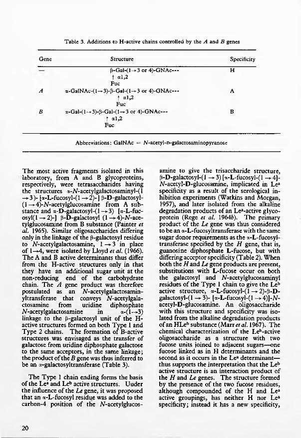
T a b le 3. A d d itio n s to H -a c tiv e c h a in s c o n tro lle d b y th e A a n d B genes
Gene Structure Specificity
A
B
P-G aH l—3 or 4)-GNAc— t al,2
Fuca-GalNAc-(l—3)-[3-Gal-(l—3 or 4)-GNAc—
t a l,2 Fuc
a-Gal-( 1 —► 3)-fl-Gal-( 1—► 3 or 4)-GNAc— t al,2
Fuc
H
A
B
Abbreviations: GalNAc = iV-acetyl-D-galactosaminopyranose
The most active fragments isolated in this laboratory, from A and B glycoproteins, respectively, were tetrasaccharides having the structures <x-N-acetylgalactosaminyl-(l -*3)- [a-L-fucosyl-(l —>2)-] p-D-galactosyl- (1 —► 4)-N-acetylglucosamine from A substance and a-D-galactosyl-(l -*3) [a-L-fuc- osyl(l ->-2)-] p-D-galactosyl (1-*4)-AT-ace- tylglucosamine from B substance (Painter et al. 1965). Similar oligosaccharides differing only in the linkage of the p-galactosyl residue to /^-acetylgalactosamine, 1 —► 3 in place of 1—*4, were isolated by Lloyd et al. (1966). The A and B active determinants thus differ from the H-active structures only in that they have an additional sugar unit at the non-reducing end of the carbohydrate chain. The A gene product was therefore postulated as an /V-acetylgalactosamin- yltransferase that conveys N-acetylgala- ctosamine from uridine diphosphate ^-acetylgalactosamine in a-(l-*3) linkage to the p-galactosyl unit of the H- active structures formed on both Type 1 and Type 2 chains. The formation of B-active structures was envisaged as the transfer of galactose from uridine diphosphate galactose to the same acceptors, in the same linkage; the product of the B gene was thus inferred to be an a-galactosyltransferase (Table 3).
The Type 1 chain ending forms the basis of the Lea and Leb active structures. Uuder the influence of the Le gene, it was proposed that an a-L-fucosyl residue was added to the carbon-4 position of the N-acetylglucos-
amine to give the trisaccharide structure, p-D-galactosyl-( 1 —*-3) [a-L-fucosyl-(l —* 4]- iV-acetyl-D-glucosamine, implicated in Lea specificity as a result of the serological inhibition experiments (Watkins and Morgan, 1957), and later isolated from the alkaline degradation products of an Lea-active glycoprotein (Rege et al. 1964b). The primary product of the Le gene was thus considered to be an a-L-fucosyltransferase with the same sugar donor requirements as the a-L-fucosyl- transferase specified by the H gene, that is, guanosine diphosphate L-fucose, but with differing acceptor specificity (Table 2). When both the Hand Le gene products are present, substitutions with L-fucose occur on both the galactosyl and N-acetylglucosaminyl residues of the Type 1 chain to give the Leb active structure, a-L-fucosyl-(l —*-2)-fl-D- galactosyl-(l -»-3)- [a-L-fucosyl-(l -+4)]-N- acetyl-D-glucosamine. An oligosaccharide with this structure and specificity was isolated from the alkaline degradation products of an HLeb substance (Marr etal. 1967). The chemical characterization of the Leb-active oligosaccharide as a structure with two fucose units joined to adjacent sugars—one fucose linked as in H determinants and the second as it occurs in the Lea determinant— thus supports the interpretation that the Leb active structure is an interaction product of the H and Le genes. The structure formed by the presence of the two fucose residues, although compounded of the H and Lea active groupings, has neither H nor Lea specificity; instead it has a new specificity,
20

Leb. An Leb gene is therefore not required to account for the appearance of this activity. Of the two types of chain endings in the blood group specific glycoproteins only the Type 1 chain can give rise to an Leb specific structure. In the presence of H and Le genes the Type 2 chains will be converted to H-active structures only, because the carbon- 4 position of the iV-acetylglucosamine in this chain is already substituted; consequently, the Le gene-specified transferase has no substrate on which to act. Therefore, as Type 1 and 2 chains occur in the same glycoprotein molecules, it is possible to understand why glycoproteins with only Leb activity have not been isolated. The chemical characterization of this type of hybrid structure, illustrates that the tenet of “one gene, one antigen”, at one time widely held, is no longer valid and the concept that a child cannot have a blood-group antigen not present in either of its parents is also seen to be only a partial truth.
So far mention has been made only of the glycoprotein blood group substances found in tissue fluids and secretions. Since the earliest attempts to identify the blood group A and B factors it appeared probable that, according to the source of the specific material, the characteristic serological properties could be associated with more than one kind of macromolecule. The work of Yamakawa and Suzuki (1952), Koscielak (1963), and Hakomori and Strycharz (1968) established that A and B specificity on the erythrocyte surface is associated, at least in part, with glycolipid molecules. The materials contain sphingosine, fatty acids and the sugars L-fucose, D-galactose, D- glucose and Af-acetylglucosamine. The A- active glycolipid has in addition M-acetyl- galactosamine. The order, glycosidic linkages and anomeric forms of the individual sugars are not established but on the basis of the molar ratios of the component sugars in the preparations isolated by Hakomori and Strycharz structures for the A and B glycolipids can be proposed that are identical with the serologically active units isolated from A- and B-active glycoproteins. Indirect evidence from haemagglutination and enzymic inhibition experiments established that the same immunodominant sugars are involved in the A and B determinants on the red cell as in the secreted substances
(Watkins et al. 1964). Therefore, although strict proof is not yet available, these results on the glycolipid substances support the belief that the serologically active structures are the same in the glycoprotein and glycolipid substances despite the overall disimilarity of the macromolecules. The same blood group gene specified glycosyltrans- ferases are considered to control the formation of the active structures in the glycolipids and the only difference in the biosynthetic pathway for the formation of the ABH erythrocyte substances is that the secretor gene Se does not control the expression of the H gene; consequently, H, A and B substances, are formed on the red cell whenever the H, A and B genes are part of the genotype.
The Lewis Lea and Leb substances taken up on to the erythrocytes were reported by Marcus and Cass (1969) to be present in the lipoprotein fraction of the plasma and the active substances are therefore assumed to be glycolipids and not glycoproteins. That the active substances in plasma are different from the secreted substances is in keeping with the observations that the glycoprotein Lea and Leb substances are not demonstrably adsorbed on to red cells. However, if the Lewis substances in plasma are indeed glycolipid the implication is that the secretor gene Se influences the functioning of the H gene at sites synthesizing both glycoprotein and glycolipid substances; Leb, the product of the H and Le genes, is found on the red cells only of ABH “secretors” . The differential action of the secretor Se gene in various cells cannot therefore be related to the type of macromolecule carrying the blood group specific structure.
Experimental verification of the biosynthetic pathways proposed for the formation of the A, B, H, Lea and Leb specific structures has been obtained by examination of tissues from donors of known blood group and secretor status, for glycosyltransferases of the required specificity. An a-N-acetyl- galactosaminyl transferase found only in tissues from group A and AB subjects occurs in human submaxillary glands and stomach mucosal linings (Hearn et al. 1968; Tuppy and Schenkel-Brunner, 1969) milk (Kobata et al. 1968a), plasma (Sawicka, 1971) and ovarian cyst linings and fluids (Hearn et al.
21

1972). An a-galactosyltransferase occurring only in group B and AB subjects is demonstrable in the same types of tissue (Race et al. 1968; Kobata et al. 19686; Sawicka, 1971; Hearn etal. 1972; Poretz and Watkins, 1972). These enzymes have an absolute requirement for H-active structures in low-molecular weight acceptors. A tetrasaccharide formed by the transfer of galactose from uridine diphosphate galactose to the trisaccharide a-L-fucosyl-(l -* 2)-p-D-galactosyl-(l -*4)- D-glucose (2'-fucosyllactose) with a particle- bound transferase from human group B stomach as enzyme source, was isolated in sufficient quantities for chemical and serological examination (Race and Watkins, 1970). Methylation analysis confirmed that the a-galactosyl residue was linked to the carbon-3 position of the subterminal galactosyl residue and this was the first proof that the positional linkage of the transferred galactose was the same as in the B-active determinant. The synthesized tetrasaccharide inhibited the agglutination of B cells by anti-B serum to the same extent as a B-active tetrasaccharide isolated from human B- specific glycoprotein. With macromolecular acceptors the position of the transferred sugar residue is more difficult to establish, but Tuppy and Schenkel-Brunner (1970), using a group A stomach mucosal enzyme, demonstrated the transfer of jV-acetyl- galactosamine to H-substance with the appearance of A specificity. Moreover, similar preparations from group A and B subjects converted human group O erythrocytes into A and B reactive cells by N- acetylgalactosaminyl and galactosyl transfer, sespectively (Schenkel-Brunner and Tuppy, 1970).
The a-N-acetylgalactosaminyl- and a- galactosyltransferases associated with the A and B blood groups, occur in the tissues of both “secretors” and “non-secretors” of the appropriate blood groups (Hearn et al, 1968; Kobata et al, 1968a and b; Race and Watkins, 1969); thus supporting the concept that failure to secrete A and B substances arises from the absence of the requisite H-specific substrate and not from the lack of expression of the A and B genes. Direct evidence that secretor status is dependent on the expression of the H gene has also been demonstrated. 2-a-L-Fuco- syltransferase activity is detectable in milk
(Shen et al, 1968) and submaxillary glands (Chester and Watkins, 1969) from “secretors” but is not found in these same tissues from “non-secretors” . This enzyme transfers L-fucose to the carbon-2 position of the terminal ^-galactosyl residue of disaccharides corresponding to the Type 1 and Type 2 chain endings. However, the secretor gene is not expressed uniformly throughout the body even in mucous producing tissues; Hartmann (1941) showed the persistence of small amounts of A and B substances in stomach tissues from “non-secretors” and Szulman (1966) confirmed by the immunofluorescence technique that in the deeper reaches of gastric mucous secreting membranes synthesis of ABH substances goes to completion irrespective of secretor status. In agreement with these observations when stomach preparations were used as sources of fuco- syltransferases an enzyme that transferred L-fucose to the carbon-2 position of galactose was found even in tissues from donors grouped as non-secretors from tests on saliva (Chester, 1971).
The postulated Le gene-specified a-4-L- fucosyltransferase was demonstrated in milk from women of the red cell phenotype Le (a+b —) and was not found in milk from Le(a —b —) women (Grollman et al, 1969). With human submaxillary gland and stomach mucosa as the enzyme source this transferase was found, as anticipated, in tissues from Le(a+b —) and Le(a —b + ) donors (Chester and Watkins, 1969) but a weak fucosyltransferase giving a product in which the fucose appeared to be 4-linked was also detectable in L e(a-b —) donors (Chester,1971). Confirmation of this finding must await the isolation of the reaction product in sufficient quantities for chemical and serological analysis; if, however, the le gene is not inactive but is controlling the synthesis of an enzyme with the same specificity, but with less activity, than the allele Le this would be in line with many other genetic polymorphisms in which quantitative differences in enzyme activity are found (see Harris, 1970). The level of the soluble fucosyltransferase activity in milk is much lower than in the particle-bound preparations isolated from submaxillary gland and stomach mucosa and the failure to detect a-4-L- fucosyltransferase activity in milk of Le (a —b —) donors may be attributable to the
22

very low level of activity that would be expected in this secretion.
In the proposed biosynthetic pathways the change induced in the precursor by the product of the Le gene was placed before the step controlled by the product of the H gene (Watkins and Morgan, 1959). This order was inferred from the enzyme degradation experiments in which H-active structures were exposed before Lea-active structures. With increasing knowledge of the chemical structures responsible for A, B, H, Lea and Leb specificities, it became clear that the enzyme degradation results are determined by the position in the carbohydrate chains of the fucosyl residue added by the Le gene- specified enzyme. This transferase catalyses the addition of fucose to the same glycoprotein precursor as the A, B, and H genes but the Lea structure is not part of the A, B and H determinants and the transferase is not competing for the same sugar residues in the carbohydrate chains. Hence, there is no a priori reason why the substitutions controlled by the Le gene should occur at any precise stage in biosynthesis. That certain consequences follow from the order in which the H and Le controlled fucosyl- transfer steps take place was, however, revealed by the biosynthesis experiments with low-molecular weight acceptors. The preferred pathway for the biosynthesis of an Leb-active structure appears to be (1) the formation of an H-active structure, followed by (2) the addition of fucose to the carbon-4 position of iV-acetylglucosamine catalysed by the Le gene-specified enzyme (Shen et al. 1968; Chester, 1971). The Lea-active structure accepts a second fucose residue much less readily than the H determinant. If the same mechanism holds for the glycoprotein acceptors, a Type 1 carbohydrate chain ending that is first substituted by the Le gene controlled fucosyltransferase is, therefore, unlikely to undergo further change and would remain available for reactivity with anti-Lea serum. The presence of small amounts of Lea activity even in the glycoproteins isolated from ABH “secretors” is therefore explicable. A Type 1 chain that is substituted by the H gene controlled fucosyltransferase before the Le enzyme acts on that chain may be further changed in one of two ways; the H-active structure may constitute the substrate for the products of the A and
B genes, and hence form the basis of A- and B-active structures, or it may be converted into an Leb-active structure by the Le gene controlled addition of a second fucosyl residue. The Leb structure once formed is not an acceptor for the A and B gene- specified glycosyltransferases (Hearn et al., 1968; Kobata et al., 1968a and 6; Race and Watkins, 1969); therefore, the Type 1 chains in a glycoprotein in which the Leb structure is formed before the H structure is converted into the A and B active determinants do not undergo further change and are available for reactivity with anti-Leb serum. In the completed glycoproteins Leb structures may underly the A and B determinants but in this instance the second fucosyl residue conveyed by the Le gene-specified transferase must be added after the formation of the A- and B- active structures. The heterogeneity of the carbohydrate chain endings, and hence the multiple specificities that occur on a glycoprotein molecule, can thus be accounted for, at least in part, by the fact that the H and Le gene specified enzymes do not catalyse the transfer of fucose in a predetermined order to the Type 1 carbohydrate chains; the subsequent biosynthetic steps depend on which of the two transferases adds the first fucosyl residue.
The proof that A, B, H, Lea and Leb determinants are formed by the sequential action of gene-controlled glycosyltransferases has clarified the relationship between the blood group genes and the immunologically active structures. At the same time other areas of study have been opened up. The Lewis polymorphism is seen to depend on two alleles, one, Le, that determines an a-4-L-fucosyltransferase, and the other, le, that is either inactive or determines a weaker version of an enzyme with the same specificity. This system thus conforms to the pattern of other known genetic enzyme polymorphisms (see Harris, 1970); the only remarkable fact in the Lewis system is the interaction of the Le gene product with the H gene product to give a new serologically specific structure Leb. The A and B genes, on the other hand, are the first examples of alleles determining the synthesis of alternative enzymes with qualitatively different specificities. The next stage is therefore to purify these enzymes and from a comparison of their properties, composition and struc
23

ture to assess whether the degree of similarity is compatible with that expected of primary protein products of two functional alleles at the same genetic locus. The discovery of soluble a-iV-acetylgalactosaminyl- and a- galactosyltransferases, associated with the blood group A and B characters, respectively, in ovarian cyst fluids gives a potentially valuable source from which to attempt their isolation and purification. In serum the soluble enzymes with these specificities are present in low concentration, but from the point of view of investigating the sub-groups and unusual variants of A and B, the detection of the transferases in serum opens up considerable possibilities because, in contrast to the post-mortem and post-operative sources of the enzymes, blood samples can be obtained from selected donors of the appropriate groups. Already, examination of serum from donors of the rare “Bombay” Oh phenotype has revealed an a-A-acetylgalacto- saminyltransferase in the serum of one donor who, from the family pedigree, could be judged to be carrying an A gene, although A was not expressed on his red cells or in his secretions (C. Race, unpublished). Another question, of considerable importance to an understanding of differentiation in mammalian cells, is the mechanism by which the expression of the H gene is controlled by the secretor gene Se in certain tissues but not in others. Interaction of the genes could occur at the level of transcription into messenger RNA of the information in the DNA constituting the H gene, at the level of translation of this message into protein formation, or by interaction of the enzyme protein with some other molecule specified by the Se gene. Obviously the third possibility is the one most readily open to experimental exploration.
Now that the determinant structures are established in chemical terms, and the enzymic products of the blood group genes characterized, the question might reasonably be asked whether we are any nearer to an understanding of the function of blood group substances or of the selective pressures that maintain the ABO polymorphism. Unfortunately, the evidence so far obtained seems to present more puzzles that it does answers to these questions. Viewed superficially the ABO blood group genes appear to be a singularly inessential part of man’s genetic make-up. Only two of the alleles, A and B, produce enzymically active products and yet individuals homozygous for the O gene, which is functionally analogous to the mutant alleles that at other loci give rise to the enzyme deficiencies characteristic of “inborn errors of metabolism” (see Harris, 1967), thrive perfectly well and, indeed, O is the commonest of the genes in the ABO allelic series. The idea of the earlier geneticists that a gene is unlikely to persist if it does not alter the biological fitness of its recipient has been challenged in recent years (Kimura, 1968) and the ABO polymorphism may be a relic of past selection that has outlived its usefulness. Until recently, however, explanations for the maintenance of ABO polymorphism have invoked the specific determinants, or the corresponding antibodies, as the causative agents (see Gershowitz and Neel, 1970). Now that it is recognised that the immunological determinants are but the final products in a chain of reactions it is possilbe to re-examine the problem in terms of the glycosyltrans- ferases which are the intermediate agents between the genes and the determinant structures. These enzymes, or the protein molecules that carry the enzymically specific sites, may have as yet unrecognised functions that hold the key to selection.
ADAMS, J. B.Linkage of carbohydrate to hydroxyamino acids in mucopolysaccharides and mucoproteins. (1965) B ioch em . J . , 97, 345.
ANDERSON, B„ RILEY, J. G „ HOFFMAN, P. and MEYER, K.■Serine and threonine linkages in mucopolysaccharides. (1964) P ro c . S ix th In t. C on gr. B io ch em is try , New York, p. 138.
24

Blood group with characteristic phenotypical aspects. (1948) Acta path, microbiol. scand., 24, 616.
BERNSTEIN, F.Ergebnisse einer biostatischen zusammenfassenden Betrachtung ilber die erblichen Blutstrukturen des Menschen. (1924) Klin. Wschr., 3, 1495.
BIRD, G. W. G.Haemagglutinins in seeds. (1959) Brit. Med. Bull., 15, 165.
CEPPELLINI, R.On the genetics of secretor and Lewis characters: a family study. (1955) Proc. Fifth Int. Congr. Blood Transfusion, (Paris, 1954), p. 207.
CEPPELLINI, R.Physiological genetics of human blood factors. (1959) in Ciba Found. Symp. on Biochemistry o f Human Genetics, Little Brown and Co., Boston, p. 242.
CHESTER, M. A.The role of fucosyltransferases in the biosynthesis of blood group substances. (1971) Thesis for Ph. D degree, University of London.
CHESTER, M. A. and WATKINS, W. M.a-L-fucosyltransferases in human submaxillary gland and stomach tissues associated with the H, Lea and Leb blood group characters and ABH secretor status. (1969) Biochem. biophys. Res. Commun., 34, 835.
A N D R E S E N , P . H .
CRICK, F. H. C.The genetic code. (1967) Proc. Roy. Soc. B ., 164, 331.
DONALD, A. S. R., CREETH, J. M., MORGAN, W. T. J. and WATKINS, W. M.The peptide moiety of human blood group active glycoproteins associated with the ABO and Lewis groups. (1969) Biochem. J ., 115, 125.
GERSCHOW ITZ, H. and NEEL, J. V.The blood group polymorphisms: Why are they there? (1970) in Blood and Tissue Antigens, Ed. D. Aminoff, Academic Press, Inc., New York, p. 33.
GROLLMAN, E. F., KOBATA, A. and GINSBURG, V.An enzymatic basis for Lewis blood types in man. (1969) J . Clin. Invest., 48, 1489.
GRUBB, R.Observations on the human blood group system Lewis. (1951) Acta path, microbiol. scand., 28, 61.
HAKOMORI, S. and STRYCHARZ, G. D.Investigations on cellular blood-group substances. I. Isolation and chemical composition of blood-group ABH and Leb isoantigens of sphingoglycolipid nature. (1968) Biochemistry, 7, 1279.
HARRAP, G. J. and W ATKINS, W. M.Characterisation of the enzyme from Trichomonas foetus that destroys the serological specificity of blood-group A substance. (1964) Biochem. J ., 93, 9P.
HARRIS, H.Enzyme variation in man: some general aspects. (1967) Proc. Third Int. Congr. Human Genetics, (Chicago, 1966), p. 207.
HARRIS, H.The principles of human biochemical genetics. (1970) North-Holland Publishing Co., Amsterdam, London.
HARTMANN, G.Group antigens in human organs. (1941) Munksgaards, Copenhagen.
HEARN, V. M „ RACE, C. and WATKINS, W. M.a-N-Acetylgalactosaminyl- and a-galactosyltransferases in human ovarian cyst epithelial linings and fluids. (1972) Biochem. biophys. Res. Commun., 46, 948.
25

HEARN, V. M., SM ITH , Z. G. and WATKINS, W. M.An a-AT-acetylgalactosaminyltransferase associated with the human blood- group A character. (1968) Biochem. J ., 109, 315.
ISEKI, S. and MASAKI, S.Transformation of blood group substance by bacterial enzyme. (1953). Proc. Japan Acad., 29, 460.
KABAT, E. A.The blood group substances. (1956) Academic Press, Inc., New York.
KABAT, E. A., BASSETT, E. W „ PRYZWANSKY, K., LLOYD, K. O., KAPLAN, M. E. and LAYUG, E. J.
Immunochemical studies on blood groups. XXXIII. The effects of alkaline borohydride and of alkali on blood group A, B and H substances. (1965) Biochemistry, 4, 1632.
KIMURA, M.Evolutionary rate at the molecular level. (1968) Nature, Lond., 217, 624.
KOBATA, A., GROLLMAN, E. F. and GINSBURG V.An enzymatic basis for blood type A in humans. (1968a) Arch. Biochem. Biophys. 124, 609.
KOBATA, A., GROLLMAN, E. F. and GINSBURG, V.An enzymatic basis for blood type B in humans. (19682>) Biochem. biophys. Res. Commun., 32, 272.
KOSCIELAK, J.Blood group A specific glycolipids from human erythrocytes. (1963) Biochim. biophys. Acta., 78, 313.
KUHN, R.Aminozucker. (1957) Angew. Chem., 69, 23.
LANDSTEINER, K.Zur kenntnis der antifermentativen, lytischen und agglutinierenden Wirkungen des Blutserums und der Lymphe. (1900) Zbl. Bakt., 27, 357.
LANDSTEINER, K.The specificity o f serological reactions. (1947) Harvard University Press, Cambridge, Mass.
LEHRS, H.Uber gruppenspezifishe Eigenschaften des menslichen Speichels. (1930) Z. ImmunForsch., 66, 175.
LELOIR, L. FNucleoside diphosphate sugars and saccharide synthesis. (1964) Biochem. J ., 91, 1.
LEW ONTIN, R. C.An estimate of average heterozygosity in man. (1967) A m er.J . Hum. Genet., 19, 681.
LLOYD, K. O., KABAT, E. A., LAYUG, E. J. and GRUEZO, F.Immunochemical studies on blood groups. XXXIV. Structures of some oligosaccharides produced by alkaline degradation of blood group A, B and H substances. (1966) Biochemistry, 5, 1489.
LLOYD, K. O., KABAT, E. A. and LICERIO, E.Immunochemical studies on blood groups. XXXVIII. Structures and activities of oligosaccharides produced by alkaline degradation of blood-group Lewis3 substance. Proposed structure of the carbohydrate chains of human blood-group A, B, H, Lea and Leb substances. (1968) Biochemistry, 7, 2976.
MARCUS, D. M. and CASS, L. E.Glycosphingolipids with Lewis blood group activity. Uptake by human erythrocytes. (1969) Science, 164, 553.
MARR, A. M. S., DONALD, A. S. R., W ATKINS, W. M. and MORGAN, W. T. J.. Molecular and genetic aspects of human blood group Leb specificity.
(1967) Nature, Lond., 215, 1345.
26

A contribution to human biochemical genetics; the chemical basis of blood group specificity . (1960) Proc. Roy. Soc. B ., 151, 308.
MORGAN, W. T. J.Carbohydrate structures responsible for antigenic specificity. (1970) in British Biochemistry Past and Present, Ed. T. W. Goodwin, Academic Press, Inc., London, p. 99.
MORGAN, W. T. J. and van HEYNINGEN, R.The occurrence of A, B and O blood group substances in pseudomucinous ovarian cyst fluids. (1944) B rit.J . Exp. Path., 25, 5.
MORGAN, W. T. J, and WATKINS, W. M.The detection of a product of the blood group O gene and the relationship of the so-called O-substance to the agglutinogens A and AB. (1948) B rit.J . Exp. Path., 29, 159.
MORGAN, W. T. J. and WATKINS, W. M.The inhibition of the haemagglutinins in plant seeds by human blood group substances and simple sugars. (1953) B rit.J . Exp. Path., 34, 94.
MORGAN, W. T. J. and WATKINS, W. M.The product of the human blood group A and B genes in individuals belonging to group AB. (1956) Nature, Lond., 177, 521.
MOURANT, A. E.A “new” human blood group antigen of frequent occurrence. (1946) Nature, Lond., 158, 237.
PAINTER, T. J., WATKINS, W. M. and MORGAN, W. T. J.Serologically active fucose-containing oligosaccharides from human blood group A and B substances. (1965) Nature, Lond., 206, 594.
PORETZ, R. D. and WATKINS, W. M.Galactosyltransferases in human submaxillary glands and stomach mucosa associated with the biosynthesis of blood group B specific glycoproteins. (1972) E ur.J . Biochem., 25, 455.
PUSZTAI, A. and MORGAN, W. T. J.The amino acid composition of the human blood group A, B, H and Lea substances. (1963) Biochem. J ., 88, 546.
M O R G A N , W . T . J.
PUTKONEN, T.Uber die gruppenspezifischen Eigenschaften verschiedener Korperflussig- keiten. (1930) Acta Soc. Med. Fenn., Duodecim A, 14, No. 2.
RACE, C. and WATKINS, W. M.Properties of an a-D-galactosyltransferase in human tissues obtained from blood group B donors. (1969) Biochem. J ., 114, 86P.
RACE, C. and W ATKINS, W. M.The biosynthesis of a blood group B active tetrasaccharide. (1970)F E B S Letters, 10, 279.
RACE. C„ ZIDERMAN, D. and WATKINS, W. M.An a-D-galactosyltransferase associated with the blood group B character. (1968) Biochem. J ., 107, 733.
RACE, R. R. and SANGER, R.Blood groups in M an. (1968) 5th edition, Blackwell Scientific Publications, Oxford.
REGE, V. P„ PAINTER, T. J., W ATKINS, W. M. and MORGAN, W. T. J.Three new trisaccharides obtained from human blood group A, B, H and Lea substances: possible sugar sequences in the carbohydrate chains. (1963) Nature, Lond., 200, 532.
REGE, V. P„ PAINTER, T. J., W ATKINS, W. M. and MORGAN, W. T. J.Isolation of serologically active fucose-containing oligosaccharides from human blood-group H substance. (1964a) Nature, Lond., 203, 360.
27

REGE, V. P„ PAINTER, T. J., WATKINS, W. M. and MORGAN, W. T. J.Isolation of a serologically active fucose-containing trisaccharide from human Lea substance. (19646) Nature, Lond., 204, 740.
SAWICKA, T.Glycosyltransferases in human plasma. (1971) F E B S Letters, 16, 346.
SCHENKEL-BRUNNER, H. and TUPPY, H.Enzymes from human gastric mucosa conferring blood group A and B specificities upon erythrocytes. (1970) E ur.J . Biochem., 17, 218.
SCHIFF, F.Uber den serologische nachweiss der blutgruppeneigenschaft O. (1927) Klin. Wschr., 6, 303.
SCHIFF, F. and SASAKI, H.Der Ausscheidungstypus ein auf serologischen Wege nachweisbares mendelndes Merkmal. (1932) Klin. Wschr., 11, 1426.
SHEN, L„ GROLLMAN, E. F. and GINSBURG, V.An enzymatic basis for secretor status and blood group substance specificity in humans. (1968) Proc. N at. Acad. Sei., 59, 224.
SNEATH, J. and SNEATH, P. H. A.Adsorption of blood group substances from serum on to red cells. (1959) Brit. med. Bull., 15, 154.
STEALEY, J. R. and W ATKINS, W. M.Separation of a-L-fucosidases in extracts of Trichomonas foetus. (1972) Biochem. J ., 126, 16P.
SZULMAN, A. E.The histological distribution of blood group substances A and B in man. (1960) J . exp. Med., I l l , 785.
SZULMAN, A. E.The histological distribution of the blood group substances in man as disclosed by immunofluorescence. II. The H antigen and its relation to A and B antigens. (1962) J . exp. Med., 115, 977.
SZULMAN, A. E.Chemistry, distribution and function of blood group substances. (1966) Ann. Rev. M ed., 17, 307.
TUPPY, H. and SCHENKEL-BRUNNER, H.Occurrence and assay of alpha-N-acetylgalactosaminyltransferase in the gastric mucosa of humans belonging to blood-group A. (1969) Vox Sang., 17, 139.
TUPPY, H. and SCHENKEL-BRUNNER, H.Formation of blood-group A substance from H substance by an a-N - acetylgalactosaminyltransferase. Partial purification of the enzyme from hog gastric mucosa. (1969) E ur.J . Biochem., 10, 152.
WATKINS, W. M.The appearance of H specificity following the enzymic inactivation of blood group B substance. (1956) Biochem. J . , 64, 21P.
WATKINS, W. M.The multiple specificities of blood group mucopolysaccharides. (1958) Proc. Seventh Congr. Ini. Soc. Blood Transfusion, (Rome), p. 206.
W ATKINS, W. M.Some genetical aspects of the biosynthesis of human blood group substances. (1959) in Ciba Found. Symp. on Biochemistry of Human Genetics, Little Brown and Co, Boston, p. 217.
W ATKINS, W. M.Changes in the specificity of blood group mucopolysaccharides induced by enzymes from Trichomonas foetus. (1962) Immunology, 5, 245.
28

Relationship between structure, specificity and genes within the ABO and Lewis blood group systems. (1965) Proc. Tenth Congr. Int. Soc. Blood Transfusion, (Stockholm, 1964), p. 443.
WATKINS W. M.Blood group specific substances. (1966) in Glycoproteins: Their structure and function, Ed. A. Gottschalk, Elsevier, Amsterdam, p. 462.
WATKINS, W. M.The possible enzymic basis of the biosynthesis of blood group substances. (1967) Proc. Third Int. Congr. Human Genetics, (Chicago, 1966), p. 171.
WATKINS, W. M. and MORGAN, W. T. J.Some observations on the O and H characters of human blood and secretions. (1955a) Vox Sang., (old series), 5, 1.
W ATKINS, W. M. and MORGAN, W. T. J.Inhibition by simple sugars of enzymes which decompose blood group substances. (1955f>) Nature, Lond., 175, 676.
WATKINS, W. M. and MORGAN, W. T. J.Role of 0-(3-D-galactopyranosyl-(l-*4)-N-acetyl-D-glucosamine as inhibitor of the precipitation of blood group substances by anti- Type XIV pneumococcus serum. (1956) Nature, Lond., 178, 1289.
W ATKINS, W. M. and MORGAN, W. T. J.The A and H character of the blood group substances secreted by persons belonging to group A2. (1957a) Acta Genet. Statist. Med., 6, 521.
WATKINS, W. M. and MORGAN, W. T. J.Specific inhibition studies relating to the Lewis blood group system. (1957fc) Nature, Lond., 180, 1038.
WATKINS, W. M. and MORGAN, W. T. J.Possible genetical pathways for the biosynthesis of blood group mucopolysaccharides. (1959) Vox Sang., 4, 97.
WATKINS, W. M., KOSCIELAK, J. and MORGAN, W. T. J.The relationship between the specificity of the blood-group A and B substances isolated from erythrocytes and from secretions. (1964) Proc. Ninth Congr. Int. Soc. Blood Transfusion, (Mexico, 1962), p. 213.
WITEBSKY, E. and KLENDSHOJ, N. C.The isolation of an O specitic substance from gastric juice of secretors and carbohydrate-like substances from gastric juice of non-secretors. (1941) J . exp. Med., 73, 655.
YAMAKAWA, T. and SUZUKI, S.The chemistry of the lipids of post-hemolytic residue or stroma of erythrocytes. III. Globoside, the sugar containing lipid of human blood stroma. (1952)
J . Biochem., Tokyo, 39, 393.
W A T K IN S , W . M .
29
RESEARCHES IN 1971MICROBIOLOGYTrypanosomiasisColumn-separation. The column-separation of trypanosomes, improved for diagnosis, was successful in detecting as few as 10 organisms per ml of infected blood (Williams, Lanham, Godfrey).Soluble antigens. The release of the variant- specific immunogens depended on the time taken to prepare the trypanosomes from infected plasma, although viability and ultrastructure were unimpaired by many washes in buffer (Taylor, Lanham). Unlike Trypanosoma brucei brucei and T.lewisi, both T.vivax and T.congolense were immobilized and aggregated by low ionic strength, but isotonic, buffers.Aminotransferases. Sera from infected rabbits inhibited, and normal serum stimulated, trypanosomal aminotransferase activity. The inhibition was greater against the enzymes of the infecting trypanosome than against those of other species. The proportion of alanine to aspartate aminotransferase corresponded to the mitochondrial development of the species. Alanine aminotransferase was purified from T.b.brucei (Godfrey, Kilgour).
Whooping Cough BacillusWork on stabilising a triple vaccine suitable for large-scale production containing diphtheria, tetanus toxoids and a purified pertussis component continues (Gilligan) ; and work on the production of a small quantity of highly purified protective antigen proceeds (Ackers).
Bacterial lipopolysaccharide is being analysed and tested (Ackers, with L. Szabo of the University of Paris) to determine which parts of the molecule are responsible for inducing bactericidal antibody, acting as an adjuvant for other antigens, and killing mice sensitized to actinomycin D.
The purified antigen inducing bactericidal antibody when cell-free did not protect mice actively against small doses of organisms, although its antibody passively protected mice in that there was an immediate reduction in bacterial multiplication (Report 1971). However, whole cell vaccines that
contained this antigen were more effective against small infections than those that did not (J. Dolby).
Inheritance in BacteriaPlasmid isolation. Plasmid DNA can be isolated as super-coils in 70% yield by dissolving bacteria directly in alkali, neutralizing, and removing chromosomal DNA on nitro-cellulose filters (Chandrabose, G. G. Meynell).Membrane-bound plasmid DNA. Bacterial membranes were shown elsewhere to adsorb to crystals formed by magnesium ions and the detergent, sarkosyl; and to take with them chromosomal DNA. Binding now proves to be specific or non-specific, depending on the conditions. Under specific conditions, plasmid as well as chromosomal DNA is absorbed and is therefore presumed to be membrane-attached (Dowman, G. G. Meynell).Salmonella typhimurium, Strain LT2. This strain, long used in bacterial genetics, carries an unsuspected small supernumary chromosome, or plasmid. However, this plasmid is not responsible for the reversion of the histidine-requiring polar mutant, hisG203, to ability to grow on histidinol (G. G. Meynell).Bacterial sex factors and sex pili. T ransfer of drug-resistance factors is specifically inhibited by antibody to their sex pili. Antigenic analysis by this means can be successfully applied to wild type sex factors where only about 0.1% of the bacteria produce pili and act as genetic donors. The method should therefore enable pilus serotype to be used as an epidemiological marker with R factors (Harden, E. Meynell). Antigens of sex pili. The problem of preparing sufficient quantities of purified pili for analysis has largely been solved and the number of antigens is being determined by immunodiffusion (Lemcke, E. Meynell with Matthews of the Electron Microscopy Unit).Limitation of genetic transfer by “exclusion". For this to occur, the recipient must carry a sex factor determining sex pili of identical serotype to those of the donor. Exclusions
30

may be abolished, for different reasons, by a second sex factor in donor or recipient (Ewins, E. Meynell).Hybrid plasmid. Analysis of a recombinant plasmid in which the specificity of the sex pili differs from the specificity of replication indicates which aspects of plasmid behaviour are related to the sex pilus (Ewins, E. Meynell).Expression of colicin genes. Comparison of eleven independent colicin factors shows that, whereas some colicins behave like phage products, others are probably formed continually; that is, the latter colicin genes are constitutive (Hardy, G. G. Meynell).Female-specific phages. These phages were so named because they grew efficiently inE. coli K12 only when the F sex factor was absent. However, several of these phages also fail to grow on F~ strains due to a hitherto unsuspected chromosomal gene in strain K12 (Williams, G. G. Meynell).
Electron MicroscopySex factors of enterobacteria. When a culture of enterobacteria carrying an I-like sex factor is treated with antiserum directed against the homologous sex pili, a large increase in the number of sex pili occurs (Lawn, E. Meynell). The pili of sex factor ColV-K30 belong to a new serotype cross-reacting with other F-like serotypes, in contrast to those of ColV-K94 which cannot be distinguished from F pili (Lawn, E. Meynell).Distribution of N-methyl lysine along flagella. Modification of the protein subunits of the flagella of Salmonella typhimurium (H 1,2,3) by N-methylation was found to be uniform by examination with the electron microscope after labelling with specific antibody (Lawn).Early events after entry of trachoma {TRIC) organisms into macrophages. In order to explain the toxicity of viable elementary bodies for macrophages a quantitative study was made of the distribution of TRIC organisms and latex particles in different types of cytoplasmic vacuole within 2 hours of their ingestion by macrophages. In contrast to cells that support growth of TRIC organisms, macrophages degraded even intact elementary bodies, which the macrophages could distinguish from other ingested particles (Lawn, Blyth, Taverne).
MycoplasmaFixation for electron microscopy. The osmolar concentration of the fixative was shown to affect the shape and ultrastructure of the organisms in ultra-thin section (Lemcke).Association of Mycoplasma fermentons with rheumatoid arthritis. Suspensions of M. fermentons and M. gallisepticum disrupted by ultrasonication, or membranes separated from such suspensions, failed to distinguish healthy subjects and patients with rheumatoid arthritis. The serum antibodies against the two mycoplasma species were similar in the sera of patients and of healthy controls (Lemcke in collaboration with R. N. Maini, Kennedy Institute for Rheumatology, and G. D. Windsor, Wellcome Research Laboratories).Haemagglutinating antigen of Mycoplasma gallisepticum. Titres of haemagglutinin were reduced if cultures were harvested when the pH had fallen below 6.8 (Goel).
TRACHOMA AND INCLUSION CONJUNCTIVITISChemical analysis. As a preliminary to the chemical analysis of TRIC agents, a system for growing large quantities in suspended BHK-21 cells was devised (Garrett, with Dr. G. N. Mowat and Mr. P. J. Radlett, Animal Virus Research Institute, Pirbright). Methods of purification and preservation are being investigated (Harrison).
The polysaccharide isolated from cells infected with TRIC agent resembles a glycogen; two components were separated by centrifugation (Garrett). The enzyme synthesizing it is associated with the TRIC agent rather than with the host cell, and first appears 20-23 hr after infection (Evans, née Barton).Toxic antigen. Studies on the production and properties of TRIC agent toxin and its antitoxin were begun; the toxin proved highly labile, and methods of preservation are under study (Manire).Isolation of TRIC agents. An examination of the value of irradiating cells used for isolating TRIC agents was begun (Blyth, Taverne, with Dr. F. B. Gordon, Naval Medical Research Institute, Bethesda, Md., U.S.A.).Interactions between macrophages and TRIC
31

agents. Research continued on the mechanisms of toxicity of TRIC organisms for macrophages by studying changes in the lysosomal enzymes of cells that had ingested organisms (Blyth, Taverne). Morphological aspects of the interactions between macrophages and TRIC organisms were also studied (Lawn, Blyth, Taverne).Immune responses. A study is being made of the immune responses of baboons to trachoma agent, with special emphasis on the implication of delayed hypersensitivity in pathogenesis (Collier, Reynard).Trachoma vaccine. Assay of trachoma vaccines in baboons was improved by the use of statistical techniques (Collier, with Mrs. E. Lightman and Dr. I. Sutherland, M.R.C. Statistical Research and Services Unit). An attenuated TRIC agent inoculated into the eyes did not protect baboons against challenge with a pathogenic strain (Collier, Harris).Investigations overseas. Researches on the relation of conjunctival and serum antibodies to clinical and microbiological features of trachoma were concluded; the results are being analyzed (S. Sowa, J. Sowa and Collier).
VIROLOGY Infectious HepatitisResearch on the possible role of adenovirus in infectious hepatitis continued; the incidence of antibodies neutralizing adenovirus type 5 was 100% in acute and convalescent hepatitis sera, and 46% in control sera. There appeared to be no antigenic homology between liver cells and adenovirus. A method for maintaining adult rat hepatocytes in vitro was devised. The cytotoxic factor present in some hepatitis sera is being studied (Alwen).
AdenovirusA rapid and reliable method of titrating the infectivity of adenovirus was devised (Alwen). The structure of intra-nuclear crystals in adenovirus-infected cells was examined (Alwen, Lawn).
Vaccinia VirusMechanisms of immunity. Studies on the immune response of mice to infection with
vaccinia virus continued. Measurements were made of the cell-mediated response in terms of skin tests, lymphocyte transformation and macrophage migration inhibition (Hutt).Smallpox vaccine. Studies on antigenic differences between extracellular and intracellular vaccinia virus were completed (Turner). The stability of freeze-dried smallpox vaccine was investigated with reference to residual water and SH groups. Study of the immunogenicity of viral protein fragments continued (Robinson). Methods for the assay of the potency of smallpox vaccine were evaluated (Murray). Haemagglutinin. Investigations into the purification and properties of vaccinial haemagglutinin were continued (Runkel).
Rabies VirusStudies of the nature of the immune response to rabies vaccine continued; interferon was shown to play no role in vaccine-induced protection against rabies in mice, hamsters and rabbits. Work was also done on the optimal conditions for the adequate growth of virus for vaccine production in WI-38 cells (Turner).
IMMUNOLOGY AND EXPERIMENTAL PATHOLOGY Antitoxin ProductionOptimum conditions were defined for the purification of antitoxin by specific adsorption to, and elution from, diphtheria toxin fixed to an insoluble matrix (D. E. Dolby).
Diphtheria toxin polymerized with glutar- aldehyde in acid was more antigenic than when made in alkali, but was slower in toxoiding (D. E. Dolby).
AnaphylaxisAn eosinophil-stimulating factor. Rat IgG (y2) anaphylactic antibodies were shown to be less effective than mast cell-sensitizing (reaginic) antibodies in mediating eosino- philia (Jeffery). The eosinophil-stimulating substance in cell-free extracts of anaphylactic guinea-pig lung (Reports 1964, 1965) was partially purified and shown to be selective for eosinophils in vivo but not in vitro (Parish). Anaphylactic tissues in vitro attract both neutrophils and eosinophils (Jeffery, Parish).
32

Exogenous Antigens Acquired by Tissue Culture CellsResearch continued on the adsorption of bacterial antigens to tissue cells as a mechanism that predisposed them to allergic damage, such as occurs in eczema and vasculitis. Antigens from different genera of bacteria were not adsorbed equally well by cells in culture (Parish).
Human InfertilityThe examination of anti-spermatozoal antibodies in women was resumed. Techniques to detect these antibodies were compared and the results related to spermatozoal damage (Jeffery).
Mechanisms of Inflammation and InfectionMaternal transfer of monocytosis promoting factor. The relative insensitivity of the offspring of adjuvant-sensitized guinea-pigs to a monocytosis promoting stimulus was found to be independant of sex or subsequent period of gestation from 22 days upward. In controls, latex particles of 1.97 nm diameter were more potent in promoting monocytosis than those of 0.10 (¿m diameter. Inbred guinea-pigs of strain 2 gave more uniform monocyte responses than the outbred strains previously used (Wells).Patency of injured capillaries of rat skeletal muscle. By comparison with results in injured cremaster muscle of carbon tested rats, many more damaged capillaries were patent in muscle not exposed to circulating carbon during the delayed phase of injury (Wells).
Normal rats undergo a fall in body temperature under the influence of drugs antagonistic to prostaglandin-E which suppress the delayed inflammatory response to local injury, suggesting that a substance similar to prostaglandin may function in the maintenance of normal body temperature (Wells).Plasma kinin systems. In further studies of the kinin-releasing system in human and guinea-pig plasma, two prekininogenase activators were distinguished, and two pathways to kinin release were found (Mason, with Dr. D. L. McConnell, College of Physicians and Surgeons, Columbia University, New York).
BIOCHEMISTRYHuman Blood-group SubstancesStructure. Investigations on the macro- molecular structure of the blood group specific glycoproteins were continued (Report 1971). The materials rich in serine, threonine and proline obtained by pronase digestion had a higher degree of O-glycosidic substitution of the threonine and serine residues than did the intact blood group substances (Donald). Partial sequence analyses were carried out on peptides, free of carbohydrate, that were isolated from degradation products of the glycoproteins (Goodwin, Watkins).
Blood group A and H substances isolated from human submaxillary glands and purified by standard techniques were essentially similar in chemical and serological properties to the well characterized glycoproteins isolated from ovarian cyst fluids (Simpson, Watkins).
Attempts were made to identify more exactly the carbohydrate structure responsible for human blood group Pj specificity. Tentative evidence was obtained that O- a- D-galactosyl-(l —*-4)-galactose is part of the P, determinant (Morgan, Watkins, Cory). Biosynthesis. a-Galactosyltransferases (Reports 1969, 1970, 1971) were detected in a particle-bound form in human kidney, lung and bone marrow from group B subjects and as soluble enzymes in group B and AB serum. No activity was found in particulate fractions from heart, spleen or liver. The serum enzymes converted group O cells into group B-active cells in the presence of UDP- galactose. The requirement of H-active structures for this conversion was supported by the failure to transform “Bombay” Oh erythrocytes (C. Race, Watkins).
Tissues from group A, and A3 donors were examined for a-iV-acetylgalactosaminyltrans- ferase activity (Reports 1969, 1970). Their acceptor specificity indicates that the enzymes are qualitatively similar but the preparations from group A2 are generally less active than those from group A, donors. a-AT-Acetylgalactosaminyltransferase activity was detected in the serum of a donor of the “Bombay” Oh phenotype; indicating that the donor carried an A gene that was not expressed because the H substrate was missing (C. Race, Watkins).
33

Methods were investigated for the purification of the a-N-acetylgalactosaminyl- and x-galactosyltransferases in ovarian cyst fluids and in soluble preparations from human stomach mucosa (Topping, C. Race).
Continued research on x-L-fucosyltrans- ferases (Reports 1969, 1970, 1971) demonstrated their presence in sublingual and parotid glands, heart muscle, lung, kidney, liver, spleen, bone marrow, serum and ovarian cyst fluids (Chester, Watkins). In serum the *-2' -fucosyltransferase, the product of the blood group H gene, was detected in all samples except those from donors of the “Bombay” Oh phenotype (Schenkel- Brunner, Chester). Attempts were made to purify the fucosyltransferases in stomach mucosal extracts (Chester).Purification of glycosidases. Investigations on the a-1,2 and x-l,4-L-fucosidases in extracts of Trichomonas foetus were continued (Report 1971). The *-1,4 enzyme, purified by ammonium sulphate fractionation, zone electrophoresis and DEAE cellulose chromatography, destroyed blood group Lea and Leb activity in glycoproteins (Stealey, Watkins).
BIOPHYSICS Serum AlbuminFurther work on the influence of serum albumin polymers in potentiating the agglutination of sensitized Rh-positive red cells (Report 1969) indicated that maximum potentiating capacity is reached when about 15% of the molecules are polymerized by ethanol treatment; at this level false positive agglutination occurs. Products polymerized by gamma irradiation induce false positive agglutination before attaining the maximum potentiating capacity attainable with ethanol- treated samples (Goldsmith, Kelwick).
Blood-group Specific GlycoproteinsPhysico-chemical characterization of these substances (Reports 1969, 1970) was continued, and a two-stage density-gradient ultracentrifugation method was devised for their separation from ovarian cyst fluid. The fractions obtained by this method are more homogeneous in density than those obtained by solvent-extraction methods. A detailed comparison of fractions .obtained by the two methods is in progress (Creeth, Bhaskar).
Human Bronchial Mucous SecretionsThe separation methods that proved successful with ovarian cyst fluids were applied to the much smaller samples of pathological bronchial secretions, with the aim of characterizing the isolated protein and glycoprotein components. Cases of chronic bronchitis and asthma are under investigation, in collaboration with staff of the Institute of Diseases of the Chest (Creeth, Bhaskar).
BLOOD PRODUCTS LABORATORY Stability of Human Normal ImmunoglobulinMolecular changes were studied in solutions of human normal immunoglobulin stored for long periods at various temperatures. Anticomplement assay was more sensitive than gel chromatography for detecting these changes and both methods were more sensitive than ultracentrifugal analysis. At ethanol concentrations below 6 jxg per mg. protein, negligible aggregation occurred during storage at 4°C for four years (Mackay, Vallet, Combridge).
Australia (hepatitis-associated)AntigenAn immunoelectrophoretic method using a discontinuous buffer system was devised to detect Australia antigen (Combridge, Shaw). Immunoglobulin containing antibody to the antigen was prepared (Ellis, Stickley).
Extension of Blood Products LaboratoryTrials of certain equipment and of certain methods designed for use in the extension were successfully completed (Maycock, Vallet, Wesley).
Plasma Fractionation Laboratory, OxfordFactor VIII. A preparation suitable for selfadministration by haemophiliacs treated at home was developed and tested clinically (Snape).Extension of Laboratory. This was completed, apart from minor works, on 31st December 1971.
34

BLOOD GROUP UNIT New Antibodies and AntigensFive “new” antibodies were found which failed to react only with cells of very rare phenotypes; one in the Rh system, three in the Kell system and one in the Lutheran system. It is hoped that the inheritance of the corresponding antigens will provide some insight into the background of these systems. Among “new” antibodies to very infrequent antigens one was found which further divided the already complicated MNSs system (Tippett, Gavin, Sanger).
The scope of the Sd system was increased by the recognition of the existence of rare families whose Sda antigen is vastly stronger than that of ordinary families, and by the recognition that anti-Sda is present in extracts of seeds of Dolichos biflorus (Sanger, Tippett, Gavin, Teesdale).
Application of Blood Groups to Human GeneticsThe Xg groups were again applied to the mapping of the X and to problems of sex- chromosome aneuploidy. Xg and all the autosomal blood group markers were used to test families with inherited chromosomal translocations, deletions and suspected inversions. Out of this emerged the demonstration that the Aça allele can still produce its antigen even when sited on a broken X or on a late replicating X, and that, wherever it may be on the X, the Xg locus is not sited on the distal third of the long arm (Sanger, Tippett, Gavin).
Several examples of twin chimerism and dispermic chimerism were investigated: one of the twin pairs, by having mixtures of Xg(a+) and Xg(a —) cells, disposed of a theoretical possibility that could have questioned the evidence from Xg that the inactivation (lyonization) of one of the Xs of the human female does not involve the whole length of the chromosome (Sanger, Tippett, Gavin, Teesdale).Space prevents the acknowledgement owed to very many collaborators in Great Britain, Europe, Canada and the United States.
BLOOD GROUP REFERENCE LABORATORY Standardization and Control of Blood-Grouping ReagentsThe total annual output of grouping serum from the Laboratory exceeded, for the first time, 1,000 litres, showing a rise of more than 25 per cent over the previous year (Ikin, Dawes, Brooks, Garner and Moghaddam).
During the last two months of 1971, automated screening of potential blood grouping sera was undertaken using an AutoAnalyzer system (Report 1971). This automated screening was carried out in parallel with existing manual techniques and showed encouraging results (Dawes, Moghaddam).
In collaboration with a Working Party of the British Committee for Standardization in Haematology, a final specification for anti-D for rapid typing (Report 1971) was prepared for Blood Transfusion Centres in England and Wales (Goldsmith).
Research continued on the effect of polymerizing bovine albumin used for agglutinating Rh-sensitized cells (Goldsmith, Kek- wick) while, in collaboration with a Working Party of the International Society of Blood Transfusion, an international collaborative study was commenced on albumin preparations used as diluents in Rh testing (Goldsmith, Ikin, Kekwick, Phillips).
Red Cell SerologyInvestigations were performed on various high-frequency antigens, including Csa, Yka, Chido, Kna, Holmes, Englund and Savery and their possible relationship studied (Giles, Poole). Two “new” low-frequency antigens, Powell and Weeks, were also examined (Giles, Poole). ABO variants from Thailand were examined (Giles) as also were a number of D variants (Giles).
A human serum that appeared to contain pure anti-M, was studied in conjunction with a red cell sample shown to be M,-positive (Giles, Poole). Examples of weakly reacting Fyb and Vel antigens from Sweden were also examined (Giles).
35

Antibodies to Serum AntigensAn example of an anti-antibody causing difficulty in ABO grouping was investigated and steps taken to see that such antibodies were not present in red-cell typing sera processed by the Laboratory (Brazier, Goldsmith). Various methods were examined for treating red cells with chromic chloride so that they could adsorb human proteins on to their surfaces (Brazier).
International Panel of Donors of Rare Blood TypesThe Panel was greatly enlarged by the addition of names of many more donors. In most cases, the red cells of these individuals were fully typed at the Laboratory. Requests were received from various countries for help in locating donors for patients whose red cells lacked high-frequency Kpb, Ge and Lub antigens. In each case, suitable donors were located (Giles, Poole, Goldsmith).
Standardization of Fluorescein- conjugated Anti-human SeraIn collaboration with a WHO Working Party, a study was made of a research standard that had been produced by the National Institute for Medical Research (Phillips).
36

Publications
ACKERS, J. P. and DOLBY, J. M.The antigen of Bordetella pertussis that induces bactericidal antibody and its relationship to protection of mice. (1972) J . gen. Microbiol., 70, 371.
ALWEN, J. and GALLAI-HATCHARD, J. J.A method of maintaining parenchymal cells from adult rat liver in vitro. (1972) J . Cell Sci., 10, 272.
BAKER, J. R. and TAYLOR, A. E. R.Experimental infections of the chimpanzee (Pan troglodytes) with Trypanosoma brucei brucei and Trypanosoma brucei rhodesiense. (1971) Ann. trop. Med. Parasit., 65, 471.
BEALE, A. J., PARISH, W. E„ DOUGLAS, A. P. and HOBBS, J. R.Impaired IgA responses in coeliac disease. (1971) Lancet I, 1198.
BIDWELL, E„ DIKE, G. W. R. and FORD, W. H.Therapeutic materials. (1972) in Human Blood Coagulation, Haemostasis and Thrombosis, Ed. R. Biggs, Blackwell Scientific Publications, Oxford, p. 225.
BLYTH, W. A. and TAVERNE, J.Some consequences of the multiple infection of cell cultures by TRIC organisms. (1972) J . Hyg., Carnb., 70, 33.
BLYTH, W. A., TAVERNE, J. and GARRETT, A. J.Trachoma inclusions separated from cells. (1971) in Trachoma and Related Disorders Caused by Chlamydial Agents, Ed. R. L. Nichols, Excerpta Medica, Amsterdam, p. 79.
COLLIER, L. H. and LIGHTM AN, E.Criteria for measuring the efficacy of trachoma vaccines in baboons. (1971) J . Hyg., Camb., 69, 717.
COMBRIDGE, B. S. and SHAW, C.Immunoelectro-osmophoresis using discontinuous buffer system to detect Australia antigen in pooled human plasma. (1971) Lancet, ii, 1184.
DELGIUDICE, R. A., CARSKI, T. R„ BARILE, M. F„ LEMCKE, R. M. and TULLY, J. G.Proposal for classifying human strain Navel and related Simian mycoplasmas as Mycoplasma primatum sp.n. (1971) J . Bact., 108, 439.
DIKE, G. W. R„ BIDWELL, E. and RIZZA, C. R.Preparation and clinical use of a new concentrate containing Factor VII. (1972) Br. J . Haemal., 22, 469.
EVANS, A.The development of TRIC organisms in cell cultures during multiple infection. (1972) J . Hyg., Camb., 70, 39.
EVANS, D. G.Introductory address. (1971) Int. Symposium on BCG vaccine (Frankfurt- on-Main, 1970), Symp. series immunobiol. Standard., 17, 1 (Karger, Basel).
GODFREY, D. G. and KILGOUR, V.The antigenicity of the alanine amino-transferase of Trypanosoma brucei brucei. Abstract (1972) Twelfth Seminar on Trypanosomiasis (London, 1971), in Trans. R. Soc. trop. Med. Hyg., 66, 351.
GODFREY, D. G. and LANHAM, S. M.The diagnosis of Gambian trypanosomiasis in man by isolating trypanosomes from blood passed through DEAE-cellulose. (1971) Bull. W ld Hlth Org., 45, 13.
GOODW IN, S. D. and WATKINS, W. M.Peptides from human blood-group specific glycoproteins. (1971) Biochem.J., 125, 79P.
37

HARDEN, V. and MEYNELL, E.Inhibition of gene transfer by antiserum and identification of serotypes of sex pili. (1972) J . Bact., 109, 1067.
HEARN, V. M., RACE, C. and WATKINS, W. M.a-iV-Acctylgalactosaminyl- and a-galactosyltransferases in human ovarian cyst epithelial linings and fluids. (1972) Biochem. biophys. Res. Commun., 46, 948.
HOLLINGDALE, M. R. and LEMCKE, R. M.Membrane antigens of Mycoplasma hominis. (1972) J . Hyg., Camb., 70, 85.
JONES, J. M., CREETH, J. M. and KEKW ICK, R. A.Thiol reduction of human a 2-macroglobulin; the sub-unit structure. (1972). Biochem. J ., 127, 187.
KEKW ICK, R. A., MACKAY, M. E„ MAYCOCK, W. d’A. and VALLET, L.Preparation and characterization of concentrates of human normal immunoglobulin. (1971) Spec. Rep. Ser. med. Res. Court., No. 310, p. 96.
KILGOUR, V. and GODFREY, D. G.Some aspects of aminotransferases of trypanosomes. Abstract (1972) Twelfth Seminar on Trypanosomiasis (London, 1971) in Trans. R. Soc. trap. Med. Hyg., 66, 350.
LAWN, A. M. and MEYNELL, E.Antibody-stimulated increase in sex pili in R - enterobacteria. (1972) Nature, Land., 235, 441.
LAWN, A. M „ MEYNELL, E. and COOKE, M.Mixed infections with bacterial sex factors; sex pili of pure and mixed phenotype. (1971) Annls. Inst. Pasteur, Paris, 120, 3.
LAWN, A. M., W ILSON, E. W. and FIN N , C. A.The ultrastructure of human decidual and predecidual cells. (1971) J . Reprod. Fert., 26, 85.
MEYNELL, G. G.Dodecylamine in the isolation of bacterial DNA. (1971) Biochim. biophys. Acta, 240, 37.
MOGG, A. E„ COLLIER, L. H. and HARRIS, R. G.The antibody response to primary and repeated intravenous injections of TRIC agent. (1971) in Trachoma and Related Disorders Caused by Chlamydial Agents, Ed. R. L. Nichols, Excerpta Medica, Amsterdam, p. 185.
PARISH, W. E.Complexes of bacterial antigens with IgG or IgM antibodies in cutaneous vasculitis. (1970) in Immune Complex Diseases, Ed. L. Bonomo and J. L. Turk, Carlo Erba, Milan, p. 98.
PARISH, W. E.Detection of reaginic and short-term sensitizing anaphylactic or anaphylactoid antibodies to milk in sera of allergic and normal persons. (1971) Clin. Allergy, 1, 369.
PARISH, W. E.Effectors of immunologic responses: their expression and modes of action in skin. (1971) in Advances in Biology o f Skin, Vol. XI, Ed. W. Montagna and R. E. Billingham, Appleton-Century-Crofts, New York, p. 1.
PARISH, W. E.General concepts of autosensitivity in disease. (1971) in Advances in Biology o f Skin, Vol. XI, Ed. W. Montagna and R. E. Billingham, Appleton-Century- Crofts, New York, p. 233.
PARISH, W. E..Studies on vasculitis. I. Immunoglobulins, |31C, C-reactive protein, and bacterial antigens in cutaneous vasculitis lesions. (1971) Clin. Allergy, 1, 97.
38

Studies on vasculitis. II. Some properties of complexes formed of antibacterial antibodies from persons with or without vasculitis. (1971) Clin. Allergy, 1, 111.
PARISH, W. E.Cutaneous vasculitis: antigen-antibody complexes and prolonged fibrinolysis. (1972) Proc. R. Soc. Med., 65, 276.
PARISH, W. E.Studies on vasculitis. III. Decreased formation of antibody to M protein, group A polysaccharide and to some exotoxins, in persons with cutaneous vasculitis after streptococcal infection. (1971) Clin. Allergy, 1, 295.
PARISH, W. E.Studies on vasculitis. IV. The low incidence of antibacterial anaphylactic antibodies in the sera of persons with cutaneous vasculitis following bacterial infection. (1971) Clin. Allergy, 1, 433.
PORETZ, R. D. and WATKINS, W. M.Galactosyltransferases in human submaxillary glands and stomach mucosa associated with the biosynthesis of blood group B specific glycoproteins. (1972) E ur.J . Biochem., 25, 455.
SALZMAN, T. C.Co-ordination of sex pili with their specifying R factors. (1971) Nature New Biology, 230, 278.
SOWA, J., COLLIER, L. H. and SOWA, S.A comparison of the iodine and fluorescent antibody methods for staining trachoma inclusions in the conjunctiva. (1971) J . H yg., Camb., 69, 693.
P A R IS H , W . E .
SOWA, J. and RACE, M. W.Sensitivity of trachoma agent to streptomycin and related antibiotics. (1971) J . Hyg., Camb., 69, 709.
STEALEY, J. R. and W ATKINS, W. M.Separation of a-L-fucosidases in extracts of Trichomonas foetus. (1972) Biochem. J ., 126, 16P.
TAVERNE, J. and BLYTH, W. A.Interactions between trachoma organisms and macrophages. (1971) in Trachoma and Related Disorders Caused by Chlamydial Agents, Ed. R. L. Nichols, Excerpta Medica, Amsterdam, p. 88.
TAYLOR, A. E. R. and LANHAM, S. M.Partial purification of immunogenic (protective) antigens of Trypanosoma brucei brucei. Abstract (1972) Twelfth Seminar on Trypanosomiasis (London, 1971), in Trans. R. Soc. trop. Med. Hyg., 66, 345.
TURNER, G. S.Rural rabies. (1971) Rur. Med. 2, 108.
TURNER, G. S.Some dubious inhibitors of rabies virus. (1971) Proceedings o f Symposium on Human Diploid Cells (Zagreb, 1970), Karger, Basel, p. 195.
TURNER, G. S. and SQUIRES, E. J.Inactivated smallpox vaccine: immunogenicity of inactivated intracellular and extracellular vaccinia virus. (1971) J . gen. Virol., 13, 19.
WATKINS, W. M.Blood group specific substances. (1972) in Glycoproteins: Their Composition, Structure and Function. Second edition, Ed. A. Gottschalk, Elsevier, Amsterdam, chapter 7, section 5.
W ATKINS, W. M. and HEARN, V. M.Biosynthesis of human blood group active glycoproteins: characterization of two a-Ar-acetylgalactosaminyltransferases. Abstract (1971) Hoppe-Seyler’s Z. physiol. Chem., 352, 21.
WELLS, F. R.The site of vascular response to thermal injury in skeletal muscle. (1971) B r .J . exp. Path., 52, 292.
39

WELLS, F. R.Standardization of biological ink for the study of vascular injury in inflammation. (1972) Experientia, 28, 371.
W ILLIAM S, L. and MEYNELL, G. G.Female-specific phages and F-minus strains of Escherichia coli K12. (1971) Molec. gen. Genet., 113, 222.
BLOOD GROUP UNITALBREY, J. A., VINCENT, E. E. R„ HUTCHINSON, J„ MARSH, W. L„ ALLEN, F. H „ GAVIN, J. and SANGER, R.
A new antibody anti-Fy3, in the Duffy blood group system. (1971) Vox Sang., 20, 29.
BUCKTON, K. E„ CUNNINGHAM , C„ NEW TON, M. S„ O’RIORDAN, M. L. and SANGER, R.Anomalous Xg inheritance with a probable explanation. (1971) Lancet, i, 371.
BUCKTON, K. E„ JACOBS, P. A., RAE, L. A., NEW TON, M. S. and SANGER, R.An inherited AT-autosome translocation in man. (1971) Ann. hum. Genet., 35, 171.
DUCOS, J., MARTY, Y„ SANGER, R. and RACE, R. R.Xg and X chromosome inactivation. (1971) Lancet, ii, 219.
MYLLYLA, G., and SANGER, R.
FURUHJELM , U „ NORDLING, S„ PIRKOLA, A., T IPPETT, P„ GAVIN, J.
Persistent mixed field polyagglutinability: electrokinetic and serological aspects. (1971) Vox Sang., 20, 7.
PEARCE, W. G „ JOHNSON, G. J. and SANGER, R.Ocular albinism and Xg. (1971) Lancet, i, 1072.
Blood groups in human genetics. (1971) Fourteenth Conference of the German Society of Blood-transfusion (Giessen, 1970) in Biblthca haemat., No. 37, 10.
SANGER, R. and RACE, R. R.Some contributions of blood groups to human genetics. (1971) Am. J . clin. Path., 55, 635.
SANGER, R., GAVIN, J., T IPPET T , P., TEESDALE, P. and ELDON, K.Plant agglutinin for another human blood-group. (1971) Lancet, i, 1130.
SANGER, R. and RACE, R. R.Immunogenetics of certain antigens of the red cell membrane. (1971) Nouv. Revue fr . Hemat., 11, 878.
SANGER, R., T IPPETT, P. and GAVIN, J.Xg groups and sex abnormalities in people of northern European ancestry. (1971) J . med. Genet., 8, 417.
SANGER, R., T IPPETT, P. and GAVIN, J.The X-linked blood group system X g: tests on unrelated people and families of northern European ancestry. (1971) J . med. Genet., 8, 427.
T IPPETT, P.A case of suppressed Lua and Lub antigens. (1971) Vox Sang., 20, 378.
T IPPET T , P., FURUHJELM , U„ NEVANLINNA, H. R„ SANGER, R„ GAVIN, J. and GOOCH, A.The red cell phenotype En(a-). 1. Serological tests. (1971) ProceedingsTwelfth Congress of the International Society of Blood Transfusion (Moscow, 1969), in Biblthca. haemat., No. 38 (Part 1), 44.
BLOOD GROUP REFERENCE LABORATORYBOWLEY, C. C , G OLDSM ITH, K. L. G. and MAYCOCK, W. d’A.
Blood transfusion. A guide to the formation and operation of a transfusion service. (1971) Edited on behalf of the World Health Organization, the
. International Society of Blood Transfusion and the League of Red Cross Societies. World Health Organization, Geneva.
40
GILES, C. M „ CROSSLAND, J. D., HAGGAS, W. K. and LONGSTER, G.An Rh gene complex which results in a “new” antigen detectable by a specific antibody, anti-Rh 33. (1971) Vox Sang., 21, 289.
GILES, C. M. and SKOV, F.The CDe Rhesus gene complex; some considerations revealed by a study of a Danish family with an antigen of the Rhesus gene complex (C) D (e) defined by a “new” antibody. (1971) Vox Sang., 20, 328.
GOLDSM ITH, K. L. G.International panel of donors of rare blood types. (1971) Proceedings Twelfth Congress of the International Society of Blood Transfusion (Moscow, 1969), in Biblthca. haemat., No. 38 (Part 1), 122.
GOLDSM ITH, K. L. G„ JONES, J. M. and KEKW ICK, R. A.The effect of polymers on the efficiency of bovine serum albumin used as diluent in the detection of “ incomplete” Rh antibodies. (1971) Proceedings Twelfth Congress of the International Society of Blood Transfusion (Moscow, 1969), in Biblthca haemat., No. 38, (Part 1), 138.
IKIN , E. W., METAXAS-BUHLER, M „ METAXAS, M. N., BOWLEY, C. C. and STAPLETON, R.Two further examples of anti-Cob. (1970) Vox Sang., 19, 537.
LIOTTA, I., PURPURA, M „ DAWES, B. J. and GILES, C. M.Some data on the low frequency antigens Wra and Bpa (1970) Vox Sang., 19, 540.
MOGHADDAM, M., GOLDSM ITH, K. L. G. and KEKW ICK, R. A.The preparation of blood grouping serum from human citrated plasma (1971) Vox Sang., 20, 277.
SIMMONS, M., STAPLETON, R. R„ BRAZIER, D. M. and GOLDSM ITH, K. L G.An example of an anti-antibody causing difficulty in ABO grouping. (1971). Vox Sang.,21, 284.
41


THE LISTER INSTITUTE OF PREVENTIVE MEDICINE
Balance Sheet and Accounts31 DECEMBER
1972
CHELSEA BRIDGE ROAD . LONDON, S.W.i. . 24 MAY, 1973


The Governing Body
Professor A. NEUBERGER, cbe, md, frcp, frc path, frs , Chairman R. A. McNEILE, mbe, Hon. Treasurer Professor Sir DOUGLAS BLACK, M sc, md, frcp
C. E. GUINNESSProfessor HENRY HARRIS, MA, mb, d ph il , frs
The Rt Hon the EARL OF IVEAGH Professor Sir EWART JONES, MA, ph d , d sc, frs Professor W. T. J. MORGAN, cbe, ph d , d sc, frs
Professor WINIFRED WATKINS, ph d , d sc, frs
Clerk to the Governors S. A. WHITE, ACCA
1

Financial Report of the Governing Body
The Governing Body presents the Accounts of the Institute for the year ended 31st December 1972.
1. ResultsThe General Fund income and expendi
ture account shows income for the year as £538,910 compared with £405,796 in 1971. Expenditure amounts to £614,740 against £545,583 last year. The deficit for the year is £39,608 compared with a deficit of £139,787 in 1971. However the deficit for the year is after crediting Income and Expenditure Account with profit on sales of investments of £36,222. In previous years profits on sales of investments have been taken to Investment Reserve or Re-endow- ment Fund as appropriate (see note 10).
The fourth of five annual instalments of £1,000 from the Grocers’ Company has been added to the Capital Fund.2. Sinking and Re-Endowment Funds
These two funds have now been added to the general funds of the Institute and their respective balances of £55,369 and £43,006 have been transferred to the Capital Fund (see note 5).3. General and Sinking Fund Invest
ment ReserveAs the market value of the Institute’s investments greatly exceeds their cost the reserve has been transferred to the Capital Fund (see note 8).4. Principal ActivitiesThe Institute continues to carry out research work in connection with the prevention of diseases by producing for sale Sera and Bacterial and Virus Vaccines the profits from which are utilised for its research and experimental work.5. Exports
Sera and vaccines to the value of £257,773 were exported from the United Kingdom during the year.
6. Fixed AssetsThe movements in fixed assets during the
year are set out in the table in note 1 on the Accounts. These include further payments on account of the new wing at Chelsea and on roads and buildings at Elstree.7. Interests in Land
The market value of the Institute’s properties is now in excess of the amount at which they are included in the Balance Sheet, but the amount of this excess cannot be accurately determined by the Governing Body.8. Governing Body
Dr. A. F. B. Standfast and ProfessorD. G. Evans retired from the Governing Body in June and October 1972 respectively. Professor Winifred Watkins joined the Governing Body in June 1972 and Professor W. T. J. Morgan in November 1972. Other members of the Governing Body shown on page 1 held office during the whole of the year ended 31st December, 1972.
9. EmployeesThe average number of persons employed
by the Institute in each week during the year ended 31st December 1972 was 320. The aggregate remuneration paid or payable in respect of that year to these employees amounts to £563,597.
10. AuditorsThe auditors have informed the Institute
that the name in which they practice has been changed from Cooper Brothers & Co., to Coopers & Lybrand, with effect from 1 st April 1973. They will continue in office in accordance with section 159 (2) of the Companies Act 1948.
A. NEUBERGER Chairman
2
Report of the Auditors to the MembersIn our opinion the accounts set out on pages 4 to 9 give a true and fair view of the state of the company’s affairs at 31st December, 1972 and of its deficit for the year ended on that date and comply with the Companies Acts 1948 and 1967.
London, 25th May 1973COOPERS & LYBRANDChartered Accountants.
3

The Lister Institute of Preventive Medicine BALANCE SHEET * 31 December 1972
1971 £ e e£
633,492 FIXED ASSETS (note 1) ....................................................... 677,358
473,537 INVESTMENTS AND UNINVESTED CASH (note 2) .... 453,770
1,107,029 1,131,128
CURRENT ASSETS184,824 Stock (note 3) ............................................................... 149,824127,808 Debtors .......................................................................... 118,40645,226 Cash and Bank Balances............................................. 3,812
357,858 272,042
Less:CURRENT LIABILITIES
69,727 Creditors ....................................................................... 104,90462,630 Bank Overdraft (note 14) .......................................... 4,549
132,357 109,453
225,501 162,589
£1,332,530 £1,293,717
Represented by914,691 CAPITAL FUND (note 4) ..................................................... 1,249,121116,464 SPECIFIC FUNDS (note 5) ................................................. 16,53925,834 BEQUEST FUNDS (note 6) ................................................. 27,282
1,479 SPECIFIC GRANTS AND LEGACIES UNEXPENDED(note 7) .............................................................................. 775
274,062 INVESTMENT RESERVE (note 8) ...................................... _
£1,332,530 £1,293.717
A. NEUBERGER ) Members of theC. E. GUINNESS * Governing Body
The notes on pages 6 to 9 form part of these accounts. Audit report on page 3.
4

The Lister Institute of Preventive Medicine INCOME AND EXPENDITURE ACCOUNT for the year ended 31 December 1972
1971
£
INCOME340,764 Sales of sera and bacterial and virus vaccines (note 9)
25,669 Stock adjustment (note 3) .................................................366,433
Investment Income:General fund
22,351 Quoted .................................................................2,466 Unquoted .............................................................
10,117 Rent ......................................................................................4,429 Other income .....................................................................
405,796
537,851(35,000)
502,851
17,456839
18,29512,304
5,460
538,910
Totalexpenditure
Externalcontributions
EXPENDITURE308,097 Salaries and wages ............................................................. 595,959 246,295 349,664
23,865 Superannuation premiums ................................................. 32,828 10,918 21,91012,022 Rates and insurances ......................................................... 12,557 — 12,55726,670 Gas, water, fuel and electricity ........................................ 46,554 20,233 26,32115,769 Office expenses, stationery and printing ........................ 16,605 — 16,605
950 Audit fee .............................................................................. 2,050 — 2,0509,485 Interest on overdraft............................................................. 2,007 — 2,0077,865 Chelsea research ................................................................. 21,679 15,497 6,182
68,269 Elstree research and production .................................... 94,159 8,123 86,03630,104 Animals and forage ............................................................. 38,971 4,382 34,58918,465 Alterations, repairs and renewals .................................... 22,324 — 22,32413,926 General expenses.................................................................
Depreciation18,793 4,205 14,588
3,296 Buildings ..................................................................... 12,292 — 12,2926,461 Furniture, fittings, scientific apparatus and books 7,615 — 7,615
545,244 £924,393 £309,653 614,740
139,448 Excess of expenditure over income ................................ 75,830339 Exceptional items — n e t..................................................... —
139,787 75,830
— Less: Profit on sales of investments (note 10) ............... 36,222
£139,787 Deficit transferred to capital fund .................................... £39,608
The notes on pages 6 to 9 form part of these accounts. Audit report on page 3.
5

NOTES ON THE ACCOUNTS * 31 December 1972
1. FIXED ASSETS
CostAt 1st January 1972 ... Additions at c o s t..........
At 31st December 1972
Freehold property Land and Queensburybuildings, LodgeChelsea Estate,
Elstree
£ £
472,35145,460
£517,811
128,1993,078
£131,277
Furniture, fittings and scientific apparatus
£
71,68215,235
£86,917
Total
£
672,23263,773
£736,005
DepreciationAt 1st January 1972 .......................................Charged to income and expenditure account
At 31st December 1972 ...................................
Net book value at 31st December 1972 ......
5,400 14,234 19,106 38,7409,804 2,488 7,615 19,907
£15,204 £16,722 £26,721 £58,647
£502,607 £114,555 £60,196 £677,358
DepreciationDepreciation is calculated to write off the book value on a straight line basis over the expected useful
lives of the assets concerned. Buildings have been depreciated at the annual rate of 2% from the date of completion, and furniture, fittings and scientific apparatus at the annual rate of 10%.
INVESTMENTS AND UNINVESTED CASH £ £ £ £ £Quoted at cost
In Great Elsewhere Britain
Unquoted at cost
Uninvestedcash
Total
General ......................................................... 253,355 143,512 13,082 — 409,949
SpecificPension fund ......................................... 14,262 — — 2,277 16,539
BequestJenner Memorial studentship fund .... 14,479 — — 4,271 18,750Morna Macleod scholarship fund .... 5,653 — — 2,879 8,532
£287,749 £143,512 £13,082 £9,427 £453,770
1971 (£272,447) (£146,012) (£39,647) (£15,431) (£473,537)
Market value of quoted Investments ....... 1972 £834,871 (1971 £772,965)Unquoted investments valued by Institute's
Investment advisers ................................ 1972 £17,993 (1971 £37,022)
6
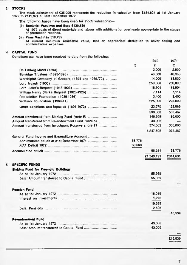
3. STOCKSThe stock adjustment of £35,000 represents the reduction in valuation from £184,824 at 1st January
1972 to £149,824 at 31st December 1972.The following bases have been used for stock valuations:—(i) Bacterial Vaccines and Sera £130,529
At 1972 costs of direct materials and labour with additions for overheads appropriate to the stages of production reached.
(ii) Virus Vaccines £19,295At current minimum realisable value, less an appropriate deduction to cover selling and administrative expenses.
4. CAPITAL FUNDDonations etc. have been received to date from the following:—
1972 1971£ £ £
Dr. Ludwig Mond (1893) ................................................................. 2,000 2,000Berridge Trustees (1893-1898) ..................................................... 46,380 46,380Worshipful Company of Grocers (1894 and 1969/72) ........... 14,000 13,000Lord Iveagh (1900) .......................................................................... 250,000 250,000Lord Lister’s Bequest (1913-1923) ................................................. 18,904 18,904William Henry Clarke Bequest (1923-1926) ................................ 7,114 7,114Rockefeller Foundation (1935-1936) 3,400 3,400Wolfson Foundation (1969-71) ..................................................... 225,000 225,000
Other donations and legacies (1891-1972) ................................ 23,270 22,669
590,068 588,467Amount transferred from Sinking Fund (note 5) ................................ 140,369 85,000Amount transferred from Re-endowment Fund (note 5) ................... 43,006 —Amount transferred from Investment Reserve (note 8) ................... 574,062 300,000
1,347,505 973,467
General Fund Income and Expenditure A ccoun t................................Accumulated deficit at 31st December 1971 ................................ 58,776Add: Deficit 1972 .............................................................................. 39,608
Accumulated d e fic it.................................................................................. 98,384 58,776£1,249,121 £914,691
5. SPECIFIC FUNDSSinking Fund for Freehold Buildings
As at 1st January 1972 ................................................................. 55,369Less: Amount transferred to Capital Fund.................................... 55,369
Pension FundAs at 1st January 1972 ................................................................. 18,089Interest on investments ................................................................. 1,276
19,365Less: Pensions .................................................................................. 2.826
16,539
Re-endowment FundAs at 1st January 1972 .................................................................. 43,006Less: Amount transferred to Capital Fund.................................... 43,006
£16,539
7

£ £ £Jenner Memorial Studentship Fund
6. BEQUEST FUNDS
As at 1st January 1972 ................................................................. 17,836Interest on investments ................................................................. 914
18,750
Morna Macleod Scholarship FundAs at 1st January 1972 ................................................................. 7,998Interest on investments ................................................................. 534
8,532
£27,282
7. SPECIFIC GRANTS AND LEGACIES£ £ £
Nuffield Foundation GrantsAs at 1st January 1972 ................................................................ 1,210Less: Laboratory expenses............................................................. 435
775
Guinness-Lister Research GrantAs at 1st January 1972 ................................................................ 269Amounts received.............................................................................. 11,250
11,519Less: Salaries and wages ............................................................. 9,089
Laboratory expenses............................................................. 2,430
11,519
£775
8. GENERAL AND SINKING FUNDS INVESTMENT RESERVE£ £ £
As at 1st January 1972 .................................................................General .............................................................................................. 263,852Sinking Fund...................................................................................... 10,210
274,062Less: Amount transferred to Capital Fund .................................... 274,062
9. TURNOVERTurnover has been arrived at after deducting commissions due to agents from the invoice value of sales of sera, vaccines and virus vaccines.
10. PROFITS ON SALES OF INVESTMENTSProfits on sales of investments have in previous years been credited to General and Sinking Fund Investment Reserve or to Re-endowment Fund as appropriate. The profit transferred to these accounts in 1971 was £127,093.
8

11. EMOLUMENTS OF MEMBERS OF THE GOVERNING BODY1972 1971
Emoluments in an executive capacity .......................................................................... £17,896 £16,814
Particulars of emoluments of the Governing Body in accordance with Section 6 of the Companies Act 1967
1972 1971
Emoluments of the Chairman of the Governing Body ..................................... Nil NilEmoluments of the highest paid member of the Governing Body ................ £8,750 £6,297
Numbers of members of the Governing Body whose emoluments were within the range
No emoluments ...................................................................................................... 7 7£1 — £2,500 1 —
£2,501 — £5,000 2 3£5,001— £7,500 1 1
12. CAPITAL EXPENDITURE SCHEMES1972 1971
The position at 31st December 1972 was as follows:—Commitments in respect of contracts ................................................................. 3,000 32,075Approved by the Governing Body in addition to commitments, for the new
laboratories at Elstree ...................................................................................... 42,500 —
£45,500 £32,075
13. CONTINGENT LIABILITIESAt 31st December 1972 there were contingent liabilities amounting to £10,188 in respect of indemnities issued to third parties.
14. BANK OVERDRAFTThe overdraft is secured by the Institute’s investments.
15. RESEARCH EXPENDITUREExpendiure on research is written off in the year in which it is incurred.
9


THE LISTER INSTITUTE OF PREVENTIVE MEDICINE
Reportof theGOVERNING BODY
1973
CHELSEA BRIDGE ROAD : LONDON : SW1W 8RH


The Governing Body
Professor A. NEUBERGER, cbe, md, frcp, frc path, frs, Chairman R. A. McNEILE, mbe, Hon. Treasurer Professor Sir DOUGLAS BLACK, m sc, md, frcp
C. E. GUINNESSProfessor HENRY HARRIS, ma, mb, d phil, frs
The Rt Hon the EARL OF IVEAGH Professor Sir EWART JONES, ma, ph d, d sc, frs
Professor W. T. J. MORGAN, cbe, ph d, d sc, frs
Professor WINIFRED WATKINS, ph d, d sc, frs
Clerk to the Governors S. A. WHITE, ACCA
3


The Council
A. LAWRENCE ABEL, MS, frcs R ep resen tin g th e B r it ish M e d ic a l A sso c ia tio n
Professor Sir DOUGLAS BLACK, M sc, md, frcp R ep resen tin g the M em b ers o f th e I n s titu te
The Rt. Hon. Lord BROCK, ms, frcs R ep resen tin g th e M em bers o f the In s titu te
Dame HARRIETTE CHICK, dbe, d sc R ep resen tin g th e M em b ers o f the I n s titu te
Professor P. J. COLLARD, md, mrcp R ep resen tin g th e U n iv e r s ity o f M a n ch es ter
M. L. CONALTY, md, mrc path, dph , MRIA R ep resen tin g the R o y a l Ir ish A c a d e m y
Major L. M. E. DENT, dso R ep resen tin g th e W o rsh ip fu l C o m p a n y o f G rocers
Sir ALAN N. DRURY, cbe, ma, md, frcp, frs R ep resen tin g th e M em b ers o f th e I n s titu te
Professor D. G. EVANS, cbe, d sc. frc path, frs R ep resen tin g th e R o y a l S o c ie ty
Professor R. I. N. GREAVES, ba, md, frcp R ep resen tin g the U n iv e r s ity o f C a m b rid g e
C. E. GUINNESS R ep resen tin g th e M em b ers o f th e I n s titu te
Professor HENRY HARRIS, mb, d ph il , frs R ep resen tin g the U n iv e r s ity o f O x fo r d
The Rt. Hon. the EARL OF IVEAGH R ep resen tin g th e M e m b ers o f th e I n s titu te
Professor Sir EWART JONES, ma, ph d, d sc, frs R ep resen tin g the M em b ers o f th e In s titu te
R. A. McNEILE, mbe R ep resen tin g the M e m b ers o f th e I n s titu te
Professor B. P. MARMION, md, d SC, frc path R ep resen tin g the U n iv e r s ity o f E din bu rgh
Professor Sir ASHLEY M ILES, cbe, md, frc path, frcp, frs R ep resen tin g th e M e m b ers o f th e I n s titu te
Professor J. S. M ITCH ELL, cbe, ma, md, frs R ep resen tin g th e M e m b ers o f th e I n s titu te
Professor W. T. J. MORGAN, cbe, ph d, d sc, frs R ep resen tin g th e M em b ers o f th e I n s titu te
Professor A. NEUBERGER, cbe, md, frcp, frc path, frs R ep resen tin g th e M e m b ers o f th e I n s titu te
The President of the ROYAL COLLEGE OF PHYSICIANS R ep resen tin g th e R o y a l C ollege o f P h y sic ia n s, L on don
The President of the ROYAL COLLEGE OF SURGEONS R epresen tin g th e R o y a l C ollege o f Surgeons o f E n g la n d
The President of the ROYAL COLLEGE OF VETERINARY SURGEONS R ep resen tin g th e R o y a l C ollege o f V e te r in a ry Su rgeon s
A. STEELE-BODGER, ma, b sc, mrcvs R ep resen tin g th e R o y a l A g r ic u ltu r a l S o c ie ty
Professor F. S. STEWART, md R ep resen tin g th e U n iv e r s ity o f D u b lin
W ILLIAM J. THOM PSON R ep resen tin g th e W o rsh ip fu l C o m p a n y o f G rocers
Sir GRAHAM W ILSON, MD, frcp R ep resen tin g th e U n iv e r s ity o f L on don
5
The Staff
D ire c to r :Professor W. T. J. Morgan, cbe, frs
D e p u ty D ire c to r :fProfessor L. H. Collier, md, d SC, mrcp
MICROBIOLOGYVirology
|L . H. Collier, md, d sc, mrcp (P rofessor o f V iro lo g y) G. S. Turner, B SC, PH DW. A. Blyth, bsc, p h d (M .R .C . T rach om a U n it) Lindsey M. Hutt, b sc
Bacteriology*Ruth M. Lemcke, b sc, ph d Valerie M. Harden, B scM. C. Goel, MV sc, PH d (C o m m o n w ea lth S ch o la r) {In d ia )Nawal M. Allam, bv sc {E g y p t)
Trypanosomiasis Research Group*D. G. Godfrey, obe, b sc, ph d { M .R .C . E x te r n a l S c ien tific S ta f f ) Angela E. R. Taylor, ph d, d SC Sheila M. Lanham, B sc Veronica Kilgour, b sc P. J. Toye, B sc { M .R .C . S tu d e n t)
Electron Microscopy*A. M. Lawn, b sc, ph d, mrcvs
BIOCHEMISTRYfWinifred M. Watkins, ph d, d sc, frs {P ro fe sso r o f B io ch em is try )
M. A. Chester, m sc, b tech, ph d { B e i t M e m o r ia l F e llo w )Hilary M. Simpson, b sc {R esearch S tu d e n t)A. S. R. Donald, b sc, ph d { M .R .C . G ra n tee )Caroline Race, b sc, ph d { M .R .C . G ra n tee )M. D. Topping, B sc { M .R .C . G ra n tee )Professor W. T. J. Morgan, cbe, ph d, d sc, md (he), d sc {h e), fric, frs
BIOPHYSICSf j . M. Creeth, B sc, PH d, fric {R ea d er in B io p h ysics)
K. R. Bhaskar, m SC, ph b { M .R .C . G ra n tee ) {In d ia )
6

VACCINES AND SERA LABORATORY (ELSTREE)*W. E. Parish, m a , p h d , b v s c , m r c v s , m r c p a t h
J. Rodican, b sc *H. G. S. Murray, MD
L. C. Robinson, b s c , p h d Jean M. Dolby, MA, PH DM. P. Banks, b sc A. P. Hunt, b scS. T. A. Gilligan, b sc Jennifer A. Mumford, b s c , p h d
BLOOD PRODUCTS (ELSTREE)*W. d’A. Maycock, c b e , m v o , m d , f r c p , f r c p a t h
L. Vallet, m a^Margaret E. Mackay, M sc, p h d (M .R .C . E x te rn a l S c ien tific S ta f f )D. Ellis, b s c , p h d Constance Shaw, m sc , d i p b a c t L. Singleton, b s c , p h d , f r icE. D. Wesley, b p h a r m
Plasma Fractionation Laboratory (at Oxford)Ethel Bidwell, b s c , p h d , f r ic R. Godfrey, M s c , PH DT. J. Snape, BA
m e d ic a l r e s e a r c h c o u n c il u n it s h o u s e d a t t h e in s t it u t eBlood Group Unit
§R. R. Race, c b e , m d (he), p h d , f r c p , f r c p a t h , frs Ruth Sanger, b s c , p h d , frs Patricia Tippett, B sc, p h d E. June Gavin, b sc Phyllis W. Teesdale, b sc
Blood Group Reference LaboratoryK . L. G. Goldsmith, p h d , m b , b s , m r c p , m r c p a t hToby T. B. Phillips, m b , c h bElizabeth W. I k in , b s c , p h dCarolyn M. Giles, b s c , p h dB. J. Dawes, b sc
ADMINISTRATIONS e c r e ta ry a n d A c co u n ta n t E lstree S e c r e ta ry a n d E s ta te M a n a g er A s s is ta n t S e c r e ta ry A d m im s ta t iv e A s s is ta n t A c co u n ta n t (E ls tree L a b o ra to rie s )A s s is ta n t A c co u n ta n t (E ls tree L a b o ra to rie s) A d m in is tr a tiv e A s s is ta n t
S. A. White, a cc a G. J. Roderick, B c o m Barbara A. PrideauxC. L. Beard P. J. O’Keefe Irene Bissett Beryl I. Coussens
Solicitors: Auditors:Field Fisher & Martineau, Coopers & Lybrand,296 High Holborn, W.C.l Abacus House, Gutter Lane, E.C.2 * *
tA p p o i n te d T e a c h e r o f th e U n iv e r s i t y o f L o n d o n §H o n o r a r y M e m b e r o f th e I n s t i t u t e S t a f f*R e c o g n is e d T e a c h e r o f th e U n iv e r s i t y o f L o n d o n
7

Annual General Meeting of the Lister Institute of
REPORT OF THE GOVERNING BODYGOVERNING BODYThe Governing Body takes much pleasure in recording that the honour of Knighthood was conferred upon Professor D. A. K. Black in the New Year Honours of 1973; and that Professor A. Neuberger has been awarded the Morris J. Kaplun Prize for Chemistry for 1 9 7 2 by the Hebrew University of Jerusalem.
At its last meeting the Council reappointed Professor A. Neuberger, Professor D. A. K. Black and Sir Ewart Jones as its representatives on the Governing Body until 31st December, 1973.
In June 1972 Professor Winifred Watkins succeeded Dr. A. F. B. Standfast as the Scientific Staff’s representative.
DIRECTORSHIP OF THE INSTITUTEWith the agreement of the Governing Body, the Medical Research Council invited Professor D. G. Evans to accept the directorship of a new institute comprising the Division of Biological Standards and Immunological Products Control. It was felt that this was a post for which Professor Evans was uniquely suited and he accordingly resigned the directorship of the Institute on 31st October, 1972. The Governors record their appreciation of his services to the Institute both as Director and as a member of the Governing Body since 1965.
The new Director is Professor W. T. J. Morgan, a scientist of great distinction who has, of course, spent most of his working life at the Institute. The Governors wish to place on record their great appreciation of his willingness to return from retirement, albeit an active one, and undertake this new and difficult task.
COUNCILAt last year’s Annual General Meeting Dame Harriette Chick and Sir Alan Drury were reappointed as representatives of the Members. The third retiring member of Council, Mr. H. P. G. Channon, did not offer himself for reappointment. Sir Ewart Jones was appointed to the Council as a representative
of the Members.The three members of the Council due to
retire this year in accordance with the Articles of Association, but who are eligible for reappointment are Mr. W. J. Thompson, a representative of the Grocers’ Company Professor F. S. Stewart, the representative of the University of Dublin, and the President of the Royal College of Physicians, the representative of the College.
MEMBERSMr. J. Rodican became a Member of the Institute during the year.
STAFF AND STUDENTSDr. A. F. B. Standfast retired on 30th September, 1972. The Governing Body wish him a long and happy retirement and place on record their appreciation of his services to the Institute both as a recent member of the Governing Body and as Head of the Bacterial Vaccines Department since his appointment in 1946.
Dr. W. d’A. Maycock, who for the past twenty-five years has been Elstree Superintendent, is now devoting his whole time to the work of the greatly enlarged Blood Products Laboratory. The Governing Body wishes to express its warm appreciation of Dr. Maycock’s services as Superintendent during this long period. In October 1972 Dr. W. E. Parish was appointed head of the reorganised Vaccines and Sera Laboratories.
As a regrettable consequence of the reorganisation at Elstree and of the closing of the Experimental Pathology Department at Chelsea in February 1973, the following members of the Scientific Staff were made redundant: Dr. D. E. Dolby and Mr. G. G. Beadle, Serum Department; Dr. J. P. Ackers, Bacterial Vaccines Department; Dr. F. R. Wells and Miss B. Mason, Experimental Pathology Department; and seven members of the laboratory and clerical staff.
Mr. K. G. Hardy of the Guinness-Lister Unit, Mr. R. A. Matthews of the Electron
8

Preventive Medicine 26 June 1973
Microscopy Unit and Mrs. S. D. Goodwin of the Biochemistry Department resigned during the year and Mr. L. V. Runkel of the Virus Vaccines Department, Mrs. C. J. Shanbury of the Bacterial Vaccines Department and Mrs. V. J. Stickley of the Blood Products Laboratory completed their respective terms of appointment, and Mr. J. R. Stealey completed his tenure of the Grocers’ Company Studentship.
It is with much regret that the Governing Body reports the closing of the Guinness- Lister Research Unit on 30th September 1972. The Unit began active work on 1st October, 1953, with Professor B. A. D. Stocker as its head. He was succeeded on 1st January, 1966, by Professor G. G. Meynell, whose impending resignation was announced in last year’s Report. The Unit has been supported to a very large extent by Arthur Guinness, Son & Co. Ltd. and it is gratifying that the generosity of the company has enabled the Unit to carry out work which has attracted considerable attention in the scientific world.
The Governors were very glad to learn that Dr. R. R. Race and Dr. Ruth Sanger were two of this year’s five recipients of the Gairdner Foundation (Toronto) awards “for their contributions to the knowledge of human blood groups and for their application of this knowledge to problems in the fields of immunology, genetics and clinical medicine” ; that Dr. Sanger was made an Honorary Member of the Deutsche Gesellschaft fur Bluttransfusion; and that Dr. Race was made an Honorary Fellow of the Royal College of Pathologists.
In August 1972 Professor W. T. J. Morgan and Professor Winifred Watkins attended the X lllth Congress of the International Society of Blood Transfusion in Washington, U. S. A., and in December 1972 they took part by invitation in a conference on “ Specific Sites on Biomembranes” organised by the Israel National Council for Research and Development.
Dr. W. d’A. Maycock attended, in February, May and November, 1972, meetings of the Group of Experts No. 15B of the European Pharmacopoeia Commission. He also attended, as the representative of the Department of Health and Social Security, a meeting of the Council of Europe Subcommittee of Experts on Blood Problems in Luxembourg in May 1972.
Dr. W. E. Parish took part by invitation in the International Congress of Dermatology in Venice in May 1972 and in the meeting of the European Society of Allergology and Clinical Immunology in Oslo in August 1972. In October 1972 he took part by invitation in the Conference on Asthma at Airlie House, near Washington and in the International Symposium on Reagin-mediated Hypersensitivity, at Montreal. With Dr.H. G. S. Murray he also visited the Rijks Instituut, Utrecht, in July 1972 to discuss procedures used in the preparation of viral vaccines.
In October 1972 Dr. Murray and Dr. L. C. Robinson took part by invitation in the Smallpox Vaccine Conference held at the Rijks Instituut, Utrecht.
Dr. G. S. Turner participated by invitation in the First International Colloquium on Rhabdovirus at Roscoff, France, in June 1972, and in the Second International Symposium on Rabies at Lyon, France, in December 1972.
During 1972 Dr. J. M. Creeth visited the Universities of Melbourne and Adelaide, and the Flinders and Australian National Universities for discussions on proteins and glycoproteins.
In April 1972 Dr. A. M. Lawn attended a symposium on bacterial sex pili at Duquesne University, Pittsburgh, and took part in seminars at Harvard Medical School, Boston, and at the Medical School, University of Washington, St. Louis.
For the academic year 1972-3 there are ten postgraduate research workers at the
9

Institute registered for higher degrees of the University. Three Ph.D. and one D.Sc. degrees were awarded during 1972.
DONATIONS AND GRANTSReference has been made earlier in the Report to the closing of the Guinness-Lister Research Unit and this is, therefore, an appropriate time for the Governing Body to acknowledge the Institute’s great indebtedness to Arthur Guinness, Son & Company Limited for the very generous financial support that the company has given to the Unit over the past twenty years. Although they will be on a lesser scale, the Governors were pleased to learn that the company will continue to make annual donations to the Institute.
The Governing Body records its appreciation of the many other bodies whose benefactions and grants support research work in the Institute. These include a grant from the Arthritis and Rheumatism Council to investigate the possible role of Mycoplasma fermentans antigens in the immune response of rheumatoid arthritis patients; from the Asthma Research Council for the investigation of antibodies in anaphylactic disorders ; from the British Heart Foundation to study endocarditis and vasculitis induced by bacterial toxins and by antigen-antibody complexes; from the Children’s Research Fund to examine the various antibodies mediating milk allergy; grants from the Medical Research Council for research on Bordetella pertussis in mice; for immunochemical investigations on human blood-group specific glycoproteins ; for biochemical investigations on the products of the blood group H, Lewis and Secretor genes; on the characterisation of the enzymic products of the A and B genes ; on the characterisation of the human blood group active Pi substance in hydatid cyst fluid; on the characterisation of blood-group specific glycoproteins by density-gradient methods; and on the genetics of drug resistance factors and other bacterial plasmids.
Grants were also received from the Overseas Development Administration of the Foreign and Commonwealth Office and from the Medical Research Council for studies on the biology of trypanososomes with special reference to" their surface proper
ties; and from the Science Research Council for studies in the replication of bacterial plasmids.
The Governing Body also gratefully acknowledges donations from the Prudential Assurance Company and the Royal London Mutual Insurance Society Limited.
PRODUCTION AT ELSTREEThe steady increase in production of vaccines and sera over the past few years was continued. Production of almost every product surpassed all previous records and totalled about 15 million doses of bacterial vaccines; over seven million doses of smallpox vaccine were issued, and although the increase in the number of horses has not resulted in an increased sale of sera, there was a proportionate increase in refined material which will be available for sale during 1973.
Arrangements for expansion of production throughout 1973 are progressing as planned.
VISITORSThe following visitors, in addition to those listed under Staff, worked for short periods in the Institute’s laboratories; Dr. L. C. Dantanarayana, Medical Research Institute, Colombo, Srilanka; Dr. R. Franco Granado, Instituto Español de Hematologia, Madrid, Spain; Dr. Malinee Jaijarat, National Blood Centre, Bangkok, Thailand; Dr. L. Kaptué, Blood Transfusion Service, Yaoundé, Cameroons; Mr. Maung Maung Mya, Burma Pharmaceutical Industry, Rangoon, Burma; Dr. D. C. Old, Bacteriology Department, The University, Dundee; Dr. B. Seydian, Instituí Pasteur de l’lran, Teheran Iran; Dr. E. Tahamtani, Chief of Laboratory of Biological Production Control, Ministry of Health, Teheran, Iran; Dr. Myint Pasi Tha, Burma Pharmaceutical Industry, Rangoon, Burma; Mrs. Ashara Vichitanand, Government Pharmaceutical Organisation, Bangkok, Thailand.
10

In the last Annual Report I drew attention to the serious financial position of the Institute and indicated that this problem was occupying the continuous attention of the Governing Body. Steps have been taken to reduce our expenditure and in some cases this has involved painful decisions that have led to redundancy for some of our colleagues who have been with us for some years. We have also been able to increase our income, although this has been due to special circumstances and our financial position is far from being satisfactory. There are, however, grounds for hope, and our belief that the Institute will survive this present crisis is not based on unreasonable optimism.
It has also become increasingly clear that the Institute will have to find a new policy which will give it an important additional function within an overall national scheme of biomedical research. This will lead to a shift of emphasis in our general scientific policy. Apart from the financial problems, this scientific reorientation has also occupied the attention of the Governing Body. It is hoped that during the present year there will be progress towards the resolution of both the financial and scientific problems and the Institute may expand again in new directions.
This has been a difficult year for all members of staff and the Governing Body wishes to record its thanks most warmly to all members of the staff, scientific, administrative and technical, for their understanding, loyalty and co-operation.
A. NEUBERGER, Chairman.
11

REVIEW ARTICLE
LIFE AT THE BORDERA review of the work of the MRC Trachoma Unit
L. H. Collier
Department o f Virology and
Medical Research Council Trachoma Unit
The activities of the Medical Research Council’s Trachoma Unit, which has been based at the Institute since 1957, are soon to end. The invitation to write the review article for the 1973 Annual Report is therefore both welcome and appropriate, since it provides an excellent and possibly unique opportunity of summarizing the discoveries and achievements, the disappointments and excitements attending a series of researches spanning a decade and a half. The paths we travelled led to an intriguing borderland between bacteria and viruses, in which the frontier has but recently been defined with any precision. The micro-organisms causing trachoma and related infections stand near this demarcation line; and their somewhat unusual characteristics determine many features of pathogenesis and immunity.
Although the various topics explored by the Unit are interrelated in a rather complex manner, it will perhaps help the reader if I try as far as is practicable to trace the development of each main study individually.
TRACHOMA AND INCLUSION CONJUNCTIVITISThe ravages of trachoma have been known for thousands of years, and are described in the earliest medical writings. It is a chronic inflammatory disease of the eye, affecting the conjunctival membrane and almost always the cornea. In the conjunctiva, characteristic “sago-grain” follicles appear, which give the disease its name (Gr. rpa^copa—
“roughness”). Inflammatory changes in the cornea and invasion by new blood vessels (pannus) may lead to loss of transparency and impaired vision. Untreated, the disease may persist for years with eventual scarring of the eyelids; the consequent deformity causes the lids to turn inwards (entropion) so that the lashes rub against the eyeball (trichiasis) causing yet more damage to the cornea. Trachoma is enormously widespread in the developing countries, particularly in the Far East, the Indian subcontinent, Africa and South America; and is the greatest single cause of blindness and impaired vision. In 1907, Halberstaedter and von Prowazek for the first time observed in conjunctival cells the “inclusion bodies” that we now know to be micro-colonies of the causal agent.
Around the turn of the century, a form of follicular conjunctivitis rather like trachoma but without corneal lesions and scarring was recognized in European countries. This disease, inclusion conjunctivitis (inclusion blennorrhoea), was most often seen in newborn infants. Epithelial cells from their eyes, and from the genital tracts of their parents, contained cytoplasmic inclusions indistinguishable from those of trachoma, and it became apparent that the two syndromes were closely related.
During the next half century, many unsuccessful attempts were made to isolate the causal agent of trachoma in the laboratory. In the 1930s microbes resembling each other
12

in morphology and staining reactions and possessing a common antigenic component were shown to cause psittacosis in birds and a sexually transmitted disease of man, lymphogranuloma venereum (LGV). In 1934 Thygeson, on the basis of similarities in their inclusions, suggested that the trachoma and inclusion conjunctivitis agents might be related to the organism causing psittacosis. These micro-organisms are now grouped together in the genus Chlamydia.
The active stages of trachoma and inclusion conjunctivitis are usually treated by a tetracycline drug. Oral therapy is best, but is expensive and impracticable for large- scale use. In mass campaigns, such as those sponsored by WHO, the drug is applied as ointment to the affected lids, but this treatment must be maintained for long periods. When medical aid is lacking, patients with entropion/trichiasis often seek relief by plucking the inturned eyelashes with primitive forceps, an operation that was well- known to the Ancient Egyptians. The Ebers Papyrus (ca 1500 B.c.) advocates “bat’s blood, rim-of-a-new-hennu-vessel and honey, to be powdered and placed where the hair has been pulled out” (Bryan, 1930). Unfortunately, removal of lashes often results in bristly stumps that do even more damage. The only remedy for entropion is a plastic operation to evert the lids, and even this is not always successful. Corneal grafts are not of much use in dealing with the opacities caused by trachoma.
FORMATION OF THE MRC TRACHOMA UNITIn 1956 the Medical Research Council formed a research group under the honorary directorship of Sir Stewart Duke- Elder; the scientific staff consisted of an ophthalmologist (Mr. M. Gilkes), a virologist (Dr. C. H. Smith) and a bacteriologist (Mr. J. Sowa). This team started to investigate the clinical aspects of trachoma in Jordan; and in collaboration with the new Department of Virology at the Lister Institute, to attempt isolation of the trachoma agent. Before long, however, the Suez crisis dictated the return of the team to England. Following; a favourable report by Smith on its suitability for trachoma studies,
the Unit was transferred to The Gambia, West Africa, where the MRC maintains a large research establishment at Fajara. Soon afterwards, however, Smith and Gilkes left, and in September 1957 I took over the honorary directorship from Sir Stewart Duke-Elder.
ISOLATION AND IDENTIFICATION OF THE TRACHOMA AND INCLUSION CONJUNCTIVITIS AGENTSTrachomaIsolations in China. At about this time, T ’ang and coworkers (1957) reported from Peking the isolation of infective agents from trachoma patients by inoculating conjunctival material into the yolk sacs of 7-day old chick embryos; they described the morphology of these micro-organisms, their pathogenicity for various hosts, and the influence of various physical and chemical agents upon their viability. I had previously read a number of papers on purported isolations of trachoma agent, but this was the first that carried conviction. I immediately wrote to T ’ang for samples, which in due course were brought to London by Dr.E. T. C. Spooner of the London School of Hygiene, who was visiting Peking when my letter arrived. Two strains inoculated into chick embryos at the Institute grew without difficulty; I was able to confirm T ’ang’s description of their properties, and also to demonstrate the presence of the Chlamydia group complement-fixing antigen. Electron micrography by the late Dr. Robin Valentine confirmed the morphological similarity of the T ’ang agent to other members of the genus (Collier & Sowa, 1958).Isolations in West Africa. The next task was to determine whether similar agents could be isolated at our newly-established Gambian laboratory. I went there in November 1957 to find Josef Sowa ensconced in a small laboratory, as yet without screens and therefore subject to continual invasion by mice, frogs and a formidable variety of insects. Two somewhat decrepit farmhouse egg incubators were located and put into
13

service, and chick embryos were inoculated with material from Gambian children with trachoma. Although the Chinese technique was followed as carefully as possible, I was not optimistic about the chances of success under these rather difficult conditions; but in the event an agent (“G l”) apparently indistinguishable from the Chinese strains was isolated from the first series of inoculations and sent to the Institute.
Proof of aetiological role. The finding of similar agents in people with trachoma in widely separated countries was strong presumptive evidence that the causal microbe had at last been isolated, but this supposition had still to be proved. Because of previous unsubstantiated reports, the WHO Expert Committee on Trachoma (1956) stipulated that no such claim could be accepted unless the organism were shown, inter alia, to induce all the cardinal signs of trachoma in human volunteers, and the characteristic inclusion bodies in the conjunctiva. Accordingly, with the clinical collaboration of Sir Stewart Duke-Elder and Mr. (now Professor) Barrie Jones, a blind human volunteer was inoculated with the Gl strain. Both he and a second volunteer who was later inoculated with another Gambian strain (“G17”) developed trachoma; typical inclusions were demonstrated in their conjunctival cells, and the infective agents were again isolated from their eyes by inoculating conjunctival scrapings into chick embryos (Collier & Sowa, 1958; Collier, Duke-Elder & Jones, 1958; Collier, Duke-Elder & Jones, 1960). These experiments went most of the way toward satisfying Koch’s criteria for establishing the aetiological relationship of a micro-organism with a given disease. We had only to confirm that the agent in question could be demonstrated in a satisfactorily high proportion of patients with active trachoma. This was soon done; by April 1958 Sowa had—by using conjunctival scrapings instead of swabs, and by treating them with a much increased concentration of streptomycin— improved the isolation technique to a point at which he was able to isolate Chlamydia from 24 of 30 trachoma patients, of whom only 18 were inclusion-positive (Sowa & Collier, 1960).
Inclusion ConjunctivitisIn the summer of 1958, micro-organisms seemingly identical with the trachoma agent were isolated from the eyes of a newborn English baby with inclusion conjunctivitis, and from the genital tract of the mother of another baby with this syndrome (Jones, Collier & Smith, 1959). Their aetiological relationship to inclusion conjunctivitis was soon proved by laboratory tests, inoculation of baboons (Collier, 1959; 1962) and—again in collaboration with Barrie Jones—induction of the disease in volunteer subjects (Jones & Collier, 1962). The close similarity between the trachoma and inclusion conjunctivitis micro-organisms and the overlap in their respective clinical syndromes later led to adoption of the acronymous term “TRIC agents”* for referring to them jointly.
Some early questionsThe isolation and identification of the elusive TRIC agents made possible two major lines of investigation into the control of trachoma: a search for drugs more effective than the tetracyclines commonly used for mass treatment compaigns; and the devising of a vaccine. Our Unit was not suited to an adequate investigation of new antibiotics or chemotherapeutic substances; and, in any event, I felt that the major impediment to successful mass therapy lay not so much in the lack of effective drugs, but in the logistic and financial difficulties of applying them for long periods to large and often scattered populations in primitive areas. We therefore decided, as a main objective, to attempt preparation of a vaccine that by virtue of conferring reasonably prolonged immunity might be easier to administer than continual drug treatment.
Another topic now open to investigation concerned the much disputed relationship between trachoma and inclusion conjunctivitis; as we shall see, the Unit made some contribution to the study of this subject.
The decision to attempt production of a trachoma vaccine immediately posed a series
* Henceforth strains will be designated by the “Montreal system” (Gear e t a l . , 1963); the abbreviated form (Collier, 1963) is used, followed at first mention, when appropriate, by the original designation in parentheses.
14

of questions whose solution often seemed to depend on the answers to yet other unsolved problems; in other words, we were in for a long period of pulling ourselves up by our own boot-straps. In an early review of some of these questions (Collier, 1961a) I referred to the necessity of choosing the right sort of host cell for vaccine production, the respective merits of live and inactivated vaccines, the problems of serotype and antigenicity, of purification, preservation and potency testing; and discussed what information would be needed about the epidemiology of trachoma in a vaccine trial area. I shall now describe how these and other problems were tackled.
RESEARCHES IN THE GAMBIA, 1958-1962
It was apparent at the very outset that the newfound ability to isolate trachoma agents, if verified, would be a powerful tool for studying the natural history and epidemiology of trachoma; and that information of this sort would be essential to the rational planning of any vaccine trials. During my first visit to Fajara, and even before the first isolations there, an outline programme was drawn up for the consideration of the MRC. It provided for an intensive study of trachoma in one or two carefully selected villages, to include periodic clinical and microbiological examination of the eyes of the entire population; the average age of onset of trachoma and other eye infections would be determined and babies would be examined monthly from birth in order to observe trachoma in its earliest stages. Two important steps in implementing this programme were the recruitment in 1958 of an ophthalmologist, Miss Shiona Melville, later to become Mrs. Sowa; and the building at Fajara of a new trachoma laboratory with funds generously provided by the Wellcome Foundation.It The Gambia is situated at the tip of the ‘bulge” of West Africa; it extends for about
250 miles along both banks of the river that gives it its name, and for much of its length is only 15-20 miles wide. In 1964 the population was about 315,000, mostly distributed in scattered communities of 10 0-1,0 0 0 people; living standards are low in
these villages and disease, especially malaria, is widespread. Although communications are now improving, they were rudimentary 15 years ago; for villages situated inland, the only practicable access was by Land-Rover down bush tracks that were often quite impassable in the rainy season.
These topographical considerations to some extent determined our plan of investigation. An epidemiological study can be extensive, covering a large population, or intensive, involving more careful observation of a comparatively small group. We decided upon the second alternative, and chose Marakissa, a village of some 400 inhabitants 15 miles south of Fajara. The investigations there lasted more than two years; the findings were embodied in an MRC Special Report (Sowa, Sowa, Collier & Blyth, 1965) and represent perhaps the most detailed field study of trachoma and allied infections ever undertaken. Here, it is possible to provide only the briefest summary.
In The Gambia, as in other highly endemic areas, trachoma was found to have an early onset; about a quarter of the babies aged less than one year were affected, and the incidence rose to 91 % in the 5-9 year age group. Fresh infections were rare after this age, a finding that might be explained in terms of immunity engendered by past infection, or perhaps by the observation that physical contacts made by playing and rolling about together in the streets were less frequent in children approaching adolescence. Unlike what is seen in, for example, North Africa, there was nothing to suggest the implication of flies in the spread of eye diseases. Trachoma is probably transmitted by direct physical contact, or through recently infected fomites such as clothing or bedding; TRIC agent was found to survive long enough on cloth to make this possible.
The first stage of trachoma, i.e. before follicles are obvious, was hitherto believed to last only a few weeks, but in fact sometimes persisted for 1-2 years. The disease ran a comparatively benign course, and caused severe visual defects in only 2-5% of the population, perhaps because of the relative freedom from concurrent bacterial conjunctivitis which is such a problem in some other countries. But although overt con
15
junctivitis was rare, only 3% of the population had bacteriologically sterile conj unctivae; and since in some countries haemophilus bacilli are often associated with trachoma and may exacerbate its course, a special investigation of these organisms—which were present in the eyes of one-third of patients with trachoma—was made at Elstree by Miss Georgina Sampson, Mrs. Jean Dolby and Dr. A. F. B. Standfast (1965). A study of 502 strains isolated in Marakissa suggested that indole production distinguished H. aegytius from H. influenzae better than possession of haemagglutinin. It also led to an improved method of serotyping strains by gel- precipitin tests that might be useful in epidemiological studies; but in the absence of haemophilus conjunctivitis in epidemic form, there was no opportunity of testing this method in the field.
The most important aspect of the microbiological work was detection of TRIC agent in the conjunctiva. Isolation in chick embryos proved more reliable than the finding of inclusions; in one survey, for example, TRIC agent was isolated from 69% of patients with active trachoma, whereas inclusions were found in only 47%. In subsequent studies on special groups, the isolation rate was nearly 100%, a figure that has not to my knowledge been approached elsewhere. Success rates of this order were attained only through meticulous precautions against bacterial contamination, both by improving the technique of collecting conjunctival scrapings and by attention to the choice and concentrations of bacteriostatic antibiotics (Sowa et al., 1965; Sowa & Race, 1971).
While these epidemiological studies were proceeding in West Africa, a wide range of investigations was being pursued at Chelsea in kitchens, bedrooms and bathrooms that formerly belonged to the directorial apartments, but were now converted to laboratories. A brief digression into taxonomy may help the general reader to follow these researches more easily.
THE NATURE OF THE C H L A M Y D I AThe Chlamydia, including TRIC agents, are not viruses, although they used to be referred to as such. Like viruses, they multiply only
within living cells within which they undergo a non-infective period before the new generation of infectious particles is produced. But these resemblances are superficial: the Chlamydia differ fundamentally from viruses in retaining their physical identity throughout the growth cycle; in possessing both DNA and RNA; in multiplying by fission; in possessing a cell wall and ribosomes; and in their susceptibilities to various antimetabolites. They resemble the Rickettsiales in some of these attributes; but they do not have arthropod hosts, and differ so much in other respects that it is obviously incorrect to include them in this taxonomic Order. There now seems no doubt that, despite their obligate intracellular existence, these micro-organisms stand clearly on the bacterial side of the border.
The genus Chlamydia is divided into two groups, known colloquially as A and B. Group A (type species: C. trachomatis) includes the agents responsible for trachoma, inclusion conjunctivitis and lymphogranuloma venereum in man; and others causing pneumonitis in mice and hamsters. These agents form compact inclusions containing elementary bodies in a carbohydrate matrix and are susceptible to sulphonamides and D-cycloserine. Group B (type species: C. psittaci) is much larger and contains the aetiological agents of a wide variety of syndromes in many species of birds and mammals. The best known is psittacosis, a generalised infection of birds which is sometimes transmitted to man. Other members of the group cause enteritis, pneumonitis, polyarthritis, encephalomyelitis or enzootic abortion both in wild and domestic animals. They form diffuse inclusions that do not contain a carbohydrate matrix and are insusceptible to sulphonamides and D-cyclo- serine. Groups A and B closely resemble each other morphologically and in possessing similar complement-fixing antigens; but they differ considerably in terms of other serological tests and of DNA homology. It is not unlikely that their origins were diverse, and that their similarities reflect only adaptation to the same sort of intracellular environment.
STUDIES OF REPLICATIONInjectivity. By 1959 many strains of TRIC
16

agent had been isolated in several countries and the technique of propagating them in the chick embryo yolk sac presented little difficulty. This method however has disadvantages. Infectivity can be titrated only by the comparatively imprecise methods of determining the highest dilutions that infect or kill a stated proportion—usually 50%— of embryos. Furthermore, suspensions of TRIC agent grown in yolk sac are difficult to purify; and representing as they do the yield from many cycles of multiplication they can but provide a mixed bag of all the developmental forms. It seemed that propagation of TRIC agents in cell cultures might overcome these disadvantages, and we made many attempts to grow two Gambian strains in this way. In the United States, Gordon and coworkers (1960) induced inclusions in explants of chick embryo yolk sac but did not apparently make serial passages. At about the same time, Dr. Weston Hurst (ICI Laboratories) and Dr. P. Reeve of the Trachoma Unit adapted one of T ’ang’s original trachoma strains to grow in mouse brain, with rigorous precautions to exclude other infective agents (Hurst & Reeve, 1960). Soon afterwards Mrs. Jane Stocker induced inclusions in HeLa and other cell lines, in the first instance with mouse brain infected with the T ’ang strain and then with this and our own MRC-l/G (LB1) strain of inclusion conjunctivitis grown in chick embryos; these infections could be maintained indefinitely in cell culture by serial passage (Furness, et al., 1960). In the same year, Bernkopf, Mashiah and Maythar (1960) reported similar findings in Israel. This advance was rapidly exploited: in our laboratory, a method for titrating infectivity by counting inclusions in monolayers of cells was devised; it had a standard error of only ± 1 0 % and was thus a great improvement on the chick embryo method (Furness, Graham & Reeve, 1960). Since the development of the inclusions could be watched directly under the microscope, it was possible to make observations at defined points of the growth cycle. After entry into the cell, the infectious particle or “elementary body” became transformed into a larger basophilic “initial body” containing much ribonucleic acid (RNA) and at the
same time lost its infectivity. The initial body—now usually termed the reticulate body—underwent a series of divisions resulting in the production of a new generation of elementary bodies in which the predominant nucleic acid was DNA. Infectivity reappeared 2 2 hr after infection and attained its maximum at about 38 hr, by which time the inclusions were packed with new elementary bodies capable of initiating growth in other susceptible cells (Furness & Fraser, 1962; Furness, Henderson,Csonka and Fraser, 1962).Particle counts. For quantitative studies of replication, it was important to be able not only to assay infectivity accurately, but also the number of elementary bodies in a given suspension. Because of their small size, this had to be done very laboriously by electron microscopy. However, Dr. Reeve and Dr. Janice Taverne (1962a) noticed that elementary bodies stained with Giemsa appeared as brilliant yellow particles on a black background when viewed with an ordinary microscope by dark field illumination. Their appearance was so distinctive that suspensions containing numbers of particles of the order of 1 0 1(,/ml could be rapidly counted with an error of only ± 2 0 ° 0. The dark field method also facilitated the counting of inclusions in infectivity titrations.Fast-killing mutants. Not all TRIC strains behaved similarly in cell cultures: some formed numerous inclusions and could be readily passaged in cells, whereas others formed few or no inclusions and could not be serially propagated in this way. Reeve and Taverne (1963) then noticed another difference : at equivalent doses some strains appeared to kill chick embryos more rapidly than others. Fortunately, from the very earliest days, individual records had been kept of the many thousands of eggs inoculated; a retrospective study showed that the strains killing chick embyros quickly had acquired this ability quite suddenly at different times during the course of routine passages in eggs. This property was related to that of infecting cell cultures, and in some instances at least, with loss or diminution of pathogenicity for the primate conjunctiva (Collier, 1961 b, 1962; Jones & Collier, 1962). Furthermore, these fast
17

killing (/) strains had much lower particle/ infectivity ratios than either their slow- killing (s) parent strains or other s strains that had remained unaltered since they were isolated (Taverne, Blyth & Reeve, 1964a). The most likely explanation of these findings is the emergence of mutants whose increased virulence for chick embryos is associated with acquisition of pathogenicity for mouse brain and perhaps loss of pathogenicity for the primate conjunctiva. The ability to kill chick embryos quickly depended not on an increased content of the toxin associated with the elementary bodies, (Reeve, 1964; Taverne, Blyth & Reeve, 19646) but on a decrease in the lag phase preceding replication and on an enhanced multiplication rate (Reeve & Taverne, 19676).
So far the cell culture work had all been done with mutant and hence atypical strains, and the inability to grow the “wild-type” 5 strains was frustrating. When Gordon and his colleagues infected chick endoderm explants, they did so by centrifuging the inoculum onto the cells. Reeve and Taverne (1967c) found that several cell lines could be infected with s strains by a similar technique and established the optimum conditions for doing so. Centrifugation also increased the infectivity of / strains for cell cultures but to a lesser degree. Morphologically, the growth cycle of 5 strains was broadly similar to that of / strains. Their failure to infect cells without the help in adsorption afforded by centrifugation is probably due to the combination of a low proportion of infectious particles and heat inactivation.Isolation methods. The centrifugation method found practical use in the isolation of TRIC agents from the eye and genital tract. Gordon and coworkers (1969) reported that centifugation of epithelial scrapings onto monolayers of irradiated McCoy cells was more efficient and gave much more rapid results than inoculation of chick embryos. This technique proved most valuable in a number of diagnostic laboratories, but carried the disadvantage that the McCoy cells had to be irradiated with cobalt-60 before inoculation. In experiments with laboratory strains and with conjunctival scrapings from T h e ' Gambia and genital
tract material from patients in London, Blyth and Taverne recently showed that irradiated McCoy cells held no advantage over unirradiated baby hamster kidney (BHK-21) cells (Report 1973); this finding should be of considerable practical use to diagnostic laboratories.Folic acid synthesis. The type B chlamydiae or psittacosis-like organisms are resistant to sulphonamide because they can use exogenous sources of folic acid; but like the sulphonamide-sensitive bacteria, the Type A organisms, including TRIC agents, synthesize their own folic acid, a compound that possibly acts as a co-enzyme for the synthesis of such essential metabolites as purines, pyrimidines, and amino acids (Moulder, 1964). To gain further information about this metabolic pathway, which is of course important in relation to chemotherapy, Reeve, Taverne and Bushby (1968) studied the influence on replication of drugs that compete with folic acid by inhibiting dihydrofolate reductase. Trimethoprim, a derivative of 2,4-diaminopyrimidine, inhibited the growth of several TRIC agents in chick embryos and cell cultures. The reversal of this action by calcium leucovorin confirmed that endogenous rather than exogenous folic acid synthesis was implicated, and strongly suggested that TRIC agents contain a dihydrofolic acid reductase. The action of trimethoprim was slightly potentiated by sulphafurazole. It is interesting that a related pyrimidine derivative, the antimalarial drug pyrimethamine, also inhibited a trachoma agent; this factor would have to be considered in field studies on trachoma undertaken in areas where antimalarial drugs are widely used.
SEROLOGICAL STUDIESComplement-fixing antigen. I mentioned that one of the first tests on the newly isolated TRIC agents was to verify that they contained the heat-stable antigen common to the Chlamydia, originally detected in the psittacosis agent by Bedson in 1936. Investigations by others on lymphogranuloma venereum and feline pneumonitis indicated that the group antigen is a lipoprotein-polysaccharide complex. Reeve and Taverne (19626) found that this was also
18

true of the TRIC antigen, which in infected cell cultures was present both in soluble form, and in a sedimentable form presumably associated with the elementary bodies. As we have seen, TRIC agents, unlike viruses, retain their physical identity throughout their growth cycle. By concentration methods, Reeve and Taverne were able for the first time to detect Chlamydia group antigen in HeLa cells throughout the latent period following adsorption; they also showed that the pattern of the subsequent increase in sedimentable antigen closely resembled the one-step infectivity growth curve determined by Furness and Fraser (1962).Neutralizing antibody. As far as a vaccine is concerned, antibodies that neutralize infectivity are of particular interest. Bernkopf (1959) described a test in which sera from immunized rabbits, when mixed with trachoma agents, neutralized their capacity to induce pulmonary lesions in mice. There was however, no comparable in vitro test until Reeve and Graham (1962) reported that neutralizing antibodies to TRIC agent could be accurately and reproducibly titrated in terms of their ability to reduce the numbers of inclusions in cell cultures. By this method they demonstrated a serological relationship between a Chinese strain of trachoma and two inclusion conjunctivitis strains isolated in London. However, the antibody titres were low; various methods of preparing antiserum in rabbits were tried (Blyth, Reeve, Graham & Taverne, 1962), but even the best of them resulted in a titre of only 1/24, whereas the corresponding titres of complement-fixing antibody sometimes exceeded 1/20,000. Recently (Report, 1973) Blyth and Taverne again studied the problem in the light of a claim that titres of antibody that neutralize psittacosis agent could be enhanced by adding antiserum directed against the IgG of the host species in which the psittacosis antiserum was prepared. With antisera prepared against TRIC agents in rabbits, mice, baboons and cockerels there was enhancement in the presence of the appropriate anti-IgG; but control tests with normal serum suggested that this enhancement was at least in part caused by non-specific blocking of infectivity.
TRIC serotypes. Chlamydiae contain a toxin that kills mice when injected intravenously. Bell, Snyder and Murray (1959) found that a sublethal dose of TRIC agent protected against a subsequent larger dose of the same strain; and by cross-tests 3 main serotypes ( 1 , lb and 2) were elicited. Alexander and coworkers (1967) found that most strains isolated from ophthalmic trachoma belonged to these types, whereas those from inclusion conjunctivitis and genital infections fell into additional serotypes (D, E and F). The discovery of these antigenic differences was important for epidemiological studies, but the uppermost question when they were first reported was their implication for the vaccination of man, which by then interested workers in several laboratories. A correlation between protection against toxicity and resistance to infection would mean that a trachoma vaccine must contain the serotypes prevalent in the country of use. Although some investigators maintain that such a relationship exists, it may not be absolute; Dr. Doris Graham (1965; 1967) devised methods for comparing the two sorts of immunity in mice and found that they did not necessarily correspond.
The mouse test fulfilled an important function, but the labile nature of the toxin caused considerable technical problems; it has now been superseded by indirect immunofluorescence methods (Bell and McComb, 1967). The introduction of a micro-method (Wang, 1971) has greatly facilitated exploration of the antigenic relationships between various chlamydiae. Use of serotyping for field studies. Immunofluorescence typing has also been exploited for epidemiological purposes, notably by the Harvard/ARAMCO team in Saudi Arabia. In that country, Nichols and coworkers (1971) found that types 1, lb and 2 were all present; but in the two neighbouring villages yielding most of the strains tested, their prevalences were quite different. Given families tended to be infected by the same serotype, and changes of serotype in individual patients were infrequent, implying that in this highly endemic area the trachoma syndrome is the result of a chronic continuous infection rather than of repeated reinfections, as some workers have suggested.
19

The predominance of a single serotype in a given community was also observed in The Gambia by Mr. and Mrs. Sowa, where almost all the strains isolated from two widely separated villages were respectively types 1 and 2 ; type lb was never isolated in that country (Report, 1971). There was also evidence that one or the other strain was dominant in a given dwelling-compound.
A curious observation made by Josef Sowa during the investigation has yet to be explained. Of the first strains isolated in The Gambia two differed from the others in forming compact aggregates of particles in the yolk sac, as well as the usual free elementary bodies. These aggregates consisted of elementary bodies embedded in a matrix surrounding one or more vacuoles; the vacuoles contained a carbohydrate that stained like glycogen (Sowa & Collier, 1960). Sowa recently observed that the presence of aggregates is closely related to serotype : 19 of 21 Type 1 strains produced them, but only 2 of 35 Type 2 strains (Report, 1973). The type-specific antigens almost certainly reside in the Chlamydia cell wall, and this observation could throw light on differences between the cell surface characteristics of Types 1 and 2.The use of immunofluorescence for diagnosis. When Halberstaedter and von Prowazek first observed trachoma inclusions in 1907, they used Giemsa, and this stain remained a favourite for many years. It has the disadvantage that conjunctival scrapings must be scanned under the high-power objective —a tedious proceeding. Rice (1936) discovered that trachoma inclusions possess a matrix containing a carbohydrate which stains a deep coppery-brown with iodine. This method was not greatly exploited for diagnostic purposes until in 1958 Gilkes, Smith and Sowa modified and improved it. Sowa later introduced the use of ammoniated iodine and this technique was used for much of our work in The Gambia. It has the advantage that the deeply stained inclusions are easily visible with a low power objective, and in our hands gave results that compared favourably with those of other methods. Nichols and McComb (1962) later devised a method of staining inclusions with fluorescent antibody: the main advantages are its
ability to detect early inclusions that have not yet formed a polysaccharide matrix and hence do not stain with iodine; and its use in examining epithelial cells from the genital tract, which themselves contain much glycogen and stain intensely with iodine. In a comparison of the two methods in The Gambia, the FA method detected inclusions in a somewhat higher proportion of conjunctival scrapings, although the degree of advantage was not statistically significant; but the numbers of inclusions found in positive scrapings stained by FA were significantly higher than in duplicates stained with iodine. We concluded that the FA method is preferable where good laboratory facilities and well-trained observers are available; but that the iodine method, which is rapid and requires only the simplest reagents and equipment, holds advantages for field use (Sowa, Collier & Sowa, 1971).
Complement-fixation with group antigen is useful in the serological diagnosis of LGV; but its results in trachoma are irregular, and various alternatives have been examined. Bernkopf, Orfila & Maythar (1966) detected antibodies in the conjunctival secretions and sera of trachoma patients by immunofluorescence. We used this method to determine the prevalence of antibodies in Gambian children and the classes of immunoglobulin involved. IgG antibodies to TRIC agent were found in a substantial proportion of those with active trachoma. IgG and, less frequently, IgA antibodies were present in the conjunctival secretions of a proportion of children with serum antibody, but not in sero-negative children. Serum antibody titres of 1 / 1 0 or more were always associated with a clinical diagnosis of trachoma; and were more reliable as a diagnostic aid than the finding of inclusions (Collier, Sowa & Sowa, 1972).
For this investigation we used as slide antigens infected monolayers of BHK-21 cells in preference to yolk sac suspensions. They were prepared at the Institute; standardization of fluorescence measurements in the London and West African laboratories was helped by using sets of density filters made in duplicate by a photographic method (Collier, 1968).
2 0
RELATION BETWEEN TRACHOMA AND INCLUSION CONJUNCTIVITISAs I have mentioned, trachoma is closely related to the inclusion conjunctivitis (IC) syndrome, which also affects the eye, and, as inclusion urethritis or cervicitis, the genital tract. It was maintained, notably by Thygeson (1962) that trachoma is an ophthalmic infection found in the less- developed countries, and can be sharply distinguished from IC, a sporadic infection of more advanced countries that does not occur in trachoma-endemic areas: trachoma causes corneal lesions and scarring, whereas IC does not; trachoma spreads only from eye to eye, but IC is primarily a genital tract infection sometimes transmitted to the infant eye at birth, or to the adult eye through media such as infected swimming pools. Other workers took a different view. For example, Bietti (1959) described people with trachoma in Japan, Taiwan, the Phillipines and Italy in whom corneal lesions were minimal or absent; and experimental inoculations of the eye and genital tract with infected epithelial scrapings led Mitsui (1949) to the extreme opinion that there is no basic difference between the two syndromes: trachoma can alternate between the eye and genital tract, and IC is merely the infantile form of trachoma.
Investigations in London. Observations on English patients suggested to Barrie Jones (1961) the idea of a spectrum of clinical pictures, with classical trachoma and inclusion conjunctivitis at the extremities, and a number of intermediate syndromes that do not fall clearly into either category. The isolation of TRIC agents at once provided the means of obtaining better experimental evidence concerning the relationship between these syndromes, and Barrie Jones and I proceeded to inoculate a blind volunteer with two strains isolated in London. The first was MRC-l/G, from the female genital tract; it had been passaged 23 times in chick embryos. The inoculation induced only a mild transient conjunctivitis. The significance of this finding became apparent later, when, as we have seen, it was realised that some strains, including this one, had changed their characteristics during repeated yolk-sac passage. The results of the second experi
ment, a year later, were much more interesting. Strain MRC-4 (LB4) was inoculated into the other eye of the same man. This strain also generated a “fast-killing” mutant later on, but the early passage material used as inoculum was still in the s phase. It induced a very severe inclusion-positive conjunctivitis with keratitis and pannus. These corneal lesions are supposed to be characteristic of trachoma, yet were induced by a TRIC agent that was isolated from the eye of an English baby with neonatal inclusion conjunctivitis, and hence originated from a parental genital tract infection (Jones & Collier, 1962).
This experiment seemed to confirm that TRIC agents causing a syndrome identical with trachoma exist in the genital tract; but in the light of later monkey experiments by Wang and Grayston (1967) is open to the objection that the earlier inoculation with MRC-I/G could have induced delayed hypersensitivity predisposing to pannus. Corneal lesions are not normally induced in simian species by TRIC agents, and the only recorded instances are in Taiwan monkeys previously inoculated by the parenteral or conjunctival routes. This is not however true of man, who readily develops corneal lesions as a result of a primary infection. But, whatever the validity of this objection, observations of naturally acquired infections in The Gambia complemented those of Barrie Jones in London and provided yet more evidence of an overlap in the trachoma and IC syndromes.Investigations in The Gambia. During the Marakissa surveys, all 79 babies born in the village over a 3 year period were frequently examined for evidence of TRIC infection. In 5 (including a pair of twins) TRIC agent was detected soon after birth, and isolations from the genital tracts of 3 of the 4 mothers suggested that their infections were acquired from this source. In 3 infants the disease resembled trachoma more than inclusion conjunctivitis (Sowa et al., 1965). In a later study of 297 newborn infants in the general hospital at Bathurst, the capital, 4 were found to have TRIC infections, and in them the syndrome was more characteristic of trachoma than inclusion conjunctivitis. One of the mothers had cervicitis, and her
21

husband had a mild urethritis from which TRIC agent was isolated (Sowa, Sowa & Collier, 1968). This strain and that from the baby’s eye were kindly tested by Dr. Kim at Seattle, and were both serotype F ; this is the only time that this serotype has been isolated in The Gambia (Collier, Sowa & Sowa, 1969). The serological findings made it virtually certain that the baby’s neonatal trachoma was contracted at birth.
The observations described in this section disprove the notions that trachoma and inclusion conjunctivitis are clinically and epidemiologically distinct; that ophthalmic trachoma cannot be acquired from the genital tract; and that genital tract infections with TRIC agents do not exist in areas where trachoma is endemic.
TRACHOMA VACCINE Laboratory ExperimentsImmunization of baboons. Many of the findings from our studies of growth characteristics and serological properties contributed to the eventual production of the experimental vaccines tested in the field; but while these problems were being solved, others came to the fore : the first was to devise a method for testing the potency of a preventive vaccine. Established vaccines can usually be assayed in terms of an easily measured property—such as infectivity titre or concentration of micro-organisms—known to be correlated with protective effect. We were not in this fortunate position and had to depend on direct tests of ability to protect against conjunctival infections. Only primates seem to be susceptible to TRIC infections of the conjunctiva: we had therefore to find first, a suitable species: and second, TRIC agents sufficiently pathogenic to induce an easily recognisable clinical syndrome, with—as a more objective index of infection—the regular appearance of conjunctival inclusions. Several strains were tested for pathogenicity in baboons and rhesus monkeys ; baboons appeared to be the more suitable, and had the great advantage of being much less susceptible to adventitious chest and bowel infections. The infections induced by several trachoma and inclusion conjunctivitis agents were characterised in terms of clinical signs, cytology of con
junctival scrapings and appearance of inclusions (Collier, 1962). The MRC-4 strain was satisfactory and was employed for many of the subsequent experiments.
At the outset, the responses of the immunized animals were compared with those of controls by means of a scoring system. The technique was soon refined by applying statistical tests of significance, including analysis of variance; and by determining the minimum infective doses of the strains used for challenge and adjusting the eye inocula accordingly. Later still, the experience gained from many baboon experiments resulted in further improvement of the criteria for measuring the immuno- genicity of experimental trachoma vaccines (Collier & Lightman, 1971). The first tests (Collier, 19616) showed that live MRC-4 could be injected parenterally into baboons without ill effects. Three spaced subcutaneous doses conferred partial protection against subsequent eye inoculation with the same strain; and 2 subcutaneous doses followed by an intravenous injection gave almost complete immunity. Later experiments (Collier & Blyth, 1966u, b; Collier, Blyth, Larin & Treharne, 1967) elicited more information, which can be summarised but briefly. Live vaccines prepared from MRC-4.S grown in chick embryos protected well against challenge with the same strain, and performed best when a final intravenous dose was given; but this immunity waned considerably during the ensuing year, and could not be prolonged by the use of various adjuvants. The / variant of MRC-4 grown in HeLa cells also immunized, but not so well as its parent strain. The number of cross-tests possible with different strains was limited by failure of some of them to induce conjunctival infection when used for challenge; the existence of antigenic differences in terms of such tests was however established. Inactivation by formalin, gentle heating, or carefully controlled exposure to ultra-violet light impaired or completely abolished immunogenicity.Immunization of guinea-pigs. The necessity for testing vaccines by tedious and expensive experiments in baboons was a considerable bottleneck in this investigation; while it was proceeding, attempts were made to devise
2 2

protection tests in small animals, and at the same time to learn more about the mechanisms of immunity. Graham had previously shown that mice could be infected by the pulmonary route and protected against such infections by parenteral immunization. Blyth (1967a, b) found that TRIC agents injected into guinea-pig skin induced lesions within which the organisms multiplied; s strains were detectable in the regional lymph nodes and very occasionally in the spleen and liver. By contrast, / strains induced infections of longer duration, were more often detectable in the viscera and probably multiplied in the spleen. The absence of TRIC agent from this organ proved to be the most sensitive index of the resistance to infection induced by preliminary immunization. The best immunity was afforded by an intracutaneous injection of live organisms followed by formalized vaccine given intravenously; its rapid induction two days after the second dose and disappearance within two weeks recalled similar findings in mice by Graham (1966), and suggested the possibility of mediation by mechanisms other than humoral antibody.
Antibody response. During the baboon experiments it became clear that although the most immunogenic vaccines in general induced the highest serum titres of complement-fixing antibody, there was no correlation between titre and the degree of immunity in individual animals. There was little information about the time-course of the humoral response to trachoma vaccines, and none about the classes of immunoglobulin involved. Accordingly we studied the induction of complement-fixing antibody in rabbits given well-spaced intravenous injections of inactivated TRIC agent (Mogg, Collier & Harris, 1971). The pattern differed considerably from that of the classical primary and secondary responses to protein antigens: the responses to the first and subsequent doses did not differ much in magnitude or in the amount of antibody associated respectively with IgM and IgG, and closely resembled that to Gram-negative bacteria. In particular, the persistence of IgM antibody was characteristic of the response to bacterial poly
saccharides, and accorded with the observation, previously referred to, that the Chlamydia group antigen is a lipoprotein- polysaccharide complex. These experiments also demonstrated that neutralizing antibody was associated with both the IgG and IgM immunoglobulins.
Immunogenicity of live vaccines. With few exceptions, most work in laboratories other than our own revolved around the use of inactivated vaccines. In our experience, however, live vaccines were much more effective; but we did not know whether this was because inactivation damaged a protective antigen, or because live TRIC agent multiplied within the host, thus giving an enhanced stimulus. The contention that TRIC agents would multiply in vivo only within epithelial cells was part of the folklore surrounding trachoma, but was contradicted by Blyth’s work with guinea-pigs. We therefore decided to find out whether live TRIC agents injected parenterally would also replicate in a primate host, and if so, where. In the first instance baboons were injected subcutaneously or intravenously with MRC-4/; at intervals thereafter the amounts of infective TRIC agents in various organs were titrated; representative baboons were challenged by conjunctival inoculation with MRC-4s so that the immunity induced by different vaccination schedules could be assessed in relation to the degree of multiplication within the host. Five minutes after subcutaneous injection all the TRIC agent inoculated was recoverable from the injection site indicating that the technique of isolation was efficient. Subsequently, TRIC agent multiplied in the skin and regional lymph nodes, and was present at low titre in the spleen for up to 28 days. Soon after intravenous inoculation it was detectable in the blood and liver for a short period, but persisted up to a month in peripheral lymph nodes. Intravenous injection resulted in much higher spleen titres than did subcutaneous injection; and as in previous experiments, it induced better immunity to conjunctival infections (Collier & Smith, 1967). By contrast, the parent MRC-4r strain multiplied to a more limited extent in the skin, and made but a transient appear
23

ance in the viscera; these findings were associated with lack of resistance to conjunctival challenge (Collier & Mogg, 1969). Our results accorded well with Blyth’s observations in guinea-pigs; and considered with those of other experiments suggested that the immunogenicity of TRIC agents injected parenterally may depend on the attainment of a large antigenic mass; or possibly upon the manufacture during replication in vivo of essential antigens that might be lost during the purification of a vaccine.
Field TrialsBy 1963 experiments in baboons and other animals had provided sufficient evidence of the safety and immunogenicity of prototype live vaccines to justify a small-scale field trial in The Gambia; we had also by this time acquired the necessary information about the epidemiology of trachoma in that country, and about methods of growing and purifying TRIC agents in quantities sufficient for vaccine production on a moderate scale. At this point, due acknowledgement must be made to Dr. I. Sutherland of the MRC Research and Statistical Services Unit for his invaluable advice which not only helped in the design and analysis of all our field trials, but also contributed greatly to the statistical aspects of the potency tests in baboons.Trial I. It is reasonable to suppose that the chronicity of trachoma is due to an inadequacy in the patient’s immune response; we decided in the first instance to determine whether vaccination of trachomatous children would result in clinical improvement and disappearance of TRIC agent from the conjunctiva. This experiment had the advantage of requiring relatively few children; it also provided the opportunity of testing a method of scoring the clinical and microbiological findings designed to make them amenable to statistical analysis. The vaccine was made from a locally isolated strain, MRC-221, grown in chick embryos and given live in aqueous suspension. The control children received a dummy vaccine; and as in all our trials, full “double-blind” precautions against observer bias were employed.
There was no change in the control children but there was a significant improve
ment in the vaccinated group associated with disappearance of TRIC agent from the conjunctiva. This improvement was most pronounced 8 weeks after vaccination and was still evident at 17 weeks. One year after vaccination, however, there was no difference between the vaccinated and control children (Collier, Sowa, Sowa & Blyth, 1963).Trial II. The short duration of the immune response to an aqueous vaccine in Trial I was disappointing, but not altogether unexpected in view of the results of our baboon experiments. Although prophylactic oil adjuvant vaccines had not performed very well in these animals—which were challenged with large doses—it was decided to test such a vaccine in the field in the hope that the immunity induced would be enough to protect against the much smaller dose assumed to infect under natural conditions; and might persist longer than that afforded by aqueous vaccines.
This trial started in May 1963. With the help of Dr. G. Turner the vaccine was prepared at Elstree from another Gambian strain, MRC-187, which had already been tested in baboons in combination with a mineral oil adjustment; it was purified by an improved method involving treatment with molar potassium chloride. On the basis of the trachoma attack rates determined in Marakissa, Dr. Sutherland computed the numbers of children needed to demonstrate significant protective effects over periods of one and two years with vaccines of various assumed degrees of efficacy. Since in The Gambia the average age of onset is early, the children to be vaccinated had to be recruited in the main from those aged four years and under; in calculating the number required account had to be taken of the melancholy fact that the mortality rate within the first two years of life may be as high as 40%. For this trial, we needed about 300 trachoma-free children; the Marakissa surveys indicated that a total population of at least 3,000 would be needed to yield this number. Rural communities of this size are relatively uncommon in The Gambia, and we could not spread our net too widely because of transport problems; but eventually we found two suitable villages, Salekini and Katchang, with a total population of 4,600. They were
24

15 miles apart on the north bank of the River Gambia, and more or less opposite the MRC up-river station at Keneba on the south bank, where most of the team lived during vaccination and follow-up periods. The logistics of Trial II were complex and involved many river crossings in “Tricsy”, our Dowty Turbocraft. This fast motor boat rendered sterling service. Its ability to plane at 30 knots greatly shortened the time needed for the 10-18 mile river journeys. The absence of a conventional propeller enabled us to hug the bank closely, when as often happened, the river was choppy; and also permitted navigation of small shallow creeks. The main disadvantage was the tendency of the water-jet intake to become clogged with enormous mangrove seeds, which had to be removed by descending into the crocodile- infested water. Josef Sowa, a great cutter of Gordian knots, overcame the problem by enlarging the intake so that the pods were sucked in and expelled as an homogenate.
The results of this trial were also disappointing; some protective effect was apparent six months after vaccination, but as in Trial I, the influence of vaccination was no longer apparent at one year (Sowa, Sowa, Collier & Blyth, 1969).Trials I I I and IV. In 1961 the Trachoma Unit entered into a collaborative agreement with the Institute, two pharmaceutical firms—Pfizer Ltd. and Evans Medical Ltd. —and the National Research Development Corporation. This arrangement was directed to the development and production of a trachoma vaccine and much experimental work to this end was done in the four laboratories concerned. By 1965 we were able to apply the “know how” derived from our researches on replication in chick embryos to the manufacture of vaccines of improved purity and titre; such a vaccine, in the form of both live and formalin- inactivated aqueous suspension, was prepared by Pfizer and Evans and subjected to tests in the field. The third trial was also undertaken at Salekini and Katchang, and involved only the live vaccine. One year after vaccination there was no evidence of protection; at two years the severity of the disease in vaccinated children who had acquired trachoma was on average greater
than in the controls, and the tendency to healing by cicatrization was less pronounced (Sowa et al., 1969).Trial IV was started three months after the start of Trial III, and included a comparison of the live and formalized vaccines. It was undertaken in 40 villages in the Malayer district of north-west Iran in collaboration with the Institute of Ophthalmology, London, and the University of Tehran. The findings differed from those in The Gambia; vaccination significantly reduced the trachoma attack rate during the first year of observation; and in vaccinated children who did acquire trachoma, there were significant reductions in the incidence of necrotic follicles in the upper tarsal plate, keratitis and pannus. There were no detectable differences between the live and inactivated vaccines in terms of efficiency. By the end of the second year, the benefit of vaccination had largely disappeared, and there were significantly more fresh infections in the vaccinated children than in the controls. It seems probable that in this trial the effect of vaccination was to delay rather than to prevent the onset of trachoma.Adverse responses to immunization. In 1962, Grayston, Woolridge and Wang reported that in monkeys and human volunteers given experimental trachoma vaccines the response to subsequent conjunctival challenge was sometimes enhanced, rather than lessened or abolished. This observation was later confirmed in monkeys (Wang et al., 1967; Mordhorst, 1967) and in baboons (Collier & Blyth, 19666); and reflected the findings at the second-year follow-up of our third field trial. Induction of delayed hypersensitivity is the most likely explanation, at least as far as the animal experiments are concerned; but the lapse of two years before the effect was observed in children is puzzling. It may be that the physical signs in vaccinated children who acquire trachoma are the resultant of two forces: an initial resistance to infection that decreases comparatively quickly; and delayed hypersensitivity that may persist for much longer.
The Outlook for Trachoma VaccineIn 1966, reviewing the current state of vaccination studies in the USA, Saudi
25

Arabia, Taiwan, Ethiopia and Africa, I concluded t ha t . . . “a fully effective vaccine has not yet been reported. Although a measure of protection can be obtained against both experimental and naturally occurring trachoma, and there is some evidence of therapeutic effect, there is no assurance of solid immunity; and several reports suggest that any beneficial effect is of comparatively short duration” (Collier, 1966). Seven years later, I find myself unable to change a word of that opinion. Looking at our findings as a whole, it is interesting to see how closely in their essentials they resemble those of workers in other countries, despite wide variations in the epidemiology of trachoma, in methods of examination and assessment, and in the type of vaccine, vaccine strains and immunization schedules. The general picture that emerges is one of short-lived immunity induced in a barely significant proportion of vaccinated children, with the possibility of vaccine-induced delayed hypersensitivity looming in the background. The impact on trachoma research of this rather depressing situation is illustrated by analysis of the papers presented at international symposia held in 1961, 1966 and 1970; the proportion dealing directly with trachoma vaccine fell from 13% in 1966 to 5% in 1971; by contrast, communications under the heading of “basic immunology” rose from 2 0 % to 42% (Collier, 1971). These trends suggest that at best, there is a return to the drawing board as far as trachoma vaccine is concerned; and at worst, some people have abandoned the idea altogether.
Is there any hope on the horizon ? Some have suggested that more concentrated vaccines might be of use; and it is certainly true that the inactivated trachoma vaccines so far produced have 1 0 - 1 0 0 times less antigenic mass than a typical bacterial vaccine. This is because the content of antigen is governed by the yield that can be obtained in practice from chick embryos or cell cultures. Unless some revolutionary method can be devised for increasing yield by an order of magnitude, realistic estimates suggest that the costs of making conventional-type vaccines ten times more concentrated than those used hitherto would be prohibitive (Collier, 1972). So far
there is no convincing evidence that adjuvants are sufficiently effective to be of any significant value.
The Ancient Egyptians used “Milk-of-a- woman-who-had-borne-a-son” for treating eyes suffering from “flow of matter” (Ebers Papyrus: Bryan, 1930). This idea foreshadowed in a curious way the recent appreciation of the role of secretory antibody in immunity to chlamydial infection (Murray & Charbonnet, 1971). Antibodies can be induced in the guinea-pig conjunctiva by instilling inactivated TRIC agent into the eye (Zakay-Rones et al., 1969). This procedure also raised local and circulating antibodies in owl monkeys, and induced a degree of resistance to challenge given two weeks later (McComb et al., 1971). It is however difficult to see how immunity induced in this way could be maintained in man for reasonably long periods. We approached this problem by instilling into the eyes of baboons an attenuated TRIC agent that could undergo limited multiplication without causing overt disease; but this procedure failed to induce immunity to a pathogenic strain inoculated six months later (Reports 1970, 1971). Needless to say, immunization by topical application also carries the potential danger of inducing delayed hypersensitivity.
In the early days, it was thought that the poor immune response evoked by naturally acquired trachoma resulted from localization of the infection to the eye; and that resistance might be enhanced by parenteral injection of a vaccine. This can certainly be done to a limited degree, but it is now apparent that the immune response to Chlamydia is complex, involving cell- mediated factors at least as much as antibody. By 1967 I felt that although it might still be possible to achieve prolongation of vaccine- induced immunity, the dangers of inducing hypersensitivity would not be solved without considerable further effort and expenditure; and even then, there could be no guarantee of ultimate success. In particular, I felt that it would be unethical to mount further field trials unless it could be proved beyond doubt that the danger of sensitization had been eliminated. With reluctance therefore, we abandoned the tactical researches on
26

trachoma vaccine and turned to more fundamental investigations into the nature of Chlamydia and the immunopathology of the infections caused by them.
CHEMICAL STUDIESWe have seen that during the growth cycle of Chlamydia the reticulate body multiplies by fission to give rise to new infective elementary bodies. The fission of TRIC agents is inhibited by penicillin and D- cycloserine which act on specific enzymes involved in the synthesis of bacterial cell walls. The demonstration of compounds such as muramic acid, diaminopimelic acid and D-amino acids peculiar to bacterial mucopeptide and its precursors would throw further light on the relationship of Chlamydia to free-living bacteria; and might in the long term help in the discovery of more effective drugs. Some work along these lines has already been reported. Perkins & Allison (1963) detected muramic acid in the agents of psittacosis, mouse pneumonitis and feline pneumonitis; the evidence was less firm in regard to trachoma. Purified preparations were not used and the results were not quantitative. Manire & Tamura (1967) did not find muramic acid in purified cell walls of meningopneumonitis agent, possibly because of autolysis during purification, or an insufficiently sensitive assay method. These workers analysed pure suspensions of elementary bodies; there were no reticulate bodies which, as the vegetative form, perhaps contain proportionately more muramic acid. Dr. J. Garrett and Miss Margaret Harrison examined the problem by somewhat different techniques. Their investigation demanded the growth of TRIC agent in large quantities, a suitable method of purification, and use of an analytical method that would detect muramic acid in extremely small amounts.Growth and Purification of TRIC Agent in QuantityTRIC agents are fastidious in their growth requirements and use of cells growing under less than optimal conditions seriously impairs the yield. Light was thrown upon another factor influencing yield by the observation that in cells infected with more than one elementary body the resulting loci of growth coalesce (Bernkopf et al., 1962; Blyth &
Taverne, 1972). Mrs. Andrea Evans (1972) confirmed by quantitative experiments that such inclusions contain more elementary bodies than those derived from single infections. With increasing multiplicity of infection (the ratio between the numbers of infectious units in the inoculum and cells inoculated) the yield increased, although not proportionately. Garrett exploited this information in defining the optimum conditions for growing TRIC agent on a large scale in cell cultures. Group B chlamydiae had been propagated in suspended cells, but TRIC agents proved more intractable in this respect. After much experimentation, he and Miss Harrison succeeded in growing the MRC-4/strain in litre quantities of suspended baby hamster kidney (BHK-21) cells and mouse (L) cells, with yields as high as 1,500 infective units per inclusion, or 1 0 9 per ml of culture; 25 mgm (dry weight) of highly purified elementary bodies was eventually prepared for analysis (Report, 1973).Muramic acid. The analytical method was designed to prevent autolysis of wall components, and detected as little as ljig. These researches are still in progress at the time of writing; but the indications are that muramic acid can be detected in the MRC-4/ strain of TRIC agent. It is hoped also to test meningopneumonitis agent, kindly provided by Professor G. P. Manire, University of North Carolina, USA.Polysaccharide. The growth of large quantities of TRIC agent provided a good opportunity for examining the polysaccharide contained in the inclusion matrix. We have already noted that it resembles glycogen in staining brown with iodine and that this property is useful in microscopy; but little was known of its synthesis, structure or function. Garrett found that one litre of suspended BHK-21 cells produced about 40 mg, 10-20% of which was recovered from the medium (Report, 1971). Forty-two hours after inoculation, infected cells contained an average of 20-30 pg glucose equivalents per inclusion (Report, 1972). Digestion of purified material with isoamylase and [3-amylase yielded only maltose and glucose, products expected from a glycogen, and a trace of unidentified reducing material. The polysaccharide proved to have
27

an average chain length of 14-16 glucose units and an average exterior chain length of 1 1 units.
Until recently, it was not known whether the polysaccharide is elaborated by the micro-organism, or whether it represents a response of the host cell to infection. Jenkin & Fan (1971) reported that in uninfected HeLa cells glycogen synthetase used UDP-glucose as substrate preferentially; but in cells infected with TRIC agent, ADP-glucose was preferred, a pathway characteristic of bacteria. Mrs. Evans(1973) approached the problem differently by inhibition experiments with chloramphenicol. This antimetabolite inhibits protein synthesis in bacteria but not in mammalian cells. She established that polysaccharide formation is governed by the TRIC agent rather than by the host cell, and that synthesis of the relevant enzyme system takes place at some time between 19 and 24 hr after infection of the cell. The amount of polysaccharide in an inclusion is not necessarily related to the number of elementary bodies formed; it can indeed be synthesized within inclusions in which elementary body formation has been inhibited by trimethroprim and other antimetabolites (Reeve, Taverne & Bushby, 1968). Under these circumstances, however, iodine may stain the carbohydrate blue or mauve instead of brown, suggesting that the degree of branching is less than in normally synthesized molecules (Blyth & Taverne, unpublished observations).
Oxytetracycline added later than 24 hr after infection caused accumulation of polysaccharide; there is no obvious explanation for this finding, which may however prove useful in obtaining the carbohydrate in larger quantities (Report, 1972).
The function of the polysaccharide matrix is still open to conjecture. Reeve & Taverne (1967c) observed that it is formed late in the growth cycle, attaining its maximum when the numbers of new elementary bodies in the inclusions are also at their greatest. This time factor makes it unlikely that, as some have suggested, it functions as an energy reservoir like the glycogens of other bacteria. It may have a structural role : I have already mentioned that the inclusions of type B
chlamydiae, which do not possess a carbohydrate matrix, are diffuse, with elementary bodies scattered through the host cell cytoplasm, whereas type A inclusions are compact. Blyth, Taverne & Garrett (1971) found that TRIC inclusions containing carbohydrate could actually be separated as discrete bodies from the host cells; their observations suggested that the carbohydrate must be intimately associated with whatever structural component maintains the physical integrity of the inclusions. It will be recalled that the aggregates of elementary bodies usually found in yolk sacs infected with Gambian TRIC agents belonging to serotype 1 also contain much polysaccharide. These aggregates can be seen in sections of intact yolk sac, and maintain their integrity in impression preparations (Sowa & Collier, 1960).
The polysaccharide may possess some serological activity: Mrs. Evans recently found that a crude extract and an antiserum prepared against it reacted in gel-precipitin tests. At low dilution, the antiserum neutralized the infectivity of TRIC agent for cell cultures; a protein component may well be implicated in this reaction, but to what extent is as yet uncertain (Report,1973).
IMMUNOPATHOLOGY OF TRACHOMACorneal lesions. The most distressing lesions of trachoma are those that impair vision by affecting the cornea. Infiltration with inflammatory cells and invasion by new blood vessels (pannus) is one of the most characteristic lesions of trachoma, and one of the least understood. It has features in common with the corneal reactions first described by Wessely (1911) that can be induced by injecting antigen into the cornea of an animal with the corresponding antibody in its serum (Collier, 1967). There was however no obvious relationship between the onset of pannus in Gambian children and the appearance of antibody in the serum or conjunctival secretions (Collier, Sowa & Sowa, 1972). The pannus observed by Wang and Grayston (1967) in a proportion of experimentally infected Taiwan monkeys previously inoculated into the eye
28

or injected with TRIC agent was ascribed to a hypersensitivity reaction. I recently tried to reproduce these findings under controlled conditions by inoculating the eyes of baboons, in groups of 6 , with single doses of TRIC agent, repeated doses, or repeated doses preceded by an injection of TRIC agent in Freund’s adjuvant; the animals were observed for 35 weeks. Only one baboon— given repeated doses—developed slight pannus. The results of this experiment— which included tests for conjunctival and serum antibodies and for cell mediated immunity—are not yet complete; but they do not support the notion that repeated infections increase either the severity of infection or the incidence of corneal lesions.
For this experiment, Dr. Wang kindly provided the same TRIC agent that induced pannus in his Taiwan monkeys; the difference in results may therefore be accounted for partly or wholly by the use of different species, and is a reminder that variations between monkey and man in terms of response to infection may be even more pronounced. At the time of writing, therefore, the pathogenesis of pannus in man remains in doubt.Interactions between TRIC agents and cells. So far we have considered the relation between cell and micro-organism only in terms of the ability of the one to support replication of the other; but about three years ago research was started on other aspects of this relationship that have wider implications.
The microscopic appearance of a conjunctival scraping from a patient with active trachoma reflects various features of the pathological process. Many of the epithelial cells show characteristic changes: the nuclei are enlarged and eosinophilic and the chromatin is distorted; the cytoplasm has a moth-eaten appearance, and its disintegration accounts for the large amount of amorphous basophilic debris commonly seen. Polymorphonuclear leucocytes are usually outnumbered by lymphocytes, and lymphoblasts may be present in quantity if follicles have been ruptured. The lymphocytes, together with plasma cells, originate from the characteristic dense subepithelial infiltration. Macrophages are also in evidence;
their cytoplasm may contain ingested debris but never inclusions, which are seen only in epithelial cells; unlike Group B chlamydiae, TRIC agents do not parasitize macrophages in vivo.
Inclusions may on occasion be present in their hundreds, but more often are comparatively scanty. This may be accounted for to some extent by Blyth and Taverne’s observation (Report, 1970) that in BHK-21 cells, wild type s strains of TRIC agent form inclusions that grow and release their contents more slowly than inclusions formed by / mutants; furthermore, the s type inclusions contain 5-50 times fewer infective particles. These findings, if paralleled in the eye, might help to explain the relative inefficiency with which TRIC agent multiplies in the conjunctiva—if inefficiency is the word; for this inability to infect all the susceptible cells contributes to the chronicity of infection and hence favours the parasite.
Blyth and Taverne noticed that TRIC agents grow well in lines of cells that contain scanty lysosomes, but less well in certain primary cultures and not at all in macrophages that are rich in these organelles. Lysosomes contain various enzymes, and are part of the cell’s digestive system; as such, they might be able to dispose of TRIC elementary bodies and thus prevent infection. However, experiments with various cell lines failed to establish a definite correlation between content of lysosomes and susceptibility to infection. This was disappointing, but an interesting dividend was yielded by the attempts to grow TRIC agents in macrophages: not surprisingly, the macrophages, which are scavenger cells, killed the elementary bodies; but the elementary bodies also killed the macrophages.
Suspensions of TRIC agent—either s ox f —exerted a cytopathic effect as soon as 6 hr after addition to mouse or guinea-pig peritoneal macrophages. This effect could be induced by a single live elementary body (EB) per cell, but several hundred heat- killed organisms were needed. In suspension cultures seeded at low multiplicities, the damaged macrophages sometimes exceeded in number those that had ingested EBs. Morphological changes were apparent before
29

permeability to vital stains or the power of phagocytosis were affected, suggesting that the primary action of the EB was directed against some internal function of the cell rather than against the outer membrane.
In collaboration with Dr. Reeve, cell protein synthesis was found to be “switched off” to an extent depending on the number of organisms and the time since ingestion (Taverne & Blyth, 1971). But this was a secondary effect; the primary action involved the lysosomes, although not quite in the manner anticipated. After inoculation with EBs the total amount of acid phosphatase in the cells, assayed after its release from the lysosomes by a detergent, decreased progressively with time; but there was an increase in free enzyme measured in the absence of detergent. These effects were directly proportional to the number of viable organisms ingested. Analogous results were obtained with another lysosomal enzyme, [3-glucuronidase. By contrast, in BHK-21 cells, which support the growth of TRIC agent, there was no cytopathic effect except with overwhelming doses. Protein synthesis increased during the replication of TRIC agent, and there was no alteration in the total amount of lysosomal enzymes or in their location within the lysosomes.
The chlamydial toxin has already been referred to in connection with its ability to kill mice and its antigenic specificities. It is highly labile, and procedures that destroy the infectivity of elementary bodies normally result in loss of toxicity as well. It was therefore rather surprising that although EBs rapidly lost infectivity after ingestion by macrophages, they retained their ability to kill mice and to cause lesions in guinea-pig skin.
The finding of a clear difference in the behaviour of macrophages and BHK-21 cells vis-à-vis EBs prompted Lawn, Blyth and Taverne (1973) to examine by electron microscopy the early intracellular events in both sorts of cell after ingestion of microorganisms. Cells were seeded with an inoculum containing all the developmental forms of the micro-organisms and adventitious debris. Macrophages phagocytosed this material indiscriminately; soon after ingestion, EBs were observed in vacuoles that
invested them tightly (“T vacuoles”); they eventually underwent degenerative changes, and were transferred to lysosomes. By contrast, BHK-21 cells ingested only EBs, which were taken into small phagocytic vacuoles that remained separate from the lysosomal system. Within 2 hr after their ingestion the organisms enlarged, and a uniform electron-transparent zone appeared between the wall and the cell membrane of the organism. This morphological change probably reflects a very early stage in the development of the elementary bodies.
These findings have implications for our knowledge both of the cell/parasite relationship and of the immunopathology of chlamydial infections. As far as we know, the uptake of chlamydiae by cells is not governed by a special receptor system of the sort operating in some viral infections. The mechanism by which infectious EBs are selectively ingested by cells in which they multiply is therefore obscure, but must involve recognition of some physico-chemical surface characteristic, possibly involving electrical charge. It is more difficult to account for the means by which EBs evade contact with the lysosomal system of BHK-21 cells, a finding reminiscent of the behaviour of other intracellular parasites such as certain mycobacteria, rickettsiae and toxoplasmas. Macrophages also treat elementary bodies somewhat differently from reticulate bodies and debris; but in these cells the EBs too are eventually taken into lysosomes and then damage the lysosomal membrane. The consequent release of enzymes not only autolyses the cell immediately affected, but can also affect others not entered by micro-organisms. Secondary toxic damage of this sort might well account for the extensive necrotic changes in the trachomatous conjunctiva; and the ability of TRIC agents to kill macrophages in this way might significantly affect the cellular immune response to infection.
CODACod'a. n. (mus.). Independent and often elaborate passage introduced after the natural conclusion of a movement (Oxford Concise Dictionary, 5th ed.).
This final section is not elaborate, but is
30

independent in the sense that having reviewed a number of researches, many of which were the work of others, I shall conclude with a few more personal observations.
First, I should not wish to lose this chance of thanking the very many people who have helped so much during the life of the Trachoma Unit, in particular the scientific staff for their loyal support and hard work over the years; the list of references provides ample evidence of their achievements. I am sure that in turn they would wish to join me in thanking our technical and auxiliary staff for the innumerable ways in which they have assisted our researches; and here a special word of gratitude is due to the senior technicians—Mr. Ivan Barnett and Mr. Malcolm Race in The Gambia and Miss Fay Storey at the Institute—who did so much to keep the laboratory organization functioning smoothly, and who took an active part in many of our investigations. We are also most grateful for the many kindnesses that we have had from the scientific, administrative and maintenance staff of the Institute; and in particular, for the encouragement and advice of Sir Ashley Miles, during whose directorship most of our work was undertaken.
It goes without saying that during the period I have reviewed, many researches on chlamydial infections have been in progress elsewhere, some on lines parallel with ours, others on different aspects. Readers of this article will appreciate that it is intended to cover only the work of the MRC Trachoma Unit, and that it has been practicable to quote only key references to investigations by others that impinged directly on our own; indeed, considerations of space have not allowed me to cover our own work completely, or to mention all our publications. Nevertheless, I hope that I have given a reasonable conspectus of our investigations, and some flavour of the manner in which we set about them.
In conclusion, I should like to indicate in brief some probable future trends in research on trachoma and allied infections. Improved methods of isolation and serotyping should facilitate study of the transmission and epidemiology both of ocular and of genital tract infections. Newer methods of detecting
antibodies, such as radioisotope precipitation, may be valuable for diagnosis; and research on mechanisms of immunity, particularly the cell-mediated aspects, should help in understanding various aspects of pathogenesis. As we have seen, TRIC agents possess several advantages in their role of opponents to be fought with immunological weapons. They can avoid destruction by the lysosomes of susceptible cells, within which they multiply to an extent sufficient to ensure their own survival without endangering that of their host. They can kill macrophages, the cells that normally play a large part in dealing with particulate antigens. In terms of its value for combating infection, the antibody response that they evoke is poor in quantity and in quality. But although these characteristics are obstacles to artificial immunization, their continued investigation might elucidate the relationship between parasite and host, and thus lead to methods of tipping the balance in our favour. We may look forward to further advances in the molecular biology of replication, to which Becker and his colleagues in Israel have already made a substantial contribution. Further study of the ribosome system of the Chlamydia may solve the puzzle of how these organisms not only induce interferon but are inhibited by it. Standing as they do on the border between intra- and extracellular existence, the chlamydiae show promise of becoming useful tools for exploring not only microbial metabolic pathways, but also those of their eukaryotic host cells. Progress in this direction increases the chance of devising more effective drugs; and in the long term may even bring about the achievement of propagating these infective agents in a cell-free medium.
However esoteric these investigations may become, it is to be hoped that those who undertake them will never forget that their ultimate purpose is the relief of suffering in the hundreds of millions of people—and animals—affected by chlamydial diseases. The isolation by T ’ang of the trachoma agent 16 years ago offered real hope of controlling these infections by scientific methods. As often happens, the road now seems longer than it did at the outset; but I should be very glad to think that by our efforts the end is a little nearer.
31

ALEXANDER, E. R., WANG, S.P. and GRAYSTON, J. T.Further classification of TRIC agents from ocular trachoma and other sources by the mouse toxicity prevention test. (1967) A m .J . Ophthal., 63, 1469.
BEDSON, S. P.Observations bearing on the antigenic composition of psittacosis virus. (1936) B r.J . exp. Path., 17, 109.
BELL, S. D. and McCOMB, D. E.Differentiation of trachoma Bedsoniae in vitro. (1967) Proc. Soc. exp. Biol. Med., 124, 34.
BELL, S. D„ SNYDER, J. C. and MURRAY, E. S.Immunization of mice against toxic doses of homologous elementary bodies of trachoma. (1959) Science, 130, 626.
BERNKOPF, H.The susceptibility of white mice to a strain of trachoma virus and their use in neutralization tests. (1959) Bull. res. Coun. Israel, 8E, 25.
BERNKOPF, H., MASHIAH, P. and MAYTHAR, B.Cultivation of a strain of trachoma virus in the FL line of human amnion cells. (1960) Bull. res. Conn. Israel, 8E, 121.
BERNKOPF, H„ MASHIAH, P. and BECKER, Y.Correlation between morphological and biochemical changes and the appearance of infectivity in FL cell cultures infected with trachoma agent.(1962) Ann. N .Y . Acad. Sci., 98, 62.
BERNKOPF, H., ORFILA, J. and MAYTHAR, B.Fluorescent antibodies in the fluid of the conjunctival sac of trachoma patients.(1966) Nature, Loud., 209, 725.
BIETTI, G. B.I principali problemi da resolvere e le acquisizioni recenti piu importanti in materia di tracoma. (1959) Rass. med. Sarda, 61, 515.
BLYTH, W. A.Infection in guinea-pigs produced by trachoma and inclusion conjunctivitis agents. (1967a) Br. J . exp. Path., 48, 142.
BLYTH, W’. A.Studies on TRIC organisms in tissues of normal and immune guinea pigs. (19676) Amer. J . Ophthal., 63, 1153.
BLYTH, W. A., REEVE, P„ GRAHAM, D. M. and TAVERNE, J.Some consequences of the multiple infection of cell cultures by TRIC organisms. (1972) J . Hyg., Camb., 70, 33.
BLYTH, W. A., TAVERNE, J. and GARRETT, J.Trachoma inclusions separated from cells. (1971) In Trachoma and Related Disorders, pp. 79-87. Ed. R. L. Nichols, Excerpta Medica, Amsterdam.
BRYAN, C. P.In The Papyrus Ebers: translated from the German version. Bles, London (1930).
COLLIER, L. H.Recent advances in the virology of trachoma, inclusion conjunctivitis and allied diseases. (1959) Br. med. Bull., 15, 231.
COLLIER, L. H.Trachoma and allied infections. (1961a) Trans, ophthal. Soc. U K, 81, 351.
COLLIER, L. H.Experiments with trachoma vaccines. Experimental system using inclusion blennorrhoea virus. (19616) Lancet i, 795.
COLLIER, L. H.Experimental infection of baboons with inclusion blennorrhea and trachoma.(1962) Ann. N .Y . Acad. Sci., 98, 188.
COLLIER, L. H.Nomenclature of isolates of virus from trachoma and inclusion blennorrhoea.(1963) Nature Lond., 198, 1229.
32
The present status of trachoma vaccination studies, f l966) Bull. W ld Hlth Org., 34, 233.
COLLIER, L. H.The immunopathology of trachoma: some facts and fancies. (1967) Arch. ges. Virusforsch., 22, 280.
COLLIER, L. H.The use of graded density filters made by photography in fluorescence microscopy. (1968) J. Path. Bact., 96, 288.
COLLIER, L. H.Closing panel discussion. (1971) In Trachoma and Related Disorders, p. 560. Ed. R. L. Nichols, Excerpta Medica, Amsterdam.
COLLIER, L. H.Some aspects of trachoma control and provisional estimates of the cost of vaccine production. (1972) Israel J . med. Sci., 8 , 1114.
COLLIER, L. H. and BLYTH, W. A.Immunogenicity of experimental trachoma vaccines in baboons. I. Experimental methods, and preliminary tests with vaccines prepared in chick embryos and in HeLa cells. (1966a) J. Hyg., Camb., 64, 513.
COLLIER, L. H. and BLYTH, W. A.Immunogenicity of experimental trachoma vaccines in baboons. II. Experiments with adjuvants, and tests of cross-protection. (19 6 6b)J. Hyg., Camb., 6 4 , 5 2 9 .
COLLIER, L. H „ BLYTH, W. A., LARIN, N. M. and TREHARNE, J.Immunogenicity of experimental trachoma vaccines in baboons. III. Experiments with inactivated vaccines. (1967) J . Hyg., Camb., 65, 97.
COLLIER, L. H., DUKE-ELDER, S. and JONES, B. R.Experimental trachoma produced by cultured virus. (1958) Br. J . Ophthal., 42, 705.
COLLIER, L. H „ DUKE-ELDER, S. and JONES, B. R.Experimental trachoma produced by cultured virus. Part II. (1960) Br. J . Ophthal., 44, 65.
COLLIER, L. H. and LIGHTM AN, E.Criteria for measuring the efficacy of trachoma vaccines in baboons. (1971) J . Hyg., Camb., 69, 717.
COLLIER, L. H. and MOGG, A. E.Dissemination and immunogenicity of live TR IC agent in baboons after parenteral injection. II. Experiments with a ‘slow-killing’ strain. (1969) J . Hyg., Camb., 67, 449.
COLLIER, L. H. and SM ITH, A.Dissemination and immunogenicity of live TR IC agent in baboons after parenteral injection. (1967) A m er.J. Ophthal., 63, 1589.
COLLIER, L. H. and SOW A, J.Isolation of trachoma virus in embryonate eggs. ( 1958) Lancet, 1,9 9 3 .
COLLIER, L. H„ SOWA, S. and SOWA, J.TRIC agent neonatal conjunctivitis. (1969) Lancet, i, 101 (correspondence).
COLLIER, L. H „ SOWA, J. and SOWA, S.The serum and conjunctival antibody response to trachoma in Gambian children. (1972) J . H yg., Camb., 7 0 , 727.
COLLIER, L. H., SOWA, S., SOWA, J. and BLYTH, W. A.Experiments with trachoma vaccines: therapeutic effect on established trachoma. (1963) Orient. Archs Ophthal., 1, 67.
EVANS, A.The development of TR IC organisms in cell cultures during multiple infection. (1972) J . Hyg., Camb., 70, 39.
EVANS, A.Inhibition by chloramphenicol of the synthesis of polysaccharide in inclusions formed by the trachoma agent in cell cultures. (1973) J . gen. Microbiol. In press.
C O L L I E R , L . H .
33

FURNESS, G. and FRASER, E. F.One-step growth curves for inclusion blennorrhoea virus in HeLa cell monolayers. (1962) J . gen. Microbiol., 27, 299.
FURNESS, G„ GRAHAM, D. M. and REEVE, P.The titration of trachoma and inclusion blennorrhoea viruses in cell culture. (1960) J. gen. Microbiol., 23, 613.
FURNESS, G„ GRAHAM, D „ REEVE, P. and COLLIER, L. H.The growth of trachoma and inclusion blennorrhoea viruses in cell culture. (I960) Rev. int. Trachome, 37, 574.
FURNESS, G., HENDERSON, W. G., CSONKA, G. W. and FRASER, E. F.A study by fluorescence microscopy of the replication of inclusion blennorrhoea virus in HeLa cell monolayers. (1962)J .gen . Microbiol.. 28, 571.
GEAR, J. H. S„ GORDON, F. B„ JONES, B. R. and BELL, S. D.Nomenclature of isolates of virus from trachoma and inclusion blennorrhoea. (1963) Nature, Loud., 197, 26.
GILKES, M. J., SM ITH , C. H. and SOWA, J.Staining of the inclusion bodies of trachoma and inclusion conjunctivitis. (1958) B r.J . O phthal, 42, 473.
GORDON, F. B„ HARPER, I. A., QUAN, A. L„ TREHARNE, J. D., DWYER, R. St. C. and GARLAND, J. A.
Detection of Chlamydia (Bedsonia) in certain infections of man. I. Laboratory procedures: comparison of yolk sac and cell culture for detection and isolation. (1969) J. inf. Dis., 120, 451.
GORDON, F. B„ QUAN, A. L. and TRIM M ER, R. W.Morphologic observations on trachoma virus in cell cultures. (1960) Science, 131, 733.
GRAHAM, D. M.Growth and neutralization of the trachoma agent in mouse lungs. (1965) Nature, Lond., 207, 1379.
GRAHAM, D. M.The behaviour of trachoma and allied agents in mice and cell cultures. (1966) Ph.D. Thesis, University of London.
GRAHAM, D. M.Growth and immunogenicity of TRIC agents in mice. (1967) Amer. J. Ophthal., 63, 1173.
GRAYSTON, J. T „ WOOLRIDGE, R. L. and WANG, S. P.Trachoma vaccine studies in Taiwan. (1962) Ann. N .Y . Acad. Sei., 98, 352.
HALBERSTAEDTER, L. and von PROWAZEK, S.Über Zelleinschlüsse parasitärer Natur beim Trachom. (1907) Arb. Gesundhamt. Berl., 26, 44.
HURST, E. W. and REEVE, P.Transmission of the viruses of trachoma, inclusion blennorrhoea and lymphogranuloma to mice. (1960) Nature, Lond., 186, 336.
JENKIN, H. M. and FAN, V. S. C.Contrast of glycogenesis of Chlamydia trachomatis and Chlamydia psittaci strains in HeLa cells. (1971) In Trachoma and Related Disorders, pp. 52-59. Ed. R. L. Nichols, Excerpta Medica, Amsterdam.
JONES, B. R.TRIC virus infections in London. (1961) Trans, ophthal. Soc. U K, 81, 367.
JONES, B. R. and COLLIER, L. H.Inoculation of man with inclusion blennorrhea virus. (1962) Ann. N .Y . Acad. Sei., 98, 212.
JONES, B. R., COLLIER, L. H. and SM ITH , C. H.Isolation of virus from inclusion blennorrhoea. (1959) Lancet, i, 902.
LAWN, A. M „ BLYTH, W. A. and TAVERNE, J.Interactions of TR IC agents with macrophages and BHK-21 cells observed by electron microscopy. (1973) J. Hyg., Camb. (in Press).
34

MANIRE, G. P. and TAMURA, A.Preparation and chemical composition of the cell walls of mature infectious dense forms of meningopneumonitis organisms. (1967) J . Bact., 94, 1178.
McCOMB, D. E„ PETERS, J. H „ FRASER, C. E. O., MURRAY, E. S., MACDONALD, A. B. and NICHOLS, R. L.
Resistance to trachoma infection in owl monkeys correlated with antibody status at the outset in an experiment to test the response to topical trachoma antigens. (1971) In Trachoma and Related Disorders, pp. 396-406. Ed. R. L. Nichols, Excerpta Medica, Amsterdam.
M ITSU I, Y.Etiology of trachoma. (1949) Am er.J. Ophthal., 32, 1189.
MOGG, A. E„ COLLIER, L. H. and HARRIS, R. G.The antibody response to primary and repeated intravenous injections of TR IC agent. (1971) In Trachoma and Related Disorders, pp. 185-195. Ed. R. L. Nichols, Excerpta Medica, Amsterdam.
M ORDHORST, C. H.Experimental infections and immunogenicity of TRIC agents in monkeys. (1967) Am. J . Ophthal., 63, 1603.
MOULDER, J. W.The psittacosis group as bacteria. (1964) John Weley and Sons, Inc., New York.
MURRAY, E. S. and CHARBONNET, L. T.Experimental conjunctival infection of guinea pigs with the guinea pig inclusion conjunctivitis organism. (1971) In Trachoma and Related Disorders, pp. 369-376. Ed. R. L. Nichols, Excerpta Medica, Amsterdam.
NICHOLS, R. L., von FRITZINGER, K. and McCOMB, D. E.Epidemiological data derived from immunotyping of 338 trachoma strains isolated from children in Saudi Arabia. (1971) In Trachoma and Related Disorders, pp. 337-357. Ed. R. L. Nichols, Excerpta Medica, Amsterdam.
NICHOLS, R. L. and McCOMB, D. E.Immunofluorescent studies with trachoma and related antigens. (1962) J . Immun., 89, 545.
NICHOLS, R. L. and McCOMB, D. E.Serologic strain differentiation in trachoma. (1964) J. exp. Med., 120, 639.
PERKINS, H. R. and ALLISON, A. C.Cell-wall constituents of rickettsial and psittacosis-lymphogranuloma organisms. (1963) J . gen. Microbiol., 30, 469.
REEVE, P.Trachoma viruses isolated in United States. VII. Relative toxicity of different strains for white mice. (1964) Proc. Soc. exp. Biol. Med., 116, 373.
REEVE, P. and GRAHAM, D. M.A neutralization test for trachoma and inclusion blennorrhoea viruses grown in HeLa cell cultures. (1962) J . gen. Microbiol., 27, 177.
REEVE, P. and TAVERNE, J.A simple method for total particle counts of trachoma and inclusion blennorrhoea viruses. (1962a) Nature, Lond., 195, 923.
REEVE, P. and TA VERNE, J.Some properties of the complement-fixing antigens of the agents of trachoma and inclusion blennorrhoea and the relationship of the antigens to the developmental cycle. (19626) J . gen. Microbiol., 27, 501.
REEVE, P. and TAVERNE, J.Observations on the growth of trachoma and inclusion blennorrhoea viruses in embryonate eggs. (1963) J . Hyg., Camb., 61, 67.
REEVE, P. and TAVERNE, J.The significance of strain differences in the behavior of TRIC agents in the chick embryo. (1967a) Am er.J. Ophthal., 63, 1162.
REEVE, P. and TAVERNE, J.h The first cycle of growth in the chick embryo of the agents of trachoma and
inclusion blennorrhoea. (19676) J . Hyg., Camb., 65, 109.
35

REEVE, P. and TAVERNE, J.Strain differences in the behaviour of TRIC agents in cell cultures. (1967c) A m er.J. Ophthal., 63, 1167.
REEVE, P„ TAVERNE, J. and BUSHBY, S. R. M.Inhibition by pyrimidine analogues of the synthesis of folic acid by trachoma agents. (1968) J . Hyg., Camb., 66 , 295.
RICE, C. E.The carbohydrate matrix of the epithelial-cell inclusion in trachoma. (1936) A m er.J. Ophthal., 19, 1.
SAMPSON, G. W., DOLBY, J. M. and STANDFAST, A. F. B.Haemophilus bacilli isolated in The Gambia. (1965) In Trachoma and allied infections in a Gambian village, pp. 83-85. Spec. Rep. Ser. med. Res. Conn. No. 308.
SOW A, J. and COLLIER, L. H.Isolation of trachoma virus from patients in West Africa. (1960) J . Hyg., Camb., 58, 99.
SOWA, J., COLLIER, L. H. and SOWA, S.A comparison of then iodine and fluorescent antibody methods for staining trachoma inclusions in the conjunctiva. (1971) J . Hyg. Camb., 69, 693.
SOWA, J. and RACE, M. W.Sensitivity of trachoma agent to streptomycin and related antibiotics. (1971) J . Hyg., Camb., 69, 709.
SOWA, S„ SOWA, J. and COLLIER, L. H.Investigations of neonatal conjunctivitis in The Gambia. (1968) Lancet, ii, 243.
SOWA, S„ SOWA, J.. COLLIER, L. H. and BLYTH, W. A.Trachoma and allied infections in a Gambian village. (1965) Spec. Rep. Ser. med. Res. Coun. no. 308.
SOWA, S„ SOWA, J., COLLIER, L. H. and BLYTH, W. A.Trachoma vaccine field trials in The Gambia. (1969) J . Hyg., Camb., 67, 669.
T ’ANG, F. F., CHANG, H. L„ HUANG, Y. T. and WANG, K. C.Studies on the etiology of trachoma with special reference to isolation of the virus in chick embryo. (1957) Chin. m ed.J., 75, 429.
TAVERNE, J. and BLYTH, W. A.Interactions between trachoma organisms and macrophages (1971) In Trachoma and Related Disorders, pp. 88-107. Ed. R. L. Nichols, Excerpta Medica, Amsterdam.
TAVERNE, J., BLYTH, W. A. and REEVE, P.Growth in the chick embryo of strains of trachoma and inclusion blennorrhoea virus of differing virulence. (1964a) J . gen. Microbiol., 37, 277.
TAVERNE, J„ BLYTH, W. A. and REEVE, P.Toxicity of the agents of trachoma and inclusion conjunctivitis. (19646) J . gen. Microbiol., 37, 271.
THYGESON, P.The etiology of inclusion blennorrhea. (1934) Am er.J. Ophthal., 17, 1019.
THYGESON, P.The limbus and cornea in experimental and natural human trachoma and inclusion conjunctivitis. (1962) Ann. N .Y . Acad. Sci., 98, 201.
WANG, S. P.A micro immunofluorescence method. Study of antibody response to TRIC organisms in mice. (1971) In Trachoma and Related Disorders, pp. 273. Ed. R. L. Nichols, Excerpta Medica, Amsterdam.
WANG, S. P. and GRAYSTON, J. T.Pannus with experimental trachoma and inclusion conjunctivitis agent infection of Taiwan monkeys. (1967) Am er.J. Ophthal., 63, 1133.
36

Über anaphylaktische Erscheinungen an der Hornhaut. (Experimentelle Erzeugung einer parenchymatösen Keratitis durch Artfremdes Serum). (1911) M ü n sch . m ed . W sch r., 58, 1713.
WORLD HEALTH ORGANIZATIONExpert Committee on Trachoma: Second Report. (1956) Tech. R e p . S e r . W ld H lth O r g ., No. 106.
ZAKAY-RONES, Z„ LEVI, R., MAYTHAR, B. and BECKER, Y.The antibody response of guinea pigs to topical and intramuscular administration of trachoma antigens. (1969) J . Im m u n ., 102, 1290.
W E S S E L Y , K .
37

RESEARCHES IN 1972
MICROBIOLOGYT rypanosomiasisImmunology. Improvements were made in agglutination tests and in the counting of trypanosomes. The preparation of suspensions of living trypanosomes in good condition, uncontaminated with host blood cells or plasma, was best achieved by washing column-separated organisms in bicine- buffered saline-glucose. Motility was normal, infectivity was unimpaired, no ultra- structural change occurred and agglutin- ability increased only marginally (Taylor, Lanham, Williams). Twelve months protection against challenge with homologous organisms was aiforded by purified soluble immunogens. Phosphoglucose isomerase, but not aminotransferase, activity persisted in these antigens after prolonged storage frozen at low temperatures (Lanham, Taylor).Aminotransferases. The proportion of alanine to aspartate aminotransferase activity in bloodstream trypanosomes was taxonomically significant and probably corresponds to the relative mitochondrial activity (Godfrey, Kilgour). For the first time, by electrophoresis of the aminotransferases, species- characteristic isoenzymes were demonstrated in trypanosomes (Kilgour). A high concentration in the host diet of pyridoxine, a co-factor of aminotransferases, enhanced parasitaemia in the early stages of an infection; parasitaemia was reduced in the middle stages, presumably because of the metabolic stimulation of antibody production by the pyridoxine. Finally, parasitaemia was exacerbated just before the host died, when pyridoxine was readily available to the organisms and the immune response had failed (Kilgour).
Whooping Cough BacillusWork continued on the large scale production of a vaccine containing purified pertussis component (Gilligan) and on the isolation of small amounts of the antigen that immunises mice against intracerebral challenge with B. pertussis (Ackers). One
whole cell vaccine containing only small amounts of agglutinogen 1 was poorly protective (Shanbury and J. Dolby).
There is evidence that the antigen eliciting bactericidal antibody is made up of a heat labile component as well as the heat stable lipopolysaccharide (Ackers and J. Dolby).
Tests were continued to determine the nature of the immunity of vaccinated mice to B. pertussis challenge, by passive transfer of various antibodies and cells to normal mice. Normal mice were protected by intraperitoneal transfer of both lymphocytes and serum obtained from mice vaccinated once (Shanbury and J. Dolby), and by intracerebral transfer of concentrated serum globulins (Ackers, D. Dolby, J. Dolby).
The 7S antibody, which is bactericidal in vitro, afforded immediate protection against a small challenge only (Report, 1972). Another 19 or 11S antibody, not bactericidal in vitro, conferred a stronger immediate protection in vivo. A third antibody 11 or 7S afforded protection to the mice 3-4 days after challenge, as judged by the number of organisms in the brain.
Introduction of vaccine directly into the mouse brain stimulated formation of anti- bod}', but immediate killing of the inoculum effected by this procedure was non-specific at doses greater than a single protective dose (J. Dolby, D. Dolby).
CholeraA mouse protection test is being examined to assess its value for the routine potency assay of cholera vaccine (Mumford). The vibriocidal test is more sensitive than agglutination in detecting antibody in sera of persons recently vaccinated against cholera (Mumford). Attempts are being made to purify cholera toxin, and to prepare stable toxoids that may be better vaccines than whole killed organisms (Mumford).
Inheritance in BacteriaPhylogeny of colicin factors. Two major
38

groups can be distinguished in naturally- occurring factors by a variety of criteria, irrespective of the host range of the colicin: a low molecular weight (ca. 5 X 10⁶) group containing three E and one K factors; and a high molecular weight (ca. 65 X 106) group containing B, I and V factors (Hardy,G. G. Meynell).Plasmid replication. Many copies of Col factor E2-P9 are present in a stationary phase cell of E. coli. When a stationary culture is transferred to fresh medium, the percentage of colicin-synthesizing cells halves in successive generations, suggesting that Col factor replication is prevented and that its pre-existent copies are simply diluted amongst the dividing cells. Eventually the percentage reaches the minimum characteristic of exponential growth and replication presumably recommences since no Col'cells occur (Hardy, G. G. Meynell).Specificity in co-transfer of non-transmissible by transmissible plasmids. Transfer of ColE2 and ColK occurs at high frequency but is only mediated by I-like sex factors. In certain other cases, although transfer is at a lower rate, the type of sex factor is immaterial (Harden, E. Meynell).Inhibition of I-like sex pili production by F-like sex factors and non-transmissible plasmids. In addition to the specific repression of sex pilus production by type- specific repressors (fr, fi- system), certain I-like R factors, notably R62I, can inhibit production of the opposite (F-like) type of pilus. This non-specific inhibition is also shown by certain non-transmissible colicin factors. Mutants of F-like factors can be selected which are insensitive to the action of R62I; they produce abnormally large quantities of sex pili once R62I is removed (E. Meynell).Extraction of sex pili for immunochemical examination. Mutants of F-like sex factors isolated using R62I (see above) provide an abundant source of sex pili. They were purified by exploiting their tendency to adhere to bacteria, and each other, except in extremely low salt concentrations. Pilus proteins from these preparations were examined by polyacrylamide gel electrophoresis (Lemcke, E. Meynell).
Electron MicroscopySex factors of enterobacteria. The pili associated with the drug resistance transfer factor R124 were shown to be of the R538-1 serotype using the electron microscope. One of the sex factors from isolate R73 was of the R192 serotype (F-like factor) and the other was of the Col I serotype (I-like factor) (E. Meynell, Harden, Lawn). Antibody specific for the ends of sex pili was demonstrated with suitably absorbed sera lacking the major population of antibodies that react with the sides of the pili. The proportion of end- specific antibody was increased by inoculating with sonicated pili fragments instead of whole pili (E. Meynell, Lawn, Matthews).
Serology of common pili of enterobacteria. Although, as with sex pili, small amounts of antibody specific for the tips of common pili could be demonstrated in the electron microscope with suitably absorbed sera this anti-tip antibody failed to distinguish between the tips of haemagglutinating common pili and closely related non-haemagglutinating variants (Dr. D. C. Old, Bacteriology Department, University of Dundee, Lawn).
Anti-DNA antibodies. Rabbits were inoculated with various antigens reported to induce antibodies that combine specifically with DNA. Complement-fixing antibody to denatured DNA was demonstrated. Various methods for preparing DNA for examination in the electron microscope were tested for their suitability for demonstrating specific attachment of antibody molecules to DNA strands. The Kleinschmidt technique was not ideal for this purpose because of the coating of the DNA strands by cytochrome C (E. Meynell, Matthews).
Early events following entry of TRIC organisms into BHK-21 cells. BHK-21 cells ingested only elementary bodies from suspensions containing all forms of TRIC particles; the organisms were tightly enveloped by small cytoplasmic vacuoles which eventually became inclusions and at all times remained separate from the lysosomal system. The earliest developmental stage was seen about two hours after inoculation and was observed as a slight swelling of the
39

organism accompanied by separation of its wall from the underlying plasma membrane. At this time, tiny vesicles clustered round the vacuoles containing TRIC organisms. Four hours later, the separation was absent and the change to a reticulate body was more obvious (Blyth, Lawn, Taverne).
MycoplasmaAssociation of Mycoplasma fermentans with rheumatoid arthritis. Evidence of cell- mediated immunity to mycoplasma antigens was sought by the use of extracts derived from M. fermentans membranes in the leucocyte migration inhibition (MI) and lymphocyte transformation tests. In the MI test on matched pairs of rheumatoid and healthy controls, leucocytes from the rheumatoid patient were inhibited more than those of the healthy control in a significant number of instances, but some of those of the controls were appreciably inhibited. Investigation of components responsible for inhibition proceeds (Lemcke in collaboration with R. N. Maini, Kennedy Institute for Rheumatology).The haemagglutinating antigen of Mycoplasma gallisepticum. Neuraminidase activity, which is associated with the haemagglutinin of myxoviruses, was not found in seven strains of Ai. gallisepticum by the use of three different substrates. The haemagglutinin was associated with membrane fractions of the mycoplasma, but the organism is difficult to lyse by osmotic shock. A new method of lysis which gives high yields of membranes with a high level of haemagglutinin activity was devised (Goel).Isolation and characterization of my coplasmas from horses. Five mycoplasmas belonging to at least two species were isolated from the respiratory tract of horses with respiratory disease or from healthy animals (Allam).
TRACHOMA AND INCLUSION CONJUNCTIVITISChemical analysis. A method of maintaining the infectivity of TRIC agents in intact suspended cells at —196 C was devised (Harrison). Elementary bodies were purified in large quantities from suspended cell cultures, and a method of detecting very small amounts of muramic acid in bacterial cells was perfected (Garrett, Harrison).
The polysaccharide found in cells infected with TRIC agent was characterized as a glycogen with an average chain length of 14-16 glucose units (Garrett). The time course of its production by various strains was determined; crude extracts of soluble material in inclusions induced neutralizing antibody to TRIC agents in rabbits, but the contribution to antigenicity of the carbohydrate itself is not yet clear (A. Evans). Isolation of TRIC agents. Unirradiated BHK-21 cells were as efficient as irradiated McCoy cells both for growing laboratory strains of TRIC agent, and for isolating the micro-organisms from patients with trachoma or genital tract infections. This finding should be of considerable benefit to clinical pathology laboratories (Blyth, Taverne; clinical material was provided by Dr. D. Oriel, St. Thomas’s Hospital and Dr. S. Sowa, MRC Trachoma Unit, The Gambia). Neutralizing antibody. Titres of psittacosisneutralizing antibody are said to be enhanced by adding antiserum against the IgG of the animal species in which the psittacosis antiserum is prepared. With TRIC agent, the apparent enhancement obtained seemed to be partly due to non-specific blocking of infectivity by the anti-IgG (Blyth, Taverne). Interaction between macrophages and TRIC agents. TRIC agents ingested by macrophages do not multiply; there is instead a mutually lethal interaction between the micro-organisms and cells involving liberation of enzymes from the lysosomes into which the organisms are taken. By contrast, TRIC organisms ingested by BHK-21 cells evade contact with the host cell lysosomes and are able to replicate. These findings may help to explain the toxicity of trachoma agents for the host’s tissues, and the relative inefficiency of the cellular defence mechanisms (Blyth, Taverne).Immunopathology. Trachomatous corneal lesions similar to those in man were for the first time induced in baboons. Neither their incidence nor the severity of infection in general were enhanced by repeated eye inoculations or by preliminary sensitisation with TRIC agent in Freund’s adjuvant. The various classes of antibody appearing in the blood and conjunctival secretions of these baboons and the histology of the corneal
40

lesions are now under study (Collier). Investigations in The Gambia. TRIC agents are said to be completely resistant to streptomycin, but some strains were found to be partially sensitive, a finding of importance in isolation studies (J. Sowa).
Sulphadoxine, a long-acting sulphonamide, cured a very high proportion of trachomatous children as judged by clinical response, disappearance of TRIC agent from the conjunctiva and diminution of serum antibody. In some children the disease recurred within a year; serotyping of the TRIC agents isolated suggested that this was sometimes due to re-infection rather than relapse (S. Sowa, J. Sowa, Collier).
A morphological difference between two serotypes of trachoma agent was observed for the first time (J. Sowa).
VIROLOGY Infectious HepatitisThe survey of virus-neutralizing antibodies in infectious hepatitis sera was extended to include antibodies against rubella and cytomegalovirus; their incidence was not higher than in control sera. More hepatitis sera were shown to be cytotoxic for human embryo liver cultures (Report 1972) but this effect could not always be reproduced and was not transmissible in series.
The aggregation of adult rat hepatocytes in vitro and the formation of junctions and “bile ducts” were examined by electron microscopy (Alwen, Lawn).
AdenovirusA rapid and reliable neutralization test for adenoviruses was devised (Alwen).
Rabies VirusStudies on the nature of the immune response to rabies vaccines were continued. In a comparison with smallpox vaccine, both transfer of immune spleen cells and treatment with antilymphocyte serum confirmed the role of cellular immunity in poxvirus infection; but immunity to rabies appeared to be mediated solely by humoral antibody (Turner). An investigation of the species of immunoglobulin involved in rabies protection was started (Turner, Runkel).
Vaccinia VirusInfection and. immunity. Researches on immunity to vaccinia infection were continued. Comparison of normal and specific pathogen-free mice of different strains indicated that variations both in humoral and cell-mediated responses depended more on strain differences than on the degree of freedom from microbial pathogens.
The antibody responses to cell-associated and free virus were both biphasic, but differed quantitatively and in their time courses. Cell-mediated responses are being studied in terms of lymphocyte transformation and of histology (Hutt).Vaccinia assay. In tests on assay procedures of smallpox vaccine, higher relative potencies were found in tissue culture than in chick chorioallantoic membranes (Murray).Preparation and stability of viral vaccines. Suspension cultures of tissue cells offer a very efficient means of preparing large amounts of viral antigens for vaccines (Robinson).
Influenza virus particles can be concentrated and purified by a partition method (Robinson). This technique can be used to concentrate large volumes of viral antigen in a form suitable for the preparation of vaccines.
Further studies (Reports 1970-2) were made of the effect of residual water and oxidation of the sulphydryl groups on the stability of freeze-dried virus (Robinson). Such determinations are important when assessing the shelf-life of vaccines.
IMMUNOLOGY AND EXPERIMENTAL PATHOLOGYAnaphylaxisAn eosinophil-stimulating factor. In further tests on substances attracting eosinophils (Reports 1964,1965,1969-72) anaphylactoid- eliciting substances were found to induce tissue eosinophilia in rats (Jeffery). The local tissue eosinophilia appears to be due to substances selectively retaining infiltrating eosinophils rather than attracting them (Parish). Eosinophils are attracted to bacteria in vitro, and ingest them, but kill only a small proportion (Parish).
41

Human IgG anaphylactic antibody. Some human sera have an IgG, heat-stable antibody that sensitizes human and monkey skin in vivo and in vitro, and human lung and basophils in vitro for short periods (Parish).Allergic VasculitisRheumatoid factors synthesised locally in cutaneous chronic vasculitis fix aggregated IgG to form complexes. Thus vasculitis induced by complexes of bacterial antigen with antibody (Reports 1966, 1967) may be perpetuated by the formation of further endogenous complexes (Parish).Mechanisms of Inflammation and InfectionMaternal transfer of monocytosis promoting factor. In the offspring of adjuvant-sensitized guinea-pigs, the degree of insensitivity to a non-specific stimulus depends upon the period of gestation at which sensitization was carried out. Thus the course of transfer of the factor is like that of the passive transfer of specific antibody, which begins at about 35 days before birth and intensifies until term. Serum transfer of the factor was effected in inbred guinea-pigs of strain 2 , but the degree of modification of the effect by maternal pre-treatment of donors was uncertain. Transfers were most effective not more than four days after donor challenge and specific (M . tuberculosis) adjuvant was a more powerful inducer than non-specific (latex) adjuvant (Wells).Patency of carbon-labelled capillaries of rat skeletal muscle injured by phosoholipase C. The number of carbon labelled vs. unlabelled vessels was proportional to the dose of toxin given, but the number of occluded vessels depended on the duration of exposure to circulating carbon, regardless of toxin dosage up to TO mouse intravenous LD/50 doses. With short exposure to carbon about half the capillaries labelled were fully patent and apparently labelled intramurally (Wells). Plasma kinin system. In studies of the kininreleasing system in human and guinea-pig plasma, a second activator was identified that is also present in Hageman-factor deficient plasma (Mason).BIOCHEMISTRYHuman Blood-group SubstancesStructure. Work was continued on the
macromolecular structure of the blood group specific glycoproteins (Reports 1971, 1972). The effect on the amino acid composition of more exhaustive treatment with pronase was examined and new techniques were investigated for obtaining the peptide moiety free from sugar (Donald). Thirteen glycopep- tides, obtained by acid hydrolysis procedures, were purified and their amino acid sequences determined (Goodwin, Watkins).
Investigations were continued (Report1972) on the structure of the oligosaccharide fragments isolated from the acid hydrolysis products of blood group Pj substance (Cory, Morgan).Biosynthesis. Serum samples from patients with fucosidosis, from blood donors with rare ABO phenotypes and from baboons were examined for the activity and distribution of the /1 -gene specified a-iV-acetylgalacto- saminyl transferase and the B-gene specified a-galactosyl transferase (Race, Watkins).
Attempts to determine the enzymic basis for the difference between blood groups A x and A2 were continued (Report 1972). The most striking difference was in the pH optima of the x-N-acetylgalactosaminyl transferases in sera of donors of the two groups (Race, Watkins).
Soluble a-galactosyl- and a-lV-acetyl- galactosaminyl-transferases in an ovarian cyst fluid from a group AB patient were purified 200- and 500-fold, respectively, by density gradient centrifugation of the cyst fluid, followed by ammonium sulphate fractionation of the protein layer and isoelectric focusing of the active fraction. The isoelectric point of both enzymes was between pH 9 0 and 9-3 (Topping, Watkins). The properties of the purified enzymes were examined (Race, Topping).
The a-2-L- and a-3-L-fucosyltransferases in human serum were further investigated (Report, 1971). A simplified assay procedure was devised for the a-2-L-fuco- syltransferase and the kinetic properties of the enzyme were examined. The levels of activity of the 2 -fucosyltransferase in normal donors of different ABO groups, and in patients with leukaemia and fucosidosis were compared. Attempts to purify the fucosyltransferases in human stomach muco
42

sal linings and salivary glands were continued (Chester, Watkins).
The in vitro biosynthesis of blood group substances by slices of fresh submaxillary glands and stomach mucosa was examined with [ 1 *C] glucose and [14C] threonine as radioactive labels. Both isotopes were incorporated into materials specifically précipitable by blood group antisera. [14C] Glucose was utilized more efficiently than threonine and the radioactivity from this labelled sugar was found in all the sugars in the blood group substances indicating that, under the conditions used, de novo synthesis of the carbohydrate chains had occurred (Simpson, Watkins).Purification of glycosidases. Investigations on the Lea-destroying enzyme of Trichomonas foetus were continued (Reports, 1971, 1972). Purification procedures gave a 300-fold increase in the specific activity of the enzyme. Competition studies revealed that the a-1, 4-L-fucosidase which destroys Lea activity also hydrolyses a-1, 3 linked L-fucosyl residues. The specificity changes induced in blood group A15 A2, B, HLeb and Lea-active glycoproteins by controlled degradation with the Lea-destroying enzyme were examined (Stealey, Watkins).
b io p h y s ic sBlood-group Specific GlycoproteinsDetailed physico-chemical and analytical characterization was made of a series of ten fractions obtained from a single ovarian cyst fluid by a two-stage density gradient ultracentrifugation procedure. The results showed well defined trends in both physical and analytical properties consistent with the concept that the variation of molecular weight of these substances arises in the carbohydrate rather than the peptide moiety (Creeth, Bhaskar).
Human Bronchial Mucous SecretionA survey was made of the bronchial secretion of patients with chronic bronchitis and asthma, in collaboration with Professor L. Reid of the Institute of Diseases of the Chest. The distribution of glycoprotein components was observed by analytical density-gradient ultracentrifugation: the
samples showed general similarities, but an apparently significant variation was observed in some cases of asthma (Creeth, Bhaskar).
BLOOD PRODUCTS LABORATORY Extension of LaboratoryThe building was finally taken over on 21st February 1972 and was commissioned during the ensuing months. In spite of the lack of certain items of equipment, it was possible to begin the preparation of plasma fractions by the end of this year (Vallet, Wesley, Maycock).
Testing FacilitiesThe pyrogen testing laboratory and animal house were commissioned and testing started (Singleton).
Australia (hepatitis-associated) Antigen (HBAg)Testing for the detection of HBAg was extended (Shaw, Combridge). Immunoglobulin containing antibody to HBAg was prepared for clinical trial (Ashton, Ellis, Stickley).
Pharmacologically Active Substance in Human BloodEvidence was obtained that the reaction caused in guinea-pigs by the injection of human blood infected with gram-negative bacteria is related to the presence of white cells in the injected blood (Mackay).
Treatment of Haemophilia and Allied DisordersThe preparation of a more concentrated fraction of Factor VIII and of Factor IX was begun (Ellis, Stickley).
Plasma Fractionation Laboratory, OxfordExtension of Laboratory. This extension was commissioned and brought into use during the year (Bidwell, Snape and Dike).Factor IX . The amounts of this factor needed for present methods of treating patients with Christmas Disease were prepared (Dike, Snape).
43

BLOOD GROUP UNIT New Antibodies and AntigensThe finding of anti-Dob, an expected antibody, promoted Dombrock to fifth place amongst red cell antigen systems in order of potential usefulness in discriminating between white people (Tippett). Analysis of many families suggests that the Dombrock locus is within measurable distance of the MNSs locus (Tippett, Gavin, Sanger).
The genetical analysis of a new antigen, Anu, was completed. So far this antigen seems to be practically confined to Finland (Gavin, Sanger).
A known dominant inhibitor of the Lutheran antigen Luh was found to inhibit Lua also; the inhibitor locus was shown not to be part of the Lutheran locus. Two “new” antibodies associated with the Lutheran system were investigated. The antigen corresponding to one of them was shown in all probability not to be controlled from the Lutheran locus (Tippett).
A “new” antibody associated with the Duffy system was investigated (Gavin).
The opportunity arose to test small samples of blood from two coelacanths: enough could be done to show that coelacanths have something like blood groups (Tippett, Teesdale).
Application of Blood Groups to Human GeneticsThe Xg groups continued to be applied to the mapping of the X and to problems of X chromosome aneuploidy. The locus for an X-linked form of mental retardation is probably within measurable distance of the locus for Xg (Sanger).
The Xg groups of the parents of an XXXY boy showed the aneuploidy to have arisen by successive non-disjunction at the first and second meiotic division at paternal meiosis (Sanger).
The loci for both the Duffy and the Rh blood groups having been shown, by others, to be on autosome No. 1, our records were re-analysed and again showed that the two loci are too far apart to be within direct measurable distance of each other (Sanger, Tippett, Gavin, Race).
Many families with chromosomal translocations were grouped but without giving any hoped for information about blood group gene location on the autosomes. More twin chimeras and dispermie chimeras were grouped and evidence found that dispermie chimerism is more frequent than it was thought to be and that many examples are being overlooked (Tippett, Gavin, Teesdale).
Space prevents acknowledgment to very many collaborators in Great Britain and abroad.
BLOOD GROUP REFERENCE LABORATORYStandardization and Control of Blood-grouping ReagentsThe total annual output of grouping serum exceeded, for the second year running,1,000 litres (Ikin, Dawes, Brooks, Garner, Moghaddam, Giles, Poole and Goldsmith).
Automated screening of potential bloodgrouping sera using an AutoAnalyser system, commenced in 1971, proved reliable for the exclusion of many unwanted blood group antibodies, particularly of anti-Wra, anti-Mia and anti-Vw (Dawes, Moghaddam). The scheme has been extended to test certain batches of bovine serum albumin for their suitability for use in AutoAnalyser systems (Dawes).
A number of batches of commercial bovine serum albumin that had proved very inefficient in agglutinating sensitized Rh- positive red cells, were examined in an attempt to determine the cause of failure of these particular reagents (Phillips, Ikin, Goldsmith).
In collaboration with the Working Party of the International Society of Blood Transfusion and International Committee for Standardization in Haematology, the characteristics of various batches of serum albumin were examined with a view to determining the requirements of batches suitable for the detection of Rh-sensitized red cells (Phillips, Goldsmith).
In collaboration with a working party of the British Committee for Standardization in Haematology, and with an international working party, antiglobulin sera were tested
44

with a view to defining a broad-spectrum reagent optimal for routine use (Ikin, Goldsmith). In addition, experiments were performed with antiglobulin reagents in an effort to determine the cause of falsepositive reactions that sometimes occurs with red cells previously incubated with inert AB serum (Giles, Poole).
An international collaborative study on a batch of anti-c being tested for its suitability as a proposed international Standard for anti-c blood-typing serum has been completed and a report on the subject is being prepared for the Expert Committee on Biological Standardization of the World Health Organisation (Ikin, Goldsmith).
Red Cell SerologyInvestigations were performed on various red cell antigens and antibodies. Family studies were performed to examine the possible relationship of one “new” antigen to the Kell blood group system and studies were also performed on another to determine its relationship to the MN system. A number of D-variant antigens were investigated that came from individuals some of whom had anti-D in their serum (Giles, Poole).
Many sera containing anti-Bg antibodies were tested manually and by automated techniques (Giles, Poole, Dawes). A number of problems involving low-frequency antigens were investigated, some involving polyagglutination. In particular, studies were performed on Miltenberger antigens and antibodies received from Thailand (Giles, Poole).
Antibodies to Serum AntigensIn determining the probable origin of blood samples which were being investigated by Dr. Giles in her research on red cell antigens use was made of the fact that Asiatic and Negro populations have distinctive Gm and Inv phenotypes (Brazier).
National and International Panels of Donors of Rare Blood TypesThe names of many new donors whose red cells lack high-frequency antigens were added to both Panels (Giles, Poole, Goldsmith).
A number of urgent blood transfusion problems were examined and, in some cases, compatible blood was obtained from donors whose names are present on either National or International Panels. Among the antibodies examined were two examples of anti-H, one of anti-P+P 1+ P k, one anti-Kpb and one anti-Vel as well as one anti-Csa (Giles, Poole).
Among other complex blood transfusion problems investigated was one containing anti-H, anti-c and anti-Jkb while another contained anti-S, anti-P,, anti-Lea, anti-Fya, anti-Bga together with auto-anti-C+e (Giles, Poole).
45

Publications
AUSTEN, D. E. G. and BIDWELL, E.Carbohydrate structure in Factor VIII. (1972) T hrom b. D ia th . h a em o rrh ., 28, 464.
CHAMPION, R. H. and PARISH, W.E.Atopie dermatitis. (1972) in T ex tb o o k o f D e r m a to lo g y , Second Edition, Ed. A. Rook, D. S. Wilkinson and F. J. G. Ebling, Blackwell Scientific Publications, Oxford, p. 295.
COLLIER, L. H.Some aspects of trachoma control and provisional estimates of the cost of vaccine production. (1972). Isra e l J . m ed . S c i . , 8, 1114.
COLLIER, L. H., SOWA, J. and SOWA, S.The serum and conjunctival antibody response to trachoma in Gambian children. (1972) J . H y g ., C a m b ., 70, 727.
DOLBY, J. M.Passive protection of mice against intracerebral infections with B o rd e te lla pertu ssis . (1972) J . H y g . , C a m b ., 70, 707.
DOLBY, J. M. and STEPHENS, S.Pertussis antibodies in the sera of children exposed to B o r d e te lla per tu ss is by vaccination or infection. (1973) J . H y g ., C a m b ., 71, 44.
EVANS, D. G.Vaccination against pertussis. (1972) M é m . r. M é d . B e lg ., 7 ,447.
GARDAS, A. and KOSCIELAK, J.New form of A-, B- and H-blood-group-active substances extracted from erythrocyte membranes. (1973) E u r .J . B io ch em ., 32, 178.
GODFREY, D. G. and KILGOUR, V.Studies on the alanine aminotransferase of T ryp a n o so m a brucei brucei. (1971) C o m p tes-R en d u s 1er M u ltico llo q u e européen de P a ra sito lo g ie (Rennes, 1971), Federation Européenne de Parasitologie, p. 130.
GODFREY, D. G. and TAYLOR, A. E. R.T ryp a n o so m a bru ce i bru ce i: Polycations and the release of alanine aminotransferase. (1972) E x p l P a r a s i t . , 31, 407.
GRAY, G. M.The effect of testosterone on the biosynthesis of the neutral glycosphingolipids in the C i ; /BL mouse kidney. (1971) B io ch im . b ioph ys. A c ta , 239, 494.
HARDY, K. G. and MEYNELL, G, G.Colicin factors and mitomycin-C. (1972) J . gen . M ic ro b io l., 73, 547.
HARDY, K. G. and MEYNELL, G. G.‘Induction’ of colicin factor E2-P9 by mitomycin-C. (1972) J . B a d . , 112,1007.
HARDY, K. G. and NEIMARK, H. C.Separation of colicins by polyacrylamide gel electrophoresis. (1972) J . a p p l. B a c t. , 35, 297.
HOLLINGDALE, M. R. and MANCHEE, R. J.The role of mycoplasma membrane proteins in the adsorption of animal cells to M y c o p la sm a h om in is colonies . (1972) J . gen . M ic ro b io l., 70, 391.
HOLT, J. C. and CREETH, J. M.Studies of the dénaturation and partial renaturation of ovalbumin. (1972) B ioch em . J . , 129, 665.
JEFFERY, W. and PARISH, W. E.Allergic infertility: laboratory techniques to detect antispermatozoal antibodies. (1972) C lin . A l le r g y 2, 261.
46

The preparation of pure suspensions of living trypanosomes for immunological studies. (1971) C o m p te s-R en d u s I er M u ltico llo q u e européen d e P a r a sito log ie (Rennes, 1971), Federation Européenne de Parasitologie, p. 125.
LANHAM, S. M. and TAYLOR, A. E. R.Some properties of the immunogens (protective antigens) of a single variant of T ryp a n o so m a bru ce i brucei. (1972) J . gen . M ic ro b io l., 72, 101.
LANHAM, S. M „ W ILLIAM S, J. E. and GODFREY, D. G.Detection of low concentrations of trypanosomes in blood by column-separation and membrane-filtration. (1972) T ra n s. R . S oc . tro p . M e d . H y g ., 66, 624.
LEMCKE, R. M.In Report of Subcommittee on the Taxonomy of M y c o p la sm a ta le s : Proposal for minimal standards for descriptions of new species of the order M y c o p la s m a ta le s . (1972) I n t . J . S y s t . B a c te r io l., 22, 184.
LEMCKE, R. M.Osmolar concentration and fixation of mycoplasmas. (1972) J . B a c t . , 110, 1154.
MASON, B.Permeability factor and kininogenase in guinea-pig serum fractions. (1972) B r . J . exp . P a th . , 53, 427.
MASON, B.The profile of vascular permeability factors in dilute guinea-pig plasma. (1972) B r . J . exp . P a th . , 53, 597.
MAYCOCK, W. d’A.Hepatitis in transfusion services. (1972) B r . m ed . B u ll ., 28, 163.
MEYER, D „ BIDW ELL, E. and LARRIEU, M. J.Cross-reacting material in genetic variants of haemophilia B. (1972) J . clin . P a th . , 25, 433.
m e y n e l l , g . g .B a c te r ia l P la sm id s: C o n ju g a tio n , C o lic in ogen y a n d T ran sm issib le K D ru g - R esistan ce . (1972) Macmillan, London.
M ILES, A. A.Allocution de Sir Ashley Miles à le Centenaire de la naissance de Jules Bordet. (1972) M é d . r. M é d . B e lg ., 7 , 345.
NEIMARK, H. and LEMCKE, R. M.Occurrence and properties of lactic dehydrogenases of fermentative mycoplasmas. (1972) J . B a c t . , I l l , 633.
PARISH, W. E.Allergy. (1972) in T ex tb o o k o f D e r m a to lo g y , Second Edition, Ed. A. Rook, D. S. Wilkinson and F. J. G. Ebling, Blackwell Scientific Publications, Oxford, p. 219.
PARISH, W. E.Eosinophilia I. Eosinophilia in guinea-pigs mediated by passive anaphylaxis and by antigen-antibody complexes containing homologous IgG la and IgGlb. (1972) Im m u n o lo g y , 22, 1087.
PARISH, W. E.Eosinophilia II. Cutaneous eosinophilia in guinea-pigs mediated by passive anaphylaxis with IgG l, or reagin, and antigen-antibody complexes; its relation to neutrophils and to mast cells. (1972) Im m u n o lo g y , 23, 19.
PARISH, W. E.Eosinophilia III. The anaphylactic release from isolated human basophils of a substance that selectively attracts eosinophils. (1972) C lin . A l le r g y , 2,381.
p a r i s h , w . e .Host damage resulting from hypersensitivity to bacteria. (1972) S y m p . S oc . gen . M ic r o b io l.,XXII, 157.
L A N H A M , S . M .
47

Immunofluorescence. (1972) in T ex tb o o k o f D e r m a to lo g y , Second Edition, Ed. A. Rook, D. S. Wilkinson and F. J. G. Ebling, Blackwell Scientific Publications, Oxford, p. 76.
POSNER, A. M. and CREETH, J. M.A study of humic acid by equilibrium ultracentrifugation. (1972) J . S o il S c i ., 23, 333.
RACE, C. and W ATKINS, W. M.The action of the blood group B gene-specified a-galactosyltransferase from human serum and stomach mucosal extracts on group O and ‘Bombay’ Oh erythrocytes. (1972) V o x S a n g ., 23, 385.
RACE, C. and W ATKINS, W. M.The enzymic products of the human A and B blood group genes in the serum of ‘Bombay’ Oh donors. (1972) F E B S L e t t . , 27, 125.
P A R IS H , W . E .
ROBINSON, L. C.A gas chromatographic method of measuring residual water in freeze-dried smallpox vaccine. (1972) B u ll. W ld . H lth O r g ., 47, 7.
SCHENKEL-BRUNNER, H„ CHESTER, M. A. and W ATKINS, W. M.a-L-Fucosyltransferases in human serum from donors of different ABO, Secretor and Lewis blood-group phenotypes. (1972) E u r .J . B io ch em ., 30, 269.
STANDFAST, A. F. B. and DOLBY, J. M.The influence of the route of immunisation on the protection of mice infected intracerebrally with B o r d e te lla p er tu ss is . (1972) J . H y g ., C a m b ., 70, 487.
TAYLOR, A. E. R. and LANHAM, S. M.Partial purification of immunogenic antigens of T ryp a n o so m a bru ce i brucei.(1971) C o m p tes-R en d u s 1 er M u ltico llo q u e européen P a ra s ito lo g ie (Rennes, 1971), Federation Européenne de Parasitologie, p. 129.
TURNER, G. S.Humoral and cellular immune responses of mice to rabies and smallpox vaccines. (1973) N a tu r e , N e w B io lo g y , 241, 90.
TURNER, G. S.Rabies vaccines and interferon. (1972) J . H y g . , C a m b ., 70, 445.
W ATKINS, W. M.Structure, genetics and biosynthesis of blood-group-specific glycoproteins.(1972) B ioch em . J . , 128, 114P.
WELLS, F. R.A comparison of the cellular and vascular responses in delayed hypersensitivity to tuberculin, of rat and guinea-pig dorsal skin. (1972) B r . J . exp . P a th . , 53, 277.
WELLS, F. R.The site of vascular response to the a-toxin of C lo s tr id iu m perfr in gen s type A in skeletal muscle. (1972) B r . J . exp . P a th . , 53, 445.
W H ITTLESTON E, P., LEMCKE, R. M. and OLDS, R. J.Respiratory disease in a colony of rats. II. Isolation of M y c o p la sm a pu lm on is from the natural disease, and the experimental disease induced with a cloned culture of this organism. (1972) J . H y g . , C a m b ., 70, 387.
W ILKINSON, R. G., GEM SKI, P. Jnr. and STOCKER, B. A. D.Non-smooth mutants of S a lm o n e lla typ h im u riu m : differentiation by phage sensitivity and genetic mapping. (1972) J . gen . M ic ro b io l., 70, 527.
48

BLOOD GROUP UNITFURUHJELM , U „ NEVANLINNA, H. R„ GAVIN, J. and SANGER, R.
A rare blood group antigen Ana (Ahonen). (1972) J . med. Genet., 9, 385.GRÜTZNER, P„ SANGER, R. and SPIVEY, B. E.
Linkage studies in X-linked retinitis pigmentosa. (1972) Humangenetik, 14, 155.
NAGEL, V., KNEIPHOFF, H., PEKKER, S„ HOPPE, H. H „ SANGER, R. and TIPPETT, P.Unexplained appearance of antibody in an Rh donor. (1972) Vox Sang., 22, 519.
RACE, R. R. and SANGER, R.Blood group mosaics. (1972) Haematologia, 6 , 63.
RACE, R. R. and SANGER, R.Red cell classes and chromosomes. (1972) in Progress in Transfusion and Transplantation, Transfusion Congress (Washington, 1972), Ed. P. J. Schmidt, American Association of Blood Banks, p. 81.
SANGER, R„ T IPPET T , P., GAVIN, J. and RACE, R. R.Failure to demonstrate linkage between the loci for the Rh and Duify blood groups. (1972) Ann. hum. Genet., 36, 353.
T IPPET T , P.A present view of Rh. (1972) A tti dell Convegno sui Problemi A ttuali della Malattia Emolitica del Neonato (Genoa, 1971), p. 29.
TIPPETT, P„ GAVIN, J. and SANGER, R.The Dombrock system: linkage relations with other blood group loci. (1972) J . med. Genet., 9, 392.
BLOOD GROUP REFERENCE LABORATORYBRAZIER, D. M „ GOLDSM ITH, K. L. G„ GUNSON, H. H. and LOGHEM, E. van
The first human example of anti-Gm (17): Gm(z). (1971) Vox Sang., 21, 558.FIELD, T. E., W ILSON, T. E„ DAWES, B. J. and GILES, C. M.
Haemolytic disease of the newborn due to anti-M ta. (1972) Vox Sang., 22, 432.
GREENWALT, T. J. and GOLDSM ITH, K. L. G.Delayed haemolytic transfusion reaction with renal failure. (1972) Lancet, 1, 327.
LIBMAN, L. J. and GOLDSM ITH, K. L. G.Quinidine-induced thrombocytopenia. (1972) Proc. R . Soc. Med., 65, 590.
METAXAS, M. N., METAXAS-Bt)HLER, M „ HEIKEN, A., VAMOSI, M „ IK IN , E. W. andb u l l , w .
Further examples of Miltenberger cell Class V, one of them inherited with a depressed M antigen. (1972) Vox Sang., 23, 420.
METAXAS-BOHLER, M „ METAXAS, M. N. and GILES, C. M.A Swiss family showing inheritance of the Swann antigen with Lua. (1972) Vox Sang., 23, 429.
POOLE, J.A new example of anti-KL. (1972) Med. Lab. Tech., 29, 62.
SEYFRIED, H „ GORSKA, B„ MAJ, S„ SYLWESTROWICZ, T „ GILES, C. M. and GOLDSM ITH,K. L. G.
Apparent depression of antigens of the Kell blood group system associated with autoimmune acquired haemolytic anaemia. (1972) Vox Sang., 23, 528.
SRINGARM, S„ CHUPUNGART, C. and GILES, C. M.The use of Ulex europaeus and Dolichos biflorus extracts in routine ABO grouping of blood donors in Thailand. Some unexpected findings. (1972) Vox Sang., 23, 537.
49


THE LISTER INSTITUTE OF PREVENTIVE MEDICINE
Balance Sheet and Accounts31 DECEMBER
1973
CHELSEA BRIDGE ROAD . LONDON, SWIW 8RH . 21 MAY, 1974


The Governing Body
Professor A. NEUBERGER, cbe, md , frcp, frc path, frs , Chairman R. A. McNEILE, mbe, Hon. Treasurer Professor Sir DOUGLAS BLACK, m sc, md , frcp
C. E. GUINNESSProfessor HENRY HARRIS, MA, MB, D PHIL, FRS
The Rt Hon the EARL OF IVEAGH Professor Sir EWART JONES, MA, PH D, D SC, FRS
Professor W. T. J. MORGAN, CBE, PH D, D sc, FRS
Professor WINIFRED WATKINS, ph d , d sc, frs
Clerk to the Governors S. A. White, ACCA
Deputy Clerk to the Governors Barbara A. Prideaux

Financial Report of the Governing Body
The Governing Body presents the Accounts of the Institute for the year ended 31st December 1973.
1. Results
The General Fund income and expenditure account shows income for the year as £583,521 compared with £538,910 in 1972. Expenditure amounts to £604,256 against £614,740 last year. After crediting income and expenditure account with profit on sales of investments of £83,275 there is a surplus of £62,540 compared with a deficit of £39,608 in 1972.
The fourth and final instalment of £75,000 from the Wolfson Foundation and the fifth and final instalment of £ 1 ,0 0 0 from the Grocers Company have been added to the Capital Fund.
2. Principal Activities
The Institute continues to carry out research work in connection with the prevention of diseases. It produces for sale Sera and Bacterial and Virus Vaccines the profits from which are utilised for its research and experimental work.
3. Exports
Sera and vaccines to the value of £349,479 were exported from the United Kingdom during the year.
4. Fixed Assets
The movements in fixed assets during the year are set out in the table in note 2 on the Accounts. These include further payments on account on roads and buildings at Elstree.
5. Interests in Land
In the opinion of the Governing Body the market value of the Institute’s properties is now in excess of the amount at which they are included in the Balance Sheet, but the amount of this excess cannot be accurately determined.
6. Governing Body
There have been no changes in the Governing Body during the year ended 31st December 1973.
7. Employees
The average number of persons employed by the Institute in each week during the year ended 31st December 1973 was 278. The aggregate remuneration paid or payable in respect of that year to these employees amounts to £530,857.
8. Auditors
Coopers & Lybrand will continue in office in accordance with section 159 (2 ) of the Companies Act 1948.
A. NEUBERGER Chairman
2

Report of the Auditors to the Members■n our opinion the accounts set out on pages 4 to 8 give a true and fair view of the state of the Institute’s affairs at 31st December, 1973 and of its results for the year ended on that date and comply with the Companies Acts 1948 and 1967.
London, 22nd May 1974COOPERS & LYBRANDChartered Accountants
3

The Lister Institute of Preventive Medicine BALANCE SHEET • 31 December 1973
1972 £ £ E
£677,358 FIXED ASSETS fnofe 2) ..................................................... 701,382
453,770 INVESTMENTS AND UNINVESTED CASH (note 3) .... 470,596
1,131,128 1,171,978
CURRENT ASSETS149,824 Stock (note 4) ................................................................................. 158,718118,406 Debtors ............................................................................................. 112,527
3,812 Cash and Bank Balances................................................................ 103,255
272,042 374,500
Less:CURRENT LIABILITIES
104,904 Creditors .............4,549 Bank Overdraft
109,453
162,589
£1,293,717
113,922
113,922
260,578
£1,432,556
Represented by1,249,121 CAPITAL FUND (note 5) ..................................................... 1,388,267
16,539 PENSION FUND (note 6) .................................................. 14,99627,282 BEQUEST FUNDS (note 7) .................................................. 28,663
775 NUFFIELD FOUNDATION GRANT (note 8) ..................... 630
£1,293,717 £1,432,556
A. NEUBERGER
H. HARRISÌ Members of the ) Governing Body
4
The notes on pages 6 to 8 form part of these accounts. Audit report on page 3.

The Lister Institute of Preventive Medicine INCOME AND EXPENDITURE ACCOUNT for the year ended 31 December 1973
1972£
INCOME
£ £ £
537,851 Sales of sera and bacterial and virus vaccines (note 9) 529,290(35,000) Stock adjustment (note 4) ................................................ 8,894
502,851Investment Income:
538,184
General fund16,617 Quoted ................................................................. 21,938
839 Unquoted ............................................................. 95922,897
839 Bank interest ......................................................................... 8,78212,304 Rent ...................................................................................... 12,7795,460 Other income ..................................................................... 879
538,910
EXPENDITURE
Totalexpenditure
Externalcontributions
583,521
349,664 Salaries and wages ............................................................. 567,801 261,162 306,63921,910 Superannuation premiums ................................................ 29,738 10,338 19,40012,557 Rates and insurances ......................................................... 16,233 2,679 13,55426,321 Gas, water, fuel and electricity ........................................ 42,565 17,038 25,52716,605 Office expenses, stationery and printing ........................ 22,121 6,654 15,4672,050 Audit fee .............................................................................. 1,700 — 1,7002,007 Interest on overdraft............................................................. 704 — 7046,182 Chelsea research ................................................................. 42,827 37,017 5,810
86,036 Elstree research and production .................................... 128,819 15,825 112,99434,589 Animals and forage ............................................................. 44,818 7,066 37,75222,324 Alterations, repairs and renewals .................................... 30,646 5,737 24,90914,588 General expenses.................................................................
Depreciation21,989 5,478 16,511
12,292 Buildings ..................................................................... 12,558 — 12,5587,615 Furniture, fittings, scientific apparatus and books 10,731 — 10,731
614,740 £973,250 £368,994 604,256
75,830 Excess of expenditure over income ................................ 20,73536,222 Profit on sales of investments............................................ 83,275
(£39,608) Surplus (Deficit) transferred to Capital Fund ............... £62,540
The notes on pages 6 to 8 form part of these accounts. Audit report on page 3.
5

NOTES ON THE ACCOUNTS * 31 December 1973
1. ACCOUNTING POLICIESThe main accounting policies are described under the appropriate headings in the notes below.
2. FIXED ASSETSFreehold properly Total
Land and Queensbury Furniture,buildings,Chelsea
£
LodgeEstate,Elstree
£
tittlngs and scientific apparatus
£ £
CostAt 1st January 1973 ................................................Additions at c o s t.........................................................
517,811 131,27719,845
86,91727,468
736,00547,313
At 31st December 1973 ............................................ £517,811 £151,122 £114,385 £783,318
DepreciationAt 1st January 1973 ................................................Charged to income and expenditure accoun t.......
15,2049,803
16,7222,755
26,72110,731
58,64723,289
At 31st December 1973 ............................................ £25,007 £19,477 £37,452 £81,936
Net book value at 31st December 1973 ............... £492,804 £131,645 £76,933 £701,382
DepreciationDepreciation is calculated to write off the book value on a straight line basis over the expected useful
lives of the assets concerned. Buildings have been depreciated at the annual rate of 2% from the date of completion, and furniture, fittings and scientific apparatus at the annual rate of 10%.
INVESTMENTS AND UNINVESTED CASH £ £ £ £ £Quoted at cost Unquoted Uninvested Total
In Great Britain
Elsewhereat cost cash
General ......................................................... 310,809 103,458 12,670 426,937
Pension fund ................................................. 12,659 — — 2,337 14,996
BequestJenner Memorial studentship fund .... 16,488 — ___ 3,195 19,683Morna Macleod scholarship fund .... 6,265 — — 2,715 8,980
£346,221 £103,458 £12,670 £8,247 £470,596
1972 (£287,749) (£143,512) (£13,082) (£9,427) (£453,770)
Market value of quoted investments ....... 1973 £568,309 (1972 £834,871 )Unquoted investments valued by Institute's
investment advisers ................................ 1973 £22,077 (1972 £17,993)
6

4. STOCKS(i) Sera amounting to £55,150 are valued at net realisable value which is below the current cost of
production.( ii) Bacterial Vaccines and Virus Vaccines totalling £103,568 are valued at cost.Cost is based on 1973 direct materials and labour with additions for overheads appropriate to the stages
of production reached.
5. CAPITAL FUNDDonations and endowments have been received to date from the following:—
1973 1972£ £ £
Dr. Ludwig Mond (1893) ................................................................. 2,000 2,000Berridge Trustees (1893-1898) ..................................................... 46,380 46,380Worshipful Company of Grocers (1894 and 1969/73) ................ 15,000 14,000Lord Iveagh (1900) ......................................................................... 250,000 250,000Lord Lister’s Bequest (1913-1923) .................................. 18,904 18,904William Henry Clarke Bequest (1923-1926) ................................ 7,114 7,114Rockefeller Foundation (1935-1936) ............................................ 3,400 3,400Wolfson Foundation (1969-73) ..................................................... 300,000 225,000Other donations and legacies (1891-1973) ................................ 23,876 23,270
666,674 590,068
Income and Expenditure Account .........................................................Accumulated balance at
31st December 1972 ..................................................................... 659,053 (58,776)
Amount transferred fromSinking F und .................................................................................. 140,369Re-endowment F und ..................................................................... 43,006Investment Reserve ..................................................................... 574,062
Add surplus (deficit) ..................................................................... 62,540 (39,608)
Accumulated balance at --------- --------------31st December 1973 ..................................................................... 721,593 659,053
£1,388,267 £1,249,121
6. PENSION FUNDAs at 1st January 1973 ................................................................. 16,539Interest on investments ................................................................. 1,125Profit on sale of investments ......................................................... 247
17,911
Less: Pensions .................................................................................. 2,915£14,996
7 b e q u e s t f u n d sJenner Memorial Studentship Fund
As at 1st January 1973 ................................................................. 18,750Interest on investments ................................................................. 933
19,683
7

Morna Macleod Scholarship FundAs at 1st January 1973 ................................................................. 8,532Interest on investments ................................................................. 352Profit on sale of investments ......................................................... 96 8,980
£28,663
£ £ £
8 . NUFFIELD FOUNDATION GRANT£ £
As at 1st January 1973 ................................................................. 775Less: Salaries .................................................................................. 145
£630
9. TURNOVERTurnover has been arrived at after deducting commissions due to agents from the invoice value of sales of sera, vaccines and virus vaccines.
10. EMOLUMENTS OF MEMBERS OF THE GOVERNING BODY
Emoluments in an executive capacity .........................................................................Particulars of emoluments of the Governing Body in accordance with Section 6
of the Companies Act 1967
Emoluments of the Chairman of the Governing Body ....................................Emoluments of the highest paid member of the Governing Body ...............
Numbers of members of the Governing Body whose emoluments were within the range
No emoluments ......................................................................................................£1 — £2,500 ..........................................................................................
£2,501 — £5,000 ..........................................................................................£5,001 — £7,500 ..........................................................................................£7,501— £10,000 ..........................................................................................
11. CAPITAL EXPENDITURE SCHEMES1973 1972
The position at 31st December 1973 was as follows:—Commitments in respect of contracts ................................................................. 10,500 3,000Approved by the Governing Body in addition to commitments, for the new
laboratories at Elstree ...................................................................................... 10,197 42,500
£20,697 £45^500
12. CONTINGENT LIABILITIESAt 31st December 1973 there were contingent liabilities amounting to £6,760 in respect of indemnities issued to third parties.
13. RESEARCH EXPENDITUREExpenditure on research is written off in the year in which it is incurred.
1973 1972£16,548 £17,896
1973 1972Nil Nil
£9,000 £8,750
7 71 2 1
2 —
8


THE LISTER INSTITUTE OF PREVENTIVE MEDICINE
Report1974


THE LISTER INSTITUTE OF PREVENTIVE MEDICINE
Reportof theGOVERNING BODY
1974
CHELSEA BRIDGE ROAD, LONDON, SW1W 8RH

The Governing Body
Professor A. NEUBERGER, cbe, md, frcp, frc path, frs, Chairman R. A. McNEILE, mbe, Hon. Treasurer Professor Sir DOUGLAS BLACK, M sc, md, frcp
C. E. GUINNESSProfessor HENRY HARRIS, ma, mb, d phil, frs
The Rt Hon the EARL OF IVEAGH Professor Sir EWART JONES, ma, ph d, d sc, frs
Professor W. T. J. MORGAN, cbe, ph d, d sc, frs
Professor WINIFRED WATKINS, ph d, d sc, frs
Clerk to the Governors S. A. White, ACCA
Deputy Clerk to the Governors Barbara A. Prideaux
2
The Council
A. LAWRENCE ABEL, MS, frcs R ep resen tin g th e B r it ish M e d ic a l A sso c ia tio n
Professor Sir DOUGLAS BLACK, M sc, md, frcp R ep resen tin g the M em b ers o f the I n s titu te
The Rt. Hon. Lord BROCK, ms, frcs R ep resen tin g th e M e m b ers o f th e In s titu te
Dame HARRIETTE CHICK, dbe, d sc R ep resen tin g th e M em b ers o f the I n s titu te
Professor P. J. COLLARD, md, mrcp R ep resen tin g th e U n iv e r s ity o f M a n ch es te r
M. L. CONALTY, md, mrc path, dph , mria R ep resen tin g th e R o y a l Irish A c a d e m y
Major L. M. E. DENT, dso R ep resen tin g th e W o rsh ip fu l C o m p a n y o f G rocers
Sir ALAN N. DRURY, cbe, ma, md, frcp, frs R ep resen tin g th e M em b ers o f th e I n s titu te
Professor D. G. EVANS, cbe, d sc, frc path, frs R ep resen tin g the R o y a l S o c ie ty
Professor R. I. N. GREAVES, ba, md, frcp R e p resen tin g th e U n iv e r s ity o f C a m b rid g e
C. E. GUINNESS R ep resen tin g the M em b ers o f th e I n s titu te
Professor HENRY HARRIS, mb, d ph il , frs R ep resen tin g the U n iv e r s ity o f O x fo rd
The Rt. Hon. the EARL OF IVEAGH R ep resen tin g th e M em b ers o f th e I n s titu te
Professor Sir EWART JONES, ma, ph d, d sc, frs R ep resen tin g the M em b ers o f th e In s ti tu te
R. A. McNEILE, mbe R ep resen tin g th e M em b ers o f th e I n s titu te
Professor B. P. MARMION, md, d sc, frc path R ep resen tin g the U n iv e r s ity o f E din bu rgh
Professor Sir ASHLEY M ILES, cbe, md, frc path, frcp, frs R ep resen tin g th e M e m b ers o f the In s ti tu te
Professor J. S. M ITCH ELL, cbe, ma, md, frs R ep resen tin g th e M e m b ers o f the I n s titu te
Professor W. T. J. MORGAN, cbe, ph d, d sc, frs R ep resen tin g th e M em b ers o f th e I n s titu te
Professor A. NEUBERGER, cbe, md, frcp, frc path, frs R ep resen tin g the M em b ers o f the I n s titu te
The President of the ROYAL COLLEGE OF PHYSICIANS R ep resen tin g th e R o y a l C ollege o f P h y sic ia n s, L on don
The President of the ROYAL COLLEGE OF SURGEONS R ep resen tin g the R o y a l C ollege o f Su rgeon s o f E n g la n d
The President of the ROYAL COLLEGE OF VETERINARY SURGEONS R ep resen tin g th e R o y a l C ollege o f V e te r in a ry Su rgeon s
A. STEELE-BODGER, ma, b sc, mrcvs R ep resen tin g th e R o y a l A g r ic u ltu r a l S o c ie ty
Professor F. S. STEW ART, md R ep resen tin g th e U n iv e r s ity o f D u b lin
W ILLIAM J. THOM PSON R ep resen tin g th e W o rsh ip fu l C o m p a n y o f G rocers
Sir GRAHAM W ILSON, md, frcp, frc path R ep resen tin g th e U n iv e r s ity o f L on don
3

The Staff
D ire c to r :Professor W. T. J. Morgan, cbe, frs
D e p u ty D ire c to r :■j-Professor L. H. Collier, md, d sc, mrcp
MICROBIOLOGYVirology
fL . H. Collier, md, d sc, mrcp (P rofessor o f V ir o lo g y ) G. S. Turner, b sc, ph d
Bacteriology*Ruth M. Lemcke, B sc, PH D Nawal M. Allam bv sc (E g y p t)
Trypanosomiasis Research Group*D. G. Godfrey, obe, b sc, ph d (M .R .C . E x te r n a l S c ien tific S ta f f ) Angela E. R. Taylor, ph d, d sc
Veronica Kilgour, b sc
P. J. Toyé, b sc ( M .R .C . S tu d e n t)
Electron Microscopy*A. M. Lawn, b sc, ph d, mrcvs
BIOCHEMISTRYfW inifred M. Watkins, ph d, d sc, frs (P ro fesso r o f B io ch em is try )
A. S. R. Donald, B sc, ph dMay-Jean King, B sc (G ro cers’ C o m p a n y R esearch S tu d e n t)L. R. Came, b sc (R esearch S tu d e n t)Hilary M. Simpson B sc ( M .R .C . G ra n tee )M. D. Topping, B sc ( M .R .C . G ra n tee )Professor W. T. J. Morgan, cbe, ph d, d sc, md (h e ), d sc (h e ), fric, frs
BIOPHYSICStJ . M. Creeth, B sc, ph D, fric (R e a d e r in B io p h ysics)
K. R. Bhaskar, M SC,’ ph d ( M .R .C . G ra n tee ) (In d ia )
Sheila M. Lanham, b sc
4

VACCINES AND SERA LABORATORY (ELSTREE)*W. E. Parish, ma, ph d, bv sc, mrcvs, mrc pathJ. Rodican, B scL. C. Robinson, B sc, PH DM. P. Banks, B sc A. P. Hunt, b scS. T. A. Gilligan, b sc
BLOOD PRODUCTS LABORATORY (ELSTREE)*W. d’A. Maycock, cbe, mvo, md, frcp, frc path
L. Vallet, maD. Ellis, BSC, PH D Constance Shaw, m sc, dip bact L. Singleton, b sc, ph d, fricE. D. Wesley, B pharm R. Hanford, B sc, ph dN. Pettet, b scC. R. Rackham, b sc
Plasma Fractionation Laboratory (at Oxford)Ethel Bidwell, b sc, ph d, fric R. Godfrey, m sc, ph dT. J. Snape, ba
MEDICAL RESEARCH COUNCIL UNITS HOUSED AT THE INSTITUTEBlood Group UnitRuth Sanger, B sc, ph d, frs Patricia Tippett, b sc, ph d E. June Gavin, B sc Phyllis W. Teesdale, B sc
Marcela Contreras, md (B r itish C ou n cil S c h o la r , U n iv e r s ity o f C h ile)G. L. Daniels, b sc
§R. R. Race, cbe, md (h e ), ph d, frcp, frc path, frs( V o lu n ta ry W o rk er)
Blood Group Reference LaboratoryK. L. G. Goldsmith, ph d, mb, bs, mrcp, mrc path Elizabeth W. Ikin, b sc, ph d Carolyn M. Giles, B sc, ph dB. J. Dawes, b sc
ADMINISTRATIONS e c r e ta r y a n d A c co u n ta n tE ls tree S e c r e ta ry a n d E s ta te M a n a g e rA s s is ta n t S e c r e ta ryA d m in is tr a tiv e A s s is ta n tA s s is ta n t A c c o u n ta n t (E ls tree L a b o ra to rie s )A d m in is tr a t iv e A s s is ta n t
S. A. White, acca G. J. Roderick, B com Barbara A. PrideauxC. L. Beard Irene Bissett Beryl I. Coussens
Solicitors: Auditors:Field Fisher & Martineau, Coopers & Lybrand,296 High Holborn, W .C.l Abacus House, Gutter Lane, E.C.2.
\ A p p o i n t e d T e a c h e r o f th e U n iv e r s i t y o f L o n d o n § H o n o r a r y M e m b e r o f th e I n s t i t u t e S t a f f• R e c o g n is e d T e a c h e r o f th e U n iv e r s i t y o f L o n d o n
5

Annual General Meeting of the Lister Institute of
REPORT OF THE GOVERNING BODYThe Governing Body has the honour to present its report of the Institute for the year1973. The scientific section consists of a brief summary of the various researches and an article by Dr. W. E. Parish describing his researches on substances mediating eosin- ophilia.GOVERNING BODYThe Governing Body takes much pleasure in recording that the title of Emeritus Professor of Chemical Pathology has been conferred upon Professor A. Neuberger by the University of London.
At its last meeting the Council reappointed Professor A. Neuberger, Sir Douglas Black and Sir Ewart Jones as its representatives on the Governing Body until 31st December1974.
The Governing Body records with great regret the death of Sir Charles Dodds in December. Sir Charles became a member of the Governing Body in June 1956 and was its Chairman from 1961 until 1968. The Institute is greatly indebted to him for his energetic support of the application to the Wolfson Foundation which led to the munificent gift of £300,000 for the erection of the new wing at Chelsea. Sir Charles remained keenly interested in the affairs of the Institute and only a few days before his death he was actively concerned in seeking further endowments.COUNCILAt last year’s Annual General Meeting Mr. W. J. Thompson, Professor F. S. Stewart and the President of the Royal College of Physicians were reappointed.
The three members of the Council due to retire this year in accordance with the Articles of Association, but who are eligible for reappointment, are Sir Ashley Miles, Professor J. S. Mitchell and Professor W. T.J. Morgan, each a representative of the Members of the Institute.
It is with great sorrow that the Governors record the death of Dr. Muriel Robertson in
June 1973. Dr. Robertson was a distinguished protozoologist who worked at the Institute from 1910 until 1961 and was a member of the Council from 1950 until 1969.MEMBERSThe Governors also record with much regret the death of Dr. Marjorie G. Macfarlane in July. Dr. Macfarlane, a member of the Department of Biochemistry, became a Member of the Institute in 1946 and worked at the Institute from 1926 until her retirement in 1965.STAFF AND STUDENTSDr. W. E. Parish, Head of the Vaccines and Sera Laboratory, Elstree, will be leaving the Institute at the end of August this year. Professor L. H. Collier, Deputy Director of the Institute and a long standing member of the Institute’s staff, both at Chelsea and Elstree, has been appointed to succeed Dr. Parish.
Dr. H. G. S. Murray, who had worked at the Institute since 1960, resigned from the Vaccines and Sera Laboratory staff in October 1973. The Governing Body wishes him every success in his new post at the Public Health Laboratory at Neasden Hospital.
Mr. R. Hanford, Mr. N. Pettet and Mr. C. Rackham were appointed to the Blood Products Laboratory. Miss M-J. King and Mr. L. R. Carne were awarded the Grocers’ Company Studentship and an Institute Studentship respectively, both tenable in the Department of Biochemistry.
The Governing Body records with great pleasure that Dr. R. R. Race was made a member of the Deutsche Akademie der Naturforscher Leopoldina; that Dr. Race and Dr. Ruth Sanger were made honorary members of the Norwegian Society for Immunohaematology; and that Dr. Sanger received the 1973 Oliver Memorial Award for Blood Transfusion.
In January 1973 Professor W. T. J. Morgan and Professor Winifred Watkins
6
Preventive Medicine 25 June 1974
participated in a meeting at the Max-Planck Institut für Immunbiologie, Freiburg and in September attended a meeting of the European Society for Immunology in Strasbourg.
Dr. W. E. Parish, at the invitation of the Finnish Medical Society, presented two papers on the immunology of lung disorders at its conference in Helsinki in January1973. In June he returned to Finland as the guest of the Finnish Anti-Tuberculosis Association, to present a paper at the Association’s Conference at Kiipula. He spoke by invitation in July at the Workshop on Chemo- taxis of Leucocytes held at Hôpital Kremlin- Bicetre, Paris. In October, as a WHO Consultant and at the invitation of the Burmese Government, he visited the laboratories of the Burma Pharmaceutical Industry, Rangoon, and Medical Centres in Upper Burma. He then went to Bangkok in November as a guest of the Thailand Ministry of Defence to advise at the Armed Forces Pharmaceutical Factory.
Dr. J. M. Creeth and Dr. K. R. Bhaskar attended the Ninth International Congress of Biochemistry at Stockholm in July 1973 and presented a paper in the Poster Session.
Dr. D. G. Godfrey and Mrs. Veronica Kilgour presented papers, by invitation, at the Ninth International Congress of Tropical Medicine and Malaria in Athens in October 1973. They also visited the Swiss Tropical Institute in Basle and the National Research Foundation in Athens to discuss aminotransferases of trypanosomes. Dr. Angela E. R. Taylor also attended the Congress in Athens.
Dr. Ruth Lemcke participated, by invitation, in a conference on mycoplasma and mycoplasma-like agents of human, animal and plant diseases, organised by the New York Academy of Sciences in New York in January 1973.
In June 1973 Dr. Ethel Bidwell attended the IVth International Congress on Thrombosis and Haemostasis and the Task Force
on Factor IX of the International Committee on Thrombosis and Haemostasis in Vienna. In October she attended, by invitation, Las Primeras “Jornadas de Hemofilia” Madrid.
Mr. T. J. Snape visited the New York Community Blood Center, the Center for Blood Research, Boston, and the School of Medicine, the New York University Medical Center in September 1973.
For the academic year 1973-4 there are ten postgraduate research workers at the Institute registered for higher degrees of the University. Four Ph.D. and one D.Sc. degrees were awarded during 1973.DONATIONS AND GRANTSThe Governing Body records its appreciation of the many bodies whose benefactions and grants support research work in the Institute. These include a grant from the Arthritis and Rheumatism Council to investigate the possible role of Mycoplasma fermentans antigens in the immune response of rheumatoid arthritis patients; from the Asthma Research Council for the investigation of antibodies in anaphylactic disorders; from the British Heart Foundation to study endocarditis and vasculitis induced by bacterial toxins and by antigen-antibody complexes; from the Children’s Research Fund to examine the various antibodies mediating milk allergy; from the Horserace Betting Levy Board for research on mycoplasmas in the respiratory tract of horses; grants from the Medical Research Council for research on immunity in Bordet- ella pertussis; for biochemical investigations on the products of the blood group H, Lewis and Secretor genes; on the characterisation of the enzymic products of the A and B blood group genes; on the characterisation of the human blood-group active P t substance in hydatid cyst fluid; on the biosynthesis of blood-group specific glycoproteins in human tissues; on glycosidases from Trichomonas foetus; on the characterisation of blood-group specific glycoproteins by density gradient methods; on the separation and characterisation of glycoproteins by physicochemical
7

methods; on mechanisms of immunity to rabies and their relation to its pathogenesis and for an investigation into tissue damage resulting from antibody or cell-mediated responses to bacteria; and a grant from the Smith Kline and French Foundation for investigations of the immunochemistry and biosynthesis of blood-group specific glycoproteins. Grants were also received from the Department of Health and Social Security for the development of a bacteriologically sterile smallpox vaccine; from the Overseas Development Administration of the Foreign and Commonwealth Office and from the Medical Research Council for studies on the biology of trypanosomes with special reference to their surface properties; and from the World Health Organisation for an investigation into the aminotransferases of trypanosomes.
The Governing Body also gratefully acknowledges donations from Arthur Guinness, Son & Company Limited, the Prudential Assurance Company Limited and the Royal London Mutual Insurance Society Limited.
PRODUCTION AT ELSTREEIncreases in the volume of vaccines and sera produced have been reported annually during the past few years. This year further increases are reported. About 18-5 million doses of bacterial vaccines were issued, but by the end of the year the production rate had risen to an equivalent of about 30 million doses per annum, which would represent about a tenfold increase over the past four years. Smallpox vaccine production was maintained at a high level throughout the year and stocks of dried vaccine were replaced after their depletion in previous years. A total of about 12 million doses of dried and glycerolated vaccine were completed in the year. Increases in serum production were smaller than expected, mainly due to difficulties in obtaining sufficient numbers of horses. Work in the new enlarged production section for rabies was seriously delayed owing to a reappraisal of safety measures. These difficulties are now resolved and production started early in 1974. Further increases in production of bacterial vaccines will be marginal as the existing facilities have now been exploited to their maximum capacity.
VISITORSThe following visitors, in addition to those listed under Staff, worked for short periods in the Institute’s laboratories: Dr. R. P. Bajpai, State Vaccine Institute, Patwadangar, Nainital, India; Dr. A. de la Chapelle, Folkhálsan Institute of Genetics, Helsinki, Finland; Dr. R. Dobbelaer, Instituut voor Hygiene en Epidemiologie, Brussels; Dr. Kyi Khin, National Blood Bank, Mandalay, Burma; Dr. F. Lopez-Bueno, Centro Nacional de Virologia y Ecologia Sanitarias, Spain.
During the last few years the financial position of the Institute has given rise to much concern, and as pointed out in the last Annual Report, effective steps have been taken to reduce our expenditure. This and other measures have led to a marked reduction in our deficit. However, our financial situation is far from satisfactory and our scientific activities have been reduced to a level which would be intolerable if it were to continue for a long period. Every effort must be made to increase substantially our income and to build up again our former scientific activities and expand into new fields. This is by no means an easy task and the prospects of obtaining public funds at this time to cover our overhead expenses on a long-term basis are not favourable. In order to expand our scientific activities, as we must, we shall have to attract private funds and this is a matter which is receiving the most urgent attention of the Governing Body.
8

It would be inappropriate for me to discuss here the future scientific policy of the Institute in any detail, but I wish to make a few points. It appears that the main function of the Institute must always be to supply basic knowledge relevant to medicine. The fields to be chosen, however, must be influenced to a large extent by the needs of the country and of the world at large. We must continue to support work traditional for the Institute, provided it is scientifically exciting and that first-class people can be found to carry out the research. But we must also be prepared to shift our emphasis and to expand into new fields. We must not forget that the quality of research depends not only on the topics chosen but also on the imaginative powers, the persistence and the technical skill of those who carry out the research.
It is much more than a formal duty, indeed it is a pleasure, for me to record the thanks of the Governing Body to the staff of the Institute for their loyalty and unfailing support. I also wish to express our special appreciation to the Director who has been a source of strength to everybody associated with the Lister Institute.
A. NEUBERGER,Chairman

REVIEW ARTICLE
Substances from Mast Cells, Basophils and Antigen-activated Lymphocytes that Elicit Eosinophilia
W. E. PARISHVaccines and Sera Laboratory,
Elstree
Though the eosinophil polymorphonuclear leucocyte was first described in 1846, is easily recognised, and occurs in increased numbers in several common disorders, little is known of the stimuli regulating its behaviour and nothing certain is known of its function.
Eosinophils are polymorphonuclear leucocytes with numerous, characteristic, large, cytoplasmic granules that stain brightly with eosin; hence the name of the cell. They are formed in the bone marrow which contains a large reserve of mature cells, but there is evidence that in some species, eosinophils are modified or matured in the spleen after leaving the marrow. There are very few eosinophils in the blood of a normal subject; they form 2% to 5% of the total leucocytes, and they circulate for a few hours only before penetrating the tissues. They survive a few days in the tissues so there is a continual release of cells into the blood to replace them. Eosinophils are normally found in the lamina propria of the bowel, and less abundantly in the lung and spleen. The sites of degradation and disposal of the dead cells have not been determined, though damaged eosinophils are occasionally seen to be ingested by macrophages in anaphylactic or granulomatous tissues.
An increase in the number of eosinophils in blood and tissues occurs in many disorders. They are particularly evident in anaphylaxis, hay fever and asthma, in which eosinophilia is a diagnostic sign. Eosinophilia also occurs during infestation by helminth parasites,' particularly those which
penetrate tissues, and in skin diseases, pemphigus, pemphigoid and some vasculitis lesions; in allergic granulomatosis up to 80% of the infiltrating cells may be eosinophils. Eosinophilia occurs in Hodgkin’s disease, and sometimes in pernicious anaemia, and in burnt skin. In such a variety of disorders it is evident that eosinophilia is elicited by more than one substance, and that eosinophils probably have more than one function.
Though eosinophilia occurs in many forms of tissue damage, it is usually a manifestation of allergic change. Anaphylaxis has already been mentioned. Formation of antigen-antibody complexes which activate complement do not normally elicit eosinophilia in vivo but generate substances that selectively attract eosinophils in vitro. Delayed hypersensitivity (lymphocyte- mediated immune response) may occur concomitantly with eosinophilia when induced by antigens of helminth larvae or certain chemicals, though other antigens, e.g. tuberculin and dinitrochlorobenzene, induce delayed sensitivity without eosinophilia.
This account reviews studies on substances that attract or retain randomly migrating eosinophils in tissues which have anaphylactic changes or which contain antigen-activated lymphocytes. Also reviewed are studies on substances or cells that mobilise eosinophils from the tissue reserves into the blood, and on the lymphoid cells that stimulate proliferation or formation of increased numbers of eosinophils in the bone marrow.
10

ANTIBODIES FORMING COMPLEXES THAT ATTRACT EOSINOPHILS
The properties and concentrations of antibodies mainly determine the nature of antibody-induced allergies. Antibodies may form complexes with antigen while free in the blood or tissue spaces, or sensitize tissues for anaphylaxis; other antibody- mediated allergies are not reviewed. However, phenomena observed in vitro may not be relevant to those in vivo, because complexes formed of antibody with antigen when incubated in fresh serum containing complement, generate substances that attract leucocytes in vitro, including eosinophils (Keller and Sorkin 1969; Ward 1969), but when such complexes are tested in vivo they normally attract neutrophils in large numbers, resulting in Arthus-type responses with little or no eosinophilia. The eosinophilactivating properties of antibodies can be examined in guinea-pigs which are susceptible to anaphylaxis and to eosinophilia, and have immunoglobulins separable into non-anaphy- lactic IgG2 which form complexes with antigen fixing all components of complement, and into anaphylactic IgGl which form complexes fixing C3 and later complement components. There are two anaphylactic IgGl, and another anaphylactic antibody, the IgE-like reagin (Parish 19706) which is present in serum in such small amounts that it does not form macroscopic precipitates, or fix complement.
Complexes Attracting Eosinophils In VitroLeucocyte-attraction by complexes or by other substances in vitro is usually studied in Boyden-type chambers, in which counts are made of the number of leucocytes attracted from an upper chamber through a Millipore filter towards a test substance in a lower chamber.
Complexes formed of purified IgG2 antibodies with antigen when incubated with complement attract both eosinophils and neutrophils in vitro (Kay 19706; Parish 1970a; 19726). Complexes of IgGl and antigen were found by Kay (19706) to be as effective as those of IgG2 in attracting both these polymorphonuclear leucocytes, where
as Parish (1970a; 19726) reported that complexes of IgGl antibody attracted eosinophils only weakly in vitro. The conflicting results reflect differences in the methods of preparing the complexes, because Kay incubated preformed IgGl complexes in complement-containing serum—a procedure which fixes complement by the “alternative pathway” (Osier et al. 1969)— and Parish concomitantly mixed IgGl, antigen and complement, which does not fix or activate complement. Therefore it is not the antigen-antibody complex that attracts leucocytes, but substances they generate when they activate complement. Some substances that selectively attract eosinophils in vitro have been separated from complex- activated serum. One is believed to be a fragment split from the fifth component of complement (Kay 19706) and two others, designated cytotaxin and cocytotaxin, are peptides related to anaphylatoxin (Wissler et al. 1972).
Human eosinophils are similarly attracted in vitro to substances generated when normal human serum is incubated with antigen- antibody complexes (Ward 1969). Purified aggregated human IgG and IgM that fix complement generate similar leucocyte- attracting activity, but aggregated IgE, which does not fix complement, is ineffective in this test (Parish 1972c).
Complexes Attracting Eosinophils In VivoComplexes of antigen with guinea-pig purified immunoglobulins evoke in vivo a different eosinophil response from that in vitro. Complexes prepared from IgG2, antigen and complement, which attract eosinophils in vitro, do not do so when injected into skin or peritoneum of normal guinea-pigs, whereas complexes of IgGl, which only weakly attract eosinophils in vitro if prepared by the addition of antigen to IgGl antibody in complement, attract many eosinophils into the peritoneum and skin in vivo (Kay 1970a; Parish 1970a; 1972a 6), and the eosinophils infiltrating injected skin persist longer than do the neutrophils also infiltrating the site (Parish 19726; Parish and Luckhurst, to be published).
11

These results are obtained in animals with few eosinophils in the blood when the complexes are injected. If however the recipient has many blood eosinophils, skin injected with IgG2-containing complexes becomes infiltrated by eosinophils in proportion to the number of eosinophils in the blood (Parish 1970a; 1972ft), but they persist in the lesion no longer than do the neutrophils also infiltrating the site. It was thought, and subsequently demonstrated, that the much greater ability of complexes of anaphylactic IgGl to attract eosinophils in vivo than in vitro resulted from activation or release of a substance from skin that selectively retains eosinophils. Complexes of non-anaphylactic IgG2 released no such substance, and could only attract eosinophils if the cells were numerous, as in the in vitro tests, and could not retain them in the tissue.
The ability of IgGl anaphylactic antibody to mediate eosinophilia in skin, and the failure of non-anaphylactic IgG2 to do so, is well shown in passive cutaneous anaphylaxis, when skin is injected with IgGl, and after four hours or longer, the animal is challenged with antigen intravenously. In such tests, sites prepared with IgGl become infiltrated with eosinophils starting about four hours after challenge and reaching the greatest number at about twelve hours. Both IgGla and IgGlb are effective. Sites prepared with IgG2 show no such eosinophilia, though infiltrated by neutrophils (Kay 1970a; Parish 1970a; 1972ft). The allergic cellular changes in these tests tend to be obscured by the inflammatory trauma from injecting the sera into the skin. This was avoided by injecting antibody intravenously, and challenging with antigen by prick test. In these tests, the proportion of eosinophils to neutrophils was greater than found after the conventional passive cutaneous anaphylaxis technique. The number of eosinophils entering the anaphylactic skin was proportional to the number circulating in the blood, and there was no change in the number of circulating eosinophils before or after challenge (Parish 1972ft).
The evidence thus far adduced indicates that antibodies mediating local cutaneous anaphylaxis activate or release a tissue substance that induces local accumulation of
eosinophils, and no increase in the number in the blood. Eosinophils and neutrophils appear to infiltrate the anaphylactic tissue by random migration rather than by attraction, but the eosinophils are selectively retained while the neutrophils continue to emigrate. The next step was to identify the tissue substance retaining the eosinophils.ANAPHYLACTIC EOSINOPHILIAIt was shown that anaphylactic antibodies mediated release of some tissue substance retaining or attracting eosinophils. Anaphylaxis occurs when mast cells or basophils, sensitized by “tissue-sensitizing” antibodies, react with antigen. This induces release or activation of several substances, of which histamine is the best known example, that mediate the observed tissue changes. Several of these substances have been examined for their ability to elicit anaphylactic eosinophilia.Histamine-induced Retention of EosinophilsIt has long been thought that histamine attracts eosinophils and elicits blood eosinophilia, and that the function of eosinophils is to degrade excess histamine and other mediators of anaphylaxis (Archer 1963). Though no eosinophilia is induced by local histamine injections in rats (Halpern and Benos 1951; Hungerford 1964), or consistently in guinea- pigs (Litt 1964; Cohen and Sapp 1965; Parish and Coombs 1968), it has been reported that intradermal injections of histamine in horses induced local eosinophilia (Archer 1963), as did similar injections or topical applications to abraded skin of atopic persons, though not in all investigations (see Parish 1970a).
We were unable to show that histamine attracted eosinophils, or elicited a blood eosinophilia resulting from a mobilisation of stored eosinophils, but histamine in tissues could retain eosinophils, giving the appearance of a selective attraction (Parish and Coombs 1968; Parish 1970a; 1974). This is based on the observations
(1) Anti-histamine drugs modifying acute anaphylaxis in guinea-pigs, failed to reduce the eosinophilia of blood or lungs.
(2) Intravenous histamine in physiological excess resulted in a rapid decrease in
12

the number of eosinophils in the blood, but in physiological doses of 2 or 20(i.g histamine base there was little or no decrease in the number of eosinophils, though a slight to moderate increase in the number in the blood at 24 hours. If such treated animals were killed at 3 to 6 hours, the lungs contained more eosinophils than found at 24 hours in injected animals, or at any time in the saline injected control animals. It is believed that the eosinophilia of the histamine- treated animals results from a temporary sequestration of blood eosinophils in the lungs. These are subsequently released, and when added to those which had been entering the blood from the bone marrow at the normal rate of release, result in temporarily increased numbers.
(3) The failure of histamine to mobilise eosinophils from the bone marrow reserves has recently been confirmed by injecting guinea-pigs with physiological doses of histamine daily for 7 and for 14 days. This did not significantly change the number in the peripheral blood after the first 24 hours, or in the bone marrow at any time.
(4) Eosinophils were not attracted to histamine in vitro as monolayers tested in glass chambers, or in Boyden-type chambers when a gradient of increasing concentration of histamine perfused through the bottom test chamber failed to attract eosinophils from the upper chamber into the dividing porous membrane.
(5) Selective retention of eosinophils in histamine-treated tissues was seen in the exposed mesenteries of anaesthetized guinea-pigs which were continually observed under phase contrast illumination for up to 2 hours. Addition of 0 02 ml containing 2-5 or 5 ¡rg histamine base/ml, among other changes, induced emigration of eosinophils and neutrophils from venules, but whereas the neutrophils emigrated further, or disintegrated, the eosinophils tended to round up and remain
close to the vessel. Thus there was an appearance of a selective eosinophil emigration, because most of the neutrophils had disappeared.
These results are believed to illustrate an important property of altered tissue, the ability of the tissue to selectively retain a particular type of cell that enters it by random migration, and not by selective attraction. Histamine does not attract eosinophils, and does not mobilise eosinophils from tissue reserves, but it does appear to retain eosinophils in tissue longer than it retains neutrophils.
Other mediators of anaphylaxis or of changes similar to anaphylaxis—serotonin, bradykinin, slow reacting substance of anaphylaxis and prostaglandins—do not attract eosinophils in vitro or elicit eosinophilia in vivo (Parish 1970a, and unpublished; Kay et al. 1971).
Substances from Anaphylactic Tissues that Elicit Eosinophilia and that Attract or Retain EosinophilsThe existence of an anaphylactic substance selectively inducing eosinophilia in blood was first shown by Samter et al. (1953) in perhaps one of the greatest single advances in studies on eosinophilia. They found that pieces of guinea-pig lung, when implanted into the peritoneal cavity of normal guinea- pigs, induced eosinophilia in the blood of the recipient twenty-four hours later. When confirming this property of anaphylactic lung, Litt (1960) and Parish and Coombs (1968) showed that the eosinophilia was induced by a soluble substance. This substance passed through 0-8 ;j.m filter pores, but did not pass, or completely pass, through dialysis membranes (Parish and Coombs 1968), though subsequently the eosinophil-eliciting substance was found to diffuse through dialysis membranes leaving activity inside and outside the sac. The variable results may be due to differences in samples of the Visking dialysis tubing and to differences in osmotic pressure in each preparation.
At the time that we tested the eosinophilia- inducing properties of cell-free extracts of anaphylactic lung, Litt’s (1961; 1964) assertion that antigen-antibody complexes attract eosinophils and elicit eosinophilia was
13

generally accepted. Lung extracts were therefore examined for eosinophil-inducing antigen-antibody complexes before investigating the dialysable substance. The eosino- philia-inducing activity of the extracts was not due to complexes, because immuno- adsorbents which removed antigen-antibody complexes and complement C3 did not remove the ability of the extracts to induce eosinophilia in normal guinea-pigs.
A dialysable substance from anaphylactic guinea-pig lung that selectively attracts eosinophils in vitro was reported to be released concomitantly with histamine and slow- reacting substance of anaphylaxis (SRS-A) (Kay et al. 1971). Its release requires the same divalent cations and is independent of complement as is release of histamine, and its activity is destroyed by boiling in alkaline solution. The molecular weight is between 500 and 1000. It was designated ECF-A (eosinophil chemotactic factor of anaphylaxis). This substance forms part of the eosinophil-activating lung extracts examined in the in vivo tests of Litt (1960) and Parish and Coombs (1968). We confirmed these properties and further found that the in vitro attracting substance was not degraded by trypsin or chymotrypsin. However, this substance is not the only eosinophilactivating substance in extracts of anaphylactic lung, because separation of the extracts by
filtration on Sephadex G 25 or G 15 columns revealed at least two substances with activity for eosinophils. One is the low-molecular weight substance that attracts eosinophils in vitro, which we found would attract eosinophils into skin in vivo but would not elicit a blood eosinophilia; the other of higher molecular weight did not attract, or only weakly attracted eosinophils in vitro, but elicited blood eosinophilia in vivo.
Anaphylactic lung is not the only organ to release the eosinophil-eliciting substance. Litt (1960) found that anaphylactic guinea- pig peritoneum elicited peritoneal eosinophilia when implanted intraperitoneally into normal guinea-pigs. Similarly Parish and Coombs (1968) showed that anaphylactic diaphragm elicited blood eosinophilia. It has since been shown that guinea-pig anaphylactic lung, diaphragm, peritoneum, mesentery and skin, when implanted intraperitoneally into normal guinea-pigs, elicit a blood eosinophilia, and release one or more substances in vitro which likewise elicit eosinophilia in blood and peritoneum in normal guinea-pigs, and attract eosinophils when injected into skin. The substances from these anaphylactic tissues attract eosinophils, but not neutrophiis in vitro, with the exception of skin which also attracts neutrophils (Table 1). Liver and kidney taken from anaphylactic guinea-pigs, or treated with
Table 1. Eosinophilia-eliciting or eosinophil-attracting activities of various tissues from anaphylactically sensitized guinea-pigs after treatment with antigen in v i tr o
Tissue Organ (a) No. eos. blood
Extract in v iv o No. eosinophils
Extract in v i t r o (d)
blood peritoneum(b) skin(c) Eos. neuts.
Lung 221 134 954 18 14 6Diaphragm 52 36 424 13 11 0Peritoneum 36 29 402 12 7 4Mesentery 41 62 616 14 12 0Skin 101 40 733 13 18 31Liver 9 19 88 5 0 2Kidney 11 14 93 6 1 3Tyrode’s soln. 18 16 96 4 0 1Nil, pre-test 15 17 268 2 not applicable(a) Av. no. eosinophils per cu.m.m. in blood of recipient 24 hours after tissue implanted intraperitoneally.(b) Av. no. eosinophils per cu.m.m. in blood or peritoneal fluid of recipient 9 or 12 hours after intraperi
toneal injection of anaphylactic tissue extract.(c) Av. no. eosinophils per 66 fields in skin, 16 hours after injection of 0.1 ml tissue extract.(d) Av. no. eosinophils or neutrophils per field on membranes of Boyden-type chambers after tests with
tissue extracts.The pre-test in v iv p eosinophil counts are omitted to simplify the table: the Tyrode’s soin, and Nil test are suitable controls.
14

IgGl tissue-sensitizing antibody in vitro, and then with antigen, neither elicited eosinophilia in normal recipients nor released eosinophil-activating substances (Parish1974). These tests showed that eosinophilactivating substances are released only by the anaphylactic “shock” tissues, which are the tissues infiltrated by eosinophils after acute systemic anaphylaxis. Liver and kidney which failed to activate eosinophils in these tests, are not infiltrated by eosinophils in the anaphylactic animal, unless there are so many eosinophils in the peripheral blood that some of them accumulate in these tissues when the circulation is impeded. The anaphylactic “shock” tissues of the guinea-pig have another feature in common, in that they all contain numerous mast cells; liver and kidney do not.
The tests on guinea-pigs were soon shown to be relevant to man. Anaphylactic human lung releases concomitantly with the histamine, an eosinophil-attracting substance (designated ECF-A) with properties similar to that released by guinea-pig tissues. The substance is released from human lung sensitized by IgE, by treatment with antigen or with anti-IgE (Kay and Austen 1971). Human skin also releases histamine and eosinophil-attracting substance(s), but in vitro passive sensitization of human skin also activates complement following aggregation of the Fc portion of IgG or IgM on stratum corneum, which complicates the tests for substances selectively attracting eosinophils (Parish 1974). No doubt other human tissues that bind reaginic IgE will one day be shown to release eosinophil-attracting substances.Substances from Anaphylactic Mast Cells and Basophils that Attract Eosinophils or Elicit EosinophiliaHaving shown that anaphylactic tissues release substances that attract eosinophils or elicit eosinophilia, it was then necessary to identify the cells from which the eosinophil- attracting substance was released. Reference was made above to the observation that the tissues that become infiltrated by eosinophils during anaphylaxis also contain many mast cells. The relation between mast cells and anaphylactic eosinophilia was examined in guinea-pig mesentery and subcutaneous tissue after in vivo anaphylaxis, and also by
testing isolated pure suspensions of rat anaphylactic mast cells for their ability to attract rat eosinophils in vitro and in vivo.Guinea-pig mast cells. Mast cells enlarge during anaphylaxis and loose most of their cytoplasmic granules which are replaced by homogeneous metachromatic material, a little of which leaks from the cell.
In mesenteric spreads from guinea-pigs dying of acute systemic anaphylaxis after intravenous antigen, or killed after intra- peritoneal injection of a very dilute solution of antigen, about 80% of the changed mast cells had attracted eosinophils leaving a surrounding zone devoid of eosinophils, though they were dispersed diffusely in other parts of the mesentery (Parish 1970a). Some of the eosinophils lay within the metachromatic cytoplasmic substance surrounding the mast cell, others lay on the membrane or even appeared to enter the cell. Similarly in subcutaneous tissue spreads after passive cutaneous anaphylaxis the changed mast cells attracted eosinophils (Parish 1972ft) Thirty minutes after intravenous antigenic challenge the mast cells showed the characteristic anaphylactic changes, and the first eosinophils to infiltrate the site were attracted to them until one to three eosinophils surrounded each cell and entered or overlay it. By four hours after antigen challenge, though few eosinophils remained close to mast cells, many others were diffusely dispersed in the tissues.
It is believed that these changes reflect the early anaphylactic release from mast cells of a substance selectively attracting eosinophils. The first eosinophils to enter the tissue within 30 to 60 minutes are attracted directly to the mast cells where there is the greatest concentration of the attracting substance. Thereafter the attracting substance either diffuses away, or is inactivated so that eosinophils infiltrating the tissue after four hours are retained there without being attracted directly to the mast cells.Rat mast cells. Guinea-pig mast cells cannot be isolated in pure suspensions; rat mast cells can. Therefore suspensions of rat mast cells, about 96% pure, were treated in vitro with a scrum fraction containing rat IgE-like antibody, or with serum containing IgE-like and
15
IgG2 antibody, or with normal serum. The cell-free supernatant fluids obtained after antigenic treatment of cells sensitized with IgE-like antibody released a substance which on testing in Boyden-type chambers, attracted rat eosinophils but not neutrophils (Parish 1972c; 1974). Histamine was released concomitantly with the eosinophil-attracting substance; release of both mediators was greatest at about 1 minute, and continued for 5 to 15 minutes. More eosinophils were attracted to the supernatant fluid of anaphylactic mast cells which had been passively sensitized than that of mast cells from actively sensitized rats, and to the mast cell supernatant fluids than to the fluids from in vitro tests on mesentery, lung, skin, kidney or blood buffy coat.
Undamaged unsensitized mast cells, treated with normal serum, or Eagle’s tissue culture medium, released small amounts of histamine and no significant amounts of eosinophil-attracting substance when incubated for 2 hours at 37°C or when kept for 4 hours at 4°C. However, when normal mast cells were homogenized, the supernatant fluid strongly attracted eosinophils, being about five-fold more potent than the supernatant fluid from the anaphylactic mast cells. Thus the eosinophil-attracting substance appears to be preformed, as is histamine. Similarly the nonallergic release of histamine, from mast cells by the histamine releasing compound 48/80, or from cells of the Lister hooded strain of rats by dextran, is accompanied by release of eosinophil- attracting substance. A further similarity in mediator release is the effect of cholera toxin at 2-5 to 5 ¡ig/ml on sensitized mast cells, which greatly reduces or abolishes release of histamine and of eosinophil-attracting substance on antigenic challenge 15 to 30 minutes later. Cholera toxin increases the amount of endogenous cyclic AMP which suppresses the anaphylactic release of histamine, and evidently, release of eosinophil-attracting substance.
Substance(s) from rat mast cells also attract eosinophils in vivo. Suspensions of intact mast cells, or cells disrupted by freezing and thawing, injected subcutan
eously into normal rats, induced a greater infiltration of eosinophils than occurred in sites injected with blood buffy coat cells or erythrocytes (Parish 1970a). When the supernatant fluid from anaphylactic mast cells that attracted eosinophils in vitro was injected into rats intraperitoneally, it elicited eosinophilia of the blood and peritoneal fluid. The increase in the number of eosinophils was about the same as that occurring in rats passively sensitized with IgE-like antibodies, and challenged in vivo, and was always much less than the number of eosinophils found after in vivo challenge of actively sensitized rats which at time of challenge had low levels of serum antibody or circulating eosinophils. It is concluded that in the rat, the anaphylactic mast cell-derived substance probably elicits most of the eosinophilia of passively sensitized rats, but other mediators probably contribute to the eosinophilia of active anaphylaxis.
Human basophils. Anaphylactic monkey mast cells sensitized with human IgE antibody also release histamine and eosinophil- attracting substance (Parish 1973). As suitable human tissue was not available to isolate mast cells, suspensions of the mast cell-like basophils, 92 to 97% pure, were isolated from human blood, sensitized in vitro with human reaginic IgE, and treated with antigen or with anti-IgE, when they released histamine and a substance attracting human eosinophils in vitro (Parish 1972c). Heating the IgE antibody (which destroys its ability to confer anaphylactic sensitivity on tissues) or treating the sensitized basophils with anti-IgG (as IgG is not normally a human anaphylactic antibody) resulted in no significant release of histamine or eosinophil attracting substance (Table 2).
Anaphylactic tissues therefore release substances that selectively attract eosinophils in vitro, attract and retain eosinophils in skin in vivo, and mobilise eosinophils into the blood. The low molecular weight substance that attracts eosinophils in vitro is released from mast cells and basophils where it is preformed; it does not mobilise eosinophils in the blood as effectively as do extracts of anaphylactic lung.
16

Table 2. Concomitant release of eosinophil-attracting substance and histamine from sensitized basophils treated with antigen or with anti-IgE
Post-treatment supernatant fluid activity
Originbasophils
Treatment of Reaginic serum
Treatment of Sensitized cell
Chemotaxis Histamine release (% total)
Neut. Eos.
Man Unheated Antigen 5 32 76Heated Antigen 3 1 12Unheated Anti-IgE 1 18 85Unheated Anti-IgG 4 0 14
Monkey Unheated Antigen 7 22 33Heated Antigen 7 3 5
Neut: neutrophils Eos: eosinophils
DELAYED SENSITIVITY AND EOSINOPHILIAEosinophilia is such an obvious feature of anaphylaxis, and so readily elicited in anaphylactic animals, that examination of the eosinophilia in lymph nodes, granulomata or connective tissue disorders in which there is no evidence of anaphylactic sensitivity, tended to be neglected. Eosinophilia occurring during infestation by helminth parasites was attributed to anaphylaxis because many worms have extremely potent allergens which induce formation of anaphylactic antibodies. This contention was very ably challenged by Professor Beeson and his colleagues when they showed that ‘activated’ or ‘sensitized’ lymphocytes mediated most of the eosinophilia in parasitised rats.
Rats fed trichinella larvae had a ten-fold increase in the number of eosinophils in the blood and a four-fold increase of those in the bone marrow, before the appearance of agglutinins. Living or dead larvae injected intravenously induced eosinophilia provided that the larvae were deposited in the lungs (Basten et al. 1970; Walls and Beeson 1972). The ability to respond to the trichinella larvae with eosinophilia, was greatly reduced if the rats were previously depleted of lymphocytes by prolonged thoracic duct drainage or by treatment with anti-lymphocyte serum which inactivates lymphocytes. X-ray irradiation, which destroys lymphoid and bone marrow tissue, abolished eosinophil responses, but the ability to have an eosinophil response returned after the animals were injected with lymphocytes and bone marrow cells from normal aninals to
replace the irradiated tissues (Basten and Beeson 1970; McGarry et al. 1971; Walls et al. 1971). Injection of trichinella larvae into rats also stimulated proliferation of eosinophils in the bone marrow (Spry 1971a) and accelerated release from the bone marrow of eosinophils which were possibly modified in the spleen (Spry 19716). Spry reports the existence of ‘an eosinophil releasing factor’ in rat plasma that mobilises release of eosinophils from the bone marrow and possibly from the spleen into the blood. Blood and serum from anaphylactic guinea- pigs will also elicit eosinophilia in normal recipients (Litt 1960). These substances may resemble or be identical with the eosino- philia-eliciting substance in extracts of anaphylactic lung (Parish 1974 and above).
LYMPHOCYTE SUBSTANCES ATTRACTING EOSINOPHILSOne lymphocyte-derived substance is known to attract selectively eosinophils in vitro and in vivo. When lymphocytes from guinea-pigs with delayed sensitivity are cultured with the appropriate antigen, they synthesise and release substances into the culture fluid, one of which is the precursor of an eosinophil- attracting substance (ECFp). If the culture fluid is incubated with complexes of antibody combined with the same antigen that stimulated the lymphocytes, the ECFp loses an antigen fragment that it contains and becomes activated eosinophil-attracting substance (ECF) (Cohen and Ward 1971; Torisu et al. 1973).
The relevance of this phenomenon to eosinophilia in vivo is still to be determined.
17

It is just possible that this ECF substance attracts eosinophils to lymph nodes draining sites of antigen (Litt 1963; 1964), to peritoneal mononuclear cells after local antigen injection in sensitized animals (Speirs 1958; Speirs and Speirs 1963), or to large basophilic mononuclear cells in mesenteric lymphoid milk spots after anaphylactic intravenous challenge (Parish 1970a). This attraction between mononuclear cells and lymphocytes appears to be mediated by a lymphocyte- synthesised substance, because puromycin which inhibits this lymphocyte synthesis prevents lymph node eosinophilia on first but not on repeated exposure to antigen (Litt 1972; Parish 1974), as does anti-lymphocyte globulin, which also prevents the accumulation of eosinophils round anaphylactic mesenteric, basophilic, mononuclear cells (Parish 1970a; 1974). Such speculation is not consistent with all the data. Eosinophils infiltrate lymph nodes after first stimulation with antigen, more quickly than antibody is released to form the complexes necessary to activate the lymphocyte substance, and, moreover, lymph node eosinophil infiltration occurs after local injection of non-antigenic substances, e.g. latex, bentonite or sephadex beads, which are not known to activate synthesis of lymphocyte substances.
Pentose nucleotides, which are reported to induce eosinophilia, are unlikely to mediate the attraction between mononuclear cells and eosinophils. Subcutaneous injections of guanylic acid are said to induce eosinophilia in mice, whereas adenylic acid induces leucocytosis but no eosinophilia (Barakan et al. 1948). We have been unable to confirm this in tests on three strains of mice. Instead, we found that the nucleoside guanosine incubated with mouse serum generates a substance that attracts mouse neutrophils and eosinophils in vitro-, adenosine does not. Similarly guanosine incubated with guinea-pig serum, attracts guinea-pig neutrophils and eosinophils.
TESTS DISTINGUISHING BETWEEN DELAYED HYPERSENSITIVITY AND LYMPHOCYTE-MEDIATED EOSINOPHILIAIn order to study the relation between delayed sensitivity and eosinophilia, strain
13 guinea-pigs were sensitized to 2:4 dinitrochlorobenzene (DNCB), which induces delayed sensitivity but no eosinophilia, or to citraconic anhydride (CA) which induces delayed sensitivity together with eosinophilia at the site of the skin reaction and in the blood. Lymphocytes were subsequently transferred to normal strain 13 guinea-pigs, to avoid graft versus host reactions.
Passive transfer of delayed hypersensitivity was effected by separating lymphocytes from peritoneal exudates of the sensitized guinea- pigs, to provide pools of cells sensitized to DNCB, or to CA. These were injected intravenously into normal recipients; half the recipients were skin-tested to show transfer of delayed sensitivity, and the blood of all was examined daily for eosinophilia. Weak to moderate delayed sensitivity responses were seen in all recipients tested with the appropriate antigen, but eosinophilia occurred only in recipients of lymphocytes from donors sensitized to CA (Table 3). It appears therefore that the ability of lymphocytes to elicit eosinophilia is not a property of those mediating delayed sensitivity, but of specialised lymphocytes that are activated concomitantly with delayed sensitivity responses. Nevertheless, lymphocytes mediating eosinophilia on transfer lost this property, and the ability to transfer delayed sensitivity, if they were treated with antilymphocyte globulin, or with inhibitors of protein synthesis, mitomycin C or puromycin, indicating that both activities depend upon lymphocyte synthesis of mediators.
If such specialised lymphocytes exist, they are also present in skin when eosinophilia occurs in delayed sensitivity responses. Skin taken from DNCB reaction sites of DNCB sensitized guinea-pigs six hours after challenge, freed of all the stratum corneum and most of the epidermis, and implanted intraperitoneally into normal guinea-pigs, decreased the number of eosinophils in the blood and peritoneum of the recipients. Skin of sensitized guinea-pigs tested with CA when implanted into normal animals decreased the number of blood eosinophils, but increased the number in the peritoneum. Thus, some substance or cells in the CA- tested skin attracted eosinophils into the
18

Tabic 3. Results of transferring to normal recipients, lymphocytes or antigen-tested skin from donor guinea-pigs sensitized to 2:4 dinitrochlorobenzcne (DNCB)
or to citraconic anhydride (CA).DNCB CA
Donors delayed sensitivity no eosinophilia
delayed sensitivity with eosinophilia
lymphocytes
treat ALG or puromycin
inject intravenously
Recipients delayedsensitivity nilno eosinophilia
skin
implantperitoneally
nil
lymphocytes
treat ALG or puromycin
inject intravenously
delayed |sensitivity nilwith eosinophilia
skin
implantperitoneally
peritonealeosinophilia
peritoneal cavity, though did not mobilise eosinophils in the blood. Unfortunately it was not possible to show the presence of a diffusible substance in the antigen-treated skin by placing skin samples in diffusion chambers implanted intraperitoneally into normal recipients, because, as in the earlier tests on lung tissue (Parish and Coombs 1968) the procedure decreased the numbers of eosinophils in all animals.
It will be necessary to repeat these tests to examine the in vitro activity of any soluble substances in the skin lesions in order to reconcile our findings with those of Cohen et al. (1973), who found that extracts obtained from skin sites of delayed sensitivity attracted monocytes and lymphocytes in vitro and in vivo, but did not attract neutrophils. In our tests, in vivo, there was neutrophilia of the peritoneal cavity implanted with skin tested with DNCB or CA, and eosinophilia also with skin tested with CA. These findings confirm and extend earlier reports, particularly those of Basten and Beeson (1970), that living lymphocytes from sensitized animals will transfer eosinophil- eliciting activity to normal animals. They further show that the ability to induce eosinophilia appears to be a property of specialised lymphocytes, because eosinophilia does not occur in lymphocyte- transferred delayed hypersensitivity if the lymphocyte donors have no eosinophilia.
BONE MARROW EOSINOPHILIAThe bone marrow contains many more mature eosinophils than does any other organ. In guinea-pigs there are about 400 eosinophils in the marrow for each eosinophil in the blood (Hudson 1960). In rats the proportion is about 300:1 (Rytomaa 1960) and in man it is said to be 100:1. Normally there is a continual release of eosinophils into the blood where they remain for a few hours until they enter the tissues. In certain disorders large numbers may be released rapidly resulting in many eosinophils in the blood and other tissues, though it is not known what proportion of those in the blood are released directly from the bone marrow or are mobilised from the spleen and other organs. The nature of the stimuli promoting proliferation of eosinophils within the marrow and mediating their release is unknown, but it is evident that both humoral and lymphocytic substances participate.
Antigenic stimulation results in proliferation of bone marrow eosinophils, and subsequent re-exposure to the antigen mediates their release. Three injections of foreign protein doubled the number of mature eosinophils in the marrow of guinea-pigs, presumably by proliferation of eosinophil precursors (Hudson 1963). A fourth injection of the protein halved the number of eosinophils in the marrow within 29 hours, but the number in the blood was less than 1 0 % of
19

that discharged from the marrow. Therefore about 90% of the released cells entered the tissues; some had accumulated at the site of the protein injection. The injections of foreign protein presumably sensitized the animals anaphylactically while also stimulating proliferation of eosinophils. It is noteworthy that there was also an increase of basophils, which were released or ‘disappeared’ after the fourth injection. This observation may be relevant to the eosinophil-attracting substances in human anaphylactic basophils (see above).
In rats, proliferation of eosinophils after stimulation by trichinella worm antigen is rapid, the eosinophil population is doubled in 49 hours and thereafter the rapid proliferation is reflected in the greater number of eosinophils appearing in the blood in 2 to 10 days after stimulation (Spry 1971a). Antigenic stimulation releases eosinophils from the bone marrow twice as quickly as occurs in normal rats (Spry 19716).
The stimulus to the sudden release of eosinophils from bone marrow is unknown. It has been thought analogous to the Leucocytosis Promoting Factor of inflammatory exudates, which on injection into normal animals mediates release of many neutrophils from the marrow within four hours (Harris et al. 1956; Hudson 1963), though this is four to five times quicker than that observed in eosinophilia.
Antigen stimulated proliferation of eosinophils requires bone marrow cells and thoracic duct lymphocytes. When Basten and Beeson (1970) irradiated normal rats to suppress lymphoid and haemopoetic activities, an eosinophilia was only induced when the irradiated rats were injected with bone marrow cells and thoracic duct lymphocytes, before exposure to antigen. Marrow cells or lymphocytes alone were insufficient. When the thoracic duct lymphocytes were obtained from a donor sensitized to the antigen, the resulting eosinophilia in the recipient was greater than when lymphocytes were obtained from an unsensitized donor. Marrow cells from sensitized donors did not, however, increase the eosinophilia in recipients above that seen after transfer of marrow from normal donors. Therefore the large lympho
cytes as found in the thoracic duct appear to be the cells activating bone marrow eosino- poiesis. If this is so, then very few of these lymphocytes are required to activate the marrow stem cells, because Spry (1972) found that on injection of radio-isotope labelled large lymphocytes from sensitized rats, into normal recipients, very few were found in the marrow. Most of them were found in the lymphoid tissues.
THE INTERRELATION OF SUBSTANCES MEDIATING EOSINOPHILIAThe properties of the substances which stimulate eosinopoiesis and accelerate release of eosinophils from bone marrow, mobilise eosinophils into the blood, and selectively attract and retain eosinophils in tissues are summarized in Table 4.
Increased proliferation of eosinophils occurs in bone marrow after antigenic stimulation, irrespective of whether the animal develops anaphylactic or delayed hypersensitivity. The stimulus to the increased proliferation appears to be mediated by large lymphocytes which have to be intact, and probably requires cell to cell contact with the appropriate marrow stem cells. In anaphylactic sensitization, increased proliferation may not result in accelerated release of eosinophils into the blood unless the animal is re-exposed to the antigen, but in lymphoid cell-mediated eosinophilia, as in parasitised rats, bone marrow proliferation is followed by prolonged blood eosinophilia.
Anaphylactic eosinophilia varies according to the nature of the inducing stimulus. It may occur without proliferation of eosinophils in the bone marrow, when an animal is passively sensitized by antibody and challenged to elicit eosinophilia on first contact with antigen. In generalised active or passive anaphylaxis there is usually blood eosinophilia when the eosinophils are mobilised or released from the tissue reserves to infiltrate the “shock” organs. The source of these eosinophils is not yet known, some are probably released from the bone marrow, others from the spleen. The stimulus to mobilisation may be the “eosinophil releasing factor” of Spry (19716) or the “eosinophilia eliciting substance” of Parish (1974). Eosino-
20

T a b le 4. S o m e o f th e p ro p e rtie s o f m e d ia to rs o f eo s in o p h ilia
In v i t r o In v iv o eosinophiliaEffectors Substance attraction at reaction in blood in blood in bone marrow
site 1st to 6th day 1st day 6th day2nd day e t seq.
Histamine _ ± — — _ _G-pig IgG2 + + — — — —
+ Ag + C G-pig IgGl + + +
ANAPHYLAXIS + Ag + CAND Anaphylactic
lung substance+ + + — — —
ANAPHYLACTOID Purified ECF-A + + — — _ _
REACTIONS Anaphylactic rat + + + — _ _
mast cells Anaphylatoxin (a) assoc, substances
+ +
LYMPHOCYTE T lymphocytes(b) + _ + +MEDIATED parasitised rats (granulomata)CHANGES Lymph node subs.(c)
+Ag —Ab complexes+ +
(a) Wissler e t a l. 1972. (b) See references of Beeson, Basten, Spry and Walls, (c) Cohen and Ward 1971.
phils also accumulate in sites of local tissue anaphylaxis. This may occur with or without concomitant blood eosinophilia. As eosinophils accumulate in such sites in proportion to the number in circulation, they are thought to infiltrate the tissue randomly and are selectively retained rather than selectively attracted. Histamine and ECF-A from mast cells and basophils participate in selective retention of eosinophils.
There are several possible explanations for the selective retention of one type of cell in a tissue. The cells may be retained because they stick there, or because their respiration is so altered that they can migrate no further, or because they enter a pool of the substance attracting them so uniform in concentration that there is no concentration gradient to attract them further.
Lymphocyte-mediated eosinophilia is probably a manifestation of delayed hypersensitivity but does not always occur with it. Sensitization to DNCB or to tuberculin induces delayed hypersensitivity without eosinophilia, whereas citraconic anhydride induces both delayed hypersensitivity and eosinophilia (Parish 1970a and above). Eosinophilia can be passively transferred to
normal animals by lymphocytes, and the transfer can be prevented by substances that abolish lymphocyte activity (Basten and Beeson 1970). The difference between the lymphocytes that transfer delayed hypersensitivity and those that transfer the stimuli to eosinopoiesis and accelerated release into the blood is still to be examined.
Lymphoid cells also attract eosinophils, as occurs in the lymph nodes, mesenteric lymphoid milk spots and parasitic granulo- mata. There is almost certainly more than one stimulus to lymphoid cell-eosinophil attraction, but the only known stimulus is that generated when a lymphocyte- synthesised substance is incubated with complexes containing the sensitizing antigen, (Cohen and Ward 1971; Torisu et al. 1973). This is unlikely to account for all eosinophil infiltrations of lymphoid, or granulomatous tissue.
Lymphocytes therefore appear to mediate similar in vivo eosinophil changes as observed in antibody-mediated anaphylaxis. They induce an accelerated release of eosinophils from bone marrow, possibly mobilise eosinophils from other tissues into the blood, and certainly attract or retain eosinophils in tissues.
21

Eosinophilia occurring in patients, or in actively sensitized animals is probably elicited both by anaphylactic and lymphocyte- synthesised substances; the activity derived from each type of allergic response will
depend upon the sensitivity of the subject and the nature of the antigenic stimulus. An important study in the future will be the interrelation between activators and inhibitors of eosinophilia.
ARCHER, R. K.The Eosinophil Leucocytes. (1963) Oxford, Blackwell.
BARAKAN, T. H „ BARKER, G. R„ GULLAND, J. M. and PARSONS, L. D.Tissue changes in mice treated with pentose nucleotides and related compounds. (1948) J . P a th . B a c t . , 60, 441.
BASTEN, A. and BEESON, P. B.Mechanism of eosinophilia. II. Role of the lymphocyte. (1970) J . ex p . M e d . , 131, 1288.
BASTEN, A., BOYER, M. H. and BEESON, P. B.Mechanism of eosinophilia. I. Factors affecting the eosinophil response of rats to T rich in ella sp ira lis . (1970) J . ex p . M ed .., 131, 1271.
COHEN, S. G. and SAPP, T. M.Experimental eosinophilia. VIII. Cellular responses to altered globulins within cutaneous tissue. (1965) J . A lle r g y , 36, 415.
COHEN, S. and WARD, P. A.In v i t r o and in v iv o activity of a lymphocyte and immune complex-dependent chemotactic factor for eosinophils. (1971) J . ex p . M e d . , 133, 133.
COHEN, S„ WARD, P. A., YOSHIDA, T. and BUREK, C. L.Biologic activity of extracts of delayed hypersensitivity skin reaction sites. (1973) C e llu la r Im m u n o lo g y , 9, 363.
HALPERN, B. N. and BENOS, S. A.Action de l’histamine sur l’eosinophilie sanguine chez l’animal et l’homme. (1951) C .r . S ea n c . S o c . B io l . , 145, 31.
HARRIS, P. F„ M ENKIN, V. and YOFFEY, J. M.The action of lcucocytosis-promoting factor on the blood and bone marrow of the guinea-pig. (1956) B lo o d , 11, 243.
HUDSON, G.Eosinophil populations in blood and bone marrow of normal guinea-pigs. (1960) A m e r . J . P h y s io l . , 198, 1171.
HUDSON, G.Changes in the marrow reserve of eosinophils following re-exposure to foreign protein. (1963) B r it . J . H a e m a t. , 9, 446.
HUNGERFORD, G. F.Role of histamine in producing the eosinophilia of magnesium deficiency. (1964) P ro c . S o c . ex p . B io l. M e d . , 115, 182.
KAY, A. B.Studies on eosinophil leucocyte migration. I. Eosinophil and neutrophil accumulation following antigen-antibody reactions in guinea-pig skin. (1970a) C lin . ex p . Im m u n o l., 6, 75.
2 2

KAY, A. B.Studies on eosinophil leucocyte migration. II. Factors specifically chemotactic for eosinophils and neutrophils generated from guinea-pig serum by antigen- antibody complexes. (19706) C lin . exp . Im m u n o l., 7, 723.
KAY, A. B. and AUSTEN, K. F.The IgE-mediatcd release of an eosinophil leukocyte chemotactic factor from human lung. (1971) J . Im m u n o l., 107, 899.
KAY, A. B„ STECHSCHULTE, J. and AUSTEN, K. F.An eosinophil leukocyte chemotactic factor of anaphylaxis. (1971) 7. exp . M e d ., 133, 602.
KELLER, H. U. and SORKIN, E.Studies on Chemotaxis. X III. Differences in the chemotactic response of neutrophil and eosinophil polymorphonuclear leucocytes. (1969) I n t. A rch . A lle r g y , 35, 279.
L IT T , M.Studies in experimental eosinophilia. II. Induction of peritoneal eosinophilia by the transfer of tissues and tissue extracts. (1960) B lo o d , 16, 1330.
L IT T , M.Studies in experimental cosinophilia. III. The induction of peritoneal eosinophilia by the passive transfer of serum antibody. (1961) J . Im m u n o l., 87, 522.
L IT T , M.
Studies in experimental eosinophilia. V. Eosinophils in lymph nodes of guinea-pigs following primary antigenic stimulation. (1963) A m . J . P a th ., 42, 529.
L IT T , M.Eosinophils and antigen-antibody reactions. (1964) A n n . N .Y . A c a d . S c i., 116,964.
L IT T , M.Studies in experimental eosinophilia. VIII. Induction of eosinophilia by homologous 7Syl antibody and by extremely minute doses of antigen. Allergology. (1967) E x c e rp ta M e d ic a In t. C o n g r ., Series No. 162, p. 38.
L IT T , M.Studies in experimental eosinophilia. IX. Inhibition by puromycin of the eosinophil response which haemocyanin elicits in guinea-pig lymph nodes. (1972) J . Im m u n o l., 109, 222.
McGARRY, M. P., SPEIRS, R. S., JENKINS, V. K. and TR EN TIN , J. J.Lymphoid cell dependence of eosinophil response to antigen. (1971) J . exp . M e d . , 134, 801.
OSLER, A. G„ OLIVEIRA, B„ SHIN, H. S. and SANDBERG, A. L.The fixation of guinea-pig complement by yl andy2 immunoglobulins. (1969) J . Im m u n o l., 102, 269.
PARISH, W. E.Investigations on eosinophilia. The influence of histamine, antigen-antibody complexes containing yl or y2 globulins, foreign bodies (phagocytosis) and disrupted mast cells. (1970a) B r . J . D e r m ., 82, 42.
PARISH, W. E.Homologous serum passive cutaneous anaphylaxis in guinea-pigs mediated by two yl or yl-type heat-stable globulins and a non-yl heat-labile reagin. (19706)7- Im m u n o l., 105, 1296.
PARISH, W. E.Eosinophilia. I. Eosinophilia in guinea-pigs mediated by passive anaphylaxis and by antigen-antibody complexes containing homologous IgGla and IgGlb. (1972a) Im m u n o lo g y , 22, 1087.
23

Eosinophilia. II. Cutaneous eosinophilia in guinea-pigs mediated by passive anaphylaxis with IgGl or reagin, and antigen-antibody complexes: its relation to neutrophils and to mast cells. (19726) Im m u n o lo g y , 23, 19.
PARISH, W. E.Eosinophilia. III. The anaphylactic release from isolated human basophils of a substance that selectively attracts eosinophils. (1972c) C lin ic a l A lle r g y , 2, 381.
PARISH, W. E.Reaginic and nonreaginic antibody reactions on anaphylactic participating cells. (1973) In M ech an ism s in A lle r g y . R e a g in -m e d ia te d H y p e r s e n s i tiv i ty . Ed. L. Goodfriend, A. H. Sehon, R. P. Orange, New York, Marcel Dekker, p. 197.
PARISH, W. E.Substances that attract eosinophils in v i t r o and in v iv o , and that elicit blood eosinophilia. (1974) In A n tib io tic s a n d C h em o th era p y , 19, in the press.
PARISH, W. E. and COOMBS, R. R. A.Peripheral blood eosinophilia in guinea-pigs following implantation of anaphylactic guinea-pig and human lung. (1968) B r . J . H a e m a t. , 14, 425.
PARISH, W. E.
RYTOMAA, T.Organ distribution and histochemical properties of eosinophil granulocytes in rat. (1960) A c ta p a th , m icrob io l. S c a n d ., 50, Suppl. 140, 1.
SAMTER, M „ KOFOED, M. A. and PIPER, W.A factor in the lungs of anaphylactically shocked guinea-pigs which can induce eosinophilia in normal animals. (1953) B lo o d , 8, 1078.
SPEIRS, R. S.Advances in the knowledge of the eosinophil in relation to antibody formation. (1958) A n n . N . Y . A c a d . S c i . , 73, 283
SPEIRS, R. S. and SPEIRS, E. E.Cellular localization of radioactive antigen in immunized and nonimmunized mice. (1963) J . Im m u n o l., 90, 561.
SPRY, C. J. F.Mechanism of eosinophilia. V. Kinetics of normal accelerated eosinopoiesis. (1971a) C e ll T issue K in e t . , 4, 351.
SPRY, C. J. F.
Mechanism of eosinophilia. VI. Eosinophil mobilization. (19716) C e ll T issue K in e t . , 4, 365.
SPRY, C. J. F.The origin, kinetics and distribution of large lymphocytes from the thoracic duct of rats with trichinosis. (1972) Im m u n ology 22, 663.
TO RISU , M „ YOSHIDA, T „ WARD, P. A. and COHEN, S.Lymphocyte-derived eosinophil chemotactic factor. II. Studies on the mechanism of activation of the precursor substance by immune complexes. (1973) J . Im m u n o l., I l l , 1450.
WARD, P. A.Chemotaxis of human eosinophils. (1969) A m . J . P a th . , 54, 121.
WALLS, R. S., BASTEN, A., LEUCHARS, E. and DAVIES, A. J. S.Mechanisms for eosinophilic and neutrophilic leucocytoses. (1971) B r i t . m e d .J ., ill, 157.
WALLS, R. S. and BEESON, P. B.Mechanism of eosinophilia. V III. Importance of local cellular reactions in stimulating eosinophil production. (1972) C lin . ex p . Im m u n o l, 12, 111.
W ISSLER, J. H., STECHER, V. J. and SORKIN, E.Biochemistry and biology of a leucotactic binary serum peptide system related to anaphylatoxin. (1972) I n t. A r c h . A l le r g y . , 42, 722.
24

PREVIOUS REVIEW ARTICLES
MEYNELL, G. G.Implications of bacterial sexuality. Lister Report 1971, p.12.
W ATKINS, W. M.The biochemical basis of human blood group ABO and Lewis polymorphism. Lister Report 1972, p.12.
COLLIER, L. H.Life at the border. A review of the work of the MRC Trachoma Unit. Lister Report 1973, p.12.
2 5

RESEARCHES IN 1973MICROBIOLOGYTrypanosomiasisEnzymes. For the first time, the feasibility of strain typing of trypanosomes was demonstrated by the electrophoresis of various isoenzyme systems. Alanine aminotransferases were especially useful for identifying the salivarían trypanosomes (Kilgour, Godfrey), but variations in glucose-6 -phosphate dehydrogenase, “malic enzyme”, phospho- glucose isomerase and phosphoglucose mutase were also valuable (Taylor). Aspartate aminotransferase patterns were the most useful for Trypanosoma cruzi (Toyé). Tyrosine and phenyl-pyruvate-glutamine aminotransferase activities were found in trypanosomal extracts; the activities of phosphoglucose isomerase and alanine aminotransferase were separated by column chromatography (Lanham).Surface coat. The surface coat of T. vivax was unaffected by washing (Taylor, Lanham). Experiments with ferritin-labelled antibody to detect variant-specific antigens in the coat were inconclusive (Taylor).BacteriologyReactions to bacterial vaccines. Sera and tissues of persons reacting abnormally to several bacterial vaccines were examined to determine the class and amount of the immunoglobulins probably mediating the reaction. Persons with local reactions to tetanus toxoid mostly had high titres of IgG precipitins. Those with a generalised rash usually had IgE or anaphylactic IgG antibodies, but less neutralising antibody (Parish, Richer).Diphtheria toxin. The conditions necessary to obtain optimal amounts of diphtheria toxin are being examined by analytical techniques requiring polarographic and gas chromatographic procedures (Robinson).Cholera. The mouse protection test was shown to be reliable for determining the potency of cholera vaccine (Report 1973), and will be used as a routine procedure; provisional reference reagents were prepared (Mumford). The titres of agglutinins and
vibriocidal antibodies in mice inoculated with cholera vaccine reflect the potency of the vaccine (Mumford). A method was devised for partially purifying cholera toxin (Report 1973), as a preliminary to preparing a toxoid vaccine (Mumford).Electron MicroscopyElectron microscopy of antigen-antibody reactions. The binding of antibody to filamentous protein antigens such as bacterial pili and phages may produce surface patterns when no surface detail can be resolved without antibody. These patterns can be enhanced by the use of monovalent Fab fragments of immunoglobulins rather than intact molecules (Lawn and Dr. Elinor Meynell, Department of Microbiology, University of Kent, Canterbury).Sex pili of enterobacteria. Vigorous washing on a membrane filter of bacteria carrying sex factors having I-like sex pili causes a large increase in the number of sex pili per bacterium. If ice cold fluids are used the increase in the sex pili is not obtained (Lawn).Immuno-electron microscopy of hepatitis B antigen. A survey was made of the criteria used to identify hepatitis B antigen by this method at nine laboratories in this country. The survey has included the evaluation of a set of electron micrographs of various types of particles found in human plasma pools. The results are being analysed (Lawn, Maycock).MycoplasmaAssociation of Mycoplasma fermentans with rheumatoid arthritis. Leucocyte migration inhibition tests to detect cellular hypersensitivity to membrane antigens of M. fermentans (Report 1973) were extended to include patients without joint disorders as well as rheumatoid arthritis patients and healthy individuals. The number of persons tested in each group was also increased. With membrane preparations which contained IgG adsorbed from the growth medium, the mean value of inhibition of leucocyte migration was significantly higher in rheumatoid patients than in healthy individuals. The
2 6

inhibition observed in patients without joint disorders was intermediate between that of the rheumatoid and healthy subjects. To determine the significance of IgG in these reactions, membrane preparations free from IgG are now being tested (Lemcke, in collaboration with R. N. Maini, Kennedy Institute).Haemagglutinating antigen of M. gallisepti- cum. A glycoprotein which inhibited haem- agglutination of M. gallisepticum was obtained from membranes of this mycoplasma after treatment with lithium diiodo- salicylate and fractionation by caesium chloride density gradient centrifugation. Haemagglutinating activity was found in a complex lipoprotein fraction at the top of the gradient, and was associated with a protein rather than a lipid sub-fraction (Goel).Isolation and characterisation of mycoplasmas from horses. Thirteen mycoplasmas were isolated from the respiratory tract of horses with respiratory disease and from healthy animals. They belong to seven different species, of which at least three are probably peculiar to the horse. All except one of the new species were found in both healthy and sick horses (Allam, Lemcke; material for isolation was provided by Mr. D. G. Powell, Equine Research Station, Newmarket and Dr. G. D. Windsor, Wellcome Research Laboratories, Beckenham).
TRACHOMA AND INCLUSION CONJUNCTIVITISChemical analysis. The question whether Chlamydia contain muramic acid, a structural component of bacterial cell walls, has important implications for the taxonomy of these intracellular parasites. Highly purified TRIC agent, a Group A Chlamydia, contained a very small quantity—less than 0 04% of wall material. This finding casts doubts on the structural role of muramic acid in chlamydiae and poses the problem of how penicillin inhibits their replication. No muramic acid was detected in whole elementary bodies or purified cell walls of meningo- pneumonitis agent, a Group B Chlamydia kindly provided by Miss A. Tanaka, University of North Carolina, U.S.A. (Garrett, Harrison). This report is the last on the
activities of the MRC Trachoma Unit, which has now concluded its investigations.
VIROLOGY Rabies VirusFurther investigations of the cellular immune responses to rabies showed that the CVS strain of rabies virus was rapidly taken up by mouse peritoneal macrophages in vitro and rendered non-infective. Agents that specifically blocked the activity of peritoneal macrophages in vivo increased the infectivity of virus administered by the intraperitoneal route (Turner).
Investigation of the immunoglobulin (IgM and IgG) responses of mice to several rabies vaccines suggests that the persistent IgM responses to repeated doses of rabies vaccine observed in other species (Report 1973) may be peculiar to certain vaccines. Methods were devised for obtaining mouse IgM of sufficient potency to compare with IgG in passive protection tests (Turner). Attempts to identify the components of rabies virus responsible for interferon induction were begun in conjunction with workers at Pir- bright (Turner, in collaboration with Drs. J. Crick and F. Brown, Animal Virus Diseases Institute). Various methods for assaying interferon were investigated (Collier and Turner).
Antibody assays on serum samples from individuals undergoing prophylactic vaccination against rabies confirmed that antibody responses to cell culture vaccines were superior to those elicited by duck embryo vaccine (Turner).
Vaccinia VirusInfection and immunity. The study of immunity to vaccinia was concluded. Intravenous inoculation of mice with live virus induced prolonged resistance to re-infection; immunity was associated with production of antibodies to both cell-associated and free virus, delayed hypersensitivity and capacity of lymphoid cells to transform in response to vaccinia antigens. The variability in results of tests for macrophage migration inhibition factor was due to complex and sometimes mutually antagonistic reactions between viral antigens, lymphokines and cells (Hutt).
2 7

Viral EncephalitisA new investigation on the immunopathology of acute viral encephalitis was begun. Encephalitis induced by vaccinia and Sem- liki Forest viruses—containing DNA and RNA respectively—will be studied in terms of the influence of antibody, interferon and cell-mediated mechanisms on the outcome of infection (Collier).Viral VaccinesThe partition purification process developed by Dr. Robinson was used to obtain concentrates of influenza virus, which were successfully inactivated in carefully controlled conditions (Robinson). Five licensing batches of the vaccine were made, and are being tested rigorously for potency and quality before being submitted for official approval (Robinson, Squires, Bird, Parish).
The technique for preparing suspension cultures of rabbit kidney cells on DEAE sephadex beads was improved, and was used experimentally to prepare large amounts of vaccinia virus (Robinson).
The importance of sulphydryl groups and residual water on the stability of freeze-dried vaccines was further examined (Reports 1970-3) and the results were applied to longterm preservation of freeze-dried bacteria in collaboration with the National Collection of Type Cultures (Robinson).
IMMUNOLOGY AND EXPERIMENTAL PATHOLOGYAnaphylaxisAn eosinophil-stimulating substance. Extracts of anaphylactic tissues previously shown to attract eosinophils (Reports 1964, 1965, 1969-73) were separated by chromatography into fractions that attracted eosinophils, and other fractions that mobilised release of eosinophils from various organs into the blood (Parish).
Eosinophils are unable to kill ingested bacteria (Report 1973) even after incubating the cell/bacteria suspension for 24 hours (Parish).Allergic VasculitisStreptolysin O was found in some human cutaneous and cardiac vascular lesions. An experimental model confirmed earlier results
that this bacterial substance, like others (Reports 1966, 1967, 1973) predisposed the tissues to deposition of harmful complexes, and formed part of the complex (Parish).Biologically Active Substances Released from Mononuclear CellsThe examination of the biological activities of substances released by lymphocytes in vitro was continued (Report 1970) to show that substances influencing activities of macrophages, fibroblasts and bacteria after treatment for three hours appeared to differ from those active after 24 hours treatment. The substances inhibited or promoted cellular activity according to concentration (Parish).Human InfertilityIn response to requests to examine inconsistencies in techniques used in clinics to detect agglutinins to spermatozoa (Reports 1964, 1968,1969,1972) a protein was found in some male and female sera that agglutinated spermatozoa or particles treated with seminal plasma, thereby confusing the interpretation of the results of tests for antibody (Parish).
BIOCHEMISTRYHuman Blood Group SubstancesStructure. Work was continued on the macro- molecular structure of the blood group specific glycoproteins (Reports 1971-1973). The results of pronase digestion of a series of blood group active fractions isolated from a single ovarian cyst fluid suggested that the peptide backbones of the glycoprotein molecules are not all identical (Donald).
The rate of release of the various sugar components of the glycoprotein in the formation of jV-acetylgalactosaminyl-peptides (Report 1970) was investigated. The unexpectedly slow release of sialic acid indicated that some residues of this sugar are located near the peptide moiety (Donald).Biosynthetic enzymes. Investigations of the properties of the A 1 and A 2 gene-associated u-N-acetylgalactosaminyltransferases and the B gene-associated a-galactosyltransferase were continued (Report 1973). Further isoelectric focusing experiments with partially purified enzymes from human ovarian cyst fluids confirmed that the A'2 and B transferases had isoelectric points of 9 0 -9 ’5. The
2 8
A 1 transferase, however, had an isoelectric point of 10-10-5, indicating a structural difference between the A 1 and A 2 gene products. All three enzymes were readily separable by isoelectric focusing from the peptidyl-a-lV-acetylgalactosaminyl- and "- galactosyltransferases that are also present in the cyst preparations (Topping, Watkins).
Examination of the A and B gene-specified glycosyltransferases in sera or plasma from donors of rare ABO blood groups was continued (Reports 1972, 1973). The transferase levels in the sera from two pairs of Chimera twins indicated that the true ABO genotype of the twins can be predicted from these assays irrespective of the blood group of the predominant red cell population (Race, Topping, Came). Serum samples from three donors of the anomalous cis AB phenotype had apparently normal a-N-acetylgalacto- saminyltransferase activities but barely detectable «-galactosyltransferase activities (Race, Watkins).
Attempts are being made to devise a radioimmunoassay for the A and B gene-specified glycosyltransferases (Carne, Watkins).
The properties of the H and Le gene- specified fucosyltransferases (Reports 1971—1973) were further investigated (Chester, Watkins). Methods for the preparation of unlabelled GDP-fucose were examined. Porcine liver promises to be a suitable source for the isolation of the enzyme that converts GDP-mannose to GDP-fucose (King, Watkins). The relationship between the fucosidase activity in human stomach homogenates and the yield of products obtained by the action of the H and Le gene-associated fucosyltransferases was examined (King, Watkins).
Studies on the in vitro biosynthesis of blood group substances by slices of human gastric mucosal tissue were continued (Report 1973). An indication that almost all addition of sugars takes place on to nascent peptide chains was obtained from inhibition experiments with puromycin. Incorporation of label from either [14C] glucose or [14C] threonine was inhibited to almost the same degree. When either [14C] glucosamine or [14C] galactos- amine were used as the labelled precursor
the radioactivity in the isolated blood group substances was located in the hexosamines and in no other sugars. The incorporation of label from [14C] galactosamine suggests that a previously unidentified minor pathway may exist for the conversion of galactosamine to UDP-iV-acetylgalactosamine (Simpson, Watkins).Purification of glycosidases. Considerable purification was achieved of the enzyme in extracts of Trichomonas foetus that destroys the serological activity of purified blood group Pt substance. Attempts are being made to determine the specificity of this enzyme with low molecular weight substrates (Yates, Watkins, Morgan).
BIOPHYSICSBlood-group Specific GlycoproteinsAnalytical and physicochemical characterisation of the fractions prepared by density gradient methods led to the establishment of molar ratios among the constituent sugars and amino acids which showed informative trends and regularities of structure. The absence of the sparingly-soluble components in these fractions prompted a study of the effect of caesium chloride and guanidinium chloride on solvent-extracted preparations (Creeth, Bhaskar).Theoretical Studies on Density Gradient MethodsMathematical expressions giving the distribution of macromolecules whose buoyant densities lie at the extremes, or outside the span of, density gradients were derived. The expressions give guidance in the design of separation experiments (Creeth).Human Bronchial Mucous SecretionTwo sputa were examined by the density gradient methods developed for these secretions, and their glycoprotein components separated, analysed and characterised. General similarities between the carbohydrate components of cyst fluid glycoproteins and those of the sputum preparations were observed, whereas the amino acid profiles of the latter glycoprotein resembled those of the sparingly-soluble cyst components (Creeth, Bhaskar).
2 9

BLOOD PRODUCTS LABORATORYExtension of Laboratory and Modernisation of Original BuildingCommissioning of all major items of equipment was completed and by the end of 1973 plasma fractions were being prepared on the planned scale (Vallet, Wesley, Ellis, May- cock).
Hepatitis B Antigen (HB Ag)Collaboration with the MRC Working Party on HB Ag Immunoglobulin continued. The laboratory prepared all the immunoglobulin used by the Working Party (Ellis, Wesley); and participated in a clinical investigation of the efficacy of HB Ag immunoglobulin for removing antigen from the blood of patients with chronic hepatitis (Maycock).
Pharmacologically Active Substances in Human BloodAn agent which liberates histamine from human white cells was isolated from plasma that had been enriched with human white blood cells and used as a medium for growing cryophilic gram-negative bacteria for 4 weeks at 4°C. The histamine-liberating agent is a gamma globulin and can be defined by polyacrylamide gel electrophoresis (Mackay).
Treatment of Haemophilia and Allied Disorders
The laboratory provided the preparation of Factor VIII concentrate used in an investigation for prophylactic treatment of haemophilia (Ellis).
Plasma Fractionation Laboratory, OxfordFactor VIII. Investigation of the possibility of separating biological Factor VIII (clotting factor) from Factor VIII antigen, which induces sensitivity to antihaemophilic factor, was begun (Snape).Factor IX . The laboratory participated in an investigation at King’s College Hospital, London of Factor IX concentrate for the treatment of hepatic disorders (Bidwell).
BLOOD GROUP UNIT Blood GroupsAbout half the time of the Unit was taken up by problems related to blood transfusion.
Several antibodies were found directed at ‘new’ antigens of very low or very high frequency, some perhaps heralding new systems and some subdividing those known (Tippett, Gavin, Teesdale, Contreras, Daniels).
The double population of cells in several twin and also tetragametic chimeras were investigated (Tippett, Gavin, Teesdale).
Progress was made in understanding the dominant inhibitor of the Lutheran antigens. It is now becoming clear that the inhibitor is not part of the Lutheran locus but that, wherever sited, it inhibits other antigens too, Aua, P, and i. Since the P system is genetically independent of Lutheran there is no longer reason to suppose that Au belongs to Lutheran, which had long seemed a possibility. This realization calls in question the current interpretation of the reaction of a number of new antibodies thought to belong to the Lutheran and Kell systems; it also anticipated the later finding of an apparent exception to the inheritance of the P groups (Tippett, Contreras, Sanger).
A Canadian family gave evidence, tantaliz- ingly close to significance, of linkage between the Wr and Sd blood group systems: alternatively, the absence of recombinants in the family could point to the two antigens being controlled from one complex locus (Tippett, Gavin, Sanger).Applications o f Blood Groups to Human GeneticsThe Xg groups continued to be applied to the mapping of the X. The locus for an eye condition, retinoschisis, was at last established as being within measurable distance of Xg, about 27 centimorgans away; it had long been a ‘probable’ linkage. Lesch-Nyhan syndrome (an enzyme deficiency) is becoming a slightly hopeful candidate for linkage with Xg and there is a hint, from Xg linkage tests, that the low thyroxine binding globulin may be genetically heterogeneous (Tippett, Gavin, Teesdale, Sanger).
The Xg groups of samples from many parts of Europe continue to give information about the accident at gametogenesis responsible for the various X chromosome aneuploidies, several of which cause Klinefelter’s or Turner’s syndrome. Amongst the most puzzling of these aneuploidies are the very
30
rare XX males, first recognized ten years ago. The Unit has tested 49 such patients and the distribution of their Xg groups differs significantly from that of XX females but not from that of XY or XXY males, suggesting that these people do have a Y though it is not detectable by cytogenetic methods. In some cases the Xg groups have shown both Xs to be of maternal origin (Tippett, Gavin, Teesdale, Sanger).
Towards mapping of the autosomes the Unit continued to group many families with genes for abnormalities or with inherited chromosomal upsets such as translocations and inversions: though nothing dramatic emerged the data will eventually contribute to autosomal cartography. The possible linkage between the MNSs and Dombrock blood groups is still being investigated: the estimated distance between the two loci is so great that a long search for informative three generation families will be needed to settle the question (Tippett, Gavin, Teesdale, Contreras, Daniels).
Lack of space prevents acknowledgement to very many collaborators in Great Britain and abroad.
BLOOD GROUP REFERENCE LABORATORY Standardization and Control of Blood-grouping ReagentsThe total annual output of grouping serum again exceeded 1,000 litres (Ikin, Dawes, Garner, Moghaddam, Giles, Poole, Brazier, Goldsmith). More than 25,000 serum investigations were performed on potential grouping reagents, both manual and automated techniques being employed.
Studies continued on batches of commercial bovine serum albumin used as diluents in Rh testing, these being examined by serological and physicochemical means in a study of their variable reactions (Phillips, Ikin, Dawes, Goldsmith). An international collaborative study on batches of serum albumin used for Rh testing was completed and a paper is being prepared for submission to the International Committee for Standardization in Haematology and to the International Society of Blood Transfusion (Phillips, Goldsmith).
In collaboration with a working party of the British Committee for Standardization in Haematology and with an international working party, batches of antiglobulin serum were tested to see if they were suitable for routine use (Ikin, Giles, Poole, Goldsmith).
Red Cell SerologyGerbich-negative red cell samples that also bore weak antigens o f the Kell system were discovered and studied. Investigations performed on blood samples from Thailand revealed a number bearing various Milten- berger satellite antigens. An antibody was discovered that resembled Chido in its activity against a red-cell and a serum antigen. Two ‘new’ blood group antigens, possibly the expression of antithetical genes, were dis- cov ered. One of these two antigens was found in an Indian population. A study was undertaken on the Vel groups of a Swedish population (Giles, Poole). Automated techniques were improved so that a number of antibodies first detected with the AutoAnalyzer could also be identified with this apparatus without recourse to manual methods (Dawes).
Antibodies to Serum AntigensAnti-Gm (21) was prepared in rabbits and an antibody with the same specificity was identified in a human serum. As a result, stocks of anti-Gm (21) are now large enough for it to be available for routine investigations (Brazier).
National and International Panels of Donors of Rare Blood TypesNew editions of both panels were produced and the names of many more donors of rare blood groups added (Giles, Goldsmith). The International Panel was used by transfusion centres throughout the world to locate blood for eight patients for whom compatible blood was extremely difficult to find. Among those helped were a severely anaemic LW-negative woman, and a patient whose red cells bore the ii phenotype.
Proficiency Assessment of Blood Group SerologyThe Department of Health and Social Security asked that techniques in all Labora-
31
tories be assessed and, so far as blood group serology was concerned, a number of Regional Blood Transfusion Centres participated in pilot studies by distributing to their hospitals samples of red cells and serum to be tested. These samples were also examined at the Blood Group Reference Laboratory to make sure that they were suitable for the purpose. In addition, summaries of results from each Regional Centre were assessed at the Blood Group Reference Laboratory (Goldsmith, Giles).
32

Publications
ALLAM, N. M., POWELL, D. G„ ANDREWS, B. E. and LEMCKE, R. M.The isolation of M y c o p la sm a species from horses. (1973) V e t. R e c ., 93, 402.
BLYTH, W. A. and TAVERNE, J.Cultivation of TRIC agent: a comparison between the use of BHK-21 and irradiated McCoy cells. (1974) J . H y g ., C a m b ., 72, 121.
BLYTH, W. A. and TAVERNE, J.Neutralization of TRIC organisms by antibody: enhancement by antisera prepared against immunoglobulins. (1974) J . H y g . , C a m b ., 72, 129.
CREETH, J. M. and BHASKAR, K. R.The fractionation of ovarian cyst fluid glycoproteins by a two-stage density gradient equilibrium procedure. Abstract No. le 5 (1973) N in th In te rn a tio n a l C ongress o f B io ch em is try (Stockholm, 1973), p. 31.
DONALD, A. S. R.The products of pronase digestion of purified blood group-specific glycoproteins. (1973) B ioch im . b io p h ys. A c ta , 317, 420.
DOWMAN, J. E.Implications of stochastic inheritance. (1973) J . th eor. B io l . , 39, 55.
DOWMAN, J. E. and MEYNELL, G. G.Chromosomal and plasmid DNA of bacteria: interaction with crystals of cadmium lauroyl sarkosinate. (1973) B ioch em . b io p h ys. A c ta , 299, 218.
GARRETT, A. J. and HARRISON, M. J.The development of a TR IC agent (C h la m y d ia tra ch o m a tis ) and its associated polysaccharide in suspended cell cultures. (1973) J . gen . M ic ro b io l., 78, 297.
GARRETT, A. J., HARRISON, M. J. and MANIRE, G. P.A search for the bacterial mucopeptide component, muramic acid, in C h la m y d ia . (1974) J . gen . M ic ro b io l., 80, 315.
GODFREY, D. G. and KILGOUR, V.The relative activities of alanine and aspartate aminotransferases in bloodstream trypanosomes. Abstract (1973) Thirteenth Seminar on Trypanosomiasis (London, 1972) in T ra n s. R . S o c . tro p . M e d . H y g . , 67, 260.
GOEL, M. C.New method for the isolation of membranes from M y c o p la sm a g a llisep ticu m . (1973) J . B a d . , 116, 994.
GRATZER, W. B„ CREETH, J. M. and BEAVEN, G. H.Presence of trimers in glucagon solution. (1972) E u r .J . B io ch em ., 31, 505.
HARDY, K. G., MEYNELL, G. G „ DOWMAN, J. E. and SPRATT, B. G.Two major groups of colicin factors: their evolutionary significance. (1973) M o le c . g en . G e n e t., 125, 217.
KILGOUR, V.Isoenzymes of alanine aminotransferase as possible specific characters of trypanosomes. Abstract (1973) in T ra n s. R . S o c . tro p . M e d . H y g . , 67, 11.
KILGOUR, V. and GODFREY, D. G.Species-characteristic isoenzymes of two aminotransferases in trypanosomes. (1973) N a tu r e , N e w B io lo g y , 244, 69.
KILGOUR, V. and GODFREY, D. G.The influence of dietary pyridoxine on infections of T ryp a n o so m a bru ce i brucei in mice. Abstract (1973) Thirteenth Seminar on Trypanosomiasis (London, 1972) in T ra n s. R . S o c . tro p M e d . H y g . , 67, 258.
33

LAWN, A. M.The ultrastructure of the endometrium during the sexual cycle. (1973) in A d v a n c e s in R e p ro d u c tiv e P h y s io lo g y , Vol. 6, Ed. M. H. Bishop, Paul Elek (Scientific Books), London, p. 61.
LAWN, A. M., BLYTH, W. A. and TAVERNE, J.Interactions of TRIC agents with macrophages and BHK-21 cells observed by electron microscopy. (1973)^. H y g ., C a m b ., 71, 515.
LEMCKE, R. M.Introductory remarks on immunology of mycoplasmas. (1973) J . in fec t. D is ., 127, (Suppl.), S29.
LEMCKE, R. M.Summary of discussion on immunology of mycoplasmas. (1973) J . in fec t. D is ., 127, (Suppl.), S66.
LEMCKE R. M.The relationship of a caprine mycoplasma to PG3, the type strain of M y c o p la sm a m yco ides subsp. ca p r i. (1974) R es. v e t . S c i . , 16, 119.
MACKAY, M., VALLET, L. and COMBRIDGE, B. S.The characterisation and stability during storage of human immunoglobulin prepared for clinical use. (1973) V o x S a n g ., 25, 124.
MEYNELL, E. and EWINS, A.Effect on exclusion of alterations to the sex pilus. (1973) J . B a c t . , 113, 71.
MURRAY, H. G. S„ ROBINSON, L. C. and M U SSETT, M. V.The potency assay of smallpox vaccines prepared in cell cultures. (1972) International Symposium on Smallpox Vaccine, (Bilthoven, 1972), S y m p . series im m u nobio l. S ta n d a r d ., 19, 101.
PARISH, W. E.A human heat-stable anaphylactic or anaphylactoid antibody which may participate in pulmonary disorders. (1973) in A s th m a . P h y s io lo g y , Im m u n o- p h a rm a co lo g y a n d T re a tm e n t, Ed. K. F. Austen and L. M. Lichtenstein, Academic Press, New York, p. 71.
PARISH, W. E.Substances from anaphylactic tissues attracting eosinophils in v i t r o and in v iv o . (1973) N o u v . R e v u e f r . H e m a t. , 13, 892.
PARISH, W. E.Reaginic and nonreaginic antibody reactions on anaphylactic participating cells. (1973) in M ech an ism s in A lle r g y . R e a g in -m e d ia te d H y p e r s e n s i t iv i ty , Ed. L. Goodfriend, A. H. Sehon and R. P. Orange, Marcel Dekker, New York, p. 197.
PARISH, W. E. and CHAMPION, R. H.Atopic dermatitis. (1973) in R e cen t A d v a n c e s in D e rm a to lo g y , Third Edition, Ed. A. Rook, Churchill Livingstone, Edinburgh, p. 193.
PARR, C. W. and GODFREY, D. G.The measurement of enzyme ratios as a means of differentiating trypanosomes. Abstract (1973) Thirteenth Seminar on Trypanosomiasis (London, 1972) in T ran s. R . S o c . tro p . M e d . H y g . , 67, 260.
POLK, H. C. and M ILES, A. A.The decisive period in the primary infection of muscle by E sch erich ia coli. (1973) B r . J . ex p . P a th . , 54, 99.
REED, W. D„ EDDLESTON, A. L. W. F., CULLENS, H., W ILLIAM S, R., ZUCKERMAN, A. J., PETERS, D. K „ W ILLIAM S, D. G. and MAYCOCK, W. d’A.
Infusion of hepatitis-B antibody in antigen-positive active chronic hepatitis. (1973) L a n c e t, ii, 1347.
ROBINSON, L. C. and MURRAY, H. G. S.Factors influencing the stability of freeze-dried smallpox vaccine. (1972) International Symposium on Smallpox Vaccine (Bilthoven, 1972) S y m p . series im m u nobio l. S ta n d a r d ., 19, 127.
34

Purification of viral suspensions by partition and by stable-flow free boundary electrophoresis. (1973) in M e th o d o lo g ica l D eve lo p m en ts in B io c h e m is try , Vol. 2, Ed. E. Reid, Longman, London, p. 87.
SOWA, J„ COLLIER, L. H. and SOWA, S.Serotypes of trachoma isolated in The Gambia: with an observation on the relationship between serotype and morphology. (1974) J . H y g . , C a m b ., 72, 389.
TAVERNE, J„ BLYTH, W. A. and BALLARD, J. C.Interactions of TRIC agents with macrophages: effects of lysosomal enzymes of the cell. (1974) J . H y g ., C a m b ., 72, 297.
TAYLOR, A. E. R„ LANHAM, S. M „ SCOTT, C. M. and W ILLIAM S, J. E.The stability of the surface coat of trypanosomes after column separation and washing. Abstract (1973) in T ran s. R . S o c . n o p . M e d . H y g . , 67, 433.
ROBINSON, L. C.
TURNER, G. S.Rabies—some contemporary concepts. (1973) B u ll. H a ffk in e I n s t ., 1, 38.
VALLET, L.Freezing plasma in 5-litre bags for storage and thawing for use. (1973) V ox S a n g ., 24, 550.
VALLET, L.Plasma fractionation in England and Wales. (1972) V o x S a n g ., 23, 58.
WARD, A. P„ CAPNER, K. and JEFFERY, W.The incidence of substances in the seia of fertile and infertile women agglutinating seminal plasma-coated particles. (1973) J . R e p ro d . B e r t . , 35, 97.
W ATKINS, W. M.Blood group substances: Their nature and genetics. (1974) in T he R e d B lo o d C e ll, Second Edition, Ed. D. Surgenor, Academic Press, New York, p. 293.
WINDSOR, G. D „ NICHOLLS, A., M AINI, R. N „ EDWARD, D. G. ff„ LEMCKE, R. M. and DUM ONDE, D. C.
The search for mycoplasma in synovial fluids from patients with rheumatoid arthritis. (1974) A n n . rheum . D is . , 33, 70.
35

BLOOD G RO U P UNITBEHZAD, O., LEE, C. L„ GAVIN, J. and MARSH, W. L.
A new anti-erythrocyte antibody in the Duffy system: anti-Fy4. (1973) V ox S a n g ., 24, 337.
FRIED, K. and SANGER, R.Possible linkage between X g and the locus for a gene causing mental retardation with or without hydrocephalus. (1973) J . m ed. G e n e t., 10, 17.
H E IS T 0 , H „ GUfiVIN, R.-M „ TALIANO, V., MANN, J., MACILROY, M „ MARSH, W. L„ T IPPET T , P. and GAVIN, J.
Three further antigen-antibody specificities associated with the Kell blood group system. (1973) V o x S a n g ., 24, 179.
LEW IS, M „ KAITA, H., CHOWN, B„ T IPPET T , P., GAVIN, J., SANGER, R., G IBLETT, E. and STEINBERG, A. G.
A family with the rare red cell antigens Wra and “ super” Sda. (1973) V ox S a n g ., 25, 336.
M OLTHAN, L„ CRAWFORD, M. N. and T IPPETT, P.Enlargement of the Dombrock blood group system: the finding of anti-Dob. (1973) V o x S a n g ., 24, 382.
PFEIFFER, R. A. and SANGER, R.Origin of 48, XXXY: the evidence of the Xg blood groups. (1973) J . m ed. G e n e t., 10, 142.
RACE, R. R.Is the Xg blood group locus subject to inactivation ? (1972) P roceed in gs o f th e F ou rth I n te r n a tio n a l C on gress on H u m a n G en etics (Paris, 1971 ) ,E x c e r p ta M e d ic a , Amsterdam, p. 311.
SINCLAIR, M., BUCHANAN, D. I., T IPPET T , P. and SANGER, R.Another antibody related to the Lutheran blood group system (Much.). (1973) V o x S a n g ., 25, 156.
TALIANO, V., GUfiVIN, R.-M. and T IPPET T , P.The genetics of a dominant inhibitor of the Lutheran antigens. (1973) V o x S a n g ., 24, 42.
T IPPETT, P. and TEESDALE, P.Limited blood group tests on samples from two coelacanths (L a lim e r ia ch a lu m n ae). (1973) V o x S a n g ., 24, 175.
BLOOD G RO U P REFEREN CE LABORATORYBADAKERE, S. S„ JOSHI, S. R„ BHATIA, H. M „ DESAI, P. K„ GILES, C. M. and GO LD SM ITH ,K. L. G.
Evidence for a new blood group antigen in the Indian population (A preliminary report). (1973) In d ia n J . m ed . R e s ., 61, 563.
CEDERGREN, B. and GILES, C. M.An FyxFyx individual found in Northern Sweden. (1973) V o x S a n g ., 24, 264.
IK IN , E. W., MOURANT, A. E., KOPEC, A. C., LEHMANN, H., SCOTT, R. A. P. and HORSFALL, J.
The blood groups and haemoglobins of the Jews of the Tafilalet oases of Morocco. (1972) M a n : N S , 7, 595.
WARWICK, R„ RAYNES, A. E„ IK IN , E. W. and MOURANT, A. E.The blood groups of the inhabitants of Lipari (Aeolian Islands, Italy). (1972) H u m . B io l . , 44, 649.
36


THE LISTER INSTITUTE OF PREVENTIVE MEDICINE
Balance Sheet and Accounts31 DECEMBER
1974
CHELSEA BRIDGE ROAD . LONDON, SWIW 8RH . 22 MAY, 1975

MÜf
HNM
H

The Governing Body
Professor A. NEUBERGER, cbe, md, frcp, frc path, frs , Chairman R. A. McNEILE, MBE, Hon. Treasurer Professor Sir DOUGLAS BLACK, m sc, md , frcp
C. E. GUINNESSProfessor HENRY HARRIS, ma, mb, d ph il , frs
The Rt Hon the EARL OF IVEAGH Professor Sir EWART JONES, MA, ph d , d sc, frs
Professor W. T. J. MORGAN, cbe, ph d , d sc, frs
Professor WINIFRED WATKINS, PH D, D SC, frs
Clerk to the Governors S. A. White, fcca
1

Financial Report of the Governing Body
The Governing Body presents the Accounts of the Institute for the year ended 31st December 1974.
1. Results
The General Fund income and expenditure account shows income for the year as £554,557 compared with £583,521 in 1973. Expenditure amounts to £692,866 against £604,256 last year. After debiting income and expenditure account with losses on sales of investments of £48,484 there is a deficit of £186,793 compared with a surplus of £62,540 in 1973.
2. Principal Activities
The Institute continues to carry out research work in connection with the prevention of diseases. It produces for sale Sera and Bacterial and Virus Vaccines the profits from which are utilised for its research and experimental work.
3. Exports
Sera and vaccines to the value of £358,865 were exported from the United Kingdom during the year.
4. Fixed Assets
The movements in fixed assets during the year are set out in the table in note 2 on the Accounts. These include further payments on roads and buildings at Elstree.
5. Interests in Land
In the opinion of the Governing Body the market value of the Institute’s properties is now in excess of the amount at which they are included in the Balance Sheet, but the amount of this excess cannot be accurately determined. The laboratories at Chelsea will close at the end of this year and are on offer for sale.
6 . Governing Body
There have been no changes in the Governing Body during the year ended 31st December 1974.
7. Employees
The average number of persons employed by the Institute in each week during the year ended 31st December 1974 was 279. The aggregate remuneration paid or payable in respect of that year to these employees amounts to £602,563.
8. Auditors
Coopers & Lybrand will continue in office in accordance with section 159 (2) of the Companies Act 1948.
A. NEUBERGER Chairman
1

Report of the Auditors to the Members
We report on the Institute’s accounts set out on pages 4 to 8.
The Institute has during 1974 discontinued its costing records which were used for the valuation of the opening stock of sera and vaccines at £158,718. As indicated in note 4, sera and bacterial vaccines totalling £141,662 are valued at 1973 average costs with estimated adjustments in respect of changes in circumstances.
We have been unable to confirm these adjustments and are therefore unable to satisfy ourselves as to the accuracy of the closing valuation of sera and bacterial vaccines.
With this exception, in our opinion, the accounts give a true and fair view of the state of affairs at 31st December 1974 and of the loss for the year ended on that date and comply with the Companies Acts 1948 and 1967.
London, 23rd May 1975COOPERS & LYBRANDChartered Accountants
3

The Lister Institute of Preventive Medicine BALANCE SHEET • 31 December 1974
1973
£
701,382
470,596
1,171,978
158,718112,527103,255
374,500
113,922
113,922
260,578
£1,432,556
1,388,26714,99628,663
630
£1,432,556
£ £
FIXED ASSETS (note 2) .....................................................
INVESTMENTS AND UNINVESTED CASH (note 3) ....
CURRENT ASSETSStock (note 4) ............................................................. 221,478Debtors ......................................................................... 138,776Cash and Bank Balances................................................................ 31,377
391,631
Less:CURRENT LIABILITIES
Creditors ..................................................................... 110,951Bank Overdraft ............................................................. 94,512
205,463
Represented byCAPITAL FUND (note 5) ...............................PENSION FUND (note 6) ..........................BEQUEST FUNDS (note 7) ...........................NUFFIELD FOUNDATION GRANT (note 8)
A. NEUBERGER i Members oI theR. A. McNEILE > Governing Body
£
706,464
366,359
1,072,823
186,168
£1,258,991
1,214,08513,74830,614
544
£1,258,991
4
The notes on pages 6 to 8 form part of these accounts. Audit report on page 3.

The Lister Institute of Preventive Medicine INCOME AND EXPENDITURE ACCOUNT for the year ended 31 December 1974
1973
£ £ £ £
INCOME529,290 Sales of sera and bacterial and virus vaccines (note 9) 444,796
8,894 Stock adjustment (note 4) .................................................. 62,760
538,184 507,556Investment Income:
General fund21,938 Quoted ................................................................... 26,093
959 Unquoted .............................................................. 1,07927,172
8,782 Bank in te res t.......................................................................... 7,15412,779 Rent ....................................................................................... 12,675
879 Other Income ...................................................................... —
583,521 554,557
To ta lexp en d itu re
E xte rn a lcon trib u tio n s
EXPENDITURE3 0 6 ,6 3 9 Salaries and wages ............................................................. 643,510 303,645 339,865
1 9 ,4 0 0 Superannuation premiums ................................................ 34,782 12,930 21,8521 3 ,5 5 4 Rates and insurances ......................................................... 30,322 15,315 15,0072 5 ,5 2 7 Gas, water, fuel and electricity ........................................ 53,161 21,205 31,9561 5 ,4 6 7 Office expenses, stationery and printing ........................ 29,227 9,100 20,127
1 ,7 0 0 Audit fee .............................................................................. 2,500 — 2,500704 Interest on overdraft............................................................. 3,797 — 3,797
5 ,8 1 0 Chelsea research ................................................................. 21,042 14,858 6,1841 1 2 ,9 9 4 Elstree research and production .................................... 149,518 22,383 127,135
3 7 ,7 5 2 Animals and forage ............................................................. 60,399 9,855 50,5442 4 ,9 0 9 Alterations, repairs and renewals .................................... 42,101 15,946 26,15516 .5 1 1 General expenses.................................................................
Depreciation29,308 6,378 22,930
1 2 ,5 5 8 Buildings ..................................................................... 12,818 — 12,81810 ,7 3 1 Furniture, fittings, scientific apparatus and books 11,996 — 11,996
6 0 4 ,2 5 6 £1,124,481 £431,615 692,866
2 0 ,7 3 5 Excess of expenditure over income ................................ 138,309(8 3 ,2 7 5 ) Loss (Profit) on sales of investments ............................ 48,484
(£ 6 2 ,5 4 0 ) Deficit (Surplus) transferred to Capital Fund ............... £186,793
The notes on pages 6 to 8 form part of these accounts. Audit report on page 3.
5

NOTES ON THE ACCOUNTS * 31 December 1974
1. ACCOUNTING POLICIESThe main accounting policies are described under the appropriate headings in the notes below.
2. FIXED ASSETSFreehold property Total
Land and Queensbury Furniture,buildings, Lodge fittings andChelsea Estate, scientific
El st ree apparatus
£ £ £ £Cost
At 1st January 1974 ................................................. 517,811 151,122 114,385 783,318Additions at c o s t......................................................... 74 15,479 14,343 29,896
At 31st December 1974 ............................................ £517,885 £166,601 £128,728 £813,214
DepreciationAt 1st January 1974 ................................................. 25,007 19,477 37,452 81,936Charged to Income and expenditure accoun t....... 9,804 3,014 11,996 24,814
At 31st December 1974 ............................................ £34,811 £22,491 £49,448 £106,750
Net book value at 31st December 1974 ............... £483,074 £144,110 £79,280 £706,464
DepreciationDepreciation is calculated to write off the book value on a straight line basis over the expected useful
lives of the assets concerned. Buildings have been depreciated at the annual rate of 2% from the date of completion, and furniture, fittings and scientific apparatus at the annual rate of 10%.
INVESTMENTS AND UNINVESTED CASH £ £ £ £ £Quoted at cost Unquoted Uninvested Total
General .........................................................
at f'nctIn Great Elsewhere Britain
241,681 68,041 12,275
cash
321,997
Pension fund ................................................. 11,069 — — 2,679 13,748
BequestJenner Memorial studentship fund .... 16,488 _ — 4,261 20,749Morna Macleod scholarship fund .... 6,265 — — 3,600 9,865
£275,503 £68,041 £12,275 £10,540 £366,359
1973 (£346,221) (£103,458) (12,670) (£8,247) (£470,596)
Market value of quoted investments ....... 1974 £343,718 (1973 £568,309)Unquoted investments valued by Institute’s
investment advisers ............................ 1974 £17,404 (1973 £22,077)
6

4. STOCKS(i) Virus Vaccines totalling £79,816 are valued at average 1974 cost of production. Cost is the sum of
materials and labour and direct overheads.(il) Sera and Bacterial Vaccines totalling £141,662 are valued at 1973 average costs with percentage addi
tions, varying between 15% and 25%, to cover cost Inflation in 1974, less deductions to cover anticipated wastage in subsequent production.
(iii) At 31st December 1973 stocks were valued at the lower of cost and net realisable value.
5. CAPITAL FUNDDonations and endowments have been received to date from the following:— 1974 1973
£ £ £Dr. Ludwig Mond (1893) ................................................................. 2,000 2,000Berridge Trustees (1893-1898) ..................................................... 46,380 46,380Worshipful Company of Grocers (1894 and 1969/73) ............... 15,000 15,000Lord Iveagh (1900) ......................................................................... 250,000 250,000Lord Lister's Bequest (1913-1923) ................................................. 18,904 18,904William Henry Clarke Bequest (1923-1926) ................................ 7,114 7,114Rockefeller Foundation (1935-1936) ............................................ 3,400 3,400Wolfson Foundation (1969-73) ..................................................... 300,000 300,000Other donations and legacies (1891-1974) ................................ 36,487 23,876
679,285 666,674Income and Expenditure Account .........................................................
Accumulated balance at31st December 1973 721,593 659,053
Less deficit (surplus) ..................................................................... 186,793 (62,540)
Accumulated balance at31st December 1974..................................................................... 534,800 721,593
£1,214,085 £1,388,267
6. PENSION FUNDAs at 1st January 1974 ................................................................. 14,996Interest on Investments ................................................................. 1,090Profit on sale of investments ......................................................... 260
16,346Less: Pensions .................................................................................. 2,598 ________
£13,748
7. BEQUEST FUNDSJenner Memorial Studentship Fund
As at 1st January 1974 ................................................................. 19,683Interest on Investments ................................................................. 1,066
20,749Morna Macleod Scholarship Fund
As at 1st January 1974 ................................................................. 8,980Interest on investments ................................................................. 885 9,865
£30,614
7

8. NUFFIELD FOUNDATION GRANT
As at 1st January 1974 Less: Expenses ..........
£ £630
86
£544
9. TURNOVERTurnover has been arrived at after deducting commissions due to agents from the invoice value of sales of sera, vaccines and virus vaccines.
10. EMOLUMENTS OF MEMBERS OF THE GOVERNING BODY
Emoluments in an executive capacity .........................................................................
Particulars of emoluments of the Governing Body in accordance with Section 6 of the Companies Act 1967
Emoluments of the Chairman of the Governing B o d y ....................................Emoluments of the highest paid member of the Governing B o d y ...............
Numbers of members of the Governing Body whose emoluments were within the range
No emoluments ......................................................................................................£1 — £2,500 ..........................................................................................
£2,501 — £5,000 ..........................................................................................£5,001 — £7,500 ...........................................................................................£7,501 — £10,000 ..........................................................................................
1974 1973£17,578 £16,548
1974 1973Nil Nil
£9,408 £9,000
7 7
2 2
11. CAPITAL EXPENDITURE SCHEMES1974 1973
The position at 31st December 1974 was as follows:—
Commitments in respect of con tracts ................................................................. 1,718 10,500Approved by the Governing Body in addition to commitments, for new
laboratories at Elstree ...................................................................................... 102,500 10,197
£104,218 £20,697
12. CONTINGENT LIABILITIESAt 31st December 1974 there were contingent liabilities amounting to £10,162 in respect of indemnities issued to third parties.
13. RESEARCH EXPENDITUREExpenditure on research is written off in the year in which it is incurred.
8


THE LISTER INSTITUTE OF PREVENTIVE MEDICINE
Report1975

THE LISTER INSTITUTE OF PREVENTIVE MEDICINE
Reportof theGOVERNING BODY
1975
CHELSEA BRIDGE ROAD, LONDON, SW1W 8RH

The Governing Body
Professor A. NEUBERGER, cbe, md, frcp, frc path, frs, Chairman R. A. McNEILE, mbe, Hon. Treasurer Professor Sir DOUGLAS BLACK, M sc, md, frcp
C. E. GUINNESSProfessor HENRY HARRIS, ma, mb, d phil, frs
The Rt Hon the EARL OF IVEAGH Professor Sir EWART JONES, ma, ph d, d sc, frs
Professor W. T. J. MORGAN, cbe, ph d, d sc, frs
Professor WINIFRED WATKINS, ph d, d sc, frs
Clerk to the Governors S. A. White, FCCA
2

The Council
A. LAWRENCE ABEL, ms, frcs R ep resen tin g the B r it ish M e d ic a l A sso c ia tio n
Professor Sir DOUGLAS BLACK, M SC, md, frcp R ep resen tin g the M em b ers o f th e In s titu te
The Rt. Hon. Lord BROCK, MS, frcs R ep resen tin g th e M e m b ers o f th e I n s titu te
Dame HARRIETTE CHICK, dbe, d sc R ep resen tin g th e M em b ers o f th e In s titu te
Professor P. J. COLLARD, md, mrcp R ep resen tin g th e U n iv e r s ity o f M a n ch es te r
M. L. CONALTY, md, mrc path, dph , mria R ep resen tin g th e R o y a l Ir ish A c a d e m y
Major L. M. E. DENT, dso R ep resen tin g th e W o rsh ip fu l C o m p a n y o f G rocers
Sir ALAN N. DRURY, cbe, ma, md, frcp, frs R ep resen tin g th e M em b ers o f the In s titu te
Professor D. G. EVANS, cbe, d sc, frc path, frs R ep resen tin g th e R o y a l S o c ie ty
Professor R. I. N. GREAVES, ba, md, frcp R ep resen tin g th e U n iv e r s ity o f C a m b rid g e
C. E. GUINNESS R ep resen tin g the M em b ers o f the I n s titu te
Professor HENRY HARRIS, mb, d ph il , frs R ep resen tin g th e U n iv e r s ity o f O x fo rd
The Rt. Hon. the EARL OF IVEAGH R ep resen tin g th e M em b ers o f the I n s titu te
Professor Sir EWART JONES, ma, ph d , d sc, frs R ep resen tin g th e M e m b ers o f th e In s ti tu te
R. A. McNEILE, mbe R ep resen tin g th e M e m b ers o f th e I n s titu te
Professor B. P. MARMION, md, d sc, frc path R ep resen tin g the U n iv e r s ity o f E din bu rgh
Professor Sir ASHLEY M ILES, cbe, md, frc path, frcp, frs R ep resen tin g th e M e m b ers o f th e I n s t i tu te
Professor W. T . J. MORGAN, cbe, ph D, D sc, frs R ep resen tin g th e M e m b ers o f th e In s ti tu te
Professor A. NEUBERGER, cbe, md, frcp, frc path, frs R ep resen tin g th e M em b ers o f th e In s ti tu te
The President of the ROYAL COLLEGE OF PHYSICIANS R ep resen tin g th e R o y a l C o llege o f P h y sic ia n s, L on don
The President of the ROYAL COLLEGE OF SURGEONS R ep resen tin g th e R o y a l C ollege o f S u rg eo n s o f E n g la n d
The President of the ROYAL COLLEGE OF VETERINARY SURGEONS R ep resen tin g th e R o y a l C ollege o f V e te r in a ry Su rgeon s
A. STEELE-BODGER, ma, b sc, mrcvs R ep resen tin g th e R o y a l A g r ic u ltu r a l S o c ie ty
Professor F. S. STEWART, md R ep resen tin g th e U n iv e r s ity o f D u b lin
W ILLIAM J. THOM PSON R ep resen tin g th e W o rsh ip fu l C o m p a n y o f G rocers
Sir GRAHAM W ILSON, md, frcp, frc path R ep resen tin g th e U n iv e r s ity o f L on don
3
The Staff
D ire c to r :
Professor W. T. J. Morgan, cbe, frs
D e p u ty D ire c to r :
fProfessor L. H. Collier, md, d sc, mrcp * *
MICROBIOLOGYBacteriology
*Ruth M. Lemcke, B sc, ph d
Trypanosomiasis Research Group*D. G. Godfrey, obe, b sc, ph d (M . R . C . E x te r n a l S c ien tific S ta f f )Angela E. R. Taylor, ph d, d sc, fi b io l 'I Sheila M. Lanham, b sc Veronica Kilgour, b sc (J. E . Williams, m i biol J
O versea s D e v e lo p m e n t A d m in is tr a tio n
P. J. Toyé, b sc ( M .R .C . S tu d e n t)
Electron Microscopy*A. M. Lawn, B SC, PH D, mrcvs
BIOCHEMISTRYJWinifred M. Watkins, ph d, d sc, frs (P ro fesso r o f B io ch em is try )A. S. R. Donald, b sc, ph dMay-Jean King, b sc (G rocers’ C o m p a n y R esearch S tu d e n t)L. R. Carne, b sc (R esearch S tu d e n t)Hilary M. Richardson, b sc, ph d ( M .R .C . G ra n tee )M. D. Topping, b sc ( M .R .C . G ra n tee )A. Gardas, dr (P o la n d )W. J. Kuhns, md ( U .S .A . )Professor W. T . J. Morgan, cbe, ph d, d sc, md (h e ), D sc (h e ), fric, frs
BIOPHYSICSf j . M. Creeth, B sp, ph d, fric (R e a d e r in B io p h ys ic s)
K. R. Bhaskar, M sc, ph d ( M .R .C . G ra n tee ) ( In d ia )
4

VACCINES AND SERA LABORATORY (ELSTREE)fL . H. Collier, md, d sc, mrcp
M. Campbell, b s c . .G. S. Turner, b sc, ph dL. C. Robinson, b sc, ph dM. P. Banks, b sc A. P. Hunt, b scS. T. A. Gilligan, b sc J. A. Green, b sc, ph d Susan B. Wilmot, B sc
BLOOD PRODUCTS LABORATORY (ELSTREE)*W. d’A. Maycock, cbe, mvo, md, frcp, frc path
L. Vallet, maD. Ellis, B sc, PH DL. Singleton, b sc, ph d, fricE. D. Wesley, b pharm R. Hanford, B SC, ph dN. Pettet, b scC. R. Rackham, B sc Hilde M. Citrin, b sc G. M. Bailey (A d m in is tr a t iv e A s s is ta n t)
Plasma Fractionation Laboratory (at Oxford)Ethel Bidwell, b sc, ph d, fric R. Godfrey, m sc, ph dT. J. Snape, ba
MEDICAL RESEARCH COUNCIL UNITS HOUSED AT THE INSTITUTEBlood Group UnitRuth Sanger, b sc, ph d, frs Patricia Tippett, B sc, ph d E. June Gavin, B sc Phyllis W. Teesdale, b sc
G. L. Daniels, B scMarcela Contreras, md (B r itish C o u n c il S ch o la r , U n iv e r s ity o f C h ile )
§R. R. Race, cbe, md (h e ), ph d, frcp, frc path, frs( V o lu n ta ry W o rk er )
Blood Group Reference LaboratoryK. L. G. Goldsmith, ph d , mb, bs, mrcp, mrc pathS. A. Hashmi, md, dpbElizabeth W. Ikin, b sc, ph dCarolyn M. Giles, b sc, ph dB- J. Dawes, b sc
ADMINISTRATIONS e c r e ta ry a n d A c c o u n ta n tE ls tree S e c r e ta r y a n d E s ta te M a n a g e rA c c o u n ta n t (E ls tree )A d m in is tr a t iv e A s s is ta n t A d m in is tr a t iv e A s s is ta n t A s s is ta n t A c c o u n ta n t (E lstree ) A d m in is tr a t iv e A s s is ta n t
S. A. White, fcca G. J. Roderick, b com A. G. W. Bailey, ACis C. L. Beard F. E. Carter Irene Bissett Beryl I. Coussens
Solicitors: Auditors:Field Fisher & Martineau, Coopers & Lybrand,296 High Holborn, W .C.l Abacus House, Gutter Lane, E.C.2 *
t A p p o in te d T each er o f th e U n iv e r s ity o f L o n d o n §H o n o ra ry M e m b e r o f th e I n s ti tu te S ta f f* R eco g m sed T ea ch er o f th e U n iv e r s ity o f L on don
5

Annual General Meeting of the Lister Institute of
REPORT OF THE GOVERNING BODYThe Governing Body has the honour to present its report of the Institute for the year 1974.GOVERNING BODYThere have been no changes in the Governing Body during the year under review and at its last meeting the Council reappointed Professor A. Neuberger, Sir Douglas Black and Sir Ewart Jones as its representatives on the Governing Body until 31st December1975.COUNCILThe Governing Body and members of the staff sent to Dame Harriette Chick, on the occasion of her 100th birthday, their most sincere congratulations and good wishes for the future. Dame Harriette was a distinguished member of the Institute’s scientific staff from 1905 until 1947.
At last year’s Annual General Meeting Sir Ashley Miles and Professor W. T. J. Morgan, each a representative of the Members of the Institute, were unanimously reappointed to the Council. The third retiring member of the Council, ProfessorJ. S. Mitchell, did not offer himself for reappointment.
The three members of the Council due to retire this year in accordance with the Articles of Association, but who are eligible for reappointment, are Lord Brock and Lord Iveagh, each a representative of the Members of the Institute and ProfessorB. P. Marmion, the representative of the University of Edinburgh.MEMBERSThe Governors record with regret the deaths of Professor R. Cruickshank and Sir Zachary Cope, both Members since 1945. As the representative of the British Medical Association Sir Zachary was also a member of the Council from 1945 until 1957.STAFF AND STUDENTSMr. J. Rodican,. Production Manager at Elstree, left in November 1974 to take up a
post in Buenos Aires with the Pan American Sanitary Bureau. Miss B. A. Prideaux, Assistant Secretary, left at the end of 1974 after twenty one years service at Chelsea to become Secretary of the Royal College of Pathologists. The Governing Body wishes Mr. Rodican and Miss Prideaux every success in their new posts.
Miss Constance Shaw, a member of the scientific staff of the Blood Products Laboratory for eighteen years, retired in September 1974.
Mr. M. Campbell was appointed Production Manager in the Vaccines and Sera Laboratory in May 1975 in succession to Mr. Rodican. The Governing Body is most grateful to Dr. A. F. B. Standfast for returning and so ably undertaking the duties of Production Manager in the interim period. Dr. J. A. Green and Miss Susan Wilmot were appointed to the Vaccines and Sera Laboratory; Mrs. H. M. Citrin and Mr. G. M. Bailey (Administrative Assistant) to the Blood Products Laboratory; Mr. A. G. W. Bailey was appointed Accountant (Elstree) and Mr. F. E. Carter an Administrative Assistant at Elstree.
The Governing Body records with great pleasure that Dr. R. R. Race was made an Honorary Fellow of the Royal Society of Medicine and Dr. K. L. G. Goldsmith a Korrespondierendes Mitglied of the Deutsche Gesellschaft fur Bluttransfusion.
By the time this Report is published it is expected that the Trypanosomiasis Research Group under Dr. D. G. Godfrey will have moved to new accommodation at the London School of Hygiene and Tropical Medicine.
In March 1974 Professor W. T. J. Morgan and Professor Winifred Watkins lectured by invitation at the opening of a new Cancer Immunology Laboratory in the University of Lund, Sweden.
Dr. W. d’A. Maycock attended, in March 1974, a meeting of the Group of
6

Preventive Medicine 26 June 1975
Experts No. 15B of the European Pharmacopoeia Commission. He attended as the representative of the Department of Health and Social Security, a meeting of the Council of Europe Subcommittee of Experts on Blood Problems in Montpelier in May 1974 and in December 1974, he presented a paper at the Symposium on Viral Hepatitis arranged by the International Association of Biological Standardization in Milan.
Dr. Creeth took a sabbatical leave in Australia in the period October 1973 - April 1974. He was located in the Department of Physical Biochemistry, Australian National University, Canberra, for most of that time, but visited also the Universities of Adelaide, Sydney and Melbourne.
In September 1974 Dr. Ethel Bidwell attended a meeting of the International Committee on Thrombosis and Haemostasis in Basle and visited the Swiss Red Cross Blood Transfusion Laboratory in Berne, Switzerland.
Dr. G. S. Turner participated by invitation in the Second International Colloquium on Rhabdoviruses at Roscoff, France, in June 1974.
Dr. Ruth Lemcke participated, by invitation, in an International Congress on mycoplasmas of man, animals, plants and insects at the University of Bordeaux II, in September 1974.
Dr. D. G. Godfrey and Mrs. Veronica Kilgour worked for 14 weeks in Nigeria on enzyme variation in a trypanosome of cattle. Dr. Godfrey and Mr. P. J. Toye attended in August a congress on Parasitology at Munich, where Mr. Toye delivered a paper.
In May 1974, Mr. E. D. Wesley attended the 22nd Annual Colloquium “Protides of the Biological Fluids” and the Meeting of the Council of Europe Working Party on Fractionation, Separation, Concentration and Detection in Bio-Science, in Bruges.
In January 1974, Mr. T. J. Snape attended the conference “Haemophilia: Recent Advances”, organized by New York Academy of Sciences and visited the Division of Blood Products, Bureau of Biologies, Food and Drug Administration, and the American National Red Cross Blood Research Laboratories, both at Bethesda, Maryland, U.S.A.
For the academic year 1974-5 there are eleven postgraduate research workers at the Institute registered for higher degrees of the University. Three Ph.D. and one M.Phil. degrees were awarded during 1974.
DONATIONS AND GRANTSThe Governing Body records its appreciation of the many bodies whose benefactions and grants support research work in the Institute. These include a grant from the Asthma Research Council for the investigation of antibodies in anaphylactic disorders; from the British Heart Foundation to study endocarditis and vasculitis induced by bacterial toxins and by antigen-antibody complexes; from the Horserace Betting Levy Board for research on mycoplasmas in the respiratory tract of horses; grants from the Medical Research Council for research on the characterisation of the enzyme products of the A and B blood group genes; on the biosynthesis of blood group specific glycoproteins in human tissue; for studies on the human blood group P system; the identification of the Pk determinant in chemical terms; on glyco- sidases from Trichomonas foetus; on the separation and characterisation of glycoproteins by physiochemical methods; on the physiochemical basis of the rheological properties of the bronchial mucus (jointly with the Brompton Hospital); on the characterisation of blood group specific glycoproteins by density-gradient methods; on the immunopathology of acute viral encephalitis; and on the mechanisms of immunity to rabies and their relation to its pathogenesis; from the Royal Society for
7

the investigation of ordered binding of antibody to protein polymers; from the Smith, Kline and French Foundation in connection with research on the mechanisms of immunity to rabies; and from the Wellcome Trust for research on the immuno- chemistry and biosynthesis of the ABO blood group antigens. Grants were also received from the Department of Health and Social Security for the development of an improved smallpox vaccine; from the Overseas Development Administration of the Foreign and Commonwealth Office for studies on the biology of trypanosomes ; and from the World Health Organisation for work on enzyme analysis as a means of taxonomic differentiation of trypanosomes.
The Governing Body also gratefully acknowledges donations from Arthur Guinness, Son & Company Limited, the Prudential Assurance Company Limited and the Royal London Mutual Insurance Society Limited.
PRODUCTION AT ELSTREEThe production of bacterial vaccines is now running at its maximum rate and cannot be increased until capital expenditure is incurred on equipment and buildings. It is still impossible to meet the sales demand both in the UK and abroad. By far the major part of the total production of bacterial vaccines is exported; but it is gratifying to note that the sales of diphtheria, tetanus and pertussis vaccine and its components in the United Kingdom also rose considerably during the last two years, from 2-8 million doses in 1972 to 4T million in 1974.
A high incidence of sickness in the horses created difficulties in the production of antitoxins but these problems are now virtually resolved and serum production is again on the increase. The manufacture of rabies vaccine continues and is being steadily expanded.
A programme of major improvements to the production buildings was begun and “Lister Ridge” (formerly residential accommodation) was converted into a new centralized quality control and assay laboratory. This important new facility will improve the efficiency of these procedures
and release space in other areas for production activities.
VISITORSThe following visitors, in addition to those listed under Staff, worked for short periods in the Institute’s laboratories: Dr. Ko Ko Gyi and Dr. U Zaw Win, Pharmaceutical Industries, Rangoon, Burma; Dr. K. Ige, Nigerian Institute for Trypanosomiasis Research; Dr. S. Rajalakshmi, King Institute of Preventive Medicine, Madras, India; Dr. N. Scaloumbacas, WHO Fellow, National Blood Transfusion Centre and Blood Derivatives Unit, Greece.
In the Last Annual Report I indicated that effective steps had been taken to reduce our expenditure, and the hope was expressed that we might be able to balance our books and, ultimately, to expand again our scientific activities. A warning was sounded, however, that our financial position was far from satisfactory. Indeed, during the last year the rising rate of inflation with the resulting increase in our expenditure, together with a very marked loss in the market value of our investments, has confronted the Governing Body with a most difficult situation. Our efforts to attract either private or public funds of any magnitude to the Institute were unsuccessful, owing to the unfavourable economic situation of the country at large. After most careful consideration, the Governing Body came to the conclusion that, in fairness to the existing staff, it had to close our main laboratories at Chelsea Bridge Road, and this is to take effect at the end of the year 1975.
8

The decision to disband the Chelsea laboratories does not affect the activities of the Elstree laboratory where, together with some developmental research, the production of vaccines and sera takes place. Indeed, the Governing Body has full confidence in the national value of these activities, which can be expected to remain entirely or largely self-supporting. Nor does the decision affect the activities of the Blood Products Laboratory and Plasma Fractionation Laboratory (Oxford), which the Institute runs on behalf of the Medical Research Council and the Department of Health and Social Security.
At this point I wish to record my deeply felt appreciation to all members of the staff who have supported the Governing Body in its great efforts to keep the Lister Institute going. The loyalty of all our colleagues, scientific, technical, and admini
strative during these difficult years, is beyond praise. We shall, I hope, be able to make relatively generous arrangements for those who have become redundant. It is also expected that we shall be able to help some of our senior scientists to carry on with their research in other centres.
It is with deep regret that what is probably the oldest institute devoted to medical research in the United Kingdom has to close its doors, and a whole chapter of medical research has to come to an end. This is not the place to record all the outstanding achievements with which the name of the Lister Institute is associated. Without being immodest one can claim that the Institute has played an important part in the advancement of medical research, it has produced some outstanding scientists, and altogether encouraged research of a very high order.
A. NEUBERGER, Chairman
9

RESEARCHES IN 1974
MICROBIOLOGY T rypanosomiasisEnzyme variation. Isolates of Trypanosoma brucei gambiense, the causative organism of chronic human trypanosomiasis, were generally differentiated from the morphologically identical subspecies, T.b. brucei and T.b. rhodesiense, by differences in electrophoretic mobility of alanine aminotransferase (ALAT) in thin-layer starch-gel.
It is uncertain whether one apparently missing “malic” enzyme (ME) band in T.b. gambiense was due to a much reduced activity or a true absence. Aspartate aminotransferase (ASAT) differences were associated with antigenic differences among T.b. gambiense isolates. Phosphoglucomutase variations were characteristic of individual isolates in each of the subspecies (Kilgour, Godfrey, Taylor). Three distinctive aminotransferase patterns were found among samples of T. vivax from naturally infected Nigerian cattle; each ALAT pattern was associated with a particular ASAT mobility (Kilgour, Godfrey). In collaboration with the Department of Veterinary Parasitology, Ahmadu Bello Univeristy, a convenient red cell lysis method was developed for field use with infected blood to provide trypanosome material for electrophoresis (Godfrey, Kilgour). Progress was made in staining enzymes after thin-layer polyacrylamide gel electrophoresis, and the method has been applied to the minute amounts of material normally available from the important ruminant pathogen T. congolense (Williams).
Using cultured trypanosomes, 5 enzyme systems differentiated between T. cruzi, T. ranged, T. conorhini and T. lewisi. Comparisons with enzymes showed major subspecific groupings among a large number of isolates of T. cruzi, the causative organism of human trypanosomiasis in South America (Toye). In collaboration with the Liverpool School of Tropical Medicine, ALAT and ASAT variations' were demonstrated be
tween isolates of Leishmania, a protozoan parasite of man closely related to the trypanosomes (Kilgour, Godfrey).Enzyme antigenicity. Substrate-specific enzyme activity was demonstrated in certain precipitin lines formed between trypanosome material and antisera after double-diffusion in gels; hexokinase, glucophosphate iso- merase (GPI) and ME readily induced precipitating antibody, but malate dehydrogenase was not as efficient. Considerably increased antiserum specificity against the enzyme was produced in the homologous host by inoculating ME-precipitin lines cut from gels (Lanham). Better resolution of GPI-precipitins was achieved by immune electrophoresis (Taylor).
Bacteriology.Reactions to bacterial vaccines. Sera and tissues of persons reacting abnormally to bacterial vaccines were examined to determine the nature of the reaction (Report1974). Most persons with local reactions to tetanus toxoid had high titres of IgG precipitins, but the few persons with persistent local reactions had only small amounts of specific IgG antibodies, and the reactions were associated with, and possibly mediated by, cryoglobulins, anti-IgG antibodies or abnormal lymphocyte responses (Parish, Richer). Anti-tetanus toxoid antibody occurs in all IgG subclasses, but abnormal reactions are mainly mediated by subclass IgGl. Cholera vaccine may induce formation of IgE antibody (Parish).Cholera vaccine. Further examination of the mouse protection test (Reports 1973, 1974) confirmed its value as a routine potency assay procedure for cholera vaccine (Richer). This test will eventually become mandatory. Sera of persons inoculated with cholera vaccine formed more agglutinating antibody to protein than to polysaccharide antigens, and a few persons also formed agglutinins/ neutralisins for cholera toxin. Most anti
10
cholera polysaccharide antibodies were IgM and IgA, and occasionally IgD (Mumford, Parish). When IgG anti-cholera polysaccharide antibodies were formed they were in subclasses IgG2 and IgGl (Parish).Quality control of bacterial vaccines. New techniques for assaying aluminium, thio- mersalate and formaldehyde by polaro- graphy were devised and are now in routine use; they are more accurate, sensitive and rapid than those employed hitherto (Robinson).
Electron MicroscopyElectron microscopy of antigen-antibody reactions. The patterns produced when monovalent Fab fragments of specific antibody bind to filamentous protein polymers such as bacterial pili are being investigated by optical diffractometry. The diffraction patterns obtained from electron micrographs confirm the presence of an ordered arrangement of antibody fragments on the surface of the antigen, but a relationship between this pattern and that of the subunits in the underlying antigen has not yet been demonstrated.The structure of bacterial flagella. A study of the surface structure of the flagella of 50 H reference strains of Escherichia coli revealed a constant structure for each H serotype but, in many instances, remarkable differences in diameter and surface pattern between serotypes.
The flagella proteins (flagellins) were examined by SDS polyacrylamide gel electrophoresis, the structure of the flagella was examined using optical diffractometry and in an in vitro system it was demonstrated that flagellin of one serotype could readily be polymerized onto fragments of flagella of another, morphologically different, serotype. These three types of investigation indicated that surface patterns which have previously been attributed to the presence of a separate sheath are, on the contrary, the result of a special arrangement of the outer part of a single layer of subunits. In spite of considerable variation in molecular weight of flagellin between species, the subunits appear to be assembled in an identical arrangement.
MycoplasmasAssociation of Mycoplasma fermentans with rheumatoid arthritisInvestigation of the claim that rheumatoid arthritis patients show cellular hypersensitivity to membrane antigens of M. fermentans (Reports, 1973, 1974) was completed. It was established that the migration of leucocytes from patients with active rheumatoid arthritis was inhibited to a significantly greater extent than those from healthy controls or hospital patients without joint disorders by antigens of M. fermentans prepared and tested under the conditions reported by the original investigators.Contrary to the original claim, however, leucocytes from some individuals in the non-rheumatoid control groups were appreciably inhibited, the percentage of inhibitory responses being significantly greater in controls aged 55 years and over.
Since it is known that migration of rheumatoid leucocytes is inhibited by aggregated IgG, the possibility that M. fermentans membrane preparations might inhibit leucocyte migration because of IgG adsorbed to them during growth of the mycoplasma in serum-containing medium was investigated using two types of membrane preparation, one which was proved to be free from IgG, the other containing adsorbed IgG. Migration tests were carried out both in an IgG-free medium and in a medium containing IgG. With leucocytes from rheumatoid patients, a very high percentage of inhibitory responses was observed when IgG-containing M. fermentans membranes were used in IgG- containing medium but only a very low percentage with IgG-free membranes in medium free from IgG.
These results suggest that inhibition by M. fermentans membranes of leucocytes from rheumatoid arthritis patients is due, at least in part, to contaminating IgG and that mycoplasma membrane antigens may not be directly involved. Thus, we have found no evidence in rheumatoid patients of a cell-mediated immune response specific to M. fermentans. These results correlate with our failure to detect complement-fixing or growth-inhibiting antibodies in the serum
11

of such patients or to isolate M. fermentans from rheumatoid joints. A causal relationship between M. fermentans and rheumatoid arthritis therefore seems improbable. (Lemcke, in collaboration with R. N. Maini, Kennedy Institute and G. D. Windsor, Wellcome Research Laboratories).
Mycoplasmas from horsesThe identification of mycoplasmas isolated from the equine respiratory tract during a two year collaborative survey, sponsored by the Horse Race Betting Levy Board, into agents associated with equine respiratory disease was completed. In all, twenty strains were identified as belonging to seven different species, three of which were sterol non-requiring Acholeplasma and four sterol- requiring Mycoplasma. Three species, Acholeplasma equifoetale, Mycoplasma equirhinis and a glucose-utilizing Mycoplasma species so far not named are new and apparently peculiar to the horse. The other four species, A. laidlawii, A. oculi, M. fehs and M. pulmonis have all been isolated previously from other animal hosts. The mycoplasma flora of the equine respiratory tract has thus been defined for the first time (Lemcke and Allam).
Since the significance of the various mycoplasmas in equine respiratory disease is still obscure, a new survey was initiated in November 1974 to determine the mycoplasma flora in health and disease by a regular examination of selected horses in seven different stables. The results will be correlated with those from a survey of mycoplasma antibodies in sera taken at the same time (Lemcke, in collaboration with Mr. D. G. Powell, Equine Research Station, Newmarket, Dr. M. Butler, University of Surrey, Guildford and Dr. J. Poland, Royal Veterinary College, London).
An investigation has also begun of the mycoplasmas in aborted foetuses and in the genital tract of mares, since mycoplasmas have been suggested as possible agents in cases of abortion or genital infection which cannot be attributed to bacteria (Lemcke, in collaboration with Dr. G. D. Windsor and Mr. G. R. Craig, Wellcome Research Laboratories).
VIROLOGY Rabies VirusStudies of the cell-mediated and humoral immune responses to rabies virus infection were continued. At 37°C the inactivation rate of both CVS and Flury strains after phagocytosis by mouse peritoneal macrophages was similar to that in cell-free medium, suggesting that the inactivation in macrophages previously observed (Report 1974) was a simple temperature effect. At 33°C however, inactivation was minimal; neither strain grew in adult or suckling mouse macrophages, but the Flury virus adapted to tissue culture multiplied vigorously in baby hamster kidney (BHK-21) cells. Co-cultivation of infected macrophages with BHK-21 cells at 33°C showed that virus spread from suckling mouse macrophages but not from those of adult mice. These findings reflect and may help to explain the age-dependent susceptibility of mice to extra-neural rabies infection.
Mouse anti-rabies IgM and IgG globulins were prepared, characterised and adjusted to the same neutralizing potency. Passive protection tests in suckling mice suggested that only the IgG antibody is protective (Turner).
Viral VaccinesSmallpox vaccine. In further experiments on the growth of vaccinia virus in cell cultures (Report 1974) good yields were obtained from disposable plastic vessels containing a spirally-wound polystyrene film of large surface area on which rabbit kidney cells are propagated. This technique is being explored in parallel with the DEAE— Sephadex suspended culture method, and may prove equally or more satisfactory for the routine production of smallpox vaccine.
Before smallpox vaccine prepared from cell cultures can be used in man, it must be shown to be free of contaminating viruses; this cannot be done without first selectively neutralising all the live vaccinia virus present. This is difficult by conventional methods; preliminary results suggest that it can be done much more efficiently by passing the vaccine through a column containing anti-vaccinia immunoglobulin cross-linked
12

with polyacrylamide gel (Robinson). This technique might be very useful in the quality control of other live viral vaccines.
The thermal stability of vaccinia virus grown in cell culture and freeze-dried for various periods of time is influenced by the amount of residual water and by the loss of sulphydryl groups; prevention of such loss by adding glutathione or —SH substituted polyethylene glycol improves the thermal stability (Robinson).Rabies vaccine. Antibody assays were undertaken on serum samples from volunteers immunized with a prophylactic vaccine prepared at the Merieux Laboratories in human diploid cells. The results so far indicate that this vaccine induces an excellent antibody response. (Turner, in collaboration with Drs. F. Aoki, D. A. J. Tyrrell and Lisa Hill, MRC Clinical Research Centre.)Influenza vaccine. Pilot-scale batches of influenza virus were prepared and experiments on the purification and concentration of the virus were continued (Bird).
In the preparation of the vaccine, virus is inactivated with formaldehyde; a method of estimating this chemical by polarography was devised and is being used to study the kinetics of virus inactivation (Robinson).
IMMUNOLOGY AND EXPERIMENTAL PATHOLOGY AnaphylaxisAn eosinophil-stimulating factor. Further studies on substances inducing eosinophilia confirmed and extended previous work (Reports 1964, 1965, 1969-74). Apart from the anaphylactic substance that selectively attracts eosinophils, another substance can be separated from anaphylactic tissue that mobilises eosinophils from tissues into the blood but which does not attract them to a particular site. The accumulation of eosinophils in granulomata induced by antigen- treated Sephadex beads was found to be mediated by lymphocytes without release or synthesis of the anaphylactic substances (Parish).Human IgG anaphylactic antibody. The human IgG heat-stable antibody that sensitizes human and monkey tissues in vivo and
in vitro (Report 1973) was shown to be associated with, but not identified as IgG4 subclass (Parish).
Allergic VasculitisHuman spontaneous cutaneous vasculitis is sometimes induced by complexes of bacterial antigen with antibody (Reports 1966, 1967) and may be perpetuated by locally formed complexes containing rheumatoid factors (Report 1974). Other phenomena contributing to the persistence of the lesions were found to be cryoglobulins and decreased ability to degrade fibrin (Parish). Herpes virus antigen forming complexes with antibody and complement was found in the vascular lesions of two patients, and in vitro tests showed such complexes to be tissuedamaging (Parish, Squires).
BIOCHEMISTRYHuman Blood Group SubstancesStructure. Attempts to obtain the protein moiety of blood group specific glycoproteins free from carbohydrate were continued (Reports 1970-1974). The action on the glycoproteins of 6 Ai HC1 in methanol was investigated in place of the sulphuric acid-acetic acid reagent used earlier (Report 1970). The glycopeptides obtained by this procedure contained ^-acetylgalactosamine and a small amount of iV-acetylglucosamine but were virtually free from galactose. These glycopeptides were good substrates for a glycosidase preparation from Lumbricus terrestris which removed nearly all of the Af-acetylhexosamine. Attempts are now being made to free the glycosidase preparation from proteolytic enzymes that break down the peptide backbone of the glycopeptides after release of the carbohydrate (Donald).
The isolation of blood group I substance from human erythrocyte membranes was begun with a view to carrying out structural studies on this determinant (Gardas).
Work was continued on the serologically active oligosaccharides isolated from the acid hydrolysis products of the glycoprotein with blood group Pi specificity from sheep hydatid cyst fluid (Report 1973). One
13

oligosaccharide with strong Pi serological activity was characterised as a trisaccharide composed of galactose and glucosamine in the molar ratio of 2:1. Reduction with sodium borohydride gave galactose and glucosaminitol. Methylation analysis and degradation with specific a- and p- galacto- sidases showed the structure of the Pi determinant to be D-galactosyl-a-(l ->4)-D- galactosyl-P(l -> 4)-W-acetyl - Z)-glucosamine (Cory, Yates, Donald, Watkins and Morgan).
Biosynthetic enzymes. Investigations of the properties of the A 1 and A 2 gene associated a-N-acetylgalactosaminyltransferases and the B gene associated a-galactosyltransferase were continued (Reports 1973, 1974). Isoelectric focusing experiments with serum samples revealed that the isoelectric points of the A 1 and B transferases in serum were similar to those of the corresponding enzymes in ovarian cyst fluids (pH 9-5- 10-0). The A 2 transferases in serum samples however, had isoelectric points in the range pH 6-7 in contrast to the values of pH 9-5-10-0 found for the transferases in ovarian cyst fluids from A2 donors. These results indicate that the isoelectric point is the result of post-translational modification of the A 2 gene specified transferase and is not an invariable property arising from the primary structure of the enzyme protein. The difference in p i values of the A 1 and A 2 transferases enabled the two enzymes in the serum from a donor of the genotype A 1 A 2 to be preparatively separated by the isoelectric focusing technique (Topping, Watkins).
Although isoelectric focusing offers an effective method of purifying the A and B gene specified transferases the yields of enzyme are very low. Purification by ion exchange chromatography on DEAE cellulose and by partition in aqueous two- phase systems are therefore being investigated (Carne, Watkins).
Examination of the A and B gene associated glycosyltransferases in serum or plasma from donors of rare ABO blood groups was continued (Reports 1972-1974). Serum samples from several Bm donors (weak B variants) contained levels of a-galactosyltransferase that were commen
surate with the fact that the donors were secretors of B substance. The red cells of the Bm donors did not lack acceptors for the a-galactosyltransferase and could be converted into normally reactive B cells in the presence of UDP-galactose and their own serum. This variant appears to arise from a block in the expression of the B gene transferase at the site of synthesis of the red cell antigens (Yates, Watkins). Further attempts have been made to determine whether serum from donors of the anomalous cis AB phenotype contain one enzyme that can transfer both iV-acetylgalactosamine and galactose to H-active receptors or whether separate a-AT-acetylgalactosaminyl and a-D- galactosyltransferases are present in the sera. The very weak activity of the a- galactosyltransferase has so far prevented any definite conclusion from being reached (Carne, Watkins).
The purification and properties of the H-gene specified a-2-L-fucosyltransferase were further examined (King, Kuhns and Watkins). In homogenates of human gastric mucosa the activity was largely associated with the particulate fraction sedimenting at100,000 g. Solubilisation with Triton X-100 yielded an active supernatant that could be further purified by ammonium sulphate precipitation and Sephadex G-200 chromatography (King). Affinity chromatography on columns of P-phenyl thiogalactoside- Sepharose and GTP-Sepharose are being examined as methods for the further purification of the a-2-fucosyltransferase in the solubilised fractions from stomach mucosa and of the soluble enzyme in human serum. When a reasonably pure enzyme fraction is obtained attempts will be made to induce a specific antibody which could be used to determine whether the a-2-fucosyltransferase occurs as a surface membrane-associated enzyme in tissues where fucose containing glycolipids and/or glycoproteins occur as cell surface structures (Kuhns, Watkins).
The incorporation of radioactive label from [14C]galactosamine into blood group substances in in vitro experiments with slices of human gastric mucosal tissue suggested that a pathway must exist for the conversion of galactosamine into UDP-A/-
acetylgalactosamine (Report 1974). An enzyme that converts AT-acetylgalactosamine- 1-phosphate into UDP-A-acetylgalacto- samine has now been clearly demonstrated in human gastric mucosal tissue (Richardson, Watkins). The other requisite enzymes in the metabolic pathway from galactosamine to N-acetylgalactosamine-l-phosphate are being investigated.Purification of glycosidases. The enzyme in extracts of Trichomonas foetus that destroys the serological activity of purified blood group Pi substance has been characterised in chemical terms. Sephadex G.200 chromatography of the T. foetus extract, followed by isoelectric focusing, yields a Pi destroying enzyme that hydrolyses a-galactose residues linked (l->4) to galactose but no other a-galactosidic linkage. The enzyme is clearly distinguished from the blood group B- destroying enzyme in the same crude T. foetus extract that hydrolyses a-galactosyl residues linked (l->3) to the subterminal sugar (Yates, Morgan and Watkins).
b io p h y s ic sBlood-group specific glycoproteinsVariations in amino-acid profile and their correlation with solubility and gel-forming character have long been among the most interesting features of blood-group specific glycoproteins. The absence of the aspartic acid-glutamic acid rich fractions from a series prepared by density-gradient centrifugation in CsCl prompted a study of the action of pronase on these fractions, in collaboration with Dr. Donald of the Department of Biochemistry. Although pronase has relatively little effect on their composition, the molecular weights are significantly reduced, by a factor of at least two. Thus the existence of a cross-linking peptide is confirmed, even in those fractions which are not particularly rich in aspartic and glutamic acids (Creeth, Bhaskar).
Human Bronchial Mucous SecretionThe studies of sputa by density gradient methods referred to previously were extended: a total of seven such materials has now been examined and the glycoproteins characterised in conjunction with
Professor Reid’s group at the Brompton Hospital. Most attention was directed to those samples where difficulty was experienced in separating the glycoproteins from accompanying protein: several cases were encountered where separation was incomplete under normally satisfactory conditions. Repeated centrifugations finally eliminated uncombined protein, but the glycoproteins remaining were found to exhibit unusual slow conformational changes.
This characteristic was found to be most marked in the preparations containing a high proportion of sialic acid. All the preparations examined have conformed to the amino-acid profile characteristic of the sparingly soluble glycoproteins, and they are accordingly thought to be extensively cross-linked (Creeth, Bhaskar).
BLOOD PRODUCTS LABORATORYExtension of Laboratory and Modernisation of Original BuildingDuring the year it became clear that a defect in a subzero temperature laboratory, first noticed in 1973, was extensive. Considerable investigation was necessary to determine its cause. Its correction will inevitably interrupt large-scale fractionation of plasma for several weeks. This defect did not interrupt in 1974 the preparation of human normal immunoglobulin and human albumin fractions (Wesley). The preparation of these fractions was more affected by the volume of plasma received from Regional Transfusion Centres of the National Blood Transfusion Service and by staff vacancies.
Hepatitis B Surface AntigenThe laboratory continued to collaborate with the M.R.C. Working Party on Anti- HBsAg Immunoglobulin and prepared the anti-HBsAg immunoglobulin used in the Working Party’s study of the value of this immunoglobulin in preventing hepatitis B following inoculation injuries (Rackham, Wesley). The laboratory is also taking part in the study at King’s College Hospital of the use of this specific immunoglobulin in the treatment of certain forms of chronic hepatitis (Maycock). Comparative studies of the sensitivity of counter-immunoelectro-
15

phoresis and reversed passive haemagglutin- ation for detecting HBsAg were performed and the association between the presence of certain bacterial antibodies and positive reverse passive haemagglutination tests was investigated (Shaw, Combridge).
Specific ImmunoglobulinIn addition to.anti-HBsAg immunoglobulin, the laboratory was able to prepare larger amounts than previously of other specific immunoglobulins, for some of which, particularly anti-varicella immunoglobulin, there is an increasing need associated with the more extensive use of immunosuppressive treatment (Wesley, Rackham). Credit for this more satisfactory state of affairs is shared with the Regional Transfusion Centres, several of which were able to adopt screening methods to detect antibody-containing plasma.
Coagulation FactorsThe total amount of Factor VIII Concentrate prepared in the year was increased but staff vacancies caused the rate of preparation to fall sharply at the end of the year (Ellis, Pettet).
An investigation of fractionation procedures was made (Ellis, Pettet) in readiness for the much increased scale of preparation of Factor VIII concentrate by the National Blood Transfusion Service and the Blood Products Laboratory and Plasma Fractionation Laboratory, Oxford, being planned by the Department of Health and Social Security.
Alkaline Phosphatase (Bone Isoenzyme) and other EnzymesFollowing preliminary studies, a method, involving large scale preparative columns, for the separation and purification of the bone isoenzyme from specially collected plasma was developed (Hanford, Vallet).Three batches were prepared for clinical use in a study of the value of replacement treatment of hypophosphatasaemia by Professor C. E. Dent, University College Hospital.
Preliminary investigation of methods for preparing a clinically usable concentrate of Ci esterase inhibitor have begun (Hanford).
Electrophoretic Separation of Human Plasma ProteinsThe collaborative study with Dr. A. R. Thomson, aere, Harwell, of the separation of human plasma proteins by electrophoresis continued (Vallet). Globulin and albumin fractions were separated and examined respectively for purity and antibody and for purity and thermal stability.
Plasma Fractionation Laboratory, OxfordFactor V IIA concentrate of this factor was prepared and used successfully to treat a patient with congenital Factor VII deficiency (Bidwell, Dike, Snape).Factor VIIIFurther investigation of the stability of certain preparations of Factor VIII was begun (Snape).Factor IXThe laboratory collaborated closely with the MRC Working Party on Factor IX which is considering investigation of the clinical value of Factor IX concentrate for treating conditions other than congenital deficiency of Factor IX, in particular hepatic insufficiency (Bidwell). A re-examination of methods for in vitro testing of potential thrombogenicity of Factor IX concentrate was started (Bidwell, Dike).
BLOOD GROUP UNIT Blood GroupsAs in recent years, about half the time of the Unit was taken up by problems related to blood transfusion.
The finer structure of the Rh antigen D continues to be studied, particularly in people whose red cells contain variants of the antigen and whose plasma contains anti-D. During this work the second example of an antibody, anti-D w, specific for one of the variants was encountered. An antibody, anti-Bea, the cause of severe haemolytic disease of the newborn, which had been a puzzle for 21 years was at last proved to belong to the Rh system: the antigen Bea is a variant of the ce antigen. Samples from
16

the critical family were sent by Professor Ducos of Toulouse (Tippett, Gavin, Teesdale).
New facts have been gathered about the suppressed and depressed antigenic conditions, Rhnun and Rhmod> in the Rh system and comparable phenotypes in the Kell system (Tippett, Gavin, Teesdale).
Many antisera containing antibodies to very common or very infrequent antigens were examined and some of them found to be “new” ones whose genetic relationships are being studied (Gavin, Teesdale, Contreras, Daniels, Tippett).
In collaboration with Dr. T. E. Cleghorn of the North London Blood Transfusion Centre, extensive blood group tests were done on 200 plasmapheresis volunteers who are mostly being immunized for the production of powerful anti-D for the prevention of haemolytic disease of the newborn. The serum of these donors is being screened for other antibodies they could theoretically make. Though the main point of the investigation concerns the primary and secondary antibody response, the frequent donations provide a splendid source of fully grouped control cells for the general work of the Unit (Contreras, Daniels, Teesdale, Gavin and Tippett).
The proportion of own and twin’s red cells in the circulation of chimerical twin pairs is not related to their true genetic groups : analysis suggests that maternal anti-A or anti-B may be a factor in determining the quantitative success of the intra-twin grafting (Sanger).
Applications of Blood Groups to Human GeneticsBecause of the rapid advances in human chromosome mapping, international workshops are now held annually; the latest was in the Netherlands where a summary was given of the contributions of the Unit to the mapping of the X during the last twelve years (Sanger). This work is mainly concerned with linkage between the blood group locus Xg and other X-borne loci but the most recent positive result was the establishment of linkage between colour
blindness and muscular dystrophy of the Becker type (with Dr. M. Zatz of Sao Paulo).
The Unit collaborated with Dr. Fellous of Paris, and Professor Bodmer of Oxford, in attempts, which are meeting with some success, to demonstrate the presence of the antigen Xga on fibroblasts. If this prove possible Xg will be open to investigation by in vitro mouse-man hybrid cell culture methods.
During the year a further two hundred or so patients with the wrong number of, or with deformity of, their sex chromosomes were tested for the Xg groups together with many of their relatives. A fresh analysis of such families is now due, the last was made four years ago.
As a contribution to the eventual mapping of the autosomes the Unit goes on blood grouping many families with inherited diseases or with chromosomal abnormalities such as deletions, inversions and translocations (with many collaborators in Britain and abroad).
During the last eighteen months a sixth edition of Blood Groups in Man was prepared (Race and Sanger).
The Unit is grateful to the Institute for ideal conditions in which to work for nearly thirty years, and thanks the present and past staff for their friendship, their knowledge, and so much of their blood.
THE BLOOD GROUP REFERENCE LABORATORY Standardization and Control of Blood-Grouping ReagentsThe annual output of grouping serum showed an increase over the previous years, exceeding 1,100 litres (Ikin, Dawes, Jenkins, Garner, Moghaddam, Giles, Poole, Brazier, Goldsmith). In addition to testing grouping reagents being processed in this Laboratory, an independent opinion was provided on the reagents being produced elsewhere in the United Kingdom or overseas. More than 45,000 serum investigations were performed on potential grouping reagents, manual and automated techniques being employed.
17

Standardization of Immunoglobulin Anti-DDuring the year, the Laboratory took over responsibility from the MRC Experimental Haematology Unit for the standardization of immunoglobulin anti-D used therapeutically for the prevention of Rh sensitization. Previously, the material had been standardized by radioimmune assay but, in this Laboratory, an AutoAnalyzer was adapted for the purpose (Dawes). Samples of immunoglobulin anti-D are standardized on behalf of the Blood Products Laboratory at the Lister Institute, Elstree, for the Scottish National Blood Transfusion Service Protein Fractionation Centre at Edinburgh and also on behalf of a number of laboratories throughout the world (Dawes).
Red Cell SerologyThe study of Gerbich-negative red cell samples carrying weak Kell antigens continued while an antibody was discovered to a “new” red cell antigen that may be related to the HL-A system (Giles). A study was made of Rhnun phenotypes from Japan (Giles) while an investigation was undertaken on Lutheran antigens carried by cells of the Lu (a— b—) phenotype (Poole). The first example in the United Kingdom of the Gy (a-) phenotype was found (Poole).
Antibodies to Serum AntigensInvestigations were performed on the frequencies of various Gm antigens in samples from various parts of the World, particularly among Gurkhas (Brazier).
National and International Panels of Donors o f Rare Blood TypesUse was made of both these Panels during the year. Among patients helped were two who were Yt (a—), one being in Denmark and the other in Sweden (Giles, Goldsmith).In both cases, blood was sent from the United Kingdom.
Proficiency Assessment of Blood Group SerologyOn behalf of the Department of Health and Social Security, the Laboratory acts as the central laboratory for a scheme being developed on a national basis. Most of the Regional Transfusion Centres in England and Wales distribute to their hospitals samples of serum known to contain blood group antibodies and red cells to be matched against them. Regional Transfusion Centres send aliquots of all these reagents to the Blood Group Reference Laboratory where they are examined to ensure that they are suitable for the purpose. Hospital laboratories submit their results to their respective Regional Transfusion Centres and these results are also examined at the Blood Group Reference Laboratory. In addition, the Blood Group Reference Laboratory distributes to Regional Transfusion Centres from time to time samples of serum known to contain multiple antibodies in order that these may be detected and identified. In this way, the proficiency of serology in Regional Transfusion Centres is also controlled (Goldsmith, Giles).
18

Publications
AOKI, F. Y., TYRRELL, D. A. J., H ILL , L. E. and TURNER, G. S.Immunogenicity and acceptability of a human diploid-cell culture rabies vaccine in volunteers. (1975) L a n c e t. 1, 660.
ALWEN, J. and EMMERSON, A. M.Antibodies against adeno-, cytomegalo- and rubella viruses in Australia- antigen-negative sera from patients with infectious hepatitis. (1974) J. Hyp., C a m b ., 72, 433.
ALWEN, J. and LAWN, A. M.The reaggregation of adult rat liver cells maintained in vitro. (1974) E x p . C e ll R e s ., 89, 197.
BHASKAR, K. R. and CREETH, J. M.The macromolecular properties of blood-group specific glycoproteins: Characterization of a series of fractions obtained by density-gradient ultracentrifugation. (1974) B io ch em . J . , 143, 669.
BIDW ELL, E., D IK E, G. W. R. and SNAPE, T. J.“Therapeutic Materials”, chapter in 2nd edition of “Human Blood Coagulation, Haemostasis and Thrombosis” , editor Rosemary Biggs. (1975) Oxford, B la c k w e lls S c ien tific P u b lica tio n s .
BIGGS, R„ RIZZA, C. R. C„ BLACKBURN, E. K., CLEGHORN, T. E„ CUM M ING, R„ DELAMORE, I. W„ DORMANDY, K. M „ DOUGLAS, A. S„ GRANT, J., HARDISTY, R. M „ INGRAM, G. I. C„ KEKW ICK, R. A., MAYCOCK, W. d’A. and WALLACE, J.
Factor V III Concentrates made in the United Kingdom and the treatment of Haemophilia based on studies made during 1969-72. (1974) B r . J . H a e m a l. , 27, 391.
COLLIER, L. H.The MRC Trachoma Unit. A review of its researches 1956-73. (1974) R e v . in t . T ra ch o m e, 51, 7.
CORY, H. T ., YATES, A. D „ DONALD, A. S. R„ W ATKINS, W. M. amd MORGAN, W. T. J.The nature of the human blood group Pi determinant. (1974) B ioch em . B io p h y s . R es. C om m u n . 61, 1289.
CREETH, J. M., BHASKAR, K. R„ DONALD, A. S. R. and MORGAN, W. T. J.The macromolecular properties of blood-group-specific glycoproteins: Characterization of a series of fractions obtained by solvent fractionation.(1974) B ioch em . J . , 143, 159.
DOLBY, J. M. and BRONNE-SHANBURY, C. J.The use of spheroplast-derived strains to differentiate between B o rd e te lla p er tu ss is heat-labile agglutinogens and protective antigen for mice. (1975) J . B io l. S ta n d . , 3, 89.
FRANKLAND, A. W. and PARISH, W. E.Anaphylactic sensitivity to human seminal plasma. (1974) C lin ic a l A lle r g y , 4, 249.
GOODW IN, S. D. and W ATKINS, W. M.The peptide moiety of blood-group specific glycoproteins : Some amino acid sequences in the regions carrying the carbohydrate chains. (1974) E u r. J . B io ch em ., 47, 371.
H U T T , L. M.The immune response to infection with vaccinia virus in mice. I. Infection and the production of antibody neutralizing cell-associated and cell-free virus.(1975) J . H y g . , C a m b ., 74, 301.
19

The immune response to infection with vaccinia virus in mice. II. Appearance of hypersensitivity, production of macrophage migration inhibitory factor and transformation of spleen cells in response to viral antigens. (1975) J . H y g . , C a m b ., 74, 315.
KILGOUR, V., GARDENER, P. J„ GODFREY, D. G. and PETERS, W.Demonstration of electrophoretic variation of two aminotransferases in L eish m a n ia . (1974) A n n ls iro p . M e d . P a r a s i t . , 68, 245.
LAWN, A. M. and MEYNELL, E.Extrusion of sex pili by rapidly washed R + Escherichia coli. (1975) J . gen . M ic ro b io l., 86, 188.
LEEFLANG, P„ BLOTKAMP, C., GODFREY, D. G. and KILGOUR, V.A convenient hypotonic lysis method for concentrating trypanosomes from infected blood. (1974) T ra n s. R . S o c . tro p . M e d . H y g . , 68, 412.
HUTT, L. M.
LEM CKE, R. M.Serological reactions of mycoplasmas. (1973) A n n . N . Y . A c a d . S c i . , 225, 46.
LEMCKE, R. M. and ALLAM, N. M.Some problems arising during the characterization of mycoplasmas from the equine respiratory tract. (1974) Les Mycoplasmes/Mycoplasmas. L es C ollogu es d e V ln s t i tu t N a t io n a l de la S a n te e t de la R ech erch e M e d ic a te , 33, 153.
LENDRUM , R., WALKER, J. G„ MAYCOCK, W.d’A., STERN, H „ TAYLOR, P. and ZUCKER-MAN, A. J. . . .
Post-transfusion hepatitis in a London hospital: results of a two-year prospective study. A Report to the MRC Blood Transfusion Research Committee by the MRC Working Party on Post-transfusion Hepatitis. (1974) J . H y g . , 73, 173.
M AINI, R. N „ LEMCKE, R. M ., W INDSOR, G. D „ MAGRATH, I. T . and DUM ONDE, D. C.The significance of leucocyte migration inhibition by M . fe rm e n ta n s . (1975) International Symposium on Immunological Aspects of Rheumatoid Arthritis. (Montpelier, 1974)
M EYNELL, E„ M ATTHEW S, R. A. and LAWN, A. MAntigenic differences between the ends and shafts of sex pili. (1974) J . gen . M ic ro b io l., 82, 203.
MORGAN, W. T. J.
PARISH, W. E.
PARISH, W. E.
PARISH, W. E.
PARISH, W. E.
Immunological specificity of carbohydrate structures: From Pasteur to the present day. (1974) B u ll. In s t. P a s te u r , 72, 131.
Cutaneous vasculitis. The occurrence of complexes of bacterial antigens with antibody, and of abnormalities associated with chronic inflammation. (1973) in lm m u n o p a th o lo g y o f th e sk in . L a b e lle d a n tib o d y stu d ies . Eds. E. H. Beutner, T . P. Chorzelski, S. F. Bean and R. E. Jordon. Dowden, Hutchinson and Ross, Pennsylvania, p. 153.
Substances that attract eosinophils in v i t r o and in v iv o , and that elicit blood eosinophilia. (1974) in A n tib io tic s a n d C h e m o th e ra p y , vol. 19, C h e m o ta x is : i ts b io lo g y a n d b io ch em istry . Ed. E. Sorkin, Karger, Basel, p. 233.
Skin sensitizing non-IgE antibodies. Association between human IgG S-TS and IgG4. (1974) in P rogress in Im m u n ology I I , vol. 4. Eds. L. Brent and J. Holborow, North-Holland, Amsterdam, p. 19.
Some biological activities of IgG subclass antibodies after inoculation and in disease. (1974) in A lle r g y 74 , Pitman Medical, London.
20

PARISH, W. E„ WARDLE, G. R. and COWAN, S. I.Short-term direct, and macrophage stimulated bactericidal properties of antigen-activated lymph node cell culture fluids. (1974) S ca n d . J . resp . D is . S u p p l . , 89, 15.
RACE, C. and W ATKINS, W. M.Inhibition of the blood group A 1 and A 2 gene-specified N-acetyl- -D- galactosaminyl transferases by uridine diphosphate D-galactose. (1974) C a r b o h y d ra te R e sea rch , 37, 239.
TAYLOR, A. E. R., LANHAM, S. M. and W ILLIAM S, J. E.Influence of methods of preparation on the infectivity, agglutination, activity and ultrastructure of bloodstream trypanosomes. (1974) E x p l. P a r a s i l . , 35, 196.
TURNER, G. S.Some observations related to the validity of potency tests of rabies vaccine. (1974) International Symposium on Rabies (II) (Lyons, 1972). S y m p . S erie s im m u nobio l. S ta n d a r d , 21, 324.
TURNER, G. S.Rabies research in Britain (1975). S p ec tru m . (Central Office of Information). No. 126, p. 3.
W ATKINS, W. M.Genetic regulation of the structure of blood-group specific glycoproteins. (1974) B ioch em . S o c . S y m p . , 40, 125.
WROBEL, D. M., McDONALD, I., RACE, C. and W ATKINS, W. M.‘True’ genotype of chimeric twins revealed by blood-group gene products in plasma. (1974) V o x S a n g ., 27, 395.
21

BLOOD GROUP UNIT
CHAPELLE, A. DE LA, SCHRÖDER, J., RANTANEN, P„ THOM ASSON, B„ N IEM I, M., T IIL I- KAINEN, A., SANGER, R. and ROBSON, E. B.
Early fusion of two human embryos? (1974) A n n . hum . G e n e t., 38, 63.
CHAPELLE, A. DE LA, SCHRÖDER, J„ STENSTRAND, K „ FELLM AN, J„ HERVA, R., SAARNI, M „ ANTTOLAINEN, L, TALLILA, L, TERVILÄ, L., HUSA, L„ TALLQVIST, G., ROBSON,E. B., COOK, P. J. L. and SANGER, R.
Pericentric inversions of human chromosomes 9 and 10.(1974) A m e r .J . hum . G e n e t., 26, 746.
CONTRERAS, M. and T IPPET T , P.The Lu(a-b-) syndrome and an apparent upset of Pi inheritance. (1974) V o x S a n g ., 27, 369.
CRAWFORD, M. N „ T IPPET T , P. and SANGER, R.Antigens Aua, i and Pi of cells of the dominant type of Lu(a-b-). (1974) V o x S a n g ., 26, 283.
DYBKJAER, E„ LYLLOFF, K. and T IPPET T , P.Weak Lu9 antigen in one Lu:-6 member of a family. (1974) V o x S a n g ., 26, 94.
Immunogenetics: Blood Group Systems, Chapter 1 in H u m a n G en etics 1974. Ed. E. Hackel, American Association of Blood Banks, Washington.
ZATZ, M „ ITSKAN, S. B„ SANGER, R., FROTA-PESSOA, O. and SALDANHA, P. H.New linkage data for the X-linked types of muscular dystrophy and G6PD variants, colour blindness, and Xg blood groups. (1974) J . m ed . G e n e t., 11,321.
BLOOD G R O U P REFEREN CE LABORATORY
CROSSLAND, J. D ., KORNSTAD, L. and GILES, C. M.Third example of the blood group antigen To*. (1974). V o x S a n g ., 26, 280.
GILES, C. M. and HOW ELL, P.An antibody in the serum of an MN patient which reacts with the M | antigen.(1974) V o x S a n g ., 27, 43.
MOURANT, A. E„ KOPEC, A. C„ IK IN , E. W „ LEHMANN, H., BOW EN-SIM PKINS, P„ FER- GUSSON, I. L. C., HELLIER, M. D., JONES, R. D. and ROBERTS, I. A. M.
The blood groups and haemoglobins of the Kunama and Baria of Eritrea, Ethiopia. (1974) A n n a ls o f H u m a n B io lo g y , 1, 383.
STRANGE, J. J.,. KENW ORTHY, R. J., WEBB, A. J. and GILES, C. M.Wka (Weeks), a new antigen in the Kell blood group system. V o x S a n g ., 27,81.
22


i
i
'

>


THE LISTER INSTITUTE OF PREVENTIVE MEDICINE
Balance Sheet and Accounts31 DECEMBER
1975
ELSTREE, HERTFORDSHIRE WD6 3AX • 1 JUNE 1976

.
I

The Governing Body
Professor A. NEUBERGER, cbe, md, frcp, FRCPath, frs , Chairm an
R. A. McNEILE, mbe, H on. Treasurer
Professor Sir DOUGLAS BLACK, msc, md, frcp
Professor L. H. COLLIER, md, dsc, mrcp, FRCPath
C. E. GUINNESSProfessor HENRY HARRIS, ma, mb, DPhii, frs
The Rt Hon the EARL OF IVEAGH Professor Sir EWART JONES, ma, PhD, dsc, frs
C lerk to th e G overn ors G. J. Roderick, BCom
1

Financial Report of the Governing Body
The Governing Body presents the Accounts of the Institute for the year ended 31st December 1975
1. ResultsThe General Fund income and expenditure account shows income for the year as £772,607 compared with £554,557 in1974. Expenditure amounts to £854,560 against £692,866 last year. The deficit at the end of the year was reduced to £25,407 (compared with a deficit of £186,793 in 1974) after allowing for profits on sales of investments of £56,546 and taking into account increased stock valuations.
2. Principal ActivitiesThe Institute continues to produce for sale sera and bacterial and virus vaccines the profits from which are utilised for its research and development work.
3. ExportsSera and vaccines to the value of £457,721 were exported from the United Kingdom during the year.
4. Fixed AssetsThe movements in fixed assets during the year are set out in the table in Note (2) on the Accounts.
5. Chelsea BuildingsThe Governing Body regrets to report that, although offers have been received for the Chelsea laboratories, none was acceptable and the buildings remain unsold. The reluctance of Westminster City Council to grant permission for change of use has proved a major hindrance but negotiations on this point are now in progress.
6. EmployeesThe average number of persons employed by the Institute in each week during the year ended 31st December 1975 was 274, including the staff at Chelsea and Oxford. The aggregate remuneration paid in respect of that year to these employees amounts to £866,661.
7. AuditorsMessrs Coopers & Lybrand will continue in office in accordance with Section 159 (2) of the Companies Act 1948.
A. NEUBERGER C hairm an
2

Report of the Auditors to the Members
We report on the accounts set out in pages 4 to 8.
Our report on the accounts for the preceding year stated that there was insufficient information to enable us to verify the amount of £141,662 at which the stocks of sera and bacterial vaccines were stated in the balance sheet at 31st December 1974.
In our opinion :
(a) with this reservation, the income and expenditure account shows a true and fair view of the Institute’s results for the year ended 31st December 1975.
(b) the balance sheet shows a true and fair view of the state of the Institute’s affairs at 31st December 1975.
(c) with the above reservation, the accounts comply with the Companies Acts 1948 and 1967.
London, 2nd June 1976 COOPERS & LYBRAND C h artered A c c o u n ta n ts

The Lister Institute of Preventive Medicine BALANCE SHEET • 31 December 1975
1974£ £ £ £
706,464 FIXED ASSETS (note 2 ) ...................................................... 694,204
366,359 INVESTMENTS AND UNINVESTED CASH (note 3) ........ 159,106
1,072,823 853,310
CURRENT ASSETS221,478 Stock (note 4) ............................................................... 391,701138,776 Debtors ............................................................................ 185,82731,377 Cash and Bank Balances .......................................... 15,477
391,631 593,005
Less:CURRENT LIABILITIES
110,951 Creditors .................................................................... 152,71894,512 Bank Overdrafts ........................................................ 55,595
205,463
186.168
£1,258,991
208,313
384,692
£1,238,002
Represented by1,214,085 CAPITAL FUND (note 5) ..................................................... 1,193,186
13,748 PENSION FUND (note 6) .................................................... 12,37130.614 BEQUEST FUNDS (note 7) ................................................. 32,445
544 NUFFIELD FOUNDATION GRANT (note 8) .................... -
£1,258.991 £1,238,002
A. NEUBERGER i Members o f theR.A. McNEILE 1 Governing Body
4
The notes on pages 6 to 8 form part o f these accounts. A u d it report on page 3.

The Lister Institute of Preventive Medicine INCOME AND EXPENDITURE ACCOUNT for the year ended 31 December 1975
1974
£
INCOME444,796 Sales of sera and bacterial and virus vaccines (note 9)
62.760 Stock adjustment (note 4 ) ...............................................
507,556Investment Income:
General fund26,093 Quoted ............................................................
1,079 Unquoted .......................................................
7.154 Bank Interest ................................................................12.675 Rent ................................................................................
— Other Income ................................................................
554,557
£ £ £
590,777140,295
731,072
20,611 1,158
21,7693,681
13,2132,872
772,607
EXPENDITURE339.865 Salaries, wages and redundancy payments....................
21,852 Superannuation premiums ...........................................15,007 Rates and Insurances .....................................................31,956 Gas, water, fuel and electricity ....................................20,127 Office expenses, stationery and printing ....................
2,500 Audit fee .........................................................................3,797 Interest on overdraft ......................................................5.184 Chelsea research .............................................................
127,135 Elstree research and production ..................................50,544 Animals and forage ........................................................26,155 Alterations, repairs and renewals ..................................22,930 General expenses .............................................................
Depreciation12,818 Buildings ....................................................................11.996 Furniture, fittings, scientific apparatus and books
- Loss on disposals of equipm ent..............................
592.866
Total Externalexpenditure contributions
866,661 392,886 473,775
31,799 13,987 17,812
35,680 18,361 17,319
67,065 30,095 36,970
37,003 11,622 25,381
3,100 - 3,100
2,983 - 2,983
13,226 8,399 4,827
178,073 27,396 150,677
50,104 8,276 41,828
41,475 12,980 28,495
32,370 9,555 22,815
12,855 — 12,855
13,023 - 13,023
2,700 - 2.700
£1,388,117 £533,557 854,560
138,309 Excess of expenditure over Income ^*8A 84 ) Profit (Loss) on sales o f investments
5186,793 Deficit transferred to Capital Fund ,
81,95356,546
£25,407
The notes on pages 6 to 8 form part A u d it report on page
o f these accounts. 3.
5

Notes on the Accounts : 31 December 19751. ACCOUNTING POLICIES
The main accounting policies are described under the appropriate headings in the notes below.
FIXED ASSETSFreehold property Fum i ture. Total
Chelsea E Istree fittings andCost equi pm en t
A t 1st January 1975 ............................................ 517,885 166,601 128,728 813,214Additions at cost .................................................. - 1,819 16,020 17,839
517,885 168,420 144,748 831,053Less: disposals ....................................................... - - 7,449 7,449
A t 31 st December 1975 ..................................... £517,885 £168,420 £137,299 £823,604
DepreciationA t 1st January 1975 ........................................... 34,811 22,491 49,448 106,750Charged to income and expenditure account .... 9,805 3,050 13,023 25,878
44,616 25,541 62,471 132,628Less: disposals........................................................ - - 3,228 3,228
A t 31 December 1975 .......................................... £44,616 £25,541 £59,243 £129,400
Net book value at 31 December 1975.................. £473,269 £142,879 £78,056 £694,204
DepreciationDepreciation is calculated to write o ff the book value on a straight line basis over the expected
useful lives o f the assets concerned. Buildings have been depreciated at the annual rate o f 2% from the date o f completion, and furniture, fittings and scientific apparatus at the annual rate of 10%.
3. INVESTMENTS AND UNINVESTED CASH
Quoted at cost Unquoted Uninvested TotalIn U.K. Elsewhere at cost cash
General ..................................................... 70,616 31,818 11,855 114,289
Pension fund ............................................ 9,326 - - 3,045 12,371
BequestJenner Memorial studentship fund .. 16,485 _ _ 5,400 21,885Morna Macleod scholarship fund .... 6,265 - - 4,296 10,561
£102,692 £31,818 £11,855 £12,741 £159,106~
Market value (unquoted investmentsvalued by Institute's investment advisers) £200,662 £49,564 £9,994 £12,740 £272,960
Excess o f market value over cost ........... £113,854
1974 cost .................................................. £275,503 £68,041 £12,275 £10,540 £366,359
Market value............................................. £248,586 £95,132 £17,404 £10,540 £371,662
Excess of market value over cost .......... £5,303
6

4. STOCKS(i) Stocks of sera and vaccines are valued at the lower of cost and net realisable value. Cost is the sum of direct materials and labour with additions for overheads appropriate to the stages of production reached.
(ii) Stocks of packing materials, which in previous years were written o ff when purchased, have been valued in the accounts at the invoice cost of £29,928.
5. CAPITAL FUNDDonations and endowments have been received to date from the fo llow ing:— 1975 1974
£ £ £
Dr. Ludwig Mond (1893) .......................................................... 2,000 2,000Berridge Trustees (1893-1898) ........................................................................ 46,380 46,380Worshipful Company of Grocers (1894 and 1969/73) ............................... 15,000 15,000Lord Iveagh (1900) .......................................................................................... 250,000 250,000Lord Lister's Bequest (1913-1923) ................................................................ 18,904 18,904William Henry Clarke Bequest (1923-1926) ............................ 7,114 7,114Rockefeller Foundation (1935-1936) ....................................... 3,400 3,400Wolfson Foundation (1969-73) .................................................................... 300,000 300,000Other donations and legacies (1891-1975) ................................................... 40,995 36,487
683,793 679,285Income and Expenditure Account ..................................................
Accumulated balance at31st December 1974 .................................................................... 534,800 721,593
Less: deficit ............................................................................... 25,407 186,793
Accumulated balance at31 st December 1975 ..................................................................... 509,393 534,800
£1,193,186 £1,214,085
6- PENSION FUNDAs at 1st January 1975 ................................................................. 13,748Interest on investments .......................................................... 831Profit on sale of investments .................................................. 56
14,635Less: Pensions ........................................................................... 2,264
£12,371
7 BEQUEST FUNDSJenner Memorial Studentship Fund
As at 1st January 1975 ................................................................. 20,749Interest on investments .............................................................. 1,136
--------- 21,885Morna Macleod Scholarship Fund
As at 1st January 1975 ................................................................. 9-865Interest on investments ............................................................... 898 10,560
£32,445
8- NUFFIELD FOUNDATION GRANT
As at 1st January 1975 ................................................................. 544Less: Expenses .......................................................................... 544
7

9. TURNOVERTurnover has been arrived at after deducting commissions due to agents from the invoice value o f sales of sera, vaccines and virus vaccines.
10. EMOLUMENTS OF MEMBERS OF THE GOVERNING BODY1975 1974
£ £
Emoluments in an executive capacity........................................................................ £25,400 £17,578
Particulars of emoluments of the Governing Body in accordance w ith Section 6 o f the Companies Act 1967
1975 1974Emoluments o f the Chairman of the Governing Body .................................. Nil NilEmoluments o f the highest paid member of the Governing Body ................ 10,375 9,408
Numbers of members of the Governing Body whose emoluments were w ithin the range
No emoluments ................................................................................................... 7 7£5,001 - £7,500 .................................................................................................... 1£7,501 -£10,000 .................................................................................................... 1 2
£10,001 -£12,500 .................................................................................................... 1
11. CAPITAL EXPENDITURE SCHEMES
1975 1974The position at 31st December 1975 was as follows: —
Commitments in respect o f contracts ............................................................... Nil 1,718Approved by the Governing Body in addition to commitments, fo r new
laboratories at Elstree ...................................................................................... 102,500 102,500
£102,500 £104,218
12. CONTINGENT LIABILITIESAt 31 st December 1975 there were contingent liabilities amounting to £10,178 in respect of redundancy payments to former members of Chelsea staff.
13. RESEARCH EXPENDITUREExpenditure on research is written o ff in the year in which it is incurred.
8


THE LISTER INSTITUTE OF PREVENTIVE MEDICINE
Report of the Governing Body
1976
ELSTREE, HERTFORDSHIRE WD6 3AX


THE LISTER INSTITUTE OF PREVENTIVE MEDICINE
Report of the Governing Body
1976
ELSTREE, HERTFORDSHIRE VVD6 3AX

I

The Governing Body
Professor A. NEUBERGER,cbe, m d , f r c p , F R C P a t h , f r s , ChairmanR. A. McNEILE, m b e , lion. TreasurerProfessor Sir DOUGLAS BLACK, m s c , m d , f r c p
Professor L. H. COLLIER, m d , d s c , m r c p , F R C P a t h
C. E. GUINNESSProfessor HENRY HARRIS, m a , m b , D P h i l , f r s
The Rt Hon the EARL OF IVEAGH Professor Sir EWART JONES, m a , P h D , d s c , f r s
Clerk to the Governors G. J. Roderick, B C o m
3

The Council
A. LAWRENCE ABEL, m s , f r c s Representing the British Medical AssociationProfessor Sir DOUGLAS BLACK, m s c , m d , f r c p Representing the Members of the InstituteThe Rt. Hon. Lord BROCK, m s , f r c s Representing the Members of the InstituteDame HARRIETTE CHICK, d b e , d s c Representing the Members of the InstituteProfessor P. J. COLLARD, m d , m r c p Representing the University of ManchesterM. L. CONALTY, m d , M R C P a t h , d p h , m r i a Representing the Royal Irish AcademyMajor L. M. E. DENT, d s o Representing the Worshipful Company of GrocersSir ALAN N. DRURY, c b f , m a , m d , f r c p , f r s Representing the Members of the InstituteProfessor D. G. EVANS, c b e , d s c , F R C P a t h , f r s Representing the Royal SocietyProfessor R. I. N. GREAVES, b a , m d , f r c p Representing the University of CambridgeC. E. GUINNESS Representing the Members of the InstituteProfessor HENRY HARRIS, m b , D P h i i , f r s Representing the University of OxfordThe Rt. Hon. the EARL OF IVEAGH Representing the Members of the InstituteProfessor Sir EWART JONES, m a , P h D , d s c , f r s Representing the Members of the InstituteR. A. McNEILE, m b f . Representing the Members of the InstituteProfessor Sir ASHLEY MILES, c b f . , m d , F R C P a t h , f r c p , f r s Representing the Members of the Institute Professor W. T. J. MORGAN, c b e , P h D , d s c , f r s Representing the Members o f the Institute Professor A. NEUBERGER, c b e , m d , f r c p , F R C P a t h , f r s Representing the Members of the InstituteThe President of the ROYAL COLLEGE OF PHYSICIANS Representing the Royal College of Physicians, LondonThe President of the ROYAL COLLEGE OF SURGEONS Representing the Royal College of Surgeons of EnglandThe President of the ROYAL COLLEGE OF VETERINARY SURGEONS Representing the Royal College of Veterinary SurgeonsA. STEELE-BODGER, m a , b s c , m r c v s Representing the Royal Agricultural SocietyProfessor F. S. STEWART, m d Representing the University of DublinWILLIAM J. THOMPSON Representing the Worshipful Company of GrocersProfessor A. W. WILKINSON, chM, F R C S E d , f r c s , F A A P ( H o n ) Representing the University of EdinburghSir GRAHAM WILSON, m d , f r c p , F R C P a t h Representing the University of London
4

The Staff
VACCINES AND SERA LABORATORIES
Director: L. H. Collier, m d , d s c , m r c p , F R C P a t h t M. Campbell, b s c
G. S. Turner, b s c , P h D
L. C. Robinson, b s c , P h D
M. P. Banks, b s c
A. P. Hunt, b s c
J. A. Green, b s c , P h D
S. A. Sidani, B S P h c
A. J. Barry, b s c
BLOOD PRODUCTS LABORATORY
Director: W. d ’ A . M a y c o c k , C B E , m v o , m d , p r c p , F R C P a t h * *
L. Vallet, m a
D. Ellis, B S c , P h D
L. Singleton, B S c , P h D , C C h e m , F R I C
E. D. Wesley, BPham R. Hanford, b s c , P h D
N. Pettet, bscC. R. Rackham, b s c
Hilde M. Citrin, b s c
G. M. Bailey, a f c (Administrative Assistant)
Plasma Fractionation Laboratory (Oxford)Ethel Bidwell, B S c , P h D , C C h e m , F R I C
J- K. Smith, b s c , P h D
T. J. Snape, b a
a d m in is t r a t io n
SecretaryAccountantAdministrative Assistant A dministrative A ssistant A d ministrati ve A ssistan t Assistant Accountant
G. J. Roderick, B C o m
A. G. W. Bailey, a c is C.L. BeardBeryl I. Coussens F. E. CarterB. M. Hieger
Solicitors:.Field Fisher & Martineau, 296 High Holbom, W.C.l.
Auditors :Coopers & Lybrand,Abacus House, Gutter Lane, E.C.2.
t Appointed Teacher o f the University o f London* Recognised Teacher of the University of London
5

Annual General Meeting of the Lister Institute of
R E P O R T OF TH E G O V E R N IN G B O D YThe Governing Body has the honour to present its report of the Institute for the year 1975.GOVERNING BODYProfessor L. H. Collier joined the Governing Body. Professor W. T. J. Morgan resigned from the Governing Body on the closure of the Chelsea laboratories, and Professor Watkins retired as Staff Representative.
COUNCILAt last year’s Annual General Meeting Lord Brock and Lord Iveagh, each a representative of the Members of the Institute, were unanimously reappointed to the Council. During the year Professor A. W. Wilkinson was appointed as the representative of the University of Edinburgh in place of Professor Marmion who had resigned.
The three members of the Council due to retire this year in accordance with the Articles of Association, but who are eligible for
reappointment, are Professor Henry Harris, Professor A. Neuberger and Professor Sir Douglas Black.STAFFThe Governing Body is pleased to note that after the closure of the Chelsea Laboratories all members of the scientific staff obtained satisfactory posts elsewhere and that both the Blood Group Research Unit and the Trypanosomiasis Research Group were successful in obtaining alternative accommodation. The Governing Body wishes to record its appreciation of the services of all those members of the staff who have left the Institute and wishes them every success for the future.
The Governors wish particularly to thank Professor W. T. J. Morgan and Mr. S. A. White for their many years of loyal and devoted service to the Institute and wish them well in their retirement.
VACCINES AND SERA LABO RATO RIES
Professor L. H. Collier was appointed Director with effect from 1st January 1976.
Mr. S. A. Sidani was appointed Deputy Head of the Blending, Filling and Packing Unit and Mr. A. J. Barry joined the staff of the Bacterial Vaccines Unit.
Miss Susan Wilmot resigned her appointment and Mr. S. T. A. Gilligan left to take up an appointment as Technical Advisor to the Biochemical and Bioengineering Research Centre for the Arya Mehr University, Tehran, Iran. Mr. M. P. Banks resigns his appointment on 18th June, 1976, to take up an appointment at the Institute of Hygiene, Caracas, Venezuela.
Donations and GrantsThe Governing Body records its thanks to the Medical Research Council for supporting researches on the-mechanisms of immunity to
rabies and on the antibody response to a prophylactic rabies vaccine; to the relatives of one of the rabies victims, Mr. Apps, who died in this country last year, for a donation to assist these investigations; and to Arthur Guinness, Son and Company, Limited, for their donations to the Institute’s research funds.
ProductionTlie increasing demand for single-dose containers and the more stringent inspection procedures introduced during 1975 imposed an abnormally heavy burden on the Blending, Filling and Packing Unit; the consequent constraints on the output of finished goods resulted in an accumulation of bulk bacterial vaccines and antitoxins. It is proposed shortly to effect a major upgrading of this Unit in terms of layout, sterile air supply, and use of
6

Preventive Medicine 28 June 1976
high capacity vial-washing, filling and labelling equipment; these improvements will eliminate the present bottleneck between bulk products and finished goods.
It is gratifying that production of sera (Report, 1975) continued to improve, and that increased sales of scorpion antivenin made a substantial contribution to revenue.
Research and DevelopmentBacterial vaccines. Research was undertaken on variables in the culture medium that affect yield in the large-scale preparation of diphtheria antigen; they include studies of the concentration of iron (Gilligan, Green) and of the presence of various amino-acids (Robinson). This work is directed towards the introduction of a new and improved medium for routine production. A method is being sought for preparing cholera vaccine of satisfactory immunogenicity in suspension cultures (Green).Rabies. Studies of the mechanisms of immunity to rabies vaccine were continued (Reports 1974, 1975). Nude (athymie) mice showed neither primary nor secondary responses to a rabies vaccine prepared in cell culture: they produced no antibody and were not protected against challenge by the intramuscular route. Similar doses of vaccine induced high antibody titres and complete protection in the normal litter mates of these mice. In other control experiments, antigens known to be thymus- dependent or independent (E. coli lipopolysaccharide and sheep red blood cells respectively) induced the appropriate responses in nude and normal mice. These results show that the protective and antibody-inducing properties of rabies antigen(s) are thymus-dependent.
The preparation was continued of homologous high titre anti-rabies IgM globulin in a quantity sufficient to determine its value in the passive protection of mice (Report 1975). The immunosuppressant cyclophosphamide, given after rabies vaccine, “switches off” the
IgG response and thus may be useful for this purpose (Turner).
Rabies antibody was assayed in large numbers of serum samples taken from volunteers immunized with Merieux vaccine prepared in human diploid cells (Report,1975). The results continue to confirm the excellent immunogenicity of the vaccine: they also show that the antibody response is prolonged and that the vaccine is effective in small doses (Turner, in collaboration with Drs. F. Aoki, D. A. J. Tyrell, K. G. Nicholson and Lisa E. Hill, MRC Clinical Research Centre). Volunteers in the trial with high titres of antibody have donated plasma for preparing stocks of human anti-rabies immunoglobulin.
At the invitation of the World Health Organization a variety of tests on a rabies vaccine were undertaken as part of an international collaborative study designed to introduce a new Standard Preparation (Turner). Smallpox vaccine. Good progress was made in the development of a smallpox vaccine prepared in primary rabbit kidney cells (DHSS grant, Report 1974). From crude sheep vaccine stocks of seed virus free from contamination with bacteria, fungi and mycoplasma were prepared. A method of growing large quantities of virus in disposable plastic bulk culture vessels (Report, 1975) proved suitable for the production process, which includes simple but effective techniques for purifying the virus and concentrating it to the required potency. Experimental glycerolated vaccines prepared in this way are similar to sheep vaccine in terms of thermal stability over a wide range of temperatures. Work is now directed mainly to establishing suitable quality control tests for extraneous viruses (Collier, Wilmot, Bird).Overseas VisitsDr. Turner attended the 3rd International Congress for Virology, Madrid, in September 1975. He also attended by invitation a meeting at the Institut Merieux, Lyon, in December 1975.
7

VisitorsTlie following visitors worked in the Vaccines and Sera Laboratories: Dr. C. Aronsohn, Instituto Bacteriológico, Santiago, Chile; Mr. P. Mwisa, Ministry of Health, Nairobi, Kenya; Dr. Z. Woldehiwot, Imperial Central Labora
tory and Research Institute, Addis Ababa, Ethiopia; Dr. S. Basu, Central Research Institute, Kasauli, India; and Mr. J. K. Pawoolkar, Maharashtra State Drugs Control Laboratory, Bombay, India.
BLOOD PR O D U C T S LA BO RA TO RY, AND THE PLASM A FR A C T IO N A T IO N LABO RATO RY AT O X FO RD
Dr. W. d’A. Maycock was appointed Director with effect from 1st January 1976.Extension of Laboratory and Modernization of Original BuildingIn the latter part of 1975, after a series of meetings, decisions were finally reached regarding the methods to be used to correct the serious defects in the floors of the subzero fractionation laboratory and adjoining rooms and in the insulation of the —25°Ccold rooms.
Repair of the floors will occupy about six months and will greatly affect the work of the laboratory.
A scheme for providing more storage space is being prepared.
During the year parts of the original building were adapted in readiness for increasing the preparation of antihaemophilic globulin concentrate in accordance with a programme prepared by DHSS in conjunction with the Regional Transfusion Centres. By June 1977 it is planned to fractionate each week about 1000 - 1200 L of fresh plasma (i.e. separated and frozen within eighteen hours of collection) which will be obtained from about 340,000 blood donations per year.
A hutted laboratory, separate from the main building, was installed for work with hepatitis B surface antigen.Research and DevelopmentAlkaline phosphatase (bone isoenzyme) and other enzymes. The batches of alkaline phosphatase prepared on a pilot scale were used successfully by Professor C. E. Dent, University College Hospital, in a fifty year-old patient with hypophosphatasia. Although there was insufficient to produce any beneficial clinical effect, it was shown that the preparation was well-tolerated, that its half-life was about six days and some pointers to probable necessary
dosage were observed. It was agreed with Professor Dent to prepare further batches to treat a younger patient for a longer period if suitable plasma can be obtained (Hanford, Vallet, Maycock).Pseudocholinesterase. A new method for separating this enzyme from fraction IV and yielding a 1500-fold purification was devised, and its use in certain conditions is being discussed (Hanford).Recovery o f albumin. A method, using column techniques, for recovering albumin from fraction IV and from red cell wash fluid was investigated on a bench scale (Hanford).Continuous flow electrophoretic separation o f human plasma proteins. A programme of joint investigation with Biochemistry Group, AERE, Harwell, was completed and application made to DHSS for a research and development grant to be applied at AERE. The programme is designed to investigate the value of the method for preparing concentrates of selected proteins or classes of proteins (Vallet).Specific immunoglobulins. Anti-bee venom immunoglobulin was prepared in collaboration with the Department of Medicine, Guy’s Hospital Medical School, for the treatment of persons sensitized to bee-stings. In collaboration with the MRC Clinical Research Centre, plasma is being collected from volunteers immunized in the trial of human diploid cell rabies vaccine. Part of the anti-rabies immunoglobulin produced will be used in a trial to investigate its life in vivo and effect upon active immunization (Wesley and Rackham). Hepatitis B surface antigen. Equipment was obtained and installed for the radioimmunoassay of hepatitis B surface antigen. Blood Products Laboratory and Plasma Fractionation Laboratory can now become independent of
8

outside help in testing all plasma and plasma fractions for the presence of this antigen. Because of the publicity given to hepatitis, both types A and B, apparently associated with the use of commercial preparations of antihaemophilic globulin concentrate, similar concentrates prepared at Elstree and Oxford were formerly tested by radioimmunoassay by Professor A. J. Zuckerman and occasionally by Dr. T. Flewett, Dr. D. I. McGrath and Dr. D. S. Dane, to all of whom we are much indebted for their help.Antihaemophilic globulin. Apparatus for the antihaemophilic globulin programme was
designed and installed, in particular equipment for thawing and mixing the large amounts of frozen plasma within narrowly controlled temperature limits (Vallet).Potential thrombogenicity o f factor IX concentrate. Work on the development of in vitro tests to detect and measure thrombogenicity continued (Snape).Factor IX concentrate for treatment o f factor VIII deficient patients with inhibitors. Investigation of possible use of factor IX concentrate was begun (Snape and Smith).
Production :
I. Normal Immunoglobulin and A Ibumin
(a) Plasma fractionated 85,628 L
(b) Fractions prepared for clinical use:
Plasma protein fraction (4.5g% 400ml) 96,202 containersAlbumin, salt poor, freeze-dried (25g) 7,431Albumin 10g% solution (2.5ml) 8,609
( 1 0 .0 ml) 456Albumin, reprecipitated, 10g% solution (10.0ml) 739Normal immunoglobulin (250mg) 58,635
(750mg) 34,220
Specific ImmunoglobulinsPrepared Issued Fractionfor issue ( vials) II Stock
Anti-Rh(D) (100//g)
( vials)
63,956 59,622 1
31 Dec 1975
13.80 kg(50/<g) 25,791 20,812 i
Anti-tetanus (1500iu) 0 275 30.75 kg
Ariti-HBS (500mg) 1,814 1,415 5.14kg
Anti-varicella (500mg) 1,639 1,665 0
Anti-vaccinia (500mg) 0 1,559 I 1.28 kg(250mg) 0 797 1
Anti-herpes simplex (500mg) 0 29 2.32 kg
Anti-mumps (500mg) 149 0 0.14 kg
9

3. Coagulation Factors
Antihaemophilic globulin concentrate 250iu
Normal fibrinogen l-2g150mg
Fibrinogen for isotopic labelling
Fibrin Foam
Thrombin
Factor IX concentrate (800 units)
4. Dried small pool plasma (400ml)
Overseas VisitsDr. E. Bidwell:Fifth Congress of the International Society on Thrombosis and Haemostasis — Paris, 19-27 July, 1975.XIV International Congress of the International Society of Blood Transfusion 27 July - 1 August, 1975 and the meeting of the World Federation of Haemophilia, Helsinki 31 July - 1 August, 1975.
Dr. Bidwell was the Chairman of the session on Factor IX at the XIV Blood Transfusion Congress in Helsinki. She visited the laboratories o f the C.N.T.S. in Paris and the Finnish Red Cross in Helsinki.Dr. W. d’A. Maycock:Human Albumin Workshop,National Institutes of Health, Bethesda, Md., USA. 13 and 14 February, 1975.Group of Experts 15B, European Pharma
Elstree Oxford
8,1 78 containers 4,450 containers
2,635651
135
584
2,170
7,486
14,587 containers
copoeia Commission, Council of Europe, Strasbourg, 22-23 April, 1975.Group of Experts 6B, European Pharmacopoeia Commission, Council of Europe, Strasbourg, 27-28 November, 1975.Joint Meeting of WHO and International League of Red Cross Societies on Use and Supply of Human Blood and Blood Products, Berne, 9-13 December, 1975.L. Vallet:XIV International Congress of the International Society of Blood Transfusion — Helsinki, 27 July - 1 August, 1975.VisitorsThe following visitors worked in the Plasma Fractionation Laboratory at Oxford: Dr. S. Bejrachandra from the Blood Transfusion Centre, Bangkok;and Dr. A. Andrade Azevedo of the Federal University o f Pernambu-Co, Brazil.
On the 31st December 1975 the main laboratories of the Lister Institute at Chelsea Bridge Road closed, and what is left are Mr King, the caretaker, and, for the time being, the Office o f the Chairman of the Governing Body. Thus, a not unimportant chapter in British medical research has -come to an end. This is not the right occasion for an objective assessment of
the contribution which the Lister Institute has made to medical science, and to a large extent this has already been done by Dame Harriette Chick, Margaret Hume and Marjorie Macfarlane in their book entitled “War on Disease” published in 1971. But it is appropriate to recall briefly some of the outstanding features of the history of the Institute.
10

Its foundation was launched at a party given by the Lord Mayor of London at the Mansion House on July 1st 1889. The Plan was to raise money for the new venture from private individuals, and not to depend on the State for financial support. Even in Victorian times it was not easy to collect funds, but on July 21st 1891 the Institute acquired legal identity as “The British Institute of Preventive Medicine” . One o f the first major donors was the Worshipful Company o f Grocers, who have been friends and generous supporters of the Institute throughout its history. The Institute was then housed in Bloomsbury, but in 1898 the first Lord Iveagh made the truly magnificent gift of £250,000, which provided a steady income for many years to come. The Guinness family have steadily supported the Institute not only financially, but have taken a close personal interest in the scientific activities both at Chelsea and Elstree. The Chelsea site had been acquired in 1893, and the building was being occupied in 1898, at a total cost of somewhat over £36,000.
Over a period of seventy-seven years at Chelsea, the Institute had five Directors, and each of them possessed a strong personality and differed markedly from one another. The man who made the greatest impact on the Institute was Sir Charles Martin, who “ reigned” from 1903 to 1930. He made important contributions to physiology, bacteriology, protein chemistry and nutrition. He was remarkably good at spotting ability in young research workers, was able to give informed advice and encouragement in most fields of medical research, and exercised perfect leadership without suppressing personal initiative. Martin was succeeded by the distinguished bacteriologist, or rather virologist, Sir John Ledingham, who retired in the middle of the Second World War. The third Director was Sir Alan Drury, who was largely responsible for initiating and supporting research on serum Proteins, which led ultimately to the establishment of the Blood Products Laboratory. Drury was succeeded by Sir Ashley Miles, who maintained the scientific status o f the Institute in the face of increasing financial difficulties.
For more than twenty years the Institute at Chelsea was the largest and most important institution in Great Britain devoted to medical research. After 1920, the Medical Research Council started to develop its institute and
units, and the national importance of the Lister Institute relatively declined. But its impact on medical science was very much greater than the size of its staff might suggest. The Institute made important contributions to bacteriology, virology and parasitology, and at one time it was not easy to find a professor of one of the fields of pathology in the United Kingdom who had not spent some period of scientific training at the Lister Institute. Another field in which the Institute has achieved scientific eminence of a high order was in biochemistry. Thus, Sir Arthur Harden, who had joined the Institute in 1897, started experiments on alcoholic fermentation by yeast and in this work discovered “co-zymase” , and established the fact that glycolysis involved phosphorylation of glucose. Harden’s work represented indeed one of the important landmarks of modern biochemistry. Another distinguished contribution of the Lister Institute was the elucidation of the chemical basis of the specificity of the ABO human blood group system. This work, which was initiated and successfully pursued over many years by Professor Morgan, and more recently by Professor Watkins, is of considerable importance both in the field of chemistry of natural products and also in the realm of human genetics.
The Institute has also played an important part in developing the science of nutrition, and this is largely associated with the name of Harriette Chick. Dame Harriette joined the Institute in 1905 and she is still closely concerned in our affairs seventy-one years later. The physical chemistry of proteins was taken up by the Institute in 1935, and the application of the scientific principles involved led ultimately to the production of important biological products from blood proteins. Indeed, blood has been for some time one of the major research interests of the Institute.
Considering the size of the staff and the financial resources available, the amount of first-class scientific work for which the Institute has been responsible is impressive, and it is worthwhile considering the reasons for these successes. In the first place it was due, I believe, to a deliberate policy of picking first-rate people and providing for them an environment which was both congenial and challenging. There was probably little conscious direction, at least of the more senior

staff, but a lot o f encouragement and advice. In addition, some of the people responsible for the guidance of the Institute over the years have been particularly effective, and in this connection a special tribute must be paid to Sir Charles Martin, who had the perfect combination of qualities required in a Director of a research institute. Successive Directors have been assisted by a Governing Body which had among its members some of the most outstanding medical scientists at the time. Its first Chairman was Lord Lister, and amongst his successors were the great parasitologist, Sir David Bruce, and more recently, Sir Henry Dale.
The foundation of a research institute, dependant largely on private donations, was already a somewhat risky undertaking in the late Victorian days when the Institute was founded. Indeed, Sir Ray Lankesteris reported to have said in 1889: “We cannot by private subscription maintain an institution for scientific research. It has been tried. It cannot be done. It is simply out of the question.” The development of the Institute showed that it was possible, at least in the past, to finance research partly by private donations and partly by sales of biological products. Indeed, up to two years ago, we hoped that Chelsea would survive and be able to carry on its work for some time to come. However, the worsening of the economic position of the country, and the rapid rate of inflation in the last two years, have made this impossible. Thus, we must, with heavy hearts, say goodbye to Chelsea. All the staff of the Institute, scientific, administrative, technical and maintenance, who supported the Governing Body over the last difficult years, deserve our special apprecia
tion. Tribute must be paid in particular to Professor Morgan, who had been retired for some years but was still active in the laboratory, and came back as Director at a time when it was hoped the Institute could continue at Chelsea. Instead, he had to preside over the difficult task of disbanding Chelsea.
When the Institute was founded, the production of vaccines and other biological products was considered an important part of the Institute’s activities. This part of our effort has been carried on over many years at Elstree, where a certain amount of research was combined with the large scale production of vaccines and sera. Elstree has greatly increased the production of these biological products, and under the leadership of our senior staff the volume of production has been increased over the last ten years, as well as the general efficiency. We owe a great deal to the enthusiasm and hard work of our Elstree colleagues, the continuation of whose work the Governing Body fully supports. The structure of Elstree will be decided, it is hoped, in the next year or two, but it is clear that the public interest, as represented by the Department of Health and Social Security, is markedly involved. It must also be clear that it is essential for the future of the Lister Institute at Elstree that our efforts in production should be combined with a greatly increased activity in research, such research being closely connected with our production activities. Without research there cannot be any long-term future for Elstree.
A. Neuberger. Chairman of the Governing Body.
12

Publications
ALLAM, N. M. and LEMCKE, R. M.Mycoplasmas isolated from the respiratory tract of horses. (1975)/ Hyg. Camb 74, 385.
B1DWELL, E„ DIKE, G. W. R. and SNAPE, T. J.Therapeutic Materials (1976) in Human Blood Coagulation, Haemostasis and Thrombosis, 2nd Edition. Ed. Rosemary Biggs, Oxford, Blackwell Scientific Publications.
BIRD, G. W. G„ BATTEY, D. A.,GREENWELL, P., MORTIMER, C. W„ WATKINS, W. M. and WINGHAM, J.Case Report. Further observations of the Birmingham Chimaera. (1976) J. Med. Genet., 13, 70.
BUCHANAN, D. I., SINCLAIR, M„ SANGER, R., GAVIN, J. and TEESDALE, P.An Alberta Cree Indian with a rare Duffy antibody, anti-Fy3. (1976) Vox Sang., 30, 114.
CHAPELLE, A. de la, VUOPIO, P„ SANGER, R. and TEESDALE, P.Monosomy-7 and the Colton blood-groups. (1975) Lancet ii, 817.
CHICK, HARRIETThe Discovery of Vitamins. (1975) Progress in food and nutrition science, 1,1.
CLARKE, M. J., POOLE, J., BARNES, R. M., MILLER, J. F. and SMITH, D. S.Study of the Gregory blood group in an English family. (1975) Vox Sang., 29, 301.
COMBRIDGE, B. S. and KENNEDY, B. J.Detection of hepatitis-B surface antigen in plasma samples by reversed passive haemagglutination tests. (1976) Vox Sang., 31,32.
COMBRIDGE, B. S. and SHAW, CONSTANCEGel precipitin tests for hepatitis B surface antigen. False positive reactions due to the presence of staphylococcal antigen in contaminated plasma samples. (1975) Vox Sang., 29, 456.
d a w es , b . j . and g o l d s m it h , k . l . g .Automated red cell antibody detection. An improved method. (1975) Vox Sang., 29, 248.
DOLBY, JEAN M. and ACKERS, J. P.Taxonomic distribution of the antigen eliciting bactericidal antibody for Bordetella pertussis. (1975)/ gen. Microbiol., 87, 239.
DOLBY, JEAN M., ACKERS, J. P. and DOLBY, D. E.The effect of the antigen which elicits the bactericidal antibody and of the mouse- protective antigen on the growth of Bordetella pertussis in the mouse brain.(19 7 5 )/ Hyg. Camb., 74, 71.
DOLBY, JEAN M., DOLBY, D. E. and BRONNE-SHANBURY, CAROLINE, J.The effects of humoral, cellular and non-specific immunity on intracerebral Bordetella pertussis infections in mice. (1975)/. Hyg. Camb., 74, 85.
GAZZARD, B. G„ LEWIS, M. L., ASH, G., RIZZA, C. R., BIDWELL, E. and WILLIAMS, R.Coagulation factor concentrate in the treatment of the haemorrhagic diathesis of fulminant hepatic failure. (1974) Gut, 15, 993.
1 3

Antithetical relationship of anti-Ina with the Salis antibody. (1975) Vox Sang., 29, 73.
GODFREY, R„ RHYMES, I. L., B1DWELL, E. and BARR0WCL1FFE, T. W.The buffering of anticoagulants for blood collection. (1975) Thrombosis et Diathesis Haemorrhagica, 34, 879.
GOEL, M. C. and LEMCKE, RUTH M.Dissociation of Mycoplasma gallisepticum membranes with lithium diiodosalicylate and isolation of a glycoprotein. (1975), Ann. Microbiol. (Inst. Pasteur), 126B, 299.
GOLDSMITH, K. L. G.Quality control in blood transfusion (1975) Proc Roy. Soc. Med., 68, 632.
GOLDSMITH, K. L. G.Blood Transfusion Serology (1975) in Clinical Aspects o f Immunology, 3rd Edition, Ed. P. G. H. Gell, R. R. A. Coombs and P. J. Lachmann, Blackwell Scientific Publications, Oxford, p. 195.
GOLDSMITH, K.L.G.A study performed on batches of serum albumin used as diluents in Rh testing. A report to the International Society of Blood Transfusion/International Committee for Standardization in Haematology by their Albumin Working Party.(1976) Br. J. Haemat. 32, 215.
LANE, J. L„ RIZZA, C. R. and SNAPE, T. J.A five year experience of the use of factor IX type DE(1) concentrate for the treatment of Christmas disease at Oxford. (1975) Br. J. Haemat. 30, 435.
LALEZARI, P., TALLEYRAND, N. P„ WENZ, B„ SCHOENFELD, M. E. and TIPPETT, P.Development of direct antiglobulin reaction-accompanying allo-immunization in a patient with Rhd (D, Category III) phenotype. (1975) Vox Sang., 28, 19.
LENDRUM, R., WALKER, J. G., MAYCOCK, W. d’A., STERN, H., TAYLOR, PATRICIA and ZUCKERMAN, A. J.
Post-transfusion hepatitis in a London hospital: results of a two year prospective study. (1974),/ Hyg. Camb., TS, 173.
LONGSTER, G. and GILES, C. M.A new antibody specificity, anti-Rga, reacting with a red cell and serum antigen. (1976) Vox Sang., 30, 175.
MOGHADDAM, M.An improved method for the reconstitution of frozen red blood cells (1976) Vox Sang., 30, 317.
MOGHADDAM, M„ GOLDSMITH, K. L. G. and BRAZIER, D. M.Difficulties encountered in the large-scale production of blood-grouping sera from human citrated plasma. (1976) Vox Sang., 30, 315.
MOLTHAN, L. and GILES, C. M.A new antigen Yk1 (York) and its relationship to Csa (Cost). (1975) Vox Sang., 29, 145.
PARISH, W. E.Concentrations of total IgG and of subclasses IgG! and IgG4 in asthma. Occurrence of human anaphylactic IgG S-TS, and failure to generate anaphylatoxin in whole asthmatic sera. (1974) Allergol. et Immunopath., Supplement II, 9.
GILES, C. M.

PEARSON, P. L., SANGER, R. and BROWN, J. A.Report of the Committee on the Genetic Constitution of the X Chromosome.(1975) C y to g e n e t . C e ll G e n e t . , 14, 190.
RACE, R. R. and SANGER, R.Blood group polymorphism. (1975) in T ra n sfu s io n a n d I m m u n o lo g y Ed. Ikkala and Nykanen, Vammala, Vammalan Kirjapaino Oy, p. 1.
SYRETT, J. E„ GRAY, J. E„ GRACE, E„ BOWSER RILEY, S. M„ COOK, P. J. L., ROBSON, E. B. and TEESDALE, P. W.
Two informative lp translocation families. (1975) C y to g e n e t . C e ll G e n e t . , 14, 439.
TIPPETT, P.New aspects of existing blood group systems. (1975) P ro c . 5 th I n te r n a tio n a le T a g u n g d e r G e s e l ls c h a f t f i t r fo r e n s is c h e B lu tg r u p p e n k u n d e e. V ., Amsterdam 1973.
TOPPING, M. D. and WATKINS, W. M.Isoelectric points of the human blood group A 1 , A 2 and B gene-associated glycosyltransferases in ovarian cyst fluids and serum (1975) B io c h e m . B io p h y s . R e s . C o m m u n ., 64, 89.
TURNER, G. S.Rabies begins at Calais (1975) T h e G e n e r a l P r a c t i t io n e r October 24th p.22.
TURNER, G. S. and BALLARD, R.Interaction of mouse peritoneal macrophages with fixed rabies virus in v ivo and in v itro . (1976)/ g en . V iro l. 30, 223.
WALLACE, M. E., BOUYSOU, C., DE JONGH, D. S., MANN, J. M„ TEESDALE, P„ 0YEN, R. and MARSH, W. L.
Anti-K14: an antibody specificity associated with the Kell blood group system.(1976) V o x S a n g ., 30, 300.
WATKINS, W. M. and MORGAN, W. T. J.Immunochemical observations on the human blood group P system (1976) /. I m m u n o g e n e t ic s 3, 15.
YATES, A. D„ DONALD, A. S. R„ WATKINS, W. M. and MORGAN, W. T. J.Immunochemical observations on the human group P-system (1975) Abstract: X I V th C o n g re ss o f th e I n te r n a t io n a l S o c i e t y o f B lo o d T ra n sfu s io n , H e ls in k i, 1975 p. 95.
YATES, A. D„ MORGAN, W. T. J. and WATKINS, W. M.Linkage-specific a-D-galactosidases from T r ic h o m o n a s f o e tu s : characterisation of the blood-group B-destroying enzyme as a 1 ,3a-galactosidase and the blood-group P,-destroying enzyme as a 1, 4-a-galactosidase (1975) F E B S L e t t . 60, 281.
1 5


THE LISTER INSTITUTE OF PREVENTIVE MEDICINE
Balance Sheet and Accounts31 DECEMBER
1976
ELSTREE, HERTFORDSHIRE WD6 3AX • 19 MAY 1977

The Governing Body
Professor A. NEUBERGER,cbe, md, frcp, FRCPath, frs , Chairm an
R. A. McNEILE, mbe, H on. Treasurer
Professor Sir DOUGLAS BLACK, msc, md, prcp
Professor L. H. COLLIER, md, dsc, mrcp, FRCPath
C. E. GUINNESSProfessor J. H. HUMPHREY, cbe, md, frs
The Rt Hon the EARL OF IVEAGH Professor Sir ASHLEY MILES, cbe, md, frs
C lerk to th e G overn ors G. J. Roderick, BCom

Financial Report of the Governing Body
The Governing Body presents the Accounts of the Institute for the year ended 31st December 1976
1. ResultsThe income and expenditure account on page 5 shows income for the year as £904,224 compared with £772,607 in1975. Expenditure amounts to £794,143 against £854,560 last year. There was a surplus at the end of the year of £110,564 (compared with a deficit of £25,407 in 1975) after allowing for a profit on sales of investments of £483 and taking into account increased stock valuations.
2. Principal ActivitiesThe Institute continues to produce for sale sera and bacterial and virus vaccines the profits from which are utilised for its research and development work.
3. ExportsSera and vaccines to the value of £512,900 were exported from the United Kingdom during the year.
4. Fixed AssetsThe movements in fixed assets during the year are set out in the table in note (2) on the Accounts.
5. Chelsea BuildingsThe Governing Body is pleased to report that Westminster City Council has granted permission for change of use to a
hospital or clinic and, although the buildings are not yet sold, negotiations for their sale are in progress. It is hoped to sell the property, which is freehold, at a price in excess of the book value.
6. Governing BodyThe members of the Governing Body as shown on page 1 were in office for the whole of the year ended 31st December 1976 except as follows :Professor Sir Ashley Miles, cbe, md, frs , was appointed 28 June 1976.Professor J. H. Humphrey, cbe, md, frs , was appointed 25 August 1976.Professor Henry Harris, ma, mb, DPhil, frs , resigned 1 June 1976.Professor Sir Ewart Jones, ma, PhD, d s c , frs , resigned 28 June 1976.
7. EmployeesThe average number of persons employed by the Institute in each week during the year ended 31st December 1976 was 246, including the staff at Oxford and the few staff at Chelsea. The aggregate remuneration paid in respect of that year to these employees amounts to £836,083.
8. AuditorsA resolution to re-appoint the Auditors, Coopers and Lybrand, will be proposed at the General Meeting.
A. NEUBERGER Chairman
2

Report of the Auditors to the Members
In our opinion the accounts set out on pages 4 to 9 give a true and fair view of the state of the Institute’s affairs at 31st December 1976, and of its surplus and source and application of funds for the year ended on that date, according to the historical cost convention, and comply with the Companies Acts 1948 and 1967.
London19th May, 1977
COOPERS & LYBRAND Chartered Accountants

The Lister Institute of Preventive Medicine BALANCE SHEET • 31 December 1976
1975
£
694,204
159,106
853,310
391,701185,827
15,477
593,005
152,71855,595
208,313
384,692
£1,238,002
1,193,18612,37132,445
£1.238,002
£ £ £
FIXED ASSETS (note 2 ) ....................................................... 732,353
INVESTMENTSAND UNINVESTED CASH (note 3 ) ........ 161,348
893,701
CURRENT ASSETSStocks and work in progress (note 4 ) ..................... 526,391Debtors ...................................................................... 203,217Cash and Bank Balances ......................................... 46,369
775,977
Less:CURRENT LIABILITIES
Creditors ................................................................... 247,024Bank Overdrafts (£64,270 secured)....................... 69,693
316,717
459,260
£1,352,961
Represented byCAPITAL FUND (note 5) .................................................... 1,307,307PENSION FUND (note 6) ..................................................... 10,971BEQUEST FUNDS (note 7) ................................................. 34,683
£1,352,961
A. NEUBERGER i Members o f theR. A. McNEILE ' Governing Body
The notes on pages 6 to 8 form part o f these accounts. A u d it report on page 3.

The Lister Institute of Preventive Medicine INCOME AND EXPENDITURE ACCOUNT for the year ended 31 December 1976
1975
£
INCOME£ £ £
590,777 Sales of sera and bacterial and virus vaccines (note 8) 695,380140,295 Stock adjustment (note 4 ) ............................................. 146,899
731,072Investment Income:
842,279
General fund20,611 Quoted ............................................................. 9,238
1.158 Unquoted ......................................................... 1,158
10,3963.681 Bank Interest .................................................................. 1,621
13,213 Rent .................................................................................. 13,4212,872 Other Income .................................................................. 20,380
__ Sale o f Chelsea Library................................................... 16,127
272,607
EXPENDITURE
Totalexpenditure
Externalcontributions
904,224
473,775 Salaries, wages and redundancy payments.................... 836,083 442,881 393,20217,812 Superannuation premiums ........................................... 40,592 18,890 21,70217.319 Rates and Insurances ..................................................... 32,622 16,881 15,74136,970 Gas, water, fuel and electricity .................................... 65,731 34,468 31,26325.381 Office expenses, stationery and printing ..................... 31,667 12,563 19,1043.100 Audit fee ........................................................................ 7,203 - 7,2032.983 Interest on overdraft ...................................................... 633 - 6334.827 Chelsea research ............................................................. — - -
150.677 Elstree research and production .................................. 266,989 90,744 176,24541,828 Animals and forage ........................................................ 55,215 9,655 45,56028,495 Alterations, repairs and renewals .................................. 109,275 76,867 32,40822,815 General expenses .............................................................
Depreciation31,994 14,043 17,951
12.855 Buildings ................................................................... 12,876 - 12,87613.023 Furniture, fittings, scientific apparatus and books 20,255 - 20,2552.700 Loss on disposals o f equ ipm ent..................................... - — —
£s54,560 £1,511,135 £716,992 794,143
181.953) Excess of income (expenditure) ................................... 110,08156.546 Profit on sales of investments ...................................... 483
(£25,407) Surplus (deficit) transferred to Capital Fund ............. £110,564
The notes on pages 6 to 8 form part o f these accounts. A ud it report on page 3.
5

Notes on the Accounts : 31 December 1976
1. ACCOUNTING POLICIESThe main accounting policies are described under the appropriate headings in the notes below.
2. FIXED ASSETS
CostA t 1 st January 1976 ...Additions at cost ........
A t 31st December 1976
Freehold propertyChelsea E Istree
517,885 168,420- 1,036
517,885 169,456
Furniture, fittings and equipment
Total
137,299 823,60470,244 71,280
207,543 894,884
The Chelsea freehold property has been substantially unoccupied during the year and negotiations are currently proceeding fo r its sale. The amount by which its market value exceeds the book value of £463,464 cannot at present be quantified.
DepreciationA t 1 st January 1976 ...........................................Charged to income and expenditure account ....
44,6169,805
25,5413,071
59,24320,255
129,40033,131
A t 31st December 1976 ........................................ 54,421 28,612 79,498 162,531
Net book value at 31st December 1976 .............. £463,464 £140,844 £128,045 £732,353
Depreciation is calculated to write o ff the book value on a straight line basis over the expected useful lives o f the assets concerned. Buildings have been depreciated at the annual rate o f 2% from the date o f completion, and furniture, fittings and scientific apparatus at the annual rate o f 10%.
INVESTMENTS AND UNINVESTED CASH
General .....................................................
Pension fund ............................................
BequestJenner Memorial studentship fund .. Morna Macleod scholarship fund ....
Quoted at cost In U.K. Elsewhere
72,467 31,818
9,326
16,6096,290
Unquoted at cost
11,410
Uninvestedcash
1,645
6,7175,066
Total
115,695
10,971
23,32611,356
£104,692 £31,818 £11,410 £13,428 £161,348u -
Market value (unquoted investmentsvalued by Institute's investment advisers)
Excess o f market value over 1976 cost ...
£177,752 £73,354 £9,529 £13,428 £274,063
£112,715
1975 cost .................................................. £102,693 £31,818 £11,855 £12,740 £159,106
Market value ..............................................
Excess o f market value over 1975 cos t...
£200,662 £49,564 £9,994 £12,740 £272,960
£113,854
6

4. STOCKS AND WORK IN PROGRESS
Bacterial Vaccines Virus Vaccines ... Serum .................
Packing materials
1976 1975£ £
383,156 230,49754,344 69,17071,172 62,106
508,672 361,77317,719 29,928
526,391 391,701
(i) Stocks o f sera and vaccines are valued at the lower of cost and net realisable value. Cost is the sum of direct materials and labour with additions fo r overheads appropriate to the stages of production reached. Net realisable value is the price at which the stocks can be realised in the normal course of business after allowing fo r the costs of realisation and, where appropriate the cost of conversion from their existing state to a finished condition. Provision is made fo r obsolescent, slow-moving and defective stocks.
(ii) Stocks of packing materials are valued at invoice costs.
5. CAPITAL FUNDDonations and endowments have been received to date from the following: — 1976 1975
£ £ £Dr. Ludwig Mond (18931 .......................................................... 2,000 2,000Berridge Trustees (1893-1898) ................................................ 46,380 46,380Worshipful Company of Grocers (1894 and 1969/73) .......... 15,000 15,000Lord Iveagh (1900) ................................................................... 250,000 250,000Lord Lister's Bequest (1913-1923) ......................................... 18,904 18,904William Henry Clarke Bequest (1923-1926) .......................... 7,114 7,114Rockefeller Foundation (1935-1936) .................................... 3,400 3,400Wolfson Foundation (1969-73) .............................................. 300,000 300,000Other donations and legacies (1891-1976) ............................. 44,552 40,995
Income and Expenditure Account .................................................687,350 683.793
Accumulated balance at31st December 1975 ............................................................... 509,393 534,800
Add: surplus (de fic it)................................................................ 110,564 (25,407)
Accumulated balance at31st December 1976 ................................................................ 619,957 509,393
£1,307,307 £1,193,186
6- PENSION FUNDAs at 1st January 1976 Interest on investments
Less: Pensions
12,371663
13,0342,063
£10,971
7

7. BEQUEST FUNDSJenner Memorial Studentship Fund
As at 1st January 1976 ............................................................ 21,885Interest on investments ......................................................... 1,317Profit on sale o f investments .................................................. 125 23,327
Morna Macleod Scholarship FundAs at 1st January 1976 ............................................................ 10,560Interest on investments .......................................................... 771Profit on sale of investments .................................................. 25 11,356
£34,683
8. TURNOVERTurnover has been arrived at after deducting commissions due to agents from the invoice value o f sales of sera and vaccines.
9. EMOLUMENTS OF MEMBERS OF THE GOVERNING BODY
In an executive capacity ............................................1976 1975
£11,663 £25,400
10. EMPLOYEES EMOLUMENTSParticulars o f the numbers of employees receiving emolu- 1976 1975ments in excess o f £10,000 disclosed in accordance withSection 8 of the Companies Act 1967 are as fo llo w s .............. £10,001 - £12,500 2 -
11. CAPITAL EXPENDITURE SCHEMES1976 1975
The position at 31st December 1976 was as follows: —
Commitments in respect of contracts ............................................................. 69,880 NilApproved by the Governing Body in addition to commitments fo r new
laboratories at Elstree....................................................................................... Nil 102,500
£69,880 £102,500
12. CONTINGENT LIABILITIESAt 31st December 1976 there were contingent liabilities as follows: —
(a) Arising in the normal course of business, £7,114.
(b) The Institute is a defendant, together w ith the Department of Health and Social Security, in two cases brought by parents o f children stated to have suffered brain damage following whooping cough vaccination. If the plaintiffs were to win their cases, or if they lost and were unable to meet the Institute's costs, the Institute's financial liab ility could be considerable. It is impossible to supply an estimate at this stage.
13. RESEARCH EXPENDITUREExpenditure on research is written o ff in the year in which it is incurred.
14. DONATIONSIt is the accounting policy o f the Institute to credit donations received direct to the Capital Fund.
8

Statement of Source and Application of Funds for the year ended 31 December 1976
1976
£
SOURCE OF FUNDS
Surplus (defic it) ................................ 110,564Adjustment fo r items not involving
movements o f fundsD eprecia tion ................................... 33,131Profit on sales of investments..... (483)
Total generated from operations.... 143,212
FUNDS FROM OTHER SOURCES
Sale o f Fixed Assets ......................... -Proceeds o f sale o f investments...... 3,129Donations.......................................... 3,558
149,899
1975
£
(25,407)
22,650(56,546)
(59,303)
7,449264,254
4,507
216,907
APPLICATION OF FUNDS
Purchase o f Fixed Assets ................ (71,280) (17,839)Purchase o f investments .................. (4,052) (75,332) - (17,839)
74,567 199,068
INCREASE/DECREASE IN WORKING CAPITAL
Increase in S tocks........................... 134,690 170,223Increase in Debtors......................... 17,390 47,051(Increase) in Creditors ....................Decrease in Nuffield Foundation
(94,306) (41,767)
Grant ...........................................Movement in net liquid funds:—
Increase (Decrease) in cash balances....................................... .. 30,892 (15,900)
544
Bank overdra ft........................... .. (14,099) 16,793 38,917 23,017
74,567 199,068


THE LISTER INSTITUTE OF PREVENTIVE MEDICINE
Report of the Governing Body
1977
ELSTREE, HERTFORDSHIRE WD6 3AX


The Governing Body
Professor A. NEUBERGER, c b e , m d , f r c p , FRCPath, f r s , ChairmanR. A. McNEILE, m b e , Hon. TreasurerProfessor Sir DOUGLAS BLACK, msc, m d , pr o p
Professor L. H. COLLIER, m d , d s c, m r c p , FRCPath
C. E. GUINNESSProfessor J. H. HUMPHREY, c b e , m d , f r s
The Rt Hon the EARL OF IVEAGHProfessor Sir ASHLEY MILES, c b e , m d , FRCPath, f r c p , f r s
Clerk to the Governors G. J. Roderick, BCom

The Council
A. LAWRENCE ABEL, m s, fr c s Representing the British Medical Association Professor Sir DOUGLAS BLACK, m sc, m d , prcp Representing the Members o f the Institute The Rt. Hon. Lord BROCK, ms, fr c s Representing the Members o f the Institute Dame HARRIETTE CHICK, d b e , dsc Representing the Members o f the Institute Professor P. J. COLLARD, m d , m rc p Representing the University o f Manchester M. L. CONALTY, m d , MRCPath, d ph , m ria Representing the Royal Irish Academy Sir ALAN N. DRURY, c b e , m a , m d , fr c p , fr s Representing the Members o f the Institute Professor Sir DAVID EVANS, c b e , dsc, FRCPath, fr s Representing the Royal Society Professor R. I. N. GREAVES, ba , m d , fr c p Representing the University of Cambridge C. E. GUINNESS Representing the Members of the Institute Professor HENRY HARRIS, mb, DPhii, fr s Representing the University of Oxford The Rt. Hon. the EARL OF IVEAGH Representing the Members o f the Institute Professor Sir EWART JONES, m a , PhD, dsc, fr s Representing the Members of the Institute R. A. McNEILE, mbe Representing the Members of the InstituteProfessor Sir ASHLEY MILES, c be , m d , FRCPath, fr c p , fr s Representing the Members of the InstituteProfessor W. T. J. MORGAN, c be , PhD, dsc, fr s Representing the Members of the InstituteProfessor A. NEUBERGER, c be , m d , f r c p , FRCPath, fr s Representing the Members of the InstituteThe President of the ROYAL COLLEGE OF PHYSICIANS Representing the Royal College o f Physicians,LondonThe President of the ROYAL COLLEGE OF SURGEONS Representing the Royal College of Surgeons of EnglandThe President of the ROYAL COLLEGE OF VETERINARY SURGEONS Representing the Royal College o f Veterinary SurgeonsA. STEELE-BODGER, m a , bsc, m rcvs Representing the Royal Agricultural SocietyProfessor F. S. STEWART, m d Representing the University of DublinWILLIAM J. THOMPSON Representing the Worshipful Company of GrocersProfessor A. W. WILKINSON,chM, FRCSF.d, fr c s , FAAP(Hon) Representing the University o f EdinburghSir GRAHAM WILSON, m d , fr c p , FRCPath Representing the University of London
2

The Staff
VACCINES AND SERA LABORATORIES
Director: L. H. Collier, m d , dsc, m rcp , FRCPathfM. Campbell, bsc
G. S. Turner, bsc, PhDL. C. Robinson, bsc, PhDA. P. Hunt,BScJ. A. Green, bsc, PhDS. A. Sidani, BSPhcA. J. Barry, bsc
BLOOD PRODUCTS LABORATORY
Director: W. d’A. Maycock, c b e , m v o , m d , f r c p , F R C P a t h * *
R. S. Lane, m d , M R C P a t h *
L. Vallet, m a
D. Ellis, BSc, PhDL. Singleton, BSc, PhD, CChem, FRICE. D. Wesley, BPharm R. Hanford, bsc, PhD *N. Pettet, bscC. R. Rackham, bsc Hilde M. Citrin, bscG. M. Bailey, a f c , a m b i m (Administrative Assistant)Plasma Fractionation Laboratory (Oxford)Ethel Bidwell, b s c , P h D , c c h e m , f r i c
J. K. Smith, b s c , P h D
T. J. Snape, b a
ADMINISTRATION
SecretaryAccountantAdministrative Assistant Administrative Assistant Administrative Assistant Assistant Accountant
Solicitors:Macfarlanes,Dowgate Hill House, London EC4R 2SY.
G. J. Roderick, BComA. G. W. Bailey, a c i s , a m b i m
C. L. BeardBeryl 1. CoussensF. E. CarterB. M. Hieger
Auditors:Coopers & Lybrand,Abacus House, Gutter Lane, London EC2.
t Appointed Teacher o f the University of London* Recognised Teacher of the University of London* Died 9th February, 1977
3

Annual General Meeting of the Lister Institute of
R E P O R T OF TH E G O V E R N IN G B O D Y
The Governing Body has the honour to present its report of the Institute for the year 1976.GOVERNING BODYProfessor Henry Harris, representing the Royal Society, and Professor Sir Ewart Jones, elected by the Members, resigned from the Governing Body and were replaced respectively by Professor John H. Humphrey and Professor Sir Ashley Miles.COUNCILAt last year’s Annual General Meeting Professor A. Neuberger and Professor Sir Douglas Black, each a representative of the Members of the Institute, were unanimously reappointed to the Council.
The three members of the Council due to retire this year in accordance with the Articles of Association, but who are eligible for reappointment, are Mr. C. E. Guinness, Mr. R. A. McNeile and Mr. A. Steele-Bodger. Major L. M. E. Dent, representing the Worshipful Company of Grocers, resigned during the year.
The Governing Body takes much pleasure in recording that the honour of Knighthood was conferred upon Professor David G. Evans in the New Year Honours of 1977, and wishes to convey its congratulations to Sir David.
The Governing Body also wishes to record its congratulations to Sir Douglas Black on his election as President of the Royal College of Physicians.MEMBERSMr. M. Campbell became a Member of the Institute during the year.
STAFFR OB IN HANFORD (194 7-1977).Robin Hanford, B.Sc. (City University), Ph.D. (London), a member of the staff of Blood Products Laboratory, died on 9 February 1977 after a short illness. He joined the Laboratory in 1973 and quickly applied his specialized knowledge of chromatographic and enzymological techniques to problems of plasma fractionation. During his short time with BPL he devised new and simpler methods of preparing alkaline phosphatase and pseudocholinesterase for clinical use. In the months before his death he had completed and put into operation a method for the chromatographic separation of albumin.
He was an energetic and intense worker who drove himself at a high pitch. He was held in affection and esteem by all who knew him. His death is a great loss to the Blood Products Laboratory and the Institute. He leaves a widow and two daughters, to whom we extend our deepest sympathy.DR. R. S. LANE.The Governing Body is very pleased to welcome to the Institute Dr. Richard S. Lane who joined the staff in April, 1977 as Medical Deputy Director and Director Designate of the Blood Products Laboratory.MRS. II. M. CITRIN.Mrs. Hilde Citrin (BPL) resigned her appointment on 31 March, 1977 and the Governors wish to convey their best wishes to her on the occasion of her remarriage.
VACCINES AND SERA LABO RATO RIES
Grants and DonationsThe Governing Body records its thanks to the Medical Research Council for grants for (a) research on “Mechanisms of immunity to
rabies and their relation to its pathogenesis.” MRC Project Grant No. G 973/269B (terminated 31 December 1976); and for(b) “Clinical
4

Preventive Medicine 27 June 1977
trial of a new rabies vaccine prepared from human diploid cells and of human anti-rabies immunoglobulin.” MRC Special Project Grant No. G 976/611.
The British Council is also thanked for donating the costs of Dr. Turner’s visit to Tiibingen to the 3rd International Colloquium on Rhabdoviruses during July 1976.
The Governing Body also gratefully acknowledges donations from Arthur Guinness, Son & Company Ltd., the W. Guinness Charitable Trust, the Prudential Assurance Company Ltd., W.H. Smith & Son Ltd., and others, and is particularly appreciative of donations from the following schools: —
Wycombe High School, Marlow Hill, High Wycombe, Buckinghamshire (1977) Form 3Y, Grammar School for Girls, Shottery Manor, Stratford-upon-Avon(1977)Fort Pitt School for Girls, Kent.
ProductionProduction of bulk antigens for those bacterial vaccines in greatest demand continued at a high rate throughout the year; the preparation of tetanus antigen was suspended in the autumn of 1976 because stocks were adequate to meet requirements for some time to come.
In terms of volume, production of crude antitoxin regained the ground lost during 1974 when there was much sickness among the horses. Yields from the refining process were not however as high as they should be; this problem is under investigation.
The Blending and Filling area was out of commission for two months from November 1976 during extensive refurbishing (Report,1976). In addition to extensive structural alterations, the new facilities installed include a complete water treatment unit with apparatus for reverse osmosis, deionization and distillation; an automatic vial and ampoule washing machine; sterilization equipment; air conditioning and filtration
plant; and an improved inspection room.During 1976 total sales were 25.75 million
doses of bacterial vaccines; 3.48 million doses of smallpox vaccine; and 35,400 packs of antitoxins and 148,350 doses of scorpion antitoxin.
Research and DevelopmentBacterial vaccines. Work on improving the culture medium used for preparing diphtheria antigen (Report, 1976) was fora considerable period interrupted by the departure of Mr. Gilligan; this study has now been resumed by Mr. Barry. Further progress was made toward a better definition of the factors, including aeration, pH and availability of amino-acids, that promote or inhibit high yields of toxin (Barry, Robinson).Tetanus and diphtheria vaccines, alone or combined with whooping-cough vaccine, often cause transient but uncomfortable local reactions and sometimes indisposition. The true incidence of such reactions at various ages is unknown; more information is needed about their cause, which may at least in part be related to the amount of antibody circulating in the blood as a result of previous inoculations. In collaboration with the Epidemiological Research Laboratory of the Public Health Laboratory Service and with the kind co-operation of the Hertfordshire Area Health Authority, an investigation of this problem was begun. In the first instance, children receiving a booster dose of tetanus vaccine on leaving school will be studied; it is hoped later to extend these researches to school entrants in another health area (Collier, with Dr. T. M. Pollock and Mr. W. B. Fletcher, Epidemiological Research Laboratory).
Rabies. Studies on the mechanisms of immunity to rabies were continued; these researches are especially relevant to the devising of optimum schedules of immunization. In an investigation of the protective capacity of early-appearing (IgM) immunoglobulin in mice (Reports, 1975, 1976), the
5
type, dose and time of administration of vaccine and the use of immunosuppressants were varied; the results are not yet conclusive but suggest that these early antibodies do not protect well against infection. Similar experiments in guinea pigs showed that neutralization of rabies virus by IgM antibody is complement-dependent (Turner).
As part of a Medical Research Council trial, now in its third year, the assay of rabies antibody in numerous serum samples from volunteers immunized with Mérieux rabies vaccine was continued (Reports, 1975, 1976). The results continued to confirm the good durability of the antibody response and to yield further information about the efficacy of booster doses administered by various routes. With a view to avoiding the use of large numbers of mice in these tests, methods for estimating rabies antibody in cell cultures and by enzyme-linked immunoassay are being examined (Turner).
Tests on two batches of human rabies immunoglobulin prepared from plasma donated by volunteers in the Mérieux vaccine trial showed them to be stable on storage for at least six months. Their high titre of an tibody enabled them to be used in small doses in tests on volunteers that are still in progress (Turner, in collaboration with Drs. K. Nicholson, D. A. J. Tyrell and Lisa E. Hill, MRC Clinical Research Centre; and Mr. C. Rackham and Dr. W. d’A. Maycock, Blood
Products Laboratory. The co-operation of many of the staff of the National Blood Transfusion Service is gratefully acknowledged.)
Tests undertaken at the invitation of WHO as part of an international collaborative study on the introduction of a new Standard Preparation of rabies vaccine were completed. The results accorded very well with those of the other nine participating laboratories (Turner).Smallpox vaccine. The experimental work on a glycerolated vaccine prepared in rabbit kidney cell cultures was virtually completed (Reports, 1975, 1976); and a scheme of quality control tests for submission to the licensing authorities was devised (Collier, Bird).
Overseas VisitsProfessor CollierAttended a symposium on rabies vaccines in Tunis in August 1976.Dr. TurnerParticipated by invitation in the 3rd International Colloquium on Rhabdoviruses in Tiibingen during July 1976.
VisitorsDr. M. S. Kulkarni, Haffkine Biopharmaceut- ical Corporation, worked for a period in the Vaccines and Sera Laboratories.
BLO O D PR O D U C T S LA BO RATO RY, AND PLASM A FR A C T IO N A T IO N LABORATORY AT O XFO RD
Research and DevelopmentRecovery o f albumin. Dr. Hanford continued the work on development of the method of separating albumin by affinity chromatography with cross-linked agarose as a matrix and Cibracron Blue, bound directly, as a ligand. For the automatic operation of a chromatographic column charged with this adsorbent, he designed a controller to programme the independent action of six valves with associated recording equipment.
In his absence in the latter half of the year, the work was continued on the lines he had initiated and following his indications. Using
the controller, the separation of albumin from fraction IV by cyclic use of the column was demonstrated.
Mr. Vallet is now examining and comparing the albumin so obtained with that separated by cold ethanol fractionation.Continuous flow electrophoretic separation of human plasma proteins. Following a pilot joint investigation with Biochemistry Group, AERE, Harwell, Dr. A. R. Thomson (Harwell) and Mr. Vallet and Mr. Rackham began a more extensive programme to separate and examine human plasma proteins with the apparatus developed at AERE from the
6

Philpot continuous flow electrophoresisapparatus. The work at Harwell is supported by a direct grant from DHSS (See Report 1976).Alkaline phosphatase (bone isoenzyme) and pseudocholinesterase. Further work was suspended due to the absence of Dr. Hanford in the latter part of the year. However, arrangements had been completed to collect the special plasma needed for preparing the former enzyme, and will be put into action when necessary (See Report 1976).Factor VIII concentrate. Dr. Ellis and Mr. Pettet and, independently, at Plasma Fractionation Laboratory, Oxford, Dr. Smith and Mr. Snape devised modifications in the fractionation procedure resulting in a more concentrated preparation so that 200-250 units of activity are contained in 30 ml of solution and are freeze-dried in vials instead of transfusion bottles. By the end of 1976 the rate of fractionation had reached the target of 1000 to 1200 L. of plasma per week set by DHSS at BPL.Cryoprecipitate working party o f regional transfusion directors meeting. Dr. Bidwell and Mr. Snape became members of this working party which is investigating factors which influence the loss of factor VIII during the preparation of plasma and cryoprecipitate.Thrombin. An International Standard Unit of activity, established by WHO during the year, was adopted in place of the unit defined in 1954 by Professor R. A. Kekwick and Dr. Margaret Mackay working in the Institute. Factor IX: potential thrombogenicity. Dr. Bidwell and Mr. Snape continued investigation of the several tests proposed to detect in vitro the potential thrombogenicity in vivo of this clotting factor preparation, the safety of which for treating inherited factor IX deficiency has been amply demonstrated, but the use of which for treating other haemato- logical abnormalities has been reported to be associated with thrombus formation in some circumstances. The MRC clinical trial to investigate the value and dangers of this clotting factor preparation in such disorders began in the autumn.Identification o f sources o f inter-laboratory variation in factor VIII assay. Mr. Snape took a large part in the organization of this workshop, planned by Dr. C. R. Rizza, at
Haemophilia Centre, Oxford, which was attended by workers from many U.K. laboratories including BPL and PF Lab. The most important observation was that variation in reagents is the main cause of differences between laboratories; another observation of importance was that two-stage methods, in this trial, gave results with significantly smaller margins of error than one-stage methods. The results and their statistical analysis will be published in British Journal of Haematology.Trials. The Blood Products Laboratory and Plasma Fractionation Laboratory continue to collaborate in the following studies:(a) A trial of factor VIII concentrates in
home treatment of haemophilia (Dr. C. R. Rizza and Professor C. I. G. Ingram).
(b) A trial of factor VIII in prophylaxis (Dr. Aronstam).
(c) Survey of hepatitis associated with use of factor VIII concentrate (Haemophilia Centres at Newcastle and Lord Mayor Treloar College).
Specific immunoglobulins. Mr. Rackham (in conjunction with Dr. G. Turner) prepared human anti-rabies immunoglobulin (Report 1976) which is being used by the Clinical Research Centre in a trial in volunteers to investigate its half-life and to determine a dose likely to confer passive protection without suppressing active immunization by the vaccine.Lectins. In the course of work on the pyrogen test, lectin-like substances (LLS) were detected in peanut and hazel nut extracts, which were shown to have an affinity with conconavalin A. It has been shown that LLS can be detected by a variety of immunological techniques. Peanut LLS was partially purified by affinity chromatography with conconavalin labelled Sepharose; several entities are eluted by the eluent (a-methyl D-mannopyranoside) and the relationship between them is being studied. Typing o f Ps fluorescens. Ps fluorescens is one of the commoner contaminants of preparations of human blood and it is important to be able to trace its source. Mrs. Citrin investigated the value of fluocin and phage production by strains of pseudomonads as means of strain identification and attempted to isolate fluocin marker strains which would
7

permit division of pseudomonad isolates into groups. The typing achieved is not yet an entirely reliable means of identification; it may be necessary to achieve greater control over growth and fluocin production. Mrs. Citrin showed that variations in sensitivity to certain antibiotics could be used as a subsidiary means of grouping. From the isolates, thirty types have thus been recognized, the majority falling into eleven of them.
The investigation of the nature of the fluorescent pigment was begun with gel fdtration, thin-layer chromatography and electrophoresis of culture fdtrates.Preliminary results suggest that all strains, producing fluorescing culture filtration, produce the same fluorescent substances.Hepatitis B surface antigen and antibody. Re-accommodation for hepatitis antigen testing (Report 1976) made it possible for Mr. Combridge to extend screening so that all plasma received at BPL in 1976 was tested for the presence of antigen with first a micromodification of the reverse passive haemag- glutination (RPH) test and more recently by radioimmunoassay (RIA). Although RPH detected some antigens which had been missed at Regional Transfusion Centres, which with one exception use RPH, RIA has detected even more. All fractions are tested by RIA before issue.Accommodation. Since the new extension was occupied in 1972, defects have occurred, the more serious of which have been mentioned in previous reports. During the first half of 1976, the floors in the main fractionation area were completely replaced. The routine work of the laboratory was greatly disorganised and the preparation of immunoglobulin and albumin fractions was unavoidably diminished. The effects of this reconstruction affected all parts of the laboratory and much is owed to all members of the "staff for their cheerful and effective co-operation. After the main fractionation area had been re-occupied in August, batches of plasma, considerably greater than those for which the laboratory was planned, were successfully fractionated and it is proposed to continue this practice.
The final stages of conversion of the accommodation for preparation of anti- haemophilic globulin, which were begun late in the year and will be completed early in 1977, did not hinder the development of the programme for increased production of antihaemophilic globulin concentrate.
At Plasma Fractionation Laboratory, Oxford, negotiations with Area Health Authority (Teaching) for additional storage space were completed.
Overseas VisitsMr. G. W. R. Dike.Visited laboratories of the German Red Cross Transfusion Service at Hagen, Westphalia, 20 April 1976.Dr. W. d’A. Maycock.Group of Experts 6B (Chairman), European Pharmacopoeia Commission, Council of Europe, Strasbourg, 23-25 February 1976, and 31 May — 3 June 1976, followed by visit to French National Transfusion Centre at Orsay, 4-5 June.Council of Europe Public Health Committee, Subcommittee of Experts on Blood Problems, Reykjavik, 2-7 June 1975 and Brussels 17-22 May 1976.WHO Consultation, Good Manufacturing Practices for Blood and Blood Components, Geneva, 28 June — 3 July 1976.WHO Consultation, Development of Blood Transfusion Services and their extension to the Peripheral Level, Geneva, 8-13 November1976.Dr. J. K. Smith.Visited Central Laboratory of the Netherlands Red Cross, Amsterdam on 20 February 1976 and was an invited speaker at the 9th International Symposium on Bleeding Disorders in Brussels on 21 February 1976.Mr. T. J. Snape.Workshop organised by the 1st Dept, of Medicine, University of Vienna, on “Inhibitors of factors VIII and IX” ih Vienna, 26-27 January 1976.
8

In the report for the Governing Body for 1976 I had to record with deep regret the closure of our laboratories in Chelsea Bridge Road, and I referred in a somewhat inadequate manner to the distinguished contribution to medical science which had been made at Chelsea. The year 1977 has not yet resolved our problems. The buildings in Chelsea have been on the market, but have not been sold. I am pleased to report, however, that the Institute has obtained permission from Westminster City Council to the change of use of the buildings to a hospital or clinic and it is likely therefore that during the current year we shall be able to dispose of the property and thus significantly increase our capital resources.
We have been engaged in discussions with the Department of Health and Social Security about our relationship with the Department and the part the Lister Institute should play within the medical services of the country. It is hoped that within the next few months this position will be clarified.
In Elstree, where all our activities are now concentrated, we had to face a number of difficult problems. We have increased production of many of our products in terms of volume, and our gross income has thus markedly increased, but the high rate of inflation has greatly added to our costs. However we have so far managed to run our financial affairs reasonably well without making great use of the overdraft facilities which were available to us. Our buildings and the general layout of our production have not been modernised to any great extent over many years, and we had to spend some capital on the improvement of our facilities. An additional reason for improvement and modernisation were the requirements of recent Acts of Parliament and other statutory provisions; and these mean that we shall have to go on spending fairly large amounts of money on modernisation and improvement of our production facilities. It is hoped that these problems will be satisfactorily resolved during the next few years.
Looking back over the last few years we can derive some satisfaction from the fact that all our senior Staff employed at Chelsea have found new satisfying appointments either in universities or research council establishments. Their work has been inter
rupted to a much smaller extent than we could have foreseen, and we can, I feel, on the whole be satisfied with the manner in which this most unpleasant and almost distasteful task of disbanding our Chelsea laboratories has been tackled.
In my last report I paid some tribute to Professor Morgan, who was the last Director of the old Institute. I should like now to record our appreciation to Mr. White, who retired as Secretary of the Lister Institute on 31 December, 1975, although continuing to serve the Institute on a part-time basis until the end of April 1976. He has been a faithful servant of the Lister Institute for many years, and I wish on behalf of the Institute to thank him for the hard work he has put in during that time, and for the help he has given me personally since I became Chairman of the Governing Body.
The day-to-day affairs of our Vaccines and Sera Laboratories at Elstree have been run in a very competent manner by a Management Committee consisting of Professor Collier, Mr. Roderick and Mr. Campbell. I wish to thank the members of this Committee for their devoted service to the Institute, and I wish to extend our gratitude to all other members of the Staff who have served us so well.
In addition to the Vaccines and Sera Laboratories, which are our complete responsibility, we continue to discharge administrative and certain scientific responsibilities for the Blood Products Laboratory. While the Department of Health and Social Security is financially responsible for the expenditure of this laboratory, under the able leadership of Dr. Maycock this Laboratory has expanded its activities and continues to meet its national responsibilities in a very satisfactory manner. During the year, Dr. Richard Lane has been appointed Medical Deputy Director of this laboratory with the understanding that he will ultimately succeed Dr. Maycock. It should also be mentioned here that it has been decided to keep the position of Director of the Lister Institute in abeyance for the time being, and Dr. Maycock and Professor Collier have been appointed Directors of then- respective Laboratories. We wish them well in the discharge of their expanded duties.
Finally, I would like to say a few words about the future. In our present somewhat
9
uncertain position our research efforts have been greatly reduced and are now on a level which is not acceptable on any long — or even medium-term basis. There is no doubt in my mind that the Lister Institute can only continue as an institution devoted to the advancement of medical knowledge in the field of applied immunology in which it specializes. Its production activities must be commercially successful, but it is clear that such success must provide the basis on which research takes place. Our aim must therefore be to help to recreate an institution which is
devoted to research and also occupies an important position in the supply of vaccines nationally and internationally. How these tasks can be combined, and what organization is appropriate in the future, cannot be stated at the present time, but it is clear that the next few years must be used both to provide a sound economic basis for our production and to rebuild our research activities in a satisfactory and solid manner.
A. Neuberger.Chairman of the Governing Body.
10

Publications
BIDWELL, E., RIZZA, D. R., DIKE, G. W. R. and SNAPE, T. J.Clinical Use of Factor IX Concentrates. (1976) Thrombosis and Haemostasis, 35,488.
BRONNE-SHANBURY, CAROLINE J.The importance of agglutinin production in mice in the determination of the definitive serotype of Bordetella pertussis. (1976)7. Hyg. Camb., 76, 257.
BRONNE-SHANBURY, CAROLINE J. and DOLBY, JEAN M.The stability of the serotypes of Bordetella pertussis with particular reference to serotype 1,2, 3, 4. (1976)7. Hyg. Camb., 76, 277.
BRONNE-SHANBURY, CAROLINE J., MILLER DELPHINE and STANDFAST A.F.B.The serotypes of Bordetella pertussis isolated in Great Britain between 1941 and
PARISH, W. E.
1968 and a comparison with the serotypes observed in other countries over this period. (1976)7. Hyg. Camb., 76, 265.
Some Biological Activities of IgG Subclass Antibodies after Inoculation and in Disease. (1975) In Allergy ’74, eds. M. A. Ganderton and A. W. Frankland, Pitman Medical Publishing Co., Tunbridge Wells pp. 153-175.
PARISH, W. E.Studies on vasculitis VI. Antiglobulins or rheumatoid-like factors in cutaneous vasculitis lesions detected by an improved immunofluorescence procedure. (1976) C/m. Allergy., 6, 543.
PARISH, W. E.Studies on vasculitis VII. C-Reactive protein as a substance perpetuating chronic vasculitis. Occurrence in lesions and concentrations in sera. (1976) Clin. Allergy.,6 , 543.
PARISH, W. E„ WELBOURN, ESTHER and CHAMPION, R. H.Hypersensitivity to bacteria in eczema. II. Titre and immunoglobulin class of antibodies to staphylococci and micrococci. (1976) Br. J. Derm., 95, 285.
PARISH, W. E., WELBOURN, ESTHER and CHAMPION, R. H.Hypersensitivity to bacteria in eczema. IV. Cytotoxic effect of antibacterial
TURNER, G. S.antibody on skin cells acquiring bacterial antigens. (1976) Br. J. Derm., 95, 493.
A review of the world epidemiology of rabies. (1976) Trans.roy.Soc. trop.Med. Hyg., 70, 175.
TURNER, G. S.Thymus dependence of rabies vaccine. (1976)7.gen. Virol., 33, 535.
TURNER, G. S.An assessment of the current position of rabies vaccination in man. (1977). In Recent Advances in Clinical Virology. Vol. 1 Ed. A. P. Waterson, Churchill Livingstone, London, pp 79-92.
11

TURNER, G. S.Rabies vaccines and immunity to rabies. (1977). In Rabies: the facts. Ed. C. Kaplan, Oxford University Press, Oxford, pp 104-113.
TURNER, G. S„ AOKI, F„ TYRELL, D. A.J., NICHOLSON, K.G. and HILL, LISA E.Human diploid cell strain rabies vaccine (rapid prophylactic immunization of volunteers with small doses). (1976) Lancet i, 1379.
WELBOURN, ESTHER, CHAMPION, R. H. and PARISH, W. E.Hypersensitivity to bacteria in eczema. I. Bacterial culture, skin tests and immunofluorescent detection of immunoglobulins and bacterial antigens. (1976) Br. J. Derm., 94, 619.
WELBOURN, ESTHER, CHAMPION, R. H. and PARISH, W. E.Hypersensitivity to bacteria in eczema. III. Arthus-like responses to bacterial antigens without specific antibody. (1976) Br. J. Derm., 95, 379.
12


The Lister Institute ofPreventive Medicine
Balance Sheet and Accountsfor the year ended 31st December
1977
Elstree, Hertfordshire WD6 3AX 25 May 1978


The Lister Institute ofPreventive Medicine
The Governing Body
Professor A. Neuberger, cbe, m d , frcp, FRCPath, frs , Chairman
R. A. McNeile, m be , Hon. Treasurer
Professor L. H. Collier, m d , dsc, mrcp, FRCPath
C. E. Guinness
Professor J. H. Humphrey, cbe , m d , frs
The Rt Hon the Earl of Iveagh
Professor Sir Ashley Miles, cbe , m d , FRCPath, frcp , frs
Professor Sir Cyril Astley Clarke, k be , m d , frcp, frs
Dr W. d'A. Maycock, cbe , m v o , m d , frcp, FRCPath
L. Vallet, ma
Clerk to the Governors G. J. Roderick, BCom

Financial Report of thè Governing Body
The Lister Institute ofPreventive Medicine
The Governing Body presents the accounts of the Institute for the year ended 31st December 1977.
ResultsThe results of the Institute for the year ended 31st December 1977 are set out on page 4 and a detailed income and expenditure account is set out on page 11. The surplus for the year is £224,112 compared with a surplus of £110,564 in 1976, after taking into account profit on sales of investments of £14,734 (1976 £483).
ExportsSera and vaccines to the value of £780,000 were exported from the United Kingdom during the year.
Principal activitiesThe Institute produces for sale sera and bacterial and virus vaccines the profits from which are utilised for its research and development work. However, for activities since the year end see Future Operations.
Future operationsThe Governing Body, after careful and prolonged consideration, reluctantly decided and announced on 17th April 1978 that the production of sera and bacterial and virus vaccines would cease on or before 31st August 1978. The Governing Body are confident that any costs arising out of the decision to cease production will be covered by existing surpluses and net income to be realised.
Fixed assetsThe movements in fixed assets during the year are set out in the table in Note 5 on the accounts.
Chelsea buildingsNegotiations for the sale of the Chelsea Buildings continue. It is hoped to sell the property, which is freehold, at a price in excess of the book value.
Governing BodyThe members of the Governing Body as shown on page one were in office for the whole of the year ended 31st December 1977 except as follows:
Professor Sir Cyril Astley Clarke KBE, MD, FRCP, FRS, was appointed 27th June 1977.Dr. W. d'A. Maycock CBE, MVO, MD, FRCP, FRCPath, was appointed 27th June 1977.Mr. L. Vallet MA, was appointed 5th December 1977.Professor Sir Douglas Black, MSc, MD, FRCP, resigned 27th June 1977.
EmployeesThe average number of persons employed by the Institute in each week during the year ended 31st December 1977 was 280 including the staff at the Plasma Fractionation Laboratory Oxford, the Blood Group Reference Laboratory, Chelsea and the few staff remaining at Chelsea. The aggregate remuneration paid in respect of that year to these employees amounted to £979,975.
AuditorsA resolution to re-appoint the Auditors, Coopers and Lybrand, will be proposed at the General Meeting.
A. NEUBERGER Chairman

Report of the Auditorsto the members of Lister Institute of Preventive Medicine
The Lister Institute ofPreventive Medicine
In our opinion the accounts set out on pages 4 to 10 give a true and fair
view of the state of the Institute's affairs at 31st December 1977, and
of its surplus and source and application of funds for the year ended on
that date, according to the historical cost convention, and comply with
the Companies Acts 1948 and 1967.
COOPERS & LYBRAND Chartered Accountants London
25th May 1978

The Lister Institute ofPreventive Medicine
Income and Expenditure Accountfor the year ended 31st December 1977
Notes 1977£
1977£
1976£
Turnover 3 916,678 695,380
Excess of income over expenditure
after charging:
Emoluments of members of the Governing Body
Auditor's remuneration Depreciation
224,112 110,564
11
5
11,8473,207
34,919
11,6637,203
33,131
and after crediting:
Investment income Bank interest Rental income
12,995538
16,831
10,3961,621
13,421
Accumulated balanceat 1st January 197 7 619,957 509,393
Accumulated balance ---------- ------------------- 'at 31st December 1977 £844,069 £619,957
The notes on pages 6 to 9 form part of these accounts. Audit report on page 3

The Lister Institute ofPreventive Medicine
Balance Sheetat 31st December 1977 w,: i
Notes1977
£1977
£1977
£1976 v
£ :
Employment of capital • ■: ’ i
Fixed assets 5 327,770;
2Q8,889 n
Property held for resale 4 463,464 463,464 i
Investments and uninvested cash 6 166,631 161,348
957,865 893,701 i'
Current assets
Stocks and work in progress DebtorsCash and bank balances
7 663,952404,653
22,625
526,391 : 203,217
46,369 i ;
less :
Current liabilities
1,091,230 775,977 I;
CreditorsBank overdrafts (£160,972 secured) (1976 £64,270)
305,703161,137
247,024 69,693 ;
466,840 316,7171'
459,260 *-
Net current assets 624,390
1,582,255 1,352,961 :
Capital employedI
Capital fund 8 1,534,957 1,307,307 .
Pension fund 9 10,265 10,971 j
Bequest funds 10 37,033 34,683 \
1,582,255 1,352,961 ;
iA. Neuberger Members of theR. A. McNeile Governing Body
The notes on pages 6 to 9 form part of these accounts. Audit report on page 3

The Lister Institute ofPreventive Medicine
Notes on the Accounts
1 Accounting policiesThe main accounting policies are as follows: —
a Depreciation
Depreciation is calculated to write off the book value on a straight line basis over the expected useful lives of the assets concerned. Elstree buildings have been depreciated at the annual rate of 2% from the date of completion, and furniture fittings and scientific apparatus at the annual rate of 10%. The Chelsea freehold property which is unoccupied has been depreciated at the rate of 2% up to 31st December 1976. As from that date this property is not being depreciated.
b StocksSera and vaccines are valued at the lower of cost and net realisable value. Cost is the sum of direct materials and labour with additions for overheads appropriate to the stages of production reached. Net realisable value is the price at which the stocks can be realised in the normal course of business after allowing for the costs of realisation and, where appropriate the cost of conversion from their existing state to a finished condition. Provision is made for obsolescent, slow-moving and defective stocks.Packing materials are valued at invoice cost,
c Research expenditureExpenditure on research is written o ff in the year in which it is incurred,
d DonationsDonations received are credited direct to the Capital Fund.
2 Post balance sheet eventAs stated in the financial report of the Governing Body, on 17th April 1978, the Governing Body announced that the production of Sera, Bacterial and Virus Vaccines would cease on or before 31st August 1978. It is intended that the stocks and investments will be realized in the normal course of business and negotiations are in hand concerning the disposal of the other assets of the Institute.The Governing Body are confident that any additional costs arising out of the decision to cease production will be covered by the existing surplus and future net income.No decision has yet been made by the Governing Body as to how the resulting net surplus will be used in furthering medical research and development.No adjustments to the accounts prepared for the year ended 31st December 1977 have been considered necessary.
3 TurnoverTurnover has been arrived at after deducting commission due to agents from the invoice value of sales of sera and vaccines.
4 Property held for resaleThe property held for resale is the Chelsea freehold building which is included in the balance sheet at a net amount of £463,464, being the cost of the property £517,885 less accumulated depreciation to 31st December 1976 of £54,421-
The property is unoccupied and negotiations are continuing for its sale.
The amount by which its market value exceeds the book value of £463,464 cannot at present be quantified.

5 Fixed assets Freehold Furniture jproperty fittings and
CostElstree equipment Total
pAt 1st January 1977 169,456 207,543 376,999 ''Additions at cost 51,935 41,865 93,800 ,
ji
At 31st December 1977
Depreciation
221,391 249,408 470,799 1i
\At 1st January 1977 28,612 79,498 108,110Charged to income and expenditure account 4,110 30,809 34,919
At 31st December 1977 32,722 110,307f.
143,029 ;
Net book value at 31st December 1977 £188,669 £139,101 £327,770
If
y
6 Investments and uninvested cash Lis ted at cost in U K elsewhere
U n lis ted a t cost
Uninvestedcash T o ta l i
General 8 6 ,116 31,051 2 ,1 66 - 119 ,333 ;
Pension fu n d 4 ,1 25 - - 6 ,1 40 10 ,265
BequestsJenner M em oria l s tud en tsh ip fu n d Morna Macleod scho la rsh ip fu n d
16,6106,289
- - 8 ,2235,911
24 ,833 l;12 ,200 i
j.
£ 1 13 ,1 40 £31,051 £ 2 ,166 £20 ,274 £166,631
M arke t value (un lis ted investm ents are valued by In s titu te 's inves tm en t advisers) £ 2 40 ,1 74 £ 5 3 ,29 6 £ 1 ,885 £2 0 ,274
11
£ 3 1 5 ,6 2 9 j
Excess o f m arke t value over 1977 costi
£1 4 8 ,9 9 8 II
1976 cost £ 1 04 ,6 92 £3 1 ,8 1 8 £1 1 ,41 0 £1 3 ,4 2 81
£1 61 ,3 48
M arket value £ 1 77 ,7 52 £ 7 3 ,35 4 £9 ,529 £1 3 ,4 2 8 £2 74 ,0 63
Excess o f m arke t value over 1976 cost £ 1 1 2 ,7 1 5 :
^ Stocks and work in progress 1977
I
1976 |
Bacteria l vaccines Mirus vaccines Serum
£4 5 7 ,86 9
9 7 ,6 4 68 4 ,418
E j38 3 ,15 6
54 ,344 ; 71 ,172
Packing m ateria ls6 3 9 ,93 3
2 4 ,019508 ,672
17,719

8 Capital Fund
Donations and endowments have been received to date from:
Dr Ludwig Mond (1893)Berridge Trustees (1893-1898)Worshipful Company of Grocers (1894 and 1969/73)Lord Iveagh (1900)Lord Lister's Bequest (1913-1923)William Henry Clarke Bequest (1923-1926)Rockefeller Foundation (1935-1936)Wolfson Foundation (1969-73)Other donations and legacies (1891-1977)
£1977
£2,000
46,38015,000
250.000 18,9047,1143,400
300.000 48,090
1976£
2,00046,38015,000
250.000 18,9047,1143,400
300.000 44,552
Income and Expenditure Account690,888 687,350
Accumulated balance at 31st December 1976 add surplus
619,957224,112
509,393110,564
Accumulated balance at 31st December 1977 844,069 619,957
£1,534,957 £1,307,307
9 Pension Fund
As at 1st January 1977 Interest on investments Profit on sale of investments
10,971797450
less Pensions12,218
1,953
£10,265
10 Bequest Funds
Jenner Memorial Studentship Fund As at 1st January 1977 Interest on investments
23,3271,506 24,833
Morna Macleod Scholarship Fund As at 1st January 1977 Interest on investments
11,356844 12,200
£37,033
11 Emoluments of members of the Governing Body1977 1976
In an executive capacity £11,847 £11,663

’ î.}
1977 1976 Îi;£10,001-£12,500 3 2£12,501-£15,000 1 - 1
ïj;
Ù
£6,629 £69,880 :
14 Contingent liabilities
At 31 st December 1977 there were contingent liabilities as follows:
a Arising in the normal course of business £6082.
b The Institute is still a defendant together with the Department of Health and Social Security, in two cases brought by parents of children stated to have suffered brain damage following whooping cough vaccination. If the plaintiffs were to win their cases, or if they lost and were unable to meet the Institute's costs, the Institute's financial liability could be considerable.
However in view of the Government's recent announcement relating to compensation for these children, it is probable that the liability may in fact not now exist.
12 Employees' emoluments
Particulars of the numbers of employees receiving emoluments in excess of £10,000 disclosed In accordance with Section 8 of the Companies Act 1967 are as follows
13 Capital expenditure schemes
Commitments in respect of contracts as at 31st December 1977

Statement of source and application of fundsfor the year ended 31st December 1977
The Lister Institute ofPreventive Medicine
1977 1976
Source of funds£ £
SurplusAdjustment for items not involving
movements of funds: —
224,112 110,564
Depreciation 34,919 33,131Profit on sales of investments (14,734) (483)
Total generated from operations 244,297 143,212
Funds from other sources
Proceeds of sale of investments 33,453 3,129Donations 3,538 3,558
281,288 149,899
Application of funds
Purchase of fixed assets (93,800) (71,280) ,Purchase of investments (22,358) (116,158) (4,052) (75,332)
165,130 74,567
Increase/decrease in working capital
Increase in stocks 137,561 134,690Increase in debtors 201,436 17,390(Increase) in creditors (58,680) (94,306)Movement in net liquid funds: —
Increase (Decrease) in cash balances (23,744) 30,892(Increase) in bank overdraft (91,443) (115,187) (14,099) 16,793
165,130
Audit report on page 3
- i n
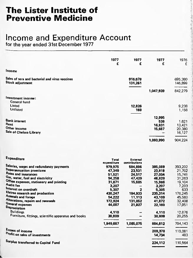
The Lister Institute ofPreventive Medicine
Income and Expenditurefor the year ended 31st December 1977
Account
A, 1 ■ v
V
I' t¡1
•I
1977 1977 1977 1976£ £ £ £ -
Income
Sales of sera and bacterial and virus vaccines 91 6 ,67 8 6 9 5 ,38 0Stock adjustment 131,261
1 ,047 ,939
146,899
8 4 2 ,27 9
Investment income:General fu n dListed 12 ,826 9 ,2 3 8U n listed 169
12 ,995
1,158
Bank interest 538 1,621Rent 16,831 13,421Other income 15 ,687 20 ,380Sale of Chelsea Library
1 ,093 ,990
16,127
9 0 4 ,2 2 4
Expenditure Totalexpenditure
Externalcontributions
39 3 ,20 2Salaries, wages and redundancy payments 97 9 ,9 7 5 59 4 ,8 8 6 38 5 ,0 8 9Superannuation premiums 4 7 ,3 4 9 23,531 23 ,818 2 1 ,702Rates and insurances 51,521 24 ,517 27 ,004 15,741J?as, water, fuel and electricity 9 4 ,258 4 7 ,4 2 9 4 6 ,8 2 9 31 ,263Office expenses, stationery and printing 31,671 15 ,685 15 ,986 19,104Audit fee 3 ,2 07 — 3 ,2 07 7,203Interest on overdraft 5,307 2 5 ,3 05 633Elstree research and production 4 3 0 ,2 4 7 194 ,933 2 3 5 ,3 1 4 176,245Animals and forage 5 4 ,222 11 ,113 4 3 ,1 0 9 45 ,560Alterations, repairs and renewals 17 2 ,924 13 1 ,052 4 1 ,872 32 ,408^aneral expenses Oepreciation
4 4 ,087 2 1 ,927 22 ,160 17,951 !
Buildings 4 ,1 1 0 — 4 ,1 1 0 12 ,876Furniture, fittings, scientific apparatus and books 3 0 ,809 - 30 ,809 2 0 ,255
Excess of income rofit on sales of investments
Surplus transferred to Capital Fund
1 ,9 49 ,68 7 1 ,065 ,075 88 4 ,6 1 2
20 9 ,3 7 814 ,734
22 4 ,1 1 2
7 9 4 ,1 4 3 !'
110,081483
110,564

'
I
fL

i

Vf *lìs '
i1-ni '
3:
il
!
i

The Lister Institute ofPreventive Medicine
lA'
Report of the Governing Body 1978
i■
if
Elstree, Hertfordshire WD6 3AX


The Governing Body
Professor A. Neuberger, cbe , m d , frcp, FRCPath, frs , Chairman
R. A. McNeile, m be , Hon. Treasurer
Professor Sir Cyril Clarke, kbe , m d , frcp, frs
Professor L. H. Collier, m d , dsc, mrcp, FRCPath
C. E, Guinness
Professor J. H. Humphrey, cbe , m d , frs
The Rt Hon the Earl o f Iveagh
Sir William Maycock, cbe , m v o , m d , frcp, FRCPath
Professor Sir Ashley Miles, cbe , m d , FRCPath, frcp, frs
L. Vallet, m a
Clerk to the Governors G. J. Roderick, BCom

The Council
Dr G. S. Andrews, m d , FRCPath Representing the British Medical Association
Professor Sir Douglas Black, msc, m d , prcp Representing the Members o f the Institute
The Rt Hon Lord Brock, ms, frcs Representing the Members o f the Institute
Professor Sir Cyril Clarke, kbe , m d , frcp, frs Representing the Members o f the Institute
Professor P. J. Collard, m d , mrcp Representing the University o f Manchester
M. L. Conalty, m d , MRCPath, dph , m r ia Representing the Royal Irish Academy
Sir Alan N. Drury, cbe , m a , m d , frcp, frs Representing the Members o f the Institute
Professor Sir David Evans, cbe , dsc, FRCPath, frs Representing the Royal Society
Professor R. I. N. Greaves, b a , m d , frcp Representing the University o f Cambridge
C. E. Guinness Representing the Members o f the Institute
Professor Henry Harris, m b , DPhii, frs Representing the University o f Oxford
Professor J. H. Humphrey, cbe , m d , frs Representing the Members o f the Institute
The Rt Hon the Earl o f Iveagh Representing the Members o f the Institute
Professor Sir Ewart Jones, m a , PhD, dsc, frs Representing the Members o f the Institute
R. A. McNeile, mbe Representing the Members o f the Institute
Professor N. H. Martin, m a , bm , BCh Representing the Members o f the Institute
Professor Sir Ashley Miles, cbe , m d , FRCPath, frcp, frs Representing the Members o f the Institute
Professor W. T. J. Morgan, cbe, PhD, dsc, frs Representing the Members o f the Institute
Professor A. Neuberger, cbe , m d , frcp, FRCPath, fbs Representing the Members o f the Institute
The President of the Royal College of Physicians Representing the Royal College o f Physicians, London
The President o f the Royal College o f Surgeons Representing the Royal College o f Surgeons o f England
The President of the Royal College o f Veterinary Surgeons Representing the Royal College o f Veterinary Surgeons
A. Steele-Bodger, m a , bsc, frcvs Representing the Royal Agricu ltura l Society
Professor F. S. Stewart, md Representing the University o f Dublin
W illiam J. Thompson Representing the Worshipful Company o f Grocers
Professor A. W. Wilkinson, ChM, FRCS(Ed), frcs , FAAP(Hon) Representing the University o f Edinburgh
Sir Graham Wilson, m o , frcp , FRCPath Representing the University o f London

The Staff
Vaccines and Sera LaboratoriesDirector: L. H. Collier,m d , dsc, mrcp, FRCPath t M. Campbell, BSc J. A. Green,bsc, PhDG. S. Turner, bsc, PhD S. A. Sldani.BSPhcL. C. Robinson, bsc, PhD A. J. Barry, bsc
A. P. Hunt, bsc
Blood Products LaboratoryDirector: Sir William Maycock, cbe , m v o , m d , prop, FRCPath * *R. S. Lane, m d , MRCPath *L. Vallet, ma
D. Ellis, BSc, PhDL. Singleton, BSc, PhD, CChem, FRICE. D. Wesley, BPharmM. J. Harvey, bsc, PhDN. Pettet, bsc
C. R. Rackham, BScA. M. W. Greulich, bsc, msc Jane R. Webb, bsc Deborah K. Woodward, bsc
B. Montgomery, h n d , a m ie e , DipEEG. M. Bailey, a fc , a m b im (Administrative Assistant)
Plasma Fractionation Laboratory (Oxford)Ethel Bidwell, bsc, PhD, cchem, fr ic T. J. Snape, ba J. K. Smith, bsc, PhD
Blood Group Reference LaboratoryActing D irector: Carolyn M. Giles, bsc, PhD A. Redfearn, m a , m b , BChir B.J. Dawes, bsc
Elizabeth W. Ikin, bsc, PhD
AdministrationSecretary: G. J. Roderick, BCom Accountant: A. G. W. Bailey, a c is , a m b im , f a a i Administrative Assistant: C. L. Beard,msexoc Administrative Assistant: Beryl I. Coussens Administrative Assistant: F. E. Carter Assistant Accountant: B. M. Hieger
Solicitors:Macfarlanes, Dowgate Hill House, London EC4R 2SY.
Auditors:Coopers & Lybrand,Abacus House, Gutter Lane, London EC2V 8AH.
t Appointed Teacher o f the University o f London* Recognised Teacher o f the University o f London

The Lister Institute of Preventive MedicineAnnual General Meeting 18 July 1978
Report of the Governing Body
The Governing Body hasthe honour to present its report of the Institute for the year 1977.
Governing BodyProfessor Sir Douglas Black, representing the Members of the Institute, resigned from the Governing Body and was replaced by Professor Sir Cyril Clarke. Under the terms of the revised Memorandum of Association Professor L. H. Collier and Dr W.d'A. Maycock were, as Laboratory Directors, elected to the Governing Body, and Mr L. Vallet was elected to replace Professor Collier as the Scientific Staff's representative.
CouncilAt last year's Annual General Meeting Mr C. E. Guinness and Mr. R. A. McNeile, both representatives of the Members of the Institute, and Mr A. Steele-Bodger, representing the Royal Agricultural Society, were unanimously reappointed to the Council.
The three members of the Council due to retire this year in accordance with the Articles of Association, but who are eligible for reappointment are Professor Sir Ewart Jones, Professor Sir David Evans and Dr M. L. Conalty.
Blood Group Reference LaboratoryOn 1st October 1977 the responsibility for the administration of the Blood Group Reference Laboratory, Gatliff Road, off Ebury Bridge Road, London SW1W 8QJ was transferred from the Medical Research Council to the Institute, and the Governors extend a warm welcome to the staff of that laboratory, some 35 persons. The Governing Body is pleased to announce the appointment, as from 1st February 1978, of Dr A. M. Holburn, MD, ChB, MRCPath, as Director of the Blood Group Reference Laboratory.
S taffThe Governing Body was very pleased to promote Dr Carolyn M. Giles to Deputy
Director of the Blood Group Reference Laboratory as from 1st February 1978 in recognition of her long and distinguished service to the Unit.
The Governing Body has pleasure in welcoming to the Scientific Staff of the Blood Products Laboratory, Mr Andreas Greulich, Dr Michael Harvey, Miss Jane Webb and Miss Deborah Woodward.
The Governing Body takes much pleasure in recording that the honour of Knighthood was conferred upon Dr W. d'A. Maycock in the Queen's Birthday Honours of 1978, and wishes to convey its congratulations to Sir William.
The Governors congratulate Sir William on his election as honorary member of the British Society for Haematology, 30th March 1978.
Vaccines and Sera Laboratories
Grants and DonationsGrants. The Governing Body records its thanks to the Medical Research Council for a grant for "Clinical trial of a new rabies vaccine prepared in human diploid cells and of human anti-rabies immunoglobulin" MRC Special Project Grant No. G 976/611.
Donations. Donations of cash were received from the following sources:High School, Kidderminster; Penzance County Grammar School (two donations); Haberdashers' Girls' School, Elstree; Merchant Taylors School for Girls, Liverpool (three donations); Gravesend School for Girls (two donations); Fulmerston School, Thetford; Fort Pitt School, Chatham (2nd donation); Lord MayorTreloar Hospital, Alton; Collegiate School for Girls, Bristol; Alec Hunter High School, Braintree.From Dr N. Mahoney, a former member of staff, 500 Dutch guilders as a contribution towards travelling expenses of members of staff of the Vaccines and Sera Laboratories visiting the Netherlands in connection with their work at the Institute.

From Silverson Machines Limited, Chesham, Buckinghamshire: a sealed unit blending machine valued at £350 for use in Dr Turner's research on rabies.
ProductionBacterial vaccines. Compared with 1976 there was a 12% increase in the amount of bulk antigens prepared for making bacterial vaccines (excluding tetanus, production of which was not recommenced until October1977). This improvement wasachieved despite a reduction of three staff in the media and production departments.
Smallpox vaccine. No crude vaccine was prepared since stocks were sufficient to meet demand; 3.64 million doses of vaccine were sold, compared with 3.48 million in 1976.
Antitoxins. The yield of crude tetanus antitoxin from the horses was poor in the first half of the year; and, although it improved dramatically thereafter, the total amount of all crude antitoxins produced was 10% less than in 1976. The problem of poor yields from the refining process (Report, 1977) has however been overcome.
Blending, Filling and Packing Department. Following the closure for refurbishing at the start of the year (Report, 1977) the blending and filling area was again shut down for three weeks in August for decorations to be completed. Despite these interruptions to work, there were increases over the 1976 figures of 6% in terms of containers filled and of 35% in terms of doses: over 34 million doses of bacterial vaccines were filled during 1977.
Research and DevelopmentTetanus vaccine. Professor Collier continued his studies on the incidence and severity of reactions to tetanus vaccine and on the antibody response (Report, 1977). In collaboration with Dr Sheila Polakoff (Central Public Health Laboratory, Colindale) he examined children receiving their routine school-leaving dose of vaccine: 1100 children in 15 schools in Hertfordshire were seen at the time of inoculation and 48 hours later; most were examined again on the 6th or 7th day. From a sample consisting of 286 volunteers in 5 schools, blood was taken at the time of inoculation and again 4 weeks later to determine (a) whether the severity of reaction was related to the tetanus antibody titre at the time of inoculation and (b) the serological response to the booster dose. In 3 schools, the reactions and antibody responses to plain and adsorbed toxoids are being compared. The results of this study are now being analysed. The help of Dr L. A. Crawford, Hertfordshire Area Health Authority, and her staff, is gratefully acknowledged, as is the cooperation of the pupils and staff of the schools concerned.
In parallel with this investigation Professor Collier is comparing the antibody responses to primary tetanus immunization with (a) 1 dose of adsorbed vaccine and 2 doses of plain formol toxoid and (b) 3 doses of adsorbed vaccine; this study is being done in volunteers on the Institute's staff and their assistance is also much appreciated.
Cholera vaccine. The vaccine prepared at the Institute is grown on solid medium; this technique is less economical and convenient than those employing fluid media but has the advantage of yielding a more potent vaccine. Dr Green is trying to devise a method of preparing a satisfactory vaccine in suspended culture. In the first instance he is using a continuous culture chemostat to determine whether alteration of growth rate (by limitation of carbon, magnesium and nitrogen) influences antigenicity. Dr Green found that the sensitivity of organisms to lysis by cold shock varies with nutrient limitation; this property might prove a useful marker if it becomes necessary to reproduce in a batch- fed production system the conditions defined in chemostat experiments.RabiesDr Turner continued his studies of the IgM antibody responses of rabbits and mice to different schedules of vaccination with Semple, duck embryo and several cell-culture vaccines (Reports 1975, 1976, 1977). The results showed that the prolonged IgM response reported by other workers is related more to the quality and quantity of antigen than to the number, timing and route of administration of the doses.
Virus-neutralizing antibody composed solely of IgM globulin was obtained from mice four days after a single intravenous dose of concentrated cell-culture vaccine. Mice were not passively protected by this early antibody but were protected by IgG antibody of the same neutralizing titre; this finding was confirmed in mice in which the transition from IgM to IgG response was arrested by cyclophosphamide treatment.
In collaboration with Dr F.Y. Aoki and Dr K. Nicholson (MRC Clinical Research Centre) Dr Turner assayed rabies antibody in numerous serum samples from volunteers in the Medical Research Council trial of Merieux rabies vaccine (Report 1977). These studies, now in their fourth year, continue to show that adequate and durable antibody responses can be obtained by small doses of vaccine.
Large numbers of assays were also performed for the Scottish and the National Blood Transfusion Services to screen plasma for the preparation of human antirabies immunoglobulin (HRIG). Tests of the first batch of HRIG in 70 human volunteers were completed (Report 1977). The HRIG was

well tolerated and exerted but little immunosuppression on the active response to vaccine; i t could probably be used in doses larger than those currently recommended. This study was done in collaboration w ith Dr K. Nicholson (MRC Clinical Research Centre) and with Mr C. Rackham and Dr W .d 'A . Maycock (Blood Products Laboratory). The cooperation o f many o f the staff o f the Transfusion Service and o f the volunteers from the Royal Veterinary College is gratefully acknowledged.
With Dr K. Nicholson, Dr Turner also continued to examine methods that might avoid the use o f large numbers o f mice in antibody assays (Report 1977). So far, enzyme-linked immunoassays are unsatisfactory; assays in cell culture making use of plaque reduction techniques or resistance to superinfection by other viruses are now being investigated.
Tests on the thermal stability o f two candidate vaccines for a new international reference preparation were undertaken at the invitation o f the World Health Organization (Report 1977). The results again accorded w ith those o f the other participating laboratories and were incorporated in recommendations promulgated by WHO.
Overseas VisitsProfessor CollierLectured by invitation at the Institute for Social and Preventive Medicine at the University o f Zürich on the world smallpox eradication programme and on immunization against whooping cough, in June 1977. Participated in a World Health Organization Working Group on the Production and Testing of Pertussis Vaccine at Geneva in October1977.Dr TurnerGave by invitation a seminar on "Rabies and In terferon" in the Department o f Medical V irology and Immunology o f the University of Essen in June 1977.Participated by invitation in the WHO/IABS Joint Symposium on "Standardization o f rabies vaccines fo r human use produced in tissue cu lture", Marburg 21-23 November, 1977.
VisitorsThe follow ing visitors worked fo r periods of tw o weeks or more in the Vaccines and Sera Laboratories:Mr Stanley Maung Sein, The Burma Pharmaceutical Industry, Rangoon.Mrs Paz Ona Frisnedi, Alaband Serum and Vaccine Laboratory, Muntinlupa, Philippines. Dr F. L. Saldanha, Haffkine Bio-Pharmaceutical Corporation Ltd, Bombay.Dr P. Georgiou-Boufas, Central Public Health Laboratory, Athens.
Blood Products Laboratory, and Plasma Fractionation Laboratory, Oxford
Research and DevelopmentRecovery o f Albumin. Dr M. Harvey confirmed and continued the earlier work o f Dr Robin Hanford (Report 1977) using a ffin ity chromatography and Cibacron Blue as ligand for the recovery o f albumin from certain fractions which are at present discarded. Dr Harvey reassessed the capacity o f the method, the experimental conditions and the yield and purity o f the separated albumin. It has proved possible to use the same automated Cibacron Blue agarose chromatography column several hundred times w ithou t loss o f resolution o f albumin. Dr Harvey also began an examination o f ligandsother than Cibacron Blue.
Applied protein and immune studies. Dr R.S. Lane began, using chromatographic and advanced small scale preparative methods, to prepare and study certain protein antigens and corresponding antibodies: (1) antiglobulin constituents in serological immune diagnostic reagents; (2) antibodies fo r immune quantitation o f plasma protein levels; (3) antibodies to blood coagulation proteins, factor V111R and fibrinogen fo r use in quality control o f the purification process. Sterile non-pyrogenic IgM anti-D was prepared for the Regional Transfusion Centre, Lancaster.
Continuous flow electrophoresis for separation o f human plasma proteins. Mr Vallet and Mr Rackham continued w ith Dr A. R. Thomson, Biochemistry Group, AERE Harwell, the investigation of fractions separated by the Philpot continuous flow electrophoresis apparatus. As a result o f studying the various proteins, the experimental conditions of separation and yields, immunoglobulins and coagulation factor V III were selected for closer investigation which w ill be carried out in 1978 and w ill be coupled w ith a study of the separation o f sterile and pyrogen-free fractions by the apparatus. The work at Harwell was supported by a direct grant from DHSS.
Lectins. Dr Singleton continued the analysis, by a variety o f methods, of the lectin-like substances (LLS) from extracts of certain nuts. Among other observations he showed that the LLS agglutinated human group 0 red cells on which the receptor sites were revealed by treatment w ith neuraminidase and that the haemagglutinating activity is eluted by lactose or galactose from a Sepharose column using conconcavalin A as ligand. The preliminary results o f disc electrophoresis suggest that this eluate and that removed by N-methyl D-man- nopyranoside are heterogeneous. T itra tion

against neuraminidase treated human red cells shows that the galactose eluate is purified fourfold.Factor VII. Mr Dike and Mr Griffiths devised a method for separating Factor VII. This concentrate produced a good clinical response in three patients and its use was unattended by side effects.
Factor VIII. Dr J.K . Smith continued to investigate the variables affecting yield and purity of factor V III with the aim of preparing a more highly purified concentrate which would be of value in certain circumstances for treating haemophilia A. Mr Snape showed that factor V III, after absorption with aluminium hydroxide, was stable at 18°C for at least 24 hours.
Factor IX. Dr Smith began an investigation of means whereby the potentially thrombo- genic components could be excluded from factor IX concentrates prepared forclinical use. Removal of these contaminating "materials" would contribute to the safety of factor IX concentrates. In conformity with general usage, the Kingdon test (non-activated partial thromboplastin time) for the thrombogenicity of factor IX concentrates was adopted in place of the recalcification test hitherto used.
Factor X III. A method of preparing factor XIII was devised, using Cohn fraction I as starting material. At the request of the factor X III Working Party of the International Society on Thrombosis and Haemostasis, a provisional reference preparation, on which stability studies were started, was made.
Coagulation Factor Assay. Mr Snape established a one-stage assay system for factor V III which includes the use of a semi-artificial factor VIII-deficient substrate and factor V prepared from bovine blood.
Chromogenic Substrates. Mr Snape used synthetic chromogenic substrates, specific for plasmin, thrombin, factor Xa and kallikrein, to detect qualitatively these proteases at various stages during the preparation of factor V III concentrate. It is intended to develop quantitative methods. In collaboration with Mr Vallet, Mr Snape began to develop a method, using kallikrein chromogenic substrate to assay the amount or pre-kalIikrein activator (PKA) in plasma protein fraction. The presence of PKA has been found in USA to be associated with vasodepressor reactions in man.
ProductionCoagulation factors. During the year the target for the preparation of factor V III concentrate, contained in DHSS expansion programme 1974, was reached and then exceeded, in spite of the fact that two regional transfusion centres have not yet begun to
provide their quotas of fresh frozen plasma. Twice as much factor VIII was issued to NHS in 1977 as in 1976. Plans were prepared for doubling the preparation of factor V III concentrate in 1978.
The amounts of prothrombin complex (factors II, IX and X) were also augmented by the initiation at Elstree of the preparation of this complex which is taken to the final stage. The concentrate is then sent to Plasma Fractionation Laboratory, Oxford, where final filtration, filling, quality control and freezedrying are carried out.
The responsibility for final assays of activity in factor V III and factor IX concentrates was transferred to Mr Snape, Plasma Fractionation Laboratory, Oxford. The opportunity for certain research and development, which was expected to occur as a result of this re-organi- zation, disappointingly did not develop because of increased needs for fibrinogen for isotopic labelling at Radiochemical Centre and because of loss of staff.
Albumin and Plasma Protein Fraction. Under the direction of Mr Vallet and Mr Wesley, means were found to exceed the planned capacity of the large scale fractionation laboratory by some 60 per cent and plans were made for further growth in 1978.
Immunoglobulins. The need for normal immunoglobulin remains fairly constant. Policy changes regarding smallpox vaccination were followed by a decline in requests for anti-vaccinia immunoglobulin.Overseas VisitsDr W. d'A. MaycockGroup of Experts 6B (Chairman), European Pharmacopoeia Commission, Council of Europe, Strasbourg, 21-23 February 1977, 14-15 June 1977 and 14-16 November 1977. Council of Europe Public Health Committee, Subcommittee of Experts on Blood Problems, Frankfurt, 23-26 May 1977.Temporary Adviser, World Health Organization, Geneva, to revise WHO monograph "Blood Transfusion: A Guide to the Formation and Operation of a Transfusion Service", 18-23 July 1977.Mr L. Vallet, Mr E.D. Wesley and Dr J.K . SmithInternational Workshop on Technology for Protein Separation and Improvement of Blood Plasma Fractionation, Reston, Virginia, USA, 7-9 September 1977 and, subsequently, American National Red Cross Blood Research Laboratory, Bethesda, Md., and New York Blood Center, New York, USA.Mr L. ValletNetherlands Red Cross Central Transfusion Laboratory, Amsterdam, German Red Cross Regional Blood Transfusion Centre and

Institute, Hagen, Westphalia and Swiss Red Cross Central Transfusion Laboratory, Bern. 28 March to 1 April 1977.Workshop on Measurement of Potentially Hypotensive Agents, Bureau of Biologies, Food and Drugs Administration, Bethesda, Md., USA. 15-16 September 1977.Mr E. D. Wesley and Dr J. K. Smith Dept, of Medicine, University of North Carolina, Chapel Hill, Cutter Laboratories Inc., Clayton, North Carolina, and Blood Products Laboratory, Michigan State Department of Health, Lansing, Michigan, USA. September 1977.Mr E. D. WesleyCenter for Blood Research Inc., Boston, Mass., and Blood Products Laboratory, Commonwealth of Massachussetts Department of Health Laboratories, Jamaica Plain, Mass., USA. September 1977.Dr E. Bidwell and Dr D. Ellis Symposium on "Clotting Factor Production: Factor V III and Factor IX Complex", Swiss Red Cross Central Transfusion Laboratory, Bern, 27-28 April 1977.Dr E. BidwellX llth Congress of World Federation of Haemophilia, New York, USA. 21-24 June 1977.Task Force Meetings of International Society on Thrombosis and Haemostasis, Philadelphia, USA. 25-28 June 1977.Vlth International Congress of International Society on Thrombosis and Haemostasis, 27 June - 2 July 1977, Philadelphia, and, subsequently, American National Red Cross Blood Research Laboratory, Bethesda, Md., Dept, of Medicine, University of North Carolina, Chapel Hill, New York Blood Center, and New York University Medical Center, USA.
VisitorThe following visitor worked for a period of two weeks or more in the Plasma Fractionation Laboratory, Oxford:Dr J. P. Allain, Centre National deTransfusion Sanguine, Paris.
Blood Group Reference Laboratory
For the Blood Group Reference Laboratory 1977 was a difficult year marked by major administrative changes, staff shortages at senior level and threats to accommodation. Some of these problems have now been resolved. The transfer of the administration of the Blood Group Reference Laboratory on behalf of the Department of Health & Social Security from the Medical Research Council
to the Lister Institute was completed on 1st October, 1977. A new Director, Dr A.M. Holburn, has been appointed.
GrantsThe Blood Group Reference Laboratory is grateful to the World Health Organisation for two grants in support of its work as the WHO Collaborating Centre for Reference and Research in Blood Grouping and as the WHO Collaborating Centre for Reference on the use of Immunoglobulin anti-D for the Prevention of Rh sensitisation.
Production of Blood Grouping ReagentsDuring 1977, 1,037,370 ml of ABO sera, 25,648 ml of other human sera and 5,671 ml of animal sera were issued to Blood Transfusion Centres and Hospitals within the United Kingdom. Sera of more than 80 different specificities are available for issue. Improvements in procedure, including changes in techniques of bacteriological control, have been introduced to meet the demand but any further increase in output will necessitate expanded facilities (Jenkins and Moghaddam).
Standardisation of Blood Grouping ReagentsThe Blood Group Reference Laboratory continues to assess the suitability of reagents produced by the Regional Transfusion Centres in addition to its own products ( Ik in). Dr Giles is a participant of two Working Parties on the Standardisation of Antiglobulin Reagents (International Committee for Standards in Haematology and M.R.C.).
Proficiency TestingQuality control exercises art being issued to hospitals by the Regional Transfusion Centres in increasing numbers and each exercise is tested by the Blood Group Reference _aboratory. In addition, the Blood Group Reference Laboratory issues sera containing multiple antibodies to the Regional Transfusion Centres for identification.
Anti-D QuantitationThere has been a further increase in the number of samples assayed with material being received from the Blood Products Laboratory, Transfusion Centres and from overseas laboratories and manufacturers (Dawes). Variations in the results of assays between Transfusion Centres and the Blood Group Reference Laboratory inspired an investigation of assay methods and standards used in the UK. A collaborative study undertaken by the Working Party on Anti-D Immunisation has revealed that there are important discrepancies in the stated values of anti-D concentrations between national and international reference preparations.

International Panel of Donors of Rare Blood TypesThe Blood Group Reference Laboratory continues to receive blood samples from donors of rare groups for confirmation and fu ll typing for listing in the International Panel. Blood has been provided from UK donors fo r two patients in Switzerland and other requests fo r the location o f rare blood have been received.
Platelet and white cell serologySamples are received fo r the investigation of immune, including drug-induced, throm bocytopenias and leucopenias.
Red Cell Reference WorkThe red cell antigen and antibody problems investigated continue to provide much variety and interest (Giles and Poole). Rare antibodies such as anti-Lan, anti-Y ta, anti-D ib, anti-Vel and anti-P in a P,k patient were all received from overseas fo r identification. Two autoantibodies o f unusual specificities, anti-Vel and anti-Leb, have been found. Of many low frequency antigens studied, two have been shown to be present transiently on the donor cells. A collaborative study o f IVIN variants has facilitated the correlation o f the serological (Blood Group Reference Laboratory) and biochemical abnormalities (Regional Transfusion Centre, Bristol).
Research and DevelopmentResearch projects have stemmed mainly from routine activities and have included investigations of the G1m(1) system (Brazier) and of the effects on antibody activity o f conversion of plasma to serum (Jenkins). Red cell reference work provides a rich source of material but other commitments have not permitted fu ll advantage to be taken (Poole and Giles).
AccommodationThe eventual sale o f the Chelsea Laboratories of the Lister Institute has very serious implications fo r the Blood Group Reference Laboratory. Loss o f laboratory space in the Svedberg building together w ith loss of essential storage space in the stables and loss of ease of access w ill very seriously impair the capacity of the Blood Group Reference Laboratory to maintain bulk production o f reagents. In addition, it would not be possible to rehouse the AutoAnalysers fo r anti-D quantitation. The search fo r alternative accommodation fo r the Blood Group Reference Laboratory must therefore be a high p rio rity fo r 1978,

Chairman's Statement
It is now more than two years since our laboratories in Chelsea Bridge Road had to close. Since that time we have been working hard trying to improve and expand our laboratories at Elstree in the hope that our activities would become sufficiently profitable to support and to increase our activities in medical research. We have indeed been most fortunate in having a very competent and devoted staff, but at the beginning o f this year we came to the decision that we should discontinue the activities o f our Vaccines and Sera Laboratories in 1978. This decision was made after detailed exploration by a full meeting o f the Governing Body, where the final conclusion was reached unanimously. The decision was also supported by a combined special meeting of the Council and Members o f the Institute.
There were several reasons fo r this decision. Recent legislation requires a very high standard not only in production but also in the processing o f our products. A fter careful investigation we came to the conclusion that we had to spend at least £700,000 to bring our facilities up to the required standard. In addition, the following points have to be remembered. None o f our facilities are purpose-built; our laboratories have been put up in a haphazard manner over a long period o f time. The general layout is uneconomic, and the laboratories are in every way outmoded. In order to compete e ffic iently, extensive rebuilding would be necessary, which might bring the total cost to about £2M. We also have to remember that the Institute has not put out a new product fo r many years, and some o f our existing products are likely to be phased out over a relatively short time. In order to compete internationally we would have to invest at least £2M, and probably more, in research and development o f new products. In other words, w ithout considering research in the wider sense, we fe lt we required at least £4M to remain efficient and competitive. Even w ith the sale o f the Chelsea buildings under the most favourable conditions, we could not see our way to obtaining capital sums o f that magnitude.
I have so far referred only to production and such research as is immediately related to the day-to-day needs of an efficient production unit. It must be clear, however, that whilst we value our contribution to the national wellbeing by the manufacture o f vaccines, one of our main aims must be the advancement of
medical knowledge, and this means research. In order to mount even a relatively modest research e ffo rt would need at least another £21/2M. The figures I am quoting were obtained after careful examination o f all relevant aspects, and some of the figures may in fact be under-estimates.
The conclusion we have reached is very painful and depressing. We have explored so far as we know every possible avenue of finance both in this country and abroad, but the results were completely disappointing.I am conscious of the hardship which this decision w ill impose on our staff, all o f whom have served us fa ith fu lly and w ith devotion. Many have been w ith us fo r a very long time, and their lives have been bound up w ith the Lister Institute. We sympathise w ith them deeply, and we shall try to do what we can to ease the problems which w ill arise by the closure o f the Elstree laboratories. We are at present engaged in designing a redundancy scheme which w ill be considerably more generous than that laid down by legislation. We shall also try to deal w ith individual cases of special hardship to the best o f our ability.I wish to use this opportunity o f thanking all the staff fo r their understanding and fo r their loyal service. I have been most impressed over the last few years especially by the high morale, which, made our task much easier than it would otherwise have been. Speaking for myself, up to a few months ago I was still quite hopeful that we would pull through.
The Governing Body has not yet made any decision about the use o f such funds as may be available when all our affairs at Elstree are wound up. Our buildings in Chelsea are still unsold, and we can only guess at present what the size o f our assets w ill be. However, the Governing Body has made the decision in principle that such funds as may be available shall be used fo r medical research. How this can be achieved remains to be seen, but I feel fa irly confident that the Lister Institute w ill continue to exist in some form for a long time to come.
The activities of our Blood Products Laboratory and Blood Group Reference Laboratory w ill, o f course, continue in some way which is still to be decided.
A. Neuberger. Chairman o f the Governing Body.
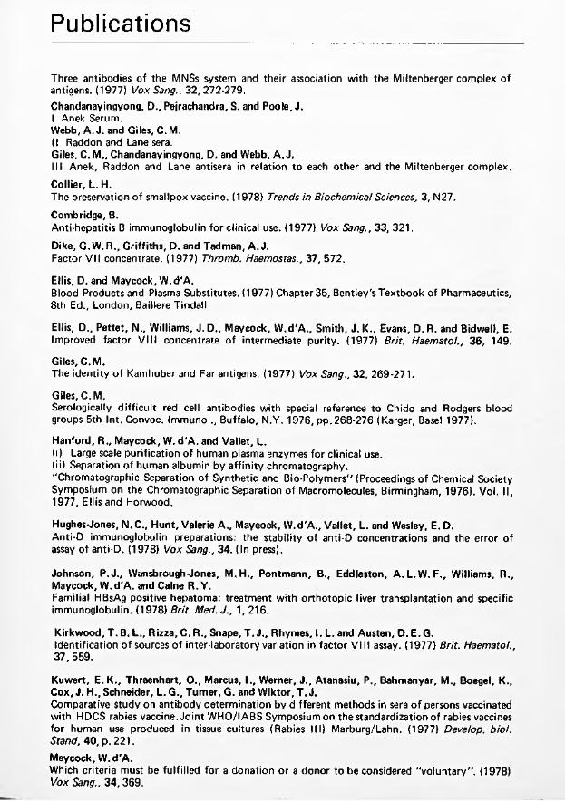
Publications
Three antibodies of the MNSs system and their association w ith the Miltenberger complex of antigens. (1977) Vox Sang., 32, 272-279.Chandanayingyong, D., Pejrachandra, S. and Poole, J.I Anek Serum.Webb, A. J. and Giles, C. M.II Raddon and Lane sera.Giles, C. M., Chandanayingyong, D. and Webb, A. J.III Anek, Raddon and Lane antisera in relation to each other and the Miltenberger complex.
Collier, L.H.The preservation o f smallpox vaccine. (1978) Trends in Biochemical Sciences, 3, N27.
Combridge, B.Anti-hepatitis B immunoglobulin fo r clinical use. (1977) Vox Sang., 33, 321.
Dike, G. W.R., Griffiths, D. and Tadman, A. J.Factor V II concentrate. (1977) Thromb. Haemostas., 37, 572.
Ellis, D. and Maycock, W. d'A.Blood Products and Plasma Substitutes. (1977) Chapter 35, Bentley's Textbook o f Pharmaceutics, 8th Ed., London, Baillere Tindall.
Ellis, D., Pettet, N., Williams, J. D., Maycock, W.d'A., Smith, J. K., Evans, D. R. and Bidwell, E.Improved factor V III concentrate o f intermediate purity. (1977) Brit. Haematol., 36, 149.
Giles, C. M.The identity o f Kamhuber and Far antigens. (1977) Vox Sang., 32, 269-271.
Giles, C. M.Serologically d ifficu lt red cell antibodies w ith special reference to Chido and Rodgers blood groups 5th Int. Convoc. Immunol., Buffalo, N.Y. 1976, p p .268-276 (Karger, Basel 1977).
Hanford, R „ Maycock, W. d 'A. and Vallet, L.(i) Large scale purification o f human plasma enzymes fo r clinical use.(ii) Separation o f human albumin by a ffin ity chromatography."Chromatographic Separation o f Synthetic and Bio-Polymers" (Proceedings o f Chemical Society Symposium on the Chromatographic Separation o f Macromolecules, Birmingham, 1976). Vol. II, 1977, Ellis and Horwood.
Hughes Jones, N.C., Hunt, Valerie A., Maycock, W.d'A., Vallet, L. and Wesley, E. D.Anti-D immunoglobulin preparations: the stability o f anti-D concentrations and the error of assay o f anti-D. (1978) Vox Sang., 34. (In press).
Johnson, P.J., Wansbrough Jones, M.H., Pontmann, B., Eddleston, A.L.W . F., Williams, R., Maycock, W.d'A. and Caine R. Y.Familial HBsAg positive hepatoma: treatment w ith orthotopic liver transplantation and specific immunoglobulin. (1978) Brit. Med.J., 1, 216.
Kirkwood, T. B. L., Rizza, C. R., Snape, T. J., Rhymes, I. L. and Austen, D. E. G.Identification o f sources o f inter-laboratory variation in factor V III assay. (1977) Brit. Haematol., 37, 559.
Kuwert, E.K., Thraenhart, O., Marcus, I., Werner, J., Atanasiu, P., Bahmanyar, M., Boegel, K., Cox, J. H., Schneider, L.G., Turner, G. and Wiktor, T. J.Comparative study on antibody determination by different methods in sera o f persons vaccinated w ith HDCS rabies vaccine. Jo int WHO/IABS Symposiumon the standardization of rabies vaccines fo r human use produced in tissue cultures (Rabies III) Marburg/Lahn. (1977) Develop, biol. Stand, 40, p. 221.
Maycock, W.d'A.Which criteria must be fu lfilled for a donation or a donor to be considered "vo lun tary". (1978) Vox Sang., 34, 369.

Nicholson, K. G., Turner, G. S. and Aoki, F. Y.Immunization w ith human diploid cell strain rabies vaccine: 2 year results. (1978) J. inf. Dis. (In press).
Nicholson, K.G. and Turner, G.S.Studies w ith human diploid cell strain rabies vaccine and human antirabies immunoglobulin in man (1978). Jo int WHO/IABS Symposium on the standardization o f rabies vaccines for human use produced in tissue cultures (Rabies III) Marburg/Lahn. (1977) Develop, biol. Stand, 40, 11 5, Basel S. Karger.
Smith, J. K., Snape, T. J., Bidwell, E., Edwards, R. and Gunson, H. H.Preparation o f factor IX concentrate from platelet-rich plasma. (1977) Thromb. Haemostas., 37, 363.
Turner, G.S.Interferon und Tollw ut — eine Übersicht. (1977) Im m unität und Infektion, 5, 208.
Wiktor, T.J., Atanasiu, P., Bahmanyar, M., Boegel, K., Cox, J.H., Diaz, A.M., Fitzgerald, E. A., Kuwert, E., Netter, R., Selimov, M., Turner, G. and van Steenis, G.Comparison studies on potency tests fo r rabies vaccines. Joint WHO/IABS Symposium on the standardization o f rabies vaccines fo r human use produced in tissue cultures (Rabies III) Marburg/Lahn. (1977) Develop, biol. Stand, 40, p. 171.
Wiktor, T. J., Boegel, K., Cox, J. H., Diaz, A.M., Fitzgerald, E., Netter, R., Turner, G., Schneider, L. G. and van Steenis, G.Studies on candidate reference preparations o f rabies vaccines. Jo int WHO/IABS Symposium on the standardization o f rabies vaccines fo r human use produced in tissue cultures (Rabies III) Marburg/Lahn. (1977) Develop, biol. Stand, 40, p. 187.


The Lister Institute ofPreventive Medicine
Balance Sheet and Accountsfor the year ended 31st December
1978
Elstree, Hertfordshire WD6 3AX 21 May 1979


The Lister Institute ofPreventive Medicine
The Governing Body
Professor A. Neuberger, cbe, m d , frcp , FRCPath, fr s , Chairman
R. A. McNeile, m be ,Hon. Treasurer
Professor Sir Cyril Astley Clarke, k b e , m d , frcp, frs
C. E. Guinness
Professor J. H. Humphrey, cbe , m d , frs
The Rt Hon the Earl o f Iveagh
Professor Sir Ashley Miles, cbe , m d , FRCPath, frcp , frs
Clerk to the Governors G.J. Roderick, BCom

Financial Report of thè Governing Body
The Lister Institute ofPreventive Medicine
The Governing Body presents the accounts of the Institute fo r the year ended 31st December 1978
ResultsThe results o f the Institute fo r the year ended 31 st December 1978 are set out on page 4 and a detailed income and expenditure account is set out on page 11. The surplus fo r the year is £168,749 compared w ith a surplus of £224,112 1977 after taking into account p ro fit on sales of investments of £151,775 (1977 £14,734).
ExportsSera and vaccines to the value of £901,530 (1977 £780,000) were exported from the United Kingdom during the year, despite the cessation o f production activities in August 1978.
Principal activitiesThe Institute produced fo r sale sera and bacterial and virus vaccines the profits from which were utilised fo r research and development work.
Future operationsAs previously reported the production of sera and bacterial and virus vaccines ceased on 31st August 1978. The Governing Body are still confident that any costs arising out o f the decision to cease production w ill be covered by existing surpluses and net income to be realised.
Fixed assetsThe movements in fixed assets during the year are set out in the table in Note 5 on the accounts. Negotiations are in hand concerning the sale of the land and buildings at Elstree. It is expected that the price w ill be in excess of the book value.
Chelsea buildingsNegotiations fo r the sale o f the Chelsea Buildings continue. It is hoped to sell the property, which is freehold, at a price in excess o f the book value.
Governing BodyThe members of the Governing Body as shown on page one were in office fo r the whole o f the year ended 31st December 1978 except the following, who ceased to be eligible on 30th September 1978:Professor L. H. Collier, MD, DSc, MRCP, FRCPathSir William d'A. Maycock, CBE, MVO, MD, FRCP, FRCPathMr. L. Vallet MA,
EmployeesThe average number of persons employed by the Institute in each week during the period 1st January to 31st August, 1978 when production ceased was 278 and from 1st September to 31st December 1978 was 209 including the staff at the Blood Products Laboratory, the Plasma Fractionation Laboratory, Oxford, the Blood Group Reference Laboratory, Chelsea and the few staff remaining at Chelsea. The aggregate remuneration paid in respect o f the year ended 31st December 1978 to these employees amounted to £1,250,071.
AuditorsA resolution to re-appoint the Auditors, Coopers and Lybrand, w ill be proposed at the General Meeting.
A.NEUBERGER Chairman

Report of the Auditorsto the members of Lister Institute of Preventive Medicine
The Lister Institute ofPreventive Medicine
In our opinion the accounts set out on pages 4 to 10 give a true and fair
view o f the state o f the Institute's affairs at 31st December 1978 and
o f its surplus and source and application o f funds fo r the year ended on
that date, according to the historical cost convention, and comply w ith
the Companies Acts 1948 and 1967.
COOPERS & LYBRAND Chartered Accountants London
21st May 1979
3

The Lister Institute ofPreventive Medicine
Income and Expenditure Accountfor the year ended 31st December 1978
Notes 1978 1978 1977£ £ £
Turnover 3 £1,200,338 £916,678
Excess of income over expenditure and beforee x t r a o r d in a r y ite m s 192,126 224,112
after charging:
Emoluments o f members o f the Governing Body
Auditor's remuneration Depreciation
11
5
33,3364,815
26,292
18,4373,207
34,919
and after crediting :
Investment income Bank interest Rental income
7,17537,75615,382
12,995538
16,831
Extraordinary items 13 (23,377)
168,749 224,112
Accumulated balanceat 1st January 197 8 844,069 619,957
Accumulated balance ---------- ------ —'at 31st December 1978 £1,012,818 £844,069
The notes on pages 6 to 9 form part o f these accounts. A ud it report on page 3
A

The Lister Institute ofPreventive Medicine
Balance Sheetat 31st December 1978
uhi
!|
1978 1978 1978 1977
Notes £ £ £ £
Employment of capital
Fixed assets 5 304,996 327,770
Property held fo r resale 4 463,464 463,464
Investments and uninvested cash 6 72,615 166,631
841,075 957,865
Current assets
Stocks and work in progress DebtorsCash and bank balances
7 164,542295,835846,011
663,952404,653
22,625
1,306,388 1,091,230
less:
Current liabilities
CreditorsBank overdrafts (secured 1977 £160,972)
395,113 305,703161,137
395,113 466,840
Net current assets 911,275 624,390
£1,752,350 £1,582,255
Capital employed
Capital fund 8 1,703,753 1,534,957
Pension fund 9 9,383 10,265
Bequest funds 10 39,214 37,033
£1,752,350 £1,582,255
A. Neuberger R. A. McNeile
Members o f the Governing Body
The notes on pages 6 to 9 form part o f these accounts. A ud it report on page 3

The Lister Institute ofPreventive Medicine
Notes on the Accounts
1 Accounting policiesThe main accounting policies are as follows: —
a Depreciation
Depreciation is calculated to write o ff the book value on a straight line basis over the expected useful lives of the assets concerned. Elstree buildings have been depreciated at the annual rate o f 2% from the date o f completion, and furniture fittings and scientific apparatus at the annual rate of 10%. The Chelsea freehold property which is. unoccupied has been depreciated at the rate of 2% up to 31st December 1976. As from that date this property is not being depreciated.
b Stocks
Sera and vaccines are valued at the lower of cost and net realisable value. Cost is the sum of direct materials and labour w ith additions for overheads appropriate to the stages of production reached. Net realisable value is the price at which the stocks can be realised in the normal course of business after allowing for the costs of realisation and, where appropriate the cost o f conversion from their existing state to a finished condition. Provision is made for obsolescent, slow-moving and defective stocks.Packing materials are valued at invoice cost,
c Research expenditure
Expenditure on research is written o ff in the year in which it is incurred,
d Donations
Donations received are credited direct to the Capital Fund.
2 Future operations
As anticipated in the financial report of the Governing Body, on 17th April 1978, the production of Sera, Bacterial and Virus Vaccines ceased on 31st August 1978. It is intended that the stocks and investments w ill be realized in the normal course of business and negotiations are in hand concerning the disposal of the other assets of the Institute.
The Governing Body are still confident that any additional costs arising out of the decision to cease production will be covered by the existing surplus and future net income.No decision has yet been made by the Governing Body as to how the resulting net surplus w ill be used in furthering medical research and development.No adjustments to the accounts prepared fo r the year ended 31st December 1978 have been considered necessary.
3 TurnoverTurnover has been arrived at after deducting commission due to agents from the invoice value of sales o f sera and vaccines.
4 Property held for resaleThe property held fo r resale is the Chelsea freehold building which is included in the balance sheet at a net amount of £463,464, being the cost o f the property £517,885 less accumulated depreciation to 31st December 1976 of £54,421-
The property is unoccupied and negotiations are continuing fo r its sale.
The amount by which its market value exceeds the book value of £463,464 cannot at present be quantified.

5 Fixed assets Freehold Furnitureproperty fittings and
Elstree equipment TotalCostAt 1st January 1978 221,391 249,408 470,799Additions at cost 4,949 2,844 7,793Disposals at cost - (4,773) (4,773)
A t 31st December 1978 226,340 247,479 473,819
Depreciation At 1st January 1978 32,722 110,307 143,029Charged to income and expenditure account 4,209 22,083 26,292Accumulated on disposals - (498) (498)
At 31st December 1978 36,931 131,892 168,823
Net book value at 31st December 1978 £189,409 £115,587 £304,996
Net book value at 31st December 1977 £188,669 £139,101 £327,770
6 Investments and uninvested cash Listed at cost Unlisted Uninvestedin UK elsewhere at cost cash Total
General 22,358 1,660 - 24,018
Pension fund 4,125 - 5,258 9,383
BequestsJenner Memorial studentship fund 16,610 9,522 26,132Morna Macleod scholarship fund 6,289 - - 6,793 13,082
£49,382 £1,660 £21,573 £72,615
Market value (unlisted investments are valued by Institute's investment advisers) £61,243 £1,549 £21,573 £84,365
Excess of market value over 1978 cost £11,750
1977 cost £113,140 £31,051 £2,166 £20,274 £166,631
Market value £240,174 £53,296 £1,885 £20,274 £315,629
Excess of market value over 1977 cost £148,998
? Stocks and work in progress 1978 1977£ £
Bacteria l vaccines 89,164 457,869V irus vaccines 7,858 97,646Sera 66,549 84,418
Packing m ateria ls163,571 639,933
971 24,019
£164,542 £663,952

8 Capital Fund
Donations and endowments have been received to date from :
Dr Ludwig Mond (1893)Berridge Trustees (1893-1898)Worshipful Company of Grocers (1894 and 1969/73)Lord Iveagh (1900)Lord Lister's Bequest (1913-1923)William Henry Clarke Bequest (1923-1926)Rockefeller Foundation (1935-1936)Wolfson Foundation (1969-73)Other donations and legacies (1891-1978)
£1978
£2,000
46,38015,000
250.000 18,9047,1143,400
300.000 48,137
1977£
2,00046,38015,000
250.000 18,9047,114 3,400
300.000 48,090
Income and Expenditure Account690,935 690,888
Accumulated balance at 31st December 1977 add surplus
844,069168,749
619,957224,112
Accumulated balance at 31st December 1978 1,012,818 844,069
9 Pension Fund£1,703,753 £1,534,957
As at 1st January 1978 Interest on investments
10,265720
less Pensions10,985
1,602
£ 9,383
10 Bequest Funds
Jenner Memorial Studentship Fund As at 1st January 1978 Interest on investments
24,8331,299 26,132
Morna Macleod Scholarship Fund As at 1st January 1978 Interest on investments
12,200882 13,082
11 Emoluments of members of the Governing Body£39,214
Particulars of emoluments of members of the Governing Body disclosed in accordance w ith Sections 6 Companies Act 1967 are as follows : —
1978Emoluments of the highest paid Governor £12,317
and 7 of the
1977£11,847
Number of other Governors whose emoluments were w ith in the ranges : — £NIL
£ 5,001 £ 7,501 £10,001
— £ 7,500- £10,000 -£ 1 2 ,5 0 0
7
11
81
8

12 Employees' emoluments
Particulars o f the numbers o f employees receiving emoluments inexcess of £10,000 disclosed in accordance w ith Section 8 of the 1978 1977Companies Act 1967 are as follows £10,001 - £12,500 1 3
£12,501 - £15,000 1 1 ■£15,001 - £17,500 1
13 Extraordinary Items
a In accordance w ith a decision ratified by the Governing Body on 18th July, 1978, £:
a substantial part o f the Institutes portfo lio was sold giving rise to a p ro fit on saleit
amounting to 151,775 *|(1
b Redundancy payments arising out of the decision to cease production (175,152)
£(23,377)
14 Capital expenditure schemes
Commitments in respect of contracts as at 31 st December 1 978 £2,046 £6,629
15 Contingent liabilities
A t 31st December 1978 there were contingent liabilities as follows:
a The Institute is still a defendant together w ith the Department of Health and Social Security, in two cases brought by parents of children stated to have suffered brain damage following whooping cough vaccination. If the plaintiffs were to win their cases, or if they lost and were unable to meet the Institute's costs, the Institute's financial liab ility could be considerable.
However in v iew o f the G ove rnm en t's announcem en t re la tin g to com pensation fo r these ch ild re n , i t is p robab le th a t the 4 l ia b il ity m ay in fa c t n o t now ex is t.
b There were no contingent liabilities arising in the normal course o f business (1977 £6,082).
Audit report on page 3
j

Statement of source and application of fundsfor the year ended 31st December 1978
The Lister Institute ofPreventive Medicine
1978 1977Source of funds £ £
Surplus 192,126 224,112Extraordinary items (23,377) -
Adjustment fo r items not involving 168,749 224,112movements o f funds:—Depreciation 26,292 34,919Profit on sales o f investments (151,775) (14,734)
Total generated from operations 43,266 224,297
Funds from other sources
Proceeds of sale of investments 247,090 33,453Donations 47 3,538
290,403 281,288
Application of funds
Purchase o f fixed assets (3,518) (93,800)Purchase o f investments (3,518) (22,358) (116,158)
£286,885 £165,130
Increase/decrease in working capital
Increase (decrease) in stocks (499,410) 137,561Increase (decrease) in debtors (108,818) 201,436(Increase) in creditors (89,410) (58,680)Movement in net liquid funds: —
Increase (Decrease) in cash balances 823,386 (23,744)Decrease (Increase) in bank overdraft 161,137 984,523 (91,443) (115,187)
£286,885 £165,130
The notes on pages 6 to 9 form part of these accounts. Audit report on page 3

S
Income and Expenditure Accountfor the year ended 31st December 1978 i
The Lister Institute ofPreventive Medicine
1978£
1978£
1978£
1977 •( £ *
IncomeIf
Sales of sera and bacterial and virus vaccines Stock adjustment
1,200,338(476,362)
916,678131,261
--------------- - ■
723,976 £1,047,939 ¡1
Investment income: General fund Listed Unlisted
7,035140
12,826 ¡1 169
7,175 12,995 I
Bank interest RentOther income
37,75615,38219,737
53816,83115,687
804,026 1,093,990 i
ExpenditureTotal
expenditureExternal
contributions
Salaries, wages and redundancy paymentsSuperannuation premiumsRates and insurancesGas, water, fuel and electricityOffice expenses, stationery and printingAudit feeInterest on overdraft Elstree research and production Animals and forage Alterations, repairs and renewals General expenses Depreciation
BuildingsFurn itu re , fitt in g s , scientific apparatus and books
1,250,07168,57057,56997,32745,249
4,815131
520,27421,074
108,92948,510
4,20922,083
799,857 47,168 31,812 66,798 27,032
2354,941
14,78588,12131,243
450,21421,40225,75730,52918,217
4,815129
165,3336,289
20,80817,267
4,20922,083
385,08923,81827,00446,82915,986
3,2075,305
235,31443,10941,87222,160
4,11030,809
£2,248,811 £1,461,759 787,052 884,612
Excess of income (after redundancy payments of £175,152) Profit on sales of investments
16,974151,775
209,37814,734
Surplus transferred to Capital Fund £168,749 £224,112

i;
li
i*:
ìiiSi
íi -M
:i
'


•V¡ '¡jH, ,
I!!!!' '
s; ■i
"i; .
I

The Lister Institute ofPreventive Medicine
Report of the Governing Body1979
Elstree, Hertfordshire WD6 3AX

¡i
rr : i
» ;
i i ' 8 :
" ! :

The Lister Institute ofPreventive Medicine
Report of the Governing Body 1979


The Governing Body
Professor A. Neuberger, cbe, md, frcp, FRCPath, frs , Chairman
R. A. McNeile, mbe, Hon. Treasurer
Professor Sir Cyril Clarke, k b e , md, frcp, frs
C. E. Guinness
Professor J. H. Humphrey, cbe, md, frs
The Rt Hon the Earl of Iveagh
Professor Sir Ashley Miles, c b e , md, FRCPath, frcp, frs
Clerk to the Governors G. J. Roderick, BCom

The Council
Dr G. S. Andrews, m d , FRCPath Representing the British Medical Association
Professor Sir Douglas Black, msc, m d , prcp Representing the Members o f the Institute
The Rt Hon Lord Brock, ms, frcs Representing the Members o f the institute
Professor Sir Cyril Clarke, k be , m d , frcp, frs Representing the Members o f the Institute
Professor P. J. Collard, m d , mrcp Representing the University o f Manchester
Sir Alan N. Drury, cbe , m a , m d , frcp, frs Representing the Members o f the Institute
Professor R. I. N. Greaves, b a , m d , frcp Representing the University o f Cambridge
C. E. Guinness Representing the Members o f the Institute
Professor Henry Harris, m b , DPhii, frs Representing the University o f Oxford
Professor J. H. Humphrey, cbe , m d , frs Representing the Royal Society
The Rt Hon the Earl o f Iveagh Representing the Members o f the Institute
R. A. McNeile, mbe Representing the Members o f the Institute
Professor N. H. Martin, m a , bm , BCh Representing the Members o f the Institute
Professor Sir Ashley Miles, cbe , m d , FRCPath, frcp, frs Representing the Members o f the Institute
Professor W. T. J. Morgan, cbe, PhD, dsc, frs Representing the Members o f the Institute
Dr. Thomas Murphy, m d , frcpi, m r ia Representing the Royal Irish Academy
Professor A. Neuberger, cbe, m d , frcp, FRCPath, frs Representing the Members o f the Institute
The President o f the Royal College o f Physicians Representing the Royal College o f Physicians, London
The President of the Royal College of Surgeons Representing the Royal College o f Surgeons o f England
The President o f the Royal College of Veterinary Surgeons Representing the Royal College o f Veterinary Surgeons
A. Steele-Bodger, m a , bsc. frcvs Representing the Royal Agricultural Society
Professor F. S. Stewart, m d Representing the University o f Dublin
William J, Thompson Representing the Worshipful Company o f Grocers
Professor A. W. Wilkinson, ch M , FRcsiEd), frcs, FAAPiHon) Representing the University o f Edinburgh
Sir Graham Wilson, m d , frcp, FRCPath Representing the University o f London

The Staff
Secretary: G. J. Roderick, BCom A ccou n tan t: B. M. Hieger A dm in is tra tive Assistant: C. L. Beard, MSExec A dm in is tra tive Assistant: F. E. Carter
Solicitors:Macfarlanes, Dowgate Hill House, London EC4R 2SY.
Auditors:Coopers & Lybrand,Abacus House, Gutter Lane, London EC2V 8AH.


The Lister Institute of Preventive MedicineAnnual General Meeting 11 July 1979
Report of the Governing Body
Governing BodyAs the result of the closure of the Institute's Vaccines and Sera Laboratories on 31st August 1978 and the subsequent transfer of the administrative responsibility for the Blood Products Laboratory to the North West Thames Regional Health Authority, Professor L. H. Collier, Sir William Maycock and Mr. L. Vallet ceased to be eligible to serve as Governors on 30th September 1978.
CouncilAt last year's Annual General Meeting it was reported that the Royal Irish Academy had appointed Dr. Thomas Murphy, President of University College, Dublin, a member of the Council to replace Dr. M. L. Conalty; that Professor Sir David Evans had resigned and that the Royal Society had appointed Professor J. H. Humphrey in his place; and that Professor Sir Ewart Jones had also resigned.
The three members of the Council due to retire this year in accordance with the Articles of Association, but who are eligible for reappointment are Professor R. I. N. Greaves, the representative of the University of Cambridge, and the Presidents of the Royal College of Surgeons of England and of the Royal College of Veterinary Surgeons.
Chairman's Statement
In the statement I made in the Report of the Governing Body for 1978 I explained the reasons which compelled us to complete the closure of the Institute and to give notice to our Staff. This closure is now almost complete. It has necessarily meant a certain amount of hardship to all our staff, but we hope this was greatly mitigated by our generous redundancy scheme and by providing as far as possible special help in individual cases. It has been possible for the senior members of our scientific staff, with one exception, to find new posts, and their research activities have not been as much interrupted as might have been feared.

I wish to thank again the staff at all levels for their understanding and devoted service over many years.
Our estate at Elstree has been sold to the Department of Health & Social Security, and the legal formalities will, I hope, be completed within the next few months. We also hope to dispose of our Chelsea buildings before long but the price we may receive for this property depends on several factors, particularly the possible extension of the planning consent for change of use. We are also at present selling some of our remaining vaccines and sera and other assets, and it is expected that this process will also come to an end by the late summer. The administration of the Blood Products Laboratory and of the Blood Group Reference Laboratory has been handed over to the Department of Health & Social Security. This will more or less complete the disposal of our assets and responsibilities.
We shall of course retain our responsibilities to our pensioners, and payments under this heading will have first call on such financial resources as will remain. It is too early to state the exact size of our financial assets, but we may be confident that they will be in excess of £2,000,000. The precise way in which we shall use our financial resources to support medical research will occupy the attention of the Governing Body during the next few months. As soon as we are able to formulate definite proposals, we will of course consult our Members and the Council of the Institute. We are also considering whether constitutional changes are necessary, but this is again a matter on which we are taking advice.
In conclusion I wish to thank my colleagues on the Governing Body for their most helpful co-operation, and I want to express my great appreciation for the help I have received from Professor Collier, who has been Director of the Vaccines & Sera Laboratories at Elstree. We are glad that he has been appointed to a Chair of Virology in the University of London at the London Hospital Medical College. Above all, I want to express my warmest thanks to Mr. Roderick whose competence, devotion, and hard work are beyond praise.
A. Neuberger


The Lister Institute ofPreventive Medicine
Balance Sheet and Accountsfor the year ended 31st December
1979
Elstree, Hertfordshire WD6 3AX 10 June 1980


The Lister Institute ofPreventive Medicine
The Governing Body
Professor A. Neuberger, cbe, m d , frcp, FRCPath, frs . Chairman
R. A. McNeile, mbe. Hon. Treasurer
Professor Sir Cyril Astley Clarke, kbe , m d , frcp, frs
C. E. Guinness
Professor J. H. Humphrey, cbe, m d , frs
The Rt Hon the Earl o f Iveagh
Professor Sir Ashley Miles, cbe, m d , FRCPath, frcp, frs
Professor W. T. J. Morgan, cbe, PhD, dsc, frs
Clerk to the Governors G J . Roderick, BCom

Financial Report of thè Governing Body
The Lister Institute ofPreventive Medicine
The Governing Body presents the accounts of the Institute for the year ended 31st December 1979.
ResultsThe results o f the Institute fo r the year ended 31st December 1979 are set out on page 4 and a detailed income and expenditure account is set out on page 11. The surplus fo r the year is £1,047,164 compared with a surplus of £168,749 in 1978 after taking into account p ro fit on sales o f fixed assets o f £447,813 (1978 Nil).
ExportsSera and vaccines to the value o f £539,579 (1978 £901,530) were exported from the United Kingdom during the year, despite the cessation o f production activities in August 1978.
Principal activitiesThe Institute sold sera and bacterial and virus vaccines produced in previous years, the profits from which are intended to be utilised for research and development work.
Future operationsThe Governing Body are actively engaged in considering plans fo r the direct financial assistance o f research in bio-medicine.
Fixed assetsThe movements in fixed assets during the year are set out in the table in Note 5 on the accounts. The land and buildings at Elstree were sold during the year at a price which exceeded the book value.
Chelsea buildingsNegotiations for the sale o f the Chelsea Buildings continue. It is hoped to sell the property, which is freehold, at a price in excess o f the book value.
Governing BodyThe members o f the Governing Body as shown on page one were in office fo r the whole of the year ended 31st December 1979 except Professor W.T.J. Morgan who was appointed on 21st March 1979.
EmployeesThe average number of persons employed by the Institute in each week during the period 1st January to 31st March 1979 was 247, including the staff at the Blood Products Laboratory, the Plasma Fractionation Laboratory, Oxford, the Blood Group Reference Laboratory, Chelsea and the few staff remaining at Chelsea. For the last 7 months on average only 9 people were employed. The aggregate remuneration paid in respect o f the year ended 31st December 1979 to these employees amounted to £405,221.
AuditorsA resolution to re-appoint the Auditors, Coopers and Lybrand, w ill be proposed at the General Meeting.
A. NEUBERGER Chairman
2

Report of the Auditorsto the members of Lister Institute of Preventive Medicine
The Lister Institute ofPreventive Medicine
In our opinion the accounts set out on pages 4 to 10 give a true and fair
view of the state of the Institute's affairs at 31st December 1979 and
of its surplus and source and application of funds for the year ended on
that date, according to the historical cost convention, and comply with
the Companies Acts 1948 and 1967.
COOPERS & LYBRAND Chartered Accountants London10th June 1980.
3

Income and Expenditure Accountfor the year ended 31st December 1979
The Lister Institute ofPreventive Medicine
Notes 1979 1979 1978£ £ £
T u rn o v e r 3 £787,814 £1,200,338
Excess o f in c o m e o v e r e x p e n d itu re and b e fo re e x tra o rd in a ry ite m s
after charging:
Emoluments o f members of the Governing Body
Auditor's remuneration Depreciation
702,125 192,126
10
53,000
120
33,3364,815
26,292
and after crediting:
Investment income Bank interest Rental income
17,312203,303
32,834
7,17537,75615,382
E x tra o rd in a ry ite m s 11 345,039 (23,337)
1,047,164 168,749
Accumulated balanceat 1st January 1979 1,012,818 844,069
Accumulated balanceat 31st December 1979 £2,059,982 £1,012,818
The notes on pages 6 to 9 form part of these accounts. Audit report on page 3
4

The Lister Institute ofPreventive Medicine
Balance Sheetat 31st December 1979
1979 1979 1978Notes £ £ £
Employment of capital
Fixed assets 5 960 304,996Property held for resale 4 463,464 463,464Investments and uninvested cash 6 873,150 72,615
1,337,574 841,075
Current assets
Stocks and work in progress 18,500 164,542Debtors 64,249 295,835Cash and bank balances 1,517,083 846,011
1,599,832 1,306,388less:Current liabilities
Creditors 121,965 395,113
Net current assets 1,477,867 911,275
£2,815,441 £1,752,350
Capital employed
Capital fund 7 2,750,922 1,703,753Pension fund 8 9,012 9,383
Bequest funds 9 55,507 39,214
£2,815,441 £1,752,350
A. Neuberger I Members o f theR.A. McNeile I Governing Body
The notes on pages 6 to 9 form part of these accounts. Audit report on page 3
5

The Lister Institute ofPreventive Medicine
Notes on the Accounts
1 Accounting policies
The main accounting policies are as follows: —
a Depreciation
Depreciation is calculated to write o ff the book value on a straight line basis over the expected useful lives of the assets concerned. Elstree buildings have been depreciated at the annual rate of 2% from the date of completion, and furniture fittings and scientific apparatus at the annual rate of 10%. The Chelsea freehold property which is unoccupied has been depreciated at the rate of 2% up to 31st December 1976. As from that date this property is not being depreciated.
b Stocks
Sera and vaccines are valued at the lower of cost and net realisable value. Cost is the sum o f direct materials and labour with additions for overheads appropriate to the stages of production reached. Net realisable value is the price at which the stocks can be realised in the normal course of business after allowing for the costs of realisation and, where appropriate, the cost of conversion from their existing state to a finished condition. Provision is made for obsolescent, slow-moving and defective stocks.Packing materials are valued at invoice cost,
c Investment income
Credit is taken for income from investments on a cash received basis, except for gilt-edged securities which is accrued
d Donations
Donations received are credited direct to the Capital Fund.
2 Future operationsNo decision has yet been made by the Governing Body as to how the net surpluses arising in this and previous years will be used in furthering medical research and development. However, the Governing Body are actively engaged in considering plans for the direct financial assistance of research in bio-medicine.
No adjustments to the accounts prepared for the year ended 31st December 1979 have been considered necessary in this respect.
3 TurnoverTurnover has been arrived at after deducting commission due to agents from the invoice value of sales of sera and vaccines.
4 Property held for resaleThe property held for resale is the Chelsea freehold buildj^g which is included in the balance sheet at a net amount of £463,464, being the cost of the property £517,885 lesj^ccumulated depreciation to 31st December 1976 of £54,421-
The property is unoccupied and negotations are continuing for its sale.
The amount by which its market value exceedyfrie book value of £463,464 cannot at present be quantified.
See l e t t e r dated 10th June 1980 from th e Chairman, a tta c h e d .
6

5 Fixed assets Freeholdproperty
Elstree
Furniture fittings and equipment Total
CostA t 1st January 1979 Disposals at cost
226,340(226,340)
247,479(219,634)
473,819(445,974)
A t 31st December 1979 Nil 27,845 27,845
Depreciation A t 1st January 1979Charged to income and expenditure account Accumulated on disposals
36,931
(36,931)
131,892120
(105,127)
168,823120
(142,058)
A t 31st December 1979 Nil 26,885 26,885
Net book value at 31st December 1979 Nil £960 £960
Net book value at 31st December 1978 £189,409 £115,587 £304,996
6 Investments and uninvested cash Listed at cost in UK
Unlisted at cost
Uninvestedcash Total
General 807,447 1,185 - 808,632
Pension Fund 4,125 - 4,887 9,012
BequestsJenner Memorial studentship fund Morna Macleod scholarship fund
22,54313,708
- 11,2677,988
33,810 21,696
£847,823 £1,185 £24,142 £873,150
Market value (unlisted investments are valued by Institute's investment advisers) £855,155 £1,078 £24,142 £880,375
Excess of market value over 1979 cost £7,225
1978 cost £49,382 £1,660 £21,573 £72,615
Market value £61,243 £1,549 £21,573 £84,365
Excess of market value over 1978 cost £11,750

lOth June 1980.
TO: MEMBERS OF TILE LISTER INSTITUTE OF PREVENTIVI: MEDICINE
Dear Member,
Chelsea Buildings.
The Financial Report of the Governing Body and Notes on the Accounts refer to negotiations for the sale of the Chelsea buildings. Subsequent to the year end and the final printing of the Accounts, the Institute has sold the Chelsea buildings for £2,700,000. The sales proceeds exceed the book value of £463,464 by over £2.2 million and the net surplus arising, which is free of tax, will be included in the Capital Fund in the Accounts for the year ending 31st December 1980.
A. Neuberger. Chairman.
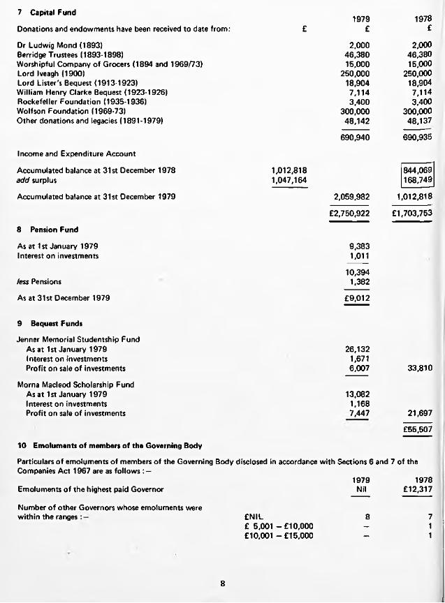
7 Capital Fund19781979
Donations and endowments have been received to date from: £ £ £
Dr Ludwig Mond (1893) 2,000 2,000Berridge Trustees (1893-1898) 46,380 46,380Worshipful Company of Grocers (1894 and 1969/73) 15,000 15,000Lord Iveagh (1900) 250,000 250,000Lord Lister's Bequest (1913-1923) 18,904 18,904William Henry Clarke Bequest (1923-1926) 7,114 7,114Rockefeller Foundation (1935-1936) 3,400 3,400Wolfson Foundation (1969-73) 300,000 300,000Other donations and legacies (1891-1979) 48,142 48,137
690,940 690,935
Income and Expenditure Account
Accumulated balance at 31st December 1978 1,012,818 844,069add surplus 1,047,164 168,749
Accumulated balance at 31st December 1979 2,059,982 1,012,818
£2,750,922 £1,703,753
8 Pension Fund
As at 1st January 1979 9,383Interest on investments 1,011
10,394less Pensions 1,382
As at 31st December 1979 £9,012
9 Bequest Funds
Jenner Memorial Studentship FundAs at 1st January 1979 26,132Interest on investments 1,671Profit on sale of investments 6,007 33,810
Morna Macleod Scholarship FundAs at 1st January 1979 13,082Interest on investments 1,168Profit on sale of investments 7,447 21,697
£55,507
10 Emoluments of members of the Governing Body
Particulars o f emoluments of members of the Governing Body disclosed in accordance with Sections 6 and 7 of theCompanies Act 1967 are as follows : —
1979 1978Emoluments o f the highest paid Governor Nil £12,317
Number o f other Governors whose emoluments werewithin the r a n g e s £ N I L 8 7
£ 5 ,001 -£10 ,000 - 1£10 ,001-£15,000 - 1
8

11 Extraordinary Items
a In accordance with a decision ratified by the Governing Body on 18th July, 1978, a substantial part o f the Institute's portfolio was sold giving rise to a profit on sale amounting to
1979£
1978£
151,775
b Redundancy payments arising out of the decision to cease production (102,774) (175,152)
447,813c Profit on sale o f the freehold property and equipment at Elstree
£345,039 (£23,377)
12 Capital expenditure schemes
Commitments in respect of contracts as at 31st December 1979 were NIL (1978 — £2,046)
13 Contingent liabilities
At 31st December 1979 there were contingent liabilities as follows:
a The Institute is still a defendant together with the Department of Health and Social Security, in two cases brought by parents of children stated to have suffered brain damage following whooping cough vaccination. If the plaintiffs were to win their cases, or if they lost and were unable to meet the Institute's costs, the Institute's financial liability could be considerable.
However in vew o f the Government's announcement relating to compensation for these children, it is probable that the liability does not in fact now exist.
b There is a contingent liability of £17,000 in respect of payments that may be required to be made to the National Health Superannuation Scheme in respect of the transfer to that Scheme of ex-employees.
c There were no contingent liabilities arising in the normal course of business (1978 Nil).
Audit report on page 3
9

Statement of source and application of fundsfor the year ended 31st December 1979
The Lister Institute ofPreventive Medicine
Source of funds 1979 1978£ £
Surplus 702,125 192,126Extraordinary items 345,039 (23,377)
Adjustment for items not involving movements o f funds :-
1,047,164 168,749
Depreciation 120 26,292Loss (Profit) on sales of investments 258 (151,775)(Profit) on sales of fixed assets (447,813) -
Total generated from operations 599,729 43,266
Funds from other sources
Sale of fixed assets 751,730 __Proceeds of sale of investments 22,575 247,090Donations 5 47
1,374,039 290,403
Application of funds
Purchase of fixed assets _ (3,518)Purchase of investments (807,447) -
£566,592 £286,885
Increase/decrease in working capital
(Decrease) in stocks (Decrease) in debtors Decrease (Increase) in creditors
Movement in net liquid funds:— Increase in cash balances Decrease in bank overdraft
(146,042)(231,586)273,148
671,072671,072
£566,592
(499,410)(108,818)
(89,410)
823,386161,137 984,523
£286,885
The notes on pages 6 to 9 form part of these accounts. Audit report on page 3
10

Income and Expenditure Accountfor the year ended 31st December 1979
The Lister Institute ofPreventive Medicine
Income
Sales of sera and bacterial and virus vaccines Stock adjustment
Investment income: General fund Listed Unlisted
Bank interestRent and service chargesOther income
Expenditure
Salaries, wages, pensions and redundancy paymentsSuperannuation premiumsRent, rates and insurancesGas, water, fuel and electricityOffice expenses, stationery and printingProfessional feesInterest on overdraftElstree research and productionAnimals and forageAlterations, repairs and renewalsGeneral expensesGrantsDepreciation
Excess of income (after redundancy payments °* £102,774; 1978: £175,152)Profit on sale of fixed assets Profit/( Loss) on sale of investments
Surplus transferred to Capital Fund
1979 1979 1979 1978£ £ £ £
787,814 1,200,338(145,071) (476,362)
642,743 723,976
TotalExpenditure
17,210102
Externalcontributions
17,312203,303
32,83423,896
£920,088
7,035140
7,17537,75615,38219,737
£804,026
405,221 200,216 205,005 450,21413,333 12,101 1,232 21,40232,135 2,877 29,258 25,75729,030 22,048 6,982 30,52921,039 15,011 6,028 18,21716,858 — 16,858 4,815
_ — _ 129207,101 172,243 34,858 165,333
3,704 3,704 — 6,28944,925 39,577 5,348 20,80845,563 32,838 12,725 17,267
2,065 — 2,065 —120 - 120 26,292
£821,094 £500,615 £320,479 £787,052
599,609 16,974447,813 —
(258) 151,775
£1,047,164 £168,749
11

■


The Lister Institute ofPreventive Medicine
Report of the Governing Body1980
Elstree, Hertfordshire WD6 3AX


The Governing Body
Professor A. Neuberger, c b e , m d , fr c p , FRCPath, fr s . Chairman
R. A. McNeile, m b e ,H on. Treasurer
Professor Sir Cyril Clarke, k b e , m d , fr c p , frs
C. E. Guinness
Professor J. H. Humphrey, c b e , m d , frs
The Rt Hon the Earl of IveaghProfessor Sir Ashley Miles, c b e , m d , FRCPath, fr c p , frs
Professor W. T. J. Morgan, c b e , PhD, dsc, fr s
Secretary, and C lerk to the Governors G. J. Roderick, BCom
Solicitors:Macfarlanes, Dowgate Hill House, London EC4R 2SY.
Auditors:Coopers & Lybrand,Abacus House, Gutter Lane, London EC2V 8AH.

The Council
Dr G. S. Andrews, m d , FRCPath Representing the British Medical Association
Professor Sir Douglas Black, msc, m d , prcp Representing the Members o f the institute
The Rt Hon Lord Brock, ms, frcs Representing the Members o f the institute
Professor Sir Cyril Clarke, k be , m d , frcp, frs Representing the Members o f the institute
Professor P. J. Collard, m d , mrcp Representing the University o f Manchester
Sir Alan N. Drury, cbe, m a , m d , frcp, frs Representing the Members o f the institute
C. E. Guinness Representing the Members o f the Institute
Professor Henry Harris, m b , dpwi, frs Representing the University o f Oxford
Professor J. H. Humphrey, cbe, md , frs Representing the Royal Society
The Rt Hon the Earl o f Iveagh Representing the Members o f the institute
R. A. McNeile, mbe Representing the Members o f the institute
Professor N. H. Martin, m a , bm , BCh Representing the Members o f the institute
Professor Sir Ashley Miles, cbe , m d , FRCPath, frcp, frs Representing the Members o f the institute
Professor W. T. J. Morgan, cbe, PhD, dsc, frs Representing the Members o f the institute
Dr. Thomas Murphy, m d , frcpi, m r ia Representing the Royal Irish Academy
Professor A. Neuberger, cbe, m d , frcp, FRCPath, frs Representing the Members o f the institute
The President o f the Royal College o f Physicians Representing the Royal College o f Physicians, London
The President o f the Royal College o f Surgeons Representing the Royal College o f Surgeons o f England
A. Steele-Bodger, cbe, m a , bsc, frcvs Representing the Royal Agricultural Society
Professor F. S. Stewart, m d Representing the University o f Dublin
William J. Thompson Representing the Worshipful Company o f Grocers
Dr Olga Uvarov, cbe, dsc, frcvs Representing the Royal College o f Veterinary Surgeons
Professor Peter Wildy, m b , mrcs, FRCPath Representing the University o f Cambridge
Professor A. W. Wilkinson, chM, FRCS(Ed), frcs, FAAP(Hon) Representing the University o f Edinburgh
Sir Graham-Wilson, md , frcp, FRCPath Representing the University o f London

The Lister Institute of Preventive MedicineAnnual General Meeting 9th July 1980
Report of the Governing Body
Governing BodyProfessor W. T. J. Morgan, c b e , PhD, dsc, fr s was co-opted on to the Governing Body on 21st March, 1979, in accordance with Clause 4(2) of the Memorandum of Association.
CouncilAt last year's Annual General Meeting it was reported that the University of Cambridge had appointed Professor Peter Wildy a member of the Council to replace Professor R. I. N. Greaves; that the Royal College of Veterinary Surgeons had appointed Dr Olga Uvarov to serve on the Council in lieu of the President; and that the President of the Royal College of Surgeons of England would continue to serve on the Council as the representative of the Royal College.
The three members of the Council due to retire this year in accordance with the Articles of Association, but who are eligible for reappointment, are Sir Graham Wilson, the representative of the University of London, Professor P. J. Collard, the representative of the University of Manchester and Sir Alan Drury, a representative of the Members.
Chairman's StatementIn a statement I made in the Report of the Governing Body for the year 1979, I reported the closure of the Institute and on our efforts to dispose of our assets, which mainly consisted of our buildings at Chelsea and our estate at Elstree. I reported that our Elstree estate had been sold, and I can now confirm that the legal formalities were completed soon afterwards. We are now renting from the DHSS on a short-term basis certain very limited accommodation at Elstree in order to enable us to sell our remaining vaccine and sera stocks, equipment and furniture, and to facilitate the completion of several important administrative functions. I am pleased to be able to report the sale of our Chelsea

property to the St Martin's Property Corporation Ltd. Completion took place on the 27th May 1980, and we have obtained what I believe is a good price for the Chelsea buildings. Both these transactions, that is, the sale of the Elstree estate and that of our Chelsea property, had of course to be approved by the Charity Commissioners. We have now also disposed of such vaccines and sera as could be marketed, and we have made arrangements to sell the remaining stock of scorpion antitoxin through our agents, there being sufficient material available to meet demand for about four years.
When all these transactions are completed we shall be in a reasonably strong financial position. Our policy with regard to investment must be directed on the one hand to protecting the real value of our capital, but at the same time we must aim to get the maximum income compatible with our intention to protect the real value of our investments. Details of such a policy are being worked out at present. We shall also have to consider during the next few months changes in our organizational structure and the defining in detail of the aims of our scientific policy. These are matters which will remain the preoccupation of the Governing Body during the coming year.
We are anxious to continue to maintain a close connection with medical research whilst considering our future long-term policy, and for this reason we have as an interim measure decided to give additional support to those of our senior scientists who were in our employment during the last few years before the Institute closed. We have therefore given grants for periods up to three years to former members of the staff to a total value, in the first year, of about £40,000. At a later date, when our future policy has been defined, we hope to make concrete long-term proposals both to Council and to our Members.
It gives me great pleasure to thank my fellow Governors, particularly our Honorary Treasurer, for continued support. I also wish to record again my personal gratitude to Mr Roderick for his devotion, hard work, and competence. The success with which the depressing task of winding up the Institute has been carried out is mainly due to the work of Mr Roderick.
Albert Neuberger.

I


The Lister Institute ofPreventive Medicine
Balance Sheet and Accountsfor the year ended 31st December
1980
Elstree, Hertfordshire WD6 3AX 22 June 1981


The Governing Body
The Lister Institute ofPreventive Medicine
Professor A. Neuberger, cbe , m d , frcp , FRCPath, frs , Chairman
R. A. McNeile, m be , Hon. Treasurer
Professor Sir Cyril Astley Clarke, k b e , m d , frcp , frs
C. E. Guinness
Professor J. H. Humphrey, cbe, m d , frs
The Rt Hon the Earl o f Iveagh
Professor Sir Ashley Miles, cbe , m d , FRCPath, frcp , frs
Professor W. T. J. Morgan, cbe , PhD, dsc, frs
Clerk to the Governors G.J. Roderick, BCom

Financial Report of the Governing Body
The Lister Institute ofPreventive Medicine
The Governing Body presents the accounts of the Institute fo r the year ended 31st December 1980.
ResultsThe results o f the Institute fo r the year ended 31st December 1980 are set out on page 4 and a detailed income and expenditure account is set out on page 11. The surplus fo r the year is £2,878,511 compared w ith a surplus of £1,047,164 in 1979 after taking into account p ro fit on sales o f fixed assets o f £2,175,579 (1979 £447,813).
ExportsSera and vaccines, mainly scorpion anti-toxin, to the value o f £247,693 (1979 £539,579) were exported from the United Kingdom during the year. Sales o f scorpion anti-toxin are continuing during 1981.
Principal activitiesThe Institute sold sera produced in previous years, the profits from which are being utilised fo r the support of medical research.
Future operationsThe Governing Body are actively engaged in consolidating plans fo r more direct financial assistance of research in bio-medicine.
Fixed assetsThe movements in fixed assets during the year are set out in the table in Note 5 on the accounts.
Chelsea buildingsThe Institute sold the Chelsea buildings fo r £2,700,000. The sales proceeds exceeded the book value of £463,464 and the net surplus arising, which is free of tax, has been included in the capital fund.
Governing BodyThe members o f the Governing Body as shown on page 1 were in office fo r the whole of the year ended 31st December 1980.
AuditorsA resolution to re-appoint the Auditors, Coopers and Lybrand, w ill be proposed at the General Meeting.
A.NEUBERGER Chairman
2
Auditors’ Reportto the members of The Lister Institute of Preventive Medicine
The Lister Institute ofPreventive Medicine
We have audited the accounts on pages 4 to 10 in accordance w ith
approved Auditing Standards. The accounts have been prepared under
the historical cost convention.
In our opinion the accounts give a true and fair view o f the state of
affairs o f the company at 31st December 1980 and o f its p ro fit and
source and application o f funds fo r the year then ended and comply
w ith the Companies Acts 1948 to 1980.
COOPERS & LYBRANDChartered Accountants London22nd June 1981
3

Income and Expenditure Accountfor the year ended 31st December 1980
The Lister Institute ofPreventive Medicine
Notes 1980 1980 1979£ £ £
Turnover 3 £269,085 £787,814
Excess of income over expenditure and before extraordinary items
after charging:
Emoluments o f members o f the Governing Body
Auditors' remuneration Depreciation
738,240 704,190
10
53,000
4803,000
120
and after crediting :
Investment income Bank interest Rental income
395,686217,162
17,312203,303
32,834
Extraordinary items 11 2,157,720 345,039
Grants 142,895,960
(17,449)1,049,229
(2,065)
2,878,511 1,047,164
Accumulated balance at 1st January 1980 2,059,982 1,012,818
Accumulated balance at 31st December 1980 £4,938,493 £2,059,982
The notes on pages 6 to 9 form part o f these accounts. A ud it report on page 3
4

The Lister Institute ofPreventive Medicine
Balance Sheetat 31st December 1980
E m p lo y m e n t o f c a p ita l
Notes 1980 1980 £ £
1979£
Fixed assets 5 480 960
Property held fo r resale 4 - 463,464
Investments and uninvested cash 6 4,040,486 873,150
4,040,966 1,337,574
Current assets
Stocks and work in progress DebtorsCash and bank balances
31,062275,029
1,454,996
18,50064,249
1,517,083
1,761,087 1,599,832
less:
Current liabilities
Creditors 101,759 121,965
Net current assets 1,659,328 1,477,867
£5,700,294 £2,815,441
C a p ita l e m p lo y e d
Capital fund 7 5,629,438 2,750,922
Pension fund 8 9,940 9,012
Bequest funds 9 60,916 55,507
£5,700,294 £2,815,441
A. IMeuberger Members o f theR .A . McNeile Governing Body
The notes on pages 6 to 9 form part o f these accounts. A ud it report on page 3
5

Notes on the Accounts
The Lister Institute ofPreventive Medicine
1 Accounting policies
The main accounting policies are as fo llows:—
a Depreciation
Depreciation is calculated to write o ff the book value on a straight line basis over the expected useful lives of the assets concerned. The Chelsea freehold property, sold during the year, was depreciated at the rate of 2% up to 31st December 1976. As from that date the property was not depreciated.
b Stocks
Sera and vaccines are valued at the lower of cost and net realisable value. Bulk stocks are included at a nil valuation, as their realisable value is uncertain. Final product stocks are stated at the lower o f packaging and handling costs and net realisable value.
c Investments
Investments are stated at cost. Provision is made for any permanent dim inution in value,
d Investment income
Credit is taken fo r income from investments on a cash received basis, except fo r gilt-edged securities which is accrued,
e Donations
Donations received are credited direct to the Capital Fund.
2 Future operations
The Governing Body is actively engaged in consolidating its plans fo r the use of the net surpluses arising in this and previous years in furthering medical research and development including the direct financial assistance of research in bio-medicine.
3 TurnoverTurnover has been arrived at after deducting commission due to agents from the invoice value of sales of sera and vaccines.
4 Property held for resaleThe property held fo r resale at 31st December 1979, was included in the balance sheet at a net amount of £463,464, being the cost of the property £517,885 less accumulated depreciation to 31st December 1976 o f £54,421.
The property was sold for £2,700,000, less expenses of realisation £78,816, during the year.
6

5 Fixed assets Furniturefittings andequipment
Cost£
A t 1st January 1980 27,845Disposals at cost (26,645)
A t 31st December 1980 1,200
DepreciationA t 1st January 1980 26,885Charged to income and expenditure account 480Accumulated on disposals (26,645)
A t 31st December 1980 720
Net book value at 31st December 1980 £480
Net book value at 31st December 1979 £960
6 Investments and uninvested cash Listed at cost Unlisted Uninvestedin UK at cost cash Total
£ £ £ £
General 3,742,727 226,903 - 3,969,630
Pension Fund 2,108 - 7,832 9,940
BequestsJenner Memorial studentship fund 22,543 14,678 37,221Morna Macleod scholarship fund 13,708 - 9,987 23,695
£3,781,086 £226,903 £32,497 £4,040,486
Market value (unlisted investments are valued by Institute's investment advisers) £3,846,862 £234,213 £32,497 £4,113,572
Excess of market value over 1980 cost £73,086
1979 cost £847,823 £1,185 £24,142 £873,150
Market value £855,155 £1,078 £24,142 £880,375
Excess of market value over 1979 cost £7,225
7

7 Capital Fund 1980 1980 1980 1979£ £ £ £
Donations and endowments have been received to date from:
Dr Ludwig Mond (1893) 2,000 2,000Berridge Trustees (1893-1898) 46,380 46,380Worshipful Company of Grocers (1894 and 1969/73) 15,000 15,000Lord Iveagh (1900) 250,000 250,000Lord Lister's Bequest (1913-1923) 18,904 18,904William Henry Clarke Bequest (1923-1926) 7,114 7,114Rockefeller Foundation (1935-1936) 3,400 3,400Wolfson Foundation (1969-73) 300,000 300,000Other donations and legacies (1891-1980) 48,147 48,142
690,945 690,940
Income and Expenditure Account
Accumulated balance at 31st December 1979 2,059,982 1,012,818add surplus 2,895,960 1,049,229Less grants (17,449) (2,065)
2,878,5114,938,493 2,059,982
Accumulated balance at 31st December 1980 £5,629,438 £2,750,922
8 Pension Fund
As at 1st January 1980 Interest on investments Profit on sale of investments
less Pensions
As at 31st December 1980
9 Bequest Funds
Jenner Memorial Studentship Fund As at 1st January 1980 Interest on investments
Morna Macleod Scholarship Fund As at 1 st January 1980 Interest on investments
9,0121,395
708
11,1151,175
£9,940
33,8103,411
21,6971,998
37,221
10 Emoluments of members of the Governing Body
Particulars o f emoluments of members o f the Governing Body disclosed in accordance w ith Sections 6 and 7 of the Companies Act 1967 are as follows : —
No member of the Governing Body received any emoluments during 1980 or 1979.
8

11 Extraordinary Items 1980£
1979£
a Profit on sale of the freehold property at Chelsea 2,157,720
b Profit on sale of the freehold property and equipment at Elstree
c Redundancy payments arising out of the decision to cease production in
447,813
August 1978. (102,774)
£2,157,720 £345,039
12 Capital expenditure schemes
There were no commitments in respect of contracts as at 31st December 1980 (1979: NIL)
13 Contingent liabilities
A t 31st December 1980 there were contingent liabilities as follows:
The Institute is still a defendant together w ith the Department of Health and Social Security, in two cases brought by parents of children stated to have suffered brain damage following whooping cough vaccination. If the plaintiffs were to win their cases, or if they lost and were unable to meet the Institute's costs, the Institute's financial liab ility could be considerable.
However in view of the Government's announcement relating to compensation for these children, it is probable that the liab ility does not in fact now exist.
14 Grants
The Governing Body whilst considering its policy for the giving of grants in support of medical research, decided as an interim measure, to invite certain senior ex-members of staff to apply for grants to aid their research. As a result several grants for a maximum period of three years are being made.
Audit report on page 3
9

Statement of source and application of fundsfor the year ended 31st December 1980
The Lister Institute ofPreventive Medicine
Source of funds 1980 1979£ £
Surplus 738,240 704,190Extraordinary items 2,157,720 345,039
2,895,960 1,049,229Adjustment fo r items not involving
movements o f funds: — Depreciation 480 120Loss on sales of investments — 258(Profit) on sales of fixed assets (2,175,579) (447,813)
Total generated from operations 720,861 601,794
Funds from other sources
Net surplus (deficit) fo r the year on the Pension Fund 220 (371)Income from Bequest Fund investments 5,409 2,838Sale of fixed assets 2,639,043 751,730Proceeds of sale of investments — 22,575Donations 5 5Profit on sale of Pension Fund investments 708 —
Profit on sale of Bequest Fund investments - 13,454
3,366,246 1,392,025
Application of funds
Purchase of investments (3,167,336) (823,368)Grants (17,449) (2,065)
£181,461 £566,592
Increase/fdecrease) in working capital
|ncrease/(Decrease) in stocks 12,562 (146,042)lncrease/(Decrease) in debtors 210,780 (231,586)Decrease in creditors 20,206 273,148
Movement in net liquid funds: — (Decrease)/!ncrease in cash balances (62,087)
£181,461
671,072£566,592
The notes on pages 6 to 9 form part of these accounts. Audit report on page 3
10

Income and Expenditure Accountfor the year ended 31st December 1980
The Lister Institute ofPreventive Medicine
1980 1980 1979Income £ £ £
Sales of sera and bacterial and virus vaccines 269,085 787,814Stock adjustment
Investment income: General fu n d
12,562
281,647
(145,071)
64 2 ,743
Listed 392,206 17,210U n lis ted 3,480
395,686102
17,312
Bank interest 217,162 2 0 3 ,30 3Rent and service charges - 32 ,834Other income
£894,495
23 ,896
£9 20 ,088
Expenditure
Salaries, wages, pensions and redundancy payments 72,004 2 0 5 ,00 5Superannuation premiums — 1,232Rent, rates and insurances 3,472 29 ,258Gas, water, fuel and electricity 4,479 6,982Office expenses, stationery and printing 3,004 6,028Professional fees 5,198 16,858Elstree research and production 77,335 34 ,858Alterations, repairs and renewals 214 5,348General expenses 7,928 12,725Depreciation 480
£174,114
120
£3 18 ,4 14
Excess of income (after redundancy paymentsof £NIL; 1979 £102,774) 720,381 601 ,674Profit on sale of fixed assets 2,175,579 4 4 7 ,8 1 3 ,(Loss) on sale of investments - (258)
Surplus transferred to Capital Fund £2,895,960 £1 ,049 ,2 29
11