Pediatric and Congenital Cardiology, Cardiac Surgery and ...
Upload
independentCategory
view
0download
0
Atypical teratoid/ rhabdoid tumor of the central nervous system inan 18-year-old patient
Vassilis Samaras1, Angeliki Stamatelli2,*, Efstathios Samaras3,*, Ioanna Stergiou4,Paraskevi Konstantopoulou4, Vassilis Varsos3, Alexander R. Judkins5, Jaclyn A.Biegel5,6, and Calypso Barbatis11Department of Pathology, Hellenic Red Cross Hospital, Athens, Greece21st Department of Pathology, University of Athens Medical School, Athens, Greece3Department of Neurosurgery, Hellenic Red Cross Hospital, Athens, Greece4Department of Radiology, General Hospital of Lamia, Lamia, Greece5Department of Pathology, University of Pennsylvania School of Medicine, The Children’s Hospitalof Philadelphia, Philadelphia, PA, USA6Department of Pediatrics, University of Pennsylvania School of Medicine, The Children’s Hospitalof Philadelphia, Philadelphia, PA, USA
AbstractObjective—Atypical teratoid/rhabdoid tumors are aggressive neoplasms of the central nervoussystem occurring mainly in the early childhood and rarely in adults. We described a case of this tumorin an 18-year-old male patient without previous medical history.
Material-method—The neoplasm was localized in the right fronto-temporal area of the brain andwas totally excised. The specimen was fixed in formalin and embedded in paraffin. The histologicaland immunohistochemical features of the neoplasm were assessed, while sequencing analysis as wellas interphase fluorescence in situ hybridization (FISH) were performed.
Results—Histological and immunohistochemical analysis demonstrated atypical rhabdoid cellsstrongly and diffusely positive for EMA and Vimentin as well as focally immunoreactive for SMAand GFAP. Additionally, though no abnormalities detected in the coding sequence of the INI1 gene,interphase FISH studies were consistent with a homozygous deletion of the INI1 gene in the majorityof examined nuclei. INI1 immunostaining demonstrated diffuse loss of nuclear INI1 expression intumor cells. Taken together, the results were consistent with a diagnosis of atypical teratoid/rhabdoidtumor (ATRT).
Conclusions—26 previous cases of ATRT have been reported in adults, thus far. To ourknowledge, this is the eighth case of an ATRT reported in an adult patient having genetic confirmationand the first one in which the tumor is, partly, localized in the right temporal area of the brain. Thisunusual presentation underlines the necessity of considering this devastating neoplasm in thedifferential diagnosis of malignant brain tumors of young adults.
Correspondence: Vassilis Samaras, M.D., Department of Pathology, Hellenic Red Cross Hospital, 1 Red Cross and Athanasaki, 11526Ambelokipi, Athens, Greece., Tel: +30 (210) 641-4849, Fax: +30 (210) 641-4629, E-mail: E-mail: [email protected].*Both authors have equally contributed to the manuscriptConflict of Interest Statement:We declare that we have no conflict of interest.
NIH Public AccessAuthor ManuscriptClin Neuropathol. Author manuscript; available in PMC 2009 July 17.
Published in final edited form as:Clin Neuropathol. 2009 ; 28(1): 1–10.
NIH
-PA Author Manuscript
NIH
-PA Author Manuscript
NIH
-PA Author Manuscript

KeywordsAtypical; Teratoid/Rhabdoid; Tumor-malignant; neoplasm-adults
INTRODUCTIONAtypical Teratoid/Rhabdoid Tumor (ATRT), according to the World Health Organization(WHO) Classification of Tumors, is a highly malignant neoplasm (grade IV) of the CentralNervous System (CNS) that preferentially manifests in children less than three years of age[Rorke et al 1996, Judkins et al. 2007]. This tumor is mainly composed of rhabdoid cells, withthe addition or not of areas demonstrating characteristics of primitive neuroectodermal tumor,epithelial tissue, neoplastic mesenchyme and neuronal or glial differentiation [Rorke andBiegel 2000, Judkins et al. 2007]. The most distinctive feature of ATRT is its association withinactivation of the hSNF5/INI1 gene, located in chromosome band 22q11.2, in the majority ofcases [Versteege et al. 1998, Biegel et al. 1999].
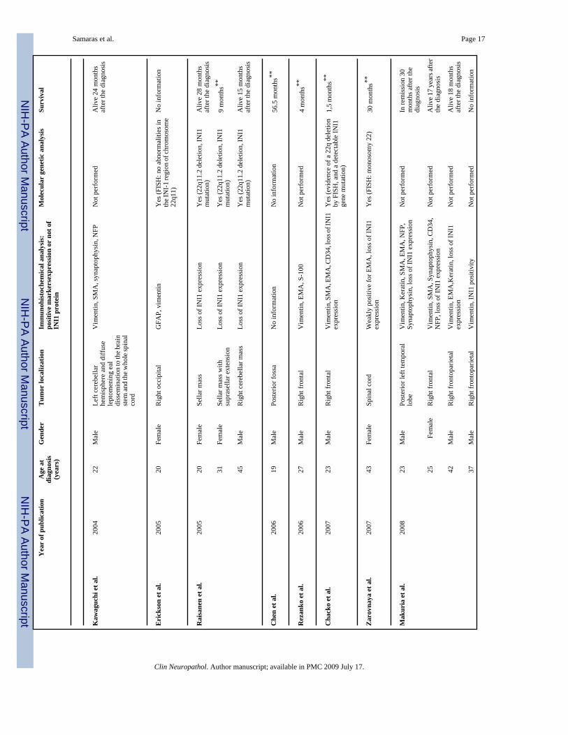
Given that ATRT prevails in children under the age of 3, only 26 cases of this neoplasm havebeen reported in adult patients (18 years of age or older), so far [Horn et al. 1992, Cossu et al.1993, Fisher et al. 1996, Ashraf et al. 1997, Byram 1999, Sugita et al. 1999, Kuge et al.2000, Arrazola et al. 2000, Lutterbach et al. 2001, Bruch et al. 2001, Pimentel et al. 2003,Kachhara et al. 2003, Kawaguchi et al. 2004, Erickson et al. 2005, Raisanen et al. 2005, Chenet al. 2006, Rezanko et al. 2006, Chacko et al. 2007, Zarovnaya et al. 2007, Makuria et al.2008]. In particular, only 7 of these cases had molecular genetic confirmation, based on findingsby mutation analysis or fluorescence in situ hybridization (FISH) [Bruch et al. 2001, Raisanenet al. 2005, Chacko et al. 2007, Zarovnaya et al. 2007].
In this context, our aim was to present an unusual case of an ATRT in an 18-year-old malepatient, in which we describe the main clinical and histological features of this particularlyinteresting but also devastating tumor. Moreover, we performed a short review of the relativemedical literature about ATRT in adulthood. To the best of our knowledge, we present theeighth case of an ATRT with molecular genetic confirmation in an adult patient. Moreover,this is the first report of an ATRT extending to the right temporal area of the brain.
CASE HISTORYAn 18-year-old male patient, without previous medical problems, was admitted to theDepartment of Neurosurgery of our hospital with headache and seizures of one month duration.Neurological examination showed no major abnormalities except from mild hypoesthesia ofthe right face.
Magnetic Resonance Imaging (MRI) of the brain revealed a solid and partially cystic mass inthe right fronto-temporal area with decreased density on T1-weighted images and intensecontrast (gadolinium) enhancement [Figure 1]. Hemorrhage was mainly observed in theanterior portion of the neoplasm while extensive peritumoral edema was identified.
The tumor compressed the ipsilateral cerebral cortex as well as the right basal ganglia,displacing the midline and deforming the ventricles. These findings, according to theradiologist’s opinion, were compatible with that of a malignant, primary, brain tumor.Afterwards, the patient underwent right craniotomy and the neoplasm was totally excised. Thepatient received radiation therapy in another hospital but unfortunately succumbed to thedisease 4 months later.
Samaras et al. Page 2
Clin Neuropathol. Author manuscript; available in PMC 2009 July 17.
NIH
-PA Author Manuscript
NIH
-PA Author Manuscript
NIH
-PA Author Manuscript

MATERIAL-METHODSAfter total excision of the tumor, a soft red-brown mass, measuring 2.5 × 2 × 0.8 cm, wasreceived at our pathology department. Multiple formalin-fixed paraffin-embedded tissuesections were examined with hematoxylin/eosin stain as well as with the immunohistochemicalmethod of Envision-HRP for detection of: GFAP, MCK, EMA, Vimentin, CD56, CD57,Synaptophysin, Chromogranin-A, NSE, NFP, CD99, SMA, Desmin, AFP, hCG, Melan-A,HMB-45, c-Kit, CD34, S-100, Ki67 and p53. Immunohistochemistry for INI1 was performedas described [Judkins et al. 2004].
Genomic DNA was extracted from formalin fixed and paraffin embedded tissue using acommercially available kit (Gentra systems). Exons 2–9 of the INI1 gene were amplified andsequenced as previously described [Biegel et al. 1999]. Sections prepared from the formalin-fixed and paraffin-embedded tissue were analyzed by interphase FISH as previously described[Biegel et al. 1999].
RESULTSHistologic examination demonstrated a highly cellular, malignant, solid, neoplasm, withextensive foci of hemorrhages and necrosis [Figure 2a, 2b]. A distinctive feature under lowmagnification was the sharp demarcation between neoplastic and normal tissue [Figure 2c].
The majority of malignant cells were of medium size, round/oval or polygonal, with eccentricnuclei, conspicuous nucleoli and a finely granular eosinophilic or vacuolated cytoplasm [Figure3a, 3b, 3c]. These cells with the typical ‘rhabdoid’ morphology were intimately associated withother malignant cells featured by indistinct cell borders, arranged in a more solid fashion andcharacterized by nuclei with a vesicular chromatin pattern [Figure 4a].
Interestingly, a minor mesenchymal component was recognized, featured by spindle tumorcells with a sarcomatoid distribution [Figure 4b]. Finally, foci of fibrosis were identified,whereas the mitotic activity was prominent [Figure 4c].
Immunohistochemical studies showed diffuse, intense, cytoplasmic expression of Vimentinfrom the majority of malignant cells [Figure 5a]. Regarding EMA expression, a strongmembranous and cytoplasmic expression was noted in a large fraction of the neoplastic tissue[Figure 5b]. There was intense SMA expression, though with a focal distribution [Figure 5c].
Additionally, immunostaining for GFAP depicted positivity in some areas of rhabdoid cells[Figure 5d]. Cytokeratins (MCK) [Figure 6], Synaptophysin and NFP were focally expressedin few malignant cells. Staining for: Chromogranin-A, S-100, NSE, CD56, CD57, Desmin,AFP, hCG, Melan-A, HMB-45, c-Kit, CD99 and CD34, all showed no expression. INI1immunostaining revealed diffuse loss of nuclear INI1 expression in tumor cells [Figure 7].Finally, the Ki-67 proliferation index was computed at 50% [Figure 8] and the mutant p53protein was detected in a percentage of 40%, out of the total number of malignant cells counted.
There were no abnormalities detected in the coding sequence of the INI1 gene. Interphase FISHstudies, however, demonstrated a homozygous deletion of the INI1 gene in 72/100 nucleiexamined, compared to retention of both copies of the EWS 22q12 control probe.
The histopathological and immunohistochemical characteristics along with the findings of themolecular analysis were consistent with a diagnosis of ATRT .
Samaras et al. Page 3
Clin Neuropathol. Author manuscript; available in PMC 2009 July 17.
NIH
-PA Author Manuscript
NIH
-PA Author Manuscript
NIH
-PA Author Manuscript

DISCUSSIONThe initial description of a rhabdoid tumor localized within the CNS, was given in 1985[Montgomery et al. 1985]. Rorke et al. in 1996, first characterized this tumor as ‘atypicalteratoid/rhabdoid tumor’, based on the disparate combination of rhabdoid, primitiveneuroepithelial, epithelial and mesenchymal components [Rorke et al. 1996]. It is also ofinterest that this neoplasm has a male predilection and a unique similarity with the malignantrhabdoid tumor of the kidney [Rorke and Biegel 2000].
It is believed that ATRT accounts for approximately 1–2% of pediatric brain tumors [Judkinset al. 2007]. This neoplasm is extremely rare in adulthood. Based on the literature, we identifiedonly 26 reported cases in adults (18 years of age or older) between 1992 and 2008, with a meanage at diagnosis of approximately 29 years (range: 18–45 years) [Table 1]. Among thesepatients, 16 were males and 10 females, with a mean age at diagnosis of approximately 29years for both groups (range: 18–45 and 20–43 years, respectively). It should be noted,however, that only one case has been previously described affecting an 18-year-old male patient[Cossu et al. 1993].
Regarding localization, ATRTs of adults are basically located in the cerebral hemispheres[Judkins et al. 2007] [Table 1], while infratentorial neoplasms located in the cerebellum arerelatively rare [Lutterbach et al. 2001, Kawaguchi et al. 2004, Raisanen et al. 2005]. It isuncommon for ATRT to be localized in the spinal cord of adults [Bruch et al. 2001, Zarovnayaet al. 2007] or children [Tanizaki et al. 2006].
Similar to the current case, a right frontal localization of the tumor was identified in 5 of theadult cases in the literature [Rezanko et al. 2004, Chacko et al. 2007, Makuria et al. 2008].Handless frequently left temporal tumors have been reported [Horn et al. 1992, Byram 1999,Makuria et al. 2008]. However, there is no report with a neoplasm extending to the righttemporal area of the brain.
With regard to imaging studies, ATRTs are usually of increased density on unenhancedComputerized Tomography (CT) images and heterogeneously enhancing with theadministration of contrast material [Bambakidis et al. 2002, Judkins et al. 2007]. On MRIstudies, decreased density on T1-weighted images and enhancement with gadolinium, isusually detected, as it was the case in our patient [Rorke and Biegel 2000].
Grossly, these, mainly soft and pinkish-red, neoplasms are, in part, well-demarcated from thesurrounding brain structures and characteristically contain areas of hemorrhage and necrosis,similarly to our case [Rorke and Biegel 2000, Reddy 2005, Rezanko et al. 2006, Chacko et al.2007].
As far as histopathological features are concerned, the rhabdoid cellular component isoutstanding, within most ATRTs of children as well as of adults [Wick et al. 1995, Rorke etal. 1996, Arrazola et al. 2000, Judkins et al. 2007, Zarovnaya et al. 2007, Makuria et al.2008]. Our case summarized the main features of these cells with the brisk mitotic activity andthe marked proliferation rate (estimated with the Ki67 index). Furthermore, one can observeepithelial-like and mesenchymal areas [Lutterbach et al. 2001] as well as variable elements ofprimitive neuroectodermal tumor [Rorke and Biegel 2000, Reddy 2005]. The mesenchymalcomponent was shown in our case, although in a lesser extent.
ATRTs are polyphenotypic tumors which characteristically show expression of EMA,vimentin and SMA, as well as various other markers, by immunohistochemical staining [Rorkeand Biegel 2000, Judkins et al. 2007] [Table 1]. Additionally, expression of GFAP,Neurofilaments, S-100, Synaptophysin, NSE, CD68, α1-antitrypsin, α1-antichymotrypsin and
Samaras et al. Page 4
Clin Neuropathol. Author manuscript; available in PMC 2009 July 17.
NIH
-PA Author Manuscript
NIH
-PA Author Manuscript
NIH
-PA Author Manuscript

Keratin may be detected, depending on the abovementioned different cellular composition ofthe neoplasm [Rorke et al. 1996, Rorke and Biegel 2000] [Table 1]. However, it should bepointed out that ATRTs typically do not express desmin or any of the markers for germ celltumors [Rorke and Biegel 2000, Judkins et al. 2007] [Table 1]. What is more, though the INI1nuclear protein (the product of hSNF5/INI1 gene) is typically expressed in normal brain tissueand in the majority of other neoplasms, loss of its expression has been observed in most ATRTs,including those presented in adults [Judkins et al 2004, Judkins et al. 2007] [Table 1].
In this context, it ought to be highlighted that mutations or loss of the INI1 locus at 22q11.2represent a characteristic finding of most ATRTs, both in children and in adults [Biegel et al.1999, Judkins et al. 2007]. In our review, we noted that in only 8/26 cases molecular geneticanalysis was performed and in 7 of them a confirmation of the diagnosis was done [Table 1].Cossu et al., in particular, described the only previous case of ATRT in an 18-year-old patientbased on histological and immunohistochemical findings, without presenting molecular-genetic evidence [Cossu et al. 1993].
Regarding differential diagnosis of ATRT, variable metastatic tumors, rhabdoid meningioma,choroids plexus carcinoma and medulloblastoma/primitive neuroectodermal tumor (PNET)constitute the main entities under consideration [Rorke and Biegel 2000, Strother 2005]. Theabsence of any other extra-CNS primary lesion in our patient, the lack of conventional areasof meningiomas as well as the immunohistochemical and molecular features of our case,excluded these entities.
As far as histogenesis is concerned, there is a great deal of controversy. Many proposalshighlight the histiocytic, mesenchymal, meningeal, neuroectodermal or even germ cell lineageof ATRT [Rorke and Biegel 2000, Judkins et al. 2007].
Finally, the prognosis of patients with ATRTs is extremely poor [Zimmerman et al. 2005,Judkins et al. 2007]. It is generally believed that most of the patients die within approximately1 year after the initial diagnosis, probably as a result of metastatic lesions through thecerebrospinal pathway, which are common in this aggressive neoplasm [Rorke and Biegel2000, Zimmerman et al. 2005, Strother 2005, Judkins et al. 2007]. Our analysis of adults’ casesdemonstrated that the mean survival time of patients who died of the disease and whose datawere available, was approximately 21 months after diagnosis (range: 1–72). Interestingly,males appear to have a more favorable outcome compared to females [mean survival: 27months (range: 1–72) and 11,33 months (range: 6–30), respectively] [Table 1].
CONCLUSIONTo summarize, we presented a case of an ATRT in an 18-year-old male patient. ATRT is arare tumor of uncertain origin which must be considered in the differential diagnosis ofintracranial neoplasms both in adult and pediatric population. Further genetic studies of theINI1 gene located in chromosome 22 may shed light upon the dismal biological behavior ofthis tumor.
AcknowledgmentsThe authors acknowledge the technical assistance of Luanne Wainwright and Laura Tooke. This study was supportedin part by a grant from the NIH (CA 46274) to J.A. Biegel.
REFERENCESArrazola J, Pedrosa I, Méndez R, Saldaña C, Scheithauer BW, Martínez A. Primary malignant rhabdoid
tumour of the brain in an adult. Neuroradiology 2000;42(5):363–367. [PubMed: 10872158]
Samaras et al. Page 5
Clin Neuropathol. Author manuscript; available in PMC 2009 July 17.
NIH
-PA Author Manuscript
NIH
-PA Author Manuscript
NIH
-PA Author Manuscript

Ashraf R, Bentley RC, Awan AN, McLendon RE, Ragozzino MW. Implantation metastasis of primarymalignant rhabdoid tumor of the brain in an adult (one case report). Med Pediatr Oncol 1997;28(3):223–227. [PubMed: 9024522]
Bambakidis NC, Robinson S, Cohen M, Cohen AR. Atypical teratoid/rhabdoid tumors of the centralnervous system: clinical, radiographic and pathologic features. Pediatr Neurosurg 2002;37(2):64–70.[PubMed: 12145514]
Biegel JA, Zhou JY, Rorke LB, Stenstrom C, Wainwright LM, Fogelgren B. Germ-line and acquiredmutations of INI1 in atypical teratoid and rhabdoid tumors. Cancer Res 1999;59:74–79. [PubMed:9892189]
Bruch LA, Hill DA, Cai DX, Levy BK, Dehner LP, Perry A. A role for fluorescence in situ hybridizationdetection of chromosome 22q dosage in distinguishing atypical teratoid/rhabdoid tumors frommedulloblastoma/central primitive neuroectodermal tumors. Hum Pathol 2001;32(2):156–162.[PubMed: 11230702]
Byram D, Regarding Weiss, et al. IJROBP 41:1013–1019; 1998. Int J Radiat Oncol Biol Phys 1999;45(1):247. [PubMed: 10477033]
Chacko G, Chacko AG, Dunham CP, Judkins AR, Biegel JA, Perry A. Atypical teratoid/rhabdoid tumorarising in the setting of a pleomorphic xanthoastrocytoma. J Neurooncol 2007;84(2):217–222.[PubMed: 17431546]
Chen YW, Wong TT, Ho DM, Huang PI, Chang KP, Shiau CY, Yen SH. Impact of radiotherapy forpediatric CNS atypical teratoid/rhabdoid tumor (single institute experience). Int J Radiat Oncol BiolPhys 2006;64(4):1038–1043. [PubMed: 16406394]
Cossu A, Massarelli G, Manetto V, Viale G, Tanda F, Bosincu L, Iuzzolino P, Cossu S, Padovani R,Eusebi V. Rhabdoid tumours of the central nervous system. Report of three cases withimmunocytochemical and ultrastructural findings. Virchows Arch A Pathol Anat Histopathol1993;422(1):81–85. [PubMed: 7679853]
Erickson ML, Johnson R, Bannykh SI, de Lotbiniere A, Kim JH. Malignant rhabdoid tumor in a pregnantadult female: literature review of central nervous system rhabdoid tumors. J Neurooncol 2005;74(3):311–319. [PubMed: 16132523]
Fisher BJ, Siddiqui J, Macdonald D, Cairney AE, Ramsey D, Munoz D, Del Maestro R. Malignantrhabdoid tumor of brain: an aggressive clinical entity. Can J Neurol Sci 1996;23(4):257–263.[PubMed: 8951203]
Horn M, Schlote W, Lerch KD, Steudel WI, Harms D, Thomas E. Malignant rhabdoid tumor: primaryintracranial manifestation in an adult. Acta Neuropathol 1992;83(4):445–448. [PubMed: 1575023]
Judkins, AR.; Eberhart, CG.; Wesseling, P. Atypical teratoid/rhabdoid tumour. In: Louis, DN.; Ohgaki,H.; Wiestler, OD.; Cavenee, WK., editors. WHO Classification of Tumours of the Central NervousSystem. Lyon: IARC Press; 2007. p. 147-149.
Judkins AR, Mauger J, Ht A, Rorke LB, Biegel JA. Immunohistochemical analysis of hSNF5/INI1 inpediatric CNS neoplasms. Am J Surg Pathol 2004;28(5):644–650. [PubMed: 15105654]
Kachhara R, Retnam TM, Kumar S, Nair S, Bhattacharya RN, Krishnamoorthy T, Radhakrishnan VV.Rhabdoid tumor of the thalamus. Neurol India 2003;51(2):273–274. [PubMed: 14571026]
Kalpana GV, Marmon S, Wang W, Crabtree GR, Goff SP. Binding and stimulation of HIV-1 integraseby a human homolog of yeast transcription factor SNF5. Science 1994;266(5193):2002–2006.[PubMed: 7801128]
Kawaguchi T, Kumabe T, Watanabe M, Tominaga T. Atypical teratoid/rhabdoid tumor withleptomeningeal dissemination in an adult. Acta Neurochir (Wien) 2004;146(9):1033–1038.[PubMed: 15340816]
Kuge A, Kayama T, Tsuchiya D, Kawakami K, Saito S, Nakazato Y, Suzuki H. Suprasellar primarymalignant rhabdoid tumor in an adult: a case report. No Shinkei Geka 2000;28(4):351–358. [PubMed:10769834]
Lutterbach J, Liegibel J, Koch D, Madlinger A, Frommhold H, Pagenstecher A. Atypical teratoid/rhabdoid tumors in adult patients: case report and review of the literature. J Neurooncol 2001;52(1):49–56. [PubMed: 11451202]
Samaras et al. Page 6
Clin Neuropathol. Author manuscript; available in PMC 2009 July 17.
NIH
-PA Author Manuscript
NIH
-PA Author Manuscript
NIH
-PA Author Manuscript

Makuria AT, Rushing EJ, McGrail KM, Hartmann DP, Azumi N, Ozdemirli M. Atypical teratoidrhabdoid tumor (AT/RT) in adults: review of four cases. J Neurooncol 2008;88(3):321–330.[PubMed: 18369529]
Montgomery P, Kuhn JP, Berger PE. Rhabdoid tumor of the kidney: a case report. Urol Radiol 1985;7(1):42–44. [PubMed: 2984819]
Pimentel J, Silva R, Pimentel T. Primary malignant rhabdoid tumors of the central nervous system:considerations about two cases of adulthood presentation. J Neurooncol 2003;61(2):121–126.[PubMed: 12622450]
Raisanen J, Biegel JA, Hatanpaa KJ, Judkins A, White CL, Perry A. Chromosome 22q deletions inatypical teratoid/rhabdoid tumors in adults. Brain Pathol 2005;15(1):23–28. [PubMed: 15779233]
Reddy AT. Atypical teratoid/rhabdoid tumors of the central nervous system. J Neurooncol 2005;75(3):309–313. [PubMed: 16195799]
Rezanko T, Tunakan M, Kahraman A, Sucu HK, Gelal F, Akkol I. Primary rhabdoid tumor of the brainin an adult. Neuropathology 2006;26(1):57–61. [PubMed: 16521480]
Rorke, LB.; Biegel, JA. Atypical teratoid/rhabdoid tumour. In: Kleihues, P.; Cavenee, WK., editors.Pathology and Genetics Tumours of the Nervous System. Lyon: IARC Press; 2000. p. 123-148.
Rorke LB, Packer RJ, Biegel JA. Central nervous system atypical teratoid/rhabdoid tumors of infancyand childhood: definition of an entity. J Neurosurg 1996;85(1):56–65. [PubMed: 8683283]
Strother D. Atypical teratoid rhabdoid tumors of childhood: diagnosis, treatment and challenges. ExpertRev Anticancer Ther 2005;5(5):907–915. [PubMed: 16221059]
Sugita Y, Takahashi Y, Hayashi I, Morimatsu M, Okamoto K, Shigemori M. Pineal malignant rhabdoidtumor with chondroid formation in an adult. Pathol Int 1999;49(12):1114–1118. [PubMed:10632935]
Tanizaki Y, Oka H, Utsuki S, Shimizu S, Suzuki S, Fujii K. Atypical teratoid/rhabdoid tumor arisingfrom the spinal cord--case report and review of the literature. Clin Neuropathol 2006;25(2):81–85.[PubMed: 16550741]
Versteege I, Sevenet N, Lange J, Rousseau-Merck MF, Ambros P, Handgretinger R, Aurias A, DelattreO. Truncating mutations of hSNF5/INI1 in aggressive paediatric cancer. Nature 1998;394(6689):203–206. [PubMed: 9671307]
Wick MR, Ritter JH, Dehner LP. Malignant rhabdoid tumors: a clinicopathologic review and conceptualdiscussion. Semin Diagn Pathol 1995;12(3):233–248. [PubMed: 8545590]
Zarovnaya EL, Pallatroni HF, Hug EB, Ball PA, Cromwell LD, Pipas JM, Fadul CE, Meyer LP, Park JP,Biegel JA, Perry A, Rhodes CH. Atypical teratoid/rhabdoid tumor of the spine in an adult: case reportand review of the literature. J Neurooncol 2007;84(1):49–55. [PubMed: 17377740]
Zimmerman MA, Goumnerova LC, Proctor M, Scott RM, Marcus K, Pomeroy SL, Turner CD, Chi SN,Chordas C, Kieran MW. Continuous remission of newly diagnosed and relapsed central nervoussystem atypical teratoid/rhabdoid tumor. J Neurooncol 2005;72(1):77–84. [PubMed: 15803379]
Samaras et al. Page 7
Clin Neuropathol. Author manuscript; available in PMC 2009 July 17.
NIH
-PA Author Manuscript
NIH
-PA Author Manuscript
NIH
-PA Author Manuscript

Figure 1.T1-weighted Magnetic Resonance Imaging (MRI): Tumor enhancement with contrast material.
Samaras et al. Page 8
Clin Neuropathol. Author manuscript; available in PMC 2009 July 17.
NIH
-PA Author Manuscript
NIH
-PA Author Manuscript
NIH
-PA Author Manuscript

Figure 2.a): Hematoxylin/Eosin (X100): Area of hemorrhage within the neoplastic tissue.b): Hematoxylin/Eosin (X200): Area of necrosis within the neoplasm.c): Hematoxylin/Eosin (X100): Sharp demarcation of the tumor from adjacent brainparenchyma.
Samaras et al. Page 9
Clin Neuropathol. Author manuscript; available in PMC 2009 July 17.
NIH
-PA Author Manuscript
NIH
-PA Author Manuscript
NIH
-PA Author Manuscript
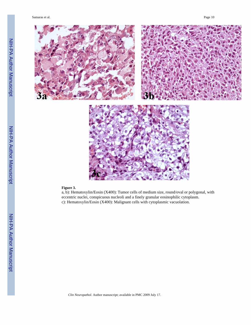
Figure 3.a, b): Hematoxylin/Eosin (X400): Tumor cells of medium size, round/oval or polygonal, witheccentric nuclei, conspicuous nucleoli and a finely granular eosinophilic cytoplasm.c): Hematoxylin/Eosin (X400): Malignant cells with cytoplasmic vacuolation.
Samaras et al. Page 10
Clin Neuropathol. Author manuscript; available in PMC 2009 July 17.
NIH
-PA Author Manuscript
NIH
-PA Author Manuscript
NIH
-PA Author Manuscript

Figure 4.a): Hematoxylin/Eosin (X400): Nuclei of neoplastic cells with characteristic vesicularchromatin.b): Hematoxylin/Eosin (X200): Spindle cell component of the tumor.c): Hematoxylin/Eosin (X400): Conspicuous mitotic activity within the neoplasm.
Samaras et al. Page 11
Clin Neuropathol. Author manuscript; available in PMC 2009 July 17.
NIH
-PA Author Manuscript
NIH
-PA Author Manuscript
NIH
-PA Author Manuscript
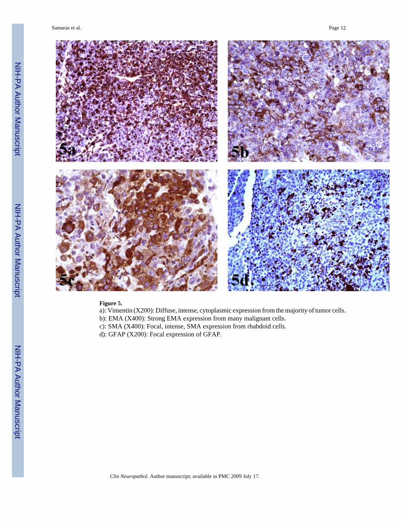
Figure 5.a): Vimentin (X200): Diffuse, intense, cytoplasmic expression from the majority of tumor cells.b): EMA (X400): Strong EMA expression from many malignant cells.c): SMA (X400): Focal, intense, SMA expression from rhabdoid cells.d): GFAP (X200): Focal expression of GFAP.
Samaras et al. Page 12
Clin Neuropathol. Author manuscript; available in PMC 2009 July 17.
NIH
-PA Author Manuscript
NIH
-PA Author Manuscript
NIH
-PA Author Manuscript
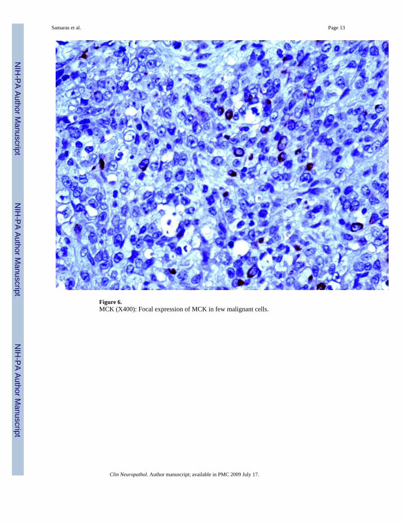
Figure 6.MCK (X400): Focal expression of MCK in few malignant cells.
Samaras et al. Page 13
Clin Neuropathol. Author manuscript; available in PMC 2009 July 17.
NIH
-PA Author Manuscript
NIH
-PA Author Manuscript
NIH
-PA Author Manuscript

Figure 7.INI1 (X200): Diffuse loss of nuclear INI1 expression in tumor cells with retained expressionin normal vascular structures and inflammatory cells within the tumor.
Samaras et al. Page 14
Clin Neuropathol. Author manuscript; available in PMC 2009 July 17.
NIH
-PA Author Manuscript
NIH
-PA Author Manuscript
NIH
-PA Author Manuscript

Figure 8.Ki67 (X400): High nuclear expression of Ki67 proliferation index.
Samaras et al. Page 15
Clin Neuropathol. Author manuscript; available in PMC 2009 July 17.
NIH
-PA Author Manuscript
NIH
-PA Author Manuscript
NIH
-PA Author Manuscript

NIH
-PA Author Manuscript
NIH
-PA Author Manuscript
NIH
-PA Author Manuscript
Samaras et al. Page 16Ta
ble
1Su
mm
ary
of c
ases
rega
rdin
g at
ypic
al te
rato
id/rh
abdo
id tu
mor
of t
he C
NS
in a
dults
.
Yea
r of
pub
licat
ion
Age
at
diag
nosi
s(y
ears
)
Gen
der
Tum
or lo
caliz
atio
nIm
mun
ohis
toch
emic
al a
naly
sis:
posi
tive
mar
kers
expr
essi
on o
r no
t of
INI1
pro
tein
Mol
ecul
ar g
enet
ic a
naly
sis
Surv
ival
Hor
n et
al.
1992
21M
ale
Left
tem
pora
lV
imen
tin, E
MA
, a1-
antic
hym
otry
psin
Not
per
form
ed72
mon
ths**
Cos
su e
t al.
1993
18M
ale
Left
Fron
tal
Vim
entin
, EM
A, K
erat
inN
ot p
erfo
rmed
18 m
onth
s**
Fish
er e
t al.
1996
32M
ale
Left
fron
tal
Vim
entin
, S10
0, G
FAP
Not
per
form
ed1
mon
th**
Ash
raf e
t al.
1997
34M
ale
Left
cere
bral
Vim
entin
Not
per
form
ed6
mon
ths**
Byr
am19
9935
*M
ale*
Left
tem
pora
lV
imen
tin, E
MA
, Ker
atin
*N
ot p
erfo
rmed
60 m
onth
s**
Sugi
ta e
t al.
1999
27M
ale
Pine
al re
gion
Chr
omog
rani
n A
, syn
apto
phys
in,
Vim
entin
, EM
A, S
-100
, NSE
, SM
A,
HH
F-35
Not
per
form
ed24
mon
ths**
Kug
e et
al.
2000
32Fe
mal
eSu
pras
ella
rV
imen
tin, E
MA
, Ker
atin
, SM
AN
ot p
erfo
rmed
No
info
rmat
ion
Arr
azol
a et
al.
2000
20M
ale
Left
parie
tal
EMA
, Ker
atin
,Vim
ent,
S- 1
00N
ot p
erfo
rmed
Aliv
e 24
mon
ths
afte
r the
firs
tcr
anio
tom
y
Lut
terb
ach
et a
l.20
0130
Fem
ale
Cer
ebel
lum
Vim
entin
, S10
0, N
FP, K
erat
in, E
MA
,G
FAP
Not
per
form
ed11
mon
ths**
Bru
ch e
t al.
2001
21Fe
mal
eSp
inal
cor
dEM
A, v
imen
tin, a
nd S
MA
Yes
(22q
del
etio
n by
FIS
H)
6 m
onth
s**
34Fe
mal
ePa
rieta
l6
mon
ths**
Pim
ente
l et a
l.20
0331
Fem
ale
Rig
ht p
arie
tal
Vim
entin
CD
68, N
FP, a
1-an
tichy
mot
ryps
in,?
a1-
antit
ryps
in, G
FAP,
S100
, HH
F35
Not
per
form
ed6
mon
ths**
Kac
hhar
a et
al.
2003
35M
ale
Thal
amus
Vim
entin
Not
per
form
edN
o in
form
atio
n
Clin Neuropathol. Author manuscript; available in PMC 2009 July 17.
NIH
-PA Author Manuscript
NIH
-PA Author Manuscript
NIH
-PA Author Manuscript
Samaras et al. Page 17Y
ear
of p
ublic
atio
nA
ge a
tdi
agno
sis
(yea
rs)
Gen
der
Tum
or lo
caliz
atio
nIm
mun
ohis
toch
emic
al a
naly
sis:
posi
tive
mar
kers
expr
essi
on o
r no
t of
INI1
pro
tein
Mol
ecul
ar g
enet
ic a
naly
sis
Surv
ival
Kaw
aguc
hi e
t al.
2004
22M
ale
Left
cere
bella
rhe
mis
pher
e an
d di
ffus
ele
ptom
enin
g ea
ldi
ssem
inat
ion
to th
e bra
inst
em an
d th
e who
le sp
inal
cord
Vim
entin
, SM
A, s
ynap
toph
ysin
, NFP
Not
per
form
edA
live
24 m
onth
saf
ter t
he d
iagn
osis
Eri
ckso
n et
al.
2005
20Fe
mal
eR
ight
occ
ipita
lG
FAP,
vim
entin
Yes
(FIS
H: n
o ab
norm
aliti
es in
the I
NI-
1 re
gion
of c
hrom
osom
e22
q11)
No
info
rmat
ion
Rai
sane
n et
al.
2005
20Fe
mal
eSe
llar m
ass
Loss
of I
NI1
exp
ress
ion
Yes
(22q
11.2
del
etio
n, IN
I1m
utat
ion)
Aliv
e 28
mon
ths
afte
r the
dia
gnos
is
31Fe
mal
eSe
llar m
ass w
ithsu
pras
ella
r ext
ensi
onLo
ss o
f IN
I1 e
xpre
ssio
nY
es (2
2q11
.2 d
elet
ion,
INI1
mut
atio
n)9
mon
ths**
45M
ale
Rig
ht c
ereb
ella
r mas
sLo
ss o
f IN
I1 e
xpre
ssio
nY
es (2
2q11
.2 d
elet
ion,
INI1
mut
atio
n)A
live
15 m
onth
saf
ter t
he d
iagn
osis
Che
n et
al.
2006
19M
ale
Post
erio
r fos
saN
o in
form
atio
nN
o in
form
atio
n56
.5 m
onth
s**
Rez
anko
et a
l.20
0627
Mal
eR
ight
fron
tal
Vim
entin
, EM
A, S
-100
Not
per
form
ed4
mon
ths**
Cha
cko
et a
l.20
0723
Mal
eR
ight
fron
tal
Vim
entin
, SM
A, E
MA
, CD
34, l
oss o
f IN
I1ex
pres
sion
Yes
(evi
denc
e of
a 2
2q d
elet
ion
by F
ISH
, and
a d
etec
tabl
e IN
I1ge
ne m
utat
ion)
1,5
mon
ths**
Zar
ovna
ya e
t al.
2007
43Fe
mal
eSp
inal
cor
dW
eakl
y po
sitiv
e fo
r EM
A, l
oss o
f IN
I1ex
pres
sion
Yes
(FIS
H: m
onos
omy
22)
30 m
onth
s**
Mak
uria
et a
l.20
0823
Mal
ePo
ster
ior l
eft t
empo
ral
lobe
Vim
entin
, Ker
atin
, SM
A, E
MA
, NFP
,Sy
napt
ophy
sin,
loss
of I
NI1
exp
ress
ion
Not
per
form
edIn
rem
issi
on 3
0m
onth
s afte
r the
diag
nosi
s
25Fe
mal
eR
ight
fron
tal
Vim
entin
, SM
A, S
ynap
toph
ysin
, CD
34,
NFP
, los
s of I
NI1
exp
ress
ion
Not
per
form
edA
live 1
7 ye
ars a
fter
the
diag
nosi
s
42M
ale
Rig
ht fr
onto
parie
tal
Vim
entin
, EM
A,K
erat
in, l
oss o
f IN
I1ex
pres
sion
Not
per
form
edA
live
18 m
onth
saf
ter t
he d
iagn
osis
37M
ale
Rig
ht fr
onto
parie
tal
Vim
entin
, IN
I1 p
ositi
vity
Not
per
form
edN
o in
form
atio
n
Clin Neuropathol. Author manuscript; available in PMC 2009 July 17.

NIH
-PA Author Manuscript
NIH
-PA Author Manuscript
NIH
-PA Author Manuscript
Samaras et al. Page 18* in
form
atio
n ba
sed
on L
utte
rbac
h et
al.
2001
, Rez
anko
et a
l. 20
06, M
akur
ia e
t al.
2008
**pa
tient
succ
umbe
d to
the
neop
lasm
Abb
revi
atio
ns: E
MA
: epi
thel
ial m
embr
ane
antig
en, G
FAP:
glia
l fib
rilla
ry a
cidi
c pr
otei
n, H
HF3
5: a
nti-h
uman
mus
cle
actin
, NFP
: neu
rofil
amen
t pro
tein
, NSE
: neu
ron-
spec
ific
enol
ase,
SM
A: α
-sm
ooth
mus
cle
actin
.
Clin Neuropathol. Author manuscript; available in PMC 2009 July 17.
Copyright © 2022 FDOKUMEN