Association of Genome-Wide Variation With the Risk of Incident Heart Failure in Adults of European...
19
The Association of Genome-Wide Variation with the Risk of Incident Heart Failure in Adults of European and African Ancestry: A Prospective Meta-Analysis from the CHARGE Consortium Nicholas L. Smith, PhD * , Janine F. Felix, MD, PhD * , Alanna C. Morrison, PhD * , Serkalem Demissie, PhD * , Nicole L. Glazer, PhD, Laura R. Loehr, MD, PhD, L. Adrienne Cupples, PhD, Abbas Dehghan, MD, DSc, Thomas Lumley, PhD, Wayne D. Rosamond, PhD, Wolfgang Lieb, MD, Fernando Rivadeneira, MD, PhD, Joshua C. Bis, PhD, Aaron R. Folsom, MD, Emelia Benjamin, MD, Yurii S. Aulchenko, PhD, Talin Haritunians, PhD, David Couper, PhD, Joanne Murabito, MD, Ying A. Wang, PhD, Bruno H. Stricker, MMed, PhD, John S. Gottdiener, MD, Patricia P. Chang, MD, MHS, Thomas J. Wang, MD, Kenneth M. Rice, PhD, Albert Hofman, MD, PhD, Susan R. Heckbert, MD, PhD, Ervin R. Fox, MD, Christopher J. O’Donnell, MD, Andre G. Uitterlinden, PhD, Jerome I. Rotter, MD, James T. Willerson, MD, Daniel Levy, MD, Cornelia M. van Duijn, PhD, Bruce M. Psaty, MD, PhD ** , Jacqueline C. M. Witteman, PhD ** , Eric Boerwinkle, PhD ** , and Ramachandran S. Vasan, MD ** Abstract Background—Although genetic factors contribute to the onset of heart failure (HF), no large- scale genome-wide investigation of HF risk has been published to date. We investigated the association of 2,478,304 single nucleotide polymorphisms (SNPs) with incident HF by meta- analyzing data from 4 community-based prospective cohorts: the Atherosclerosis Risk in Communities Study, the Cardiovascular Health Study, the Framingham Heart Study, and the Rotterdam Study. Methods and Results—Eligible participants for these analyses were of European or African ancestry and free of clinical HF at baseline. Each study independently conducted genome-wide scans and imputed data to the ~2.5 million SNPs in HapMap. Within each study, Cox proportional Corresponding author: Nicholas L. Smith, PhD, Cardiovascular Health Research Unit 1730 Minor Ave, Suite 1360, Seattle WA 98105 phone: 206.287.2784; fax: 206.287.2662, [email protected]. Denotes lead* and senior** authors for each cohort who equally contributed to the manuscript. Cardiovascular Health Study: Dept. of Epidemiology (NLS, SRH, BMP), Med (NLG, JCB, BMP), Biostatistics (TL, KMR), and Health Services (BMP) Univ of Washington, Seattle, WA; Seattle Epidemiologic Research and Information Ctr of the Dept of Veterans Affairs Office of Research and Development, Seattle, WA (NLS); Group Health Ctr for Health Studies, Group Health, Seattle, WA (SRH, BMP); Dept. of Cardio, Univ of Maryland (JSG); Med Genetics Inst, Cedars-Sinai Med Center, Los Angeles, CA (TH, JIR). Artherosclerosis Risk in Communities Study: Univ of Texas at Houston Health Science Ctr, Houston, TX (ACM, EB, JTW); Dept of Med (LRL, PPC), Biostatistics (DC), and Epidemiology (WDR) Univ of North Carolina at Chapel Hill, Chapel Hill, NC; Nat Inst of Environmental Health Sciences, Research Triangle Park, NC (LRL), Univ of Minnesota School of Public Health, Minneapolis, MN (ARF); Dept of Med, Univ of Mississippi Med Ctr (ERF) and the Nat Heart, Lung, and Blood Inst Jackson Heart Study (ERF), Texas Heart Inst (JTW). Framingham Heart Study: Dept of Biostatistics, Boston Univ School of Public Health, Boston, MA (SD, LAC, YAW); Sections of Preventive Med, and Cardio, Boston Univ School of Med, Boston, MA (RSV); Nat Heart Lung and Blood Inst, Bethesda, MD (CJO, DL); Mass Gen Hospital, Harvard Med School, Boston, MA (TJW, CJO); Framingham Heart Study, Framingham, MA (LAC, WL, EB, JM, TJW, CJO, DL, RSV). Rotterdam Study: Dept of Epidemiology (JFF, AD, YSA, BHS, AH, CMvD, JCMW) and Internal Med (FR, AGU), Erasmus MC, Rotterdam, the Netherlands; Member of the Netherlands Consortium on Healthy Aging (JFF, JCMW) Conflict of Interest Disclosures: None NIH Public Access Author Manuscript Circ Cardiovasc Genet. Author manuscript; available in PMC 2011 June 1. Published in final edited form as: Circ Cardiovasc Genet. 2010 June 1; 3(3): 256–266. doi:10.1161/CIRCGENETICS.109.895763. NIH-PA Author Manuscript NIH-PA Author Manuscript NIH-PA Author Manuscript
Transcript of Association of Genome-Wide Variation With the Risk of Incident Heart Failure in Adults of European...

The Association of Genome-Wide Variation with the Risk ofIncident Heart Failure in Adults of European and AfricanAncestry: A Prospective Meta-Analysis from the CHARGEConsortium
Nicholas L. Smith, PhD*, Janine F. Felix, MD, PhD*, Alanna C. Morrison, PhD*, SerkalemDemissie, PhD*, Nicole L. Glazer, PhD, Laura R. Loehr, MD, PhD, L. Adrienne Cupples,PhD, Abbas Dehghan, MD, DSc, Thomas Lumley, PhD, Wayne D. Rosamond, PhD,Wolfgang Lieb, MD, Fernando Rivadeneira, MD, PhD, Joshua C. Bis, PhD, Aaron R.Folsom, MD, Emelia Benjamin, MD, Yurii S. Aulchenko, PhD, Talin Haritunians, PhD, DavidCouper, PhD, Joanne Murabito, MD, Ying A. Wang, PhD, Bruno H. Stricker, MMed, PhD,John S. Gottdiener, MD, Patricia P. Chang, MD, MHS, Thomas J. Wang, MD, Kenneth M.Rice, PhD, Albert Hofman, MD, PhD, Susan R. Heckbert, MD, PhD, Ervin R. Fox, MD,Christopher J. O’Donnell, MD, Andre G. Uitterlinden, PhD, Jerome I. Rotter, MD, James T.Willerson, MD, Daniel Levy, MD, Cornelia M. van Duijn, PhD, Bruce M. Psaty, MD, PhD**,Jacqueline C. M. Witteman, PhD**, Eric Boerwinkle, PhD**, and Ramachandran S. Vasan,MD**
AbstractBackground—Although genetic factors contribute to the onset of heart failure (HF), no large-scale genome-wide investigation of HF risk has been published to date. We investigated theassociation of 2,478,304 single nucleotide polymorphisms (SNPs) with incident HF by meta-analyzing data from 4 community-based prospective cohorts: the Atherosclerosis Risk inCommunities Study, the Cardiovascular Health Study, the Framingham Heart Study, and theRotterdam Study.
Methods and Results—Eligible participants for these analyses were of European or Africanancestry and free of clinical HF at baseline. Each study independently conducted genome-widescans and imputed data to the ~2.5 million SNPs in HapMap. Within each study, Cox proportional
Corresponding author: Nicholas L. Smith, PhD, Cardiovascular Health Research Unit 1730 Minor Ave, Suite 1360, Seattle WA 98105phone: 206.287.2784; fax: 206.287.2662, [email protected] lead* and senior** authors for each cohort who equally contributed to the manuscript.Cardiovascular Health Study: Dept. of Epidemiology (NLS, SRH, BMP), Med (NLG, JCB, BMP), Biostatistics (TL, KMR), andHealth Services (BMP) Univ of Washington, Seattle, WA; Seattle Epidemiologic Research and Information Ctr of the Dept ofVeterans Affairs Office of Research and Development, Seattle, WA (NLS); Group Health Ctr for Health Studies, Group Health,Seattle, WA (SRH, BMP); Dept. of Cardio, Univ of Maryland (JSG); Med Genetics Inst, Cedars-Sinai Med Center, Los Angeles, CA(TH, JIR). Artherosclerosis Risk in Communities Study: Univ of Texas at Houston Health Science Ctr, Houston, TX (ACM, EB,JTW); Dept of Med (LRL, PPC), Biostatistics (DC), and Epidemiology (WDR) Univ of North Carolina at Chapel Hill, Chapel Hill,NC; Nat Inst of Environmental Health Sciences, Research Triangle Park, NC (LRL), Univ of Minnesota School of Public Health,Minneapolis, MN (ARF); Dept of Med, Univ of Mississippi Med Ctr (ERF) and the Nat Heart, Lung, and Blood Inst Jackson HeartStudy (ERF), Texas Heart Inst (JTW). Framingham Heart Study: Dept of Biostatistics, Boston Univ School of Public Health,Boston, MA (SD, LAC, YAW); Sections of Preventive Med, and Cardio, Boston Univ School of Med, Boston, MA (RSV); Nat HeartLung and Blood Inst, Bethesda, MD (CJO, DL); Mass Gen Hospital, Harvard Med School, Boston, MA (TJW, CJO); FraminghamHeart Study, Framingham, MA (LAC, WL, EB, JM, TJW, CJO, DL, RSV). Rotterdam Study: Dept of Epidemiology (JFF, AD,YSA, BHS, AH, CMvD, JCMW) and Internal Med (FR, AGU), Erasmus MC, Rotterdam, the Netherlands; Member of theNetherlands Consortium on Healthy Aging (JFF, JCMW)Conflict of Interest Disclosures: None
NIH Public AccessAuthor ManuscriptCirc Cardiovasc Genet. Author manuscript; available in PMC 2011 June 1.
Published in final edited form as:Circ Cardiovasc Genet. 2010 June 1; 3(3): 256–266. doi:10.1161/CIRCGENETICS.109.895763.
NIH
-PA Author Manuscript
NIH
-PA Author Manuscript
NIH
-PA Author Manuscript
hazards regression models provided age- and sex-adjusted estimates of the association betweeneach variant and time to incident HF. Fixed-effect meta-analyses combined results for each SNPfrom the 4 cohorts to produce an overall association estimate and p-value. A genome-widesignificance p-value threshold was set a priori at 5.0×10⁻⁷. During a mean follow-up of 11.5years, 2,526 incident HF events (12%) occurred in 20,926 European-ancestry participants. Themeta-analysis identified a genome-wide significant locus at chromosomal position 15q22(1.4×10⁻⁸), which was 58.8 kb from USP3. Among 2,895 African-ancestry participants, 466incident HF events (16%) occurred during a mean follow-up of 13.7 years. One genome-widesignificant locus was identified at 12q14 (6.7×10⁻⁸), which was 6.3 kb from LRIG3.
Conclusions—We identified 2 loci that were associated with incident HF and exceededgenome-wide significance. The findings merit replication in other community-based settings ofincident HF.
Keywordsepidemiology; genetics; heart failure; genome-wide variation; incidence
Heart failure (HF) is a serious cardiovascular condition that affects 1 in 5 people during theirlifetime.1 Failure arises primarily from the burden of systemic hypertension, coronaryischemia, or cardiac valvular disease.2, 3 Regardless of the clinical mechanism responsiblefor reduced cardiac output in HF, long-term strain triggers maladaptive cellular function andgrowth, possibly resulting in irreversible myocardial injury.4,5
Genetic factors contribute to HF onset, and an estimated ~18% of the risk of HF may beattributable to parental HF.6 To date, approaches to identifying genetic variants associatedwith HF risk have relied on candidate genes, primarily those of the renin-angiotensin-aldosterone system, adrenergic receptors, and sarcomeres and cytoskeletal proteins.7, 8 Nolarge-scale genome-wide investigation of HF risk has been published.9 This report presentsfindings from an investigation of ~2.5 million single nucleotide polymorphisms (SNP) andtheir associations with incident HF among 23,821 adults of European or African ancestry.
MethodsSetting
The setting for this meta-analysis is the Cohorts for Heart and Aging Research in GenomicEpidemiology (CHARGE) Consortium and includes data from 4 prospective, population-based cohorts of adults in the US and the Netherlands: the Atherosclerosis Risk inCommunities (ARIC) Study, the Cardiovascular Health Study (CHS), the FraminghamHeart Study (FHS), and the Rotterdam Study (RS).10 The designs of these studies have beendescribed elsewhere.11–16 Briefly, ARIC recruited 15,792 participants 45–64 years of agefrom 1987 to 1989 from 4 US communities. The Cardiovascular Health Study recruitedparticipants 65 years of age and older from 4 US communities in 2 waves: 5,201 participantsin 1989–1990, and an additional 687 African Americans in 1992–1993. The FraminghamHeart Study recruited 2 generations of participants 28–72 years of age in 2 time periods:5,209 Original Cohort participants were recruited in 1948 from Framingham, Massachusetts;and 5,124 Offspring Cohort participants were recruited in 1971–1975. The Rotterdam Studyrecruited 7,983 participants 55 years of age and older from 1990 to 1993 from Rotterdam,the Netherlands. The ARIC and CHS studies included African-American participants, 27%and 15%, respectively, while FHS and RS were almost exclusively participants of Europeanancestry. Each study independently conducted genome-wide scans of study participants andwithin-study analyses were conducted according to a pre-specified plan and results werecombined for this prospective meta-analysis.
Smith et al. Page 2
Circ Cardiovasc Genet. Author manuscript; available in PMC 2011 June 1.
NIH
-PA Author Manuscript
NIH
-PA Author Manuscript
NIH
-PA Author Manuscript
SubjectsEligible participants for these analyses were of European or African ancestry and free ofclinical HF. Details about cohort-specific methods for ascertaining prevalent HF at baselinehave been published elsewhere.6, 17–20 Race was self-reported. Each study received IRBapproval and all participants provided written informed consent for the use of their DNA forresearch.
MeasuresBaseline—For this analysis, baseline measures of clinical and demographic characteristicswere obtained at the time of cohort entry for ARIC, CHS, and RS, and at the time of DNAcollection for FHS. Measures were taken using standardized methods as specified by eachstudy and included in-person measures of height, weight, systolic and diastolic bloodpressure, and total cholesterol; self-reported history and treatment of hypertension anddiabetes; and self-reported history of prevalent coronary artery disease (CAD, including ahistory of myocardial infarction [MI], angina, or coronary revascularization) andcerebrovascular disease (stroke or transient ischemic attack). For CHS, FHS, and RS,prevalent cardiovascular disease was validated by medical record review.
Follow-up and Heart Failure Diagnosis—Potential incident HF events were identifiedduring follow-up by annual or semi-annual self-report, from administrative data, or fromperiodic in-study examinations. Briefly, ARIC relied on ICD-9 codes collected from hospitaldischarge summaries and death certificates.20, 21 For CHS, FHS, and RS, HF eventsidentified by self-report or administrative data were validated by physician review ofmedical records. CHS and Framingham applied their published criteria,22, 23 and RS appliedthe European Society of Cardiology criteria.18, 24 Details can be found in supplementalTable S1 and in previously published reports.
Genotyping and Imputation—Genotyping used DNA collected from phlebotomy.Genome-wide assays of SNPs were conducted independently in each cohort using varioustechnologies: Affymetrix 6.0 for ARIC, Illumina 370CNV for CHS, Affymetrix 500K and50K for FHS, and Illumina 550K v3 for RS. Genotype quality control and data cleaningwere conducted independently by each study and are summarized in Supplemental Table S2.Methods were very similar across studies and reflect those used in other published reportson genome-wide associations.25, 26
We investigated genetic variation in the 22 autosomal chromosomes. For European-ancestryparticipants, each study independently imputed their genotype data to the ~2.5 million SNPsidentified in HapMap CEU samples.27, 28 For African-ancestry participants, CHS imputedto combined CEU and YRI samples and ARIC did not impute. Supplemental Table S2provides details about the imputation methods for each cohort and the quality control criteriaapplied to the imputed genotypes. Imputation results were summarized as an “allele dosage”defined as the expected number of copies of the minor allele at that SNP (a continuous valuebetween 0 and 2) for each genotype. Each cohort calculated a ratio of observed versusexpected variance (OEV) of the dosage statistic for each SNP. This value, which generallyranges from 0 to 1 (poor to excellent), is a measure of imputation quality. High-signal SNPsthat had low OEV values in CHS (<0.8) were directly measured to improve the quality ofthe signal. No other cohort conducted additional genotyping.
Statistical AnalysesInvestigators from all cohorts jointly developed the pre-specified analytic plan. Within eachstudy, Cox proportional hazards regression models were used to test the association betweeneach SNP and time to incident HF while adjusting for sex and baseline age. Time-to-event
Smith et al. Page 3
Circ Cardiovasc Genet. Author manuscript; available in PMC 2011 June 1.
NIH
-PA Author Manuscript
NIH
-PA Author Manuscript
NIH
-PA Author Manuscript
models are standard for longitudinal study designs. CHS adjusted for study site. FHSadjusted for generation, and for ancestry using principal components. 29 Time of entry to theanalysis was the date of cohort entry (ARIC, CHS, RS) or DNA collection (FHS) and failuretime was the time of HF diagnosis. Participants without incident HF were censored at thetime of death, last date of contact, or at the end of follow-up, whichever came first. For eachSNP, additive genetic models were used to estimate the regression coefficient for the hazardratio (HR) for allele dosage, and its respective standard error. In ARIC and CHS, analyseswere conducted separately in 2 ancestry groups, European and African. For each analysis, agenomic control coefficient (λ) was calculated, that estimated the extent of underlyingpopulation structure.
Meta-analyses were performed using MetABEL software(http://mga.bionet.nsc.ru/~yurii/ABEL/).30 For the populations of European ancestry, fixed-effect meta-analyses combined regression β coefficients and λ-adjusted standard errorsacross the 4 cohorts for each SNP to produce an overall β coefficient, standard error, and p-value. We found that asymptotic assumptions of survival analysis models were not accuratewhen the number of cases carrying the variant allele was small, so we excluded SNPs forwhich the post-meta-analysis population-size-weighted minor allele frequency (MAF) wasless than 0.015.
We created a quantile-quantile (Q-Q) plot of the distribution of the observed and expected p-values associated with the successful SNPs along with a plot of all the p-values according tolocation within each of the 22 chromosomes. The a priori threshold of genome-widesignificance was set at 5.0×10⁻⁷, also used by the Wellcome Trust Case ControlConsortium.31 For 2.5 million tests, this threshold limits the expected number of genome-wide false positives to approximately 1. We also report high-signal SNPs that wereassociated with p-values of less than 1.0×10⁻⁵, the point where observations deviate fromthe expected on the Q-Q plot. When more than 1 SNP clustered at a locus, we picked theSNP with the smallest p-value as the locus marker. Forest plots that included cohort-specificfindings were created for genome-wide significant SNPs.
In sensitivity analyses, we subjected our top associations to time-to-event analyses thatexcluded participants with a baseline history of clinical MI and censored those with anincident MI during follow-up. This approach allowed us to estimate the association ofselected SNPs and HF onset among participants whose HF was less likely to be ischemic inorigin.
For participants of African ancestry, a similar analytic approach was undertaken in ARICand CHS. The ARIC cohort did not have imputed data on those of African ancestry sotesting was restricted to successfully genotyped SNPs on their Affymetrix 6.0 chip that hadan average weighted MAF of at least 0.075 in meta-analyses. Genome-wide significancewas set at 5.0×10⁻⁷. High-signal SNPs from the meta-analysis in the European- andAfrican-ancestry populations were compared.
ResultsA total of 20,926 eligible participants of European ancestry and 2,895 participants ofAfrican ancestry were free of HF at cohort entry and had genome-wide data that met cohort-specific quality control measures. Characteristics of the 23,821 cohort participants includedin these analyses are provided in Table 1. The average age ranged from 53.3 to 72.6 for the 4cohorts, and the majority of the participants were women (57%). Other demographic andclinical characteristics were similar across the 4 cohorts. A total of 2,526 incident HF events(12.1%) were identified over a weighted average of 11.5 years of follow-up among those of
Smith et al. Page 4
Circ Cardiovasc Genet. Author manuscript; available in PMC 2011 June 1.
NIH
-PA Author Manuscript
NIH
-PA Author Manuscript
NIH
-PA Author Manuscript

European ancestry and 466 events (16.1%) over a weighted average of 13.7 years amongthose of African ancestry. The average age at the time of HF onset was 73.6 years (range 44to 101) and 52% of the 2,992 events occurred among women. Among European and Africanancestry participants, 73% and 78%, respectively, of the events were diagnosed in theabsences of a prior MI.
Participants of European AncestryA total of 2,478,304 SNPs were investigated across the 4 cohorts. The λ coefficients foranalyses of European participants in each of the 4 cohorts were small (≤1.03) and suggestednegligible test statistic inflation. Supplemental Figure 1A presents the Q-Q plot of meta-analyzed p-values derived from the 2,478,304 tests performed plotted against the expecteddistribution. Overall, evidence for population admixture was small (λ = 1.02). At the lowestp-values, the observed exceeded the expected.
Figure 1A presents all 2,478,304 p-values organized by chromosome and genomic position.Among these SNPs, 5 exceeded the genome-wide significance threshold (5.0×10⁻⁷) andmarked 2 loci, 1 on chromosome 13 (13q22) and the other on chromosome 15 (15q22). (SeeSupplemental Table S3.) Top SNPs at both loci were poorly imputed in CHS so each wasgenotyped. After inclusion of the newly measured SNPs in CHS, only the 15q22 locusretained genome-wide significance. An additional 12 loci on 9 chromosomes were markedby 28 SNPs with p-values less than 1.0×10⁻⁵. (See Supplemental Table S3.) Top SNPs for 7loci were poorly imputed in CHS so these were genotyped. Among the 7 SNPs, 3 retained ap-value less than 1.0×10⁻⁵ after inclusion of newly measured SNPs. Table 2 lists the topSNP for each of the 9 loci, after genotyping in CHS.
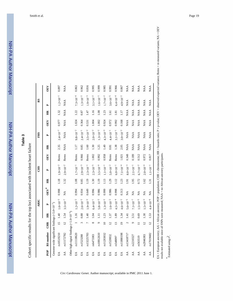
Genome-wide Significant Locus in European-Ancestry Participants—Chromosomal position 15q22 was marked by 1 SNP, rs10519210, which had a MAF of0.032 and was associated with a p-value of 1.4×10⁻⁸. The HR for this SNP was 1.53 per Gallele. This SNP is located between 2 genes: 58.8 kb from USP3 (ubiquitin specificpeptidase 3) and 63.9 kb from CA12 (carbonic anhydrase XII). Figure 2a presents detailedinformation about the LD and p-values of regional markers for this locus. This SNP wasgenotyped in 1 population (ARIC) and imputed in the 3 others. The cohort-specificestimates are provided in Table 3 and Figure 3a. Risk estimates were generally consistentacross cohorts.
There were 19,195 participants of European ancestry at risk for HF who did not have ahistory of clinical MI at baseline. Among these participants, 1,854 had an incident HF eventwithout a prior incident MI during follow-up. For locus 15q22 (rs10519210), the p-valueand the HR were diminished slightly but similar to findings for all HF events: p-value =2.8×10⁻⁶ and HR = 1.51 (see supplemental Table S4).
Loci with a P-value <1.0×10⁻⁵ in European-Ancestry Participants—Nineadditional loci on chromosomes 1, 3, 7, 8, 9, 10, 12, 13 and 19, were associated with SNPsthat had p-values less than 1.0×10⁻⁵ (Tables 2 and S3). Among the 9, 2 loci were intronic:rs11118620 (MAF = 0.292) in LOC100129376 and rs11880198 (MAF = 0.134) in GNA15(guanine nucleotide binding protein [G protein], alpha 15 [Gq class]). Four loci were closeto known genes: rs13225783 (MAF = 0.048) within 43.9 kb of EVX1 (even-skippedhomeobox 1); rs10812610 (MAF = 0.496) within 4.1 kb of MOBKL2B (MOB1, Mps OneBinder kinase activator-like 2B) and 4.1 of IFNK (interferon, kappa); rs11203032 (MAF =0.103) within 1.1 kb of CH25H (cholesterol 25-hydroxylase) and 8.7 kb of LIPA (lipase A,lysosomal acid, cholesterol esterase), and rs548097 (MAF = 0.018) within 82.5 kb ofTBC1D4 (TBC1 domain family, member 4). The remaining 3 loci were not within 100 kb of
Smith et al. Page 5
Circ Cardiovasc Genet. Author manuscript; available in PMC 2011 June 1.
NIH
-PA Author Manuscript
NIH
-PA Author Manuscript
NIH
-PA Author Manuscript
known genes and nearest genes were BCHE (butyrylcholinesterase) for rs1523288, SNX16(sorting nexin 16) for rs6473383, and PRICKLE1 (prickle homolog 1) for rs1520832.Supplemental Figures S2a-i present detailed information about the LD and p-values ofregional markers for these 9 loci. Cohort-specific details on these loci are presented in Table3, and sensitivity analyses restricted to those without a prior MI are presented inSupplemental Table S4.
Participants of African AncestryA total of 692,330 SNPs were investigated across the 2 cohorts that included participants ofAfrican ancestry. The λ coefficients were small (<1.02) and suggested negligible populationstructure. Supplemental Figure 1B presents the Q-Q plot from the 692,330 tests performedand Figure 1B presents all p-values organized by chromosome and position. No SNP had ap-value that exceeded the genome-wide significance threshold (5.0×10⁻⁷). Seven loci on 4chromosomes were marked by 10 SNPs with p-values less than 1.0×10⁻⁵. (SeeSupplemental Table S3.) Five loci were marked by poorly imputed SNPs in CHS and 2 ofthese were genotyped. Among the 2, 1 reached genome-wide significance (12q14) and 1 didnot retain a p-value less than 1.0×10⁻⁵. Table 2 lists the top SNP for the 6 loci with a p-value less than 1.0×10⁻⁵.
Genome-wide Significant Locus in African-Ancestry Participants—Chromosomal position 12q14 was marked by 1 SNP, rs1172782, which had a MAF of 0.29and was associated with a p-value of 6.7×10⁻⁸. The HR for this SNP was 1.46 per G allele.This SNP is located 6.3 kb from LRIG3 (leucine-rich repeats and immunoglobulin-likedomains 3). Figure 2b presents detailed information about the LD and p-values of regionalmarkers for this locus. The cohort-specific estimates are provided in Table 3 and Figure 3b.Risk estimates were consistent across both cohorts.
There were 2,631 participants of African ancestry at risk for HF who did not have a historyof clinical MI at baseline. Among these participants, 372 had an incident HF event without aprior incident MI during follow-up. For locus 12q14 (rs1172782), the p-value and the HRwere similar to findings for all HF events: p-value = 6.5×10⁻⁸ and HR = 1.53 (seesupplemental Table S4).
Loci with a P-value <1.0×10⁻⁵ in African-Ancestry Participants—Five loci onchromosomes 2, 9, 11 and 12 were associated with SNPs that had p-values less than1.0×10−5 (Table 2). Two loci were close to hypothesized or known genes: rs13418717(MAF = 0.202) within 4.8 kb of LOC339760 and rs563519 (MAF = 0.344) within 43.6 kbof RPUSD4 (RNA pseudouridylate synthase domain containing 4). The remaining 3 lociwere not within 100 kb of known genes and nearest genes were SH3GL2 (SH3-domainGRB2 [growth factor receptor-bound protein 2]-like 2) for rs2210327, TMTC1(transmembrane and tetratricopeptide repeat containing 1) for rs2046383, and BTG1 (B-celltranslocation gene 1, anti-proliferative) for rs17019682. Supplemental Figures S2j-n presentdetailed information about the LD and p-values of regional markers for the 5 loci. Cohort-specific details on these loci are presented in Table 3 and sensitivity analyses restricted tothose without a prior MI are presented in supplemental Table S4.
Across-Ancestry ComparisonsThe genome-wide significant SNP in European-ancestry participant, rs10519210, was notassociated with HF in those of African ancestry: 7.6×10⁻¹ (HR = 1.06; MAF = 0.179). Thegenome-wide significant SNP in African-ancestry participant, rs11172782, was uncommonand not associated with HF in those of European ancestry: 5.6×10⁻¹ (HR = 0.91; MAF =0.018).
Smith et al. Page 6
Circ Cardiovasc Genet. Author manuscript; available in PMC 2011 June 1.
NIH
-PA Author Manuscript
NIH
-PA Author Manuscript
NIH
-PA Author Manuscript

From among the 9 high-signal SNPs identified in European-ancestry participants, none wasgenotyped in ARIC and therefore none was included in African-ancestry genome-wideanalyses. When these 9 SNPs were examined in CHS participants of African ancestry, 1 ofthe 9 SNPs was associated with a p-value of less than 0.05: rs10812610 had a p-value of9.1×10−3 (HR = 1.52; MAF = 0.766 [C allele was not minor allele in these participants]).When meta-analyzed with the European-ancestry findings, the resulting combined-ancestryp-value for this locus was 7.5×10⁻⁷. From among the 5 high-signal SNPs in African-ancestry participants, none was associated with HF risk in European-ancestry participants(all p-values >5.0×10⁻²).
CommentsAmong 20,926 European-ancestry participants with 2,526 incident HF events, we identified1 locus with a SNP whose p-value (1.4×10⁻⁸) exceeded the genome-wide statisticalsignificance threshold of 5×10⁻⁷ and was associated with a 53% increase in HF risk. Among2,895 African-ancestry participants who had 466 incident events, we identified 1 locus witha SNP whose p-value (6.7×10⁻⁸) exceeded the genome-wide statistical significance and wasassociated with a 46% increase in HF risk. For both loci, risk estimates were similar forthose subjects who HF was not attributable to MI. We identified an additional 14 loci inEuropean-ancestry and African-ancestry participants that were marked by high-signal SNPs,p-value less than 1.0×10⁻⁵, but that did not reach genome-wide significance. For most loci,risk estimates were modest and did not appear to differ in a subject of participants withoutan MI preceding HF onset.
Genes Associated with Genome-wide Significant and High-Signal FindingsOur results suggest that there may be several genomic regions associated with new-onset HFin older adults, and support the hypothesis that common genetic variation, regardless of theclinical mechanism responsible for reduced cardiac output in HF, contribute to risk. The 2loci with marker SNPs that reached genome-wide significance were within 60kb of knowngenes: USP3 and CA12 in European-ancestry participants and LRIG3 in African-ancestryparticipants. The product of the USP3 gene is a ubiquitin-specific protease (USP). Ubiquitinis a highly conserved 76-amino acid protein involved in the regulation of intracellularprotein breakdown, cell cycle regulation, and stress response. It is released from degradedproteins by disassembly of the polyubiquitin chains, which is mediated by USPs.32 TheUSP3 gene may also have a role in genome stability.33 The gene product from CA12 is amembrane protein highly expressed in many tissue beds and may have a role in cancerprognosis.34 The LRIG3 gene is a member of the LRIG family of genes that is widelyexpressed and impacts tissue development and tumor progression 35–37
Among the 14 high-signal SNPs, 2 were intronic, 1 of which in a known gene, GNA15, and1 in hypothetical gene LOC100129376. The GNA15 gene, marked by rs11880198, is aguanine nucleotide binding protein of the Gq class, found in some G-protein-coupledreceptors. This family of receptors include adrenergic, endothelin, and angiotensin IIreceptors, which regulate cardiac output, arterial pressure, and blood volume.38, 39 Threeimportant amino acid substitutions (1 nonsense, 2 missense) and 1 frame shift variant havebeen identified in the GNA15 gene. None of these variants were included in the imputedSNP panel and LD with the HF SNP in our data is not known. Two additional high-signalSNPs were within 10kb of known genes, which included MOBKL2B and IFNK, and CH25Hand LIPA. None appears to have known functional qualities related to cardiac processes.40,41 Three additional high-signal SNPs were within 100kb of known genes including EVX1,TBC1D4, and RPUSD4. Several papers have been published on the protein product of theTBC1D4 gene, known as AS160, and its role in handling insulin in skeletal muscles.42–44
The remaining 6 SNPs were not within 100 kb of genes.
Smith et al. Page 7
Circ Cardiovasc Genet. Author manuscript; available in PMC 2011 June 1.
NIH
-PA Author Manuscript
NIH
-PA Author Manuscript
NIH
-PA Author Manuscript

Although we have described genes nearest to high-signal markers as potential culprits in theobserved associations it is possible that the true casual variant—if one exists—lies inneighboring genes or within non-protein coding transcripts in intergenic regions.45 In thisarticle, we have identified the genes nearest to the high-signal marker as good candidates butidentifying the true causal genes will require further scientific investigation.
The consistency of findings across the European-ancestry and African-ancestry populationswas modest at best. For the 2 genome-wide significant findings, the MAFs differed greatlybetween African-ancestry and European-ancestry participants. These high signal markers areprobably not themselves causal and are likely to be in linkage disequilibrium with the causalvariants. In view of the large differences in MAFs and the different patterns of LD betweenthe populations, it is perhaps not surprising that the associations are not consistent acrossancestral groups. Indeed, the optimal replication sample for the findings in those ofEuropean ancestry would be other similar populations of European ancestry.
Strengths and LimitationsThis is the first large-scale attempt to discover genetic risk variants for incident HF, andincluded more than 23,000 individuals and ~2.5 million markers spread throughout thegenome. Data came from 4 large, population-based cohort studies that includedcardiovascular outcomes as primary study endpoints. Over 2,500 incident HF events amongEuropean-ancestry participants were included in our analyses which allowed 80% power (2-sided α=5×10⁻⁷) to detect HRs as small as 1.6, 1.4, and 1.3 for variants with a MAF of 0.05,0.1, and 0.2, respectively. For African-ancestry participants, minimal detectable HRs werelarger: 2.6, 2.2, and 1.9, respectively. Statistical power for African-ancestry analyses waslimited. We found 1 SNP of genome-wide significance for each ancestry population, on parwith what one would expect by chance alone. Replication of associations between theseSNPs and incident HF will clarify what role—if any—these SNPs have in the genesis ofclinical HF. Each cohort relied on slightly different criteria to classify HF events and it wasnot feasible to reclassify all 2,992 events using a standard protocol. The between-studyvariation in HF classification likely introduced some heterogeneity that may havediminished our power to detect associations. Furthermore, we did not have comparable andcomplete data across cohorts on the type of failure. Previously published data from CHS andFHS indicate that half of patients with new-onset HF have cardiac systolic dysfunction at thetime of clinical onset.46, 47 Not all 2,478,304 variants tested in those of European ancestrywere directly genotyped and the imputation quality varied across SNPs. For poorly imputedSNPs there was reduced statistical power to detect an association. We excluded SNPs withlow MAF from meta-analyses since asymptotic assumptions of survival analysis modelsmay not be accurate if the number of cases with the variant allele is low. For European-ancestry participants, the MAF threshold of 0.015 translates to ~75 participants with incidentHF who are carriers of 1 or more variant alleles. This relatively large number of events isnot expected to violate asymptotic assumptions and p-values are likely accurate. Most of theacross-ancestry comparisons were limited to data from CHS, where imputation quality forAfrican-ancestry participants was generally poor because the Illumina 370CNV chip was notdesigned to optimize coverage in this ancestry.
SummaryUsing data from 4 prospective cohort studies that included 23,821 participants andprospectively identified 2,992 incident HF events in older adults, we identified 2 SNPs thatreached genome-wide significance, 1 in European-ancestry and 1 in African-ancestryparticipants. An additional 14 sub-threshold but high-signal loci were identified andincluded intronic markers in attractive candidate genes. Our findings merit replication inother community-based settings of incident HF.
Smith et al. Page 8
Circ Cardiovasc Genet. Author manuscript; available in PMC 2011 June 1.
NIH
-PA Author Manuscript
NIH
-PA Author Manuscript
NIH
-PA Author Manuscript

Genetic factors contribute to heart failure (HF) onset and, to date, most approaches toidentifying genetic variants associated with HF risk have relied on candidate genes. Nolarge-scale genome-wide investigation of HF risk has been published. We investigatedthe association of ~2.5 million single nucleotide polymorphisms (SNPs) with incident HFby meta-analyzing data from 4 community-based prospective cohorts: theAtherosclerosis Risk in Communities Study, the Cardiovascular Health Study, theFramingham Heart Study, and the Rotterdam Study. Among 20,926 European-ancestryparticipants with 2,526 incident HF events, we identified 1 locus with a SNP whose p-value (1.4×10⁻⁸) exceeded the genome-wide statistical significance threshold of 5×10⁻⁷
and was associated with a 53% increase in HF risk. Among 2,895 African-ancestryparticipants who had 466 incident events, we identified 1 locus with a SNP whose p-value (6.7×10⁻⁸) exceeded the genome-wide statistical significance and was associatedwith a 46% increase in HF risk. We identified an additional 14 loci in European-ancestryand African-ancestry participants that were marked by high-signal SNPs, p-value lessthan 1.0×10⁻⁵. For most loci, risk estimates were modest and did not appear to differ insubjects without an MI preceding HF onset. Our results suggest that there may be severalgenomic regions associated with new-onset HF in older adults, and support thehypothesis that common genetic variation, regardless of the clinical mechanismresponsible for reduced cardiac output in HF, contribute to risk. These findings meritreplication in other community-based settings of incident HF.
Supplementary MaterialRefer to Web version on PubMed Central for supplementary material.
AcknowledgmentsThe authors acknowledge the essential role of the CHARGE (Cohorts for Heart and Aging Research in GenomeEpidemiology) Consortium in development and support of this manuscript. CHARGE members include NationalHeart, Lung, and Blood Institute’s (NHLBI) Atherosclerosis Risk in Communities (ARIC) Study, NIA’s IcelandAge, Gene/Environment Susceptibility (AGES) Study, NHLBI’s Cardiovascular Health Study and FraminghamHeart Study, and the Netherland’s Rotterdam Study. The authors also acknowledge the thousands of studyparticipants who volunteered their time to help advance science and the scores of research staff and scientists whohave made this research possible. The Rotterdam Study authors thank Mila Jhamai, Pascal Arp, Dr Michael J.Moorhouse, Marijn Verkerk and Sander Bervoets for their help in creating the database and Maxim Struchalin forhis contributions to the imputations of the data.
Funding Sources: The Atherosclerosis Risk in Communities Study is carried out as a collaborative study supportedby National Heart, Lung, and Blood Institute (NHLBI) contracts N01-HC-55015, N01-HC-55016, N01-HC-55018,N01-HC-55019, N01-HC-55020, N01-HC-55021, N01-HC-55022, R01HL087641, R01HL59367 andR01HL086694; National Human Genome Research Institute contract U01HG004402; and National Institutes ofHealth (NH) contract HHSN268200625226C. Infrastructure was partly supported by Grant NumberUL1RR025005, a component of the NIH and NIH Roadmap for Medical Research. This research was supported inpart by the intramural research program of the NIH, National Institutes of Environmental Health Sciences.
The Cardiovascular Health Study is supported by contract numbers N01-HC-85079 through N01-HC-85086, N01-HC-35129, N01 HC-15103, N01 HC-55222, N01-HC-75150, N01-HC-45133, grant numbers U01 HL080295 andR01 HL 087652 from NHLBI with additional contribution from the National Institute of Neurological Disordersand Stroke. A full list of principal CHS investigators and institutions can be found athttp://www.chs-nhlbi.org/pi.htm. DNA handling and genotyping was supported in part by National Center forResearch Resources grant M01RR00425 to the Cedars-Sinai General Clinical Research Center Genotyping core,National Institute of Diabetes and Digestive and Kidney Diseases grant DK063491 to the Southern CaliforniaDiabetes Endocrinology Research Center, and Cedars-Sinai Board of Governors’ Chair in Medical Genetics (JIR).
The Framingham Heart Study was supported by NHLBI (Contract No. N01-HC-25195) and its contract withAffymetrix, Inc for genotyping services (Contract No. N02-HL-6-4278). This work was also supported in part bygrants from the NHLBI 2K24HL04334, R01HL077477, and R01HL093328 (all to RSV). A portion of this researchutilized the Linux Cluster for Genetic Analysis (LinGA-II) funded by the Robert Dawson Evans Endowment of the
Smith et al. Page 9
Circ Cardiovasc Genet. Author manuscript; available in PMC 2011 June 1.
NIH
-PA Author Manuscript
NIH
-PA Author Manuscript
NIH
-PA Author Manuscript

Department of Medicine at Boston University School of Medicine and Boston Medical Center. The analyses reflectintellectual input and resource development from the Framingham Heart Study investigators participating in theSNP Health Association Resource (SHARe) project.
The Rotterdam Study is supported by the Erasmus Medical Center and Erasmus University Rotterdam; theNetherlands Organization for Scientific Research (NWO); the Netherlands Organization for Health Research andDevelopment (ZonMw); the Research Institute for Diseases in the Elderly (RIDE); The Netherlands HeartFoundation (Nederlandse hartstichting); the Ministry of Health Welfare and Sports; the Ministry of Education,Culture and Science; the European Commission (DG XII); and the Municipality of Rotterdam. Support forgenotyping was given by NWO (175.010.2005.011, 911.03.012) and RIDE. This study was supported by theNetherlands Genomics Initiative (NGI)/NWO project number. 050-060-810 (Netherlands Consortium for HealthyAgeing).
The funding sources had no role in the study design, analyses, or drafting of the manuscript. The NHLBI reviewsall manuscripts submitted for publication but it was not involved in the decision to publish. Drs NL Smith, JF Felix,AC Morrison, and S Demissie had full access to all of the data in the study and take responsibility for the integrityof the data and the accuracy of the data analysis.
References1. Lloyd-Jones DM, Larson MG, Leip EP, Beiser A, D’Agostino RB, Kannel WB, Murabito JM,
Vasan RS, Benjamin EJ, Levy D. Lifetime risk for developing congestive heart failure: theFramingham Heart Study. Circulation 2002;106:3068–3072. [PubMed: 12473553]
2. Ho KK, Pinsky JL, Kannel WB, Levy D. The epidemiology of heart failure: the Framingham Study.J Am Coll Cardiol 1993;22:6A–13A. [PubMed: 8509564]
3. Gottdiener JS, Arnold AM, Aurigemma GP, Polak JF, Tracy RP, Kitzman DW, Gardin JM,Rutledge JE, Boineau RC. Predictors of congestive heart failure in the elderly: the CardiovascularHealth Study. J Am Coll Cardiol 2000;35:1628–1637. [PubMed: 10807470]
4. Mann DL, Bristow MR. Mechanisms and models in heart failure: the biomechanical model andbeyond. Circulation 2005;111:2837–2849. [PubMed: 15927992]
5. Mudd JO, Kass DA. Tackling heart failure in the twenty-first century. Nature 2008;451:919–928.[PubMed: 18288181]
6. Lee DS, Pencina MJ, Benjamin EJ, Wang TJ, Levy D, O’Donnell CJ, Nam BH, Larson MG,D’Agostino RB, Vasan RS. Association of parental heart failure with risk of heart failure inoffspring. N Engl J Med 2006;355:138–147. [PubMed: 16837677]
7. Kitsios G, Zintzaras E. Genetic variation associated with ischemic heart failure: a HuGE review andmeta-analysis. Am J Epidemiol 2007;166:619–633. [PubMed: 17644825]
8. Karkkainen S, Peuhkurinen K. Genetics of dilated cardiomyopathy. Ann Med 2007;39:91–107.[PubMed: 17453673]
9. Larson MG, Atwood LD, Benjamin EJ, Cupples LA, D’Agostino RB Sr, Fox CS, Govindaraju DR,Guo CY, Heard-Costa NL, Hwang SJ, Murabito JM, Newton-Cheh C, O’Donnell CJ, Seshadri S,Vasan RS, Wang TJ, Wolf PA, Levy D. Framingham Heart Study 100K project: genome-wideassociations for cardiovascular disease outcomes. BMC Med Genet 2007;8 (Suppl 1):S5. [PubMed:17903304]
10. Psaty BM, O’Donnell CJ, Gudnason V, Lunetta KL, Folsom AR, Rotter JI, Uitterlinden AG,Harris TB, Witteman JC, Boerwinkle E. Cohorts for Heart and Aging Research in GenomicEpidemiology: CHARGE Consortium. Design of prospective meta-analysis of genome-wideassociation studies from 5 cohorts. Circ Cardiovasc Genet 2009;2:73–80. [PubMed: 20031568]
11. The ARIC investigators. The Atherosclerosis Risk in Communities (ARIC) Study: design andobjectives. Am J Epidemiol 1989;129:687–702. [PubMed: 2646917]
12. Fried LP, Borhani NO, Enright P, Furberg CD, Gardin JM, Kronmal RA, Kuller LH, Manolio TA,Mittelmark MB, Newman A, O’Leary DH, Psaty BM, Rautaharju P, Tracy RP, Weiler PG. TheCardiovascular Health Study: design and rationale. Ann Epidemiol 1991;1:263–276. [PubMed:1669507]
13. Dawber TR, Meadors GF, Moore FE Jr. Epidemiological approaches to heart disease: theFramingham Study. Am J Public Health Nations Health 1951;41:279–281. [PubMed: 14819398]
Smith et al. Page 10
Circ Cardiovasc Genet. Author manuscript; available in PMC 2011 June 1.
NIH
-PA Author Manuscript
NIH
-PA Author Manuscript
NIH
-PA Author Manuscript
14. Feinleib M, Kannel WB, Garrison RJ, McNamara PM, Castelli WP. The Framingham OffspringStudy. Design and preliminary data. Prev Med 1975;4:518–525. [PubMed: 1208363]
15. Hofman A, Breteler MM, van Duijn CM, Janssen HL, Krestin GP, Kuipers EJ, Stricker BH,Tiemeier H, Uitterlinden AG, Vingerling JR, Witteman JC. The Rotterdam Study: 2010 objectivesand design update. Eur J Epidemiol 2009;24:553–572. [PubMed: 19728115]
16. Hofman A, Grobbee DE, de Jong PT, van den Ouweland FA. Determinants of disease anddisability in the elderly: the Rotterdam Elderly Study. Eur J Epidemiol 1991;7:403–422. [PubMed:1833235]
17. Psaty BM, Kuller LH, Bild D, Burke GL, Kittner SJ, Mittelmark M, Price TR, Rautaharju PM,Robbins J. Methods of assessing prevalent cardiovascular disease in the Cardiovascular HealthStudy. Ann Epidemiol 1995;5:270–277. [PubMed: 8520708]
18. Bleumink GS, Knetsch AM, Sturkenboom MC, Straus SM, Hofman A, Deckers JW, Witteman JC,Stricker BH. Quantifying the heart failure epidemic: prevalence, incidence rate, lifetime risk andprognosis of heart failure The Rotterdam Study. Eur Heart J 2004;25:1614–1619. [PubMed:15351160]
19. Mosterd A, Hoes AW, de Bruyne MC, Deckers JW, Linker DT, Hofman A, Grobbee DE.Prevalence of heart failure and left ventricular dysfunction in the general population; TheRotterdam Study. Eur Heart J 1999;20:447–455. [PubMed: 10213348]
20. Loehr LR, Rosamond WD, Chang PP, Folsom AR, Chambless LE. Heart failure incidence andsurvival (from the Atherosclerosis Risk in Communities study). Am J Cardiol 2008;101:1016–1022. [PubMed: 18359324]
21. White AD, Folsom AR, Chambless LE, Sharret AR, Yang K, Conwill D, Higgins M, WilliamsOD, Tyroler HA. Community surveillance of coronary heart disease in the Atherosclerosis Risk inCommunities (ARIC) Study: methods and initial two years’ experience. J Clin Epidemiol1996;49:223–233. [PubMed: 8606324]
22. Ives DG, Fitzpatrick AL, Bild DE, Psaty BM, Kuller LH, Crowley PM, Cruise RG, Theroux S.Surveillance and ascertainment of cardiovascular events. The Cardiovascular Health Study. AnnEpidemiol 1995;5:278–285. [PubMed: 8520709]
23. McKee PA, Castelli WP, McNamara PM, Kannel WB. The natural history of congestive heartfailure: the Framingham study. N Engl J Med 1971;285:1441–1446. [PubMed: 5122894]
24. Remme WJ, Swedberg K. Guidelines for the diagnosis and treatment of chronic heart failure. EurHeart J 2001;22:1527–1560. [PubMed: 11492984]
25. Levy D, Ehret GB, Rice K, Verwoert GC, Launer LJ, Dehghan A, Glazer NL, Morrison AC,Johnson AD, Aspelund T, Aulchenko Y, Lumley T, Kottgen A, Vasan RS, Rivadeneira F,Eiriksdottir G, Guo X, Arking DE, Mitchell GF, Mattace-Raso FU, Smith AV, Taylor K, ScharpfRB, Hwang SJ, Sijbrands EJ, Bis J, Harris TB, Ganesh SK, O’Donnell CJ, Hofman A, Rotter JI,Coresh J, Benjamin EJ, Uitterlinden AG, Heiss G, Fox CS, Witteman JC, Boerwinkle E, Wang TJ,Gudnason V, Larson MG, Chakravarti A, Psaty BM, van Duijn CM. Genome-wide associationstudy of blood pressure and hypertension. Nat Genet. 2009
26. Ganesh SK, Zakai NA, van Rooij FJ, Soranzo N, Smith AV, Nalls MA, Chen MH, Kottgen A,Glazer NL, Dehghan A, Kuhnel B, Aspelund T, Yang Q, Tanaka T, Jaffe A, Bis JC, Verwoert GC,Teumer A, Fox CS, Guralnik JM, Ehret GB, Rice K, Felix JF, Rendon A, Eiriksdottir G, Levy D,Patel KV, Boerwinkle E, Rotter JI, Hofman A, Sambrook JG, Hernandez DG, Zheng G, BandinelliS, Singleton AB, Coresh J, Lumley T, Uitterlinden AG, Vangils JM, Launer LJ, Cupples LA,Oostra BA, Zwaginga JJ, Ouwehand WH, Thein SL, Meisinger C, Deloukas P, Nauck M, SpectorTD, Gieger C, Gudnason V, van Duijn CM, Psaty BM, Ferrucci L, Chakravarti A, Greinacher A,O’Donnell CJ, Witteman JC, Furth S, Cushman M, Harris TB, Lin JP. Multiple loci influenceerythrocyte phenotypes in the CHARGE Consortium. Nat Genet 2009;41:1191–1198. [PubMed:19862010]
27. Servin B, Stephens M. Imputation-based analysis of association studies: candidate regions andquantitative traits. PLoS Genet 2007;3:e114. [PubMed: 17676998]
28. Li Y, Abecasis GR. Mach 1.0: rapid haplotype reconstruction and missing genotype inference. AmJ Hum Genet 2006;S79:2290.
Smith et al. Page 11
Circ Cardiovasc Genet. Author manuscript; available in PMC 2011 June 1.
NIH
-PA Author Manuscript
NIH
-PA Author Manuscript
NIH
-PA Author Manuscript
29. Price AL, Patterson NJ, Plenge RM, Weinblatt ME, Shadick NA, Reich D. Principal componentsanalysis corrects for stratification in genome-wide association studies. Nat Genet 2006;38:904–909. [PubMed: 16862161]
30. Aulchenko YS, Ripke S, Isaacs A, van Duijn CM. GenABEL: an R library for genome-wideassociation analysis. Bioinformatics 2007;23:1294–1296. [PubMed: 17384015]
31. Genome-wide association study of 14, 000 cases of seven common diseases and 3, 000 sharedcontrols. Nature 2007;447:661–678. [PubMed: 17554300]
32. Sloper-Mould KE, Eyre HJ, Wang XW, Sutherland GR, Baker RT. Characterization andchromosomal localization of USP3, a novel human ubiquitin-specific protease. J Biol Chem1999;274:26878–26884. [PubMed: 10480896]
33. Nicassio F, Corrado N, Vissers JH, Areces LB, Bergink S, Marteijn JA, Geverts B, HoutsmullerAB, Vermeulen W, Di Fiore PP, Citterio E. Human USP3 is a chromatin modifier required for Sphase progression and genome stability. Curr Biol 2007;17:1972–1977. [PubMed: 17980597]
34. Pastorekova S, Zatovicova M, Pastorek J. Cancer-associated carbonic anhydrases and theirinhibition. Curr Pharm Des 2008;14:685–698. [PubMed: 18336315]
35. Guo D, Holmlund C, Henriksson R, Hedman H. The LRIG gene family has three vertebrateparalogs widely expressed in human and mouse tissues and a homolog in Ascidiacea. Genomics2004;84:157–165. [PubMed: 15203213]
36. Abraira VE, Del Rio T, Tucker AF, Slonimsky J, Keirnes HL, Goodrich LV. Cross-repressiveinteractions between Lrig3 and netrin 1 shape the architecture of the inner ear. Development2008;135:4091–4099. [PubMed: 19004851]
37. Yi W, Haapasalo H, Holmlund C, Jarvela S, Raheem O, Bergenheim AT, Hedman H, HenrikssonR. Expression of leucine-rich repeats and immunoglobulin-like domains (LRIG) proteins in humanependymoma relates to tumor location, WHO grade, and patient age. Clin Neuropathol2009;28:21–27. [PubMed: 19216216]
38. Rockman HA, Koch WJ, Lefkowitz RJ. Seven-transmembrane-spanning receptors and heartfunction. Nature 2002;415:206–212. [PubMed: 11805844]
39. Kilts JD, Grocott HP, Kwatra MM. G alpha(q)-coupled receptors in human atrium functionthrough protein kinase C epsilon and delta. J Mol Cell Cardiol 2005;38:267–276. [PubMed:15698833]
40. Bea S, Salaverria I, Armengol L, Pinyol M, Fernandez V, Hartmann EM, Jares P, Amador V,Hernandez L, Navarro A, Ott G, Rosenwald A, Estivill X, Campo E. Uniparental disomies,homozygous deletions, amplifications and target genes in mantle cell lymphoma revealed byintegrative high-resolution whole genome profiling. Blood 2008;113:3059–69. [PubMed:18984860]
41. Nardelli B, Zaritskaya L, Semenuk M, Cho YH, LaFleur DW, Shah D, Ullrich S, Girolomoni G,Albanesi C, Moore PA. Regulatory effect of IFN-kappa, a novel type I IFN, on cytokineproduction by cells of the innate immune system. J Immunol 2002;169:4822–4830. [PubMed:12391192]
42. Miinea CP, Sano H, Kane S, Sano E, Fukuda M, Peranen J, Lane WS, Lienhard GE. AS160, theAkt substrate regulating GLUT4 translocation, has a functional Rab GTPase-activating proteindomain. Biochem J 2005;391:87–93. [PubMed: 15971998]
43. Kramer HF, Taylor EB, Witczak CA, Fujii N, Hirshman MF, Goodyear LJ. Calmodulin-bindingdomain of AS160 regulates contraction- but not insulin-stimulated glucose uptake in skeletalmuscle. Diabetes 2007;56:2854–2862. [PubMed: 17717281]
44. Witteles RM, Fowler MB. Insulin-resistant cardiomyopathy clinical evidence, mechanisms, andtreatment options. J Am Coll Cardiol 2008;51:93–102. [PubMed: 18191731]
45. Birney E, Stamatoyannopoulos JA, Dutta A, Guigo R, Gingeras TR, Margulies EH, Weng Z,Snyder M, Dermitzakis ET, Thurman RE, Kuehn MS, Taylor CM, Neph S, Koch CM, Asthana S,Malhotra A, Adzhubei I, Greenbaum JA, Andrews RM, Flicek P, Boyle PJ, Cao H, Carter NP,Clelland GK, Davis S, Day N, Dhami P, Dillon SC, Dorschner MO, Fiegler H, Giresi PG, Goldy J,Hawrylycz M, Haydock A, Humbert R, James KD, Johnson BE, Johnson EM, Frum TT,Rosenzweig ER, Karnani N, Lee K, Lefebvre GC, Navas PA, Neri F, Parker SC, Sabo PJ,Sandstrom R, Shafer A, Vetrie D, Weaver M, Wilcox S, Yu M, Collins FS, Dekker J, Lieb JD,
Smith et al. Page 12
Circ Cardiovasc Genet. Author manuscript; available in PMC 2011 June 1.
NIH
-PA Author Manuscript
NIH
-PA Author Manuscript
NIH
-PA Author Manuscript
Tullius TD, Crawford GE, Sunyaev S, Noble WS, Dunham I, Denoeud F, Reymond A, KapranovP, Rozowsky J, Zheng D, Castelo R, Frankish A, Harrow J, Ghosh S, Sandelin A, Hofacker IL,Baertsch R, Keefe D, Dike S, Cheng J, Hirsch HA, Sekinger EA, Lagarde J, Abril JF, Shahab A,Flamm C, Fried C, Hackermuller J, Hertel J, Lindemeyer M, Missal K, Tanzer A, Washietl S,Korbel J, Emanuelsson O, Pedersen JS, Holroyd N, Taylor R, Swarbreck D, Matthews N, DicksonMC, Thomas DJ, Weirauch MT, Gilbert J, Drenkow J, Bell I, Zhao X, Srinivasan KG, Sung WK,Ooi HS, Chiu KP, Foissac S, Alioto T, Brent M, Pachter L, Tress ML, Valencia A, Choo SW,Choo CY, Ucla C, Manzano C, Wyss C, Cheung E, Clark TG, Brown JB, Ganesh M, Patel S,Tammana H, Chrast J, Henrichsen CN, Kai C, Kawai J, Nagalakshmi U, Wu J, Lian Z, Lian J,Newburger P, Zhang X, Bickel P, Mattick JS, Carninci P, Hayashizaki Y, Weissman S, HubbardT, Myers RM, Rogers J, Stadler PF, Lowe TM, Wei CL, Ruan Y, Struhl K, Gerstein M,Antonarakis SE, Fu Y, Green ED, Karaoz U, Siepel A, Taylor J, Liefer LA, Wetterstrand KA,Good PJ, Feingold EA, Guyer MS, Cooper GM, Asimenos G, Dewey CN, Hou M, Nikolaev S,Montoya-Burgos JI, Loytynoja A, Whelan S, Pardi F, Massingham T, Huang H, Zhang NR,Holmes I, Mullikin JC, Ureta-Vidal A, Paten B, Seringhaus M, Church D, Rosenbloom K, KentWJ, Stone EA, Batzoglou S, Goldman N, Hardison RC, Haussler D, Miller W, Sidow A, TrinkleinND, Zhang ZD, Barrera L, Stuart R, King DC, Ameur A, Enroth S, Bieda MC, Kim J, Bhinge AA,Jiang N, Liu J, Yao F, Vega VB, Lee CW, Ng P, Shahab A, Yang A, Moqtaderi Z, Zhu Z, Xu X,Squazzo S, Oberley MJ, Inman D, Singer MA, Richmond TA, Munn KJ, Rada-Iglesias A,Wallerman O, Komorowski J, Fowler JC, Couttet P, Bruce AW, Dovey OM, Ellis PD, LangfordCF, Nix DA, Euskirchen G, Hartman S, Urban AE, Kraus P, Van Calcar S, Heintzman N, KimTH, Wang K, Qu C, Hon G, Luna R, Glass CK, Rosenfeld MG, Aldred SF, Cooper SJ, Halees A,Lin JM, Shulha HP, Zhang X, Xu M, Haidar JN, Yu Y, Ruan Y, Iyer VR, Green RD, Wadelius C,Farnham PJ, Ren B, Harte RA, Hinrichs AS, Trumbower H, Clawson H, Hillman-Jackson J,Zweig AS, Smith K, Thakkapallayil A, Barber G, Kuhn RM, Karolchik D, Armengol L, Bird CP,de Bakker PI, Kern AD, Lopez-Bigas N, Martin JD, Stranger BE, Woodroffe A, Davydov E,Dimas A, Eyras E, Hallgrimsdottir IB, Huppert J, Zody MC, Abecasis GR, Estivill X, BouffardGG, Guan X, Hansen NF, Idol JR, Maduro VV, Maskeri B, McDowell JC, Park M, Thomas PJ,Young AC, Blakesley RW, Muzny DM, Sodergren E, Wheeler DA, Worley KC, Jiang H,Weinstock GM, Gibbs RA, Graves T, Fulton R, Mardis ER, Wilson RK, Clamp M, Cuff J, GnerreS, Jaffe DB, Chang JL, Lindblad-Toh K, Lander ES, Koriabine M, Nefedov M, Osoegawa K,Yoshinaga Y, Zhu B, de Jong PJ. Identification and analysis of functional elements in 1% of thehuman genome by the ENCODE pilot project. Nature 2007;447:799–816. [PubMed: 17571346]
46. Smith NL, Chan JD, Rea TD, Wiggins KL, Gottdiener JS, Lumley T, Psaty BM. Time trends in theuse of beta-blockers and other pharmacotherapies in older adults with congestive heart failure. AmHeart J 2004;148:710–717. [PubMed: 15459605]
47. Vasan RS, Larson MG, Benjamin EJ, Evans JC, Reiss CK, Levy D. Congestive heart failure insubjects with normal versus reduced left ventricular ejection fraction: prevalence and mortality ina population- based cohort. J Am Coll Cardiol 1999;33:1948–1955. [PubMed: 10362198]
Smith et al. Page 13
Circ Cardiovasc Genet. Author manuscript; available in PMC 2011 June 1.
NIH
-PA Author Manuscript
NIH
-PA Author Manuscript
NIH
-PA Author Manuscript
Figure 1.Figures 1a–b. The log, base-10, p-value of the additive genetic model for each singlenucleotide polymorphisms with a minor allele prevalence of greater than or equal to 1.5% %for European-ancestry participants (1a) and 7.5% for African-ancestry participants (1b)according to location in the 22 autosomal chromosomes. Horizontal line indicates the5×10−7 threshold of genome-wide significance.
Smith et al. Page 14
Circ Cardiovasc Genet. Author manuscript; available in PMC 2011 June 1.
NIH
-PA Author Manuscript
NIH
-PA Author Manuscript
NIH
-PA Author Manuscript
Figure 2.Figures 2a–b. Regional plots for genome-wide significant marker loci: 2a. rs10519210 inEuropean-ancestry participants; 2b. rs11172782 in African-ancestry participants. The SNPwith the smallest p-value is presented in black font and neighboring variants are presented indifferent black saturations based on linkage disequilibrium: dark gray: r2 ≥ 0.5 and <0.8;light gray: r2 ≥ 0.2 and < 0.5; and white: r2 < 0.2. There were no SNPs in the region with alinkage disequilibrium r2 ≥ 0.8 with the top hit.
Smith et al. Page 15
Circ Cardiovasc Genet. Author manuscript; available in PMC 2011 June 1.
NIH
-PA Author Manuscript
NIH
-PA Author Manuscript
NIH
-PA Author Manuscript

Figure 3.Figures 3a–b. Meta-analysis and study-specific β-coefficients and confidence intervals(α=0.0000005) for genome-wide significant findings for genome-wide significant singlenucleotide polymorphisms: 3a. rs10519210 in European-ancestry participants; 3b.rs11172782 in African-ancestry participants.
Smith et al. Page 16
Circ Cardiovasc Genet. Author manuscript; available in PMC 2011 June 1.
NIH
-PA Author Manuscript
NIH
-PA Author Manuscript
NIH
-PA Author Manuscript

NIH
-PA Author Manuscript
NIH
-PA Author Manuscript
NIH
-PA Author Manuscript
Smith et al. Page 17
Tabl
e 1
Bas
elin
e ch
arac
teris
tics o
f par
ticip
ants
incl
uded
in in
cide
nt h
eart
failu
re a
naly
ses s
tratif
ied
by c
ohor
t and
race
Cha
ract
eris
tics
AR
ICC
HS
FHS
RS
EA
AA
EA
AA
Parti
cipa
nts,
n7,
644
2,32
13,
295
574
4,26
85,
719
Mea
n ag
e, y
rs54
.253
.372
.372
.664
.669
.1
Age
rang
e, y
rs45
to 6
445
to 6
465
to 9
865
to 9
230
to 1
0055
to 9
9
Fem
ale,
%52
.661
.760
.963
.955
.359
.1
BM
I, kg
/m2
26.9
29.5
26.3
28.5
27.7
26.3
Syst
olic
blo
od p
ress
ure,
mm
Hg
118.
312
8.7
135.
214
2.5
130.
713
9.3
Dia
stol
ic b
lood
pre
ssur
e, m
mH
g71
.780
.070
.476
.974
.173
.8
Tota
l cho
lest
erol
, mg/
dL21
4.8
215.
921
2.9
209.
420
2.4
255.
2
Trea
ted
hype
rtens
ion,
%25
.354
.430
.251
.735
.623
.2
Trea
ted
diab
etes
, %8.
0*19
.0*
5.4
11.2
6.5
3.8
Prev
alen
t CA
D, %
8.8
6.9
00
12.1
13.8
M
yoca
rdia
l inf
arct
ion,
%3.
63.
10
04.
910
.3
A
ngin
a, %
4.4
3.4
00
8.6
3.2
C
oron
ary
reva
scul
ariz
atio
n, %
3.1
1.8
00
4.9
2.5
Prev
alen
t CB
D, %
2.1
2.9
00
5.0
4.1
Cur
rent
smok
er, %
24.9
29.9
11.3
16.6
14.2
22.8
Mea
n fo
llow
-up,
yrs
15.3
14.6
11.8
10.1
5.5
10.8
Inci
dent
hea
rt fa
ilure
, n (%
)69
1 (9
.0)
331
(14.
3)83
8 (2
5.4)
135
(23.
5)24
9 (5
.8)
748
(13.
1)
EA =
Eur
opea
n an
cest
ry; A
A =
Afr
ican
anc
estry
; BM
I = b
ody
mas
s ind
ex; C
AD
= c
oron
ary
arte
ry d
isea
se (m
yoca
rdia
l inf
arct
ion,
ang
ina,
reva
scul
ariz
atio
n); C
BD
= c
ereb
rova
scul
ar d
isea
se (s
troke
, tra
nsie
ntis
chem
ic a
ttack
, and
/or c
arot
id e
ndar
tere
ctom
y); N
A =
not
ava
ilabl
e.
Not
e: c
lass
ifica
tion
of c
omor
bidi
ties v
arie
s by
coho
rt an
d m
ay n
ot b
e di
rect
ly c
ompa
rabl
e.
* Incl
udes
dia
betic
s with
out t
reat
men
t.
Circ Cardiovasc Genet. Author manuscript; available in PMC 2011 June 1.

NIH
-PA Author Manuscript
NIH
-PA Author Manuscript
NIH
-PA Author Manuscript
Smith et al. Page 18
Tabl
e 2
Des
crip
tion
and
asso
ciat
ion
of si
ngle
-nuc
leot
ide-
poly
mor
phis
m m
arke
rs o
f the
top
loci
ass
ocia
ted
with
inci
dent
hea
rt fa
ilure
POP
RS
num
ber
CH
RR
egio
nPo
sitio
nV
aria
ntM
AF
P-va
lue
HR
(CI)
*C
lose
st G
ene
Gen
ome-
wid
e si
gnifi
cant
find
ings
(<5.
0×10
−7 )
EArs
1051
9210
1515
q22
6152
4978
T→G
0.03
21.
4×10
−8
1.53
(1.0
5–2.
24)
58.8
kb
from
USP
3
AA
rs11
1727
8212
12q1
457
5458
95A→
G0.
290
6.7×
10−
81.
46 (1
.03–
2.09
)6.
3 kb
from
LR
IG3
Oth
er h
igh-
sign
al fi
ndin
gs (<
1.0×
10−
5 )
EArs
1111
8620
11q
4121
9095
131
T→C
0.29
27.
3×10
−6
1.15
(0.9
8–1.
35)
0.0
kb fr
om L
OC
1001
2937
6 (in
tron
)
EArs
1523
288
33q
26.1
1667
6290
3T→
C0.
354
6.1×
10−
60.
87 (0
.74–
1.02
)21
0.5
kb fr
om B
CH
E
EArs
1322
5783
77p
1527
2965
81C→
T0.
048
6.8×
10−
61.
38 (0
.96–
1.99
)43
.9 k
b fr
om E
VX
1
EArs
6473
383
88q
21.1
8383
1675
G→
A0.
145
3.1×
10−
61.
19 (0
.99–
1.44
)91
8.3
kb fr
om S
NX
16
EArs
1081
2610
99p
2127
5239
84A→
C0.
496
5.1×
10−
61.
14 (0
.99–
1.31
)4.
1 kb
from
MO
BK
L2B
EArs
1120
3032
1010
q23
9095
4594
T→G
0.10
38.
3×10
−6
1.22
(0.9
7–1.
53)
1.1
kb fr
om C
H25
H
EArs
1520
832
1212
q12
4153
9682
G→
T0.
036
1.2×
10−
61.
39 (0
.99–
1.95
)26
9.9
kb fr
om P
RIC
KLE
1
EArs
5480
9713
13q2
274
6742
69T→
G0.
018
6.4×
10−
71.
62 (1
.00–
2.63
)82
.5 k
b fr
om T
BC
1D4
EArs
1188
0198
1919
p13.
331
1076
9A→
G0.
134
5.6×
10−
61.
23 (0
.98–
1.54
)0.
0 kb
from
GN
A15
(int
ron)
AA
rs13
4187
172
2q20
1273
7936
7T→
A0.
202
3.4×
10−
61.
46 (0
.97–
2.20
)4.
8 kb
from
LO
C33
9760
AA
rs22
1032
79
9p22
1809
9235
T→A
0.18
56.
8×10
−7
3.14
(0.9
9–10
.0)
282.
0 kb
from
SH
3GL2
AA
rs56
3519
1111
q24
1255
3392
7C→
T0.
344
3.3×
10−
60.
69 (0
.46–
1.03
)43
.6 k
b fr
om R
PUSD
4
AA
rs20
4638
312
12p1
1.2
2999
5409
T→G
0.30
23.
3×10
−6
1.39
(0.9
7–1.
97)
166.
5 kb
from
TM
TC1
AA
rs17
0196
8212
12q2
290
8294
01A→
T0.
165
2.0×
10−
61.
47 (0
.98–
2.22
)23
1.6
kb fr
om B
TG1
POP
= po
pula
tion;
CH
R =
chr
omos
ome;
MA
F =
min
or a
llele
freq
uenc
y; H
R =
haz
ard
ratio
from
add
itive
gen
etic
mod
el; C
I = c
onfid
ence
inte
rval
; EA
= E
urop
ean
ance
stry
; AA
= A
fric
an a
nces
try
* Rep
rese
nts c
onfid
ence
inte
rval
s bas
ed o
n a
2-si
ded α
= 0.
0000
005.
Circ Cardiovasc Genet. Author manuscript; available in PMC 2011 June 1.
NIH
-PA Author Manuscript
NIH
-PA Author Manuscript
NIH
-PA Author Manuscript
Smith et al. Page 19
Tabl
e 3
Coh
ort s
peci
fic re
sults
for t
he to
p lo
ci a
ssoc
iate
d w
ith in
cide
nt h
eart
failu
re
POP
RS
num
ber
CH
R
AR
ICC
HS
FHS
RS
HR
PO
EV
*H
RP
OE
VH
RP
OE
VH
RP
OE
V
Gen
ome-
wid
e si
gnifi
cant
find
ings
(<5.
0×10
−7 )
EArs
1051
9210
151.
401.
6×10
−2
0.99
81.
181.
9×10
−2
Rem
x2.
352.
4×10
−7
0.97
71.
321.
2×10
−1
0.89
7
AA
rs11
1727
8212
1.54
3.5×
10−
7N
A1.
322.
9×10
−2
Rem
xN
AA
NA
AN
AA
NA
AN
AA
NA
A
Oth
er h
igh-
sign
al fi
ndin
gs (<
1.0×
10−
5 )
EArs
1111
8620
11.
161.
2×10
−2
0.99
41.
081.
7×10
−1
Rem
x1.
179.
8×10
−2
1.02
41.
227.
3×10
−4
0.86
3
EArs
1523
288
30.
882.
6×10
−2
0.93
40.
852.
9×10
−3
0.96
60.
852.
4×10
−1
0.36
40.
871.
3×10
−2
0.96
2
EArs
1322
5783
71.
401.
9×10
−2
0.64
91.
192.
3×10
−1
Rem
x1.
603.
5×10
−2
0.53
11.
471.
9×10
−3
0.81
6
EArs
6473
383
81.
046.
4×10
−1
0.99
61.
302.
3×10
−5
1.00
21.
302.
0×10
−2
1.00
41.
163.
1×10
−2
0.99
5
EArs
1081
2610
91.
115.
8×10
−2
0.98
61.
193.
5×10
−4
0.99
21.
251.
3×10
−2
1.00
21.
081.
6×10
−1
0.99
9
EArs
1120
3032
101.
231.
3×10
−2
0.99
91.
131.
1×10
−1
0.95
81.
494.
3×10
−3
0.92
41.
231.
7×10
−2
0.98
5
EArs
1520
832
121.
371.
6×10
−2
0.98
61.
295.
0×10
−2
Rem
x0.
814.
4×10
−1
0.97
21.
613.
6×10
−5
0.99
1
EArs
5480
9713
1.89
4.5×
10−
40.
998
1.12
6.0×
10−
1R
emx
1.38
3.1×
10−
10.
992
1.81
6.4×
10−
40.
966
EArs
1188
0198
191.
344.
6×10
−2
0.31
31.
237.
1×10
−4
1.02
12.
053.
0×10
−2
0.16
81.
174.
9×10
−2
0.86
7
AA
rs13
4187
172
1.41
5.6×
10−
5N
A1.
918.
0×10
−3
0.34
8N
AA
NA
AN
AA
NA
AN
AA
NA
A
AA
rs22
1032
79
3.51
7.1×
10−
7N
A1.
952.
1×10
−1
0.18
9N
AA
NA
AN
AA
NA
AN
AA
NA
A
AA
rs56
3519
110.
691.
5×10
−5
NA
0.72
6.7×
10−
20.
512
NA
AN
AA
NA
AN
AA
NA
AN
AA
AA
rs20
4638
312
1.30
1.5×
10−
3N
A1.
622.
0×10
−4
0.97
2N
AA
NA
AN
AA
NA
AN
AA
NA
A
AA
rs17
0196
8212
1.53
4.4×
10−
6N
A1.
311.
1×10
−1
0.81
7N
AA
NA
AN
AA
NA
AN
AA
NA
A
EA =
Eur
opea
n an
cest
ry; A
A =
Afr
ican
anc
estry
; PO
P =
popu
latio
n; C
HR
= c
hrom
osom
e; H
R =
haz
ards
ratio
; P =
p-v
alue
; OEV
= o
bser
ved-
expe
cted
var
ianc
e; R
emx
= re
-mea
sure
d va
riant
s; N
A =
OEV
resu
lts n
ot a
vaila
ble
sinc
e al
l SN
Ps w
ere
mea
sure
d; N
AA
= n
o A
fric
an-a
nces
try p
artic
ipan
ts.
* Estim
ated
usi
ng r2
.
Circ Cardiovasc Genet. Author manuscript; available in PMC 2011 June 1.