Assessment of Movement Coordination Strategies to Inform
38
1 Assessment of Movement Coordination Strategies to Inform Health of Movement and Guide Retraining Interventions For Publication in Musculoskeletal Science and Practice Please note: this is the final draft of the accepted article: Mottram S & Blandford L Assessment of Movement Coordination Strategies to Inform Health of Movement and Guide Retraining Interventions Accepted: 6 th December 2019 Please use the flowing link for the final, fully-proofed and peer- reviewed journal online: https://www.sciencedirect.com/science/article/abs/pii/S2468781219304102?via%3Dihu b
-
Upload
khangminh22 -
Category
Documents
-
view
0 -
download
0
Transcript of Assessment of Movement Coordination Strategies to Inform

1
Assessment of Movement Coordination Strategies to Inform Health of Movement and Guide Retraining Interventions For Publication in Musculoskeletal Science and Practice Please note: this is the final draft of the accepted article: Mottram S & Blandford L Assessment of Movement Coordination Strategies to Inform Health of Movement and Guide Retraining Interventions Accepted: 6th December 2019 Please use the flowing link for the final, fully-proofed and peer-reviewed journal online: https://www.sciencedirect.com/science/article/abs/pii/S2468781219304102?via%3Dihub

2
ABSTRACT
Introduction
Exploring the characteristics of human movement has long been the focus of
clinicians and researchers. Changes in movement coordination strategies have
been identified in the presence of pain highlighting the need for assessment in
clinical practice. A major development in the understanding of movement related
disorders is recognition of individual differences in presentation and consequently
the need to tailor interventions based on assessment.
Purpose
The purpose of this masterclass is to build a rationale for the clinical assessment
of movement coordination strategies, exploring loss of movement choices,
coordination variability, and to present a clinical framework for individualised
management, including the use of cognitive movement control tests and
retraining interventions. An approach for the qualitative rating of movement
coordination strategies is presented. A compromised movement system may be
one characterised by a lack of ability to access motor abundance and display
choice in the use of movement coordination strategies. The identification of lost
movement choices revealed during the assessment of movement coordination
strategies is proposed as a marker of movement health.
Implications for practice The health of the movement system may be informed by the ability to display
choice in movement coordination strategies. There is evidence that restoring
these choices has clinical utility and an influence on pain and improved function.
This approach seeks to provide individuals with more flexible problem solving,
enabled through a movement system that is robust to each unique challenge of

3
function. This assessment framework sits within a bigger clinical reasoning
picture for sustained quality of life.
KEY WORDS
cognitive movement control tests; movement assessment; movement
coordination strategies; movement retraining; pain

4
INTRODUCTION
Comprised of numerous physically and functionally interlinked systems, the
human body constitutes a complex machine. This entity possesses a vast array
of degrees of freedom (Santuz et al., 2018), which may be harnessed during the
actions and functions that characterise human behaviour, and is observable as
movement. Typically, movement is considered as a means of locomotion,
however movement is utilised for a multitude of purposes. The importance of
movement in the maintenance of health and quality of life is documented (Haskell
et al., 2007). The value of movement is increasingly recognised within the clinical
setting in both acute management and longer-term preservation of health,
participation and independence (Gates et al., 2017, Newell and Verhoeven, 2017,
Skou et al., 2018). Fundamental to participation and wellbeing, promotion of the
health of movement and means of maintaining this status is becoming a focus of
health policies and interventions (Roos et al., 2018).
The value of movement has always been central to the Physical Therapy
profession and exploring the varying characteristics of human movement remains
the focus of clinicians and researchers (Sahrmann, 2002, Hides et al., 2019,
Everard et al., 2018, Shield and Bourne, 2018). The exploration of motor control
has been prevalent in the last 25 years, however with this interest a number of
terms have been advocated, for example neuromuscular control, neuromotor
control, and core stability, leading to debates over terminology and conceptual
explanations in clinical practice, education, and research (Low, 2018). A major
development in the understanding of movement related disorders, is the

5
recognition of individual differences in presentation and the need to tailor
interventions based on assessment (van Dieen et al., 2019, Falla et al., 2007).
A recent commentary presented key principles of four clinical physical therapy
approaches, Movement Systems Approach, Mechanical Diagnosis and Therapy,
Motor Control Training, and the Integrated Systems Model (Hides et al., 2019).
All approaches incorporated detailed assessment to guide individualised
treatment, but the elements addressed differed. Although these approaches
focussed on the evaluation of movement, they did not explore an individual’s
ability to display choice in their patterns of movement coordination strategies
(Dingenen et al., 2018). The present paper, by two of the authors of Dingenen’s
Masterclass (Dingenen et al 2018), will explore the concept of displaying choice
in movement coordination strategies and its use in a clinical setting.
Pain, pathology, compromised function and the many dynamic and interacting
factors that may be associated with any presentation, make the management of
each patient complex (Bittencourt et al., 2016, Hides et al., 2019). Acknowledging
the role movement assessment and retraining may play in the clinical
environment, Dingenen et al (2018) proposed a version of the dynamical systems
model, Figure 1, adapted from Holt et al (Dingenen et al., 2018, Holt et al., 2010).
The model represents human movement as the observable response to
opportunities and challenges posed by the continual interaction of what Newell
identified as task, environmental and individual constraints (Newell, 1986).
Dingenen et al (2018) emphasised placing a clinical focus on the assessment of
the movement emerging from these interactions, described as movement

6
coordination strategies, in contrast to focusing upon any particular constraint, for
example pain, myofascial restriction or a pathoanatomical structure (Dingenen et
al., 2018). Dingenen et al (2018) proposed that one aspect of assessing
movement coordination strategies was to evaluate loss of movement choices,
which has been proposed as a marker of movement health (McNeill and
Blandford, 2015). The authors of the present paper have experience in the
practical application of these assessment and retraining strategies in clinical and
performance environments, and research interests in this subject which are
reflected in this Masterclass.
Figure 1. Movement is influenced by an interaction of constraints of the task, individual and environment.

7
The purpose of this masterclass is to build a rationale for the clinical assessment
of movement coordination strategies, exploring loss of movement choices,
coordination variability, and present a clinical framework for individualised
management, including the use of cognitive movement control tests and
retraining interventions. The first part of the masterclass presents the concept of
movement coordination strategies, explores movement quality including
movement as problem solving, movement variability, movement health and
choice in movement. The second part presents a clinical framework to explore
loss of movement choices through testing patterns of movement coordination
strategies with CMCTs. The assessment procedure is detailed including the
neutral training region and single joint and multi-joint testing with practical
illustrations. Retraining by restoring movement choices is outlined. Finally, this is
placed into a clinical reasoning context.
MOVEMENT COORDINATION STRATEGIES
Technological innovations, allowing the capture, interpretation and targeting of
kinetic and kinematic measures have had a positive impact on clinical outcomes
(Al Attar et al., 2017, King et al., 2018, Worsley et al., 2013). King et al (2018)
and Worsley et al (2013) used kinematic assessment of a functional task (cutting
manoeuvre and arm elevation, respectively) to demonstrate the effectiveness of
retraining intervention protocols. However, these papers did not use kinematic
measures to steer retraining. Therefore, kinematics were employed as an
outcome measure, but not as a means to guide interventions. Whist the utility of
the retraining interventions can be translated into clinical practice, the
quantification of movement in clinical environments is challenging because of the

8
associated financial and technical burdens. In addition to showing retraining
interventions change kinematic measures, a proof of context case report
illustrates that assessment of movement coordination strategies can also inform
the direction and effectiveness of individualised retraining programmes (Mottram
et al., 2019). King et al (2018) and Worsley et al (2013) included the
characteristics of movement that meet the description of coordination as defined
by Kent (2006) in retraining interventions; i.e. the integration of different body
parts during the performance of a specific movement pattern (Kent, 2006). It is
apparent that coordination refers to more than this observable change in
configuration body parts (Nordin and Dufek, 2019, Kent, 2006) but also intra and
intermuscular activation dynamics (Hug and Tucker, 2017, Hawkes et al., 2019)
in addition to consideration of the cognitive and perceptual processes linked to
the generation of movement coordination strategies (Newell, 1986, Raisbeck and
Yamada, 2019). Therefore, this observable characteristic of movement
(coordination) is influenced by multiple elements further supporting the value of
evaluating the movement coordination strategy. In the absence of technology, a
clinically applicable approach to evaluating movement coordination strategies is
presented.
Movement quality
Movement quality has been described as qualitative identification and rating of
functional compensations, asymmetries, impairments or efficiency of movement
control through transitional (e.g. squats, sit to stand) or dynamic movement (e.g.
running, landing, cutting) tasks (Whittaker et al., 2017). Some movement quality

9
protocols seek to rate an individual’s patterns of coordinated movement against
a predetermined ‘template’ with observable deviations from this model rated as
aberrant/error and requiring correction(Cook et al., 2014, Padua et al., 2011).
The Landing Error Scoring System evaluates movement quality during jump-
landing and changes in movement quality been associated with anterior cruciate
ligament injury (Padua et al., 2015). The movement quality perspective has
received criticism, from those questioning whether any movement can be
considered aberrant (Guccione et al., 2019). Guccione et al (2019) suggest the
phenomenon of movement represents a problem-solving property, employed by
individuals to accommodate the challenges presented by numerous constraints.
Movement as problem solving
A major challenge for qualitative rating of movement as a biomarker is the lack
of clear definitions for optimal task performance (Glazier and Mehdizadeh, 2019,
Konig et al., 2016). When movement is perceived as a problem-solving
phenomenon, it appears individuals solve problems through varying solutions in
response to a perpetual state of changing demands between and within
constraints to achieve a consistent outcome (Davids et al., 2003, Guccione et al.,
2019). Motor redundancy, now reconceptualised as ‘motor abundance’, supplies
the potential for this problem solving facilitating a consistent task outcome
(Bernstein, 1967, Davids et al., 2003, Latash, 2012). Rather than the existence
of a single ‘optimal’ strategy, to respond to changing demands, it appears
individuals find a ‘good enough’ solution (Loeb, 2012). The use of a wide range

10
of solutions may allow the stresses of function to be shared across a range of
tissues (Bouillard et al., 2014, Blandford et al., 2018a, James et al., 2014).
Characteristics of movements are seen to change in clinical populations (Hodges
and Smeets, 2015), for example changes in kinematics are seen in the presence
of pain and injury (Christe et al., 2017, Dingenen et al., 2019). These altered
movement coordinated strategies may illustrate altered problem solving and may
be linked to specific constraints. Although distinctions between groups are
identifiable, there is a lack of consensus as to whether metrics that reach
statistical significance possess clinical relevance, and perhaps other
characteristics of movement should be explored.
Movement variability
Defined as the variation in motor performance over multiple repetitions of a task
(James et al., 2000), movement variability has been explored in clinical
presentations including, acute injury and pain (Seay et al., 2011, Weir et al.,
2019), overuse injury (Hamill et al., 2012), injury recurrence (Edwards et al.,
2017, Davis et al., 2019), pathology (Kawakami et al., 2019) and aging (Hausdorff
et al., 2001). Therefore, movement variability is of interest to the clinician. The
degree of movement variability may be a marker of a robust movement system,
a theme apparent within clinically focussed, coordination variability literature
(Davis et al., 2019, Kawakami et al., 2019, Weir et al., 2019). Although these
emerging papers are highlighting the importance of considering movement
variability, currently there is little direct translation into the clinic in terms of

11
assessment and rehabilitation. Interestingly Weir et al (2019) observed changes
in coordination patterns at the rearfoot and midfoot lead to forefoot hypermobility
in people with hallux valgus, illustrating a mutlisegmental approach is needed in
assessment and rehabilitation. Some authors have suggested the existence of
an optimal window of variability (Konig et al., 2016, Harbourne and Stergiou,
2009) in which healthy subjects function. Clinical groups illustrate both reduced
and increased coordination variability (Madeleine et al., 2008, Hodges and
Tucker, 2011). Pain has been associated with increased coordination variability
which may indicate a strategy used by people in pain to search for less painful
movement patterns (Hodges and Tucker, 2011; Srinivasan and Mathiassen,
2012). Altered proprioception following injury is linked with increased coordination
variability (Baida et al., 2018). Reduced coordination variability is suggested to
be a risk factor for overuse injury (Nordin and Dufek, 2019, Hamill et al., 2012)
but clinically also seen as a solution to minimise pain provoking movement
coordination strategies. Coordination variability can be quantified in the
laboratory using kinematic measures but is not clinic viable. There are calls for
movement variability analysis to extend into routine clinical evaluations
(Harbourne and Stergiou, 2009, Needham et al., 2015). This masterclass
presents the use of cognitive movement control tests as a qualitative rating to
inform on coordination variability by the assessment of movement coordination
strategies within a clinical setting.
Movement health and choice in movement

12
The concept of movement health is defined as ‘a state in which the individual is
more than just injury free but possesses choice in their movement outcomes’ and
this is suggested to act as a marker of the current status of the movement system
(McNeill and Blandford, 2015). Conceptually, individuals with a more robust state
of movement health can sustain the achievement of any desired movement task
by accessing the wealth of movement coordination strategies within motor
abundance (Dingenen et al., 2018). A compromised movement system may be
one characterised by a lack of ability to access motor abundance and display
choice in the use of movement coordination strategies. The identification of lost
movement choices revealed during the assessment of movement coordination
strategies is a marker of movement health.
TESTING PATTERNS OF MOVEMENT COORDINATION STRATEGIES /
MOVEMENT CHOICES
This present masterclass presents a clinical framework to explore loss of
movement choices. The protocol demands a cognitive display of a movement
task suggested to represent an individual’s ability to access what motor
abundance supplies, at will. An inability to vary a movement coordination strategy
illustrates loss of movement choices. However, as this performance is
accompanied by a lack of choice, it would infer the presence of the inflexible
problem solving linked to overuse injury (Nordin and Dufek, 2019). Alternatively,
an individual’s performance that is characterised by both a constant variation in
movement coordination strategy, and an inability to cognitively demonstrate
precision, appears to represent the high variability associated with compromised
neuromuscular regulation of movement (Baida et al., 2018). Rather than

13
suggesting there is an ideal movement coordination strategy, the protocol seeks
to reveal whether; i) an individual consistently employs one strategy that is
invariant even when provided the opportunity to change or ii) constantly varies
strategy but cannot demonstrate consistency in their performance if so required.
This possession of choice of movement can be evaluated with cognitive
movement control tests (CMCT).
Cognitive movement control tests for assessing patterns of movement
coordination strategies
We propose that CMCTs allow for a qualitative rating of coordination variability
that can be used to test movement coordination strategies. We put forward the
term loss of movement choices (LMC) is used to inform on the inability to vary a
movement coordination strategy. CMCTs demand an individual to cognitively
coordinate movement at a specific joint or region (site) in a particular plane of
movement (direction), under low and high threshold loading during single and
multi-joint tests, while producing movement at another joint segment to a
benchmark standard (Dingenen et al., 2018, Mischiati et al., 2015, Monnier et al.,
2012, Wilson et al., 2018, Mottram and Comerford, 2008, Comerford and
Mottram, 2012, Mottram et al., 2019, Botha et al., 2014, Roussel et al., 2009).
These CMCTs ultimately seek to reveal what has been described as uncontrolled
movement, defined as ‘an inability to cognitively control movement at a specific
site and direction, while moving elsewhere to benchmark standards’ (Comerford
and Mottram, 2012). Uncontrolled movement can be considered to represent
LMC (loss of movement choices) and can be notated by the Site, Direction,

14
Threshold ®. The site is the region e.g. hip, scapula, the direction is a
physiological motion (for example, flexion, extension, rotation) and/or accessory
motion (anterior translation), recruitment threshold is low or high (Dingenen et al.,
2018, Mischiati et al., 2015). The reality is that it is impossible to prevent
movement occurring at any joint or region; however, within clinical practice, an
inability to cognitively prevent ‘observable’ movement at this site is deemed a loss
of choice. This notation of Site, Direction and Threshold ® represents a clinical
tool allowing loss of choice in movement coordination strategies to be qualified
and considered within the bigger picture of clinical reasoning. The CMCTs are
not tests of functional performance but are suggested to inform of loss of choice
in movement. CMCTs have demonstrated good to excellent inter- and intra-rater
reliability (Luomajoki et al., 2007, Mischiati et al., 2015, Rajasekar et al., 2017,
Lenzlinger-Asprion et al., 2017, Segarra et al., 2015, Webb et al., 2018, Monnier
et al., 2012).
Assessment procedure for cognitive movement control tests
The CMCTs described in the present paper follow clear principles of assessment
procedure; a clearly defined start position, end position and benchmark that must
be achieved through the test Examples include: Arm Flexion Test (Comerford
and Mottram, 2012, Mottram, 2003), Kinetic Medial Rotation Test (Morrissey et
al., 2008, Comerford and Mottram, 2012, Rajasekar et al., 2017), Small Knee
Bend Test (Comerford and Mottram, 2012, Mischiati et al., 2015, Botha et al.,
2014), Double Knee Swing Test (Comerford and Mottram, 2012, McNeill, 2014,

15
Mischiati et al., 2015) and Split Squat and Fast Feet Change Test (Mischiati et
al., 2015). Details of the principles of test procedures are set out in Table 1.
Cognitive Movement Control Test: Procedure
Start position Neutral training region
Teaching the test movement
Teach the test movement with varying strategies:
• Visually demonstrate the test’s ‘shape’ and movement • Verbally explain and describe the test movement and use of
imagery • Facilitate / guide the person through the test movement
Active learning Practice the movement with facilitation and feedback
3-8 repetitions are usually sufficient for facilitation and learning
Test When confident that the person understands the test movement or action, perform the test to the benchmark, without visual or tactile feedback, verbal facilitation, or corrective instruction
Rating On the test procedure, the therapists observe the performance of the test. Any observable uncontrolled movement is notated as site (X) and direction (X) of loss of movement choice
Table 1 Cognitive Movement Control Test: Principles of Procedure
To set up a CMCT test, the individual is made aware of the required movement
with visual, auditory and kinaesthetic cues. An opportunity to practice is provided,
and feedback given. To pass the test, the individual must display the ability to
consciously maintain the desired alignment at the region of interest (site and
direction) whilst the region above or below, or the same joint in a different
direction is actively moved to achieve a pre-determined benchmark. For example,
Arm Flexion Test (Table 3); limiting observable movement of the site (scapula),
whilst moving the glenohumeral joint into 90 degrees of flexion and return without

16
observing scapular downward rotation (direction); or during Kinetic Medial
Rotation Test (Table 4) limiting observable movement of glenohumeral joint (site),
anterior translation whilst moving the glenohumeral joint into medial rotation.Test
performance is observed and evaluated during both the achievement of the
benchmark and its return (Mottram and Comerford, 2008, Comerford and
Mottram, 2012).
Neutral training region
Testing with CMCTs requires the region of interest to be positioned in a neutral
alignment. Figure 2 represents the possible range the neutral position can sit
within the three planes of movement. Rather than equating to ‘ideal’, the neutral
alignment supplies the opportunity for a loss of movement choice to present in
any available direction. If testing consistently began with the site of interest at end
range, any loss of choice following this motion path would not be observed; low
variability in coordination accompanied by a loss of choice would not appear.
Additionally, in contrast to end range positions, which facilitate heightened joint
sense position (Safran et al., 2001) a greater challenge may be imposed if the
neutral alignment is required to be maintained. Such a challenge may then inform
the presence of high coordination variability, suggested to accompany diminished
proprioception (Baida et al., 2018).

17
Figure 2. The neutral training region. The solid circle represents the neutral
position of a region within three planes of movement. The dotted line illustrates a
change in the training region and the shaded area is outside the neutral position
at or near the end of range. Single joint and multi-joint testing
The protocol for CMCTs can be applied to both single joint and multi-joint testing
(Comerford and Mottram, 2012, Mischiati et al., 2015). Single joint tests have
been shown to have clinical utility, especially in the management of pain (Worsley
et al., 2013, Luomajoki et al., 2008, Mottram et al., 2019). However, it is apparent
individuals employ whole body movement coordination strategies, reducing
variability at one region whilst increasing it at another (Edwards et al., 2017,
Brown et al., 2012). Therefore, multi-joint testing protocols may allow for this
dynamic problem-solving to be captured in a more ecologically relevant manner
once pain is resolved (Mischiati et al., 2015, Mottram et al., 2019).
Practical illustrations of cognitive movement control tests

18
Five examples of CMCTs are described in Table 2 and protocols in Tables 3–7.
The results of these CMCTs inform retraining priorities. Sites and directions of
LMCs related to pain presentations are considered along with those related to
performance. As movement represents a dynamic problem-solving
phenomenon, these tests can be used to evaluate change over time.
Test
Identifies the Site, Direction, Threshold ® of loss of
movement choices
Clinical Judgements
Table 3 (Single Joint) Arm Flexion Test (Comerford and Mottram, 2012, Mottram, 2003)
Site: scapula Direction: downward rotation Threshold: low
• Identifies uncontrolled scapula downward rotation associated with ‘impingement type’ symptoms (Worsley et al., 2013)
• Kinematic differences between individuals noted on the test (Warner et al., 2015)
Table 4 (Multi-joint) Kinetic Medial Rotation Test (Comerford and Mottram, 2012)
Site: Scapula Direction: forward tilt Threshold: low Site: glenohumeral joint Direction: anterior Threshold: low
• Useful to identify primary loss of choice of scapula forward tilt or glenohumeral translation (Morrissey et al., 2008)
• Reliability, (Rajasekar et al., 2017, Lluch et al., 2014, Mischiati et al., 2015)
Table 5 (both single joint and multi-joint) Small Knee Bend Test (Comerford and Mottram, 2012)
Site: hip Direction: flexion Threshold: low
• Clinical utility in academy footballers with femoroacetabular impingement syndrome (Botha et al., 2014)
• Reliability (Mischiati et al., 2015)
Table 6 (Multi-joint) The Double Knee Swing Test (McNeill, 2014, Comerford and Mottram, 2012)
Site: low back and pelvis Direction: rotation Threshold: low Site: low back and pelvis Direction: side-bend Threshold: low Site: hip Direction: flexion Threshold: low Site: lower leg Direction: lateral rotation Threshold: low Site: foot Direction: eversion
• Identifies primary loss of movement choices at pelvis and leg on a rotary challenge
• Clinical utility: (Mottram et al., 2019)
• Reliability: (Mischiati et al., 2015)

19
Threshold: low
Table 7 (Multi-joint) Split Squat and Fast Feet Change Test (Mischiati et al., 2015)
Site: low back and pelvis Direction: side-bend Threshold: high Site: hip Direction: flexion Threshold: high Site: hip Direction: medial rotation Threshold: high Site: tibia Direction: lateral rotation Threshold: high Site: foot Direction: inversion Threshold: high
• Identifies primary loss of movement choices at pelvis and leg on a sagittal challenge, with fast movement
• Clinical utility: (Mottram et al., 2019)
• Reliability: (Mischiati et al., 2015)
Table 2. Outline of five Cognitive Movement Control Tests
Arm Flexion Test
Tests for scapula control (scapular downward rotation)
Start position
Standing, arm resting by side in neutral rotation (palm in), scapula in neutral. Maintain scapula as arm lifts through 900 of shoulder flexion then lower back to side.
Test movement
Move: arm to 900 gleno-humeral joint flexion and return with a neutral humeral rotation (palm in, thumb up)
Benchmark: 90° gleno-humeral flexion – arm horizontal in front
Test pass There is no observable movement of the scapula into downward rotation
Presence of loss of movement choice Observable loss of scapula orientation into downward rotation
Table 3. Test Description CMCT: Arm Flexion

20
Kinetic Medial Rotation Test
Tests for scapula control (scapular forward tilt) and glenohumeral control (anterior translation)
Start position
Supine with 900 humeral abduction (hand to ceiling with humerus) in plane of scapula (use to block/towel for support) – palpate coracoid and humeral head
Test movement
Move: arm to 900 gleno-humeral joint medial rotation and return
Benchmark: 600 gleno-humeral medial rotation – arm abducted 900
Test pass No observable/palpable loss of scapula orientation into forward tilt or glenohumeral joint into anterior translation
Presence of loss of movement choice Observable/palpable loss of scapula orientation into forward tilt or glenohumeral joint into anterior translation
Table 4. Test Description CMCT: Kinetic Medial Rotation Test

21
Single Leg Small Knee Bend Test
Tests for control of hip flexion
Start position
Stand feet hip width apart with inside borders of feet parallel, stance is upright with upper body vertical, trunk level, neutral pelvis and weight balanced over midfoot. Shift weight onto one foot and lift other foot just clear of the floor. In this single leg stance the 2nd metatarsal is aligned along neutral line (a line that is 100 lateral to the sagittal plane). No lateral deviation, tilt or rotation of the trunk or pelvis. The head, sternum and pubic symphysis should be vertically aligned above the inside edge of the stance foot with the shoulders level. The trunk is upright.
Test movement
Perform small knee bend, by flexing knee and dorsiflexing ankle, keeping heel on floor (body weight on heel not ball of foot). Line of femur ideally remains on the 10° ‘neutral line’ (knees out over 2nd toe. Trunk stays vertical.
Benchmark: Knee flexion to 3 - 8 cm past toes with trunk upright
Test pass No observable movement of hip into flexion (trunk lean)
Presence of loss of movement choice Observable loss of hip into flexion (trunk lean)
Table 5. Test Description CMCT: Single Leg Small Knee Bend

22
The Double Knee Swing Test
Tests for control of low back and pelvis rotation, low back and pelvis side-bend, hip flexion, lower leg lateral rotation, foot eversion
Start position
Stand with the feet under hips (10-15 cms apart). Inside edges of the feet parallel and aligned straight ahead. Keeping heels down and trunk upright, bend the knees and lower the hips into a ¼ squat position until the knees are flexed approximately 5cm in front of toes, with the thighs aligned out over the second toe (Small Knee Bend position) The back should be straight and vertical as if sliding down a wall
Test movement
Swing both legs simultaneously to the left and then right to 20° range of hip rotation (E.g. as the knees swing to the (L), the (L) hip should laterally rotate to 20° away from the midline and the (R) hip should simultaneously medially rotate 20° across the midline. The pelvis should not rotate or laterally shift to follow the knees. As the knees swing side to side, allow the feet to roll and shift weight from the inside edge (pronation) to the outside edge (supination). As the knee swings out into lateral rotation, do not allow the foot to invert. Keep the 1st metatarsal head (base of the big toe) fully weight bearing on the floor. Do not allow it to unload or lift off
Benchmark: The range of knee swing with both knees moving simultaneously should be 20° to each side
Test pass No observable movement of low back and pelvis rotation, low back and pelvis side-bend, hip flexion, lower leg lateral rotation, foot eversion
Presence of loss of movement choice Observable movement of low back and pelvis rotation, low back and pelvis side-bend, hip flexion, lower leg lateral rotation, foot eversion
Table 6. Test Description CMCT: The Double Knee Swing Test

23
Split Squat and Fast Feet Change
Tests for control of low back and pelvis side-bend, hip flexion, hip medial rotation, tibia lateral rotation, foot inversion
Start position
Step out with one foot (4 foot length), feet facing forwards and arms folded across chest Keeping the trunk upright, drop down into a lunge.
Test movement
Rapidly switch feet in a split squat movement, control the landing Then lift the heel of the front foot to full plantarflexion and hold this heel lift in the deep lunge for 5 seconds, then lower the heel and without straightening up, rapidly switch feet in a split squat movement, control the landing After the landing, again lift the heel of the front foot to full plantarflexion and hold this heel lift in the deep lunge for 5 seconds Repeat the heel lift twice with each leg in the forward position
Benchmark: Deep lunge (4 x foot length) with heel lift (5 second hold) and rapid split squat x 4 reps)
Test pass Minor deviations in body alignment but followed by a rapid restoration of original alignment in low back and pelvis side-bend, hip flexion, hip medial rotation, tibia lateral rotation, foot inversion
Presence of loss of movement choice Large amplitudes of movement in any of the following sites and directions or an inability to immediately restore original body alignment; low back and pelvis side-bend, hip flexion, hip medial rotation, tibia lateral rotation, foot inversion. Oscillating in any plane constitutes a fail
Table 7. Test Description: Split Squat and Fast Feet Change

24
RESTORING MOVEMENT CHOICES
If failing a test represents an LMC then retraining must be steered to restore this
choice. The clinical application of movement retraining interventions, focusing on
restoring movement choices, aim to provide the individual with the ability to
access the wealth of potential present within motor abundance; giving individuals
more ‘ways’ to achieve movement outcomes. Retraining movement coordination
strategies demands observable movement to be controlled whilst moving
elsewhere. Such cognitively and biomechanically constrained patterns of
coordination rarely appear during function. However, this intervention seeks to
restore choice in the building blocks of coordination, required for more complex
movement coordination strategies. Movement coordination strategy retraining
interventions cannot be considered to be the end goal of this individual’s journey
but do represent a stepping stone on this path.
Strategies to restore movement choices The fundamental aim of movement retraining interventions is to transition
individuals towards a more robust state of movement health. This is illustrated in
Figure 3. Some considerations for restoring movement choices with cognitive
movement control retraining are detailed in Table 8, and strategies and
illustrations previously described (Mottram and Comerford, 2008, Mottram et al.,
2019, Worsley et al., 2013).

25
Figure 3. This figure is a conceptualised representation of how movement
choices may be lost. The aim of retraining is to restore movement choices.
(Reproduced with permission of Comera Movement Science)
Considerations for Restoring Movement Choices with Cognitive Movement Control Retraining
Restoring Loss of Movement Choices (LMC) The person is taught to move an adjacent joint above or below in the same direction as the loss of movement choice (LMC), or same joint (in a different direction of the LMC) only as far as:
• movement is independent of the LMC • control can be maintained at the site of the LMC • any joint or myofascial restriction permits
Facilitation Feedback tools can be employed to teach and facilitate the restoration of choice in coordination
• visual feedback (watch the movement), • visualisation (including imagery), • palpation feedback (with the person's own hands) • kinaesthetic feedback (with adhesive tape and skin tension) • verbal instruction and verbal correction, and motion monitoring equipment (e.g.
pressure biofeedback) • the body or limb weight may be unloaded (supported) • proprioceptive input • encourage normal breathing patterns • the person should regain awareness of alignment, movement precision, muscle
tension and effort, the sensation of ‘easy’ patterning, multi-joint motion differences Repetition
Slow, low effort repetitions of the movement pattern are required • practise until it feels familiar and natural • numerous repetitions are required to elicit change in the patterns of coordination
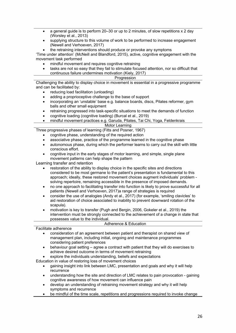
26
• a general guide is to perform 20–30 or up to 2 minutes, of slow repetitions x 2 day (Worsley et al., 2013)
• supplying structure to this volume of work to be performed to increase engagement (Newell and Verhoeven, 2017)
• the retraining interventions should produce or provoke any symptoms ‘Time under attention’ (McNeill and Blandford, 2015), active, cognitive engagement with the movement task performed
• mindful movement and requires cognitive retraining • tasks are not so easy that they fail to stimulate focused attention, nor so difficult that
continuous failure undermines motivation (Kiely, 2017) Progression
Challenging the ability to display choice in movement is essential in a progressive programme and can be facilitated by:
• reducing load facilitation (unloading) • adding a proprioceptive challenge to the base of support • incorporating an ‘unstable’ base e.g. balance boards, discs, Pilates reformer, gym
balls and other small equipment • retraining progressed into task-specific situations to meet the demands of function • cognitive loading (cognitive loading) (Burcal et al., 2019) • mindful movement practices e.g. Garuda, Pilates, Tai Chi, Yoga, Feldenkrais
Motor Learning Three progressive phases of learning (Fitts and Posner, 1967)
• cognitive phase, understanding of the required action • associative phase, practice of the programme learned in the cognitive phase • autonomous phase, during which the performer learns to carry out the skill with little
conscious effort. • cognitive input in the early stages of motor learning, and simple, single plane
movement patterns can help shape the pattern Learning transfer and retention
• restoration of the ability to display choice in the specific sites and directions considered to be most germane to the patient’s presentation is fundamental to this approach; ideally, these restored movement choices augment individuals’ problem -solving repertoire, remaining accessible in the presence of imposed demands.
• no one approach to facilitating transfer into function is likely to prove successful for all patients (Newell and Verhoeven, 2017)a range of strategies is required
• consider the use of analogies (Andy et al., 2017) (for example, ‘smiling clavicles’ to aid restoration of choice associated to inability to prevent downward rotation of the scapula).
• motivation is key to transfer (Pugh and Bergin, 2006, Gokeler et al., 2019) the intervention must be strongly connected to the achievement of a change in state that possesses value to the individual.
Adherence & Education Facilitate adherence
• consideration of an agreement between patient and therapist on shared view of management plan, including initial, ongoing and maintenance programmes considering patient preferences
• behaviour goal setting – agree a contract with patient that they will do exercises to achieve desired outcome in terms of movement retraining
• explore the individuals understanding, beliefs and expectations Education in value of restoring loss of movement choices
• gaining insight into link between LMC, presentation and goals and why it will help recurrence
• understanding how the site and direction of LMC relates to pain provocation - gaining cognitive awareness of how movement can influence pain
• develop an understanding of retraining movement strategy and why it will help symptoms and recurrence
• be mindful of the time scale, repetitions and progressions required to invoke change

27
• allow person to demonstrate an ability to perform the retraining movement strategy – help them learn to judge when they are controlling the LMC
Table 8. Considerations for Restoring Loss of Movement Choices with Cognitive Movement Control Retraining
The movement retraining intervention is informed by identifying the site, direction
and threshold ® of LMC and a clinical reasoning process to match priorities to
client’s goals. For example, a goal may be to manage pain and local tissue stress
by sharing the demands of function across a range of tissues or restore
movement choices for improved performance. The process of retraining
movement coordination strategies has been shown to have clinical utility at the
shoulder (Worsley et al., 2013, Struyf et al., 2013) and hip and groin (Wilson et
al., 2018, Mottram et al., 2019). These papers support proof of concept of this
approach, however more robust evidence is required. A systematic review and
meta-analysis of eleven publications in 2018 explored the effectiveness of
movement control exercises, on patients with non-specific low back pain and
movement control impairments (Luomajoki et al., 2018). There was low to
moderate quality evidence, suggesting a positive effect of movement control
exercises on disability and pain severity, in the short-term and long-term, in
nonspecific low back pain. Clearly, further high quality RCT’s are required into
the effectiveness of this approach. However, eligibility criteria must include the
presence of LCMs. The present authors propose using CMCTs to identify the
presence of LCMs can be used to stratify patients likely to respond to retraining.
This approach empowers people to restore choice in movement (access motor
abundance) and sits within the bigger clinical reasoning picture.
CLINICAL REASONING

28
This paper champions ‘movement’ as the primary means of intervention to
manage pain, limit its recurrence and enhance quality of life. Ultimately, these
interventions seek to move individuals towards a sustained, robust state of
movement health, by restoring movement choices. However, this process will be
optimised if it fits within a person-centred approach, which considers the influence
of multiple constraints on any movement coordination strategy.
A multi-dimensional, individual-centred, clinical reasoning framework is proposed
based on the consideration of the numerous factors influencing movement
choices including:
1. Evaluation of Movement Health, in terms of Site, Direction, Threshold ® of
loss of choices in movement
2. Evaluation of syndrome, pathology, clinical signs and imaging findings
3. Consideration of pain mechanism
4. Consideration of any other individual, environmental and task constraints
(Comerford and Mottram, 2012) (Figure 4).

29
Figure 4. A clinical reasoning framework based on four key factors influencing movement choices (Reproduced with permission of Comera Movement Science)
The interactions of these elements will drive the priorities of clinical interventions.
Figure 5 illustrates a model for consideration of assessing and restoring LMC. At
the centre of this pathway is the status of an individual’s movement health.

30
Figure 5. The assessment and retraining of loss of movement choices within a clinical reasoning framework (Reproduced with permission of Comera Movement Science)
Movement health is constantly in a state of flux in response to the influence of
numerous constraints. Despite the ever-changing influence of constraints,
maintaining movement health across the lifespan may facilitate an individual’s
progress towards their highest attainable standard of health. Empowering
individuals to consider the relationship between life goals, their status of
movement health and a sense of how their efforts to improve this property can
influence their life outcomes, is central to the movement health concept.
Numerous other therapeutic and educational interventions can positively
influence movement choices, for example, manual therapy, soft tissue and fascial
approaches pain neuroscience education and cognitive behavioural approaches.

31
The CMCT system can be utilised as an outcome measure to track progression
(Mottram et al., 2019).
Interpreting muscle synergies Muscle synergy characteristics have been shown to change in the presence and
history of pain (Blandford et al., 2018b, Feeney et al., 2018, Liew et al., 2018).
These changes are accompanied by alterations in joint coordination in the lower
extremity (Kim et al., 2019). In the shoulder, Worsley et al observed that scapular
downward rotation in individuals with shoulder pain was accompanied by
changes in recruitment of serratus anterior and lower trapezius (Worsley et al.,
2013). The consistent patterns of movement coordination strategies employed by
these individuals on arm elevation, was characterised by more downward rotation
than in pain-free individuals. Following retraining these individuals appeared to
employ movement coordination strategies as used by those in a more robust
state of movement health. There is promising clinical support for the assessment
of movement coordination strategies to infer upon relevant changes in muscle
synergies. The clinical framework in Figure 5 includes the consideration of altered
muscle behaviour to support clinical decision making. Once the site and direction
of LMC has been established, the associated muscle synergies can be explored
in respect to movement health. This is an emerging area if interest to the
researcher and clinician (Liew et al., 2018, Kim et al., 2019, Mehrabi et al., 2019).
SUMMARY

32
The health of the movement system may be informed by the ability to display
choice in movement, accessing the wealth of movement coordination strategies
afforded by motor abundance. An assessment framework for evaluating patterns
of movement coordination strategies is detailed with CMCTs. Restoration of
movement choices identified as lost on testing are explored through cognitive
movement control retraining interventions. Sitting along-side other interventions,
this approach supports sustained, robust problem solving to facilitate enhanced
quality of life across the lifespan.
CONCLUSION
This Masterclass sets out a perspective on how movement and its problem-
solving capacity can be assessed and modified in the clinical setting. For
clinicians wishing to adopt a movement-based approach, the identification of
LMC may contribute to the clinical reasoning process. Proof of concept of this
perspective has been outlined, and clearly further research is needed to
improve the quality of evidence to support this approach.

33
REFERENCES
AL ATTAR, W. S. A., SOOMRO, N., SINCLAIR, P. J., PAPPAS, E. & SANDERS, R. H. 2017. Effect of Injury Prevention Programs that Include the Nordic Hamstring Exercise on Hamstring Injury Rates in Soccer Players: A Systematic Review and Meta-Analysis. Sports Med, 47, 907-916.
ANDY, C. Y., WONG, T. W. & MASTERS, R. S. 2017. Examining motor learning in older adults using analogy instruction. Psychology of Sport and Exercise 28, 78-84.
BAIDA, S. R., GORE, S. J., FRANKLYN-MILLER, A. D. & MORAN, K. A. 2018. Does the amount of lower extremity movement variability differ between injured and uninjured populations? A systematic review. Scand J Med Sci Sports, 28, 1320-1338.
BERNSTEIN, N. 1967. The coordination and regulation of movements. , Oxford, UK, Pergamon Press.
BITTENCOURT, N. F. N., MEEUWISSE, W. H., MENDONCA, L. D., NETTEL-AGUIRRE, A., OCARINO, J. M. & FONSECA, S. T. 2016. Complex systems approach for sports injuries: moving from risk factor identification to injury pattern recognition-narrative review and new concept. Br J Sports Med, 50, 1309-1314.
BLANDFORD, L., MCNEILL, W. & CHARVET, I. 2018a. Can we spread the risk? A demand-share perspective to sustained hamstring health. J Bodyw Mov Ther, 22, 766-779.
BLANDFORD, L., THEIS, N., CHARVET, I. & MAHAFFEY, R. 2018b. Is neuromuscular inhibition detectable in elite footballers during the Nordic hamstring exercise? Clin Biomech (Bristol, Avon), 58, 39-43.
BOTHA, N., WARNER, M., GIMPEL, M., MOTTRAM, S., COMERFORD, M. & STOKES, M. 2014. Movement patterns during a Small Knee Bend test in Academy Footballers with Femoroacteabular impingement (FAI). Working Papers in Health Sciences 1, 1-5.
BOUILLARD, K., JUBEAU, M., NORDEZ, A. & HUG, F. 2014. Effect of vastus lateralis fatigue on load sharing between quadriceps femoris muscles during isometric knee extensions. J Neurophysiol, 111, 768-76.
BROWN, C., BOWSER, B. & SIMPSON, K. J. 2012. Movement variability during single leg jump landings in individuals with and without chronic ankle instability. Clin Biomech (Bristol, Avon), 27, 52-63.
BURCAL, C. J., NEEDLE, A. R., CUSTER, L. & ROSEN, A. B. 2019. The Effects of Cognitive Loading on Motor Behavior in Injured Individuals: A Systematic Review. Sports Med.
CHRISTE, G., KADE, F., JOLLES, B. M. & FAVRE, J. 2017. Chronic low back pain patients walk with locally altered spinal kinematics. J Biomech, 60, 211-218.
COMERFORD, M. & MOTTRAM, S. 2012. Kinetic Control, The Management of Uncontrolled Movement, Australia, Churchill Livingstone
COOK, G., BURTON, L., HOOGENBOOM, B. J. & VOIGHT, M. 2014. Functional movement screening: the use of fundamental movements as an assessment of function - part 1. Int J Sports Phys Ther, 9, 396-409.

34
DAVIDS, K., GLAZIER, P., ARAUJO, D. & BARTLETT, R. 2003. Movement systems as dynamical systems: the functional role of variability and its implications for sports medicine. Sports Med, 33, 245-60.
DAVIS, K., WILLIAMS, J. L., SANFORD, B. A. & ZUCKER-LEVIN, A. 2019. Assessing lower extremity coordination and coordination variability in individuals with anterior cruciate ligament reconstruction during walking. Gait Posture, 67, 154-159.
DINGENEN, B., BLANDFORD, L., COMERFORD, M., STAES, F. & MOTTRAM, S. 2018. The assessment of movement health in clinical practice: A multidimensional perspective. Phys Ther Sport, 32, 282-292.
DINGENEN, B., MALLIARAS, P., JANSSEN, T., CEYSSENS, L., VANELDEREN, R. & BARTON, C. J. 2019. Two-dimensional video analysis can discriminate differences in running kinematics between recreational runners with and without running-related knee injury. Phys Ther Sport, 38, 184-191.
EDWARDS, S., BROOKE, H. C. & COOK, J. L. 2017. Distinct cut task strategy in Australian football players with a history of groin pain. Phys Ther Sport, 23, 58-66.
EVERARD, E., LYONS, M. & HARRISON, A. J. 2018. Examining the association of injury with the Functional Movement Screen and Landing Error Scoring System in military recruits undergoing 16 weeks of introductory fitness training. J Sci Med Sport, 21, 569-573.
FALLA, D., FARINA, D. & GRAVEN-NIELSEN, T. 2007. Experimental muscle pain results in reorganization of coordination among trapezius muscle subdivisions during repetitive shoulder flexion. Exp.Brain Res., 178, 385-393.
FEENEY, D. F., CAPOBIANCO, R. A., MONTGOMERY, J. R., MORREALE, J., GRABOWSKI, A. M. & ENOKA, R. M. 2018. Individuals with sacroiliac joint dysfunction display asymmetrical gait and a depressed synergy between muscles providing sacroiliac joint force closure when walking. J Electromyogr Kinesiol, 43, 95-103.
FITTS, P. & POSNER, M. 1967. Human Performance, TN, USA,, Brooks/Cole. GATES, A. B., KERRY, R., MOFFATT, F., RITCHIE, I. K., MEAKINS, A., THORNTON, J. S.,
ROSENBAUM, S. & TAYLOR, A. 2017. Movement for movement: exercise as everybody's business? Br J Sports Med, 51, 767-768.
GLAZIER, P. S. & MEHDIZADEH, S. 2019. In search of sports biomechanics' holy grail: Can athlete-specific optimum sports techniques be identified? J Biomech.
GOKELER, A., NEUHAUS, D., BENJAMINSE, A., GROOMS, D. R. & BAUMEISTER, J. 2019. Principles of Motor Learning to Support Neuroplasticity After ACL Injury: Implications for Optimizing Performance and Reducing Risk of Second ACL Injury. Sports Med, 49, 853-865.
GUCCIONE, A. A., NEVILLE, B. T. & GEORGE, S. Z. 2019. Optimization of Movement: A Dynamical Systems Approach to Movement Systems as Emergent Phenomena. Phys Ther, 99, 3-9.
HAMILL, J., PALMER, C. & VAN EMMERIK, R. E. 2012. Coordinative variability and overuse injury. Sports Med Arthrosc Rehabil Ther Technol, 4, 45.
HARBOURNE, R. T. & STERGIOU, N. 2009. Movement variability and the use of nonlinear tools: principles to guide physical therapist practice. Phys Ther, 89, 267-82.
HASKELL, W. L., LEE, I. M., PATE, R. R., POWELL, K. E., BLAIR, S. N., FRANKLIN, B. A., MACERA, C. A., HEATH, G. W., THOMPSON, P. D. & BAUMAN, A. 2007. Physical activity and public health: updated recommendation for adults from the

35
American College of Sports Medicine and the American Heart Association. Med Sci Sports Exerc, 39, 1423-34.
HAUSDORFF, J. M., RIOS, D. A. & EDELBERG, H. K. 2001. Gait variability and fall risk in community-living older adults: a 1-year prospective study. Arch Phys Med Rehabil, 82, 1050-6.
HAWKES, D. H., KHAIYAT, O. A., HOWARD, A. J., KEMP, G. J. & FROSTICK, S. P. 2019. Patterns of muscle coordination during dynamic glenohumeral joint elevation: An EMG study. PLoS One, 14, e0211800.
HIDES, J. A., DONELSON, R., LEE, D., PRATHER, H., SAHRMANN, S. A. & HODGES, P. W. 2019. Convergence and Divergence of Exercise-Based Approaches That Incorporate Motor Control for the Management of Low Back Pain. J Orthop Sports Phys Ther, 49, 437-452.
HODGES, P. W. & SMEETS, R. J. 2015. Interaction between pain, movement, and physical activity: short-term benefits, long-term consequences, and targets for treatment. Clin J Pain, 31, 97-107.
HODGES, P. W. & TUCKER, K. 2011. Moving differently in pain: a new theory to explain the adaptation to pain. Pain, 152, S90-8.
HOLT, K. G., WAGENAAR, R. O. & SALTZMAN, E. 2010. A dynamic systems/constraints approach to rehabilitation. Rev Bras Fisioter, 14, 446-63.
HUG, F. & TUCKER, K. 2017. Muscle Coordination and the Development of Musculoskeletal Disorders. Exerc Sport Sci Rev, 45, 201-208.
JAMES, C. R., ATKINS, L. T., DUFEK, J. S. & BATES, B. T. 2014. An exploration of load accommodation strategies during walking with extremity-carried weights. Hum Mov Sci, 35, 17-29.
JAMES, C. R., DUFEK, J. S. & BATES, B. T. 2000. Effects of injury proneness and task difficulty on joint kinetic variability. Med Sci Sports Exerc, 32, 1833-44.
KAWAKAMI, W., TAKAHASHI, M., IWAMOTO, Y. & SHINAKODA, K. 2019. Coordination Among Shank, Rearfoot, Midfoot, and Forefoot Kinematic Movement During Gait in Individuals with Hallux Valgus. Journal of Applied Biomechanics, 35, 44-51.
KENT, M. 2006. The Oxford dictionary of sports science and medicine, Oxford, Oxford University Press.
KIELY, J. 2017. The Robust Running Ape: Unraveling the Deep Underpinnings of Coordinated Human Running Proficiency. Front Psychol, 8, 892.
KIM, H., SON, S. J., SEELEY, M. K. & HOPKINS, J. T. 2019. Altered movement strategies during jump landing/cutting in patients with chronic ankle instability. Scand J Med Sci Sports.
KING, E., FRANKLYN-MILLER, A., RICHTER, C., O'REILLY, E., DOOLAN, M., MORAN, K., STRIKE, S. & FALVEY, E. 2018. Clinical and biomechanical outcomes of rehabilitation targeting intersegmental control in athletic groin pain: prospective cohort of 205 patients. Br J Sports Med, 52, 1054-1062.
KONIG, N., TAYLOR, W. R., BAUMANN, C. R., WENDEROTH, N. & SINGH, N. B. 2016. Revealing the quality of movement: A meta-analysis review to quantify the thresholds to pathological variability during standing and walking. Neurosci Biobehav Rev, 68, 111-119.
LATASH, M. L. 2012. The bliss (not the problem) of motor abundance (not redundancy). Exp Brain Res, 217, 1-5.

36
LENZLINGER-ASPRION, R., KELLER, N., MEICHTRY, A. & LUOMAJOKI, H. 2017. Intertester and intratester reliability of movement control tests on the hip for patients with hip osteoarthritis. BMC Musculoskelet Disord, 18, 55.
LIEW, B. X. W., DEL VECCHIO, A. & FALLA, D. 2018. The influence of musculoskeletal pain disorders on muscle synergies-A systematic review. PLoS One, 13, e0206885.
LLUCH, E., BENITEZ, J., DUENAS, L., CASANA, J., ALAKHDAR, Y., NIJS, J. & STRUYF, F. 2014. The shoulder medial rotation test: an intertester and intratester reliability study in overhead athletes with chronic shoulder pain. J Manipulative Physiol Ther, 37, 198-205.
LOEB, G. E. 2012. Optimal isn't good enough. Biol Cybern, 106, 757-65. LOW, M. 2018. A Time to Reflect on Motor Control in Musculoskeletal Physical Therapy.
J Orthop Sports Phys Ther, 48, 833-836. LUOMAJOKI, H., KOOL, J., DE BRUIN, E. D. & AIRAKSINEN, O. 2007. Reliability of
movement control tests in the lumbar spine. BMC Musculoskelet Disord, 8, 90. LUOMAJOKI, H., KOOL, J., DE BRUIN, E. D. & AIRAKSINEN, O. 2008. Movement control
tests of the low back; evaluation of the difference between patients with low back pain and healthy controls. BMC Musculoskelet Disord, 9, 170.
LUOMAJOKI, H. A., BONET BELTRAN, M. B., CAREDDU, S. & BAUER, C. M. 2018. Effectiveness of movement control exercise on patients with non-specific low back pain and movement control impairment: A systematic review and meta-analysis. Musculoskelet Sci Pract, 36, 1-11.
MADELEINE, P., MATHIASSEN, S. E. & ARENDT-NIELSEN, L. 2008. Changes in the degree of motor variability associated with experimental and chronic neck-shoulder pain during a standardised repetitive arm movement. Exp Brain Res, 185, 689-98.
MCNEILL, W. 2014. The Double Knee Swing Test - a practical example of The Performance Matrix Movement Screen. J Bodyw Mov Ther, 18, 477-81.
MCNEILL, W. & BLANDFORD, L. 2015. Movement health. J Bodyw Mov Ther, 19, 150-9. MEHRABI, N., SCHWARTZ, M. H. & STEELE, K. M. 2019. Can altered muscle synergies
control unimpaired gait? J Biomech, 90, 84-91. MISCHIATI, C. R., COMERFORD, M., GOSFORD, E., SWART, J., EWINGS, S., BOTHA, N.,
STOKES, M. & MOTTRAM, S. L. 2015. Intra and inter-rater reliability of screening for movement impairments: movement control tests from the foundation matrix. J Sports Sci Med, 14, 427-40.
MONNIER, A., HEUER, J., NORMAN, K. & ANG, B. O. 2012. Inter- and intra-observer reliability of clinical movement-control tests for marines. BMC Musculoskelet Disord, 13, 263.
MORRISSEY, D., MORRISSEY, M. C., DRIVER, W., KING, J. B. & WOLEDGE, R. C. 2008. Manual landmark identification and tracking during the medial rotation test of the shoulder: An accuracy study using three-dimensional ultrasound and motion analysis measures. Man.Ther.
MOTTRAM, S. & COMERFORD, M. 2008. A new perspective on risk assessment. Phys Ther Sport, 9, 40-51.
MOTTRAM, S., WARNER, M., BOOYSEN, N., BAHAIN-STEENMAN, K. & STOKES, M. 2019. Retraining in a female elite rower with persistent symptoms post-arthroscopy for femoroacetabular impingement syndrome: a proof-of-concept case report. Journal of Functional Morphology and Kinesiology 4.

37
MOTTRAM, S. L. 2003. Dynamic stability of the scapula. In: BEETON & KS (eds.) Manual therapy masterclasses – the peripheral
joints. Edinburgh: Churchill Livingstone. NEEDHAM, R. A., NAEMI, R. & CHOCKALINGAM, N. 2015. A new coordination pattern
classification to assess gait kinematics when utilising a modified vector coding technique. J Biomech, 48, 3506-11.
NEWELL, K. M. 1986. Constraints on the development of coordination, Berlin, Germany, Springer.
NEWELL, K. M. & VERHOEVEN, F. M. 2017. Movement rehabilitation: are the principles of re-learning in the recovery of function the same as those of original learning? Disabil Rehabil, 39, 121-126.
NORDIN, A. D. & DUFEK, J. S. 2019. Reviewing the Variability-Overuse Injury Hypothesis: Does Movement Variability Relate to Landing Injuries? Res Q Exerc Sport, 90, 190-205.
PADUA, D. A., BOLING, M. C., DISTEFANO, L. J., ONATE, J. A., BEUTLER, A. I. & MARSHALL, S. W. 2011. Reliability of the landing error scoring system-real time, a clinical assessment tool of jump-landing biomechanics. J Sport Rehabil, 20, 145-56.
PADUA, D. A., DISTEFANO, L. J., BEUTLER, A. I., DE LA MOTTE, S. J., DISTEFANO, M. J. & MARSHALL, S. W. 2015. The Landing Error Scoring System as a Screening Tool for an Anterior Cruciate Ligament Injury-Prevention Program in Elite-Youth Soccer Athletes. J Athl Train, 50, 589-95.
PUGH, K. J. & BERGIN, D. A. 2006. Motivational influences on transfer. Educational Psychologist, 41, 147-160.
RAISBECK, L. D. & YAMADA, M. 2019. The effects of instructional cues on performance and mechanics during a gross motor movement. Hum Mov Sci, 66, 149-156.
RAJASEKAR, S., BANGERA, R. K. & SEKARAN, P. 2017. Inter-rater and intra-rater reliability of a movement control test in shoulder. J Bodyw Mov Ther, 21, 739-742.
ROOS, E. M., BARTON, C. J., DAVIS, A. M., MCGLASSON, R., KEMP, J. L., CROSSLEY, K. M., LIU, Q., LIN, J. & SKOU, S. T. 2018. GLA:D to have a high-value option for patients with knee and hip arthritis across four continents: Good Life with osteoArthritis from Denmark. Br J Sports Med, 52, 1544-1545.
ROUSSEL, N. A., NIJS, J., MOTTRAM, S., VAN, M. A., TRUIJEN, S. & STASSIJNS, G. 2009. Altered lumbopelvic movement control but not generalized joint hypermobility is associated with increased injury in dancers. A prospective study. Man.Ther.
SAFRAN, M. R., BORSA, P. A., LEPHART, S. M., FU, F. H. & WARNER, J. J. 2001. Shoulder proprioception in baseball pitchers. J Shoulder.Elbow.Surg., 10, 438-444.
SAHRMANN, S. A. 2002. Diagnosis & Treatment of Movement Impairment Syndromes, Mosby, USA.
SANTUZ, A., EKIZOS, A., JANSHEN, L., MERSMANN, F., BOHM, S., BALTZOPOULOS, V. & ARAMPATZIS, A. 2018. Modular Control of Human Movement During Running: An Open Access Data Set. Front Physiol, 9, 1509.
SEAY, J. F., VAN EMMERIK, R. E. & HAMILL, J. 2011. Low back pain status affects pelvis-trunk coordination and variability during walking and running. Clin Biomech (Bristol, Avon), 26, 572-8.
SEGARRA, V., DUENAS, L., TORRES, R., FALLA, D., JULL, G. & LLUCH, E. 2015. Inter-and intra-tester reliability of a battery of cervical movement control dysfunction tests. Man Ther, 20, 570-9.

38
SHIELD, A. J. & BOURNE, M. N. 2018. Hamstring Injury Prevention Practices in Elite Sport: Evidence for Eccentric Strength vs. Lumbo-Pelvic Training. Sports Med, 48, 513-524.
SKOU, S. T., BRICCA, A. & ROOS, E. M. 2018. The impact of physical activity level on the short- and long-term pain relief from supervised exercise therapy and education: a study of 12,796 Danish patients with knee osteoarthritis. Osteoarthritis Cartilage, 26, 1474-1478.
STRUYF, F., NIJS, J., MOLLEKENS, S., JEURISSEN, I., TRUIJEN, S., MOTTRAM, S. & MEEUSEN, R. 2013. Scapular-focused treatment in patients with shoulder impingement syndrome: a randomized clinical trial. Clin Rheumatol, 32, 73-85.
VAN DIEEN, J. H., REEVES, N. P., KAWCHUK, G., VAN DILLEN, L. R. & HODGES, P. W. 2019. Analysis of Motor Control in Patients With Low Back Pain: A Key to Personalized Care? J Orthop Sports Phys Ther, 49, 380-388.
WARNER, M. B., WHATLING, G., WORSLEY, P. R., MOTTRAM, S., CHAPPELL, P. H., HOLT, C. A. & STOKES, M. J. 2015. Objective classification of scapular kinematics in participants with movement faults of the scapula on clinical assessment. Comput Methods Biomech Biomed Engin, 18, 782-9.
WEBB, A. L., ROWSOME, K., EWINGS, S., COMERFORD, M., STOKES, M. & S, M. 2018. A ‘Movement Screening Test’ of Functional Control Ability in Female Recreation Golfers and Non-Golfers over the Age of 80 Years: A Reliability Study
. Journal Functional Morphology Kinesiology, 3. WEIR, G., VAN EMMERIK, R., JEWELL, C. & HAMILL, J. 2019. Coordination and variability
during anticipated and unanticipated sidestepping. Gait Posture, 67, 1-8. WHITTAKER, J. L., BOOYSEN, N., DE LA MOTTE, S., DENNETT, L., LEWIS, C. L., WILSON, D.,
MCKAY, C., WARNER, M., PADUA, D., EMERY, C. A. & STOKES, M. 2017. Predicting sport and occupational lower extremity injury risk through movement quality screening: a systematic review. Br J Sports Med, 51, 580-585.
WILSON, D. A., BOOYSEN, N., DAINESE, P., HELLER, M. O., STOKES, M. & WARNER, M. B. 2018. Accuracy of movement quality screening to document effects of neuromuscular control retraining exercises in a young ex-footballer with hip and groin symptoms: A proof of concept case study. Med Hypotheses, 120, 116-120.
WORSLEY, P., WARNER, M., MOTTRAM, S., GADOLA, S., VEEGER, H. E., HERMENS, H., MORRISSEY, D., LITTLE, P., COOPER, C., CARR, A. & STOKES, M. 2013. Motor control retraining exercises for shoulder impingement: effects on function, muscle activation, and biomechanics in young adults. J Shoulder Elbow Surg, 22, e11-9.