PROTEUS: Artefact-driven Constructionist Assessment within Tablet PC-based Low-fidelity Prototyping
Upload
cez-archaeometrieCategory
view
5download
0
lable at ScienceDirect
Journal of Archaeological Science 57 (2015) 130e141
Contents lists avai
Journal of Archaeological Science
journal homepage: http: / /www.elsevier .com/locate/ jas
Focus article
Artificial patination in Early Iron Age Europe: an analytical case studyof a unique bronze artefact
Daniel BergerCurt-Engelhorn-Zentrum Arch€aometrie gGmbH, D6, 3, D-68159 Mannheim, Germany
a r t i c l e i n f o
Article history:Received 14 April 2014Received in revised form19 January 2015Accepted 24 January 2015Available online 17 February 2015
Keywords:Artificial patinationTenoriteEarly Iron AgeBronze axeX-ray diffraction analysisMicro Raman spectroscopyFT-IR spectroscopy
E-mail address: daniel.berger@cez-archaeometrie.
http://dx.doi.org/10.1016/j.jas.2015.01.0250305-4403/© 2015 Elsevier Ltd. All rights reserved.
a b s t r a c t
Direct evidence for the intentional patination of metal objects is difficult to ascertain and thereforestudies concerning this technique are controversial. Only a couple of objects with artificially patinatedsurfaces have been positively identified, ranging in date from the early 2nd millennium BC to Roman andmedieval times. In this paper a skillfully crafted and coloured Early Iron Age bronze axe belonging to theVillanova culture of ancient Italy was selected for examination. Optical microscopy and multiplemineralogical analyses of the surface of this object demonstrate that it was patinated deliberately by athermal treatment technology (thermal patination). So far, this Iron Age axe is the oldest European objectwhere this technology had been applied.
© 2015 Elsevier Ltd. All rights reserved.
1. Introduction
The seminal research of Giumlía-Mair and Craddock (1993)provided empirical evidence that ancient metal craft specialistsintentionally manipulated the colourful appearance of metalthrough various chemical treatments. In particular, metal objectsfrom the Mediterranean region with metallic inlays e sometimesreferred to as damascenings made of alloys named corinthium aes,kuwano or ḥmty km e are often quoted to have been patinatedartificially. This is suggested because of their dark surface layers andthe rather weak colour contrasts between the metal bases and in-lays. The most prominent examples having artificial surface layersare the Bronze Age daggers and vessels from the Mycenaean shaftgraves and other Greek locations. In addition, a large corpus ofEgyptian and Roman artefacts with metal inlays has been identifiedas intentionally patinated in the last decades (Craddock, 1982;Giumlía-Mair and Craddock, 1993; Giumlía-Mair, 1996a; 1996b;Giumlía-Mair and Riederer, 1998; Mathis et al. 2007, 2009;Aucouturier et al. 2010; Giumlía-Mair, 2012).
Besides their characteristic patinas, the copper alloys of theseobjects have distinct compositions. Often the metal inlays or bases
de.
contain appreciable amounts of gold and silver (from 0.5 up to3 wt.%) which in case of gold clearly exceed natural impurities ofcopper. In fact, it has been shown that copperegold (-silver) alloysreadily develop dark purple or blue to nearly black corrosion crustsof cuprite (Cu2O) with a high lustre and adherence when treatedwith chemical solutions (e.g. acetic solutions with copper salts) orsimply when held in hand for some time (Giumlía-Mair and Lehr,1998, 2003; van Bellegem et al. 2007; Craddock et al. 2009;O'Dubhghaill and Jones, 2009). Since minor gold concentrationsneither change the colour of copper nor its casting and mechanicalproperties (Berger, 2012a, 24e26) this behaviour is taken as themain, yet not undisputed (cf. Jacobson and Weitzman, 1995) line ofevidence to explain how pastmetal smiths used artificial patinationtechnologies on these kinds of objects.
Less unambiguous are black coloured copper sulphide patinas.Well-known objects with this kind of patination are the Hellenisticbronze sculptures and pieces of furniture found off the coasts ofRiace, Italy, and Mahdia, Tunisia. Their surface layers are smoothand blue black and consist of different sulphide minerals (Formigli,1985; Willer, 1994; Eggert, 1994). Because of their appearance,composition as well as manufactural details some authors sug-gested that such patinas reflect deliberate colouring treatments(Zenghelis, 1930; Formigli, 1985; Willer, 1994; Garbassi and Mello,1984, 176e179). Although it was easy for ancient people to

D. Berger / Journal of Archaeological Science 57 (2015) 130e141 131
achieve such sulphide containing patinas (e.g. with sulphur or eggyolk), there is still a controversy amongst scientists whether thesurface layers developed from the intentional actions of past metalsmiths or result from natural corrosion in anaerobic environments(Schwab et al. 2008, 20e21; Quaranta, 2009, 107e110). Such con-ditions are often observed in underwater sites and waterloggedsoils (Tylecote, 1979, 352; MacLeod, 1991; Schweizer, 1994).
Similarly controversial is the intentional patination by thermaltreatment. Several ancient metal objects from Egypt show charac-teristic patination patterns that were likely produced by controlledannealing in order to serve as aesthetic oxide layers. Mohamed andDarweesh (2012) assembled a group of Egyptian bronze artefactswithout deliberate gold alloying, including statuettes, situlas andplaques, which possess superficial black patinas, too. The authorsdemonstrated that the patinas of many of these objects consist ofblack copper oxide tenorite (CuO) together with cuprite (Cu2O) andsecondary corrosion products which distinguish them from blackpatinated copperegold alloys (Giumlía-Mair, 2000). The authorsassumed that thermal patination (annealing with scale production)was the most likely colouring procedure in all cases because largequantities of tenorite almost exclusively develop during the heatingof copper or copper base alloys (Mohamed and Darweesh, 2012,184; Scott,1997, 96e97). Natural corrosion in soils seldom producestraces of this mineral under highly alkaline conditions (Nord et al.1998). Without knowing the details of the depositional context,the site formation processes and the object's life history, it is,however, impossible to rule out accidental events. For example, siteconflagration or cremation could also theoretically stimulate theformation of tenorite. Despite this possibility it is more likely thatthe patinas on the Egyptian examples are the result of intentionalapplication.
Currently no other Bronze or Iron Age object outside Egypt hasbeen identified with similar patination treatments. However, there
Fig. 1. Front face of the winged axe from the Museum für Vor-und Frühgeschichte Berlin, GeD. Berger).
are two black patinated examples from the Roman Empire withtenorite. They have initially been investigated by Mathis (2005,Fig. 72, 163e164), but later on Aucouturier et al. (2010) confirmedthem to be treated thermally deliberately at around 600 �C. Incontrast to the Egyptian items, one of the two Roman objects hascopper inlays in a bronze base. This appears fairly unusual becauseduring prolonged heating at temperatures of 500 �C or above thealloying elements from the metal base (especially tin) mightmigrate into the copper inlays and potentially cause the formationof irregular and undesired seams. Therefore, the reconstruction issomewhat doubtful in this case, even though deliberate thermaltreatment was also suspected on an inlayed antique bronzemask inthe British Museum (Jenkins, 1993, 299).
The above examples demonstrate the difficulties associatedwith determining the original surface colour on ancient metal ar-tefacts. On the one hand, it is all but impossible to identify exactlythose objects amongst the large quantity of prehistoric metal findsthat were actually deliberately patinated. In many cases greencoloured natural corrosion crusts conceal original produced patinaslying underneath. In addition, present patinas, especially on olderfinds, may be inauthentic and merely the product of modernrestoration. On the other hand, artificially grown mineral phasesoften cannot be distinguished from natural corrosion productsbecause almost the same species form during intentional patina-tion and long-time burial. One important exception is the blackcopper mineral tenorite that rarely occurs under burial conditionsbecause its formation is kinetically impeded relative to the ubiq-uitous cuprite (Scott, 2002, 95; Quaranta, 2009, 19). If circum-stances like conflagration can be ruled out, the presence of tenoritethus appears to be a good indicator for the deliberate colouring ofmetal surfaces by thermal treatment. This paper utilises thisframework to examine in detail a conspicuously coloured Early IronAge winged axe from the collection of the Museum für Vor- und
rmany. The back face is decorated in almost the same way. Total length 178 mm (photo:

Fig. 2. Similar Early Iron Age axes found in the territory of the Villanova culture in Italy. (a) and (b) were found in Bologna and belong to the Benacci axe type decorated in the samemanner like the axe from the MVF. (c) and (d) are axes of an associated type with concentric circles from unknown locality (c) and Rimini (d) (drawings: after Carancini, 1984; Tav.95.3385e3386; 96.3394e3395).
Fig. 3. Distribution of axe finds of the Benacci type and associated types in the territory of the Villanova culture (hatched) (map: C. Frank and D. Berger, made with Natural Earth).
D. Berger / Journal of Archaeological Science 57 (2015) 130e141132

Fig. 4. Detail of the concentric circles on the back face of the axe. The arrows indicatetraces from drilling; image width ca. 15 mm (photo: D. Berger).
Fig. 5. The bent cutting edge of the axe with the region analysed by mXRF (arrow);image width ca. 15 mm (photo: D. Berger).
D. Berger / Journal of Archaeological Science 57 (2015) 130e141 133
Frühgeschichte, Berlin, Germany (Fig. 1). The results from micro-scopic and mineralogical analyses suggest the presence of diag-nostic mineral phases, which are discussed below after a shortpresentation of the axe and a description of the method.
2. Materials and methods
2.1. The bronze axe from the Museum für Vor-und Frühgeschichte,Berlin
The investigated object (Fig. 1) is part of the archaeologicalcollection of the Museum für Vor-und Frühgeschichte (MVF) whichnow can be visited in the ‘Neues Museum’ on the ‘Museumsinsel’ inBerlin, Germany. The winged axe formerly belonged to the collec-tion of the Prussian officer and antiquarian Heinrich Menu vonMinutoli who possibly acquired it during one of his expeditionsacross Europe in the first half of the 19th century. One can, however,only speculate about the circumstances of its discovery becauseonly a brief record is found in the catalogue of the ‘K€oniglichesMuseumvaterl€andischer Alterthümer’, the precursor of the presentMVF (cf. Berger, 2012b; Abb. 2). Apart from a short description ofthe artefact, nothing is said about the exact find spot of the axe,although ‘Italy’ was added later on to the record. This location ap-pears to be plausible for the type of axe. Winged axes of the so-called Benacci type, which are characterised by a bell shapedblade, two large wings in the upper part, a ledge at two third of theaxe length as well as linear decorations, have been recoveredexclusively from cemeteries in the environment of Bologna and
Table 1Conditions and parameters of the analytical facilities employed in the study.
Analytical method Device type, company Parameters and con
mXRF XGT-5000, Horiba X-ray tube with Rhin situ analysis on 5
mXRD D8 Discover GADDS, Bruker AXS X-ray tube with Copinhole collimator,60 s measuring tim
Raman microscopy(mRaman spectroscopy)
Spectrometer: LabRam, Dilor;microscope: BX40, Olympus
Confocal microscopCCD detector, spectin situ analysis, mea
FT-IR spectroscopy Tensor 37, Bruker AXS Globar IR source, Mspectral resolution:software: OPUS, Bru
Verucchio, both Emilia-Romagna, Italy (Figs. 2e3; Carancini, 1984;Vinicio Gentili, 2003). Unlike stated elsewhere (Berger, 2012b), theyare dated to the second half of the 9th or the first half of the 8thcentury BC (Bologna II period) and are ascribed to the Early Iron AgeVillanova culture which is an indirect successor of the Proto-villanova phenomenon described by Amann (2005). Although notverifiable directly, the typological analogies with other objectsmake it most likely that the axe studied here was originally man-ufactured in the same region and probably belonged to the gravegoods of one of the burials found in the cemeteries near Bologna orVerucchio.
In contrast to other examples of the Benacci type, the axe inquestion has a unique and extraordinary decoration. On both faces,the blade is covered with a dark brown to almost black patina thatcontrasts with green coloured stripes interspersed with a fewbluish spots (Fig. 1). The stripes are accompanied by manyconcentric circles which had been deepened by some kind of dril-ling process. This is seen by concentric drill marks inside the cir-cular depressions (Fig. 4) that would not appear as edged if thecircles had been cast.
The same circle decoration is found at the exterior of the wings,but in this case they do not display the same green patina as on theblade. The corrosion crust on this part of the axe is uniformly darkbrown to black coloured with dispersed green spots. As mentionedabove, other axes of the type do not exhibit a similar colour deco-ration, but it is significant that the engravings on their blades andwings form the same ornaments like on the axe from the MVF (cf.Fig. 2). This decisively underscores its membership to the Benacciaxe group.
ditions
anode, 30 kV, 0.3e0.4 mA, monocapillary with 400 mm spot, Si(Li) detector,measuring spots, 60 s measuring time for each spot, standardless quantificationanode, 30 kV, 40 mA, graphite monochromator, monocapillary with 300 mm2D-Hi Star-detector, in situ analysis, measuring at three detector positions,e for each frame, software: X'Pert HighScore Plus, PANalytical BV, PDF 2 databasee optics, notch filter, HeeNe laser (l¼ 632.8 nm), laser output: 10 mW,ral resolution: 1 nm, spectral range: 70e1050 cm�1, different laser filters,suring time: variableCT-A detector, RockSolid-interferometer, transmittance mode, KBr pellets,4 cm�1, spectral range: 400e4000 cm�1, averaging of 32 single spectra,ker Optik GmbH
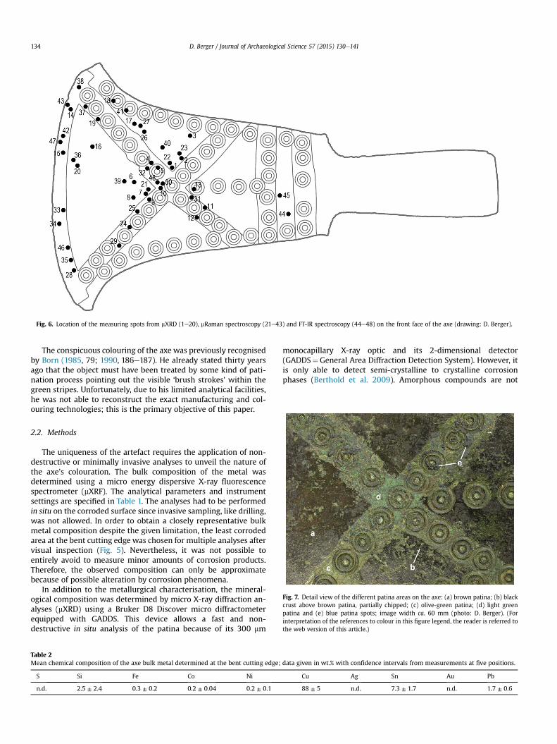
Fig. 6. Location of the measuring spots from mXRD (1e20), mRaman spectroscopy (21e43) and FT-IR spectroscopy (44e48) on the front face of the axe (drawing: D. Berger).
D. Berger / Journal of Archaeological Science 57 (2015) 130e141134
The conspicuous colouring of the axe was previously recognisedby Born (1985, 79; 1990, 186e187). He already stated thirty yearsago that the object must have been treated by some kind of pati-nation process pointing out the visible ‘brush strokes’ within thegreen stripes. Unfortunately, due to his limited analytical facilities,he was not able to reconstruct the exact manufacturing and col-ouring technologies; this is the primary objective of this paper.
Fig. 7. Detail view of the different patina areas on the axe: (a) brown patina; (b) blackcrust above brown patina, partially chipped; (c) olive-green patina; (d) light greenpatina and (e) blue patina spots; image width ca. 60 mm (photo: D. Berger). (Forinterpretation of the references to colour in this figure legend, the reader is referred tothe web version of this article.)
2.2. Methods
The uniqueness of the artefact requires the application of non-destructive or minimally invasive analyses to unveil the nature ofthe axe's colouration. The bulk composition of the metal wasdetermined using a micro energy dispersive X-ray fluorescencespectrometer (mXRF). The analytical parameters and instrumentsettings are specified in Table 1. The analyses had to be performedin situ on the corroded surface since invasive sampling, like drilling,was not allowed. In order to obtain a closely representative bulkmetal composition despite the given limitation, the least corrodedarea at the bent cutting edge was chosen for multiple analyses aftervisual inspection (Fig. 5). Nevertheless, it was not possible toentirely avoid to measure minor amounts of corrosion products.Therefore, the observed composition can only be approximatebecause of possible alteration by corrosion phenomena.
In addition to the metallurgical characterisation, the mineral-ogical composition was determined by micro X-ray diffraction an-alyses (mXRD) using a Bruker D8 Discover micro diffractometerequipped with GADDS. This device allows a fast and non-destructive in situ analysis of the patina because of its 300 mm
Table 2Mean chemical composition of the axe bulk metal determined at the bent cutting edge;
S Si Fe Co Ni
n.d. 2.5 ± 2.4 0.3 ± 0.2 0.2 ± 0.04 0.2 ± 0.1
monocapillary X-ray optic and its 2-dimensional detector(GADDS¼General Area Diffraction Detection System). However, itis only able to detect semi-crystalline to crystalline corrosionphases (Berthold et al. 2009). Amorphous compounds are not
data given in wt.% with confidence intervals from measurements at five positions.
Cu Ag Sn Au Pb
88 ± 5 n.d. 7.3 ± 1.7 n.d. 1.7 ± 0.6

Fig. 8. Diffractograms obtained from (a) green and blue areas contrasted with (b) browneblack patina zones. The differences in FWHM of cassiterite are marked by the arrows(diffractograms: D. Berger).

D. Berger / Journal of Archaeological Science 57 (2015) 130e141136
detectable with XRD for which reason further measurements withRaman microscopy (mRaman) and Fourier transform infraredspectroscopy (FT-IR spectroscopy) were included in the study.These methods allow identification of non-crystalline compoundsthat are known to be often present on corroded metal artefacts,especially made from bronze (Robbiola et al. 1998; Nord et al. 1998;Piccardo et al. 2007, 2013).
Whereas mXRD and mRaman spectroscopy are non-destructive,it was necessary to take tiny samples from the patina for FT-IRexamination in transmission mode. Patina samples were extrac-ted with a scalpel from corrosion areas with distinct colouration,ground in an agate mortar and subsequently embedded in dewa-tered potassium bromide. The resulting pellets of KBr were intro-duced into the FT-IR device whose specifications and analysingparameters along with those of the Raman microscope and themicro diffractometer are listed in Table 1. Fig. 6 shows the distri-bution of the measurement spots of the various analytical methodsused to characterise the green/blue and brown/black corrosionareas.
Table 3Results of the phase analyses on the front face of the axe on the olive-green (ogr), the greepatina within the concentric circles (gr/cir).
Spot Patina colour Analytical method Detected m
XRD Raman FT-IR Substrate
1 br � �2 br � �3 br � �4 gr/bl �5 gr/cir � �6 br � �7 b � �8 br � �9 br � �10 gr/bl � �11 ogr �12 gr � �13 gr/cir � �14 ogr �15 gr/bl �16 br � �17 b � �18 gr/cir �19 gr �20 br � �21 br �22 br �23 br �24 ogr �25 ogr �26 b �27 b �28 bl �29 bl �30 gr �31 gr �32 gr �33 bl �34 bl �35 bl �36 br �37 ogr �38 gr �39 br �40 br �41 bl �42 ogr �43 ogr �44 b �45 br �46 bl �47 ogr �48 gr �
Prior to analytical investigation, the entire object was thor-oughly studied by optical microscopy. This should allow recon-struction of the manufacturing process on the one hand and thedecoration procedure on the other, so that, finally, the completechaîne op�eratoire is established. The latter is described in detail byBerger (2012b), whereas the next sections only discuss the col-ouring method.
3. Results
The results of the bulk metal analyses with mXRF are shown inTable 2. The axe is made of a common bronze alloy which containson average about 7 wt.% tin and a significant quantity of lead(1.7 wt.%). Because of possible element enrichment due to corrosionprocesses (cf. Robbiola et al. 1998) and the analysis without refer-ence materials, the composition is just semi-quantitative and thusonly suitable for exploratory purposes. This point is stressed by thepresence of trace amounts of silicon and iron with high confidenceintervals (Table 2) which are certainly derived from the soil
n (gr), the blue (bl), the brown (br) and the black (b) patina parts as well as the green
ineral phases
Cuprite Tenorite Cassiterite Malachite Azurite
� � � �� � � �� � � �� � � �� � �� � � �� � � �� � � �� � � �� � � �� � �
� �� � �� � �� � � �� � � �� � �� � � �� � � ��� �
high fluorescence (no peaks detected)��
high fluorescence (no peaks detected)high fluorescence (no peaks detected)
�high fluorescence (no peaks detected)
���
high fluorescence (no peaks detected)�
high fluorescence (no peaks detected)�
high fluorescence (no peaks detected)�
���
��
� �� �� � �� �� �

D. Berger / Journal of Archaeological Science 57 (2015) 130e141 137
attached to the formerly buried axe. The nickel and cobalt contents,in contrast, may represent natural impurities from the copper ores.Significantly, no gold and silver could be detected in the alloy andthus the metal does not belong to the corinthium aes metal typedescribed at the beginning of the paper.
Visual examination of the axe's surface revealed that thedifferent patina areas did not uniformly develop. The green col-oured regions consist of an olive-green, rather smooth corrosionlayer which is often overgrown by crusty and light green mineralphases. These crusty products are sometimes associated with bluecorrosion phases (Fig. 7). The brown patina is also a complexassemblage. While in some areas small green corrosion productsdeveloped above the brown patina or in grooves, a smooth blackcrust directly adhering to the brown layer can be seen in other areasthat seem to be partly chipped. Therefore, the black crust appearsnot to be pliable but rather fragile.
The various patina components were analysed by micro X-raydiffraction and the results are summarised in Fig. 8 and Table 3. It isevident that the patina of the green stripes contains the threeminerals cuprite (Cu2O), malachite (CuCO3$Cu(OH)2) and cassit-erite (SnO2) regardless whether the olive-green areas or the lightgreen parts were analysed (Fig. 8a). The bluish corrosion zones alsocontain some azurite (2CuCO3$Cu(OH)2), the blue and carbonaterich version of hydrated copper carbonate. The identified mineralsare frequently found on buriedmetal artefacts (Robbiola et al. 1998;Nord et al. 1998; Piccardo et al. 2007) for which reason there is noevidence for an artificial corrosion treatment. Irrespective of this,cassiterite is poorly crystallised or partially hydrated in these partsof the object (Gaber et al. 2013) because its diffraction peaks arebroad compared with those of cuprite and malachite (cf. below).
The mineral assemblage described above was also observedwithin the green corroded circles that are located inside andoutside the green stripes on the blade (Table 3, nos. 13 and 18). Thisshows that both regions must have been corroded under the sameconditions. This is not the case for the brown and black patinas:Although traces of malachite are present as well, the brown surfacelayer mainly consists of cuprite, well crystallised cassiterite (sharp
Fig. 9. Microscopic images of the patina zones where different roughness states can be seencorrosion zones; image width (a) ca. 155 mm (photo: D. Berger). (For interpretation of the rearticle.)
diffraction peaks) and tenorite (Fig. 8b). The black incrusted patinahas a similar mineralogy. As mentioned earlier, the black copperoxide tenorite primarily forms during metal heating which,therefore, may be the explanation for its presence on the axe'ssurface. The fact that tenorite has not been detected in the greencorrosion layer instead precludes an alternative formation of theoxide, for instance during cremation of mortal remains or otherheating events (MacLeod, 1991, 228; Scott, 2002, 95; Krieg, 2012).For the same reason a natural corrosive origin can be ruled outsince tenorite should occur within the green patina, too. Thus, thelocalised occurrence of tenorite in the browneblack and not in thegreen corrosion areas can be reasonably explained by artificialpatination.
Differences in the surface texture and surface levels of the axewhich are related to differences in colour and the mineralogicalcomposition of the patinas support this explanation. Fig. 9a and bshow microscopic images where green and brown patina parts canbe distinguished because of their different surface roughness: thegreen patina region is much deeper and more evenly scratched(parallel scratches) than the brown patina which only shows slightand irregular scratches. The latter are characteristic for ordinarymetal polishing, whereas the deep and regular striations at thegreen stripes can be explained by oriented grinding with the aim toremove an existing surface layer. The lower surface level of theolive-green patina relative to that of the browneblack one supportsthis idea. Both the presence of tenorite in the dark areas and thegrinded and lowered surface in the green parts are diagnostic forrecognising an artificial colouring treatment.
The mRaman and FT-IR analyses reveal no additional corrosionproducts, such as amorphous hydrated tin (II) or (IV)-species, andthus do not expand the results obtained by mXRD (Figs. 10e11).Admittedly, the copper oxides were not detectable by FT-IR spec-troscopy, which is possibly linked to the small sample volumes.Hence, these analyses should be repeated with more patina ma-terial. However, the presence of tenorite in the brown and blackcoloured zones has been confirmed by the Raman spectra whichshow a sharp peak of the copper oxide at 300 cm�1 and a broad one
. The dashed lines in both images mark the interface of the green and the browneblackferences to colour in this figure legend, the reader is referred to the web version of this

Fig. 10. Raman spectra from the different patina zones on the axe. The wavenumbers (Dn) in black are ascribed to malachite, the dark grey wavenumbers to cuprite and those inlight grey to tenorite (diagrams: D. Berger).
D. Berger / Journal of Archaeological Science 57 (2015) 130e141138
at 625 cm�1 (Fig. 10, no. 36; Bouchard and Smith, 2007, 120e123).In the green and blue coloured parts no tenorite was noticed again.
In addition to the qualitative statement, the Raman spectra ofcuprite from the brown coloured zones (Fig. 10a) are significantlydifferent from those obtained from the green/blue corroded areas(Fig. 10b). Some of the peaks of the brown patina (178 cm�1,201 cm�1, 220 cm�1, 418 cm�1) are clearly suppressed orcompletely missing, which is not the case for the other regions.Moreover, the broad peak around 630 cm�1 of cuprite in the bluezones occurs at lower wavenumbers in the brown zones. Suchspectral variations of cuprite have previously been observed byMathis (2005, 274e275) on different patina parts from a Romanbowl, whereas Berger (2012a) found spectral differences duringexamination of ammonia induced corrosion layers that contrastwith naturally grown cuprite. These variations could be explainedby differences in crystal size, growing characteristics, crystaldistortion due to copper deficiency (and thus a disordered oxidestructure, i.e. Cu2�xO rather than Cu2O) or cation and anion sub-stitution in the cuprite lattice (Scott, 1997, 93; Bouchard and Smith,2007, 122). Although the reason for the spectral variations is notwell understood in the present case, the Raman characteristics ofcuprite may be helpful to identify intentional corrosion layers (i.e.the brown one) by direct comparison with natural ones (i.e. thegreen one) on the same object. Further research is needed toproperly comprehend the physical background.
The full width at half maximum (FWHM) of the cassiterite peaksin the XRD spectra might also help to recognise artificial patination.There are obvious differences in the FWHM of the oxide peaks fromthe green and the browneblack patinas (Fig. 8, arrows) which
indicates different crystallisation states. Such differences can arisefrom the presence of hydrated tin species and the crystal growth atdifferent temperatures as for example Pouretedal et al. (2012) orGaber et al. (2013) have shown with precipitated and calcined tindioxide. Although not directly comparable, it is conceivable thatthese parameters played some role during the crystallisation ofcassiterite on the axe, i.e. during high-temperature oxidation andburial corrosion. In this case, this mineral would be diagnostic invalidating the object's life history.
4. Discussion and conclusions
The results of the phase analyses allow the recognition of anartificial patination treatment of the axe from the MVF. This isprimarily concluded from the phase composition (determined bymXRD and mRaman) showing that the rarely occurring mineraltenorite is only present in the browneblack patina. This excludesthe possibility of a natural or accidental formation (corrosion, fireetc.) as well as an undesired side effect of metallurgical working (i.e.scaling through soft-annealing). Otherwise, tenorite would bepresent in the green/blue patina as well. This conclusion iscorroborated by the different surface textures and levels of thediverse patina areas (Fig. 9) which demonstrate that a previouslypresent patina has been removed where now green corrodedstripes can be seen. Thus, natural corrosion was able to attack themetal directly because no protective surface layer once existed onthese object parts. The fact that only few secondary corrosionproducts (i.e. malachite, azurite) are present in the browneblackpatina indicates that these parts possessed a protective layer of

Fig. 11. FT-IR transmittance spectra obtained from samples of the green and blue as well as the brown and black patina areas. Wavenumbers (n) in black belong to malachite, thosein dark grey to azurite and the singular one in light grey to cassiterite; the underlined wavenumber corresponds to adsorbed water (diagrams: D. Berger).
D. Berger / Journal of Archaeological Science 57 (2015) 130e141 139
copper and tin oxides before burial, thus, preventing severe naturalalteration (Gesmundo et al. 1979; Scott, 2002, 11; Piccardo et al.2007). Therefore, the dark patina has to be regarded as the orig-inal artificial corrosion layer that once perfectly contrasted with ayellowish and shiny metal colour of the stripes. The circles on theblade also played an important role in the visual appearance of the
Fig. 12. Schematic reconstruction of the presumed original colour appearance of the axe frofigure legend, the reader is referred to the web version of this article.)
axe because their composition, which is analogous to that of thegreen stripes, demonstrates that they were originally unpatinated,too. According to these findings, the original appearance of the axehas been reconstructed in Fig. 12. Although it cannot be ruled outthat the circles were already laid out through casting the visibledrill marks and scratches as well as their patina composition and
m the MVF (drawing: D. Berger). (For interpretation of the references to colour in this

Fig. 13. Morphology model of thermally produced oxide layers on tin bronzes as a function of temperature: (a) below 300 �C only two distinct layers of mixed cassiterite þ cupriteand separate cuprite grow; tenorite is present as very thin layer at best; (b) three layers grow between 300 and 500 �C and (c) only thick tenorite above mixed cassiterite þ cupritelayer grows above 500 �C. The original surface always situates on top of the cassiterite þ cuprite zone (redrawn from: Mathis, 2005, Fig. 84).
D. Berger / Journal of Archaeological Science 57 (2015) 130e141140
colour which are similar to those of the stripes (Fig. 4; Fig. 9)indicate that they received their final shapes by drilling afterpatination of the entire axe and the exposure of the stripes.
But was it possible to do that in a rather fragile patina? The blackcorrosion crust suggest that mineral components had been partiallychipped off which certainly took already place in ancient times. It isnot clear, however, whether the flacking of the crust is directlylinked with the patina scraping and the drilling process. Notwith-standing, the fragility and the mineral composition of the patinahelp to reconstruct the probable patination history of the artefact.Gesmundo et al. (1979) andMathis (2005) investigated the thermaloxidation of low and high tin bronzes and the composition, growthmechanism and microstructure of the resulting corrosion layers.Depending on the temperature (285e800 �C) and the tin contentthey found variably composed and structured surface layers thatare often quite fragile and not well adhered, even at low temper-atures (Mathis, 2005). The latter observation is important for theinterpretation of the partly chipped patina on the axe becauseartificially produced black layers on copperegold alloys or sulphidepatinas are much more durable and often adhere firmly (Willer,1994; Giumlía-Mair, 2001, 772e773). For this reason and becausegold and sulphur or sulphides could be detected neither in themetal nor in the patina, patination treatments producing suchlayers (i.e. oxidation of copperegold alloys and sulphidation) can beexcluded.
Another important observation of other scientists is that oxidelayers consisting of cuprite and cassiterite with an outer layer oftenorite develop when tin bronzes are heated to temperatures of300 �C and above (Gesmundo et al. 1979; Mathis, 2005, 76e94)(Fig. 13). The similar patina systems indicate very likely that thewinged bronze axe had been subjected to the same annealingtreatment as it was already proposed for the Roman and Egyptianobjects. It is difficult, however, to infer from the observations herehow long and at what temperature the artefact had been treated.Considerable amounts of cupric oxide and tin dioxide, identified onthe axe by mXRD, develop readily at high temperatures around560 �C (Mathis, 2005), and at lower temperatures only after pro-longed annealing (several hours). A treatment at about 560 �C ap-pears to be the more plausible explanation for the detected three-phase patina in the present case, albeit this is not for sure. Forexample one could imagine that the object was kept in embers for alonger time until the temperature had decreased considerably. Thisapproach would produce the same patina composition and wouldhave avoided much internal stress within the oxide layers as it isthe case during fast cooling. It is known from unalloyed copper thatsudden cooling from low and medium annealing temperatures(below 600 �C) causes cracking and chipping because of the growth
of brittle oxide layers. At temperatures above 600 �C, the layers areductile and adherent and thus are more durable, provided that thecopper is not subjected to mechanical stress (Dies, 1967, 128e130;Tylecote, 1950a; 1950b). Similar conditions seem to apply to tinbronzes, although there are much less scientific data for theadherence of oxide scales on such alloys. Investigations carried outby Gesmundo et al. (1979) and DeAsmundis et al. (1983) revealedthat oxide layers produced at elevated temperatures possess goodadherence properties whereas Mathis (2005) has recognised theirfragile character at lower temperatures. However, recent experi-ments by Binggeli and Krieg (2013) obtained durable oxide layerson high tin bronzes (11 wt.% tin) already at temperatures around385 �C and subsequent quenching. The discrepancies between thelow temperature experiments are somewhat difficult to interpretbecause especially Binggeli and Krieg (2013) do not specify theexact parameters of their metal specimens.
Observations of the winged axe indicate that the metal was nottreated at high temperatures above 600 �C, otherwise one mightexpect a better adherence of the oxide layers. For a more elaboratedassertion, it would be useful to evaluate the layer morphology onthe axe. On basis of his experiments Mathis (2005) proposed amodel for low and high tin bronzes where the morphology of thescale is attributed to a specific temperature range (Fig. 13). Giventhis, it should in principle be possible to deduce the heating tem-perature to what archaeological objects had been undergone. Sinceother analytical methods would be necessary for such informationthe model can actually not directly be used for the axe from theMVF. Nevertheless, the results presented here demonstrate theearliest known occurrence of this intentional and artificial patina-tion technique on the European continent.
Acknowledgements
The author would like to thank Dr. Alix H€ansel, Museum fürVor-und Frühgeschichte, Berlin, Germany, for allowing to studythe object and to Mario Schulz, Landeskriminalamt Sachsen-Anhalt, Germany, Dr. Frank Heyroth, Interdisziplin€ares Zentrumfür Materialwissenschaften, Martin Luther University Halle-Wittenberg, Halle (Saale), Germany, and Dr. Jens Lange, Depart-ment of Physics, Martin Luther University Halle-Wittenberg, Halle(Saale), Germany, for providing support and access to the analyt-ical devices. Further thanks go to the Deutsche For-schungsgemeinschaft (DFG) (WU 233/2e3) for financiallysupporting the research on the topic as well as to Joseph W.Lehner, University of California, Los Angeles, USA, and Dr. GerhardBrügmann, Curt-Engelhorn-Zentrum, Arch€aometrie, Mannheimfor revising the paper.

D. Berger / Journal of Archaeological Science 57 (2015) 130e141 141
References
Amann, P., 2005. Das, Protovillanova'-Ph€anomen im endbronzezeitlichen Italienund seine Relevanz für die Herausbildung der früheisenzeitlichen Kultur-gruppen der italienischen Halbinsel. In: Karl, R., Leskovar, J. (Eds.), InterpretierteEisenzeiten. Fallstudien, Methoden, Theorie. Tagungsbeitr€age der 1. LinzerGespr€ache zur interpretativen Eisenzeitarch€aologie, Studien zur Kulturge-schichte von Ober€osterreich, 18. Ober€osterreichisches Landesmuseum, Linz,pp. 15e30.
Aucouturier, M., Mathis, F., Robcis, D., Castaing, J., Salomon, J., Pichon, L., Delange, E.,Descamps, S., 2010. Intentional patina of metal archaeological artefacts. Non-destructive investigation of Egyptian and Roman museum treasures. Corros.Eng. Sci. Technol. 45 (5), 314e321.
Berger, D., 2012a. Bronzezeitliche F€arbetechniken an Metallobjekten n€ordlich derAlpen. Eine arch€aometallurgische Studie zur pr€ahistorischen Anwendung vonTauschierung und Patinierung anhand von Artefakten und Experimenten. For-schungsberichte des Landesmuseums für Vorgeschichte Halle, 2. Land-esmuseum für Vorgeschichte, Halle.
Berger, D., 2012b. Schwarz auf gelb e Untersuchungen zur künstlichen Korrosionpr€ahistorischer Metallgegenst€ande am Beispiel eines sp€atbronzezeitlichenLappenbeils in der Sammlung des Berliner Museums für Vor- und Frühge-schichte. Acta Praehist. Archaeol. 44, 59e77.
Berthold, C., Bjeoumikhov, A., Brügemann, L., 2009. Fast XRD2 microdiffraction withfocusing X-ray microlenses. Part. Part. Syst. Charact. 26 (3e4), 107e111.
Binggeli, M., Krieg, M., 2013. Patina-Versuche, in Les lits en bronze d'Avenches.D�eveloppement des aspects techniques et �epigraphes (A. Dauvauchelle, M.Krieg, S. Delbarre-B€artschi and A. Bielman S�achez). Bull. l'Assoc. Pro Aventico54, 107e108.
Born, H., 1985. Arch€aologische Bronzen. Antike Kunst e Moderne Technik. Reimer,Berlin.
Born, H., 1990. Patinated and painted bronzes. Exotic technique or ancient tradi-tion? In: True, M., Podany, J. (Eds.), Small Bronze Sculpture from the AncientWorld. Papers Delivered at a Symposium Organized by the Departments ofAntiquities and Antiquities Conservation and Held at the J. Paul Getty MuseumMarch 16e19, 1989. Paul Getty Museum, Malibu, pp. 179e196.
Bouchard, M., Smith, D.C., 2007. Raman microscopy of corroded metals in archae-ology and art history. In: Rull-P�erez, F., Edwards, H.G.M., Smith, D.C.,Vandenabeele, P. (Eds.), Selected Topics in Raman Spectroscopic Applications.Geology e Bio-materials e Art. Publidisa, Valladolid, pp. 99e158.
Carancini, G.L., 1984. In: Le Asce Nell'Italia Continentale II Pr€ahistorische Bronze-funde, IX, vol. 12. C. H. Beck, München.
Craddock, P.T., 1982. Gold in antique copper alloys. Gold Bull. 15 (2), 69e72.Craddock, P.T., van Bellegem, M., Fletcher, P., Blurton, R., La Niece, S., 2009. The black
bronzes of Asia. In: Mei, J., Rehren, T. (Eds.), Metallurgy and Civilisation. Eurasiaand Beyond. Proceedings of the 6th International Conference on the Beginningsof the Use of Metals and Alloys (BUMA VI). Archtype Publications, London,pp. 44e52.
DeAsmundis, C., Gesmundo, F., Nanni, P., 1983. Scaling behaviour of a Cu 27.6% Snalloy at 550e725�C under 1 atm oxygen. Werkst. Korros. 34 (3), 95e101.
Dies, K., 1967. Kupfer und Kupferlegierungen in der Technik. Springer, Berlin.Eggert, G., 1994. Schwarzf€arbung oder Korrosion? In: Hellenkemper Salies, G., von
Prittwitz und Gaffron, H.-H., Bauchhenß, G. (Eds.), Das Wrack. Der antikeSchiffsfund von Mahdia, vol. 2. Rheinland-Verlag, K€oln, pp. 1033e1040.
Formigli, E., 1985. Die Restaurierung einer griechischen Großbronze aus dem Meervon Riace/Italien. In: Born, H. (Ed.), Arch€aologische Bronzen. Antike Kunst eModerne Technik. Reimer, Berlin, pp. 168e175.
Gaber, A., Abdel-Latief, A.Y., Abdel-Rahim, M.A., Abdel-Salam, M.N., 2013. Thermallyinduced structural changes and optical properties of tin dioxide nanoparticlessynthesized by a conventional precipitation method. Mater. Sci. Semicond.Process. 16 (6), 1784e1790.
Garbassi, F., Mello, E., 1984. Surface spectroscopic studies on patinas of ancientmetal objects. Stud. Conserv. 29 (4), 172e180.
Gesmundo, F., DeAsmundis, C., Merlo, S., 1979. The high temperature corrosionresistance of a-phase bronzes. Werkst. Korros. 30 (2), 114e123.
Giumlía-Mair, A.R., 1996a. Das Krokodil und Amenemhat III. aus el-Faiyum. ḥmtikm-Exemplare aus dem Mittleren Reich. Antike Welt 27, 313e321.
Giumlía-Mair, A.R., 1996b. Das Sichelschwert aus Balaṭa-Sichem. Antike Welt 27,340.
Giumlía-Mair, A.R., 2000. Pyropus, Pinos and Graecanicus Colos. Surface Treatmentson Copper Alloys in Roman Times K€olner Jahrbuch 33, 593e606.
Giumlía-Mair, A.R., 2001. Colouring treatments on ancient copper-alloys. La Rev.M�etallurgie CIT 98 (9), 767e776.
Giumlía-Mair, A.R., 2012. The Enkomi cup. Niello versus kuwano. In:Kassianidou, V., Papasavvas, G. (Eds.), Eastern Mediterranean Metallurgy andMetalwork in the Second Millennium BC. A Conference in Honour of James D.Muhly, Nicosia, 10e11th October 2009. Oxbow Books, Oxford, pp. 107e116.
Giumlía-Mair, A.R., Craddock, P.T., 1993. Das schwarze Gold der Alchimisten. Cor-inthium aes Zaberns Bildb€ande zur Arch€aologie, 11. Philipp von Zabern, Mainz.
Giumlía-Mair, A.R., Lehr, M., 1998. Patinating black bronzes. Texts and tests. In:Proceedings of the Fourth International Conference on the Beginning and Useon Metals and Alloys. May 25e27, 1998. Kunibiki Messe, Matsue, Shimane,Japan, pp. 103e108 (Aoba).
Giumlía-Mair, A.R., Lehr, M., 2003. Experimental reproduction of artificially pati-nated alloys, identified in ancient Egyptian, Palastinian, Mycenaean and Roman
objects. In: Bellintani, P., Moser, L. (Eds.), Archeologie sperimentali. Metodologieed esperienze fra verifica, riproduzione, comunicazione e simulazione. Atti delConvegno, Comano Terme e Fiav�e (Trento, Italy), 13e15 settembre 2001,pp. 291e302. Trento.
Giumlía-Mair, A.R., Riederer, J., 1998. Das tauschierte Krummschwert in der€agyptischen Sammlung München. Berl. Beitr€age zur Arch€aometrie 15, 91e94.
Jacobson, D.M., Weitzman, M.P., 1995. Black bronze and the 'Corinthian alloy'. Class.Q. 45 (2), 580e583.
Jenkins, I., 1993. The Masks Dionysos/Pan e Osiris e Apis. Jahrbuch des DeutschenArch€aologischen Instituts 108, 273e300.
Krieg, M., 2012. Bis ans Ende der Patina. Vergleichende Untersuchungen vonGrundmetall und Patina arch€aologischer Kupferlegierungs-Objekte ausAvenches (VD). Berner Fachhochschule (unpublished thesis).
MacLeod, I.D., 1991. Identification of corrosion products on non-ferrous metal ar-tifacts recovered from shipwrecks. Stud. Conserv. 36 (4), 222e234.
Mathis, F., 2005. Croissance et propri�et�es des couches d’oxydation et des patines �ala surface d’alliages cuivreux d’int�eret arch�eologique ou artistique (PhD thesis).Universit�e Paris-Sud, Paris.
Mathis, F., Delange, E., Robcis, D., Aucouturier, M., 2009. HMTY-KM (black copper)and the Egyptian bronzes' collection of the Mus�ee du Louvre. J. Cult. Herit. 10(1), 63e72.
Mathis, F., Salomon, J., Pag�es-Camagna, S., Dubus, M., Robcis, D., Aucouturier, M.,Descamps, E., Delange, S., 2007. Corrosion patina or intentional patina.Contribution of non-destructive analyses to the surface study of copper-basedarchaeological objects. In: Dillmann, P., Piccardo, P., Matthiesen, H.,Beranger, G. (Eds.), Corrosion of Metallic Heritage Artefacts. Investigation,Conservation and Prediction of Long Term Behaviour, vol. 48. Woodhead Pub-lishing Ltd., Norwich, NY, pp. 219e238. European Federation of Corrosionpublications.
Mohamed, W., Darweesh, S., 2012. Ancient Egyptian black-patinated copper alloys.Archaeometry 54 (1), 175e192.
Nord, G.A., Mattsson, E., Trommer, K., 1998. Mineral phases on corroded archaeo-logical bronze artefacts excavated in Sweden. Neues Jahrb. für Mineral. Mon-atsh. 1998 (6), 265e277.
O'Dubhghaill, C., Jones, A.H., 2009. Japanese irogane alloys and patination. A studyof production and application. In: Bell, E. (Ed.), The Santa Fe Symposium onJewelry Manufacturing Technology 2009. Proceedings of the Twenty-thirdSanta Fe Symposium in Albuquerque, New Mexico. Met-Chem Research,Albuquerque, pp. 289e324.
Piccardo, P., Mille, B., Robbiola, L., 2007. Tin and copper oxides in corrodedarchaeological bronzes. In: Dillmann, P., Piccardo, P., Matthiesen, H.,Beranger, G. (Eds.), Corrosion of Metallic Heritage Artefacts: Investigation,Conservation and Prediction of Long Term Behaviour. Woodhead PublishingLtd., Norwich, NY, pp. 239e262.
Piccardo, P., M€odlinger, M., Ghiara, G., Campodonico, S., Bongiorno, V., 2013.Investigation on a tentacle-like corrosion feature on Bronze Age tin-bronzeobjects. Appl. Phys. A 113 (4), 1039e1047.
Pouretedal, H.R., Shafeie, A., Keshavarz, M.H., 2012. Preparation, characterizationand catalytic activity of tin dioxide and zero-valent tin nanoparticles. J. KoreanChem. Soc. 56 (4), 484e490.
Quaranta, M., 2009. On the Degradation Mechanisms under the Influence ofPedological Factors through the Study of Archaeological Bronze Patina (PhDthesis). University of Bologna.
Robbiola, L., Blengino, J.-M., Fiaud, C., 1998. Morphology and mechanisms of for-mation of natural patinas on archaeological Cu-Sn alloys. Corros. Sci. 40 (12),2083e2111.
Schwab, R., Eggert, G., Pernicka, E., Willer, F., 2008. Zu den Bronzefunden aus demSchiffswrack von Mahdia. Alte Proben, Neue Untersuchungen. Bonner Jahr-bücher, 208, 5e28.
Schweizer, F., 1994. Bronze objects from lake sites. From patina to “biography”. In:Scott, D.A., Podany, J., Considine, B.B. (Eds.), Ancient & Historic Metals. Con-servation and Scientific Research. Proceedings of a Symposium Organized bythe J. Paul Getty Museum and the Getty Conservation Institute, November 1991.The Getty Conservation Institute, Singapore, pp. 34e50.
Scott, D.A., 1997. Copper compounds in metals and colorants. Oxides and hydrox-ides. Stud. Conserv. 42, 93e100.
Scott, D.A., 2002. Copper and Bronze in Art. Corrosion, Colorants, Conservation. TheGetty Conservation Institute, Los Angeles.
Tylecote, R.F., 1950a. Review of published information on the oxidation and scalingof copper and copper-base alloys. J. Inst. Metals 78, 259e300.
Tylecote, R.F., 1950b. The adherence of oxide scales on copper. J. Inst. Metals 78,301e326.
Tylecote, R.F., 1979. The effect of soil conditions on the long-term corrosion ofburied tin-bronzes and copper. J. Archaeol. Sci. 6 (4), 345e368.
van Bellegem, M., Fletcher, P., Craddock, P.T., La Niece, S., Blurton, R., 2007. The blackbronzes of Burma. Br. Mus. Tech. Res. Bull. 1, 55e63.
Vinicio Gentili, G., 2003. Verucchio villanoviana. Il sepolcreto in localita Le Pegge ela necropoli al piede della Rocca Malatestiana. Monumenti Antichi 59, SerieMonografica 6. Giorgio Bretschneider Editore, Roma.
Willer, F., 1994. Fragen zur intentionellen Schwarzpatina an den Mahdiabronzen. In:Hellenkemper Salies, G., von Prittwitz und Gaffron, H.-H., Bauchhenß, G. (Eds.),Das Wrack. Der antike Schiffsfund von Mahdia, vol. 2. Rheinland-Verlag, K€oln,pp. 1023e1032.
Zenghelis, G., 1930. �Etude des bronzes antiques et la patine des bronzes. Chim. Ind.23 (3), 556e563.
Copyright © 2022 FDOKUMEN