Aqueous Humor Dynamics: A Review - The Open ...
8
52 The Open Ophthalmology Journal, 2010, 4, 52-59 1874-3641/10 2010 Bentham Open Open Access Aqueous Humor Dynamics: A Review Manik Goel, Renata G. Picciani, Richard K. Lee and Sanjoy K. Bhattacharya * Bascom Palmer Eye Institute, University of Miami, Miami, FL 33136, USA Abstract: Glaucoma is a family of optic neuropathies which cause irreversible but potentially preventable vision loss. Vision loss in most forms of glaucoma is related to elevated IOP with subsequent injury to the optic nerve. Secretion of aqueous humor and regulation of its outflow are physiologically important processes for maintaining IOP in the normal range. Thus, understanding the complex mechanisms that regulate aqueous humor circulation is essential for management of glaucoma. The two main structures related to aqueous humor dynamics are the ciliary body and the trabecular meshwork (TM). Three mechanisms are involved in aqueous humor formation: diffusion, ultrafiltration and active secretion. Active secretion is the major contributor to aqueous humor formation. The aqueous humor flow in humans follows a circadian rhythm, being higher in the morning than at night. The aqueous humor leaves the eye by passive flow via two pathways - the trabecular meshwork and the uveoscleral pathway. In humans, 75% of the resistance to aqueous humor outflow is localized within the TM with the juxtacanalicular portion of the TM being the main site of outflow resistance. Glycosaminoglycan deposition in the TM extracellular matrix (ECM) has been suggested to be responsible for increased outflow resistance at this specific site whereas others have suggested deposition of proteins, such as cochlin, obstruct the aqueous humor outflow through the TM. The uveoscleral outflow pathway is relatively independent of the intraocular pressure and the proportion of aqueous humor exiting the eye via the uveoscleral pathway decreases with age. Keywords: Aqueous humor, glaucoma, intra-ocular pressure (IOP). INTRODUCTION Glaucoma is a heterogeneous group of eye diseases from the viewpoint of pathogenesis and clinical expression. Glauoma is characterized by optic nerve damage, leading ultimately to irreversible blindness. Glaucoma is estimated to affect approximately 70 million people worldwide [1, 2] and more than 2 million people in the USA. In the years to come, this disease is expected to affect even greater populations, especially as the elderly population grows disproportionately [3]. Glaucoma is known to be multi-factorial in origin, with established genetic and biological risk factors. However, the fundamental causes remain unknown for many types of glaucoma. Glaucoma is often classified into primary open-angle (POAG), primary angle-closure (PACG), secondary angle- closure, and secondary open-angle, congenital and juvenile glaucomas. The most common type may differ from one region of the world to another. For instance, PACG is more prevalent in certain regions in Asia, whereas POAG is more equally distributed throughout the world and is the most common form of the disease [2]. Vision loss in most forms of glaucoma is related to elevated intraocular pressure (IOP) with subsequent injury to the optic nerve [4]. The secretion of aqueous humor and regulation of its outflow are physiologically important processes for the normal function of the eye. In the healthy eye, flow of aqueous humor against resistance generates an average *Address correspondence to this author at the Bascom Palmer Eye Institute, 1638 NW 10th Avenue, Suite 706A, University of Miami, Miami, FL 33136, USA; Tel: 305-482-4103; Fax: 305-326-6547; E-mail: [email protected] intraocular pressure of approximately 15mmHg [5]. IOP is necessary to inflate the eye and maintain the proper shape and optical properties of the globe. The basic concept that impairment in aqueous humor outflow results in elevation of the IOP is a central tenet of glaucoma pathology and treatment. Therefore, understanding the complex mechan- isms that regulate aqueous humor circulation is essential for improved management of glaucoma. FUNCTIONAL ANATOMY The main ocular structures related to aqueous humor dynamics are the ciliary body (the site of aqueous humor production), and the trabecular meshwork and the uveoscleral pathway (the principal locations of aqueous humor outflow). The ciliary body attaches to the scleral spur and has the shape of a right triangle. Occupying the innermost and anterior most portion of this structure, in a region called pars plicata, are the ciliary processes. The ciliary processes are the sites of aqueous humor production. The ciliary processes been shown to have increased basal and lateral interdigitat- ions, mitochondria and rough endoplasmic reticulum in the non-pigmented ciliary epithelium, a thinner layer of ciliary stroma, and increased numbers of cellular organelles and gap junctions as compared to other regions of the ciliary body [6]. The epithelium of the ciliary processes has two layers: an inner, non-pigmented layer in contact with the aqueous humor in the posterior chamber, and an external, pigmented layer in contact with the ciliary process stroma. The apical surfaces of the two layers lie in apposition to each other [7, 8]. The non-pigmented ciliary epithelium represents the continuation of the retina; the pigmented epithelium, the continuation of the retinal pigmented epithelium [9]. The posterior part of the ciliary body, called the pars plana, has a
-
Upload
khangminh22 -
Category
Documents
-
view
0 -
download
0
Transcript of Aqueous Humor Dynamics: A Review - The Open ...
52 The Open Ophthalmology Journal, 2010, 4, 52-59
1874-3641/10 2010 Bentham Open
Open Access
Aqueous Humor Dynamics: A Review
Manik Goel, Renata G. Picciani, Richard K. Lee and Sanjoy K. Bhattacharya*
Bascom Palmer Eye Institute, University of Miami, Miami, FL 33136, USA
Abstract: Glaucoma is a family of optic neuropathies which cause irreversible but potentially preventable vision loss.
Vision loss in most forms of glaucoma is related to elevated IOP with subsequent injury to the optic nerve. Secretion of
aqueous humor and regulation of its outflow are physiologically important processes for maintaining IOP in the normal
range. Thus, understanding the complex mechanisms that regulate aqueous humor circulation is essential for management
of glaucoma. The two main structures related to aqueous humor dynamics are the ciliary body and the trabecular
meshwork (TM). Three mechanisms are involved in aqueous humor formation: diffusion, ultrafiltration and active
secretion. Active secretion is the major contributor to aqueous humor formation. The aqueous humor flow in humans
follows a circadian rhythm, being higher in the morning than at night. The aqueous humor leaves the eye by passive flow
via two pathways - the trabecular meshwork and the uveoscleral pathway. In humans, 75% of the resistance to aqueous
humor outflow is localized within the TM with the juxtacanalicular portion of the TM being the main site of outflow
resistance. Glycosaminoglycan deposition in the TM extracellular matrix (ECM) has been suggested to be responsible for
increased outflow resistance at this specific site whereas others have suggested deposition of proteins, such as cochlin,
obstruct the aqueous humor outflow through the TM. The uveoscleral outflow pathway is relatively independent of the
intraocular pressure and the proportion of aqueous humor exiting the eye via the uveoscleral pathway decreases with age.
Keywords: Aqueous humor, glaucoma, intra-ocular pressure (IOP).
INTRODUCTION
Glaucoma is a heterogeneous group of eye diseases from the viewpoint of pathogenesis and clinical expression. Glauoma is characterized by optic nerve damage, leading ultimately to irreversible blindness. Glaucoma is estimated to affect approximately 70 million people worldwide [1, 2] and more than 2 million people in the USA. In the years to come, this disease is expected to affect even greater populations, especially as the elderly population grows disproportionately [3]. Glaucoma is known to be multi-factorial in origin, with established genetic and biological risk factors. However, the fundamental causes remain unknown for many types of glaucoma.
Glaucoma is often classified into primary open-angle (POAG), primary angle-closure (PACG), secondary angle-closure, and secondary open-angle, congenital and juvenile glaucomas. The most common type may differ from one region of the world to another. For instance, PACG is more prevalent in certain regions in Asia, whereas POAG is more equally distributed throughout the world and is the most common form of the disease [2]. Vision loss in most forms of glaucoma is related to elevated intraocular pressure (IOP) with subsequent injury to the optic nerve [4].
The secretion of aqueous humor and regulation of its outflow are physiologically important processes for the normal function of the eye. In the healthy eye, flow of aqueous humor against resistance generates an average
*Address correspondence to this author at the Bascom Palmer Eye Institute,
1638 NW 10th Avenue, Suite 706A, University of Miami, Miami, FL
33136, USA; Tel: 305-482-4103; Fax: 305-326-6547;
E-mail: [email protected]
intraocular pressure of approximately 15mmHg [5]. IOP is necessary to inflate the eye and maintain the proper shape and optical properties of the globe. The basic concept that impairment in aqueous humor outflow results in elevation of the IOP is a central tenet of glaucoma pathology and treatment. Therefore, understanding the complex mechan-isms that regulate aqueous humor circulation is essential for improved management of glaucoma.
FUNCTIONAL ANATOMY
The main ocular structures related to aqueous humor dynamics are the ciliary body (the site of aqueous humor production), and the trabecular meshwork and the uveoscleral pathway (the principal locations of aqueous humor outflow).
The ciliary body attaches to the scleral spur and has the shape of a right triangle. Occupying the innermost and anterior most portion of this structure, in a region called pars plicata, are the ciliary processes. The ciliary processes are the sites of aqueous humor production. The ciliary processes been shown to have increased basal and lateral interdigitat-ions, mitochondria and rough endoplasmic reticulum in the non-pigmented ciliary epithelium, a thinner layer of ciliary stroma, and increased numbers of cellular organelles and gap junctions as compared to other regions of the ciliary body [6]. The epithelium of the ciliary processes has two layers: an inner, non-pigmented layer in contact with the aqueous humor in the posterior chamber, and an external, pigmented layer in contact with the ciliary process stroma. The apical surfaces of the two layers lie in apposition to each other [7, 8]. The non-pigmented ciliary epithelium represents the continuation of the retina; the pigmented epithelium, the continuation of the retinal pigmented epithelium [9]. The posterior part of the ciliary body, called the pars plana, has a
Aqueous Humor Dynamics The Open Ophthalmology Journal, 2010, Volume 4 53
flatter inner surface and joins the choroid at the ora serrata. Both sympathetic and parasympathetic nerves supply the ciliary body. Parasympathetic fibers come from the Edinger-Westphal nucleus [10] and pterygopalatine ganglion [11]. Sympathetic fibers originate from the cervical superior ganglion and from the carotid plexus [10], and sensory fibers originate from the trigeminal ganglion by way of the ophthalmic nerve.
The limbus is a transitional zone between the cornea and the sclera. On its inner surface is an identation, the scleral sulcus, which has a sharp posterior margin, the scleral spur and an inclined anterior border that extends to the peripheral cornea [12, 13].
The trabecular meshwork is the structure that overpasses the scleral sulcus and converts it into a circular channel, called Schlemm’s canal. The TM is a triangular, porous structure, in cross section, that consists of connective tissue surrounded by endothelium. TM can be divided in three components: uveal meshwork, corneoscleral meshwork and juxtacanalicular meshwork [14]. Sympathetic innervation of the TM originates from the superior sympathetic ganglion. Parasympathetic innervation derives from the ciliary ganglion. Sensory nerves originate from the trigeminal ganglion [15].
The uveal meshwork forms the lateral border of the anterior chamber, extending from the iris root and ciliary body to the peripheral cornea. The uveal meshwork consists of bands of connective tissue, with irregular openings that measure between 25 to 75 m [16].
The corneoscleral meshwork extends from the scleral spur to the anterior wall of the scleral sulcus and is the most extensive portion of the TM. It is composed of perforated sheets that become progressively smaller nearing Schlemm’s canal (Flocks 1956). The corneoscleral meshwork is organized into four concentric layers, viz. from within outwards connective tissue with collagen fiber layer, elastic fiber layer, “glass membrane” layer (delicate filaments embedded in ground substance) and endothelial layer [17-19].
The outermost part of the trabecular meshwork, composed of a layer of connective tissue lined on either side by endothelium, is called the juxtacanalicular meshwork [20]. The central connective tissue layer has variable thickness and is non-fenestrated and the outer endothelial layer comprises the inner wall of Schlemm’s canal [18, 20].
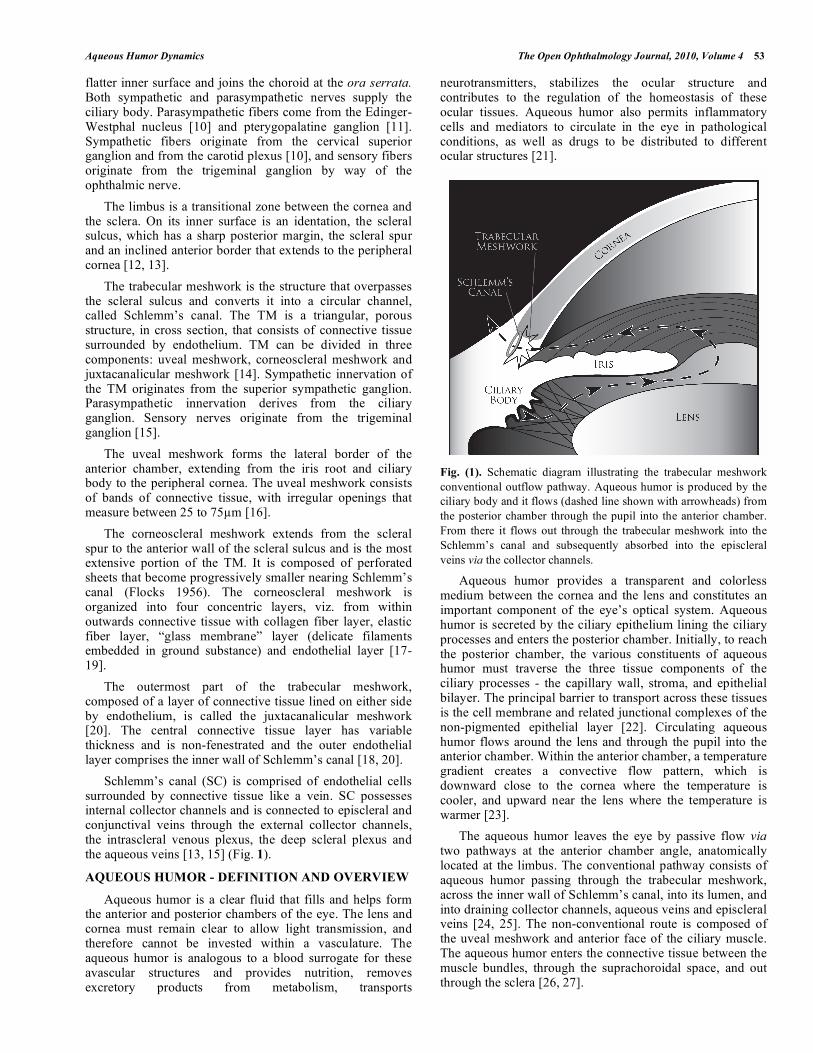
Schlemm’s canal (SC) is comprised of endothelial cells surrounded by connective tissue like a vein. SC possesses internal collector channels and is connected to episcleral and conjunctival veins through the external collector channels, the intrascleral venous plexus, the deep scleral plexus and the aqueous veins [13, 15] (Fig. 1).
AQUEOUS HUMOR - DEFINITION AND OVERVIEW
Aqueous humor is a clear fluid that fills and helps form the anterior and posterior chambers of the eye. The lens and cornea must remain clear to allow light transmission, and therefore cannot be invested within a vasculature. The aqueous humor is analogous to a blood surrogate for these avascular structures and provides nutrition, removes excretory products from metabolism, transports
neurotransmitters, stabilizes the ocular structure and contributes to the regulation of the homeostasis of these ocular tissues. Aqueous humor also permits inflammatory cells and mediators to circulate in the eye in pathological conditions, as well as drugs to be distributed to different ocular structures [21].
Fig. (1). Schematic diagram illustrating the trabecular meshwork
conventional outflow pathway. Aqueous humor is produced by the
ciliary body and it flows (dashed line shown with arrowheads) from
the posterior chamber through the pupil into the anterior chamber.
From there it flows out through the trabecular meshwork into the
Schlemm’s canal and subsequently absorbed into the episcleral
veins via the collector channels.
Aqueous humor provides a transparent and colorless medium between the cornea and the lens and constitutes an important component of the eye’s optical system. Aqueous humor is secreted by the ciliary epithelium lining the ciliary processes and enters the posterior chamber. Initially, to reach the posterior chamber, the various constituents of aqueous humor must traverse the three tissue components of the ciliary processes - the capillary wall, stroma, and epithelial bilayer. The principal barrier to transport across these tissues is the cell membrane and related junctional complexes of the non-pigmented epithelial layer [22]. Circulating aqueous humor flows around the lens and through the pupil into the anterior chamber. Within the anterior chamber, a temperature gradient creates a convective flow pattern, which is downward close to the cornea where the temperature is cooler, and upward near the lens where the temperature is warmer [23].
The aqueous humor leaves the eye by passive flow via two pathways at the anterior chamber angle, anatomically located at the limbus. The conventional pathway consists of aqueous humor passing through the trabecular meshwork, across the inner wall of Schlemm’s canal, into its lumen, and into draining collector channels, aqueous veins and episcleral veins [24, 25]. The non-conventional route is composed of the uveal meshwork and anterior face of the ciliary muscle. The aqueous humor enters the connective tissue between the muscle bundles, through the suprachoroidal space, and out through the sclera [26, 27].
54 The Open Ophthalmology Journal, 2010, Volume 4 Goel et al.
Equilibrium exists between the production and drainage of aqueous humor. Disruption of aqueous outflow, usually through the conventional pathway, results in elevation of IOP, which is a major risk factor in the pathogenesis of glaucoma [28].
AQUEOUS HUMOR – FORMATION AND COMPO-SITION
Three mechanisms are involved in aqueous humor formation: diffusion, ultrafiltration and active secretion [5]. The first two processes are passive and do not entail active cellular participation.
Diffusion occurs when solutes, especially lipid soluble substances, are transported through the lipid portions of the membrane of the tissues between the capillaries and the posterior chamber, proportional to a concentration gradient across the membrane [29].
Ultrafiltration is the flow of water and water-soluble substances, limited by size and charge, across fenestrated ciliary capillary endothelia into the ciliary stroma, in response to an osmotic gradient or hydrostatic pressure [29].
Diffusion and ultrafiltration are responsible for the accumulation of plasma ultrafitrate in the stroma, behind tight junctions of the non-pigmented epithelium, from which the posterior chamber aqueous humor is derived [30, 31].
Active secretion is thought to be the major contributor to aqueous formation, responsible for approximately 80% to 90% of the total aqueous humor formation [32, 33]. The main site for active transport is believed to be the non-pigmented epithelial cells. Active transport takes place through selective trans-cellular movement of anions, cations, and other molecules across a concentration gradient in blood-aqueous barrier. This is mediated by protein transporters distributed in the cellular membrane. Aquaporins (AQPs) are molecular water channels which aid with rapid bulk transport of fluid or transport of fluids against an insufficient osmotic pressure gap. Two AQP’s, AQP1 and AQP4, have been shown to contribute to aqueous humor secretion [34]. The energy required for the transport is generated by hydrolysis of adenosine triphosphate (ATP) to adenosine diphosphate (ADP), which is activated by Na
+
and K+ (66) mediated by Na
+-K
+-ATPase, an enzyme located
in both the non-pigmented and pigmented ciliary epithelia [35]. Na
+-K
+-ATPase can be inhibited by many different
molecules, including cardiac glycosides, dinitrophenol [36], vanadate [37], and possibly acetazolamide through pH changes [38]. Thus, Na
+-K
+-ATPase is of particular interest
in pharmacological studies of aqueous humor dynamics.
Another enzyme, carbonic anhydrase, found in the non-
pigmented and pigmented ciliary epithelia [39], mediates the
transport of bicarbonate across the ciliary epithelium by the
reversible hydration of CO2 to form HCO3-and protons
through the following reaction: CO2 + H2O H2CO3
HCO3- + H
+ [40]. Bicarbonate formation influences fluid
transport by affecting Na+, possibly by regulating the pH for
optimal active ion transport [38].
The movement of electrolytes across the ciliary epithelium is regulated by electrochemical gradients and, although there is a net direction of secretion across the
epithelium [32], hydrostatic and oncotic forces favor resorption of aqueous humor [41]. Chloride ion is the major anion transported across the epithelium through Cl
- channels
[35]. Other molecules are also actively transported, including ascorbic acid, which is secreted against a concentration gradient by sodium-dependent vitamin C transporter 2 (SVCT2) [42] and certain amino acids, which are secreted by at least three different solute carriers [43]. Active transport produces an osmotic gradient across the ciliary epithelium, which promotes the movement of other plasma constituents by ultrafiltration and diffusion [44].
The rate of aqueous humor turnover is estimated to be 1.0% to 1.5% of the anterior chamber volume per minute [32], which is 2.4 ± 0.6μl/min (mean ± SD, daytime measurements in adults aged 20–83 years) [45]. Using fluorophotometry, diurnal variations were observed in aqueous humor turnover rates, reflecting a pattern known as the circadian rhythm of aqueous humor flow in humans. Aqueous humor flow is higher in the morning than at night. Aqueous humor flow is normally about 3.0μl/min in the morning, 2.4μl/min in the afternoon, and drops to 1.5μl/min at night [45]. The mechanism that controls this biologic rhythm is poorly understood. Circulating epinephrine available to the ciliary epithelia may be a major driving force [46]. The effect of timolol, epinephrine and acetazolamide on the rate of aqueous humor flow through the anterior chamber has been studied. Epinephrine increased the rate of aqueous flow in sleeping subjects to a greater extent than it did in awake subjects. Timolol reduced the rate in awake individuals, but not in sleeping ones and acetazolamide reduced the rate of flow in both awake and epinephrine-stimulated subjects [47]. Norepinephrine has also been shown to stimulate aqueous flow, but not as effectively as epinephrine [46]. Another hypothesis supporting epinephrine influence on circadian rhythm could be a ciliary production of this hormone. However, epinephrine concentration in human aqueous humor appears to be very low, ranging from 0 to 0.1 ng/ml [48]. Moreover, in patients with surgical adrenalectomy or Horner syndrome (reduced or absent sympathetic innervation on one side), the circadian flow pattern was observed to be normal [49, 50]. Other hormones, such as melatonin, hormones related to pregnancy, and anti-diuretic hormones, do not appear to alter the normal circadian rhythm of the aqueous flow [46]
Measuring the relative concentrations of substances in the aqueous humor and the plasma, as well as in the anterior and posterior chambers, separately make it possible to obtain information about the composition of the aqueous humor [51]. Aqueous humor composition depends not only on the nature of its production, but also on the metabolic interchanges that occur within various tissues throughout its intraocular route [52].
The major components of the aqueous humor are organic and inorganic ions, carbohydrates, glutathione, urea, amino acids and proteins, oxygen, carbon dioxide and water. Aqueous humor is slightly hypertonic to plasma in a number of mammalian species [53-55], except from eyes of rhesus monkeys, in which no significant difference was observed [56]. When comparing anterior chamber and posterior chamber fluids separately, no differences were found in osmolarity, total concentration of dissolved substances or pH
Aqueous Humor Dynamics The Open Ophthalmology Journal, 2010, Volume 4 55
[57]. Most studies have shown the Na+
concentration in plasma and aqueous humor to be similar [57, 58]. The greatest differences in aqueous humor relative to plasma, are the concentrations of protein (200 times less) and ascorbate (20 to 50 times higher) [59]. The protein content of aqueous humor has both quantitative and qualitative differences compared to plasma. Most aqueous humor proteins are intrinsic glycoproteins of the vitreous, which are secretory products of the inner epithelial layer of the ciliary body [60]. Specific classes of immunoglobulins, such as IgG, were found to be in a higher concentration in the aqueous humor as compared to IgM and IgA levels [61, 62]. Relative concentrations of free amino acids vary, with ratios to plasma concentration ranging from 0.08 to 3.14, reinforcing the concept of active transport of amino acids [63]. Glucose and urea in the aqueous humor are approximately 80% of the plasma levels. Important anti-oxidant substances can also be found in the aqueous humor, such as glutathione (derived by diffusion from the blood) and ascorbate (which helps protect against light-induced oxidative damage) [32]. A number of molecules involved in the maintenance of the extracellular matrix, such as collagenase, have been identified in human aqueous humor, which may influence TM outflow resistance and, consequently, the IOP [64]. In addition, growth factors have been detected in the aqueous humor, as well as receptors for many of these factors on target tissues, including transferrin [65], transforming growth factors [66], endothelin-1 [67] and indoleamine 2,3-dioxygenase [68].
AQUEOUS HUMOR – OUTFLOW
As mentioned earlier, the aqueous humor exits the eye through both conventional and unconventional pathways. The physiology of these two routes differs in several important ways. For example, the outflow through the unconventional pathway is relatively independent of the intraocular pressure unlike the conventional pathway [69], their behavior in response to different pharmacological agents [70], also rate limiting step in unconventional pathway is flow through the ciliary muscle and in the conventional pathway it’s the flow through the inner wall of schlemm’s canal [71]. Regulation of the extracellular matrix (ECM) composition appears to influence aqueous humor outflow resistance in both the pathways [72].
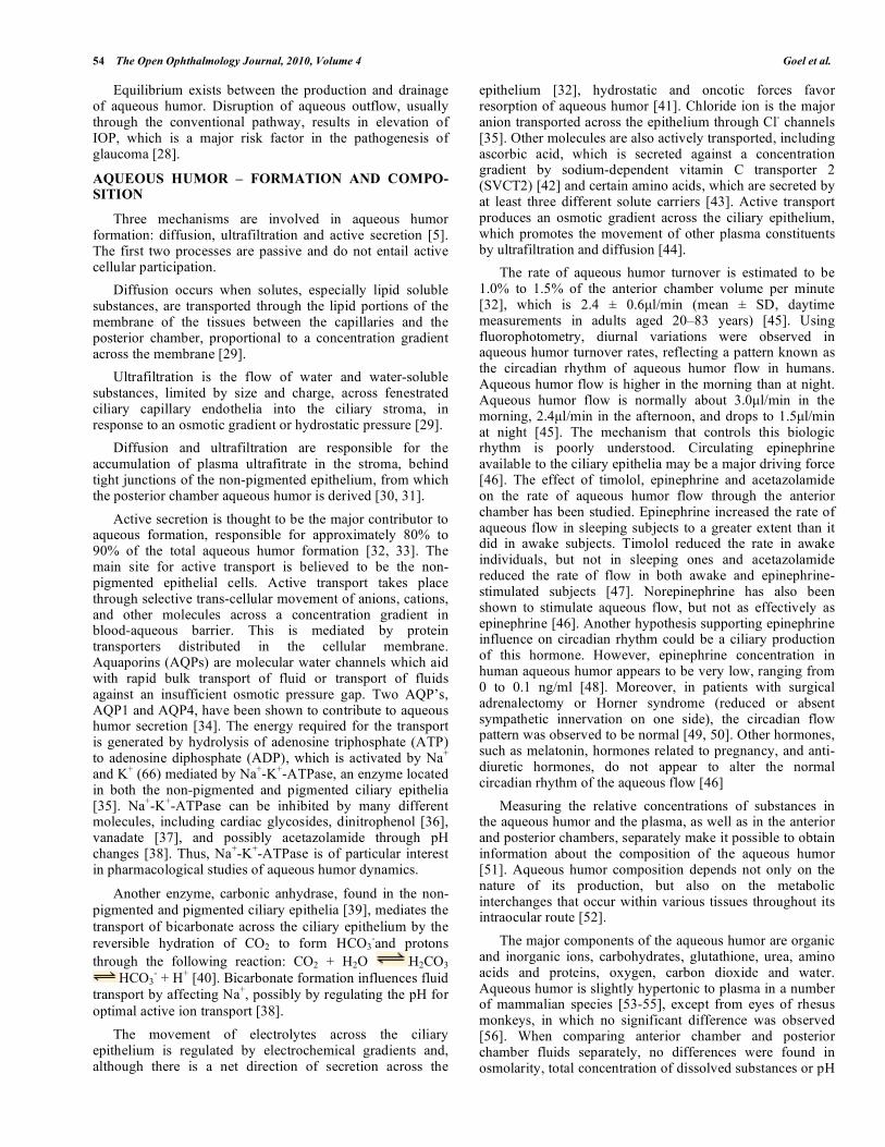
Another mechanism reported to influence aqueous humor outflow involves age-dependent changes. For instance, in the healthy aging human eye, a reduction in the production of aqueous humor is balanced by a reduction in its drainage through the uveoscleral outflow pathway, thereby leaving intraocular pressure relatively unchanged [73] (Fig. 2).
Fluid movement takes place down a pressure gradient from the TM into Schlemm’s canal and through the inner wall of Schlemm’s canal, following the conventional route, and appears to be a passive pressure-dependent transcellular mechanism, frequently associated with paracellular routes, such as giant vacuoles and pores acting as one-way valves [74]. These pores range in size from 0.1 to 3 m in diameter, and are the main passageway not only for aqueous humor, but also for particulate materials, such as cells, ferritin and microspheres [75-77]. Changes in IOP bring about changes in the structure of the endothelium lining Schlemm’s canal. Elevated IOP leads to an increase in the number and size of
these vacuoles, and vice versa [78, 79]. The inner wall of Schlemm’s canal is a complex tissue that is poorly understood - there is still doubt if it influences outflow facility in normal or glaucomatous eyes, even though circumstantial evidence points in that direction [80].
Fig. (2). Schematic diagram illustrating the uveoscleral outflow
pathway. Aqueous humor is produced by the ciliary body, in
uveoscleral route, it flows from the posterior chamber through the
pupil into the anterior chamber and then (shown by dashed lines
and arrowheads) through the face of the ciliary body and iris root to
the ciliary muscle and suprachoroidal space to either veins in the
choroid and sclera or through scleral pores to episcleral tissue.
After exiting Schlemm’s canal, the aqueous humor enters the aqueous veins and, subsequently, mixes with blood in the episcleral veins, where the pressure is approximately 8–10 mmHg [81, 82], and the resistance of the conventional aqueous drainage tissues is approximately 3–4 mmHg/ l/min. This results in an average IOP of 15.5 ± 2.6 mmHg (mean ± SD) for the general population [83].
In humans, 75% of the resistance to the aqueous humor outflow is localized to the TM, and 25% occurs beyond Schlemm’s canal [84]. On this basis, trabeculotomy and trabeculectomy were proposed as surgical therapies for treatment of POAG [85]. The major site of resistance within the TM structure has not yet been well characterized, but direct pressure measurements [86, 87] and circumstantial evidence [80] indicate that it resides in the juxtacanalicular portion [88, 89].
Some studies suggest that glycosaminoglycans, which constitute the fundamental substance of the ECM of the TM [90-92], are partly responsible for increased resistance to outflow. The osmotic forces exerted by glycosaminoglycans may induce hydration (edema) of the TM, which can cause obstruction of the trabecular structure [93]. Catabolic enzymes released from lysosomes depolymerize glycosaminoglycans and prevents this obstruction. This effect is also inhibited by corticosteroids, which prevent the release of the enzymes by stabilizing the lysosomal membranes and has been associated with a role in outflow obstruction and glaucoma pathogenesis [93, 94]. In
56 The Open Ophthalmology Journal, 2010, Volume 4 Goel et al.
glaucomatous eyes, an increase in the ECM thickness beneath the inner wall of Schlemm’s canal and in the juxtacanalicular meshwork compared with age-matched healthy controls has been observed [95]. Other studies suggest that the interaction of ECM components with different proteins may induce formation of deposits that obstruct aqueous humor outflow through the TM. For instance, proteomic analyses have identified cochlin, a protein of incompletely understood function, in the glaucomatous TM but not in healthy controls. Functionally, cochlin undergoes multimerization induced by shear stress and other changes in the microenvironment. Cochlin, along with mucopolysaccharide deposits, have been found exclusively in glaucomatous TM [96, 97].
The influence of the iris and ciliary muscle, two contractile structures innervated with cholinergic nerves, on the resistance to aqueous outflow has also been contemplated. The anterior tendons of the ciliary muscle insert into the outer portion of the corneoscleral meshwork and into the juxtacanalicular meshwork [98]. During contraction, the ciliary muscle moves in an anterior and inward direction, resulting in spreading of the TM and dilation of Schlemm’s canal, thus decreasing outflow resistance. During relaxation, the opposite occurs, thereby increasing outflow resistance [99]. Studies in various animal species demonstrated that voluntary accommodation, electrical stimulation of the trigeminal nerve, and local or systemic administration of cholinergic agents decrease outflow resistance [100-104].
Direct and indirect acting muscarinic cholinergic agonists have been used in the medical management of primary open angle glaucoma. Recent studies demonstrate the presence of at least two different subtypes of muscarinic receptors in the ciliary muscle and in the trabecular meshwork tissue or cell cultures from human eyes [105, 106]. In addition, cholinergic agonists, such as oxotremorine, induce the contraction of the ciliary muscle by binding selectively to receptors located in the longitudinal portion of the muscle, indicating that these agents may modulate the outflow facility independently from accommodation and miosis [105]. Ultrastructural and histochemical differences between the longitudinal (more relevant to outflow facility) and circular (more relevant to accommodation) portions of the ciliary muscle have been observed in monkey eyes. However, these differences in muscarinic receptor subtypes do not appear to play a role in dissociation of accommodative and outflow resistance responses [107, 108].
Uveoscleral outflow was described by observing the exit of radioactive tracers into the anterior chamber of the cynomologous monkey eye [109]. Further characterization of this pathway derives mostly from animal experiments and from mathematical calculations of an expanded Goldmann equation (i.e. F= (Pi-Pe) X C+U where F is the rate of aqueous humor formation, Pi is the intraocular pressure, Pe is the episcleral venous pressure, C is the tonographic facility of outflow and U is the pressure insensitive parameter to symbolize uveoscleral outflow) [109]. This model has been utilized for many years and, in the opinion of many investigators, views the aqueous outflow as passive fluid movement down a pressure gradient [110, 111]. The main obstacles in the assessing more accurately aqueous outflow
values with the Goldman equation are: the pressure of the episcleral venous plexus into which aqueous humor flows, and that uveoscleral flow must be independent of the IOP for the equation to be relevant. Although the relationship established is acceptable, it is oversimplified, since this formula implies the recipient pressure for all pressure-dependent outflows can be represented by a single episcleral vessel [69]. In monkeys, uveoscleral flow is not truly pressure-independent and the relationship between flow and IOP is not linear [70]. Ciliary muscle contraction greatly affects uveoscleral outflow [112], and prostaglandin F2 greatly increases uveoscleral outflow by decreasing the flow resistance of the interstitial spaces in the ciliary muscle [113, 114].
Some investigators consider uveoscleral outflow analogous to lymphatic drainage of tissue fluid in other organs, since the fluid may be drawn osmotically into the veins and may mix with tissue fluid from the ciliary muscle, ciliary processes and choroid [27]. In non-human primates, 40-50% of aqueous humor exits the eye by the uveoscleral route. In human eyes, most data has been collected by indirect calculations, with results suggesting a similar fraction, at least in eyes from younger individuals. An age-dependent reduction in uveoscleral flow in human eyes may explain the initial difference seen between non-human primate and human eyes [71]. In eyes treated with atropine, uveoscleral flow accounts for 4 to 27% of the total outflow, but with pilocarpine it was only 0 to 3% [115].
The biomechanics of aqueous humor flow within the anterior chamber also need to be considered. IOP is the loading force to which the outflow system normally responds. Evidence of tissue and cellular deformation in response to an IOP-induced load places TM resistance to IOP at the level of the endothelium of Schlemm’s canal. Schlemm’s canal endothelium attaches tightly to the TM by extending cytoplasmic processes into the juxtacanalicular space [78, 116, 117]. Well-characterized desmosomes, capable of sustaining cellular stress, are present between cell process attachments [118, 119]. The trabecular lamellae contain type I and III collagen, which provide structural support in tension, and elastin, and a recoverable response over large excursions. The organization and distribution of elastin in trabecular lamellae is similar to that found in tendons [120] and enables a mechanism for reversible deformation in response to cyclic hydrodynamic loading [78]. Progressive deformation of Schlem’s canal juxtacanalicular cells and trabecular lamellae with increasing IOP occurs in concert with progressive enlargement of the juxtacanalicular space. This movement causes cellular elements and ECM material to become less compact and progressively reduce the ability of the juxtacanalicular space to participate as a resistance element [78, 119, 121]. However, as IOP increases, resistance to aqueous outflow also increases [122, 123]. This makes the juxtacanalicular region an unlikely source of hydraulic resistance [124]. Trabecular and vascular endothelial cells are mechanosensors [125] that direct vessel wall self-organization [125] in order to optimize wall and shear stress. Pressure and shear stress-mediated signals in endothelia initiate a series of responses at the cellular, molecular, and genetic levels, and induce both rapid responses and slow adaptive changes that regulate pressure and flow [125, 126].
Aqueous Humor Dynamics The Open Ophthalmology Journal, 2010, Volume 4 57
These processes are not linear but are part of a highly complex interactive network [125] in which an alteration in any component requires a contemporaneous adjustment of numerous other components in an interactive fashion resulting in long-term homeostasis [127, 128].
Current pharmacological therapies for lowering the intraocular pressure in glaucoma include increasing aqueous humor outflow and suppression of aqueous humor production. For example, aqueous humor production is reduced by both topical and systemic carbonic anhydrase inhibitors which decrease the production of aqueous humor by the epithelial cells of the ciliary body. Aqueous humor outflow is increased by prostaglandin agonists that increase outflow mainly through the uveoscleral pathway, possibly through the activation of matrix metalloproteinases, and also through the trabecular meshwork. Other glaucoma medications such as adrenergic agonists decrease outflow resistance through mechanisms that are not completely clear.
Current surgical therapies also seek to lower the intraocular pressure by increasing aqueous outflow or decreasing aqueous humor production. For example, ciliary body ablative laser treatments such as cyclcophotocoagulation laser (either transcleral or endopscopic) seek to partially destroy the cililary body to decrease aqueous production. In contrast, laser trabeculoplasty modifies the trabecular meshwork (in a manner still not known) to decrease outflow resistance. Other surgeries such as trabeculectomy, glaucoma drainage implants, and glaucoma shunts (which penetrate the trabecular meshwork and cannulate Schlemm’s canal or create a path through the scleral wall) bypass outflow resistance by shunting aqueous humor through or around the trabecular meshwork. Newer procedures such as canaloplasty (which dilates Schlemm’s canal) and other shunts which seek to open the uveoscleral pathway by creating a mini-cyclodialysis cleft all involve increasing outflow by opening existing aqueous drainage pathways or creating new pathways.
ACKNOWLEDGEMENTS
Some of the pertinent original work in our laboratories were supported by National Institutes of Health Grants R01 EY16112, K08 EY016775, and P30 EY014801; a career development award (SKB) and an unrestricted grant to the University of Miami from Research to Prevent Blindness. We thank Dr. Carol Toris for her helpful comments on a previous version of the manuscript.
REFERENCES
[1] Thylefors B, Negrel AD, Pararajasegaram R, Dadzie KY. Global
data on blindness. Bull World Health Organ 1995; 73(1): 115-21. [2] Quigley HA. Number of people with glaucoma worldwide. Br J
Ophthalmol 1996; 80(5): 389-93. [3] Friedman DS, Wolfs RC, O'Colmain BJ, et al. Prevalence of open-
angle glaucoma among adults in the United States. Arch Ophthalmol 2004; 122(4): 532-8.
[4] Hollows FC, Graham PA. Intra-ocular pressure, glaucoma, and glaucoma suspects in a defined population. Br J Ophthalmol 1966;
50(10): 570-86. [5] Millar C, Kaufman PL. Aqueous humor: secretion and dynamics.
In: Tasman W, Jaeger EA, Eds. Duane's foundations of clinical ophthalmology. Philadelphia: Lippincott-Raven 1995.
[6] Hara K, Lutjen-Drecoll E, Prestele H, Rohen JW. Structural
differences between regions of the ciliary body in primates. Invest Ophthalmol Vis Sci 1977; 16(10): 912-24.
[7] Smelser GK. Electron microscopy of a typical epithelial cell and of the normal human ciliary process. Trans Am Acad Ophthalmol
Otolaryngol 1966; 70(5): 738-54. [8] Tormey JM. The ciliary epithelium: an attempt to correlate
structure and function. Trans Am Acad Ophthalmol Otolaryngol 1966; 70(5): 755-66.
[9] Ozanics V, Jakobiec FA. Prenatal development of the eye and its adnexa. In: Duane TD, Jaeger EA, Eds. Biomedical foundations of
ophthalmology. Philadelphia: Harper and Row 1982; p. 1-35. [10] Williams PL, Warwick R. The oculomotor nerve. In: Williams PL,
Warwick R, Eds. Functional neuroanatomy of man. Edinburgh: Churchill Livingstone 1975; pp. 999-1000.
[11] Ruskell GL. An ocular parasympathetic nerve pathway of facial nerve origin and its influence on intraocular pressure. Exp Eye Res
1970; 10(2): 319-30. [12] Thoft RA. The role of the limbus in ocular surface maintenance and
repair. Acta Ophthalmol Suppl 1989; 192: 91-4. [13] Hogan MH, Alvarado JA, Weddell JE. The limbus. In: Hogan MH,
Alvarado JA, Weddell JE, Eds. Histology of the human eye. Philadelphia: WB Saunders 1971.
[14] Van Buskirk EM. Clinical Atlas of Glaucoma. Philadelphia: WB Saunders 1986.
[15] Gong H, Tripathi RC, Tripathi BJ. Morphology of the aqueous outflow pathway. Microsc Res Tech 1996; 33(4): 336-67.
[16] Flocks M. The anatomy of the trabecular meshwork as seen in tangential section. AMA Arch Ophthalmol 1956; 56(5): 708-18.
[17] Ashton N, Brini A, Smith R. Anatomical studies of the trabecular meshwork of the normal human eye. Br J Ophthalmol 1956; 40(5):
257-82. [18] Fine BS. Structure of the trabecular meshwork and the canal of
Schlemm. Trans Am Acad Ophthalmol Otolaryngol 1966; 70(5): 777-90.
[19] Gong HY, Trinkaus-Randall V, Freddo TF. Ultrastructural immunocytochemical localization of elastin in normal human
trabecular meshwork. Curr Eye Res 1989; 8(10): 1071-82. [20] Fine BS. Observations on the drainage angle in man and rhesus
monkey: a concept of the pathogenesis of chronic simple glaucoma. a light and electron microscopic study. Invest Ophthalmol 1964; 3:
609-46. [21] Sires B. Orbital and ocular anatomy. In: Wright, Ed. Textbook of
Ophthalmology. Baltimore, MD: Williams and Wilkins 1997. [22] Hogan MH, Alvarado JA, Weddell JE. Histology of the human eye.
Philadelphia: WB Saunders 1971. [23] Heys JJ, Barocas VH. A boussinesq model of natural convection in
the human eye and the formation of Krukenberg's spindle. Ann Biomed Eng 2002; 30(3): 392-401.
[24] Goldmann H. Minute volume of the aqueous in the anterior chamber of the human eye in normal state and in primary
glaucoma. Ophthalmologica 1950; 120(1-2): 19-21. 25] Ascher KW. Veins of the aqueous humor in glaucoma. Boll Ocul
1954; 33(3): 129-44. [26] Bill A, Hellsing K. Production and drainage of aqueous humor in
the cynomolgus monkey (Macaca irus). Invest Ophthalmol 1965; 4(5): 920-6.
[27] Johnson M, Erickson K. Mechanisms and routes of aqueous humor drainage. In: Albert DM, Jakobiec FA, Eds. Principles and practice
of ophthalmology. Philadelphia: WB Saunders 2000. [28] Kass MA, Hart WM, Jr., Gordon M, Miller JP. Risk factors
favoring the development of glaucomatous visual field loss in ocular hypertension. Surv Ophthalmol 1980; 25(3): 155-62.
[29] Civan MM, Macknight AD. The ins and outs of aqueous humour secretion. Exp Eye Res 2004; 78(3): 625-31.
[30] Smith RS, Rudt LA. Ultrastructural studies of the blood-aqueous barrier. 2. The barrier to horseradish peroxidase in primates. Am J
Ophthalmol 1973; 76(6): 937-47. [31] Uusitalo R, Palkama A, Stjernschantz J. An electron microscopical
study of the blood-aqueous barrier in the ciliary body and iris of the rabbit. Exp Eye Res 1973; 17(1): 49-63.
[32] Gabelt BT, Kaufman PL. Aqueous humor hydrodynamics. In: Hart WM, Ed. Adler's Physiology of the Eye, 9th ed. St. Louis, MO:
Mosby; 2003. [33] Mark HH. Aqueous humor dynamics in historical perspective. Surv
Ophthalmol 2009; 55(1): 89-100.
58 The Open Ophthalmology Journal, 2010, Volume 4 Goel et al.
[34] Yamaguchi Y, Watanabe T, Hirakata A, Hida T. Localization and
ontogeny of aquaporin-1 and -4 expression in iris and ciliary epithelial cells in rats. Cell Tissue Res 2006; 325(1): 101-9.
[35] Coca-Prados M, Sanchez-Torres J. Molecular approaches to the study of the Na+-K+-ATPase and chloride channels in the ocular
ciliary epithelium. In: Civan MM, editor. The eye's aqueous humor. San Diego: Academic Press 1998.
[36] Cole DF. Ocular Fluids. In: Davson H, Ed. The Eye - Vegetative physiology and biochemistry. Orlando: Academic Press 1984.
[37] Becker B. Vanadate and aqueous humor dynamics. Proctor Lecture. Invest Ophthalmol Vis Sci 1980; 19(10): 1156-65.
[38] Maren TH. The rates of movement of Na+, Cl-, and HCO-3 from plasma to posterior chamber: effect of acetazolamide and relation
to the treatment of glaucoma. Invest Ophthalmol 1976; 15(5): 356-64.
[39] Dobbs PC, Epstein DL, Anderson PJ. Identification of isoenzyme C as the principal carbonic anhydrase in human ciliary processes.
Invest Ophthalmol Vis Sci 1979; 18(8): 867-70. [40] Wistrand PJ. Carbonic anhydrase in the anterior uvea of the rabbit.
Acta Physiol Scand 1951; 24(2-3): 145-8. [41] Bill A. The role of ciliary blood flow and ultrafiltration in aqueous
humor formation. Exp Eye Res 1973; 16(4): 287-98. [42] Tsukaguchi H, Tokui T, Mackenzie B, et al. A family of
mammalian Na+-dependent L-ascorbic acid transporters. Nature 1999; 399(6731): 70-5.
[43] Reddy VN. Dynamics of transport systems in the eye. Friedenwald Lecture. Invest Ophthalmol Vis Sci 1979; 18(10): 1000-18.
[44] Tornquist P, Alm A, Bill A. Permeability of ocular vessels and transport across the blood-retinal-barrier. Eye 1990; 4( Pt 2): 303-9.
[45] Brubaker RF. Measurement of aqueous flow by fluorophotometry. In: Ritch R, Shields MB, Krupin T, Eds. The Glaucomas. St. Louis:
Mosby 1989; p.p 337-44. [46] Brubaker RF. Clinical measurements of aqueous dynamics:
implication for addressing glaucoma. In: Civan MM, Ed. The eye's aqueous humor from secretion to glaucoma. New York: Academic
Press 1998. [47] Topper JE, Brubaker RF. Effects of timolol, epinephrine, and
acetazolamide on aqueous flow during sleep. Invest Ophthalmol Vis Sci 1985; 26(10): 1315-9.
[48] Cooper RL, Constable IJ, Davidson L. Aqueous humor catecholamines. Curr Eye Res 1984; 3(6): 809-13.
[49] Maus TL, Young WF, Jr., Brubaker RF. Aqueous flow in humans after adrenalectomy. Invest Ophthalmol Vis Sci 1994; 35(8): 3325-
31. [50] Larson RS, Brubaker RF. Isoproterenol stimulates aqueous flow in
humans with Horner's syndrome. Invest Ophthalmol Vis Sci 1988; 29(4): 621-5.
[51] Kinsey VE, Reddy DVN. Chemistry and dynamics of aqueous humor. In: Prince JH, Ed. The rabbit in eye research. Springfield,
IL: Charles C. Thomas 1964. [52] De Berardinis E, Tieri O, Iuglio N, Polzella A. The composition of
the aqueous humour of man in aphakia. Acta Ophthalmol (Copenh) 1966; 44(1): 64-8.
[53] Benham GH, Duke-Elder WS, Hodgson TH. The osmotic pressure of the aqueous humour in the normal and glaucomatous eye. J
Physiol 1938; 92(3): 355-60. [54] Kinsey VE. The chemical composition and the osmotic pressure of
the aqueous humor and plasma of the rabbit. J Gen Physiol 1951; 34(3): 389-402.
[55] Levene RZ. Osmolarity in the normal state and following acetazolamide. AMA Arch Ophthalmol 1958; 59(4): 597-602.
[56] Gaasterland DE, Pederson JE, MacLellan HM, Reddy VN. Rhesus monkey aqueous humor composition and a primate ocular
perfusate. Invest Ophthalmol Vis Sci 1979; 18(11): 1139-50. [57] Kinsey VE. Comparative chemistry of aqueous humor in posterior
and anterior chambers of rabbit eye, its physiologic significance. AMA Arch Ophthalmol 1953; 50(4): 401-17.
[58] Sears ML. The aqueous In: Moses RA, Ed. Adler's physiology of the eye, 7th ed. St. Louis, MO: Mosby 1981.
[59] Reiss GR, Werness PG, Zollman PE, Brubaker RF. Ascorbic acid levels in the aqueous humor of nocturnal and diurnal mammals.
Arch Ophthalmol 1986; 104(5): 753-5. [60] Haddad A, Laicine EM, de Almeida JC. Origin and renewal of the
intrinsic glycoproteins of the aqueous humor. Graefes Arch Clin Exp Ophthalmol 1991; 229(4): 371-9.
[61] Allansmith MR, Whitney CR, McClellan BH, Newman LP.
Immunoglobulins in the human eye. Location, type, and amount. Arch Ophthalmol 1973; 89(1): 36-45.
[62] McClellan BH, Whitney CR, Newman LP, Allansmith MR. Immunoglobulins in tears. Am J Ophthalmol 1973; 76(1): 89-101.
[63] Dickinson JC, Durham DG, Hamilton PB. Ion exchange chromatography of free amino acids in aqueous fluid and lens of
the human eye. Invest Ophthalmol 1968; 7(5): 551-63. [64] Vadillo-Ortega F, Gonzalez-Avila G, Chevez P, Abraham CR,
Montano M, Selman-Lama M. A latent collagenase in human aqueous humor. Invest Ophthalmol Vis Sci 1989; 30(2): 332-5.
[65] Tripathi RC, Borisuth NS, Tripathi BJ, Gotsis SS. Quantitative and qualitative analyses of transferrin in aqueous humor from patients
with primary and secondary glaucomas. Invest Ophthalmol Vis Sci 1992; 33(10): 2866-73.
[66] Cousins SW, McCabe MM, Danielpour D, Streilein JW. Identification of transforming growth factor-beta as an
immunosuppressive factor in aqueous humor. Invest Ophthalmol Vis Sci 1991; 32(8): 2201-11.
[67] Lepple-Wienhues A, Becker M, Stahl F, et al. Endothelin-like immunoreactivity in the aqueous humour and in conditioned
medium from cultured ciliary epithelial cells. Curr Eye Res 1992; 11(11): 1041-6.
[68] Malina HZ, Martin XD. Indoleamine 2,3-dioxygenase activity in the aqueous humor, iris/ciliary body, and retina of the bovine eye.
Graefes Arch Clin Exp Ophthalmol 1993; 231(8): 482-6. [69] Brubaker RF. Measurement of uveoscleral outflow in humans. J
Glaucoma 2001; 10(5 Suppl 1): S45-8. [70] Bill A. Some thoughts on the pressure dependence of uveoscleral
flow. J Glaucoma 2003; 12(1): 88-93. [71] Alm A, Nilsson SF. Uveoscleral outflow--a review. Exp Eye Res
2009; 88(4): 760-8. [72] Morrison JC, Acott TS. Anatomy and physiology of aqueous
humor outflow. In: Morrison JC, Pollack IP, Eds. Glaucoma - Science and Practice. New York: Thieme Medical Publishers Inc.;
2003; pp. 34-41. [73] Toris CB, Yablonski ME, Wang YL, Camras CB. Aqueous humor
dynamics in the aging human eye. Am J Ophthalmol 1999; 127(4): 407-12.
[74] Bill A, Svedbergh B. Scanning electron microscopic studies of the trabecular meshwork and the canal of Schlemm--an attempt to
localize the main resistance to outflow of aqueous humor in man. Acta Ophthalmol (Copenh) 1972; 50(3): 295-320.
[75] Inomata H, Bill A, Smelser GK. Aqueous humor pathways through the trabecular meshwork and into Schlemm's canal in the
cynomolgus monkey (Macaca irus). An electron microscopic study. Am J Ophthalmol 1972; 73(5): 760-89.
[76] Shabo AL, Maxwell DS. Erythrocyte passage into Schlemm's canal. Am J Ophthalmol 1972; 74(1): 169-71.
[77] Epstein DL, Rohen JW. Morphology of the trabecular meshwork and inner-wall endothelium after cationized ferritin perfusion in the
monkey eye. Invest Ophthalmol Vis Sci 1991; 32(1): 160-71. [78] Johnstone MA, Grant WG. Pressure-dependent changes in
structures of the aqueous outflow system of human and monkey eyes. Am J Ophthalmol 1973; 75(3): 365-83.
[79] Grierson I, Lee WR. Pressure effects on flow channels in the lining endothelium of Schlemm's canal. A quantitative study by
transmission electron microscopy. Acta Ophthalmol (Copenh) 1978; 56(6): 935-52.
[80] Ethier CR. The inner wall of Schlemm's canal. Exp Eye Res 2002; 74(2): 161-72.
[81] Phelps CD, Armaly MF. Measurement of episcleral venous pressure. Am J Ophthalmol 1978; 85(1): 35-42.
[82] Brubaker RF. Determination of episcleral venous pressure in the eye. A comparison of three methods. Arch Ophthalmol 1967;
77(1): 110-4. [83] Schottenstein EM. Intraocular pressure. In: Ritch R, Shields MB,
Krupin T, Eds. The glaucomas. St. Louis: Mosby 1989; pp. 301-17. [84] Grant WM. Futher studies on facility of flow through the trabecular
meshwork. Arch Ophthalmol 1958; 60: 523-33. [85] Sugar HS. Experimental trabeculectomy in glaucoma. Am J
Ophthalmol 1961; 51: 623-7. [86] Maepea O, Bill A. The pressures in the episcleral veins, Schlemm's
canal and the trabecular meshwork in monkeys: effects of changes in intraocular pressure. Exp Eye Res 1989; 49(4): 645-63.
Aqueous Humor Dynamics The Open Ophthalmology Journal, 2010, Volume 4 59
[87] Maepea O, Bill A. Pressures in the juxtacanalicular tissue and
Schlemm's canal in monkeys. Exp Eye Res 1992; 54(6): 879-83. [88] Ethier CR, Kamm RD, Palaszewski BA, Johnson MC, Richardson
TM. Calculations of flow resistance in the juxtacanalicular meshwork. Invest Ophthalmol Vis Sci 1986; 27(12): 1741-50.
[89] Seiler T, Wollensak J. The resistance of the trabecular meshwork to aqueous humor outflow. Graefes Arch Clin Exp Ophthalmol 1985;
223(2): 88-91. [90] Berggren L, Vrabec F. Demonstration of a coating substance in the
trabecular meshwork of the eye and its decrease after perfusion experiments with different kinds of hyaluronidase. Am J
Ophthalmol 1957; 44(2): 200-8. [91] Southren AL, Gordon GG, l'Hommedieu D, Ravikumar S, Dunn
MW, Weinstein BI. 5 beta-Dihydrocortisol: possible mediator of the ocular hypertension in glaucoma. Invest Ophthalmol Vis Sci
1985; 26(3): 393-5. [92] Zimmerman LE. Demonstration of hyaluronidase-sensitive acid
mucopolysaccharide in trabecula and iris in routine paraffin sections of adult human eyes; a preliminary report. Am J
Ophthalmol 1957; 44(1): 1-4. [93] Francois J. Corticosteroid glaucoma. Ann Ophthalmol 1977; 9(9):
1075-80. [94] Johnson DH, Bradley JM, Acott TS. The effect of dexamethasone
on glycosaminoglycans of human trabecular meshwork in perfusion organ culture. Invest Ophthalmol Vis Sci 1990; 31(12):
2568-71. [95] Lütgen-Drecoll E, Rohen JW. Morphology of aqueous outflow
pathways in normal and glaucomatous eyes. In: Ritch R, Shields MB, Krupin T, Eds. The glaucomas. St. Louis: Mosby 1989.
[96] Bhattacharya SK, Annangudi SP, Salomon RG, Kuchtey RW, Peachey NS, Crabb JW. Cochlin deposits in the trabecular
meshwork of the glaucomatous DBA/2J mouse. Exp Eye Res 2005; 80(5): 741-4.
[97] Bhattacharya SK, Rockwood EJ, Smith SD, et al. Proteomics reveal Cochlin deposits associated with glaucomatous trabecular
meshwork. J Biol Chem 2005; 280(7): 6080-4. [98] Rohen JW, Lutjen E, Barany E. The relation between the ciliary
muscle and the trabecular meshwork and its importance for the effect of miotics on aqueous outflow resistance. A study in two
contrasting monkey species, Macaca irus and Cercopithecus aethiops. Albrecht Von Graefes Arch Klin Exp Ophthalmol 1967;
172(1): 23-47. [99] Barany EH. The mode of action of miotics on outflow resistance. A
study of pilocarpine in the vervet monkey Cercopithecus ethiops. Trans Ophthalmol Soc UK 1966; 86: 539-78.
[100] Armaly MF. Studies on intraocular effects of the orbital parasympathetic pathway. III. Effect on steady-state dynamics.
Arch Ophthalmol 1959; 62: 817-27. [101] Armaly MF. Studies on intraocular effects of the orbital
parasympathetics. II. Effect on intraocular pressure. AMA Arch Ophthalmol 1959; 62(1): 117-24.
[102] Armaly MF. Studies on intraocular effects of the orbital parasympathetic pathway. I. Technique and effects on morphology.
AMA Arch Ophthalmol 1959; 61(1): 14-29. [103] Armaly MF, Burian HM. Changes in the tonogram during
accommodation. AMA Arch Ophthalmol 1958; 60(1): 60-9. [104] Barany E, Christensen RE. Cycloplegia and outflow resistance in
normal human and monkey eyes and in primary open-angle glaucoma. Arch Ophthalmol 1967; 77(6): 757-60.
[105] Gupta N, McAllister R, Drance SM, Rootman J, Cynader MS. Muscarinic receptor M1 and M2 subtypes in the human eye: QNB,
pirenzipine, oxotremorine, and AFDX-116 in vitro autoradiography. Br J Ophthalmol 1994; 78(7): 555-9.
[106] Gupta N, Drance SM, McAllister R, Prasad S, Rootman J, Cynader MS. Localization of M3 muscarinic receptor subtype and mRNA in
the human eye. Ophthalmic Res 1994; 26(4): 207-13. [107] Poyer JF, Gabelt BT, Kaufman PL. The effect of muscarinic
agonists and selective receptor subtype antagonists on the
contractile response of the isolated rhesus monkey ciliary muscle.
Exp Eye Res 1994; 59(6): 729-36. [108] Gabelt BT, Kaufman PL. Inhibition of aceclidine-stimulated
outflow facility, accommodation and miosis in rhesus monkeys by muscarinic receptor subtype antagonists. Exp Eye Res 1994; 58(5):
623-30. [109] Bill A. The aqueous humor drainage mechanism in the cynomolgus
monkey (Macaca irus) with evidence for unconventional routes. Invest Ophthalmol 1965; 4(5): 911-9.
[110] Kupfer C, Gaasterland D, Ross K. Studies of aqueous humor dynamics in man. II. Measurements in young normal subjects using
acetazolamide and L-epinephrine. Invest Ophthalmol 1971; 10(7): 523-33.
[111] Kupfer C, Ross K. Studies of aqueous humor dynamics in man. I. Measurements in young normal subjects. Invest Ophthalmol 1971;
10(7): 518-22. [112] Crawford K, Kaufman PL. Pilocarpine antagonizes prostaglandin
F2 alpha-induced ocular hypotension in monkeys. Evidence for enhancement of Uveoscleral outflow by prostaglandin F2 alpha.
Arch Ophthalmol 1987; 105(8): 1112-6. [113] Gabelt BT, Kaufman PL. Prostaglandin F2 alpha increases
uveoscleral outflow in the cynomolgus monkey. Exp Eye Res 1989; 49(3): 389-402.
[114] Lutjen-Drecoll E, Tamm E. Morphological study of the anterior segment of cynomolgus monkey eyes following treatment with
prostaglandin F2 alpha. Exp Eye Res 1988; 47(5): 761-9. [115] Bill A, Phillips CI. Uveoscleral drainage of aqueous humour in
human eyes. Exp Eye Res 1971; 12(3): 275-81. [116] Johnstone MA. The morphology of the aqueous outflow channels.
In: Drance SM, Ed. Glaucoma: Applied pharmacology in medical treatment of glaucoma. New York: Grune & Stratton 1984; pp. 87-
109. [117] Grierson I, Lee WR. Pressure-induced changes in the ultrastructure
of the endothelium lining Schlemm's canal. Am J Ophthalmol 1975; 80(5): 863-84.
[118] Grierson I, Lee WR. Junctions between the cells of the trabecular meshwork. Albrecht Von Graefes Arch Klin Exp Ophthalmol
1974; 192(2): 89-104. [119] Grierson I, Lee WR. The fine structure of the trabecular meshwork
at graded levels of intraocular pressure. (1) Pressure effects within the near-physiological range (8-30 mmHg). Exp Eye Res 1975;
20(6): 505-21. [120] Hernandez MR, Gong H. Extracellular matrix of trabecular
meshwork and optic nerve head. In: Ritch R, Shields MB, Krupin T, Eds. The Glaucomas. St. Louis: Mosby 1996.
[121] Grierson I, Lee WR. The fine structure of the trabecular meshwork at graded levels of intraocular pressure. (2) Pressures outside the
physiological range (0 and 50 mmHg). Exp Eye Res 1975; 20(6): 523-30.
[122] Kaufman PL. Pressure-dependent outflow. In: Ritch R, Shields MB, Krupin T, Eds. The Glaucomas. St. Louis: Mosby 1996; pp.
307-33. [123] Moses RA. The effect of intraocular pressure on resistance to
outflow. Surv Ophthalmol 1977; 22(2): 88-100. [124] Van Buskirk EM. Anatomic correlates of changing aqueous
outflow facility in excised human eyes. Invest Ophthalmol Vis Sci 1982; 22(5): 625-32.
[125] Ingber DE. Mechanical signaling and the cellular response to extracellular matrix in angiogenesis and cardiovascular physiology.
Circ Res 2002; 91(10): 877-87. [126] Davies PF, Barbee KA, Volin MV, et al. Spatial relationships in
early signaling events of flow-mediated endothelial mechanotransduction. Annu Rev Physiol 1997; 59: 527-49.
[127] de Jong H. Modeling and simulation of genetic regulatory systems: a literature review. J Comput Biol 2002; 9(1): 67-103.
[128] Ingber DE. Tensegrity II. How structural networks influence cellular information processing networks. J Cell Sci 2003; 116(Pt
8): 1397-408.
Received: March 3, 2010 Revised: April 6, 2010 Accepted: June 17, 2010
© Goel et al.; Licensee Bentham Open.
This is an open access article licensed under the terms of the Creative Commons Attribution Non-Commercial License (http: //creativecommons.org/licenses/by-
nc/3.0/) which permits unrestricted, non-commercial use, distribution and reproduction in any medium, provided the work is properly cited.